Note: Descriptions are shown in the official language in which they were submitted.
CA 02964798 2017-04-13
WO 2016/061547 PCT/US2015/056080
AUTOMATIC PATIENT POSITIONING WITHIN A LASER EYE SURGERY SYSTEM
RELATED APPLICATION
[0001] This application is a non-provisional application and claims the
benefit under 35
U.S.C. 119(e) of U.S. Provisional Application Serial No. 62/065,178, filed
October 17, 2014,
which is incorporated herein in its entirety as if fully set forth. Full Paris
Convention priority is
hereby expressly reserved.
FIELD OF THE INVENTION
[0002] The present application pertains to systems and methods for
automatically positioning
a patient relative to a laser-assisted eye surgery system.
BACKGROUND
[0003] A cataract is formed by opacification of the crystalline lens or
its envelope - the lens
capsule - of the eye. The cataract obstructs passage of light through the
lens. A cataract can vary in
degree from slight to complete opacity. Early in the development of an age-
related cataract, the
power of the lens may be increased, causing near-sightedness (myopia). Gradual
yellowing and
opacification of the lens may reduce the perception of blue colors as those
wavelengths are absorbed
and scattered within the crystalline lens. Cataract formation typically
progresses slowly resulting in
progressive vision loss. If left untreated, cataracts may cause blindness.
[0004] A common cataract treatment involves replacing the opaque
crystalline lens with an
artificial intraocular lens (TOL). Every year, an estimated 15 million
cataract surgeries are
performed worldwide. Traditionally, cataract surgery has been typically
performed using a
technique called phacoemulsification in which an ultrasonic tip with
associated irrigation and
aspiration ports is used to sculpt the relatively hard nucleus of the lens to
facilitate removal through
an opening made in the anterior lens capsule. Access to the lens nucleus can
be provided by
performing an anterior capsulotomy in which a small round hole is formed in
the anterior side of the
lens capsule using a surgical. Access to the lens nucleus can also be provided
by performing a
manual continuous curvilinear capsulorhexis (CCC) procedure. After removal of
the lens nucleus, a
synthetic foldable intraocular lens (TOL) can be inserted into the remaining
lens capsule of the eye.
-1-
CA 02964798 2017-04-13
WO 2016/061547 PCT/US2015/056080
[0005]
One of the most technically challenging and critical steps in the cataract
extraction
procedure is providing access to the lens nucleus for removal of the cataract
by phacoemulsification.
The desired outcome is to provide a smooth continuous circular opening through
which
phacoemulsification of the nucleus can be performed safely and easily, and
also through which an
intraocular lens may be easily inserted. Because of the criticality of this
step, some surgeons prefer a
surgical laser beam over manual tools like microkeratomes and forceps since
the laser beam can be
focused precisely on extremely small amounts of eye tissue, thereby enhancing
the accuracy and
reliability of the capsulotomy procedure.
[0006]
Many cataract patients also have refractive visual errors such as
astigmatism.
Astigmatism can occur when the corneal curvature is unequal. A toric IOL can
be used to correct
astigmatism, but requires precise rotational and central placement.
Additionally, IOLs are not
typically used for correction beyond 5D of astigmatism. Many patients,
however, have astigmatic
visual errors exceeding 5D. For these patients, higher correction beyond 5D
typically requires
reshaping the cornea to make it more spherical. There are numerous existing
approaches for
reshaping the cornea, including Corneaplasty, Astigmatic Keratotomy, Corneal
Relaxing Incision
(CRI), and Limbal Relaxing Incision (LRI). In Astigmatic Keratotomy, Corneal
Relaxing Incision
(CRI), and Limbal Relaxing Incision (LRI), corneal incisions are made in at a
depth in a well-
defined manner, allowing the cornea to change shape and become more spherical.
[0007]
Several commercial laser-assisted eye surgery systems are available to
facilitate
cataract removal and astigmatism correction. The CATALYS Precision Laser
System from Abbott
Medical Optics is indicated for anterior capsulotomy, phacofragmentation, and
the creation of single
plane and multi-plane arc cuts/incisions in the cornea to correct astigmatism.
The CATALYS
System uses a two-piece liquid-filled interface that docks with the patient's
eye and provides a clear
optical path for real-time video, OCT imaging, and laser treatment.
Aspects of the
CATALYS System are disclosed in US Patent No. 8,394,084, US Patent No.
8,500,724, US Patent
No. 8,425,497, U.S. Patent Publication 2014/0163534, U.S. Patent Application
Serial No.
14/256,307, filed April 18, 2014 (published as U.S. Patent Publication No. US
2015/0018674 on
January 15, 2015), and U.S. Patent Application Serial No. 14/255,430, filed
April 17, 2014
(published as U.S. Patent Publication No. 2014/0343541 on November 20, 2014),
the contents of all
of which are incorporated herein by reference as if fully set forth. Other
systems for laser cataract
surgery are the LenSx Laser from Alcon Laboratories, Inc., the LENSAR Laser
System from
-2-
CA 02964798 2017-04-13
WO 2016/061547 PCT/US2015/056080
LENSAR, Inc., and the VICTUS Femtosecond Laser Platform from TECHNOLAS Perfect
Vision
GmbH a Bausch + Lomb Company.
[0008] One drawback with current systems is the time spent on, and
ancillary equipment
required for, determining the astigmatic axis of the patient's eye relative to
the laser system. In
conventional keratometry using topography principles, the astigmatic axis of a
patient's eye is
determined by analyzing reflections off the eye from circles of light
typically generated by circular
patterns of LEDs. The accuracy of this method depends on a number of factors
that affect
measurements used in assessing the astigmatic power and axis. Consequently,
improved methods
and systems for positioning the patient and determining the astigmatic power
and axis of a patient's
eye during preparation for a laser-assisted surgery are needed.
SUMMARY
[0009] Hence, to obviate one or more problems due to limitations and
disadvantages of the
related art, one object of this disclosure provides improved laser eye surgery
systems, and related
methods. The laser eye surgery systems use a laser to form precise incisions
in the cornea, in the
lens capsule, and/or in the crystalline lens nucleus. In a preferred
embodiment, a laser eye surgery
system includes a laser cutting subsystem to produce a laser pulse treatment
beam to incise tissue
within the eye. It further includes an optical coherence tomography (OCT)
scanning subsystem to
measure the spatial disposition of external and internal structures of the eye
in which incisions can
be formed. The laser eye surgery system further includes an alignment
subsystem, shared optics
operable to scan the treatment beam, and an alignment subsystem relative to
the laser eye surgery
system. The alignment subsystem can include a video subsystem that can be used
to, for example,
provide images of the eye during docking of the eye to the laser eye surgery
system. In a preferred
embodiment, a liquid interface is used between a patient interface lens and
the eye. The use of the
liquid interface avoids imparting undesirable forces to the patient's eye,
thereby reducing patient
discomfort and folds in the cornea. The alignment and OCT subsystems may be
used to detect
structures involved with the patient interface.
[0010] The present application provides a number of techniques for
automatically
positioning the patient's eye with respect to the various subsystems in the
overall laser eye surgery
system. Preferably, the various subsystems are located within a single unit
under which the patient's
position in a prone position looking up. The patient lies on a horizontal
patient support chair that has
-3-
CA 02964798 2017-04-13
WO 2016/061547 PCT/US2015/056080
a mechanical positioning subsystem built into it controlled by the same
electronics which control the
various subsystems.
[0011] A first exemplary technique for automatically positioning the
patient utilizes a closed-
loop iterative method using the OCT subsystem to determine the Z-axis distance
from an objective
camera lens to the eye. The method involves using the OCT scanner to measure
the distance
between the lens and the eye, adjusting the vertical position of the patient
support chair, and then re-
measuring the distance between the lens and the eye. This sequence continues
until an optimum
distance between the lens and the eye is reached, depending on several
metrics. For example, an
optimum distance between the lens and the eye occurs when the reflection from
a pattern of circular
light dots on the eye is in focus or in extremely sharp contrast. The optimal
distance from the
camera objective to the cornea that maximizes the contrast of the reflection
may be pre-determined
by a theoretical analysis of the optical system.
[0012] In one embodiment, the laser eye surgery system incorporates an
optical coherence
tomography (OCT) subsystem that is used to sense the distance between a camera
objective on the
underside of the laser eye surgery system and the patient's eye. Control
electronics compare the
sensed distance with a pre-determined target distance, and reposition a
movable patient support
toward or away the camera objective until the sensed distance is at the pre-
determined target
distance. A subsequent measurement dependent upon the spacing between the
camera objective and
the patient's eye is performed, such as determining the astigmatic axis by
observing the reflection of
a plurality of point source LEDs, or dots, arranged in concentric rings off
the eye.
[0013] The second technique also uses a closed-loop iteration in which
the visible contrast
from the reflection of a series of LEDs off the eye is measured and maximized.
For instance, a
series of light circles comprising individual LEDs may be shone onto the eye
and the reflections
observed to determine the astigmatic axis of the eye. The automatic
positioning technique involves
moving the patient support chair in a first direction until the contrast of
the reflected LEDs is
maximized. The contrast can be maximized by subtraction of adjacent pixels or
by averaging power
of the midrange frequencies of the 2d Fourier after an annular mask is
applied.
[0014] A still further method of automatically positioning the patient
relative to the laser eye
system utilizes a phase detection subsystem to determine when the image of LED
dots is in focus.
When the split reflections in the face detector are co-registered, the image,
and therefore chair
position, is optimized. If not, the chair moves to co-register the split
reflections.
-4-
CA 02964798 2017-04-13
WO 2016/061547 PCT/US2015/056080
[0015] Finally, another method for automatically positioning a patient
uses a light field
measurement to gather the reflected image and then a processor calculates the
distance at which the
reflected rays would have to originate for the image to be in focus. This
distance can then be used to
drive the chair to the correct height.
[0016] This summary and the following detailed description are merely
exemplary,
illustrative, and explanatory, and are not intended to limit, but to provide
further explanation of the
invention as claimed. Additional features and advantages of the invention will
be set forth in the
descriptions that follow, and in part will be apparent from the description,
or may be learned by
practice of the invention. The objectives and other advantages of the
invention will be realized and
attained by the structure particularly pointed out in the written description,
claims and the appended
drawings.
INCORPORATION BY REFERENCE
[0017] All publications, patents, and patent applications mentioned in
this specification are
herein incorporated by reference to the same extent as if each individual
publication, patent, or
patent application was specifically and individually indicated to be
incorporated by reference.
BRIEF DESCRIPTION OF THE DRAWINGS
[0018] The novel features of the invention are set forth with
particularity in the appended
claims. A better understanding of the features and advantages of the present
invention will be
obtained by reference to the following detailed description that sets forth
illustrative embodiments,
in which the principles of the invention are utilized, and the accompanying
drawings of which:
[0019] FIG. 1 is a perspective view showing a laser eye surgery system,
in accordance with
the present application.
[0020] FIG. 2 is a simplified block diagram showing a top level view of
the configuration of
a laser eye surgery system, in accordance with the present application.
[0021] FIG. 3 is a patient interface mechanically coupled with an
overhead with a diagnostic
and interventional system.
[0022] FIG. 4 depicts one embodiment of a focusing lens placed into
direct contact with the
cornea and/or sclera of the eye and through which system optics view and act
on the eye.
-5-
CA 02964798 2017-04-13
WO 2016/061547 PCT/US2015/056080
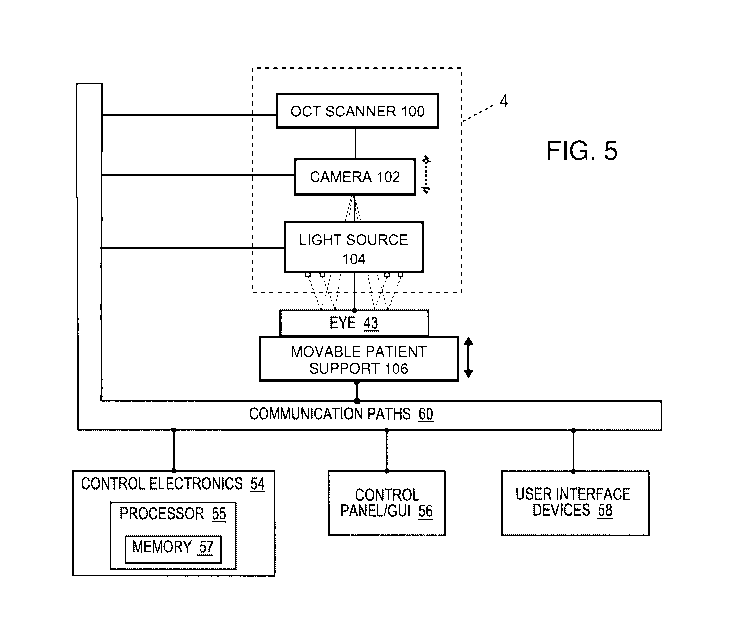
[0023] FIG. 5 is a simplified block diagram showing a top level view of
the configuration of
an automatic patient position system for determining the astigmatic axis
incorporated into the laser
eye surgery system of the present application.
[0024] FIG. 6 is a side view of a patient positioned under the diagnostic
and interventional
system during determination of the astigmatic axis.
[0025] FIG. 7 is a graph of set of 2000 A-scans taken at 1000Hz by an OCT
scanner when
measuring the initial distance from a camera objective to the eye.
[0026] FIGS. 8A, 8B, and 8C are different views of the reflected light
from the eye seen by a
camera within the diagnostic and interventional system of the present
application.
DETAILED DESCRIPTION
[0027] Methods and systems related to laser eye surgery are disclosed. A
laser is used to
form precise incisions in the cornea, in the lens capsule, and/or in the
crystalline lens nucleus. In a
preferred embodiment, a laser eye surgery system includes a laser cutting
subsystem to produce a
laser pulse treatment beam to incise tissue within the eye, a ranging
subsystem to measure the spatial
disposition of external and internal structures of the eye in which incisions
can be formed, an
alignment subsystem, and shared optics operable to scan the treatment beam, a
ranging subsystem
beam, and/or an alignment beam relative to the laser eye surgery system. The
alignment subsystem
can include a video subsystem that can be used to, for example, provide images
of the eye during
docking of the eye to the laser eye surgery system and also provide images of
the eye once the
docking process is complete. In a preferred embodiment, a liquid interface is
used between a patient
interface lens and the eye. The use of the liquid interface avoids imparting
undesirable forces to the
patient's eye.
[0028] The present application pertains to systems and methods for
automatically positioning
a patient relative to a laser-assisted eye surgery system. The positioning
techniques described herein
can be utilized for a number of purposes, including quickly establishing a
preferred distance between
the patient's eye and a camera while determining the astigmatic axis. Another
use is to rapidly locate
the patient's eye when measuring the power or curvature of the cornea. A
number of different
techniques are described herein, each of which can be used with various laser-
assisted ophthalmic
surgical systems, such as the commercial systems described in the background
discussion. A
preferred such commercial system is the CATALYS Precision Laser System from
Abbott Medical
-6-
CA 02964798 2017-04-13
WO 2016/061547 PCT/US2015/056080
Optics. Prior to a detailed description of the preferred auto-positioning
techniques, the main
components of laser-assisted ophthalmic surgical systems will be introduced.
[0029] Laser System Configuration
[0030] Figure 1 shows a laser eye surgery system 2, in accordance with
the present
application, operable to form precise incisions in the cornea, in the lens
capsule, and/or in the
crystalline lens nucleus. The system 2 includes a diagnostic and
interventional unit 4, a patient chair
6, a dual function footswitch 8, and a laser footswitch 10.
[0031] The diagnostic and interventional unit 4 houses many primary
subsystems of the
system 2. For example, externally visible subsystems include a touch-screen
control panel 12, a
patient interface assembly 14, patient interface vacuum connections 16, a
docking control keypad 18,
a patient interface radio frequency identification (RFID) reader 20, external
connections 22 (e.g.,
network, video output, footswitch, USB port, door interlock, and AC power),
laser emission
indicator 24, emergency laser stop button 26, key switch 28, and USB data
ports 30.
[0032] The patient chair 6 includes a base 32, a patient support bed 34,
a headrest 36, a
positioning mechanism (internal, not shown), and a patient chair joystick
control 38 disposed on the
headrest 36. The positioning control mechanism is coupled between the base 32
and the patient
support bed 34 and headrest 36. The patient chair 6 is configured to be
adjusted and oriented in
three axes (x, y, and z) using the patient chair joystick control 38. The
headrest 36 and a restrain
system (not shown, e.g., a restraint strap engaging the patient's forehead)
stabilize the patient's head
during the procedure. The headrest 36 includes an adjustable neck support to
provide patient
comfort and to reduce patient head movement. The headrest 36 is configured to
be vertically
adjustable to enable adjustment of the patient head position to provide
patient comfort and to
accommodate variation in patient head size.
[0033] The patient chair 6 allows for tilt articulation of the patient's
legs, torso, and head
using manual adjustments. The patient chair 6 accommodates a patient load
position, a suction ring
capture position, and a patient treat position. In the patient load position,
the chair 6 is rotated out
from under the diagnostic and interventional unit 4 with the patient chair
back in an upright position
and patient footrest in a lowered position. In the suction ring capture
position, the chair is rotated
out from under the diagnostic and interventional unit 4 with the patient chair
back in reclined
position and patient footrest in raised position. In the patient treat
position, the chair is rotated under
-7-
CA 02964798 2017-04-13
WO 2016/061547 PCT/US2015/056080
the diagnostic and interventional unit 4 with the patient chair back in
reclined position and patient
footrest in raised position.
[0034] The patient chair 6 is equipped with a "chair enable" feature to
protect against
unintended chair motion. The patient chair joystick 38 can be enabled in
either of two ways. First,
the patient chair joystick 38 incorporates a "chair enable" button located on
the top of the joystick.
Control of the position of the patient chair 6 via the joystick 38 can be
enabled by continuously
pressing the "chair enable" button. Alternately, the left foot switch 40 of
the dual function
footswitch 8 can be continuously depressed to enable positional control of the
patient chair 6 via the
joystick 38.
[0035] In a preferred embodiment, the patient control joystick 38 is a
proportional controller.
For example, moving the joystick a small amount can be used to cause the chair
to move slowly.
Moving the joystick a large amount can be used to cause the chair to move
faster. Holding the
joystick at its maximum travel limit can be used to cause the chair to move at
the maximum chair
speed. The available chair speed can be reduced as the patient approaches the
patient interface
assembly 14.
[0036] The emergency stop button 26 can be pushed to stop emission of all
laser output,
release vacuum that couples the patient to the system 2, and disable the
patient chair 6. The stop
button 26 is located on the system front panel, next to the key switch 28.
[0037] The key switch 28 can be used to enable the system 2. When in a
standby position,
the key can be removed and the system is disabled. When in a ready position,
the key enables power
to the system 2.
[0038] The dual function footswitch 8 is a dual footswitch assembly that
includes the left
foot switch 40 and a right foot switch 42. The left foot switch 40 is the
"chair enable" footswitch.
The right footswitch 42 is a "vacuum ON" footswitch that enables vacuum to
secure a liquid optics
interface suction ring to the patient's eye. The laser footswitch 10 is a
shrouded footswitch that
activates the treatment laser when depressed while the system is enabled.
[0039] In a preferred embodiment, the system 2 includes external
communication
connections. For example, the system 2 can include a network connection (e.g.,
an RJ45 network
connection) for connecting the system 2 to a network. The network connection
can be used to
enable network printing of treatment reports, remote access to view system
performance logs, and
remote access to perform system diagnostics. The system 2 can include a video
output port (e.g.,
-8-
CA 02964798 2017-04-13
WO 2016/061547 PCT/US2015/056080
HDMI) that can be used to output video of treatments performed by the system
2. The output video
can be displayed on an external monitor for, for example, viewing by family
members and/or
training. The output video can also be recorded for, for example, archival
purposes. The system 2
can include one or more data output ports (e.g., USB) to, for example, enable
export of treatment
reports to a data storage device. The treatments reports stored on the data
storage device can then be
accessed at a later time for any suitable purpose such as, for example,
printing from an external
computer in the case where the user without access to network based printing.
[0040]
Figure 2 shows a simplified block diagram of the system 2 coupled with a
patient eye
43. The patient eye 43 comprises a cornea, a lens, and an iris. The iris
defines a pupil of the eye 43
that may be used for alignment of eye 43 with system 2. The system 2 includes
a cutting laser
subsystem 44, a OCT imaging system 46, an alignment guidance system 48, shared
optics 50, a
patient interface 52, control electronics 54, a control panel/GUI 56, user
interface devices 58, and
communication paths 60. The control electronics 54 are operatively coupled via
the communication
paths 60 with the cutting laser subsystem 44, the OCT imaging system 46, the
alignment guidance
subsystem 48, the shared optics 50, the patient interface 52, the control
panel/GUI 56, and the user
interface devices 58.
[0041]
In a preferred embodiment, the cutting laser subsystem 44 incorporates
femtosecond
(FS) laser technology. By using femtosecond laser technology, a short duration
(e.g., approximately
10-13 seconds in duration) laser pulse (with energy level in the micro joule
range) can be delivered to
a tightly focused point to disrupt tissue, thereby substantially lowering the
energy level required as
compared to the level required for ultrasound fragmentation of the lens
nucleus and as compared to
laser pulses having longer durations.
[0042]
The cutting laser subsystem 44 can produce laser pulses having a wavelength
suitable to the configuration of the system 2. As a non-limiting example, the
system 2 can be
configured to use a cutting laser subsystem 44 that produces laser pulses
having a wavelength from
1020 nm to 1050 nm. For example, the cutting laser subsystem 44 can have a
diode-pumped solid-
state configuration with a 1030 (+/- 5) nm center wavelength.
[0043]
The cutting laser subsystem 44 can include control and conditioning
components.
For example, such control components can include components such as a beam
attenuator to control
the energy of the laser pulse and the average power of the pulse train, a
fixed aperture to control the
cross-sectional spatial extent of the beam containing the laser pulses, one or
more power monitors to
-9-
CA 02964798 2017-04-13
WO 2016/061547 PCT/US2015/056080
monitor the flux and repetition rate of the beam train and therefore the
energy of the laser pulses, and
a shutter to allow/block transmission of the laser pulses. Such conditioning
components can include
an adjustable zoom assembly to adapt the beam containing the laser pulses to
the characteristics of
the system 2 and a fixed optical relay to transfer the laser pulses over a
distance while
accommodating laser pulse beam positional and/or directional variability,
thereby providing
increased tolerance for component variation.
[0044] The OCT imaging system 46 is configured to measure the spatial
disposition of eye
structures in three dimensions. The measured eye structures can include the
anterior and posterior
surfaces of the cornea, the anterior and posterior portions of the lens
capsule, the iris, and the limbus.
In a preferred embodiment, the OCT imaging system 46 utilizes optical
coherence tomography
(OCT) imaging. As a non-limiting example, the system 2 can be configured to
use an OCT imaging
system employing wavelengths from 780 nm to 970 nm. For example, the OCT
imaging system 46
can include an OCT imaging system that employs a broad spectrum of wavelengths
from 810 nm to
850 nm. Such an OCT imaging system can employ a reference path length that is
adjustable to
adjust the effective depth in the eye of the OCT measurement, thereby allowing
the measurement of
system components including features of the patient interface that lie
anterior to the cornea of the
eye and structures of the eye that range in depth from the anterior surface of
the cornea to the
posterior portion of the lens capsule and beyond.
[0045] There are many suitable possibilities for the configuration of the
OCT imaging
system. For example, alternative suitable configurations include time and
frequency domain
approaches, single and dual beam methods, swept source, etc., such as those
described in U.S. Patent
Nos. 5,748,898; 5,748,352; 5,459,570; 6,111,645; and 6,053,613.
[0046] The alignment guidance subsystem 48 can include a laser diode or
gas laser that
produces a laser beam used to align optical components of the system 2. The
alignment guidance
subsystem 48 can include LEDs or lasers that produce a fixation light to
assist in aligning and
stabilizing the patient's eye during docking and treatment. The alignment
guidance subsystem 48
can include a laser or LED light source and a detector to monitor the
alignment and stability of the
actuators used to position the beam in X, Y, and Z. The alignment guidance
subsystem 48 can
include a video system that can be used to provide imaging of the patient's
eye to facilitate docking
of the patient's eye 43 to the patient interface 52. The imaging system
provided by the video system
can also be used to direct via the GUI the location of cuts. The imaging
provided by the video
-10-
CA 02964798 2017-04-13
WO 2016/061547 PCT/US2015/056080
system can additionally be used during the laser eye surgery procedure to
monitor the progress of the
procedure, to track movements of the patient's eye 43 during the procedure,
and to measure the
location and size of structures of the eye such as the pupil and/or limbus.
[0047] The shared optics 50 provides a common propagation path that is
disposed between
the patient interface 52 and each of the cutting laser subsystem 44, the OCT
imaging system 46, and
the alignment guidance subsystem 48. In a preferred embodiment, the shared
optics 50 includes
beam combiners to receive the emission from the respective subsystem (e.g.,
the cutting laser
subsystem 44, and the alignment guidance subsystem 48) and redirect the
emission along the
common propagation path to the patient interface. In a preferred embodiment,
the shared optics 50
includes an objective lens assembly that focuses each laser pulse into a focal
point. In a preferred
embodiment, the shared optics 50 includes scanning mechanisms operable to scan
the respective
emission in three dimensions. For example, the shared optics can include an XY-
scan mechanism(s)
and a Z-scan mechanism. The XY-scan mechanism(s) can be used to scan the
respective emission in
two dimensions transverse to the propagation direction of the respective
emission. The Z-scan
mechanism can be used to vary the depth of the focal point within the eye 43.
In a preferred
embodiment, the scanning mechanisms are disposed between the laser diode and
the objective lens
such that the scanning mechanisms are used to scan the alignment laser beam
produced by the laser
diode. In contrast, In a preferred embodiment, the video system is disposed
between the scanning
mechanisms and the objective lens such that the scanning mechanisms do not
affect the image
obtained by the video system.
[0048] The patient interface 52 is used to restrain the position of the
patient's eye 43 relative
to the system 2. In a preferred embodiment, the patient interface 52 employs a
suction ring that is
vacuum attached to the patient's eye 43. The suction ring is then coupled with
the patient interface
52, for example, using vacuum to secure the suction ring to the patient
interface 52. In a preferred
embodiment, the patient interface 52 includes an optically transmissive
structure (lens) having a
posterior surface that is displaced vertically from the anterior surface of
the patient's cornea and a
region of a suitable liquid (e.g., a sterile buffered saline solution (BSS))
is disposed between and in
contact with the posterior surface and the patient's cornea and forms part of
a transmission path
between the shared optics 50 and the patient's eye 43. The optically
transmissive structure may
comprise a lens 84 (see FIG. 3) having one or more curved surfaces.
Alternatively, the patient
interface 52 may comprise an optically transmissive structure having one or
more substantially flat
-11-
CA 02964798 2017-04-13
WO 2016/061547 PCT/US2015/056080
surfaces such as a parallel plate or wedge. In a preferred embodiment, the
patient interface lens is
disposable and can be replaced at any suitable interval, such as before each
eye treatment.
[0049] The control electronics 54 controls the operation of and can
receive input from the
cutting laser subsystem 44, the OCT imaging system 46, the alignment guidance
subsystem 48, the
patient interface 52, the control panel/GUI 56, and the user interface devices
58 via the
communication paths 60. The communication paths 60 can be implemented in any
suitable
configuration, including any suitable shared or dedicated communication paths
between the control
electronics 54 and the respective system components.
[0050] The control electronics 54 can include any suitable components,
such as one or more
processor, one or more field-programmable gate array (FPGA), and one or more
memory storage
devices. In a preferred embodiment, the control electronics 54 controls the
control panel/GUI 56 to
provide for pre-procedure planning according to user specified treatment
parameters as well as to
provide user control over the laser eye surgery procedure.
[0051] The control electronics 54 may comprise a processor/controller 55
(referred to herein
as a processor) that is used to perform calculations related to system
operation and provide control
signals to the various system elements. A computer readable medium 57 (also
referred to as a
database or a memory) is coupled to the processor 55 in order to store data
used by the processor and
other system elements. The processor 55 interacts with the other components of
the system as
described more fully throughout the present specification. In an embodiment,
the memory 57 can
include a look up table that can be utilized to control one or more components
of the laser system as
described herein.
[0052] The processor 55 can be a general purpose microprocessor
configured to execute
instructions and data, such as a Pentium processor manufactured by the Intel
Corporation of Santa
Clara, California. It can also be an Application Specific Integrated Circuit
(ASIC) that embodies at
least part of the instructions for performing the method in accordance with
the embodiments of the
present disclosure in software, firmware and/or hardware. As an example, such
processors include
dedicated circuitry, ASICs, combinatorial logic, other programmable
processors, combinations
thereof, and the like.
[0053] The memory 57 can be local or distributed as appropriate to the
particular application.
Memory 57 may include a number of memories including a main random access
memory (RAM) for
storage of instructions and data during program execution and a read only
memory (ROM) in which
-12-
CA 02964798 2017-04-13
WO 2016/061547 PCT/US2015/056080
fixed instructions are stored. Thus, memory 57 provides persistent (non-
volatile) storage for
program and data files, and may include a hard disk drive, flash memory, a
floppy disk drive along
with associated removable media, a Compact Disk Read Only Memory (CD-ROM)
drive, an optical
drive, removable media cartridges, and other like storage media.
[0054] The user interface devices 58 can include any suitable user input
device suitable to
provide user input to the control electronics 54. For example, the user
interface devices 58 can
include devices such as, for example, the dual function footswitch 8, the
laser footswitch 10, the
docking control keypad 18, the patient interface radio frequency
identification (RFID) reader 20, the
emergency laser stop button 26, the key switch 28, and the patient chair
joystick control 38.
[0055] Referring to FIG. 3, one embodiment of a patient interface 52 is
shown interfaced
with a diagnostic and interventional unit 4 such as that described in
reference to FIG. 1, the patient
interface 52 comprising an interfacial seal configuration 80 in contact with
the eye 43, a conical
lower housing portion 82 which houses a focusing lens 84, and a cylindrical
upper housing portion
86 with a proximal aspect configured to mechanically interface and couple with
the diagnostic and
interventional unit 4. Preferably, in the depicted embodiment and other
illustrative embodiments that
follow, the patient interface 52 is coupled to the diagnostic and
interventional unit 4 with a load
sensing interface, such as a platform comprising one or more load cells or
load sensors (such as
MEMS load sensors available from Honeywell, Inc.) configured to provide the
operator with output
signals or feedback regarding loads being applied at such interface due to
coupling with the eye of
the patient (i.e., such loads may be monitored since they are representative
of contact loads applied
to the eye of the patient by the patient interface assembly 52. This feedback
may be presented to the
user on the control panel/GUI 56 (FIG. 2) for use in adjusting the
directionality of positioning
control mechanism between the base 32 and the patient support bed 34 and
headrest 36 (FIG. 1)
during patient coupling to the system.
[0056] FIG. 4 depicts one embodiment of a focusing lens 84 configuration
wherein a distal
aspect 90 of the lens 84 is placed into direct contact with the cornea and/or
sclera 92 of the eye 43.
The scanned beam 94 of the cutting laser subsystem 44 exits the unit 4 crosses
the proximal surface
96 of the lens 84, passes through the lens, exits across the distal surface
90, crosses the cornea and/or
sclera 92, and eventually reaches the crystalline lens 98 to facilitate
interventional steps such as
capsulorhexis .
-13-
CA 02964798 2017-04-13
WO 2016/061547 PCT/US2015/056080
[0057] Automatic Patient Positioning
[0058] It should be understood that the preceding discussion of an
exemplary laser assisted
eye surgery system provides the context in which the present application is
useful. For instance, the
automatic positioning techniques described herein may be used to more
accurately and quickly
determine the astigmatic axis of the patient. Consequently, the subsequent
steps of using the cutting
laser subsystem 44 via the patient interface assembly 52 are done with the
knowledge of the location
of the astigmatic axis. The present automatic positioning techniques are
accomplished prior to
engaging the patient interface assembly 52, as shown, and require no
additional equipment from
what is already used for the laser surgery steps. Moreover, it is believed
that the automatic
positioning techniques are more accurate than those used previously, and
therefore the location of
the astigmatic axis is more accurate, leading to better outcomes.
[0059] FIG. 5 is a simplified block diagram showing a top level view of
the configuration of
an automatic patient position system for setting the patient position (and
then determining the
astigmatic axis) incorporated into the laser eye surgery system of the present
application. The
aforementioned diagnostic and interventional unit 4 is shown housing the OCT
scanner 100, a
camera 102 which may be a video camera, and a light source 104 having a
plurality of concentric
circles of point source LEDs, or dots, which shine downward toward the eye 43
of the patient. As
mentioned previously, the patient resides upon a patient support bed 34 having
a headrest 36 and an
internal positioning mechanism (FIG. 1), which are here symbolically
represented by a movable
patient support 106. A double-headed vertical arrow next to the patient
support 106 indicates it may
be automatically vertically adjusted, but it should also be understood it may
be capable of horizontal
adjustment. Further, a dashed-line double-headed vertical arrow next to the
camera 102 is intended
to indicate that it may be moved relative to a stationary patient support 106
in the alternative to
establish the desired spacing. Each of these subsystems or components are in
communication with
the control electronics 54 of the system, and are instructed and monitored by
the control panel 56
and user interface devices 58.
[0060] FIG. 6 is a side view of a patient positioned under the diagnostic
and interventional
system 4 during determination of the astigmatic axis. The OCT scanner 100 is
focused downward
toward the eye 43 through the center of the light source 104 by virtue of
being positioned directly
above the light source 104 or through the shared optics 50 as described above.
Likewise, an
objective 108 of the camera 102 is aimed directly downward at the eye 43
through the center of light
-14-
CA 02964798 2017-04-13
WO 2016/061547 PCT/US2015/056080
source 104. The camera 102 thus provides an image of the eye, viewed from
above, to capture and
evaluate the reflections off the eye 43 from the individual LEDs in the light
source 104. This
captures the topography of the cornea and provides a steep axis measurement of
more conical
corneas. An optimal distance from the camera objective 108 to the cornea that
maximizes the
contrast of the reflection of the light source 104 may be pre-determined by a
theoretical analysis of
the optical system, or through simulation.
[0061] The primary benefit of distance optimization is in accurate
determination of the
corneal astigmatic power and axis. A significant source of error in corneal
astigmatic measurements
is the knowledge of the location of the cornea relative to the machine. An
exact knowledge of the
location of the cornea affects the subsequent calculation, in addition to a
precise focus of the
reflection of the light source 104. In this regard, establishing an accurate
distance from the camera
objective 108 to the cornea is a primary concern, while maximizing focus is
secondary.
Furthermore, adjustment of the distance between the eye and the objective may
be done by moving
the chair or the objective relative to one another, as mentioned above.
Likewise, the OCT may be
used just to measure the distance, without the need to move anything else.
[0062] An exemplary technique for automatically positioning the patient
to focus the
reflections of the light source LEDs on the camera objective 108 comprises a
closed-loop iteration
using the OCT as a position sensor to drive the vertical motion of the movable
patient support 106,
or chair. First, a set of 2000 A-scans taken at 1000Hz measures the initial
distance from the
objective to the eye. The focus of the OCT 100 is always placed as close as
possible to the zero
"Optical Path Distance." The "Optical Path Distance" is changed with the Zed
encoder to control
the window of space imaged by the OCT. The "Optical Path Distance" is
continuously swept
through a range of 20mm, so that every A-scan images a progressively deeper
area than the previous.
A stationary object, such as the cornea of the eye 43 will look like an
inclined line using this Zed
movement. Because the OCT images are the addition of the real and imaginary
components of the
spectrum, they display an addition of what is below the zero "Optical Path
Length" and above it,
making the stationary object look like an inverted "V", as indicated in FIG.
7, which is easily
detectable. The vertex of the inverted "V" is located in the A-scan acquired
when the zero "Optical
Path Length" was at the eye surface. This method produces a very accurate
measurement of the eye
distance to the objective 108 of the camera 102, whose position is fixed and
known with respect to
the OCT scanner lens.
-15-
CA 02964798 2017-04-13
WO 2016/061547 PCT/US2015/056080
[0063] In a preferred embodiment, the physician presses and maintains
pressed a control,
such as a button on the joystick control 38, to start the OCT controlled chair
motion. The next step
is to move the chair 106 consistent with the detected position. Since the
chair speed depends on the
patient's weight and has a system¨to-system variability, the final location
may be slightly off from
the target. In this case a second iteration of the process above described is
implemented. For the
second and subsequent iteration (in case they were needed) only 1000A-scans
are used. Shorter
OCT time is justified because there is now a better estimate of the location
of the eye 43. Preferably,
the iterative algorithm works only while the doctor is pressing the control,
and if the doctor stops
pressing that control, the chair motion stops. The chair 106 also stops when
it gets to the pre-
determined optimal location where the reflection of the LEDs is focused or has
the greatest
sharpness, even if the control button is still pressed. During this process
force sensors (not shown)
in the objective 108 are closely monitored for contact of the patient with the
objective, in which case
the chair is backed away from the objective and the iteration is stopped. Once
the eye is measured
by the OCT to be within a tolerance from the pre-determined target location,
the physician gets a
visual that she/he can proceed to perform the function whose outcome depends
on a proper
positioning between the eye 43 and the objective 108.
[0064] That is, the system measures the astigmatic axis, measures the
power or curvature of
the cornea, or performs some other such function. For example, the camera 102
captures an image
of the reflection from the light source 104 off the eye 43. The image can then
be viewed on the
control panel 56 for direct evaluation by the physician, and is internally
stored for use by the control
electronics 54 such as when controlling the action of the cutting laser
subsystem 44. FIGS. 8A, 8B,
and 8C are different views of the reflected light from the light source 104
off the eye 43 as seen by
the camera 102 within the diagnostic and interventional system 4. The images
show 4 concentric
rings of dots, or LED point reflections, though more or less rings may be
used. A preferred
embodiment is between 2-4 rings. FIG. 8A illustrates a relatively even,
concentric pattern of circles
reflected back from the circular arrays of LEDs, indicating little or no
astigmatism. FIG. 8B shows a
pattern of reflected LED dots that is slightly offset from a central axis and
oval, indicating slight
stigmatism. Finally, FIG. 8C shows a pattern which is highly irregular and
offset from a central
axis, indicating severe astigmatism. In each case, the orientation of the axis
of the astigmatism is
determined from the pattern of reflected dots, and the result is stored in the
memory 57 of the laser
eye surgery system.
-16-
CA 02964798 2017-04-13
WO 2016/061547 PCT/US2015/056080
[0065] An alternative technique for automatically positioning the patient
relative to the laser
system is to compute a contrast metric from the part of the video image that
contains the reflection of
the LEDs. The patient support 106 is then moved away from the objective 108 a
short distance and
the contrast measured again. If the contrast improves, the system then
continues to move the chair
106 in the same direction until the contrast gets worst, then backs up to the
previous maximum
contrast location. If, on the other hand, the contrast gets worst, then the
system moves the chair 106
towards the objective 108 until the contrast decreases. Then back to the
maximum contrast location.
This process is analogous to the autofocus feature that some cameras have.
[0066] A third method, also used in some camera autofocusing mechanisms,
is phase
detection. A partial beam splitter picks off some light from the reflected
image and directs it to a
number of micro lens pairs. These lenses project to small, independent
sensors. Their data can be
compared, such as with cross-correlation. Since these pairs of sensors
correspond to specific regions
in the camera's 102 field of view, the direction in which to move into focus
can be found directly.
Although more involved, this allows for faster focus finding and positioning.
[0067] A fourth method utilizes a light field camera to measure the
reflected image. These
kinds of cameras can produce several images, each with a different focal
plane, from a single
measurement. They do this by measuring not only the position of incoming light
rays, but also the
angle from which they came, using a microlens array in front of the sensor.
From this data, the plane
in which the dots are in focus can be calculated, yielding the current
distance of the dots from the
camera sensor. This distance can then be used to tell the chair to move
directly in the correct
direction. Similar to phase detection, this method also allows for direction
movement from the data,
in addition to looser tolerance of initial positioning, but at the cost of
greater computational
complexity and computation time.
[0068] All of these methods can be used in applications where iris
registration is required as
well (e.g. the VISX excimer laser). Instead of converging towards a reflection
of dots on the cornea,
the feedback of information would drive the patient chair to bring the iris
plane into sharpest focus.
[0069] The use of the terms "a" and "an" and "the" and similar referents
in the context of
describing the invention (especially in the context of the following claims)
are to be construed to
cover both the singular and the plural, unless otherwise indicated here or
clearly contradicted by
context. The terms "comprising," "having," "including," and "containing" are
to be construed as
open-ended terms (i.e., meaning "including, but not limited to,") unless
otherwise noted. The term
-17-
CA 02964798 2017-04-13
WO 2016/061547 PCT/US2015/056080
"connected" is to be construed as partly or wholly contained within, attached
to, or joined together,
even if there is something intervening. Recitation of ranges of values here
are merely intended to
serve as a shorthand method of referring individually to each separate value
falling within the range,
unless otherwise indicated herein, and each separate value is incorporated
into the specification as if
it were individually recited herein. All methods described here can be
performed in any suitable
order unless otherwise indicated here or otherwise clearly contradicted by
context. The use of any
and all examples, or exemplary language (e.g., "such as") provided herein, is
intended merely to
better illuminate embodiments of the invention, and does not pose a limitation
on the scope of the
invention unless otherwise claimed. No language in the specification should be
construed as
indicating any non-claimed element as essential to the practice of the
invention.
[0070] While preferred illustrated embodiments of this disclosure have
been shown and
described in an exemplary form with a certain degree of particularity, those
skilled in the art will
understand that the embodiments are provided by way of example only, and that
various variations
can be made without departing from the spirit or scope of the invention. Thus,
it is intended that this
disclosure cover all modifications, alternative constructions, changes,
substitutions, variations, as
well as the combinations and arrangements of parts, structures, and steps that
come within the spirit
and scope of the invention as generally expressed by the following claims and
their equivalents.
-18-