Note: Descriptions are shown in the official language in which they were submitted.
CA 02965500 2017-04-21
WO 2016/065295 PCT/US2015/057179
ENRICHMENT OF SMALL NUCLEIC ACIDS
This application claims priority to United States provisional patent
application
serial number 62/068,443, filed October 24, 2014, which is incorporated herein
by
reference in its entirety.
FIELD OF INVENTION
Provided herein is technology related to processing samples of nucleic acids
and
particularly, but not exclusively, to methods for enriching samples for small
nucleic
acids, such as small circulating cell-free DNA that finds use, e.g., in
prenatal testing,
oncology testing, and infectious disease applications.
BACKGROUND
Prenatal diagnosis or prenatal screening refers to testing for diseases or
conditions in a fetus or embryo before it is born. The aim is to detect birth
defects such
as neural tube defects, Down syndrome, chromosome abnormalities, genetic
diseases
and other conditions, such as spina bifida, cleft palate, Tay Sachs disease,
sickle cell
anemia, thalassemia, cystic fibrosis, Muscular dystrophy, and fragile X
syndrome.
Screening can also be used for prenatal sex discernment. Common testing
procedures
include amniocentesis, ultrasonography including nuchal translucency
ultrasound,
serum marker testing, or genetic screening. In some cases, the tests are
administered to
diagnose high-risk pregnancies early so that delivery can be scheduled in a
tertiary care
hospital where the baby can receive appropriate care.
Diagnostic prenatal testing can be by invasive or non-invasive methods. An
invasive method involves probes or needles being inserted into the uterus,
e.g.
amniocentesis, which can be done from about 14 weeks gestation, and usually up
to
about 20 weeks, and chorionic villus sampling, which can be done earlier
(between 9.5
and 12.5 weeks gestation) but which may be slightly more risky to the fetus.
Chorionic
villi sample and amniocentesis have related miscarriage risks of approximately
1 in 100
pregnancies and 1 in 200 pregnancies, respectively. Less risky procedures for
non-
invasive prenatal diagnosis have been implemented in the US and other
countries.
These techniques include examinations of the woman's womb through
ultrasonography
and maternal serum screens. For example, blood tests for select trisomies
based on
detecting fetal DNA present in maternal blood have become available (e.g.,
tests for
Down syndrome in the United States and tests for Down and Edwards syndromes in
China). The presence of fetal DNA in maternal plasma was first reported in
1997,
1
CA 02965500 2017-04-21
WO 2016/065295 PCT/US2015/057179
offering the possibility for non-invasive prenatal diagnosis simply through
the analysis
of a maternal blood sample (Lo et al (1997), Lancet 350:485-487).
As technology progresses, tests will shift from current more risky tests to
less
risky non-invasive tests. Leading medical bodies (e.g., the American College
of
Obstetricians and Gynecologists, the American College of Medical Genetics and
Genomics, and the Society of Maternal and Fetal Medicine) currently endorse
non-
invasive prenatal screening for high-risk pregnancies. In addition, several
companies
(e.g., Sequenom, Verinata, Ariosa, Natera) are offering aneuploid testing
services (e.g.,
to detect trisomy of chromosomes 21, 18, 13, X, and Y) based on laboratory-
developed
tests (LDT) developed under the Clinical Laboratory Improvement Amendments
(CLIA)
program. Furthermore, several payors (e.g. BCBS, Kaiser) offer reimbursement
for
trisomy testing in the face of increasing consumer demand driven by the
overwhelming
desire by expectant mothers to opt for modern non-invasive testing
alternatives.
One particularly advantageous non-invasive test involves the analysis of cell-
free
fetal DNA (cffDNA). In a particular application, non-invasive prenatal
aneuploidy
testing of cffDNA is predicated on detecting the small fractional excess of
DNA exhibited
in instances of aneuploidy (e.g., trisomy) compared to a normal euploid fetus.
In these
tests, trisomy detection represents a problem of distinguishing 3 copies from
2 copies of
a chromosome in a mixture where approximately 90% of the sample is euploid
(e.g.,
disomic).
However, in practice, circulating cffDNA constitutes a minor fraction
(approximately 3% to 6% (see, e.g., Lo et al. (1998) Am J Hum Genet 62: 768)
or up to
10% to 20% according to some measures (see, e.g., Lun et al (2008) Clin Chem
54: 1664)
of the total cell-free DNA in maternal plasma. In general, fractional
circulating cffDNA
concentration averages approximately 10% in early pregnancy (see, e.g., Chiu
et al
(2011) BMJ 342: c7401). This limitation poses a considerable challenge for non-
invasive
prenatal testing strategies that rely on direct chromosome enumeration methods
for
detecting fetal aneuploidy status (such as digital PCR, next-generation
sequencing, or
mass spectrometry).
For instance, assuming a 10% fetal DNA content in maternal plasma, the
fractional increase of DNA in a fetal trisomy (e.g., involving chromosome 13,
18, 21, X,
Y, or another chromosome) compared to a normal fetus is expected to be 1.05
(that is, 21
total copies for a trisomy compared to 20 copies for euploidy). This subtle
difference in
DNA content is measured by ultra-high density statistical counting methods
that
2
CA 02965500 2017-04-21
WO 2016/065295 PCT/US2015/057179
discriminate between the 1 and 1.05 ratio values observed in normal euploid
and
trisomic pregnancy cases, respectively.
In the routine clinical setting, the fetal DNA content of maternal plasma is
commonly less than 10%, resulting in even smaller chromosomal disparities
between
trisomies and euploidies (e.g., ratios of approximately 1.02 to 1.03). The
ability to enrich
the cffDNA fraction by several-fold to modest levels (e.g., approximately 5-
fold to 10-fold
enrichment resulting in approximately 25% to 40% fetal DNA content) reduces
the
coverage and/or partition requirements for NGS and digital PCR applications,
respectively (e.g., decreases the "digital real estate" associated with the
technologies).
Fetal DNA enrichment also facilitates fetal aneuploidy detection by mass
spectrometry-
based methods. Consequently, technologies are needed to enrich maternal blood
samples
for cffDNA to improve prenatal non-invasive diagnostic testing.
SUMMARY
Apoptotic fetal trophoblasts shed cffDNA directly into maternal blood in the
placenta during gestation. It is estimated that cffDNAs are liberated into
maternal
plasma at a rate of approximately 20,000 per minute in 2.5 liters of maternal
plasma
(approximate total blood volume of a typical female is 5 liters) and are
detected by some
tests in circulating maternal plasma by approximately the 10th or 11th week of
gestation and, in some studies, as early as the 5th week (see, e.g., Holmberg
et al (2013),
PLoS One 8(8):e73068) or, by some tests, as early as the 18th day of gestation
(see, e.g.,
Guibert et al (2003) Hum Reprod 18:1733-6). A quasi-steady state relationship
exists
between cffDNA biogenesis in maternal plasma and cffDNA degradation by
maternal
plasma nucleases. As a result of these competing processes, it is estimated
that cffDNA
has a half-life of approximately 16 minutes in maternal plasma, which
corresponds to
approximately 7 x 10⁵ copies of cffDNA in total maternal circulation at any
given time or
approximately 300 copies per milliliter of maternal blood. Thus, any
particular cffDNA
molecule is cleared from maternal plasma to undetectable levels within
approximately
24 hours of its delivery to the maternal plasma. As a result, cffDNA is
cleared from the
maternal plasma within approximately 24 hours of childbirth and is thus
associated
with one pregnancy. In addition, compared to adult maternal DNA, fetal DNA is
generally expected to be widely and actively transcribed during the
gestational
development program, suggesting that it may be more accessible (e.g.,
structurally
unwound and de-condensed) and less-complexed with histones than maternal DNA.
The
net effect is that cffDNA is distinguishable from cell-free circulating
maternal DNA by
3
CA 02965500 2017-04-21
WO 2016/065295 PCT/US2015/057179
its smaller physical size distribution (see, e.g., Chan et al (2004) Clin Chem
50: 88-92;
Lo et al (2010) Sci Trans l Med 2).
In general, cffDNA is present predominantly at sizes of approximately 100 bp,
bases, or nt to 200 bp, bases, or nt. Approximately 99% of fetal DNA has a
length shorter
than approximately 350 bp, bases, or nt (see, e.g., Chan et al. (2004)
Clinical Chemistry
50(1): 88) and many recent studies indicate that fetal DNA generally tends to
be less
than approximately 300 bp, bases, or nt in size and maternal DNA is greater
than 300
bp, bases, or nt in size (see, e.g., Gahan (2013) Int J Womens Health 5: 177-
186). The
technology provided herein exploits differences in DNA size distribution to
enrich
samples obtained from maternal blood for fetal DNA.
Accordingly, provided herein is technology for selectively isolating and
enriching
small cell-free circulating fetal nucleic acid (e.g., DNA or RNA), e.g.,
comprising less
than approximately 100 bp, bases, or nt to 200 bp, bases, or nt, such as the
cffDNA that
is present in the maternal plasma of pregnant women at approximately 10 to 11
weeks
gestation, from the background of higher molecular weight maternal cell-free
circulating
DNA (e.g., comprising more than approximately 200 bp, bases, or nt).
In general, the technology provides methods for the selective enrichment of
low-
molecular weight nucleic acids (e.g., DNA or RNA) from a complex distribution
of higher
molecular weight nucleic acids. Accordingly, the technology finds use in some
embodiments to detect, quantify, and characterize circulating cell-free DNA
that does
not originate from a fetus, e.g., in a male, in a non-pregnant female, or in a
pregnant
female for a use other than for pre-natal testing of a fetus, e.g., to assess
the medical
status of the adult male or female. The technology finds use in the non-
invasive analysis
of circulating cell-free nucleic acids (e.g., DNA or RNA) in the diagnosis,
assessment,
treatment, and monitoring of cancer, liver disease, cardiovascular (e.g.,
heart) disease,
kidney disease, inflammatory disease, and pulmonary disease in a subject. For
example,
the technology finds use in detecting, quantifying, and characterizing a
biomarker (e.g.,
the technology finds use in detecting, quantifying, and characterizing
methylated Septin
9 (ms9)) for colorectal cancer detection and screening, e.g., as provided by a
bisulfite
PCR assay (e.g., as provided commercially by the Abbott Molecular mS9
bisulfite PCR
assay, e.g., on a m2000rt real-time PCR platform).
Further, the technology finds use in selectively isolating and enriching small
cell-
free circulating fetal nucleic acid (e.g., DNA or RNA) fragments from other
biological
samples, e.g., urine, cerebrospinal fluid (CSF), and peritoneal fluid.
4
CA 02965500 2017-04-21
WO 2016/065295 PCT/US2015/057179
The technology finds use in facilitating the non-invasive prenatal analysis of
cell-
free circulating fetal nucleic acid derived directly from maternal plasma
samples, e.g.,
obtained after 5 weeks of gestation, e.g., at 10 to 11 weeks of gestation.
Fetal nucleic
acid enrichment reduces the statistical counting burden imposed by routine
chromosome
enumeration methods for aneuploidy analysis and detection, e.g., PCR (e.g.,
digital
PCR), mass spec, and/or next generation sequencing (e.g., high-throughput
shotgun
sequencing, next-generation sequencing). That is, enrichment of maternal
plasma
samples for fetal nucleic acids provides a method in which less nucleic acid
is evaluated
than in existing methods ¨ e.g., fewer total fetal and maternal chromosomes,
alleles,
markers, and/or nucleic acid molecules are counted to detect a euploid (ratio
of 1.00) or
aneuploid ratio (ratio that is not 1, e.g., a ratio that is greater than
1.00). In particular,
methods for aneuploidy detection comprise quantifying maternal and fetal
alleles,
chromosomes, nucleic acid molecules, and/or markers and calculating ratios of
fetal to
maternal alleles, chromosomes, nucleic acid molecules, and/or markers to
distinguish 3
copies from 2 copies of a fetal chromosome or chromosomal fragment. In a
mixture
where approximately 90% or more of the sample is euploid nucleic acid from the
mother
(e.g., disomic) and 10% fetal nucleic acid, the fractional increase of nucleic
acid (e.g.,
DNA) in a fetal trisomy compared to a normal fetus is expected to be 1.05
(that is, 21
total copies for a trisomy compared to 20 copies for euploidy). Consequently,
enumeration of a large number of alleles, chromosomes, nucleic acid molecules,
and/or
markers is required to provide statistically significant discrimination of a
value of 1.05
from a value of 1.00. As the fraction of fetal nucleic acid decreases in the
sample (e.g., to
less than 10%), the ratio that is indicative of aneuploid status decreases,
e.g., to 1.04,
1.03, 1.02, etc., which are values that require very sensitive detection and
even more
extensive enumeration to provide statistically significant discrimination from
a value of
1.00.
In contrast, in an enriched sample comprising greater than 10% fetal nucleic
acid
(e.g.,as provided by the present technology), the ratio indicating aneuploidy
is
increasingly more than 1.05 (e.g., 1.06, 1.07, 1.08, 1.09, 1.1, 1.2, 1.3, 1.4,
1.5).
Accordingly, fewer alleles, chromosomes, nucleic acid molecules, and/or
markers are
enumerated to provide a statistically significant indication that the value is
1.00
(indicative of euploidy) or greater than 1.00 (indicative of aneuploidy).
Moreover,
additional enumeration provides greater confidence of the discrimination
between a
value that is 1.00 and a value that is greater than 1.00. As such, the
technology provides
methods for discriminating an aneuploid ratio from a euploid ratio based on
CA 02965500 2017-04-21
WO 2016/065295 PCT/US2015/057179
enumeration of fewer total fetal and maternal chromosomes, alleles, markers,
and/or
nucleic acid molecules relative to existing technologies.
The reduced statistical counting burden translates to shorter effective assay
turn-around times (e.g., improving digital PCR and next-generation sequencing
applications), increased sensitivity, increased throughput by increased
multiplexing of
patient samples, and expanded coverage of assays, e.g., to encompass or
include
additional chromosomal or sub-chromosomal targets and/or markers. For next-
generation sequencing-based methods, the assay non-validity rate (e.g., the no-
call rate
or invalidity rate) is improved by minimizing the number of sequence tags
(e.g.,
sequence reads) necessary for enumerating an accurate call.
Accordingly, some embodiments of the technology provide a method for producing
an output sample comprising an increased concentration of small nucleic acids
(e.g.,
DNA (e.g., cffDNA) and/or RNA) compared to an input sample, the method
comprising
one or more of. (a) eluting small nucleic acid fragments preferentially from
silica; (b)
retaining large nucleic acid fragments preferentially on silica; (c) enriching
small nucleic
acid based on differences in methylation relative to other nucleic acid (e.g.,
by
methylated DNA immunoprecipitation (MeDIP, e.g., as provided by Cyprus
Genetics)
(e.g., with antibody-coated particles that are captured by a magnetic field
(e.g.,
paramagnetic or magnetic particles) or capture on a solid support (e.g., onto
an affinity
column (e.g., an antibody-coated spin column, e.g., as provided by Molzyme) or
other
solid support such as, e.g., a microtiter plate, bead, slide, nanostructure,
etc.)); (d)
enriching small nucleic acid by size exclusion; (e) enriching small nucleic
acids by
synchronous (or non-synchronous) coefficient of drag alteration sizing (SCODA,
e.g., as
provided by Boreal Genomics); (f) enriching small nucleic acids by solid phase
reversible
immobilization sizing (e.g., using carboxylated magnetic beads); (g) enriching
small
nucleic acids by electrophoresis-based sizing; (h) enriching small nucleic
acids by affinity
chromatography using iron oxide; i) enriching small nucleic acids by affinity
chromatography, e.g., the affinity of a nucleic acid for a positively charged
substrate
(e.g., a polycation, a metal ion (e.g., a chelated metal ion, e.g., a
composition comprising
multiple chelated metal ions), a composition comprising hydroxyapatite, or
hydroxyapatite coated magnetic particles); or (j) enriching small nucleic
acids by use of
simultaneous anion exchange and size exclusion (e.g., using microparticles
comprising
an anion exchange functional group (e.g., an amine, e.g., a weak amine) and
surface
irregularities that create micron and sub-micron sized pores that are
accessible to target
(e.g., small) nucleic acids), wherein processing the input sample with one or
more of
6
CA 02965500 2017-04-21
WO 2016/065295 PCT/US2015/057179
these techniques produces an output sample comprising a higher concentration
of small
nucleic acids than the concentration of small nucleic acids in the input
sample.
Some related embodiments provide a method for evaluating a blood sample
comprising fetal nucleic acids (e.g. DNA and/or RNA), the method comprising 1)
obtaining a blood sample from a pregnant woman; 2) producing an output sample
from
the blood sample using one or more of. a) eluting small nucleic acids
preferentially from
silica; b) retaining large nucleic acids preferentially on silica; c)
enriching small nucleic
acids by methylated DNA immunoprecipitation or capture with antibody-coated
particles that are captured by a magnetic field (e.g., paramagnetic or
magnetic
particles); d) enriching small nucleic acids by size exclusion; e) enriching
small nucleic
acids by coefficient of drag alteration sizing; 0 enriching small nucleic
acids by solid
phase reversible immobilization sizing; g) enriching small nucleic acids by
electrophoresis-based sizing; (h) enriching small nucleic acids by affinity
chromatography using iron oxide; i) enriching small nucleic acids by affinity
chromatography, e.g., the affinity of a nucleic acid for a positively charged
substrate
(e.g., a polycation, a metal ion (e.g., a chelated metal ion, e.g., a
composition comprising
multiple chelated metal ions), a composition comprising hydroxyapatite, or
hydroxyapatite coated magnetic particles); or enriching small nucleic acids by
use of
simultaneous anion exchange and size exclusion (e.g., using microparticles
comprising
an anion exchange functional group (e.g., an amine, e.g., a weak amine) and
surface
irregularities that create micron and sub-micron sized pores that are
accessible to target
(e.g., small) nucleic acids), and 3) testing the small nucleic acids for a
genetic
abnormality, wherein processing the blood sample with one or more of these
techniques
produces an output sample comprising a higher concentration of small nucleic
acids
than the concentration of small nucleic acids in the blood sample.
In some embodiments, methods further comprise minimizing and/or eliminating
lysis of maternal cells to minimize and/or eliminate maternal nucleic acid in
the sample.
For example, minimizing the time a sample is stored, minimizing processing
time,
adding a reagent to stabilize cells (e.g., prevent lysis (e.g., prevent lysis
of maternal
white blood cells)), using a cell-stabilizing tube, adding a preservative,
removing
maternal cells, minimizing physical movement of the sample (e.g., handling,
agitation,
transport), minimizing temperature changes, and encapsulating maternal cells.
In some embodiments, the methods are automated through use of robotics and
other apparatuses (e.g., a programmable and/or computer-controlled apparatus).
In
some embodiments, the methods find use in a microfluidic apparatus.
7
CA 02965500 2017-04-21
WO 2016/065295 PCT/US2015/057179
Some embodiments of the technology provide a method for producing an output
sample comprising an increased concentration of small nucleic acids relative
to an input
sample, the method comprising combining one or both of: A) eluting small
nucleic acids
fragments preferentially from silica and/or retaining large nucleic acids
fragments
preferentially on silica; with one or more of: B) enriching by methylated DNA
immunoprecipitation or capture with antibody-coated particles that are
captured by a
magnetic field (e.g., paramagnetic or magnetic particles) or affinity columns;
enriching
by size exclusion; enriching by coefficient of drag alteration sizing;
enriching by solid
phase reversible immobilization sizing; enriching by electrophoresis-based
sizing; and/or
enriching by combined anion exchange and size exclusion, wherein processing
the input
sample with one or more of the silica based techniques combined with one or
more of
enrichment techniques produces an output sample comprising a higher
concentration of
small nucleic acids than the concentration of small nucleic acids in the input
sample.
In some embodiments, eluting small nucleic acids preferentially from silica
comprises eluting in 5 to 25% ethanol, 5 to 25% methanol, 5 to 25%
acetonitrile, 5 to
25% DMSO, 1 to 25% formamide, greater than 1 M NaC1, a high concentration of a
chaotropic salt; eluting at a temperature lower than 16 C or eluting at a pH
at or below
the pKa of the surface silanol groups of the silicon surface; electroeluting
small nucleic
acids by continuous forward-field electro-elution, continuous reverse-field
electro-
elution, or oscillating-field electro-elution; and/or using an ion exchange
column. In some
embodiments, retaining large nucleic acids preferentially on silica comprises
treating
the silica with a polymer coating, volume-exclusion agent, or absorptive
agent, doping
the silica membrane with an amine-binding surface doping agent or a
polyphosphate-
binding surface doping agent; and/or cross-linking large nucleic acids with
ultraviolet
radiation, by forming thymidine dimers, by use of psoralen, or with a chemical
cross-
linking agent (e.g., formalin, alkylating agents (e.g., 1,3-bis(2-chloroethyl)-
1-nitrosourea
(BCNU, carmustine)), nitrogen mustard, cisplatin, nitrous acid, aldehydes
(e.g.,
malondialdehyde, acrolein, crotonaldehyde), chloroethylating agents,
nitrosoureas,
triazenes, alkyl sulfonates, epoxides, diepoxybutane, carzinophilin,
azinomycin B, cis-
Diamminedichloroplatinum (II), sandramycin, luzopeptins, isochrysohermidin,
pyrrolobenzodiazepine agents, cyclophosphamide, N, N, N, N', N', N'-
hexamethylmelamines, pyrrolizidine alkaloids, anthracyclines, mitomycin C,
aziridinylbenzoquinones, biselezin). In some embodiments, retaining large
nucleic acids
preferentially on silica comprises treating the silica with 0.5 to 2%
acrylamide/bis-
acrylamide comprising a 19:1 to 29:1 cross-linking ratio, 0.01 to 0.5%
agarose, 0.01 to
8
CA 02965500 2017-04-21
WO 2016/065295 PCT/US2015/057179
1.0% polyethylene glycol having an average molecular weight of 1000 to 10,000,
1 to 10%
dextran sulfate, 1 to 10% ficoll, 1 to 10% sorbitol, 1 to 10% aldohexose
polymer, 1 to 10%
polyvinyl alcohol, 1 to 10% polyamines, nylon, polyester, or polystyrene. In
some
embodiments, retaining large nucleic acids preferentially on silica comprises
cross-
linking large nucleic acids with a cross linking agent (e.g., formalin,
alkylating agents
(e.g., 1,3-bis(2-chloroethyl)-1-nitrosourea (BCNU, carmustine)), nitrogen
mustard,
cisplatin, nitrous acid, aldehydes (e.g., malondialdehyde, acrolein,
crotonaldehyde),
chloroethylating agents, nitrosoureas, triazenes, alkyl sulfonates, epoxides,
diepoxybutane, carzinophilin, azinomycin B, cis-Diamminedichloroplatinum (II),
sandramycin, luzopeptins, isochrysohermidin, pyrrolobenzodiazepine agents,
cyclophosphamide, N, N, N, N', N', N'-hexamethylmelamines, pyrrolizidine
alkaloids,
anthracyclines, mitomycin C, aziridinylbenzoquinones, biselezin) or a DTT-
cleavable,
thiol-labile bis-acrylamide/acrylamide mixture.
In some embodiments, small DNA is enriched based on it having a different
methylation status relative to other DNA. In some embodiments, enriching based
on
methylation status comprises use of agents that are specific for methylated
DNA
relative to non-methylated DNA (e.g., an antibody recognizing methylated DNA,
e.g., an
antibody specific for methyl-cytosine or an antibody specific for methyl-
cytosine in a
CpG dinucleotide, e.g., in a CpG island). In some embodiments enriching based
on
methylation status comprises use of a solid support comprising (e.g., linked
to) an agent
that is specific for methylated DNA relative to non-methylated DNA (e.g., an
antibody
recognizing methylated DNA, e.g., an antibody specific for methyl-cytosine or
an
antibody specific for methyl-cytosine in a CpG dinucleotide, e.g., in a CpG
island). In
some embodiments, the solid support is an affinity column (e.g., in some
embodiments
enriching based on methylation status comprises use of an affinity column
comprising
(e.g., linked to) an agent that is specific for methylated DNA relative to non-
methylated
DNA (e.g., an antibody recognizing methylated DNA, e.g., an antibody specific
for
methyl-cytosine or an antibody specific for methyl-cytosine in a CpG
dinucleotide, e.g.,
in a CpG island)).
In some embodiments, enriching based on methylation status comprises use of
methylated DNA immunoprecipitation (MeDIP), e.g., using an antibody-coated
solid
support (e.g., antibody-coated particles that are captured by a magnetic field
(e.g.,
antibody-coated paramagnetic particles or antibody-coated magnetic particles))
and a
method that comprises: incubating the eluate from a silica-based isolation
method with
the solid support (e.g., antibody-coated particles that are captured by a
magnetic field
9
CA 02965500 2017-04-21
WO 2016/065295 PCT/US2015/057179
(e.g., antibody-coated paramagnetic particles or antibody-coated magnetic
particles))
functionalized with an antibody that recognizes methylated DNA; eluting the
small
DNA from the solid support (e.g., antibody-coated paramagnetic particles or
antibody-
coated magnetic particles)) using excess 5-methylcytosine, using heat
denaturation, or
using inactivation of the antibody; and/or purifying or amplifying the small
DNA.
In some embodiments, enriching by size exclusion comprises using
ultrafiltration,
size-exclusion chromatography, use of beads having an irregular surface, or
dialysis. In
some embodiments, enriching by solid phase reversible immobilization sizing
comprises
use of a crowding agent. In some embodiments, methods comprise use of PEG at a
concentration of less than 5.1% weight per volume or less than 4.8% weight per
volume.
In some embodiments methods comprise use of PEG 8000. In some embodiments,
methods comprise use of PEG 8000 (e.g., PEG having an average molecular weight
of
approximately 8000) at a concentration of less than 10%, 9%, 8%, 7%, 6%, e.g.,
less than
5.1% weight per volume, e.g., less than 4.8% weight per volume. In some
embodiments,
enriching by electrophoresis comprises use of agarose gel electrophoresis,
acrylamide gel
electrophoresis, or capillary electrophoresis. In some embodiments, eluting
small nucleic
acids preferentially from silica comprises the use of magnetic beads. For
example, some
embodiments comprise a size selection using magnetic bead purification and
control of
binding buffer composition. In some embodiments, PEG 8000 is used as a binding
buffer
with the magnetic beads and the concentration of PEG is adjusted to provide
the desired
size selection. In particular, the higher the percentage of PEG in the binding
buffer, the
more DNA is bound to the beads. Also, decreasing the percentage of PEG
promotes the
binding of larger DNA and hinders the binding of the smaller fragments.
The technology is adaptable to a range of cutoff values for differentiating
small
DNA from large DNA. For example, PEG concentration can be adjusted to provide
for
the desired cutoff (e.g., using PEG (e.g., PEG 8000) concentrations of 4 to
5%, e.g., 4.0%,
4.1%, 4.2%, 4.3%, 4.4%, 4.5%, 4.6%, 4.7%, 4.8%, 4.9%, or 5.0%). Accordingly,
embodiments provide methods for enriching a sample for small DNA, wherein
small
DNA is DNA having a length less than a length cutoff value of 1000, 900, 800,
700, 600,
500, 400, 300, 275, 250, 225, 200, 175, 150, 125, 100, 75, or 50 base pairs,
bases, or
nucleotides. In some embodiments, the distribution and relative abundance of
fragment
sizes smaller than a length cutoff value in the output sample and the
distribution and
relative abundance of fragment sizes of fragment sizes smaller than a length
cutoff
value in the input sample are the same or similar. In some embodiments, a
higher
CA 02965500 2017-04-21
WO 2016/065295 PCT/US2015/057179
concentration of PEG (e.g., PEG 8000) is used, e.g., 15% to 20% (e.g., 15%,
16%, 17%,
18%, 19%, or 20%).
Accordingly, some embodiments of the technology provide a method for producing
an output sample comprising an increased concentration of small nucleic acids
(e.g.,
DNA (e.g., cffDNA) and/or RNA) compared to an input sample, the method
comprising
eluting small nucleic acids preferentially from a substrate that has affinity
for nucleic
acids. For example, some embodiments enrich a sample for small nucleic acids
by
affinity chromatography, e.g., by a method based on the affinity of a nucleic
acid for a
positively charged substrate (e.g., a polycation, a metal ion (e.g., a
chelated metal ion,
e.g., a composition comprising multiple chelated metal ions), a composition
comprising
hydroxyapatite, or hydroxyapatite coated magnetic particles). In some
embodiments, the
method comprises eluting small nucleic acids from one or more of iron oxide,
hydroxyapatite, and/or hydroxyapatite-coated magnetic particles using
solutions of a
phosphate containing counter ion at concentrations that selectively elute
small DNA as
compared to higher molecular weight fractions of DNA.
In some embodiments, the technology provides a method for producing an output
sample comprising an increased concentration of small nucleic acids relative
to an input
sample. In particular, methods comprise providing an input sample (e.g., a
biological
sample, e.g., a blood sample or a sample derived from a blood sample)
comprising nucleic
acids (e.g., comprising small nucleic acids); incubating the input sample with
a SPRI
substrate (e.g., beads, e.g., magnetic beads, e.g., carboxylated paramagnetic
beads) and a
crowding agent (e.g., PEG, e.g., PEG 8000) to produce a bound fraction
comprising large
nucleic acids and a supernatant fraction comprising small nucleic acids; and
removing
the supernatant fraction to produce an output sample comprising small nucleic
acids
(e.g., at a concentration greater than the concentration of small nucleic
acids in the
input sample). In some embodiments, methods comprise providing an input sample
(e.g.,
a biological sample, e.g., a blood sample or a sample derived from a blood
sample)
comprising small nucleic acids; incubating the input sample with carboxylated
paramagnetic beads and PEG having an average molecular weight of approximately
8000 to produce a bound fraction comprising large nucleic acids and a
supernatant
fraction comprising small nucleic acids; and removing the supernatant fraction
to
produce an output sample comprising small nucleic acids (e.g., at a
concentration
greater than the concentration of small nucleic acids in the input sample). In
some
embodiments, the technology provides methods comprising providing an input
sample
(e.g., a biological sample, e.g., a blood sample or a sample derived from a
blood sample)
11
CA 02965500 2017-04-21
WO 2016/065295 PCT/US2015/057179
comprising small nucleic acids; incubating the input sample with carboxylated
paramagnetic beads and PEG having an average molecular weight of approximately
8000 and a concentration of 4% to 5% weight to volume to produce a bound
fraction
comprising large nucleic acids and a supernatant fraction comprising small
nucleic
acids; and removing the supernatant fraction to produce an output sample
comprising
small nucleic acids (e.g., at a concentration greater than the concentration
of small
nucleic acids in the input sample).
In some embodiments, the technology provides methods comprising providing an
input sample (e.g., a blood sample or a sample derived from a blood sample)
comprising
small nucleic acids; incubating the input sample with carboxylated
paramagnetic beads
and PEG having an average molecular weight of approximately 8000 and a
concentration of approximately 4.8% weight to volume to produce a bound
fraction
comprising large nucleic acids and a supernatant fraction comprising small
nucleic acids
having a size that is less than or equal to approximately 1000 bp, bases, or
nt; and
removing the supernatant fraction to produce an output sample comprising small
nucleic acids (e.g., at a concentration greater than the concentration of
small nucleic
acids in the input sample).
In some embodiments, the technology provides methods comprising providing an
input sample (e.g., a biological sample, e.g., a blood sample or a sample
derived from a
blood sample) comprising small nucleic acids; incubating the input sample with
carboxylated paramagnetic beads and PEG having an average molecular weight of
approximately 8000 and a concentration of approximately 5.1% weight to volume
to
produce a bound fraction comprising large nucleic acids and a supernatant
fraction
comprising small nucleic acids having a size that is less than or equal to
approximately
600 bp, bases, or nt; and removing the supernatant fraction to produce an
output sample
comprising small nucleic acids (e.g., at a concentration greater than the
concentration of
small nucleic acids in the input sample).
In some embodiments, the technology provides methods comprising providing an
input sample (e.g., a biological sample (e.g., a blood sample, a urine sample,
a peritoneal
fluid sample, a cerebrospinal fluid sample, or a sample derived or isolated
from a blood
sample, a urine sample, a peritoneal fluid sample, or a cerebrospinal fluid
sample)
comprising small nucleic acids; incubating the input sample with a solid
support (e.g.,
beads, e.g., magnetic beads, e.g., carboxylated paramagnetic beads) and a
crowding
agent (e.g., PEG, e.g., PEG having an average molecular weight of
approximately 5000
to 10,000; e.g., PEG having an average molecular weight of approximately 5000;
6000;
12
CA 02965500 2017-04-21
WO 2016/065295 PCT/US2015/057179
7000; 8000; 9000; or 10,000) at a concentration of approximately 4.0% to 6.0%
(e.g.,
4.1%, 4.2%, 4.3%, 4.4%, 4.5%, 4.6%, 4.7%, 4.8%, 4.9%, 5.0%, 5.1%, 5.2%, 5.3%,
5.4%,
5.5%, 5.6%, 5.7%, 5.8%, 5.9%, 6.0%) weight to volume to produce a bound
fraction
comprising large nucleic acids and a supernatant fraction comprising small
nucleic acids
having a size that is less than or equal to approximately 500 to 1200 bp,
bases, or nt
(e.g., 500, 550, 600, 650, 700, 750, 800, 850, 900, 950, 1000, 1050, 1100,
1150, 1200 bp,
bases, or nt); and removing the supernatant fraction to produce an output
sample
comprising small nucleic acids (e.g., at a concentration greater than the
concentration of
small nucleic acids in the input sample).
In some embodiments, said methods comprising incubating a sample with a solid
support and a crowding agent as described above additionally include one or
more of. (a)
eluting small nucleic acid fragments preferentially from silica; (b) retaining
large nucleic
acid fragments preferentially on silica; (c) enriching small nucleic acid
based on
differences in methylation relative to other nucleic acid (e.g., by methylated
DNA
immunoprecipitation (MeDIP, e.g., as provided by Cyprus Genetics) (e.g., with
antibody-
coated particles that are captured by a magnetic field (e.g., paramagnetic or
magnetic
particles) or capture on a solid support (e.g., onto an affinity column (e.g.,
an antibody-
coated spin column, e.g., as provided by Molzyme) or other solid support such
as, e.g., a
microtiter plate, bead, slide, nanostructure, etc.)); (d) enriching small
nucleic acid by
size exclusion; (e) enriching small nucleic acids by synchronous (or non-
synchronous)
coefficient of drag alteration sizing (SCODA, e.g., as provided by Boreal
Genomics); (0
enriching small nucleic acids by solid phase reversible immobilization sizing
(e.g., using
carboxylated magnetic beads); (g) enriching small nucleic acids by
electrophoresis-based
sizing; (h) enriching small nucleic acids by affinity chromatography using
iron oxide; i)
enriching small nucleic acids by affinity chromatography, e.g., the affinity
of a nucleic
acid for a positively charged substrate (e.g., a polycation, a metal ion
(e.g., a chelated
metal ion, e.g., a composition comprising multiple chelated metal ions), a
composition
comprising hydroxyapatite, or hydroxyapatite coated magnetic particles); or
(j) enriching
small nucleic acids by use of simultaneous anion exchange and size exclusion
(e.g., using
microparticles comprising an anion exchange functional group (e.g., an amine,
e.g., a
weak amine) and surface irregularities that create micron and sub-micron sized
pores
that are accessible to target (e.g., small) nucleic acids).
The methods find use in non-invasive prenatal testing; thus, in some
embodiments the input sample is a blood sample, a sample derived from,
produced from,
and/or comprising a blood sample, and/or the input sample is provided by
obtaining a
13
CA 02965500 2017-04-21
WO 2016/065295 PCT/US2015/057179
blood sample from a pregnant woman. Additional embodiments provide a method
for
testing a subject for a chromosomal aberration, the method comprising testing
the
output sample for the chromosomal aberration. In some embodiments, the
chromosomal
aberration is an aneuploidy. The technology is not limited in the testing that
is applied
to the enriched sample for the pre-natal testing. For example, in some
embodiments the
testing comprises use of PCR (digital PCR, quantitative PCR, droplet digital
PCR),
digital counting by sequencing, sequencing (e.g., massively parallel
sequencing, next-
generation sequencing, high-throughput shotgun sequencing), and/or mass
spectrometry. In some embodiments, the technology provides a sample enriched
for
small DNA produced by a method as described herein. In some embodiments, the
ratio
of small DNA in the output sample relative to the small DNA in the input
sample is 2, 5,
10, 50, or 100.
Moreover, in some embodiments, the technology provides a method for producing
an output sample comprising an increased concentration of small DNA relative
to an
input sample (e.g., the ratio of small DNA in the output sample relative to
the small
DNA in the input sample is 2, 5, 10, 50, or 100), the method comprising
combining one
or both of: A) eluting small nucleic acids preferentially from silica (e.g.,
comprising
eluting in 5 to 25% ethanol, 5 to 25% methanol, 5 to 25% acetonitrile, 5 to
25% DMSO, 1
to 25% formamide, greater than 1 M NaC1, a high concentration of a chaotropic
salt;
eluting at a temperature lower than 16 C or eluting at a pH at or below the
pKa of the
surface silanol groups of the silicon surface; electroeluting small nucleic
acids by
continuous forward-field electro-elution, continuous reverse-field electro-
elution, or
oscillating-field electro-elution; and/or using an ion exchange column);
and/or retaining
large nucleic acids preferentially on silica (e.g., comprising treating the
silica with a
polymer coating, volume-exclusion agent, or absorptive agent, doping the
silica
membrane with an amine-binding surface doping agent or a polyphosphate-binding
surface doping agent; and/or cross-linking large nucleic acids with
ultraviolet radiation,
by forming thymidine dimers, by use of psoralen, or with a chemical cross-
linking agent
(e.g., formalin, alkylating agents (e.g., 1,3-bis(2-chloroethyl)-1-nitrosourea
(BCNU,
carmustine)), nitrogen mustard, cisplatin, nitrous acid, aldehydes (e.g.,
malondialdehyde, acrolein, crotonaldehyde), chloroethylating agents,
nitrosoureas,
triazenes, alkyl sulfonates, epoxides, diepoxybutane, carzinophilin,
azinomycin B, cis-
Diamminedichloroplatinum (II), sandramycin, luzopeptins, isochrysohermidin,
pyrrolobenzodiazepine agents, cyclophosphamide, N, N, N, N', N', N'-
hexamethylmelamines, pyrrolizidine alkaloids, anthracyclines, mitomycin C,
14
CA 02965500 2017-04-21
WO 2016/065295 PCT/US2015/057179
aziridinylbenzoquinones, biselezin); comprising treating the silica with 0.5
to 2%
acrylamide/bis-acrylamide comprising a 19:1 to 29:1 cross-linking ratio (and,
optionally,
employing a DTT-cleavable, thiol-labile bis-acrylamide cross-linker), 0.01 to
0.5%
agarose, 0.01 to 1.0% polyethylene glycol having an average molecular weight
of 1000 to
10,000, 1 to 10% dextran sulfate, 1 to 10% ficoll, 1 to 10% sorbitol, 1 to 10%
aldohexose
polymer, 1 to 10% polyvinyl alcohol, 1 to 10% polyamines, nylon, polyester, or
polystyrene; comprising cross-linking large nucleic acids with a cross-linking
agent (e.g.,
formalin, alkylating agents (e.g., 1,3-bis(2-chloroethyl)-1-nitrosourea (BCNU,
carmustine)), nitrogen mustard, cisplatin, nitrous acid, aldehydes (e.g.,
malondialdehyde, acrolein, crotonaldehyde), chloroethylating agents,
nitrosoureas,
triazenes, alkyl sulfonates, epoxides, diepoxybutane, carzinophilin,
azinomycin B, cis-
Diamminedichloroplatinum (II), sandramycin, luzopeptins, isochrysohermidin,
pyrrolobenzodiazepine agents, cyclophosphamide, N, N, N, N', N', N'-
hexamethylmelamines, pyrrolizidine alkaloids, anthracyclines, mitomycin C,
aziridinylbenzoquinones, biselezin); with one or more of: B) enriching by
methylated
DNA immunoprecipitation with antibody-coated particles that can be captured
with a
magnetic field (e.g., antibody-coated paramagnetic particles or antibody-
coated magnetic
particles)) (e.g., comprising incubating the eluate from a silica-based
isolation method
with paramagnetic beads functionalized with an antibody recognizing methylated
DNA;
eluting the small DNA from the paramagnetic beads using excess 5-
methylcytosine,
using heat denaturation, using inactivation of the antibody; and purifying or
amplifying
the small DNA); enriching by size exclusion (e.g., using ultrafiltration, size-
exclusion
chromatography, or dialysis; using a crowding agent; using PEG (e.g., PEG
8000) at a
concentration of less than 5.1% weight per volume or less than 4.8% weight per
volume;
using agarose gel electrophoresis, acrylamide gel electrophoresis, or
capillary
electrophoresis); enriching by coefficient of drag alteration sizing;
enriching by solid
phase reversible immobilization sizing; and/or enriching by electrophoresis-
based sizing,
wherein processing the input sample with one or both of the silica based
techniques
combined with one or more of the enrichment techniques produces an output
sample
comprising a higher concentration of small DNA (e.g., having a length less
than a length
cutoff value of 1000, 900, 800, 700, 600, 500, 400, 300, 275, 250, 225, 200,
175, 150, 125,
100, 75, or 50 base pairs, bases, or nucleotides) than the concentration of
small DNA in
the input sample and wherein the distribution of fragment sizes and relative
abundance
of fragment sizes smaller than a length cutoff value in the output sample and
the
CA 02965500 2017-04-21
WO 2016/065295 PCT/US2015/057179
distribution of fragment sizes and relative abundance of fragment sizes
smaller than a
length cutoff value in the input sample are the same or similar.
In some embodiments, the technology provides a method for evaluating a blood
sample comprising nucleic acids by obtaining a blood sample from a subject,
producing
an output sample comprising small nucleic acids from the blood sample, and
testing the
small nucleic acids. In some embodiments, producing the output sample uses
methods
comprising various permutations and/or combinations of: eluting small nucleic
acids
preferentially from silica, retaining large nucleic acids preferentially on
silica, enriching
small nucleic acids by methylated DNA immunoprecipitation with an antibody-
coated
solid support, enriching small nucleic acids by size exclusion, enriching
small nucleic
acids by coefficient of drag alteration sizing, enriching small nucleic acids
by solid phase
reversible immobilization sizing, enriching small nucleic acids by
electrophoresis-based
sizing, and enriching small nucleic acids by affinity chromatography. In some
embodiments, the methods comprise permutations and/or combinations using any 2
of,
any 3 of, any 4 of, any 5 of, any 6 of, any 7 of, or all 8 of eluting small
nucleic acids
preferentially from silica, retaining large nucleic acids preferentially on
silica, enriching
small nucleic acids by methylated DNA immunoprecipitation with an antibody-
coated
solid support, enriching small nucleic acids by size exclusion, enriching
small nucleic
acids by coefficient of drag alteration sizing, enriching small nucleic acids
by solid phase
reversible immobilization sizing, enriching small nucleic acids by
electrophoresis-based
sizing, and enriching small nucleic acids by affinity chromatography.
For example, in some embodiments the technology comprises enriching small
nucleic acids using solid phase reversible immobilization (SPRI). In some
embodiments
comprising use of solid phase reversible immobilization, small nucleic acids
are enriched
using a solid support such as a bead (e.g., a paramagnetic bead comprising
carboxylate
groups), a crowding agent (e.g., PEG), and a salt (e.g., NaC1). In some
embodiments
comprising use of a solid support such as a bead (e.g., a magnetic (e.g., a
paramagnetic)
bead comprising carboxylate groups), a crowding agent (e.g., PEG (e.g., PEG
8000)) at
approximately 3% to 8% (or 3%, 4% (e.g., 4.8%), 5% (e.g., 5.1%), e.g., 5.5%,
6%, 6.5%, 7%,
7.5%, etc.) weight per volume), and a salt (e.g., NaC1), large nucleic acids
are
preferentially bound to the solid support, thus enriching the surrounding
buffer with
small nucleic acids (e.g., nucleic acids less than 500 bases, bp, or nt (e.g.,
less than 450,
400, 350, 300, 250, 200, 150, or 100 bases, bp, or nt).
In some embodiments, the technology comprises enriching small nucleic acids
using a silica column and wash buffers that promote the binding of large
nucleic acids to
16
CA 02965500 2017-04-21
WO 2016/065295 PCT/US2015/057179
the silica columns and promote the small nucleic acids to wash off the column
in the
wash buffer. For example, in some embodiments, small nucleic acids are
enriched by
using a silica column and a wash buffer comprising 70% Et0H and a ratio of
wash
buffer volume to sample volume of approximately 0.5 to 1 to 0.4 to 1. In some
embodiments, the technology comprises enriching small nucleic acids using a
silica
column and a wash buffer comprising Tween-20, ethanol, and MgC12 (e.g., 10%
Tween-
20, 15% ethanol, and 20 mM MgC12) at a wash buffer volume to sample volume of
approximately 0.5 to 1 to 0.4 to 1.
Thus, in some embodiments, the technology comprises enriching a sample for
small nucleic acids (e.g., DNA) using a combination of enrichment by solid
phase
reversible immobilization (SPRI) and enrichment using a silica column. In some
embodiments comprising enrichment by solid phase reversible immobilization and
a
silica column, a PEG buffer comprising approximately at least 4% to at least
5% PEG
(e.g., PEG 8000 (e.g., 4.8% or 5.1% PEG 8000)) is used to increase recovery of
small
nucleic acids from the SPRI substrate and a wash buffer comprising ethanol
(e.g., 70%
ethanol) or a wash buffer comprising Tween-20, ethanol, and MgC12 (e.g., at a
wash
buffer volume to sample volume ratio of 0.5 to 1 to 0.4 to 1) is used to
increase recovery
of small nucleic acids from the silica column in the wash buffer. In some
embodiments,
the PEG buffer promotes binding of large nucleic acids to the SPRI substrate
and thus
promotes recovery of small nucleic acids in the flow-through, wash, and/or
eluate.
Similarly, in some embodiments the silica column wash buffer (e.g., comprising
ethanol
or comprising Tween-20, ethanol, and MgC12) promotes the binding of large
nucleic acids
to the silica substrate and thus promotes recovery of small nucleic acids in
the wash.
In some embodiments, the methods comprise eluting small nucleic acids
preferentially from silica and enriching small nucleic acids by methylated DNA
immunoprecipitation with an antibody-coated solid support. In some
embodiments, the
methods comprise eluting small nucleic acids preferentially from silica and
enriching
small nucleic acids by size exclusion. In some embodiments, the methods
comprise
eluting small nucleic acids preferentially from silica and enriching small
nucleic acids by
coefficient of drag alteration sizing. In some embodiments, the methods
comprise eluting
small nucleic acids preferentially from silica and enriching small nucleic
acids by solid
phase reversible immobilization sizing. In some embodiments, the methods
comprise
eluting small nucleic acids preferentially from silica and enriching small
nucleic acids by
electrophoresis-based sizing. In some embodiments, the methods comprise
eluting small
17
CA 02965500 2017-04-21
WO 2016/065295 PCT/US2015/057179
nucleic acids preferentially from silica and enriching small nucleic acids by
affinity
chromatography.
In some embodiments, the methods comprise retaining large nucleic acids
preferentially on silica and enriching small nucleic acids by methylated DNA
immunoprecipitation with an antibody-coated solid support. In some
embodiments, the
methods comprise retaining large nucleic acids preferentially on silica and
enriching
small nucleic acids by size exclusion. In some embodiments, the methods
comprise
retaining large nucleic acids preferentially on silica and enriching small
nucleic acids by
coefficient of drag alteration sizing. In some embodiments, the methods
comprise
retaining large nucleic acids preferentially on silica and enriching small
nucleic acids by
solid phase reversible immobilization sizing. In some embodiments, the methods
comprise retaining large nucleic acids preferentially on silica and enriching
small
nucleic acids by electrophoresis-based sizing. In some embodiments, the
methods
comprise retaining large nucleic acids preferentially on silica and enriching
small
nucleic acids by affinity chromatography.
In some embodiments, the methods comprise eluting small nucleic acids
preferentially from silica and retaining large nucleic acids preferentially on
silica. In
some embodiments, the methods comprise eluting small nucleic acids
preferentially
from silica and enriching small nucleic acids by methylated DNA
immunoprecipitation
with an antibody-coated solid support. In some embodiments, the methods
comprise
eluting small nucleic acids preferentially from silica and enriching small
nucleic acids by
size exclusion. In some embodiments, the methods comprise eluting small
nucleic acids
preferentially from silica and enriching small nucleic acids by coefficient of
drag
alteration sizing. In some embodiments, the methods comprise eluting small
nucleic
acids preferentially from silica and enriching small nucleic acids by solid
phase
reversible immobilization sizing. In some embodiments, the methods comprise
eluting
small nucleic acids preferentially from silica and enriching small nucleic
acids by
electrophoresis-based sizing. In some embodiments, the methods comprise
eluting small
nucleic acids preferentially from silica and enriching small nucleic acids by
affinity
chromatography.
In some embodiments, the methods comprise retaining large nucleic acids
preferentially on silica and eluting small nucleic acids preferentially from
silica. In some
embodiments, the methods comprise retaining large nucleic acids preferentially
on silica
and enriching small nucleic acids by methylated DNA immunoprecipitation with
an
antibody-coated solid support. In some embodiments, the methods comprise
retaining
18
CA 02965500 2017-04-21
WO 2016/065295 PCT/US2015/057179
large nucleic acids preferentially on silica and enriching small nucleic acids
by size
exclusion. In some embodiments, the methods comprise retaining large nucleic
acids
preferentially on silica and enriching small nucleic acids by coefficient of
drag alteration
sizing. In some embodiments, the methods comprise retaining large nucleic
acids
preferentially on silica and enriching small nucleic acids by solid phase
reversible
immobilization sizing. In some embodiments, the methods comprise retaining
large
nucleic acids preferentially on silica and enriching small nucleic acids by
electrophoresis-based sizing. In some embodiments, the methods comprise
retaining
large nucleic acids preferentially on silica and enriching small nucleic acids
by affinity
chromatography.
In some embodiments, the methods comprise enriching small nucleic acids by
methylated DNA immunoprecipitation with an antibody-coated solid support and
eluting small nucleic acids preferentially from silica. In some embodiments,
the methods
comprise enriching small nucleic acids by methylated DNA immunoprecipitation
with
an antibody-coated solid support and retaining large nucleic acids
preferentially on
silica. In some embodiments, the methods comprise enriching small nucleic
acids by
methylated DNA immunoprecipitation with an antibody-coated solid support and
enriching small nucleic acids by size exclusion. In some embodiments, the
methods
comprise enriching small nucleic acids by methylated DNA immunoprecipitation
with
an antibody-coated solid support and enriching small nucleic acids by
coefficient of drag
alteration sizing. In some embodiments, the methods comprise enriching small
nucleic
acids by methylated DNA immunoprecipitation with an antibody-coated solid
support
and enriching small nucleic acids by solid phase reversible immobilization
sizing. In
some embodiments, the methods comprise enriching small nucleic acids by
methylated
DNA immunoprecipitation with an antibody-coated solid support and enriching
small
nucleic acids by electrophoresis-based sizing. In some embodiments, the
methods
comprise enriching small nucleic acids by methylated DNA immunoprecipitation
with
an antibody-coated solid support and enriching small nucleic acids by affinity
chromatography.
In some embodiments, the methods comprise enriching small nucleic acids by
size exclusion and eluting small nucleic acids preferentially from silica. In
some
embodiments, the methods comprise enriching small nucleic acids by size
exclusion and
retaining large nucleic acids preferentially on silica. In some embodiments,
the methods
comprise enriching small nucleic acids by size exclusion and enriching small
nucleic
acids by methylated DNA immunoprecipitation with an antibody-coated solid
support.
19
CA 02965500 2017-04-21
WO 2016/065295 PCT/US2015/057179
In some embodiments, the methods comprise enriching small nucleic acids by
size
exclusion and enriching small nucleic acids by coefficient of drag alteration
sizing. In
some embodiments, the methods comprise enriching small nucleic acids by size
exclusion and enriching small nucleic acids by solid phase reversible
immobilization
sizing. In some embodiments, the methods comprise enriching small nucleic
acids by
size exclusion and enriching small nucleic acids by electrophoresis-based
sizing. In some
embodiments, the methods comprise enriching small nucleic acids by size
exclusion and
enriching small nucleic acids by affinity chromatography.
In some embodiments, the methods comprise enriching small nucleic acids by
coefficient of drag alteration sizing and eluting small nucleic acids
preferentially from
silica. In some embodiments, the methods comprise enriching small nucleic
acids by
coefficient of drag alteration sizing and retaining large nucleic acids
preferentially on
silica. In some embodiments, the methods comprise enriching small nucleic
acids by
coefficient of drag alteration sizing and enriching small nucleic acids by
methylated
DNA immunoprecipitation with an antibody-coated solid support. In some
embodiments,
the methods comprise enriching small nucleic acids by coefficient of drag
alteration
sizing and enriching small nucleic acids by size exclusion. In some
embodiments, the
methods comprise enriching small nucleic acids by coefficient of drag
alteration sizing
and enriching small nucleic acids by solid phase reversible immobilization
sizing. In
some embodiments, the methods comprise enriching small nucleic acids by
coefficient of
drag alteration sizing and enriching small nucleic acids by electrophoresis-
based sizing.
In some embodiments, the methods comprise enriching small nucleic acids by
coefficient
of drag alteration sizing and enriching small nucleic acids by affinity
chromatography.
In some embodiments, the methods comprise enriching small nucleic acids by
solid phase reversible immobilization sizing and eluting small nucleic acids
preferentially from silica. In some embodiments, the methods comprise
enriching small
nucleic acids by solid phase reversible immobilization sizing and retaining
large nucleic
acids preferentially on silica. In some embodiments, the methods comprise
enriching
small nucleic acids by solid phase reversible immobilization sizing and
enriching small
nucleic acids by methylated DNA immunoprecipitation with an antibody-coated
solid
support. In some embodiments, the methods comprise enriching small nucleic
acids by
solid phase reversible immobilization sizing and enriching small nucleic acids
by size
exclusion. In some embodiments, the methods comprise enriching small nucleic
acids by
solid phase reversible immobilization sizing and enriching small nucleic acids
by
coefficient of drag alteration sizing. In some embodiments, the methods
comprise
CA 02965500 2017-04-21
WO 2016/065295 PCT/US2015/057179
enriching small nucleic acids by solid phase reversible immobilization sizing
and
enriching small nucleic acids by electrophoresis-based sizing. In some
embodiments, the
methods comprise enriching small nucleic acids by solid phase reversible
immobilization
sizing and enriching small nucleic acids by affinity chromatography. In some
embodiments, the methods comprise enriching small nucleic acids by
electrophoresis-
based sizing and eluting small nucleic acids preferentially from silica. In
some
embodiments, the methods comprise enriching small nucleic acids by
electrophoresis-
based sizing and retaining large nucleic acids preferentially on silica. In
some
embodiments, the methods comprise enriching small nucleic acids by
electrophoresis-
based sizing and enriching small nucleic acids by methylated DNA
immunoprecipitation
with an antibody-coated solid support. In some embodiments, the methods
comprise
enriching small nucleic acids by electrophoresis-based sizing and enriching
small nucleic
acids by size exclusion. In some embodiments, the methods comprise enriching
small
nucleic acids by electrophoresis-based sizing and enriching small nucleic
acids by
coefficient of drag alteration sizing. In some embodiments, the methods
comprise
enriching small nucleic acids by electrophoresis-based sizing and enriching
small nucleic
acids by solid phase reversible immobilization sizing. In some embodiments,
the
methods comprise enriching small nucleic acids by electrophoresis-based sizing
and
enriching small nucleic acids by affinity chromatography.
In some embodiments, the methods comprise enriching small nucleic acids by
affinity chromatography and eluting small nucleic acids preferentially from
silica. In
some embodiments, the methods comprise enriching small nucleic acids by
affinity
chromatography and retaining large nucleic acids preferentially on silica. In
some
embodiments, the methods comprise enriching small nucleic acids by affinity
chromatography and enriching small nucleic acids by methylated DNA
immunoprecipitation with an antibody-coated solid support. In some
embodiments, the
methods comprise enriching small nucleic acids by affinity chromatography and
enriching small nucleic acids by size exclusion. In some embodiments, the
methods
comprise enriching small nucleic acids by affinity chromatography and
enriching small
nucleic acids by coefficient of drag alteration sizing. In some embodiments,
the methods
comprise enriching small nucleic acids by affinity chromatography and
enriching small
nucleic acids by solid phase reversible immobilization sizing. In some
embodiments, the
methods comprise enriching small nucleic acids by affinity chromatography and
enriching small nucleic acids by electrophoresis-based sizing. In some
embodiments, the
methods comprise dual and simultaneous anion exchange and size exclusion using
21
CA 02965500 2017-04-21
WO 2016/065295 PCT/US2015/057179
amine-functionalized beads having an irregular surface (e.g., comprising
micron or sub
micron sized pores).
The methods find use in non-invasive prenatal testing; thus, in some
embodiments the input sample is a blood sample (e.g., a blood sample from a
pregnant
woman) and the methods comprise testing a subject for a chromosomal aberration
such
as an aneuploidy by PCR or digital counting by sequencing. Some embodiments
find use
in detecting monogenic fetal disorders and placental-related disorders.
Other embodiments find utility in cancer applications for screening,
diagnosis,
prognosis, and monitoring residual disease or disease recurrence (e.g., to
detect,
quantify, and/or characterize a biomarker (e.g., methylated septin 9 (ms0 for
colorectal
cancer detection and screening, e.g., as provided by a bisulfite PCR assay
(e.g., as
provided commercially by the Abbott Molecular m59 bisulfite PCR assay on the
m2000rt
real-time PCR platform)). In some embodiments, the technology finds use in
detecting,
characterizing, and/or quantifying small circulating cell-free nucleic acids
(e.g., small
circulating cell-free DNA and/or small circulating cell-free RNA) for
applications related
to testing (e.g., assessing risk (e.g., of acquiring or developing); detecting
a presence of,
an absence of, a predisposition to develop, or a predisposition not to
develop; screening;
diagnosis; prognosis; and/or monitoring residual disease or recurrence)
associated with a
variety of human and non-human, acute and chronic disease state pathologies
(pathophysiologies) including, but not limited to: oncology, hematology,
infectious
disease, liver disease, cardiovascular (e.g., heart) disease, renal disease,
inflammatory
disease (e.g., rheumatic, arthritic, bronchial, gastrointestinal, dermal,
cerebrospinal,
etc.), and various forms of pulmonary disease (e.g. emphysema, COPD,
mesothelioma).
Additional embodiments will be apparent to persons skilled in the relevant art
based on the teachings contained herein.
BRIEF DESCRIPTION OF THE DRAWINGS
These and other features, aspects, and advantages of the present technology
will
become better understood with regard to the following drawings:
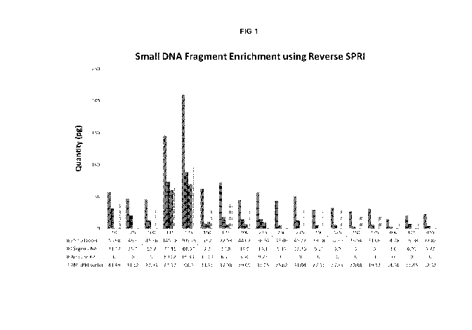
Figure 1 is a plot showing the enrichment of small nucleic acids using
"reverse
SPRI" methodology.
Figure 2 is a series of plots showing the uniform enrichment small nucleic
acids
using "reverse SPRI" methodology relative to other techniques. Figure 2A shows
the size
distribution prior to enrichment. Figure 2B shows the size distribution in an
output
sample after enrichment with a commercial kit designed to isolate free-
circulating DNA
22
CA 02965500 2017-04-21
WO 2016/065295 PCT/US2015/057179
and RNA from human plasma or serum (Qiagen Circulating Nucleic Acid kit).
Figure 2C
shows the size distribution in an output sample after enrichment using Beckman
AMPure SPRI beads. Figure 2D shows the size distribution in an output sample
after
enrichment using Abbott Molecular SPRI beads. The amounts of each fragment of
the
test sample were quantified before and after enrichment using gel
electrophoresis and
densitometric analysis of gel images. Figure 2E shows that both the AMPure and
the
Abbott Molecular SPRI methods provided an enrichment of small fragments (e.g.,
less
than 500 bp, bases, or nt) of approximately 150%.
Figure 3 is a plot showing that reducing the concentration of polyethylene
glycol
in buffers reduces the recovery of smaller nucleic acids.
Figure 4 is a series of plots showing the size selection on silica columns as
a
function of the type and volume of wash buffer used. Figure 4A shows results
of tests of
an ethanol wash buffer and Figure 4B shows results of tests of a wash buffer
comprising
10% Tween-20, 15% ethanol, and 20 mM MgCl2.
Figure 5 shows capillary electrophoresis data for enrichment of small nucleic
acids using magnetic beads.
Figure 6 is a series of plots showing that amine-functionalized beads having a
rough surface (e.g., beads comprising surface irregularities that result in
micron and
sub-micron sized pores) provide for an improved enrichment of samples for
small nucleic
acids relative to amine-functionalized beads having a relatively smooth
surface. Figure
6A shows results using irregular surface contour beads and Figure 6B shows
results
from smooth surface contour beads.
It is to be understood that the figures are not necessarily drawn to scale,
nor are
the objects in the figures necessarily drawn to scale in relationship to one
another. The
figures are depictions that are intended to bring clarity and understanding to
various
embodiments of apparatuses, systems, and methods disclosed herein. Wherever
possible,
the same reference numbers will be used throughout the drawings to refer to
the same
or like parts. Moreover, it should be appreciated that the drawings are not
intended to
limit the scope of the present teachings in any way.
DETAILED DESCRIPTION
Provided herein is technology related to processing samples of nucleic acids
and
particularly, but not exclusively, to methods for enriching samples for small
nucleic
acids, such as small circulating cell-free DNA that finds use in prenatal
testing and in
human disease testing. In some embodiments, the technology relates to
detecting,
23
CA 02965500 2017-04-21
WO 2016/065295 PCT/US2015/057179
characterizing, and/or quantifying small circulating cell-free nucleic acids
for cancer-
related applications such as screening, diagnosis, prognosis, and monitoring
residual
disease or disease recurrence. In some embodiments, the technology relates to
identifying, characterizing, and/or quantifying small circulating cell-free
DNA and/or
small circulating cell-free RNA) for applications related to testing (e.g.,
assessing risk
(e.g., of acquiring or developing); detecting a presence of, an absence of, a
predisposition
to develop, or a predisposition not to develop; screening; diagnosis;
prognosis; and/or
monitoring residual disease or recurrence) associated with a variety of human
and non-
human, acute and chronic disease state pathologies (pathophysiologies)
including, but
not limited to: oncology, hematology, infectious disease, liver disease,
cardiovascular
(e.g., heart) disease, renal disease, inflammatory disease (e.g., rheumatic,
arthritic,
bronchial, gastrointestinal, dermal, cerebrospinal, etc.), and various forms
of pulmonary
disease (e.g. emphysema, COPD, mesothelioma).
In this description of various embodiments of the technology, the section
headings used herein are for organizational purposes only and are not to be
construed as
limiting the described subject matter in any way. In addition, for purposes of
explanation, numerous specific details are set forth to provide a thorough
understanding
of the embodiments disclosed. One skilled in the art will appreciate, however,
that these
various embodiments may be practiced with or without these specific details.
In other
instances, structures and devices are shown in block diagram form.
Furthermore, one
skilled in the art can readily appreciate that the specific sequences in which
methods
are presented and performed are illustrative and it is contemplated that the
sequences
can be varied and still remain within the spirit and scope of the various
embodiments
disclosed herein.
All literature and similar materials cited in this application, including but
not
limited to, patents, patent applications, articles, books, treatises, and
internet web
pages are expressly incorporated by reference in their entirety for any
purpose. Unless
defined otherwise, all technical and scientific terms used herein have the
same meaning
as is commonly understood by one of ordinary skill in the art to which the
various
embodiments described herein belongs. When definitions of terms in
incorporated
references appear to differ from the definitions provided in the present
teachings, the
definition provided in the present teachings shall control.
24
CA 02965500 2017-04-21
WO 2016/065295 PCT/US2015/057179
Definitions
To facilitate an understanding of the present technology, a number of terms
and
phrases are defined below. Additional definitions are set forth throughout the
detailed
description.
Throughout the specification and claims, the following terms take the meanings
interpreted consistently with the understanding of one of ordinary skill in
the related
art as explicitly associated herein, unless the context clearly dictates
otherwise. The
phrase "in one embodiment" as used herein does not necessarily refer to the
same
embodiment, though it may. Furthermore, the phrase "in another embodiment" as
used
herein does not necessarily refer to a different embodiment, although it may.
Thus, as
described below, various embodiments of the invention may be readily combined,
without departing from the scope or spirit of the invention.
In addition, as used herein, the term "or" is an inclusive "or" operator and
is
equivalent to the term "and/or" unless the context clearly dictates otherwise.
The term
"based on" is not exclusive and allows for being based on additional factors
not
described, unless the context clearly dictates otherwise. In addition,
throughout the
specification, the meaning of "a", "an", and "the" include plural references.
The meaning
of "in" includes "in" and "on."
As used herein, the term "SPRI" refers to the technology of "Solid Phase
Reversible Immobilization" wherein target nucleic acids are selectively
precipitated
under specific buffer conditions in the presence of beads or other solid phase
materials
that are often carboxylated and paramagnetic. The precipitated target nucleic
acids
immobilize to said beads and remain bound until removed by an elution buffer
according
to the operator's needs (see, e.g., DeAngelis et al. (1995) Nucleic Acids Res
23: 4742-
4743). The term "carboxylated" as used herein refers to the modification of a
material,
such as a microparticle, by the addition of at least one carboxyl group (e.g.,
COOH or
C00-). In some embodiments, SPRI is used to bind nucleic acids of interest to
the solid
phase and in some embodiments SPRI is used to bind and retain nucleic acids
that are
not of interest, e.g., the nucleic acids of interest remain in the non-bound
liquid phase
(e.g., "reverse SPRI").
As used herein, the term "paramagnetic" as used herein refers to the
characteristic of a material wherein said material's magnetism occurs only in
the
presence of an external, applied magnetic field and does not retain any of the
magnetization once the external, applied magnetic field is removed.
CA 02965500 2017-04-21
WO 2016/065295 PCT/US2015/057179
As used herein, the term "bead" refers to any type of solid phase particle of
any
convenient size, of irregular or regular shape, and which is fabricated from
any number
of known materials such as cellulose, cellulose derivatives, acrylic resins,
glass, silica
gels, polystyrene, gelatin, polyvinyl pyrrolidone, co-polymers of vinyl and
acrylamide,
polystyrene cross-linked with divinylbenzene, or the like (as described, e.g.,
in Merrifield
(1964) Biochemistry 3: 1385-1390), polyacrylamides, latex gels, polystyrene,
dextran,
rubber, silicon, plastics, nitrocellulose, natural sponges, silica gels,
controlled pore glass
(CFG), metals, cross-linked dextrans (e.g., SephadexTm), agarose gel
(SepharoseTm), and
other solid phase bead supports known to those of skill in the art.
"Support", as used herein, refers to a matrix on or in which nucleic acid
molecules, microparticles, and the like may be immobilized, e.g., to which
they may be
covalently or noncovalently attached or in or on which they may be partially
or
completely embedded so that they are largely or entirely prevented from
diffusing freely
or moving with respect to one another.
As used herein, the terms "chromosomal abnormality", "chromosomal
aberration", and "chromosomal alteration" are used herein interchangeably.
They refer
to a difference (e.g., a variation) in the number of chromosomes or to a
difference (e.g., a
modification) in the structural organization of one or more chromosomes as
compared to
chromosomal number and structural organization in a karyotypically normal
individual.
As used herein, these terms are also meant to encompass abnormalities taking
place at
the gene level. Examples of aneuploidy are trisomy 21 and trisomy 13. In some
contexts,
the terms "chromosomal abnormality" and "chromosomal aberration" are used
interchangeably to refer to numerical and structural alterations in a
chromosome that
give rise to an abnormal or pathological phenotype. Chromosomal abnormalities
can be
of several types, for example, extra or missing individual chromosomes, extra
or missing
portions of a chromosome (segmental duplications or deletions), breaks, rings
and
rearrangements, among others.
As used herein, a "copy-number variation" ("CNV") refers to an alteration of
DNA
(e.g., in a genome) that results in a cell having an abnormal number of copies
of one or
more sections of the DNA. Typically, CNVs correspond to relatively large
regions of a
genome that have been deleted (e.g., the genome comprises fewer than the
normal
number) or duplicated (e.g., the genome comprises more than the normal number)
on
certain chromosomes. For example, a chromosome that normally has sections in
order as
L-M-N-0 might instead have sections L-M-N-N-0 (e.g., a duplication of N) or L-
M-0
(e.g., a deletion of N). CNV can also result from aneuploidy and insertion
events. In
26
CA 02965500 2017-04-21
WO 2016/065295 PCT/US2015/057179
humans, CNV accounts for roughly 12% of human genomic DNA and each variation
typically ranges from hundreds or thousands of bases (e.g., from approximately
one
kilobase (1000 nucleotide bases)) to several megabases in size.
The presence of an abnormal number of (e.g., either too many or too few)
chromosomes or chromosome fragments is called "aneuploidy", e.g., the
occurrence of at
least one more or one less chromosome than the normal diploid number of
chromosomes
leading to an unbalanced chromosome complement. Chromosomal aneuploidy is
associated with a large number of genetic disorders and degenerative diseases.
While
many examples are provided herein of aneuploidies comprising an abnormally
high
number of chromosomes, the technology is equally applicable to aneuploidies
comprising
an abnormally low number of chromosomes. In particular, in descriptions of the
technology for discriminating more than 2 (e.g., 3 or more) chromosomes in an
aneuploid
state versus 2 chromosomes in a euploid state, the technology is to be
understood as
applicable also to detecting fewer than 2 chromosomes in an aneuploid state
(e.g., 1 or 0
chromosomes).
As used herein, the term "disease or condition associated with a chromosomal
abnormality" refers to any disease, disorder, condition, or defect that is
known or
suspected to be caused by a chromosomal abnormality. Exemplary diseases or
conditions
associated with a chromosomal abnormality include, but are not limited to,
trisomies
(e.g., Down syndrome (trisomy 21), Edward syndrome (trisomy 18), Patau
syndrome
(trisomy 13), Kleinfelter syndrome (XXY), triple X syndrome (XXX), and XYY
disease),
Turner syndrome (absence of X chromosome, e.g., XO), and X-linked disorders
(e.g.,
Duchenne muscular dystrophy, hemophilia A, certain forms of severe combined
immunodeficiency, Lesch-Nyhan syndrome, and Fragile X syndrome). Additional
examples of diseases or conditions associated with chromosomal abnormalities
are
described in Harrison's Principles of Internal Medicine, Wilson et al. (ed.),
1991 (12th
ed.), Mc Graw Hill, New York, NY, pp. 24-46, which is incorporated herein by
reference
in its entirety.
As used herein, a "nucleic acid" refers to a DNA, an RNA, modified DNA,
modified RNA, and the like. A nucleic may comprise any number of nucleotides,
e.g.,
from 2 to over a million nucleotides.
The term "sample of DNA" or "DNA sample" refers to a sample comprising DNA
or nucleic acid representative of DNA isolated from a natural source and in a
form
suitable for evaluation by an assay (e.g., as a soluble aqueous solution).
27
CA 02965500 2017-04-21
WO 2016/065295 PCT/US2015/057179
As used herein, the terms "small DNA", "small RNA", "small RNA fragment",
¶small DNA fragment", "small nucleic acid", etc., refer to a nucleic acid
(e.g., a DNA or
an RNA) (e.g., a collection of individual nucleic acids in a sample) that is
smaller than a
"cutoff' value. In exemplary embodiments, small nucleic acid refers to a
nucleic acid that
has a size smaller than 1000, 900, 800, 700, 600, 500, 400, 300, 275, 250,
225, 200, 175,
150, 125, 100, 75, or 50 base pairs (bp), bases, or nucleotides (nt);
preferably, the small
nucleic acid is from approximately 50 to approximately 500 base pairs, bases,
or
nucleotides or from approximately 50 to approximately 400 base pairs, bases,
or
nucleotides, or from approximately 50 to approximately 300 base pairs, bases,
or
nucleotides or from approximately 50 to approximately 200 base pairs, bases,
or
nucleotides or from approximately 50 to approximately 100 base pairs, bases,
or
nucleotides. As used herein, a "large DNA", "large RNA", "large RNA fragment",
"large
DNA fragment", "large nucleic acid", etc., refer to a nucleic acid (e.g., a
DNA or an RNA)
(e.g., a collection of individual nucleic acids in a sample) that is larger
than the cutoff
value.
Size may be defined by mass, length, or other suitable size measures. The
length
of a nucleic acid may be expressed in units indicating as a number of "base
pairs"
(abbreviated "bp"), a number of "bases", or a number of nucleotides ("nt" or
"nts").
Lengths of double stranded nucleic acids (e.g., DNA) are typically, but not
exclusively,
expressed in units of base pairs (bp). Lengths of single stranded nucleic
acids (e.g., DNA)
are typically, but not exclusively, expressed in units of nucleotides (nt).
Lengths
expressed in units of bases may apply to either double stranded nucleic acids
or single
stranded nucleic acids. These units are modifiable with standard SI prefixes
to indicate
multiples of powers of 10, e.g., kbp, Mbp, Gbp, kilobase, Megabase, Gigabase,
etc.),
The size measurement can be performed in various ways known in the art, e.g.,
paired-end sequencing and alignment of nucleic acids, electrophoresis,
centrifugation,
optical methods, mass spectrometry, etc. A statistically significant number of
nucleic
acids can be measured to provide an accurate size profile of a sample. In some
embodiments, the data obtained from a physical measurement is received at a
computer
and analyzed to accomplish the measurement of the sizes of the nucleic acids.
For
example, the electropherogram resulting from electrophoresis can be analyzed
(e.g., by
densitometric analysis of electropherogram bands, peaks, etc.) to determine
the sizes. In
one implementation, analyzing the nucleic acids does include the actual
process of
sequencing or subjecting nucleic acids to electrophoresis, while other
implementations
perform an analysis of the resulting data. In some embodiments, a parameter
provides a
28
CA 02965500 2017-04-21
WO 2016/065295
PCT/US2015/057179
statistical measure of the size distribution (e.g., a histogram) of nucleic
acids in the
biological sample. The parameter may be referred to as a size parameter since
it is
determined from the sizes of the plurality of nucleic acids.
By "maternal host of a fetus" is meant a woman who is pregnant with a fetus
whose DNA is sought to be detected and/or tested for a genetic condition. The
term
"maternal host of a fetus", "maternal host", and "mother" are used
interchangeably. By
"fetus" is meant an offspring developing in utero at any gestational stage.
Fetal DNA
can be detected prior to the "fetal period" which begins at 10 or 11 weeks of
gestation in
a human. Therefore, "fetus" encompasses not only the developing offspring in
the fetal
period but also in the earlier embryonic stages of development prior to the
10th or 11th
week of human gestation.
A "subject" is a vertebrate, preferably a mammal, more preferably a human.
Mammals include, but are not limited to, murines, simians, humans, farm
animals,
sport animals, and pets.
As used herein, "cell-free DNA" refers to DNA that is not within a cell. In
one
embodiment, cell free DNA includes DNA circulating in blood. In another
embodiment,
cell free DNA includes DNA existing outside of a cell. In yet another
embodiment, cell
free DNA includes DNA existing outside of a cell as well as DNA present in a
blood
sample after such blood sample has undergone partial or gentle cell lysing.
As used herein, "cell-free fetal DNA" ("cffDNA") refers to DNA that originated
from the fetus and not the mother and is not within a cell. In one embodiment,
cell free
fetal DNA includes fetal DNA circulating in maternal blood. In another
embodiment,
cell free fetal DNA includes fetal DNA existing outside of a cell, for example
a fetal cell.
In yet another embodiment, cell free fetal DNA includes fetal DNA existing
outside of a
cell as well as fetal DNA present in maternal blood sample after such blood
sample has
undergone partial or gentle cell lysing. A review of fetal DNA in maternal
plasma and
serum is provided by Peril and Bianchi (2001), as well as in Lo (2000).
By "biological sample" is meant any sample that is derived from the maternal
host of the fetus. In one embodiment, the biological sample of a maternal host
includes
any processed or unprocessed, solid, semi-solid, or liquid biological sample,
e.g., blood,
urine, saliva, and mucosal samples (e.g., samples from uterus or vagina,
etc.). For
example, the biological sample of a maternal host can be a sample of whole
blood,
partially lysed whole blood, plasma, and/or partially processed whole blood.
In one
embodiment, the biological sample of a maternal host is a sample of cell free
DNA or
free floating DNA from the whole blood of the maternal host.
29
CA 02965500 2017-04-21
WO 2016/065295 PCT/US2015/057179
As used herein, the term "Tween-20" refers to a polysorbate surfactant, e.g.,
a
polyoxyethylene derivative of sorbitan monolaurate (e.g., Polysorbate 20
(polyoxyethylene (20) sorbitan monolaurate)). Tween-20 is distinguished from
other
members in the polysorbate range by the length of the polyoxyethylene chain
and the
fatty acid ester moiety. Accordingly, other Tween surfactants are Tween-40
(Polysorbate
40 (polyoxyethylene (20) sorbitan monopalmitate)), Tween-60 (Polysorbate 60
(polyoxyethylene (20) sorbitan monostearate)), Tween-80 (Polysorbate 80
(polyoxyethylene (20) sorbitan monooleate)), etc. The number 20 following
G4polyoxyethylene" refers to the total number of oxyethylene ¨(CH2CH20)¨
groups found
in the molecule. The number following "polysorbate" is related to the type of
fatty acid
associated with the polyoxyethylene sorbitan part of the molecule. Monolaurate
is
indicated by 20, monopalmitate is indicated by 40, monostearate by 60, and
monooleate
by 80. In some embodiments where Tween-20 is described another detergent or
surfactant can be substituted, e.g., a Tween-40, a Tween-60, and/or a Tween-
80.
Description
The technology provides a variety of approaches to use alone or in combination
to
enrich low-abundance nucleic acids (e.g., DNA and/or RNA such as small
circulating
fetal DNA fragments, mRNA, miRNA, piRNA, etc.) from the abundant background of
circulating maternal nucleic acids (e.g., DNA and/or RNA) present in maternal
plasma
specimens. In some embodiments, the technology is applicable to the selective
enrichment of small nucleic acids (e.g., having a length of less than
approximately 200
bp, bases, or nt) from a background milieu of larger molecular weight nucleic
acids (e.g.,
having a length of greater than approximately 200 bp, bases, or nt and
extending up to
approximately 20,000 or more bp, bases, or nt). The technology is not limited
in the size
cutoff (e.g., in bp, bases, or nt) that is used to differentiate "small" from
"large" or "non
small" nucleic acids. As used herein, reference to a 200-bp, 200-base, or 200-
nt cut-off to
differentiate between small nucleic acids and large nucleic acids in size
distributions is
intended to be exemplary and is not intended to limit the technology to that
particular
size cut-off. Accordingly, in some embodiments a size cut-off of 200 bp,
bases, or nt is
used to differentiate small nucleic acids (e.g., DNA (e.g., cffDNA) and/r RNA)
from large
nucleic acids (e.g., maternal nucleic acids). In some embodiments, cut-off
values of, e.g.,
1000, 900, 800, 700, 600, 500, 400, 300, 275, 250, 225, 200, 175, 150, 125,
100, 75, or 50
base pairs, bases, or nucleotides are used. Desirable cut-off values can be
determined by
one of skill in art for particular applications.
CA 02965500 2017-04-21
WO 2016/065295 PCT/US2015/057179
In addition, the technology provided does not introduce detectable or
otherwise
discernible genome and/or sequence representational bias other than size
discrimination
that could have the potential to compromise downstream sensitivity in
aneuploid
marker analyses for non-invasive pre-natal testing (NIPT) applications.
The technology is not limited in the type of nucleic acid that is enriched.
Accordingly, the technology is applicable to the isolation, enrichment, and
detection of
DNA (e.g., cffDNA and other small DNA and DNA fragments). The technology is
applicable to the isolation, enrichment, and detection of RNA, such as
messenger RNAs
(mRNA), micro RNAs (microRNA or miRNA), piRNA, and other small nucleic acids
(e.g.,
nucleic acids present in blood) as well as for the isolation, enrichment, and
detection of
fetal messenger RNAs (mRNA) and fetal micro RNAs (microRNA or miRNA) present
in
maternal blood.
The technology is exemplified herein by two general types of strategies and
sub
-
technologies. However, it is understood that the technology is not limited to
these
illustrative examples. The first set of sub-technologies provides a selective
and
preferential (e.g., enriched) elution of small nucleic acids directly from
silica-based
capture membranes and matrices (e.g., the Qiagen Circulating Nucleic Acid Kit
or the
Qiagen DNeasy kit). The second set of sub-technologies relies on secondary
enrichment
processes that require additional laboratory instrumentation such as
methylated DNA
immunoprecipitation (e.g., "MeDIP") with antibody-coated particles that can be
captured
with a magnetic field (e.g., antibody-coated paramagnetic particles or
antibody-coated
magnetic particles)) or capture onto affinity columns (e.g., antibody-coated
spin
-
columns, e.g., as provided commercially by Molzyme) or other solid supports
(e.g.,
microtiter plates, beads, slides, or nanostructures), solid phase reversible
immobilization
(SPRI) beads, automated electrophoresis and electroelution (e.g., life
technologies Pippin
Prep), LabChip XT, synchronous coefficient drag of alteration (SCODA),
simultaneous
anion exchange and size exclusion, etc. These methods can either be used
directly and
independently to isolate small nucleic acids from plasma (e.g., to provide a
single-
enrichment method) and/or sequentially on the back-end of a silica membrane
capture
process (e.g., to provide a double-enrichment method, e.g., used in
conjunction with a
silica-based isolating method such as a Qiagen Circulating Nucleic Acid Kit,
which is
modified in some embodiments according to the technology provided herein).
Although
the disclosure herein refers to certain illustrated embodiments, it is to be
understood
that these embodiments are presented by way of example and not by way of
limitation.
31
CA 02965500 2017-04-21
WO 2016/065295 PCT/US2015/057179
1. Preferential elution of small nucleic acids from silica
In some embodiments of the technology, small nucleic acids (e.g., small DNA
(e.g., cffDNAs) and/or small RNA) are isolated from or enriched in a sample
(e.g., a
sample prepared or derived from maternal blood or plasma) by preferential
and/or
selective elution. For example, in some embodiments small nucleic acids are
preferentially eluted or otherwise recovered from a silica matrix or membrane
after
capture (e.g., binding) of bulk or total nucleic acid from the sample on the
silica matrix
or membrane.
Nucleic acids (e.g., DNA, RNA) bind non-specifically to silica surfaces in the
presence of certain salts and under certain pH conditions, usually under
conditions of
high ionic strength. For example, DNA adsorption is most efficient in the
presence of a
buffer solution having a pH at or below the pKa of the surface silanol groups
of the
silicon surface. In some embodiments, a nucleic acid (e.g.,DNA) binds to
silica in the
presence of a chaotrope (e.g., salts, butanol, ethanol, guanidinium chloride,
guanidine
thiocyanate, lithium perchlorate, lithium acetate, magnesium chloride, phenol,
propanol, sodium dodecyl sulfate, thiourea, and urea), which denatures
biomolecules by
disrupting the shell of hydration around them. In some embodiments, the
nucleic acid is
washed with high salt and ethanol, and typically eluted with an elution buffer
comprising low salt.
Accordingly, in some embodiments, after binding of total or bulk nucleic acids
(e.g., from a maternal blood sample), elution conditions promote the selective
and
preferential release of small nucleic acids from the silica surface relative
to large nucleic
acids. In exemplary embodiments, solvent effects act to promote the elution of
small
nucleic acids from silica. Accordingly, embodiments provide for the
preferential elution
of small nucleic acids from silica in an elution buffer comprising 5 to 25%
ethanol, 5 to
25% methanol, 5 to 25% acetonitrile, 5 to 25% DMSO, and/or 1 to 25% formamide.
In
some embodiments, ionic strength effects act to promote the preferential
elution of small
nucleic acids from silicon. Accordingly, embodiments provide for the
preferential elution
of small nucleic acids from silica in an elution buffer comprising an elevated
sodium
chloride concentration (e.g., greater than 500 mM, e.g., 1 M or higher) to
screen counter-
ion binding interactions and/or comprising an elevated chaotropic salt
concentration to
stabilize binding of larger nucleic acids to the silica surface. In some
embodiments, pH
effects preferentially promote the elution of small nucleic acids from silica
membranes
or columns. In some embodiments, elution conditions are adjusted to modify the
kinetics
of adsorption and release from the silica to favor the release of small
nucleic acids from
32
CA 02965500 2017-04-21
WO 2016/065295 PCT/US2015/057179
the silica, e.g., at low temperatures (e.g., 2 C to 16 C) small nucleic acids
are
preferentially eluted from a silica column.
2. Preferential retention of large nucleic acids on silica
Furthermore, in some embodiments of the technology, small nucleic acids (e.g.,
DNA (e.g., cffDNAs) and/or small RNA) are isolated from or enriched in a
sample (e.g., a
sample prepared or derived from maternal blood or plasma) by selective and
preferential
retention of large nucleic acids by post-capture silica column sequestration.
In some
embodiments, after binding of total or bulk nucleic acids (e.g., from a
maternal blood
sample), elution conditions promote the selective and preferential retention
of large
nucleic acids by the silica surface relative to small nucleic acids.
For example, in some embodiments a silica surface (membrane, column, etc.) is
treated with a polymer coating, volume-exclusion agent, or absorptive agent to
sequester
large nucleic acids (e.g., greater than 200 bp, bases, or nt) onto the column
and promote
the elution of small nucleic acids (e.g., less than 200 bp, bases, or nt).
Exemplary
embodiments provide a method in which nucleic acids (e.g., DNA) are adsorbed
to a
silica surface treated with 0.5 to 2% acrylamide / bis-acrylamide (e.g.,
comprising a 19:1
to 29:1 cross-linking ratio), 0.01 to 0.5% agarose, 0.01 to 1.0% polyethylene
glycol (PEG
having an average molecular weight of 1000 to 10,000), 1 to 10% dextran
sulfate, 1 to
10% ficoll, sorbitol, or sugar (e.g., aldohexose) polymer, 1 to 10% polyvinyl
alcohol (PVA),
1 to 10% polyamines (e.g., spermine, spermidine, etc.), or a low-level
synthetic polymer
such as, e.g., nylon, polyester, polystyrene, etc.
In some embodiments, a doped silica membrane and/or column matrix sequesters
large nucleic acids (e.g., greater than 200 bp, bases, or nt) onto the column
and promotes
the elution of small nucleic acids (e.g., less than 200 bp, bases, or nt) from
the column.
For example, some embodiments comprise the use of silica embedded with an
amine-
binding surface doping agent (e.g., carboxylate group derivatization). In some
embodiments, silica is embedded with a polyphosphate-binding surface doping
agent
(e.g., amino group derivatization). In some embodiments, silica is embedded
with one or
more of a reversible, controllable, or amine-reactive surface group for the
controlled,
reversible, and preferential elution of small nucleic acids. In some
embodiments, silica is
embedded with one or more of a reversible, controllable, or carboxyl-reactive
surface
group for the controlled, reversible, or preferential elution of small nucleic
acids.
In some embodiments, the technology provides methods comprising preferential
cross-linking of large nucleic acids relative to small nucleic acids to retain
the large
33
CA 02965500 2017-04-21
WO 2016/065295 PCT/US2015/057179
nucleic acids on a substrate (e.g., a silica column). For example, in some
embodiments
methods comprise low-level ultraviolet radiation cross-linking of large
nucleic acids, e.g.,
at low energy intensities to entrap large cross-linked molecular weight
nucleic acids,
e.g., greater than 200 bp, bases, or nt. In some embodiments, methods comprise
endogenous cross-linking of nucleic acids, e.g., to from cyclobutane and/or
thymidine
dimers preferentially in large nucleic acids (e.g., DNA) relative to small
nucleic acids. In
some embodiments, methods comprise cross-linking large nucleic acids
preferentially
using ultraviolet radiation in the presence of an exogenous cross-linking
agent such as,
e.g., psoralen.
In some embodiments, methods comprise the use of low-level chemical cross-
linking (e.g., to promote the reversible, controlled, and preferential elution
of small size
nucleic acids). For example, some embodiments provide a method comprising
preferentially cross-linking large nucleic acids using a solution comprising
10% neutral,
buffered formalin. Formalin treatment is followed, in some embodiments, by
heat
restoration with or without treatment with EDBE (e.g., the Gundling reagent
for
preparing formalin-fixed paraffin embedded samples; see, e.g., U.S. Pat. Appl.
Pub. No.
20130323815, incorporated herein by reference).
In some embodiments, methods comprise use of a reversible, bi-functional cross-
linking agent (e.g. a DTT-cleavable, thiol-labile bis-acrylamide/ acrylamide
mixture). In
embodiments associated with testing maternal blood for fetal nucleic acids,
these low-
level cross-linking methods exploit the lower abundance of smaller (e.g.,
fetal) nucleic
acids (e.g., DNA) relative to longer (e.g., maternal) nucleic acids. Without
being bound
by theory, it is expected both statistically and stochastically that larger
(e.g., maternal)
nucleic acid molecules will be preferentially cross-linked to one another
compared to the
less abundant smaller (e.g., fetal) nucleic acids. This effect is further
enhanced by the
relative lengths of the large and small nucleic acid molecules because larger
species are
more likely to be cross-linked by both intra-strand and inter-strand cross-
linking events.
Some embodiments comprise use of a cross-linking agent comprising formalin,
alkylating agents (e.g., 1,3-bis(2-chloroethyl)-1-nitrosourea (BCNU,
carmustine)),
nitrogen mustard, cisplatin, nitrous acid, aldehydes (e.g., malondialdehyde,
acrolein,
crotonaldehyde), chloroethylating agents, nitrosoureas, triazenes, alkyl
sulfonates,
epoxides, diepoxybutane, carzinophilin, azinomycin B, cis-
Diamminedichloroplatinum
(II), sandramycin, luzopeptins, isochrysohermidin, pyrrolobenzodiazepine
agents,
cyclophosphamide, N, N, N, N', N', N'-hexamethylmelamines, pyrrolizidine
alkaloids,
anthracyclines, mitomycin C, aziridinylbenzoquinones, or biselezin.
34
CA 02965500 2017-04-21
WO 2016/065295 PCT/US2015/057179
Some embodiments provide electroelution methods. For instance, some
embodiments provide methods comprising the preferential electroelution of
small
nucleic acids from a column. Some embodiments comprise continuous forward-
field
electro-elution (e.g., low-field, cathode assisted electro-elution),
continuous reverse-field
electro-elution (e.g., low-field, anode assisted electro-elution), or
oscillating-field electro-
elution (e.g., high frequency, low-field, reversible anode/cathode electrical
oscillation-
driven electro-elution).
Some embodiments provide methods comprising use of an ion-exchange column
with selective elution of small nucleic acids. Examples of column, matrix, or
membrane
media include but are not limited to silica, DEAE, Dionex, or other
derivatized
chromatography column media, matrix, or membrane. Some embodiments comprise
elution (e.g., isocratic and/or non-isocratic elution) with increasing or
decreasing salt
gradient, elution (e.g., isocratic and/or non-isocratic elution) with
increasing or
decreasing pH gradient, and/or elution (e.g., isocratic and/or non-isocratic
elution) with
an aqueous/non-aqueous (e.g. methanol, ethanol, acetonitrile) dual-solvent,
gradient
elution system.
3. Preferential retention of large nucleic acids during column washing
In related embodiments of methods, large nucleic acids are retained on a solid
support (e.g., a silica column) and/or small nucleic acids are not retained on
the solid
support during a washing step (e.g., prior to an elution step). For example,
in some
embodiments, a sample comprising nucleic acids is flowed over a column to bind
nucleic
acids to the column. Then, the column (comprising bound nucleic acids) is
washed with a
wash buffer. After washing the column with the wash buffer, some nucleic acids
remain
adsorbed to the column (e.g., preferential adsorption of large nucleic acids
to the column;
e.g., enrichment of large nucleic acids on the column) and some nucleic acids
are
removed from the column and are present in the wash buffer (e.g., preferential
removal
of small nucleic acids from the column in the wash buffer; e.g., enrichment of
small
nucleic acids in the wash buffer). Thus, in some embodiments, the wash buffer
preferentially removes small nucleic acids from the column (e.g., large
nucleic acids
remain adsorbed to the column). Subsequently, an elution buffer is flowed over
the
column to remove nucleic acids from the column that remained bound during the
wash.
The preferential binding of nucleic acids (e.g., large nucleic acids) to the
column during
the wash step will produce an eluate enriched for large nucleic acids and a
wash buffer
enriched for small nucleic acids. Thus, an increase of recovery of large
nucleic acids in
CA 02965500 2017-04-21
WO 2016/065295 PCT/US2015/057179
the eluate indicates that the wash buffer flow-through was enriched for small
nucleic
acids.
In some embodiments, the ratio of wash buffer to sample volume is controlled
to
stabilize binding of large nucleic acids to the silica during the wash step,
which allows
smaller nucleic acids to be recovered in the wash buffer (e.g., prior to
eluting the large
nucleic acids in the elution buffer). In some embodiments, the wash buffer
volume to
sample buffer volume is 0.7 to 1, 0.6 to 1, 0.5 to 1, 0.4 to 1, 0.3 to 1.
Further, in some
embodiments the wash buffer comprises ethanol (e.g., 50-60% ethanol (e.g., 70%
ethanol)). In some embodiments, the wash buffer comprises Tween-20, ethanol,
and
MgC12 (e.g., in some embodiments the wash buffer comprises 10% Tween-20, 15%
ethanol, and 20 mM MgCl2). In some embodiments, 70% ethanol is used at a ratio
of
0.5:1, 0.4:1, or 0.3:1; in some embodiments, a buffer comprising Tween-20,
ethanol, and
MgC12 is used at a ratio of 0.5:1 or 0.4 to 1.
4. Enrichment by methylated DNA immunoprecipitation (MeDIP)
Some embodiments of the technology comprise the use of a silica column that
has
affinity for nucleic acids (e.g., a DNA-binding column, e.g., as provided
commercially in
the Qiagen Circulating Nucleic Acid kit) coupled with use of methylated DNA
immuno-
precipitation ("MeDIP") with antibody-coated particles that can be captured
with a
magnetic field (e.g., antibody-coated paramagnetic particles or antibody-
coated magnetic
particles)). In particular, methods comprising use of MeDIP comprise
incubating the
eluate from a silica-based isolation method (e.g., the eluate from a Qiagen
Circulating
Nucleic Acid kit) with beads functionalized with an antibody recognizing
methylated
DNA (e.g., antibody-coated particles that can be captured with a magnetic
field (e.g.,
antibody-coated paramagnetic particles or antibody-coated magnetic
particles)). In
particular, antibodies recognizing the 5-methycytosine moieties on methylated
DNA
selectively capture the fetal DNA in maternal blood due to the
hypermethylation of fetal
DNA relative to maternal DNA. Then, in some embodiments, methods further
comprise
the elution of hyper-methylated fetal DNA from a silica column, e.g., by
eluting in the
presence of excess 5-methylcytosine to provide competitive binding, using heat
denaturation and/or inactivation of anti-methylated DNA antibody, using
chemical
antibody denaturation and/or inactivation (e.g., using formamide, a chaotropic
agent,
pH, detergents, surfactants, or ionic liquids), or using a physical
denaturation process
(e.g., sonication (e.g., in the KHz range), ultrasonication (e.g., in the MHz
range), bead-
beating and/or mechanical agitation (e.g., using a vortexer or other device
such as an
36
CA 02965500 2017-04-21
WO 2016/065295 PCT/US2015/057179
Eppendorf EpiMotion), e.g., in the presence of solvents and/or adjuvants).
Some
embodiments further comprise purifying the enriched fetal nucleic acid
fraction (e.g.,
using a bead-based or column-based method such as some PCR cleanup methods)
and/or
comprise low-level, whole genome amplification (WGA) by random priming with
Klenow
fragment and dNTPs (e.g., as provided commercially in the Invitrogen BioPrime
Random Priming Kit). In some embodiments, methods comprising use of a silica
column
(e.g., a DNA-binding column) and MeDIP provide approximately 50-fold to 100-
fold
amplification of cffDNA from the random priming amplification reaction and an
expected yield of approximately 2 [tg to 5 [tg of random primed and amplified
fetal DNA
without introducing sequence amplification bias (e.g., resulting in unbalanced
marker
coverage) or bias in genome representation.
5. Enrichment by size exclusion
In some embodiments, the technology provides embodiments of methods in which
silica capture is coupled with size exclusion, chromatography, and/or
microdialysis
methods. For example, in some embodiments, methods comprise use of
ultrafiltration,
e.g., in particular, the use of centrifugal sample concentrators (e.g.,
Amicon, CentriCon)
that enrich small nucleic acids (e.g., DNA and/or RNA) by molecular weight-
based size-
exclusion cut-offs (e.g., molecular weight cutoff ranging from approximately
10,000 to
30,000 Daltons). Thus, in some embodiments, small nucleic acids (e.g., fetal
DNA, other
small DNA, and/or small RNA) are present in the column flow-through. In some
embodiments, methods comprise use of size-exclusion chromatography spin-
columns
(e.g., G-25, G-50, G-100, etc.) wherein the small nucleic acids are retained
in the porous
chromatography media and are not present in the high-molecular weight flow-
through,
which represents the column void-volume. In some embodiments, methods comprise
microdialysis of fetal nucleic acids followed by diluent-exchange buffer
recovery and
concentration of low-molecular weight nucleic acid in a sample. In particular,
in some
embodiments methods employ use of a dialysis membrane with a nominal molecular
weight cutoff range of approximately 10,000 to 30,000 Daltons followed by
either silica
membrane re-capture, rotary-vapor exchange buffer concentration, vacuum
concentration (e.g., by a Speed-Vac brand vacuum concentrator or equivalent),
centrifugal evaporation (e.g., by a Speed-Vac brand vacuum concentrator or
equivalent),
and/or ethanol precipitation to capture the permeate from the microdialysis
filtration
exchange buffer.
37
CA 02965500 2017-04-21
WO 2016/065295 PCT/US2015/057179
6. Synchronous coefficent of drag alteration (SCODA) sizing
In some embodiments, small nucleic acids are enriched, isolated, or obtained
from a sample using a method comprising separation by synchronous coefficient
of drag
alteration (SCODA), e.g., as implemented in a commercial product such as the
Aurora
system by Boreal Genomics (Vancouver, BC). The method is based on the non-
linear
response of nucleic acids (e.g., DNA and/or RNA) to electrophoretic fields
that causes
them to drift relative to other components under certain types of
superimposed, rotating
electric fields. See, e.g., Pet, et al. (2009) "Nonlinear electrophoretic
response yields a
unique parameter for separation of biomolecules" Proc. Nat. Acad. Sci.
U.SA.106(35):
14796.
7. Solid phase reversible immobilization (SPRI) bead-based sizing
In some embodiments, small nucleic acids are enriched, isolated, or obtained
from a sample using a method comprising solid phase reversible immobilization
(SPRI),
e.g., on a solid support, e.g., beads. See, e.g., DeAngelis et al. (1995)
"Solid-phase
reversible immobilization for the isolation of PCR products" Nucleic Acids
Res.
23(22)4742. SPRI beads comprise a paramagnetic magnetite layer between a
polystyrene core surrounded and an external polymer surface coated with
carboxylate
groups. Carboxylate groups reversibly bind nucleic acids (e.g., DNA and/or
RNA) in the
presence of a crowding agent (e.g., polyethylene glycol (PEG), e.g., at 20%
weight per
volume) and salt (e.g., 2.5 M NaC1). The crowding agent promotes nucleic acids
to bind
with the carboxyl groups on the bead surface.
The PEG concentration and/or ratio of beads to total nucleic acids are
adjusted
for size selection, e.g., to enrich a sample for small nucleic acids. As PEG
concentration
and bead-to-nucleic acid ratio vary, the length of fragments binding and/or
left in
solution changes. In general, the higher the concentrations of PEG and salt in
the
solution, the lower the cutoff size. While fragments larger than the cutoff
(e.g., based on
the solution conditions, e.g., PEG and/or salt concentrations) are bound to
the beads and
thus are removed from the sample, fragments smaller than the cutoff are
retained in the
buffer. In some contexts, the term "reverse SPRI" refers to the use of SPRI
beads to
recover small nucleic acids in the buffer rather than the long nucleic acids
bound to the
beads. Some exemplary commercial products comprising SPRI technology are,
e.g.,
Ampure XP beads from Beckman.
The technology is adaptable to a range of cutoff values for differentiating
small
DNA from large DNA. For example, PEG concentration can be adjusted to provide
for
38
CA 02965500 2017-04-21
WO 2016/065295 PCT/US2015/057179
the desired cutoff (e.g., using PEG (e.g., PEG 8000) concentrations of 4 to
5%, e.g., 4.0%,
4.1%, 4.2%, 4.3%, 4.4%, 4.5%, 4.6%, 4.7%, 4.8%, 4.9%, or 5.0%). Accordingly,
embodiments provide methods for enriching a sample for small DNA, wherein
small
DNA is DNA having a length less than a length cutoff value of 1000, 900, 800,
700, 600,
500, 400, 300, 275, 250, 225, 200, 175, 150, 125, 100, 75, or 50 base pairs,
bases, or
nucleotides. In some embodiments, the distribution and relative abundance of
fragment
sizes smaller than a length cutoff value in the output sample and the
distribution and
relative abundance of fragment sizes of fragment sizes smaller than a length
cutoff
value in the input sample are the same or similar. In some embodiments, a
higher
concentration of PEG (e.g., PEG 8000) is used, e.g., 15% to 20% (e.g., 15%,
16%, 17%,
18%, 19%, or 20%).
In some embodiments, the PEG (e.g., PEG 8000) concentration modulates the
recovery of smaller (e.g., lower molecular weight) DNA (Figure 3) relative to
larger
DNA. For example, a concentration of 5.1% PEG produced a cutoff for recovery
at about
600 bp, bases, or nt ¨ e.g., nucleic acids larger than 600 bp, bases, or nt
were bound to
beads and removed from the sample; nucleic acids smaller than 600 bp, bases,
or nt
remained in the sample, which was then enriched for small nucleic acids less
than 600
bp, bases, or nt. In addition, a concentration of 4.8% PEG produced a cutoff
for recovery
at about 1000 bp, bases, or nt ¨ e.g., nucleic acids larger than 1000 bp,
bases, or nt were
bound to beads and removed from the sample; nucleic acids smaller than 1000
bp, bases,
or nt remained in the sample, which was then enriched for small nucleic acids
less than
1000 bp, bases, or nt.
In some embodiments, SPRI beads find use in manual size fractionation
comprising pipetting, mixing, centrifuging, and transferring steps; and, in
some
embodiments, SPRI beads find use in automated SPRI size fractionation, e.g.,
using
Beckman Coulter Genomics SPRI-TE NGS robotics, which is adaptable to many
instruments for sample preparation such as those available from Precision
System
Science. In some embodiments, methods comprise steps to determine the ionic
strength
(e.g., NaC1 concentration) and PEG concentration for the desired cutoff (e.g.,
between
the appropriate small fragments and large fragments) for a size-specific
nucleic acid
precipitation on SPRI bead surfaces.
8. Electrophoresis-based sizing
In some embodiments, small nucleic acids are enriched, isolated, or obtained
from a sample using a method comprising electrophoresis-based size tuning. In
some
39
CA 02965500 2017-04-21
WO 2016/065295 PCT/US2015/057179
embodiments, electrophoresis is used in combination with other enrichment
methods
such as silica matrix methods discussed herein. Commercial devices for
electrophoretic
enrichment based on size include the Pippin Prep by Sage Sciences, which
comprises use
of agarose gel electrophoresis. These systems can size select for nucleic
acids having a
minimum size of 50 bp, bases, or nt to a maximum size of 8000 bp, bases, or nt
to 50,000
bp, bases, or nt. Another exemplary commercial product is based on capillary
electrophoresis high-resolution size selection of nucleic acids (e.g., the
Caliper Lab Chip
XT).
9. Simultaneous anion exchange and size exclusion on magnetic beads
In some embodiments, small nucleic acids are enriched, isolated, or obtained
from a sample using a method comprising preferential capture of small nucleic
acids on
magnetic beads comprising an amine (e.g., a weak amine) anion exchange
functional
group and beads comprising surface irregularities that result in micron and
sub-micron
sized pores (see, e.g., Example 4).
For example, in some embodiments, the technology utilizes a combination of
size
exclusion (e.g., as a result of surface and/or interior irregularities (e.g.,
pores and/or
cavities)) and anion exchange (e.g., as a result of functionalized surface
and/or interior)
to selectively bind, release, and purify target nucleic acids (e.g., nucleic
acids of a
selected size range); although the present invention is not limited to any
particular
mechanism of action and an understanding of the mechanism of action is not
necessary
to practice the present invention. In some embodiments, microparticles are
magnetic,
contain functional groups that allow for anion exchange of nucleic acids, and
comprise
irregular surface features (e.g., pores) that allow for size-selective
adherence and/or
release of nucleic acids. In some embodiments, magnetic particles allow, for
example,
manipulation of microparticles (e.g., with or without adhered nucleic acid).
In some embodiments, a strong cation exchange functional group, such as a
quaternary amine, for example, is employed as an anion exchange functional
group.
Additional strong anion exchange functional groups are known to those skilled
in the
art.
In other embodiments, a weak anion exchange functional group is a suitable
anion exchange functional group, such as polyethyleneimine, a charged aromatic
amine,
diethylaminomethyl, or diethylaminoethyl. Such functional groups have pKa
values of
9.0 or greater.
CA 02965500 2017-04-21
WO 2016/065295 PCT/US2015/057179
In some embodiments, the manufacturing process for microparticles creates
irregularities (e.g., micron or sub-micron sized pores or cavities) on the
particle surface
and within the particles and/or clusters of particles. The structural
irregularities (e.g.,
pores) on the microparticles adhere target nucleic acid products (e.g., of a
desired size or
size range, e.g., small nucleic acids), due to size exclusion properties,
while not adhering
non-target nucleic acids (e.g., nucleic acids of non-target size (e.g., larger
genomic
nucleic acids)). In some embodiments, surface and/or internal irregularities
(e.g., pores)
are functionalized with a weak anion exchange functional group the bind
nucleic acids.
In some embodiments, both target and non-target nucleic acids adhere to the
porous microparticles, but conditions are provided and/or adjusted to control
the binding
and release of the small nucleic acids to provide an enriched sample.
In certain embodiments, compositions and methods provided herein allow a user
to decrease large amounts of background nucleic acid from a sample (e.g.,
large nucleic
acids such as genomic nucleic acids).
In some embodiments, the present technology provides compositions comprising
a microparticle (e.g., a bead, e.g., a magnetic bead) having a surface
comprising cavities
and/or other surface irregularities and/or an aggregate comprising two or more
of said
microparticles, which aggregate comprises an opening, wherein said surface,
cavities,
opening, and/or other surface irregularities/pores are: a) functionalized with
a weak
anion exchange functional group; and b) dimensioned for size exclusion of
smaller
nucleic acid molecules from larger nucleic acid molecules. In some
embodiments, larger
nucleic acid molecules comprise or are derived from human genomic nucleic
acid. In
some embodiments, compositions further comprise smaller nucleic acid molecules
bound
to the pores. In some embodiments, the microparticle is an iron particle. In
some
embodiments, the weak anion exchange functional group is an amine. In some
embodiments, the amino is a primary, secondary, or tertiary alkyl amine. In
some
embodiments, the amine has a pKa of greater than 9. In some embodiments, the
composition comprises a plurality of said microparticles.
10. Combinations of technologies
In some embodiments, the technology comprises use, e.g., in simultaneous
and/or
sequential combination, of one or more of preferential elution of small
nucleic acids from
silica, preferential retention of large nucleic acids on silica, enrichment by
MeDIP with
antibody-coated particles that can be captured with a magnetic field (e.g.,
antibody-
coated paramagnetic particles or antibody-coated magnetic particles),
enrichment by
41
CA 02965500 2017-04-21
WO 2016/065295 PCT/US2015/057179
size exclusion, enrichment by SCODA, enrichment by SPRI bead-based sizing,
enrichment by electrophoresis-based sizing to provide a sample of small
nucleic acids
(e.g., DNA (e.g., cffDNA) and/or RNA) for analysis, and/or preferential
capture of small
nucleic acids on magnetic beads comprising an amine (e.g., a weak amine) anion
exchange functional group and comprising surface irregularities that result in
micron
and sub-micron sized pores. In some embodiments, a silica-based enrichment
method is
coupled with one or more other enrichment methods to provide a sample for
analysis. In
some embodiments, two or more technologies are applied to a sample
sequentially and
in some embodiments two or more technologies are applied simultaneously. For
example, in some embodiments a method for the preferential elution of small
nucleic
acids from silica is used at the same time with a method for the preferential
retention of
large nucleic acids by silica. In some embodiments, MeDIP is performed
directly on a
solid support (e.g., a bead, column (e.g., an affinity column), microplate,
etc. comprising
an agent (e.g., an antibody) specific for methylated DNA). In some
embodiments, two or
more technologies are applied simultaneously and additional technologies are
applied
sequentially.
11. Collection of samples
The technology is not limited in the sample that is taken and enriched for
small
nucleic acids (e.g., DNA). For example, in some embodiments, the sample is a
biological
sample (e.g., taken from a subject) such as, e.g., a urine sample, a
cerebrospinal fluid
(CSF) sample, or a peritoneal fluid sample.
In some embodiments, a blood sample is taken. Collection of blood is performed
in accordance with the standard protocol hospitals or clinics generally
follow. For
example, in some embodiments, an appropriate amount of peripheral blood, e.g.,
typically between 5 ml to 50 ml, is collected and may be stored according to
standard
procedure prior to further preparation. The analysis of nucleic acids found in
blood may
be performed using, e.g., the whole blood, serum, or plasma. The methods for
preparing
serum or plasma from blood are well known among those of skill in the art. For
example,
blood can be placed in a tube containing EDTA or a specialized commercial
product such
as Vacutainer SST (Becton Dickinson, Franklin Lakes, NJ) to prevent blood
clotting and
plasma can then be obtained from whole blood through centrifugation. In some
embodiments, serum is obtained with or without centrifugation following blood
clotting.
In embodiments comprising use of centrifugation, centrifugation is typically
performed
at an appropriate speed, e.g., 1500 to 3000 x g. Plasma or serum may be
subjected to
42
CA 02965500 2017-04-21
WO 2016/065295 PCT/US2015/057179
additional centrifugation steps before being transferred to a fresh tube for
extraction of
nucleic acids. In addition to the acellular portion of the whole blood,
nucleic acids may
also be recovered from the cellular fraction, enriched in the buffy coat
portion, which can
be obtained following centrifugation of a whole blood sample from the woman
and
removal of the plasma.
There are numerous known methods for extracting DNA from a biological sample
including blood. The general methods of DNA preparation (e.g., described by
Sambrook
and Russell, Molecular Cloning: A Laboratory Manual 3rd ed., 2001) can be
followed;
various commercially available reagents or kits, such as QiaAmp DNA Mini Kit
or
QiaAmp DNA Blood Mini Kit (Qiagen, Hilden, Germany), GenomicPrepTM Blood DNA
Isolation Kit (Promega, Madison, Wisc.), and GFXTM Genomic Blood DNA
Purification
Kit (Amersham, Piscataway, N.J.), may also be used to obtain nucleic acids
from a blood
sample. Combinations of more than one of these methods may also be used.
For example, in embodiments related to testing fetal nucleic acids, a blood
sample is obtained from a pregnant woman at a gestational age suitable for
testing
using a non-invasive diagnostic method. The suitable gestational age may vary
depending on the disorder tested.
12. Testing
The technology is applicable to testing small nucleic acids in a biological
sample,
e.g., blood, e.g., for oncology, infectious disease, fetal monitoring and
testing, etc.
In some embodiments, the technology is related to genetic testing of a subject
by
detecting the presence or absence of a genetic marker associated with a
genetic
condition in the subject. In some embodiments, the technology is related to
testing for
the presence of an infectious entity (e.g., a bacterium, virus, eukaryotic
pathogen, etc.)
in a subject by detecting the presence or absence of nucleic acids associated
with the
infectious entity in the subject.
In some embodiments, the technology comprises non-invasive genetic testing of
a
fetus by detecting the presence or absence of a genetic marker associated with
a genetic
condition in a fetus. For example, the technology contemplates the detection
of the
presence or absence of a genetic marker in a fetus by detecting the presence
or absence
of the genetic marker in a biological sample obtained from a maternal host of
a fetus.
The presence or absence of the genetic marker indicates the presence or
absence of the
genetic condition. Moreover, the technology provides in some embodiments for
detecting
the presence of fetal nucleic acids in a sample from a maternal host of fetus,
then testing
43
CA 02965500 2017-04-21
WO 2016/065295 PCT/US2015/057179
the detected fetal nucleic acids for the presence or absence of a genetic
marker
associated with a disease or condition.
By "genetic marker" is meant any genetic marker known to be associated with a
disease or condition, e.g., a SNP, a CNV, a gene, an allele, an enhancer, a
locus, a
sequence, etc. In one embodiment, the genetic marker is located within a
chromosomal
location conserved in cell free fetal DNA in the biological sample of the
maternal host. In
some embodiments, a condition is detected in a fetus by detecting the presence
or
absence of a marker located in just one chromosomal location. In other
embodiments, a
condition is detected in a fetus by detecting the presence or absence of more
than one
genetic marker, for example two, three, four, five, or more than five markers
in one or
more chromosomal locations and/or genes. In some embodiments, the genetic
marker
can be a mutation in the one or more chromosomal locations or genes. The
mutation can
be an insertion, deletion, frame shift, substitution, or any other mutations
known in the
art. The presence or absence of the genetic marker can be determined by any
method
known in the art, for example, nucleic acid sequencing, hybridization,
endonuclease
digestion, and/or PCR. In some embodiments, the genetic marker is detected in
an RNA
(e.g., a mutation is present in an RNA) and/or the amount of RNA is quantified
and is
indicative of a disease or condition (e.g., the amount of RNA is indicative of
overexpression or underexpression of a gene or genetic marker).
In some embodiments, the presence or absence of the one or more genetic
markers can be detected in enriched fetal nucleic acids derived from a whole
blood
sample from the maternal host of the fetus. By way of example, a whole blood
sample
may be taken from the maternal host of the fetus and enriched as described
herein to
obtain a sample of enriched fetal nucleic acids. The enriched fetal nucleic
acid is then
tested by any method known in the art, for example, nucleic acid sequencing or
PCR,
e.g., to detect the presence or absence of a genetic marker within one or more
chromosomal locations and/or the amount of gene expression (e.g., RNA
amounts). The
results of the fetal testing done by this method may be further compared
against the
same testing of un-enriched whole blood derived from the mother, or
fractionated nucleic
acid of larger size containing maternal nucleic acids or a nucleic acid sample
obtained
from the maternal host prior to pregnancy to confirm the presence or absence
of the
genetic marker is being detected in the fetal nucleic acid and not the
maternal nucleic
acid. The genetic condition to be detected can be any condition.
Is some embodiments, the technology finds use in determining the sex of a
fetus.
In particular embodiments, the technology finds use in detecting the presence
or
44
CA 02965500 2017-04-21
WO 2016/065295 PCT/US2015/057179
absence of a Y chromosome in the maternal blood. For example, some embodiments
provide for the detection of the gender-determining region (SRY) of the Y
chromosome
and/or other Y-chromosome associated loci (e.g., sequences) such as DYS, DYZ,
and/or
DAZ. Such tests have greater than 99% specificity and sensitivity after the
6th week of
gestation and reach over 99.9% specificity and sensitivity after the 8th week
of
gestation. In some embodiments, determination of a female gender is correlated
with the
absence of a condition such as an X-linked genetic abnormality, e.g.,
Duchene's
muscular dystrophy and hemophilia.
In some embodiments, the technology finds use in the detection of monogenic
diseases having dominant paternal inheritance patterns. In such diseases, the
maternal
genome does not have the disease-related allele and thus its detection
indicates the
presence of the disease in the fetus. Examples of such conditions are
Huntington's
disease, achondroplasia, myotonic dystrophy, and Apert syndrome.
In some embodiments, targeted sequencing of nucleic acid enriched for nucleic
acid of fetal origin provides for the noninvasive surveying of a fetal genome
for
mutations, alleles, etc. of interest. In some embodiments, the technology
provides for the
sequencing of a fetal genome.
In some embodiments, the technology finds use in determining fetal-maternal
ABO (e.g., blood type) and/or Rh factor compatibility. For instance, some
embodiments
provide for the testing of the Rh blood group D-antigen gene (RHD), e.g., by
targeting
exons 4, 5, 7 and 10 of RHD, the RHD pseudogene, and a sequence for sex
determination
(e.g., the SRY region of the Y chromosome).
The methods of the present technology are also useful in detecting the
presence
or absence of aneuploidies, including monosomies or trisomies. For example,
the
methods of the current technology are useful in karyotype analysis, e.g., for
detecting
trisomy 13, 14, 15, 16, 18, 21, 22, X, and/or Y. In a specific embodiment,
trisomy 21 is
detected by measuring the DCR gene located at chromosome 21 q22.2-21q22.3, the
CBS
gene located at chromosome 21q22.2-21q22.3, the KNO gene at 21q22.3-21q22.3,
and/or
the SOD1 gene at chromosome 21 q22.1-21q22.1, or any combination thereof.
Fetal nucleic acids are assayed, e.g., in some embodiments by probe
hybridization
(FISH, etc.), PCR, sequencing (e.g., next-generation sequencing), digital
counting (e.g.,
by next-generation sequencing; see, e.g., Chiu et al (2010) "Maternal plasma
DNA
analysis with massively parallel sequencing by ligation for noninvasive
prenatal
diagnosis of trisomy 21" Clin Chem 56: 459-63), nuclease digestion, probe
extension,
microscopy, digital PCR, real-time PCR, quantitative PCR, staining (e.g.,
karyotyping
CA 02965500 2017-04-21
WO 2016/065295 PCT/US2015/057179
and banding), etc. In some embodiments, epigenetic states are assayed such as
methylation and histone composition.
In some embodiments, testing comprises detecting the small fractional excess
of
nucleic acids (e.g., as exhibited in instances of aneuploidy (e.g., trisomy))
compared to a
normal euploid fetus. In these tests, trisomy detection distinguishes 3 copies
from 2
copies of a chromosome or a chromosomal fragment in a mixture where
approximately
90% of the sample is euploid (e.g., disomic). In some embodiments, the
fractional
increase of nucleic acids in a fetal trisomy (e.g., involving chromosome 13,
18, 21, X, Y,
or another chromosome) compared to a normal fetus is 1.05 or less (that is, 21
total
copies for a trisomy compared to 20 copies for euploidy). In some embodiments,
the
ratios is 1.04 or less, 1.03 or less, or 1.02 or less.
In some embodiments, ratios of particular alleles of a placental specific
nucleic
acid (RNA (e.g., mRNA) or DNA) are used to detect an aneuploidy; in some
embodiments, ratios of particular fetal specific methylation markers are used
to detect
an aneuploidy.
In some embodiments, aneuploidy is detected by molecular counting. In some
embodiments, aneuploidy is detected by single molecule analysis, e.g., to
count the
fractional excess or shortage of nucleic acids present in the aneuploidy
chromosomes
relative to the remaining chromosomes present in a euploid (e.g., normal)
number.
In some embodiments, allelic ratios are measured (e.g., the number of one
allele
relative to another allele) and a 2:1 ratio indicates an aneuploidy. In some
embodiments,
the copy number of one chromosome is compared to the copy number(s) of one or
more
other chromosomes in the genome. Using such a method, the ratio of a trisomic
aneuploid chromosome to a disomic euploid chromosome is expected to be 3:2 (a
value of
1.5) and the ratio of a monosomic aneuploid chromosome to a disomic euploid
chromosome is expected to be 1:2 (a value of 0.5).
In the presence of background diploid maternal DNA (e.g., as in most maternal
DNA plasma samples), the ratio for trisomy is smaller than 1.5 but is greater
than 1 and
the ratio for monosomy is greater than 0.5 but less than 1. In samples with a
high
amount of background maternal nucleic acid (e.g., in an unenriched sample
comprising
approximately 10% or less than 10% (e.g., 5 or 6%) fetal nucleic acid, these
values
approach 1.0 (e.g., slightly more than 1.0 for trisomy and slightly less than
1.0 for
monosomy). As the amount of fetal nucleic acid in a sample increases in
proportion to
the background maternal nucleic acid (e.g., in an enriched sample as provided
by the
present technology), the ratio of a trisomic aneuploid chromosome to a disomic
euploid
46
CA 02965500 2017-04-21
WO 2016/065295 PCT/US2015/057179
chromosome approaches 1.5 and the ratio of a monosomic aneuploid chromosome to
a
disomic euploid chromosome approaches 0.5. Thus, the enrichment technology
described
herein provides improved methods for detecting aneuploidies because the ratios
indicative of aneuploidy (e.g., values closer to 1.5 or 0.5) are more
different than the
euploid 1.0 ratio (and thus easier to detect) than are the ratios that are
indicative of
aneuploidy in a non-enriched sample (e.g., values closer to, and thus less
distinguishable
from, 1.0 for both trisomies and monosomies).
The overrepresentation or underrepresentation of the aneuploid chromosome is
detectable by counting a number of chromosomes (or, e.g., chromosomal markers,
alleles, genes, etc.) that is greater than the statistically predictable
threshold of noise in
the sample.
In maternal blood, most cell-free DNA is derived from the mother, who
typically
has a normal genotype (e.g., is euploid). For a trisomic fetus, the fraction
of fetal DNA in
maternal plasma is f, the ratio of the number of copies of the trisomic
chromosome to a
euploid chromosome is 1 + f/2, and the difference between the number of copies
of the
trisomic chromosome and the number of copies r of a reference chromosome is
rf/2.
This fractional increase in the trisomic chromosome is detectable provided
that
the number of molecules counted provides resolution of the signal relative to
the noise,
which scales as the square root of the counts according to the Poisson
distribution (e.g.,
assuming the normal approximation of the Poisson distribution and that the
variance of
the Poisson distribution with mean N is N) and the following equations:
4(a,5+bj2-FL2)2
r = _______________________________________ (1)
f2
where f is the fraction of fetal nucleic acid (DNA) in maternal plasma, r is
the number of
copies of a reference chromosome, and a and b are provided as follows:
c-o
(2a)
/2r
c¨(a¨r)
¨b = ___________________________________ (2b)
/a+r
where c is the cutoff value for detecting a difference between the number of
reference
chromosomes r and the number of abnormal chromosomes a (e.g., a - r> 0). The
values
47
CA 02965500 2017-04-21
WO 2016/065295 PCT/US2015/057179
a and b are related to the false positive error rate and the false negative
error rate,
respectively. The values a and r are related by the equation:
a = r (1 + 0 (3)
2
According to these equations 1-3, the number of reference chromosomes that is
required to be counted (r) depends on the fetal DNA fraction (f) and the
sensitivity and
specificity (related to a and b). Consequently, the lower the fraction of
fetal nucleic acid
in maternal blood, the more counts are needed to discriminate aneuploidy from
euploidy.
And, consequently, the higher the fraction of fetal nucleic acid in maternal
blood (e.g., at
the same given sensitivity and specificity), fewer counts (e.g., fewer digital
PCR
reactions, fewer sequencing reactions and less sequence information, etc.) are
needed to
discriminate aneuploidy from euploidy. See, e.g., Fan, Hei-Mun Christina,
Stephen
Ronald Quake, Russ Altman, and Markus Covert, "Molecular Counting: From
Noninvasive Prenatal Diagnostics to Whole-Genome Haplotyping" Thesis (Ph.D.) ¨
Stanford University, 2011, incorporated herein by reference in its entirety.
In some embodiments, chromosomal counting is performed by digital PCR. In
some embodiments, chromosomal counting is performed by sequencing. In some
embodiments, chromosomal counting is performed by fluorescent in situ
hybridization or
other probe-based method. In some embodiments, chromosomal counting is
performed
by quantitative-fluorescent PCR. In some embodiments, the number of amyloid
genes
(representing the copy number of chromosome 21) is compared to the number of
GAPDH
genes (representing the copy number of a reference chromosome (chromosome
12)).
Other chromosomal markers and haplotypes for any human chromosome are known in
the art.
In some embodiments, the technology comprises obtaining, enriching, and/or
isolating small nucleic acids (e.g., small DNA (e.g., cffDNA) and/or RNA), and
testing
the small nucleic acids in combination with performing one or more other tests
such as
amniocentesis, chorionic villus sampling, placental biopsy, cordocentesis,
cytogenetic
diagnosis, nuchal translucency screening, assessment of biochemical parameters
(e.g.,
human chorionic gonadotropin, pregnancy-associated plasma protein, alpha
fetoprotein,
free estriol, inhibin A, fetal biometry, etc.).
In some embodiments, the information obtained from assessing fetal nucleic
acids present in the maternal blood is used to inform an antenatal
intervention and/or
medical care delivery to the fetus. In some embodiments, the information
obtained from
48
CA 02965500 2017-04-21
WO 2016/065295 PCT/US2015/057179
assessing fetal nucleic acids present in the maternal blood suggests a post-
delivery
intervention and/or medical care delivery to the child.
In some embodiments, the technology is related to testing a subject for a
cancer.
Genetic abnormalities (e.g., somatic DNA mutations such as single-base
substitutions,
insertions, deletions, and translocations (e.g., gene fusions, gene
amplifications, and/or
losses of heterozygosity)) associated with cancers provide a specific
biomarker for
cancers that are detected and monitored to diagnose and treat cancers. Tumor
cells
associated with cancers release nucleic acids (e.g., small nucleic acids)
comprising
somatic mutations into the bloodstream (see, e.g., Bettegomda, et al (2014)
"Detection of
circulating tumor DNA in early- and late-stage human malignancies" Sci Trans l
Med.
6(224): 224ra24, incorporated herein by reference in its entirety). In some
cases, dying
tumor cells release small pieces of DNA into the bloodstream, which are
sometimes
referred to as "cell-free circulating tumor DNA (ctDNA)". Thus, as used
herein, "ctDNA"
refers to small fragments of nucleic acid that are not associated with cells
or cell
fragments and that provide a biomarker associated with a cancer.
Thus, in some embodiments, the technology relates to the non-invasive
detection
of a cancer biomarker ¨ e.g., the technology provides a method to detect
biological
molecules (e.g., small nucleic acids, such as small circulating cell-free DNA)
in a sample
(e.g., in the blood) that indicate the presence of a cancer or that indicate
the likelihood
that a cancer will develop. Accordingly, embodiments of the technology are
used as a
cancer diagnostic (e.g., to assess a risk of cancer; to detect the presence of
cancer (e.g., to
detect a neoplasm, to detect a cancerous cell, to detect a tumor); to detect a
genetic state
associated with a cancer; to determine a predisposition to develop a cancer in
the
future).
In some embodiments, the technology relates to detecting a small nucleic acid
biomarker for cancer from a patient sample and indicating a stage (e.g., stage
I, stage II,
stage III, or stage IV) of a cancer. In some embodiments, the amount (e.g.,
mass,
concentration, weight (e.g., in absolute or relative terms)) of a small
nucleic acid cancer
biomarker in the blood correlates with the amount of metastasis of a cancer or
the stage
of a cancer (e.g., the concentration in blood of a small circulating cell-free
DNA
comprising a cancer biomarker increased as the amount of metastasis and/or
cancer
stage increased). In some embodiments, a circulating cell-free DNA comprising
a cancer
biomarker (e.g., ctDNA) is found at a relatively high concentration in the
circulation of a
patient with a metastatic cancer and a circulating cell-free DNA comprising a
cancer
49
CA 02965500 2017-04-21
WO 2016/065295 PCT/US2015/057179
biomarker (e.g., ctDNA) is detected at a lower but detectable concentration in
a patient
with a localized cancer.
In some embodiments, quantifying and monitoring the level of small circulating
cell-free DNA comprising a cancer biomarker indicates the progression and/or
stage of a
cancer. In some embodiments, the technology provides for monitoring tumor
progression
and testing the response of a tumor to drug treatments. In some embodiments,
the
technology finds use to monitor patients being treated with targeted agents
and/or to
detect recurrence and/or to provide information relating to the genetic basis
of
resistance to one or more drugs used for cancer treatment.
In some embodiments, the technology provides for the detection, diagnosis,
monitoring, and/or treatment of pancreatic, ovarian, colorectal, bladder,
gastroesophageal, breast, melanoma, hepatocellular, and/or head and neck
cancer. In
some embodiments, the technology provides for the detection, diagnosis,
monitoring,
and/or treatment of brain, renal, prostate, and/or thyroid cancer. In some
embodiments,
small circulating cell-free DNA comprising a cancer biomarker (e.g., ctDNA) is
detectable in a patient sample that does not comprise detectable circulating
tumor cells.
In some embodiments, small circulating cell-free DNA comprising a cancer
biomarker
(e.g., ctDNA) is detectable in a patient sample that comprises detectable
circulating
tumor cells. In some embodiments, small circulating cell-free DNA comprising a
cancer
biomarker (e.g., ctDNA) is detectable in a patient sample that does not
comprise a
detectable protein biomarker of a cancer.
In some embodiments, small circulating cell-free DNA is isolated according to
the
technology provided herein and a cancer biomarker is detected using, e.g.,
amplification
(e.g., PCR), sequencing (e.g., targeted sequencing, exomic sequencing, and/or
whole-
genome sequencing), single-base extension, hybridization, etc., to identify
mutations
associated with cancer (e.g., to detect or identify a biomarker).
Cancer biomarkers associated with particular cancers are known in the art
(see,
e.g., Schwarzenbach, et al (2011) "Cell-free nucleic acids as biomarkers in
cancer
patients", Nature Reviews Cancer 11, 426-437, incorporated herein by reference
in its
entirety). For example, KRAS, BRAF, NRAS, and PIK3CA are known somatic cancer
biomarkers. Other exemplary somatic cancer and/or tumor biomarkers include,
but are
not limited to, ALK, B-raf, EGFR, K-ras, N-ras, AKT1, PIK3CA, Her2, FGFR1,
FGFR2,
FGFR3, MEK, c-Met, PTEN, ROS-1, DDR, RET, and c-kit.
In some embodiments, the technology is related to detecting the presence of or
methylation state of a Septin 9 biomarker associated with colorectal cancer ¨
in some
CA 02965500 2017-04-21
WO 2016/065295 PCT/US2015/057179
embodiments, detecting methylation of the Septin 9 promoter region (e.g., in
small
circulating cell-free DNA comprising the Septin 9 promoter region) provides
for the
detection of colorectal cancer in a patient. In some embodiments, the
technology
provides for the isolation of Septin 9 small circulating cell-free DNA.
In some embodiments, the small nucleic acid biomarker associated with cancer
provides information related to cancer prognosis (e.g., the small nucleic acid
biomarker
provides information related to the stage of cancer), cancer diagnosis, choice
of cancer
therapy (e.g., surgery, radiotherapy, chemotherapy, other pharmaceutical
therapies,
etc.), and/or predicting a response of a subject to a cancer therapy.
13. Subjects
Furthermore, in some embodiments, the technology finds use in processing a
sample obtained from a subject, e.g., blood obtained from a subject (e.g., a
pregnant
mother, an oncology patient, a subject having or suspected of having an
infectious
disease) for analysis of the state (e.g., genetic state, cancer state,
infection state) of the
subject or of a gestating fetus.
In some embodiments, a subject is selected and/or tested based on risk factors
associated with the subject's age. In particular, the incidence of some
genetic disorders
(e.g. in a gestating fetus or in the subject (e.g., a cancer or neoplasm in
the subject))
increases with the age of the subject.
In some embodiments, the subject has an age that is greater than 30, 31, 32,
33,
34, 35, 36, 37, 38, 39, 40, 41, 42, 43, 44, 45, 46, 47, 48, 49, or 50 years.
In some
embodiments, a subject is selected based on a result (e.g., an abnormal
result) of a
previous invasive or non-invasive test, such as an ultrasound, amniocentesis,
chorionic
villus sampling and testing, etc. In some embodiments, one or both parents has
or have
a known genetic abnormality (e.g., a translocation, inversion, point mutation,
insertion,
deletion, etc.), thus providing a criterion for selecting a subject from whom
to collect and
test cffDNA. In some embodiments, an existing child has a known genetic
abnormality
(e.g., a translocation, inversion, point mutation, insertion, deletion, etc.),
thus providing
a criterion for selecting a subject from whom to collect and test cffDNA of
the sibling
fetus. In some embodiments, a combination of two or more of these risk factors
indicates
the presence of a higher risk such that blood from the subject pregnant mother
should
be obtained and the cffDNA tested.
51
CA 02965500 2017-04-21
WO 2016/065295 PCT/US2015/057179
Examples
Example 1 ¨ Small DNA fragment enrichment by reverse SPRI
During the development of embodiments of the technology provided herein,
experiments were conducted to test the enrichment of small nucleic acids in a
sample
using SPRI. In particular, SPRI beads were used to bind and remove large
nucleic acids
from a sample, thus enriching the remaining liquid sample for non-bound small
nucleic
acids (e.g., "reverse SPRI"). Recovery of nucleic acids was evaluated using a
test sample
comprising a ladder of nucleic acids (E-gel 25 bp DNA ladder, Life
Technologies
catalogue number 10488095) having sizes of 25 bp, bases, or nt; 50 bp, bases,
or nt; 75
bp, bases, or nt; 100 bp, bases, or nt; 115 bp, bases, or nt; 125 bp, bases,
or nt; 150 bp,
bases, or nt; 175 bp, bases, or nt; 200 bp, bases, or nt; 225 bp, bases, or
nt; 250 bp, bases,
or nt; 275 bp, bases, or nt; 300 bp, bases, or nt; 325 bp, bases, or nt; 350
bp, bases, or nt;
375 bp, bases, or nt; 400 bp, bases, or nt; 425 bp, bases, or nt; 450 bp,
bases, or nt; 500
bp, bases, or nt; and 2652 bp, bases, or nt.
Recovery of fragments from the sample was tested using an in-house SPRI bead
protocol (Abbott Molecular ("AM") SPRI) and two commercial kits. The
commercial kits
tested were the Qiagen QIAamp Circulating Nucleic Acid (CNA) kit and Beckman
Coulter Agencourt AMPure XP. The AM SPRI protocol for enrichment of small
nucleic
acids (e.g., 50 bp to 500 bp) comprised the following steps. First, 250 ng of
a test sample
(25 bp ladder) was diluted to 1 ml using de-ionized water. To this diluted
sample, 750 ill
of AM-bind-A buffer (18% PEG-8000, 1 M NaC1, 10 mM Tris-HCI (pH ¨8), 1 mM
EDTA)
containing 1 [LM carboxyl-modified beads (Sera-Mag Magnetic SpeedBeads
Carboxylate-
Modified, Thermo Scientific) at 1 mg/ml beads was added (e.g., a 1:0.75 ratio
of
sample:AM-bind-A) and mixed well by pipetting up and down approximately 10
times.
This mixture was incubated at room temperature for approximately 5 minutes and
put
on a magnetic rack for 7 approximately minutes. A volume of 1.6 ml of
supernatant was
carefully transferred to a new tube. To the supernatant, 4 ml of AM-bind-B
buffer (14%
PEG-8000, 1.2 M NaC1, 6 mM Tris-HCI (pH ¨8), 0.6 mM EDTA, 39% iso-propyl
alcohol)
was added (1:2.5 ratio of supernatant:AM-bind-B) and mixed well by pipetting
up and
down approximately 10 times. This mixture was incubated at room temperature
for
approximately 10 minutes and put on a magnetic rack for approximately 7
minutes.
With the tube remaining in the magnetic rack, the supernatant was carefully
pipetted
out and discarded. With the tube still remaining in the magnetic rack, the
beads were
washed two times using 300 ill of 75% ethanol per wash. At the end of the 2nd
wash, the
ethanol was removed without disturbing the beads. The beads were air-dried for
52
CA 02965500 2017-04-21
WO 2016/065295 PCT/US2015/057179
approximately 5 minutes and re-suspended in 50 ill of low Tris-EDTA buffer (10
mM
Tris-HC1, 1 mM EDTA (pH-8)) off of the magnetic rack. The tube with the re-
suspended
beads was placed back on the magnetic rack for approximately 1 minute.
Finally, the
supernatant, which comprises the small DNA fragment enriched product, was
transferred to a new tube without disturbing the beads (e.g., approximately 98
ill of
supernatant was removed rather than the entire 100 I.L1). A volume of 1 I.L1
of this
supernatant was used for DNA fragment analysis, e.g., using Agilent's
Bioanalyzer 2100
with High-Sensitivity DNA chips.
Small DNA fragment enrichment using the test sample as described above and
commercial kits (e.g., Qiagen QIAamp Circulating Nucleic Acid (CNA) kit and
Beckman
Coulter Agencourt AMPure XP (Gel-free DNA size selection)) was performed
following
the protocols provided by the respective vendors. The final products were used
for DNA
fragment analysis, e.g., using an Agilent Bioanalyzer 2100 with High-
Sensitivity DNA
chips.
The data collected during the experiments indicated that nucleic acids having
a
size of 100 bp, bases, or nt or less were efficiently captured using the AM
SPRI (Figure
1). No nucleic acids of 100 bp, bases, or nt or less were captured by AMPure
XP (Figure
1). Figure 1 shows the amount of each fragment size (first row of the table)
present in
the input sample in picograms (second row of the table) and the amount
recovered of
each fragment using the three enrichment methods (third, fourth, and fifth
rows of the
table). In addition, very small amounts of sample are more efficiently
captured by the
AM SPRI method compared to the Qiagen QIAamp Circulating Nucleic Acid kit and
the
AMPure XP kit.
Further, experiments were conducted to assess the enrichment of small nucleic
acids by these three methods. In particular, enriched samples were analyzed by
comparing the fraction of small DNA in the enriched samples to the input test
sample
comprising a ladder of nucleic acids. Data were collected before (Figure 2A)
and after
enrichment with a commercial kit designed to isolate free-circulating DNA and
RNA
from human plasma or serum (Qiagen Circulating Nucleic Acid kit, Figure 2B)
and two
types of SPRI beads (Beckman AMPure, Figure 2C; AM SPRI, Figure 2D). The
amounts
of each fragment of the test sample were quantified before and after
enrichment using
gel electrophoresis and densitometric analysis of gel images.
The data collected showed that both the AMPure and the AM SPRI methods
provided an enrichment of small fragments (e.g., less than 500 bp, bases, or
nt) of
approximately 150% (Figure 2E). Specifically, the input test sample comprised
20.2 ng
53
CA 02965500 2017-04-21
WO 2016/065295 PCT/US2015/057179
of DNA in fragments less than 500 bp, bases, or nt and 32.0 ng of DNA in
fragments
greater than 500 bp, bases, or nt, which is a ratio of 38.7% fragments less
than 500 bp,
bases, or nt relative to fragments greater than 500 bp, bases, or nt. The
sample enriched
by AMPure comprised 3.3 ng of DNA in fragments less than 500 bp, bases, or nt
and 0.1
ng of DNA in fragments greater than 500 bp, bases, or nt, which is a ratio of
97.0%
fragments less than 500 bp, bases, or nt relative to fragments greater than
500 bp,
bases, or nt. The sample enriched by AM SPRI comprised 11.6 ng of DNA in
fragments
less than 500 bp, bases, or nt and 0.6 ng of DNA in fragments greater than 500
bp,
bases, or nt, which is a ratio of 95.1% fragments less than 500 bp, bases, or
nt relative to
fragments greater than 500 bp, bases, or nt. These data indicate a 150%
enrichment by
the AMPure method and a 146% enrichment by the AM SPRI method.
While the AMPure and AM SPRI methods provided similar enrichment of small
fragments, the AM SPRI method yielded a distribution of small fragments that
reflected
the input sample composition more evenly and uniformly than the AMPure method
(Compare Figure 2A with Figure 2C (AMPure) and Figure 2D (AM SPRI)). Data
collected showed that the AMPure method provided uneven enrichment of small
fragments and did not enrich fragments less than 75 bp, bases, or nt (Compare
Figure
2A with Figure 2C). The AM SPRI method provided a more uniform enrichment of
small
fragments, including fragments less than 75 bp, bases, or nt (Compare Figure
2A with
Figure 2D).
Example 2 ¨ PEG concentration contributes to size selection
During the development of embodiments of the technology provided herein,
experiments were conducted to assess recovery of small nucleic acids as a
function of
PEG concentration in buffers used for SPRI bead-based depletion of large DNA.
Magnamedics MagSi DNA-binding beads were used for DNA binding, the test sample
comprised 500 ng DNA ladder, and PEG 8000 was used in buffers at 5.1%, 4.8%,
4.5%,
and 4.2% (weight per volume). The data collected showed that reducing the PEG%
reduces the recovery of smaller (e.g., lower molecular weight) DNA (Figure 3).
The
overall yields for 4.5% and 4.2% PEG were very low (Figure 3). A concentration
of 5.1%
PEG produced a cutoff at about 600 bp, bases, or nt, e.g., nucleic acids
larger than 600
bp, bases, or nt were bound to beads and removed from the sample; nucleic
acids smaller
than 600 bp, bases, or nt remained in the sample, which was then enriched for
small
nucleic acids less than 600 bp, bases, or nt. A concentration of 4.8% PEG
produced a
cutoff at about 1000 bp, bases, or nt, e.g., nucleic acids larger than 1000
bp, bases, or nt
54
CA 02965500 2017-04-21
WO 2016/065295 PCT/US2015/057179
were bound to beads and removed from the sample; nucleic acids smaller than
1000 bp,
bases, or nt remained in the sample, which was then enriched for small nucleic
acids
less than 1000 bp, bases, or nt.
Example 3 ¨ size selection using a silica column
During the development of embodiments of the technology provided herein,
experiments were conducted to test the effects of elution buffer and wash
buffer
components on the preferential recovery of small nucleic acids from total bulk
DNA.
Experiments quantified the amounts of differently sized nucleic acids from a
DNA
ladder before and after binding to and elution from silica under varying
buffer
conditions.
The input sample was a DNA comprising a range of sizes (approximately 100 bp,
bases, or nucleotides to approximately 8000 bp, bases, or nucleotides). The
sample was
flowed over the column to bind nucleic acids to the column. The column
(comprising
bound nucleic acids) was then washed with a wash buffer. After washing the
column
with the wash buffer, some DNA remains adsorbed to the column (e.g.,
preferential
adsorption of large nucleic acids under the wash conditions) and some DNA is
removed
from the column and is present in the wash buffer (e.g., preferential released
of small
nucleic acids from the column under the wash conditions). Then, an elution
buffer is
flowed over the column to remove DNA from the column that remained bound
during
the wash. Thus, the preferential binding of DNA (e.g., large DNA) to the
column during
the wash step will produce an eluate enriched for large DNA and a wash buffer
enriched
for small DNA. Thus, an increase of recovery of large DNA in the eluate
indicates that
the wash buffer flow-through was enriched for small DNA.
Data indicated that size selection is provided by adjusting wash buffer
components and/or concentrations. To test size selection using a silica
membrane,
several ratios of wash buffer to sample volume were tested to assess the
stabilized
binding of large DNA to the silica during the wash step, which allows smaller
DNA to be
recovered in the wash buffer prior to eluting the large DNA in the elution
buffer. Two
wash buffers were tested: 70% ethanol (see Figure 4A) and wash buffer
comprising
Tween-20, ethanol, and MgC12 (e.g., "T10/15/20", which is a wash buffer
comprising 10%
Tween-20, 15% ethanol, and 20 mM MgC12) (see Figure 4B). 70% ethanol was
tested at
ratios of 0.5:1, 0.4:1, and 0.3:1; buffer T10/15/20 was tested at ratios of
0.5:1 and 0.4 to 1.
The data show increased recovery (based on % yield) of large nucleic acids in
the
eluate when the column was washed with a wash buffer comprising 70% ethanol at
a
CA 02965500 2017-04-21
WO 2016/065295 PCT/US2015/057179
wash buffer to sample volume ratio of 0.5 to 1 and at a wash buffer to sample
volume
ratio of 0.4 to 1 (Figure 4A). Less recovery of large nucleic acids in the
eluate was
detected using 70% ethanol as a wash buffer at a wash buffer to sample volume
ratio of
0.3 to 1. As such, the data indicate that 70% ethanol wash buffer was enriched
for small
nucleic acids at a wash buffer to sample volume ratio of 0.5 to 1 and at a
wash buffer to
sample volume ratio of 0.4 to 1 and to a lesser extent at a wash buffer to
sample volume
ratio of 0.3 to 1.
Similarly, the data show increased recovery (based on % yield) of large
nucleic
acids in the eluate when the column was washed with a wash buffer comprising
Tween-
20, ethanol, and MgC12 (T10/15/20 buffer) at a wash buffer to sample volume
ratio of 0.5
to 1 and at a wash buffer to sample volume ratio of 0.4 to 1 (Figure 4B). As
such, the
data indicate that T10/15/20 wash buffer was enriched for small nucleic acids
at a wash
buffer to sample volume ratio of 0.5 to 1 and at a wash buffer to sample
volume ratio of
0.4 to 1. Thus, the wash buffer comprising Tween-20, ethanol, and MgC12
results in an
enrichment of small nucleic acids (e.g., less than 1000 bp, e.g., less than
500 bp, e.g., less
than 300 bp) relative to large nucleic acids.
In sum, these experiments demonstrate enrichment of small DNA (e.g.,
fragments less than 200 bp, bases, or nt) by SPRI methods (e.g., "reverse
SPRI") and on
-
column DNA size selection. Conventional SPRI cleanup protocols do not
adequately
enrich for small nucleic acids (e.g., less than 200 bp, bases, or nt) while
minimizing large
DNA fragment carryover (e.g., greater than 200 bp, bases, or nt). The use of
AM SPRI
buffer and carboxyl-modified magnetic beads provides more uniform enrichment
of
fragments less than 75 to 100 bp, bases, or nt relative to conventional SPRI,
while
maintaining selection against large nucleic acids greater than 500 bp, bases,
or nt. In
addition, increased PEG (8000 MW) results in increased yield of smaller
nucleic acids
(4.8% PEG minimizes or eliminates recovery of nucleic acids below 1000 bp,
bases, or nt;
5.1% PEG minimizes or eliminates recovery of nucleic acids below 600 bp,
bases, or nt).
Adjusting the wash buffer composition provides for size selection directly on
the surface
of silica spin-columns.
Example 4 ¨ size selection by anion exchange and size exclusion
During the development of embodiments of the technology, experiments were
conducted to compare enrichment of samples for small DNA using beads having a
rough
surface (e.g., beads comprising surface irregularities that result in micron
and sub
micron sized pores) and beads having a relatively smooth (e.g., smoother)
surface. As
56
CA 02965500 2017-04-21
WO 2016/065295 PCT/US2015/057179
used herein, a "bead having an irregular surface" or "a bead comprising
surface pores"
refers to a particle (e.g., a nucleic acid capture particle) having surface
pores that
provide size exclusion properties to the bead appropriate for the enrichment
of small
nucleic acids in a sample (e.g., pores that are only accessible to nucleic
acids of the
target size, e.g., small nucleic acids). For example, in some embodiments a
"bead having
an irregular surface" or "a bead comprising surface pores" refers to a
particle (e.g., a
nucleic acid capture bead) comprising pores having a dimension of
approximately 1 to 10
microns or pores having a dimension of approximately less than a micron (e.g.,
pores
having a dimension of approximately 1 to 1000 nanometers or smaller).
Accordingly, as
used herein, "a bead having a smooth surface" refers to a particle (e.g., a
nucleic acid
capture particle) that does not have surface irregularities and/or that does
not have
surface pores that provide size exclusion properties to the bead appropriate
for the
enrichment of small nucleic acids in a sample. For example, in some
embodiments "a
bead having a smooth surface" does not have pores having a dimension of
approximately
1 to 10 microns, e.g., in some embodiments in which the particles have pores,
the pores
have a dimension of greater than approximately 1 to 10 microns. In some
embodiments,
the beads comprising surface irregularities that result in micron and sub-
micron sized
pores also comprise an amine (e.g., a weak amine) anion exchange functional
group.
Data were collected from capillary electrophoresis analysis experiments that
measured the polynucleotide components of a post-PCR reaction (e.g.,
comprising small
nucleic acid in the form of amplicons) that contained 12 lag of human DNA
before
enrichment (Figure 5, "Input"). A four-minute incubation in the presence of
amine
magnetic beads allowed preferential binding of small nucleic acid (e.g., the
target
amplicon) while a majority of the background DNA and primers did not bind
(Figure 5,
¶unbound"). After three washes (Figure 5, "W1", "W2", and "W3"), the small
nucleic acids
(e.g., target amplicons) were eluted from the beads while the remaining
background
DNA did not elute in appreciable quantities relative to its starting amount
(Figure 5,
"elution"). These data indicate that the majority of non-target background
nucleic acid
does not bind to the amine magnetic beads while the target small nucleic acids
are
efficiently bound and eluted, thus provided a sample enriched for small
nucleic acids.
Furthermore, data were collected indicating that adequate removal of large
nucleic acids is associated with the use of magnetic beads having an irregular
surface
contour. In particular, nucleic acid capture by magnetic beads functionalized
with an
amine group and having an irregular surface contour was compared to nucleic
acid
capture by magnetic beads functionalized with an amine group and having a
smooth
57
CA 02965500 2017-04-21
WO 2016/065295 PCT/US2015/057179
surface contour. In some embodiments, the irregular surface contour beads are
beads
sold commercially by Bangs Laboratories. In some embodiments, the smooth
surface
contour beads are Dynal magnetic beads sold commercially by Life Technologies.
In these experiments, nanomolar concentrations (e.g., from 1 to 300 nanomolar)
of a small nucleic acid (e.g., a 150-bp amplicon DNA) were isolated using the
amine-
functionalized irregular surface and amine-functionalized smooth surface beads
in the
presence and absence of 12 [tg of human genomic DNA. The resulting samples
were
analyzed by electrospray ionization mass spectrometry (ESI-MS) (see Figure 6A
and
Figure 6B; note different y-axis scales). As shown in Figure 6A, the small
nucleic acid
detected by ESI-MS (average amplicon amplitude) after enrichment by irregular
surface
beads increased as a function of the concentration of small nucleic acid
present in the
sample prior to enrichment. This phenomenon was observed both for samples
comprising only small nucleic acid and for samples comprising small nucleic
acid in the
presence of large amounts of non-target background nucleic acid (12 [tg of
human
genomic DNA). The maximum signal detected (at 300 nM small nucleic acid) was
approximately 100,000 in the presence of large amounts of non-target
background
nucleic acid (12 [tg of human genomic DNA) and approximately 200,000-250,000
in the
absence of non-target background nucleic acid.
In contrast, the small nucleic acid detected by ESI-MS (average amplicon
amplitude) after enrichment by smooth surface beads in the presence of non-
target
background nucleic acid (12 [tg of human genomic DNA) was extremely low and
did not
increase with increasing concentration of small nucleic acid present in the
sample prior
to enrichment (e.g., the amplitude was less than 10,000 for all concentration
of small
nucleic acid tested in the presence of non-target background nucleic acid).
Furthermore,
the small nucleic acid detected by ESI-MS (average amplicon amplitude) after
enrichment by smooth surface beads in the absence of non-target background
nucleic
acid reached a maximum value (approximately 100,000-120,000) that was
approximately half of the value detected for the irregular surface beads
(e.g., compare
Figure 6A and Figure 6B). Accordingly, the data collected indicated that beads
having a
rough surface (e.g., beads comprising surface irregularities that result in
micron and
sub-micron sized pores) provide for an improved enrichment of samples for
small nucleic
acids relative to beads having a relatively smooth surface.
All publications and patents mentioned in the above specification are herein
incorporated by reference in their entirety for all purposes. Various
modifications and
variations of the described compositions, methods, and uses of the technology
will be
58
CA 02965500 2017-04-21
WO 2016/065295
PCT/US2015/057179
apparent to those skilled in the art without departing from the scope and
spirit of the
technology as described. Although the technology has been described in
connection with
specific exemplary embodiments, it should be understood that the invention as
claimed
should not be unduly limited to such specific embodiments. Indeed, various
modifications of the described modes for carrying out the invention that are
obvious to
those skilled in the art are intended to be within the scope of the following
claims.
59