Note: Descriptions are shown in the official language in which they were submitted.
CA 02965872 2017-04-26
WO 2016/083364
PCT/EP2015/077484
_ 1 -
PROXIMITY ASSAYS USING CHEMICAL LIGATION AND HAPTEN
TRANSFER
FIELD OF THE INVENTION
[0001] The present invention relates to in situ proximity assays, in
particular,
proximity assays using proximity-induced chemical ligation reactions to form
covalent bonds and subsequent transfer of detectable hapten. The present
invention also features detection of protein-protein interactions, fusion
proteins,
and protein post-translational modification.
BACKGROUND OF THE INVENTION
[0002] Proximity assays are used to assess whether two particular
proteins or
portions thereof are in close proximity, e.g., proteins that are bound to each
other, fusion proteins, and/or proteins that are positioned in close
proximity.
One such assay, known as proximity ligation assay (PLA), features two
antibodies (raised in different species) bound to the targets of interest (see
Nature Methods 3,995-1000 (2006)). PLA probes, which are species-specific
secondary antibodies with a unique oligonucleotide strand attached, are then
bound to the appropriate primary antibodies. In the case of the targets being
in
close proximity, the oligonucleotide strands of the PLA probes can interact
with
additional ssDNA and DNA ligase such they can be circulated and amplified via
rolling circle amplification (RCA). When highly processive DNA polymerases
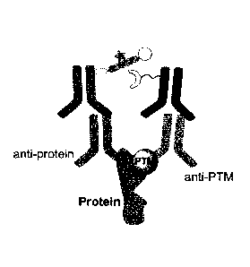
such as Phi29 DNA polymerase is used, the circular DNA template can be
replicated hundreds to thousands of times longer and as a result producing
ssDNA molecules from hundreds of nanometers to microns in length (see
Angewandte Chemie International Edition, 2008, 47, 6330-6337). After the
amplification, the replicated DNA can be detected via detection systems. Thus,
a visible signal is indicative that the targets of interest are in close
proximity.
These assays feature the use of several DNA-antibody conjugates as well as
enzymes such as DNA ligase and DNA polymerase. These conjugates and
- 2 -
enzymes can be expensive, and they also require stringent assay conditions in
order for proper function and stability. Furthermore, the approach generates
an
amplified DNA sequence, while easy to detect, may not remain co-localized
with the proximity event detected.
[0003] Another assay for investigating protein-protein interactions
includes a
dual binders (DB) assay, which utilizes a bi-specific detection agent
consisting
of two Fab fragments with fast off-rate kinetics joined by a flexible linker
(Development of Bispecific Molecules for the In Situ Detection of Protein-
Protein Interactions and Protein Phosphorylation, Chemistry & Biology 21, 1-
12,
March 20, 2014). In principle, because the dual binders comprise Fab
fragments with fast off-rate kinetics, the dual binders are washed off if only
one
of the Fab fragments is bound to its epitope (simultaneous cooperative binding
= of both Fab fragments of the dual binder prevents dissociation of the
dual
binder and leads to positive staining/visibility). These approaches require
the
specific development of fab fragments with specific binding. kinetics, which
makes their implementation to the breadth of targets of interest unreasonable.
[0004] In another approach, Grossman etal. describe an assay for
the
detection of proximal target nucleic acids by the transfer of a reporter group
from a donating probe to a nearby accepting probe. The probes are designed
to anneal adjacently to complementary segments of the target nucleic acid.
The annealing of the probe pair (in close proximity) allows for reactions
(e.g., a
thio-exchange reaction, etc.) to occur, which facilitates the transfer of the
reporter group (e.g., fluorescence quencher) (see Grossman et aL, Angew.
Chem.=Int Ed. 2008, 47, 7119-7122).
[0005] According to another approach, PCT Published Application
W02014/139980
related to proximity assays and tools for enabling the same, describes the use
of
a biotin ligase substrate and an enzyme to perform a proximity assay. The
method provides detection of target molecules and proximity while maintaining
CA 2965872 2020-02-05
CA 02965872 2017-04-26
WO 2016/083364
PCT/EP2015/077484
- 3 -
the cellular context of the sample. The use of biotin ligase such as an enzyme
from Escherichia coli and peptide substrate such as amino-acid substrate for
that enzyme provides for a sensitive and specific detection of protein-protein
interactions in FFPE samples. Because biotin ligase can efficiently
biotinylate
appropriate peptide substrate in the presence of biotin and the reaction can
only occur when the enzyme makes physical contact with the peptide substrate,
biotin ligase and the substrate can be separately conjugated to two antibodies
that recognize targets of interest respectively
SUMMARY OF THE INVENTION
[0006] The present invention features methods for detecting target
proximity.
The invention features, as described more inclusively herein, methods for
detecting target proximity wherein (i) a chemical reaction step forms a
covalent
bond between a pair of modified specific binding molecules (e.g., two
antibodies each conjugated with a bridge component adapted to form a
covalent bond (chemical ligation) when in close proximity and upon external
stimulation, one of the bridge components also comprises a cleavable bond and
a detectable moiety) and OD a subsequent/separate cleavage step cleaves the
bridge component originally associated with the detectable moiety so that the
detectable moiety is ultimately transferred to the opposing specific binding
molecule. Detection of the detectable moiety indicates target proximity as the
detectable moiety that is not engaged in chemical bond formation due to
insufficient proximity would be removed from the sample by washing.
[0007] The methods of the present invention are compatible with both
chromogenic and fluorogenic detection systems and the methods may be
performed either manually or in an automated system. Because the methods of
the present invention do not feature the use of enzymes such as DNA ligase or
DNA polymerase, the methods use less sensitive and costly reagents for
detecting target proximity and one that may have more flexibility and
tolerance
with respect to assay conditions.
- 4 -
=
[0008] Importantly, the specific binding molecules of the
present invention
(e.g., antibodies) may be conjugated with one or multiple (e.g., two, three,
four,
f(ve, 6, 7, 8, 9, 10, or more) bridge components. This may be advantageous as
multiple bridge components can both provide an enhanced (e.g., darker) signal
as well as a greater likelihood of achieving a signal.
[0009] The diversity of uses for the present invention is
surprising. For
example, the included examples demonstrate that the invention may be used to
detect protein-protein interactions and post-translational modification (PTM)
states. As such, provided are approaches for detecting various proximal
biologically significant targets such as protein dimerization, protein
fusions,
associations, and PTM including phosphorylation, glycosylation,
ubiquitination,
SUMOylation, nitrosylation, methylation, acetylation, lipidation, and/or the
like.
[0010] Any feature or combination of features described herein
are included
within the scope of the present invention provided that the features included
in
any such combination are not mutually inconsistent as will be apparent from
the context, this specification, and the knowledge of one.of ordinary skill in
the
art. Additional advantages and aspects of the present invention are apparent
in the following detailed description.
[0011] For example, in some embodiments, methods of the present
invention
comprise binding a first modified binding molecule to the first target to form
a
first complex, and binding a second modified binding molecule to the second
target to form a second complex.
[0012] The first modified binding molecule may comprise a
cleavable bridge
= component (or more than one cleavable bridge component), wherein the
cleavable bridge component comprises a cleavage site (e.g., a disulfide bond,
a
vicinal diol, a vicinal hydroxylamine, a nitrophenyl derivative, etc.), a
detectable
moiety (e.g., one, at least one, at least two, at least 3, at least 4, at
least 5, at
least 6, at least 7, at least 8, at least 9, at least 10, etc.), and a first
chemical
ligation group (e.g., an azide, a thioester, a tetrazole ring, etc.). The
cleavage
= CA 2965872 2020-02-05
CA 02965872 2017-04-26
WO 2016/083364
PCT/EP2015/077484
- 5 -
site is more proximal to the first modified binding molecule than is the
detectable moiety and the first chemical ligation group. The first chemical
ligation group is located at a terminus of the cleavable bridge component The
first chemical ligation group is stable under physiological conditions.
[0013] The second modified binding molecule may comprise a non-cleavable
bridge component (or more than one non-cleavable bridge component)
wherein the non-cleavable bridge component comprises a second chemical
ligation group (e.g., an alkyne group, a halogen group (-Cl, -Br,-I, etc.), an
alkene group, etc.). The second chemical ligation group is located at a
terminus of the non-cleavable bridge component. The second chemical ligation
group is stable under physiological conditions. The chemical ligation group is
interchangeable between the first and the second modified binding molecule.
[0014] In some embodiments, the first modified binding molecule
comprises
a first antibody, and the second modified binding molecule comprises a second
antibody. In some embodiments, the first modified binding molecule comprises
a first primary antibody and a first secondary antibody, and the second
modified
binding molecule comprises a second primary antibody and a second
secondary antibody; the first secondary antibody is specific for the first
primary
antibody and not the second primary antibody, and the second secondary
antibody is specific for the second primary antibody and not the first primary
antibody; and the cleavable bridge component is bound to the first secondary
antibody and the non-cleavable bridge component is bound to the second
secondary antibody.
[0015] The method may further comprise covalently linking the first
chemical
ligation group to the second chemical ligation group to form a covalently
bonded unit, cleaving the cleavage site of the cleavable bridge component such
that the covalently bonded unit is bound to the second modified binding
molecule and not the first modified binding molecule, removing cleavable
bridge components that are not part of a covalently bonded unit (e.g.,
washing),
CA 02965872 2017-04-26
WO 2016/083364
PCT/EP2015/077484
- 6 -
and making the detectable moiety visible (e.g., via a chromogenic system, a
fluorescence system, etc.).
[0016] The methods of the present invention may comprise binding a
first
primary antibody to the first target and binding a second primary antibody to
the second target; then binding a first secondary antibody to the first
primary
antibody and binding a second secondary antibody to the second primary
antibody, wherein the first secondary antibody is specific for the first
primary
antibody and not the second primary antibody, and the second secondary
antibody is specific for the second primary antibody and not the first primary
antibody. The first secondary antibody may be the first modified binding
molecule as described above, e.g., comprising a cleavable bridge component as
previously described. The second secondary antibody may be the second
modified binding molecule as described above, e.g., comprising a non-cleavable
bridge component as described above. The methods may further comprise
covalently linking the first chemical ligation group to the matching second
chemical ligation group to form a covalently bonded unit; cleaving the
cleavage
site of the cleavable bridge component such that the covalently bonded unit is
bound to the second secondary antibody and not the first secondary antibody;
removing cleavable bridge components that are not part of a covalently bonded
unit; and making the detectable moiety visible.
[0017] In some embodiments, visibility of the detectable moiety
indicates that
the first target and the second target are no more than 60 nm apart, no more
than 50 nm apart, no more than 40 nm apart, no more than 30 nm apart, no
more than 25 nm apart, no more than 15 nm apart, no more than 10 nm apart,
no more than 5 nm apart, etc.
[0018] Methods of the present invention may be performed manually or
using an automated staining machine. One aspect of the present is that the
reagents, and kits for performing the assay, are sufficiently stable so that
they
can be used on an automated staining instrument. For example, the reagents
CA 02965872 2017-04-26
WO 2016/083364
PCT/EP2015/077484
- 7 -
are configurable to have a shelf-life of greater than 1 month at room
temperature.
[0019] In some embodiments, the detectable moiety comprises a non-
endogenous compound, a peptide tag, (e.g., HA-tag derived from
hemagglutinin, FLAG tag, Myc Tag, V5 Tag, [-Tag, VSV Tag, etc.) or an
oligonucleotide. In some embodiments, the cleavable bridge component
comprises x polyethylene glycol groups (PEGx), wherein x 1. In some
embodiments, the non-cleavable bridge component comprises x polyethylene
glycol groups (PEGx), wherein x 1.
[0020] In some embodiments, the step of covalently linking the first
chemical
ligation group to the second chemical ligation group comprises contacting the
sample with a catalyst (e.g., copper (I) for initiating a Huisgen 1,3-dipolar
cycloaddition reaction), ultraviolet light, or a deprotection condition (e.g.,
hydrazine (N2H4)). In some embodiments, the chemical ligation groups are
adapted to form the covalently bonded unit in less than two hours.
[0021] In some embodiments, the step of cleaving the cleavage site of
the
cleavable bridge component comprises contacting the sample with a reducing
agent (e.g., dithiothreitol (DTT), beta-mercaptoethanol (13ME), or tris(2-
carboxyethyl)phosphine [[CEP)), sodium periodate (Na104), or ultraviolet
light.
In some embodiments, the cleavage site comprises a disulfide bond, a vicinal
diol, a vicinal hydroxylamine, or a nitrophenyl derivative.
[0022] One aspect of the present invention is that the assay can be
done in a
time that is both commercially reasonable and chemically robust (e.g. the
times
are sufficiently low such that the adverse impact of hydrolysis and thermal
degradation are not impactful on the assay performance - the main reason for
shorter time is more for increased test efficiency). In some embodiments, the
chemical ligation groups are adapted to form the covalently bonded unit in
less
than four hours, three hours, two hours, or one hour.
[0023] The present invention also features compositions for detecting
target
CA 02965872 2017-04-26
WO 2016/083364
PCT/EP2015/077484
- 8 -
proximity, e.g., compositions used in methods described herein. For example,
the present invention features a composition comprising a cleavable bridge
component, as described herein. The composition may further comprise an
antibody conjugated to the cleavable bridge component. In some
embodiments, the antibody is conjugated with at least one cleavable bridge
component. In some embodiments, the antibody is conjugated with at least two
cleavable bridge components, at least five cleavable bridge components, at
least ten cleavable bridge components, or at least fifteen cleavable bridge
components. The present invention also features a composition comprising a
non-cleavable bridge component, as described herein. The composition may
further comprise an antibody conjugated to the non-cleavable bridge
component. In some embodiments, the antibody is conjugated with at least one
non-cleavable bridge component. In some embodiments, the antibody is
conjugated with at least two non-cleavable bridge components, at least five
non-cleavable bridge components, at least ten non-cleavable bridge
components, or at least fifteen cleavable bridge components.
[0024] Kits for practicing the disclosed embodiments are also
disclosed. For
example, the present invention features a kit comprising a first modified
binding
molecule and/or a second modified binding molecule as described herein. The
kit may further comprise a catalyst effective for covalently linking the first
chemical ligation group to the second chemical ligation group to form a
covalently bonded unit. In some embodiments, the chemical ligation groups are
adapted to form the covalently bonded unit in less than four hours, three
hours,
two hours, or one hour. In some embodiments, the kit further comprises a
reagent for cleaving the cleavage site of the cleavable bridge component. In
some embodiments, the kit further comprises a system for making the
detectable moiety visible.
[0025] In some embodiments, the first target comprises a target
protein and
the second target comprises a post-translational modification (e.g.,
phosphorylation, ubiquitination, glycosylation, ubiquitination. etc.). Thus,
the
CA 02965872 2017-04-26
WO 2016/083364
PCT/EP2015/077484
- 9 -
assays of the present invention may be used for in situ detection of a target
protein having post-translational modifications (am phosphorylation,
ubiquitination, glycosylation, ubiquitination. etc.).
BRIEF DESCRIPTION OF THE DRAWINGS
[0026] FIG. 1A-FIG. 1C are schematic depictions of modified specific
binding
moieties. FIG. 1A and FIG. 1B show schematic depictions of a specific binding
moiety and a cleavable bridge component. The cleavable bridge components
comprise a cleavage site (cleavable linker), a detectable moiety (hapten), and
a
chemical ligation group (CL). The hapten (detectable moiety) and the chemical
ligation group can be in a linear or branched arrangement. Linkers can be
used among all three functionalities. The cleavable linker is more proximal to
the specific binding moiety than the other two groups. FIG. 1C shows a
specific
binding moiety (e.g., antibody) and a non-cleavable bridge component. The
cleavable bridge component comprises a linker and a chemical ligation group
(CL).
[0027] FIG. 2 shows non-limiting exemplary structures of the chemical
ligation reactive groups (CL-a and CL-b) and the respective condition for the
formation of covalent bond.
[0028] FIG. 3 is a schematic illustration showing the concept and
major steps
involved in a proximity assay according to the present disclosure. The top
portion shows the proximity assay in a system wherein the two targets of
interest are in close proximity. The bottom portion shows the proximity assay
in
a system wherein the two targets of interest are not in close proximity. The
present invention is not limited to the components and steps described in FIG.
3.
[0029] FIG. 4 is a schematic depiction showing a non-limiting example
of an
antibody conjugate comprising an antibody with a cleavable bridge
components (top illustration) and a non-limiting example of an antibody
conjugate comprising a non-cleavable bridge component (bottom illustration).
CA 02965872 2017-04-26
WO 2016/083364
PCT/EP2015/077484
- 10 -
[0030] FIG. 5 shows an alternative cleavable bridge component (tri-
functional
molecule) wherein the cleavable bridge component comprises a scaffold, and
the detectable moiety/hapten is bound to the scaffold.
[0031] FIG. 6 is a schematic illustration showing the detection of
protein
post-translational modification (PTM), such as a phosphorylation or
ubiquitination using an assay of the present invention.
[0032] FIG. 7 is a schematic illustration showing the synthesis of
Conjugate A
of goat anti-rabbit (GAR) with cleavable bridge component comprising a PEG8
linker, a disulfide bond, a hemagglutinin tag (HA-tag) as hapten, and an azide
group (N3).
[0033] FIG. 8 is a representative SEC chromatogram showing the
purification
of the GAR-HA peptide conjugate of FIG. 7.
[0034] FIG. 9 is a photomicrograph showing detection of the detectable
moiety/hapten HA-tag using IHC methods using a commercially available DAB
chromogenic detection system.
[0035] FIG. 10 is a flow chart showing steps used for a proximity
assay and a
subsequent immunohistochemistry assay for detection of the detectable
moiety/hapten and an alternative detection approach using a fluorophore-
tyramide (Cy5-Tyr) detection method.
[0036] FIG. 11 is a flow chart showing steps used for determining
conditions
for complete cleavage of HA-tag from Conjugate A (see Example 3).
[0037] FIG. 12A-FIG. 12D are photomicrographs showing stained tonsil
tissue
slides following assays for Ki67 using various concentrations of
dithiothreitol
(DDT), no DTT (FIG. 12A), 50mM DTT (FIG. 12B), 75 mM DTT (FIG. 12C), and
100mM DTT (24 min DTT incubation, hapten-tyramide amplification (HQ-Tyr
AMP) used) (FIG. 12D), as described in Example 3, which was performed to
establish concentrations of DTT sufficient for in situ cleavage.
[0038] FIG. 13A and FIG. 13B are photomicrographs showing stained skin
CA 02965872 2017-04-26
WO 2016/083364
PCT/EP2015/077484
- 11 -
tissue sections for which assays for E-cadherein (E-cad) have been performed
using a cleavable bridge component showing no DTT (FIG. 13A), and 50mM
DTT (24 min DTT incubation, hapten-tyramide amplification (HQ-Tyr AMP)
used) (FIG. 13B), as described in Example 3.
[0039] FIG. 14A-FIG. 14C are photomicrographs showing detection of CD20
in tonsil sections showing the effect of Cu(I)/L ratio and the attendant
staining.
FIG. 14A shows a Cu(I)/L ratio of 1:5, FIG. 14B shows a Cu(1)/1_ ratio of 1:3,
and
FIG. 14C shows a control with no Cu(I) added. A reactive acetylene group in
the conjugate of GAM-PEG4-CCH was used (see Example 3). Specific detection
of CD20 was achieved when CO) was added. No staining was observable
when Cu was omitted. This indicates the presence of reactive acetylene
functionality in GAM conjugate.
[0040] FIG. 15A and FIG. 15B are photomicrographs showing detection of
E-
cadherin(E-cad)/P-catenin(13-cat) in normal tonsil tissue sections using
commercial DAB chromogenic detection with CO) (FIG. 15A) and without CO)
(FIG. 15B).
[0041] FIG. 16A-FIG. 16D are photomicrographs showing detection of E-
cadherin(E-cad)/13-catenin(13-cat) in normal tonsil tissue sections with
commercially available hapten-tyramide amplification (VENTANA OPTI VIEW
Amplification Kit) showing proximity assay omitting Mouse anti-pi-cat (FIG.
16A), proximity assay omitting Rabbit anti-E-cad (FIG. 16B), Rabbit anti-E-cad
and Mouse anti-13-cat proximity assay with Cu(I) (FIG. 16C), and Rabbit anti-E-
cad and Mouse anti-13-cat proximity assay excluding Cu(I) (FIG. 16D).
[0042] FIG. 17A-FIG. 17D are photomicrographs showing an assay for
detecting the proximity of E-cadherin(E-cad) and p120-catenin (p120) in normal
tonsil sections slides using CO) and VENTANA OPTI VIEW DAB and VENTANA
Amplification Kit (FIG. 17A), and showing the negative control without
inclusion
of Cu(I) (FIG. 17B). FIG. 17C and FIG. 17D are photomicrographs of sections in
which E-cadherin was detected using a mouse-anti-Ecad and a rabbit-anti-
CA 02965872 2017-04-26
WO 2016/083364
PCT/EP2015/077484
- 12 -
Ecad primary antibodies to bind to different epitopes of the protein in normal
tonsil tissue sections with the proximity assay as described herein (FIG. 17C)
and the proximity assay excluding CO) (FIG. 17D).
[0043] FIG. 18A-FIG. 18D are photomicrographs showing detection of E-
cadherin(E-cad)/13-catenin(13-cat) in normal skin tissue sections stained with
VENTANA OPTIVIEW DAB with amplification where FIG. 18A shows the
proximity assay between [-cad and 13-cat and FIG. 18B is a control in which
the
CO) was excluded. FIG. 18C also shows the proximity assay between [-cad
and 13-cat in a different skin section, and FIG. 18D is the corresponding
negative
control in which the CO) catalyst was excluded.
[0044] FIG. 19A-FIG. 19C are photomicrographs showing fluorophore-
tyramide based detection of Ecad/13-cat in normal tonsil sections using Cy5-
Tyr
(Blue = DAPI, Red = Cy5 for Ecad/[3-cat).
[0045] FIG. 20A-FIG. 20D are photomicrographs showing the proximity
assay
of CD19/CD21 in normal tonsil tissue sections stained with VENTANA
OPTIVIEW DAB with amplification (FIG. 20A). FIG. 206 is a control of FIG. 20A
in which the CO) was excluded. FIG. 20C shows detection of CD21 by using
mouse-anti-CD21 and rabbit-anti-CD21 primary antibodies in normal tonsil
tissue, and FIG. 20D is the control of FIG. 20C in which the CO) was excluded.
[0046] FIG. 21A-FIG. 21C are photomicrographs showing the proximity assay
being used to detect HER2 phosphorylation using anti-HER2 (Clone 4135) and
an anti-p-Tyr antibody to detect phosphorylation. The tissue is from a 3-in-1
xenograft slides using DAB chromogenic detection with FIG. 21A showing
addition of anti-HER2 antibody only, FIG. 216 showing the proximity assay
using
the anti-HER2 (Clone 465) and the anti-p-Tyr antibody with CO), and FIG. 21C
showing the control of FIG. 216 in which CO) was excluded.
[0047] FIG. 22 is a schematic illustration of the detection of
ubiquitination of
a protein using an assay as described herein.
CA 02965872 2017-04-26
WO 2016/083364
PCT/EP2015/077484
- 13 -
[0048] FIG. 23A-FIG. 23D are photomicrographs showing detection of
ubiquitination of progesterone receptor [PR) in MCF-7 xenograft. MCF-7 cells
are PR positive. FIG. 23A shows the proximity assay between PR and Ubiquitin.
FIG. 23B is a control of the proximity assay without the CO) catalyst. FIG.
23C
and FIG. 23D are the standard IHC detection of PR (FIG. 23C) and ubiquitin
(FIG.
23D) each detected using commercially available VENTANA ULTRAVIEW DAB
detection.
[0049] FIG. 24A-FIG. 24D are photomicrographs showing detection of
ubiquitination of progesterone receptor [PR) in ZR-7551 xenograft. ZR-751
cells are PR positive. FIG. 24A shows the proximity assay between PR and
Ubiquitin. FIG. 24B is a control of the proximity assay without the CO)
catalyst.
FIG. 24C and FIG. 24D are the standard IHC detection of PR (FIG. 24C) and
ubiquitin (FIG. 24D) each detected using commercially available VENTANA
ULTRAVIEW DAB detection.
[0050] FIG. 25A-FIG. 25D are photomicrographs in which FIG. 25A is the
same as FIG. 24A except that it shows regions demarcated as (B)-(D), which
are shown at higher magnification in the photomicrographs of FIG. 25B- FIG.
25D. The higher magnification more readily shows the heterogeneity of
ubiquitinated PR.
[0051] FIG. 26A-FIG. 26D are photomicrographs showing the no detection of
ubiquitination of progesterone receptor [PR) in Calu-3 xenograft. Calu-3 cells
are PR negative. FIG. 26A shows the lack of signal for a proximity assay
between PR and Ubiquitin. FIG. 26B is a control of the proximity assay without
the CO) catalyst. FIG. 26C and FIG. 26D are the standard IHC detection of PR
(FIG. 26C) and ubiquitin (FIG. 26D) each detected using commercially available
VENTANA ULTRAVIEW DAB detection.
[0052] FIG. 27A-FIG. 27C are photomicrographs showing detection of
ubiquitination of HER2 in Calu-3 cells. Calu-3 cells are HER2 positive. FIG.
27A
shows the proximity assay between HER2 and Ubiquitin. FIG. 27B is a control of
CA 02965872 2017-04-26
WO 2016/083364
PCT/EP2015/077484
- 14 -
the proximity assay without the Cu(I) catalyst. FIG. 27C shows a region of
FIG.
27A under higher magnification. Ubiquitinated HER2 is detected in Calu-3
cells.
[0053] FIG. 28A-FIG. 28D are photomicrographs showing detection of
MLH1-
PMS2 heterodimers using the proximity assay of the present invention. FIG.
28A shows standard IHC of PMS2 in Hela cell xenograft using rabbit-anti-PMS2
mAb. FIG. 28B shows IHC of MLH1 in Hela cell xenog raft using mouse-anti-
MLH1 mAb. FIG. 28C shows a proximity assay of MLH1-PMS2 heterodimers
and FIG. 28D is a control of the proximity assay without the Cu(I) catalyst.
DESCRIPTION OF PREFERRED EMBODIMENTS
I. TERMS
[0054] Unless otherwise noted, technical terms are used according to
conventional usage. Definitions of common terms in molecular biology may be
found in Benjamin Lewin, Genes V, published by Oxford University Press, 1994
(ISBN 0-19-854287-9); Kendrew etal. (eds.), The Encyclopedia of Molecular
Biology, published by Blackwell Science Ltd., 1994 (ISBN 0-632-02182-9); and
Robert A. Meyers (ed.), Molecular Biology and Biotechnology: a Comprehensive
Desk Reference, published by VCH Publishers, Inc., 1995 (ISBN 1-56081-569-8).
Unless otherwise explained, all technical and scientific terms used herein
have
the same meaning as commonly understood by one of ordinary skill in the art to
which this disclosure belongs. The singular terms "a," "an," and "the" include
plural referents unless context clearly indicates otherwise. Similarly, the
word
"or" is intended to include "and" unless the context clearly indicates
otherwise.
It is further to be understood that all base sizes or amino acid sizes, and
all
molecular weight or molecular mass values, given for nucleic acids or
polypeptides are approximate, and are provided for description. Although
methods and materials similar or equivalent to those described herein can be
used in the practice or testing of this disclosure, suitable methods and
materials
- 15 -
are described below. The term "comprises" means "includes."
[0055]
In case of conflict, the present specification, including
explanations of terms, will control. In addition, the materials, methods, and
examples are illustrative and not intended to be limiting.
[0056] In order to facilitate review of the various
embodiments of this
disclosure, the following explanations of specific terms are provided:
[0057] Administration: To provide or give a subject an
agent, for example,
a composition, by any effective route. Exemplary routes of administration
include, but are not limited to, oral, injection (such as subcutaneous,
intramuscular, intradermal, intraperitoneal, and intravenous), sublingual,
rectal,
transdermal (e.g., topical), intranasal, vaginal and inhalation routes.
[0053] Agent: Any substance or any combination of substances that is
useful for achieving an end or result; for example, a substance or combination
of substances useful for decreasing or decreasing a protein-protein
interaction.
In some embodiments, the agent is a therapeutic agent, such as a therapeutic
agent for the treatment of cancer.
[0059] Antibody: A polypeptide ligand including at least a
light chain or
heavy chain immunoglobulin variable region which specifically binds an epitope
of an antigen or a fragment thereof. Antibodies include intact immunoglobulins
and the variants of them well known in the art, such as Fab', RabY2 fragments,
= 25 single chain Fv proteins (scFv), and disulfide
stabilized Fv proteins (dsFv). A
= scFv protein is a fusion protein in which a light chain variable region
of an
= antibody and a heavy chain variable region of an antibody are bound by a
linker, while in dsFvs, the chains have been mutated to introduce a disulfide
= bond to stabilize the association of the chains. The term also includes
CA 2965872 2020-02-05
CA 02965872 2017-04-26
WO 2016/083364
PCT/EP2015/077484
- 16 -
genetically engineered forms such as chimeric antibodies (for example,
humanized murine antibodies) and heteroconjugate antibodies (such as,
bispecific antibodies). See also, Pierce Catalog and Handbook, 1994-1995
(Pierce Chemical Co., Rockford, IL); Kuby, J., Immunology, 3rd Ed., W.H.
Freeman & Co., New York, 1997.
[0060] The antibodies disclosed herein specifically bind a defined
target (or
multiple targets, in the case of a bispecific antibody). Thus, an antibody
that
specifically binds to a target is an antibody that binds substantially to the
target,
for example cells or tissue expressing the target. It is, of course,
recognized
that a certain degree of non-specific interaction may occur between an
antibody and a non-target.
[0061] Typically, specific binding results in a much stronger
association
between the antibody and protein or cells bearing the antigen than between the
antibody and protein or cells lacking the antigen. Specific binding typically
results in greater than a 2-fold increase, such as greater than 5-fold,
greater
than 10-fold, or greater than 100-fold increase, in amount of bound antibody
(per unit time) to a protein including the epitope or cell or tissue
expressing the
target epitope as compared to a protein or cell or tissue lacking this
epitope.
Specific binding to a protein under such conditions requires an antibody that
is
selected for its specificity for a particular protein. A variety of
immunoassay
formats are appropriate for selecting antibodies or other ligands specifically
immunoreactive with a particular protein. For example, solid-phase ELISA
immunoassays are routinely used to select monoclonal antibodies specifically
immunoreactive with a protein. See Harlow & Lane, Antibodies, A Laboratory
Manual, Cold Spring Harbor Publications, New York (1988), for a description of
immunoassay formats and conditions that can be used to determine specific
immunoreactivity.
[0062] Biological sample: A biological specimen containing biological
molecules, including genomic DNA, RNA (including mRNA and microRNA),
CA 02965872 2017-04-26
WO 2016/083364
PCT/EP2015/077484
- 17 -
nucleic acids, proteins, peptides, and/or combinations thereof. In some
examples, the biological sample is obtained from a subject. In other examples,
the biological sample is a cell culture, including a cell culture grown from a
biological sample obtained from a subject. Biological samples include all
clinical samples useful for detecting disease (e.g., cancer) in subjects,
including,
but not limited to, cells, tissues, and bodily fluids, such as blood,
derivatives and
fractions of blood (such as serum); as well as biopsied or surgically removed
tissue, for example tissues that are unfixed, frozen, or fixed in formalin or
paraffin. In a particular example, a biological sample is obtained from a
subject
having or suspected of having a tumor; for example, a subject having or
suspected of having breast cancer, ovarian cancer, stomach cancer or uterine
cancer. In some embodiments, the subject has or is suspected of having a
carcinoma.
[0063] Buffers: Buffer solutions are commonly used to maintain correct
pH
levels for biological and chemical systems. Many of the exemplary
embodiments disclosed herein include using a buffer solution. Representative
buffering agents or salts that may be present in the buffer include, but are
not
limited to, Iris, Tricine, HEPES, MOPS, TAPS, Bicine, TAPSO, TES, PIPES,
Cacodylate, SSC, MES, KCI, NaCI, potassium acetate, NH4-acetate, potassium
glutamate, NH4CI, ammonium sulphate, MgCl2, magnesium acetate and the
like. One buffer solution is phosphate buffered saline (PBS). Another buffer
solution is biotin ligase reaction buffer (0.1 M KCI, 5.5 mM MgC12, 50 mM
Tris.HCI (pH = 8.0), 0.05% Brij-35, 0.1 mM dithiothreitol (DTT), 3 mM ATP, and
60 pM biotin). The amount of buffering agent will typically range from about 5
to 150 mM, usually from about 10 to 100 mM, and more usually from about 20
to 50 mM, where in certain preferred embodiments the buffering agent will be
present in an amount sufficient to provide a pH ranging from about 6.0 to
about
9.5, more typically a pH range of from about 6.5 to about 7.4 at room
temperature. Other agents that may be present in the buffer medium include
chelating agents, such as EDTA, EGTA and the like.
- 18 -
[0064] Chromogenic Staining: Chromogenic substrates have been used
widely for immunohistochemistry for many years and for in situ hybridization
more recently. Chromogenic detection offers a simple and cost-effective
detection method. Chromogenic substrates have traditionally functioned by
precipitating when acted on by the appropriate enzyme. That is, the
traditional
chromogenic substance is converted from a soluble reagent into an insoluble,
colored precipitate upon contacting the enzyme. The resulting colored
precipitate requires no special equipment for processing or visualizing. There
are several qualities that successful IHC or ISH chromogenic substrates share.
First, the substance should precipitate to a colored substance, preferably
with a
very high molar absorptivity. The enzyme substrate should have high solubility
and reagent stability, but the precipitated chromogen products should be very
insoluble, preferably in both aqueous and alcohol solutions. Enzyme turnover
rates should be very high so as to highly amplify the signal from a single
enzyme in a short amount of time. Until now, a relatively small number of
chromogenic substances have been identified that legitimately possess all of
these qualities. Reference is made to U.S. Published Application No.
2013/0260379, entitled QUINONE METHIDE PRECURSORS
AND USES THEREFORE, filed on February 24, 2014.
/0
Several commercially available chromogenic detection
kits are available from Ventana Medical Systems, Inc. and referenced herein
(AEC Detection Kit 760-020; Enhanced Alkaline Phosphatase Red Detection Kit
760-031; OPTI VIEW DAB IHC Detection Kit 760-700; iVIEW DAB Detection Kit
760-091; ULTRAVIEW Universal Alkaline Phosphatase Red Detection Kit 760-
501; and ULTRAVIEW Universal DAB Detection Kit 760-500).
[0065] Conjugate: Two or more molecules coupled together, for
example,
by a covalent bond or non-covalent interaction.
[0066] Conjugate(ing), join(ing), bond(ing) or link(ing): Coupling a
first
Date Recue/Date Received 2021-02-08
CA 02965872 2017-04-26
WO 2016/083364
PCT/EP2015/077484
- 19 -
molecule to a second molecule. This includes, but is not limited to,
covalently
bonding one molecule to another molecule, non-covalently bonding one
molecule to another (e.g., electrostatically bonding) (see, for example, U.S.
Patent No. 6,921,496), hydrogen bonding, van der Waals forces, and any and all
combinations of such couplings.
[0067] Contacting: Placement in direct association, for example solid,
liquid
or gaseous forms.
[0068] Control: A sample or standard used for comparison with a test
sample, such as a biological sample, e.g., a biological sample obtained from a
patient (or plurality of patients) or a cell culture. In some embodiments, a
cell
culture that is not incubated with a test agent serves as a control for a cell
culture that is incubated with a test agent. In some embodiments, the control
is
a sample obtained from a healthy patient (or plurality of patients) (also
referred
to herein as a "normal" control), such as a normal breast sample. In some
embodiments, the control is a historical control or standard value (i.e. a
previously tested control sample or group of samples that represent baseline
or
normal values). In some embodiments the control is a standard value
representing the average value (or average range of values) obtained from a
plurality of patient samples.
[0069] Coupled: Two or more molecules joined together, either directly or
indirectly. A first atom or molecule can be directly coupled or indirectly
coupled
to a second atom or molecule. A secondary antibody is indirectly coupled to an
antigen when it is bound to a primary antibody that is bound to the antigen.
[0070] Detecting: To identify the existence, occurrence, presence, or
fact of
something. General methods of detecting are known to a person of ordinary
skill in the art and may be supplemented with the protocols and reagents
disclosed herein. For example, included herein are methods of detecting a
first
target proximal to a second target in a biological sample.
[0071] FFPE: Formalin fixed paraffin embedded sample.
CA 02965872 2017-04-26
WO 2016/083364
PCT/EP2015/077484
- 20 -
[0072] Hapten: A hapten is a molecule, typically a small molecule that
can
combine specifically with an antibody, but typically is substantially
incapable of
being immunogenic except in combination with a carrier molecule. Many
haptens are known and frequently used for analytical procedures, such as di-
nitrophenyl, biotin, digoxigenin, fluorescein, rhodamine, or combinations
thereof. Other haptens have been specifically developed by Ventana Medical
Systems, Inc., assignee of the present application, including haptens selected
from oxazoles, pyrazoles, thiazoles, nitroaryls, benzofurans, triterpenes,
ureas,
thioureas, rotenoids, coumarins, cyclolignans, and combinations thereof, with
particular hapten examples of haptens including benzofurazan, nitrophenyl, 4-
(2-hydroxypheny1)-1H-benzo[b][1,4]diazepine-2(3H)-one, and 3-hydroxy-2-
quinoxalinecarbamide. Plural different haptens may be coupled to a polymeric
carrier. Moreover, compounds, such as haptens, can be coupled to another
molecule using a linker, such as an NHS-PEG linker.
[0073] Immune complex: The binding of antibody to a soluble antigen
forms an immune complex. The formation of an immune complex can be
detected through conventional methods known to the person of ordinary skill in
the art, for instance immunohistochemistry, immunoprecipitation, flow
cytometry, immunofluorescence microscopy, ELISA, immunoblotting (i.e.,
Western blot), magnetic resonance imaging, CT scans, X-ray and affinity
chromatography. Immunological binding properties of selected antibodies may
be quantified using methods well known in the art.
[0074] Linker: Two components may be jointed together either directly
through a bond or indirectly through a linker. Linkers may be bifunctional,
ie.,
the linker includes a functional group at each end, wherein the functional
groups are used to couple the linker to the two components, either covalently
or non-covalently. The two functional groups may be the same, i.e., a
homobifunctional linker, or different, le., a heterobifunctional linker, but
more
typically are heterobifunctional. Where linkers are employed, suitable
functional
CA 02965872 2017-04-26
WO 2016/083364
PCT/EP2015/077484
-21 -
groups are selected to allow attachment of the two components, while not
impairing the functionality of the components. Linkers of interest may vary
widely depending on the components in the conjugate. In many embodiments
the linker, when present, is biologically inert.
[00751 A linker may be used to link the detectable moiety and/or the
chemical ligation group and/or the cleavage site of the cleavable bridge
component to the specific binding molecule (or to link the chemical ligation
group of the non-cleavable bridge component to the specific binding moiety).
Linkers of different lengths can be selected. Where linkers are employed, such
groups may be chosen to allow for attachment of the two components of the
conjugate, while not impairing their functionality. Such terminal functional
groups, include by way of example and without limitation, amines, alcohols,
thiols, hydrazides, carbonyl-reactive group (such as aldehydes, acids and
esters), vinyl ketones, epoxides, isocyanates, maleimides), functional groups
capable of cycloaddition reactions, forming disulfide bonds, binding to metals
or photo-reactive groups. Specific examples include primary and secondary
amines, hydroxamic acids, N-hydroxysuccinimidyl esters, N-
hydroxysuccinimidyl carbonates, oxycarbonylimidazoles, nitrophenylesters,
trifluoroethyl esters, glycidyl ethers, vinylsulfones, and maleim ides. These
groups facilitate coupling to the specific binding moieties and other desired
compounds. In some embodiments, the linker is at least about 25 daltons, at
least about 50 daltons, at least about 100 daltons, or at least about 500
daltons
(or larger). A first class of linkers may be the aliphatic compounds, such as
aliphatic hydrocarbon chains having one or more sites of unsaturation, or
alkyl
chains. The length of the chain can vary, but typically has an upper practical
limit of about 30 atoms. Chain lengths greater than about 30 carbon atoms
have proved to be less effective than compounds having smaller chain lengths.
Thus, aliphatic chain linkers typically have a chain length of from about 1
carbon atom to about 30 carbon atoms. However, a person of ordinary skill in
the art will appreciate that, if a particular linker has greater than 30
atoms, and
- 22 -
the conjugate still functions as desired, then such chain lengths are still
within
the scope of the present invention.
[0076] En some embodiments, the linker is a straight chain or
branched alkyl
chain functionalized with reactive groups, such as an amino- or mercapto-
hydrocarbon, with more than two carbon atoms in the unbranched chain.
Examples include aminoalkyl, aminoalkenyl and aminoalkynyl groups.
Alternatively, the linker is an alkyl chain of 10-20 carbons in length, and
may be
attached through a Si-C direct bond or through an ester, Si-O-C, linkage (see
U.S. Patent No. 5,661,028 to Foote). Other
linkers are available and known to a person of ordinary skill in the art (see,
e.g.,
U.S. Patent Nos. 5,306,518, 4,711,955 and 5,707,804).
[0077] A second class of linkers includes the alkylene oxides. The
alkylene,
oxides are represented herein by reference to glycols, such as ethylene
glycols.
A person of ordinary skill in the art will appreciate that, as the number of
oxygen atoms increases, the hydrophilicity of the compound also may increase.
Thus, linkers of the present invention typically have a formula of C-OCH2CH2-
1,
where n is from about 2 to about 20, but more particularly n is from about 2
to
about 10, and even more typically from about 4 to about 8, which can be
represented as PEG4 to PEG8.
[0078] Linkers, such as heterobifunctional polyalkyleneglycol
linkers, useful
for practicing certain disclosed embodiments of the present invention are
described in assignee's co-pending applications, including "Nanoparticle
_ Conjugates," U.S. Patent Application No.11/413,778, filed April 28, 2006;
"Antibody Conjugates," U.S. Application No. 12/381,638, filed March 13, 2009;
and "Molecular Conjugate," U.S. Patent Application No. 12/687,564, filed
January 14, 2010, and U.S. Patent No. 7,695,929.
The linkers disclosed in these applications can be used to
link specific binding moieties, biotin ligases, biotin ligase substrates,
signal
CA 2965872 2020-02-05
CA 02965872 2017-04-26
WO 2016/083364
PCT/EP2015/077484
- 23 -
generating moieties and haptens in any and all desired combinations to form
conjugates for use with disclosed embodiments of the present invention.
[0079] Other examples of linkers include, but are not limited to,
peptides,
including natural and non-natural polypeptides, carbohydrates, cyclic or
acyclic
systems that may possibly contain heteroatoms. Linker groups also may
comprise ligands that bind to metals such that the presence of a metal ion
coordinates two or more ligands to form a complex. In another embodiment,
the linker is a pair of molecules, having high affinity for one another. Such
high-affinity molecules include, for example, streptavidin and biotin,
histidine
and nickel (Ni), and GST and glutathione.
[0080] Specific exemplary linkers include: ethylene glycol,
polyalkylene
glycols such as PEG2, PEG3, PEG4, PEG5, PEG8, PEG7, PEG8, PEG8, PEGio, PEG'',
PEG12, PEG13, PEG14, PEG15, PEG16, PEG17, PEG18, PEG18, PEG20, 1,4-
diaminohexane, xylylenediamine, terephthalic acid, 3,6-dioxaoctanedioic acid,
ethylenediamine-N,N-diacetic acid, 1,1'-ethylenebis(5-oxo-3-
pyrrolidinecarboxylic acid), 4,4'-ethylenedipiperidine, succinimidy1-6-
hydrazino-
nicotinamide(S-HyNic, HyNic-NHS), N-succinimidy1-4-formylbenzoate (S-4FB,
4-FB-NHS), maleimide HyNic (MHPH), maleimide 4FB (MTFB), succinimidy1-
[(N-maleimidopropionamido)-octaethyleneglycol] ester (Mal-PEG8-NHS),
succinimidy1-[(N-maleimidopropionamido)-tetraethyleneglycol] ester (Mal-
PEG4-NHS), 4-FB-PEG4-PFP, azidobenzoyl hydrazide, N-[4-Cp-
azidosalicylamino)buty1]-3'421-pyridyldithio]propionamid), bis-
sulfosuccinimidyl
suberate, dimethyladipimidate, disuccinimidyltartrate, N-
maleimidobutyryloxysuccinimide ester, N-hydroxy sulfosuccinimidy1-4-
azidobenzoate, N-succinimidy1[4-azidopheny1]-1,3'-dithiopropionate, N-
succinimidy1[4-iodoacetyl]aminobenzoate, glutaraldehyde, and succinimidy1-4-
[N-maleimidomethyl]cyclohexane-1-carboxylate, 3-(2-pyridyldithio)propionic
acid N-hydroxysuccinimide ester (SPDP), 4-(N-maleimidomethyl)-cyclohexane-
1-carboxylic acid N-hydroxysuccinimide ester (SMCC), and the like.
CA 02965872 2017-04-26
WO 2016/083364
PCT/EP2015/077484
- 24 -
[0081] Instead of chemical linkers, fusion proteins may be constructed
(e.g.,
a peptide can be coupled to a specific binding molecule at the gene level).
[0082] Nucleic acid: A polymer composed of nucleotide units
(ribonucleotides, deoxyribonucleotides, related naturally occurring structural
variants, and synthetic non-naturally occurring analogs thereof) linked via
phosphodiester bonds, related naturally occurring structural variants, and
synthetic non-naturally occurring analogs thereof. Thus, the term includes
nucleotide polymers in which the nucleotides and the linkages between them
include non-naturally occurring synthetic analogs, such as, for example and
without limitation, phosphorothioates, phosphoramidates, methyl phosphonates,
chiral-methyl phosphonates, 2-a-methyl ribonucleotides, peptide-nucleic acids
(PNAs), and the like. Such polynucleotides can be synthesized, for example,
using an automated DNA synthesizer. It will be understood that when a
nucleotide sequence is represented by a DNA sequence A, T, G, C), this
also includes an RNA sequence (i.e., A, U, G, C) in which "U" replaces "T."
[0083] Nucleotide: Term includes, but is not limited to, a base linked
to a
sugar, such as a pyrimidine, purine or synthetic analogs thereof, or a base
linked to an amino acid, as in a peptide nucleic acid (PNA). A nucleotide is
one
unit in a polynucleotide. A nucleotide sequence refers to the sequence of
bases in a polynucleotide.
[0084] Conventional notation is used herein to describe nucleotide
sequences: the left-hand end of a single-stranded nucleotide sequence is the
5'-end; the left-hand direction of a double-stranded nucleotide sequence is
referred to as the 5'-direction. The direction of 5' to 3' addition of
nucleotides
to nascent RNA transcripts is referred to as the transcription direction. The
DNA strand having the same sequence as an mRNA is referred to as the
"coding strand:" sequences on the DNA strand having the same sequence as
an mRNA transcribed from that DNA and which are located 5' to the 5'-end of
the RNA transcript are referred to as "upstream sequences;" sequences on the
CA 02965872 2017-04-26
WO 2016/083364
PCT/EP2015/077484
- 25 -
DNA strand having the same sequence as the RNA and which are 3' to the 3'
end of the coding RNA transcript are referred to as "downstream sequences."
[0085] Oligonucleotide: A linear polynucleotide sequence of between 5
and 100 nucleotide bases in length.
[0086] Proximal: Refers to the qualitative or quantitative distance between
two molecules/targets/epitopes; for example, the distance between two proteins
in a tissue sample. In some embodiments, molecules that are proximal to each
other are within about 100 nm, about 75 nm, about 50 nm, about 35 nm, about
30 nm, about 25 nm, about 20 nm, about 15 nm, about 10 nm, about 5 nm or
less distance of each other. Proximal may also provide a functional
relationship.
For examples, two targets (e.g., two proteins) may be considered proximal if
the
first target is within sufficient distance of the second target for a
component
associated with the first target to be bonded to a component associated with a
second target. Another functional definition of proximal is the dimerization
of
two proteins. Another functional definition of proximal is associated to a
genetic translocation. Two portions of the genome may be considered proximal
when they are adjacent to each other or within 500,000 bp, within 100,000 bp,
within 50,000 bp within 25,000 bp, within 10,000 bp, or within 1,000 bp of
each
other. This contrast to two portions which are not proximal due to a
translocation (either a move to another chromosome or an inversion). The term
proximal also includes a functional meaning as being the distance between
molecules which enables a biologically significant interaction. For example,
the
distance between two proteins in a protein dimer, which has biological
significance, can be said to be proximal.
[0087] Sample: Certain disclosed embodiments utilize biological samples.
A biological sample is typically obtained from a mammalian subject of
interest,
such as a human. The sample can be any sample, including, but not limited to,
tissue from biopsies, autopsies and pathology specimens. Biological samples
also include sections of tissues, for example, frozen sections taken for
CA 02965872 2017-04-26
WO 2016/083364
PCT/EP2015/077484
- 26 -
histological purposes. Biological samples also include cell cultures or
portions
of cell cultures, for example, a cell culture grown from a biological sample
taken
from a subject.
[0088] Biological samples can be obtained from a subject using any
method
known in the art. For example, tissue samples can be obtained from breast
cancer patients who have undergone tumor resection as a form of treatment.
From these patients, both tumor tissue and surrounding non-cancerous tissue
can be obtained. In some embodiments, the non-cancerous tissue sample used
as a control is obtained from a cadaver. In some embodiments, biological
samples are obtained by biopsy. Biopsy samples can be fresh, frozen or fixed,
such as formalin-fixed and paraffin embedded. Samples can be removed from
a patient surgically, by extraction (for example by hypodermic or other types
of
needles), by micro-dissection, by laser capture, or by any other means known
in
the art.
[0089] Specific binding moiety(ies): A member of a specific-binding pair.
Specific binding pairs are pairs of molecules that are characterized in that
they
bind each other to the substantial exclusion of binding to other molecules
(for
example, specific binding pairs can have a binding constant that is at least
103
greater, 104 greater or 105 greater than a binding constant for either of the
two
members of the binding pair with other molecules in a biological sample). The
specific binding moiety used to make the exemplary conjugates disclosed
herein may be any of a variety of different types of molecules, so long as it
exhibits the requisite binding affinity for the target.
[0090] The specific binding moiety may comprise a small molecule or
large
molecule. A small molecule will range in size from about 50 to about 10,000
daltons, more typically from about 50 to about 5,000 daltons, and even more
typically from about 100 to about 1000 daltons. A large molecule is one whose
molecular weight is typically greater than about 10,000 daltons. The small
molecule may be any molecule, typically an organic molecule that is capable of
CA 02965872 2017-04-26
WO 2016/083364
PCT/EP2015/077484
-27 -
binding with the requisite affinity to the target. The small molecule
typically
includes one or more functional groups allowing it to interact with the
target,
for example by hydrophobic, hydrophilic, electrostatic or covalent
interactions.
Where the target is a protein, lipid or nucleic acid, the small molecule
typically
will include functional groups allowing for structural interactions such as
hydrogen bonding, hydrophobic-hydrophobic interactions, electrostatic
interactions, etc. The small molecule ligand often includes an amine, amide,
sulfhydryl, carbonyl, hydroxyl or carboxyl group, and preferably at least two
of
these functional groups.
[0091] The small molecules often comprise cyclic and/or heterocyclic non-
aromatic structures, and/or aromatic or polyaromatic structures substituted
with
one or more of the above functional groups. Also useful small molecules
include structures found among biomolecules, including peptides, saccharides,
fatty acids, steroids, purines, pyrimidines, derivatives, structural analogs
or
combinations thereof.
[0092] The small molecule may be derived from a naturally occurring or
synthetic compound that may be obtained from a wide variety of sources,
including libraries of synthetic or natural compounds. For example, numerous
methods are available for random and directed synthesis of a wide variety of
organic compounds and biomolecules, including the preparation of randomized
oligonucleotides and oligopeptides. Alternatively, libraries of natural
compounds in the form of bacterial, fungal, plant and animal extracts are
available or readily produced. Additionally, natural or synthetically produced
libraries and compounds are readily modified through conventional chemical,
physical and biochemical means, and may be used to produce combinatorial
libraries. Known small molecules may be subjected to directed or random
chemical modifications, such as acylation, alkylation, esterification,
amid ification, etc., to produce structural analogs.
[0093] As such, the small molecule may be obtained from a library of
- 28 -
naturally occurring or synthetic molecules, including a library of compounds
produced through combinatorial means, i.e. a compound diversity combinatorial
library. When obtained from such libraries, the small molecule employed will
have demonstrated some desirable affinity for a target in a convenient binding
affinity assay. Combinatorial libraries, as well as methods for their
production
and screening, are known in the art and are described in U.S. Patent Nos.
5,741,713 and 5,734,018
Additional information concerning specific binding moieties is
provided by assignee's U.S. Patent No. 7,695,929
The specific binding moiety may comprise a large
molecule. Of particular interest as large molecule specific binding moieties
are
antibodies, as well as binding fragments and derivatives or mimetics thereof.
As such, the specific binding moiety may be either a monoclonal or polyclonal
antibody. Also of interest are antibody fragments or derivatives produced
either
recombinantly or synthetically, such as single chain antibodies or scFvs, or
other antibody derivatives such as chimeric antibodies or CDR-grafted
antibodies, where such recombinantly or synthetically produced antibody
fragments retain the binding characteristics of the above antibodies. Such
antibody fragments, derivatives or mimetics of the subject invention may be
readily prepared using any convenient methodology, such as the methodology
disclosed in U.S. Patent Nos. 5,851,829 and 5,965,371
[0094] Also suitable for use as large molecule specific binding
moieties are
polynucleic acid aptamers. Polynucleic acid aptamers may be RNA
oligonucleotides that selectively bind proteins, much in the same manner as a
receptor or antibody (Conrad etal., Methods Enzymol. (19961
267(Combinatorial Chemistry), 336-367), or DNA oligomers that complement
specific DNA target sequences.
[0095] In addition to antibody-based peptide/polypeptide or protein-
based
binding domains, the specific binding moiety may also be a lectin, a soluble
CA 2965872 2020-02-05
CA 02965872 2017-04-26
WO 2016/083364
PCT/EP2015/077484
- 29 -
cell-surface receptor or derivative thereof, an affibody or any
combinatorially
derived protein or peptide from phage display or ribosome display or any type
of combinatorial peptide or protein library. Combinations of any specific
binding
moiety may be used.
[0096] Importantly, the specific binding moiety will be one that allows for
coupling to the second component of the conjugate, or to a linker, without
substantially affecting the binding affinity of the specific binding moiety to
its
target.
[0097] Particular examples of specific binding moieties include
specific
binding proteins (for example, antibodies, lectins, avidins such as
streptavidins,
and protein A). Specific binding moiety(ies) also includes the molecules (or
portions thereof) that are specifically bound by such specific binding
proteins.
[0098] Target: Any molecule for which the presence, location and/or
concentration is or can be determined. Examples of target molecules include
proteins and haptens, such as haptens covalently bonded to proteins. Target
molecules are typically detected using one or more conjugates of a specific
binding molecule and a detectable label. Examples of specific targets include
proteins, carbohydrates, or nucleic acid molecules. Target nucleic acid
molecules include those molecules whose proximity, rearrangement,
amplification, deletion, detection, quantitation, qualitative detection, or a
combination thereof, is sought. For example, the target can be a defined
region
or particular portion of a nucleic acid molecule, for example a portion of a
genome (such as a gene or a region of DNA or RNA containing a gene (or
portion thereof) of interest). The nucleic acid molecule need not be in a
purified form. Various other nucleic acid molecules can also be present with
the target nucleic acid molecule. For example, the target nucleic acid
molecule
can be a specific nucleic acid molecule (which can include RNA or DNA), the
amplification of at least a portion thereof (such as a portion of a genomic
sequence or cDNA sequence) is intended. In some examples, a target nucleic
CA 02965872 2017-04-26
WO 2016/083364
PCT/EP2015/077484
- 30 -
acid includes a viral nucleic acid molecule, or a bacterial nucleic acid
molecule,
such as a nucleic acid molecule from Escherichia coli or Vibrio cholera.
Purification or isolation of the target nucleic acid molecule, if needed, can
be
conducted by methods known to those in the art, such as by using a
commercially available purification kit or the like.
[0099] Tyramide Signal Amplification: An enzyme-mediated detection
method that utilizes the catalytic activity of a peroxidase (such as
horseradish
peroxidase) to generate high-density labeling of a target molecule (such as a
protein or nucleic acid sequence) in situ. TSA typically involves three basic
steps: (1) binding of a specific binding member (e.g., an antibody) to the
target
followed by secondary detection of the specific binding member with a second
peroxidase-labeled specific binding member; (2) activation of multiple copies
of
a labeled tyramide derivative (e.g., a hapten-labeled tyramide) by the
peroxidase; and (3) covalent coupling of the resulting highly reactive
tyramide
radicals to residues (e.g., the phenol moiety of protein tyrosine residues)
proximal to the peroxidase-target interaction site, resulting in deposition of
haptens proximally (diffusion and reactivity mediated) to the target. In some
examples of TSA, more or fewer steps are involved; for example, the TSA
method can be repeated sequentially to increase signal. Methods of
performing TSA and commercial kits and reagents for performing TSA are
available (see, e.g., VENTANA Amplification Kit, Cat. No. 760-080, AmpMap
Detection Kit with TSATm, Cat. No. 760-121, Ventana Medical Systems, Tucson,
AZ; lnvitrogen; kit No. 1-20911, lnvitrogen Corp, Carlsbad, CA). In some
embodiments, TSA is a component of the provided PTDM. Other enzyme-
catalyzed, hapten or signaling linked reactive species can be alternatively
used
as they may become available. Suitable conditions for TSA as well as reagents
and kits for use for tyramide signal amplification are known to a person of
ordinary skill in the art (see, e.g., Bobrow et al, J. lmmuno. Meth., 125:279-
285,
1989).
CA 02965872 2017-04-26
WO 2016/083364
PCT/EP2015/077484
-31 -
[0100] Under conditions sufficient for: A phrase that is used to
describe
any environment that permits a desired activity.
II. DESCRIPTION OF EMBODIMENTS
[0101] Referring now to FIG. 1-28, the present invention features
proximity
assay methods for detecting (in situ) whether two targets of interest are in
close
proximity. The methods of the present invention feature covalent bond
formation. The methods of the present invention are compatible with both
chromogenic and fluorogenic detection systems. The methods of the present
invention may be performed manually, or the methods may be automated, (e.g.,
using an automated staining instrument such as a BENCHMARK XT, Ventana
Medical Systems, Inc., Tucson, AZ), or a combination thereof. For example, the
assay could be automated but include one or more manual steps in which a
user adds reagents. Without wishing to limit the present invention to any
theory
or mechanism, it is believed that all or most of the components involved in
the
methods of the present invention are common chemical reagents that are
readily available and inexpensive. Further, the synthesis of conjugates used
in
the assay may also be simple and/or high-yielding (similar to hapten labeling
of
antibodies).
[0102] The methods of the present invention may also be used to
perform
multiplex assays wherein more than one proximity event (e.g., two, three,
four,
etc.) is detected on the same slide. In yet other embodiments, one or more
proximity assays may be multiplexed with a single assay (e.g., a proximity
assay
for HER2-HER3 dimers multiplexed with ER, PR, Ki-67 proteins or HER2 DISH
gene assay).
[0103] The methods of the present invention feature the use of specific
binding moieties (e.g., antibodies), conjugated with a cleavable bridge
component (Conjugate A) as well as other specific binding moieties (e.g.,
antibodies) conjugated with a non-cleavable bridge component (Conjugate B).
The cleavable bridge component comprises a detectable moiety (e.g., hapten),
CA 02965872 2017-04-26
WO 2016/083364
PCT/EP2015/077484
- 32 -
as well as a cleavage site and a chemical ligation group at or near its
terminus.
The non-cleavable bridge component comprises a chemical ligation group at or
near its terminus. FIG. 3 shows exemplary conjugates (Conjugate A, Conjugate
B) which specifically recognize and are bound to primary antibodies that are
bound to the targets of interest. Alternatively, the conjugates could be bound
directly to the targets of interest. For example, the conjugates could be made
using primary antibodies. Similarly, the conjugate could be included in a
tertiary or quarternary detection stack. Exemplary conjugates could include an
anti-species antibody or an anti-hapten antibody. The conjugates are subjected
to external stimulation (e.g., a catalyst or other mechanism such as UV light,
a
deprotection condition, etc.) wherein if the two conjugates (Conjugate A,
Conjugate B) are in close proximity, a covalent bond is formed between the two
antibody-conjugated bridge components via the chemical ligation groups.
Subsequently, the cleavable bridge component is cleaved via the cleavage site,
rendering the bonded bridge components attached to only the antibody with
the non-cleavable bridge component. Following a washing step, the detectable
moiety can be detected via a detection system such as an
immunohistochemistry system or fluorescence system (or other appropriate
detection system). If the conjugates (Conjugate A, Conjugate B) are not in
close proximity, the covalent bond is not formed between the two bridge
components; when the cleavage reaction occurs, the cleavable bridge
component (with the detectable moiety) is cleaved from the binding moiety and,
following the washing step, is not able to be detected via the detection
system.
Thus, detection of the detectable moiety is indicative of proximity of the two
targets of interest.
[01041 In some embodiments, detection of the detectable moiety
indicates
the targets are no more than 40 nm apart. In some embodiments, detection of
the detectable moiety indicates the targets are no more than 35 nm apart. In
some embodiments, detection of the detectable moiety indicates the targets are
no more than 30 nm apart. In some embodiments, detection of the detectable
CA 02965872 2017-04-26
WO 2016/083364
PCT/EP2015/077484
- 33 -
moiety indicates the targets are no more than 25 nm apart. In some
embodiments, detection of the detectable moiety indicates the targets are no
more than 15 nm apart. In some embodiments, detection of the detectable
moiety indicates the targets are no more than 10 nm apart. In some
embodiments, detection of the detectable moiety indicates the targets are no
more than 5 nm apart. In some embodiments, detection of the detectable
moiety indicates the targets are no more than 2 nm apart. One aspect of the
present invention is that the distance detectable using the proximity assay
can
be tailored for a particular set of targets. For example, by varying linker or
bridge lengths, by varying the detection stack approach (e.g. conjugate being
primary, secondary, tertiary, or a quaternary detection molecule), or by using
a
hapten-tyramide amplification step within the detection stack, the distance
detectable using the methods and reagents herein can be varied as needed for
a particular application.
[0105] The chemistries used in the present invention (e.g., for covalent
bond
formation between the two bridge components) are bioorthogonal (e.g., non-
perturbing nor perturbed by biological systems). That is, the conditions under
which the covalent bonds between the conjugates form are not natural and
thus do not occur spontaneously or non-specifically. The present invention is
not limited to the chemistries described herein (see other examples of
bioorthogonal reactions in Patterson etal., ACS Chemical Biology 2014, 9, 592-
605, see also Xiang et al, Angew. Chem. mt. Ed. 2014, 53, 2190-2193).
[0106] In an illustrative embodiment, a method of detecting a first
target
located proximally to a second target in a sample includes labelling the first
target with a first conjugate comprising a cleavable bridge component, the
cleavable bridge component comprises a cleavage site, a detectable moiety,
and a first chemical ligation group labelling the second target with a second
conjugate comprising a non-cleavable bridge component, the non-cleavable
bridge component comprises a second chemical ligation group, activating the
first chemical ligation group or the second chemical ligation group so a
CA 02965872 2017-04-26
WO 2016/083364
PCT/EP2015/077484
- 34 -
covalent bond can form where the first target and the second target are
proximal; cleaving the detectable moiety from the first target site; washing
the
sample to remove unbound detectable moiety; and detecting the detectable
moiety visible.
Conjugate A and the Cleavable Bridge component
[0107] Molecules such as antibodies are modified with at least one
cleavable
bridge component. FIG. 1A and FIG. 1B show a schematic structure of a
cleavable bridge component. The cleavable bridge component comprises a
cleavage site, a detectable moiety/hapten, and a chemical ligation group (CL).
The cleavage site is positioned to be more proximal to the antibody than is
the
detectable moiety/hapten or the chemical ligation group. The chemical ligation
group is positioned at or near the terminus of the cleavable bridge component.
[0108] In some embodiments, the antibody is modified with at least two
cleavable bridge components. In some embodiments, the antibody is modified
with at least three cleavable bridge components. In some embodiments, the
antibody is modified with at least four cleavable bridge components. In some
embodiments, the antibody is modified with at least five cleavable bridge
components. In some embodiments, the antibody is modified with at least ten
(or more) cleavable bridge components.
[0109] The chemical ligation group of the cleavable bridge component (as
well as the chemical ligation group of the non-cleavable bridge component) is
bioorthogonal, e.g., inert, and stable under physiological conditions.
However,
under appropriate conditions (external stimulation), the chemical ligation
groups of the two components readily form a covalent bond.
[0110] Typically, upon external stimulation, the chemical ligation group of
the
cleavable bridge component and the chemical ligation group of the non-
cleavable bridge component can form a covalent bond in less than three hours.
In some embodiments, upon external stimulation, the chemical ligation groups
can form a covalent bond in less than two hours. In some embodiments, upon
CA 02965872 2017-04-26
WO 2016/083364
PCT/EP2015/077484
- 35 -
external stimulation, the chemical ligation groups can form a covalent bond in
less than one hour. In some embodiments, upon external stimulation, the
chemical ligation groups can form a covalent bond in less than 30 minutes.
[0111] Non-limiting examples of chemical ligation groups (CL) are
shown in
FIG. 2. For example, the chemical ligation group may comprise azide
(F -N-N =N) a thioester (see, Formula Oil a nitrogen-containing (e.g.,
tetrazole) ring (see, Formula 00), an alkyne group (FICCH), an alkene group
(RC=CH2), a halogen group (e.g., -CI, -Br, -I), or any other appropriate
chemical
ligation group. FIG. 2 also shows conditions (e.g., Cu(I), light, hydrazine)
that
may initiate the formation of the covalent bond (shown on the right side of
the
illustration) between the two latent chemical ligation groups.
Formula (I)
NN
N- N
RI
Formula (II)
[0112] In other embodiments, the chemical ligation can be
enzymatically
initiated. For example, a tyramine or quinone methide precursor can be used as
the chemical ligation group which interacts with a proximal enzyme to form a
reactive species. The reactive species forms a bond with an available reactive
moiety (e.g., an amine or thiol group) to form a covalent bond between the
tyramine and available reactive groups.
[0113] The cleavage site of the cleavable bridge component may
comprise
CA 02965872 2017-04-26
WO 2016/083364
PCT/EP2015/077484
- 36 -
any appropriate cleavage site. Commonly known cleavage sites include but are
not limited to disulfide bonds, diols, nitrophenyl derivatives, or the like. A
disulfide bond cleavage site (S-S) is shown in the schematic drawing of FIG. 4
(top illustration). A disulfide bond cleavage site may be cleaved, for
example,
by dithiothreitol (OTT), beta-mercaptoethanol (BME), tris(2-
carboxyethyl)phosphine (TCEP), the like, or a combination thereof. A diol,
specifically a vicinal diol, cleavage site may be cleaved, for example, by (by
Na104). Similarly, a vicinal hydroxylamine may be similarly cleaved. A
nitrophenyl-derivative cleavage site may be cleaved, for example, by
ultraviolet
irradiation. The present invention is not limited to the aforementioned
cleavage
sites and cleavage inducers/catalysts.
[0114] The detectable moiety (e.g., hapten) may be any appropriate
small
molecular non-endogenous compound, peptide tag, oligonucleotide, or the like.
The molecule shown in FIG. 4 features a detectable moiety comprising a human
influenza hemagglutinin (HA-tag) peptide CYPYDVPDYA, SEQ ID NO: 1). Here,
the HA peptide also serves as a scaffold to connect the cleavage site
(disulfide
bond) and the chemical ligation group (azide (N3)), CL-a. The partner antibody
is modified with a terminal alkyne group as non-cleavable CL-b to provide
Conjugate B.
[0115] Other molecules may be used as the detectable moiety. For example,
commonly used peptide haptens include FLAG Tag (DYKDDDDK, SEQ ID NO:
2), Myc Tag (EQKLISEEDL, SEQ ID NO: 3), V5 Tag (GKPIPNPLLGLDST, SEQ ID
NO: 4), E-Tag (GAPVPYPDPLEPR, SEQ ID NO: 5), and VSV Tag (YTDIEMNRLGK,
SEQ ID NO: 6).
[0116] Without wishing to limit the present invention to any theory or
mechanism, peptides may be used as detectable moieties/haptens because
they are commercially available and may also serve as a scaffold in the tri-
functional molecule. FIG. 5 shows another example of the cleavable bridge
component (tri-functional molecule) with lysine as the scaffold.
CA 02965872 2017-04-26
WO 2016/083364
PCT/EP2015/077484
- 37 -
[0117] FIG. 7 shows the synthesis of a conjugate (Conjugate A)
comprising
an antibody and a cleavable bridge component. SPDP-dPEG8-NHS ester
(Quanta BioDesign, Ltd., Plain City, OH) (see, Formula (III)) was conjugated
to a
goat-anti-rabbit antibody.
S N
0 0
0
Formula (III)
[0118] For example, 20-30 equivalence of SPDP-PEG8-NHS ester was used
and the reaction was kept at room temperature for at least 2 hours, and the
modified antibody was purified using a ZEBA spin column (Thermo Fisher
Scientific Inc., Rockford, IL) and eluted with PBS (pH=7.2). The modified
antibody was then treated with ¨10 equivalence of peptide Cys-HA-N3 (Bio-
Synthesis Inc, Lewisville, TX) (see, Formula (IV)), which comprises a
hemagglutinin (HA) detectable moiety and an azide chemical ligation group,
and incubated at room temperature overnight.
H-Gly-Tyr-Pro-Tyr-Asp-Val-Pro-Asp-Tyr-AlaSer-Gly-Cys-cm
)
0
1\13-'
Formula (IV)
CA 02965872 2017-04-26
WO 2016/083364
PCT/EP2015/077484
- 38 -
[0119] The final conjugate GAR-PEG8-SS-HA-N3 (Conjugate A) was purified
by size exclusion chromatography using Superdex 200 10/300 GL column on an
AKTA purifier (GE Healthcare Bio-Sciences, Pittsburgh, PA) and eluted with PBS
(0.1 M, pH=7.2). A typical chromatogram is shown in FIG. 8. The peak at the
end of the elution indicates the presence of 2-pyridinethione ([343 nm ¨8000
cm-1M-1) as a result of the Cys-peptide conjugation to SPDP labeled antibody.
This Conjugate A of FIG. 7 comprises a disulfide cleavable bond, a HA-tag
detectable moiety, and an azide chemical ligation group.
[0120] There are various ways to construct the modified specific binding
molecules (Conjugates A, B), and the present invention is not limited to those
described herein. In some embodiments, the cleavable linker (cleavage site) is
formed upon conjugation of a peptide to the antibody (see FIG. 7 wherein the
peptide is conjugated to the SPDP-modified antibody). Alternatively,
antibodies
can be directly modified with a molecule already contains all three
functionalities (cleavable linker/cleavage site, chemical ligation group,
detectable moiety) (e.g., see FIG. 5). One or ordinary skill in the art may
use
alternative synthesis mechanism wherein the functionalities are added to the
molecule at various or simultaneous stages.
Conjugate B and the Non-Cleavable Bridge component
[0121] Molecules such as antibodies are modified with a non-cleavable
bridge component. FIG. 1C shows a schematic structure of a non-cleavable
bridge component (a tri-functional molecule). The non-cleavable bridge
component comprises a chemical ligation group (CL) at or near its terminus.
[0122] In some embodiments, the antibody is modified with at least two non-
cleavable bridge components. In some embodiments, the antibody is modified
with at least three non-cleavable bridge components. In some embodiments,
the antibody is modified with at least four non-cleavable bridge components.
In
some embodiments, the antibody is modified with at least five non-cleavable
CA 02965872 2017-04-26
WO 2016/083364
PCT/EP2015/077484
- 39 -
bridge components. In some embodiments, the antibody is modified with at
least ten (or more) non-cleavable bridge components.
[0123] As previously discussed, the chemical ligation group of the non-
cleavable bridge component (as well as the chemical ligation group of the
cleavable bridge component) is bioorthogonal, e.g., inert, and stable under
physiological conditions. However, under appropriate conditions (external
stimulation), the chemical ligation groups of the two bridge components
readily
form a covalent bond.
[0124] As previously discussed, non-limiting examples of chemical
ligation
groups CCU are shown in FIG. 2. For example, the chemical ligation group may
comprise azide ¨N -4\1 =14 ), a thioester (see above, Formula OD, a
tetrazole
ring (see above, Formula 00), an alkyne group (FICCH), a halogen group
(-Cl, -Br, -I), an alkene group (RC=CH2), or any other appropriate chemical
ligation group. FIG. 2 also shows conditions (e.g., CO), light, hydrazine)
that
may initiate the formation of the covalent bond (shown on the right side of
the
illustration) between the two latent chemical ligation groups. The example in
FIG. 4 (bottom illustration) shows an antibody modified with a non-cleavable
linkage component comprising a terminal alkyne group as the chemical ligation
group (CL-b).
[0125] A non-limiting example of synthesis of a Conjugate B (comprising an
antibody and a non-cleavable bridge component) is described herein: Goat-
anti-mouse (GAM) or Goat-anti-rabbit (GAR) in PBS is treated with 15 to 30
equivalence of alkyne-PEG4-NHS ester (Click Chemistry Tools, Scottsdale, AZ)
(see, Formula (V)) and incubated at room temperature for at least two hours.
oo
0 0 0
0
Formula (V)
CA 02965872 2017-04-26
WO 2016/083364
PCT/EP2015/077484
- 40 -
[0126] The modified antibody is purified using a ZEBA spin column,
eluted in
PBS (0.1 M, pH=7.2) to provide GAM-PEG4-CCH or GAR-PEG4-CCH.
[0127] As previously discussed, any appropriate external stimulation
may be
provided to achieve a covalent bond between the chemical ligation groups of
the conjugates. For example, in some embodiments, the conjugates shown in
FIG. 4 are treated with copper (I), which catalyzes a Huisgen 1,3-dipolar
cycloaddition of alkynes to azides to form 1,4-disubsituted-1,2,3-triazoles.
In
some embodiments, a ligand is used in the reaction involving Cu(I). One
example is Tris(3-hydroxypropyltriazolylmethyl)amine (THPTA) (Sigma-Aldrich
or Click Chemistry Tools) (see, Formula (VI)).
OH
rff
N¨N
1\11-
HO
N
N'1\13)1
µ1\1 \--OH
Formula (VI)
[0128] The antibodies of the present invention are selected to as not
to bind
to each other or aggregate with each other so as to prevent background
signals.
[0129] Without wishing to limit the present invention to any theory or
mechanism, the design of the cleavable linker allows for the separation of the
two major steps of the assay: the chemical ligation group is stable under
physiological conditions and reacts with the partner chemical ligation group
of
- 41 -
the non-cleavable linker only upon appropriate external stimulation. And, the
cleavage site allows for the cleavage of the cleavable linker during a
subsequent and separate step.
Detectable Moieties
[0130] While not exhaustive, W02012024185
provides disclosure concerning presently available
chromogens and haptens. Embodiments of detectable labels include haptens,
such as pyrazoles, particularly nitropyrazoles; nitrophenyl compounds;
benzofurazans; triterpenes; ureas and thioureas, particularly phenyl ureas,
and
even more particularly phenyl thioureas; rotenone and rotenone derivatives,
also
referred to herein as rotenoids; oxazole and thiazoles, particularly oxazole
and=
thiazole sulfonamides; coumarin and coumarin derivatives; cyclolignans,
exemplified by Podophyllotoxin and Podophyllotoxin derivatives; and
combinations thereof. Embodiments of haptens and methods for their
preparation and use are disclosed in U.S. Patent No. 7,695,929
[0131] Exemplary haptens include, but are not limited to, BD
(benzodiazepine), BF (benzofurazan), DABSYL (4-(dimethylamino)azobenzene-
4'-sulfonamide), DCC (7-(diethylamino)coumarin-3-carboxylic acid), DIG
(cligoxigenin), DNP (dinitrophenyl), HQ (3-hydroxy-2-quinoxalinecarbamide)
NCA (nitrocinnamic acid), NP (nitropyrazole), PPT (Podophyllotoxin), Rhod
(rhodamine), ROT (rotenone), and TS (thiazolesulfonamide). Other suitable
haptens include biotin and fluorescein derivatives (FITC (fluorescein
isothiocyanate), TANIRA (tetramethylrhodamine), Texas Red, etc.).
[0132] Suitable chromophores include coumarin and coumarin derivatives.
Exemplary coumarin-based chromophores include DCC and 2,3,6,7-tetrahydro-
11-oxo-1H,5H,11H-D benzopyrano[6,7,8-ij] qu nolizine-1Marboxylic acid.
Another class of chromogenic moieties suitable for use includes diazo-
containing chromogens, such as DABSYL, which has a Amax of about 436 nm,
CA 2965872 2020-02-05
CA 02965872 2017-04-26
WO 2016/083364
PCT/EP2015/077484
- 42 -
and tartrazine, which has a Amax of about 427 nm.
[0133] In yet other embodiments, the chromophore may be a
triarylmethane
compound. Exemplary triarylmethane chromophores are provided below:
QN 03s
I
)i el e SO3
\
e
so3
N
I ; and .
Formula (VII) Formula (VIII)
[0134] Exemplary annulated chromophores include, but are not limited
to:
0X OH
CI
r I
' , CH
, 3 000H
HO' L'y'0 .
OH CH
,
Formula (IX) Formula (X)
HO 0 0 Et Et
I.I. i
Et,.N 0 N 0 N
1 N-Et 0
/
COOH 0
0
N .
'C. 05 n"OH
' S = 0 . SO2CI .
,
Formula (X) Formula (XI) Formula (XII)
CA 02965872 2017-04-26
WO 2016/083364
PCT/EP2015/077484
- 43 -
0 N N 0 N
HO3S SOH =
S02,N.-=\
; and
Formula (XIII) Formula (XIV)
other rhodamine derivatives, such as tetramethylrhodamines (including TMR,
TAMRA, and reactive isothiocyanate derivatives), and diarylrhodamine
derivatives, such as the QSY 7, QSY 9, and QSY 21 dyes.
[0135] Other exemplary detectable labels include resorufin, DAB; AEC;
CN;
BCIP/NBT; fast red; fast blue; fuchsin; NBT; ALK GOLD; Cascade Blue acetyl
azide; Dapoxylsulfonic acid/carboxylic acid succinimidyl ester; DY-405; Alexa
Fluor 405 succinimidyl ester; Cascade Yellow succinimidyl ester;
pyridyloxazole
succinimidyl ester (PyMPO); Pacific Blue succinimidyl ester; DY-415; 7-
hydroxycoumarin-3-carboxylic acid succinimidyl ester; DYQ-425; 6-FAM
phosphoramidite; Lucifer Yellow; iodoacetamide; Alexa Fluor 430 succinimidyl
ester; Dabcyl succinimidyl ester; NBD chloride/fluoride; QSY 35 succinimidyl
ester; DY-485XL; Cy2 succinimidyl ester; DY-490; Oregon Green 488 carboxylic
acid succinimidyl ester; Alexa Fluor 488 succinimidyl ester; BODIPY 493/503 C3
succinimidyl ester; DY-480XL; BODIPY FL C3 succinimidyl ester; BODIPY FL C5
succinimidyl ester; BODIPY FL-X succinimidyl ester; DYQ-505; Oregon Green
514 carboxylic acid succinimidyl ester; DY-510XL; DY-481XL; 6-carboxy-41,51-
dichloro-21,71-dimethoxyfluorescein succinimidyl ester (JOE); DY-520XL; DY-
521XL; BODIPY R6G C3 succinimidyl ester; erythrosin isothiocyanate; 5-
carboxy-441,5,71-tetrabromosulfonefluorescein succinimidyl ester; Alexa Fluor
532 succinimidyl ester; 6-carboxy-44,4',57,7-hexachlorofluorescein
succinimidyl ester (HEX); BODIPY 530/550 C3 succinimidyl ester; DY-530;
BODIPY TMR-X succinimidyl ester; DY-555; DYQ-1; DY-556; Cy3 succinimidyl
ester; DY-547; DY-549; DY-550; Alexa Fluor 555 succinimidyl ester; Alexa Fluor
CA 02965872 2017-04-26
WO 2016/083364
PCT/EP2015/077484
- 44 -
546 succinimidyl ester; DY-548; BODIPY 558/568 C3 succinimidyl ester;
Rhodamine red-X succinimidyl ester; QSY 7 succinimidyl ester; BODIPY
564/570 C3 succinimidyl ester; BODIPY 576/589 C3 succinimidyl ester; carboxy-
X-rhodamine (ROX); succinimidyl ester; Alexa Fluor 568 succinimidyl ester; DY-
590; BODIPY 581/591 C3 succinimidyl ester; DY-591; BODIPY TR-X
succinimidyl ester; Alexa Fluor 594 succinimidyl ester; DY-594;
carboxynaphthofluorescein succinimidyl ester; DY-605; DY-610; Alexa Fluor 610
succinimidyl ester; DY-615; BODIPY 630/650-X succinimidyl ester; erioglaucine;
Alexa Fluor 633 succinimidyl ester; Alexa Fluor 635 succinimidyl ester,; DY-
634;
DY-630; DY-631; DY-632; DY-633; DYQ-2; DY-636; BODIPY 650/665-X
succinimidyl ester; DY-635; Cy5 succinimidyl ester; Alexa Fluor 647
succinimidyl
ester; DY-647; DY-648; DY-650; DY-654; DY-652; DY-649; DY-651; DYQ-660;
DYQ-661; Alexa Fluor 660 succinimidyl ester; Cy5.5 succinimidyl ester; DY-677;
DY-675; DY-676; DY-678; Alexa Fluor 680 succinimidyl ester; DY-679; DY-680;
DY-682; DY-681; DYQ-3; DYQ-700; Alexa Fluor 700 succinimidyl ester; DY-703;
DY-701; DY-704; DY-700; DY-730; DY-731; DY-732; DY-734; DY-750; Cy7
succinimidyl ester; DY-749; DYQ-4; and Cy7.5 succinimidyl ester.
EXAMPLE 1
[0136] Example 1 describes non-limiting examples of antibodies used in
assays of the present invention.
[0137] Reagent anti-HA antibody: Mouse monoclonal Anti-HA-Biotin
antibody (clone HA-7) from Sigma-Aldrich (B9183-100UG)
[0138] Primary Antibodies used in the proximity assay: anti-E-cadherin
rabbit
monoclonal antibody, clone EP700Y (Ventana 760-4440), anti-E-cadherin
mouse monoclonal antibody clone 36 (Ventana 790-4497), anti-beta-catenin
mouse monoclonal antibody clone 14 (Ventana 760-4242), anti-p120 catenin
mouse monoclonal antibody clone 98 (Ventana 790-4517), anti-HER-2/neu
rabbit monoclonal antibody clone 4B5 (Ventana 790-2991), anti-PR rabbit
monoclonal antibody clone 1E2 (Ventana 790-2223), anti-CD21 rabbit
CA 02965872 2017-04-26
WO 2016/083364
PCT/EP2015/077484
- 45 -
monoclonal antibody clone EP3093 (Ventana 760-4438), anti-CD21 mouse
monoclonal antibody clone 2G9 (Ventana 760-4245), anti-PMS2 rabbit mAb
clone EPR3947 (Ventana 760-4531), anti-MLH1 mouse mAb clone M1 (Ventana
790-4535), anti-CD19 mouse monoclonal antibody clone LE-CD19 (GeneTex,
Inc. Irvine, CA), anti-phospho-Tyrosine mouse mAb clone P-Tyr-100 and anti-
ubiquitin mouse mAb clone P4D1 (Cell Signaling Technology, Inc., Danvers,
MA).
EXAMPLE 2
[0139] Example 2 describes the detection of the presence of the HA
peptide
as detectable moiety/hapten in the molecule in FIG. 7 (GAR-PEG8-SS-HA-N3)
using IHC. Detection of Ki67 in tonsil tissue was demonstrated by using anti-
Ki-
67 (30-9) rabbit monoclonal antibody (Ventana 790-4286) as the primary
antibody and GAR-PEG8-SS-HA-N3 as the secondary antibody, followed by
xHA-biotin, SA-HRP and DAB staining (see FIG. 9) Strong and specific staining
of the target is achieved and therefore confirming the presence of the HA-tag
as a hapten in the GAR conjugate.
EXAMPLE 3
[0140] Example 3 describes experiments for determining conditions for
complete HA tag cleavage from tissue:
[0141] Since the read-out of the proximity assay is, in this specific
example,
based on the detection of HA tag as a hapten, the ability to completely cleave
the HA-tag from antibody bound to non-proximity proteins is important to the
overall success of the assay. Residual un-cleaved HA tag may generate
background signal. This could be a significant issue for low expressing
proximity events or when an amplification scheme is adopted. According to this
example, disulfide is used as the cleavable linker and thus is cleaved using a
reducing agent (e.g., DTT or TCEP). To determine the conditions sufficient to
achieve complete cleavage, detection of known abundant targets such as Ki67
in tonsil and E-cadherin in skin combined with tyramide-hapten (e.g., HQ)
CA 02965872 2017-04-26
WO 2016/083364
PCT/EP2015/077484
- 46 -
amplification were examined. Where complete cleavage was achieved (no
staining after cleavage), the conditions were identified as sufficient.
Because
the conjugate structure was not modified, these conditions were understood to
be applicable for most assays using this particular conjugate. FIG. 11 shows
the
scheme used to determine conditions for complete cleavage.
[0142] As shown in FIG. 12A-FIG. 12D and in FIG. 13A and FIG. 13B, the
cleavage is complete upon treatment with 75mM DTT for Ki-67 and 50 mM DTT
for E-cadherin (the DTT concentration is the 100 uL solution added to each
slide, not the effective concentration on tissue, which is usually 1/3 or 1/4
of the
added concentration). Therefore, 20 to 25 mM DTT effective concentration with
24-minute incubation time is determined to be sufficient for the current assay
format.
EXAMPLE 4
The validation of the presence of acetylene group in GAM-PEG4-CCH
[0143] Referring now to FIG. 14, the presence of reactive acetylene group
in
the conjugate of GAM-PEG4-CCH was demonstrated by performing an in situ
click reaction with biotin-PEG7-N3 (Quanta Biodesign). Specifically, FFPE
tonsil
sections were first deparaffinization with EZ Prep solution (Ventana) and
washed with reaction buffer. Mouse-anti-CD20 primary antibody (clone L26,
Ventana 760-2531) was added to the section and incubated at 37 C for 16
minutes. After washing with reaction buffer, 100 pL of 10 pg/ml GAM-PEG4-
CCH was added and incubated at 37 C for 16 minutes followed by through
wash. To the section was then added 100 pL of 1 mM biotin-PEG7-N3, 0.6 mM
CuSO4, 20 mM sodium ascorbate with two different ligand (THPTA)
concentrations. After incubation at room temperature for 32 minutes, the
slides
were thoroughly washed. Detection of the biotin was achieved by using SA-
HRP and DAB chromogen. As shown in the following figures, specific detection
of CD20 was achieved only when Cu was added. No staining at all when Cu
was omitted. The result strongly indicated the presence of reactive acetylene
CA 02965872 2017-04-26
WO 2016/083364
PCT/EP2015/077484
- 47 -
functionality in GAM conjugate. FIG. 14A-FIG. 14C are photomicrographs
showing detection of CD20 in tonsil sections showing the effect of Cu(I)/L
ratio
and the attendant staining. FIG. 14A shows a Cu(I)/L ratio of 1:5, FIG. 14B
shows a Cu(I)/L ratio of 1:3, and FIG. 14C shows a control with no CO) added.
A reactive acetylene group in the conjugate of GAM-PEG4-CCH was used (see
Example 3). Specific detection of CD20 was achieved when CO) was added.
No staining was observable when Cu was omitted. This indicates the presence
of reactive acetylene functionality in GAM conjugate.
EXAMPLE 5
[0144] Example 5 describes experiments using the methods of the present
invention.
Experimental Procedures for the Proximity Assay
[0145] All antibodies, not explicitly described as sourced elsewhere,
and IHC
reagents were from Ventana Medical Systems, Inc. (Tucson, AZ; "Ventana")
unless otherwise specified. IHC staining was performed on a VENTANA
BenchMark XT automated slide-processing system (Ventana). For all IHC
staining, FFPE sections were first deparaffinization with EZ Prep solution
(Ventana) and washed with reaction buffer. Antigen retrieval was carried out
in
Cell Conditioning 1 (Ventana) (100 C; 92 minutes) and again washed with lx
reaction buffer (Ventana). Primary antibodies against the targets of
interested
were applied to the slide and incubated at 37 C for 32 minutes (see Results
for
the antibody pairs tested). After thorough wash with reaction buffer,
secondary
antibody conjugates of GAR-PEG8-SS-HA-N3 and GAM-PEG4-CCH (100 pL of
10 pg/mL each) were added to slides and incubated at 37 C for 32 minutes.
The slides were washed in reaction buffer and water before a drop of 100 pL of
HEPES buffer (0.15 M, pH 7.4) was added to the slides. To initiate the azide-
acetylene click reaction, 100 pL of CO) catalyst solution was added onto the
slide. One working example of the CO) solution contained 0.6 mM CuSO4, 3
mM THPTA, 10 mM HEPES (pH=7.4), 40 % DMSO (v/v) and 4 mM of sodium
CA 02965872 2017-04-26
WO 2016/083364
PCT/EP2015/077484
- 48 -
ascorbate (freshly made and added to CuSO4 immediately before adding to
slide). The click reaction was allowed to proceed for 32 minutes and the
slides
were washed thoroughly in reaction buffer. Next, 100 pL of 50-75 mM DTT
(dithiothreitol) or TCEP (tris(2-carboxyethyl)phosphine) solution (Sigma-
Aldrich)
was added to the slides and incubated for 24 minutes. The concentration of the
DTT or TCEP solution is dependent on the abundance of the target. Highly
expressed targets required higher DTT or TCEP concentration to ensure
complete cleavage of the disulfide bond in GAR-PEG8-SS-HA-N3. After
washing, the presence of HA tag as a result of proximity induced hapten
transfer was probed by addition of anti-HA antibody. In the examples
demonstrated here, a mouse monoclonal anti-HA antibody (clone HA-7)
labeled with biotin (Sigma-Aldrich, B9183) was used. One hundred microliters
of anti-HA-biotin at 2 pg/mL was added to each slide followed by washing and
addition of 100 pL of streptavidin-horseradish peroxidase [SA-HRP) conjugate
(Ventana). For chromogenic based detection using diaminobenzidine (DAB),
ready-to-use solutions of DAB, H202 and Copper from an ULTRAVIEW DAB
Detection kit (Ventana) were used, followed by counter-stain with Hematoxylin
II and Bluing (Ventana). Alternatively, the detection was achieved with
cyanine
5-tyramide conjugate (Cy5-Tyr) after the addition of SA-HRP. A TSAI' Plus
Cyanine 5 System kit from Perkin Elmer (Waltham, MA) was used. One
hundred microliters of 1:50 Cyanine 5 Tyramide in 1X Plus Amplification
Diluent
was added to each slide and incubated for 16 minutes. The slide was mounted
with VECTASHIELD HardSet Mounting Medium with DAPI CH-1500, VECTOR
LABORATORIES, INC. Burlingame, CA). Fluorescence images were acquired
using an Olympuc BX3-CBH microscope.
[01461 To enhance the detection, a tyramide signal amplification scheme
is
adopted using an OPTI VIEW Amplification Kit (Ventana 860-099), which uses
non-endogenous hapten 3-hydroxy-2-quinoxaline (HQ) coupled to tyramide.
After 8-min TSA reaction and removal of excess reagent, anti-HQ antibody
conjugated with HRP was added and incubated for 16 minutes. Finally, the
CA 02965872 2017-04-26
WO 2016/083364
PCT/EP2015/077484
- 49 -
detection was achieved using an ULTRAVIEW DAB Detection Kit (Ventana) with
12-minute reaction. The DAB staining is examined by standard bright field
microscopy. FIG. 10 shows the flow chart for the major steps for the IHC
procedure as described above.
Results
Detection of E-Cadherin/fl-Catenin/p120-Catenin Complex
[0147] The complex of E-Cadherin(E-cad)/p-Catenin(p-cat)/p120-
Catenin(p120) [Nature Reviews Molecular Cell Biology, 2005, 6, 622-634]
provide an easily accessible model for demonstration of the proximity assay.
Based on the availability of the primary antibodies, three pairs of target
were
tested: E-cad/P-cat, E-cad/p120 and [-cad Ms/Rb primary antibodies. Two
types of negative controls were also used: omission of one of the primary
antibodies or omission of CO) catalyst. In addition, the assay was performed
both on tonsil and skin tissue sections to should the generality of the assay.
Results are shown in FIG. 16-FIG. 19. Referring now to FIG. 16A-FIG. 16D,
shown are photomicrographs in which the detection of E-cadherin(E-cad)/p-
catenin(P-cat) in normal tonsil tissue sections with commercially available
hapten-tyramide amplification (VENTANA OPTI VIEW Amplification Kit) is
shown. FIG. 16A shows the assay omitting Mouse anti-13-cat, FIG. 16B shows
the assay omitting Rabbit anti-E-cad, FIG. 16C shows the Rabbit anti-E-cad and
Mouse anti-13-cat proximity assay with Cu(I), and FIG. 16D shows a Rabbit anti-
E-cad and Mouse anti-13-cat proximity assay excluding Cu(I).
[0148] Referring now to FIG. 17A-FIG. 17D, shown are photomicrographs
in
which FIG. 17A shows an assay for detecting the proximity of E-cadherin(E-cad)
and p120-catenin (p120) in normal tonsil sections slides using Cu(I) and
VENTANA OPTIVIEW DAB and VENTANA Amplification Kit, and FIG. 17B shows
the negative control without inclusion of Cu(I). FIG. 17C-FIG. 17D are
photomicrographs of sections in which E-cadherin was detected using a
mouse-anti-Ecad and a rabbit-anti-Ecad primary antibodies to bind to different
CA 02965872 2017-04-26
WO 2016/083364
PCT/EP2015/077484
- 50 -
epitopes of the protein in normal tonsil tissue sections with the proximity
assay
as described herein (FIG. 17C) and the proximity assay excluding Cu(I) (FIG.
17D). Referring to FIG. 18A-FIG. 18D, shown are photomicrographs in which
the detection of E-cadherin(E-cad)/13-catenin(13-cat) in normal skin tissue
sections stained with VENTANA OPTIVIEW DAB with amplification is shown.
FIG. 18A shows the proximity assay between [-cad and 13-cat and FIG. 18B is a
control in which the Cu(I) was excluded. FIG. 18C also shows the proximity
assay between [-cad and 3-cat in a different skin section and FIG. 18D is the
corresponding negative control in which the Cu(I) catalyst was excluded.
[0149] FIG. 19A-FIG. 19C are photomicrographs showing fluorophore-
tyramide based detection of [cad/13-cat in normal tonsil sections using Cy5-
Tyr
(Blue = DAPI, Red = Cy5 for [cad/13-cat).
[0150] In all the assays, only when all the necessary components (both
primary antibodies and Cu(I) catalyst) are present, positive staining is
achieved.
If one of the primary antibodies or the Cu(I) catalyst is omitted, no positive
staining is obtained. These results clearly demonstrate the feasibility and
specificity of the assay for FFPE tissues. Furthermore, the detection can be
achieved by either chromogenic or fluorogenic signals, suggesting the
flexibility
of method.
Detection of CD19/CD21 Complex
[0151] To further demonstrate the generality of the method, detection
of
CD19/CD21 complex in tonsil is performed. CD19 is a B-cell-specific
transmembrane glycoprotein that is expressed from the pro-B-cell to the
plasma-cell stage. On mature B cells, CD19 associates with three different
molecules to form a tetrameric complex comprising CD21 (complement
receptor type 2), CD81 (transporter for antigen processing 1) and LEU13
(interferon-induced transmembrane protein 1) (Nature Reviews Immunology 2,
354-363, 2002). Results are shown in FIG. 20A-FIG. 20D, which are
photomicrographs showing in FIG. 20A the proximity assay of CD19/CD21 in
CA 02965872 2017-04-26
WO 2016/083364
PCT/EP2015/077484
-51 -
normal tonsil tissue sections stained with VENTANA OPTIVIEW DAB with
amplification. FIG. 20B is a control of FIG. 20A in which the CO) was
excluded.
FIG. 20C shows detection of CD21 by using mouse-anti-CD21 and rabbit-anti-
CD21 primary antibodies in normal tonsil tissue, and FIG. 20D is the control
of
FIG. 20C in which the Cu(I) was excluded. Again, specific staining is only
achieved when all the necessary antibodies and CO) catalyst are applied.
EXAMPLE 6
[0152] Example 6 describes experiments using the methods of the
present
invention. The present invention may be used to detect various post-
transitional modification states such as phosphorylation, glycosylation,
ubiquitination, nitrosylation, methylation, acetylation, lipidation, and/or
the like.
Example 4 describes experiments using the methods of the present invention to
detect protein phosphorylation and ubiquitination.
[0153] Results of the detection of HER2 phosphorylation is Calu3
xenograft
are shown in FIG. 21A-FIG. 21C. Signal is only detected when both anti-HER2
and the anti-tyrosine phosphorylation antibody(p-Tyr) are applied and no
signal
when either anti-p-Tyr or CO) catalyst is omitted.
[0154] Without wishing to limit the present invention to any theory or
mechanism, it is believed that it is not possible to raise specific antibodies
to
detect specific proteins with ubiquitin modification. Unlike detection of
phosphorylated proteins for which a peptide with the phosphorylated amino
acid residue can be readily used as immunogen to generate antibodies,
ubiquitin is a 8.5kDa protein and thus makes is extremely difficult to
generate
antibodies for a specific ubiquitinated protein. However, antibodies against
ubiquitin itself are widely available (e.g., generated by using full length
ubiquitin
as the immunogen) could be paired with antibodies against specific proteins of
interest to provide a method for the detection of ubiquitinated proteins using
the proximity assay method in current invention
[0155] FIG. 22-FIG. 27 relate to assays for detecting ubiquitinated
proteins
CA 02965872 2017-04-26
WO 2016/083364
PCT/EP2015/077484
- 52 -
(e.g., PR, HER2). As depicted in FIG. 22, an antibody specific for the protein
itself
is used in combination with an antibody specific for ubiquitin. If the two
targets
are in close proximity (e.g., the protein is ubiquitinated), the covalent bond
between the bridge components can form (following the external stimulation,
e.g., catalyst, etc.). The bridge component can then be cleaved from the first
antibody, and the covalently bonded bridge components can subsequently be
detected using a detection system (e.g., chromogenic detection system). For
the assays in FIG. 23-FIG. 26, mouse anti-ubiquitin (clone P4D1) mAb was used
as a pan-anti-ubiquitin antibody (Cell Signaling Technology), and rabbit-anti-
PR
(clone 1E2, Ventana catalog #790-2223) was used as the anti-PR antibody.
[0156] As shown in FIG. 23-FIG. 26, positive signal is detected only
with all
the necessary antibodies and CO) applied. Interestingly, the signal appears to
be heterogeneous, which suggests a unique advantage of the current in situ
proximity assay over "grind and bind" methods such as western blot in which
the heterogeneity information would be lost. In addition, positive staining is
not
observed in every cell, which suggests the extremely high distance stringency
of
the current method as nuclear co-localization of PR and ubiquitin (as shown in
the standard INC) is not sufficient to generate proximity signal. Furthermore,
the staining is all localized in the cell nuclei, which is in accordance with
the
native expression pattern of the targets. For the assays in FIG. 27, rabbit-
anti-
HER2 (clone 465, Ventana catalog #790-2991) was used as the anti-HER2
antibody. And positive signal is only observed in Calu3 xenograft, in which
HER2 expression is positive.
EXAMPLE 7
[0157] Example 7 describes the detection of MLH1-PMS2 heterodimers
using the proximity assay of the present invention. FIG. 28A shows the results
of
IHC of PMS2 in Hela cell xenog raft using rabbit-anti-PMS2 mAb (clone
EPR3947, Ventana 760-4531). The expected nuclear staining is visible. FIG. 286
shows the results of IHC of MLH1 in Hela cell xenog raft using mouse-anti-
- 53 -
MLH1 mAb (clone Ml, Ventana 790-4535). The expected nuclear staining is
confirmed. FIG. 28C shows the results of the proximity assay of MLH1-PMS2
heterodimers using the current method with Cu(I) catalyst added_ Note the
nuclear localization and heterogeniety of the signal. FIG. 28D shows the
results
of the proximity assay of MLH1-PMS2 heterodimers using the current method
but with no Cu(I) catalyst added. The results further demonstrate the
specificity
and generality of the current invention.
[0158] As used herein, the term "about" refers to plus or minus 10%
of the .
referenced number. Various modifications of the invention, in addition to
those
described herein, will be apparent to those skilled in the art from the
foregoing
description. Such modifications are also intended to fall within the scope of
the
appended claims.
[0159] Although there has been shown and described the preferred
embodiment of the present invention, it will be readily apparent to those
skilled
in the art that modifications may be Made thereto which do not exceed the
scope of the appended claims. Therefore, the scope of the invention is only to
be limited by the following claims. Reference numbers recited in the claims
are
exemplary and for ease of review by the patent office only, and are not
limiting
in any way. In some embodiments, the figures presented in this patent
application are drawn to scale, including the angles, ratios of dimensions,
etc.
In some embodiments, the figures are representative only and the claims are
not limited by the dimensions of the figures. In some embodiments,
descriptions of the inventions described herein using the phrase "comprising"
includes embodiments that could be described as "consisting of", and as such
the written description requirement for claiming one or more embodiments of
the present invention using the phrase "consisting of" is met.
[0160] The reference numbers recited in the below claims are solely
for ease
of examination of this patent application, and are exemplary, and are not
CA 2965872 2020-02-05
CA 02965872 2017-04-26
WO 2016/083364
PCT/EP2015/077484
- 54 -
intended in any way to limit the scope of the claims to the particular
features
having the corresponding reference numbers in the drawings.