Note: Descriptions are shown in the official language in which they were submitted.
CA 2966311 2017-05-10
- -
QUICK-CONNECT PROSTHETIC HEART VALVE AND METHODS
[0001] The present Invention generally relates to prosthetic valves for
implantation in
body channels. More particularly, the present invention relates to prosthetic
heart valves
configured to be surgically implanted in less time than current valves.
Background of the Invention
[0002] In vertebrate animals, the heart is a hollow muscular organ having four
pumping chambers as seen in Figure 1: the left and right atria and the left
and right ventricles,
each provided with its own one-way valve. The natural heart valves are
identified as the
aortic, mitral (or bicuspid), tricuspid and pulmonary, and are each mounted in
an annulus
comprising dense fibrous rings attached either directly or indirectly to the
atrial and
ventricular muscle fibers. Each annulus defines a flow orifice.
[0003] The atria are the blood-receiving chambers, which pump blood into the
ventricles. The ventricles are the blood-discharging chambers. A wall composed
of fibrous
and muscular parts, called the interatrial septum separates the right and left
atria (see Figures
2 to 4). The fibrous interatrial septum is a materially stronger tissue
structure compared to
the more friable muscle tksue of the heart. An anatomic landmark on the
interatrial septum
is an oval, thumbprint sized depression called the oval fossa, or fossa ovalis
(shown in Figure
4).
[0004] The synchronous pumping actions of the left and right sides of the
heart
constitute the cardiac cycle. The cycle begins with a period of ventricular
relaxation, called
ventricular diastole. The cycle ends with a period of ventricular contraction,
called
ventricular systole. The four valves (see Figures 2 and 3) ensure that blood
does not flow in
the wrong direction during the cardiac cycle; that is, to ensure that the
blood does not back
flow from the ventricles into the corresponding atria, or back flow from the
arteries into the
corresponding ventricles. The mitral valve is between the left atrium and the
left ventricle,
the tricuspid valve between the right atrium and the right ventricle, the
pulmonary valve is at
the opening of the pulmonary artery, and the aortic valve is at the opening of
the aorta.
[0005] Figures 2 and 3 show the anterior (A) portion of the mitral valve
annulus
abutting the non-coronary leaflet of the aortic valve. The mitral valve
annulus is in the
911608701 v1
CA 2966311 2017-05-10
- 2 -
vicinity of the circumflex branch of the left coronary artery, and the
posterior (P) side is near
the coronary sinus and its tributaries.
[0006] The mitral and tricuspid valves are defined by fibrous rings of
collagen, each
called an annulus, which forms a part of the fibrous skeleton of the heart.
The annulus
provides peripheral attachments for the two cusps or leaflets of the mitral
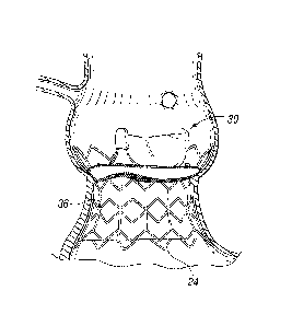
valve (called the
anterior and posterior cusps) and the three cusps or leaflets of the tricuspid
valve. The free
edges of the leaflets connect to chordae tendineae from more than one
papillary muscle, as
seen in Figure 1. In a healthy heart, these muscles and their tendinous chords
support the
mitral and tricuspid valves, allowing the leaflets to resist the high pressure
developed during
contractions (pumping) of the left and right ventricles.
[0007] When the left ventricle contracts after filling with blood from the
left atrium,
the walls of the ventricle move inward and release some of the tension from
the papillary
muscle and chords. The blood pushed up against the under-surface of the mitral
leaflets
causes them to rise toward the annulus plane of the mitral valve. As they
progress toward the
annulus, the leading edges of the anterior and posterior leaflet come together
forming a seal
and closing the valve. In the healthy heart, leaflet coaptation occurs near
the plane of the
mitral annulus. The blood continues to be pressurized in the left ventricle
until it is ejected
into the aorta. Contraction of the papillary muscles is simultaneous with the
contraction of
the ventricle and serves to keep healthy valve leaflets tightly shut at peak
contraction
pressures exerted by the ventricle.
[0008] Various surgical techniques may be used to repair a diseased or damaged
valve.
In a valve replacement operation, the damaged leaflets are excised and the
annulus sculpted
to receive a replacement valve. Due to aortic stenosis and other heart valve
diseases,
thousands of patients undergo surgery each year wherein the defective native
heart valve is
replaced by a prosthetic valve, either bioprosthetic or mechanical. Another
less drastic
method for treating defective valves is through repair or reconstruction,
which is typically
used on minimally calcified valves. The problem with surgical therapy is the
significant
insult it imposes on these chronically ill patients with high morbidity and
mortality rates
associated with surgical repair.
[0009] When the valve is replaced, surgical implantation of the prosthetic
valve
typically requires an open-chest surgery during which the heart is stopped and
patient placed
811608701 vl
CA 2966311 2017-05-10
- 3 -
on cardiopulmonary bypass (a so-called "heart-lung machine"). In one common
surgical
procedure, the diseased native valve leaflets are excised and a prosthetic
valve is sutured to
the surrounding tissue at the valve annulus. Because of the trauma associated
with the
procedure and the attendant duration of extracorporeal blood circulation, some
patients do not
survive the surgical procedure or die shortly thereafter. It is well known
that the risk to the
patient increases with the amount of time required on extracorporeal
circulation. Due to
these risks, a substantial number of patients with defective valves are deemed
inoperable
because their condition is too frail to withstand the procedure. By some
estimates, about 30
to 50% of the subjects suffering from aortic stenosis who are older than 80
years cannot be
operated on for aortic valve replacement.
[0010] Because of the drawbacks associated with conventional open-heart
surgery,
percutaneous and minimally-invasive surgical approaches are garnering intense
attention. In
one technique, a prosthetic valve is configured to be implanted in a much less
invasive
procedure by way of catheterization. For instance, U.S. Patent No. 5,411,552
to Andersen et
al. describes a collapsible valve percutaneously introduced in a compressed
state through a
catheter and expanded in the desired position by balloon inflation. Although
these remote
implantation techniques have shown great promise for treating certain
patients, replacing a
valve via surgical intervention is still the preferred treatment procedure.
One hurdle to the
acceptance of remote implantation is resistance from doctors who are
understandably anxious
about converting from an effective, if imperfect, regimen to a novel approach
that promises
great outcomes but is relatively foreign. In conjunction with the
understandable caution
exercised by surgeons in switching to new techniques of heart valve
replacement, regulatory
bodies around the world are moving slowly as well. Numerous successful
clinical trials and
follow-up studies are in process, but much more experience with these new
technologies will
be required before they are completely accepted.
[0011] Accordingly, there is a need for an improved device and associated
method of
use wherein a prosthetic valve can be surgically implanted in a body channel
in a more
efficient procedure that reduces the time required on extracorporeal
circulation. It is
desirable that such a device and method be capable of helping patients with
defective valves
that are deemed inoperable because their condition is too frail to withstand a
lengthy
conventional surgical procedure. The present invention addresses these needs
and others.
511608701 v1
CA 2966311 2017-05-10
- 4 -
Summary of the Invention
[0012] Various embodiments of the present application provide prosthetic
valves and
methods of use for replacing a defective native valve in a human heart.
Certain embodiments
are particularly well adapted for use in a surgical procedure for quickly and
easily replacing a
heart valve while minimizing time using extracorporeal circulation (i.e.,
bypass pump).
[0013] In one embodiment, a method for treating a native aortic valve in a
human
heart to replaces the function of the aortic valve, comprises: 1) accessing a
native valve
through an opening in a chest; 2) advancing an expandable base stent to the
site of a native
aortic valve, the base stent being radially compressed during the advancement;
3) radially
expanding the base stent at the site of the native aortic valve; 4) advancing
a valve component
within a lumen of the base stent; and 5) expanding a coupling stent on the
valve component
to mechanically couple to the base stent in a quick and efficient manner.
[0014] In one variation, the base stent may comprise a metallic frame. In one
embodiment, at least a portion of the metallic frame is made of stainless
steel. In another
embodiment, at least a portion of the metallic frame is made of a shape memory
material.
The valve member may take a variety of forms. In one preferred embodiment, the
valve
component comprises biological tissue. In another variation of this method,
the metallic
frame is viewed under fluoroscopy during advancement of the prosthetic valve
toward the
native aortic valve.
[0015] The native valve leaflets may be removed before delivering the
prosthetic
valve. Alternatively, the native leaflets may be left in place to reduce
surgery time and to
provide a stable base for fixing the base stent within the native valve. In
one advantage of
this method, the native leaflets recoil inward to enhance the fixation of the
metallic frame in
the body channel. When the native leaflets are left in place, a balloon or
other expansion
member may be used to push the valve leaflets out of the way and thereby
dilate the native
valve before implantation of the base stent. The native annulus may be dilated
between 1.5-5
mm from their initial orifice size to accommodate a larger sized prosthetic
valve.
[0016] In accordance with a preferred aspect, a prosthetic heart valve system
comprises a base stent adapted to anchor against a heart valve annulus and
defining an orifice
therein, and a valve component connected to the base stent. The valve
component includes a
911608701 vl
CA 2966311 2017-05-10
- 5 -
prosthetic valve defining therein a non-expandable, non-collapsible orifice,
and an
expandable coupling stent extending from an inflow end thereof. The coupling
stent has a
contracted state for delivery to an implant position and an expanded state
configured for
outward connection to the base stent. The base stent may also be expandable
with a
contracted state for delivery to an implant position adjacent a heart valve
annulus and an
expanded state sized to contact and anchor against the heart valve annulus.
Desirably, the
base stent and also the coupling stent are plastically expandable.
[00171 In one embodiment, the prosthetic valve comprises a commercially
available
valve having a sewing ring, and the coupling stent attaches to the sewing
ring. The
contracted state of the coupling stent may be conical, tapering down in a
distal direction. The
coupling stent preferably comprises a plurality of radially expandable struts
at least some of
which are arranged in rows, wherein the distalmost row has the greatest
capacity for
expansion from the contracted state to the expanded state. Still further, the
strut row farthest
from the prosthetic valve has alternating peaks and valleys, wherein the base
stent includes
apertures into which the peaks of the coupling stent may project to interlock
the two stents.
The base stent may include a plurality of radially expandable struts between
axially-oriented
struts, wherein at least some of the axially-oriented struts have upper
projections that demark
locations around the stent.
[0018] A method of delivery and implant of a prosthetic heart valve system is
also
disclosed herein, comprising the steps of:
advancing a base stent to an implant position adjacent a heart valve annulus;
anchoring the base stent to the heart valve annulus;
providing a valve component including a prosthetic valve having a non-
expandable,
non-collapsible orifice, the valve component further including an expandable
coupling
stent extending from an inflow end thereof, the coupling stent having a
contracted
state for delivery to an implant position and an expanded state configured for
outward
connection to the base stent;
advancing the valve component with the coupling stent in its contracted state
to an
implant position adjacent the base stent; and
expanding the coupling stent to the expanded state in contact with and
connected to
the base stent.
#11608701 vl
CA 2966311 2017-05-10
- 6 -
[0019] The base stent may be plastically expandable, and the method further
comprises advancing the expandable base stent in a contracted state to the
implant position,
and plastically expanding the base stent to an expanded state in contact with
and anchored to
the heart valve annulus, in the process increasing the orifice size of the
heart valve annulus by
at least 10%, or by 1.5-5 mm. Desirably, the prosthetic valve of the valve
component is
selected to have an orifice size that matches the increased orifice size of
the heart valve
annulus. The method may also include mounting the base stent over a mechanical
expander,
and deploying the base stent at the heart valve annulus using the mechanical
expander.
[0020] One embodiment of the method further includes mounting the valve
component on a holder having a proximal hub and lumen therethrough. The holder
mounts
on the distal end of a handle having a lumen therethrough, and the method
including passing
a balloon catheter through the lumen of the handle and the holder and within
the valve
component, and inflating a balloon on the balloon catheter to expand the
coupling stent. The
valve component mounted on the holder may be packaged separately from the
handle and the
balloon catheter. Desirably, the contracted state of the coupling stent is
conical, and the
balloon on the balloon catheter has a larger distal expanded end than its
proximal expanded
end so as to apply greater expansion deflection to the coupling stent than to
the prosthetic
valve.
[0021] In the method where the coupling stent is conical, the coupling stent
may
comprise a plurality of radially expandable struts at least some of which are
arranged in rows,
wherein the row farthest from the prosthetic valve has the greatest capacity
for expansion
from the contracted state to the expanded state.
[0022] The method may employ a coupling stent with a plurality of radially
expandable struts, wherein a row farthest from the prosthetic valve has
alternating peaks and
valleys. The distal end of the coupling stent thus expands more than the rest
of the coupling
stent so that the peaks in the row farthest from the prosthetic valve project
outward into
apertures in the base stent. Both the base stent and the coupling stent may
have a plurality of
radially expandable struts between axially-oriented struts, wherein the method
includes
orienting the coupling stent so that its axially-oriented struts are out of
phase with those of the
base stent to increase retention therebetween.
811608701 v1
CA 2966311 2017-05-10
- 7 -
[0023] Another aspect described herein is a system for delivering a valve
component
including a prosthetic valve having a non-expandable, non-collapsible orifice,
and an
expandable coupling stent extending from an inflow end thereof, the coupling
stent having a
contracted state for delivery to an implant position and an expanded state.
The delivery
system includes a valve holder connected to a proximal end of the valve
component, a
balloon catheter having a balloon, and a handle configured to attach to a
proximal end of the
valve holder and having a lumen for passage of the catheter, wherein the
balloon extends
distally through the handle, past the holder and through the valve component.
In the system,
the prosthetic valve is preferably a commercially available valve having a
sewing ring to
which the coupling stent attaches.
[0024] The contracted state of the coupling stent in the delivery system may
be
conical, tapering down in a distal direction. Furthermore, the balloon
catheter further may
include a generally conical nose cone on a distal end thereof that extends
through the valve
component and engages a distal end of the coupling stent in its contracted
state. Desirably,
the handle comprises a proximal section and a distal section that may be
coupled together in
series to form a continuous lumen, wherein the distal section is adapted to
couple to the hub
of the holder to enable manual manipulation of the valve component using the
distal section
prior to connection with the proximal handle section. Preferably, the balloon
catheter and
proximal handle section are packaged together with the balloon within the
proximal section
lumen.
[0025] The system of claim 21, wherein the valve component mounted on the
holder is
packaged separately from the handle and the balloon catheter.A further
understanding of the
nature and advantages of the present invention are set forth in the following
description and
claims, particularly when considered in conjunction with the accompanying
drawings in
which like parts bear like reference numerals.
Brief Description of the Drawings
[0026] The invention will now be explained and other advantages and features
will
appear with reference to the accompanying schematic drawings wherein:
[0027] Figure 1 is an anatomic anterior view of a human heart, with portions
broken
away and in section to view the interior heart chambers and adjacent
structures;
911608701 vl
CA 2966311 2017-05-10
- 8 -
[0028] Figure 2 is an anatomic superior view of a section of the human heart
showing
the tricuspid valve in the right atrium, the mitral valve in the left atrium,
and the aortic valve
in between, with the tricuspid and mitral valves open and the aortic and
pulmonary valves
closed during ventricular diastole (ventricular filling) of the cardiac cycle;
[0029] Figure 3 is an anatomic superior view of a section of the human heart
shown in
Figure 2, with the tricuspid and mitral valves closed and the aortic and
pulmonary valves
opened during ventricular systole (ventricular emptying) of the cardiac cycle;
[0030] Figure 4 is an anatomic anterior perspective view of the left and right
atria,
with portions broken away and in section to show the interior of the heart
chambers and
associated structures, such as the fossa ovalis, coronary sinus, and the great
cardiac vein;
[0031] Figures 5A-5H are sectional views through an isolated aortic annulus
showing
a portion of the adjacent left ventricle and aorta, and illustrating a number
of steps in
deployment of an exemplary prosthetic heart valve system of the present
invention;
[0032] Figure 5A shows a deflated balloon catheter having a base stent thereon
advanced into position at the aortic annulus;
[0033] Figure 5B shows the balloon on the catheter inflated to expand and
deploy the
base stent against the aortic annulus;
[0034] Figure 5C shows the deployed base stent in position within the aortic
annulus;
[0035] Figure 5D shows a valve component mounted on a balloon catheter
advancing
into position within the base stent;
[0036] Figure 5E shows the valve component in a desired implant position at
the aortic
annulus and within the base stent, with the balloon catheter advanced farther
to displace a
nose cone out of engagement with a coupling stent;
[0037] Figure 5F sliows the balloon on the catheter inflated to expand and
deploy a
valve component coupling stent against the base stent;
[0038] Figure 5G shows the deflated balloon on the catheter along with the
nose cone
being removed from within the valve component;
[0039] Figure 5H shows the fully deployed prosthetic heart valve of the
present
invention;
[0040] Figure 6 is an exploded view of an exemplary system for delivering the
prosthetic heart valve of the present invention;
#11608701 v1
=
CA 2966311 2017-05-10
- 9 -
[0041] Figure 7 is an assembled view of the delivery system of Figure 6
showing a
nose cone extending over a distal end of a valve component coupling stent;
[0042] Figure 8 is a view like Figure 7 but with a balloon catheter displaced
distally to
disengage the nose cone from the coupling stent;
[0043] Figure 9 is an assembled view of the delivery system similar to that
shown in
Figure 7 and showing a balloon inflated to expand the valve component coupling
stent;
[0044] Figure 10 is an exploded elevational view of several components of the
introducing system of Figure 9, without the balloon catheter, valve component
and holder;
[0045] Figures 11A and 11B are perspective views of an exemplary valve
component
assembled on a valve holder of the present invention;
[0046] Figure 11C is a side elevational view of the assembly of Figures 11 A
and 11B;
[0047] Figures 11D and 11E are top and bottom plan views of the assembly of
Figures
11A and 11B;
[0048] Figures 12A-12B illustrate an exemplary coupling stent in both a flat
configuration (12A) and a tubular expanded configuration (12B);
[0049] Figures 13A-13B illustrate an alternative coupling stent having a
discontinuous
upper end in both flat and tubular expanded configurations;
[0050] Figure 14-17 are plan views of a still further alternative coupling
stent;
[0051] Figure 18A-18B are flat and tubular views of an exemplary base stent
with
upper position markers and a phantom coupling stent superimposed thereover;
[0052] Figure 19 is a flat view of an alternative base stent with a coupling
stent
superimposed thereover;
[0053] Figure 20 is a sectional view of a coupling stent within a base stent
illustrating
one method of interlocking; and
[0054] Figure 21-23 is a perspective view of a device for delivering and
expanding a
base stent with mechanical fingers.
Detailed Description of the Preferred Embodiments
[0055] The present invention attempts to overcome drawbacks associated with
conventional, open-heart surgery, while also adopting some of the techniques
of newer
technologies which decrease the duration of the treatment procedure. The
prosthetic heart
#11608701v1
CA 2966311 2017-05-10
=
- 10 -
valves of the present invention are primarily intended to be delivered and
implanted using
conventional surgical techniques, including the aforementioned open-heart
surgery. There
are a number of approaches in such surgeries, all of which result in the
formation of a direct
access pathway to the particular heart valve annulus. For clarification, a
direct access
pathway is one that permits direct (i.e., naked eye) visualization of the
heart valve annulus.
In addition, it will be recognized that embodiments of the two-stage
prosthetic heart valves
described herein may also be configured for delivery using percutaneous
approaches, and
those minimally-invasive surgical approaches that require remote implantation
of the valve
using indirect visualization.
[0056] One primary aspect of the present invention is a two-stage prosthetic
heart
valve wherein the tasks of.implanting a tissue anchor first and then a valve
member are
distinct and certain advantages result. The exemplary two-stage prosthetic
heart valve of the
present invention has an expandable base stent secured to tissue in the
appropriate location
using a balloon or other expansion technique. A hybrid valve member that has
non-
expandable and expandable portions then couples to the base stent in a
separate or sequential
operation. By utilizing an expandable base stent, the duration of the initial
anchoring
operation is greatly reduced as compared with a conventional sewing procedure
utilizing an
array of sutures. The expandable base stent may simply be radially expanded
outward into
contact with the implantation site, or may be provided with additional
anchoring means, such
as barbs. The operation may be carried out using a conventional open-heart
approach and
cardiopulmonary bypass. In one advantageous feature, the time on bypass is
greatly reduced
due to the relative speed of implanting the expandable base stent.
[0057] For definitional purposes, the term "base stent," refers to a
structural
component of a heart valve that is capable of attaching to tissue of a heart
valve annulus. The
base stents described herein are most typically tubular stents, or stents
having varying shapes
or diameters. A stent is normally formed of a biocompatible metal wire frame,
such as
stainless steel or Nitinol. Other base stents that could be used with valves
of the present
invention include rigid rings, spirally-wound tubes, and other such tubes that
fit tightly within
a valve annulus and define an orifice therethrough for the passage of blood,
or within which a
valve member is mounted. It is entirely conceivable, however, that the base
stent could be
separate clamps or hooks that do not define a continuous periphery. Although
such devices
#11608701 v1
CA 2966311 2017-05-10
- 11 -
sacrifice some dynamic stability, and speed and ease of deployment, these
devices could be
configured to work in conjunction with a particular valve member.
[0058] A distinction between self-expanding and balloon-expanding stents
exists in
the field. A self-expanding stent may be crimped or otherwise compressed into
a small tube
and possesses sufficient elasticity to spring outward by itself when a
restraint such as an outer
sheath is removed. In contrast, a balloon-expanding stent is made of a
material that is
substantially less elastic, and indeed must be plastically expanded from the
inside out when
converting from a compressed diameter to an expanded. It should be understood
that the
term balloon-expanding stents encompasses plastically-expandable stents,
whether or not a
balloon is used to actually expand it. The material of the stent plastically
deforms after
application of a deformation force such as an inflating balloon or expanding
mechanical
fingers. Both alternatives will be described below. Consequently, the term
"balloon-
expandable stent" should be considered to refer to the material or type of the
stent as opposed
to the specific expansion means.
[0059] The term "valve member" refers to that component of a heart valve that
possesses the fluid occluding surfaces to prevent blood flow in one direction
while permitting
it in another. As mentioned above, various constructions of valve numbers are
available,
including those with flexible leaflets and those with rigid leaflets or a ball
and cage
arrangement. The leaflets may be bioprosthetic, synthetic, or metallic.
[0060] A primary focus of the present invention is a two-stage prosthetic
heart valve
having a first stage in which a base stent secures to a valve annulus, and a
subsequent second
stage in which a valve member connects to the base stent. It should be noted
that these stages
can be done almost simultaneously, such as if the two components were mounted
on the same
delivery device, or can be done in two separate clinical steps, with the base
stent deployed
using a first delivery device, and then the valve member using another
delivery device. It
should also be noted that the term "two-stage" refers to the two primary steps
of anchoring
structure to the annulus and then connecting a valve member, which does not
necessarily
limit the valve to just two parts.
[0061] Another potential benefit of a two-stage prosthetic heart valve,
including a base
stent and a valve member, is that the valve member may be replaced after
implantation
without replacing the base stent. That is, an easily detachable means for
coupling the valve
#11608701 v1
CA 2966311 2017-05-10
- 12 -
member and base stent may be used that permits a new valve member to be
implanted with
relative ease. Various configurations for coupling the valve member and base
stent are
described herein.
[0062] It should be understood, therefore, that certain benefits of the
invention are
independent of whether the base stent is expandable or not. That is, various
embodiments
illustrate an expandable base stent coupled to a hybrid valve member that has
non-expandable
and expandable portions. However, the same coupling structure may be utilized
for a non-
expandable base stent and hybrid valve member. Therefore, the invention should
be
interpreted via the appended claims.
[0063] As a point of further definition, the term "expandable" is used herein
to refer to
a component of the heart valve capable of expanding from a first, delivery
diameter to a
second, implantation diameter. An expandable structure, therefore, does not
mean one that
might undergo slight expansion from a rise in temperature, or other such
incidental cause.
Conversely, "non-expandable" should not be interpreted to mean completely
rigid or a
dimensionally stable, as some slight expansion of conventional "non-
expandable" heart
valves, for example, may be observed.
[0064] In the description that follows, the term "body channel" is used to
define a
blood conduit or vessel within the body. Of course, the particular application
of the
prosthetic heart valve determines the body channel at issue. An aortic valve
replacement, for
example, would be implanted in, or adjacent to, the aortic annulus. Likewise,
a mitral valve
replacement will be implanted at the mitral annulus. Certain features of the
present invention
are particularly advantageous for one implantation site or the other. However,
unless the
combination is structurally impossible, or excluded by claim language, any of
the heart valve
embodiments described herein could be implanted in any body channel.
[0065] Figures 5A-5H are sectional views through an isolated aortic annulus AA
showing a portion of the adjacent left ventricle LV and ascending aorta with
sinus cavities S.
The two coronary sinuses CS are also shown. The series of views show snapshots
of a
number of steps in deployment of an exemplary prosthetic heart valve system of
the present
invention, which comprises a two-component system. A first component is a base
stent that
is deployed against the native leaflets or, if the leaflets are excised,
against the debrided aortic
annulus AA. A second valve component fits within the base stent and anchors
thereto.
611608701 vl
CA 2966311 2017-05-10
- 13 -
Although two-part valves are known in the art, this is believed to be the
first that utilizes a
stent within a stent in conjunction with a non-expandable valve.
[0066] Figure 5A shows a catheter 20 having a balloon 22 in a deflated state
near a
distal end with a tubular base stent 24 crimped thereover. The stent 24 is
shown in a radially
constricted, undeployed configuration. The catheter 20 has been advanced to
position the
base stent 24 so that it is approximately axially centered at the aortic
annulus AA.
[0067] Figure 5B shows the balloon 22 on the catheter 20 inflated to expand
and
deploy the base stent 24 against the aortic annulus AA, and Figure 5C shows
the deployed
base stent in position after deflation of the balloon 22 and removal of the
catheter 20. The
stent 24 provides a base within and against a body lumen (e.g., a valve
annulus). Although a
stent is described for purposes of illustration, any member capable of
anchoring within and
against the body lumen and then coupling to the valve component may be used.
In a
preferred embodiment, the base stent 24 comprises a plastically-expandable
cloth-covered
stainless-steel tubular stent. One advantage of using a plastically-expandable
stent is the
ability to expand the native annulus to receive a larger valve size than would
otherwise be
possible with conventional surgery. Desirably, the left ventricular outflow
tract (LVOT) is
significantly expanded by at least 10%, or for example by 1.5-5 mm, and the
surgeon can
select a valve component 30 with a larger orifice diameter relative to an
unexpanded annulus.
On the other hand, the present invention could also use a self-expanding base
stent 24 which
is then reinforced by the subsequently implanted valve component 30. Because
the valve
component 30 has a non-compressible part, the prosthetic valve 34, and
desirably a
plastically-expandable coupling stent 36, it effectively resists recoil of the
self-expanded base
stent 24.
[0068] With continued reference to Figure 5B, the stent 24 has a diameter
sized to be
deployed at the location of the native valve (e.g., along the aortic annulus).
A portion of the
stent 24 may expand outwardly into the respective cavity adjacent the native
valve. For
example, in an aortic valve replacement, an upper portion may expand into the
area of the
sinus cavities just downstream from the aortic annulus. Of course, care should
be taken to
orient the stent 24 so as not to block the coronary openings. The stent body
is preferably
configured with sufficient radial strength for pushing aside the native
leaflets and holding the
native leaflets open in a dilated condition. The native leaflets provide a
stable base for
811608701 vl
CA 2966311 2017-05-10
- 14 -
holding the stent, thereby helping to securely anchor the stent in the body.
To further secure
the stent to the surrounding tissue, the lower portion may be configured with
anchoring
members, such as, for example, hooks or barbs (not shown).
[0069] As will be described in more detail below, the prosthetic valve system
includes
a valve component that may be quickly and easily connected to the stent 24. It
should be
noted here that the base stents described herein can be a variety of designs,
including having
the diamond/chevron-shaped openings shown or other configurations. The
material depends
on the mode of delivery (i.e., balloon- or self-expanding), and the stent can
be bare strut
material or covered to promote ingrowth and/or to reduce paravalvular leakage.
For example,
a suitable cover that is often used is a sleeve of fabric such as Dacron.
[0070] One primary advantage of the prosthetic heart valve system of the
present
invention is the speed of deployment. Therefore, the base stent 24 may take a
number of
different configurations as long as it does not require the time-consuming
process of suturing
it to the annulus. For instance, another possible configuration for the base
stent 24 is one that
is not fully expandable like the tubular stent as shown. That is, the base
stent 24 may have a
non-expandable ring-shaped orifice from which an expandable skirt stent or
series of
anchoring barbs deploy.
[0071] Figure 5D shows a valve component 30 mounted on a balloon catheter 32
advancing into position within the base stent 24. The valve component 30
comprises a
prosthetic valve 34 and a coupling stent 36 attached to and projecting from a
distal end
thereof. In its radially constricted or undeployed state, the coupling stent
36 assumes a
conical inward taper in the distal direction. The catheter 32 extends through
the valve
component 30 and terminates in a distal nose cone 38 which has a conical or
bell-shape and
covers the tapered distal end of the coupling stent 36. Although not shown,
the catheter 32
extends through an introducing cannula and valve holder.
[0072] When used for aortic valve replacement, the prosthetic valve 34
preferably has
three flexible leaflets which provide the fluid occluding surfaces to replace
the function of the
native valve leaflets. In various preferred embodiments, the valve leaflets
may be taken from
another human heart (cadaver), a cow (bovine), a pig (porcine valve) or a
horse (equine). In
other preferred variations, the valve member may comprise mechanical
components rather
#11608701 v1
CA 2966311 2017-05-10
- 15 -
than biological tissue. The three leaflets are supported by three commissural
posts. A ring is
provided along the base portion of the valve member.
[0073] In a preferred embodiment, the prosthetic valve 34 partly comprises a
commercially available, non-expandable prosthetic heart valve, such as the
Carpentier-
Edwards PERIMOUNT Magna Aortic Heart Valve available from Edwards
Lifesciences of
Irvine, California. In this sense, a "commercially available" prosthetic heart
valve is an off-
the-shelf (i.e., suitable for stand-alone sale and use) prosthetic heart valve
defining therein a
non-expandable, non-collapsible orifice and having a sewing ring capable of
being implanted
using sutures through the sewing ring in an open-heart, surgical procedure.
The particular
approach into the heart used may differ, but in surgical procedures the heart
is stopped and
opened, in contrast to beating heart procedures where the heart remains
functional. To
reiterate, the terms "non-expandable" and "non-collapsible" should not be
interpreted to
mean completely rigid and dimensionally stable, merely that the valve is not
expandable/collapsible like some proposed minimally-invasively or
percutaneously-delivered
valves.
[0074] An implant procedure therefore involves first delivering and expanding
the
base stent 24 at the aortic annulus, and then coupling the valve component 30
including the
valve 34 thereto. Because the valve 34 is non-expandable, the entire procedure
is typically
done using the conventional open-heart technique. However, because the base
stent 24 is
delivered and implanted by simple expansion, and then the valve component 30
attached
thereto by expansion, both without suturing, the entire operation takes less
time. This hybrid
approach will also be much more comfortable to surgeons familiar with the open-
heart
procedures and commercially available heart valves.
[0075] Moreover, the relatively small change in procedure coupled with the use
of
proven heart valves should create a much easier regulatory path than strictly
expandable,
remote procedures. Even if the system must be validated through clinical
testing to satisfy
the Pre-Market Approval (PMA) process with the FDA (as opposed to a 510k
submission),
the acceptance of the valve component 30 at least will be greatly streamlined
with a
commercial heart valve that is already approved, such as the Magna Aortic
Heart Valve.
[0076] The prosthetic valve 34 is provided with an expandable coupling
mechanism in
the form of the coupling stent 36 for securing the valve to the base stent 24.
Although the
511608701 vl
CA 2966311 2017-05-10
- 16 -
coupling stent 36 is shown, the coupling mechanism may take a variety of
different forms,
but eliminates the need for connecting sutures and provides a rapid connection
means.
[0077] In Figure 5E the valve component 30 has advanced to a desired implant
position at the aortic annulus AA and within the base stent 24. The prosthetic
valve 34 may
include a suture-permeable ring 42 that desirably abuts the aortic annulus AA.
More
preferably, the sewing ring 42 is positioned supra-annularly, or above the
narrowest point of
the aortic annulus AA, so as to allow selection of a larger orifice size than
a valve placed
intra-annularly. With the aforementioned annulus expansion using the base
stent 24, and the
supra-annular placement, the surgeon may select a valve having a size one or
two increments
larger than previously conceivable. As mentioned, the prosthetic valve 34 is
desirably a
commercially available heart valve having a sewing ring 42. The balloon
catheter 32 has
advanced relative to the valve component 30 to displace the nose cone 38 out
of engagement
with the coupling stent 36. A dilatation balloon 40 on the catheter 30 can be
seen just beyond
the distal end of the coupling stent 36.
[0078] Figure 5F shows the balloon 40 on the catheter 32 inflated to expand
and
deploy the coupling stent 36 against the base stent 24. The balloon 40 is
desirably inflated
using controlled, pressurized, sterile physiologic saline. The coupling stent
36 transitions
between its conical contracted state and its generally tubular expanded state.
Simple
interference between the coupling stent 36 and the base stent 24 may be
sufficient to anchor
the valve component 30 within the base stent, or interacting features such as
projections,
hooks, barbs, fabric, etc. may be utilized.
[0079] Because the base stent 24 expands before the valve component 30
attaches
thereto, a higher strength stent (self-or balloon-expandable) configuration
may be used. For
instance, a relatively robust base stent 24 may be used to push the native
leaflets aside, and
the absent valve component 30 is not damaged or otherwise adversely affected
during the
high-pressure base stent deployment. After the base stent 24 deploys in the
body channel, the
valve component 30 connects thereto by deploying the coupling stent 36, which
may be
somewhat more lightweight requiring smaller expansion forces. Also, the
balloon 40 may
have a larger distal expanded end than its proximal expanded end so as to
apply more force to
the coupling stent 36 than to the prosthetic valve 34. In this way, the
prosthetic valve 34 and
flexible leaflets therein are not subject to high expansion forces from the
balloon 40. Indeed,
#11608701 v1
CA 2966311 2017-05-10
- 17 -
although balloon deployment is shown, the coupling stent 36 may also be a self-
expanding
type of stent. In the latter configuration, the nose cone 38 is adapted to
retain the coupling
stent 36 in its constricted state prior to position in the valve component 30
within the base
stent 24.
[0080] As noted above, the base stents described herein could include barbs or
other
tissue anchors to further secure the stent to the tissue, or to secure the
coupling stent 36 to the
base stent 24. Further, the barbs could be deployable (e.g., configured to
extend or be pushed
radially outward) by the expansion of a balloon. Preferably, the coupling
stent 36 is covered
to promote in-growth and/or to reduce paravalvular leakage, such as with a
Dacron tube or
the like.
[0081] Figure 5G shows the deflated balloon 40 on the catheter 32 along with
the nose
cone 38 being removed from within the valve component 30. Finally, Figure 5H
shows the
fully deployed prosthetic heart valve system of the present invention
including the valve
component 30 coupled to the base stent 24 within the aortic annulus AA.
[0082] Figure 6 is an exploded view, and Figures 7 and 8 are assembled views,
of an
exemplary system 50 for delivering the prosthetic heart valve of the present
invention.
Modified components of the delivery system 50 are also shown in Figures 9 and
10. The
delivery system 50 includes a balloon catheter 52 having the balloon 40 on its
distal end and
an obturator 54 on a proximal end. The obturator 54 presents a proximal
coupling 56 that
receives a luer connector or other such fastener of a Y-fitting 58. The
aforementioned nose
cone 38 may attach to the distalmost end of the catheter: 52, but more
preferably attaches to a
wire (not shown) inserted through the center lumen of the balloon catheter 52.
[0083] The catheter 52 and the nose cone 38 pass through a hollow handle 60
having a
proximal section 62 and a distal section 64. A distal end of the distal handle
section 64
firmly attaches to a hub 66 of a valve holder 68, which in turn attaches to
the prosthetic heart
valve component 30. Details of the valve holder 68 will be given below with
reference to
Figures 11A-11E.
[0084] The two sections 62, 64 of the handle 60 are desirably formed of a
rigid
material, such as a molded plastic, and coupled to one another to form a
relatively rigid and
elongated tube for manipulating the prosthetic valve component 30 attached to
its distal end.
In particular, the distal section 64 may be easily coupled to the holder hub
66 and therefore
811608701 v1
CA 2966311 2017-05-10
- 18 -
provide a convenient tool for managing the valve component 30 during pre-
surgical rinsing
steps. For this purpose, the distal section 64 features a distal tubular
segment 70 that couples
to the holder hub 66, and an enlarged proximal segment 72 having an opening on
its proximal
end that receives a tubular extension 74 of the proximal handle section 62.
Figure 6 shows an
0-ring 76 that may be provided on the exterior of the tubular extension 74 for
a frictional
interference fit to prevent the two sections from disengaging. Although not
shown, the distal
tubular segment 70 may also have an 0-ring for firmly coupling to the holder
hub 66, or may
be attached with threading or the like. In one preferred embodiment, the
balloon 40 on the
catheter 52 is packaged within the proximal handle section 62 for protection
and ease of
handling. Coupling the proximal and distal handle sections 62, 64 therefore
"loads" the
system 50 such that the balloon catheter 52 may be advanced through the
continuous lumen
leading to the valve component 30.
[0085] Figures 9 and 10 illustrate a delivery system 50 similar to that shown
in Figure
7, but with alternative couplers 77 on both the proximal and distal handle
sections 62, 64 in
the form of cantilevered teeth that snap into complementary recesses formed in
the respective
receiving apertures. Likewise, threading on the mating parts could also be
used, as well as
other similar expedients. Figure 9 shows the balloon 40 inflated to expand the
valve
component coupling stent 36.
[0086] In a preferred embodiment, the prosthetic valve component 30
incorporates
bioprosthetic tissue leaflets and is packaged and stored attached to the
holder 68 but separate
from the other introduction system 50 components. Typically, bioprosthetic
tissue is
packaged and stored in ajar with preservative solution for long shelf life,
while the other
components are packaged and stored dry.
[0087] When assembled as seen in Figures 7-9, an elongated lumen (not
numbered)
extends from the proximal end of the Y-fitting 58 to the interior of the
balloon 40. The Y-
fitting 58 desirably includes an internally threaded connector 80 for
attachment to an
insufflation system, or a side port 82 having a luer fitting 84 or similar
expedient may be used
for insufflation of the balloon 40.
[0088] Figures 7 and 8 show two longitudinal positions of the catheter 52 and
associated structures relative to the handle 60 and its associated structures.
In a retracted
position shown in Figure 7, the balloon 40 primarily resides within the distal
handle section
811608701 v1
CA 2966311 2017-05-10
- 19 -
64. Figure 7 illustrates the delivery configuration of the introduction system
50, in which the
surgeon advances the prosthetic valve component 30 from outside the body into
a location
adjacent the target annulus. The nose cone 38 extends around and protects a
distal end of the
conical undeployed coupling stent 36. This configuration is also seen in
Figure 5D, albeit
with the holder 68 removed for clarity. Note the spacing S between the
proximal coupling 56
and the proximal end of the handle 60.
[0089] As explained above with respect to Figures 5A-5H, the surgeon advances
the
prosthetic valve component 30 into its desired implantation position at the
valve annulus, and
then advances the balloon 40 through the valve component and inflates it. To
do so, the
operator converts the delivery system 50 from the retracted configuration of
Figure 7 to the
deployment configuration of Figure 8, with the balloon catheter 40 displaced
distally as
indicated by the arrow 78 to disengage the nose cone 38 from the coupling
stent 36. Note
that the proximal coupling 56 now contacts the proximal end of the handle 60,
eliminating the
space S indicated in Figure 7.
[0090] It should be understood that the prosthetic valve component 30 may be
implanted at the valve annulus with a pre-deployed base stent 24, as explained
above, or
without. The coupling stent 36 may be robust enough to anchor the valve
component 30
directly against the native annulus (with or without leaflet excision) in the
absence of the base
stent 24. Consequently, the description of the system 50 for introducing the
prosthetic heart
valve should be understood in the context of operating with or without the pre-
deployed base
stent 24.
[0091] Prior to a further description of operation of the delivery system 50,
a more
detailed explanation of the valve component 30 and valve holder 68 is
necessary. Figures
11A-11E show a number of perspective and other views of the exemplary valve
component
30 mounted on the delivery holder 68 of the present invention. As mentioned,
the valve
component 30 comprises the prosthetic valve 34 having the coupling stent 36
attached to an
inflow end thereof. In a preferred embodiment, the prosthetic valve 34
comprises a
commercially available off-the-shelf non-expandable, non-collapsible
commercial prosthetic
valve. Any number of prosthetic heart valves can be retrofit to attach the
coupling stent 36,
and thus be suitable for use in the context of the present invention. For
example, the
prosthetic valve 34 may be a mechanical valve or a valve with flexible
leaflets, either
811608701 v1
CA 2966311 2017-05-10
- 20 -
synthetic or bioprosthetic. In a preferred embodiment, however, the prosthetic
valve 34
includes bioprosthetic tissue leaflets 86 (Figure 11A). Furthermore, as
mentioned above, the
prosthetic valve 34 is desirably a Carpentier-Edwards PERIMOUNT Magna Aortic
Heart
Valve (e.g., model 3000TFX) available from Edwards Lifesciences of Irvine,
California.
[0092] The coupling stent 36 preferably attaches to the ventricular (or
inflow) aspect
of the valve's sewing ring 42 during the manufacturing process in a way that
preserves the
integrity of the sewing ring and prevents reduction of the valve's effective
orifice area
(EOA). Desirably, the coupling stent 36 will be continuously sutured to sewing
ring 42 in a
manner that maintains the outer contours of the sewing ring. Sutures may be
passed through
apertures or eyelets in the stent skeleton, or through a cloth covering that
in turn is sewn to
the skeleton. Other connection solutions include prongs or hooks extending
inward from the
stent, ties, Velcro, snaps, adhesives, etc. Alternatively, the coupling stent
36 may be more
rigidly connected to rigid components within the prosthetic valve 34. During
implant,
therefore, the surgeon can seat the sewing ring 42 against the annulus in
accordance with a
conventional surgery. This gives the surgeon familiar tactile feedback to
ensure that the
proper patient-prosthesis match has been achieved. Moreover, placement of the
sewing ring
42 against the outflow side of the annulus helps reduce the probability of
migration of the
valve component 30 toward the ventricle.
[0093] The coupling stent 36 may be a pre-crimped, tapered, 316L stainless
steel
balloon-expandable stent, desirably covered by a polyester skirt 88 to help
seal against
paravalvular leakage and promote tissue ingrowth once implanted within the
base stent 24
(see Figure 5F). The coupling stent 36 transitions between the tapered
constricted shape of
Figures 11A-11E to its flared expanded shape shown in Figure 5F, and also in
Figure 10.
[0094] The coupling stent 36 desirably comprises a plurality of sawtooth-
shaped or
otherwise angled, serpentine or web-like struts 90 connected to three
generally axially-
extending posts 92. As will be seen below, the posts 92 desirably feature a
series of evenly
spaced apertures to which sutures holding the polyester skirt 88 in place may
be anchored.
As seen best in Figure 5F, the stent 36 when expanded flares outward and
conforms closely
against the inner surface of the base stent 24, and has an axial length
substantially the same as
the base stent. Anchoring devices such as barbs or other protruberances from
the coupling
811608701 v1
CA 2966311 2017-05-10
- 21 -
stent 36 may be provided to enhance the frictional hold between the coupling
stent and the
base stent 24.
[0095] It should be understood that the particular configuration of the
coupling stent,
whether possessing straight or curvilinear struts 90, may be modified as
needed. There are
numerous stent designs, as described below with reference to Figures 12-17,
any of which
potentially may be suitable. Likewise, although the preferred embodiment
incorporates a
balloon-expandable coupling stent 36, a self-expanding stent could be
substituted with certain
modifications, primarily to the delivery system. The same flexibility and
design of course
applies to the base stent 24. In a preferred embodiment, both the base stent
24 and the
coupling stent 36 are desirably plastically-expandable to provide a firmer
anchor for the valve
34; first to the annulus with or without native leaflets, and then between the
two stents. The
stents may be expanded using a balloon or mechanical expander as described
below.
[0096] Still with reference to Figures 11A-11E, the holder 68 comprises the
aforementioned proximal hub 66 and a thinner distal extension 94 thereof
forming a central
portion of the holder. Three legs 96a, 96b, 96c circumferentially
equidistantly spaced around
the central extension 94 and projecting radially outward therefrom comprise
inner struts 98
and outer commissure rests 100. The prosthetic valve 34 preferably includes a
plurality,
typically three, commissures 102 that project in an outflow direction.
Although not shown,
the commissure rests 100 preferably incorporate depressions into which fit the
tips of the
commissures 102.
[0097] In one embodiment, the holder 68 is formed of a rigid polymer such as
Delrin
or polypropylene that is transparent to increase visibility of an implant
procedure. As best
seen in Figure 1 1E, the holder 68 exhibits openings between the legs 96a,
96b, 96c to provide
a surgeon good visibility of the valve leaflets 86, and the transparency of
the legs further
facilitates visibility and permits transmission of light therethrough to
minimize shadows.
Although not described in detail herein, Figure 1 lE also illustrate a series
of through holes in
the legs 96a, 96b, 96c permitting connecting sutures to be passed through
fabric in the
prosthetic valve 34 and across a cutting guide in each leg. As is known in the
art, severing a
middle length of suture that is connected to the holder 68 and passes through
the valve
permits the holder to be pulled free from the valve when desired.
611608701 vl
CA 2966311 2017-05-10
- 22 -
[0098] Figures 11C and 11D illustrate a somewhat modified coupling stent 36
from
that shown in Figures 11A and 11B, wherein the struts 90 and axially-extending
posts 92 are
better defined. Specifically, the posts 92 are somewhat wider and more robust
than the struts
90, as the latter provide the stent 36 with the ability to expand from the
conical shape shown
to a more tubular configuration. Also, a generally circular reinforcing ring
104 abuts the
valve sewing ring 42. Both the posts 92 and the ring 104 further include a
series of through
holes 106 that may be used to secure the polyester skirt 88 to the stent 36
using sutures or the
like. A number of variants of the coupling stent 36 are also described below.
[0100] Figures 12A-12B illustrate the exemplary coupling stent 36 in both a
flat
configuration (12A) and a tubular configuration (12B) that is generally the
expanded shape.
As mentioned, the web-like struts 90 and a reinforcing ring 104 connect three
generally
axially-extending posts 92. A plurality of evenly spaced apertures 106 provide
anchors for
holding the polyester skirt 88 (see Figure 11B) in place. In the illustrated
embodiment, the
web-like struts 90 also include a series of axially-extending struts 108. An
upper end of the
coupling stent 36 that connects to the sewing ring of the valve and is defined
by the
reinforcing ring 104 follows an undulating path with alternating arcuate
troughs 110 and
peaks 112. As seen from Figure 11C, the exemplary prosthetic valve 34 has an
undulating
sewing ring 42 to which the upper end of the coupling stent 36 conforms. In a
preferred
embodiment, the geometry of the stent 36 matches that of the undulating sewing
ring 42. Of
course, if the sewing ring of the prosthetic valve is planar, then the upper
end of the coupling
stent 36 will also be planar. It should be noted also that the tubular version
of Figure I2B is
an illustration of an expanded configuration, although the balloon 40 may over-
expand the
free (lower) end of the stent 36 such that it ends up being slightly conical.
[0101] Figures 13A and 13B show an alternative coupling stent 120, again in
flattened and tubular configurations, respectively. As with the first
embodiment, the coupling
stent 120 includes web-like struts 122 extending between a series of axially-
extending struts
124. In this embodiment, all of the axially-extending struts 124 are
substantially the same
thin cross-sectional size. The upper or connected end of the stent 120 again
includes a
reinforcing ring 126, although this version is interrupted with a series of
short lengths
separated by gaps. The upper end defines a plurality of alternating troughs
128 and peaks
130, with lengths of the reinforcing ring 126 defining the peaks. The axially-
extending struts
811608701 vl
CA 2966311 2017-05-10
- 23 -
124 are in-phase with the scalloped shape of the upper end of the stent 120,
and coincide with
the peaks and the middle of the troughs.
[0102] The gaps between the lengths making up the reinforcing ring 126 permit
the
stent 120 to be matched with a number of different sized prosthetic valves 34.
That is, the
majority of the stent 120 is expandable having a variable diameter, and
providing gaps in the
reinforcing ring 126 allows the upper end to also have a variable diameter so
that it can be
shaped to match the size of the corresponding sewing ring. This reduces
manufacturing costs
as correspondingly sized stents need not be used for each different sized
valve.
[0103] Figure 14 is a plan view of a still further alternative coupling stent
132 that is
very similar to the coupling stent 120, including web-like struts 134
connected between a
series of axially-extending struts 136, and the upper end is defined by a
reinforcing ring 138
formed by a series of short lengths of struts. In contrast to the embodiment
of Figures 13A
and 13B, the peaks of the undulating upper end have gaps as opposed to struts.
Another way
to express this is that the axially-extending struts 136 are out-of-phase with
the scalloped
shape of the upper end of the stent 132, and do not correspond to the peaks
and the middle of
the troughs.
[0104] Figure 15 illustrates an exemplary coupling stent 140 again having the
expandable struts 142 between the axially-extending struts 144, and an upper
reinforcing ring
146. The axially-extending struts 144 are in-phase with peaks and troughs of
the upper end
of the stent. The reinforcing ring 146 is a cross between the earlier-
described such rings as it
is continuous around its periphery but also has a variable diameter. That is,
the ring 146
comprises a series of lengths of struts 148 of fixed length connected by
thinner bridge
portions 150 of variable length. The bridge portions 150 are each formed with
a radius so
that they can be either straightened (lengthened) or bent more (compressed). A
series of
apertures 152 are also formed in an upper end of the stent 142 provide anchor
points for
sutures or other attachment means when securing the stent to the sewing ring
of the
corresponding prosthetic valve.
[0105] In Figure 16, an alternative coupling stent 154 is identical to the
stent 140 of
Figure 15, although the axially-extending struts 156 are out-of-phase with the
peaks and
troughs of the undulating upper end.
811608701 vl
CA 2966311 2017-05-10
- 24 -
[0106] Figure 17 shows a still further variation on a coupling stent 160,
which has a
series of expandable struts 162 connecting axially-extending struts 164. As
with the version
shown in Figures 12A and 12B, the web-like struts 162 also include a series of
axiallY-
extending struts 166, although these are thinner than the main axial struts
164. A reinforcing
ring 168 is also thicker than the web-like struts 162, and features one or
more gaps 170 in
each trough such that the ring is discontinuous and expandable. Barbs 172, 174
on the axially
extending struts 164, 166 may be utilized to enhance retention between the
coupling stent 160
and a base stent with which it cooperates, or with annular tissue in
situations where there is
no base stent, as explained above.
[0107] As mentioned above, the two-component valve systems described herein
utilize an outer or base stent (such as base stent 24) and a valve component
having an inner or
valve stent (such as coupling stent 36). The valve and its stent advance into
the lumen of the
pre-anchored outer stent and the valve stent expands to join the two stents
and anchor the
valve into its implant position. It is important that the inner stent and
outer stent be correctly
positioned both circumferentially and axially to minimize subsequent relative
motion
between the stents. Indeed, for the primary application of an aortic valve
replacement, the
circumferential position of the commissures of the valve relative to the
native commissures is
very important. A number of variations of coupling stent that attach to the
valve component
have been shown and described above. Figures 18-20 illustrate exemplary base
stents and
cooperation between the two stents.
[0108] Figures 18A and 18B show an exemplary embodiment of a base stent 180
comprising a plurality of radially-expandable struts 182 extending between a
plurality of
generally axially-extending struts 184. In the illustrated embodiment the
struts 182 form
chevron patterns between the struts 184, although other configurations such as
serpentine or
diamond-shaped could alsO be used. The top and bottom rows of the radially-
expandable
struts 182 are arranged in apposition so as to form a plurality of triangular
peaks 186 and
troughs 188. The axial struts 184 are in-phase with the troughs 188.
[0109] The flattened view of Figure 18A shows four axial projections 190 that
each
extend upward from one of the axial struts 184. Although four projections 190
are shown,
the exemplary base stent 180 desirably has three evenly circumferentially
spaced projections,
as seen around the periphery in the tubular version of Figure 18B, providing
location markers
#11608701 v1
CA 2966311 2017-05-10
- 25 -
for the base stent. These markers thus make it easier for the surgeon to
orient the stent 180
such that the markers align with the native commissures. Furthermore, as the
valve
component advances to within the base stent 180, the visible projections 190
provide
reference marks such that the inner stent can be properly oriented within the
base stent. In
this regard the projections 190 may be differently colored than the rest of
the stent 180, or
have radiopaque indicators thereon.
[0110] The length of the projections 190 above the upper row of middle struts
182
may also be calibrated to help the surgeon axially position the stent 180. For
example, the
distance from the tips of the projections 190 to the level of the native
annulus could be
determined, and the projections 190 located at a particular anatomical
landmark such as just
below the level of the coronary ostia.
[0111] An undulating dashed line 192 in Figure 18A represents the upper end of
the
inner or coupling stent 140, which is shown in phantom superimposed over the
base stent
180. As such, the dashed line 192 also represents an undulating sewing ring,
and it bears
repeating that the sewing ring could be planar such that the upper end of the
coupling stent is
also planar. The coupling stent 140 includes axially-extending struts that are
in-phase with
the respective peaks and troughs of the scalloped upper end of the stent. In
the illustrated
combination, the peaks of the scalloped upper end of the coupling stent
(dashed line 192)
correspond rotationally (are in-phase) with the axial struts 184 that have the
projections 190.
Therefore, because the coupling stent 140 axial struts are in-phase with the
peaks of the upper
end thereof, they are also in-phase with the axial struts 184 of the base
stent 180. Conversely,
a coupling stent may have axial struts out-of-phase with peaks of the upper
end thereof, in
which case the respective axial struts of the two stents are also out-of-
phase.
[0112] Figure 19 shows an alternative base stent 200 that generally has the
same
components as the base stent 180 of Figure 18A, but the axial struts 184
extend between the
peaks 186 of the outer roWs of middle struts 182. In the earlier embodiment,
the axial struts
184 extended between the troughs 188. The coupling stent 154 of Figure 16 is
shown in
phantom superimposed over the base stent 200 to illustrate how the axial
struts of the two
stents are now out-of-phase to increase interlocking therebetween.
[0113] The stent 200 also exhibits different rows of middle struts 182.
Specifically, a
first row 202a defines V's having relatively shallow angles, a second row 202b
defines V's
811608701 vl
CA 2966311 2017-05-10
=
- 26 -
with medium angles, and a third row 202c defined V's with more acute angles.
The different
angles formed by the middle struts 182 in these rows helps shape the stent
into a conical form
when expanded. There is, the struts in the third row 202c which is farthest
from the
prosthetic valve have the greatest capacity for expansion to accommodate the
transition from
the collapsed conical shape of the stent to the expanded tubular shape.
[0114] Those of skill in the art will understand that there are many ways to
increase
retention between the two stents. For example, the peaks and troughs of the
web-like
expandable struts on the two stents could be oriented out-of-phase or in-
phase. In a preferred
embodiment the peaks and troughs of the two stents are out of phase so that
expansion of the
inner stent causes its peaks to deform outwardly into the troughs of the outer
stent, and
thereby provide interlocking structure therebetween. The variations described
above provide
a number of permutations of this cooperation.
[0115] Additionally, axial projections on one or both of stents could be bent
to
provide an interference with the other stent. For example, the lower ends of
the axial struts
108 in the stent 36 shown in Figure 12A could be bent outward by expansion of
a non-
uniform shaped balloon such that they extend in voids within the outer stent.
Likewise, the
embodiment of Figure 17 illustrates barbs 172, 174 that can be bent outward
into interference
with the corresponding base stent. Strut ends or barbs that transition from
one position to
another to increase retention between the two stents can be actuated by
mechanical bending,
such as with a balloon, or through an automatic shape change upon installation
within the
body. Namely, some shape memory alloys such as Nitinol can be designed to
undergo a
shape change upon a temperature change, such that they assume a first shape at
room
temperature, and a second shape at body temperature.
[0116] Figure 20 illustrates a simplified means for increasing retention
between the
two stents. An inner valve stent 210 fits within an outer base stent 212 such
that a lower end
214 thereof extends below the outer stent. By over-expansion of the balloon
within the inner
stent 210, the lower end 214 is caused to bend or wrap outward to prevent
relative upward
movement of the inner stent within the outer stent.
[0117] Figure 21 is a perspective view of a device 220 for delivering and
expanding a
base stent 222 with a mechanical expander 224. In the illustrated embodiment,
the expander
224 includes a plurality of spreadable fingers 226 over which the base stent
22 is crimped.
#11608701 v1
CA 2966311 2017-05-10
- 27 -
The device 220 includes a syringe-like apparatus including a barrel 230 within
which a
plunger 232 linearly slides. The fingers 226 are axially fixed but capable of
pivoting or
flexing with respect to the barrel 230. The distal end of the plunger 232 has
an outer
diameter that is greater than the diameter circumscribed by the inner surfaces
of the
spreadable fingers 226. Preferably there is a proximal lead-in ramp on the
inside of the
fingers 226 such that distal movement of the plunger 232 with respect to the
barrel 230
gradually cams the fingers outward. The two positions of the plunger 232 are
shown in
Figures 21 and 23.
[0118] As an alternative to simple linear movement of the plunger 232, it may
also be
threadingly received within the barrel 230. Still further, the plunger 232 may
be formed in
two parts freely rotatable with respect to one another, with a proximal part
threadingly
received within the barrel 230 while a distal part does not rotate with
respect to the barrel and
merely cams the fingers 226 outward. Still further, a mechanical linkage may
be used instead
of a camming action whereby levers hinged together create outward movement of
the fingers
226. And even further still, a hybrid version using an inflatable balloon with
mechanical
parts mounted on the outside of the balloon may be utilized. Those of skill in
the art will
understand that numerous variants on this mechanism are possible, the point
being that
balloon expansion is not only vehicle.
[0119] Desirably, the fingers 226 have a contoured exterior profile such that
they
expand the base stent 222 into a particular shape that better fits the heart
valve annulus. For
instance, the base stent 222 may be expanded into an hourglass shape with
wider upper and
lower ends and a smaller midsection, and/or an upper end may be formed with a
tri-lobular
shape to better fit the aortic sinuses. In the latter case, the tri-lobular
shape is useful for
orienting the base stent 222 upon implant, and also for orienting the coupling
stent of the
valve component that is received therewithin.
[0120] In another advantageous feature, the two-component valve system
illustrated
in the preceding figures provides a device and method that substantially
reduces the time of
the surgical procedure as Compared with replacement valves that are sutured to
the tissue
after removing the native leaflets. For example, the stent 24 of Figures 5-9
may be deployed
quickly and the valve component 30 may also be quickly attached to the stent.
This reduces
#11608701 v1
CA 2966311 2017-05-10
- 28 -
the time required on extracorporeal circulation and thereby substantially
reduces the risk to
the patient.
[0121] In addition .to speeding up the implant process, the present invention
having
the pre-anchored stent, within which the valve and its stent mount, permits
the annulus to be
expanded to accommodate a larger valve than otherwise would be possible. In
particular,
clinical research has shown that the left ventricular outflow tract (LVOT) can
be significantly
expanded by a balloon-expandable stent and still retain normal functioning. In
this context,
"significantly expanding" the LVOT means expanding it by at least 10%, more
preferably
between about 10-30%. In absolute terms, the LVOT may be expanded 1.5-5 mm
depending
on the nominal orifice size. This expansion of the annulus creates an
opportunity to increase
the size of a surgically implanted prosthetic valve. The present invention
employs a balloon-
expandable base stent, and a balloon-expandable valve stent. The combination
of these two
stents permits expansion of the LVOT at and just below the aortic annulus, at
the inflow end
of the prosthetic valve. The interference fit created between the outside of
the base stent and
the LVOT secures the valve without pledgets or sutures taking up space,
thereby allowing for
placement of the maximum possible valve size. A larger valve size than would
otherwise be
available with conventional surgery enhances volumetric blood flow and reduces
the pressure
= gradient through the valve.
[0122] It will be appreciated by those skilled in the art that embodiments of
the
present invention provide important new devices and methods wherein a valve
may be
securely anchored to a body lumen in a quick and efficient manner. Embodiments
of the
present invention provide a means for implanting a prosthetic valve in a
surgical procedure
without requiring the surgeon to suture the valve to the tissue. Accordingly,
the surgical
procedure time is substantially decreased. Furthermore, in addition to
providing a base stent
= for the valve, the stent may be used to maintain the native valve in a
dilated condition. As a
result, it is not necessary for the surgeon to remove the native leaflets,
thereby further
reducing the procedure time.
[0123] It will also be appreciated that the present invention provides an
improved
system wherein a valve member may be replaced in a more quick and efficient
manner.
More particularly, it is not necessary to cut any sutures in order to remove
the valve. Rather,
the valve member may be disconnected from the stent (or other base stent) and
a new valve
911608701 vl
CA 2966311 2017-05-10
- 29 -
member may be connected in its place. This is an important advantage when
using biological
tissue valves or other valves having limited design lives.
[0124] While the invention has been described in its preferred embodiments, it
is to
be understood that the words which have been used are words of description and
not of
limitation. Therefore, changes may be made within the appended claims without
departing
from the true scope of the invention.
=
911608701 v1