Note: Descriptions are shown in the official language in which they were submitted.
CA 02966604 2017-05-02
WO 2016/071703 PCT/GB2015/053366
CATHETER FOR RECOVERY OF DYSPHAGIA
FIELD OF THE INVENTION
The present invention relates to a catheter particularly, although not
exclusively, for facilitating
recovery from dysphagia.
BACKGROUND OF THE INVENTION
Dysphagia is the condition whereby a patient has difficulty in swallowing, or
is unable to swallow
safely. Dysphagia may be caused, for example, by stroke, neurodegenerative
diseases, brain tumours
or in some cases by other co-morbidity such as respiratory disorders.
Swallowing is a rigidly ordered sequence of events that results in the
propulsion of food from the
mouth through the pharynx and oesophagus to the stomach. At the same time,
respiration is
inhibited and food is prevented from entering into the trachea. Swallowing can
be initiated
voluntarily, but thereafter it is almost entirely under reflex control. The
swallow reflex is typically
initiated by sensory impulses from tactile receptors (particularly those
located near the opening of
the pharynx) being transmitted to certain areas in the medulla. The central
integrating areas for
swallowing lie in the medulla and lower pons; they are collectively called the
swallowing centre.
Motor impulses travel from the swallowing centre to the musculature of the
pharynx and upper
oesophagus via various cranial nerves. This lower swallowing centre in the
brainstem is under
regulatory control by higher centres in the cerebral cortex. These higher
swallowing centres or
regions control the voluntary initiation and modulation of the swallow.
Swallowing occurs in three stages. In the oral or voluntary phase, food is
moved towards the back of
the mouth by the tongue, and forced into the pharynx, where it stimulates the
tactile receptors that
initiate the swallowing reflex.
In the pharyngeal stage of swallowing, food passes through the pharynx by
constriction of the walls
of the pharynx, backward bending of the epiglottis, and an upward and forward
movement of the
larynx and trachea. During the pharyngeal stage, respiration is reflexively
inhibited.
In the oesophageal stage of swallowing, food moves down the oesophagus and
into the stomach,
assisted by one or more peristaltic waves.
Although the main function of swallowing is the propulsion of food from the
mouth into the
stomach, swallowing also serves as a protective reflex for the upper
respiratory tract, preventing
unwanted particles from entering the tract. Food or liquid that enters into
the airways may act as a
locus for infection and this type of infection can be life threatening. For
instance, dysphagia after a
stroke can be a devastating problem, as it carries a six-fold increased risk
of aspiration pneumonia.
International patent application no. PCT/GB2005/003289 describes a method for
treating dysphagia
with electrical stimulation of the pharynx. A catheter is insertable into the
body of a patient for
delivering nutrients to the stomach. Electrodes are positioned on an outer
surface of the catheter
1
CA 02966604 2017-05-02
WO 2016/071703 PCT/GB2015/053366
such that when the electrodes are positioned to be in contact with the
pharynx, electrical
stimulation is applied.
International patent application no. W02012/131303 describes a catheter for
the treatment of
dysphagia. The catheter comprises a feeding tube for delivering nutrients to a
patient's stomach and
a sleeve positioned around the catheter and movable relative to the catheter.
Electrodes are
positioned on an outer surface of the sleeve and can be moved into contact
with the pharynx by
adjusting the position of the sleeve relative to the feeding tube.
It is an aim of the present invention to provide improvements to the treatment
of dysphagia through
electrical stimulation of the pharynx. In particular it is an aim of the
present invention to provide an
improved catheter, and associated apparatus, for this purpose.
SUMMARY OF THE INVENTION
A first aspect of the invention provides a catheter for assisting recovery
from dysphagia, the catheter
comprising a feeding tube, a sleeve for receiving the feeding tube and being
movable longitudinally
relative to the feeding tube, and a retaining formation attached to the sleeve
for fixing the position
of the sleeve relative to the feeding tube, wherein the retaining formation
comprises a first part and
a second part connected by a living hinge, the second part being movable
relative to the first part
between an open position whereby the sleeve can be moved longitudinally
relative to the feeding
tube and a closed position whereby the feeding tube is clamped between the
first part and the
second part of the retaining formation to positionally fix the sleeve relative
to the feeding tube.
In some medical applications, for example in the application of intraluminal
electrical pharyngeal
stimulation, the position of the sleeve within the patient is critical to the
effective application of
treatment. The present invention allows the sleeve to move relative to the
feeding tube when
required. Once the sleeve is in the desired position, the feeding tube is
clamped to the sleeve to
create the assembled catheter. This allows optimal relative positioning of
both feeding and
treatment functions of the assembled catheter outside of the patient. When
subsequently inserted
into the patient the feeding tube part of the catheter will be in the correct
position for feeding (i.e.,
in the stomach) and the electrodes located on the outer surface of the sleeve
will be in the correct
position in the oropharynx for electrical stimulation. The entire catheter may
then be fixed in
position within the patient by taping to the patient's external anatomy for
example, meaning that
the position of both the sleeve and feeding tube functions are always located
correctly.
The second part of the retaining formation may comprise a thermoplastic
elastomer liner.
To facilitate ease of insertion into a patient, catheters are typically
flexible. Application of a clamping
force risks pinching the feeding tube and preventing fluid from passing
therethrough. Use of a
retaining formation with a thermoplastic elastomer liner inhibits longitudinal
movement of the
sleeve relative to the feeding tube by way of a combination of compressive
force and friction
provided by the compliant behaviour of the liner in contact with the feeding
tube.
2
CA 02966604 2017-05-02
WO 2016/071703
PCT/GB2015/053366
The catheter may comprise a connector for receiving a part of the sleeve and
the connector may
comprise a mounting element for receiving the retaining formation.
The mounting element may be part of a sliding interface between the connector
and the retaining
formation and may include a snap fit element co-operable with the connector.
The snap fit element may be part of the retaining formation and may be in the
form of a resilient
finger.
The retaining formation may further comprise a closure for releasably locking
the first and second
parts of the retaining formation together when in a closed condition.
The use of a closure, preferably a clasp, to lock the first and second parts
of the retaining formation
together provides a repeatable means of applying a known compressive force to
a sleeve. This
controlled compressive force in combination with the use of the high friction
elastomeric liner
provides the correct balance to prevent unwanted movement of the feeding tube
relative to the
sleeve, without restricting the passage of feed material via the internal
lumen of the feeding tube.
A second aspect of the invention provides a catheter for assisting recovery
from dysphagia, the
catheter comprising a feeding tube, a sleeve for receiving the feeding tube
and being movable
longitudinally relative to the feeding tube, and a seal located on the sleeve
and acting upon the
outer surface of the feeding tube, wherein the seal comprises a first end and
a second end with a
lumen extending therebetween, the first end of the seal receiving a proximal
end of the sleeve and
the second end of the seal receiving the feeding tube, wherein the lumen has
an internal flange for
acting on an outer surface of the feeding tube, the flange both inhibiting
fluid from a patient being
drawn up between the sleeve and the feeding tube, and, providing a means to
the clean the surface
of the feeding tube if said feeding tube is withdrawn from the patient.
In use, the feeding tube is normally left inserted into the patient for an
extended period of time. In
the event that during this extended period the feeding tube should become
irretrievably blocked by
material within its lumen, it is possible to open the closure of the retaining
formation on the sleeve
and withdraw the feeding tube from the patient whilst leaving the sleeve in
place within the patient.
Given that the feeding tube whilst located within the patient may become
coated with biological and
potentially infective material it is desirable that such material is not
withdrawn during the process of
removing the feeding tube. Provision of a seal that acts on the surface of the
feeding tube allows
this risk to be reduced. The blocked feeding tube can then be replaced with a
new feeding tube by
feeding it into the patient via the sleeve still in place within the patient.
In addition, when the sleeve is normally positioned on the feeding tube, a
narrow gap is formed
between the internal lumen of the sleeve and the external surface of the
feeding tube. In use the
terminal end of the sleeve will be located within the upper region of the
oesophagus. It is possible
that fluid from within the patient may be drawn up into that gap between the
outer surface of the
feeding tube and the inner surface of the sleeve by way of capiliary action.
Provision of a seal
between the feeding tube and the seal reduces the risk of capillary action
occurring.
3
CA 02966604 2017-05-02
WO 2016/071703
PCT/GB2015/053366
A third aspect of the invention provides a catheter for assisting recovery
from dysphagia, the
catheter comprising a feeding tube, a sleeve, having a proximal end and a
distal end, for receiving
the feeding tube and being movable longitudinally relative to the feeding
tube, wherein the sleeve is
constructed from an inner layer and an outer layer, the inner layer being
formed from a first
material selected to have a first material characteristic and the outer layer
being formed from a
second material selected to have a second, different from the first, material
characteristic.
Preferably, the first material characteristic is a low co-efficient of
friction and the second material
characteristic is flexibility. Preferably, the second material extends further
towards the distal end of
the catheter than the first material.
The low co-efficient of friction of the first material forming the inner layer
of the sleeve facilitates
the easy movement of the sleeve along the surface of the feeding tube when
located outside the
patient. It also facilitates, if required, the easy removal of the feeding
tube from the sleeve even
when the assembled catheter is located within the patient. Materials that
provide the required low
co-efficient of friction (such as fluoropolymers) tend to be relatively stiff
in nature and therefore
make the sleeve stiffer than the feeding tube.
The first material may be fluorinated ethylene propylene (FEP) and the second
material may be
polyurethane.
Use of FEP is advantageous as FEP provides a very high dielectric strength and
consequently a high
resistance value. FEP is also very low friction which will allow for a feeding
tube to move within the
sleeve with little or no resistance. FEP is also optically transparent. This
combination of features
enables a multi-functional sleeve for use in a catheter to be produced.
The sleeve may have a position indicator which may be in the form of a printed
window or ring, each
window or ring signifying a characteristic of the patient.
Prior to insertion into a patient the relative position of the sleeve and
feeding tube are adjusted
based on anatomical measurements made on that patient to create the assembled
catheter. This
ensures that the assembled catheter once inserted as a fixed unit will have
both feeding and
stimulation functions optimally located for that specific patient.
The assembled catheter is inserted into the patient nasally and through the
pharynx. The feeding
tube part is further inserted into the oesophagus and its terminal end located
in the stomach. The
sleeve, located on the surface of the feeding tube and fixed in place, is co-
inserted to the point
where its distal end is located at least within the upper oesophageal
sphincter (UOS) or below the
UOS and in the upper oesophagus. The feeding tube and the outer layer of the
sleeve are both
formed from a highly flexible thermoplastic such as polyurethane in order to
facilitate ease of
insertion and minimise patient discomfort once in place.
During insertion of the assembled catheter it must be passed through some
internal anatomical
curves, for example at the rear of the nasal cavity and in the transition from
nasal cavity to
nasopharynx. As the catheter bends to pass through the curves the terminal end
of the sleeve does
4
CA 02966604 2017-05-02
WO 2016/071703 PCT/GB2015/053366
not bend as easily as the feeding tube due to the inflexible nature of the FEP
inner layer. This can
create a sharp edge that in contact with internal patient tissue could cause
mechanical damage.
This is addressed by the third aspect of the present invention whereby the
sleeve has an additional
section of material at its distal end forming an extended tip and comprising
the same material as the
outer layer of the sleeve.
The distal end of the extended tip may be tapered such that the wall of the
tip is made thinner or
the internal diameter reduced to form a smooth transition from the sleeve to
the feeding tube.
The tapered tip not only prevents scraping of patient tissues but also
provides enhanced comfort
during insertion. An additional advantage of this flexible tip section is that
it extends the sleeve into
the U0S. This means that in the event that the feeding tube is withdrawn and
replaced, the
replacement feeding tube is directed correctly towards the oesophagus and
thereafter the stomach,
rather than towards the entrance to the airways. In effect the sleeve and tip
located within the UOS
act as a guide for replacement feeding tube insertion. An additional advantage
of the tapered distal
end of the extended tip is that it reduces the risk of ingress of unwanted
liquid biological material
entering into the gap between the terminal end of the sleeve and the outer
surface of the feeding
tube.
The catheter of each of the first, second and third aspects of the invention
may be inserted nasally
into the body of a patient.
A fourth aspect of the invention provides a method of manufacturing a catheter
comprising
providing a pre-formed storage container having one or more formations for
receiving at least part
of a catheter, providing a catheter as claimed in any of claims 1 to 34,
inserting the catheter into the
storage container such that at least part of the catheter is received and
deformed by the one or
more formations of the storage container, and exposing the storage container
to pre-determined
conditions of one or more of temperature, humidity, pressure, vacuum, gas or
radiation for a pre-
determined time, whereby upon completion of the application of those
conditions, at least part of
the catheter maintains its deformed shape when removed from the storage
container.
A fifth aspect of the invention provides a catheter for assisting recovery
from dysphagia comprising a
tube having a lumen therethrough, wherein the tube comprises a first section
having a first radius of
curvature and a second section having a second radius of curvature.
Preferably the tube comprises at least one electrode for imparting electrical
stimulation to a
patient's tissue.
When a catheter is inserted into a patient it may have to navigate certain
anatomical features before
it is located in its final position. In addition it is desirable once in its
final position that it causes
minimal discomfort and does not give rise to tissue damage through the
application of mechanical
pressure or force. This is particularly important if the catheter must be in
place for an extended
period of time. If the shape of the catheter is such that it conforms more
exactly to the general
anatomical features of the insertion path or preferred final position then it
may be more readily
tolerated by the patient or may reduce the risk of tissue trauma. A pre-formed
shape is also
CA 02966604 2017-05-02
WO 2016/071703 PCT/GB2015/053366
advantageous in that it the catheter is easy to fix in place and the at least
one electrode makes good
contact with the patient's tissue.
It is particularly advantageous if the storage container used to confer the
shape to the catheter is the
final packaging for the catheter. It is additionally particularly advantageous
if the conditions that
facilitate the change in shape are part of the manufacturing process for the
catheter, for example
terminal sterilisation of the catheter and packaging using Et0 sterilisation
providing the necessary
conditions to configure the shape.
A sixth aspect of the invention provides an insulated wire comprising one or
more strands, or cables,
encapsulated by a first, second and third insulation, each of different
materials.
Medical electrical devices require robust and effective electrical insulation
to protect the patient and
user from unintended electrical discharge. In order to meet regulatory
guidelines, this insulation
must be provided and arranged in a defined manner. The fifth aspect of the
present invention
provides a suitable insulation by providing three means of electrical
insulation, two distinct types of
insulation applied to a conductive wire and a third means provided by the
physical environment into
which this insulated wire is located.
The first insulation (an enamel layer) applied directly to the conducting wire
may preferentially
comprise a polymer film of polyamide/polyimide. This provides a first high
dielectric coating that
delivers insulation of 1500V or more with a thickness of less than 20 microns.
The second layer is
preferentially of parylene and is applied to the enamel layer. The nature of
the deposition process
for parylene means that it is non-destructive to the underlying enamel. This
provides a second high
dielectric coating delivering a further 1500V or more with an additional
thickness of less than 15
microns.
The third insulation is provided by the environment into which the doubly
insulated wire is located.
The wire is inserted into a lumen located within the wall of the sleeve. The
insulation is therefore
provided either by the outer layer of the sleeve (polyurethane) or by the
inner layer of the sleeve
(FEP). Both of these layers are capable of providing a further 1500V or
greater of electrical
insulation. After insertion the lumens in the sleeve are closed by heating the
sleeve to reflow the
material. The reflowed material bonds to the second insulation which
advantageously fixes the
position of the wires within the sleeve.
In order to meet the requirements of the applicable regulatory standards three
layers of insulation
each of which provide 1500V or more must be present. Whilst many materials
have the necessary
dielectric strength to insulate to this level, provision of multiple
independent layers with limited
thickness in a fashion that is non-destructive to underlying layers is not
obvious. The materials
selected offer excellent insulation values and were selected after extensive
testing of alternative
materials.
A seventh aspect of the invention provides a sleeve for a catheter comprising
a tube having a lumen
therethrough, said tube comprising a first, inner layer constructed from
fluorinated ethylene
propylene (FEP) and a second, outer layer constructed from polyurethane.
6
CA 02966604 2017-05-02
WO 2016/071703
PCT/GB2015/053366
Use of FEP is advantageous as FEP provides a very high dielectric strength and
consequently a high
resistance value. FEP is also very low friction which will allow for a feeding
tube to move within the
sleeve with little or no resistance. FEP is also optically transparent. This
combination of features
enables a multi-functional sleeve for use in a catheter to be produced.
FIGURES
Specific embodiments of the present invention will now be described, by way of
example only, with
reference to the accompanying drawings in which:
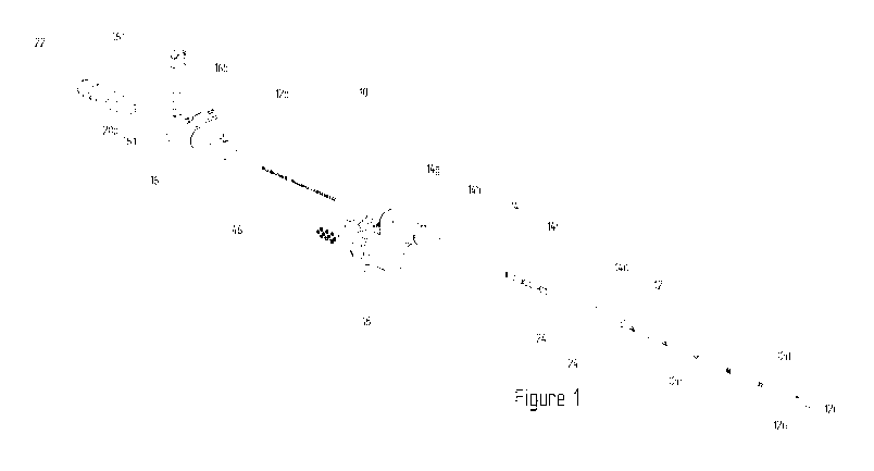
Figure 1 illustrates an isometric view of a catheter according to embodiments
of the present
invention.
Figure 2 illustrates a sleeve according to the third and sixth aspects of the
present invention.
Figure 3 shows a section through a retaining clip for locking together a
feeding tube and a sleeve
according to the first aspect of the invention.
Figure 4 shows an isometric view of the retaining clip of figure 1.
Figure 5 shows a section through a seal for providing a seal between a feeding
tube and a sleeve
according to the second aspect of the invention.
Figure 6 shows an isometric view of the seal of figure 3.
Figure 7 is an illustration of the packaging for the catheter of figure 1
according to the fourth aspect
of the invention.
Figure 8 is a diagram of the structure of a wire according to the fifth aspect
of the invention.
DETAILED SUMMARY OF THE INVENTION
Figure 1 shows a catheter 10 in accordance with a preferred embodiment of the
invention that is
suitable for providing intraluminal electrical pharyngeal neuromuscular
stimulation to a patient
suffering from dysphagia.
The catheter 10 comprises a feeding tube 12 formed from polyurethane, or other
highly flexible
material, and a fluorinated ethylene propylene and polyurethane sleeve 14. The
catheter 10 is
suitably sized for nasal insertion into a patient. The feeding tube 12 of the
catheter 10 is of a length
sufficient to enable an end to pass through the nose or mouth of the patient,
and, via the pharynx
and oesophagus, into the stomach of the patient.
The feeding tube 12 of the catheter 10 has a distal end 12a and a proximal end
12b. The proximal
end 12b of the feeding tube 12 is restrained by a Y-shaped connector 16 for
introducing nutrients
into the stomach via the feeding tube 12. The distal end 12a of the feeding
tube 12 is unrestrained.
The sleeve 14 of the catheter 10 has a distal end 14a and a proximal end 14b.
The proximal end 14a
7
CA 02966604 2017-05-02
WO 2016/071703 PCT/GB2015/053366
of the sleeve is restrained by an S-shaped connector 18 for providing an
electrical interface between
the catheter 10 and a base station (not shown). The distal end 14b of the
sleeve 14 is unrestrained.
The feeding tube 12 and sleeve 14 are arranged co-axially with the sleeve 14
surrounding the
feeding tube 12.
The feeding tube 12 comprises a rounded tip 12c at its distal end 12b for
patient comfort and ease of
insertion into the patient. Nutrients are dispersed from the tube 12 via one
or more apertures 12d in
the circumferential wall of the feeding tube 12 at the distal end 12a thereof
and through the distal
end 12a of the feeding tube 12 which is open ended. The feeding tube 12 is
provided with a plurality
of visual indicators 12e along its length, which, in conjunction with the
sleeve 14, provide a means of
making adjustments of the sleeve 14 relative to the feeding tube 12 taking
into account anatomical
measurement made on a patient. The feeding tube 12 may be printed with a 1cm
distance guide.
The polyurethane feeding tube material contains 20% barium sulphate to make
the tube 12 opaque
under X-ray.
The connector 16 is of a Y-shaped construction with a lumen therethrough. One
end of the
connector 16 receives the proximal end 12b of the feeding tube 12. The other
end of the connector
16 provides a primary port 16a which allows connection to an enteral feeding
set (not shown). A
secondary port 16b on the Y-portion of the container allows connection to a
syringe. The secondary
port 16b is closable by a cap 16c hingedly connected to the body of the
connector 16.
The primary port 16a of the connector 16 also receives a guidewire 20 to
assist with inserting the
catheter 10 into the patient. The guidewire 20 is formed from stainless steel
and is of cable twist
construction. The guidewire has a proximal end 20a and a distal end (not
shown). The proximal end
20A is received by a guidewire grip 22. The distal end is unrestrained and
terminated by a bead. The
guidewire grip 22 is a moulded component with a lumen therethrough to receive
the guidewire.
The proximal end 14b of the sleeve 14, which is restrained by the housing 18,
is surrounded by a
strain relief element 14g to reduce strain on the sleeve 14 at an interface
between the sleeve 14 and
the connector 18.
With reference to figure 2, the sleeve 14 is constructed from two distinct
layers 14c, 14d. The first,
inner layer 14c is formed from fluorinated ethylene propylene and the second,
outer layer 14d is
formed from polyurethane. A lumen 14e runs longitudinally through the centre
of the sleeve 14 to
receive the feeding tube 12. A pair of ring electrodes 24 is crimped to the
external wall of the sleeve
14. The electrodes 24 are approximately three millimetres wide, positioned
approximately ten
millimetres apart and are formed from medical grade stainless steel or
platinum. Two wires 26, 28
extend from the electrodes and are received in lumens 30, 32 in the outer 14d,
polyurethane layer
of the sleeve 14. The wires 26, 28 are connected to the connector 18 which
provides the electrical
interface between the catheter 10 and the base station.
The wires 26, 28 each comprise a single strand 26a, 28a, or cable,
encapsulated by two distinct types
of insulation, as shown in figure 8.
A basic insulation 26b, 28b comprises polyurethane, having a dielectric
strength of the order of
20kV/mm, and fluorinated ethylene propylene, having a dielectric strength of
the order of
60kV/mm. The polyurethane part of the basic insulation has a minimum thickness
of 0.075mm and
8
CA 02966604 2017-05-02
WO 2016/071703
PCT/GB2015/053366
the FEP part of the basic insulation has a minimum thickness of 0.038mm. In
combination, the
polyurethane and FEP parts of the basic insulation provide insulation of at
least 1500V.
A supplementary insulation 26c, 28c comprises a layer of enamel, having a
dielectric strength
between 170 and 230 kV/mm, and a layer of parylene, having a dielectric
strength of the order of
200kV/mm. The enamel layer has a thickness of between 0.01mm to 0.014mm and
the parylene
layer has a thickness of between 0.01mm to 0.02mm. In combination the enamel
and parylene
layers of the supplementary insulation provide insulation of between 3700V to
7080V.
The supplementary insulation is applied to the single strand, or cable using
vapour deposition in two
stages. The enamel is applied directly to the single strand, or cable, and the
parylene is applied to
the enamel layer. The desired thickness is achieved as a function of time
against vapour deposition
material density in a chamber. The basic insulation is applied to the
supplementary insulation.
The outer 14d polyurethane layer of the sleeve has a thickness which makes up
around 88 - 92% of
the wall thickness of the sleeve 14. The inner 14c layer of the sleeve has a
thickness which makes up
around 8 to 12% of the wall thickness of the sleeve 14. The outer layer 14d of
the sleeve extends
further towards the distal end 14a of the sleeve 14 than the inner layer 14c
of the sleeve 14. The
extreme distal end of the outer layer 14e forms a flexible tip.
The outer 14d, polyurethane layer of the sleeve 14 is provided with three
guide windows, or rings,
14f (see figure 1) which are marked with one, two or three dots, or other
visual marks, to signify
patients of differing height or other anatomical characteristic. The guide
windows 14f are used in
conjunction with the visual indicators of the feeding tube 12 to position the
sleeve 14 relative to the
feeding tube 12 according to a patient's anatomy before the catheter is
inserted into the patient.
The longitudinal position of the sleeve 14 relative to the feeding tube 12 is
restrainable by way of a
retaining clip 34 as illustrated in figures 3 and 4. The retaining clip 34
comprises a first part 36 and a
second part 38 connected together by way of a living hinge 40. The living
hinge is intended to take
its normal meaning in the art. The retaining clip 34 is manufactured from
polypropylene.
The first part 36 of the retaining clip 34, when viewed in cross section, has
a flat top surface 36a with
a semi-circular cut-out 36b therethrough for receiving a part of the feeding
tube 12. A bottom
surface 36c is arranged parallel to the top surface 36a. The bottom surface
36c is connected to the
top surface by a pair of curved sidewalls 36d extending upwardly and outwardly
from the edges of
the bottom surface 36c to the edges of the top surface 36a. The sidewalls 36d
each form a
substantially L-shape, as viewed in cross-section, by virtue of a recess 36e
in the first part 36 of the
retaining clip 34. The recess 36e permits the retaining clip 34 to slide on to
a mounting formation
(not shown) with lateral movement constrained by the L-shape of the sidewalls
36d. A resilient
finger 36g on the first part 36 of the retaining clip 34 engages with a recess
in the connector 18 to
restrain longitudinal movement of the retaining clip 34 relative to the
connector 18. The sidewall
36d positioned furthest away from the living hinge 40 is provided with a ridge
36f to, when the
retaining clip 34 is closed, hold the first 36 and second 38 parts of the
retaining clip 34 in
engagement with the feeding tube 12.
The second part 38 of the retaining clip 34 has a curved top wall 38a spaced
apart from a curved
bottom wall 38b. One end of the curved top wall 38a is joined to the living
hinge 40. The curved
9
CA 02966604 2017-05-02
WO 2016/071703 PCT/GB2015/053366
bottom wall 38b defines a plurality of ribs 38c extending outwardly. The end
of the curved bottom
wall 38b opposite the living hinge 40 is provided with a spring clip 38d which
is co-operable, when
the retaining clip 34 is closed, with the ridge 36f of the first part 36 of
the retaining clip 34.
In a preferred embodiment, the plurality of ribs 38c are covered by an
elastomer insert 38e
insertable into the second part 38 of the retaining clip 34. The insert 38e is
deformable and
comprises a channel 38f which engages against the sleeve 14 when the retaining
clip 34 is closed.
The high co-efficient of friction of the elastomer insert 38e inhibits
longitudinal movement of the
sleeve 14 relative to the retaining clip 34 and feeding tube 12. The elastomer
insert 38e is not shown
in figure 4.
The proximal end of the sleeve 14 is further provided with a cylindrical seal
44, as illustrated in
figures 5 and 6, which is bonded to the outer surface of the proximal end 14a
of the sleeve 14 at its
extreme end. The seal has a first end 44a and a second end 44b with a lumen
44c therebetween. The
first end 44a of the seal 44 has a first outer diameter and the second end 44b
of the seal 44 has a
second outer diameter smaller, than the first. A flange 44d extends from the
outer surface of the
seal at a position between the first 44a and the second 44b ends thereof. The
flange 44d extends
around the circumference of the seal 44 and is co-operable with a mounting
formation (not shown)
within the housing 18 for preventing longitudinal movement of the seal 44, and
thus the sleeve 14
within the housing 18.
The first end 44a of the seal 44 has a tapered opening into the lumen 44c. The
lumen 44c has a first
internal diameter leading from the tapered opening at the first end 44a of the
seal 44. The first
internal diameter is restricted at a shoulder 44e inside the lumen 44c. The
proximal end 14b of the
sleeve 14 abuts against the shoulder 44e of the lumen. A second internal
diameter of the lumen
extends from the shoulder 44e towards the second end 44b of the seal 44.
The second end 44b of the seal 44 has a tapered opening into the lumen 44c.
The tapered opening
extends to the second internal diameter of the lumen 44c. The second internal
diameter has, at its
mid-point, a flange 44f extending substantially entirely around its inner
circumference. The flange
44f, at its minimum internal diameter, is sized to act against the external
surface of the feeding tube
thus providing a seal between the sleeve 14 and the feeding tube 12.
In use, the feeding tube 12 is inserted into the second end 44b of the seal 44
and thus into the
sleeve 14. The flange 44f within the lumen 44c provides a seal between the
outer surface of the
feeding tube 12 and the inner surface of the sleeve 14 thus preventing fluid
from within a patient
being drawn up in a space therebetween by way of capillary action when the
sleeve 14 is removed
from the patient. The flange 44f also acts to clean any matter off of the
feeding tube 12 as it is
withdrawn from the patient.
The s-shaped connector 18 is formed from two substantially mirrored parts
which are joined by a
snap fit connection. The s-shaped connector 18 houses the strain relief
element 14g of the sleeve 14,
the seal 44 and several electrical components. The housing 18 is formed from
medical grade
acrylonitrile butadiene styrene. One end of the s-shaped connector 18 receives
the proximal end 14a
of the sleeve 14 and the other end of the s-shaped connector 18 houses an
electrical connector
which provides an interface between the catheter 10 and the base station. A
protective cap 46 is
attached to the s-shaped connector 18 by a lanyard and ring to protect the
electrical connector from
CA 02966604 2017-05-02
WO 2016/071703
PCT/GB2015/053366
liquid and dirt. The cap is formed from a thermoplastic elastomer. The s-
shaped connector 18
includes a mounting formation (not shown) in the form of rails for mounting
the retaining clip 34 to
the s-shaped connector 18.
The catheter 10, when assembled, is packed in a storage tray 48, as
illustrated in figure 7, moulded
to receive the specific catheter 10 components. The storage tray 48 comprises
a plurality of
formations 48a which provide a packaging space corresponding to the desired
profile of the feeding
tube 12 and/or sleeve 14. When the catheter 10 is packed into the storage tray
48, the feeding tube
12 and/or sleeve 14 take the profile of the packaging space. Once the catheter
10 has been packed
into the storage tray 48, the packaged catheter 10 is sterilised by exposing
at least a part of the
catheter to pre-determined conditions of one or more of temperature, humidity,
pressure, vacuum,
gas or radiation for a pre-determined time, whereby upon completion of the
application of those
conditions, at least part of the catheter maintains its deformed shape when
removed from the
storage container. During exposure, the material of the feeding tube 12 and/or
sleeve 14 softens
breaking the polymer bonds of the material. When the feeding tube 12 and/or
sleeve 14 are
removed from exposure to one or more of said conditions, the polymer bonds of
the material
reform such that at least a part of the feeding tube 12 and/or sleeve 14
naturally take the profile of
the packaging space when removed from the storage tray 48.
The shape of the catheter 10 is thus configured to have distinct sections,
each having a different
radius of curvature. A first section has a first radius of curvature that
corresponds to a patient's
anatomical transition between the nasal cavity and nasopharynx. A second
section has a second
radius of curvature that corresponds to a patient's anatomical transition
between the nasal cavity
and external nares.
The above description is given by way of example only and is not intended to
limit the scope of the
invention.
11