Note: Descriptions are shown in the official language in which they were submitted.
CRYSTALLINE FORM OF (S)-N-(5-((R)-2-(2,5-DIFLUOROPHENYL)-
PYRROLIDIN-1-YL)-PYRAZOLO[1,5-A]PYRIMIDIN-3-YL)-3-
HYDROXYPYRROLIDINE-1-CARBOXAMIDE HYDROGEN SULFATE
BACKGROUND
1. FIELD OF THE INVENTION
1 0 The present disclosure relates to (S)-N-(5-((R)-2-(2,5-
difluoropheny1)-pyrrolidin-1-
y1)-pyrazolo[1,5-a]pyrimidin-3-y1)-3-hydroxypyrrolidine-l-carboxamide (Formula
I) and to
pharmaceutically acceptable salts thereof, for example the hydrogen sulfate
salt, and further
to a novel crystalline form of the hydrogen sulfate salt, which exhibit Trk
family protein
tyrosine kinase inhibition, pharmaceutical compositions containing the same,
processes of
making the crystalline form, and the use of the compound and crystalline form
in the
treatment of pain, inflammation, cancer, and certain infectious diseases.
2. DESCRIPTION OF THE RELATED ART
Trk's are the high affinity receptor tyrosine kinases activated by a group of
soluble
growth factors called neurotrophins (NT). The Trk receptor family has three
members ¨
TrkA, TrkB and TrkC. Among the neurotrophins are (i) nerve growth factor (NGF)
which
activates Trick, (ii) brain-derived neurotrophic factor (BDNF) and NT-4/5
which activate
TrkB and (iii) NT3 which activates TrkC. Trk's are widely expressed in
neuronal tissue and
are implicated in the maintenance, signaling and survival of neuronal cells
(Patapoutian, A. et
al., Current Opinion in Neurobiology, 2001, 11, 272-280).
Recent literature has shown that overexpression, activation, amplification
and/or
mutation of Trk's are associated with many cancers including neuroblastoma
(Brodeur, G.
M., Nat. Rev. Cancer 2003, 3, 203-216), ovarian cancer (Davidson., B. et al.,
Clin. Cancer
Res. 2003, 9, 2248-2259), breast cancer (Kruettgen et al., Brain Pathology
2006, 16: 304-
310), prostate cancer (Dionne et al., Clin. Cancer Res. 1998, 4(8): 1887-
1898), pancreatic
cancer (Dang et al., Journal of Gastroenterology and Hepatology 2006, 21(5):
850-858),
multiple myeloma (Hu et al., Cancer Genetics and Cytogenetics 2007, 178: 1-
10),
astrocytoma amd medulloblastoma (Kruettgen et al., Brain Pathology 2006, 16:
304-310),
glioma (Hansen et al., Journal of Neurochemistry 2007, 103: 259-275),
melanoma25, thyroid
1
Date Recue/Date Received 2022-05-09
CA 02967951 2017-05-15
WO 2016/077841 PCT/1JS2015/060953
carcinoma (Brzezianska et al., Neuroendocrinology Letters 2007, 28(3), 221-
229),
lung adenocarcinoma (Perez-Pinera et al., Molecular and Cellular Biochemistry
2007,
295(1&2), 19-26), large cell neuroendocrine tumors19 (Marchetti et al., Human
Mutation
2008, 29(5), 609-616), and colorectal cancer (Bardelli, A., Science 2003, 300,
949). In
.. preclinical models of cancer, Trk inhibitors are efficacious in both
inhibiting tumor growth
and stopping tumor metastasis. In particular, non-selective small molecule
inhibitors of TrkA,
TrkB, TrkC and Trk/Fc chimeras were efficacious in both inhibiting tumor
growth and
stopping tumor metastasis25 (Nakagawara, A. (2001) Cancer Letters 169:107-114;
Meyer, J.
et al. (2007) Leukemia, 1-10; Pierottia, M.A. and Greco A., (2006) Cancer
Letters 232:90-
98; Eric Adriaenssens, E. et al. Cancer Res (2008) 68:(2) 346-351). Therefore,
an inhibitor
of the Trk family of kinases is expected to have utility in the treatment of
cancer.
In addition, inhibitors of the Trk/neurotrophin pathway have been demonstrated
to be
effective in numerous pre-clinical animal models of pain. For example,
antagonistic NGF and
TrkA antibodies (for example, RN-624) have been shown to be efficacious in
inflammatory
and neuropathic pain animal models and in human clinical trials (Woolf, C.J.
et al. (1994)
Neuroscience 62,327-331; Zahn, P.K. et al. (2004) J. Pain 5, 157-163; McMahon,
S. B. et
al., (1995) Nat. Med. 1, 774-780; Ma, Q. P. and Woolf, C. J. (1997)
Neuroreport 8, 807-
810; Shelton, D. L. et al. (2005) Pain 116, 8-16; Delafoy, L. et al. (2003)
Pain 105, 489-
497; Lamb, K. et al. (2003) Neurogastroenterol. Motil. 15, 355-361; Jaggar, S.
I. et al.
(1999) Br. J. Anaesth. 83, 442-448). Additionally, recent literature indicates
after
inflammation, BDNF levels and TrkB signaling is increased in the dorsal root
ganglion (Cho,
L. et al. Brain Research 1997, 749, 358) and several studies have shown
antibodies that
decrease signaling through the BDNF/TrkB pathway inhibit neuronal
hypersensitization and
the associated pain (Chang-Qi, L et al. Molecular Pain 2008, 4:27).
It has been shown that NGF secreted by tumor cells and tumor invading
macrophages
directly stimulates TrkA located on peripheral pain fibers. Using various
tumor models in
both mice and rats it was demonstrated that neutralizing NGF with a monoclonal
antibody
inhibits cancer related pain to a degree similar or superior to the highest
tolerated dose of
morphine. In addition, activation of the BDNF/TrkB pathway has been implicated
in
numerous studies as a modulator of various types of pain including
inflammatory pain
(Matayoshi, S., J. Physiol. 2005, 569:685-95), neuropathic pain (Thompson,
S.W., Proc. Natl.
Acad. Sci. USA 1999, 96:7714-18) and surgical pain (Li, C.-Q. et al.,
Molecular Pain, 2008,
4(28), 1-11). Because TrkA and TrkB kinases may serve as a mediator of NGF
driven
2
CA 02967951 2017-05-15
WO 2016/077841 PCT/1JS2015/060953
biological responses, inhibitors of TrkA and/or other Trk kinases may provide
an
effective treatment for chronic pain states.
The current treatment regimes for pain conditions utilize several classes of
compounds. The opioids (such as morphine) have several drawbacks including
emetic,
constipatory and negative respiratory effects, as well as the potential for
addictions. Non-
steroidal anti-inflammatory analgesics (NSAIDs, such as COX-1 or COX-2 types)
also have
drawbacks including insufficient efficacy in treating severe pain. In
addition, COX-1
inhibitors can cause ulcers of the mucosa. Accordingly, there is a continuing
need for new
and more effective treatments for the relief of pain, especially chronic pain.
In addition, inhibition of the neurotrophin/Trk pathway has been shown to be
effective in treatment of pre-clinical models of inflammatory diseases. For
example,
inhibition of the neurotrophin/Trk pathway has been implicated in preclinical
models of
inflammatory lung diseases including asthma (Freund-Michel, V; Frossard, N.;
Pharmacology & Therapeutics (2008), 117(1), 52-76), interstitial cystitis (Hu
Vivian Y; et.
.. al. The Journal of Urology (2005), 173(3), 1016-21), inflammatory bowel
diseases including
ulcerative colitis and Crohn's disease (Di Mola, F. F, et. al., Gut (2000),
46(5), 670-678) and
inflammatory skin diseases such as atopic dermatitis (Dou, Y.-C.; et. al.
Archives of
Dermatological Research (2006), 298(1), 31-37), eczema and psoriasis
(Raychaudhuri, S. P.;
et. al. Journal of Investigative Dermatology (2004), 122(3), 812-819).
The neurotrophin/Trk pathway, particularly BDNF/TrkB, has also been implicated
in
the etiology of neurodegenerative diseases including multiple sclerosis,
Parkinson's disease
and Alzheimer's disease (Sohrabji, Farida; Lewis, Danielle K. Frontiers in
Neuroendocrinology (2006), 27(4), 404-414). Modulation of the neutrophin/Trk
pathway
may have utility in treatment of these and related diseases.
The TrkA receptor is also thought to be critical to the disease process in the
infection
of the parasitic infection of Trypanosoma cruzi (Chagas disease) in human
hosts (de Melo-
Jorge, M. et al. Cell Host & Microbe (2007), 1(4), 251-261). Thus, TrkA
inhibition may
have utility in treating Chagas disease and related protozoan infections.
Trk inhibitors may also find use in treating disease related to an imbalance
of the
.. regulation of bone remodeling, such as osteoporosis, rheumatoid arthritis,
and bone
metastases. Bone metastases are a frequent complication of cancer, occurring
in up to 70
percent of patients with advanced breast or prostate cancer(1) and in
approximately 15 to 30
percent of patients with carcinoma of the lung, colon, stomach, bladder,
uterus, rectum,
thyroid, or kidney. Osteolytic metastases can cause severe pain, pathologic
fractures, life-
3
CA 02967951 2017-05-15
WO 2016/077841 PCT/1JS2015/060953
threatening hypercalcemia, spinal cord compression, and other nerve-
compression
syndromes. For these reasons, bone metastasis is a serious and costly
complication of cancer.
Therefore, agents that can induce apoptosis of proliferating osteoblasts would
be highly
advantageous. Expression of TrkA and TrkC receptors has been observed in the
bone
forming area in mouse models of bone fracture (K. Asaumi, et al., Bone (2000)
26(6) 625-
633). In addition, localization of NGF was observed in almost all bone forming
cells (K.
Asaumi, et al.). Recently, it was demonstrated that a pan-Trk inhibitor
inhibits the tyrosine
signaling activated by neurotrophins binding to all three of the Trk receptors
in human hFOB
osteoblasts (J. Pinski, et al., (2002) 62, 986-989). These data support the
rationale for the use
of Trk inhibitors for the treatment of bone remodeling diseases, such as bone
metastases in
cancer patients.
Several classes of small molecule inhibitors of Trk kinases said to be useful
for
treating pain or cancer are known (Expert Opin. Ther. Patents (2009) 19(3)).
International Patent Application Publications WO 2006/115452 and WO
2006/087538
describe several classes of small molecules said to be inhibitors of Trk
kinases which could
be useful for treating pain or cancer.
Pyrazolo[1,5-alpyrimidine compounds are known. For example, International
Patent
Application Publication WO 2008/037477 discloses pyrazolo[1,5-a]pyrimidine
compounds
bearing an alkyl, aryl or heterocyclic group at the 3-position. These
compounds are asserted
to be PI3K and/or mTOR Lipid Kinase inhibitors.
PCT Patent Publication No. WO 2008/058126 discloses pyrazolo[1,5-a]pyrimidine
compounds bearing a phenyl group at the 3-position. These compounds are
asserted to be
Pim-kinase inhibitors.
U.S. Patent Publication No. 2006/0094699 discloses pyrazolo[1,5-a]pyrimidine
compounds bearing a ¨C(=0)NH-phenyl, ¨C(=0)(4-methylpiperidinyl) or
¨C(=0)NMe(CH2-trimethylpyrazoly1) group at the 3-position for use in
combination therapy
with a glucocorticoid receptor agonist.
PCT Patent Publication Nos. WO 2010/033941, WO 2010/048314, WO 2011/006074,
and WO 2011/146336 disclose compounds which exhibit Trk family protein
tyrosine kinase
inhibition, and which are useful in the treatment of pain, cancer,
inflammation,
neurodegenerative diseases and certain infectious diseases.
WO 2010/048314 discloses in Example 14A a hydrogen sulfate salt of (S)-N-
(54(R)-2-(2,5-
difluoropheny1)-pyrrolidin-l-y1)-pyrazolo[1,5-a]pyrimidin-3-y1)-3-
hydroxypyrrolidine-1-
carboxamide. WO 2010/048314 does not disclose the particular form of the
hydrogen sulfate
4
CA 02967951 2017-05-15
WO 2016/077841 PCT/US2015/060953
salt described herein when prepared according to the method of Example 14A in
that
document. In particular, WO 2010/048314 does not disclose crystalline form (I-
HS) as
described below.
SUMMARY
The present disclosure relates to (S)-N-(54(R)-2-(2,5-difluoropheny1)-
pyrrolidin-1-
y1)-pyrazolo[1,5-a]pyrimidin-3-y1)-3-hydroxypyrrolidine-1-carboxamide (Formula
I) and to
pharmaceutically acceptable salts thereof, for example the hydrogen sulfate
salt, and further
to a novel crystalline form of the hydrogen sulfate salt, which exhibit Trk
family protein
tyrosine kinase inhibition, pharmaceutical compositions containing the same,
processes of
making the crystalline form, and the use of the compound and crystalline form
in the
treatment of pain, inflammation, cancer, and certain infectious diseases.
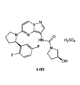
Provided herein is a novel crystalline form of the compound of Formula
N N 0
H N
F
OH
also known as (S)-N-(5-((R)-2-(2,5-difluoropheny1)-pyrrolidin- I -
y1)-pyrazolo[1,5-
alpyrimi din-3 -y1)-3 -h ydrox ypyrroli din e-1-c arbox ami de. In particular,
the novel crystalline
form comprises the hydrogen sulfate salt of the compound of Formula I in a
stable polymorph
form, hereinafter referred to as crystalline form (I-HS) and LOX0-101, which
can be
characterized, for example, by its X-ray diffraction pattern¨the crystalline
form (I-HS) having
the formula:
N N 0
HN--,f H2SO4
F N
OH
I-HS.
In some embodiments, crystalline form (I-HS) is characterized by having XRPD
5
Date Recue/Date Received 2022-05-09
CA 02967951 2017-05-15
WO 2016/077841 PCT/1JS2015/060953
diffraction peaks (20 degrees) at 18.4 0.2, 20.7 0.2, 23.1 0.2, and 24.0 0.2.
In some
embodiments, crystalline form (I-HS) is characterized by having XRPD
diffraction peaks (20
degrees) at 10.7 0.2, 18.4 0.2, 20.7 0.2, 23.1 0.2, and 24.0 0.2. In some
embodiments,
crystalline form (I-HS) is characterized by having XRPD diffraction peaks (20
degrees) at
.. 10.7 0.2, 18.4 0.2, 19.2 0.2, 20.2 0.2, 20.7 0.2, 21.5 0.2, 23.1 0.2, and
24.0 0.2. In some
embodiments, crystalline form (I-HS) is characterized by having XRPD
diffraction peaks (20
degrees) at 10.74.2, 15.34.2, 16.54.2, 18.4 0.2, 19.2 0.2, 19.9 0.2, 20.2 0.2,
20.74.2,
21.54.2, 22.1 0.2, 23.1 0.2, 24.0 0.2. 24.4 0.2, 25.6 0.2, 26.54.2, 27.6 0.2,
28.24.2,
28.7 0.2, 30.8 0.2, and 38.5 0.2.
In some embodiments, the crystalline form (I-HS) has XRPD pattern
substantially as
shown in Figure 29.
In some embodiments, the crystalline form exhibits an onset to maximum of
about 193
to about 205 Celsius, as measured by differential scanning calorimetry. In
some
embodiments, the crystalline form (1-HS) exhibits a heat of melting of about
2.415 mW, as
measured by differential scanning calorimetry. In some embodiments, the
crystalline form (I-
HS) has a DSC thermogram substantially as shown in Figure 26. In some
embodiments, the
crystalline form (I-HS) is non-hygroscopic.
Some embodiments include a pharmaceutical composition comprising a
pharmaceutically acceptable carrier and crystalline form (I-HS). Some
embodiments include
.. a pharmaceutical composition made by mixing crystalline form (I-HS) and a
pharmaceutically acceptable carrier. Some embodiments include a process of
making a
pharmaceutical composition comprising mixing crystalline form (I-HS) and a
pharmaceutically acceptable carrier.
The present disclosure also relates to methods for the treatment of cancer,
pain,
inflammation, and certain infectious diseases comprising administering to a
subject in need
thereof a therapeutically effective amount of crystalline form (1-HS). Some
embodiments
include the use of crystalline form (I-HS) in the preparation of a medicament
for treating
cancer, pain, inflammation, and certain infectious diseases, in a subject in
need thereof.
Also provided herein is a method of treating a cancer mediated by a Trk kinase
in a
subject in need thereof, the method comprising administering to the subject a
therapeutically
effective amount of crystalline form (I-HS). In some embodiments, the cancer
is mediated by
Trk; TrkB; or TrkA and TrkB. In some embodiments, a patient is diagnosed or
identified as
having a Trk-associated cancer.
6
CA 02967951 2017-05-15
WO 2016/077841 PCT/1JS2015/060953
Further provided herein is a method for treating cancer in a subject in need
thereof,
the method comprising: (a) determining if the cancer is associated with one or
more of
overexpression, activation, amplification, and mutation of a Trk kinase; and
(b) if the cancer
is determined to be associated with one or more of overexpression, activation,
amplification,
and mutation of a Trk kinasc, administering to the subject a therapeutically
effective amount
of crystalline form (I-HS). In some embodiments, a method for treating cancer
in a subject in
need thereof is provided, the method comprising: (a) determining if the cancer
is mediated by
a Trk kinase; and (b) if the cancer is determined to be mediated by a Trk
kinase,
administering to the subject a therapeutically effective amount of crystalline
form (I-HS).
Also provided herein is a method of treating a subject comprising: (a)
performing an assay
on a sample obtained from the subject to determine whether the subject has
dysregulation of a
NTRK gene, a Trk protein, or expression or level of the same; and (b)
administering to a
subject determined to have dysregulation of a NTRK gene, a Trk protein, or
expression or
activity, or level of the same a therapeutically effective amount of
crystalline form (I-HS).
In some embodiments, the dysregulation of a NTRK gene, a Trk protein, or
expression
or level of the same is a chromosome translation that results in the
translation of a Trk fusion
protein. For example, the Trk fusion protein is selected from the group
consisting of: TP53-
TrkA, LMNA-TrkA, CD74-TrkA, TFG-TrkA, TPM3-TrkA, NFASC-TrkA, BCAN-TrkA,
MPRIP-TrkA, TPR-TrkA, RFWD2-TrkA, IRF2BP2-TrkA, SQSTM1-TrkA, SSBP2-TrkA,
RABGAP1L-TrkA, C180RF8-TrkA, RNF213-TrkA, TBC1D22A-TrkA, C200RF112-TrkA,
DNER-TrkA, ARHGEF2-TrkA, CHTOP-TrkA, PPL-TrkA, PLEKHA6-TrkA, PEAR1-TrkA,
MRPL24-TrkA, MDM4-TrkA, LRRC71-TrkA, GRIPAP1-TrkA, EP S15-TrkA, DYNC2H1-
TrkA, CEL-TrkA, EPHB2-TrkA, TGF-TrkA, NACC2-TrkB, QKI-TrkB, AFAP1-TrkB,
PAN3-TrkB, SQSTM1-TrkB, TRIM24-TrkB, VCL-TrkB, AGBL4-TrkB, DAB2IP-TrkB,
ETV6-TrkC, BTBD 1 -TrkC, LYN-TrkC, RBPMS-TrkC, EML4-TrkC, HOMER2-TrkC, TFG-
TrkC, FAT1-TrkC, and TEL-TrkC.
In some embodiments, the dyregulation of a NTRK gene, a Trk protein, or
expression
or activity of the same is one or more point mutation in the gene. For
example, the NTRK gene
is a NTRK1 gene, and the one or more point mutations in the NTRK1 gene results
in the
translation of a TrkA protein having substitutions are one or more of the
following amino acid
positions: 33, 336, 337, 324, 420, 444, 517, 538, 649, 682, 683, 702, and
1879. In some
embodiments, the one or more point mutations in the NTRK1 gene results in the
translation of
a TrkA protein having one or more of the following amino acid substitutions:
R33W, A336E,
A337T, R324Q, R324W, V420M, R444Q, R444W, G517R, G517V, K538A, R649W, R649L,
7
CA 02967951 2017-05-15
WO 2016/077841
PCT/1JS2015/060953
R682S, V683G, R702C, and C1879T.
The features and advantages described in this summary and the following
detailed
description are not all-inclusive. Many additional features and advantages
will be apparent to
one of ordinary skill in the art in view of the drawings, specification, and
claims hereof.
BRIEF DESCRIPTION OF THE DRAWINGS
FIG. 1 illustrates an X-ray powder diffraction (XRPD) pattern of crystalline
form (I-
HS) prepared according to Example 2, according to one embodiment.
FIG. 2 illustrates a simultaneous thermogravimetric/differential thermal
analyzer
(TG/DTA) profile of crystalline form (I-HS) prepared according to Example 2,
according to
one embodiment.
FIG. 3 illustrates a differential scanning calorimetry (DSC) profile of
crystalline form
(I-HS) prepared according to Example 2, according to one embodiment.
FIGS. 4A and 4B illustrate polarized light microscopy (PLM) images of
crystalline
form (I-HS) prepared according to Example 2 under (A) unpolarized and (B)
polarized light,
according to some embodiments.
FIG. 5 illustrates a dynamic vapor sorption (DVS) isotherm profile of
crystalline form
(I-HS) prepared according to Example 2, according to one embodiment.
FIG. 6 illustrates an infrared (IR) spectroscopy profile of crystalline form
(I-HS)
prepared according to Example 2, according to one embodiment.
FIG. 7 illustrates an XRPD pattern of the amorphous freebase form of a
compound of
Formula I, according to one embodiment.
FIG. 8 is a graph showing the dose dependent inhibition of the proliferation
of
CUTO-3F lung adenocarcinoma cells harboring a MPRIP-NTRK1 fusion protein using
the
crystalline form (I-HS).
FIG. 9 is a graph showing the dose dependent inhibition of the proliferation
of KM12
colorectal cancer cells harboring a TPM3-NTRK1 fusion protein using the
crystalline form
(I-HS).
FIG. 10 is a graph showing the dose dependent inhibition of the proliferation
of MO-
91 acute myeloid leukemia cells harboring a ETV6-NTRK3 fusion protein using
the
crystalline form (I-HS).
FIG. 11 is an immunoblot showing that the crystalline form (I-HS) inhibits the
activation of MPRIP-TRKA kinasc, ERK1/2 in CUTO-3F cells, and AKT activity in
KM12
8
CA 02967951 2017-05-15
WO 2016/077841 PCT/1JS2015/060953
cells. The cells were treated for 2 hours with the crystalline form (I-HS) at
the indicated
doses.
FIG. 12 is an immunoblot showing that the crystalline form (I-HS) inhibits the
activation of TPM3-TRKA kinase and downstream ERK1/2 and AKT activity in KM 12
cells.
The cells were treated for 2 hours with the crystalline form (I-HS) at the
indicated doses.
FIG. 13 is an immunoblot showing that the crystalline form (1-HS)inhibits TEL-
TRKC kinase and ERK1/2 and AKT activity in MO-91 cells. The cells were treated
for 2
hours with the crystalline form (I-HS)at the indicated doses.
FIG. 14 is a schematic depicting the LMNA-NTRK1 gene fusion identified in the
patient's tumor sample: the joining of the first two exons of LMNA (NM_170707)
with exon
11-17 of NTRK1 (NM 002529).
FIG. 15 is a fluorescence micrograph from the NTRK1 break-apart FISH assay,
which
shows both paired green (5' NTRK1) and red (3' NTRK1) signals corresponding to
the
normal gene (yellow arrow), and isolated red signals (red arrows) are observed
in tumor
.. nuclei (stained blue with DAPI) indicate a chromosomal deletion that leads
to a NTRK1 gene
fusion.
FIG. 16 is a chromatograph of DNA sequencing of the RT-PCR product using LMNA
(5') and NTRK1 (3') primers indicating the fusion breakpoint between exon 2
LMNA and
exon 11 of NTRK1.
FIG. 17 is a schematic of the TRK-SHC1 proximity ligation assay (PLA). This
cartoon demonstrates the detection of proximal (< 40 nM) TRK and SHC 1
proteins in tumor
cells. The TRK antibody (rabbit) used can detect the c-terminus of TRKA
(encoded by
NTRK1), TRKB (NTRK2), or TRKC (NTRK3) proteins. SHC1 is detected by a SHC1
antibody (mouse). Binding of species-specific secondary antibodies with
covalently attached
.. complementary nucleotide sequences allows an in situ PCR reaction to
generate DNA, which
can be detected by fluorescence in situ hybridization visualized in the method
as red dots.
The assay has the potential to detect activated TRK regardless of mechanism of
activation
(gene fusion, mutation, or autocrine/paracrine activation of the wildtype) of
TRK receptor
family member (TRKA/B/C).
FIG 18 is a set of data that validate the TRK-SHC1 PLA. (A) The CUTO-3 cell
line,
derived from a malignant pleural effusion from a patient with stage IV lung
adenocarcinoma
harboring the MPRIP-NTRK1 gene fusion, was transfected with a non-targeting
control
(NTC) siRNA, NTRK1-directed siRNA, or untreated (control) and assayed for TRKA
protein
expression. Western blot analysis demonstrates a marked decrease in the TRKA
protein
9
CA 02967951 2017-05-15
WO 2016/077841
PCT/1JS2015/060953
levels, and corresponds to the MPRIP-TRKA fusion protein that migrates with an
apparent
molecular weight of 170 kD. TRK-SHC I PLA was performed in cells treated as in
(A)
demonstrating a robust positive signal in the siRNA control (B), but
proportional decrease in
the NTRK1 siRNA (C). CUTO-3 cells were treated with DMSO (D) or crystalline
form (I-
HS) at a concentration of 100nM (E) for 2 hours demonstrating disruption of
TRKA-SHC1
complexes in the crystalline form (I-HS) treated sample compared to control.
CULC001 is a
patient-derived tumor xenograft (PDX) derived from the same tumor as the CUTO-
3 cell line
and harbors the MPRIP-NTRK1 gene fusion (not shown). CULC002 is a PDX from a
NSCLC patient without a known driver (ALK, ROS1, EGFR, KRAS, and BRAF
negative)
and is negative for an NTRK1 gene fusion by NTRK1 break-apart FISH (not
shown). TRK
PLA analysis demonstrates a robust signal in CULC001 (F) but no signal in
CULC002 (G)
tumor nuclei. Panels (H) and (I) show a nerve bundle from the CULC001 PDX. TRK-
SHC1
PLA is positive only in this region of the CULC002 tumor sample and is
suggestive of
autocrine signaling in a TRKA, TRKB, or TRKC receptor as this family is
expressed in
nervous tissue and serves as internal positive control for this otherwise
negative tumor
sample.
FIG. 19 is an image from a TRK SHC1 proximity ligation assay and a control.
(A)
The TRK-SHC1 proximity ligation assay demonstrates robust signaling in the
tumor nuclei
but weak signaling in the thick walled blood vessel. Nuclei were stained with
DAPI (blue)
and the red signals represent a positive PLA indicative of TRKA-SHC1 protein
complexes.
A blood vessel is indicated within the partial ellipse (dotted white line).
(B) Adjacent tumor
tissue section stained with hematoxylin and eosin indicating a thick-walled
blood vessel
(within partial ellipse indicated by dotted white line) and flanking tumor
nuclei.
FIG. 20 are a set of images showing the TRK and ALK PLA in an ALK+ tumor
sample. FFPE tumor sample from an ALK+ patient (autopsy sample) was assayed
using the
TRK-SHC1 PLA (A) demonstrating an absence of signal or ALK-GRB2 PLA (B)
showing
robust ALK signaling.
FIG. 21 is a set of three computed tomography images from a subject having
undifferentiated sarcoma. CT images were obtained following pre-operative
chemotherapy
and primary tumor resection with arrow indicating the presence of an 18-mm
right lung
nodule (A), baseline imaging just prior to dosing with the crystalline form (I-
HS) on study
(B), and following 1 cycle (28 days) of dosing of with the crystalline fami (I-
HS) (C). The
patient was observed to have metastatic disease only in the lungs and
therefore the CT scan
images show axial (top) and coronal (bottom) images focusing on the thoracic
cavity. The
CA 02967951 2017-05-15
WO 2016/077841 PCT/1JS2015/060953
images demonstrate an initial rapid disease progression (A-B, 13 week
interval) followed by
a marked tumor response with decreased size and/or resolution of the numerous
pulmonary
metastases (B-C, 4 week interval).
FIG. 22 is a graph showing the serum CA125 levels in a patient having
undifferentiated sarcoma treated with crystalline form (I-HS) over time. Scrum
CA125 levels
were found to be elevated in this patient, and subsequently followed as a
potential indicator
of activity. Serum CA125 was drawn at baseline (day -8) prior to dosing and at
the indicated
time point points following the initiation of dosing at day -3 through day 56
demonstrating a
time-dependent decrease in this tumor marker. The dashed red line indicates
the upper limit
of normal (35 U/mL) of this laboratory test.
FIG. 23 is a graph showing the dose dependent inhibition of the proliferation
of
HCC78 cells harboring a SLC34A2-ROS1 fusion protein using the crystalline form
(I-HS).
FIG. 24 is a graph showing thermographic data for AM(HS)1. The top line of the
graph is a plot of the thermogravimetric analysis (TGA) for the compound,
while the bottom
line is a plot of the differential scanning calorimetry (DSC).
FIG. 25 is a graph showing thermographic data for AM(HS)2. The top line of the
graph is a plot of the thermogravimetric analysis (TGA) for the compound,
while the bottom
line is a plot of the differential scanning calorimetry (DSC).
FIG. 26 is a graph showing thermographic data for crystalline form (I-HS). The
top
line of the graph is a plot of the thermogravimetric analysis (TGA) for the
compound, while
the bottom line is a plot of the differential scanning calorimetry (DSC).
FIG. 27 illustrates an overlay of the X-ray powder diffraction (XRPD) patterns
of
AM(HS)1, AM(HS)2, and crystalline form (I-HS). AM(HS)1 and AM(HS)2 are the
broad
lines in the lower part of the figure, while crystalline form (I-HS) exhibits
sharp peaks.
FIG. 28 illustrates an X-ray powder diffraction (XRPD) pattern of AM(HS)1 and
AM(HS)2.
FIG. 29 illustrates an X-ray powder diffraction (XRPD) pattern of crystalline
form (1-
HS).
FIG. 30 is an image of a sample of AM(HS)1 under polarized light microscopy at
a
magnification of 20X.
FIG. 31 is an image of a sample of AM(HS)2 under polarized light microscopy at
a
magnification of 20X.
FIG. 32 is an image of a sample of crystalline form (I-HS) under polarized
light
microscopy at a magnification of 20X.
11
CA 02967951 2017-05-15
WO 2016/077841 PCT/1JS2015/060953
FIG. 33 is a plot of the hygroscopicity of AM(HS)1 using dynamic vapor
sorption
(DVS).
FIG. 34 illustrates an X-ray powder diffraction (XRPD) pattern of AM(HS)1 pre-
DVS
(top-line) and post-DVS (bottom line).
FIG. 35 is a plot of the hygroscopicity of AM(HS)2 using dynamic vapor
sorption
(DVS).
FIG. 36 illustrates an X-ray powder diffraction (XRPD) pattern of AM(HS)2 pre-
DVS
(top-line) and post-DVS (bottom line).
FIG. 37 is a plot of the hygroscopicity of crystalline farm (I-HS) using
dynamic vapor
sorption (DVS).
FIG. 38 illustrates an X-ray powder diffraction (XRPD) pattern of crystalline
form (I-
HS) pre-DVS (top-line) and post-DVS (bottom line).
FIG. 39 is a plot of tensile strength versus compression pressure for various
200 mg
direct compression blend compacts incorporating crystalline form (I-HS) or
AM(HS)2. In
the plot, (1) is a 2:1 MCC:lactose blend with AM(HS)2; (2) is a 2:1 MCC
lactose blend with
crystalline form (I-HS); (3) is a 1:1 MCC:starch blend with AM(HS)2; (4) is a
1:1
MCC:starch blend with crystalline form (I-HS).
FIG. 40 is an overlay of DSC thermographs of AM(HS)1 at TO (bottom line) and
after
5 weeks at 40 C/75%RH (top line).
FIG. 41 is an overlay of DSC thermographs of crystalline form (I-HS) at TO
(bottom
line) and after 5 weeks at 40 C/75%RH (top line).
FIG. 42 illustrates an overlay of the X-ray powder diffraction (XRPD) patterns
of
AM(HS)1 at TO (broad line) and after 5 weeks at 40 C/75%RH (sharp peaks).
FIG. 43 illustrates an overlay of the X-ray powder diffraction (XRPD) patterns
of
crystalline form (I-HS) at TO (bottom) and after 5 weeks at 40 C/75%RH (top).
FIG. 44 illustrates an overlay of the X-ray powder diffraction (XRPD) patterns
of
crystalline form (I-HS) (bottom) and AM(HS)1 (top) after 5 weeks at 40
C/75%RH.
FIG. 45 is a graph showing the percentage of change in volume of a xenograph
(human)
tumor derived from a lung adenocarcinoma CUTO-3F cell line (CUTO-3.29) over
time in mice
that were treated with vehicle (triangles) or orally administered a daily dose
of 60 mg/kg
(circles) or 200 mg/kg (squares) of crystalline form (I-HS) following
implantation of the
xenograft into the mice.
FIG. 46 is a graph showing the percentage of change in volume of a xenograph
(human)
tumor derived from a colorectal cancer KM12 cell line over time in mice that
were treated with
12
CA 02967951 2017-05-15
WO 2016/077841 PCMJS2015/060953
vehicle (triangles) or orally administered a daily dose of 60 mg/kg (circles)
or 200 mg/kg
(squares) of crystalline form (I-HS) following implantation of the xenograft
into the mice.
FIG. 47 is a graph showing the percentage of change in volume of a xenograph
(human)
tumor derived from an acute myeloid leukemia MO-91 cell line over time in mice
that were
treated with vehicle (triangles) or orally administered a daily dose of 60
mg/kg (circles) or 200
mg/kg (squares) of crystalline form (I-HS) following implantation of the
xenograft into the
mice.
The figures depict various embodiments of the present invention for purposes
of
illustration only. One skilled in the art will readily recognize from the
following discussion
that alternative embodiments of the structures and methods illustrated herein
may be
employed without departing from the principles of the invention described
herein.
DETAILED DESCRIPTION
The present disclosure relates to (S)-N-(5-((R)-2-(2,5-difluoropheny1)-
pyrrolidin-1-
1 5 y1)-pyrazolo[1,5-a]pyrimidin-3-y1)-3-hydroxypyrrolidine-l-carboxamide
(Formula I) and to
pharmaceutically acceptable salts thereof, for example the hydrogen sulfate
salt, and further
to a novel crystalline form of the hydrogen sulfate salt, which exhibit Trk
family protein
tyrosine kinase inhibition, pharmaceutical compositions containing the same,
and processes
of making the crystalline form
Provided herein is a novel crystalline form of the compound of Formula I:
N N
r) 0
HN
* F
\--j=
OH .
In particular, the novel crystalline form comprises the hydrogen sulfate salt
of the compound
of Formula I in a stable polymorph form, hereinafter referred to as
crystalline form (I-HS),
which may be characterized, for example, by its X-ray diffraction pattern.
As illustrated in FIG. 1, in some embodiments, the crystalline form (I-HS) can
be
characterized by its X-ray powder diffraction pattern (XRPD). The XRPD was
carried out on
a D5000 X-ray diffractometer with a CuKal, 0.1540562 nm long, fine focus
sealed tube
source from Siemens by scanning samples between 3 and 40 2-theta at a step
size of 0.0200
2-theta and a time per step of 1 second. The effective scan speed was 0.0200
/s with an
13
CA 02967951 2017-05-15
WO 2016/077841
PCT/1JS2015/060953
instrument voltage 40 kV and a current setting of 40 mA. Samples were analyzed
using a
divergence slit having a size of 2 mm in reflection mode under the following
experimental
conditions.
In some embodiments, crystalline form (I-HS) has an XRPD pattern with at least
the
20 characteristic peaks (20 degrees 0.3), as listed in Table 1.
Table I. XRPD peaks of crystalline form (I-HS)
Position I 2-0] FWHM [ 2-0] d-spacing [A] Relative
Intensity [%]
10.63 0.12 8.32 27.44
15.25 0.14 5.81 12.24
16.39 0.13 5.40 13.92
18.37 0.13 4.82 43.65
19.08 0.14 4.65 19.60
19.79 0.11 4.48 9.83
20.15 0.25 4.40 25.09
20.61 0.13 4.31 100.00
21.47 0.21 4.14 24.71
22.01 0.12 4.03 14.45
23.04 0.15 3.86 33.01
23.97 0.12 3.71 38.52
24.35 0.21 3.65 10.05
25.58 0.13 3.48 8.11
26.48 0.17 3.36 9.76
27.50 0.14 3.24 7.70
28.17 0.17 3.16 11.60
28.58 0.19 3.12 10.85
30.77 0.29 2.90 8.48
38.47 0.21 2.34 10.97
In some embodiments, the crystalline form (I-HS) has an XRPD pattern with at
least
the 8 characteristic peaks (20 degrees + 0.3), which comprises peaks having a
relative
intensity greater than or equal to about 15%, as listed in Table 2.
Table 2. XRPD peaks of crystalline form (I-HS)
Position I 2-01 FWHM [ 2-0] d-spacing [A] Relative
Intensity [ /0]
10.63 0.12 8.32 27.44
18.37 0.13 4.82 43.65
19.08 0.14 4.65 19.60
14
CA 02967951 2017-05-15
WO 2016/077841 PCT/US2015/060953
20.15 0.25 4.40 25.09
20.61 0.13 4.31 100.00
21.47 0.21 4.14 24.71
23.04 0.15 3.86 33.01
23.97 0.12 3.71 38.52
In some embodiments, the crystalline form (I-HS) has an XRPD pattern with at
least
the 5 characteristic peaks (20 degrees + 0.3), which comprises peaks having a
relative
intensity greater than or equal to about 25%, as listed in Table 3.
Table 3. XRPD peaks of crystalline form (I-HS)
Position 102-0] FWHM [024 d-spacing [A] Relative
Intensity [ /0]
10.63 0.12 8.32 27.44
18.37 0.13 4.82 43.65
20.61 0.13 4.31 100.00
23.04 0.15 3.86 33.01
23.97 0.12 3.71 38.52
In some embodiments, the crystalline form (I-HS) has an XRPD pattern with at
least
the 4 characteristic peaks (20 degrees 0.3), which comprises peaks having a
relative
intensity greater than or equal to about 30%, as listed in Table 4.
Table 4. XRPD peaks of crystalline form (I-HS)
Position [ 2-0] FWHM [ 2-0] d-spacing [A] Relative
Intensity [%]
18.37 0.13 4.82 43.65
20.61 0.13 4.31 100.00
23.04 0.15 3.86 33.01
23.97 0.12 3.71 38.52
In certain embodiments, crystalline form (I-HS) has an XRPD pattern that is
substantially the same XRPD pattern as shown in Figure 1.
In some embodiments, crystalline form (I-HS) is characterized by having XRPD
diffraction peaks (20 degrees) at about 18.4, 20.6, 23.0, and 24Ø In some
embodiments,
crystalline form (I-HS) is characterized by having XRPD diffraction peaks (20
degrees) at
about 10.6, 18.4, 20.6, 23.0, and 24Ø In some embodiments, crystalline form
(I-HS) is
characterized by having XRPD diffraction peaks (20 degrees) at about 10.6,
18.4, 19.1, 20.2,
20.6, 21.5, 23.0, and 24Ø In some embodiments, crystalline form (I-HS) is
characterized by
CA 02967951 2017-05-15
WO 2016/077841
PCT/1JS2015/060953
having XRPD diffraction peaks (20 degrees) at about 10.6, 15.3, 16.4, 18.4,
19.1, 19.8, 20.2,
20.6, 21.5, 22.0, 23.0, 24.0, 24.4, 25.6, 26.5, 27.5, 28.2, 28.6, 30.8, and
38.5.
In certain embodiments, crystalline form (I-HS) has an XRPD pattern that is
substantially the same XRPD pattern as shown in Figure 29.
In some embodiments, crystalline form (I-HS) has an XRPD pattern with at least
the
20 characteristic peaks (20 degrees 0.3), as listed in Table 1.
Table 5. XRPD peaks of crystalline form (I-HS)
Position ( 20) Relative Intensity (%)
10.76 29.85
15.38 13.22
16.52 16.46
18.50 48.07
19.22 22.92
19.92 16.05
20.26 30.80
20.74 100.00
21.56 23.78
22.16 15.51
23.16 32.52
24.10 33.89
24.50 12.14
25.72 8.89
26.50 10.88
27.62 8.61
28.32 11.44
28.74 10.73
30.92 8.23
38.60 8.88
In some embodiments, the crystalline form (I-HS) has an XRPD pattern with at
least
.. the 8 characteristic peaks (20 degrees 0.3), which comprises peaks having
a relative
intensity greater than or equal to about 15%, as listed in Table 6.
Table 6. XRPD peaks of crystalline form (I-HS)
Position ( 20) Relative Intensity (%)
10.76 29.85
18.50 48.07
19.22 22.92
20.26 30.80
16
CA 02967951 2017-05-15
WO 2016/077841 PCT/US2015/060953
20.74 100.00
21.56 23.78
23.16 32.52
24.10 33.89
In some embodiments, the crystalline form (I-HS) has an XRPD pattern with at
least
the 5 characteristic peaks (20 degrees + 0.3), which comprises peaks having a
relative
intensity greater than or equal to about 25%, as listed in Table 7.
Table 7. XRPD peaks of crystalline form (I-HS)
Position ( 20) Relative Intensity (%)
10.76 29.85
18.50 48.07
20.74 100.00
23.16 32.52
24.10 33.89
In some embodiments, the crystalline form (I-HS) has an XRPD pattern with at
least
the 4 characteristic peaks (20 degrees 0.3), which comprises peaks having a
relative
intensity greater than or equal to about 30%, as listed in Table 8.
Table 8. XRPD peaks of crystalline form (I-HS)
Position ( 20) Relative Intensity (%)
18.50 48.07
20.74 100.00
23.16 32.52
24.10 33.89
In some embodiments, crystalline form (I-HS) is characterized by having XRPD
diffraction peaks (20 degrees) at about 18.5, 20.7, 23.2, and 24.1. In some
embodiments,
crystalline form (I-HS) is characterized by having XRPD diffraction peaks (20
degrees) at
about 10.8, 18.5, 20.7, 23.2, and 24.1. In some embodiments, crystalline form
(I-HS) is
characterized by having XRPD diffraction peaks (20 degrees) at about 10.8,
18.5, 19.2, 20.3,
20.7, 21.6, 23.2, and 24.1. In some embodiments, crystalline form (I-HS) is
characterized by
having XRPD diffraction peaks (20 degrees) at about 10.8, 15.4, 16.5, 18.5,
19.2, 19.9, 20.3,
20.7, 21.6, 22.2, 23.2, 24.1, 24.5, 25.7, 26.5, 27.6, 28.3, 28.7, 30.9, and
38.6.
In some embodiments, given the XRPD patterns provided in FIGs. 1 and 29,
crystalline
feorm (I-HS) is characterized by having XRPD peaks (20 degrees) as shown in
Table 9.
Table 9. XRPD peaks of crystalline form (I-HS)
17
CA 02967951 2017-05-15
WO 2016/077841 PCT/1JS2015/060953
FIG. 1 FIG. 29 Difference Average
10.76 10.63 0.13 10.70
15.38 15.25 0.13 15.32
16.52 16.39 0.13 16.46
18.50 18.37 0.13 18.44
19.22 19.08 0.14 19.15
19.92 19.79 0.13 19.86
20.26 20.15 0.11 20.21
20.74 20.61 0.13 20.68
21.56 21.47 0.09 21.52
22.16 22.01 0.15 22.09
23.16 23.04 0.12 23.10
24.10 23.97 0.13 24.04
24.50 24.35 0.15 24.43
25.72 25.58 0.14 25.65
26.50 26.48 0.02 26.49
27.62 27.50 0.12 27.56
28.32 28.17 0.15 28.25
28.74 28.58 0.16 28.66
30.92 30.77 0.15 30.85
38.60 38.47 0.13 38.54
In some embodiments, crystalline form (I-HS) is characterized by having XRPD
diffraction peaks (20 degrees) at 18.4+0.2, 20.7+0.2, 23.1+0.2, and 24.0+0.2.
In some
embodiments, crystalline form (I-HS) is characterized by having XRPD
diffraction peaks (20
degrees) at 10.7+0.2, 18.4+0.2, 20.7+0.2, 23.1+0.2, and 24.0+0.2. In some
embodiments,
crystalline form (I-HS) is characterized by having XRPD diffraction peaks (20
degrees) at
10.7+0.2, 18.4+0.2, 19.2+0.2, 20.2+0.2, 20.7+0.2, 21.5+0.2, 23.1+0.2, and
24.0+0.2. In some
embodiments, crystalline form (I-HS) is characterized by having XRPD
diffraction peaks (20
degrees) at 10.7+0.2, 15.3+0.2, 16.5+0.2, 18.4+0.2, 19.2+0.2, 19.9+0.2,
20.2+0.2, 20.7+0.2,
21.5+0.2, 22.1+0.2, 23.1+0.2, 24.0+0.2. 24.4+0.2, 25.6+0.2, 26.5+0.2,
27.6+0.2, 28.2+0.2,
28.7+0.2, 30.8+0.2, and 38.5+0.2.
It will be understood that the 2-theta values of the X-ray powder diffraction
patterns
for crystalline form (I-HS) may vary slightly from one instrument to another
and also
depending on variations in sample preparation and batch to batch variation,
and so the values
quoted are not to be construed as absolute. It will also be understood that
the relative
intensities of peaks may vary depending on orientation effects so that the
intensities shown in
the XRPD trace included herein are illustrative and not intended to be used
for absolute
18
CA 02967951 2017-05-15
WO 2016/077841
PCT/US2015/060953
comparison. Accordingly, it is to be understood that the phrase "substantially
the same
XRPD pattern as shown in Figure 1 or Figure 29" means that for comparison
purposes, at
least 90% of the peaks shown in Figure 1 or Figure 29 are present. It is to be
understood that
the relative peak positions may vary 0.3 degrees from the peak positions
shown in Figure 1
or Figure 29. It is to be further understood that for comparison purposes some
variability in
peak intensities from those shown in Figure 1 and Figure 29 is allowed.
FIG. 2 illustrates a simultaneous thermogravimetric/differential thermal
analyzer
(TG/DTA) profile of crystalline form (I-HS), according to one embodiment. For
the analysis
about 5 mg of crystalline form (I-HS) was weighed into an open aluminum pan
and loaded
into a simultaneous thermogravimetric/differential thermal analyzer (TG/DTA)
and held at
room temperature. The sample was then heated at a rate of 10 Celsius/min from
25 Celsius
to 300 Celsius during which time the change in sample weight was recorded
along with any
differential thermal events. Nitrogen was used as the purge gas at a flow rate
of 100 cm3/min.
The TG/DAT profile of crystalline form (I-HS) shows an initial weight loss of
0.8% between
27.4 Celsius to 182.4 Celsius, which is followed by 4.9% weight loss in the
TG curve
between 182.4 Celsius to 225.0 Celsius, also seen as an endotherm in the DTA
curve.
These weight losses could be decomposition of the material.
FIG. 3 illustrates a differential scanning calorimetry (DSC) profile of
crystalline form
(I-HS), according to one embodiment. DSC analysis of the sample was performed
using a
Seiko D5C6200 differential scanning calorimeter (equipped with a cooler).
About 5 mg of
crystalline form (I-HS) was weighed into an aluminum DSC pan and sealed non-
hermetically
with a pierced aluminum lid. The sample pan was then loaded into a Seiko
D5C6200
(equipped with a cooler), cooled, and held at 25 Celsius. Once a stable heat-
flow response
was obtained, the sample and reference were heated to 270 Celsius at a scan
rate of 10
Celsius/min while monitoring the resulting heat flow response. In some
embodiments,
crystalline form (I-HS) has a DSC thermogram substantially as shown in Figure
3. As used
herein, "substantially as shown in Figure 3" means that the temperatures of
the endothermic
event shown in Figure 3 can vary by about 5 C.
As shown in FIG. 3, the DSC thermogram of the crystalline form (I-HS)
indicates a
small endothermic change in the baseline between 122.9 Celsius to 152.8
Celsius, followed
by a sharp endotherm that corresponds to the melting of the crystalline form
(I-HS) at an
onset temperature of melting of 190.8 Celsius, a peak temperature of melting
of 197.9
Celsius and a heat of melting of 2.415 mW. The transition following the
melting endotherm
may be caused by the decomposition of the melted crystalline form (I-HS).
19
CA 02967951 2017-05-15
WO 2016/077841 PCT/1JS2015/060953
FIGS. 4A and 4B illustrate polarized light microscopy (PLM) images of
crystalline
form (I-HS) under (A) unpolarized and (B) unpolarized light, according to some
embodiments. The presence of crystallinity (birefringence) was determined
using an
Olympus BX50 polarizing microscope, equipped with a Motic camera and image
capture
-- software (Motic Images Plus 2.0). All images were recorded using the 20x
objective. The
crystalline form (I-HS) exhibits birefringence when examined under polarized
light without
exhibiting a definite morphology or agglomerates.
FIG. 5 illustrates a dynamic vapor sorption (DVS) isotherm profile of
crystalline form
(I-HS), according to one embodiment. For the DVS measurement a sample of
crystalline
-- form (I-HS) was cycled through changing humidity conditions to detellaine
its
hygroscopicity. The sample was analyzed using a Surface Measurement System DVS-
1
Dynamic Vapor Sorption System. About 10 mg of crystalline form (I-HS) was
placed into a
mesh vapor sorption balance pan and loaded into a dynamic vapor sorption
balance as part of
the Surface Measurement System. Data was collected in 1 minute intervals.
Nitrogen was
used as the carrier gas. The sampled crystalline form (I-HS) was subjected to
a ramping
profile from 20-90% relative humidity (RH) at 10% increments, maintaining the
sample at
each step until a stable weight had been achieved (99.5% step completion).
After completion
of the sorption cycle, the sample was dried using the same procedure, but all
the way down to
0% RH and finally taken back to the starting point of 20% RH. The weight
change during the
sorption/desorption cycles were plotted, allowing for the hygroscopic nature
of the sample to
be determined.
As shown in FIG. 5, crystalline form (I-HS) appears to be non-hygroscopic. A
small
increase in mass of about 1.7% was observed between 0% and 90% RH during the
sorption
cycle. In addition, a very small hysteresis was observed between sorption and
desorption
cycles. The XRPD pattern of crystalline form (I-HS) post DVS analysis (not
shown) being
similar to its pre-DVS XRPD pattern shown in FIG. 1 or FIG. 29 indicates that
no change in
the crystalline form (I-HS) occurred during DVS.
FIG. 6 illustrates an infrared (IR) spectroscopy profile of crystalline form
(I-HS) for
the compound of Formula 1, according to one embodiment. IR spectroscopy was
carried out
on a Bruker ALPHA P spectrometer. Sufficient material of crystalline form (I-
HS) was
placed onto the center of the plate of the spectrometer with a transmittance
spectrum being
obtained using a resolution of 4 cm-1, a background scan time of 16 scans, a
sample scan time
of 16 scans, and collecting data from 4000 cm-1 to 400 cm-1. The observed IR
spectrum of
crystalline form (I-HS) is shown in FIG. 6.
CA 02967951 2017-05-15
WO 2016/077841 PCT/1JS2015/060953
The crystalline form (I-HS) has a number of properties that make it
surprisingly
superior to the amorphous form of (S)-N-(5-((R)-2-(2,5-difluoropheny1)-
pyrrolidin-1 -y1)-
pyrazolo[1,5-a]pyrimidin-3-y1)-3-hydroxypyrrolidine-1-carboxamide hydrogen
sulfate
(AM(HS)). For example, the crystalline form (I-HS) has properties which
contribute to its
manufacturability and production of a commercial product. As shown in Example
8, the
crystalline form (I-HS) has better flow properties as compared to the
amorphous API
(AM(HS)) as evidenced by the Carr's and Hausner Index. For example, the
crystalline form
(I-HS) exhibits a Can Index value of greater than 20%. In some embodiments,
the crystalline
form (I-HS) exhibits a Hausner ratio of less than 1.35 (e.g., a value of
between about 1.26 to
about 1.34). The differences in flow properties can make the development of a
solid oral
dosage form more difficult for the amorphous API vs. the crystalline API.
The crystalline form (I-HS) also evidenced better stability in an accelerated
stability
study conducted in an LDPE bag at 40 C/75% RH for five weeks. While neither
the
AM(HS) or crystalline form (I-HS) exhibited a significant changes in chemical
impurity
levels over the course of the study, the study did reveal that the crystalline
form (I-HS) has
stable physicochemical properties. The amorphous API, on the other hand,
converted into a
crystalline form substantially similar to the crystalline form (I-HS) by XRPD,
DSC, TGA, KF
and polarized light microscopy. Additionally, the amorphous API changed to an
agglomerated powder with reduced flow properties over the course of the
stability testing.
Such changes in the physical properties of the compound, including a change
from an
amorphous power to a crystalline material and/or an agglomerated powder with
reduced flow,
on storage would make it nearly impossible to manufacture a solid oral dosage
form for
patient use based on the amorphous compound. The properties observed for the
crystalline
form (I-HS), however, are consistent with that desired for a commercial
product, including
having both a stable physical and chemical structure.
The crystalline form (I-HS), as noted previously, is non-hygroscopic. As used
herein,
"non-hygroscopic" refers to a compound exhibiting less than a 2% weight gain
at 25 C and
80% RH after 24 to 48 hours (see, e.g., Example 10). The AM(HS) compound,
however, was
found to deliquesce upon exposure to humidity. Given this tendency, use of the
AM(HS)
compound would require significant handling precautions during storage and
manufacture to
prevent this change in form from occurring whereas the crystalline form (1-HS)
requires no
such precautions during manufacture of the API. This stability to humidity
would also be
expected to carry over to any solid oral dosage product prepared using the
crystalline form (I-
HS).
21
Finally, the crystalline form (I-HS) provides a significantly improved
impurity profile
versus the amorphous API. The ability to control an impurity profile is
important for patient
safety, developing a repeatable manufacturing process, and meeting
requirements by
Regulatory agencies prior to use in humans.
The compounds provided herein, including (S)-N-(54(R)-2-(2,5-difluoropheny1)-
pyrrolidin-1-y1)-pyrazolo[1,5-a]pyrimidin-3-y1)-3-hydroxypyrrolidine-1-
carboxamide
(Formula I) and pharmaceutically acceptable salts thereof, for example the
hydrogen sulfate
salt, and further a novel crystalline form of the hydrogen sulfate salt
(crystalline form (I-HS)),
exhibit Trk family protein tyrosine kinase inhibition, and the compound,
hydrogen sulfate
salt, and crystalline form thereof can be used in the treatment of pain,
inflammation, cancer,
and certain infectious diseases.
Some embodiments include the use of the crystalline form (I-HS) for the
treatment of
disorders and diseases which can be treated by inhibiting TrkA, TrkB and/or
TrkC kinases,
such as a TrkA, TrkB and/or TrkC mediated condition, such as one or more
conditions
described herein, including a Trk-associated cancer. In some embodiments, the
crystalline
form (I-HS) may be also useful in the treatment of pain, including chronic and
acute pain. In
some embodiments, the crystalline form (I-HS) may be useful in the treatment
of multiple
types of pain including inflammatory pain, neuropathic pain, surgical pain,
and pain
associated with cancer, surgery and bone fracture. In addition, the
crystalline form (I-HS)
may be useful for treating inflammation, active and chronic neurodegenerative
diseases and
certain infectious diseases. The present disclosure is further directed to
pharmaceutical
compositions comprising crystalline form (I-HS). In some embodiments, the
pharmaceutical
composition comprises crystalline form (I-HS) and a pharmaceutically
acceptable diluent or
carrier.
The ability of crystalline form (I-HS) to act as TrkA, TrkB and/or TrkC
inhibitors
may be demonstrated by the assays described in Examples A and B as disclosed
in U.S.
Patent No. 8,513,263, issued on August 20, 2013.
In some embodiments, provided herein is a method for treating a patient
diagnosed
with a TRK-associated cancer, comprising administering to the patient a
therapeutically
effective amount of crystalline form (I-HS) or a compound of Formula I or a
salt thereof,
such as a hydrogen sulfate salt (e.g., see Example 14A of U.S. Patent No.
8,513,263). Trk
family of neurotrophin receptors, TrkA, TrkB, and TrkC (encoded by NTRK1,
NTRK2, and
NTRK3 genes, respectively) and their neurotrophin ligands regulate growth,
differentiation
and survival of neurons. Dysregulation in a NTRK gene, a Trk protein, or
expression or
22
Date Recue/Date Received 2022-05-09
CA 02967951 2017-05-15
WO 2016/077841 PCT/1JS2015/060953
activity, or level of the same, such as translocations involving the NTRK
kinase domain,
mutations involving the TRK ligand-binding site, amplifications of a NTRK
gene, Trk
mRNA splice variants, and Trk autocrine/paracrine signaling are described in a
diverse
number of tumor types and may contribute to tumorigenesis. Recently NTRK1
fusions were
described in a subset of adenocarcinoma lung cancer patients2. Translocations
in NTRK1,
NTRK2, and NTRK3 that lead to the production of constitutively-active TrkA,
TrkB, and
TrkC fusion proteins are oncogenic and prevalent in a wide array of tumor
types, including
lung adenocarcinoma, thyroid, head and neck cancer, glioblastoma, and others.
In some embodiments, the dysregulation in a NTRK gene, a Trk protein, or
expression or activity, or level of the same, includes overexpression of wild-
type TrkA, TrkB,
or TrkC (e.g., leading to autocrine activation). In some embodiments, the
dysregulation in a
NTRK gene, a Trk protein, or expression or activity, or level of the same,
includes
overexpression, activation, amplification or mutation in a chromosomal segment
comprising
the NTRK1, NTRK2, or NTKR3 gene or a portion thereof. In some embodiments, the
dysregulation of a NTRK gene, a Trk protein, or expression or activity, or
level of the same,
includes one or more chromosome translocations or inversions resulting in
NTRK1, NTRK2,
or NTRK3 gene fusions, respectively. In some embodiments, the dysregulation of
a NTRK
gene, a Trk protein, or expression or activity, or level of the same, is a
result of genetic
translocations in which the expressed protein is a fusion protein containing
residues from a
non-TrkA partner protein and TrkA, a non-TrkB partner protein and TrkB, or a
non-TrkC
partner protein and TrkC proteins, and include a minimum of a functional TrkA,
TrkB, or
TrkC kinase domain, respectively.
In some embodiments, a TrkA fusion protein is one of the TrkA fusion proteins
shown in Table 10, where:
Table 10. Exemplary TrkA Fusion Proteins and Cancers
Fusion Protein Non-TrkA Fusion Partner Non-limiting Exemplary Trk-
and Synonyms of Associated
Cancer(s)
TP53-TrkA1 '11 Tumor Protein P53 Spitzoid Melanoma, Spitz tumors
LMNA-TrkA" 12 Lamin A/C Spitzoid Melanoma, Spitz
tumors,
Undifferentiated Sarcoma, Adult
Soft Tissue Sarcoma (e.g., Soft
Tissue Sarcoma Metastatic to
Lung), Soft Tissue Fibrosarcoma
CD74-TrkA2 MHC class II invariant chain Non-Small Cell Lung
Cancer
(NSCLC)
Lung adenocarcimona
23
CA 02967951 2017-05-15
WO 2016/077841
PCT/US2015/060953
Fusion Protein Non-TrkA Fusion Partner Non-
limiting Exemplary Trk-
and Synonyms of Associated
Cancer(s)
TFG-TrkA (TRK- TRK-Fused Gene Papillary Thyroid Carcinoma
T3)3 (PTC), Soft Tissue Solitary Fibrous
Tumor
TPM3-TrkA3 Tropomyosin 3 Lung Cancer, Papillary Thyroid
Carcinoma (PTC), Acute Myeloid
Leukemia (AML), Sarcoma,
Pediatric Gliomas, Colorectal
Cancer (CRC), Soft Tissue
Schwannoma
NFASC-TrkA4 Neurofascin Gliobastoma multiforme (GBM);
Glioblastoma
BCAN-TrkA4 Brevican Glioblastoma multiforme (GBM)
MF'RIP-TrkA5 Myosin Phosphatase Rho Non-small cell lung cancer
Interacting Protein or Rho (NSCLC), Lung adenocarcinoma
Interacting Protein 3
TPR-TrkA (TRK- Translocated Promoter Region, Papillary Thyroid Carcinoma
Ti or TRK-T2)3 Nuclear Basket Protein (PTC), Colorectal Cancer (CRC)A,
Non-small cell lung cancer
(NSCLC)
RFWD2-TrkA6 Ring Finger and WD Repeat Large Cell Neuroendrocine Cancer
Domain 2 (LCNEC); NSCLC
IRF2BP2-TrkA7 Interferon Regulatory Factor 2 Thyroid Cancer; Thyroid Gland
Binding Protein 2 Carcinoma
SQSTM1-TrkA7 S equesto some 1 Thyroid Cancer (e.g., Papillary
Thyroid Cancer), Thyroid Gland
Carcinoma, Soft Tissue
Fibrosarcoma
SSBP2-TrkA7 Single-Stranded DNA Binding Thyroid Cancer (e.g., Papillary
Protein 2 Thyroid Cancer); Thyroid Gland
Carcinoma
RABGAP1L- RAB GTPase Activating Intrahepatic Cholangicarcinoma
TrkA8 Protein 1-Like (ICC)
C180RF8-TrkA9 Chromosome 18 Open Reading Non-Small Cell Lung Cancer
Frame 8 (NSCLC)
RNF213-TrkA9 Ring Finger Protein 213 Non-Small Cell Lung Cancer
(NSCLC)
TBC1D22A- TBC1 Domain Family, Member Non-Small Cell Lung Cancer
TrkA9 22A (NSCLC)
C200RF112- Chromosome 20 Open Reading Non-Small Cell Lung Cancer
TrkA9 Frame 112 (NSCLC)
DNER-TrkA9 Delta/Notch-Like EGF Repeat Non-Small Cell Lung Cancer
Containing (NSCLC)
ARHGEF2- Rho Guanine Nucleotide Glioblastoma
TrkA13 Exchange Factor 2
CHTOP-TrkA13 Chromatin Target of PRMT1 Glioblastoma
PPL-TrkA13 Periplakin Thyroid Carcinoma
24
CA 02967951 2017-05-15
WO 2016/077841
PCT/US2015/060953
Fusion Protein Non-TrkA Fusion Partner Non-limiting Exemplary Trk-
and Synonyms of Associated
Cancer(s)
PLEKHA6-TrkA Pleckstrin Homology Domain-
Containing Family A Member 6
PEAR1-TrkA Platelet Endothelial
Aggregation Receptor 1
MRPL24-TrkA 39S Ribosomal Protein L24,
Mitochondrial
MDM4-TrkA Human Homolg of Mouse
Double Minute 4
LRRC71-TrkA Leucine Rich Repeat
Containing 71
GRIPAP1-TrkA GRIP1 Associated Protein 1
EPS15-TrkA Epidermal Growth Factor
Receptor Substrate 15
DYNC2H1- Dynein, Cytoplasmic 2, Heavy Sarcoma
TrkAB Chain 1
CEL-TrkA Carboxyl Ester Lipase Pancreatic adenocarcinoma
sampleD
EPHB2-TrkAB EPH Receptor B2 Lower Grade Glioma
TGF-TrkAc Transforming Growth Factor Papillary Thyroid Cancer
A Creancier et al., Cancer Lett. 365(1):107-111, 2015.
B U. S . Patent Application Publication No. 2015/0315657.
C U.S. Patent Application Publication No. 2015/0283132.
D Egren et al., Cancer Res. 75(15 Supplement): 4793, 2015.
In some embodiments, the dysregulation of a NTRK gene, a Trk protein, or
expression or activity, or level of the same, includes one or more deletions,
insertions, or
point mutation(s) in a TrkA protein. In some embodiments, the dysregulation of
a NTRK
gene, a Trk protein, or expression or activity, or level of the same, includes
a deletion of one
1 0 or more residues from the TrkA protein, resulting in constitutive
activity of the TrkA kinase
domain. In some embodiments, the deletion includes a deletion of amino acids
303-377 in
TrkA isoform 2.
In some embodiments, the dysregulation of a NTRK gene, a Trk protein, or
expression or activity, or level of the same, includes at least one point
mutation in a NTRK1
gene that results in the production of a TrkA protein that has one or more
amino acid
substitutions as compared to the wildtype TrkA protein (see, for example, the
point mutations
listed in Table 11.
Table 11. Activating TrkA Point Mutations
Point Mutation Rationale
R33W1
A336E Near NGF Binding Site
CA 02967951 2017-05-15
WO 2016/077841 PCT/1JS2015/060953
Point Mutation Rationale
A337T Near NGF Binding Site
R324Q or R324W Near NGF Binding Site
V420M Close to Membrane
R444Q or R444W Close to Membrane
G517R or G517V P-Loop
K538A Activating
R649W or R649L Arginine may stabilize auto-inhibited
conformation.
R682S Activation Loop
V683G Activation Loop
R702C Exposed, may form face-to-face disulfide linked
dimer
C1879T2
1 Zhang et al., Blood 124(21):1682, 2014. Mutation found in T-cell
prolymphocytic leukemia.
2 Park et al., Proc. Natl. Acad. Sci. U.S.A. 112(40):12492-12497, 2015.
Mutation found in
colorectal cancer.
In some embodiments, the dysregulation of a NTRK gene, a Trk protein, or
expression or activity, or level of the same, includes a splice variation in a
TrkA mRNA
which results in an expressed protein that is an alternatively spliced variant
of TrkA having at
least one residue deleted (as compared to a wild-type TrkA protein) resulting
in constitutive
activity of the TrkA kinase domain. In some embodiments, an alternatively
spliced form of
1 0 TrkA with constitutive activity has deletions of exons 8, 9, and 11
resulting in an expressed
protein missing residues 192-284 and 393-398 relative to TrkA Isoform 2, has a
deletion of
exon 10 in TrkA, or has a deletion in a NTRK1 gene that encodes a TrkA protein
with a 75
amino acid deletion in the transmembrane domain (Reuther et al., Mol. Cell
Biol. 20:8655-
8666, 2000).
Cancers identified as having dysregulation of a NTRK gene, a Trk protein, or
expression or activity, or level of the same, (see references cited herein and
also the
www.cancer.gov and www.ncen.org websites) include:
(A) Cancers wherein the dysregulation of a NTRK gene, a Trk protein, or
expression
or activity, or level of the same, includes one or more chromosome
translocations or
inversions resulting in TrkA fusion proteins, e.g., including:
Cancer Standard of Care
Non-Small Cell radiotherapy (e.g., radioiodide therapy, external-beam
radiation,
Lung Cancer2 or radium 223 therapy), chemotherapeutics as single
agents (e.g.,
afatinib dimaleate, bevacizumab, carboplatin, cetuximab,
cisplatin, crizotinib, crlotinib, gefitinib, gcmcitabine,
26
CA 02967951 2017-05-15
WO 2016/077841
PCT/1JS2015/060953
Cancer Standard of Care
methotrexate, paclitaxel, or pemetrexed) or combinations (e.g.,
carboplatin-paclitaxel, gemcitabine-paclitaxel, or
chemoradiation)
Papillary Thyroid Radiotherapies (e.g., radioiodide therapy or external-
beam
Carcinoma" radiation) and chemotherapeutics (e.g., sorafenib,
sunitinib, or
pazopanib)
Glioblastoma Chemotherapeutics (e.g., bevacizumab, everolimus,
lomustine, or
Multiformel5 temozolomide)
Colorectal Chemotherapeutics as single agents (e.g., aflibercept,
Carcinomal6 bevacizumab, capecitabine, cetuximab, fluorouracil,
irinotecan,
leucovorin, oxaliplatin, panitumumab, or regorafenib) or
combinations (e.g., folfox, folfiri, capox, folfiri-bevacizumab,
folfiri-cetuximab, or xelox)
Melanoma' Chemotherapeutics (e.g., aldesleukin, dabrafenib,
dacarbazine,
interferon alfa-2b, ipilimumab, peginterferon alfa-2b, trametinib,
or vemurafenib)
(B) Cancers wherein the dysregulation of a NTRK gene, a Trk protein, or
expression
or activity, or level of the same, includes one or more deletions, insertions,
or mutations in
the TrkA protein, e.g., including:
Cancer Standard of care
Acute Myeloid Chemotherapeutics as single agents (e.g., arsenic
trioxide,
leukemia17' 18 cyclophosphamide, cytarabine, daunorubicin, doxorubicin,
or
vincristine) or combinations (e.g., ADE)
Large Cell Radiotherapy (e.g., radioiodide therapy, external-beam
radiation,
Neuroendocrine or radium 223 therapy) and/or chemotherapeutics (e.g.,
cisplatin,
Carcinomal9 carboplatin, or etoposide)
Neuroblastoma2 Chemotherapeutics (e.g., cyclophosphamide, doxorubicin,
or
vincristine)
(C) Cancers wherein the dysregulation of a NTRK gene, a Trk protein, or
expression
or activity, or level of the same, includes overexpression of wildtype TrkA
(autocrine
activation), e.g., including:
Cancer Standard of care
Prostate Radiotherapy (e.g., radium 223 therapy) or
chemotherapeutics
Carcinoma21' 22 (e.g. abiraterone, cabazitaxel, degarelix, denosumab,
docetaxel,
27
CA 02967951 2017-05-15
WO 2016/077841
PCT/1JS2015/060953
Cancer Standard of care
enzalutamide, leuprolide, prednisone, or sipuleucel-T)
Neuroblastoma23 Chemotherapeutics (e.g., cyclophosphamide, doxorubicin,
or
vincristine)
Pancreatic Chemotherapeutics as single agents (e.g., erlotinib,
fluorouracil,
Carcinoma24 gemcitabine, or mitomycin C) or combinations (e.g.,
gemcitabine-
oxaliplatin)
Melanoma25 Chemotherapeutics (e.g., aldesleukin, dabrafenib,
dacarbazine,
interferon alfa-2b, ipilimumab, peginterferon alfa-2b, trametinib,
or vemurafenib)
Head and Neck Radiotherapy and/or chemotherapeutics (e.g., bleomycin,
Squamous Cell cetuximab, cisplatin, docetaxel, fluorouracil, or
methotrexate)
Carcinoma26
Gastric Chemotherapeutics (e.g., docetaxel, doxorubucin,
fluorouracil,
Carcinoma27 mitomycin C, or trastuzumab)
In some embodiments, the dysregulation of a NTRK gene, a Trk protein, or
expression or activity, or level of the same, includes a translocation that
results in the
expression of a TrkB fusion protein, e.g., one of the TrkB fusion proteins
shown in Table 12.
Table 12. Exemplary TrkB Fusion Proteins and Cancers
Fusion Protein Non-TrkB Fusion Partner Non-limiting Exemplary Trk-
and Synonyms of Associated
Cancer(s)
NACC2-TrkB1 NACC Family Member 2, BEN Pilocytic Astrocytoma
and BTB (POZ) Domain
Containing
QKI-TrkB1 QKI, KB Domain Containing, Pilocytic Astrocytoma
RNA Binding
AFAP 1 -TrkB7 Actin Filament Associated Lower-grade Glioma
Protein 1
PAN3-TrkB7 PAN3 Poly(A) Specific Head and Neck Squamous Cell
Ribonuclease Subunit Carcinoma
SQSTM1-TrkB7 Sequestosome 1 Lower-Grade Glioma
TRIM24-TrkB7 Tripartite Motif Containing 24 Lung adenocarcinoma
VCL-TrkB11 Vinculin Pediatric gliomas
AGBL4-TrkB" ATP/GTP Binding Protein-Like Pediatric gliomas
4
DAB2IP-TrkB Disabled Homolog 2-
Interacting Protein
28
CA 02967951 2017-05-15
WO 2016/077841
PCT/US2015/060953
In some embodiments, the dysregulation of a NTRK gene, a Trk protein, or
expression or activity, or level of the same, includes a translocation which
results in the
expression of a TrkC fusion protein, e.g., one of the TrkC fusion proteins
shown in Table 13.
Table 13. Exemplary TrkC Fusion Proteins and Cancers
Fusion Protein Non-TrkB Fusion Partner Non-limiting Exemplary Trk-
and Synonyms of Associated
Cancer(s)
ETV6-TrkC11 ETS Variant 6 Salivary Gland Cancer, Secretory
Breast Carcinoma, Acute Myeloid
Leukemia, Fibrosarcoma,
Nephroma, Melanoma, Colorectal
Cancer (CRC), Breast Cancer,
Pediatric Gliomas, Thyroid Cancer
(e.g., Papillary Thyroid Cancer),
Infantile Fibrosarcoma, Soft Tissue
Hemangioma, Gastrointestinal
Stromal Tumor (GIST) (e.g., c-kit-
negative GIST), Mammary
Carcinoma (e.g., Mammary
Analogue Secretory Carcinoma)
BTBD1-TrkC11 BTB (POZ) Domain Containing Pediatric Gliomas
1
LYN-TrkC V-Yes-1 Yamaguchi Sarcoma Head and Neck Squamous Cell
Viral Related Oncogene Carcinoma
Homolog (also known as
Lck/Yes-Related Novel Protein
Tyrosine Kinase)
RBPMS-TrkC7 RNA Binding Protein with Thyroid Cancer (e.g.,
Papillary
Multiple Splicing Thyroid Cancer)
EML4-TrkC' Echinoderm Microtubule- Fibrosarcoma
Associated Protein-Like 4
HOMER2-TrkC Homer Protein Homolog 2 Soft Tissue Sarcoma
TFG-TrkC TRK-Fused Gene Soft Tissue Solitary Fibrous
Tumor
FAT1-TrkC Cervical Squamous Cell
Carcinomac
TEL-TrkC Congenital Fibrosarcoma, Acute
Myelogenous Leukemia
Tannenbaum et al., Cold Spring Harb. Mol. Case Stud. 1:a000471, 2015.
C U.S. Patent Application Publication No. 2015/0315657.
In some embodiments, provided herein is a method for treating a patient
diagnosed
with a Trk-associated cancer, comprising administering to the patient a
therapeutically
effective amount of crystalline form (I-HS) or a compound of Formula I or a
salt thereof,
such as a hydrogen sulfate salt (e.g., see Example 14A of U.S. Patent No.
8,513,263). For
29
CA 02967951 2017-05-15
WO 2016/077841 PCT/1JS2015/060953
example, the Trk-associated cancer can be selected from the group of: non-
small cell lung
cancer, papillary thyroid carcinoma, glioblastoma multiforme, acute myeloid
leukemia,
colorectal carcinoma, large cell neuroendocrine carcinoma, prostate cancer,
neuroblastoma,
pancreatic carcinoma, melanoma, head and neck squamous cell carcinoma, gastric
carcinoma,
Spitz cancer, papillary thyroid carcinoma, colon cancer, acute myeloid
leukemia, sarcoma,
pediatric glioma, intrahepatic cholangicarcinoma, pilocytic astrocytoma, lower
grade glioma,
lung adenocarcinoma, salivary gland cancer, secretory breast cancer,
fibrosarcoma,
nephroma, and breast cancer.
In some embodiments, a Trk-associated cancer is selected from the group of:
non-
limiting examples of TRK-associated cancers include: Spitzoid melanoma, Spitz
tumors (e.g.,
metastatic Spitz tumors), non-small cell lung cancer (NSCLC), thyroid
carcinoma (e.g.,
papillary thyroid carcinoma (PTC)), acute myeloid leukemia (AML), sarcoma
(e.g.,
undifferentiated sarcoma or adult soft tissue sarcoma), pediatric gliomas,
colorectal cancer
(CRC), gliobastoma multiforme (GBM), large cell neuroendocrine cancer (LCNEC),
thyroid
cancer, intrahepatic cholangicarcinoma (ICC), pilocytic astrocytoma, lower-
grade glioma,
head and neck squamous cell carcinoma, adenocarcinoma (e.g., lung
adenocarcinoma),
salivary gland cancer, secretory breast carcinoma, breast cancer, acute
myeloid leukemia,
fibrosarcoma, nephroma, melanoma, bronchogenic carcinoma, B-cell cancer,
Bronchus
cancer, cancer of the oral cavity or pharynx, cancer of hematological tissues,
cervical cancer,
2 0 gastric cancer, kidney cancer, liver cancer, multiple myeloma, ovarian
cancer, pancreatic
cancer, salivary gland cancer, small bowel or appendix cancer, testicular
cancer, urinary
bladder cancer, uterine or endrometrial cancer, inflammatory myofibroblastic
tumors,
gastrointestinal stromal tumor, non-Hodgkin's lymphoma, neuroblastoma, small
cell lung
cancer, squamous cell carcinoma, esophageal-gastric cancer, skin cancer,
neoplasm (e.g., a
mclanocystic neoplasm), Spitz nevi, astrocytoma, mcdulloblastoma, glioma,
large cell
neuroendocrine tumors, bone cancer, and rectum carcinoma.
In some embodiments, the compounds provided herein are useful for treating Trk-
associated cancers in pediatric patients. For example, the compounds provided
herein can be
used to treat infantile sarcoma, neuroblastoma, congenital mesoblastic
nephroma, brain low-
grade glioma, and pontine glioma.
In some embodiments, the compounds provided herein are useful for treating a
Trk-
associated cancer in combination with one or more additional therapeutic
agents or therapies
that work by the same or a different mechanism of action.
In some embodiments, the additional therapeutic agent(s) is selected from the
group
of: receptor tyrosine kinase-targeted therapeutic agents, including
cabozantinib, crizotinib,
erlotinib, gefitinib, imatinib, lapatinib, nilotinib, pazopanib, pertuzumab,
regorafenib,
sunitinib, and trastuzumab.
In some embodiments, the additional therapeutic agent(s) is selected from
signal
transduction pathway inhibitors, including, e.g., Ras-Raf-MEK-ERK pathway
inhibitors (e.g.,
sorafenib, trametinib, or vemurafenib), PI3K-Akt-mTOR-S6K pathway inhibitors
(e.g.,
everolimus, rapamycin, perifosine, or temsirolimus) and modulators of the
apoptosis pathway
(e.g., obataclax).
In some embodiments, the additional therapeutic agent(s) is selected from the
group
of: cytotoxic chemotherapeutics, including, e.g., arsenic trioxide, bleomycin,
cabazitaxel,
capecitabine, carboplatin, cisplatin, cyclophosphamide, cytarabine,
dacarbazine,
daunorubicin, docetaxel, doxorubicin, etoposide, fluorouracil, gemcitabine,
irinotecan,
lomustine, methotrexate, mitomycin C, oxaliplatin, paclitaxel, pemetrexed,
temozolomide,
and vincristine.
In some embodiments, the additional therapeutic agent(s) is selected from the
group
of angiogenesis-targeted therapies, including e.g., aflibercept and
bevacizumab.
In some embodiments, the additional therapeutic agent(s) is selected from the
group
of immune-targeted agents, e.g., including aldesleukin, ipilimumab,
lambrolizumab,
nivolumab, and sipuleucel-T.
In some embodiments, the additional therapeutic agent(s) is selected from
agents
active against the downstream Trk pathway, including, e.g., NGF-targeted
biopharmaceuticals, such as NGF antibodies and panTrk inhibitors.
In some embodiments, the additional therapeutic agent or therapy is
radiotherapy,
including, e.g., radioiodide therapy, external-beam radiation, and radium 223
therapy.
In some embodiments, the additional therapeutic agent(s) includes any one of
the
above listed therapies or therapeutic agents which are standards of care in
cancers wherein
the cancer has a dysregulation of a NTRK gene, a Trk protein, or expression or
activity, or
level of the same.
Methods of detecting dysregulation of a NTRK gene, a Trk protein, or
expression or
activity, or level of the same, include, e.g., detection of NTRK gene
translocations, e.g., using
Fluorescent In Situ Hybridization (FISH) (e.g., as described in International
Application Nos.
F'CT/US2013/061211 PCT/US2013/057495).
31
Date Recue/Date Received 2022-05-09
CA 02967951 2017-05-15
WO 2016/077841 PCT/US2015/060953
In some embodiments, provided herein is a method of treating cancer (e.g., a
Trk-
associated cancer) in a patient, comprising administering to said patient
crystalline form (I-
HS) or a compound of Formula I or a salt thereof, such as a hydrogen sulfate
salt (e.g., see
Example 14A of U.S. Patent No. 8,513,263) in combination with at least one
additional
therapy or therapeutic agent. In some embodiments, the at least one additional
therapy or
therapeutic agent is selected from radiotherapy (e.g., radioiodide therapy,
external-beam
radiation, or radium 223 therapy), cytotoxic chemotherapeutics (e.g., arsenic
trioxide,
bleomycin, cabazitax el, capecitabine, carboplatin, cisplatin, cyclophosph
amide, cytarabine,
dacarbazine, daunorubicin, docetaxel, doxorubicin, etoposide, fluorouracil,
gemcitabine,
1 0 irinotecan, lomustine, methotrexate, mitomycin C, oxaliplatin,
paclitaxel, pemetrexed,
temozolomide, or vincristine), tyrosine kinase targeted-therapeutics (e.g.,
afatinib,
cabozantinib, cetuximab, crizotinib, dabrafenib, erlotinib, gefitinib,
imatinib, lapatinib,
nilotinib, pazopanib, panitumumab, pertuzumab, regorafenib, sunitinib, or
trastuzumab),
apoptosis modulators and signal transduction inhibitors (e.g. everolimus,
perifosine,
rapamycin, sorafenib, temsirolimus, trametinib, or vemurafenib), immune-
targeted therapies
(e.g., aldesleukin, interferon alfa-2b, ipilimumab, lambrolizumab, nivolumab,
prednisone, or
sipuleucel-T) and angiogenesis-targeted therapies (e.g., aflibercept or
bevacizumab), wherein
the amount of a compound provided herein or a pharmaceutically acceptable salt
thereof is, in
combination with the additional therapy or therapeutic agent, is effective in
treating said
cancer.
In some embodiments, the additional therapeutic agent is a different Trk
inhibitor.
Non-limiting examples of other Trk inhibitors include a (R)-2-
phenylpyrrolidine substituted
imadazopyridazine, AZD6918, GNF-4256, GTx-186, GNF-5837, AZ623, AG-879,
altiratinib, C1327, AR-772, AR-523, AR-786, AR-256, AR-618, AZ-23, AZD7451,
cabozantinib, CEP-701, CEP-751, PHA-739358, dovitinib, entrectinib, PLX7486,
Go 6976,
GW441756, MGCD516, ONO-5390556, PHA-848125AC, regorafenib, sorafenib,
sunitinib,
TSR-011, VM-902A, K252a, a 4-aminopyrazolylpyrimidine, and a substituted
pyrazolo[1,5-
a] pyrimidine compound.
In some embodiments, the additional therapeutic agents include: receptor
tyrosine
3 0 kinase-targeted therapeutic agents, such as afatinib, cabozantinib,
cetuximab, crizotinib,
dabrafenib, erlotinib, gefitinib, imatinib, lapatinib, lestaurtinib,
nilotinib, pazopanib,
panitumumab, pertuzumab, sunitinib, trastuzumab, AG 879, AZ-23, AZ623, Go
6976, GNF-
5837, GTx-186, GW 441756, MGCD516, RPI-1, RXDX101, and TSR-011; RET-targeted
therapeutic agents, such as alectinib, apatinib, cabozantinib, dovitinib,
lenvatinib, motesanib,
32
CA 02967951 2017-05-15
WO 2016/077841 PCT/1JS2015/060953
nintedanib, ponatinib, regorafenib, sunitinib, sorafenib, vatalanib,
vandetanib, AUY-922,
BLU6864, DCC-2157, MGCD516, NVP-AST487, PZ-1, RXDX105, SPP86, TG101209, and
XL-184; signal transduction pathway inhibitors, such as Ras-Raf-MEK-ERK
pathway
inhibitors (e.g., binimetinib, sclumetinib, cncorafinib, sorafcnib,
tramctinib, and
vemurafenib), PI3K-Akt-mTOR-S6K pathway inhibitors (e.g. cvcrolimus,
rapamycin,
perifosine, temsirolimus), other kinase inhibitors, such as baricitinib,
brigatinib, capmatinib,
danusertib, ibrutinib, milciclib, quercetin, regorafenib, ruxolitinib,
semaxanib, AP32788,
BLU285, BLU554, INCB39110, INCB40093, INCB50465, INCB52793, INCB54828,
MGCD265, NMS-088, NMS-1286937, PF 477736, PLX3397, PLX7486, PLX8394,
PLX9486, PRN1008, PRN1371, RXDX103, RXDX106, RXDX108, and TG101209;
checkpoint inhibitors, such as ipilimumab, tremelimumab, nivolumab,
pidilizumab,
MPDL3208A, MEDI4736, MSB0010718C, BMS-936559, BMS-956559, BMS-935559
(MDX-1105), AMP-224, and pembrolizumab; modulators of the apoptosis pathway
(e.g.
obataclax); cytotoxic chemotherapeutics, such as arsenic trioxide, bleomycin,
cabazitaxel,
capecitabine, carboplatin, cisplatin, cyclophosphamide, cytarabine,
dacarbazine,
daunorubicin, docetaxel, doxorubicin, etoposide, fluorouracil, gemcitabine,
irinotecan,
lomustine, methotrexate, mitomycin C, oxaliplatin, paclitaxel, pemetrexed,
temozolomide,
and vincristine; angiogenesis-targeted therapies, such as aflibercept and
bevacizumab;
immune-targeted agents, such as aldesleukin, interferon alfa-2b, ipilimumab,
lambrolizumab,
nivolumab, prednisone, sipuleucel-T; radiotherapy, such as radioiodide
therapy, external-
beam radiation, and radium 223 therapy.
Yet other additional therapeutic agents include RET inhibitors such as those
described, for example, in U.S. Patent Nos. 8,299,057; 8,399,442; 8,937,071;
9,006,256; and
9,035,063; U.S. Publication Nos. 2014/0121239; 2011/0053934; 2011/0301157;
2010/0324065; 2009/0227556; 2009/0130229; 2009/0099167; 2005/0209195;
International
Publication Nos. WO 2014/184069; WO 2014/072220; WO 2012/053606; WO
2009/017838;
WO 2008/031551; WO 2007/136103; WO 2007/087245; WO 2007/057399; WO
2005/051366; and WO 2005/044835; and Med.Chem. 2012, 55 (10), 4872-4876.
These additional therapeutic agents may be administered with one or more
compounds provided herein as part of the same or separate dosage forms, via
the same or
different routes of administration, and on the same or different
administration schedules
according to standard pharmaceutical practice known to one skilled in the art.
Also provided herein is (i) a pharmaceutical combination for treating cancer
(e.g., a
Trk-associated cancer) in a patient in need thereof, which comprises (a)
crystalline form (I-
33
CA 02967951 2017-05-15
WO 2016/077841 PCT/1JS2015/060953
HS) or a compound of Formula I or a salt thereof, such as a hydrogen sulfate
salt (e.g., see
Example 14A of U.S. Patent No. 8,513,263), (b) an additional therapeutic agent
and (c)
optionally at least one pharmaceutically acceptable carrier for simultaneous,
separate or
sequential use for the treatment of a tumor disease, wherein the amounts of
the compound or
salt thereof and of the additional therapeutic agent are together effective in
treating said
cancer; (ii) a pharmaceutical composition comprising such a combination; (iii)
the use of
such a combination for the preparation of a medicament for the treatment of
cancer (e.g., a
Trk-associated cancer); and (iv) a commercial package or product comprising
such a
combination as a combined preparation for simultaneous, separate or sequential
use; and to a
method of treatment of cancer (e.g., Trk-associated cancer) in a patient in
need thereof
Also provided are methods of treating a subject identified or diagnosed as
having a
Trk-associated cancer (e.g., a subject that has been identified or diagnosed
as having a Trk-
associated cancer through the use of a regulatory agency-approved, e.g., FDA-
approved, kit
for identifying dysregulation of a NTRK gene, a Trk protein, or expression or
activity, or
level of the same, in a subject or a biopsy sample from the subject) (e.g.,
any of the Trk-
associated cancers described herein or known in the art) that include
administering the
subject a therapeutically effective amount of crystalline form (I-HS) or a
compound of
Formula I or a salt thereof such as a hydrogen sulfate salt (e.g., see Example
14A of U.S.
Patent No. 8,513,263). Also provided is crystalline form (I-HS) or a compound
of Formula I
or a salt thereof, such as a hydrogen sulfate salt (e.g., see Example 14A of
U.S. Patent No.
8,513,263) for use in treating a Trk-associated cancer in a subject identified
or diagnosed as
having a Trk-associated cancer (e.g., a subject that has been identified or
diagnosed as having
a Trk-associated cancer through the use of a regulatory agency-approved, e.g.,
FDA-
approved, kit for identifying dysregulation of a NTRK gene, a Trk protein, or
expression or
activity, or level of the sameõ in a subject or a biopsy sample from the
subject) (e.g., any of
the Trk-associated cancers described herein or known in the art). Also
provided is the use of
crystalline form (I-HS) or a compound of Formula I or a salt thereof, such as
a hydrogen
sulfate salt (e.g., see Example 14A of U.S. Patent No. 8,513,263) for the
manufacture of a
medicament for treating a Trk-associated cancer in a subject identified or
diagnosed as
having a Trk- associated cancer (e.g., a subject that has been identified or
diagnosed as
having a Trk-associated cancer through the use of a regulatory agency-
approved, e.g., FDA-
approved, kit for identifying dysregulation of a NTRK gene, a Trk protein, or
expression or
activity, or level of the sameõ in a subject or a biopsy sample from the
subject) (e.g., any of
the Trk-associated cancers described herein or known in the art).
34
CA 02967951 2017-05-15
WO 2016/077841 PCT/1JS2015/060953
Also provided are methods of treating a subject (e.g., a subject suspected of
having a
Trk-associated cancer, a subject presenting with one or more symptoms of a Trk-
associated
cancer, or a subject having an elevated risk of developing a Trk-associated
cancer) that
include performing an assay (e.g., an assay that utilizes next generation
sequencing,
immunohistochemistry, or break apart FISH analysis) (e.g., using a regulatory
agency-
approved, e.g., FDA-approved, kit) on a sample obtained from the subject to
determine
whether the subject has dysregulation of a NTRK gene, a Trk protein, or
expression or
activity, or level of the same, and administering (e.g., specifically or
selectively
administering) a therapeutically effective amount of crystalline form (I-HS)
or a compound of
Formula I or a salt thereof, such as a hydrogen sulfate salt (e.g., see
Example 14A of U.S.
Patent No. 8,513,263) to a subject determined to have dysregulation of a NTRK
gene, a Trk
protein, or expression or activity, or levels of the same. Additional assays,
non-limiting
assays that may be used in these methods are described herein. Additional
assays are also
known in the art. Also provided is use of crystalline form (I-HS) or a
compound of Formula
I or a salt thereof, such as a hydrogen sulfate salt (e.g., see Example 14A of
U.S. Patent No.
8,513,263) for use in treating a Trk-associated cancer in a subject identified
or diagnosed as
having a Trk-associated cancer through a step of performing an assay (e.g., an
in vitro assay)
(e.g., an assay that utilizes next generation sequencing,
immunohistochemistry, or break apart
FISH analysis) (e.g., using a regulatory agency-approved, e.g., FDA-approved,
kit) on a
sample obtained from the subject to determine whether the subject has
dysregulation of a
NTRK gene, a Trk protein, or expression or activity, or level of the same,
where the presence
of dysregulation of a NTRK gene, a Trk protein, or expression or activity, or
level of the
same, identifies that the subject has a Trk-associated cancer. Also provided
is the use of
crystalline form (I-HS) or a compound of Formula I or a salt thereof, such as
a hydrogen
sulfate salt (e.g., see Example 14A of U.S. Patent No. 8,513,263) for the
manufacture of a
medicament for treating a Trk-associated cancer in a subject identified or
diagnosed as
having a Trk-associated cancer through a step of performing an assay (e.g., an
in vitro assay)
(e.g., an assay that utilizes next generation sequencing,
immunohistochemistry, or break apart
FISH analysis) (e.g., using a regulatory agency-approved, e.g., FDA-approved,
kit) on a
sample obtained from the subject to determine whether the subject has
dysregulation of a
NTRK gene, a Trk protein, or expression or activity, or level of the same,
where the presence
of dysregulation of a NTRK gene, a Trk protein, or expression or activity, or
level of the
same, identifies that the subject has a Trk-associated cancer. Some
embodiments of any of
the methods or uses described herein further include recording in the
subject's clinical record
CA 02967951 2017-05-15
WO 2016/077841
PCT/US2015/060953
(e.g., a computer readable medium) that the subject determined to have
dysregulation of a
NTRK gene, a Trk protein, or expression or activity, or level of the same,
through the
performance of the assay, should be administered a crystalline form (I-HS) or
a compound of
Formula I or a salt thereof, such as a hydrogen sulfate salt (e.g., see
Example 14A of U.S.
Patent No. 8,513,263).
In some embodiments of any of the methods or uses described herein, the
subject has
been identified or diagnosed as having a cancer with dysregulation of a NTRK
gene, a Trk
protein, or expression or activity, or level of the same (e.g., as determined
using a regulatory
agency-approved, e.g., FDA-approved, assay or kit). In some embodiments of any
of the
methods or uses described herein, the subject has a tumor that is positive for
dysregulation of
a NTRK gene, a Trk protein, or expression or activity, or level of the same
(e.g., as
determined using a regulatory agency-approved assay or kit). In some
embodiments of any
of the methods or uses described herein, the subject can be a subject with a
tumor(s) that is
positive for dysregulation of a NTRK gene, a Trk protein, or expression or
activity, or level
of the same (e.g., identified as positive using a regulatory agency-approved,
e.g., FDA-
approved, assay or kit). In some embodiments of any of the methods or uses
described
herein, the subject can be a subject whose tumors have dysregulation of a NTRK
gene, a Trk
protein, or expression or activity, or a level of the same (e.g., where the
tumor is identified as
such using a regulatory agency-approved, e.g., FDA-approved, kit or assay). In
some
embodiments of any of the methods or uses described herein, the subject is
suspected of
having a Trk-associated cancer. In some embodiments of any of the methods or
uses
described herein, the subject has a clinical record indicating that the
subject has a tumor that
has dysregulation of a NTRK gene, a Trk protein, or expression or activity, or
level of the
same (and optionally the clinical record indicates that the subject should be
treated with any
of the compositions provided herein).
Also provided are methods of treating a subject that include administering a
therapeutically effective amount of crystalline form (I-HS) or a compound of
Formula I or a
salt thereof, such as a hydrogen sulfate salt (e.g., see Example 14A of U.S.
Patent No.
8,513,263) to a subject having a clinical record that indicates that the
subject has
dysregulation of a NTRK gene, a Trk protein, or expression or activity, or
level of the same.
Also provided is the use of crystalline form (I-HS) or a compound of Formula I
or a salt
thereof, such as a hydrogen sulfate salt (e.g., see Example 14A of U.S. Patent
No. 8,513,263)
for the manufacture of a medicament for treating a Trk-associated cancer in a
subject having
a clinical record that indicates that the subject has dysregulation of a NTRK
gene, a Trk
36
CA 02967951 2017-05-15
WO 2016/077841 PCT/1JS2015/060953
protein, or expression or activity, or level of the same. Also provided is the
use of crystalline
form (I-HS) or a compound of Formula I or a salt thereof, such as a hydrogen
sulfate salt
(e.g., see Example 14A of U.S. Patent No. 8,513,263 for the manufacture of a
medicament
for treating a Trk-associated cancer in a subject having a clinical record
that indicates that the
subject has dysregulation of a NTRK gene, a Trk protein, or expression or
activity, or level of
the same. Some embodiments of these methods and uses can further include: a
step of
performing an assay (e.g., an in vitro assay) (e.g., an assay that utilizes
next generation
sequencing, immunohistochemistry, or break apart FISH analysis) (e.g., using a
regulatory
agency-approved, e.g., FDA-approved, kit) on a sample obtained from the
subject to
determine whether the subject has dysregulation of a NTRK gene, a Trk protein,
or
expression or activity, or level of the same, and recording information in a
subject's clinical
file (e.g., a computer-readable medium) that the subject has been identified
to have
dysregulation of a NTRK gene, a Trk protein, or expression or activity, or
level of the same.
Also provided are methods (e.g., in vitro methods) of selecting a treatment
for a
.. subject that include selecting a treatment including administration of a
therapeutically
effective amount of crystalline form (I-HS) or a compound of Formula I or a
salt thereof,
such as a hydrogen sulfate salt (e.g., see Example 14A of U.S. Patent No.
8,513,263) for a
subject identified or diagnosed as having a Trk-associated cancer (e.g., a
subject that has been
identified or diagnosed as having a Trk-associated cancer through the use of a
regulatory
agency-approved, e.g., FDA-approved, kit for identifying dysregulation of a
NTRK gene, a
Trk protein, or expression or activity, or level of the same, in a subject or
a biopsy sample
from the subject) (e.g., any of the Trk-associated cancers described herein or
known in the
art). Some embodiments can further include administering the selected
treatment to the
subject identified or diagnosed as having a Trk-associated cancer. Some
embodiments can
further include a step of performing an assay (e.g., an in vitro assay) (e.g.,
an assay that
utilizes next generation sequencing, immunohistochemistry, or break apart FISH
analysis)
(e.g., using a regulatory agency-approved, e.g., FDA-approved, kit) on a
sample obtained
from the subject to determine whether the subject has dysregulation of a NTRK
gene, a Trk
protein, or expression or activity, or level of the same, and identifying or
diagnosing a subject
determined to have dysregulation of a NTRK gene, a Trk protein, or expression
or activity, or
level of the same, as having a Trk-associated cancer.
Also provided are methods of selecting a treatment for a subject that include
administration of a therapeutically effective amount of crystalline form (I-
HS) or a compound
of Formula I or a salt thereof, such as a hydrogen sulfate salt (e.g., see
Example 14A of U.S.
37
CA 02967951 2017-05-15
WO 2016/077841 PCT/US2015/060953
Patent No. 8,513,263), wherein the methods include a step of performing an
assay (e.g., an in
vitro assay) (e.g., an assay that utilizes next generation sequencing,
immunohistochemistry,
or break apart FISH analysis) (e.g., using a regulatory agency-approved, e.g.,
FDA-approved,
kit) on a sample obtained from the subject to determine whether the subject
has dysregulation
of a NTRK gene, a Trk protein, or expression or activity, or level of the
same, and identifying
or diagnosing a subject determined to have dysregulation of a NTRK gene, a Trk
protein, or
expression or activity, or level of the same, as having a Trk-associated
cancer, and selecting a
therapeutic treatment including administration of a therapeutically effective
amount of
crystalline form (I-HS) or a compound of Formula I or a salt thereof, such as
a hydrogen
sulfate salt (e.g., see Example 14A of U.S. Patent No. 8,513,263) for the
subject identified or
diagnosed as having a Trk-associated cancer. Some embodiments further include
administering the selected treatment to the subject identified or diagnosed as
having a Trk-
associated cancer.
Also provided are methods of selecting a subject for treatment including
administration of a therapeutically effective amount of crystalline form (I-
HS) or a compound
of Formula I or a salt thereof, such as a hydrogen sulfate salt (e.g., see
Example 14A of U.S.
Patent No. 8,513,263) that include selecting, identifying, or diagnosing a
subject having a
Trk-associated cancer, and selecting the subject for treatment including
administration of a
therapeutically effective amount of crystalline form (I-HS) or a compound of
Formula I or a
salt thereof, such as a hydrogen sulfate salt (e.g., see Example 14A of U.S.
Patent No.
8,513,263). In some embodiments, identifying or diagnosing a subject as having
a Trk-
associated cancer can include a step of performing an assay (e.g., an in vitro
assay) (e.g., an
assay that utilizes next generation sequencing, immunohistochemistry, or break
apart FISH
analysis) (e.g., using a regulatory agency-approved, e.g., FDA-approved, kit)
on a sample
obtained from the subject to determine whether the subject has dysregulation
of a NTRK
gene, a Trk protein, or expression or activity, or level of the same, and
identifying or
diagnosing a subject determined to have dysregulation of a NTRK gene, a Trk
protein, or
expression or activity, or level of the same, as having a Trk-associated
cancer. In some
embodiments, the selecting a treatment can be used as part of a clinical study
that includes
administration of various treatments of an Alk-associated cancer.
In some embodiments of any of the methods or uses described herein, an assay
used
determine whether the subject has dysregulation of a NTRK gene, a Trk protein,
or
expression or activity, or level of the same, using a sample (e.g., a
biological sample or a
biopsy sample (e.g., a paraffin-embedded biopsy sample) from a subject (e.g.,
a subject
38
CA 02967951 2017-05-15
WO 2016/077841 PCT/1JS2015/060953
suspected of having a Trk-associated cancer, a subject having one or more
symptoms of a
Trk-associated cancer, and/or a subject that has an increased risk of
developing a Trk-
associated cancer) can include, for example, next generation sequencing,
immunohistochemistry, fluorescence microscopy, break apart FISH analysis,
Southern
blotting, Western blotting, FACS analysis, Northern blotting, and PCR-based
amplification
(e.g., RT-PCR). As is well-known in the art, the assays are typically
performed, e.g., with at
least one labelled nucleic acid probe or at least one labelled antibody or
antigen-binding
fragment thereof. Assays can utilize other detection methods known in the art
for detecting
dysregulation of a NTRK gene, a Trk protein, or expression or activity, or
levels of the same
1 0 (see, e.g., the references cited herein).
In some embodiments, crystalline form (I-HS) or a compound of Formula I or a
salt
thereof, such as a hydrogen sulfate salt (e.g., see Example 14A of U.S. Patent
No. 8,513,263)
is useful for treating chronic and acute pain, including pain associated with
cancer, surgery,
and bone fracture. Crystalline form (I-HS) or a compound of Formula I or a
salt thereof, such
as a hydrogen sulfate salt (e.g., see Example 14A of U.S. Patent No.
8,513,263) may be
useful in the treatment of multiple types of pain including inflammatory pain,
neuropathic
pain, and pain associated with cancer, surgery, and bone fracture. Crystalline
form (I-HS) or a
compound of Formula I or a salt thereof, such as a hydrogen sulfate salt
(e.g., see Example
14A of U.S. Patent No. 8,513,263) are also useful for treating cancers
including
neuroblastoma, ovarian, pancreatic and colorectal cancer. Crystalline form (I-
HS) or a
compound of Formula I or a salt thereof, such as a hydrogen sulfate salt
(e.g., see Example
14A of U.S. Patent No. 8,513,263) is also useful for treating inflammation and
certain
infectious diseases. In addition, crystalline form (I-HS) or a compound of
Formula I or a salt
thereof, such as a hydrogen sulfate salt (e.g., see Example 14A of U.S. Patent
No. 8,513,263)
may also be used to treat interstitial cystitis (IC), painful bladder syndrome
(PBS), urinary
incontinence, asthma, anorexia, atopic dermatitis, and psoriasis. Crystalline
form (1-HS) or a
compound of Formula I or a salt thereof, such as a hydrogen sulfate salt
(e.g., see Example
14A of U.S. Patent No. 8,513,263) may also be used to treat demyelination and
dysmyelination by promoting myelination, neuronal survival, and
oligodendrocyte
differentiation via blocking Sp35-TrkA interaction. Crystalline form (I-HS) or
a compound of
Formula I or a salt thereof, such as a hydrogen sulfate salt (e.g., see
Example 14A of U.S.
Patent No. 8,513,263) may be useful in the treatment of multiple types of pain
including
inflammatory pain, neuropathic pain, surgical pain and pain associated with
cancer.
Crystalline form (I-HS) or a compound of Formula I or a salt thereof, such as
a hydrogen
39
CA 02967951 2017-05-15
WO 2016/077841 PCT/1JS2015/060953
sulfate salt (e.g., see Example 14A of U.S. Patent No. 8,513,263) may be
useful in the
treatment of bone-related diseases (such as those involving bone resorption).
Examples of
bone-related diseases include metastatic bone disease, treatment-induced bone
loss,
osteoporosis, rheumatoid arthritis, ankylosing spondylitis, Paget's disease,
and periodontal
disease. The osteoporosis may be attributed to (1) menopause in women, (2)
aging in men or
women, (3) suboptimal bone growth during childhood and adolescence that
resulted in failure
to reach peak bone mass, and/or (4) bone loss secondary to other disease
conditions, eating
disorders, medications and/or medical treatments. Other osteolytic diseases
that can be
treated according to the methods provided herein are more localized. A
particular example is
1 0 metastatic tumor-induced osteolysis. In this condition, bone cancers or
bone metastases
induce localized osteolysis that causes pain, bone weakness and fractures.
Such localized
osteolysis also permits tumors to grow larger by creating more space for them
in the bone and
releasing growth factors from the bone matrix. Cancers presently known to
cause tumor-
induced osteolysis include hematological malignancies (e.g., myeloma and
lymphoma) and
.. solid tumors (e.g., breast, prostate, lung, renal and thyroid), all of
which the present
disclosure contemplates treating. As used herein, the term treatment includes
prophylaxis as
well as treatment of an existing condition.
Accordingly, also provided herein is a method of treating diseases or medical
conditions in a subject in need thereof, wherein said disease or condition is
treatable with an
inhibitor of TrkA and/or TrkB (e.g., a Trk-associated cancer), comprising
administering to
said subject crystalline form (I-HS) or a compound of Formula I or a salt
thereof, such as a
hydrogen sulfate salt (e.g., see Example 14A of U.S. Patent No. 8,513,263 in
an amount
effective to treat or prevent said disorder. In a particular embodiment,
provided herein is a
method of treating pain, cancer, inflammation, neurodegenerative disease or
Trypanosoma
cruzi infection in a mammal, which comprises administering to said mammal a
therapeutically effective amount of crystalline form (1-HS) or a compound of
Formulal or a
salt thereof, such as a hydrogen sulfate salt (e.g., see Example 14A of U.S.
Patent No.
8,513,263). In another embodiment, provided herein is a method of treating
osteolytic
disease in a mammal, which comprises administering to said subject in need
thereof a
therapeutically effective amount of crystalline form (I-HS) or a compound of
Formula I or a
salt thereof, such as a hydrogen sulfate salt (e.g., see Example 14A of U.S.
Patent No.
8,513,263).
Crystalline form (I-HS) or a compound of Formula I or a salt thereof, such as
a
hydrogen sulfate salt (e.g., see Example 14A of U.S. Patent No. 8,513,263) can
be used in
CA 02967951 2017-05-15
WO 2016/077841
PCT/1JS2015/060953
combination with one or more additional drugs that work by the same or a
different
mechanism of action. Such conjoint treatment may be achieved by way of the
simultaneous,
sequential or separate administration of the individual components of the
treatment.
Examples include anti-inflammatory compounds, steroids (e.g., dexamethasone,
cortisone
and fluticasone), analgesics such as NSAIDs (e.g., aspirin, ibuprofen,
indomethacin, and
ketoprofen), and opioids (such as morphine), and chemotherapeutic agents.
In the field of medical oncology it is normal practice to use a combination of
different
forms of treatment to treat each patient with cancer. In medical oncology the
other
component(s) of such conjoint treatment in addition to compositions provided
herein may be,
for example, surgery, radiotherapy, chemotherapy, signal transduction
inhibitors and/or
monoclonoal antibodies.
Accordingly, crystalline form (I-HS) may be administered in combination with
one or
more agents selected from mitotic inhibitors, alkylating agents, anti-
metabolites, antisense
DNA or RNA, intercalating antibiotics, growth factor inhibitors, signal
transduction
inhibitors, cell cycle inhibitors, enzyme inhibitors, retinoid receptor
modulators, proteasome
inhibitors, topoisomerase inhibitors, biological response modifiers, anti-
hormones,
angiogenesis inhibitors, cytostatic agents anti-androgens, targeted
antibodies, HMG-CoA
reductase inhibitors, and prenyl-protein transferase inhibitors.
Where the compound disclosed herein has at least one chiral center, the
compounds
2 0 may accordingly exist as enantiomers. Where the compounds possess two
chiral centers, the
compounds may additionally exist as diastereomers. That is, the compound of
Formula I, in
addition to having the desired configuration designated by the nomenclature
"(S)-N-(5-((R)-
2-(2,5-difluoropheny1)-pyrrolidin-1-y1)-pyrazolo[1,5-a]pyrimidin-3-y1)-3-
hydroxypyrrolidine-1-carboxamide hydrogen sulfate" (hereinafter referred to as
the (S ,R)
.. isomer), it may also be present in minor amounts as the isomer (R)-N-(54(R)-
2-(2,5-
difluoropheny1)-pyrrolidin-1-y1)-pyrazolo[1,5-a]pyrimidin-3-y1)-3-
hydroxypyrrolidine-1-
carboxamide hydrogen sulfate (hereinafter referred to as the (R,R) isomer)
and/or may also
be present in minor amounts as the (S)-N-(54(S)-2-(2,5-difluoropheny1)-
pyrrolidin-l-y1)-
pyrazolo[l ,5 -y1)-3-hydroxypyrrolidin e-1 -carboxami de hydrogen
sulfate
(hereinafter referred to as the (S,S) isomer), and/or may be present in minor
amounts as the
isomer (R)-N-(54(S)-2-(2,5-difluoropheny1)-pyrrolidin-1-y1)-pyrazolo[1,5-
a]pyrimidin-3-y1)-
3-hydroxypyrrolidine-1-carboxamide hydrogen sulfate" (hereinafter referred to
as the (R,S)
isomer). It is to be understood that all such isomers and mixtures thereof are
encompassed
within the scope of the present invention. Preferably, wherein the compound is
present as the
41
CA 02967951 2017-05-15
WO 2016/077841 PCT/US2015/060953
(S,R) isomer, the (S,R) isomer is present at an excess of greater than or
equal to about 80%,
more preferably at an excess of greater than or equal to about 90%, more
preferably still at an
excess of greater than or equal to about 95%, more preferably still at an
excess of greater than
or equal to about 98%, more preferably at an excess of greater than or equal
to about 99%.
It will be appreciated that crystalline form (I-HS) contains two centers of
asymmetry
and may therefore be prepared and isolated in a mixture of isomers such as a
racemic or
diastereomeric mixture, or in an enantiomerically pure form. Where
stereochemistry is
specified by a solid wedge or dashed line representing a particular
configuration, then that
stereoisomer is so specified and defined.
1 0 As used herein, unless otherwise noted, the term "isolated faun" shall
mean that the
compound is present in a form which is separate from any solid mixture with
another
compound(s), solvent system or biological environment. In some embodiments,
the
crystalline form (I-HS) is present as an isolated form.
As used herein, unless otherwise noted, the term "substantially pure form"
shall mean
that the mole percent of impurities in the isolated compound or crystalline
form is less than
about 5 mole percent, preferably less than about 2 mole percent, more
preferably, less than
about 0.5 mole percent, most preferably, less than about 0.1 mole percent. In
some
embodiments, the crystalline form (I-HS) is present as a substantially pure
form.
As used herein, unless otherwise noted, the term "substantially free of other
amorphous, polymorph or crystalline form(s)" when used to described
crystalline form (I-HS)
shall mean that mole percent of other amorphous, polymorph or crystalline
form(s) of the
isolated base of crystalline form (I-HS) is less than about 5 mole percent,
preferably less than
about 2 mole percent, more preferably, less than about 0.5 mole percent, most
preferably less
than about 0.1 mole percent. In some embodiments, the crystalline form (I-HS)
is present as a
form substantially free of other amorphous, polymorph or crystalline form(s).
The terms "polymorph" and "polymorphic form" refer to different crystalline
forms
of a single compound. That is, polymorphs are distinct solids sharing the same
molecular
formula, yet each polymorph may have distinct solid state physical properties.
Therefore, a
single compound may give rise to a variety of polymorphic forms where each
form has
3 0 different and distinct solid state physical properties, such as
different solubility profiles,
dissolution rates, melting point temperatures, flowability, and/or different X-
ray diffraction
peaks. The differences in physical properties may affect phaiinaceutical
parameters such as
storage stability, compressibility and density (which can be important in
formulation and
product manufacturing), and dissolution rate (which can be an important factor
in
42
CA 02967951 2017-05-15
WO 2016/077841 PCT/1JS2015/060953
bioavailability). Techniques for characterizing polymorphic forms include, but
are not
limited to, X-ray powder diffractometry (XRPD), differential scanning
calorimetry (DSC),
thermal gravimetric analysis (TGA), single-crystal X-ray diffractometry (XRD),
vibrational
spectroscopy, e.g., infrared (IR) and Raman spectroscopy, solid-state and
solution nuclear
magnetic resonance (NMR) spectroscopy, optical microscopy, hot stage optical
microscopy,
scanning electron microscopy (SEM), electron crystallography and quantitative
analysis,
particle size analysis (PSA), surface area analysis, solubility measurements,
dissolution
measurements, elemental analysis and Karl Fischer analysis.
The term "amorphous" means a solid in a solid state that is a non-crystalline
state.
Amorphous solids are disordered arrangements of molecules and therefore
possess no
distinguishable crystal lattice or unit cell and consequently have no
definable long range
ordering. The solid state form of a solid may be determined by polarized light
microscopy, X-
ray powder diffraction ("XRPD"), differential scanning calorimetry ("DSC"), or
other
standard techniques known to those of skill in the art.
As used herein, unless otherwise noted, the terms "treating," "treatment," and
the like,
shall include the management and care of a subject or patient (preferably
mammal, more
preferably human) for the purpose of combating a disease, condition, or
disorder and includes
the administration of a disclosed compound to alleviate the symptoms or
complications, or
reduce the rate of progression of the disease, condition, or disorder.
2 0 As used herein, unless otherwise noted, the term "prevention" shall
include (a)
reduction in the frequency of one or more symptoms; (b) reduction in the
severity of one or
more symptoms; (c) the delay or avoidance of the development of additional
symptoms;
and/or (d) delay or avoidance of the development of the disorder or condition.
As used herein, the term "Trk-associated cancer" shall be defined to include
cancers
associated with or having dysregulation of a NTRK gene, a Trk protein, or
expression or
activity, or level of the same (e.g., any of types of dysregulation of a NTRK
gene, a Trk
protein, or expression or activity, or level of the same, described herein).
Non-limiting
examples of a Trk-associated cancer are described herein.
As used herein, the term "pain" shall be defined to include acute, chronic,
inflammatory and neuropathic pain, including diabetic neuropathy. Further, the
pain may be
centrally mediated, peripherally mediated, caused by structural tissue injury,
caused by soft
tissue injury or caused by progressive disease. Any centrally mediated,
peripherally mediated,
structural tissue injury, soft tissue injury or progressive disease related
pain may be acute or
chronic.
43
CA 02967951 2017-05-15
WO 2016/077841 PCT/US2015/060953
As used herein, unless otherwise noted, pain shall include inflammatory pain,
centrally mediated pain, peripherally mediated pain, visceral pain, structural
related pain,
cancer pain, soft tissue injury related pain, progressive disease related
pain, neuropathic pain,
acute pain from acute injury, acute pain from trauma, acute pain from surgery,
headache,
dental pain, back pain (preferably lower back pain), chronic pain from
neuropathic conditions
and chronic pain from post-stroke conditions.
Some embodiments include methods for the treatment of pain, wherein the pain
is
acute pain. Some embodiments include methods for the treatment of pain,
wherein the pain is
chronic pain. Some embodiments include methods for the treatment of pain,
wherein the pain
is neuropathic pain, including diabetic neuropathy. Some embodiments include
methods for
the treatment of pain, wherein the pain is inflammatory pain.
In some embodiments, the pain is selected from the group consisting of
osteoarthritis,
rheumatoid arthritis, fibromyalgia, headache, toothache, burn, sunburn, animal
bite (such as
dog bite, cat bite, snake bite, spider bite, insect sting, and the like),
neurogenic bladder,
.. benign prostatic hypertrophy, interstitial cystitis, rhinitis, contact
dermatitis/hypersensitivity,
itch, eczema, pharyngitis, mucositis, enteritis, cellulites, causalgia,
sciatic neuritis,
mandibular joint neuralgia, peripheral neuritis, polyneuritis, stump pain,
phantom limb pain,
post-operative ileus, cholecystitis, postmastectomy pain syndrome, oral
neuropathic pain,
Charcot's pain, reflex sympathetic dystrophy, Guillain-Barre syndrome,
meralgia
paresthetica, burning-mouth syndrome, post-herpetic neuralgia, trigeminal
neuralgia,
peripheral neuropathy, bilateral peripheral neuropathy, diabetic neuropathy,
postherpetic
neuralgia, trigeminal neuralgia, optic neuritis, postfebrile neuritis,
migrating neuritis,
segmental neuritis, Gombault's neuritis, neuronitis, cervicobrachial
neuralgia, cranial
neuralgia, geniculate neuralgia, glossopharyngial neuralgia, migrainous
neuralgia, idiopathic
neuralgia, intercostals neuralgia, mammary neuralgia, Morton's neuralgia,
nasociliary
neuralgia, occipital neuralgia, red neuralgia, Sluder's neuralgia,
splenopalatine neuralgia,
supraorbital neuralgia, vidian neuralgia, inflammatory bowel disease,
irritable bowel
syndrome, labor, childbirth, menstrual cramps, cancer, back pain, lower back
pain and carpal
tunnel syndrome pain.
Acute pain includes pain caused by acute injury, trauma, illness or surgery
(for
example, open-chest surgery (including open-heart or bypass surgery)). Acute
pain also
includes, and is not limited to, headache, post-operative pain, kidney stone
pain, gallbladder
pain, gallstone pain, obstetric pain, rheumatological pain, dental pain or
pain caused by
sports-medicine injuries, carpal tunnel syndrome, burns, musculoskeletal
sprains and strains,
44
CA 02967951 2017-05-15
WO 2016/077841 PCT/1JS2015/060953
musculotendinous strain, cervicobrachial pain syndromes, dyspepsia, gastric
ulcer, duodenal
ulcer, dysmenorrhea or endometriosis.
Chronic pain includes pain caused by an inflammatory condition,
osteoarthritis,
rheumatoid arthritis or as sequela to disease, acute injury or trauma. Chronic
pain also
.. includes, and is not limited to, headache, upper back pain or lower back
pain (selected from
back pain resulting from systematic, regional or primary spine disease
(selected from
radiculopathy)), bone pain (selected from bone pain due to osteoarthritis,
osteoporosis, bone
metastases or unknown reasons), pelvic pain, spinal cord injury-associated
pain, cardiac chest
pain, non-cardiac chest pain, central post-stroke pain, myofascial pain,
cancer pain, AIDS
.. pain, sickle cell pain, geriatric pain or pain caused by headache,
migraine, trigeminal
neuralgia, temporomandibular joint syndrome, fibromyalgia syndrome,
osteoarthritis,
rheumatoid arthritis, gout, fibrositis or thoracic outlet syndromes.
Neuropathic pain includes pain resulting from chronic or debilitating
conditions or
disorders. The chronic or debilitating conditions or disorders which can lead
to neuropathic
pain include, but are not limited to, painful diabetic peripheral neuropathy,
post-herpetic
neuralgia, trigeminal neuralgia, post-stroke pain, multiple sclerosis-
associated pain,
neuropathies-associated pain such as in idiopathic or post-traumatic
neuropathy and
mononeuritis, HIV-associated neuropathic pain, cancer-associated neuropathic
pain, carpal
tunnel-associated neuropathic pain, spinal cord injury-associated pain,
complex regional pain
.. syndrome, fibromyalgia-associated neuropathic pain, lumbar and cervical
pain, reflex
sympathic dystrophy, phantom limb syndrome and other chronic and debilitating
condition-
associated pain syndromes.
"Acute neurodegenerative disorders or diseases" include, but are not limited
to,
various types of acute neurodegenerative disorders associated with neuron
death or damage
including cerebrovascular insufficiency, focal brain trauma, diffuse brain
damage, and spinal
cord injury, that is, cerebral ischemia or infarction including embolic
occlusion and
thrombotic occlusion, reperfusion following acute ischemia, perinatal hypoxic-
ischemic
injury, cardiac arrest, as well as intracranial hemorrhage of any type
(including, but not
limited to, epidural, subdural, subarachnoid and intracerebral), and
intracrani al and
3 0 .. intravertebral lesions (including, but not limited to, contusion,
penetration, shear,
compression and laceration), and whiplash shaken infant syndrome. In some
embodiments,
the acute neurodegenerative disorder is a result of stroke, acute ischemic
injury, head injury
or spinal injury.
CA 02967951 2017-05-15
WO 2016/077841 PCT/1JS2015/060953
"Chronic neurodegenerative disorders or diseases" include, but are not limited
to,
Alzheimer's disease, Pick's disease, diffuse Lewy body disease, progressive
supranuclear
palsy (Steel-Richardson syndrome), multisystem degeneration (Shy-Drager
syndrome),
chronic epileptic conditions associated with neurodegeneration, motor neuron
diseases
including amyotrophic lateral sclerosis, degenerative ataxias, cortical basal
degeneration,
ALS-Parkinson's-Dementia complex of Guam, subacute sclerosing panencephalitis,
Huntington's disease, Parkinson's disease, synucleinopathies (including
multiple system
atrophy), primary progressive aphasia, striatonigral degeneration, Machado-
Joseph
disease/spinocerebellar ataxia type 3 and olivopontocerebellar degenerations,
Gilles De La
Tourette's disease, bulbar and pseudobulbar palsy, spinal and spinobulbar
muscular atrophy
(Kennedy's disease), multiple sclerosis, primary lateral sclerosis, familial
spastic paraplegia,
Werdnig-Hoffmann disease, Kugelberg-Welander disease, Tay-Sach's disease,
Sandhoff
disease, familial spastic disease, Wohlfart-Kugelberg-Welander disease,
spastic paraparesis,
progressive multifocal leukoencephalopathy, familial dysautonomia (Riley-Day
syndrome),
and prion diseases (including, but not limited to Creutzfeldt-Jakob, Gerstmann-
Straussler-
Scheinker disease, Kuru and fatal familial insomnia). In some embodiments, the
chronic
neurodegenerative disorder is selected from Alzheimer's disease, Parkinson's
disease,
multiple sclerosis or cerebral palsy.
The term "subject" as used herein, refers to an animal, preferably a mammal,
most
preferably a human, who has been the object of treatment, observation or
experiment. In
some embodiments, the subject has experienced and/or exhibited at least one
symptom of the
disease or disorder to be treated and/or prevented. In some embodiments, a
patient is a
pediatric patient (i.e. a patient under the age of 21 years at the time of
diagnosis or treatment).
The term "pediatric" can be further divided into various subpopulations
including: neonates
(from birth through the first 28 days of life); infants (29 days of age to
less than two years of
age); children (two years of age to less than 12 years of age); and
adolescents (12 years of age
through 21 years of age (up to, but not including, the twenty-second
birthday)).
In some embodiments, the subject has been identified or diagnosed as having a
cancer
with dysregulation of a NTRK gene, a Trk protein, or expression or activity,
or level of the
same (e.g., as determined using a regulatory agency-approved, e.g., FDA-
approved, assay or
kit). In some embodiments, the subject has a tumor that is positive for
dysregulation of a
NTRK gene, a Trk protein, or expression or activity, or level of the same
(e.g., as deteimined
using a regulatory agency-approved assay or kit). The subject can be a subject
with a
tumor(s) that is positive for dysregulation of a NTRK gene, a Trk protein, or
expression or
46
CA 02967951 2017-05-15
WO 2016/077841 PCT/1JS2015/060953
activity, or level of the same (e.g., identified as positive using a
regulatory agency-approved,
e.g., FDA-approved, assay or kit). The subject can be a subject whose tumors
have
dysregulation of a NTRK gene, a Trk protein, or expression or activity, or a
level of the same
(e.g., where the tumor is identified as such using a regulatory agency-
approved, e.g., FDA-
approved, kit or assay). In some embodiments, the subject is suspected of
having a Trk-
associated cancer. In some embodiments, the subject has a clinical record
indicating that the
subject has a tumor that has dysregulation of a NTRK gene, a Trk protein, or
expression or
activity, or level of the same (and optionally the clinical record indicates
that the subject
should be treated with any of the compositions provided herein).
The term "Trk" or "Trk protein" includes any of the Trk proteins described
herein
(e.g., a TrkA, a TrkB, or a TrkC protein).
The term "NTRK gene" includes any of the NTRK genes described herein (e.g., a
NTRK1, a NTRK2, or a NTRK3 gene).
The term "wildtype" or "wild-type" describes a nucleic acid (e.g., a NTRK gene
or a
Trk mRNA) or protein (e.g., a Trk protein) that is found in a subject that
does not have a Trk-
associated cancer (and optionally also does not have an increased risk of
developing a Trk-
associated cancer or condition and/or is not suspected of having a Trk-
associated cancer or
condition) or is found in a cell or tissue from a subject that does not have a
Trk-associated
cancer or condition (and optionally also does not have an increased risk of
developing a Trk-
.. associated cancer or condition and/or is not suspected of having a Trk-
associated cancer or
condition).
The term "regulatory agency" is a country's agency for the approval of the
medical
use of pharmaceutical agents with the country. For example, a non-limiting
example of a
regulatory agency is the U.S. Food and Drug Administration (FDA).
The phrase "dysregulation of a NTRK gene, a Trk protein, or expression or
activity,
or level of the same" is a genetic mutation (e.g., a NTRK gene translocation
that results in the
expression of a fusion protein, a deletion in a NTRK gene that results in the
expression of a
Trk protein that includes a deletion of at least one amino acid as compared to
the wild-type
Trk protein, or a mutation in a NTRK gene that results in the expression of a
Trk protein with
one or more point mutations, an alternative spliced version of a Trk mRNA that
results in a
Trk protein that results in the deletion of at least one amino acid in the Trk
protein as
compared to the wild-type Trk protein), or a NTRK gene duplication that
results in
overexpression of a Trk protein) or an autocrine activity resulting from the
overexpression of
a NTRK gene a cell, that results in a pathogenic increase in the activity of a
kinase domain of
47
CA 02967951 2017-05-15
WO 2016/077841 PCT/US2015/060953
a Trk protein (e.g., a constitutively active kinase domain of a Trk protein)
in a cell. For
example, a dysregulation of a NTRK gene, a Trk protein, or expression or
activity, or level of
the same, can be a mutation in a NTRK1, NTRK2, or NTRK3 gene that encodes a
Trk
protein that is constitutively active or has increased activity as compared to
a protein encoded
by a NTRK1, NTRK2, or NTRK3 gene that does not include the mutation. For
example, a
dysregulation of a NTRK gene, a Trk protein, or expression or activity, or
level of the same,
can be the result of a gene translocation which results in the expression of a
fusion protein
that contains a first portion of TrkA, TrkB, or TrkC that includes a
functional kinase domain,
and a second portion of a partner protein (i.e., that is not TrkA, TrkB, or
TrkC). A gene
encoding a fusion protein can include, e.g., the following exons of a wild-
type NTRK1 gene:
exons 10-19, exons 12-19, exons 12-19, exons 13-19, exons 14-19, or exons 15-
19. A gene
encoding a fusion protein can include, e.g., the following exons of a wild-
type NTRK2 gene:
exons 12-21, exons 13-21, exons 15-21, exons 16-21, or exons 17-21. A gene
encoding a
fusion protein can include, e.g., the following exons of a wild-type NTRK3
gene: exons 17-
22 or exons 16-22. Non-limiting examples of fusion proteins that are a result
of a NTRK
gene translocation are described in Tables 1, 3, and 4.
A dysregulation of a NTRK gene, a Trk protein, or expression or activity, or
level of
the same, can, e.g., include a mutation(s) in a NTRK1, NTRK2, or NTRK3 gene
that results
in a TrkA, TrkB, or TrkC containing at least one (e.g., two, three, four, or
five) point
mutations (e.g., one of more of the point mutations listed in Table 6). A
dysregulation of a
NTRK gene, a Trk protein, or expression or activity, or level of the same,
can, e.g., include a
mutation in a NTRK2 gene that results in a TrkB protein including a point
mutation of
V673M. A dysregulation of a NTRK gene, a Trk protein, or expression or
activity, or level
of the same, can, e.g., include a mutation in a NTRK3 gene that results in a
TrkC protein
including a point mutation of H677Y.
A dysregulation of a NTRK gene, a Trk protein, or expression or activity, or
level of
the same, can be a mutation in a NTRK1, NTRK2, or NTRK3 gene that results in a
deletion
of one or more contiguous amino acids (e.g., at least two, at least three, at
least four, at least
5, at least 6, at least 7, at least 8, at least 9, at least 10, at least 15,
at least 20, at least 30, at
least 40, at least 50, at least 60, at least 70, at least 80, at least 90, at
least 100, at least 110, at
least 120, at least 130, at least 140, at least 150, at least 160, at least
170, at least 1 80, at least
190, at least 200, at least 210, at least 220, at least 230, at least 240, at
least 250, at least 260,
at least 270, at least 280, at least 290, at least 300, at least 310, at least
320, at least 330, at
least 340, at least 350, at least 360, at least 370, at least 380, at least
390, or at least 400
48
CA 02967951 2017-05-15
WO 2016/077841 PCT/1JS2015/060953
amino acids) in the TrkA, TrkB, or TrkC protein (except for the deletion of
amino acids in
the kinase domain of TrkA, TrkB, or TrkC that would result in inactivation of
the kinase
domain). In some embodiments, dysregulation of a NTRK gene, a Trk protein, or
expression
or activity, or level of the same, can include a deletion in a NTRK1 gene that
results in a
TrkA protein that lacks the NGF-binding site or exon 10, which includes the
NGF binding
site, the latter of which is associated with acute myeloid leukemia.
In some examples, a dysregulation of a NTRK gene, a Trk protein, or expression
or
activity, or level of the same, can include an alternate spliced form of a Trk
mRNA, e.g., a
TrkAIII spliced variant or an alternative spliced form of a TrkA rriRNA that
results in the
production of a TrkA protein that lacks the amino acids encoded by exon 10. In
some
examples, a dysregulation of a NTRK gene, a Trk protein, or expression or
activity, or level
of the same, includes an amplification of a NTRK gene (e.g., one, two, three,
or four
additional copies of the NTRK gene) that can result, e.g., in an autocrine
expression of a
NTRK gene in a cell.
The term "Trk-associated cancer or tumor" is a cancer that is associated with
dysregulation of a NTRK gene, a Trk protein, or expression or activity, or
level of the same
(e.g., a cancer that is associated with at least one example (e.g., two,
three, four, or five
examples) of dysregulation of a NTRK gene, a Trk protein, or expression or
activity, or level
of the same, described herein).
The term "mammal" as used herein, refers to a warm-blooded animal that has or
is at
risk of developing a disease described herein and includes, but is not limited
to, guinea pigs,
dogs, cats, rats, mice, hamsters, and primates, including humans.
The term "therapeutically effective amount" as used herein, means that amount
of
active compound or pharmaceutical agent that elicits the biological or
medicinal response in a
.. tissue system, animal or human that is being sought by a researcher,
veterinarian, medical
doctor or other clinician, which includes alleviation of the symptoms of the
disease or
disorder being treated. In particular, a therapeutically effective amount,
when administered
to a subject in need of such treatment, is sufficient to (i) treat or prevent
a particular disease,
condition, or disorder which can be treated with an inhibitor of TrkA and/or
TrkB, (ii)
.. attenuate, ameliorate, or eliminate one or more symptoms of the particular
disease, condition,
or disorder, or (iii) prevent or delay the onset of one or more symptoms of
the particular
disease, condition, or disorder described herein. The amount of crystalline
form (I-HS) that
will correspond to such a therapeutically effective amount will vary depending
upon factors
49
CA 02967951 2017-05-15
WO 2016/077841 PCT/1JS2015/060953
such the disease condition and its severity, the identity (e.g., weight) of
the mammal in need
of treatment, but can nevertheless be routinely determined by one skilled in
the art.
As used herein, the term "composition" is intended to encompass a product
comprising the specified ingredients in the specified amounts, as well as any
product which
.. results, directly or indirectly, from combinations of the specified
ingredients in the specified
amounts.
To provide a more concise description, some of the quantitative expressions
given
herein are not qualified with the term "about." It is understood that whether
the term "about"
is used explicitly or not, every quantity given herein is meant to refer to
the actual given
.. value, and it is also meant to refer to the approximation to such given
value that would
reasonably be inferred based on the ordinary skill in the art, including
approximations due to
the experimental and/or measurement conditions for such given value.
In some embodiments, the term "about" is used herein to mean approximately, in
the
region of, roughly, or around. When the term "about" is used in conjunction
with a numerical
range, it modifies that range by extending the boundaries above and below the
numerical
values set forth. In general, the term "about" is used herein to modify a
numerical value
above and below the stated value by a variance of 10%.
The term "about" preceding one or more peak positions in an X-ray powder
diffraction pattern means that all of the peaks of the group which it precedes
are reported in
.. terms of angular positions (two theta) with an allowable variability of
0.30. The variability
of 0.3 is intended to be used when comparing two powder X-ray diffraction
patterns. In
practice, if a diffraction pattern peak from one pattern is assigned a range
of angular positions
(two theta) which is the measured peak position 0.3 and if those ranges of
peak positions
overlap, then the two peaks are considered to have the same angular position.
For example, if
a peak from one pattern is determined to have a position of 11.00, for
comparison purposes
the allowable variability allows the peak to be assigned a position in the
range of 10.7 -11.3 .
The term "about" preceding a value for DSC, TGA, TG, or DTA, which are
reported
as degrees Celsius, have an allowable variability of 5 C.
To provide a more concise description, some of the quantitative expressions
herein
are recited as a range from about amount X to about amount Y. It is understood
that wherein
a range is recited, the range is not limited to the recited upper and lower
bounds, but rather
includes the full range from about amount X through about amount Y, or any
range therein.
Further provided herein are pharmaceutical compositions containing crystalline
form
(I-HS) with a pharmaceutically acceptable carrier. Pharmaceutical compositions
containing
CA 02967951 2017-05-15
WO 2016/077841 PCT/1JS2015/060953
crystalline form (I-HS) as the active ingredient can be prepared by intimately
mixing
crystalline form (I-HS) with a pharmaceutical carrier according to
conventional
pharmaceutical compounding techniques. The carrier may take a wide variety of
forms
depending upon the desired route of administration (e.g., oral, parenteral).
Thus for liquid
oral preparations such as suspensions, elixirs and solutions, suitable
carriers and additives
include water, glycols, oils, alcohols, flavoring agents, preservatives,
stabilizers, coloring
agents and the like; for solid oral preparations, such as powders, capsules
and tablets, suitable
carriers and additives include starches, sugars, diluents, granulating agents,
lubricants,
binders, disintegrating agents and the like. Solid oral preparations may also
be coated with
substances such as sugars or be enteric-coated so as to modulate major site of
absorption. For
parenteral administration, the carrier will usually consist of sterile water
and other ingredients
may be added to increase solubility or preservation. Injectable suspensions or
solutions may
also be prepared utilizing aqueous carriers along with appropriate additives.
Crystalline form (I-HS) may be administered by any convenient route, e.g. into
the
gastrointestinal tract (e.g. rectally or orally), the nose, lungs, musculature
or vasculature, or
transdermally or dermally. Crystalline form (I-HS) may be administered in any
convenient
administrative form, e.g. tablets, powders, capsules, solutions, dispersions,
suspensions,
syrups, sprays, suppositories, gels, emulsions, patches etc. Such compositions
may contain
components conventional in pharmaceutical preparations, e.g. diluents,
carriers, pH
modifiers, sweeteners, bulking agents, and further active agents. If
parenteral administration
is desired, the compositions will be sterile and in a solution or suspension
form suitable for
injection or infusion. Such compositions form a further aspect of the
invention.
Also provided herein are pharmaceutical compositions comprising crystalline
form (I-
HS). To prepare the pharmaceutical compositions provided herein, crystalline
form (I-HS) as
the active ingredient is intimately admixed with a pharmaceutical carrier
according to
conventional pharmaceutical compounding techniques, which carrier may take a
wide variety
of forms depending of the form of preparation desired for administration,
e.g., oral or
parenteral such as intramuscular. In preparing the compositions in oral dosage
form, any of
the usual pharmaceutical media may be employed. Thus, for liquid oral
preparations, such as
3 0 for example, suspensions, elixirs and solutions, suitable carriers and
additives include water,
glycols, glycerols, oils, cyclodextrins, alcohols, e.g., ethanol, flavoring
agents, preservatives,
coloring agents and the like; for solid oral preparations such as, for
example, powders,
capsules, caplets, gelcaps and tablets, suitable carriers and additives
include starches, sugars,
diluents, granulating agents, lubricants, binders, disintegrating agents and
the like. Suitable
51
CA 02967951 2017-05-15
WO 2016/077841 PCT/US2015/060953
binders include, without limitation, starch, gelatin, natural sugars such as
glucose or beta-
lactose, corn sweeteners, natural and synthetic gums such as acacia,
tragacandi or sodium
oleate, sodium stearate, magnesium stearate, sodium benzoate, sodium acetate,
sodium
chloride and the like. Disintegrators include, without limitation, starch,
methyl cellulose,
agar, bentonite, xanthan gum and the like.
Because of their ease in administration, tablets and capsules represent the
most
advantageous oral dosage unit form, in which case solid pharmaceutical
carriers are
obviously employed. If desired, tablets may be sugar coated or enteric coated
by standard
techniques. For parenterals, the carrier will usually comprise sterile water,
through other
ingredients, for example, for purposes such as aiding solubility or for
preservation, may be
included. Injectable suspensions may also be prepared, in which case
appropriate liquid
carriers, suspending agents and the like may be employed. The pharmaceutical
compositions
herein will contain, per dosage unit, e.g., tablet, capsule, powder,
injection, teaspoonful and
the like, an amount of the active ingredient necessary to deliver an effective
dose as described
.. above.
The pharmaceutical compositions herein will contain, per unit dosage unit,
e.g.,
tablet, capsule, suspension, solution, sachet for reconstitution, powder,
injection, I.V.,
suppository, sublingual/buccal film, teaspoonful and the like, of from about
0.1-1000 mg or
any range therein, and may be given at a dosage of from about 0.01-300
mg/kg/day, or any
range therein, preferably from about 0.5-50 mg/kg/day, or any range therein.
In some
embodiments, the pharmaceutical compositions provided herein contain, per unit
dosage unit,
about 25 mg to about 500 mg of a compound provided herein (for example, about
25 mg to
about 400 mg, about 25 mg to about 300 mg, about 25 mg to about 250 mg, about
25 mg to
about 200 mg, about 25 mg to about 150 mg, about 25 mg to about 100 mg, about
25 mg to
about 75 mg, about 50 mg to about 500 mg, about 100 mg to about 500 mg, about
150 mg to
about 500 mg, about 200 mg to about 500 mg, about 250 mg to about 500 mg,
about 300 mg
to about 500 mg, about 400 mg to about 500 mg, about 50 to about 200 mg, about
100 to
about 250 mg, about 50 to about 150 mg). In some embodiments, the
pharmaceutical
compositions provided herein contain, per unit dosage unit, about 25 mg, about
50 mg, about
.. 100 mg, about 150 mg, about 200 mg, about 250 mg, about 300 mg, about 400
mg, or about
500 mg of a compound provided herein. The dosages, however, may be varied
depending
upon the requirement of the patients, the severity of the condition being
treated and the
compound being employed. In some embodiments, the dosages are administered
once daily
(QD) or twice daily (BID).
52
CA 02967951 2017-05-15
WO 2016/077841 PCT/1JS2015/060953
Preferably these compositions are in unit dosage forms from such as tablets,
pills,
capsules, powders, granules, sterile parenteral solutions or suspensions,
metered aerosol or
liquid sprays, drops, ampoules, autoinjector devices or suppositories; for
oral parenteral,
intranasal, sublingual or rectal administration, or for administration by
inhalation or
insufflation. Alternatively, the composition may be presented in a form
suitable for once-
weekly or once-monthly administration; for example, an insoluble salt of the
active
compound, such as the decanoate salt, may be adapted to provide a depot
preparation for
intramuscular injection. For preparing solid compositions such as tablets,
crystalline form (I-
HS) is mixed with a pharmaceutical carrier, e.g. conventional tableting
ingredients such as
1 0 .. corn starch, lactose, sucrose, sorbitol, talc, stearic acid, magnesium
stearate, dicalcium
phosphate or gums, and other pharmaceutical diluents, e.g. water, to form a
solid
preformulation composition containing a homogeneous mixture of crystalline
form (1-HS).
When referring to these preformulation compositions as homogeneous, it is
meant that the
active ingredient is dispersed evenly throughout the composition so that the
composition may
be readily subdivided into equally effective dosage forms such as tablets,
pills and capsules.
This solid preformulation composition is then subdivided into unit dosage
forms of the type
described above containing from 0.1 to about 1000 mg, or any amount or range
thereof, of
the active ingredient provided herein. The tablets or pills of the novel
composition can be
coated or otherwise compounded to provide a dosage form affording the
advantage of
prolonged action. For example, the tablet or pill can comprise an inner dosage
and an outer
dosage component, the latter being in the form of an envelope over the former.
The two
components can be separated by an enteric layer which serves to resist
disintegration in the
stomach and permits the inner component to pass intact into the duodenum or to
be delayed
in release. A variety of material can be used for such enteric layers or
coatings, such materials
including a number of polymeric acids with such materials as shellac, cetyl
alcohol and
cellulose acetate.
The liquid forms in which the novel compositions provided herein may be
incorporated for administration orally or by injection include, aqueous
solutions,
cyclodextrins, suitably flavored syrups, aqueous or oil suspensions, and
flavored emulsions
with edible oils such as cottonseed oil, sesame oil, coconut oil or peanut
oil, as well as elixirs
and similar pharmaceutical vehicles. Suitable dispersing or suspending agents
for aqueous
suspensions, include synthetic and natural gums such as tragacanth, acacia,
alginate, dextran,
sodium carboxymethylcellulose, methylcellulose, polyvinyl-pyrrolidone or
gelatin. For
parenteral administration, sterile suspensions and solutions are desired.
Isotonic preparations
53
CA 02967951 2017-05-15
WO 2016/077841 PCT/US2015/060953
which generally contain suitable preservatives are employed when intravenous
administration
is desired.
Crystalline form (1-HS) can be administered in intranasal form via topical use
of
suitable intranasal vehicles, or via transdermal skin patches well known to
those of ordinary
skill in that art. To be administered in the form of a transdermal delivery
system, the dosage
administration will, of course, be continuous rather than intermittent
throughout the dosage
regimen.
To prepare a pharmaceutical compositions provided herein, crystalline form (I-
HS) as
the active ingredient is intimately admixed with a pharmaceutical carrier
according to
conventional pharmaceutical compounding techniques, which carrier may take a
wide variety
of forms depending of the form of preparation desired for administration (e.g.
oral or
parenteral). Suitable pharmaceutically acceptable carriers are well known in
the art.
Descriptions of some of these pharmaceutically acceptable carriers may be
found in The
Handbook (f Pharmaceutical Excipients, published by the American
Pharmaceutical
Association and the Pharmaceutical Society of Great Britain.
Methods of formulating pharmaceutical compositions have been described in
numerous publications such as Pharmaceutical Dosage Forms: Tablets, Second
Edition,
Revised and Expanded, Volumes 1-3, edited by Lieberman et al; Pharmaceutical
Dosage
Forms: Parenteral Medications, Volumes 1-2, edited by Avis et al; and
Pharmaceutical
Dosage Forms: Disperse Systems-, Volumes 1-2, edited by Lieberman et al;
published by
Marcel Dekker, Inc.
Compounds provided herein may be administered in any of the foregoing
compositions and according to dosage regimens established in the art whenever
treatment of
cancer, pain, inflammation, neurodegenerative disease or Trypanosoma cruzi
infection is
required.
The daily dosage of crystalline form (I-HS) may be varied over a wide range
from 1.0
to 10,000 mg per adult human per day, or higher, or any range therein. For
oral
administration, the compositions are preferably provided in the form of
tablets containing,
0.01, 0.05, 0.1, 0.5, 1.0, 2.5, 5.0, 10.0, 15.0, 25.0, 50.0, 100, 150, 200,
250 and 500
milligrams of the active ingredient for the symptomatic adjustment of the
dosage to the
patient to be treated. An effective amount of the drug is ordinarily supplied
at a dosage level
of from about 0.1 mg/kg to about 1000 mg/kg of body weight per day, or any
range therein.
Preferably, the range is from about 0.5 to about 500 mg/kg of body weight per
day, or any
range therein. More preferably, from about 1.0 to about 250 mg/kg of body
weight per day, or
54
CA 02967951 2017-05-15
WO 2016/077841 PCT/1JS2015/060953
any range therein. More preferably, from about 0.1 to about 100 mg/kg of body
weight per
day, or any range therein. :In an example, the range may be from about 0.1 to
about 50.0
mg/kg of body weight per day, or any amount or range therein. In another
example, the range
may be from about 0.1 to about 15.0 mg/kg of body weight per day, or any range
therein. In
yet another example, the range may be from about 05 to about 7.5 mg/kg of body
weight per
day, or any amount to range therein, Crystalline form (1-HS) may be
administered on a
regimen of 1 to 4 times per day or in a single daily dose.
Optimal dosages to be administered may be readily determined by those skilled
in the
art, and will vary with the mode of administration, the strength of the
preparation, the mode
.. of administration, and the advancement of the disease condition. in
addition, factors
associated with the particular patient being treated, including patient age,
weight, diet and
time of administration, will result in the need to adjust dosages.
One skilled in the art will recognize that, both in vivo and in vitro trials
using suitable,
known and generally accepted cell an.dlor animal models are predictive of the
ability of a test
compound to treat or prevent a given disorder.
One skilled in the art will further recognize that human clinical trials
including first-
in-human, dose ranging and efficacy trials, in healthy patients and/or those
suffering from a
given disorder, may be completed according to methods well known in the
clinical and
medical arts.
Acronyms found in the specification have the following meanings:
ATP adenosine triphosphate
DI dcionized
Et0H ethanol
GC gas chromatography
MOPS 3-(N-moipholino)-propanesulfonic acid
MTBE methyl tert-butyl ether
PDA photodiode array
RRT relative retention time
RT room temperature
THF tetrahydrofuran
TMB 3,3',5,5'-tetramethylbenzidine
The following examples illustrate the invention and are set forth to aid in
the
understanding of the invention, and are not intended and should not be
construed to limit in
any way the invention set forth in the claims which follow thereafter.
In the examples described below, unless otherwise indicated all temperatures
are set
forth in degrees Celsius. Reagents were purchased from commercial suppliers
such as Sigma-
Aldrich Chemical Company, EMD, JT Baker, or Pharco-Aaper, and were used
without
further purification unless otherwise indicated. Tetrahydrofuran (THF),
heptane and other
organic solvents were purchased from commercial suppliers, such as Sigma-
Aldrich
Chemical Company, ACROS, Alfa-Aesar, Lancaster, TCI, or Maybridge, and used as
received.
One skilled in the art will recognize that, where not otherwise specified, the
reaction
step(s) is performed under suitable conditions, according to known methods, to
provide the
desired product. One skilled in the art will also recognize that wherein a
reaction step as
disclosed herein may be carried out in a variety of solvents or solvent
systems, said reaction
step may also be carried out in a mixture of the suitable solvents or solvent
systems. One
skilled in the art will recognize that, in the specification and claims as
presented herein,
wherein a reagent or reagent class/type (e.g. base, solvent, etc.) is recited
in more than one
step of a process, the individual reagents are independently selected for each
reaction step
and may be the same of different from each other. For example wherein two
steps of a
2 0 process recite an organic or inorganic base as a reagent, the organic
or inorganic base selected
for the first step may be the same or different than the organic or inorganic
base of the second
step.
The reactions set forth below were done generally under a positive pressure of
nitrogen (unless otherwise stated) in "ACS grade" solvents, and the reaction
flasks were
typically fitted with rubber septa for the introduction of substrates and
reagents via syringe or
addition funnel.
Two reversed-phase high performance liquid chromatography (HPLC) systems were
used for in-process monitoring and analysis, using acetonitrile and
water/trifluoroacetic acid
as mobile phases. One system employed an Agilent Zorbax Extend C18 column at
264 nm,
while the other system (hereinafter, "TRK1PM1 HPLC") included a Waters
Xbridgel'henyl
Column at 268 nm. Unless otherwise specified, the former system was used. The
silica for
both systems was stirred in a flask with the compound, and then filtered
through a
polypropylene cloth before being analyzed.
56
Date Recue/Date Received 2022-05-09
CA 02967951 2017-05-15
WO 2016/077841 PCT/1JS2015/060953
Amorphous freebase form of compound of Formula I: About 1 gram of (S)-N-(54(R)-
2-(2,5-difluorophenyl)pyrrolidin-l-y1)-pyrazolo[1,5-a]pyrimidin-3-y1)-3-
hydroxypyrrolidine-
1-carboxamide is dissolved in minimum amount of water and cooled to a
temperature of
about
-26 Celsius followed by drying in the freeze dryer for 24 hours. About 20 mg
of the
amorphous material obtained from the freeze dryer was weighed in a vial, to
which 5 volume
aliquots of an appropriate solvent system was added. The mixture was checked
for
dissolution and if no dissolution was apparent, the mixture was heated to
about 40 Celsius
and checked again. This procedure was continued until dissolution was observed
or until 100
volumes of solvent had been added. The XRPD pattern of the amorphous material
obtained
from the freeze drying experiment is shown in FIG. 7.
Amorphous hydrogen sulfate salt of compound of Formula I was prepared as
described in Example 14A in WO 2010/048314 (see Example 3). The XRPD patterns
of the
two different lots of amorphous material prepared by this method are show in
FIG. 28.
Also provided herein is a process for the preparation of crystalline form (I-
HS). In
some embodiments, the process comprises the steps as shown in Scheme 1.
In some embodiments, provided herein is a process for the preparation of
crystalline
form (I-HS), comprising:
(a) adding concentrated sulfuric acid to a solution of (S)-N-(5-((R)-2-(2,5-
difluorophenyOpyrrolidin-l-y1)-pyrazolo[1,5-a]pyrimidin-3-y1)-3-
hydroxypyrrolidine-l-
carboxamide in Et0H to form the hydrogen sulfate salt of (S)-N-(54(R)-2-(2,5-
difluorophenyOpyrrolidin-1-y1)-pyrazolo[1,5-a]pyrimidin-3-y1)-3-
hydroxypyrrolidine-1-
carboxamide;
(b) adding heptane to the solution in Step (a) to form a slurry;
(c) filtering the slurry to isolate (S)-N-(54(R)-2-(2,5-
difluorophenyl)pyrrolidin-1-
y1)-pyrazolo[1,5-a]pyrimidin-3-y1)-3-hydroxypyrrolidine-1-carboxamide hydrogen
sulfate;
(d) mixing said (S)-N-(5-((R)-2-(2,5-difluorophenyl)pyrrolidin-l-
y1)-
pyrazolo[1,5-a]pyrimidin-3-y1)-3-hydroxypyrrolidine-1-carboxamide hydrogen
sulfate with a
5:95 w/w solution of water/2-butanone;
(e) heating the mixture from step (d) at about 65-70 C with stirring until
the
weight percent of ethanol is about 0.5% to form a slurry of the crystalline
form of (S)-N-(5-
((R)-2-(2,5-difluorophenyl)pyrrolidin-1-y1)-pyrazolo[1,5-a]pyrimidin-3-y1)-3-
hydroxypyrrolidine-1 -carboxamide hydrogen sulfate; and
(f) isolating the crystalline form of (S)-N-(5-((R)-2-(2,5-
57
CA 02967951 2017-05-15
WO 2016/077841
PCT/1JS2015/060953
difluorophenyl)pyrrolidin-l-y1)-pyrazolo[1,5-a]pyrimidin-3-y1)-3-
hydroxypyrrolidine-1-
carboxamide hydrogen sulfate by filtration.
In some embodiments, the above method further comprises: (b 1) seeding the
solution from step (a) with (S)-N-(54(R)-2-(2,5-difluorophenyl)pyrrolidin- 1-
y1)-
pyrazolo[1,5-a]pyrimidin-3-y1)-3-hydroxypyrrolidine-l-carboxamide hydrogen
sulfate at
room temperature and allowing the solution to stir until a slurry forms.
In some embodiments, provided herein is a process for the preparation of
crystalline
form (I-HS), comprising:
(a) reacting 5-chloro-3-nitropyrazolo[1,5-a]pyrimidine with (R)-2-
(2,5-
1 0 difluoropheny1)-pyrrolidine (R)-2-hydroxysuccinate in the presence of a
base to form (R)-5-
(242,5 -difluorophenyl)pyrrolidin- 1-y1)-3 -nitropyrazolo [ 1 ,5-a]pyrimidine;
(b) treating said (R)-5 -(242,5 -difluorophenyl)pyrrolidin- 1-y1)-
3 -
nitropyrazolo[1,5-a]pyrimidine with Zn and hydrochloric acid to form (R)-5-(2-
(2,5-
difluorophenyOpyrrolidin-l-yl)pyrazolo[1,5-a]pyrimidin-3-amine;
(c) treating said (R)-5-(2-(2,5-difluorophenyl)pyrrolidin-l-y1)pyrazolo[1,5-
a]pyrimidin-3-amine with a base and phenyl chloroformate to form phenyl (R)-(5-
(2-(2,5-
difluorophenyOpyrrolidin-1-yl)pyrazolo[1,5-a]pyrimidin-3-yl)carbamate;
(d) reacting said phenyl (R)-(5-(2-(2,5-difluorophenyl)pyrrolidin-
1-
yl)pyrazolo[1,5-a]pyrimidin-3-yOcarbamate with (S)-pyrrolidin-3-ol to form (S)-
N-(5-((R)-2-
2 0 (2,5-difluorophenyl)pyrrolidin- 1 -yl)pyrazolo[1,5-a]pyrimidin-3-y1)-3-
hydroxypyrrolidine-1 -
carboxamide;
(e) adding sulfuric acid to said (S)-N-(54R)-2-(2,5-
difluorophenyl)pyrrolidin-1-
y1)pyrazolo[1,5-a]pyrimidin-3-y1)-3-hydroxypyrrolidine-1-carboxamide form (S)-
N-(5-((R)-
2-(2,5-difluorophenyl)pyrrolidin-1-y1)-pyrazolo[1,5-a]pyrimidin-3-y1)-3-
hydroxypyrrolidine-
2 5 1-carboxamide hydrogen sulfate; and
(f) isolating the crystalline form of (S)-N-(54(R)-2-(2,5-
difluorophenyl)pyrrolidin-1-y1)-pyrazolo[1,5-a]pyrimidin-3-y1)-3-
hydroxypyrrolidine-1-
carboxamide hydrogen sulfate.
In some embodiments of the above step (a), the base is an amine base, such as
30 triethylamine.
In some embodiments of the above step (c), the base is an alkali metal base,
such as
an alkali metal carbonate, such as potassium carbonate.
58
CA 02967951 2017-05-15
WO 2016/077841 PCT/1JS2015/060953
Preparation A
,-N
Cl
NO2
Preparation of 5-chloro-3-nitropyrazolo[1,5-a]pyrimidine
Step A ¨ Preparation of sodium pyrazolo[1,5-a]pyrimidin-5-olate: A solution of
1H-
pyrazol-5-amine and 1,3-dimethylpyrimidine-2,4(1H,3H)-dione (1.05 equiv.) were
charged to
a round bottom flask outfitted with a mechanical stirrer, a steam pot, a
reflux condenser, a J-
Kem temperature probe and an N2 adaptor for positive N2 pressure control.
Under
mechanical stirring the solids were suspended with 4 vol. (4 mL/g) of absolute
Et0H under a
nitrogen atmosphere, then charged with 2.1 equivalentsofNa0Et (21 wt% solution
in Et0H),
.. and followed by line-rinse with 1 vol. ( 1 mL/g) of absolute Et0H. The
slurry was warmed to
about 75 Celsius and stirred at gentle reflux until less than 1.5 area % of
1H-pyrazol-5-
amine was observed by TRK1PM1 HPLC to follow the progression of the reaction
using 20
JAL of slurry diluted in 4 mL deionized water and 5 4 injection at 220 nm.
After 1 additional hour, the mixture was charged with 2.5 vol. (2.5 mL/g) of
heptane
and then refluxed at 70 Celsius for 1 hour. The slurry was then cooled to
room temperature
overnight. The solid was collected by filtration on a tabletop funnel and
polypropylene filter
cloth. The reactor was rinsed and charged atop the filter cake with 4 vol. (4
mL/g) of heptane
with the cake pulled and the solids being transferred to tared drying trays
and oven-dried at
45 Celsius under high vacuum until their weight was constant. Pale yellow
solid sodium
pyrazolo[1,5-a]-pyrimidin-5-olate was obtained in 93-96% yield (corrected) and
larger than
99.5 area% observed by HPLC (1 mg/mL dilution in deionized water, TRK1PM1 at
220 nm).
Step B ¨ Preparation of 3-nitropyrazolo[1,5-a]pyrimidin-5(4H)-one: A tared
round
bottom flask was charged with sodium pyrazolo[1,5-a]pyrimidin-5-olate that was
dissolved at
40-45 Celsius in 3.0 vol. (3.0 mL/g) of deionized water, and then
concentrated under high
vacuum at 65 Celsius in a water-bath on a rotary evaporator until 2.4 x
weight of starting
material was observed (1.4 vol/1.4 mL/g deionized water content). Gas
chromatography
(GC) for residual Et0H (30 tL of solution dissolved in ¨ 1 mL Me0H) was
performed
showing less than 100 ppm with traces of ethyl nitrate fumes being observed
below upon
later addition of H03. In some cases, the original solution was charged with
an additional
1.5 vol. (1.5 mL/g) of DI water, then concentrated under high vacuum at 65
Celsius in a
59
CA 02967951 2017-05-15
WO 2016/077841 PCT/1JS2015/060953
water-bath on a rotary evaporator until 2.4 x weight of starting material was
observed (1.4
vol/1.4 mL/g DI water content). Gas chromatograph for residual Et0H (30 pt of
solution
dissolved in about 1 mL Me0H) was performed showing <<100 ppm of residual Et0H
without observing any ethyl nitrate fumes below upon later addition of HNO3.
A round bottom vessel outfitted with a mechanical stirrer, a steam pot, a
reflux
condenser, a J-Kem temperature probe and an N2 adaptor for positive N2
pressure control was
charged with 3 vol. (3 mL/g, 10 equiv) of >90 wt% HNO3 and cooled to about 10
Celsius
under a nitrogen atmosphere using external ice-water cooling bath under a
nitrogen
atmosphere. Using a pressure equalizing addition funnel, the HNO3 solution was
charged
with the 1.75-1.95 volumes of a deionized water solution of sodium
pyrazolo[1,5-
alpyrimidin-5-olate (1.16-1.4 mL DI water/g of sodium pyrazolo[1,5-a]pyrimidin-
5-olate) at
a rate to maintain 35-40 Celsius internal temperature under cooling. Two
azeotropes were
observed without any ethyl nitrate fumes. The azeotrope flask, the transfer
line (if
applicable) and the addition funnel were rinsed with 2 x 0.1 vol. (2 x 0.1
mL/g) deionized
water added to the reaction mixture. Once the addition was complete, the
temperature was
gradually increased to about 45-50 Celsius for about 3 hours with HPLC
showing > 99.5
area% conversion of sodium pyrazolo[1,5-a]pyrimidin-5-olate to 3-
nitropyrazolo[1,5-
a]pyrimidin-5(4H)-one.
Step C ¨ Preparation of 5-chloro-3-nitropyrazolo[1,5-a]pyrimidine: 3-
nitropyrazolo[1,5-a]pyrimidin-5(4H)-one was charged to a round bottom flask
outfitted with
a mechanical stirrer, a heating mantle, a reflux condenser, a J-Kem
temperature probe and an
N2 adaptor for positive N2 pressure control. Under mechanical stirring the
solids were
suspended with 8 volumes (8 mL/g) of CH3CN, and then charged with 2,6-lutitine
(1.05
equiv) followed by warming the slurry to about 50 Celsius. Using a pressure
equalizing
addition funnel, the mixture was dropwise charged with 0.33 equivalents of
P0C13. This
charge yielded a thick, beige slurry of a trimer that was homogenized while
stirring until a
semi-mobile mass was observed. An additional 1.67 equivalents of P0C13 was
charged to the
mixture while allowing the temperature to stabilize, followed by warming the
reaction
mixture to a gentle reflux (78 Celsius). Some puffing was observed upon
warming the
3 0 mixture that later subsided as the thick slurry got thinner.
The reaction mixture was allowed to reflux until complete dissolution to a
dark
solution and until HPLC (20 jAL diluted in 5 mL of CH3CN, TRK1PM1 HPLC, 5
injection, 268 nm) confirmed that no more trimer (RRT 0.92) was present with
less than 0.5
CA 02967951 2017-05-15
WO 2016/077841 PCT/1JS2015/060953
area% of 3-nitropyrazolo[1,5-a]pyrimidin-5(4H)-one (RRT 0.79) being observed
by manually
removing any interfering and early eluting peaks related to lutidine from the
area integration.
On a 1.9 kg scale, 0 area% of the trimer, 0.25 area% of 3-nitropyrazolo[1,5-
a]pyrimidin-
5(4H)-one, and 99.5 area% of 5-chloro-3-nitropyrazolo[1,5-a]pyrimidine was
observed after
19 hours of gentle reflux using TRK1PM1 HPLC at 268 nm
Preparation B
0
ON H FIC)c)H
0 oH
Preparation of (R)-2-(2,5-difluoropheny1)-pyrrolidine (R)-2-hydroxysuccinate
Step A ¨ Preparation of tert-butyl (4-(2,5-difluoropheny1)-4-oxobuty1)-
carbamate: 2-
bromo-1,4-difluorobenzene (1.5 eq.) was dissolved in 4 volumes of THF (based
on weight of
tert-butyl 2-oxopyrrolidine-1-carboxylate) and cooled to about 5 Celsius. A
solution of 2.0
M iPrMgC1 in THF (1.4 eq.) was added over 2 hours to the mixture while
maintaining a
reaction temperature below 25 Celsius. The solution was allowed to cool to
about 5 Celsius
and stirred for 1 hour (GC analysis confirmed Grignard formation). A solution
of tert-butyl 2-
oxopyrrolidine-l-carboxylate (1.0 eq.) in 1 volume of THF was added over about
30 min
while maintaining a reaction temperature below 25 Celsius. The reaction was
stirred at about
5 Celsius for 90 min (tert-butyl 2-oxopyrrolidine-1-carboxylate was confirmed
to be less
than 0.5 area% by HPLC). The reaction was quenched with 5 volumes of 2 M
aqueous HC1
while maintaining a reaction temperature below 45 Celsius. The reaction was
then
transferred to a separatory funnel adding 10 volumes of heptane and removing
the aqueous
layer. The organic layer was washed with 4 volumes of saturated aqueous NaC1
followed by
addition of 2 x 1 volume of saturated aqueous NaCl. The organic layer was
solvent-switched
to heptane (<1%wt THF confirmed by GC) at a distillation temperature of 35-55
Celsius and
distillation pressure of 100-200 mm Hg for 2 x 4 volumes of heptane being
added with a
minimum distillation volume of about 7 volumes. The mixture was then diluted
to 10
volumes with heptane while heating to about 55 Celsius yielded a denser solid
with the
mixture being allowed to cool to room temperature overnight. The slurry was
cooled to less
than 5 Celsius and filtered through polypropylene filter cloth. The wet cake
was washed
61
CA 02967951 2017-05-15
WO 2016/077841 PCT/1JS2015/060953
with 2 x 2 volumes of heptane. The solids were dried under vacuum at 55
Celsius until the
weight was constant, yielding tert-butyl (4-(2,5-difluoropheny1)-4-oxobuty1)-
carbamate as a
white solid at about 75% to 85% theoretical yield.
Step B ¨ Preparation of 5-(2,5-difluoropheny1)-3,4-dihydro-2H-pyrrole: tert-
butyl (4-
(2,5-difluoropheny1)-4-oxobutyI)-carbamate was dissolved in 5 vol. of toluene
with 2.2 eq. of
12M HC1 being added observing a mild exotherm and gas evolution. The reaction
was heated
to 65 Celsius for 12-24 hours and monitored by HPLC. Upon completion the
reaction was
cooled to less than 15 Celsius with an ice/water bath. The pH was adjusted to
about 14 with
3 equivalents of 2M aqueous NaOH (4.7 vol.). The reaction was stirred at room
temperature
for 1-2 hours. The mixture was transferred to a separatory funnel with
toluene. The aqueous
layer was removed and the organic layer was washed with 3 volumes of saturated
aqueous
NaCl. The organic layer was concentrated to an oil and redissolved in 1.5
volumes of
heptane. The resulting suspension was filtered through a GF/F filter paper and
concentrated
to a light yellow oil of 5-(2,5-difluoropheny1)-3,4-dihydro-2H-pyrrole with a
90% to 100%
theoretical yield.
Step C ¨ Preparation of (R)-2-(2,5-difluoropheny1)-pyffolidine: Chloro-1,5-
cyclooctadiene iridium dimer (0.2 mol%) and (R)-2-(2-
(diphenylphosphino)pheny1)-4-
isopropy1-4,5-dihydrooxazole (0.4 mol%) were suspended in 5 volumes of MTBE
(based on
5-(2,5-difluoropheny1)-3,4-dihydro-2H-pyrrole) at room temperature. The
mixture was stirred
for 1 hour and most of the solids dissolved with the solution turning dark
red. The catalyst
formation was monitored using an HPLC/PDA detector. The reaction was cooled to
less than
5 Celsius and 5-(2,5-difluoropheny1)-3,4-dihydro-2H-pyrrole (1.0 eq.) was
added using a 0.5
volumes of MTBE rinse. Diphenylsilane (1.5 eq.) was added over about 20
minutes while
maintaining a reaction temperature below 10 Celsius. The reaction was stirred
for 30
minutes below 10 Celsius and then allowed to warm to room temperature. The
reaction was
stirred overnight at room temperature. The completion of the reaction was
confirmed by
HPLC and then cooled to less than 5 Celsius. The reaction was quenched with 5
volumes of
2M aqueous HCl maintaining temperature below 20 Celsius. After 10 minutes the
ice/water
bath was removed and the reaction temperature was allowed to increase to room
temperature
while stirring for 2 hours. The mixture was transferred to a separatory funnel
with 3 volumes
of MTBE. The aqueous layer was washed with 3.5 volumes of MTBE followed by
addition
of 5 volumes of MTBE to the aqueous layer while adjusting the pH to about 14
by adding
0.75 volumes of aqueous 50% NaOH. The organic layer was washed with 5 volumes
of
aqueous saturated NaCl, then concentrated to an oil, and diluted with 3
volumes of MTBE.
62
CA 02967951 2017-05-15
WO 2016/077841 PCT/1JS2015/060953
The solution was filtered through a polypropylene filter cloth and rinsed with
1 volume of
MTBE. The filtrate was concentrated to an oil of (R)-2-(2,5-difluoropheny1)-
pyrrolidine with
a 95% to 100% theoretical yield and with 75-85%ee.
Step D ¨ Preparation of (R)-2-(2,5-difluoropheny1)-pyrrolidine (R)-2-hydroxy-
succinate: (R)-2-(2,5-difluoropheny1)-pyrrolidine (1.0 eq.) was transferred to
a round bottom
flask charged with 15 volumes (corrected for potency) of Et0H (200 prf). D-
malic acid (1.05
eq.) was added and the mixture was heated to 65 Celsius. The solids all
dissolved at about
64 Celsius. The solution was allowed to cool to RT. At about 55 Celsius the
solution was
seeded with (R)-2-(2,5-difluoropheny1)-pyrrolidine (R)-2-hydroxy-succinate (
about 50 mg,
>97%ee) and stirred at room temperature overnight. The suspension was then
filtered through
a polypropylene filter cloth and washed with 2 x 1 volumes of Et0H (200 prf).
The solids
were dried under vacuum at 55 Celsius, yielding (R)-2-(2,5-difluoropheny1)-
pyrrolidine (R)-
2-hydroxy-succinate with a 75% to 90% theoretical yield and with >96%ee.
Referring to Scheme 1, suitable bases include tertiary amine bases, such as
triethylamine, and K2CO3. Suitable solvents include ethanol, heptane and
tetrahydrofuran
(THF). The reaction is conveniently performed at temperatures between 5
Celsius and 50
Celsius. The reaction progress was generally monitored by HPLC TRK1PM1.
Scheme 1
63
CA 02967951 2017-05-15
WO 2016/077841 PCMJS2015/060953
4-714-NN
F (Tv 0 NEt3
CI¨N
NO2 GNH
OH Et0H/THF
0 OH
Zn,
-N1-1µ1 6M HCI 110 ,-77NN-N K2CO3
F
THF C PhOCOCI
NO2 NH2
lv V
'N 0ph (õN
r
\ ____________________________________ Zs) OH
-N 110 VII OH F (R)
3s)
Gisr---N"N".-R\
- Et0H
HN--<
then H2SO4, HN-i(N
H2oLf4
0 heptane 0
VI I-HS
Compounds II (5-chloro-3-nitropyrazolo[1,5-a]pyrimidine) and III ((R)-2-(2,5-
difluoropheny1)-pyrrolidine (R)-2-hydroxysuccinate, 1.05 eq.) were charged to
a round
bottom flask outfitted with a mechanical stirrer, a J-Kem temperature probe
and an N2
adaptor for positive N2 pressure control. A solution of 4:1 Et0H:THF (10 mL/g
of compound
II) was added and followed by addition of triethylamine (NEt3, 3.50 eq.) via
addition funnel
with the temperature reaching about 40 Celsius during addition. Once the
addition was
complete, the reaction mixture was heated to 50 Celsius and stirred for 0.5-3
hours to yield
compound IV.
To a round bottom flask equipped with a mechanical stirrer, a J-Kem
temperature
probe, and an N2 inlet compound IV was added and followed by addition of
tetrahydrofuran
(10 mL/g of compound IV). The solution was cooled to less than 5 Celsius in
an ice bath,
and Zn (9-10 eq.) was added. 6M HC1 (9-10 eq.) was then added dropwise at such
a rate to
keep the temperature below 30 Celsius (for 1 kg scale the addition took about
1.5 hours).
Once the exotherm subsided, the reaction was allowed to warm to room
temperature and was
stirred for 30-60 min until compound IV was not detected by HPLC. At this
time, a solution
of potassium carbonate (K2CO3, 2.0 eq.) in water (5 mL/g of compound IV) was
added all at
once and followed by rapid dropwise addition of phenyl chloroformate (Ph0C0C1,
1.2 eq.).
64
CA 02967951 2017-05-15
WO 2016/077841 PCT/US2015/060953
Gas evolution (CO2) was observed during both of the above additions, and the
temperature
increased to about 30 Celsius after adding phenyl chloroformate. The
carbamate formation
was stirred at room temperature for 30-90 min. HPLC analysis immediately
followed to run
to ensure less than 1 area% for the amine being present and high yield of
compound VI in the
solution.
To the above solution amine 'VII ((S)-pyrrolidin-3-ol, 1.1 eq. based on
theoretical
yield for compound VI) and Et0H (10mLig of compound VI) was added. Compound
VII
was added before or at the same time as Et0H to avoid ethyl carbamate
impurities from
forming. The above Et0H solution was concentrated to a minimum volume (4-
5mL/g) using
the batch concentrator under reduced pressure (THF levels should be <5% by
GC), and Et0H
(10mL/g of compound VI) was back-added to give a total of 10mL/g. The reaction
was then
heated at 50 Celsius for 9-19 hours or until HPLC shows that compound VI is
less than 0.5
area%. The reaction was then cooled to room temperature, and sulfuric acid
(H2SO4, 1.0 eq.
to compound VI) was added via addition funnel to yield compound I-HS with the
temperature usually exotherming at about 30 Celsius.
Example 1
Preparation of Crystalline Form (I-HS) (Method 1)
(S)-N-(5-((R)-2-(2,5-difluorophenyl)pyrrolidin-1-y1)-pyrazolo[1,5-alpyrimidin-
3-y1)-
3-hydroxypyrrolidine-1-carboxamide (0.500 g, 1.17 mmol) was dissolved in Et0H
(2.5 mL)
and cooled to about 5 Celsius. Concentrated sulfuric acid (0.0636 mL, 1.17
mmol) was
added to the cooled solution and stirred for about 10 min, while warming to
room
temperature. Methyl tert-butyl ether (MTBE) (2 mL) was slowly added to the
mixture,
resulting in the product gumming out. Et0H (2.5 mL) was then added to the
mixture and
heated to about reflux until all solids were dissolved. Upon cooling to room
temperature and
stirring for about 1 hour, some solids formed. After cooling to about 5
Celsius, the solids
were filtered and washed with MTBE. After filtration and drying at air for
about 15 minutes,
(S)-N-(54(R)-2-(2,5-difluorophenyl)pyrro1idin-1-y1)-pyrazolo[1,5-alpyrimidin-3-
y1)-3-
hydroxypyrrolidine-1-carboxamide hydrogen sulfate was isolated as a solid.
Example 2
Preparation of Crystalline Form (I-HS) (Method 2)
Concentrated sulfuric acid (392 mL) was added to a solution of 3031 g of (S)-N-
(5-
((R)-2-(2,5-difluorophenyl)pyrrolidin-l-y1)-pyrazolo[1,5-a]pyrimidin-3-y1)-3-
CA 02967951 2017-05-15
WO 2016/077841 PCT/1JS2015/060953
hydroxypyrrolidine-l-carboxamide in 18322 mL Et0H to form the hydrogen sulfate
salt.
The solution was seeded with 2 g of (S)-N-(54(R)-2-(2,5-
difluorophenyl)pyrrolidin-l-y1)-
pyrazolo[1,5-a]pyrimidin-3-y1)-3-hydroxypyrrolidine-l-carboxamide hydrogen
sulfate and
the solution was stirred at room temperature for at least 2 hours to form a
slurry of the
hydrogen sulfate salt. Heptane (20888 g) was added and the slurry was stirred
at room
temperature for at least 60 min. The slurry was filtered and the filter cake
was washed with
1:1 heptane/Et0H. The solids were then dried under vacuum at ambient
temperature (oven
temperature set at 15 Celsius).
The dried hydrogen sulfate salt (6389 g from 4 combined lots) was added to a
5:95
wiw solution of water/2-butanone (total weight 41652 g). The mixture was
heated at about
68 Celsius with stirring until the weight percent of ethanol was about 0.5%,
during which
time a slurry formed. The slurry was filtered, and the filter cake was washed
with a 5:95 w/w
solution of water/2-butanone. The solids were then dried under vacuum at
ambient
temperature (oven temperature set at 15 Celsius) to provide the crystalline
form of (S)-N-(5-
1 5 ((R)-2-(2,5-difluoropheny1)-pyiTolidin-1-y1)-pyrazolo[1,5-a]pyrimidin-3-
y1)-3-
hydroxypyrrolidine-1-carboxamide hydrogen sulfate.
Example 3
Preparation of Amorphous Form AM(HS)
To a solution of (S)-N-(5-((R)-2-(2,5-difluorophenyl)pyrrolidin-l-
yl)pyrazolo[1,5-
a]pyrimidin-3-y1)-3-hydroxypyrrolidine-l-carboxamide (9.40 g, 21.94 mmol) in
Me0H (220
mL) was slowly added sulfuric acid (0.1 M in Me0H, 219.4 mL, 21.94 mmol) at
ambient
temperature under rapid stirring. After 30 minutes, the reaction was first
concentrated by
rotary evaporator to near dryness, then on high vacuum for 48 h to provide
amorphous form
of (S)-N-(54(R)-2-(2,5-difluorophenyl)pyrrolidin-1-yl)pyrazolo[1,5-a]pyrimidin-
3-y1)-3-
hydroxypyrrolidine-1-carboxamide sulfate (11.37 g, 21.59 mmol, 98.43 % yield).
LCMS
(apci m/z 429.1, M+H).
Example 4
Preparation of Crystalline HC1 Salt of Formula
A mixture of (S)-N-(5-((R)-2-(2,5-difluorophenyl)pyrrolidin-l-yl)pyrazolo[1,5-
a]pyrimidin-3-y1)-3-hydroxypyrrolidine-l-carboxamide (0.554 g, 1.29 mmol) in
Et0H (6 mL,
200 proof) and MTBE (10 mL) was heated to 50 C while stirring to obtain a
solution, followed
by addition of hydrogen chloride (conc.) (0.108 mL, 1.29 mmol) in one portion.
The reaction
66
CA 02967951 2017-05-15
WO 2016/077841 PCT/1JS2015/060953
mixture was then allowed to cool to ambient temperature first, then cooled to
about 5 C in an
ice-water bath with stirring to induce crystallization. The suspension was
stirred for 4 h in the
ice-water bath before it was vacuum-filtered, with the filter cake rinsed with
MTBE and dried
under vacuum at 55 C to constant weight, yielding crystalline (S)-N-(5-((R)-2-
(2,5-
difluorophenyl)pyrrolidin-l-y1)pyrazolo1,5pyrimidin-3 -y1)-3-
hydroxypyrrolidine-1-
carboxamide hydrochloride (0.534 g, 89% yield). LCMS (apci m/z 429.2, M+H).
Preparation of Crystalline HBr Salt of Formula I
A mixture of (S)-N-(5-((R)-2-(2,5-
difluorophenyl)pyrro 1-
10lidin- yl)pyrazolo [1,5 -a]pyrimidin-3 -y1)-3 -hydroxypyrro lid ine-1-carbox
amide (0.505 g, 1.18 mmol)
in Et0H (6 mL, 200 proof) and MTBE (10 mL) was heated to 50 C while stirring
to obtain a
solution, followed by addition of hydrogen bromide (33% aq.) (0.213 mL, 1.18
mmol) in one
portion. The reaction mixture was heated to reflux to obtain a mostly clear
solution with small
amount of oily residue on glass wall of reaction vessel. Upon cooled to
ambient temperature,
precipitation appeared and the oily residue solidified. The mixture was heated
to 50 C again,
then allowed to cool to room temperature and stirred for overnight. The
suspension was
vacuum-filtered, with the filter cake rinsed with MTBE and dried under vacuum
at 55 C to
constant weight, yielding crystalline (S)-N-(5-((R)-2-(2,5-
difluorophenyl)pyrrolidin-1-
yl)pyrazolo[1,5 -a] pyrimidin-3 -y1)-3 -hydroxypyrro lidine-l-carbox amide
hydrobromide (0.51
g, 85% yield). LCMS (apci m/z 429.3, M+H).
Preparation of Crystalline Mesylate Salt of Formula I
A mixture of (S)-
N-(5-((R)-2-(2,5-difluorophenyl)pyrrolidin-1-
yl)pyrazolo[1,5 -a] pyrimidin-3 -y1)-3 -hydroxypyrro lidine-1-carbox amide
(0.532 g, 1.24 mmol)
in Et0H (2.7 mL, 200 proof) and MTBE (5.3 mL) was heated to 50 C while
stirring to obtain
a solution, followed by addition of methanesulfonic acid (0.076 mL, 1.24 mmol)
in one portion.
The reaction mixture was heated to reflux to obtain a mostly clear solution
with small amount
of particulates. Upon cooled to ambient temperature, precipitation appeared
along with some
oily residue. Additional Et0H (0.5 mL, 200-proof) and methanesulfonic acid
(0.010 mL) were
added to obtain a solution. The reaction mixture was heated to 50 C again,
then allowed to
cool to room temperature and stirred for 1 h. The suspension was vacuum-
filtered, with the
filter cake rinsed with MTBE and dried under vacuum at 55 C to constant
weight, yielding
crystalline (S)-N-(5-((R)-2-(2,5-difluorophenyl)pyrrolidin-1-yl)pyrazolo [1,5 -
a] pyrimidin-3 -
67
CA 02967951 2017-05-15
WO 2016/077841 PCT/1JS2015/060953
y1)-3-hydroxypyrrolidine-1-carboxamide methanesulfonate (0.51 g, 78% yield).
LCMS (apci
mlz 429.4, M+H).
Preparation of Crystalline Camsylate Salt of Formula I
A mixture of (S)-N-(5-((R)-2-
(2,5-difluorophenyl)pyrrolidin-1-
yl)pyrazolo[1,5-alpyrimidin-3-y1)-3-hydroxypyrrolidine-1-carboxamide (0.500 g,
1.17 mmol)
and S-(+)-camphorsulfonic acid (0.271 g, 1.17 mmol) in Et0H (3 mL, 200 proof)
and MTBE
(5 mL) was heated to reflux while stirring to obtain a solution. Upon cooled
to ambient
temperature, precipitation appeared. The suspension was stirred at room
temperature for
overnight, then vacuum-filtered, with the filter cake rinsed with MTBE and
dried under vacuum
at 55 C to constant weight, yielding crystalline (S)-N-(5-((R)-2-(2,5-
difluorophenyl)pyrrolidin-1-yl)pyrazolo[1,5-a]pyrimidin-3-y1)-3-
hydroxypyrrolidine-1-
carboxamide ((1S,4R)-7,7-dimethy1-2-oxobicyclo[2.2.1]heptan-1-
yl)methanesulfonate
Example 5
Comparison of (S)-N-(54(R)-2-(2,5-difluoropheny1)-pyrrolidin-1-y1)-
pyrazolo[1,5-
a]pyrimidin-3-y1)-3-hydroxypyrrolidine-1-carboxamide salts
Other salt forms of (S)-N-(5-((R)-2-(2,5-difluoropheny1)-pyrrolidin-l-y1)-
pyrazolo[1,5-alpyrimidin-3-y1)-3-hydroxypyrrolidine-1-carboxamide, e.g.,
hydrogen
chloride, hydrogen bromide, mesylate, and camsylate salts (see Example 4),
were compared
to crystalline form (I-HS) by determining their differential scanning
calorimetry (DSC)
melting point, dynamic vapor sorption (DVS) weight gain and stability on an
aluminum slide
at 40 Celsius and 75% relative humidity (RH). The DSC and DVS measurement
were
performed as described above with the results being summarized in Table 14.
Table 14. Physicochemical Properties of Crystalline Salts of (S)-N-(54(R)-2-
(2,5-
difluoropheny1)-pyrrolidin-1-y1)-pyrazolo11,5-al pyrimidin-3-yI)-3-
hydroxypyrrolidine-
1-carboxamide
Stability at 400
DSC Melting
DVS weight gain Celsius/75%RH
Crystalline Salt Point
(outcome) (aluminum
onset to max
slide)
Hydrogen ¨1% gain at 80%
10 days
Sulfate 186-206 Celsius RH
No form change
(I-HS) A 2% gain at 95%
68
CA 02967951 2017-05-15
WO 2016/077841 PCT/1JS2015/060953
Stability at 40
DSC Melting
DVS weight gain Celsius/75%Ril
Crystalline Salt Point
(outcome) (aluminum
onset to max
slide)
RH No change
¨1% gain at 50%
RH After 1 hour a
HC1 124-134 Celsius at 50-60% RH a form change
form change occurred
occurred
¨3.9% gain at 80%
RH After 2 weeks
HBr 177-185 Celsius ¨25% gain at 90% became
RH amorphous
(deliquesced)
¨9% gain at 80%
RH
Mesylate 183-186 Celsius ¨50% gain at 90% Deliquesced
RH overnight
(crystalline)
Camsylate 170-183 Celsius Not Tested Not Tested
Example 6
TrkA and TrkB Enzyme assay
The affinity of a compound binding to Trk kinase is measured using
Invitrogen's
LanthaScreenTM Eu Kinase Binding technology. Briefly, His-tagged recombinant
human Trk
cytoplasmic domain from Invitrogen (5 nM TRK A - Cat. No. PV3144 or 10 nM TRK
B -
Cat. No. PV3616) is incubated with 5 nM Alexa-Fluor Tracer 236 (PR9078A), 2
nM
biotinylated anti-His (Cat. No. M4408), and 2 nM europium-labeled Streptavidin
(Cat No.
PV5899) along with test compound in a buffer consisting of 25 mM MOPS, pH 7.5,
5 mM
MgC12, 0.005% Triton X-100, and 2% DMSO. The compound is typically prepared in
a
three-fold serial dilution in DMSO and added to the assay to give the
appropriate final
concentration. After a 60-minute incubation at 22 C, the reaction is measured
using a
PerkinElmer EnVision multimode plate reader via TR-FRET dual wavelength
detection, and
the percent of control (POC) calculated using a ratiometric emission factor.
100 POC is
determined using no test compound and 0 POC is determined using a
concentration of control
compound that completely inhibits the enzyme. The POC values are fit to a 4
parameter
logistic curve and the IC50 value is point where the curve crosses 50 POC.
Crystalline form
69
CA 02967951 2017-05-15
WO 2016/077841 PCT/1JS2015/060953
(I-HS) had an averaged IC50 of 8.4 nM when tested in this assay for TrkA and
an averaged
IC50 of 4.2 when tested in this assay for TrkB.
Example 7
TRK Fusion Proteins Drive Onco genesis and are Inhibited by the
Crystalline Form (I-HS)
A set of experiments were performed to determine whether the crystalline form
(I-HS)
would inhibit cell proliferation in three different cancer cell line models
harboring different
Trk gene fusions: CUTO-3F cell line, KM12 cell line, and a MO-91 cell line.
The CUTO-3F
cell is derived from a patient with lung adenocarcinoma harboring the MPRIP-
NTRK1 gene
fusion. The K1V112 cell line is a colorectal cancer cell line harboring the
TPM3-NTRK1
fusion (Vaishnavi et al., Nature Med. 19:1469-1472, 2013). The MO-91 cell line
is derived
from an acute myeloid leukemia patient harboring the ETV6-NTRK3 fusion
(Taipale et al.,
Nature Biotech. 31:630-637, 2013). Measurement of the proliferation of the
cells following
treatment with the crystalline form (I-HS) demonstrated a dose-dependent
inhibition of cell
proliferation in all three tested cell lines (Figures 8-10). The IC50 was less
than 100 nm for
the CUTO-3F cells (Figure 8) and less than 10 nm for the KM12 cells and the MO-
91 cells
(Figures 9 and 10, respectively).
Consistent with the inhibition of cellular proliferation, inhibition of
phosphorylation
of the MPRIP-TRKA oncoprotein and ERK1/2 was observed in the CUTO-3F cells
using
low doses of the crystalline form (I-HS) (Figure 11), inhibition of the
phorphorylation of
TPM3-TRKA, pAKT, and pERK1/2 in the KM12 cells using low doses of the
crystalline
form (I-HS) (Figure 12), and inhibition in the phosphorylation of the TEL-TRKC
oncoprotein
(encoded by ETV6-NTRK3), pAKT, and pERK1/2 in the MO-91 cells using low doses
of the
crystalline form (I-HS) (Figure 13). Together these data show that Trk fusion
proteins arc
constitutively active, and regulate critical downstream signaling pathways,
such as MAPK
and AKT, and are inhibited by the crystalline form (I-HS). These data also
indicate that the
crystalline form (I-HS) can use used to treat different cancers that express a
dysregulated Trk
(e.g., a constitutively active form of a Trk protein (e.g., a Trk fusion
protein or a Trk point
mutation)).
Example 8
The Crystalline Form (I-HS) Successfully Treated a Subject having
Undifferentiated Sarcoma
CA 02967951 2017-05-15
WO 2016/077841 PCT/US2015/060953
A 41-year-old woman presented with a firm mass in her left groin. Initial
imaging was
used to confirm a 10-cm mass within the musculature of her anterior thigh. An
open biopsy
revealed an undifferentiated sarcoma. Initial staging scans demonstrated
multiple bilateral 4-
13 mm pulmonary nodules consistent with metastatic disease. The woman was
enrolled on a
phase 2 trial of sorafenib with chemotherapy, pre-operative radiation, and
limb-sparing surgery
(ClinicalTrials.gov number NCT02050919). After two weeks of sorafenib
administered at 400
mg daily, the patient received epirubicin at 30 mg/m2 daily and ifosfamide at
2,500 mg/m2
daily with mesna for three consecutive days, with continuation of daily
sorafenib. The tumor
became progressively more painful during these five weeks of systemic therapy.
During
simulation for pre-operative radiation, extension of the tumor was noted
cranially within the
psoas muscle, precluding the safe administration of effective radiation doses
due to predicted
bowel toxicity. The patient therefore came off the protocol and proceeded to
surgical resection.
Resection of the primary tumor achieved negative margins and review of the
pathologic
specimen confirmed 90% tumor necrosis. A restaging chest CT (shown in Figure
21A)
obtained 9 weeks after initial scans snowed worsening metastatic disease, with
the largest
nodule now measuring at 18 mm. The patient's post-operative course was
complicated by a
polymicrobial wound infection requiring repeated wound debridement and
prolonged
antibiotic therapy. Repeat chest CT was obtained before resumption of
chemotherapy and
demonstrated dramatic progression over the prior 9 weeks, with multiple
pulmonary nodules
greater than 3 cm, the largest nearly 7 cm, and a large left pleural effusion.
After placement of
a tunneled pleural drain and initiation of supplemental home oxygen, the
patient received
doxorubicin at 75 mg/m2 once, while awaiting enrollment for treatment with the
crystalline
form (I-HS).
The patient's diagnostic, open tumor biopsy was tested using the
FoundationOneHeme
panel (Foundation Medicine, Cambridge, MA). This multi-target comprehensive
genomic
profiling (CGP) assay using DNA and RNA sequencing of hundreds of cancer-
related genes
demonstrated the presence of a gene fusion encoding exons 1-2 of the lamin A/C
gene (LMNA)
and exons 11-17 of the NTRK1 gene resulting in the LMNA-NTRK1 fusion gene
(Figure 14).
COP also showed the loss of the tumor suppressor CDKN2A/B (not shown), but no
other
known oncogenic mutations.
Subsequently, a break-apart fluorescence in situ hybridization (FISH) assay
performed
on the patient's tumor sample exhibited a predominantly single 3' NTRK1 (red
fluorescence
signal) pattern in 64% of tumor nuclei, consistent with a genomic alteration
involving the
NTRK1 gene locus, most likely secondary to a genomic deletion between the two
genes given
71
CA 02967951 2017-05-15
WO 2016/077841 PCT/1JS2015/060953
the location and orientation of both LMNA and NTRK1 on the large arm of
chromosome 1
(Figure 15). mRNA expression of the novel fusion transcript from the gene
fusion was
confirmed by RT-PCR and sequencing (Figure 16).
A novel proximity ligation assay (PLA) was performed using the patient's tumor
sample in order to assess both protein expression and functional activity of
the fusion
oncoprotein. PLAs are unique because they can detect functional signaling
complexes between
a kinase and one of its adaptors in situ. In this assay, TRKA complexed with
its preferred
adaptor, SHC1, which binds to Y496 in the TRKA kinase domain, was measured
(Figure 17).
The assay was validated in both human cell lines and formalin-fixed patient-
derived tumor
xenografts (PDX) tumor samples (Figure 18). RNAi knockdown of NTRK1 was
discovered
to disrupt TRKA-SHC1 complexes in the CUTO-3 cell line harboring the MPRIP-
NTRK1
fusion gene (Figure 18A-C) as does inhibition with the crystalline form (I-HS)
(Figure 18D
and 18E). The TRK PLA detects functional signaling complexes in a FFPE tumor
sample from
a patient derived xenograft (PDX), CULC001, harboring the MPRIP-NTRK1 gene
fusions but
not the PDX CULC002, which does not harbor a known oncogenic driver mutation
(Figure
18F and 18G). The TRK-SHC PLA can also detect non-oncogenic signaling
complexes as
shown by a positive signal in a region of peripheral nerve tissue of the
CULC001 PDX, where
the TRK family of receptors have high expression an activity mediated by the
neurotrophins.
Application of this assay to the patient's tumor sample demonstrated robust
signaling
associated with tumor nuclei, but only a weak signal in the blood vessel
(human endothelial
cells express TRKA, consistent with oncogenic signaling by the LMNA-TRKA
oncoprotein
(Figure 19A and B). The TRK-SHC1 PLA demonstrated a negative result on a tumor
sample
from an ALK+ NSCLC patient, whereas the ALK-GRB2 PLA was positive (Figure 20),
further
demonstrating the ability of this assay to detect oncogenic signaling in human
tumor samples.
The presence of the LMNA-NTRK1 fusion detected by FoundationOneHeme assay and
then validated by FISH and RT-PCR combined with the evidence of TRKA protein
expression
and functional activity of the TRK pathway in the patient's tumor sample
suggests the patient
has a TRK-driven cancer suitable for treatment with a TRK-specific inhibitor.
Based on multiple lines of genetic and functional biomarker data suggesting
the
presence of a TRK driver oncogene, the patient was referred for consideration
of enrollment
into the phase 1 trial of the crystalline form (I-HS). A month later, the
patient was found
eligible for the trial and provided written informed consent. The baseline CT
scan showed
continued tumor progression with multiple large pulmonary metastases in both
lungs, although
the pleural effusion had resolved following placement of the pleural drain
(Figure 21C). On
72
CA 02967951 2017-05-15
WO 2016/077841 PCT/1JS2015/060953
clinical presentation the patient had significant exertional dyspnea and
required 5L of
supplemental oxygen to maintain an oxygen saturation of 90%. Baseline
laboratory values
were notable for an elevated CA125 tumor marker level (Figure 22). The patient
received an
initial dose of 100 mg of the crystalline form (I-HS) three days before the
initiation of
continuous dosing, followed by the same dose approximately 12 hours later on
the same day,
with 48 hours of pharmacokinetic and safety assessment as per the study
protocol. The patient
started cycle 1 day 1 three days later. The patient was seen weekly for
pharmacokinetic and
safety analysis. No drug-related adverse events were noted and the patient
experienced weekly
improvement in her exertional dyspnea during this 4-week period. The CA125
levels
.. normalized over cycle 1. A CT was performed prior to the start of cycle 2
day 1, which
demonstrated a marked improvement in multiple pulmonary metastases and was
deemed a
partial response by RECIST 1.1. A repeat CT was performed prior to cycle 3 day
1 and
demonstrated an ongoing response and thus confirmed a partial response by
RECIST 1.1
(Figure 21C). Clinically, the patient had significantly improved exertional
dyspnea and was
.. no longer requiring supplemental oxygen with an oxygen saturation of 97% on
room air. After
four months of dosing, the patient did not have any adverse events that were
attributed to the
crystalline form (I-HS). These data show that the crystalline form (I-HS) is
able to treat an
undifferentiated sarcoma in a subject, as well as other cancers that have a
dysregulated Trk
protein (e.g., a constitutively active form of a Trk protein, e.g., Trk fusion
proteins or Trk point
.. mutations).
The LMNA-NTRK1 gene fusion has been previously reported in Spitzoid nevi and
is
constitutively activated when expressed in cells resulting in activation of
ERK1/2, AKT and
PLCy demonstrating its oncogenicity (Taipale et al., Nature Biotech. 31:630-
637, 2013).
Foundation Medicine (FM) has previously tested 1272 soft tissue sarcoma
samples with the
.. FoundationOneHeme CGP test resulting in the detection of 8 NTRK1 or NTRK3
fusions,
including the patient described in this case report (Table 15). Notably, 6 of
the 8 sarcoma
patients with NTRK fusions are under the age of 25 (Fisher's exact, P-value =
4x10⁻⁴) and 4 of
the 8 are under the age of 5 (Fisher's exact, P-value = 2x10⁻⁵), indicating an
increased detection
rate of NTRK fusions among pediatric patients (4.1%; 95% CI 1 .8%-9.3 %) and
particularly
those under the age of 5 (14.3%; 95% CI 5.7%-31.5%). Also of interest, one of
the gene fusions
detected combines the majority of the NTRK3 gene (exon 1-17) to the 3' end of
the HOMER2
gene (exons 2-9), which contains a dimerization domain (coiled-coil domain),
and therefore
represents a 3' gene fusion event that has now been described for multiple
other RTK-encoding
genes such as EGFR, AXL, and FGFR3 (Sleijfer et al., Eur. J. Cancer 46:72-83,
2010; Linch
73
CA 02967951 2017-05-15
WO 2016/077841
PCT/US2015/060953
et al., Clin. Oncol. 11:187-202, 2014; Rutkowski et al., J. Eur. Acad.
Dermatol. Venereol.
25:264-270, 2011).
Table 15. Clinical Characteristics and NTRK Fusion Gene Details of Soft Tissue
Sarcoma
Patients
Histology 5' Gene 5' Last Exon 3' Gene 3' First Exon
Gender Age
soft t:ssue sarcoma (nos) (0=179) LMNA 2 NT RK1 11 F
41
softtissue samoma (nos) tri=119) LMN A 10 NTRK1 11 M
22
softtissue fibrosarcorna (n=28) MINA 10 NT RK1 12 M
Under 5
softtisstie fibrosercoma (n=28) SOSTM 1 2 NT RK1 10 F
Under 5
soft tissue schwannoma (n=3) TPM3 7 NT RK1 10 M Under
5
soft tissue ttemangloma (n=4) Era 5 NTRK3 15 F Under
5
soft tissue solitary fibrous tumor (rt8) TFG 6 NTRK3 14
M 17
soil tssne sarcoma (nos) (n=179) NTRK3 17 HOMER2 2
Further an experiment was performed to show that the crystalline form (I-HS)
specifically
inhibits the activity of a Trk kinase. For example, the crystalline form (I-
HS) did not inhibit
the cellular proliferation of a HCC78 cell line derived from a non-small cell
lung cancer that
expresses the SLC34A2-ROS1 fusion protein (Figure 23) (Vaishnavi et al., Nat
Med. 19:1469-
72, 2013).
Materials and Methods
Clinical Trial
NC102122913 is an ongoing multi-center phase 1 dose-escalation study
evaluating the
safety and pharmacokinetics of the crystalline form (I-HS), a selective pan-
TRK, in unselected
patients with metastatic or advanced solid tumors without standard therapy
options. The study
is approved by Institutional Review Boards at all institutions that that
enroll patients, and
eligible patients provide written infoiined consent to participate. The
crystalline form (I-HS)
is provided in 100 mg capsules. Enrolled patients receive escalating doses of
the crystalline
form (I-HS) according to a modified 3+3 design, and receive the crystalline
form (I-HS) daily
or twice daily until intolerable toxicity, disease progression, or withdrawal
of consent. In
patients with measurable disease, efficacy is assessed per RECIST 1.1
criteria.
Next generation Sequencing (NGS)
DNA and RNA were extracted and adaptor ligated sequencing libraries were
captured
by solution hybridization using custom bait-sets targeting 405 cancer-related
genes and 31
74
CA 02967951 2017-05-15
WO 2016/077841 PCT/1JS2015/060953
frequently rearranged genes by DNA-seq, and 265 frequently rearranged genes by
RNA-seq
(FoundationOne Heme). All captured libraries were sequenced to high depth
(11lumina HiSeq)
in a CLIA-certified CAP-accredited laboratory (Foundation Medicine), averaging
>500x for
DNA and >6M unique pairs for RNA. Sequence data from gDNA and cDNA were mapped
to
the reference human genome (hg19) and analyzed through a computational
analysis pipeline
to call genomic alterations present in the sample, including substitutions,
short insertions and
deletions, rearrangements and copy-number variants.
Fluorescence in situ hybridization (FISH)
NTRK1 break-apart FISH was performed on 4 micron slides from formalin-fixed,
paraffin embedded (FFPE) tumor samples as previously described using the Vysis
LSI NTRK1
(Cen) SpectrumGreen (Cat # 08N43-030) and Vysis LSI NTRK1 (Tel) SpectrumRed
(Abbott
Molecular, # 08N43-030 and 08N43-020, respectively) (Vaishnavi et al., Cancer
Discov. 5:25-
34, 2015).
RT-PCR and DNA Sequencing
Reverse trancriptase polymerase chain reaction (RT-PCR) was performed as
previously
described using the forward primer to LMNA (LMNA Fl, 5 'gagggcgagctgcatgat3';
SEQ ID
NO: 1) (Weisner et al., Nat. Comm. 5:3116, 2014) and the reverse primer to
NTRK1 (NTRK1
Y490rev, 5' cggcgcttgatgtggtgaac3'; SEQ ID NO: 2). DNA sequencing of the RT-
PCR product
was performed using Sanger DNA Sequencing at the Pathology Core at the
University of
Colorado.
Cell Lines
Informed consent was obtained to derive immortal cell lines from the patient.
CUTO-
3 cell line and its derivatives were initiated from the malignant pleural
effusion of a stage IV
lung adenocarcinoma patient harboring the MPRIP-NTRK1 gene fusion as
previously
described (Vaishnavi et al., Cancer Di scov. 5:25-34, 2015; Davies et al.,
PLoS One 8:e82236,
2013). KM12 and MO-91 have been previously described (Vaishnavi et al., Nature
Med.
19:1469-1472, 2013; Taipale et al., Nat. Biotech. 31:630-637, 2013).
Patient-derived xenograft generation
Informed consent was obtained from the patient to generate patient-derived
murine
xenografts. Tumor tissue from an oncogene negative lung adenocarcinoma patient
(CULC001)
CA 02967951 2017-05-15
WO 2016/077841 PCT/US2015/060953
was cut into 3 x 3 x 3 mm pieces that were transferred to DMEM supplemented
with 10% fetal
bovine serum (FBS) and 200 units/mL penicillin, and 200u g/mL streptomycin.
Tumor pieces
were dipped in matrigel (Corning) and inserted into incisions on each flank of
5 nude mice.
Pleural fluid (CULC002) from a lung adenocarcinoma patient harboring an MRPIP-
NTRK1
gene fusion was centrifuged and the resulting cell pellet was suspended in 5
ml ACK buffer
(Lonza) for 2 min allowing for the complete lysis of red blood cells. Lysis
was halted by the
addition of 20 mL PBS and centrifuging the samples. The pellet was washed
twice PBS prior
to being suspended in DMEM supplemented media as above. 100 I of cells (1 x
106 per flank)
suspended in a 1:1 mix of DMEM and matrigel (BD) were injected subcutaneously
into the
flanks of 5 nude mice. Propagation and maintenance of resulting xenografts was
previously
described (Keysar et al., Mol. Oncol. 7:776-790, 2013).
Proximity Ligation Assays
Cells were seeded onto glass coverslips (in a 48 well plate) or chamber slides
at 25-75k
cells/well. Cells were treated with the indicated doses and times then fixed
for 15 minutes by
shaking at room temperature in 4% paraformaldehyde. The cells were rinsed
twice in PBS,
and then the Duolink0 in situ PLAO kit from SigmaAldrich in mouse/rabbit (Red)
was used
according to the manufacturer's protocol (catalog # DU092101). The antibody
concentrations
were optimized using immunofluorescence prior to PLA experiments. The FFPE
tissue PLAs
from mice or patients were prepared as described in histology. Additionally,
samples were
treated with 300 mM glycine for 15 minutes prior to the blocking step,
otherwise the assay was
performed according to the manufacturer's protocol. The cells were mounted
using Prolong
gold anti-fade reagent (with DAPI) and cured overnight prior to imaging. The
images were
either taken on a Nikon standard inverted fluorescent microscope at 40x, or on
the 31 Marianas
spinning disc confocal in the University of Colorado Anschutz Medical Campus
Advance Light
Microscopy Core at 40x or 100x. The following antibodies were used: TRK
(C17F1) and ALK
(D5F3) from Cell Signaling, SHC1 from Novus, and Grb2 (610111) from BD.
Proliferation assays
All proliferation assays were performed in media supplemented with 5% FBS as
previously described using Cell Titer 96 MTS (Promega) (Bouhana et al., EORTC-
NCI-AACR
26th Symposium on Molecular Targets and Cancer Therapeutics, Barcelona, Spain
2014). Cells
were seeded 500-2000 cells/well and treated for 72 hours at the drug
concentrations described
76
CA 02967951 2017-05-15
WO 2016/077841 PCT/US2015/060953
on each graph. Each assay was performed in triplicate in at least 3
independent biological
replicates. Data were plotted and IC50 values calculated using GraphPad
software.
lmmunoblotting
Immunoblotting was performed as previously described (Vaishnavi et al., Nature
Med.
19:1469-1472, 2013). Briefly, cells were lysed in R1PA buffer with Halt
Protease and
Phosphatase Inhibitor Cocktail (Thermo Scientific) and diluted in loading
buffer (LI-COR
Biosciences). The m embranes were scanned and analyzed using the Odyssey
Imaging System
and software (LI-COR). The following antibodies were used from Cell Signaling:
pTRK Y490
(rabbit polyclonal, #9141), pERK1/2 XP T202/Y204 (#9101), total ERK1/2, pAKT
S473
(rabbit mAb, #4060), and total AKT mouse clone D3A7 (#2920). TRK (C-14) rabbit
polyclonal antibody was purchased from Santa Cruz Biotechnology. GAPDH
(MAB374) and
pTYR (4G10) are from Millipore.
Statistical Analysis
Confidence intervals for the detection rate of NTRK fusions in samples from
sarcoma
patients were calculated using the 1-sample proportions test. The disease
histology
classification was based on the Foundation Medicine disease ontology. The
enrichment of
NTRK fusions in younger patient groups was tested using Fisher's Exact Test.
All statistical
testing was performed in R v 3.1.3.
Example 9
Clinical safety and activity from a phase 1 study of crystalline form (I-HS),
a selective
TRKA/B/C inhibitor, in solid-tumor patients with NTRK gene fusions
Methods
In this on-going open-label, multicenter, 3+3 dose escalation Phase I study of
crystalline form (I-HS), 23 patients with solid tumors refractory to standard
therapy, normal
hematopoietic and major organ function have been enrolled. Crystalline form (I-
HS) was
administered orally as a single dose, followed by QD or BID doses for
continuous 28-day
cycles. Response is measured by RECIST Criteria, version 1.1. Serum is
collected for
pharmacokinetic analysis on Cycle 1 Day I and Day 8. Safety information is
collected on all
patients and the definition of dose-limiting toxicity applies to adverse
events regardless of
relationship to investigational product.
77
CA 02967951 2017-05-15
WO 2016/077841 PCT/US2015/060953
Results
To date, 23 patients were treated at each of the first five dose levels
ranging from 50
mg QD-150 mg BID. Crystalline form (I-HS) has been well tolerated; the MTD has
not been
reached and the most common adverse events are Grade 1 and 2 fatigue (35%),
dizziness (26%)
and anemia (22%). Two patients had dose limiting toxicities (elevated AST,
grade 3 (Dose
Level 150 mg BID) and delirium, grade 3 unrelated (Dose Level 100 mg BID)).
PK analysis showed maximum plasma concentrations of crystalline form (I-HS)
were
reached 30-60 minutes following dosing and exposure increased in approximate
proportion
with dose. The unbound drug levels of crystalline form (I-HS) appear
sufficient for
approximately 98% inhibition of TRKA/B/C at peak concentrations at all dose
levels.
Three of the 23 patients harbored NTRK-fusions and were treated at either 100
or 150
mg BID. These patients achieved a partial response: an undifferentiated
sarcoma with an
LMNA-NTRK1 fusion (59% decrease; 7 cycles+), a c-kit-negative GI Stromal Tumor
(GIST)
with an ETV6-NTRK3 fusion (30% decrease; 2 cycles+), and a mammary analogue
secretory
carcinoma with an ETV6-NTRK3 fusion (64% decrease; 2 cycles+). These data are
supported
by in vivo tumor growth inhibition and regression in xenograft mouse models of
TRK-fusions.
Conclusions
Crystalline form (I-HS) has been well tolerated and has sufficient systemic
exposure
for robust inhibition of the NTRK-fusions as evidenced by pharmacokinetic drug
levels, and
the ongoing clinical responses observed in the 3 NTRK-fusion patients enrolled
in this study.
These data further validate this molecular target as an oncogenic driver
across diverse tumor
histologies.
Example 10
Comparison of crystalline form (1-HS) and the amorphous sulfate salt
Various experiments were performed to compare properties of amorphous (S)-N-(5-
((R)-2-(2,5-difluoroph eny1)-pyrroli din-1 -y1)-pyrazolo[1,5 -a]pyrimi din -3-
y1)-3 -
hydroxypyrrolidine-l-carboxamide hydrogen sulfate and crystalline form (I-HS).
These
studies include impurity profiles, stability, flow properties and
hygroscopicity. In the
following studies, two lots of amorphous material (AM(HS)1 and AM(HS)2) were
compared
to a single lot of crystalline form (I-HS). AM(HS)1 and AM(HS)2 were prepared
as
described in Example 3. Crystalline form (I-HS) was prepared as described in
Example 2.
78
CA 02967951 2017-05-15
WO 2016/077841 PCT/US2015/060953
Methods
Residual Solvents
Solutions of AM(HS)1, AM(HS)2, and crystalline form (I-HS) were analyzed using
GC-MS headspace analysis.
Thermogravimetric Analysis (TGA)
Samples placed on platinum pans and subjected to 10 C/minute to 300 C.
Differential Scanning Calorimetry (DSC)
Samples were placed in crimped aluminum crucibles with a pin-hole in the lid
and
subjected to 10 C/minute to 250 C under nitrogen.
X-Ray Diffraction (XRD)
Cu Ka Radiation at 44kV, 40mA through a Ni filter with a divergence slit of
2/3 ,
Divergence H.L. slit of lOmm, Scatter slit set to "Auto" (the scatter slit is
determined by the
computer/instrument), Receiving slit of 0.3mm. Continuous scan from 3 to 40
20 at 2 /min;
sampling width (step size) of 0.02 /second, step time of 0.4 point/second.
Samples were
rotated on a plane parallel to sample surface at 60rpm.
Polarized Light Microscopy (PLM)
Samples were placed on a glass microscope slide, bathed in low-viscosity oil
and
covered with a glass coverslip. Examined under 20x objective lens with cross-
polarized lenses
and a 530 nm cut-off filter. Imaging done with a PAX-It camera and processed
by PAX-It
software.
Dynamic Vapor Sorption (DVS)
1. The hygroscopicity was studied at 25 C using an IGAsorp analyzer.
2. About 15 mg of sample was placed in a tared mesh sample holder at an
initial ambient
room humidity setting of ¨ 35%.
3. A total wet/dry nitrogen flow rate of 250 cc/min is used throughout the
study.
79
CA 02967951 2017-05-15
WO 2016/077841
PCT/US2015/060953
4. Solids were studied by performing one full cycle of the following
program: 60
minutes of drying at 40 C under dry N2, followed by settings of 0, 10, 20,
30, 40, 50,
60, 70, 80, 90 and 95%RH, with exposure time at each humidity set point
dependent
upon 99.5% confidence in the Fl fit model or 60 minutes. The maximum time
allowed at any one humidity set point was 120 min. The sample was maintained
under dry N2 after the cycle was completed.
Percent weight gain was calculated based on the dry weight basis.
Part One: Physical Characterization of crystalline form (I-HS)
AM(HS)1, AM(HS)2, and crystalline form (I-HS) were characterized by
appearance,
residual solvents, Thermogravimetric Analysis (TGA), Karl Fischer water
content (KF),
Differential Scanning Calorimetry (DSC), X-Ray Diffraction (XRD), Polarized
Light
Microscopy (PLM) and hygroscopicity by Dynamic Vapor Sorption (DVS). The data
for the
physical characterization of the three lots can be found in the Tables 16-19
and Figures 24-38.
Table 16. Appearance
Compound Appearance Munsell #
AM(HS)1 Yellow Powder free of aggregates 7.5Y
9/10
AM(HS)2 Yellow Powder free of aggregates 7.5Y
9/10
Crystalline I-HS Orange Powder free of aggregates 2.5YR 7/10
Table 17. Residual Solvents
Solvents (ppm)
Compound Methyl t-butyl
Methyl ethyl
Ethanol Methanol Heptane Tetrahydrofuran
ether
ketone
not
AM(HS)1 1075 not tested not tested not tested not tested
detected
AM(HS)2 174 7369 not tested not tested not
tested not tested
not not
Crystalline I-HS 4783 not detected not detected
3046
tested detected
CA 02967951 2017-05-15
WO 2016/077841
PCT/1JS2015/060953
Table 18. Thermalgravimetric and Karl Fischer (water content) Analyses
Thermograyimetric Analysis
Compound KF (w/w %)
Start ( C) Stop ( C) Change (%)
24.78 111.94 3.78
AM(HS)1 2.00
111.94 186.94 3.09
31.53 100.21 1.94
AM(HS)2 0.88
100.21 174.86 3.07
Crystalline I-HS 23.77 218.58 6.01 0.28
Table 19. Differential Scanning Calorimetry Analyses
Differential Scanning Calorimetry
Compound Start Onset Maximum Stop III-I
Type
( C) ( C) ( C) ( C) (J/g)
endotherm 28.7 29.9 68.3 115.8 153.7
AM(HS)1 endotherm 124.1 126.1 152.8 191.0 58.3
endotherm 204.7 205.6 211.2 220.1 7.5
endotherm 29.6 29.8 70.7 107.2 70.0
AM(HS)2 endotherm 113.7 116.0 140.3 167.0 32.7
endotherm 205.6 207.2 213.8 221.3 4.8
Crystalline I-HS endotherm 181.9 193.7 204.6 213.8 98.9
Physical Characterization Conclusion
API forms AM(HS)1 and AM(HS)2 are amorphous with no birefringence by polarized
light microscopy and the XRPD pattern is also distinctly amorphous for both
lots with no
discernible x-ray diffraction peaks. The TGA for the amorphous compounds shows
step-wise
weight loss corresponding with endothermic events observed in differential
scanning
calorimetry. The first two endothermic events are quite broad and may indicate
an evaporation
and/or de-solvation. The last endothermic event occurs at the approximate
temperature of the
melt observed in crystalline material. Both amorphous lots are rather
hygroscopic with
81
CA 02967951 2017-05-15
WO 2016/077841 PCMJS2015/060953
significant hysteresis upon desorption. AM(HS)1 gained greater than 13% of its
original mass
at 80% RH. Likewise, AM(HS)2 gained nearly 12% of its starting mass at 80% RH.
Post-DVS
XRPD indicates that there was no form change during the dynamic vapor sorption
however it
was observed that the powder deliquesced in the sample holder making removal
of the
deliquesced powder from the sample holder difficult.
Crystalline form (1-HS) is crystalline in nature with many diffraction peaks
by x-ray
diffraction and sincere birefringence by polarized light microscopy in its
agglomerate-like
morphology. Crystalline form (I-HS) shows a thermogravimetric weight loss of
6%
corresponding with an endothermic melt onset occurring at 193.7 C.
Crystalline form (I-HS)
is not hygroscopic, and gained only 1% of its starting mass at 80% RH.
Part Two: Powder Properties of Crystalline form (I-HS)
The following studies compared the crystalline hydrogen sulfate salt with the
amorphous sulfate salt powder including a study of each form's flow properties
which are
important for manufacturing a solid oral dosage form such as a tablet or
capsule. Work
performed includes bulk density, tapped density, angle of repose, and
compression profiles.
Blends
Blends were created according to the formulations presented in Tables 20 and
21.
These blends are typical of tablet formulations that could be manufactured as
a direct
compression or roller compaction based tablet or formulated capsule. First API
(i.e. either
AM(HS) or crystalline form (I-HS)), microcrystalline cellulose (MCC), and
either starch or
lactose were added to a 30 cc amber glass bottle and blended on the TURBULAO
Shaker-
mixer at 25 rpm for 3 minutes. Then the remaining excipients were added to the
bottle and
blended on the TURBULAO at 25 rpm for an additional 3 minutes.
Table 20. Tablet Formulation with 2:1 MCC:Lactose, 50% Drug Load
Target Crystalline
Amorphous
Component Grade Purpose Percentage
Mass(g) Actual
Mass(g) Actual Mass(g)
API NA Drug Substance 50.00 2.500 2.5004
2.5009
MCC PH102 NF diluent 30.30 1.515 1.5152
1.5153
Lactose Fast-Flo 316 diluent 15.20
0.760 0.7603 0.7602
Croscarmellose Sodium Ac-Di-Sol disintegrant 3.00
0.150 0.1503 0.1498
82
CA 02967951 2017-05-15
WO 2016/077841 PCMJS2015/060953
Silicon Dioxide Cabosil glidant 1.00 0.050 0.0502
0.0497
Mg. Stearate USP-NF lubricant 0.50 0.025 0.0248
0.0254
Total 100.00 5.000 5.0012
5.0013
Table 21. Tablet Formulation with 1:1 MCC:Starch, 50% Drug Load
Target Crystalline
Amorphous
Component Grade Purpose Percentage
Mass(g) Actual Mass(g) Actual Mass(g)
API N/A Drug Substance 50.00 2.500 2.4994
2.4998
MCC PH102 NF diluent 22.75 1.138 1.1385
1.1387
Pre-gelatinized starch StarCap 1500 diluent 22.75 1.138
1.1385 1.1376
Croscarmel lose Sodium Ac-Di-Sol disintegrant 3.00
0.150 0.1507 0.1503
Silicon Dioxide Cabosil glidant 1.00 0.050 0.0500
0.0498
Mg. Stearate USP-NF lubricant 0.50 0.025 0.0252
0.0250
Total 100.00 5.000 5.0023
5.0012
Angle of Repose
The angle of repose is the angle formed by the horizontal base of the surface
and the
edge of a cone-like pile of granules. It is calculated from the following
equation:
( 1 '11 \
0 = tan -
r i
The angle of repose was measured by slowly pouring approximately 1 g of sample
through a funnel with a 3/16" inner diameter of the outlet. The powder then
fell 1 11/16" to
land on the surface of an overturned crystallization dish on which a pile of
powder formed. A
picture was taken of the pile after addition of all of the material. The angle
between the dish
surface and the surface of the pile was measure via a protractor on the
pictures. Care was taken
to replicate the same positions and fall distances in the set up between
different samples.
Bulk and Tap Densities
The powder was added to a pre-weighed 10 mL graduated cylinder through a
funnel
that was not in direct contact with the graduated cylinder to avoid
transference of vibrations.
Powder was added until a 10 mL volume was reached and then the graduated
cylinder with
powder was weighed. The bulk density was calculated by Bulk Density =
(Mass of cylinder+powder)-Mass of empty cylinder
. The same sample in the graduated cylinder
10mL
83
CA 02967951 2017-05-15
WO 2016/077841 PCT/US2015/060953
was tapped for the following sequence: 100, 150, 250, 250 taps. The volume was
measured
after each interval. The
tapped density was calculated by Tapped Density =
(Mass of cylinder+powder)¨Mass of empty cylinder
Volume after 750 total taps
The Carr's Index was calculated according to the following equation:
Map
CI =
Map
The Hausner Ratio was calculated with the following equation:
Pt ap
HR= -
fbulk
Compression Profiles
Compression profiles were generated by creating 5/16" diameter, round tablets
at five
different compression pressures for each blend. The press settings were 1
second dwell time
and 15% pump speed. Tablets were created for all four powder blends using
compression
forces of 700 kg, 1000 kg, 1500 kg, and 2000 kg. The highest compression force
was selected
based on the results of the previous tablets. Tablet mass, dimensions, and
rupture force were
then measured. These data were plotted using a template resulting in a plot of
compression
pressure vs. tensile strength (FIG. 39).
Results
Table 22. Reference values
Flow Character Angle of Repose Hausner Ratio ! Compressibility Index %
Excellent 25-30 1.00-1.11
Good 31-35' 1.12-1.18 11-15
Fair 36-40' 1.19-1.25 16-20
Passable 41-45' 1.26-1.34 , 21-25
Poor 46-55' 1,35-1.45 26-31
Very Poor 56-65' 1.46-1.59 ' 32-27
Very. Very Poor ?.66 al.60 ?..38
Results are presented in Tables 23 and 24 and Figure 39. According to the U.S.
Pharmacopeial Convention (USP), all samples fall into the passable or poor
category for flow
as measured by angle of repose. Larger angles indicate worse flow. The Carr's
Index
(Compressibility Index) and Hausner Ratio fall between passable and very poor
according to
USP. The crystalline API is noticeably different from the amorphous API and
these differences
84
CA 02967951 2017-05-15
WO 2016/077841 PCT/1JS2015/060953
are present in all formulation blends irrespective of the content of amorphous
or crystalline
API. For the crystalline API, the Hausner Ratio and the Can's Index indicate
flow properties
are "Passable". The amorphous API has considerably worse flow properties,
being categorized
as "Very Poor" for both the Hausner ration and Can's Index. See Table 22 for
the relevant
USP tables.
When creating tablets for the compression profiles, the crystalline API blends
produced
multiple tablets that had breakage upon ejection from the tooling. The
amorphous API blends
seemed to produce visually better tablets, i.e. little to no breakage.
Table 23. Angle of Repose
Sample On (P(3) Ayerage( )
L-ARR10-118 50.28 50.28 50.28
AR00457470-33 47.79 43.6 45.70
2:1 MCC:Lactose L-ARR10-118 45.21 41.81 43.51
2:1 MCC:Lactose 4R00457470-33 41.01 42.64 41.83
1:1 MCC:Starch L-ARR10-118 39.52 41.01 40.27
1:1 MCC:Starch AR00457470-33 40.44 48.59 44.52
*0 and are the angles on either side of the pyramid (2D).
Table 24. Bulk and Tap Densities
Bulk Tapped
Carr Hausner
Sample Density Density
Index Ratio
(mg/m L) (mg/mL)
L-ARR10-118 594.8 762.6 22% 1.28
AR00457470-33 423.6 622.9 32% 1.47
2:1 MCC:Lactose L-ARR10-118 435.3 621.9 30% 1.43
2:1 MCC:Lactose AR00457470-
33 583.0 30% 1.43
1:1 MCC:Starch L-ARR10-118 437.9 625.5 30% 1.43
1:1 MCC:Starch AR00457470-
33 605.0 32% 1.47
Powder Properties Conclusion
By the angle of repose the crystalline form (I-HS) formulation blends,
amorphous API
and amorphous API formulation blends tested have "Very Poor" flow
characteristics. However,
by Carr's Index and Hausner Ratio the crystalline API, crystalline form (I-
HS), has "passable"
flow characteristics. The significantly better flow properties here are an
advantage for solid
CA 02967951 2017-05-15
WO 2016/077841 PCT/1JS2015/060953
oral dosage form development and manufacturing. There was also not a large
difference in the
compression profile of both blends with both lots of powder. This is an
indication that (5)-N-
(54(R)-2-(2,5 - difluoropheny1)-pyrro lidin-l-y1)-pyrazo lo[1,5pyrimidin-3 -
y1)-3-
hydroxypyrrolidine- 1 -carboxamide hydrogen sulfate whether amorphous or
crystalline did not
positively nor negatively affect the flow of the blends at a 50% drug load.
Part Three: Stability of AM(HS)1 and Crystalline form (1-HS)
Aliquots of powder of AM(HS)1 and crystalline form (I-HS) were placed into non-
capped (open) 20 mL scintillation vials and the vials placed into an LDPE bag
put into a
stability chamber maintained at 40 C/75% RH for five weeks. Upon removal from
the
chamber, the samples were physically characterized by appearance, KF, TGA,
DSC, XRD and
PLM. The samples were also analyzed by HPLC for chromatographic purity, chiral
integrity
and potency. Where applicable, the data presented in the stability portion
also includes the
original T=0 data for comparison purposes. The data can be seen in Tables 25-
29 and FIGs.
40-44.
Table 25. Appearance of Stability Samples
Time point Munsell
Compound Condition Appearance
(wks.)
Yellow Powder free of 7.5Y
AM(HS)1 NA 0
aggregates 9/10
Orange Powder some 2.5YR
AM(HS)1 40 C/75% RH 5
aggregates 7/10
Crystalline form Orange Powder free of 2.5YR
NA 0
(l-HS) aggregates 7/10
Crystalline form Orange Powder free of 2.5YR
40 C/75% RH 5
(l-HS) aggregates 7/10
Table 26. TGA and KF of Stability Samples
Time Thermogravimetric Analysis
KF (w/w
Compound Condition point Start Stop Change
0/0)
(wks.) ( C) ( C) (%)
24.78 111.94 3.78
AM(HS)1 NA 0 2.00
111.94 186.94 3.09
AM(HS)1 40 C/75% RH 5 36.27 217.16 5.32
0.56
Crystalline form NA 0 23.77 218.58 6.01 0.28
86
CA 02967951 2017-05-15
WO 2016/077841
PCT/US2015/060953
(I-HS)
Crystalline form
40 C/75% RH 5 36.66 217.87 6.00 0.21
(I-HS)
Table 27. DSC of Stability Samples
Differential Scanning Calorimetry
Time
Cmpd Condition point Start Onset Maximum Sto.SI-1
p
(wks.) Type ( C) ( C) ( C) ( C) (J/g)
115.
endotherm 28.7 29.9 68.3 153.7
8
191.
AM(HS)1 NA 0 endotherm 124.1 126.1 152.8 58.3
0
220.
endotherm 204.7 205.6 211.2 7.5
1
40 C/75% 217.
AM(HS)1 5 endotherm 182.7 196.7 206.3 95.9
RH 7
Crystalline 213.
NA 0 endotherm 181.9 193.7 204.6 98.9
form (l-HS) 8
Crystalline 40 C/75% 214.
5 endotherm 181.3 193.4 204.1 99.4
form (l-HS) RH 1
Table 28. HPLC Data of Stability Samples
Time point Total Impurities Assay Chiral Potency
Compound Condition
(wks.) (%) (%) (%)
AM(HS)1 NA 0 1.10 79.5 99.6
40 C/75%
AM(HS)1 5 1.16 80.3
RH 99.6
Crystalline form (l-HS) NA 0 0.14 79.4 99.6
40 C/75%
Crystalline form (l-HS) 5 0.07 79.6
RH 99.6
Table 29. HPLC Data: Peak Area Percent by RRT of Stability Samples
Time RRT
Sample Condition point
0.793 0.863 0.981 1.000 1.535
(wks.)
NA 0 0.00 0.98 0.12 98.89
0.00
AM(HS)1
40 C/75% RH 5 0.00 1.04 0.12 98.83
0.00
Crystalline form NA 0 0.07 0.00 0.00 99.86
0.07
(I-HS) 40 C/75% RH 5 0.07 0.00 0.00 99.93
0.00
87
CA 02967951 2017-05-15
WO 2016/077841 PCT/1JS2015/060953
Stability Conclusions
Amorphous compound AM(HS)1 was not stable in humidified conditions and tended
to crystallize over a period of time. This was evidenced by the deliquescence
of samples left in
humidity chambers and the changed appearance both by the eye and polarized
light microscopy
(data not shown). The amorphous material goes from a free flowing yellow
powder to an orange
agglomerated non-free flowing powder. The polarized light microscopy, XRPD,
DSC, TGA
and KF of the amorphous API also changed significantly over the course of the
accelerated
stability study to become the same polymorphic form as the crystalline form (I-
HS). The
XRPD pattern of the amorphous compound AM(HS)1 transforms into the XRPD
pattern of
1 0
crystalline form (I-HS) over the course of the accelerated stability study.
The polarized light
microscopy goes from non-birefringent to birefringent under cross-polarized
light which is
indicative of a change from amorphous to crystalline API. The DSC at t = 0 has
two
endothermic events with melt maximums at 68.3 C and 152.8 C that disappear
at the t = 5
week time point. There is only a single endothermic event remaining for the
amorphous
material with a melt maximum at 206 C. This melt maximum matches the
thermographic
profile of the crystalline API. The TGA profile of the amorphous material at t
= 5 weeks also
changed to match the profile and weight loss of the crystalline API.
Crystalline form (I-HS)
exhibited no hygroscopicity nor any change in color, morphology or
crystallinity after storage
under accelerated conditions.
The API chemical purity did not change significantly over the course of the
stability
study for either the AM(HS)1 or crystalline form (I-HS). The impurity profiles
of the
amorphous and crystalline form (I-HS) are, however, significantly different.
The amorphous
material contains significantly higher levels of impurities (Tables 22 and 23)
versus the
crystalline form (I-HS). The reduced impurities in the crystalline form (I-HS)
vs. the
amorphous AM(HS)1 at relative retention times (RRT) 0.863 (0.00% vs. 0.98%)
and 1.535
(0.00% vs. 0.12%) is believed to be due to the isolation of crystalline form
(I-HS) via a
crystallization process that rejects these impurities and is superior to the
method of isolation
for the amorphous AM(HS)1. The amorphous AM(HS)1 isolation process does not
appear to
reject these impurities as efficiently.
88
CA 02967951 2017-05-15
WO 2016/077841 PCT/1JS2015/060953
Overall Summary of Study
1. The crystalline form (1-HS) has better flow properties vs. the amorphous
form
AM(HS). The differences in flow properties will improve development of a solid
oral
dosage form crystalline form (I-HS) vs.the AM(HS).
2. The stability study in an LDPE bag at 40 C/75% RH for five weeks did not
show
significant changes in chemical impurity levels for either forms of the
compound. It
did, however, reveal that crystalline form (I-HS) did not have a significant
change in
its physicochemical properties over the course of the study. In contrast,
AM(HS),
converted into a crystalline form substantially similar to crystalline form (I-
HS) by
XRPD, DSC, TGA, KF and polarized light microscopy. Additionally, AM(HS)
changed to an agglomerated powder with reduced flow properties over the course
of
the stability testing. A change in the amorphous AM(HS) properties to a
crystalline
material and/or an agglomerated powder with reduced flow ability on storage of
AM(HS) would make it impossible to manufacture a solid oral dosage form for
patient use based on the amorphous compound.
3. AM(HS) deliquesced when exposed to humidity. This would require significant
handling precautions during storage and manufacture to prevent this occurrence
whereas crystalline form (I-HS) requires no such precautions during
manufacture of
crystalline form (I-HS) and any solid oral dosage product prepared using
crystalline
form (I-HS).
4. Crystalline form (I-HS) provides a significantly improved impurity profile
as
compared to AM(HS). The ability to control an impurity profile is important
for
patient safety and required by Regulatory agencies.
Example 11
The Crystalline Form (I-HS) decreases the growth of tumors
characterized as expressing a Trk Fusion Protein
A set of experiments were performed to determine whether the crystalline form
(I-HS)
would inhibit the growth of three different xenograph (human) tumors in mice,
with each
xenog-raph tumor being derived from a cancer cell line. The three different
cancer cell lines,
the CUTO-3F cell line, the KM12 cell line, and the MO-91 cell line, each
express a different
Trk gene fusion. As described in Example 7, the CUTO-3F cell line is derived
from a patient
with lung adenocarcinoma harboring the MPRIP-NTRK1 gene fusion, the KM12 cell
line is a
colorectal cancer cell line harboring the TPM3-NTRK1 fusion (Vaishnavi et al.,
Nature Med.
89
CA 02967951 2017-05-15
WO 2016/077841 PCT/US2015/060953
19:1469-1472, 2013), and the MO-91 cell line is derived from an acute myeloid
leukemia
patient harboring the ETV6-NTRK3 fusion (Taipale et al., Nature Biotech.
31:630-637,
2013). Following implantation of one of these three different xenograph
(human) tumors in
mice, the change in the volume of each tumor was monitored. The resulting mice
were left
treated with vehicle or were orally administered a daily dose of 60 mg/kg or
200 mg/kg of
crystalline form (I-HS) (Figures 45-47, respectively) following implantation
of the xenograft.
These data show that administration of the crystalline form (I-HS) is able to
effectively
halt the growth, or decrease the rate of growth, of human tumors characterized
by expression
of an oncogenic Trk fusion protein in a mammal.
While the foregoing specification teaches the principles of the present
invention, with
examples provided for the purpose of illustration, it will be understood that
the practice of the
invention encompasses all of the usual variations, adaptations and/or
modifications as come
within the scope of the following claims and their equivalents.
CA 02967951 2017-05-15
WO 2016/077841
PCT/US2015/060953
References:
1. Wiesner et at., Nature Comm. 5:3116, 2014.
2. Vaishnavi et al., Nature Med. 19:1469-1472, 2013.
3. Greco et al., Mol. Cell. Endocrinol. 28:321, 2010.
4. Kim et al., PloS ONE 9(3):e91940, 2014.
5. Vaishnavi et al., Nature Med. 19:1469-1472, 2013.
6. Femandez-Cuesta et al., "Cross-entity mutation analysis of lung
neuroendocrine
tumors sheds light into their molecular origin and identifies new therapeutic
targets,"
AACR Annual Meeting 2014, Abstract, April 2014.
7. Stransky et al., Nature Comm. 5:4846, 2014.
8. Ross et at., Oncologist 19:235-242, 2014.
9. Doebele et at., J. Clin. Oncol. 32:5s, 2014.
10. Jones et al., Nature Genetics 45:927-932, 2013.
11. Wu et at., Nature Genetics 46:444-450, 2014.
12. WO 2013/059740
13. Zheng et al., "Anchored multiplex PCR for targeted next-generation
sequencing,"
Nature Med., published online on November 10, 2014.
14. Caria et at., Cancer Genet. Cytogenet. 203:21-29, 2010.
15. Frattini et al., Nature Genet. 45:1141-1149, 2013.
16. Martin-Zanca et al., Nature 319:743, 1986.
17. Meyer et al., Leukemia 21: 2171-2180, 2007.
18. Reuther et at., Mot. Cell. Biol. 20:8655-8666, 2000.
19. Marchetti et al., Human Mutation 29(5):609-616, 2008.
20. Tacconelli et at., Cancer Cell 6:347, 2004.
21. Watch et al., Clin. Exp. Metastasis 17: 307-314, 1999.
22. Papatsoris et al., Expert Opin. Invest. Drugs 16(3):303-309, 2007.
23. Van Noesel et al., Gene 325: 1-15, 2004.
24. Zhang et al., Oncology Reports 14: 161-171, 2005.
25. Truzzi et al., J. Invest. Dermatol. 128(8):2031, 2008.
26. Kolokythas et al., J. Oral Maxillolacial Surgery 68(6):1290-1295, 2010.
27. Ni et al., Asian Pacific Journal of Cancer Prevention 13:1511, 2012.
91