Note: Descriptions are shown in the official language in which they were submitted.
CA 02969020 2017-05-26
WO 2016/086960 PCT/EP2014/076185
1
ANTI-ANGIOGENIC PROPERTIES OF COLLAGEN V DERIVED FRAGMENTS
The present invention relates to the field of angiogenesis process,
specifically
the FGF-2 induced angiogenesis. The present invention relates more
particularly to the
inhibition of the angiogenesis process in the field of cancer therapy.
PRIOR ART
Angiogenesis, the process of new blood-vessel growth, has an essential role in
development, reproduction and repair. However, pathological angiogenesis
occurs not only
in tumor formation, but also in a range of non-neoplastic diseases that could
be classed
together as 'angiogenesis-dependent diseases'.
Angiogenesis plays a pivotal role in tumor growth and metastasis. Indeed,
angiogenic factors are overexpressed in tumors. Significant efforts have been
undertaken to
develop anti-angiogenic strategies for cancer therapy.
The patent application US 2014/0100164 describes peptides presenting anti-
angiogenic activities. General peptides motifs associated with anti-angiogenic
activity were
identified from three families of human proteins: type I thrombospondin domain
containing proteins, CXC chemokines and collagens. A peptide issued from the
collagen
type IV was identified as presenting anti-angiogenic activity.
The patent application US 2013/0316950 also describes peptides derived from
collagen IV and their use for limiting angiogenesis in cancers.
The vascular endothelial growth factor (VEGF) plays a central role in the
angiogenesis phenomena. Therefore, agents that selectively target VEGF and its
receptors
have been investigated, and have shown promising activity in clinical trials.
In particular,
anti-angiogenesis drugs have been developed under the names Avastin0 and
Endostar0.
However, in both preclinical and clinical settings, the benefits of these
treatments are at best transitory, and are followed by a restoration of tumor
growth and
progression. Indeed it appears that some patients ultimately develop
resistance to these
drugs. One proposed mechanism for this resistance is the up-regulation in
tumoral tissues
of other pro-angiogenic factors, in particular of the fibroblast-growth factor
2 (FGF-2).
FGF-2, also known as I3FGF, FGF2, FGF-I3 or basic fibroblast growth factor,
belongs to the family of the heparin-binding fibroblast-growth factors. FGF-2
interacts
CA 02969020 2017-05-26
WO 2016/086960 PCT/EP2014/076185
2
with endothelial cells through two distinct classes of receptors, the high
affinity tyrosine-
kinase receptors (FGFRs) and low affinity heparan sulfate proteoglycans
(HSPGs), present
on the cell surface and in the extracellular matrix. FGF-2 acts on endothelial
cells, during
wound healing of normal tissues, and during tumor development. When the VEGF
pathway is blocked by an anti-angiogenic drug, an FGF-2 up-regulation is
observed,
allowing tumor vascularization and re-growth.
To prevent this "tumor evasion" from anti-VEGF therapy, research has focused
on the development of new anti-angiogenesis methods and drugs, in particular
directed
against FGF-2-mediated angiogenesis.
FGF-2 antagonist long-pentraxin 3 (PTX3) has been shown to bind FGF-2 with
high affinity and specificity. Synthetic peptides derived from PTX3, targeting
directly
FGF-2, show an anti-angiogenic activity (Alessi et al., 2009).
Efforts have been made in order to identify other synthetic peptides showing
significant and specific anti-FGF-2-mediated-angiogenesis activity.
SUMMARY OF THE INVENTION
Surprisingly, inventors have now identified a peptide derived from the human
collagen V proal chain, that can be used as a medicament.
This peptide may be used as a medicament, in particular as an inhibitor of
FGF-2-induced biological effects, and more particularly as an inhibitor of FGF-
2 induced
angiogenesis process, notably for treating cancer. Indeed, this peptide
presents specific
anti-angiogenic properties, when administered to animals or patients.
The peptide is characterized as comprising an amino acid sequence at least
85% identical to the amino acid sequence as shown in SEQ ID NO. 1, wherein the
residues
Lys905, Arg909, and Arg912 contained therein are present.
This peptide is derived from the fragment [11e824 to Pro951 of al chain from
collagen V, and for more clarity the numbering of amino acids in the complete
chain
a 1(V) (the pro-a 1(V) chain) has been conserved.
In a specific embodiment, the peptide is the peptide `FIEPV', a 12 kDa
fragment of the collagen V pro-al chain consisting in the residues 11e824 to
Pro950, that has
been previously described as a peptide binding to heparin (Delacoux et al.,
1998; Delacoux
et al., 2000; Ricard-Blum et al., 2006).
CA 02969020 2017-05-26
WO 2016/086960 PCT/EP2014/076185
3
A pharmaceutical composition and a kit-of-parts, comprising this peptide, are
also objects of the present application.
A peptide comprising an amino acid sequence at least 85% identical to the
amino acid sequence as shown in SEQ ID NO. 1 [11e824-Pro951, wherein the
residues
Lys9 5, Arg909, and Arg912 contained therein are present, coupled with a
detectable label is
also an object of the invention.
The present application also relates to a method for imaging angiogenesis
sites
of an animal or a human individual, comprising the step of detecting the label
of a peptide
as defined above, that has been previously administered to the said animal or
to the said
human individual.
BRIEF DESCRIPTION OF THE DRAWINGS
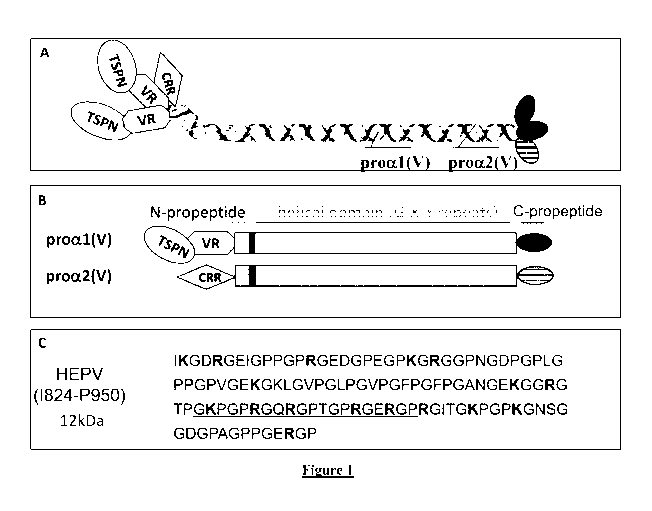
Figure 1. Structure of peptide HEPV derived from the chain proal(V)
A. Structure of the procollagen V heterotrimer [al (V)]2a2(V). CRR, cysteine-
rich repeats domain; VR, variable region; TSPN, thrombospondin N-terminal like
domain.
B. Structure of the proal(V) and proa2(V) chains. Black bars represent non-
collagenous domain (NC2) located between the small triple helix domain and the
major
triple helix domain.
C. Amino-acid sequence of the fragment HEPV. The basic residues arginine
and lysine are in bold. The sequence responsible for heparin binding is
underlined.
Figure 2. HEPV stimulates the expression of collagens IV and XVIII al
chains
Expression of COL14A/ and COL18A/ mRNA in human dermal
microvascular endothelial cells (HDMEC) treated with HEPV for 4, 12 and 24
hours,
analyzed by real-time PCR. Values are normalized to the house keeping gene
L30.
Quantification is expressed relative to controls (cells cultivated in the same
conditions
without HEPV). Values are mean SEM (n=3).
CA 02969020 2017-05-26
WO 2016/086960 PCT/EP2014/076185
4
Figure 3. Production of a non-functional mutant derived from the peptide
HEPV, HEPV-AHBS
A. SDS PAGE analysis of HEPV-AHBS. Lane 1, E. coli bacterial lysate
containing recombinant HEPV-AHBS. Lane 2, HEPV-AHBS -containing fraction after
cation exchange chromatography. Lane 3, purified HEPV-AHBS containing fraction
after
the second step of purification using cation exchange chromatography.
B. Affinity of HEPV and HEPV-AHBS for heparin. The HEPV basic residues
which have been mutated in alanine are underlined. Only the region G901-P923
of the
fragment that contains the heparin binding site is shown. Purified HEPV and
HEPV-AHBS
were passed through a heparin-sepharose affinity chromatography and eluted
with a NaC1
gradient (dotted line). HEPV is eluted with 0.35 M NaC1 while HEPV-AHBS is
eluted with
0.2 M NaCl.
Figure 4. HEPV inhibits FGF-2-induced ERK1/2 and Akt phosphorylation
in endothelial cells. Western blots of endothelial cell lysates treated with
FGF-2 (A) or
VEGF (B) in presence of HEPV or HEPV-AHBS with antibodies to ERK1/2, p-ERK1/2,
Aid and p-Aid, and quantifications. The phosphorylated p-ERK1/2 and p-Aid
proteins are
detected with specific antibodies.
Figure 5. HEPV acts on formation of blood vessels in mouse
(A) Ability of the fluorescent peptides HEPV and HEPV-AHBS to recognize
angiogenesis sites. Sponges impregnated with FGF-2 or PBS are implanted in
nude mice.
After intravenous injection of the Alexa700 labeled fluorescent peptides,
their
accumulation in angiogenic areas of the sponges is quantified using 2D
fluorescence
reflectance imaging (see arrows). The relative intensities of emitted
fluorescent light in the
sponges are then calculated and expressed as Reference Light Units (RLU)
during 200 ms.
(B) Formation of new blood vessels in nude mice implanted with a sponge
impregnated with FGF-2 or PBS. After repeated treatments with peptides HEPV or
HEPV-
AHBS, the sponges are extracted and the presence of hemoglobin quantified. The
presence
of hemoglobin reflects the blood vessels content, as documented by the photos.
CA 02969020 2017-05-26
WO 2016/086960 PCT/EP2014/076185
Figure 6. HEPV affects the tumor growth of implanted tumors in nude
mice.
(A) Murine Tsa/Pc breast cancer cells implanted subcutaneously are treated
from day 5 repeatedly by peritumoral injections of 50 1 PBS containing 50
iLig control
5 (HEPV-AHBS) of HEPV peptides every 2 days. As can be observed, tumor
growth is
significantly (p<0.05) slowed down in HEPV treated animals. Tumors sections
were
immunostained using an anti-CD31 antibody that detects blood vessels (B) or an
anti-Ki67
antibody that stains proliferating cells (C).
Formation of new blood vessels is inhibited in breast tumors implanted in nude
mice during treatment with peptides HEPV, especially during the first 20 days.
As well,
this treatment reduces the number of proliferating tumor cells.
DETAILED SPECIFICATION OF THE INVENTION
All technical terms used in the present specification are well known by the
man
skilled in the art, and are extensively defined in the reference manual from
Sambrook et at.
entitled Molecular Cloning: a Laboratory Manual .
The present invention is related to a peptide comprising an amino acid
sequence at least 85% identical to the amino acid sequence as shown in SEQ ID
NO. 1,
wherein the residues Lys905, Arg909, and Arg912 contained therein are present,
for use as a
medicament.
The residues are numerated according to their position in the complete
sequence of the proal (V) chain of the Collagen V, comprising 1838 residues,
as shown in
SEQ ID NO. 5.
The sequence SEQ ID NO. 1 represents the sequence of a peptide derived from
the human collagen proal(V) chain, comprising 127 residues starting with an
isoleucine at
the position 824, and finishing with a proline at the position 950, as
underlined in SEQ ID
5.
The phrase "an amino acid sequence at least 85% identical to the amino acid
sequence as shown in SEQ ID NO. 1" designates a candidate sequence sharing 85%
amino
acid identity with the reference sequence. This requires that, following
alignment, 85% of
the amino acids in the candidate sequence are identical to the corresponding
amino acids in
the reference sequence.
CA 02969020 2017-05-26
WO 2016/086960 PCT/EP2014/076185
6
By 'identity of amino acid' is meant that the same amino acid is observed on
both sequences. Identity does not take account of post-translation
modifications that may
occur on amino acids; for example, an hydroxylated proline is considered as
being
identical to a non-hydroxylated proline.
Identity according to the present invention is determined by aid of computer
analysis, such as the ClustalW computer alignment program, and the default
parameters
suggested therein. The ClustalW software is available from the website
http://www.clustal.org/clustal2/. By using this program with its default
settings, the part of
a query and of a reference polypeptide are aligned. The number of fully
conserved residues
are counted and divided by the length of the reference polypeptide.
The terms "at least 85%" indicates that the percentage of identity between
both
sequences, the query and the reference polypeptide of sequence SEQ ID NO. 1,
is of at
least 85, 86, 87, 88, 89, 90, 91, 92, 93, 94, 95, 96, 97, 98, 99 or 100%.
In particular, the amino acid sequence at least 85% identical to the amino
acid
sequence as shown in SEQ ID NO. 1 presents at least 90% of identity with SEQ
ID NO. 1.
In particular, the amino acid sequence at least 85% identical to the amino
acid
sequence as shown in SEQ ID NO. 1 presents at least 95% of identity with SEQ
ID NO. 1.
In particular, the amino acid sequence at least 85% identical to the amino
acid
sequence as shown in SEQ ID NO. 1 presents at least 98% of identity with SEQ
ID NO. 1.
According to the invention, the amino acid sequence showing at least 85% of
identity with the amino acid sequence as shown in SEQ ID NO. 1, presents the
following
conserved residues: Lys905, Arg909, and Arg912. These residues are essential
for the
specificity and activity of the peptide, and cannot be modified under the risk
to change the
specificity and/or activity of the peptide.
In an embodiment of the invention, the amino acid sequence showing at least
85% of identity with the amino acid sequence as shown in SEQ ID NO. 1,
presents the
following conserved residues: Lys905, Arg909, Arg912,
Arg918 and Arg921. The presence of
these amino acids, involved in the heparin binding site, might also be
important for the
activity of the peptide as a medicament.
Without wishing to be bound by the theory, inventors have observed that the
peptide according to the invention binds specifically to heparin and heparan
sulfate, both
molecules being involved in cell-matrix interactions (Delacoux et al., 2000;
Ricard-Blum
CA 02969020 2017-05-26
WO 2016/086960 PCT/EP2014/076185
7
et al., 2006). If the binding site disappears or is not functional anymore,
the FGF-2
signalization pathway is inhibited, as presented in the examples section.
The phrase "for use as a medicament" designates the use of said peptide in
therapy, in particular in human therapy.
A "medicament" is synonymous of "pharmaceutical drug", "medicine",
"medication" or "medicinal product", and designates an active compound,
intended for
internal or external use, for curing, treating, or preventing a disease.
Surprisingly, inventors have identified the peptide as described previously,
as
an active compound that can be used as a medicament. Advantageously, this
peptide is
non-toxic for animals or humans, since it does not accumulate into the liver
after injection
into the blood system.
Uses of the peptide
According to a first embodiment, the peptide according to the invention is
used
as an inhibitor of FGF-2 induced biological effects on target cells. This
embodiment can be
performed in vivo or in vitro.
The term "inhibitor" designates the mode of action of the peptide, that
reduces
or even suppresses the biological activity of the FGF-2 on its target cells.
In particular, the
biological effects of FGF-2 generally observed are reduced of at least 50%, at
least 60%, at
least 70%, at least 80%, at least 90%, and in a preferred embodiment the
biological effects
of FGF-2 on target cells are inhibited at 100%, i.e. they are completely
suppressed.
The phrase "FGF-2 induced biological effects on target cells" means all
biological effects that are specifically induced by the presence of a
sufficient amount of
FGF-2. Main effects are formation of new blood vessels, but FGF-2 acts also in
the
regulation of bone mineralization.
Target cells of FGF-2 are cells expressing receptors able to bind the factor
FGF-2, and to transmit the signal to the cells. Two classes of receptors have
been identified
up to now, the high affinity tyrosine-kinase receptors (FGFRs) and the low
affinity heparan
sulfate proteoglycans (HSPGs). Target cells are mainly endothelial cells, but
also
cardiomyocytes and osteoblasts related-cells.
FGF-2 is involved in numerous physiological functions, and therefore a peptide
acting as an inhibitor of FGF-2 induced biological effects could be used in
the treatment of
CA 02969020 2017-05-26
WO 2016/086960 PCT/EP2014/076185
8
several diseases, and in particular in the treatment of: glioblastoma
multiforme, heart
failure, Alzheimer's disease, glomerulosclerosis, and myelofibrosis with
myeloid
metaplasia.
Glioblastoma multiforme (GBM) is the most malignant form of central
nervous system tumor, and current therapies are largely ineffective at
treating the cancer. It
is also one of the most highly vascularized cancers. Secretion of FGF-2 by GBM
cells
enhances the blood brain barrier function of endothelial cells, which also
contributes to
drug resistance in GBM. It is speculated that the presence of glioblastoma
stem or stem-
like cells (GSCs), a rare type of pluripotent cancer cell that possesses the
ability to self-
renew and generate tumors, could be an important factor contributing to the
resistance to
treatment and deadliness of the cancer. It has been shown that FGF-2 plays a
significant
part in regulating GBM and GSC (Haley and Kim, Cancer letters, 2014)
Heart failures represent a major cause of morbidity and mortality. FGF-2
promotes cardiac hypertrophy and fibrosis by activating MAPK signaling through
the
activation of FGF receptor lc (FGFR). Regulating FGF-2 signaling may represent
potential
therapeutic strategies for heart failure (Itho and Ohta, Front Physiol, 2013)
Neurogenesis persists in the aged human dentate gyms but its role and
regulation in pathological conditions such as Alzheimer's disease (AD), where
the
neurotrophic environment is changed, are poorly understood. In hippocampal
progenitor
cells from adult rats, FGF-2 decreased, in a dose-dependent manner,
microtubule-
associated protein 2, and increased tau levels, indicating an FGF-2-induced
dendrite to
axon polarity shift. AD pathogenesis might involve an abnormally elevated FGF-
2-
associated dysregulation of dentate gyms neurogenesis, especially neuronal
polarity.
Cerebrolysin, a neurotrophic drug which has been shown to improve cognition
and mood
of AD patients, was found to increase neuron-like differentiated adult rat
hippocampal
progenitors in culture both by reducing apoptosis and by counteracting the FGF-
2-induced
polarity shift (Tatebayashi et al, Acta Neuropathol, 2003). Counteracting FGF-
2 activity
may represent a promising therapeutic target for this disease.
In kidney, FGF-2 increases glomerular protein permeability and acelerates
glomerulosclerosis (Chen et al, Current Vascular Pharmacology, 2004). In
glomeruli and
neointimae of allografts, a massive accumulation of FGF-2 was observed.
Profiling the
heparan sulfate polysaccharide side chains revealed conversion from a non-FGF-
2-binding
CA 02969020 2017-05-26
WO 2016/086960 PCT/EP2014/076185
9
heparan sulfate phenotype in control and isografted kidneys toward a FGF2-
binding
phenotype in allografts. FGF2-induced proliferation is dependent on sulfation
and can be
inhibited by exogenously added heparan sulfate. Counteracting FGF-2 signaling
through
the heparin binding fragment HEPV could retard development of
glomerulosclerosis and
neointima formation in chronic transplant dysfunction (Katta et al, Am J
Pathol, 2013).
Myelofibrosis with myeloid metaplasia (MMM) is a myeloproliferative
disorder characterized by clonal expansion of hematopoiesis and marrow
fibrosis. Previous
results have shown an increased production of two potent fibrogenic factors
also involved
in the regulation of primitive hematopoietic cells, namely transforming growth
factor-betal
(TGF-betal) and basic fibroblast growth factor (bFGF or FGF-2), in patients
with MMM.
The myeloproliferation characteristic of this disease may result from an
abnormal
proliferation of CD34+ hematopoietic progenitors. The very low expression of
FGF-2 and
its type I and II receptors detected in normal CD34+ cells contrasts with that
observed in
patients' CD34+ cells, which is significantly higher. The increased expression
of FGF-2
and its receptors associated with the reduction of the TGF-beta binding
receptor in CD34+
progenitors from MMM patients might facilitate the expansion of hematopoietic
progenitors, not only by stimulating their growth and/or survival, but also by
overcoming
negative regulatory signals (Le Bousse-Kerdiles, Blood, 1996; Le Bousse-
Kerdiles and
Martyre, Ann Hamatol, 1999). Counteracting FGF-2 activity may represent a
promising
therapeutic target for this disease.
Other diseases can be treated with the peptide according to the invention,
such
as the diabetic retinopathy and rheumatoid arthritis. This anti-angiogenic
peptide may also
be used in the treatment of ocular proliferative diseases, such as age-related
macular
degeneration.
According to a second embodiment, the peptide according to the invention is
used as an inhibitor of FGF-2 induced angiogenesis.
"Angiogenesis" refers to the dynamic process that includes blood vessel
formation, blood vessel remodeling, blood vessel stabilization, blood vessel
maturation,
and establishment of a functional blood vessel network. This process of
angiogenesis is
induced with the presence of a sufficient amount of FGF-2 on specific target
cells that are
mainly endothelial cells.
CA 02969020 2017-05-26
WO 2016/086960 PCT/EP2014/076185
Angiogenesis has been shown to be dysregulated in several diseases, such as in
coronary artery (CA) aneurysms in the chronic phase of Kawasaki disease (KD).
Significant neovascularization occurs in acute KD CA aneurysms and myocardium
soon
after onset of the disease and multiple angiogenesis factors are involved, and
that
5 dysregulation of angiogenesis likely contributes to KD vasculopathy
(Freeman et al, 2005,
Pediatr Cardiol). Counteracting FGF2 activity may represent a promising
therapeutic target
for this disease.
According to a third embodiment, the peptide according to the invention is
used as a drug in cancer therapy, in particular in solid tumors therapy.
10 Cancer generally refers to one of a group of diseases caused by the
uncontrolled, abnormal growth of cells that can spread to adjoining tissues or
other parts of
the body. In particular, cancer cells present uncontrolled proliferation, loss
of specialized
functions, immortality, metastatic potential, rapid growth and proliferation
rates, and
specific morphological features and cellular markers. Cancer cells can form a
solid tumor,
in which the cancer cells are massed together in a specific site of the body.
In a specific aspect, the peptide used as a drug in cancer therapy is intended
to
treat one of the most common cancers, including breast cancer, lung cancer,
prostate
cancer, colorectal cancer, stomach cancer, skin cancer, brain cancer and
cervical cancer.
Features of the peptide
The present invention is related to a peptide comprising an amino acid
sequence at least 85% identical to the amino acid sequence as shown in SEQ ID
NO. 1,
wherein the residues Lys905, Arg909, and Arg912 contained therein are present,
for use as a
medicament.
In a specific aspect of the invention, the peptide comprises a sequence as
shown in SEQ ID NO. 2 [X-K905-X-X-X-R909-X-X-R912-X-X-X-X-X-X-X-X-X-X-X],
wherein X represents any amino acid. This sequence of twenty amino acids
comprises the
conserved residues Lys905, Arg909, and Arg912 that are important for the
activity of the
peptide as a medicament.
The reference sequence SEQ ID NO. 1 consists in 127 residues. Among these
residues, a specific site of binding to heparin has been identified where the
contribution of
the conserved residues Lys905, Arg909, Arg912 is essential (see the examples
section); beside,
CA 02969020 2017-05-26
WO 2016/086960 PCT/EP2014/076185
11
the residues Arg918 and Arg921 have been identified, even if not absolutely
necessary, as
playing a role in the binding activity to heparin (Ricard-Blum et at., 2006).
The peptide according to the invention comprises these essential amino acids,
and otherwise can be modified, in particular by deletion, addition or
substitution of
residues, in the limits of 85% of identity with the reference sequence as
shown in SEQ ID
NO.1. In particular, the peptide can be modified in order to increase its half-
life, to
increase its bioavailability and/or to make it less susceptible to
proteolysis. These
modifications may include cyclization of the peptide, incorporation of D-amino
acids, or
incorporation of non-natural amino acids. None of the modifications should
substantially
interfere with the desired biological activity of the peptide.
In a specific aspect of the invention, the peptide comprises the amino acid
sequence as shown in SEQ ID NO. 3:
G-K-P-G-P-R-G-Q-R-G-P-T-G-P-R-G-E-R-G-P
According to this embodiment, the peptide comprises a sequence that presents
100% of identity with the sequence of twenty amino acids of SEQ ID NO. 3,
comprised
between the residue G904 and the residue P923, and other residues in the N-
terminal and C-
terminal portions.
In a preferred embodiment of the invention, the peptide has an amino acid
sequence that consists in the sequence as shown in SEQ ID NO. 1.
The peptide can be prepared by all means known by the man skilled in the art,
for example by chemical synthesis, or by using living systems such bacteria,
yeast or
eukaryote cells, such as animal and plant cells. Preferred microorganisms for
the synthesis
of the peptide are E. coli and yeasts.
Accordingly, a vector carrying a molecule of nucleic acid encoding the peptide
is introduced to a bacteria or eukaryote cell, by any suitable technique of
transformation.
Microorganisms are then grown under constant agitation in a suitable medium,
in a suitable
temperature, for example 37 C, and produce the peptide such as encoded by the
vector.
Said peptide is then purified, for example on ion exchange columns, before
being used as a
medicament. In particular, the purified peptide is analyzed by mass
spectrometry to check
that no bacterial contaminants are present in the purified sample.
According to the invention, the term 'peptide' always designates a 'purified'
or
'isolated' peptide, which indicates that the peptide has been separated from
other
CA 02969020 2017-05-26
WO 2016/086960 PCT/EP2014/076185
12
components such as proteins and organic molecules that are naturally present
in a growth
medium for bacteria.
In a specific embodiment of the invention, the peptide is coupled to a
detectable label.
A detectable label designates a compound that is "detectable" in particular in
an imaging procedure, because it is colored, fluorescent or luminescent. In a
particular
embodiment, the detectable label is chosen among a radioactive label, an
affinity label, a
magnetic particle, a fluorescent or luminescent label.
In particular, the detectable moiety may be a contrast agent or a detectable
protein. The man skilled in the art knows several detectable proteins such as
the Green
Fluorescent Protein, and several fluorescent dyes such as the Alexa Fluor
family.
The detectable label may be a fluorescent protein. In particular, if the
peptide is
produced in a living system, the vector carrying nucleic acid encoding the
peptide includes
also nucleic acid encoding such fluorescent protein.
In a specific aspect of the invention, both nucleic acids are organized on the
vector under the control of the same promoter, to be transcribed and
translated together, in
a way to form a fusion protein comprising both the peptide and the detectable
protein.
In another aspect of the invention, the peptide is chemically fused to a
chromophore group.
Advantageously, the peptide can be followed in a body of animal or patient, by
in vivo imaging, by techniques well known by the man skilled in the art.
Administration of the peptide
In a preferred aspect of the invention, in its use as a medicament, an
effective
amount of the peptide is administered to an animal or an individual, and an
accumulation
of said peptide in the angiogenesis or tumor site(s) is obtained.
The "effective amount" of the peptide refers to the amount necessary to elicit
the desired biological response. As can be appreciated by the man skilled in
the art, the
effective amount may vary depending on factors such as the desired biological
endpoint,
the structure of the peptide, and/or the target tissue.
The peptide can be administered by any route of administration. Suitable
routes
may include oral, buccal, by inhalation spray, sublingual, rectal,
transdermal, vaginal,
CA 02969020 2017-05-26
WO 2016/086960 PCT/EP2014/076185
13
transmucosal, nasal or intestinal administration, parenteral delivery,
including
intramuscular, subcutaneous and intravenous injections, or other modes of
delivery.
A preferred mode of administration is the parental administration into the
blood system of the animal or individual.
In the case of cancer treatment, the angiogenesis site(s) are mainly the sites
surrounding the solid tumors, where the dynamic angiogenesis process is
stimulated, in
particular by the presence of FGF-2.
The present invention relates in particular to a peptide as described above,
for
its use for treatment of cancer, by administration of an effective amount of
said peptide to
an animal or an individual, whereby an accumulation of said peptide in the
angiogenesis or
tumor site(s) is obtained.
Example 4 and figure 5A below demonstrate that, when injected into the blood
system of a mouse, the peptide HEPV comprising the amino acids Lys905, Arg909,
and
Arg912 accumulates in the site of angiogenesis, although the control peptide
where the three
essential amino acids have been replaced with alanine does not.
Pharmaceutical compositions and kits-of-part
The present invention also relates to a pharmaceutical composition comprising
an effective amount of at least one peptide as described above, and a
pharmaceutically
acceptable vehicle.
A pharmaceutically acceptable vehicle is a physiologically acceptable vehicle
prepared with nontoxic components, useful for administering an active compound
to an
animal or a patient in need.
The pharmaceutical composition may comprise different peptides, in particular
at least two types of peptides selected from the presently disclosed peptides.
This pharmaceutical composition may further comprise a compound inhibiting
angiogenesis, in particular a compound inhibiting VEGF-induced angiogenesis.
This pharmaceutical composition comprising an effective amount of at least
one peptide as described above, and a pharmaceutically acceptable vehicle may
further
comprise an anti-inflammatory compound.
CA 02969020 2017-05-26
WO 2016/086960 PCT/EP2014/076185
14
This pharmaceutical composition comprising an effective amount of at least
one peptide as described above, and a pharmaceutically acceptable vehicle may
further
comprise an anticancer active ingredient.
This pharmaceutical composition comprising an effective amount of at least
one peptide as described above, and a pharmaceutically acceptable vehicle may
further
comprise a compound inhibiting VEGF-induced angiogenesis and an anti-
inflammatory
agent.
This pharmaceutical composition comprising an effective amount of at least
one peptide as described above, and a pharmaceutically acceptable vehicle may
further
comprise a compound inhibiting angiogenesis and an anticancer active
ingredient.
This pharmaceutical composition comprising a an effective amount of at least
one peptide as described above, and a pharmaceutically acceptable vehicle may
further
comprise a compound inhibiting angiogenesis, an anti-inflammatory agent and an
anticancer active ingredient.
Advantageously, after administration of the pharmaceutical composition in the
blood system of an animal or an individual, an accumulation of said peptide in
the
angiogenesis or tumor site(s) is obtained. This feature, as shown in the
figure 5A, is highly
advantageous for the use of said peptide as a medicament.
The present invention also relates to a kit-of-parts comprising an effective
amount of the peptide as described above, and another compound inhibiting
angiogenesis,
in particular a compound inhibiting VEGF-induced angiogenesis, and/or an anti-
inflammatory compound, and/or an anticancer active ingredient.
Said kit-of-parts allows the administration to a patient of the peptide and a
compound inhibiting angiogenesis, and/or an anti-inflammatory compound, and/or
an
anticancer active ingredient, at different times. The administration of these
two or three
components can be realized concomitantly or sequentially.
In particular, the kit-of-parts comprises an effective amount of the peptide
as
described above, and a compound inhibiting angiogenesis.
In another embodiment, the kit-of-parts comprises an effective amount of the
peptide as described above, and an anticancer active ingredient, such as a
chemotherapy
compound.
CA 02969020 2017-05-26
WO 2016/086960 PCT/EP2014/076185
In another embodiment, the kit-of-parts comprises an effective amount of the
peptide as described above, and an anti-inflammatory compound.
In another embodiment, the kit-of-parts comprises an effective amount of the
peptide as described above, a compound inhibiting angiogenesis, and an
anticancer active
5 ingredient, such as a chemotherapy compound.
In another embodiment, the kit-of-parts comprises an effective amount of the
peptide as described above, a compound inhibiting angiogenesis and an anti-
inflammatory
compound.
In another embodiment, the kit-of-parts comprises an effective amount of the
10 peptide as described above, a compound inhibiting angiogenesis, an anti-
inflammatory
compound, and an anticancer active ingredient. In particular, the patient may
be treated in
a first step with a compound inhibiting angiogenesis, and/or an anti-
inflammatory
compound, and/or an anticancer active ingredient; if it appears that the
patient still presents
an active angiogenesis process around the tumors, in a second step of the
treatment, the
15 peptide according to the invention is administered, with or without an
anticancer active
ingredient.
Advantageously, the patient may be further treated with other methods. Such
methods may include, but are not limited to, chemotherapy, radiation therapy
or surgery.
The administration of a pharmaceutical composition of the present invention
may be
conducted before, during or after other cancer therapies.
The present invention also relates to a method for inhibiting the biological
effects of FGF-2 on target cells in vitro or ex vivo, comprising contacting
the cells with an
effective amount of a peptide comprising an amino acid sequence at least 85%
identical to
the amino acid sequence as shown in SEQ ID NO. 1 [11e824-Pro951, wherein the
residues
Lys905, Arg909 and Arg912 contained therein are present.
In another aspect of the invention, in this peptide, five residues Lys905,
Arg909
Arg912, Arg918
and Arg921 are conserved.
The method as described above is performed, in particular, wherein the peptide
comprises a conserved sequence as shown in SEQ ID NO. 2 [X-K905-X-X-X-R909-X-X-
R912-X-X-X-X-X- X-X-X-X-X-X], wherein X is any amino acid.
CA 02969020 2017-05-26
WO 2016/086960 PCT/EP2014/076185
16
In a specific aspect of the invention, the peptide comprises the conserved
amino acid sequence as shown in SEQ ID NO. 3 [G-K905-
P-R918-G-E-R921-G-13].
In a preferred embodiment of the invention, the peptide used in this method
has
an amino acid sequence that consists in the sequence as shown in SEQ ID NO. 1.
Labeled peptide and its uses
The present invention also relates to a peptide comprising an amino acid
sequence at least 85% identical to the amino acid sequence as shown in SEQ ID
NO. 1,
wherein the residues Lys905, Arg909, and Arg912 contained therein are present,
coupled to a
detectable label.
In another aspect of the invention, in this labelled peptide, five residues
Lys905,
Arg909 Arg9 12, Arg9 1 8
and Arg921 are conserved.
In a specific aspect of the invention, the labelled peptide comprises a
conserved
sequence as shown in SEQ ID NO. 2 [X-K905-X-X-X-R909-X-X-R912-X-X-X-X-X- X-X-X-
X-X-X], wherein X is any amino acid.
In a specific aspect of the invention, the labelled peptide comprises the
conserved amino acid sequence as shown in SEQ ID NO. 3 [G-K905-P-G-P-R909-G-Q-
R912-
G-P-T-G-P-R918-G-E-R921-G-P].
In a preferred embodiment of the invention, the labelled peptide has an amino
acid sequence that consists in the sequence as shown in SEQ ID NO. 1.
The detectable label is in particular a radioactive label, an affinity label,
a
magnetic particle, a fluorescent or luminescent label. The detectable label
may be a
fluorescent protein.
In another aspect of the invention, the peptide is chemically fused to a
chromophore group.
Advantageously, the peptide can be followed in a body of animal or patient, by
in vivo imaging, by techniques well known by the man skilled in the art.
The present invention also relates to the use of the labelled peptide as
described
above, as an imaging agent, in particular to be used in vivo.
The present invention also relates to a method for imaging angiogenesis sites
of
an animal or of a human individual, comprising the step of detecting the label
of a peptide
CA 02969020 2017-05-26
WO 2016/086960 PCT/EP2014/076185
17
as defined previously, that have been previously administered to the said
animal or to the
said human individual.
The present invention also relates to a method for imaging angiogenesis sites
of
an animal, comprising the steps of
(a) coupling a detectable label with at least one peptide such as described
above,
(b) administering said labelled peptide to said animal, and
(c) detecting the label after sacrifice of the animal.
In a preferred aspect of the invention, in its use as an imaging agent in
vivo, an
effective amount of the peptide is administered to an animal or a human
individual, and an
accumulation of said peptide in the angiogenesis or tumor site(s) is obtained.
The peptide can be administered by any route of administration. Suitable
routes
may include oral, buccal, by inhalation spray, sublingual, rectal,
transdermal, vaginal,
transmucosal, nasal or intestinal administration, parenteral delivery,
including
intramuscular, subcutaneous and intravenous injections, or other modes of
delivery.
The detection of the label is performed by any technique well known by the
man skilled in the art.
EXAMPLES
Material and Methods
Preparation and expression and purification of HEPV and AllBS-HEPV
The recombinant HEPV fragment and AHBS-HEPV construct were prepared
as previously described and inserted into the EcoRI and PstII sites of the
pT7/7 expression
vector. The pR905A plasmid previously obtained, where the arginine at position
905 was
replaced by an alanine, was used as a template to generate the triple mutant
R905/R909/R912 by using the QuikChange II site-directed mutagenesis kit
(Stratagene,
UK). Point mutations were introduced with the oligonucleotides:
= 5 '-GCGCCCAGGACCGGCGGGGGCAGGCAGGCCCAACG-3 '
(SEQ ID NO. 6), and
= 5'-CGTTGGGCCTGCCTGCCCCGCCGGTCCTGGCGC-3'
(SEQ ID NO. 7).
CA 02969020 2017-05-26
WO 2016/086960 PCT/EP2014/076185
18
The identity of AHBS-HEPV was verified by nucleotide sequencing. The
recombinant wild-type plasmid named pHEPV and the mutant pAHBS-HEPV obtained
were transformed in an E. coli strain (BL21 SI-GJ1158) that carries the T7 RNA
polymerase gene under the control of the salt inducible proU promoter. After a
20 h
induction with 0.2 M NaC1, cells were harvested by centrifugation and
resuspended in 50
mM Tris-HC1, pH 7.4, and then sonicated. After centrifugation and filtration,
bacterial
supernatants were first subjected to cation exchange chromatography using a
HiTrapSP
column (Amersham) to remove most contaminant bacterial proteins and were
purified to
homogeneity using a Mono Q column (Amersham). Recombinant protein containing
fractions were analyzed by SDS-PAGE on a 15% gel and dialyzed against 50 mM
Tris-
HC1, pH 7.5. The recombinant HEPV fragment and AHBS-HEPV were stored at ¨20 C
until use.
Heparin Affinity Chromatography
Heparin-Sepharose affinity columns (HiTrap Heparin, Amersham) were
equilibrated in 50 mM Tris-HC1 (pH 7.4). Protein samples were loaded onto a
column, and
a programmed linear gradient of 0-500 mM NaC1, 1M Tris-HC1 (pH 7.4) was
applied at a
flow rate of 0.5 ml/min, confirmed by continuous conductivity measurement.
Fractions
(1 ml) were collected, and the elution profile of protein samples was
determined by
monitoring the absorbance at 214 nm. To accurately compare elution positions
of the
mutant, its elution with NaC1 gradient was achieved versus a standard HEPV
elution.
Quantitative real-time RT-PCR
Total RNA was isolated from 8x10⁶ cells by phenol-chloroform-isopropanol
extraction (Trizol Reagent, Invitrogen). A reverse transcriptase reaction was
performed on
1 [ig of RNA using M-MLV reverse transcriptase (Promega). Quantitative PCRs
were
performed using SYBR Green Supermix (Biorad) and specific primers using a 1-
Cycler
Optical System (Biorad). The following primers were used:
= COL4A1 forward 5 '-CTGGTCCAAGAGGATTTCCA-3' (SEQ ID NO. 8);
= COL4A1 reverse 5'- TCATTGCCTTGCACGTAGAG-3' (SEQ ID NO. 9);
= COL18A1 forward 5 '-GCGCCAAAGGAGAAGTGG-3' (SEQ ID NO. 10);
CA 02969020 2017-05-26
WO 2016/086960 PCT/EP2014/076185
19
= COL18A1 reverse 5 '-TTTCAGCCTCCAACTGAAGAA-3'(SEQ ID NO.
11);
= L30 forward 5'- ATGGGGAAGGTGAAGGTCG-3' (SEQ ID NO. 12); and
= L30 reverse 5'- TAAAAGCAGCCCTGGTGACC-3'(SEQ ID NO. 13).
L30 was selected as housekeeping gene and used for normalization. Relative
transcript abundances were determined as well known by the man skilled in the
art. Relative gene expression was determined using the 2-AAcT method.
Student's t-tests
were used to determine statistical significance (n=4).
Phosphorylation Assays
HUVEC were seeded at 2.105 cells/wells in ECGM2 in 6-well plates. The cells
were treated with HEPV or AHBS-HEPV (8 g/mL) during 24 h in serum free medium
and
then stimulated with FGF-2 or VEGF (50 ng/mL) for 5 or 20 minutes at 37 C.
Cells were
then washed twice with cold PBS and scraped and lysed at 4 C in lysis buffer
1% NP-40
(150 mM NaC1, 50 mM Hepes pH 7.4, 5 mM EDTA, 10% glycerol, 1% NP-40, complete
protease inhibitor cocktail (Roche), 1 mM Na3VO4). After centrifugation (13
000 g,
15 min, 4 C), soluble proteins were collected. Protein amount was determined
by BCA
protein assay (Pierce) and equal amounts of proteins (10 g) were loaded on
SDS-PAGE
and transfer on PVDF membrane at 100 V during 1 h. The blots were blocked with
5%
BSA in TBS-T buffer (20 amM Tris¨HC1 pH 7.4 and 0.05% Tween 20) and incubated
with primary antibodies in TBST containing 5% bovine serum albumin, overnight
at 4 C.
Immunoreactivity was detected by sequential incubation with horseradish
peroxidase-
conjugated secondary antibodies purchased and ECL detection reagents purchased
from
Biorad. The antibodies used for phospho-protein detection were the followings:
anti-AKT,
anti-phospho-Akt (5er473), anti-ERK1/2 and anti-phospho-ERK1/2 (Thr202/Tyr204,
Thr185/Tyr187) (all from Cell Signaling Technology).
Animal experiments
All animal experiments were performed in agreement with the EEC guidelines
and the Principles of laboratory animal care (NIH publication 14, no. 86-23,
revised 1985);
the protocol was approved by the Animal Care and Use Committee. Female athymic
NMRI nude mice (Janvier, Le Genest-Isle, France) were used in this study and
maintained
CA 02969020 2017-05-26
WO 2016/086960 PCT/EP2014/076185
under specific pathogen-free conditions. Local surgery was performed under
general
anesthesia, which was induced via intraperitoneal injection of Domitor
(Pfizer, Orsay,
France) and Imalgene (Merial, Lyon, France).
5 Implantation of the sponges
Disc Cellspon cellulose sponges (thickness 2 mm, diameter 10 mm;
Cellomeda, Turku, Finland) were implanted under the skin of the mice. Prior to
implantation, the sponges were hydrated with 50 1 of either PBS (negative
control) or
FGF-2 (200 ng/50 1; positive angiogenic control) (recombinant FGF-2, Eurobio-
AbCys,
10 Les
Ulis, France). After implantation, the sponges were re-injected through the
skin on
days 1 and 2 with 50 1 PBS either without (negative control) or with 200 ng
FGF-2
(positive control) in the absence and presence of the HEPV peptides to be
tested.
2D fluorescence in vivo imaging
15
Angiogenesis was examined on day 7. For 2D fluorescence imaging, 200 ul
HEPV-Cy5 or HEPVAHBS-AlexFluo700 were injected intraveinously (50 [tg) into
the
mouse tail vein and imaged 3 h post-injection. Mice were illuminated with 660-
nm light-
emitting diodes equipped with interference filters and fluorescence images, as
well as
black and white pictures, which were acquired by a back-thinned charge-coupled
device
20
(CCD) camera at ¨80 C (ORCAII-BT-512G; Hamamatsu, Massy, France), fitted with
a
high-pass RG 9 filter (Schott, Clichy, France). An ROI was then positioned on
the sponge
in order to measure the number of photons/pixel during 200 ms.
Hemoglobin measurements
After implantation of the PBS or FGF-2 treated sponges and treatment with 50
1 containing 50 iLig HEP peptides at DO, 2, 3 and 5, the hemoglobin content
was measured
at D8. Mice were sacrificed via a lethal injection of Doletal 0, and the
sponges were then
rapidly excised and photographed. Each sponge was homogenized in 1 ml of RIPA
lysis
buffer containing a cocktail of protease inhibitors, centrifuged at 200 g, and
the
supernatants were quantified. The extent of vascularization of the sponge
implants was
assessed by measuring the concentration of hemoglobin with Drabkin's reagent
(Sigma-
Aldrich, Saint-Quentin Fallavier, France). The results are expressed in mg/ml.
CA 02969020 2017-05-26
WO 2016/086960 PCT/EP2014/076185
21
Antitumor activity
TS/Apc-pGL3 is a cell line derived from the original adenocarcinoma TS/Apc
mouse cell line stably transfected with the pGL3-luciferase reporter gene
(Promega,
Charbonnieres, France). Cells were cultured at 37 C in a humidified 5% CO2
incubator in
RPMI 1640 supplemented with 1% glutamine, 10% fetal bovine serum, 50 units/ml
penicillin, 50 gg/ml streptomycin, 13-mercaptoethanol (25 04) and 700 gg/ml
Geneticin0
(G418 sulphate; Gibco, Paisley, UK).
Twenty female athymic Swiss nude mice, purchased from Janvier (Le Genest
Saint Isle, France) at 6-8 weeks of age were used and maintained under
specific pathogen-
free conditions. Mice received a subcutaneous (s.c.) injection of 106 TS/Apc-
pGL3 cells
suspended in 200 gl of PBS into the right flank usually results in formation
of 6-8 mm-
diameter tumors after one week. Starting at day 5 after sc implantation, mice
received 2
peritumoral injection every 2 days of 50g1 pf PBS solution containing 50 gg of
HEP
(n=10) or HEPVAHBS (n=10) peptides. Tumor growth was evaluated using calipers.
At day 20, 3 mice /group were sacrificed for immunohistology studies. At day
35 days after implantation all remaining tumors were extracted.
Frozen sections (8 gm) from tumors were fixed in acetone for 10 min. Sections
were then washed 3x5 min in Tris-buffered saline containing 0,1% Tween 20, and
endogenous peroxidases were blocked with 0,1% H202 in methanol for 20 min.
Sections
were then sequentially incubated for lh with a rat monoclonal anti-CD31
antibody
(MEC13.3;1:500; Pharmingen) or rabbit anti-Ki67 (1:100; AbCAM) and for lh with
goat
anti-rat antibody for CD31 (1:500; Cell-signaling) or goat anti-rabbit for
Ki67 (1:200;
Dako). Peroxidase activity was revealed using diaminobenzidinetetrachloride as
a
chromogen (Dako; San Antonio, TX, USA). Sections were counterstained with
hematoxylin and all were mounted.
Staining against the endothelial marker CD31 by means of
immunohistochemistry was followed by observation under low magnification scope
(100x), five field of view of each tumor (5 tumors by condition). Then,
vessels quantity
were measured in each of these areas utilizing ImageJ software (http://
rsbweb.nih.gov/ij).
All counts were performed in a blinded manner.
CA 02969020 2017-05-26
WO 2016/086960 PCT/EP2014/076185
22
After immunohistochemical staining against Ki67, slides were observed under
high magnification scope (200x). Six to nine areas by tumor were photographed.
These
photographs were analyzed by the plugging ImmunoRatio in ImageJ software
(http://rsbweb.nih.gov/ij). The Ki67 index was evaluated in a blinded manner
and
calculated as Ki67-positive cells divided by all tumor cells in one field.
Example 1. Role of HEPV on the expression of collagens
A transcriptomic analysis has been performed, in order to identify the HEPV-
regulated genes. Endothelial HDMEC cells have been treated with HEPV or not,
for 4, 12
and 24 hours. In the list of 219 up-regulated genes, the genes COL4A1 and
COL18A1,
coding for the al chains of collagens IV and XVIII respectively, have been
identified as
being very relevant, since they encode proteins located in the vascular
endothelial basal
membranes andthat possess a strong anti-angiogenic activity after cleavage.
Up-degulation of these genes has been validated by quantitative PCR
(figure 2).
It appears therefore that HEPV induces the expression of proteins involved in
the control of the angiogenesis process.
Example 2. Preparation of a non-functional mutant of HEPV: AHBS-
HEPV
The peptide HEPV presents the sequence as shown in SEQ ID NO.1.
The peptide AHBS-HEPV presents the sequence as shown in SEQ ID NO. 4,
wherein the following residues has been replaced with alanines: Lys905, Arg909
and Arg912.
Both peptides have been produced in a bacterial system of Escherichia coli.
The peptide AHBS-HEPV is purified on ion-exchange chromatographic columns.
Figure
3A show the supernatant of Escherichia coli growth medium before (line 1),
after a first
step (line 2) and a second step of column purification (line 3).
The affinity of both peptides for heparin is compared, on a heparin-sepharose
column, determined with the necessary quantity of NaC1 to elute the peptides.
Although the HEPC peptide is eluted with a concentration of 0.35M NaC1, the
mutated peptide AHBS-HEPV is eluted with a concentration of 0.2M (Figure 3B),
close to
the physiologic concentration of NaC1 (0.15M).
CA 02969020 2017-05-26
WO 2016/086960 PCT/EP2014/076185
23
It appears therefore that the mutant presents a significant disability to bind
to
heparin, compared to the peptide HEPV, and is suitable to be used in the next
experiments
as a negative control.
Example 3. HEPV acts on signalization pathway of FGF-2 and VEGF
The aim of the experiments presented in figure 4 was to determine if, after
incubation of endothelial cells with the peptide HEPV, the response to FGF-2
was affected
by the presence of this peptide. The measured response to FGF-2 is the level
of
phosphorylation of proteins ERK 1/2 and Akt, involved in the signalization
pathway of
FGF-2 and VEGF, as determined with specific antibodies.
Endothelial cells HUVEC have been treated during 24 hours with HEPV or the
control peptide AHBS-HEPV, and have then been stimulated with FGF-2 (Fig. 4A)
or
VEGF (50 ng/ml) (Fig. 4B).
Non-treated cells present a significant increase of the phosphorylation of
ERK1
(line p-ERK1/2 for `phosphorylated ERK1/2') after stimulation with FGF-2. This
phosphorylation is inhibited in cells treated with HEPV. On the contrary, the
control
peptide AHBS-HEPV is inefficient for inhibiting the phosphorylation of ERK1/2
(Fig. 4A).
This action of HEPV is FGF-2-specific since all cells stimulated with VEGF
present a phosphorylation of ERK1/2, even after treatment with HEPV (Figure
4B).
Similar results are observed for the phosphorylation level of Akt protein,
after
stimulation with FGF-2 and VEGF.
Example 4. HEPV acts on formation of blood vessels in vivo in mouse
In nude mice, a cellulose sponge has been implanted under the skin, this
sponge containing an angiogenesis factor (FGF-2), in order to artificially
stimulate
angiogenesis. Negative control sponges comprise PBS.
HEPV and AHBS-HEPV peptides have been fused to a fluorophore (Alexa
Fluor 700) and have been injected to the blood system in mice. To follow the
localization
of the fusion proteins in vivo, pictures have been realized every 3 hours.
The quantification of the fluorescence in the site of the sponge (Figure 5A)
indicates that:
CA 02969020 2017-05-26
WO 2016/086960 PCT/EP2014/076185
24
- for sponges impregnated with FGF-2, stimulating angiogenesis, the fusion
protein HEPV-fluo accumulates strongly in the sponge;
- on the contrary, the control fusion protein AHBS-HEPV-fluo does not
accumulate in the sponge.
Moreover, in sponges impregnated with FGF-2, the number of newly formed
blood vessels is twice the number observed in control sponges (Figure 5A, on
the right).
Other results, not shown, demonstrate that the peptide HEPV does not
accumulate non-specifically in various organs, except in kidney and bladder,
the
elimination specialized-organs. Moreover the peptide does not accumulate into
the liver,
and is therefore non-toxic.
Figure 5B shows the results in terms of anti-angiogenesis activity of HEPV. 20
iLig of HEPV or AHBS-HEPV have been injected simultaneously with PBS or FGF-2,
at the
first day and then every two days. After 7 days of treatment, sponges are
taken out and
analyzed. The level of angiogenesis is measured via the level of hemoglobin
found in
sponges. When mice have been treated with HEPV, the hemoglobin level is
significantly
decreased (of 2.5 times) in comparison with mice treated with AHBS-HEPV
(Figure 5B,
on the right).
These results show that (i) HEPV peptide is able to inhibit FGF-2 induced
angiogenesis; and that (ii) a functional binding site to heparin is necessary
for obtaining
this effect.
Example 5. HEPV affects the tumoral growth
Tumors have been induced by implantation of murine breast cancer cells
(TSA) in nude mice. Once the tumors are developed, an intra-tumoral injection
of HEPV
(50 iLig) is realized every two days, during 37 days. The volume of the tumors
is measured
with a caliper.
Results are shown in Figure 6A. Up to day 18, all tumors develop according to
the same model. From day 20 of treatment, the growth of the tumors treated
with HEPV
slows down, up to the end of the experiment.
Mice from each group are sacrificed at day 20 and day 33 to check the
formation of novel blood vessels. The account of the blood vessels is realized
by
observations of the tumors samples.
CA 02969020 2017-05-26
WO 2016/086960 PCT/EP2014/076185
Results are shown in Figure 6B. At day 20, a significant decrease of the blood
vessels density is observed in mice treated with HEPV, when compared to AHBS-
HEPV-
treated-mice. Curiously, at day 33, the difference is less significant. A
possible explanation
is the fact that at this step of the treatment, the presence of necrosis zones
does not allow a
5 right follow-up of the angiogenesis process.
The proliferation of tumor cells with an antibody anti-Ki67 is also realized
on
these tumor samples.
Results are shown in Figure 6C. At day 20, a significant decrease of the tumor
cells proliferation is observed. However, at day 33, it appears that the
proliferation strikes
10 back.
Table 1 ¨ Sequences
SEQ ID
Name SEQUENCE
NO.
IKGDRGEIGPPGPRGEDGPEGPKGRGGPNGDPGPLGPP
1 HEPV (binding GEKGKLGVPGLPGYPGRQGPKGSIGFPGFPGANGEKGG
site is underlined) RGTPGKPGPRGQRGPTGPRGERGPRGITGKPGPKGNSG
GDGPAGPPGERGP
Minimal binding
2 X-K-X-X-X-R-X-X-R-X-X-X-X-X-X-X-X-X-X-X
site of HEPV
Wild-type binding
3 G-K-P-G-P-R-G-Q-R-G-P-T-G-P-R-G-E-R-G-P
site of HEPV
IKGDRGEIGPPGPRGEDGPEGPKGRGGPNGDPGPLGPP
4 HEPV aHBS GEKGKLGVPGLPGYPGRQGPKGSIGFPGFPGANGEKGG
RGTPGAPGPAGQAGPTGPRGERGPRGITGKPGPKGNSG
GDGPAGPPGERGP
MDVHTRWKARSALRPGAPLLPPLLLLLLWAPPPSRAAQPADLLKVL
DFHNLPDGITKTTGFCATRRS SKGPDVAYRVTKDAQLSAPTKQLYP
proal(V) chain of ASAFPEDFSILTTVKAKKGSQAFLVSIYNEQGIQQIGLELGRSPVFLY
the Collagen V EDHTGKPGPEDYPLFRGINLSDGKWHRIALSVHKKNVTLILDCKKK
5 (HEPV peptide is TTKFLDRSDHPMIDINGIIVFGTRILDEEVFEGDIQQLLFVSDHRAAY
underlined) DYCEHYSPDCDTAVPDTPQSQDPNPDEYYTEGDGEGETYYYEYPY
YEDPEDLGKEPTPSKKPVEAAKETTEVPEELTPTPTEAAPMPETSEG
1838 aa
AGKEEDVGIGDYDYVPSEDYYTPSPYDDLTYGEGEENPDQPTDPGA
GAEIPTSTADTSNS SNPAPPPGEGADDLEGEFTEETIRNLDENYYDPY
YDPTS SP SEIGPGMPANQDTIYEGIGGPRGEKGQKGEPAIIEPGMLIE
CA 02969020 2017-05-26
WO 2016/086960 PCT/EP2014/076185
26
GPPGPEGPAGLPGPPGTMGPTGQVGDPGERGPPGRPGLPGADGLPG
PPGTMLMLPFRFGGGGDAGSKGPMVSAQESQAQAILQQARLALRG
PAGPMGLTGRPGPVGPPGSGGLKGEPGDVGPQGPRGVQGPPGPAG
KPGRRGRAGSDGARGMPGQTGPKGDRGFDGLAGLPGEKGHRGDP
GP SGPPGPPGDDGERGDDGEVGPRGLPGEPGPRGLLGPKGPPGPPGP
PGVTGMDGQPGPKGNVGPQGEPGPPGQQGNPGAQGLPGPQGAIGP
PGEKGPLGKPGLPGMPGADGPPGHPGKEGPPGEKGGQGPPGPQGPI
GYPGPRGVKGADGIRGLKGTKGEKGEDGFPGFKGDMGIKGDRGEI
GPPGPRGEDGPEGPKGRGGPNGDPGPLGPPGEKGKLGVPGLPGYPG
RQGPKGSIGFPGFPGANGEKGGRGTPGKPGPRGQRGPTGPRGERGP
RGITGKPGPKGNSGGDGPAGPPGERGPNGPQGPTGFPGPKGPPGPPG
KDGLPGHPGQRGETGFQGKTGPPGPPGVVGPQGPTGETGPMGERG
HPGPPGPPGEQGLPGLAGKEGTKGDPGPAGLPGKDGPPGLRGFPGD
RGLPGPVGALGLKGNEGPPGPPGPAGSPGERGPAGAAGPIGIPGRPG
PQGPPGPAGEKGAPGEKGPQGPAGRDGLQGPVGLPGPAGPVGPPGE
DGDKGEIGEPGQKGSKGDKGEQGPPGPTGPQGPIGQPGP SGADGEP
GPRGQQGLFGQKGDEGPRGFPGPPGPVGLQGLPGPPGEKGETGDVG
QMGPPGPPGPRGP SGAPGADGPQGPPGGIGNPGAVGEKGEPGEAGE
PGLPGEGGPPGPKGERGEKGESGP SGAAGPPGPKGPPGDDGPKGSP
GPVGFPGDPGPPGEPGPAGQDGPPGDKGDDGEPGQTGSPGPTGEPG
P SGPPGKRGPPGPAGPEGRQGEKGAKGEAGLEGPPGKTGPIGPQGA
PGKPGPDGLRGIPGPVGEQGLPGSPGPDGPPGPMGPPGLPGLKGD SG
PKGEKGHPGLIGLIGPPGEQGEKGDRGLPGPQGS SGPKGEQGITGP S
GPIGPPGPPGLPGPPGPKGAKGS SGPTGPKGEAGHPGPPGPPGPPGEV
IQPLPIQAS RTRRNIDASQLLDDGNGENYVDYADGMEEIFGSLNS LK
LEIEQMKRPLGTQQNPARTCKDLQLCHPDFPDGEYWVDPNQGC SR
DSFKVYCNFTAGGSTCVFPDKKSEGARITS WPKENPGSWF SEFKRG
KLL SYVDAEGNPVGVVQMTFLRLLSASAHQNVTYHCYQSVAWQD
AATGSYDKALRFLGSNDEEMSYDNNPYIRALVDGCATKKGYQKTV
LEIDTPKVEQVPIVDIMFNDFGEASQKFGFEVGPACFMG
6 Oligonucleotide 5' -GCGCCCAGGACCGGCGGGGGCAGGCAGGCCCAACG- 3 '
7 Oligonucleotide 5' -CGTTGGGCCTGCCTGCCCCGCCGGTCCTGGCGC- 3 '
8 COL4A 1 forward 5 '-CTGGTCCAAGAGGATTTCCA- 3 '
9 COL4A 1 reverse 5 '- TCATTGCCTTGCACGTAGAG- 3 '
COL 1 8A1
5 '-GCGCCAAAGGAGAAGTGG- 3 '
forward
CA 02969020 2017-05-26
WO 2016/086960 PCT/EP2014/076185
27
11 COL18A1 reverse 5 '-TTTCAGCCTCCAACTGAAGAA-3 '
12 L30 forward 5 '- ATGGGGAAGGTGAAGGTCG-3 '
13 L30 reverse s'- TAAAAGCAGCCCTGGTGACC-3 '
REFERENCES
Patents
US 2014/0100164
US 2013/0316950
Bibliographic references
= Alessi P, Leali D, Camozzi M, Cantelmo A, Albini A, Presta M. Anti-FGF2
approaches as a strategy to compensate resistance to anti-VEGF therapy: long-
pentraxin
3 as a novel antiangiogenic FGF2-antagonist. Eur Cytokine Netw. 2009
Dec;20(4):225-
34.
= Delacoux F, Fichard A, Geourjon C, Garrone R, Ruggiero F. Molecular
features of the collagen V heparin binding site. J Biol Chem. 1998 Jun
12;273(24):15069-
76.
= Delacoux F, Fichard A, Cogne S, Garrone R, Ruggiero F. Unraveling the
amino acid sequence crucial for heparin binding to collagen V. J Biol Chem.
2000 Sep
22;275(38):29377-82.
= Ricard-Blum S, Beraud M, Raynal N, Farndale RW, Ruggiero F. Structural
requirements for heparin/heparan sulfate binding to type V collagen. J Biol
Chem. 2006
Sep 1;281(35):25195-204.
= Haley EM, Kim Y. The role of basic fibroblast growth factor in
glioblastoma multiforme and glioblastoma stem cells and in their in vitro
culture. Cancer
Lett. 2014 Apr 28;346(1):1-5.
= Itoh N, Ohta H. Pathophysiological roles of FGF signaling in the heart.
Front Physiol. 2013 Sep 6;4:247.
= Tatebayashi Y, Lee MH, Li L, Iqbal K, Grundke-Iqbal I. The dentate gyrus
neurogenesis: a therapeutic target for Alzheimer's disease. Acta Neuropathol.
2003
Mar;105(3):225-32.
CA 02969020 2017-05-26
WO 2016/086960 PCT/EP2014/076185
28
= Chen CH, Poucher SM, Lu J, Henry PD. Fibroblast growth factor 2: from
laboratory evidence to clinical application. Curr Vase Pharmacol. 2004
Jan;2(1):33-43.
= Katta K, Boersema M, Adepu S, Rienstra H, Celie JW, Mencke R, Molema
G, van Goor H, Berden JH, Navis G, Hillebrands JL, van den Born J. Renal
heparan
sulfate proteoglycans modulate fibroblast growth factor 2 signaling in
experimental
chronic transplant dysfunction. Am J Pathol. 2013 Nov;183(5):1571-84.
= Le Bousse-Kerdiles MC, Chevillard S, Charpentier A, Romquin N, Clay D,
Smadja-Joffe F, Praloran V, Dupriez B, Demory JL, Jasmin C, Martyr& MC.
Differential
expression of transforming growth factor-beta, basic fibroblast growth factor,
and their
receptors in CD34+ hematopoietic progenitor cells from patients with
myelofibrosis and
myeloid metaplasia. Blood. 1996 Dec 15;88(12):4534-46.
= Le Bousse-Kerdiles MC, Martyr& MC. Dual implication of fibrogenic
cytokines in the pathogenesis of fibrosis and myeloproliferation in myeloid
metaplasia with
myelofibrosis. Ann Hematol. 1999 Oct;78(10):437-44. Review.
= Freeman AF1, Crawford SE, Cornwall ML, Garcia FL, Shulman ST,
Rowley AH. Angiogenesis in fatal acute Kawasaki disease coronary artery and
myocardium. Pediatr Cardiol. 2005 Sep-Oct;26(5):578-84.