Note: Descriptions are shown in the official language in which they were submitted.
CA 02969560 2017-06-02
WO 2016/087531 PCT/EP2015/078388
BISPECIFIC ANTIBODIES AGAINST CD3EPSILON AND BCMA FOR USE IN
TREATMENT OF DISEASES
The present invention relates to bispecific antibodies against CDR and BCMA
for use in the
treatment of diseases. The present invention provides methods of determining
the response of a
patient to such treatment and related diagnostic assays.
Background of the Invention
Human B cell maturation target, also known as BCMA; TR17_HUMAN, TNFRSF17
(UniProt
Q02223), is a member of the tumor necrosis receptor superfamily that is
preferentially expressed in
differentiated plasma cells [Laabi et al. 1992; Madry et al. 1998]. BCMA is a
non glycosylated type
III transmembrane protein, which is involved in B cell maturation, growth and
survival. BCMA is a
receptor for two ligands of the TNF superfamily: APRIL (a proliferation-
inducing ligand), the high-
affinity ligand to BCMA and the B cell activation factor BAFF, the low-
affinity ligand to BCMA
(THANK, BlyS, B lymphocyte stimulator, TALL-1 and zTNF4). APRIL and BAFF show
structural
similarity and overlapping yet distinct receptor binding specificity. The
negative regulator TACI also
binds to both BAFF and APRIL. The coordinate binding of APRIL and BAFF to BCMA
and/or
TACI activates transcription factor NF-KB and increases the expression of pro-
survival Bc1-2 family
members (e.g. Bc1-2, Bc1-xL, Bcl-w, Mc1-1, Al) and the downregulation of pro-
apoptotic factors
(e.g. Bid, Bad, Bik, Bim, etc.), thus inhibiting apoptosis and promoting
survival. This combined
action promotes B cell differentiation, proliferation, survival and antibody
production (as reviewed in
Rickert RC et al., Immunol Rev (2011) 244 (1): 115-133).
Novak AJ et al. BLOOD, 103, (2004) 689-694 relates to the expression of BCMA,
TACI, and
BAFF-R on multiple myeloma cells and the mechanism for growth . Li et al., Med
Oncol 27 (2010)
439-445 mention that BCMA is expressed on plasma cells. Dispenzieri et al.,
Mayo Clin Proc 82(3),
(2007), 323-341 relates to the treatment of Multiple Myeloma Based on Mayo
Stratification of
Myeloma and Risk-Adapted Therapy (mSMART). Schaumann D. (thesis, Berlin 2006)
reports
BCMA has been found to be essential for the survival of long-lived plasma
cells and that long-lived
plasma cells are effector cells in autoimmune diseases, see also O'Connor et
al., J Exp Med.199,
( 2004) 91-98.W02012143498 relates to a method for the stratification of a
multiple myeloma (MM)
patients. WO 200932058 relates to the predicting an individual's likelihood of
having a condition
associated with autoimmune activity, such as systemic lupus erythematosus SLE.
Sanchez E, et al., Br J Haematology 158, 727-38 (2012) and W02014089335 report
that BCMA
concentrations were higher in the supernatants of cultured bone marrow
mononuclear cells from
CA 02969560 2017-06-02
WO 2016/087531 PCT/EP2015/078388
2
multiple myeloma (MM) patients than in healthy subjects and suggest that serum
BCMA levels may
be a biomarker for monitoring disease status and overall survival of MM
patients.
The TCR/CD3 complex of T-lymphocytes consists of either a TCR alpha (a)/beta
(13) or TCR
gamma (y)/delta ((3) heterodimer coexpressed at the cell surface with the
invariant subunits of CD3
labeled gamma (y), delta ((3), epsilon (8), zeta (4 and eta (i). Human CDR is
described under
UniProt P07766 (CD3E_HUMAN). An anti CDR antibody described in the state of
the art is SP34
(Yang SJ, The Journal of Immunology (1986) 137; 1097-1100). SP34 reacts with
both primate and
human CD3. SP34 is available from PharMingen. A further anti CD3 antibody
described in the state
of the art is UCHT-1 (see W02000041474). A further anti CD3 antibody described
in the state of the
art is BC-3 (Fred Hutchinson Cancer Research Institute; used in Phase I/II
trials of GvHD, Anasetti
et al., Transplantation 54: 844 (1992)).
A wide variety of recombinant bispecific antibody formats have been developed
in the recent past,
e.g. by fusion of, e.g. an IgG antibody format and single chain domains (see
Kontermann RE, mAbs
4:2, (2012) 1-16).
Antibodies against BCMA are described e.g. in Gras M-P. et al. Int Immunol. 7
(1995) 1093-1106,
W0200124811, W0200124812, W02010104949 and W02012163805. Antibodies against
BCMA
and their use for the treatment of lymphomas and multiple myeloma are
mentioned e.g. in
W02002066516 and W02010104949. W02013154760 relates to chimeric antigen
receptors (CAR)
comprising a BCMA recognition moiety and a T-cell activation moiety. Ryan, MC
et al., Mol.
Cancer Ther. 6 (2007) 3009-3018 relate to targeting of BCMA for plasma cell
malignancies and
expression of BCMA on the surface of multiple myeloma cells (MM cells).
Bispecific antibodies
against CD3 and BCMA are mentioned in W02007117600, W02009132058,
W02012066058,
W02012143498, and W02013072415, W02014122143 and W02014122144. W02013072406
and
W02014140248 mention E:T ratios in some figures and examples; however it is
only reported that in
the respective killing assay experiments there were used 10 effector cells for
1 target cell (cell lines
not patient samples). This E:T ratio is therefore artificial and there were
not shown any E:T ratios in
myeloma patient bone marrow samples or given any hint on the relation of E:T
ratio to antibody
treatment.
W02012143498 mentions a method for the stratification, diagnosing, or
selecting an antibody-based
multiple myeloma (MM) therapy of a multiple myeloma (MM) patient if malignant
B-cells express
BCMA protein on their surface.
There is a need for an improved therapy of a patient suffering from a disorder
involving plasma cells.
CA 02969560 2017-06-02
WO 2016/087531 PCT/EP2015/078388
3
Summary of the Invention
T-cell bispecific antibodies are potent compounds to effectively kill e.g.
target cells by activation of
T cells directly at the proximity of target cells. T -cell bispecific
antibodies binding to BCMA e.g. on
the surface of malignant plasma cells in the case of multiple myeloma cells or
anti-nuclear antibody
secreting-plasma cells in the case of systemic lupus erythematosus or
rheumatoid arthritis, can be
dosed dependently to kill these plasma cells. The inventors have recognized
certain parameters which
are influencing the killing of the plasma cells. These parameters are the
magnitude of BCMA
expression measured by an appropriate flow cytometry method, presence of
certain concentrations of
soluble BCMA, the ratio of T cells to malignant plasma cells and the presence
of certain
concentrations of the soluble BCMA ligand APRIL. The findings of the inventors
provide an
important guidance for e.g. the treating physician(s) to tailor the therapy
with BCMA-T-cell
bispecific antibodies to the individual patient and the findings of the
inventors also provide the
scientific basis for test kits to measure said parameters.
The invention relates to a bispecific antibody specifically binding to the
extracellular domain of
human BCMA (further named also as "BCMA") and human CDR (further named also as
for use in the treatment of a patient suffering from a disorder involving
plasma cells, and whereby in
an isolated body fluid sample of said patient, comprising CD138+ CD38+ cells,
BCMA expression on
said CD138+ CD38+ cells, measured by using an anti-BCMA antibody with a Kd
value, which is 0.70
to 1.3 fold of the Kd value of the anti-BCMA antibody part of said bispecific
antibody, is 80 or more,
preferably 100 or more, preferably 200 or more, even more preferably 300 or
more over baseline
determined as Relative Median or Mean Fluorescence Intensity MFI.
The invention relates to a bispecific antibody specifically binding to the
extracellular domain of
human BCMA and human CD3, for use in the treatment of a patient suffering from
a disorder
involving plasma cells, said disorder being characterized in that in an
isolated body fluid sample of
said patient, comprising CD138+ CD38+ cells, BCMA expression on said CD138+
CD38+ cells,
measured by using an anti-BCMA antibody with a Kd value, which is 0.70 to 1.3
fold of the Kd
value of the anti-BCMA antibody part of said bispecific antibody, is 80 or
more, preferably 100 or
more, preferably 200 or more, even more preferably 300 or more over baseline
determined as
Relative Median or Mean Fluorescence Intensity MFI.
Preferably the invention relates to a bispecific antibody for use according to
the invention,
characterized in that said bispecific antibody and said anti-BCMA antibody are
monovalent for
CA 02969560 2017-06-02
WO 2016/087531 PCT/EP2015/078388
4
BCMA binding. Preferably the Kd value of said anti-BCMA antibody (part of said
bispecific
antibody) is 100nM or lower.
Preferably the invention relates to a bispecific antibody for use according to
the invention,
characterized in that said bispecific antibody and said anti-BCMA antibody are
bivalent for BCMA
binding.
Preferably the invention relates to a bispecific antibody for use according to
the invention
characterized in that said bispecific antibody and said anti-BCMA antibody are
trivalent for BCMA
binding.
Preferably the invention relates to a bispecific antibody for use according to
the invention,
characterized in that said bispecific antibody comprises as its heavy and
light chain CDRs, CDRs of
the same amino acid sequences as said anti-BCMA antibody.
Preferably the invention relates to a bispecific antibody for use according to
the invention,
characterized in that said bispecific antibody comprises as its heavy and
light chain variable regions,
variable regions of the same amino acid sequences as said anti-BCMA antibody.
The invention relates to a bispecific antibody specifically binding to the
extracellular domain of
human BCMA and human CDR, for use in the treatment of a patient suffering from
a disorder
involving plasma cells, whereby the ratio of T cells (effector cells) to
target cells (E:T ratio) in an
isolated body fluid sample of said patient is 0.35 : 1, preferably 0.5 : 1 or
higher, preferably 1:1 or
higher, more preferably 5:1 or higher, even more preferably 10:1 or higher.
Preferably the E:T ratio
is measured as ratio of CD3+ cells to CD138+ CD38+ cells, preferably as ratio
of the CD3+ cell subset
of CD45+ CD19- CD56- T cells to CD138+ CD38+ CD45+ CD19- CD56+ cells. If the
patient suffers
from multiple myeloma, such target cells are therefore multiple myeloma cells.
The invention relates to a bispecific antibody specifically binding to the
extracellular domain of
human BCMA and human CDR, for use in the treatment of a patient suffering from
a disorder
involving plasma cells said disorder being characterized in that the ratio of
T cells (effector cells) to
target cells (E:T ratio) in an isolated body fluid sample of said patient is
0.35 : lor higher, preferably
0.5 : 1 or higher, preferably 1:1 or higher, more preferably 5:1 or higher,
preferably 10:1 or higher
and preferably 0.35 : 1 to 22 : 1. The E:T ratios found in the samples of the
patients for whom
samples could be treated effectively were found as 0.35 and higher. Samples
with E:T ratio between
0.35 : 1 and 11:1 were tested respectively. E:T ratios up to 22:1 were also
detected in patient samples
(not tested). Based on these findings the inventors recognized that patients
with such samples or with
CA 02969560 2017-06-02
WO 2016/087531 PCT/EP2015/078388
samples with even higher E:T values could also be treated effectively with a
bispecific antibody
according to the invention. Preferably the E:T ratio is measured as ratio of
CD3+ cells to CD138+
CD38+ cells, preferably as ratio of the CD3+ cell subset of CD45+ CD19- CD56-
T cells to CD138+
CD38+ CD45+ CD19- CD56+ cells. If the patient suffers from multiple myeloma,
such target cells
5 are therefore multiple myeloma cells.
The invention relates to a bispecific antibody specifically binding to the
extracellular domain of
human BCMA and human CDR, for use in the treatment of a patient suffering from
a disorder
involving plasma cells, whereby said therapy comprises successively
i) isolating from said patient a body fluid sample,
ii) measuring the amount of soluble BCMA in said sample, and
iii) if the amount of soluble BCMA in said sample is 2.5 ng/mL or higher, and
iv) if said soluble BCMA in said patient sample specifically binds to said
bispecific antibody,
treating said patient with said bispecific antibody at higher doses and/or at
a more frequent
treatment schedule.
The invention relates to a bispecific antibody specifically binding to the
extracellular domain of
human BCMA and human CDR, for use in the treatment of a patient suffering from
a disorder
involving plasma cells, said disorder being characterized in that in an
isolated body fluid sample
from said patient the amount of soluble BCMA is 2.5 ng/mL or higher, and said
soluble BCMA in
said patient sample specifically binds to said bispecific antibody,
characterized in that said treatment
of said patient with said bispecific antibody is performed with a dose per
week which is 1.5 fold up
to 10 fold or/and in that the time interval between dose-administrations is
shortened from once per
week administration up to once per day compared to a standard dose. Preferably
said treatment of
said patient with said bispecific antibody is performed with a dose per week
which is 1.5 fold up to
2.0 fold compared to a standard dose. Preferably said treatment of said
patient with said bispecific
antibody is performed in that the time interval between dose-administrations
is shortened from once
per week administration up to twice a week compared to the standard dose.
The invention relates to a bispecific antibody specifically binding to the
extracellular domain of
human BCMA and human CDR, for use in the treatment of a patient suffering from
a disorder
involving plasma cells, whereby said therapy comprises successively
i) isolating from said patient a body fluid sample,
CA 02969560 2017-06-02
WO 2016/087531 PCT/EP2015/078388
6
ii) measuring the amount of soluble BCMA in said sample, and
iii) if the amount of soluble BCMA in said sample is 2.5 ng/mL or higher, and
iv) if said soluble BCMA in said patient sample specifically binds to said
bispecific antibody,
treating said patient with said bispecific antibody at a higher dose for the
first dose or at a
more frequent treatment schedule with a shorter period between the first dose
and the second
dose of said bispecific antibody or with a shorter period between the first
dose and the third
dose of said bispecific antibody..
The invention relates to a bispecific antibody specifically binding to BCMA
and CDR which
competes with soluble BCMA for binding to human BCMA receptor and/or blocks
APRIL mediated
activation of NF-KB for use in the treatment of a patient suffering from a
disorder involving plasma
cells, whereby said therapy comprises successively
i) isolating from said patient a body fluid sample comprising plasma cells and
T cells,
ii) measuring the amount of APRIL in said sample, and
iii) if the amount of APRIL in said patient sample is more than 100 ng/mL,
treating said
patient with said bispecific antibody at higher doses and/or at a more
frequent treatment
schedule.
The invention relates to a bispecific antibody specifically binding to BCMA
and CDR which
competes with soluble BCMA for binding to human BCMA receptor and/or blocks
APRIL mediated
activation of NF-KB for use in the treatment of a patient suffering from a
disorder involving plasma
cells, said disorder being characterized in that in an isolated body fluid
sample from said patient the
amount of APRIL is higher than 10 ng/mL and up to 100 ng/mL, characterized in
that said treatment
of said patient with said bispecific antibody is performed per week with a
dose which is 1.5 fold up
to 20 fold or/and in that the time interval between dose-administrations is
shortened from once per
week administration up to once a day compared to a standard dose. Preferably
said treatment of said
patient with said bispecific antibody is performed with a dose per week which
is1.5 fold up to a
3.0fold compared to a standard dose. Preferably said treatment of said patient
with said bispecific
antibody is performed in that the time interval between dose-administrations
is shortened from once
per week administration up to three times a week compared to the standard
dose.
The invention relates to a bispecific antibody specifically binding to BCMA
and CDR which
competes with soluble BCMA for binding to human BCMA receptor, whereby said
antibody
CA 02969560 2017-06-02
WO 2016/087531 PCT/EP2015/078388
7
competes with APRIL for binding to BCMA, whereby said antibody competes with
APRIL for
binding to BCMA, whereby said antibody competes with APRIL for binding to BCMA
and/or blocks
APRIL mediated activation of NF-KB for use in the treatment of a patient
suffering from a disorder
involving plasma cells, whereby said therapy comprises successively
i) isolating from said patient a body fluid sample comprising plasma cells and
T cells,
ii) measuring the amount of APRIL in said sample, and
iii) if the amount of APRIL in said patient sample is more than 100 ng/mL,
treating said
patient with said bispecific antibody at a two times higher dose at APRIL
concentrations of
100 ng/mL and a further increased dose up to 80 times higher if APRIL
concentration
increases up to 1000 ng/mL, compared to the dose recommended for a patient
with soluble
APRIL concentration below 100 ng/mL or treating said patient with a respective
more
frequent treatment schedule to reach said higher doses with a shorter period
between any two
doses of said bispecific antibody.
The invention relates to a bispecific antibody specifically binding to BCMA
and CDR which
competes with soluble BCMA for binding to human BCMA receptor, whereby said
antibody
competes with APRIL for binding to BCMA, whereby said antibody competes with
APRIL for
binding to BCMA, whereby said antibody competes with APRIL for binding to BCMA
and/or blocks
APRIL mediated activation of NF-KB for use in the treatment of a patient
suffering from a disorder
involving plasma cells, said disorder being characterized in that in an
isolated body fluid sample of
said patient comprising plasma cells and T cells, the amount of APRIL is more
than 100 ng/mL,
characterized in treating said patient with said bispecific antibody at a two
times higher dose at
APRIL concentrations of 100 ng/mL and a further increased dose up to 80 times
higher if APRIL
concentration increases up to 1000 ng/mL, compared to the dose recommended for
a patient with
soluble APRIL concentration below 100 ng/mL or treating said patient with a
respective more
frequent treatment schedule to reach said higher doses with a shorter period
between any two doses
of said bispecific antibody.
The amount of APRIL is preferably measured by use of an ELISA method.
The invention relates to a method of determining BCMA protein expression in an
isolated body fluid
sample comprising CD138+ CD38+ cells, of a patient, suffering from a disorder
involving plasma
cells, said method comprising
CA 02969560 2017-06-02
WO 2016/087531 PCT/EP2015/078388
8
measuring BCMA expression on said CD138+ CD38+ cells by using an anti-BCMA
antibody with a
Kd value, which is 0.70 to 1.3 fold of the Kd value of the anti-BCMA antibody
part of a bispecific
antibody specifically binding to BCMA and CDR, intended for use in the
treatment of said patient,
and determining by flow cytometry whether Relative Median or Mean Fluorescence
Intensity MFI is
80 or more, preferably 100 or more, preferably 200 or more, even more
preferably 300 or more over
baseline.
The invention relates to a method of treating a patient, suffering from a
disorder involving plasma
cells, comprising
analyzing isolated body fluid sample comprising CD138+ CD38+ cells from said
patient for BCMA
expression on said CD138+ CD38+ cells by using an anti-BCMA antibody with a Kd
value, which is
0.70 to 1.3 fold of the Kd value of the anti-BCMA antibody part of a
bispecific antibody specifically
binding to BCMA and CDR, intended for use in the treatment of said patient,
and if Relative Median
or Mean Fluorescence Intensity MFI is 80 or more, preferably 100 or more over
baseline, preferably
200 or more, even more preferably 300 or more treating said patient with said
bispecific antibody.
Preferably the invention relates to selecting a treatment plan that is most
effective for a patient,
suffering from a disorder involving plasma cells, and whereby an isolated body
fluid sample of said
patient show MFI for BCMA of 80 or more, preferably 100 or more, preferably
200 or more, even
more preferably 300 or more over baseline.
The invention relates to a method for predicting the likelihood of a patient,
suffering from a disorder
involving plasma cells, to respond to a treatment with a bispecific antibody
specifically binding to
BCMA and CDR, whereas the cell-surface BCMA expression in an isolated body
fluid sample of
said patient, comprising CD138+ CD38+ cells, and measured by using an anti-
BCMA antibody with a
Kd value, which is 0.70 to 1.3 fold of the Kd value of the anti-BCMA antibody
part of said bispecific
antibody, of 80 or more, preferably 100 or more, preferably 200 or more, even
more preferably 300
or more over baseline determined as Relative Median or Mean Fluorescence
Intensity MFI is
predictive of the patient's likelihood to respond to said treatment.
The invention relates to an in vitro method of determining cell-surface BCMA
expression in an
isolated body fluid sample, comprising determining whether Relative Median or
Mean Fluorescence
Intensity MFI for said CD138+ CD38+ cells, using an anti-BCMA antibody with a
Kd value, which is
0.70 to 1.3 fold of the Kd value of the anti-BCMA antibody part of a
therapeutic bispecific antibody
CA 02969560 2017-06-02
WO 2016/087531 PCT/EP2015/078388
9
specifically binding to BCMA and CDR, is 80 or more, preferably 100 or more,
preferably 200 or
more, even more preferably 300 or more over baseline.
The invention relates to an in vitro method of selecting a treatment plan that
is most effective for
treating a patient, suffering from a disorder involving plasma cells, whereby
for said patient
cell-surface BCMA expression in an isolated body fluid sample, comprising
CD138+ CD38+ cells,
measured by using an anti-BCMA antibody with a Kd value, which is 0.70 to 1.3
fold of the Kd
value of the anti-BCMA antibody part of a therapeutic bispecific antibody,
specifically binding to
BCMA and CDR, is 100 or more , preferably 200 or more, even more preferably
300 over baseline
determined as Relative Median or Mean Fluorescence Intensity MFI, whereby the
treatment plan
involves the use of a therapeutic bispecific antibody specifically binding to
BCMA and CDR.
The invention relates to a method for selecting a therapy for treating a
patient, suffering from a
disorder involving plasma cells, comprising
i) if cell-surface BCMA expression in an isolated body fluid sample,
comprising CD138+
CD38+ cells, measured by using an anti-BCMA antibody with a Kd value, which is
0.70 to
1.3 fold of the Kd value of the anti-BCMA antibody part of a therapeutic
bispecific antibody,
specifically binding to BCMA and CDR, is 100 or more, preferably 200 or more,
even more
preferably 300 or more over baseline determined as Relative Median or Mean
Fluorescence
Intensity MFI, treating said patient with said therapeutic antibody, or
ii) if cell-surface BCMA expression in an isolated body fluid sample,
comprising CD138+
CD38+ cells, measured by using an anti-BCMA antibody with a Kd value, which is
0.70 to
1.3 fold of the Kd value of the anti-BCMA antibody part of a therapeutic
bispecific antibody,
specifically binding to BCMA and CD3e, is lower than 100, preferably lower
than 50, even
preferable lower than 10 over baseline determined as Relative Median or Mean
Fluorescence
Intensity MFI, not treating said patient with said therapeutic antibody.
The invention relates to a method for determining in an isolated body fluid
sample of a patient,
suffering from a disorder involving plasma cells, whether the ratio of CD3+
cells to CD138+ CD38+
cells is 0.35 : 1, preferably 0.5 : 1 or higher, preferably 1:1 or higher,
more preferably 5:1 or higher,
even more preferably 10:1 or higher.
The invention relates to a method of treating a patient suffering from a
disorder involving plasma
cells, comprising analyzing in an isolated body fluid sample of said patient
whether the ratio of
CA 02969560 2017-06-02
WO 2016/087531 PCT/EP2015/078388
CD3+ cells to CD138+ CD38+ cells is 0.35 : 1, preferably 0.5 : 1 or higher,
preferably 1:1 or higher,
more preferably 5:1 or higher, even more preferably 10:1 or higher.
Preferably the invention relates to selecting a treatment plan that is most
effective for a patient which
show a ratio of CD3+ cells to CD138+ CD38+ cells of 0.35 : 1, preferably 0.5 :
1 or higher, preferably
5 1:1 or higher, more preferably 5:1 or higher, even more preferably 10:1
or higher.
Preferably the invention relates to selecting a treatment plan that is most
effective for a patient,
whereby
i) if the ratio of CD3+ cells to CD138+ CD38+ cells in a body fluid sample of
said patient is
0.35 : 1, preferably 0.5 : 1 or higher, preferably 1:1 or higher, more
preferably 5:1 or higher,
10 even more preferably 10:1 or higher, treating said patient with a
therapeutic bispecific
antibody specifically binding to BCMA and CD3c in monotherapy
ii) if the ratio of CD3+ cells to CD138+ CD38+ cells in an isolated body fluid
sample of said
patient is lower than 0.5 : 1, preferably lower than 0.25 : 1, treating said
patient with a
therapeutic bispecific antibody specifically binding to BCMA and CD3c in
combination with
T-cell proliferative therapy or T-cell chemoattractant therapy.
The invention relates to a method for predicting the likelihood of a patient,
suffering from a disorder
involving plasma cells, to respond to a treatment with a bispecific antibody
specifically binding to
BCMA and CDR, by measuring in an isolated body fluid sample of said patient
whether the ratio of
CD3+ cells to CD138+ CD38+ cells is 0.35 : 1, preferably 0.5 : 1 or higher,
preferably 1:1 or higher,
more preferably 5:1 or higher, even more preferably 10:1 or higher, which is
predictive of the
patient's likelihood to respond to a treatment.
The invention relates to an in vitro method of determining in an isolated body
fluid sample of a
patient suffering from a disorder involving plasma cells whether the ratio of
CD3+ cells to CD138+
CD38+ cells is 0.5 : 1 or higher, preferably 1:1 or higher, more preferably
5:1 or higher, even more
preferably 10:1 or higher,.
The invention relates to an in vitro method of selecting a treatment plan that
is most effective for
treating a patient, suffering from a disorder involving plasma cells, whereby
in an isolated body fluid
sample of said patient the ratio of CD3+ cells to CD138+ CD38+ cells is
determined as 0.35 : 1,
preferably 0.5 : 1 or higher, preferably 1:1 or higher, more preferably 5:1 or
higher, even more
CA 02969560 2017-06-02
WO 2016/087531 PCT/EP2015/078388
11
preferably 10:1 or higher and whereby the treatment plan involves the use of a
therapeutic bispecific
antibody specifically binding to BCMA and CD3e.
The invention relates to a method for selecting a therapy with a bispecific
antibody specifically
binding to BCMA and CDR for a patient, suffering from a disorder involving
plasma cells,
comprising
i) if in an isolated body fluid sample of said patient the ratio of CD3+ cells
to CD138+
CD38+ cells is 0.35 : 1, preferably 0.5 : 1 or higher, preferably 1:1 or
higher, more preferably
5:1 or higher, even more preferably 10:1 or higher, treating said patient with
said therapeutic
antibody, or
ii) if in an isolated body fluid sample of said patient the ratio of CD3+
cells to CD138+
CD38+ cells is lower than 0.35 : 1, preferably 0.5 : 1, preferably lower than
0.25 : 1 treating
said patient with a bispecific antibody specifically binding to BCMA and CD3
in
combination with T-cell proliferative therapy or T-cell chemoattractant
therapy.
The invention relates to a method of determining in an isolated body fluid
sample comprising
CD138+ CD38+ cells, of a patient suffering from a disorder involving plasma
cells, whether the
amount of soluble BCMA in said sample is 2.5 ng/mL or higher, preferably 10
ng/mL or higher,
more preferably 50 ng/mL or higher, even more preferably 250 ng/mL or higher.
The invention relates to a method of treating a patient suffering from a
disorder involving plasma
cells, comprising determining whether the amount of soluble BCMA in said body
fluid sample is 2.5
ng/mL or higher, preferably 10 ng/mL or higher, more preferably 50 ng/mL or
higher, even more
preferably 250 ng/mL or higher.
Preferably the invention relates to selecting a treatment plan that is most
effective for a patient which
show MFI for BCMA of 80 or more, preferably 100 or more, preferably 200 or
more, even more
preferably 300 or more over baseline. The invention relates to a method for
predicting the likelihood
of a patient, suffering from a disorder involving plasma cells, to respond to
a treatment with a
bispecific antibody specifically binding to BCMA and CDR, whereby an amount of
soluble BCMA
in said body fluid sample of 2.5 ng/mL or higher, preferably 10 ng/mL or
higher, more preferably 50
ng/mL or higher, even more preferably 250 ng/mL or higher is predictive of the
patient's likelihood
to respond to a treatment.
CA 02969560 2017-06-02
WO 2016/087531 PCT/EP2015/078388
12
The invention relates to an in vitro method of determining in an isolated body
fluid sample, whether
the amount of soluble BCMA in said sample is 2.5 ng/mL or higher, preferably
10 ng/mL or higher,
more preferably 50 ng/mL or higher, even more preferably 250 ng/mL or higher.
Preferably the invention relates to selecting a treatment plan that is most
effective for a patient which
show an amount of soluble BCMA of 2.5 ng/mL or higher, preferably 10 ng/mL or
higher, more
preferably 50 ng/mL or higher, even more preferably 250 ng/mL or higher.
The invention relates to an in vitro method of selecting a treatment plan that
is most effective for
treating a patient, suffering from a disorder involving plasma cells, by
determining whether the
amount of soluble BCMA in said sample is 2.5 ng/mL or higher, preferably 10
ng/mL or higher,
more preferably 50 ng/mL or higher, even more preferably 250 ng/mL or higher,
and the treatment
plan involves the use of a bispecific antibody specifically binding to BCMA
and CD3 E.
The invention relates to a method for selecting a therapy for treating a
patient, suffering from a
disorder involving plasma cells a therapy, comprising
i) if the amount of soluble BCMA in said sample is lower than 2.5 ng/mL,
treating said
patient with said therapeutic antibody, or
ii) if the amount of soluble BCMA in said sample is 2.5 ng/mL or higher and,
if said soluble BCMA in said patient sample does not bind to said bispecific
antibody,
treating said patient with said therapeutic antibody, or
iii) if the amount of soluble BCMA in said sample is 2.5 ng/mL or higher,
preferably 10
ng/mL or higher, more preferably 50 ng/mL or higher, even more preferably 250
ng/mL or
higher and,
if said soluble BCMA in said patient sample specifically binds to said
bispecific antibody, treating
said patient with said bispecific antibody at higher doses and/or at a more
frequent treatment
schedule.
Preferably the invention relates to a method for selecting a therapy for
treating a patient, suffering
from a disorder involving plasma cells a therapy, comprising
i) if the amount of soluble BCMA in said sample is lower than 2.5 ng/mL,
treating said
patient with said therapeutic antibody, or
CA 02969560 2017-06-02
WO 2016/087531 PCT/EP2015/078388
13
ii) if the amount of soluble BCMA in said sample is 2.5 ng/mL or higher and,
if said soluble BCMA in said patient sample does not bind to said bispecific
antibody,
treating said patient with said therapeutic antibody, or
iii) if the amount of soluble BCMA in said sample is 2.5 ng/mL or higher,
preferably 10
ng/mL or higher, more preferably 50 ng/mL or higher, even more preferably 250
ng/mL or
higher and, if said soluble BCMA in said patient sample specifically binds to
said bispecific
antibody, treating said patient with said bispecific antibody at a higher dose
for the first dose
or at a more frequent treatment schedule with a shorter period between the
first dose and the
second dose of said bispecific antibody or with a shorter period between the
first dose and
the third dose of said bispecific antibody.
The invention relates to a method of determining in an isolated body fluid
sample comprising
CD138+ CD38+ cells, of a patient suffering from a disorder involving plasma
cells, whether the
amount of soluble APRIL in said sample is 100 ng/mL or higher, preferably 1000
ng/mL or higher.
The invention relates to a method of treating a patient, suffering from a
disorder involving plasma
cells and diagnosed that the amount of soluble APRIL in an isolated body fluid
sample of said patient
is 100 ng/mL or higher, preferably 1000 ng/mL or higher, with a bispecific
antibody specifically
binding to BCMA and CDR.
Preferably the invention relates to selecting a treatment plan that is most
effective for a patient which
show an amount of soluble APRIL of 100 ng/mL or higher, preferably 1000 ng/mL
or higher.
The invention relates to a method for predicting the likelihood of a patient,
suffering from a disorder
involving plasma cells, to respond to a treatment with a bispecific antibody
specifically binding to
BCMA and CDR, whereby the amount of soluble APRIL in said sample of 100 ng/mL,
preferably
1000 ng/mL or higher is predictive of the patient's likelihood to respond to a
treatment.
The invention relates to an in vitro method of determining in an isolated body
fluid sample, whether
the amount of soluble APRIL in said sample is 100 ng/mL or higher, preferably
1000 ng/mL or
higher.
The invention relates to an in vitro method of selecting a treatment plan that
is most effective for
treating a patient, suffering from a disorder involving plasma cells, by
determining whether the
amount of soluble APRIL in said sample is 100 ng/mL or higher, preferably 1000
ng/mL or higher,
CA 02969560 2017-06-02
WO 2016/087531 PCT/EP2015/078388
14
and the treatment plan involves the use of an APRIL competitive bispecific
antibody or an APRIL
non-competitive bispecific antibody.
The invention relates to a method for selecting a therapy for treating a
patient, suffering from a
disorder involving plasma cells a therapy, comprising
i) the amount of soluble APRIL in said sample is 100 ng/mL or lower,
preferably 20 ng/mL
or lower treating said patient with said therapeutic antibody, or
ii) the amount of soluble APRIL in said sample is 100 ng/mL or higher,
preferably 1000
ng/mL or higher, treating said patient with APRIL non-competitive bispecific
antibodies, or
iii) the amount the amount of soluble APRIL in said sample is higher than 100
ng/mL,
preferably 1000 ng/mL or higher, treating said patient with said bispecific
antibody at higher
doses and/or at a more frequent treatment schedule.
Preferably the invention relates to a method for selecting a therapy for
treating a patient, suffering
from a disorder involving plasma cells a therapy, comprising
i) the amount of soluble APRIL in said sample is 100 ng/mL or lower,
preferably 20 ng/mL
or lower treating said patient with said therapeutic antibody, or
ii) the amount of soluble APRIL in said sample is 100 ng/mL or higher,
preferably 1000
ng/mL or higher, treating said patient with BCMA ligand competitive bispecific
antibodies,
Or
iii) if the amount of APRIL in said patient sample is more than 100 ng/mL,
treating said
patient with said bispecific antibody at a two times higher dose at APRIL
concentrations of
100 ng/mL and a further increased dose up to 80 times higher if APRIL
concentration
increases up to 1000 ng/mL, compared to the dose recommended for a patient
with soluble
APRIL concentration below 100 ng/mL or treating said patient with a respective
more
frequent treatment schedule to reach said higher doses with a shorter period
between any two
doses of said bispecific antibody.
The invention relates to a method for determining a treatment plan that is
most effective for a patient
suffering from a disorder involving plasma cells.
CA 02969560 2017-06-02
WO 2016/087531 PCT/EP2015/078388
The invention relates to a method for determining a treatment plan for a new
patient, suffering from a
disorder involving plasma cells, comprising:
providing, utilizing at least one method for investigation the BCMA related
plasma cell status of said
new patient;
5 searching, utilizing at least the result of one method, for a prior
treatment plan for a prior patient
suffering from the same disorder with at least one similar representation; and
reviewing the prior treatment plan for the prior patient in order to determine
how to improve the
treatment of the new patient based on information in at least one prior
treatment plan.
The invention relates to a method for diagnosing and treating a disorder
involving plasma cells in a
10 patient comprising analyzing in an isolated body fluid sample of said
patient BCMA expression on
CD138+ CD38+ cells according to the invention, wherein the patient is
diagnosed having said
disease, if said BCMA expression is 80 or more, preferably 100 or more,
preferably 200 or more,
even more preferably 300 or more over baseline determined as Relative Median
or Mean
Fluorescence Intensity MFI and administering treatment with a bispecific
antibody according to the
15 invention to the diagnosed patient.
The invention relates to a method for diagnosing and treating a disorder
involving plasma cells in a
patient comprising analyzing in an isolated body fluid sample the ratio of T
cells (effector cells) to
target cells (E:T ratio), wherein the patient is diagnosed with said disease
if said ratio is 0.35 : 1,
preferably 0.35 : 1, preferably 0.5 : 1 or higher and administering treatment
with a bispecific
antibody according to the invention to the diagnosed patient.
The invention relates to a method for diagnosing and treating a disorder
involving plasma cells in a
patient comprising analyzing in an isolated body fluid sample patient the
amount of soluble BCMA
according to the invention wherein the patient is diagnosed with said disease
if said soluble BCMA is
2.5 ng/mL or higher, and said soluble BCMA in said patient sample specifically
binds to said
bispecific antibody, and administering treatment with a bispecific antibody
according to the invention
to the diagnosed patient.at higher doses and/or at a more frequent treatment
schedule.
The invention relates to a method for diagnosing and treating a disorder
involving plasma cells in a
patient comprising analyzing in an isolated body fluid sample patient the
amount of soluble BCMA
according to the invention wherein the patient is diagnosed with said disease
if said soluble BCMA is
2.5 ng/mL or higher, and said soluble BCMA in said patient sample specifically
binds to said
CA 02969560 2017-06-02
WO 2016/087531 PCT/EP2015/078388
16
bispecific antibody, and administering treatment with a bispecific antibody
according to the invention
to the diagnosed patient is performed at a higher dose for the first dose or
at a more frequent
treatment schedule with a shorter period between the first dose and the second
dose of said bispecific
antibody or with a shorter period between the first dose and the third dose of
said bispecific antibody.
The invention relates to a method for diagnosing and treating a disorder
involving plasma cells in a
patient comprising analyzing in an isolated body fluid sample patient the
amount of APRIL
according to the invention wherein the patient is diagnosed with said disease
if the amount of APRIL
is more than 100 ng/mL, and administering treatment with a bispecific antibody
according to the
invention to the diagnosed patient is performed with said bispecific antibody
which competes with
soluble BCMA for binding to human BCMA receptor and/or blocks APRIL mediated
activation of
NF-KB at higher doses and/or at a more frequent treatment schedule.
The invention relates to a method for diagnosing and treating a disorder
involving plasma cells in a
patient comprising analyzing in an isolated body fluid sample patient
comprising plasma cells and T
cells, the amount of APRIL according to the invention wherein the patient is
diagnosed with said
disease if the amount of APRIL is more than 100 ng/mL, and administering
treatment with a
bispecific antibody according to the invention to the diagnosed patient is
performed with said
bispecific antibody which competes with APRIL for binding to human BCMA
receptor and/or blocks
APRIL mediated activation of NF-KB at a two times higher dose at APRIL
concentrations of 100
ng/mL and a further increased dose up to 80 times higher if APRIL
concentration increases up to
1000 ng/mL, compared to the dose recommended for a patient with soluble APRIL
concentration
below 100 ng/mL or treating said patient with a respective more frequent
treatment schedule to reach
said higher doses with a shorter period between any two doses of said
bispecific antibody.
Preferably the disease (disorder) is selected from the group consisting of
multiple myeloma,
systemic lupus erythematosus, and rheumatoid arthritis.
Preferably valence should be similar between the diagnostic antibody and the
therapeutic antibody
(e.g. a monovalent antibody for BCMA determination should be used for patient
stratification for a
BCMA antibody therapy with monovalent binding to the tumor target on malignant
cells such as
scFV-based BiTE molecules). Even more preferably is to use a BCMA antibody for
BCMA
determination which is the same as the BCMA binder of the BCMA antibody
therapy.
CA 02969560 2017-06-02
WO 2016/087531 PCT/EP2015/078388
17
Preferably the affinity to human BCMA of said bispecific antibody is 200 nM or
lower, measured at
an antibody concentration of 25 nM in presence of human BCMA Fc fusion at a
concentration 500
nM or lower in an affinity setup surface plasmon resonance assay.
Preferably the potency (EC50) to kill BCMA-positive H929 cells (ATCC CRL-9068)
of said
bispecific antibody is measured as 2 nM or lower, when used at concentrations
of 100 nM and lower,
in presence of human PBCMs and H929 cells at a E:T ratio of 10:1 for 24h, in a
redirected T-cell
killing LDH release assay.
Preferably the affinity to human BCMA of said BCMA binding part, measured in
an antibody of
human IgG1 typeõ is 200 nM or lower at an antibody concentration of 25 nM in
presence of human
BCMA Fc fusion at a concentration of 500 nM or lower in an affinity setup
surface plasmon
resonance assay.
Preferably the antibody according to the invention is further characterized in
that it binds also
specifically to cynomolgus BCMA.
Preferably the bispecific antibody according to the invention comprising
constant heavy regions
CH2/CH3 of IgG1 subclass is characterized in comprising the mutations L234A,
L235A and P239G
(numbering according to Kabat) to avoid FcR and Clq binding and minimizing
ADCC/CDC. The
advantage is that such an antibody of the invention mediates its tumor cell
killing efficacy purely by
the powerful mechanism of T-cell redirection/activation. Additional mechanisms
of action like
effects on complement system and on effector cells expressing FcgammaR are
avoided and the risk
of side-effects is decreased.
Preferably an antibody according to the invention is characterized by showing
tumor growth
inhibition of more than 70%, preferably of more than 85%, preferably of close
to 100% in a multiple
myeloma xenograft model (e.g. xenograft with NCI-H929 cells or RPMI8226 cells
or U266B1 cells
or L-363 cells) at a dose of 1 mg/kg body weight (BW) administered
intravenously (i.v.) or
subcutaneously (s.c.) or intraperitoneal (i.p.) twice a week or once a week,
preferably 0.5 mg/kg BW
administered i.v. or i.p. or s.c. twice a week or once a week, preferably at
0.1 mg/kg BW
administered i.v. or i.p. or s.c. twice a week or once a week, preferably at
0.05 mg/kg BW
administered i.v. or i.p. or s.c. twice a week or once a week, preferably at
0.01 mg/kg BW
administered i.v. or i.p. or s.c twice a week or once a week, preferably at 5
g/kg BW administered
i.v. or i.p. or s.c. twice a week or once a week.
CA 02969560 2017-06-02
WO 2016/087531 PCT/EP2015/078388
18
Preferably an antibody according to the invention is characterized by an
elimination half-life in mice,
preferably cynomolgus monkeys of longer than 24 hours, preferably 3 days or
longer, preferably
half-life is measured for the doses which are effective in the xenograft model
at twice or once a week
administration.
Bispecific antibodies binding to a target on tumor cells and to CD3 and having
the molecular format
(scFv)2 have very short elimination half-life of 1 to 4 hours. In the clinical
trials with the (scFv)2
bispecific CD19xCD3 antibody blinatumomab, this compound had to be
administered via a pump
carried by the patients over weeks and months (Topp et al. J Clin Oncol 2011;
29(18): 2493-8).
Compared to a twice a week or once a week iv or sc administration, treatment
administered via a
pump is much less convenient for the patients and also much more risky (e.g.
failure of pump, issues
with the catheter).
Preferably an antibody according to the invention is characterized in showing
an EC50 value for
binding to NCI-H929 cells (ATCCO CRL-9068TM) of 500 nM or lower, preferably an
EC50 value of
350 nM or lower, preferably an EC50 value of 100 nM and lower.
Preferably an antibody according to the invention is characterized by its
capability to induce
redirected killing of NCI-H929 tumor cells in the presence of human T cells
with an EC50 lower than
1 nM, preferably 0.5 nM, preferably 0.1 nM and lower.
Preferably a bispecific antibody according to the invention is characterized
by its capability to induce
redirected killing of multiple myeloma patient primary myeloma cells in the
presence of human T
cells.
A further embodiment of the invention is a kit comprising a diagnostic anti-
BCMA antibody and a
therapeutic bispecific antibody against BCMA and CD3according to the
invention.
A further embodiment of the invention is a kit comprising an anti-BCMA
antibody and a bispecific
antibody against BCMA and CD3, characterized in that the anti-BCMA antibody
has a Kd value,
which is 0.70 to 1.3 fold of the Kd value of the anti-BCMA antibody part of
said bispecific antibody,
is 80 or more, preferably 100 or more, preferably 200 or more, even more
preferably 300 or more
over baseline determined as Relative Median or Mean Fluorescence Intensity MFI
and instructions
for use, in particular instructions as how to perform the methods of the
present invention. Preferably
said anti-BCMA antibody and said bispecific antibody against BCMA and CD3 are
both mono-, bi-,
or trivalent and have preferably the same CDRs or VH and VL sequence.
CA 02969560 2017-06-02
WO 2016/087531 PCT/EP2015/078388
19
The kit comprises at least one container and a label or package insert on or
associated with the
container. Suitable containers include, for example, bottles, vials, syringes,
etc. The containers may
be formed from a variety of materials such as glass or plastic. The container
can have a sterile access
port for extracting a therapeutic agent (for example the container may be an
intravenous solution bag
or a vial having a stopper pierceable by a hypodermic injection needle). The
label or package insert
can indicate that the composition is used for treating MM, SLE, RA or another
disorder involving
plasma cells.
Additionally, the kit may further comprise a second container comprising a
pharmaceutically-
acceptable buffer, such as bacteriostatic water for injection (BWFI),
phosphate-buffered saline,
Ringer's solution and dextrose solution. It may further include other
materials desirable from a
commercial and user standpoint, including other buffers, diluents, filters,
needles, and syringes.
Preferably the kit comprises;
1) For determination of cell-surface BCMA expression: Vials or tubes pre-
loaded with labelled-
antibodies (preferably four antibodies, one specifically binding to CD138, one
to CD38, one to
CD19, and one to BCMA (with the properties according to the present
invention)) to determine
BCMA on malignant PC; tubes with isotype control antibodies;
2) For determination of E:T ratio: Vials or tubes pre-loaded with labelled-
antibodies to detect
malignant PC and T cells (preferably four antibodies, one specifically binding
to CD138, one to
CD38, one to CD19, and one to CD3); tubes with isotype control antibodies;
3) For determination of soluble BCMA: ELISA kit comprising a microtiter plate,
a capture antibody
(polyclonal BCMA antibody), biotin-conjugated detection antibody (specifically
binding to BCMA
(with the properties according to the present invention)), mass-calibrated
standard, streptavidin-HRP
or streptavidin-ALP, detailed protocol, PBS, Wash Buffer - 0.05% Tween 20 in
PBS, pH 7.2-7.4,
Reagent Diluentl - 1% BSA5 in PBS, Substrate Solution - 1:1 mixture of Color
Reagent A, (H202)
and Color Reagent B (Tetramethylbenzidine), Stop Solution - 2 N H2504;
4) For determination of soluble APRIL: ELISA kit comprising microtiter plate,
capture antibody
(anti-human APRIL antibody), biotin-conjugated detection antibody (anti-human
APRIL antibody),
mass-calibrated standard, streptavidin-HRP or streptavidin-ALP, detailed
protocol, PBS, Wash
Buffer - 0.05% Tween 20 in PBS, pH 7.2-7.4, Reagent Diluentl - 1% BSA5 in PBS,
Substrate
Solution - 1:1 mixture of Color Reagent A, (H202) and Color Reagent B
(Tetramethylbenzidine),
Stop Solution - 2 N H2504.
CA 02969560 2017-06-02
WO 2016/087531 PCT/EP2015/078388
Description of the Figures
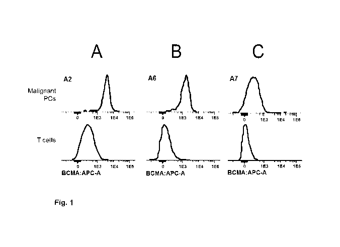
Figure 1. BCMA expression on patient malignant plasma cells as detected by
flow cytometry and
defined by relative mean or median fluorescence intensity. Representative FACS
histogram plots of
(A) Medium-high BCMA expression, (B) moderate BCMA expression and (C) low BCMA
5 expression on patient myeloma cells as detected by flow cytometry (MFI).
There is a clear shift to
the right on the x axis corresponding to positive BCMA expression on patient
myeloma cells when
compared to the negative control (APC-conjugated BCMA-1 antibody gated on T
cells). Based on
the relative MFI values, myeloma patients express BCMA on their malignant
plasma cells but
BCMA expression varies from low expression (relative MFI values < 10) to
moderate expression
10 (103 - 0.3 x 10⁴) to medium-high expression (0.3 x 10⁴ - 104) (see Example
1.1).
Figure 2. Killing potency of BCMA-TCB is influenced by BCMA expression on the
surface of target
cells: BCMAhi-expressing H929 vs. BCMAmed/10-expressing U266 myeloma cells.
BCMA-2-TCB
induced killing of BCMAhi-expressing H929 myeloma cells with an EC50 of 115 pM
and maximum
killing of 60%, while the same BCMA-TCB antibody was only able to kill
BCMAmed/10-expressing
15 U266 myeloma target cells with an EC50 of 370 pM and maximum killing at 18%
when performed
in a head-to-head comparison (see Example 1.3).
Figure 2.1. The potency of BCMA-1-TCB to induce killing of BCMA expressing
myeloma cell lines
(BCMAhi-expressing H929, BCMA1 -expressing L363 and BCMA1 -expressing RPMI-
8226 MM
cells) was tested and compared. BCMA-1- TCB induced killing of (A) BCMAhi-
expressing H929
20 myeloma cells with an EC50 of 8.49 pM and maximum killing of 82.8%, while
the same BCMA-1-
TCB antibody was only able to kill (B) BCMAmed/10-expressing L363 myeloma
target cells with an
EC50 of 12.6 pM and maximum killing at 67.1% or (C) BCMA1 -expressing RPMI-
8226 with an
EC50 of 229.3 pM and maximum killing at 28.1% when performed in a head-to-head
comparison
(Example 1.3).
Figure 3. BCMA expression on human myeloma cell lines as detected by flow
cytometry and defined
by relative mean or median fluorescence intensity.
Figure 4. Effect of APRIL-competing BCMA-TCB antibody on APRIL-induced NF-KB
activation as
detected by phosphoflow cytometry. (A) Effect of APRIL competing J6MO-TCB on
APRIL (1000
ng/mL) mediated NF-KB activation in H929 cells. Detection of intracellular
phosphorylated NF-KB
by phosphoflow cytometry (see Example 4.2.1).
Figure 5. Influence of soluble APRIL on APRIL-competing BCMA-TCB antibody to
induce T-cell
redirected killing of BCMA-positive H929 myeloma cells as detected by
colorimetric LDH release
CA 02969560 2017-06-02
WO 2016/087531 PCT/EP2015/078388
21
assay. APRIL blocking/competing J6MO-TCB in absence of exogenous soluble APRIL
and in
presence of 100 ng/mL or 1000 ng/mL of exogenous soluble APRIL. E:T ratio used
as 10 PBMCs:1
H929 cell; cells were incubated for 24h before measurement of LDH release.
APRIL
blocking/competing J6MO-TCB induced a concentration-dependent killing of BCMA-
positive H929
myeloma with a low picomolar potency (EC50ApRmo= 5.8 pM) in the absence of
exogenous APRIL.
When 100 ng/mL of APRIL was added into the culture, such concentration of
ligand only minimally
affected the killing potency mediated by J6MO-TCB as shown with an 2.4-fold
increase in the EC50
(EC50ApRiL1oo= 14.2 pM). However, when 1000 ng/mL of APRIL was added into the
culture the
killing potency mediated by J6MO-TCB was greatly reduced as reflected by an
increase in the EC50
of 84.3-fold (EC50ApRiL1000= 488.9 pM) (see Example 4.2.2).
Figure 6. Influence of soluble APRIL on APRIL-competing BCMA-TCB antibody to
induce T-cell
activation as detected by flow cytometry. Expression level of the early
activation marker CD69 (B,
D), and the late activation marker CD25 (A, C) on CD4+ and CD8+ T cells after
48 hours of
incubation (representative results from two independent experiments). APRIL-
competing/blocking
J6MO-TCB antibody induced an up-regulation of CD69 and CD25 activation markers
in a
concentration-dependent and specific manner in the presence of BCMA-positive
target cells in
absence of exogenous soluble APRIL (squares). When 100 ng/mL of soluble APRIL
was added into
the culture, a slight shift to the right of the concentration-response curves
was observed for both
activation markers CD69 and CD25 on CD4+ and CD8+ T cells. When 1000 ng/mL of
soluble
APRIL was added into the culture, there was a clear reduction of T-cell
activation on both CD4+ and
CD8+ T cells. No activation of CD4+ and CD8+ T cells was observed when human
PBMCs were
treated with DP47-TCB control antibody, suggesting that despite binding to CD3
on the T cells T-
cell activation does not occur when the TCB antibody does not bind to BCMA-
positive target cells
(data not shown). The results clearly suggest that high levels of soluble
APRIL reduce the potency
of BCMA-TCB antibodies to induce T-cell activation upon binding to the tumor
target and T cells,
especially when the BCMA-TCB is competes with APRIL (see Example 4.3).
Figure 7: Influence of BCMA expression and E:T ratio on the potency of BCMA-
TCB to induce
killing of patient bone marrow malignant plasma cells by autologous marrow
infiltrating T cells.
BCMA-1-TCB induced a concentration dependent specific killing of malignant
plasma cells from
both patient Cl (A) and patient C8 (B) already after only 24h of incubation.
However, killing of
myeloma cells was more pronounced in patient Cl bone marrow samples than in
patient C8 bone
marrow samples. This could be attributed to a more favorable E:T ratio of 11:1
and BCMA
expression (i.e. relative MFI value of 2636) in patient Cl bone marrow samples
than in patient C8
bone marrow samples with an unfavorable E:T ratio of 0.5:1 and weaker BCMA
expression on
myeloma cells (i.e. relative MFI value of 1489). The results suggest that
measurement of BCMA
CA 02969560 2017-06-02
WO 2016/087531 PCT/EP2015/078388
22
expression on malignant plasma cells in combination with a measurement of E:T
ratio in patient bone
marrow may more accurately predict whether myeloma patients may respond to
BCMA-TCB
treatment.
Detailed Description of the Invention
The inventors have recognized that disorders involving plasma cells,
especially, multiple myeloma,
systemic lupus erythematosus, and/or rheumatoid arthritis can be classified
(divided) in several
subtypes. Such subtypes are:
1. Patients, comprising CD138+ CD38+ cells in an isolated body fluid sample,
characterized by
BCMA expression on said CD138+ CD38+ cells, measured by using an anti-BCMA
antibody with a
Kd value, which is 0.70 to 1.3 fold of the Kd value of the anti-BCMA antibody
part of said bispecific
antibody, is 80 or more, 100 or more, preferably 200 or more, even more
preferably 300 or more over
baseline determined as Relative Median or Mean Fluorescence Intensity MFI.
2. Patients for whom in an isolated body fluid samplethe ratio of T cells
(effector cells) to target cells
(E:T ratio) in an isolated body fluid sample is 0.35: 1 or higher, preferably
0.5 : 1 or higher.
3. Patients, for whom the amount of soluble BCMA in an isolated body fluid
sample is 2.5 ng/mL or
higher.
4. Patients, for whom the amount of APRIL in an isolated body fluid sample is
more than 100
ng/mL.
4. Patients, comprising CD138+ CD38+ cells in an isolated body fluid,
characterized by BCMA
expression on said CD138+ CD38+ cells, measured by using an anti-BCMA antibody
with a Kd
value, which is 0.70 to 1.3 fold of the Kd value of the anti-BCMA antibody
part of said bispecific
antibody, is 80 or more, preferably 100 or more, preferably 200 or more, even
more preferably 300
or more over baseline determined as Relative Median or Mean Fluorescence
Intensity MFI and for
whom in said isolated body fluid sample the ratio of T cells (effector cells)
to target cells (E:T ratio)
sample is 0.35: 1, preferably 0.5: 1 or higher.
BCMA receptor plays a critical role for the survival of normal and malignant
plasma cells (i.e.
myeloma cells) by binding to its ligands APRIL and BAFF which are abundant in
the bone marrow
of myeloma patients and BCMA expression in myeloma cells have been detected by
many groups
both at the mRNA level and surface protein level (O'Connor et al. J Exp Med
2004, 199(1):91-8;
Novak et al. Blood 2004, 103(2): 689-94; Ryan et al. Mol Cancer Ther 2007,
6(11): 3009-18; Quinn
et al. Blood 2011, 117(3): 890; Carpenter et al. Blood 2013, 19(8): 2048-60;
Frigyesi et al. Blood
CA 02969560 2017-06-02
WO 2016/087531 PCT/EP2015/078388
23
2014, 123(9):1336-40; Claudio et al. Blood 2002,100(6):2175-86; Tai et al.
Cancer Res 2006,
123(20):3128-38; Moreaux et al., Blood 2004,103(8):3148-57; Ju et al. Clin
Biochem 2009,42(4-
5):387-99; Moreaux et al. Eur J Haematol 2009, 83(2):119-29). Therefore in
myeloma patients
BCMA is expressed on their malignant plasma cells. However the inventors have
observed that
myeloma patients do express BCMA on the cell surface but at different level of
expression, ranging
from medium/high to moderate to low, as detected by optimal measurement by
flow cytometry, that
the inventors have recognized that the use of suboptimal techniques or methods
for detection of
BCMA expression for patient stratification (e.g. use of BCMA antibody with low
affinity binding to
human BCMA) could not detect the low expression of BCMA on malignant plasma
cells of myeloma
patients and in such case misinform clinicians that these myeloma patients
would not respond to a
BCMA antibody therapy while they could.
T cell bispecific (TCB) antibodies have very high concentration/tumor-cell-
receptor-occupancy
dependent potency in cell killing (e.g. EC50 in in vitro cell killing assays
in the sub- or low
picomolar range; Dreier et al. Int J Cancer 2002), T-cell bispecific
antibodies (TCB) are given at
much lower doses than conventional monospecific antibodies. For example,
blinatumomab
(CD19xCD3) is given at a continuous intravenous dose of 5 to 15 lug/m2/day
(i.e. only 0.035 to 0.105
mg/m2/week) for treatment of acute lymphocytic leukemia or 60 ug/m2/day for
treatment of Non
Hodgkin Lymphoma, and the serum concentrations at these doses are in the range
of 0.5 to 4 ng/mL
(Klinger et al., Blood 2012; Topp et al., J Clin Oncol 2011; Goebeler et al.
Ann Oncol 2011).
Because low doses of TCB can exert high efficacy in patients, it is envisaged
that for an antibody
according to the invention subcutaneous administration is possible and
preferred in the clinical
settings (preferably in the dose range of 0.25 to 2.5 mg/m2/week). Even at
these low
concentrations/doses/receptor occupancies, TCB can cause considerable adverse
events (Klinger et
al., Blood 2012). Therefore it is critical to control tumor cell
occupancy/coverage. Therefore the
doses for treatment with a TCB should be chosen based on an effective method
for patient
classification.
Therefore, an optimal determination method for BCMA expression on patient
myeloma cells is
needed. Such optimal determination method for BCMA expression can be performed
using flow
cytometry with appropriate BCMA antibodies for determination. For example, for
use of BCMA
determination on myeloma cells for patient stratification, according to the
invention, a BCMA
antibody is used for detection that has similar affinity range to human BCMA
(e.g. as measured by
surface plasmon resonance (SPR)) as the BCMA antibody therapy. Further
preferred is that the
antibody valence should be the same between the BCMA antibody for detection
and the BCMA
antibody therapy (e.g. a monovalent antibody for BCMA detection should be used
for patient
stratification for a BCMA antibody therapy with monovalent binding to the
tumor target on
CA 02969560 2017-06-02
WO 2016/087531 PCT/EP2015/078388
24
malignant cells such as in the case of scFV-based BiTE molecules). Further
preferred is that the
avidity range (as measured by SPR) is similar between the BCMA antibody for
detection and the
BCMA antibody therapy. Further preferred is to use a BCMA antibody for
determination which is
the same as the BCMA binder of the BCMA antibody therapy.
The term "target" as used herein means either BCMA or CD3.The term "first
target and second
target" means either CD3 as first target and BCMA as second target or means
BCMA as first target
and CD3 as second target.
The term "BCMA" as used herein relates to human B cell maturation target, also
known as BCMA;
TR17_HUMAN, TNFRSF17 (UniProt Q02223), which is a member of the tumor necrosis
receptor
superfamily that is preferentially expressed in differentiated plasma cells.
The extracellular domain
of BCMA consists according to UniProt of amino acids 1 ¨ 54 (or 5-51). The
term "antibody against
BCMA, anti BCMA antibody" as used herein relates to an antibody specifically
binding to BCMA.
The term "CDR or CD3" as used herein relates to human CDR described under
UniProt P07766
(CD3E_HUMAN). The term "antibody against CD3, anti CD3 antibody" relates to an
antibody
binding to CD3 c. Preferably the antibody comprises a variable domain VH
comprising the heavy
chain CDRs of SEQ ID NO: 3, 4 and 5 as respectively heavy chain CDR1, CDR2 and
CDR3 and a
variable domain VL comprising the light chain CDRs of SEQ ID NO: 6, 7 and 8 as
respectively light
chain CDR1, CDR2 and CDR3. Preferably the antibody comprises the variable
domains of SEQ ID
NO:1 (VH) and SEQ ID NO:2 (VL). The term "antibody against CD3, anti CD3
antibody" as used
herein relates to an antibody specifically binding to CDR.
"Specifically binding to CD3 or BCMA or to CD3 c or BCMA" refer to an antibody
that is capable
of binding to the human CDR or the extracellular domain of human BCMA (the
targets) with
sufficient affinity such that the antibody is useful as a therapeutic agent in
targeting CD3 or BCMA.
In some embodiments, the extent of binding of an anti-CD3 or BCMA antibody to
an unrelated, non-
CD3 or non-BCMA protein is about 10-fold preferably >100-fold less than the
binding of the
antibody to CD3 or BCMA as measured, e.g., by surface plasmon resonance (SPR)
e.g. Biacore0,
enzyme-linked immunosorbent (ELISA) or flow cytometry (FACS). Preferably the
antibody that
binds to CD3 or BCMA has a dissociation constant (Kd) of 10-8 M or less,
preferably from 10-8 M to
10-13 M, preferably from 10-9 M to 10-13 M. Preferably the anti-CD3 and/or
anti-BCMA antibody
binds to an epitope of CD3 and/or BCMA that is conserved among CD3 and/or BCMA
from
different species, preferably among human and cynomolgus. "Bispecific antibody
specifically
binding to CD3 and BCMA" or "antibody according to the invention" refers to a
respective definition
for binding to both targets. An antibody specifically binding to BCMA (or BCMA
and CD3) does
CA 02969560 2017-06-02
WO 2016/087531 PCT/EP2015/078388
not bind to other human antigens. Therefore in an ELISA, OD values for such
unrelated targets will
be equal or lower to that of the limit of detection of the specific assay,
preferably > 0.3 ng/mL, or
equal or lower to OD values of control samples without plate-bound-BCMA or
with untransfected
HEK293 cells.
5 The term "CD3E or CD3 binding part" as used herein relates to the
combination of an antibody heavy
chain consisting of VH and CH1 and an antibody light chain consisting of VL
and CL as enclosed in
a Fab fragment of an antibody specifically binding to CD3.
The term "BCMA binding part" as used herein relates to the combination of an
antibody heavy chain
consisting of VH and CH1 and an antibody light chain consisting of VL and CL
as enclosed in a Fab
10 fragment of an antibody specifically binding to BCMA.
The term "bispecific antibody specifically binding to BCMA and CD3 or TCB or
antibody against
BCMA and CD3" relates to a bispecific antibody specifically binding to the
extracellular domain of
human BCMA and human CDR. Such antibody can be monovalent for BCMA, e.g. as
single chain
antibody as mentioned in W02013072406, W02013072415 and W02014140248, or can
be bi- or
15 trivalent as disclosed e. g. in W02014122143, W02014122144. W02013072406
and
W02014140248 mention E:T ratios in some figures and examples; however it is
only reported that in
the respective killing assay experiments there were used 10 effector cells for
1 target cell (cell lines
not patient samples). This E:T ratio is therefore artificial and there were
not shown any E:T ratios in
myeloma patient bone marrow samples or given any hint on the relation of E:T
ratio to antibody
20 treatment. Preferably the bispecific antibody comprises as CDRs of the CD3
binding part the CDRs
of SEQ ID NO: 2 to 4 and 6 to 8 and preferably the VL and VH domains of SEQ ID
NO: 1 and 5.
Preferably the bispecific antibody comprised as CDRs of the BCMA binding part
the CDRs or
preferably the VH and VL domains listed in Table 1. Preferably the bispecific
antibody comprises as
CDRs of the BCMA binding part the CDRs of SEQ ID NO: 10 to 12 and 14 to 16 or
preferably the
25 VL and VH domains of SEQ ID NO: 9 and 13. Preferably the bispecific
antibody comprises as CDRs
of the BCMA binding part the CDRs of SEQ ID NO: 18 to 20 and 22 to 24 or
preferably the VL and
VH domains of SEQ ID NO: 17 and 21. Antibody J6M0 is described in
W02012163805. A TCB
comprising J6M0 comprises as CDRs of the CD3 binding part the CDRs of SEQ ID
NO: 2 to 4 and 6
to 8 and preferably the VL and VH domains of SEQ ID NO: 1 and 5.
The term "an APRIL non-competitive bispecific antibody" relates to a
bispecific antibody,
characterized in that the binding of said antibody is not reduced by 100 ng/mL
APRIL for more than
20% measured in an ELISA assay compared to the binding of said antibody to
human BCMA
without APRIL. The term "an APRIL non-competitive anti-BCMA antibody" relates
to an anti-
CA 02969560 2017-06-02
WO 2016/087531 PCT/EP2015/078388
26
BCMA antibody, characterized in that the binding of said antibody is not
reduced by 100 ng/mL
APRIL for more than 20% measured in an ELISA assay compared to the binding of
said antibody to
human BCMA without APRIL. Such antibodies are described in W02014122143,
W02014122144,
EP14179705 and EP14194151.
Table 1
Antibody SEQ ID NO:
VL CDRL1 CDRL2 CDRL3 VH CDRH1 CDRH2 CDRH3
CH2527 1 2 3 4 5 6 7 8
(CD3)
83A10 9 10 11 12 13 14 15 16
(BCMA)
pSCHLI372 17 18 19 20 21 22 23 24
(BCMA)
The term "therapeutic antibody" refers to a bispecific antibody specifically
binding to BCMA and
CD3 that functions in depleting malignant plasma cells in a patient suffering
from multiple myeloma.
The therapeutic antibody mediates a cytotoxic effect or cell lysis,
particularly by inducing T-cell
activation followed by T-cell mediated apoptosis involving perforin and
granzyme B.
Preferably a therapeutic antibody according to the invention is characterized
in showing an EC50
value for binding to NCI-H929 cells (ATCCO CRL9068TM) of 500 nM or lower,
preferably an
EC50 value of 350 nM and lower, preferably an EC50 value of 100 nM and lower.
Preferably, a therapeutic antibody according to this invention is
characterized by its capability to bind
to U266 (ATCCO TU3-196114) cells.
In one preferred embodiment, a therapeutic antibody according to the invention
is characterized by
its capability to bind to human T cells.
Preferably, a therapeutic antibody according to this invention is
characterized by its capability to bind
to cynomolgus monkey BCMA transiently expressed on HEK-cells.
CA 02969560 2017-06-02
WO 2016/087531 PCT/EP2015/078388
27
In a preferred embodiment, a therapeutic antibody according to this invention
is characterized by its
capability to induce CD4+ and CD8+ T-cell activation in the presence of tumor
cells expressing
BCMA.
Preferably a therapeutic antibody according to the invention is characterized
by its capability to
induce redirected killing of NCI-H929 tumor cells in the presence of human T
cells with an EC50
lower than 1 nM, preferably 0.5 nM, preferably 0.1 nM and lower.
The term "diagnostic antibody" refers to an antibody specifically binding to
the extracellular domain
of BCMA. According to the invention said diagnostic antibody is, if cell-
surface BCMA expression
will be determined, an anti-BCMA antibody with a Kd value, which is 0.70 to
1.3 fold of the Kd
value of the anti-BCMA antibody part of the therapeutic bispecific antibody
intended to use for the
treatment of the patient. Preferably the antibody is derived from the BCMA
binding part of said
therapeutic bispecific antibody and preferably comprised the same CDRs or VH
and VL domains as
said BCMA binding part of said therapeutic antibody. According to the
invention said diagnostic
antibody is, if MFI is determined, preferably a labeled antibody. According to
the invention said
diagnostic antibody is, if soluble BCMA will be determined an antibody useful
for ELISA.
The terms "labeled antibody" refers to an antibody, having attached a
detectable label. Preferably the
detectable label is a fluorophore when FACS is used for analyzing. For other
analyzing methods also
all other known labels can be used (see, for example, Harlow and Lane, eds.
(Antibodies: A
Laboratory Manual (1988) Cold Spring Harbor Laboratory Press, Cold Spring
Harbor, N.Y.)).
Example for preferred fluorophores are fluorescein isothiocyanate (FITC),
Alexa Fluor, R-
phycoerythrin (PE), Allophycocyanin (APC), PerCP, tandem conjugates (e.g. PE-
Cyanine, PerCP-
Cyanine), rhodamine (tetramethyl rhodamine isothiocyanate, TRITC), green
fluorescent proteins
(GFPs), and phycobiliproteins.
The term "antibody" as used herein refers to a monoclonal antibody. An
antibody consists of two
pairs of a "light chain" (LC) and a "heavy chain" (HC) (such light chain (LC)
/heavy chain pairs are
abbreviated herein as LC/HC). The light chains and heavy chains of such
antibodies are polypeptides
consisting of several domains. Each heavy chain comprises a heavy chain
variable region
(abbreviated herein as HCVR or VH) and a heavy chain constant region. The
heavy chain constant
region comprises the heavy chain constant domains CH1, CH2 and CH3 (antibody
classes IgA, IgD,
and IgG) and optionally the heavy chain constant domain CH4 (antibody classes
IgE and IgM). Each
light chain comprises a light chain variable domain VL and a light chain
constant domain CL. The
variable domains VH and VL can be further subdivided into regions of
hypervariability, termed
complementarity determining regions (CDR), interspersed with regions that are
more conserved,
CA 02969560 2017-06-02
WO 2016/087531 PCT/EP2015/078388
28
termed framework regions (FR). Each VH and VL is composed of three CDRs and
four FRs,
arranged from amino-terminus to carboxy-terminus in the following order: FR1,
CDR1, FR2, CDR2,
FR3, CDR3, FR4. The "constant domains" of the heavy chain and of the light
chain are not involved
directly in binding of an antibody to a target, but exhibit various effector
functions.
The "light chain of an antibody" as used herein is a polypeptide comprising in
N-terminal to C-
terminal direction a light chain variable domain (VL), and a light chain
constant domain (CL),
abbreviated as VL-CL. "The "heavy chain of an antibody" as used herein is a
polypeptide comprising
in N-terminal to C-terminal direction a heavy chain variable domain (VH) and a
constant heavy chain
domain 1 (CH1).
The term "antibody" includes e.g. mouse antibodies, human antibodies, chimeric
antibodies,
humanized antibodies and genetically engineered antibodies (variant or mutant
antibodies) as long as
their characteristic properties are retained. Especially preferred are human
or humanized antibodies,
especially as recombinant human or humanized antibodies. The terms "monoclonal
antibody" or
"monoclonal antibody composition" as used herein refer to a preparation of
antibody molecules of a
single amino acid composition.
The terms "bispecific antibody" and "antibody according to the invention" as
used herein refer to an
antibody in which one of the two pairs of heavy chain and light chain (HC/LC)
is specifically binding
to BCMA and the other one is specifically binding to CD3 or preferably to CD3
and BCMA. The
term "valent" as used within the current application denotes the presence of a
specified number of
binding sites in an antibody molecule. A bivalent antibody according to this
invention has two
binding sites, one for CD3 and the other for BCMA. As such, the term
"trivalent", denote the
presence of three binding sites in an antibody according to the invention,
which are two binding sites
for BCMA and one binding site for CD3.
There are five types of mammalian antibody heavy chains denoted by the Greek
letters: a, 6, c, y, and
(Janeway CA, Jr et al (2001). Immunobiology. 5th ed., Garland Publishing). The
type of heavy
chain present defines the class of antibody; these chains are found in IgA,
IgD, IgE, IgG, and IgM
antibodies, respectively (Rhoades RA, Pflanzer RG (2002). Human Physiology,
4th ed., Thomson
Learning). Distinct heavy chains differ in size and composition; a and y
contain approximately 450
amino acids, while and c have approximately 550 amino acids. Each heavy
chain has two regions,
the constant region and the variable region. The constant region is identical
in all antibodies of the
same isotype, but differs in antibodies of different isotype. Heavy chains y,
a and 6 have a constant
region composed of three constant domains CH1, CH2, and CH3 (in a line) , and
a hinge region for
added flexibility (Woof J, Burton D Nat Rev Immunol 4 (2004) 89-99); heavy
chains and c have a
constant region composed of four constant domains CH1, CH2, CH3, and CH4
(Janeway CA, Jr et al
CA 02969560 2017-06-02
WO 2016/087531 PCT/EP2015/078388
29
(2001). Immunobiology. 5th ed., Garland Publishing). The variable region of
the heavy chain differs
in antibodies produced by different B cells, but is the same for all
antibodies produced by a single B
cell or B cell clone. The variable region of each heavy chain is approximately
110 amino acids long
and is composed of a single antibody domain.
In mammals there are two types of light chain, which are called lambda (X) and
kappa 00. A light
chain has two successive domains: one constant domain CL and one variable
domain VL. The
approximate length of a light chain is 211 to 217 amino acids. Preferably the
light chain is a kappa
(K) light chain, and the constant domain CL is preferably derived from a kappa
(K) light chain (the
constant domain CK). Preferably the heavy and light chain constant domains of
the antibody
according to the invention are human domains.
The "antibodies" according to the invention can be of any class (e.g. IgA,
IgD, IgE, IgG, and IgM,
preferably IgG or IgE), or subclass (e.g., IgG1 , IgG2, IgG3, IgG4, IgAl and
IgA2, preferably IgG1),
whereby both antibodies, from which the bivalent bispecific antibody according
to the invention is
derived, have an Fc part of the same subclass( e.g. IgG1 , IgG4 and the like,
preferably IgG1),
preferably of the same allotype (e.g. Caucasian).
A "Fab fragment of an antibody" as used herein is a fragment on an antibody
that binds to antigens.
A Fab fragment of an antibody consists of two pairs of domains. In a wild-type
antibody it is
composed of one constant and one variable domain of each of the heavy chain
(CH1 and VH) and the
light chain (CL and VL). In a wild-type antibody and according to the
invention the domain of the
heavy and light chain domain pairs of a Fab fragment are not chemically linked
together and are
therefore not scFvs (single chain variable fragments).
A "Fc part of an antibody" is a term well known to the skilled artisan and
defined on the basis of
papain cleavage of antibodies. The antibodies according to the invention
contain as Fc part,
preferably a Fc part derived from human origin and preferably all other parts
of the human constant
regions. The Fc part of an antibody is directly involved in complement
activation, Clq binding, C3
activation and Fc receptor binding. While the influence of an antibody on the
complement system is
dependent on certain conditions, binding to Clq is caused by defined binding
sites in the Fc part.
Such binding sites are known in the state of the art and described e.g. by
Lukas, TJ., et al., J.
Immunol. 127 (1981) 2555-2560; Brunhouse, R., and Cebra, J.J., MoI. Immunol.
16 (1979) 907-917;
Burton, D.R., et al., Nature 288 (1980) 338-344; Thommesen, J.E., et al., MoI.
Immunol. 37 (2000)
995-1004; Idusogie, E.E., et al., J. Immunol. 164 (2000) 4178-4184; Hezareh,
M., et al., J. Virol. 75
(2001) 12161-12168; Morgan, A., et al., Immunology 86 (1995) 319-324; and EP 0
307 434. Such
binding sites are e.g. L234, L235, D270, N297, E318, K320, K322, P331 and P329
(numbering
CA 02969560 2017-06-02
WO 2016/087531 PCT/EP2015/078388
according to EU index of Kabat, see below). Antibodies of subclass IgG1 , IgG2
and IgG3 usually
show complement activation, C 1 q binding and C3 activation, whereas IgG4 do
not activate the
complement system, do not bind Clq and do not activate C3. Preferably the Fc
part is a human Fc
part. Preferably the Fc part is a human IgGlFc part. Preferably the antibody
according to the
5 invention comprises in the human IgG1 Fc part amino acid substitution of
Pro329 with glycine or
arginine and/or substitutions L234A and L235A, preferably Pro329 with glycine
and substitutions
L234A and L235A.
Preferably the antibody according to the invention comprises as Fc part an Fc
variant of a wild-type
human IgG Fc region, said Fc variant comprising an amino acid substitution at
position Pro329 and
10 at least one further amino acid substitution, wherein the residues are
numbered according to the EU
index of Kabat, and wherein said antibody exhibits a reduced affinity to the
human FcyRIIIA and/or
FcyRIIA and /or FeyRI compared to an antibody comprising the wildtype IgG Fc
region, and wherein
the ADCC induced by said antibody is reduced to at least 20% of the ADCC
induced by the antibody
comprising a wild-type human IgG Fc region. In a specific embodiment Pro329 of
a wild-type
15 human Fc region in the antibody according to the invention is substituted
with glycine or arginine or
an amino acid residue large enough to destroy the proline sandwich within the
Fc/Fcy receptor
interface, that is formed between the proline329 of the Fc and tryptophane
residues Tip 87 and Tip
110 of FcyRIII (Sondermann et al.: Nature 406, 267-273 (20 July 2000)). In a
further aspect of the
invention the at least one further amino acid substitution in the Fc variant
is S228P, E233P, L234A,
20 L235A, L235E, N297A, N297D, or P33 1S and still in another embodiment said
at least one further
amino acid substitution is L234A (denotes that leucine 234 is substituted by
alanine) and L235A of
the human IgG1 Fc region or S228P and L235E of the human IgG4 Fc region. Such
Fc variants are
described in detail in W02012130831. An advantage of an antibody according to
the invention
comprising an Fc part, is that the elimination half-life is increased up to
¨12 days or even more and
25 offers the opportunity of once or twice/week administrations as compared to
TCBs without an Fc
portion.
The term "chimeric antibody" refers to an antibody comprising a variable
region, i.e., binding region,
from one source or species and at least a portion of a constant region derived
from a different source
or species, usually prepared by recombinant DNA techniques. Chimeric
antibodies comprising a
30 murine variable region and a human constant region are preferred. Other
preferred forms of
"chimeric antibodies" encompassed by the present invention are those in which
the constant region
has been modified or changed from that of the original antibody to generate
the properties according
to the invention, especially in regard to Clq binding and/or Fc receptor (FcR)
binding. Such chimeric
antibodies are also referred to as "class-switched antibodies". Chimeric
antibodies are the product of
expressed immunoglobulin genes comprising DNA segments encoding immunoglobulin
variable
CA 02969560 2017-06-02
WO 2016/087531 PCT/EP2015/078388
31
regions and DNA segments encoding immunoglobulin constant regions. Methods for
producing
chimeric antibodies involve conventional recombinant DNA and gene transfection
techniques are
well known in the art. See, e.g., Morrison, S.L., et al., Proc. Natl. Acad.
Sci. USA 81(1984) 6851-
6855; US Patent Nos. 5,202,238 and 5,204,244.
The term "humanized antibody" refers to antibodies in which the framework or
"complementarity
determining regions" (CDR) have been modified to comprise the CDR of an
immunoglobulin of
different specificity as compared to that of the parent immunoglobulin. In a
preferred embodiment, a
murine CDR is grafted into the framework region of a human antibody to prepare
the "humanized
antibody." See, e.g., Riechmann, L., et al., Nature 332 (1988) 323-327; and
Neuberger, M.S., et al.,
Nature 314 (1985) 268-270. Particularly preferred CDRs correspond to those
representing sequences
recognizing the targets noted above for chimeric antibodies. Other forms of
"humanized antibodies"
encompassed by the present invention are those in which the constant region
has been additionally
modified or changed from that of the original antibody to generate the
properties according to the
invention, especially in regard to Clq binding and/or Fc receptor (FcR)
binding.
The term "human antibody", as used herein, is intended to include antibodies
having variable and
constant regions derived from human germ line immunoglobulin sequences. Human
antibodies are
well-known in the state of the art (van Dijk, M.A., and van de Winkel, J.G.,
Curr. Opin. Chem. Biol.
5 (2001) 368-374). Human antibodies can also be produced in transgenic animals
(e.g., mice) that are
capable, upon immunization, of producing a full repertoire or a selection of
human antibodies in the
absence of endogenous immunoglobulin production. Transfer of the human germ-
line
immunoglobulin gene array in such germ-line mutant mice will result in the
production of human
antibodies upon target challenge (see, e.g., Jakobovits, A., et al., Proc.
Natl. Acad. Sci. USA 90
(1993) 2551-2555; Jakob ovits, A., et al., Nature 362 (1993) 255-258;
Bruggemann, M., et al., Year
Immunol. 7 (1993) 33-40). Human antibodies can also be produced in phage
display libraries
(Hoogenboom, H.R., and Winter, G., J. MoI. Biol. 227 (1992) 381-388; Marks,
J.D., et al., J. MoI.
Biol. 222 (1991) 581-597). The techniques of Cole et al. and Boerner et al.
are also available for the
preparation of human monoclonal antibodies (Cole et al., Monoclonal Antibodies
and Cancer
Therapy, Alan R. Liss, p. 77 (1985); and Boemer, P., et al., J. Immunol. 147
(1991) 86-95). As
already mentioned for chimeric and humanized antibodies according to the
invention the term
"human antibody" as used herein also comprises such antibodies which are
modified in the constant
region to generate the properties according to the invention, especially in
regard to Clq binding
and/or FcR binding, e.g. by "class switching" i.e. change or mutation of Fc
parts (e.g. from IgG1 to
IgG4 and/or IgGl/IgG4 mutation).
CA 02969560 2017-06-02
WO 2016/087531 PCT/EP2015/078388
32
The term "recombinant human antibody", as used herein, is intended to include
all human antibodies
that are prepared, expressed, created or isolated by recombinant means, such
as antibodies isolated
from a host cell such as a NSO or CHO cell or from an animal (e.g. a mouse)
that is transgenic for
human immunoglobulin genes or antibodies expressed using a recombinant
expression vector
transfected into a host cell. Such recombinant human antibodies have variable
and constant regions in
a rearranged form. The recombinant human antibodies according to the invention
have been
subjected to in vivo somatic hypermutation. Thus, the amino acid sequences of
the VH and VL
regions of the recombinant antibodies are sequences that, while derived from
and related to human
germ line VH and VL sequences, may not naturally exist within the human
antibody germ line
repertoire in vivo.
The "variable domain" (variable domain of a light chain (VL), variable region
of a heavy chain
(VH)) as used herein denotes each of the pair of light and heavy chains which
is involved directly in
binding the antibody to the target. The domains of variable human light and
heavy chains have the
same general structure and each domain comprises four framework (FR) regions
whose sequences
are widely conserved, connected by three "hypervariable regions" (or
complementarity determining
regions, CDRs). The framework regions adopt a 13-sheet conformation and the
CDRs may form loops
connecting the 13-sheet structure. The CDRs in each chain are held in their
three-dimensional
structure by the framework regions and form together with the CDRs from the
other chain the target
binding site. The antibody heavy and light chain CDR3 regions play a
particularly important role in
the binding specificity/affinity of the antibodies according to the invention
and therefore provide a
further object of the invention.
The terms "hypervariable region" or "target-binding region of an antibody"
when used herein refer to
the amino acid residues of an antibody which are responsible for target-
binding. The hypervariable
region comprises amino acid residues from the "complementarity determining
regions" or "CDRs".
"Framework" or "FR" regions are those variable domain regions other than the
hypervariable region
residues as herein defined. Therefore, the light and heavy chains of an
antibody comprise from N- to
C-terminus the domains FR1, CDR1, FR2, CDR2, FR3, CDR3, and FR4. CDRs on each
chain are
separated by such framework amino acids. Especially, CDR3 of the heavy chain
is the region which
contributes most to target binding. CDR and FR regions are determined
according to the standard
definition of Kabat et al., Sequences of Proteins of Immunological Interest,
5th ed., Public Health
Service, National Institutes of Health, Bethesda, MD (1991).
The term "epitope" includes any polypeptide determinant capable of specific
binding to an antibody.
In certain embodiments, epitope determinant include chemically active surface
groupings of
molecules such as amino acids, sugar side chains, phosphoryl, or sulfonyl,
and, in certain
CA 02969560 2017-06-02
WO 2016/087531 PCT/EP2015/078388
33
embodiments, may have specific three dimensional structural characteristics,
and or specific charge
characteristics. An epitope is a region of a target that is bound by an
antibody.
As used herein, "expression" refers to the process by which a nucleic acid is
transcribed into mRNA
and/or to the process by which the transcribed mRNA (also referred to as
transcript) is subsequently
being translated into peptides, polypeptides, or proteins. The transcripts and
the encoded
polypeptides are collectively referred to as gene product. If the
polynucleotide is derived from
genomic DNA, expression in a eukaryotic cell may include splicing of the mRNA.
The bispecific antibodies according to the invention are preferably produced
by recombinant means.
Such methods are widely known in the state of the art and comprise protein
expression in prokaryotic
and eukaryotic cells with subsequent isolation of the antibody polypeptide and
usually purification to
a pharmaceutically acceptable purity. For the protein expression, nucleic
acids encoding light and
heavy chains or fragments thereof are inserted into expression vectors by
standard methods.
Expression is performed in appropriate prokaryotic or eukaryotic host cells
like CHO cells, NSO
cells, 5P2/0 cells, HEK293 cells, COS cells, yeast, or E.coli cells, and the
antibody is recovered from
the cells (supernatant or cells after lysis).The bispecific antibodies may be
present in whole cells, in a
cell lysate, or in a partially purified or substantially pure form.
Purification is performed in order to
eliminate other cellular components or other contaminants, e.g. other cellular
nucleic acids or
proteins, by standard techniques, including alkaline/SDS treatment, column
chromatography and
others well known in the art. See Ausubel, F., et al., ed., Current Protocols
in Molecular Biology,
Greene Publishing and Wiley Interscience, New York (1987).
With the CD19xCD3 T-cell bispecific (TCB) antibody blinatumomab response rates
up to 80% have
been shown in patients with relapsed/refractory Acute Lymphocytic Leukemia
ALL, As for ALL for
Multiple Myeloma and other plasma cell diseases there is still a high medical
need. Despite all today
available treatment, five years after first diagnosis approx 60% of Multiple
Myeloma patients already
died. There is still a need for an effective treatment for patients with
Multiple Myeloma.
The term "a disorder involving plasma cells" refers to a disease with an
increase in the serum/plasma
level of its corresponding product, the monoclonal immunoglobulin protein (M-
protein). M-proteins
may consist of both heavy and light chains or of only one type of chain.
Plasma cell disorders are
multiple myeloma or other B-cell disorders expressing BCMA. Multiple myeloma
is a B-cell
malignancy characterized by a monoclonal expansion and accumulation of
abnormal plasma cells in
the bone marrow compartment. Multiple myeloma also involves circulating clonal
B cells with same
IgG gene rearrangement and somatic hypermutation. Multiple myeloma arises from
an
asymptomatic, premalignant condition called monoclonal gammopathy of unknown
significance
CA 02969560 2017-06-02
WO 2016/087531 PCT/EP2015/078388
34
(MGUS), characterized by low levels of bone marrow plasma cells and a
monoclonal protein.
Multiple myeloma cells (MM cells) proliferate at low rate. Multiple myeloma
results from a
progressive occurrence of multiple structural chromosomal changes (e.g.
unbalanced translocations).
Multiple myeloma involves the mutual interaction of malignant plasma cells and
bone marrow
microenvironment (e.g. normal bone marrow stromal cells). Clinical signs of
active Multiple
myeloma include monoclonal antibody spike, plasma cells overcrowding the bone
marrow, lytic
bone lesions and bone destruction resulting from overstimulation of
osteoclasts (Dimopulos &
Terpos, Ann Oncol 2010; 21 suppl 7: vii143-150). Another B-cell disorder
involving plasma cells
i.e. expressing BCMA is systemic lupus erythematosus (SLE), also known as
lupus. SLE is a
systemic, autoimmune disease that can affect any part of the body and is
represented with the
immune system attacking the body's own cells and tissue, resulting in chronic
inflammation and
tissue damage. It is a Type III hypersensitivity reaction in which antibody-
immune complexes
precipitate and cause a further immune response (Inaki & Lee, Nat Rev
Rheumatol 2010; 6: 326-
337).
The invention relates preferably to the therapy of multiple myeloma. The
invention relates in a
further embodiment also to the therapy of other B-cell disorders involving
plasma cells. Such a
disorder, wherein plasma cells expressing BCMA are involved, is systemic lupus
erythematosus
(SLE), also known as lupus. Further disorders, wherein plasma cells expressing
BCMA are involved,
are disorders involving production of anti-nuclear antibodies (anti-dsDNA
antibodies), lupus
nephritis, and RA, type 1 autoimmune hepatitis. Plasma cell disorders are also
classified according to
http://www.merckmanuals.com.
Table 2
Classification of Plasma Cell Disorders
Symptoms 'Description 'Examples
Monoclonal gammopathy of undetermined significance*
Asymptomatic, Associated with Carcinomas of the breasts, biliary
usually nonlymphoreticular tumors tree, GI tract, kidneys, and
prostate
nonprogressive Associated with chronic Chronic cholecystitis,
osteomyelitis,
inflammatory and infectious pyelonephritis, RA, TB
Occurring in conditions
apparently Associated with various other Familial hypercholesterolemia,
healthy people disorders Gaucher disease, Kaposi sarcoma,
lichen myxedematosus, liver
disorder, myasthenia grayis,
pernicious anemia, thyrotoxicosis
Malignant plasma cell disorders
Symptomatic, 'Excess production of IgM IMacroglobulinemia
CA 02969560 2017-06-02
WO 2016/087531 PCT/EP2015/078388
progressive Most often IgG, IgA, or light Multiple myeloma
chains (Bence Jones) only
Usually light chains (Bence Nonhereditary primary systemic
Jones) only, but occasionally amyloidosis
intact immunoglobulin
molecules (IgG, IgA, IgM,
IgD)
Heavy chain diseases IgG heavy chain (7-chain) disease
(sometimes benign)
IgA heavy chain (a-chain) disease
IgM heavy chain (n-chain) disease
IgD heavy chain (6-chain) disease
*Age-related incidence.
Multiple Myeloma can be staged according to the International Staging System
for Multiple
Myeloma (http://www.cancer.org). This system divides myeloma into 3 stages
based only on the
serum beta-2 microglobulin and serum albumin levels:
5 Stage I: Serum beta-2 microglobulin is less than 3.5 (mg/L) and the
albumin level is above
3.5 (g/L)
Stage II: Neither stage I or III, meaning that either: the beta-2
microglobulin level is between
3.5 and 5.5 (with any albumin level), or the albumin is below 3.5 while the
beta-2
microglobulin is less than 3.5.
10 Stage III: Serum beta-2 microglobulin is greater than 5.5.
However this staging system does not give a hint whether a patient is
susceptible for a therapy with a
bispecific antibody specifically binding to BCMA and CD3. Also the fact, that
BCMA is selectively
induced during plasma cell differentiation and expressed at high levels in
malignant plasma cells
(Ryan, MC et al., Mol. Cancer Ther. 6 (2007) 3009-3018; Novak AJ et al.,
Blood. 2004 Jan
15 15;103(2):689-94. Epub 2003 Sep 25.; Maus MV and June CH, Clin Cancer Res
2013;19:2048-60)
and therefore patients, expressing BCMA on the surface of their MM cells would
be susceptible for a
therapy with a bispecific antibody specifically binding to BCMA and CD3 is not
sufficient for an
effective therapy. The inventors have recognized that MM cells of different
patients are differently
sensitive to a therapy with a bispecific antibody against CD3 and BCMA. At
least one of the
20 following features
CA 02969560 2017-06-02
WO 2016/087531 PCT/EP2015/078388
36
-the diagnostic antibody for the measurement for BCMA expression and the
therapeutic
antibody are related to a certain extent (MFI),
-the E:T ratio for multiple myeloma,
-the amount of soluble BCMA and/or APRIL in a body fluid sample, especially in
a bone
marrow aspirate sample, if the patient suffers from multiple myeloma and
synovial fluid, if
the patient suffers from SLE or RA
is of relevance for a successful therapy. Preferably two, three or all four
features are combined.
Preferably the determination of two, three or all four features are combined
for the method of
treatment, selecting a therapy, selecting a treatment plan, and predicting the
likelihood according to
the invention.
The term "standard dose" refers to the FDA approved weekly dose for treatment
of the respective
plasma cell disorder, especially selected from the group consisting of
multiple myeloma, lupus
erythematosus and rheumatoid arthritis. If the FDA approved dose differs for
different weeks, the
tern "standard dose" refers to the dose in the respective week.
The term "at higher doses and/or at a more frequent treatment schedule" means,
starting from an
acknowledged/approved therapy plan for a prior patient treated with a
bispecific antibody
specifically binding to BCMA and CD3 (preferably the FDA approved dose), the
dose is increased
for a factor of 1.5 to 2.0, to 2.0 or even up to a factor of 10 and more
and/or the time interval between
dose-administrations is shortened from once per week administration to twice
per week or even three
times a week or even once a day.
In regard to APRIL concentrations above 100 and close to 1000 ng/mL up to a
factor of 80 a dose
increase is preferred if a APRIL binding to BCMA competing BCMA-T-cell
bispecific antibody is
used for therapy (see table 9 and figure 5). In a further preferred embodiment
the time interval
between dose-administrations is shortened from once per week administration to
twice per week or
even three times a week or even once a day. A preferred therapy plan is one
established in patients,
which were selected based on that the amount of soluble BCMA was 2.5 ng/mL or
lower, and/or
APRIL concentration was lower than 100 ng/mL in an isolated body fluid sample
of said patients. A
preferred therapy for using a bispecific antibody specifically binding to BCMA
and CD3 in patients
who are above 100 ng/mL of APRIL and/or 2.5 ng/mL of soluble BCMA is to adapt
dose or dose
interval. This is confirmed by the EC50 values for killing of tumor cells with
an APRIL competitive
bispecific antibody in Table 9 with a factor of 2.4 at 100 ng/mL APRIL but
already a factor of 80 at
1000 ng/mL of APRIL.
CA 02969560 2017-06-02
WO 2016/087531 PCT/EP2015/078388
37
The term "treatment plan" refers to a standardized treatment plan. In the
context of the present patent
application it is about adapting the dose and dosing intervals to the measured
parameters BCMA
expression (MFI), soluble BCMA, APRIL, and E:T ratio. The guidance given is
preferably:
Do not treat with a TCB if MFI is 50 or less,
Adapt dose/dose schedule if soluble BCMA above 2.5ng/mL,
Adapt dose/dose schedule if APRIL above 100 ng/mL or just take a non-
competing
BCMA-TCB,
Combine with a T cell expanding/enhancing therapy if E:T is below 0.5:1
The term "selecting a treatment plan that is most effective for a patient
which shows MFI of 80 or
more, preferably 100 or more", and preferably 200 or more, even more
preferably 300 or more over
baseline, refers to a method for selecting a treatment plan for a new patient,
suffering from a disorder
involving plasma cells, comprising:
determining BCMA expression on CD138+ CD38+ cells of said patient, by using an
anti-BCMA
antibody with a Kd value, which is 0.70 to 1.3 fold of the Kd value of the
anti-BCMA antibody part
of said bispecific antibody, and if MFI is found as 80 or more, preferably 100
or more over baseline,
searching, utilizing the result of said determination method for a prior
patient suffering from the
same disorder with similar (preferably same) representation; and
reviewing the prior treatment plan for the prior patient in order to determine
how to improve the
treatment of the new patient based on information in the prior treatment plan.
The term "selecting a treatment plan that is most effective for a patient
which shows MFI of 80 or
more, preferably 100 or more, preferably 200 or more, even more preferably 300
or more over
baseline" refers preferably to a method for selecting a treatment plan for a
new patient, suffering
from a disorder involving plasma cells, comprising:
determining BCMA expression on CD138+ CD38+ cells of said patient, by using an
anti-BCMA
antibody with a Kd value, which is 0.70 to 1.3 fold of the Kd value of the
anti-BCMA antibody part
of said bispecific antibody, and if MFI is found as 80 or more, preferably 100
or more over baseline,
to treat the patient with BCMA- T-cell bispecific antibody therapy, but to
consider not to treat at MFI
lower than 100, preferably lower than 50, even preferable lower than 10 over
baseline.
The term "selecting a treatment plan that is most effective for a patient
which show an E:T ratio of
0.35 : 1, preferably 0.5 : 1 or higher", refers to a method for selecting a
treatment plan for a new
patient, suffering from a disorder involving plasma cells, comprising:
CA 02969560 2017-06-02
WO 2016/087531 PCT/EP2015/078388
38
determining whether the ratio of CD3+ cells to CD138+ CD38+ cells (further
named also as E:T ratio)
in an isolated body fluid sample of said patient is 0.35 : 1, preferably 0.5 :
1 or higher, and
searching, utilizing the result of said determination method for a prior
patient suffering from the
same disorder with similar (preferably same) representation; and
reviewing the prior treatment plan for the prior patient in order to determine
how to improve the
treatment of the new patient based on information in the prior treatment plan.
The term "selecting a treatment plan that is most effective for a patient
which show an E:T ratio of
0.35 : 1, preferably 0.5 : 1 or higher", refers preferably to a method for
selecting a treatment plan for
a new patient, suffering from a disorder involving plasma cells, comprising
determining whether the
ratio of CD3+ cells to CD138+ CD38+ cells (further named also as E:T ratio) in
an isolated body
fluid sample of said patient is 0.35 : 1, preferably 0.5 : 1 or higher, and to
consider combination with
a T-cell proliferative or T cell chemoattractant therapy in case ratio is
below 0.35 : 1, preferably 0.5 :
1.
The term "selecting a treatment plan that is most effective for a patient
which shows soluble BCMA
values of 2.5 ng/mL or higher" refers to a method for selecting a treatment
plan for a new patient,
suffering from a disorder involving plasma cells, comprising:
determining whether the amount of soluble BCMA in an isolated body fluid
sample of said patient is
2.5 ng/mL or higher, and
searching, utilizing the result of said determination method for a prior
patient suffering from the
same disorder with similar (preferably same) representation; and
reviewing the prior treatment plan for the prior patient in order to determine
how to improve the
treatment of the new patient based on information in the prior treatment plan.
The term "selecting a treatment plan that is most effective for a patient
which shows soluble BCMA
values of 2.5 ng/mL or higher" refers preferably to a method for selecting a
treatment plan for a new
patient, suffering from a disorder involving plasma cells, comprising
determining whether the
amount of soluble BCMA in an isolated body fluid sample of said patient is 2.5
ng/mL or higher, and
to then consider to switch at higher doses and/or at a more frequent treatment
schedule.
The term "selecting a treatment plan that is most effective for a patient
which show an APRIL value
of 100 ng/mL or higher" refers to a method for selecting a treatment plan for
a new patient, suffering
from a disorder involving plasma cells, comprising:
CA 02969560 2017-06-02
WO 2016/087531 PCT/EP2015/078388
39
determining whether the amount of APRIL in an isolated body fluid sample of
said patient is 100
ng/mL or higher, and
searching, utilizing the result of said determination method for a prior
patient suffering from the
same disorder with similar (preferably same) representation; and
reviewing the prior treatment plan for the prior patient in order to determine
how to improve the
treatment of the new patient based on information in the prior treatment plan.
The term "selecting a treatment plan that is most effective for a patient
which show an APRIL value
of 100 ng/mL or higher" refers preferably to a method for selecting a
treatment plan for a new
patient, suffering from a disorder involving plasma cells, comprising
determining whether the
amount of APRIL in an isolated body fluid sample of said patient is 100 ng/mL
or higher, and to
then either use a BCMA-T-cell bispecific antibody not competing with APRIL for
the binding to
BCMA or otherwise to consider to switch at higher doses and/or at a more
frequent treatment
schedule.
The term "investigation the BCMA related plasma cell status of a patient"
relates to the investigation
of said plasma cells by one, two, three or all four methods selected from the
group consisting of
determining BCMA expression on CD138+ CD38+ cells of said patient, by using an
anti-BCMA
antibody with a Kd value, which is 0.70 to 1.3 fold of the Kd value of the
anti-BCMA antibody part
of said bispecific antibody, and if MFI is found as 80 or more, preferably 100
or more over baseline,
determining whether the ratio of CD3+ cells to CD138+ CD38+ cells (further
named also as E:T ratio)
in an isolated body fluid sample of said patient is 0.35: 1, preferably 0.5: 1
or higher,
determining whether the amount of soluble BCMA in an isolated body fluid
sample of said patient
is 2.5 ng/mL or higher, and
determining whether the amount of APRIL in an isolated body fluid sample of
said patient is 100
ng/mL or higher.
The term "T-cell proliferative therapy" refers to a therapeutic treatment or a
biological treatment
which induces the proliferation or expansion of T cells such as e.g.
recombinant cytokines (e.g.
interferons (1FN) 1FN-gamma, IFN-alpha; interleukins (IL) IL-1, IL-2, IL-7, IL-
9, IL-15, IL-16, IL-
17, IL-21), agonistic antibodies against costimulatory molecules, checkpoint
inhibitors (e.g. anti-PD-
1, anti-PD-L1), preferably the proliferation or expansion of T cells is
specific to the tumor site.
CA 02969560 2017-06-02
WO 2016/087531 PCT/EP2015/078388
The term "T-cell chemoattractant therapy" refers to a therapeutic treatment or
a biological treatment
which induces the chemotaxis of T cells to the tumor site, such as e.g.
chemokines (e.g. CCL1,
CCL2, CCL22, CCL17, IP-10).
The term "body fluid sample" refers to an isolated body fluid of a human
patient. Preferred body
5 fluids according to the invention are bone marrow aspirate, blood, serum,
plasma, urine, saliva,
synovial fluid and spinal fluid.
The term "bone marrow aspirate" refers to a sample retrieved by or trephine
biopsy. Bone marrow
aspiration and trephine biopsy are usually performed on the back of the
hipbone, or posterior iliac
crest. An aspirate can also be obtained from the sternum (breastbone). Bone
marrow aspiration may
10 also be performed on the tibial (shinbone). In case of patients suffering
from a disorder involving
plasma cells, like malignant cells of multiple myeloma, blood and bone marrow
aspirate are the
preferred body fluid samples. Especially preferred is to use according to the
invention bone marrow
aspirate in case of patients suffering from multiple myeloma.
The term "blood sample" refers to a blood sample comprising cells (i.e.
erythrocytes (red blood
15 cells), leucocytes (white blood cells), thrombocytes (platelets)) and
plasma.
In case of patients suffering from SLE or RA blood samples and preferably
synovial fluids (fluid
surrounding the inflamed joints) are the preferred body fluid samples used
according to the invention.
In case of patients suffering from lupus nephritis and type 1 autoimmune
hepatitis blood samples are
the preferred body fluid samples used according to the invention.
20 The term "CD138 + CD38 + cells" refers to plasma cells in healthy
individuals and in patients with
multiple myeloma. Because malignant plasma cells are usually found in greater
frequency than
normal plasma cells in the bone marrow of myeloma patients, CD138 + CD38 +
cells can be considered
as myeloma cells of this expression profile when referred to patients with
multiple myeloma. CD38
is an antigen expressed on plasma cells. Because plasma cells are the only
cells in the bone marrow
25 that express CD138 (i.e. syndecan-1), this marker can be used to identify
and isolate this population.
Because the immunophenotype of myeloma cells is not significantly different
between untreated and
treated patients, the CD138 antigen could be used for analysis in both
patients groups. Preferably
CD138+ cells are initially gated followed by subsequent selection using CD38.
To distinguish
between "normal" plasma cells and "malignant" plasma cells (i.e. myeloma
cells), CD56 and CD19
30 antigens are useful markers to include in the immunophenotypic analysis.
From the population of
plasma cells identified as CD138 + CD38 + by flow cytometry, re-gating of
cells with CD19+ CD56-
CA 02969560 2017-06-02
WO 2016/087531 PCT/EP2015/078388
41
refers to normal plasma cells and with CD19- CD56+ refers to malignant plasma
cells- (Rawstron
AC, et al. Report of the European Myeloma Network on multiparametric flow
cytometry in multiple
myeloma and related disorders. Haematologica. 2008;93:431-438, and Tae-Dong
Jeong et al.,
Korean J Hematol. Dec 2012; 47(4): 260-266).
The term "target cell" refer to a cell, expressing BCMA on its surface. In
case of the patient is
suffering from multiple myeloma, said cell is a multiple myeloma plasma cell.
In case of the patient
is suffering from SLE or RA said cell is a cell secreting anti-nuclear
antibodies, preferably a plasma
cell secreting anti-nuclear antibodies. Preferably the target cell is a CD138
+ CD38+ cell.
The term "effector cells" refers to cells or a group of cells which can induce
cytotoxicity,
cell death or apoptosis of tumor cells (e.g. malignant plasma cells or myeloma
cells), and
refers to peripheral blood mononuclear cells (PBMC), preferably CD3+ T cells.
The term "CD3+ cells or CD3+ T cells" refers to cells which are positive for
CD3. T cells are also
positive for T-cell receptor (TCR) and can also be identified by surface
expression of TCR. T cells
are also positive for CD45 and negative for CD19 and CD56 and can therefore
also be identified by
determination of surface expression of CD45 and negative for CD19 (negative)
and CD56 (negative).
Effector cells can also be identified as CD3+ cells, selected from the group
consisting of CD3+ CD4+
helper T cells, CD3+ CD8 + cytotoxic T cells, CD3+ CD45RA+ CD197- naive T
cells, CD3+ CD45RA+
CD197- CD4 + naive CD4 T cells, CD3+ CD45RA+ CD197- CD8 + naive CD8 T cells,
CD3+ CD45RA-
memory T cells, CD3+ CD45RA- CD4 + memory CD4 T cells, CD3+ CD45RA- CD8 +
memory CD8
T cells, CD3+ CD45RA- CD197+ central memory T cells, CD3+ CD45RA- CD197+ CD4+
central
memory CD4 T cells, CD3+ CD45RA- CD197+ CD8+ central memory CD8, CD3+ CD45RA-
CD197-
effector memory T cells, CD3+ CD45RA- CD197- CD4+ effector memory CD4 T cells;
CD3+
CD45RA- CD197- CD8+ effector memory CD8 T, CD3+ CD45RA+ CD197- effector T
cells, CD3+
CD45RA+ CD197- CD4 + effector CD4 T cells, CD3+ CD45RA+ CD197- CD8 + effector
CD8 T
cells, CD3+ CD4 + CD25hi CD1271 regulatory T cells, CD3+ PD-1- non-exhausted
T cells, and CD3+
PD-1- Tim-3- non-exhausted T cells.
The term "baseline determined as Relative Median or Mean Fluorescence
Intensity MFF' relates to
the baseline defined for the FACS apparatus used for the determination
according to the invention. A
baseline is defined by MFI of a T-cell as reference.
The term "soluble BCMA" refers to BCMA present in fluid samples of a patient.
Preferably an
Enzyme-linked immunosorbent assay is used for determination of BCMA
concentrations in body
CA 02969560 2017-06-02
WO 2016/087531 PCT/EP2015/078388
42
fluid samples, preferably serum, preferably plasma. Preferably the assay does
not cross react with
human APRIL, BAFF, BAFF-R or TACI.
The term "measuring the amount of soluble APRIL" refers preferably to by the
use of an ELISA
method.
In a further embodiment, the invention relates to a bispecific antibody
against CDR and BCMA for
use in the treatment of autoimmune diseases. The present invention provides
methods of determining
the responsiveness of a patient to such treatment and related diagnostic
assays.
In a further embodiment, the invention relates to a bispecific antibody
against CDR and BCMA for
use in the treatment of multiple myeloma. The present invention provides
methods of determining the
responsiveness of a patient to such treatment and related diagnostic assays.
All embodiments of the invention relates to the field of therapy and diagnosis
of humans.
In the following specific embodiments of the invention are listed:
1. A bispecific antibody specifically binding to the extracellular domain of
human BCMA (further
named also as "BCMA") and human CDR (further named also as "CD3"), for use in
the treatment of
a patient suffering from a disorder involving plasma cells, and whereby in an
isolated body fluid
sample of said patient, comprising CD138+ CD38+ cells, BCMA expression on said
CD138+ CD38+
cells, measured by using an anti-BCMA antibody with a Kd value, which is 0.70
to 1.3 fold of the
Kd value of the anti-BCMA antibody part of said bispecific antibody, is 100 or
more, preferably 200
or more, even more preferably 300 or more over baseline determined as Relative
Median or Mean
Fluorescence Intensity MFI.
2. The bispecific antibody for use according to embodiment 1, characterized in
that said bispecific
antibody and said anti-BCMA antibody are monovalent for BCMA binding.
3. The bispecific antibody for use according to embodiment 1, characterized in
that said bispecific
antibody and said anti-BCMA antibody are bivalent for BCMA binding.
4. The bispecific antibody for use according to embodiment 1, characterized in
that said bispecific
antibody and said anti-BCMA antibody are trivalent for BCMA binding.
5. The bispecific antibody for use according to any one of embodiments 1 to 4,
characterized in that
said bispecific antibody comprises as its heavy and light chain CDRs, CDRs of
the same amino acid
sequences as said anti-BCMA antibody.
CA 02969560 2017-06-02
WO 2016/087531 PCT/EP2015/078388
43
6. The bispecific antibody for use according to any one of embodiments 1 to 5,
characterized in that
said bispecific antibody comprises as its heavy and light chain variable
regions, variable regions the
same amino acid sequences as said anti-BCMA antibody.
7. A bispecific antibody specifically binding to the extracellular domain of
human BCMA and human
CDR, for use in the treatment of a patient suffering from a disorder involving
plasma cells, whereby
the ratio of T cells to effector cells (E:T ratio) in an isolated body fluid
sample of said patient is
0.35: 1, preferably 0.5 : 1 or higher.
8. A bispecific antibody specifically binding to the extracellular domain of
human BCMA and human
CDR, for use in the treatment of a patient suffering from a disorder involving
plasma cells, whereby
said therapy comprises successively
i) isolating from said patient a body fluid sample,
ii) measuring the amount of soluble BCMA in said sample, and
iii) if the amount of soluble BCMA in said sample is 2.5 ng/mL or higher, and
iv) if said soluble BCMA in said patient sample specifically binds to said
bispecific antibody,
treating said patient with said bispecific antibody at a higher dose for the
first dose or at a
more frequent treatment schedule with a shorter period between the first dose
and the second
dose of said bispecific antibody or with a shorter period between the first
dose and the third
dose of said bispecific antibody.
9. A bispecific antibody specifically binding to BCMA and CDR which competes
with soluble
BCMA for binding to human BCMA receptor, whereby said antibody competes with
APRIL for
binding to BCMA and/or blocks APRIL mediated activation of NF-KB for use in
the treatment of a
patient suffering from a disorder involving plasma cells, whereby said therapy
comprises
successively
i) isolating from said patient a body fluid sample comprising plasma cells and
T cells,
ii) measuring the amount of APRIL in said sample by use of an ELISA method,
and
iii) if the amount of APRIL in said patient sample is more than 100 ng/mL,
treating said
patient with said bispecific antibody at a 50% to 100% higher dose at APRIL
concentrations
of 100 ng/mL to 1000 ng/mL and above 1000 ng/mL APRIL concentration more than
2
times and up to 80 times higher compared to the dose recommended for a patient
with
CA 02969560 2017-06-02
WO 2016/087531 PCT/EP2015/078388
44
soluble APRIL concentration below 100 ng/mL or treating said patient with a
respective
more frequent treatment schedule to reach said higher doses with a shorter
period between
any two doses of said bispecific antibody.
10. A method of determining BCMA protein expression in an isolated body fluid
sample comprising
CD138+ CD38+ cells, of a patient, suffering from a disorder involving plasma
cells, said method
comprises measuring BCMA expression on said CD138+ CD38+ cells by using an
anti-BCMA
antibody with a Kd value, which is 0.70 to 1.3 fold of the Kd value of the
anti-BCMA antibody part
of a bispecific antibody specifically binding to the extracellular domain of
human BCMA and human
CDR, intended for use in the treatment of said patient, and determining
whether Relative Median or
Mean Fluorescence Intensity MFI is 80 or more, preferably 100 or more,
preferably 200 or more,
even more preferably 300 or more over baseline.
11. A method of treating a patient, suffering from a disorder involving plasma
cells, comprising
analyzing isolated body fluid sample comprising CD138+ CD38+ cells from said
patient for BCMA
expression on said CD138+ CD38+ cells by using an anti-BCMA antibody with a Kd
value, which is
0.70 to 1.3 fold of the Kd value of the anti-BCMA antibody part of a
bispecific antibody specifically
binding to the extracellular domain of human BCMA and human CDR, intended for
use in the
treatment of said patient, and if Relative Median or Mean Fluorescence
Intensity MFI is 80 or more,
preferably 100 or more over baseline, preferably 200 or more, even more
preferably 300 or more
treating said patient with said bispecific antibody.
12. A method for selecting a treatment plan that is most effective for a
patient, suffering from a
disorder involving plasma cells, and wherein an isolated body fluid sample of
said patient show MFI
for BCMA of 80 or more, preferably 100 or more, preferably 200 or more, even
more preferably 300
or more over baseline.
13. A method for predicting the likelihood of a patient, suffering from a
disorder involving plasma
cells, to respond to a treatment with a bispecific antibody specifically
binding to BCMA and CDR,
whereas the cell-surface BCMA expression in an isolated body fluid sample of
said patient,
comprising CD138+ CD38+ cells, and measured by using an anti-BCMA antibody
with a Kd value,
which is 0.70 to 1.3 fold of the Kd value of the anti-BCMA antibody part of
said bispecific antibody,
as 80 or more, preferably 100 or more, preferably 200 or more, even more
preferably 300 or more
over baseline determined as Relative Median or Mean Fluorescence Intensity MFI
is predictive of the
patient's likelihood to respond to said treatment.
14. An in vitro method of determining cell-surface BCMA expression in an
isolated body fluid
sample, comprising determining whether Relative Median or Mean Fluorescence
Intensity MFI for
CA 02969560 2017-06-02
WO 2016/087531 PCT/EP2015/078388
said CD138+ CD38+ cells, using an anti-BCMA antibody with a Kd value, which is
0.70 to 1.3 fold
of the Kd value of the anti-BCMA antibody part of a therapeutic bispecific
antibody specifically
binding to BCMA and CDR, is 80 or more, preferably 100 or more, preferably 200
or more, even
more preferably 300 or more over baseline.
5 15. An in vitro method of selecting a treatment plan that is most effective
for treating a patient,
suffering from a disorder involving plasma cells, whereby for said patient
cell-surface BCMA
expression in an isolated body fluid sample, comprising CD138+ CD38+ cells,
measured by using an
anti-BCMA antibody with a Kd value, which is 0.70 to 1.3 fold of the Kd value
of the anti-BCMA
antibody part of a therapeutic bispecific antibody, specifically binding to
BCMA and CD3e, is 80 or
10 more, preferably 100 or more over baseline determined as Relative Median or
Mean Fluorescence
Intensity MFI, whereby the treatment plan involves the use of a therapeutic
bispecific antibody
specifically binding to BCMA and CDR.
16. A method for selecting a therapy for treating a patient, suffering from a
disorder involving
plasma cells a therapy, comprising
15 i) if cell-surface BCMA expression in an isolated body fluid sample,
comprising CD138+
CD38+ cells, measured by using an anti-BCMA antibody with a Kd value, which is
0.70 to
1.3 fold of the Kd value of the anti-BCMA antibody part of a therapeutic
bispecific antibody,
specifically binding to BCMA and CDR, is 100 or more, preferably 200 or more,
even more
preferably 300 or more over baseline determined as Relative Median or Mean
Fluorescence
20 Intensity MFI, treating said patient with said therapeutic antibody,
or
ii) if cell-surface BCMA expression in an isolated body fluid sample or an
isolated blood
sample, comprising CD138+ CD38+ cells, measured by using an anti-BCMA antibody
with a
Kd value, which is 0.70 to 1.3 fold of the Kd value of the anti-BCMA antibody
part of a
therapeutic bispecific antibody, specifically binding to BCMA and CDR, is
lower than 100,
25 preferably lower than 50, even preferable lower than 10 over baseline
determined as Relative
Median or Mean Fluorescence Intensity MFI, not treating said patient with said
therapeutic
antibody.
17. A method for determining in an isolated body fluid sample of a patient,
suffering from a disorder
involving plasma cells, whether the ratio of CD3+ cells to CD138+ CD38+ cells
is 0.35 : 1, preferably
30 0.5 : 1 or higher, preferably 1:1 or higher, more preferably 5:1 or higher,
even more preferably 10:1
or higher.
CA 02969560 2017-06-02
WO 2016/087531 PCT/EP2015/078388
46
18. A method of treating an patient suffering from a disorder involving plasma
cells, comprising
analyzing in an isolated body fluid sample of said patient whether the ratio
of CD3+ cells to CD138+
CD38+ cells is 0.35 : 1, preferably 0.5 : 1 or higher, preferably 1:1 or
higher, more preferably 5:1 or
higher, even more preferably 10:1 or higher whereby said treatment involves
the use of a therapeutic
bispecific antibody specifically binding to BCMA and CDR.
19. A method for selecting a treatment plan that is most effective for a
patient, suffering from a
disorder involving plasma cells, which show in an isolated body fluid sample a
ratio of CD3+ cells to
CD138+ CD38+ cells of 0.35 : 1, preferably 0.5 : 1 or higher, preferably 1:1
or higher, more
preferably 5:1 or higher, even more preferably 10:1 or higher, whereby said
treatment plan involves
the use of a therapeutic bispecific antibody specifically binding to BCMA and
CD3e.
20. A method for selecting a treatment plan that is most effective for a
patient, suffering from a
disorder involving plasma cells, whereby
i) if the ratio of CD3+ cells to CD138+ CD38+ cells in an isolated body fluid
sample of said
patient is 0.35 : 1, preferably 0.5 : 1 or higher, preferably 1:1 or higher,
more preferably 5:1
or higher, even more preferably 10:1 or higher, treating said patient with a
therapeutic
bispecific antibody specifically binding to BCMA and CDR in monotherapy.
ii) if the ratio of CD3+ cells to CD138+ CD38+ cells in an isolated body fluid
sample of said
patient is lower than 0.35 : 1, preferably 0.5 : 1, preferably lower than 0.25
: 1, treating said
patient with a therapeutic bispecific antibody specifically binding to BCMA
and CDR in
combination with T-cell proliferative therapy or T-cell chemoattractant
therapy.
21. A method for predicting the likelihood of a patient, suffering from a
disorder involving plasma
cells, to respond to a treatment with a bispecific antibody specifically
binding to BCMA and CDR,
by measuring in an isolated body fluid sample of said patient whether the
ratio of CD3+ cells to
CD138+ CD38+ cells is 0.35 : 1, preferably 0.5 : 1 or higher, preferably 1:1
or higher, more
preferably 5:1 or higher, even more preferably 10:1 or higher, which is
predictive of the patient's
likelihood to respond to a treatment.
22. An in vitro method of determining in an isolated body fluid sample of a
patient suffering from a
disorder involving plasma cells whether the ratio of CD3+ cells to CD138+
CD38+ cells is 0.35 : 1,
preferably 0.5 : 1 or higher, preferably 1:1 or higher, more preferably 5:1 or
higher, even more
preferably 10:1 or higher,.
CA 02969560 2017-06-02
WO 2016/087531 PCT/EP2015/078388
47
23. An in vitro method of selecting a treatment plan that is most effective
for treating a patient,
suffering from a disorder involving plasma cells, whereby in an isolated body
fluid sample of said
patient the ratio of CD3+ cells to CD138+ CD38+ cells is determined as 0.35 :
1, preferably 0.5 : 1 or
higher, preferably 1:1 or higher, more preferably 5:1 or higher, even more
preferably 10:1 or higher
and whereby the treatment plan involves the use of a therapeutic bispecific
antibody specifically
binding to BCMA and CDR.
24. A method for selecting a therapy with a bispecific antibody specifically
binding to BCMA and
CDR for a patient, suffering from a disorder involving plasma cells,
comprising
i) if in an isolated body fluid sample of said patient the ratio of CD3+ cells
to CD138+
CD38+ cells is 0.35 : 1, preferably 0.5 : 1 or higher, preferably 1:1 or
higher, more preferably
5:1 or higher, even more preferably 10:1 or higher, treating said patient with
said therapeutic
antibody, or
ii) if in an isolated body fluid sample of said patient the ratio of CD3+
cells to CD138+
CD38+ cells is lower than 0.35 : 1, preferably 0.5 : 1, preferably lower than
0.25 : 1 treating
said patient in combination with T-cell proliferative therapy or T-cell
chemoattractant
therapy.
25. A method of determining in an isolated body fluid sample comprising CD138+
CD38+ cells, of a
patient suffering from a disorder involving plasma cells, whether the amount
of soluble BCMA in
said sample is 2.5 ng/mL or higher, preferably 10 ng/mL or higher, more
preferably 50 ng/mL or
higher, even more preferably 250 ng/mL or higher.
26. A method of treating a patient suffering from a disorder involving plasma
cells, comprising
determining whether the amount of soluble BCMA in an isolated body fluid
sample is 2.5 ng/mL or
higher, preferably 10 ng/mL or higher, more preferably 50 ng/mL or higher,
even more preferably
250 ng/mL or higher.
27. A method for predicting the likelihood of a patient, suffering from a
disorder involving plasma
cells, to respond to a treatment with a bispecific antibody specifically
binding to BCMA and CDR,
whereby an amount of soluble BCMA in an isolated body fluid sample of 2.5
ng/mL or higher,
preferably 10 ng/mL or higher, more preferably 50 ng/mL or higher, even more
preferably 250
ng/mL or higher is predictive of the patient's likelihood to respond to a
treatment.
28. An in vitro method of determining in an isolated body fluid sample of a
patient suffering from a
disorder involving plasma cells, whether the amount of soluble BCMA in said
sample is 2.5 ng/mL
CA 02969560 2017-06-02
WO 2016/087531 PCT/EP2015/078388
48
or higher, preferably 10 ng/mL or higher, more preferably 50 ng/mL or higher,
even more preferably
250 ng/mL or higher.
29. A method for selecting a treatment that is most effective for a patient of
a patient, suffering from
a disorder involving plasma cells, which show in an isolated body fluid sample
an amount of soluble
BCMA of 2.5 ng/mL or higher, preferably 10 ng/mL or higher, more preferably 50
ng/mL or higher,
even more preferably 250 ng/mL or higher, whereby the treatment involves the
use of a bispecific
antibody specifically binding to BCMA and CD3r.
30. An in vitro method of selecting a treatment plan that is most effective
for treating a patient,
suffering from a disorder involving plasma cells, by determining whether the
amount of soluble
BCMA in an isolated body fluid sample is 2.5 ng/mL or higher, preferably 10
ng/mL or higher, more
preferably 50 ng/mL or higher, even more preferably 250 ng/mL or higher, and
the treatment plan
involves the use of a bispecific antibody specifically binding to BCMA and
CDR.
31. A method for selecting a therapy for treating a patient, suffering from a
disorder involving
plasma cells a therapy, comprising
i) if the amount of soluble BCMA in an isolated body fluid sample of said
patient is lower than 2.5
ng/mL, treating said patient with said therapeutic antibody, or
ii) if the amount of soluble BCMA in said sample is 2.5 ng/mL or higher and,
if said soluble BCMA in said patient sample does not bind to said bispecific
antibody,
treating said patient with said therapeutic antibody, or
iii) if the amount of soluble BCMA in said sample is 2.5 ng/mL or higher and,
if said soluble
BCMA in said patient sample specifically binds to said bispecific antibody,
treating said
patient with said bispecific antibody at a higher dose for the first dose or
at a more frequent
treatment schedule with a shorter period between the first dose and the second
dose of said
bispecific antibody or with a shorter period between the first dose and the
third dose of said
bispecific antibody.
32. A method of determining in an isolated body fluid sample comprising CD138+
CD38+ cells, of a
patient suffering from a disorder involving plasma cells, whether the amount
the amount of soluble
APRIL in said sample is 100 ng/mL or higher, preferably 1000 ng/mL or higher.
33. A method of treating a patient, suffering from a disorder involving plasma
cells and diagnosed
that the amount of soluble APRIL in an isolated body fluid sample of said
patient is 100 ng/mL or
CA 02969560 2017-06-02
WO 2016/087531 PCT/EP2015/078388
49
higher, preferably 1000 ng/mL or higher, with a bispecific antibody
specifically binding to BCMA
and CDR, characterized in involving the use of a bispecific antibody
specifically binding to BCMA
and CDR..
34. A method for selecting a treatment plan that is most effective for a
patient suffering from a
disorder involving plasma cells which show in an isolated body fluid sample an
amount of soluble
APRIL of 100 ng/mL or higher, characterized in that said treatment plan
involves the use of a
bispecific antibody specifically binding to BCMA and CDR.
35. A method for predicting the likelihood of a patient, suffering from a
disorder involving plasma
cells, to respond to a treatment with a bispecific antibody specifically
binding to BCMA and CD3,
whereby the amount of soluble APRIL, in an isolated body fluid sample of said
patient, is 100 ng/mL
or higher is predictive of the patient's likelihood to respond to a treatment.
36. An in vitro method of determining in an isolated body fluid sample of a
patient, suffering from a
disorder involving plasma cells, whether the amount of soluble APRIL in said
sample is 100 ng/mL
or higher, preferably 1000 ng/mL or higher.
37. An in vitro method of selecting a treatment plan that is most effective
for treating a patient,
suffering from a disorder involving plasma cells, by determining whether the
amount of soluble
APRIL in said sample is 100 ng/mL or higher, preferably 1000 ng/mL or higher,
and the treatment
plan involves the use of an APRIL competitive bispecific antibody or an APRIL
non-competitive
bispecific antibody.
38. A method for selecting a therapy for treating a patient, suffering from a
disorder involving
plasma cells a therapy, comprising
i) if the amount of soluble APRIL in an isolated body fluid sample of said
patient is 100
ng/mL or lower, treating said patient with an APRIL competitive bispecific
antibody or
APRIL non-competitive bispecific antibody, or
ii) if the amount of soluble APRIL in said sample is 100 ng/mL or higher,
treating said
patient with an APRIL non-competitive bispecific antibody.
39. A method of treating a patient, suffering from a disorder involving plasma
cells, characterized in
that the amount of APRIL in said patient sample is more than 100 ng/mL,
treating said patient with
said bispecific antibody at a two times higher dose at APRIL concentrations of
100 ng/mL and a
further increased dose up to 80 times higher if APRIL concentration increases
up to 1000 ng/mL,
compared to the dose recommended for a patient with soluble APRIL
concentration below 100
CA 02969560 2017-06-02
WO 2016/087531 PCT/EP2015/078388
ng/mL or treating said patient with a respective more frequent treatment
schedule to reach said higher
doses with a shorter period between any two doses of said bispecific antibody.
40. A bispecific antibody specifically binding to the extracellular domain of
human BCMA and
human CDR, for use in the treatment of a patient, suffering from a disorder
involving plasma cells,
5 characterized in that the amount of APRIL in said patient sample is more
than 100 ng/mL, treating
said patient with said bispecific antibody at a two times higher dose at APRIL
concentrations of 100
ng/mL and a further increased dose up to 80 times higher if APRIL
concentration increases up to
1000 ng/mL, compared to the dose recommended for a patient with soluble APRIL
concentration
below 100 ng/mL or treating said patient with a respective more frequent
treatment schedule to reach
10 said higher doses with a shorter period between any two doses of said
bispecific antibody.
41. A method for determining a treatment plan for a new patient, suffering
from a disorder involving
plasma cells, comprising:
providing, utilizing at least one method for investigation the BCMA related
plasma cell status of said
new patient;
15 searching, utilizing at least the result of one method, for a prior
treatment plan for a prior patient
suffering from the same disorder with at least one similar representation; and
reviewing the prior treatment plan for the prior patient in order to determine
how to improve the
treatment of the new patient based on information in at least one prior
treatment plan, whereby the
treatment plan involves the use of a bispecific antibody specifically binding
to BCMA and CD3 c, its
20 dose and the treatment schedule for the period at least between the first
dose and the second dose of
said bispecific antibody.
42. A kit for use of determination of cell-surface BCMA expression, comprising
Vials or tubes pre-
loaded with four labelled-antibodies, one specifically binding to CD138, one
to CD38, one to CD19,
and one anti-BCMA antibody with a Kd value, which is 0.70 to 1.3 fold of the
Kd value of the anti-
25 BCMA antibody part of a therapeutic bispecific antibody specifically
binding to BCMA and CD3e.
43. A kit for use of determination of E:T ratio, comprising: Vials or tubes
pre-loaded with four
labelled-antibodies to detect malignant PC and T cells one specifically
binding to CD138, one to
CD38, one to CD19, and one to CD3).
44. A kit for use of determination of soluble BCMA, comprising a polyclonal
anti-BCMA antibody
30 as capture antibody, and a detection anti-BCMA antibody with a Kd value,
which is 0.70 to 1.3 fold
CA 02969560 2017-06-02
WO 2016/087531 PCT/EP2015/078388
51
of the Kd value of the anti-BCMA antibody part of a therapeutic bispecific
antibody specifically
binding to BCMA and CDR.
45. 41. A method for determining a treatment plan for a new patient, suffering
from a disorder
involving plasma cells, comprising providing, utilizing at least one method of
the BCMA mediated
plasma cell status of said new patient and based on that adapt the
acknowledged/approved therapy
with a BCMA-T-cell bispecific antibody by adapting dose or treatment schedule.
46. A bispecific antibody specifically binding to the extracellular domain of
human B-cell maturation
antigen (further named also as "BCMA") and human CDR (further named also as
"CD3"), for use in
the treatment of a patient suffering from multiple myeloma disease, said
disease being characterized
in that in an isolated body fluid sample of said patient, comprising CD138+
CD38+ cells, BCMA
expression on said CD138+ CD38+ cells, measured by using an anti-BCMA antibody
with a Kd
value, which is 0.70 to 1.3 fold of the Kd value of the anti-BCMA antibody
part of said bispecific
antibody, is 80 or more over baseline determined as Relative Median or Mean
Fluorescence Intensity
MFI.
47. A bispecific antibody specifically binding to the extracellular domain of
human BCMA and
human CDR, for use in the treatment of a patient suffering from multiple
myeloma disease, said
disease being characterized in that the ratio of T cells (effector cells) to
target cells (E:T ratio) in an
isolated body fluid sample of said patient is 0.35 : 1.
48. A bispecific antibody specifically binding to the extracellular domain of
human BCMA and
human CDR, for use in the treatment of a patient suffering from multiple
myeloma disease, said
disease being characterized in that in an isolated body fluid sample from said
patient the amount of
soluble BCMA is 2.5 ng/mL or higher, and said soluble BCMA in said patient
sample specifically
binds to said bispecific antibody, characterized in that said treatment of
said patient with said
bispecific antibody is performed at higher doses and/or at a more frequent
treatment schedule.
49. A bispecific antibody specifically binding to the extracellular domain of
human BCMA and
human CDR, for use in the treatment of a patient suffering from multiple
myeloma disease, said
disease being characterized in that in an isolated body fluid sample from said
patient the amount of
soluble BCMA is 2.5 ng/mL or higher and said soluble BCMA in said patient
sample specifically
binds to said bispecific antibody, characterized in that said treatment of
said patient with said
bispecific antibody is performed at a higher dose for the first dose or at a
more frequent treatment
schedule with a shorter period between the first dose and the second dose of
said bispecific antibody
or with a shorter period between the first dose and the third dose of said
bispecific antibody.
CA 02969560 2017-06-02
WO 2016/087531 PCT/EP2015/078388
52
50. A bispecific antibody specifically binding to BCMA and CDR which competes
with soluble
BCMA for binding to human BCMA receptor and/or blocks APRIL mediated
activation of NF-KB
for use in the treatment of a patient suffering from multiple myeloma disease,
said disease being
characterized in that in an isolated body fluid sample from said patient the
amount of APRIL is more
than 100 ng/mL, characterized in that said treatment of said patient with said
bispecific antibody is
performed at higher doses and/or at a more frequent treatment schedule.
51. A bispecific antibody specifically binding to BCMA and CDR which competes
with soluble
BCMA for binding to human BCMA receptor, whereby said antibody competes with
APRIL for
binding to BCMA, whereby said antibody competes with APRIL for binding to
BCMA, whereby
said antibody competes with APRIL for binding to BCMA and/or blocks APRIL
mediated activation
of NF-KB for use in the treatment of a patient suffering from multiple myeloma
disease, said disease
being characterized in that in an isolated body fluid sample of said patient
comprising plasma cells
and T cells, the amount of APRIL is more than 100 ng/mL, characterized in
treating said patient with
said bispecific antibody at a two times higher dose at APRIL concentrations of
100 ng/mL and a
further increased dose up to 80 times higher if APRIL concentration increases
up to 1000 ng/mL,
compared to the dose recommended for a patient with soluble APRIL
concentration below 100
ng/mL or treating said patient with a respective more frequent treatment
schedule to reach said higher
doses with a shorter period between any two doses of said bispecific antibody.
52. A bispecific antibody specifically binding to the extracellular domain of
human B-cell maturation
antigen (further named also as "BCMA") and human CDR (further named also as
"CD3"), for use in
the treatment of a patient suffering from multiple myeloma disease, said
disease being characterized
in that in an isolated body fluid sample of said patient, comprising CD138+
CD38+ cells, BCMA
expression on said CD138+ CD38+ cells, measured by using an anti-BCMA antibody
with a Kd
value, which is 0.70 to 1.3 fold of the Kd value of the anti-BCMA antibody
part of said bispecific
antibody, is 80 or more over baseline determined as Relative Median or Mean
Fluorescence Intensity
MFI.
52. A bispecific antibody specifically binding to the extracellular domain of
human BCMA and
human CDR, for use in the treatment of a patient suffering from multiple
myeloma disease, said
disease being characterized in that the ratio of T cells (effector cells) to
target cells (E:T ratio) in an
isolated body fluid sample of said patient is 0.35: 1 or higher.
54. A bispecific antibody for use according to embodiment 52, characterized in
that the E:T ratio is
0.35 : 1 to 22:1.
CA 02969560 2017-06-02
WO 2016/087531 PCT/EP2015/078388
53
55. A bispecific antibody specifically binding to the extracellular domain of
human BCMA and
human CDR, for use in the treatment of a patient suffering from multiple
myeloma disease, said
disease being characterized in that in an isolated body fluid sample from said
patient the amount of
soluble BCMA is 2.5 ng/mL or higher, and said soluble BCMA in said patient
sample specifically
binds to said bispecific antibody, characterized in that said treatment of
said patient with said
bispecific antibody is performed with a dose per week which is 1.5 fold up to
10 fold or/and in that
the time interval between dose-administrations is shortened from once per week
administration up to
once per day compared to a standard dose.
56. The bispecific antibody for use according to embodiment 55, characterized
in that said treatment
of said patient with said bispecific antibody is performed with a dose per
week which is 1.5 fold up
to 2.0 fold compared to a standard dose.
57. The bispecific antibody for use according to embodiment 55, characterized
in that said treatment
of said patient with said bispecific antibody is performed in that the time
interval between dose-
administrations is shortened from once per week administration up to twice a
week compared to the
standard dose.
58. A bispecific antibody specifically binding to BCMA and CDR which competes
with soluble
BCMA for binding to human BCMA receptor and/or blocks APRIL mediated
activation of NF-KB
for use in the treatment of a patient suffering from multiple myeloma disease,
said disease being
characterized in that in an isolated body fluid sample from said patient the
amount of APRIL is
higher than 10 ng/mL and up to 100 ng/mL, characterized in that said treatment
of said patient with
said bispecific antibody is performed per week with a dose which is 1.5 fold
up to 20 fold or/and in
that the time interval between dose-administrations is shortened from once per
week administration
up to once a day compared to a standard dose.
59. The bispecific antibody for use according to embodiment 58, characterized
in that said treatment
of said patient with said bispecific antibody is performed with a dose per
week which is 1.5 fold up
to a 3.0fold compared to a standard dose.
60. The bispecific antibody for use according to embodiment 58, characterized
in that said treatment
of said patient with said bispecific antibody is performed in that the time
interval between dose-
administrations is shortened from once per week administration up to three
times a week compared to
the standard dose.
Materials & general methods
CA 02969560 2017-06-02
WO 2016/087531 PCT/EP2015/078388
54
Cell culture techniques
Standard cell culture techniques are used as described in Current Protocols in
Cell Biology (2000),
Bonifacino, J. S., Dasso, M., Harford, J.B., Lippincott-Schwartz, J. and
Yamada, K.M. (eds.), John
Wiley & Sons, Inc.
Isolation of prima?), human pan T cells from PBMCs
Peripheral blood mononuclear cells (PBMCs) were prepared by Histopaque density
centrifugation
from enriched lymphocyte preparations (buffy coats) obtained from local blood
banks or from fresh
blood from healthy human donors. Briefly, blood was diluted with sterile PBS
and carefully layered
over a Histopaque gradient (Sigma, H8889). After centrifugation for 30 minutes
at 450 x g at room
temperature (brake switched off), part of the plasma above the PBMC containing
interphase was
discarded. The PBMCs were transferred into new 50 ml Falcon tubes and tubes
were filled up with
PBS to a total volume of 50 ml. The mixture was centrifuged at room
temperature for 10 minutes at
400 x g (brake switched on). The supernatant was discarded and the PBMC pellet
washed twice with
sterile PBS (centrifugation steps at 4 C for 10 minutes at 350 x g). The
resulting PBMC population
was counted automatically (ViCell) and stored in RPMI1640 medium, containing
10% FCS and 1%
L-alanyl-L-glutamine (Biochrom, K0302) at 37 C, 5% CO2 in the incubator until
assay start.
T cell enrichment from PBMCs was performed using the Pan T Cell Isolation Kit
II (Miltenyi Biotec
#130-091-156), according to the manufacturer's instructions. Briefly, the cell
pellets were diluted in
40 pi cold buffer per 10 million cells (PBS with 0.5% BSA, 2 inM EDTA, sterile
filtered) and
incubated with 10 Lit Biotin- Antibody Cocktail per 10 million cells for 10
min at 4 C. 30 pi cold
buffer and 20 pi Anti-Biotin magnetic beads per 10 million cells were added,
and the mixture
incubated for another 15 min at 4 C. Cells were washed by adding 10-20x the
current volume and a
subsequent centrifugation step at 300 x g for 10 min. Up to 100 million cells
were resuspended in
500 Lit buffer. Magnetic separation of unlabeled human pan T cells was
performed using LS columns
(Miltenyi Biotec #130-042-401) according to the manufacturer's instructions.
The resulting T cell
population was counted automatically (ViCell) and stored in AIM-V medium at 37
C, 5% CO2 in the
incubator until assay start (not longer than 24 h).
Is o la tio n of'primaiy human naive T cells from PBMCs
Peripheral blood mononuclar cells (PBMCs) were prepared by Histopaque density
centrifugation
from enriched lymphocyte preparations (buffy coats) obtained from local blood
banks or from fresh
blood from healthy human donors. T-cell enrichment from PBMCs was performed
using the Naive
CD8+ T cell isolation Kit from Miltenyi Biotec (#130-093-244), according to
the manufacturer's
CA 02969560 2017-06-02
WO 2016/087531 PCT/EP2015/078388
instructions, but skipping the last isolation step of CD8+ T cells (also see
description for the isolation
of primary human pan T cells).
BCMA-positive human myeloma cell lines
BCMA-positive human myeloma cell lines (NCI-H929, RPMI-8226, U266B1 and L-363)
were used.
5 NCI-H929 cells ((H929) ATCCO CRL-9068TM) were cultured in 80-90% RPMI 1640
with 10-20%
heat-inactivated FCS and could contain 2 mM L-glutamine, 1 mM sodium pyruvate
and 50 [EM
mercaptoethanol. RPMI-8226 cells ((RPMI) ATCCO CCL155TM) were cultured in a
media
containing 90% RPMI 1640 and 10% heat-inactivated FCS. U266B1 ((U266) ATCCO
TIB-196114)
cells were cultured in RPMI-1640 medium modified to contain 2 mM L-glutamine,
10 mM HEPES,
10 1 mM sodium pyruvate, 4500 mg/L glucose, and 1500 mg/L sodium bicarbonate
and 15% heat-
inactivated FCS. L-363 cell line (Leibniz Institute DSMZ ¨ German collection
of microorganisms
and cell cultures; DSMZ No. ACC 49) was cultured in 85% RPMI 1640 and 15% heat-
inactivated
FCS.
15 Examples
Example 1: Optimized measurement of BCMA expression on patient myeloma cells
Example 1.1: Qualitative measurement of BCMA expression on patient myeloma
cells as
detected by flow cytometry (median fluorescence intensity)
20 Blood and bone marrow aspirates were collected from multiple myeloma
patients after informed
consent is given, in accordance with local ethical committee guidelines and
the Declaration of
Helsinki. Qualitative expression of BCMA was measured on the cell surface of
patient myeloma
cells from bone marrow aspirates by flow cytometry. APC-conjugated bivalent
BCMA-1 antibody,
which has an affinity to human BCMA of 10.9 2.7 nM as detected by surface
plasmon resonance,
25 was used to determine the median fluorescence intensity (MFI).
To determine the expression of BCMA receptor on patient bone marrow myeloma
cells,
immunophenotypic analyses were performed using freshly isolated bone marrow
aspirates.
Erythrocyte-lysed K3-EDTA (ethylenediaminetetraacetic acid) anticoagulated
whole bone marrow
samples were used for the immunophenotypic analyses. A total of 2 x 10⁶ cells
per tube were stained
30 using a direct immunofluorescence technique and multicolor staining, which
was aimed at the
specific identification and immunophenotypic characterization of malignant
plasma cells identified
CA 02969560 2017-06-02
WO 2016/087531 PCT/EP2015/078388
56
as CD138+ CD38+ CD45+ CD56+ CD19-. The bone marrow cells were then stained
using a panel of
fluorochrome-conjugated antibodies including at least CD138-APCC750/CD38-
FITC/CD56-
PE/CD19-PerCP-Cy7/CD45-V450/BCMA-APC for 20 to 30 min on ice, protected from
light.
Fluorochrome-labelled antibodies used were purchased from BD Biosciences (San
Jose, CA) and
Caltag Laboratories (San Francisco CA). APC-conjugated bivalent anti-human
BCMA-1 antibody
was used in the immunophenotypic analyses. Acquisition was performed using a
multicolor flow
cytometer and installed software (e.g. CantoII device running FACS Diva
software or FACS Calibur
flow cytometer using the CellQUEST software). The Paint-A-Gate PRO program (BD
Biosciences)
was used for data analysis. BCMA expression was measured by gating on the
malignant plasma cell
population and median fluorescence intensity values were determined and
compared among the
myeloma patients. Relative MFI values of BCMA expression on myeloma cells were
calculated by
subtracting the absolute MFI value of an APC-conjugated isotope control
antibody gated on CD138+
CD38+ CD45+ CD56- CD19- myeloma cells or APC-conjugated BCMA-1 antibody gated
on CD3+ T
cells (known to be BCMA-negative) from the absolute MFI value of an APC-
conjugated BCMA-1
antibody gated on myeloma cells. Figure 1 shows the representative FACS
histogram plots of (A)
Medium-high BCMA expression, (B) moderate BCMA expression and (C) low BCMA
expression
on patient myeloma cells as detected by flow cytometry (MFI). As shown in
Figure 1, there is a clear
shift to the right corresponding to positive BCMA expression on patient
myeloma cells when
compared to the negative control (APC-conjugated BCMA-1 antibody gated on T
cells). Based on
the relative MFI values, all myeloma patients express BCMA on their malignant
plasma cells but
BCMA expression varies from low expression (relative MFI values < 10) to
moderate expression
(103 - 0.3 x 10⁴) to medium-high expression (0.3 x 10⁴ - 104). BCMA expression
with relative MFI
values > 104 was not observed among the patient bone marrow samples
investigated. Table 3
summarizes the relative MFI values of BCMA expressed on patient bone marrow
myeloma cells.
Table 3: BCMA expression on patient myeloma cells in bone marrow: relative
median
fluorescence intensity
Patient No. Relative MFI values BCMA expression
Al 2636 Moderate
A2 3199 Medium-high
A3 557 Low
A4 342 Low
CA 02969560 2017-06-02
WO 2016/087531 PCT/EP2015/078388
57
AS 880 Low
A6 1387 Moderate
A7 235 Low
A8 1489 Moderate
A9 93 Low
Al 0 88 Low
All 1415 Moderate
Al2 2396 Moderate
A13 356 Low
A14 1964 Moderate
Al5 541 Low
A16 858 Low
A17 1574 Moderate
A18 1147 Moderate
A19 1847 Moderate
A20 913 Low
A21 3422 Medium-high
A22 547 Low
A23 136 Low
Example 1.2: Quantitative measurement of BCMA on patient myeloma cells as
detected by
flow cytometry (specific antigen binding capacity)
The quantitative expression of BCMA i.e. the specific antigen binding capacity
(SABC) of BCMA
was also measured on the cell surface of patient myeloma cells using flow
cytometry. The Qifikit
(Dako) method was used to quantify BCMA antigen copy number or specific
antigen binding
capacity on the cell surface of patient's bone marrow myeloma cells in
comparison to H929 human
myeloma cell line. Bone marrow aspirates were collected and cells were washed
with FACS buffer
CA 02969560 2017-06-02
WO 2016/087531 PCT/EP2015/078388
58
(100 ul/well; 350 x g for 5 min) and adjusted to 1 Mio cells/ml. 50 ul (= 0.5
Mio cells) of the cell
suspension were transferred into each well of a 96 round bottom well plate.
Then, 50 1 of mouse
anti-human BCMA IgG (BioLegend #357502) or a mouse IgG2a isotype control
(BioLegend #
401501) diluted in FACS buffer (PBS, 0.1% BSA) to a final concentration of 25
ug/m1 (or at
saturation concentrations) were added and staining was performed for 30 min at
4 C in the dark.
Next, 100 ul of the Set-up or Calibration Beads were added in separate wells
and the cells, as well as
the beads were washed twice with FACS buffer. Cells and beads were resuspended
in 25 tl FACS
buffer, containing fluorescein conjugated anti-mouse secondary antibody (at
saturation
concentrations), provided in the Qifikit. Cells and beads were stained for 45
min at 4 C in the dark.
The cells were washed once and all samples were resuspended in 100 ul FACS
buffer. Samples were
analyzed on a multicolor flow cytometer and installed software (e.g. CantoII
device running FACS
Diva software or FACS Calibur flow cytometer using the CellQUEST software).
The Paint-A-Gate
PRO program (BD Biosciences) was used for data analysis. Table 4 summarizes
quantitative BCMA
expression on patient's bone marrow myeloma cells as measured by specific
antigen binding capacity
(SABC).
Table 4: BCMA expression on patient myeloma cells in bone marrow: specific
antigen binding
capacity
Patient No. SABC values BCMA expression
B3 679 Low
B4 145 Low
B5 957 Low
B6 969 Low
B7 554 Low
B8 4'479 Moderate
B9 350 Low
B10 414 Low
B11 2756 Moderate
B12 2911 Moderate
B13 1267 Moderate
CA 02969560 2017-06-02
WO 2016/087531 PCT/EP2015/078388
59
B14 3453 Moderate
B15 1006 Moderate
B16 1097 Moderate
B17 1622 Moderate
B18 429 Low
B19 1684 Moderate
B20 383 Low
B21 1602 Moderate
B22 799 Low
B23 204 Low
H929 38'000 Very high
Example 1.3: Killing potency of BCMA-TCB is influenced by BCMA expression on
the surface
of target cells: BCMAhi-expressing 11929 vs. BCMArnedik-expressing U266 vs.
BCMArnediki-
expressing L363 vs. BCMAb-expressing RPMI-8226 myeloma cells
The potency of BCMA-TCB antibodies can be influenced by the level of
expression of BCMA on
the cell surface of myeloma cells. The killing potency of BCMA-TCB was
measured in a redirected
T-cell cytotoxicity assay using different human myeloma cell lines as target
cells i.e. BCMAhi-
expressing H929 vs. BCMAined/10-expressing U266 myeloma cells.
Briefly, human BCMAhi-expressing H929 or BCMAined/10-expressing U266 multiple
myeloma target
cells were harvested with Cell Dissociation Buffer, washed and resuspended in
RPMI supplemented
with 10% fetal bovine serum (Invitrogen). Approximately, 30,000 cells per well
were plated in a
round-bottom 96-well plate and the respective dilution of the TCB constructs
were added for a
desired final concentration (in triplicates); final concentrations of BCMA-TCB
ranging from 0.1 pM
to 10 nM. For an appropriate comparison, all TCB constructs and controls were
adjusted to the
same molarity. Human PBMCs (effector) were added into the wells to obtain a
final E:T ratio of
10:1. Negative control groups were represented by effector or target cells
only. As a positive control
for the activation of human pan T cells, 1 [tg/m1 PHA (Sigma #L8902) was used.
For normalization,
maximal lysis of the BCMAhi-expressing H929 or BCMArnedil -expressing U266
myeloma target cells
CA 02969560 2017-06-02
WO 2016/087531 PCT/EP2015/078388
(= 100%) was determined by incubation of the target cells with a final
concentration of 1% Triton X-
100, inducing cell death. Minimal lysis (= 0%) was represented by target cells
co-incubated with
effector cells only, i.e. without any T cell bispecific antibody. After 24 h
incubation at 37 C, 5%
CO2, LDH release from the apoptotic/necrotic BCMAhi-expressing H929 or
BCMAmed/10-expressing
5 U266 myeloma target cells into the supernatant was then measured with the
LDH detection kit
(Roche Applied Science), following the manufacturer's instructions. The
percentage of LDH release
was plotted against the concentrations of anti-BCMA/anti-CD3 T cell bispecific
antibodies in
concentration-response curves. The EC50 values were measured using Prism
software (GraphPad)
and determined as the TCB antibody concentration that results in 50% of
maximum LDH release. As
10 shown in Figure 2, BCMA-2- TCB induced killing of BCMAhi-expressing H929
myeloma cells with
an EC50 of 115 pM and maximum killing of 60%, while the same BCMA-TCB antibody
was only
able to kill BCMAmed/10-expressing U266 myeloma target cells with an EC50 of
370 pM and
maximum killing at 18% when performed in a head-to-head comparison. Table 5
summarizes the
EC50 values and maximum killing of BCMA-2-TCB to kill BCMAhi-expressing H929
or BCMA1 -
15 expressing U266 myeloma target cells.
The potency of another BCMA-TCB antibody, BCMA-1-TCB, to induce killing of
BCMA
expressing myeloma cell lines (BCMAhi-expressing H929, BCMAm10-expressing L363
and
BCMA1 -expressing RPMI-8226 MM cells) was also tested using similar
experimental conditions.
20 As shown in Figure 2.1, BCMA-1- TCB induced killing of (A) BCMA-expressing
H929 myeloma
cells with an EC50 of 8.49 pM and maximum killing of 82.8%, while the same
BCMA-1-TCB
antibody was only able to kill (B) BCMAmed/10-expressing L363 myeloma target
cells with an EC50
of 12.6 pM and maximum killing at 67.1% or (C) BCMA1 -expressing RPMI-8226
with an EC50 of
229.3 pM and maximum killing at 28.1% when performed in a head-to-head
comparison. Table 5.1
25 summarizes the EC50 values and maximum killing of BCMA-1-TCB to kill BCMAhi-
expressing
H929, BCMAmed/10-expressing L363 or BCMA1 -expressing RPMI-8226 myeloma target
cells.
Table 5: Potency of BCMA-2-TCB to kill BCMA expressing myeloma cell lines is
influenced by
BCMA expression on target cells
30 __________________________________________________________________
Human cell line Killing potency EC50 (pM) Maximum killing
BCMA-expressing H929 115 60%
B CMAmedl -expressing U266 370 18%
CA 02969560 2017-06-02
WO 2016/087531 PCT/EP2015/078388
61
Table 5.1: Potency of BCMA-1-TCB to kill BCMA expressing myeloma cell lines is
influenced
by BCMA expression on target cells
Human cell line Killing potency EC50 (pM) Maximum killing
BCMA-expressing H929 8.49 82.8%
BCMAinedil - expressing L363 12.6 67.1%
BCMA1 -expressing RPMI-8226 229.3 28.1%
Example 2: Measurement of effector cells to tumor cells (E:T) ratio in myeloma
patient bone
marrow aspirates
The potency of BCMA antibodies can be influenced by the level of expression of
BCMA on the cell
surface of myeloma cells. However, for BCMA-TCB antibodies the killing potency
of even cells
expressing high levels of BCMA on the surface can also be influenced by E:T
ratios which can
significantly varied as observed in multiple myeloma patients.
Example 2.1: Determination of CD3+ T cells to CD138+ CD38+ myeloma cells (E:T)
ratio in
myeloma patient bone marrow aspirates by flow cytometry
Example 2.1.1: Measurement of myeloma cells in myeloma patient bone marrow
aspirates
To determine the percentage and absolute counts of bone marrow infiltrating
malignant plasma cells
in myeloma patients, immunophenotypic analyses were performed using freshly
isolated bone
marrow aspirates. Erythrocyte-lysed K3-EDTA (ethylenediaminetetraacetic acid)
anticoagulated
whole bone marrow samples were used for the immunophenotypic analyses. A total
of 2 x 10⁶ cells
per tube were stained using a direct immunofluorescence technique and
multicolor staining, which
was aimed at the specific identification and immunophenotypic characterization
of malignant plasma
cells identified as CD138+ CD38+ CD45+ CD56+ CD19-. The bone marrow cells were
then stained
using a panel of fluorochrome-conjugated antibodies including at least CD138-
APCC750/CD38-
FITC/CD56-PE/CD19-PerCP-Cy7/CD45-V450 for 20 to 30 min on ice, protected from
light.
Fluorochrome-labelled antibodies used were purchased from BD Biosciences (San
Jose, CA) and
Caltag Laboratories (San Francisco CA). Acquisition was performed using a
multicolor flow
CA 02969560 2017-06-02
WO 2016/087531 PCT/EP2015/078388
62
cytometer and installed software (e.g. CantoII device running FACS Diva
software or FACS Calibur
flow cytometer using the CellQUEST software). The Paint-A-Gate PRO program (BD
Biosciences)
was used for data analysis. Percentage of malignant plasma cells was
determined by gating on
CD138+ CD38+ CD45+ CD56+ CD19- population. To calculate the absolute counts of
malignant
plasma cells, the percentage of malignant plasma cells was multiplied by the
volume of the bone
marrow aspirate sample measured for example with a hematology analyzer (Advia0
120 System,
Siemens). In some experiments, Trucounem tubes (BD Biosciences, San Jose CA
USA) were used
to determine the absolute counts of leucocytes in blood.
Example 2.1.2: Measurement of T cells and T cell subsets in myeloma patient
bone marrow
aspirates
For BCMA-TCB, effector cells are mainly T cells including many T-cell subsets.
To determine the
percentage and absolute counts of T cells and T-cell subsets, immunophenotypic
analyses were
performed using freshly isolated bone marrow aspirates.
Erythrocyte-lysed K3-EDTA
(ethylenediaminetetraacetic acid) anticoagulated whole bone marrow samples
were used for the
immunophenotypic analyses. A total of 2 x 10⁶ cells per tube were stained
using a direct
immunofluorescence technique and multicolor staining, which was aimed at the
specific
identification and immunophenotypic characterization of T cells can be
identified as CD45+ CD3+
CD56- or CD3+ or CD45+ CD19- CD56-. Bone marrow cells were then stained using
a panel of
fluorochrome-conjugated antibodies including CD138-AP CC750/CD38 -F ITC/CD56-
PE/CD19-
PerCP-Cy7/CD45-V450 for 20 to 30 min on ice, protected from light.
Fluorochrome-labelled
antibodies used were purchased from BD Biosciences (San Jose, CA) and Caltag
Laboratories (San
Francisco CA). Acquisition was performed using a multicolor flow cytometer and
installed software
(e.g. CantoII device running FACS Diva software or FACS Calibur flow cytometer
using the
CellQUEST software). The Paint-A-Gate PRO program (BD Biosciences) was used
for data analysis.
Percentage of bone marrow infiltrating T cells was determined by gating on
CD45+ CD19- CD56-
population. To calculate the absolute counts of T cells, the percentage of T
cells was multiplied by
the volume of the bone marrow aspirate sample.
Effector cells representing total T cells or T-cell subsets are also measured
by staining of myeloma
patient bone marrow cells with fluorochrome-conjugated antibodies: CD3+ T
cells, CD3+ CD4+
helper T cells, CD3+ CD8 + cytotoxic T cells, CD3+ CD45RA+ CD197- naive T
cells, CD3+ CD45RA+
CD197- CD4 + naive CD4 T cells, CD3+ CD45RA+ CD197- CD8 + naive CD8 T cells,
CD3+ CD45RA-
memory T cells, CD3+ CD45RA- CD4 + memory CD4 T cells, CD3+ CD45RA- CD8 +
memory CD8
T cells, CD3+ CD45RA- CD197+ central memory T cells, CD3+ CD45RA- CD197+ CD4 +
central
CA 02969560 2017-06-02
WO 2016/087531 PCT/EP2015/078388
63
memory CD4 T cells, CD3+ CD45RA- CD197+ CD8+ central memory CD8, CD3+ CD45RA-
CD197- effector memory T cells, CD3+ CD45RA- CD197- CD4 + effector memory CD4
T cells; CD3+
CD45RA- CD197- CD8 + effector memory CD8 T, CD3+ CD45RA+ CD197- effector T
cells, CD3+
CD45RA+ CD197- CD4 + effector CD4 T cells, CD3+ CD45RA+ CD197- CD8 + effector
CD8 T
cells, CD3+ CD4 + CD25h1 CD1271 regulatory T cells, CD3+ PD-1- non-exhausted
T cells, CD3+ PD-
1- Tim-3- non-exhausted T cells. To calculate the absolute counts of the T-
cell subsets, the percentage
of that T-cell subset is multiplied by the volume of the bone marrow aspirate
sample.
Because circulating T cells can infiltrate the bone marrow and therefore
influence the E:T ratio, the
percentage and absolute counts of circulating T cells are also measured. Blood
is collected from the
patients using heparinized tube or containing sodium citrate. Whole blood is
then stained with
fluorochrome-conjugated antibody directed at CD3 on ice for 20 min, protected
from light. Red
blood cells are then lysed with lysis buffer (BD Bioscience) and cells are
washed twice with washing
buffer. Acquisition is performed using a multicolor flow cytometer and
installed software (e.g.
CantoII device running FACS Diva software or FACS Calibur flow cytometer using
the CellQUEST
software). The Paint-A-Gate PRO program (BD Biosciences) is used for data
analysis. Percentage of
circulating T cells is determined by gating on CD3+ population. To calculate
the absolute counts of T
cells, the percentage of T cells is multiplied by the volume of blood sample.
With the percentages of infiltrating myeloma / malignant plasma cells (i.e.
tumor cells) and T cells
(e.g. effector cells) measured in the bone marrow aspirates of myeloma
patients, E:T ratios could
then be determined. As depicted in Table 6, the E:T ratios (T cells to myeloma
cells) can vary
considerably in multiple myeloma patients.
Evaluation of the T-cell subsets CD4 + and CD8 + was also measured. Cell
suspension was collected
from the untreated bone marrow aspirate culture (24h or less) and stained with
fluorochrome-
conjugated antibodies against human CD4 and human CD8 (BD Bioscience, San Jose
CA). Table
6.1. shows the E:T ratios (CD4+ T cells to myeloma cells) in multiple myeloma
patients and Table
6.2. shows the E:T ratios (CD8+ T cells to myeloma cells) in multiple myeloma
patients.
E:T ratios with non-exhausted CD4 + and CD8 + T cells were also evaluated by
measuring the
percentage of CD4 or CD8 T-cell subset expressing PD-1 or TIM-3 on the cell
surface. Cell
suspension was collected from the untreated bone marrow aspirate culture and
stained with
fluorochrome-conjugated antibodies against human PD-1 and human TIM-3 (BD
Bioscience, San
Jose CA). Table 6.3. shows the E:T ratios (CD4+ PD-1- T cells to myeloma
cells) in multiple
myeloma patients and Table 6.4. shows the E:T ratios (CD8+ PD-1- T cells to
myeloma cells) in
CA 02969560 2017-06-02
WO 2016/087531 PCT/EP2015/078388
64
multiple myeloma patients. Table 6.5. shows the E:T ratios (CD4+ TIM-3- T
cells to myeloma cells)
in multiple myeloma patients and Table 6.6. shows the E:T ratios (CD8+ TIM-3-
T cells to myeloma
cells) in multiple myeloma patients.
Table 6: Effector cells (T cells) to tumor cells (E:T) ratios in untreated
multiple myeloma
patients
Bone marrow-infiltrated Bone marrow- E:T ratio in bone
Patient No.
myeloma cells (%) infiltrated T cells (%) marrow
Cl 2.0 21.89 11:1 (11.0)
C2 3.3 7.07 2:1 (2.0)
C3 2.74 18.09 7:1 (7.0)
C4 2.0 11.32 6:1 (6.0)
C5 20.18 9.12 0.5:1 (0.5)
C6 0.40 8.88 22:1 (22.0)
C7 4.37 15.75 3.6:1 (3.6)
C8 20.0 10.34 0.5:1 (0.5)
C9 8.20 8.38 1.02
C10 2.60 8.52 3.28
C11 18.00 6.59 0.37
C12 22 7.7 0.35
C13 12.40 6.72 0.54
C14 4.20 12.21 2.91
C15 1.76 7.16 4.07
C16 31.30 13.62 0.44
C17 12 10.43 0.87
C18 10 27.89 2.79
CA 02969560 2017-06-02
WO 2016/087531 PCT/EP2015/078388
C19 2.30 5.53 2.40
C20 7.00 2.9 0.41
C21 6.5 6.67 1.03
C22 6.10 8.23 1.35
C23 5.00 14.77 2.95
Table 6.1: Effector cells (CD4+ T cells) to tumor cells (E:T) ratios in
untreated multiple
myeloma patient bone marrow aspirates
Bone marrow-
Bone marrow-infiltratedE:T ratio in bone
Patient No. infiltrated CD4+ T
myeloma cells (%) marrow
cells (%)
C12 19.24 2.6 0.14
C13 2.02 2.6 1.29
C17 11.98 10.6 0.88
C18 6.35 4.8 0.76
C19 5.86 4.5 0.77
C21 2.27 6.1 2.69
C22 3.35 2.3 0.69
C23 0.76 20.5 26.97
5 Table 6.2: Effector cells (CD8+ T cells) to tumor cells (E:T) ratios in
untreated multiple
myeloma patient bone marrow aspirates
Bone marrow-
Bone marrow-infiltratedE:T ratio in bone
Patient No. infiltrated CD8+ T
myeloma cells (%) marrow
cells (%)
C12 19.24 6.9 0.36
C13 2.02 4.7 2.33
CA 02969560 2017-06-02
WO 2016/087531 PCT/EP2015/078388
66
C17 11.98 12.3 1.03
C18 6.35 25.9 4.08
C19 5.86 2.0 0.34
C21 2.27 6.3 2.78
C22 3.35 3.51 1.05
C23 0.76 5.6 7.37
Table 6.3: Effector cells (CD4+ PD-1- T cells) to tumor cells (E:T) ratios in
untreated multiple
myeloma patient bone marrow aspirates
Bone marrow-
Bone marrow-infiltratedinfiltrated CD4+ PD- E:T ratio in bone
Patient No.
PD-
myeloma cells (%) marrow
1- T cells (%)
CC1 1.19 18.51 15.55
CC2 1.05 10.51 10.01
CC3 2.12 2.00 0.94
CC4 11.01 2.74 0.25
CC5 12.02 1.61 0.13
CC6 3.08 3.85 1.25
CC7 0.1 0.54 5.40
CC8 2 6.10 3.05
Table 6.4: Effector cells (CD8+ PD-1- T cells) to tumor cells (E:T) ratios in
untreated multiple
myeloma patient bone marrow aspirates
Bone marrow-
Bone marrow-infiltratedinfiltrated CD8+ PD- E:T ratio in bone
Patient No.
PD-
myeloma cells (%) marrow
1- T cells (%)
CC1 1.19 11.75 9.87
CA 02969560 2017-06-02
WO 2016/087531 PCT/EP2015/078388
67
CC2 1.05 5.90 5.62
CC3 2.12 3.34 1.58
CC4 11.01 4.06 0.37
CC5 12.02 4.47 0.37
CC6 3.08 5.17 1.68
CC7 0.1 1.57 15.70
CC8 2 5.55 2.78
Table 6.5: Effector cells (CD4+ TIM-3- T cells) to tumor cells (E:T) ratios in
untreated multiple
myeloma patient bone marrow aspirates
Bone marrow-
Bone marrow-infiltratedinfiltrated CD4+ E:T ratio in bone
Patient No.
myeloma cells (%) marrow
TIM-3- T cells (%)
CC1 1.19 19.76 16.61
CC2 1.05 11.24 10.70
CC3 2.12 2.02 0.95
CC4 11.01 2.74 0.25
CC5 12.02 1.67 0.14
CC6 3.08 4.07 1.32
CC7 0.1 0.54 5.40
CC8 2 6.76 3.38
Table 6.6: Effector cells (CD8+ TIM-3- T cells) to tumor cells (E:T) ratios in
untreated multiple
myeloma patient bone marrow aspirates
Bone marrow-
Bone marrow-infiltrated
Patient No. infiltrated CD8
myeloma cells (%) marrow
TIM-3- T cells (%)
CA 02969560 2017-06-02
WO 2016/087531 PCT/EP2015/078388
68
CC1 1.19 12.19 10.24
CC2 1.05 7.07 6.73
CC3 2.12 3.53 1.67
CC4 11.01 4.28 0.39
CC5 12.02 4.84 0.40
CC6 3.08 5.48 1.78
CC7 0.1 1.58 15.80
CC 8 2 6.56 3.28
Example 2.2: Killing potency of BCMA-TCB is influenced by E:T ratio despite of
high BCMA
expression detected on the surface of 11929 myeloma target cells
The killing potency of BCMA-1-TCB was measured in a redirected T-cell
cytotoxicity assay using
different E:T ratios and human myeloma cell lines with high vs. low level of
BCMA expression on
the cell surface.
Example 2.2.1: Qualitative measurement of BCMA on human myeloma cell lines as
detected by
flow cytometry (median fluorescence intensity)
The qualitative expression of BCMA was first measured on the cell surface of
H929 and U266
myeloma target cells using flow cytometry. Briefly, cells were harvested,
washed, counted for
viability, resuspended at 50,000 cells/well of a 96-well round bottom plate
and incubated with anti-
human BCMA antibody (Abcam, #ab54834, mouse IgG1) at 10 ug/m1 for 30 min at 4
C (to prevent
internalization). A mouse IgG1 was used as isotype control (BD Biosciences,
#554121). Cells were
then centrifuged (5 min at 350 x g), washed twice and incubated with the FITC-
conjugated anti
mouse secondary antibody for 30 min at 4 C. At the end of incubation time,
cells were centrifuged (5
min at 350 x g), washed twice with FACS buffer, resuspended in 100 ul FACS
buffer and analyzed
on a CantoII device running FACS Diva software. Figure 3 depicts BCMA
expression on H929 and
U266 myeloma cells. There was a clear shift to the right as compared to
negative for both human
myeloma cell lines control with H929 cells being BCMAhi-expressing cells and
U266 being
BCMAined/10-expressing cells.
CA 02969560 2017-06-02
WO 2016/087531 PCT/EP2015/078388
69
Example 2.2.2: Quantitative measurement of BCMA on human myeloma cell lines as
detected
by flow cytometry (specific antigen binding capacity SABC)
The quantitative expression of BCMA i.e. the specific antigen binding capacity
(SABC) of BCMA
was also measured on the cell surface of human myeloma cell lines using flow
cytometry. The Qifikit
(Dako, #K0078) method was used to quantify BCMA antigen copy number on the
cell surface of
H929 (ATCCO CRL-9068TM) and U266 (ATCCO T1B-196114) human myeloma cell lines.
Cells
were once washed with FACS buffer (100 [El/well; 350 x g for 5 min) and
adjusted to 1 Mio cells/ml.
50 ul (= 0.5 Mio cells) of the cell suspension are transferred into each well
of a 96 round bottom well
plate, as indicated. Then, 50 ul of mouse anti-human BCMA IgG (BioLegend
#357502) or a mouse
IgG2a isotype control (BioLegend # 401501) diluted in FACS buffer (PBS, 0.1%
BSA) to a final
concentration of 25 [tg/m1 (or at saturation concentrations) are added and
staining is performed for 30
min at 4 C in the dark. Next, 100 ul of the Set-up or Calibration Beads are
added in separate wells
and the cells, as well as the beads are washed twice with FACS buffer. Cells
and beads are
resuspended in 25 p1 FACS buffer, containing fluorescein conjugated anti-mouse
secondary antibody
(at saturation concentrations), provided by the Qifikit. Cells and beads are
stained for 45 min at 4 C
in the dark. The cells are washed once and all samples are resuspended in 100
[El FACS buffer.
Samples are analyzed on a multicolor flow cytometer and installed software
(e.g. CantoII device
running FACS Diva software or FACSCalibur flow cytometer using the CellQUEST
software). As
shown in Table 7, H929 cells expressed human BCMA with the highest density, up
to 5-6-fold
higher more than other myeloma cell lines and was defined as BCMAhi-expressing
H929 in contrast
to U266 which was defined as BCMAined/10-expressing myeloma cells. BCMA
quantitative
expression (SABC) correlated well with BCMA qualitative expression (relative
MFI).
Table 7: Quantitative measurement of BCMA expression on human myeloma cell
lines as
detected by by flow cytometry (specific antigen binding capacity SABC)
Human cell line Relative BCMA SABC values
H929 50,000
U266 6,000
Table 7.1: Quantitative measurement of BCMA expression on human myeloma cell
lines as
detected by flow cytometry (specific antigen binding capacity SABC)
CA 02969560 2017-06-02
WO 2016/087531 PCT/EP2015/078388
Human myeloma Relative BCMA Specific antigen binding capacity (SABC)
cell lines Donor 1 Donor 2 Donor 3 Donor 4 Donor 5
H929 19357 54981 44800 100353 98050
L363 16970 11300 11228
U266 12852 11757 9030
RPMI-8226 1165 5461 11361 2072
Example 1.3: Potency of BCMA-TCB in the redirected T-cell cytotoxicity assay
is influenced by
E:T ratio.
The influence of E:T ratio on the killing potency of BCMA-TCB antibodies was
tested. Briefly,
5 human BCMAhi-expressing H929 and BCMAm1d/10-expressing U266 multiple myeloma
target cells
were harvested with Cell Dissociation Buffer, washed and resuspended in RPMI
supplemented with
10% fetal bovine serum (Invitrogen). Approximately, 30,000 cells per well were
plated in a round-
bottom 96-well plate and the respective dilution of the construct was added
for a desired final
concentration (in triplicates); final concentrations ranging from 0.1 pM to
100 nM. For an
10 appropriate comparison, all TCB constructs and controls were adjusted to
the same molarity. Human
total T cells (effector) were added into the wells to obtain a final E:T ratio
of 5:1. Human PBMC
were used as effector cells at different E:T ratios including 10:1, 2.5:1,
1:1. Negative control groups
were represented by effector or target cells only. As a positive control for
the activation of human
pan T cells, 1 ug/m1 PHA-M (Sigma #L8902) was used. For normalization, maximal
lysis of the
15 H929 MM target cells (= 100%) was determined by incubation of the target
cells with a final
concentration of 1% Triton X-100, inducing cell death. Minimal lysis (= 0%)
was represented by
target cells co-incubated with effector cells only, i.e. without any T cell
bispecific antibody. After
24h incubation at 37 C, 5% CO2, LDH release from the apoptotic myeloma target
cells into the
supernatant was then measured with the LDH detection kit (Roche Applied
Science), following the
20 manufacturer's instructions. The percentage of LDH release was plotted
against the concentrations of
BCMA-TCB antibodies in concentration-response curves. The EC50 values were
measured using
Prism software (GraphPad) and determined as the TCB antibody concentration
that results in 50% of
maximum LDH release. As shown in Table 8, the potency of BCMA-1-TCB to kill
BCMAhi-
expressing H929 cells and BCMAmid/10-expressing U266 cells was influenced i.e.
the killing potency
CA 02969560 2017-06-02
WO 2016/087531 PCT/EP2015/078388
71
of BCMA-1-TCB was reduced as the E:T ratio diminished. The reduction in
killing potency (i.e.
increase in EC50 values) was more pronounced in BCMAm10-expressing U266 cell
lines and
BCMA-1-TCB killing capability was lost when E:T ratio was reduced from 10:1 to
2.5:1 and 1:1.
Based on the results that 1) E:T ratios can vary considerably among multiple
myeloma patients and
2) killing potency of BCMA-TCB can be influenced by E:T ratio, a patient
stratification method
including measurement of E:T ratio before treatment with BCMA-TCB is needed as
myeloma
patients with high E:T ratio would have more chance to respond to the BCMA-TCB
therapy and
patients with low E:T ratio could have less chance to respond to the BCMA-TCB
therapy.
Table 8: Potency of BCMA-1-TCB (EC50 values) to kill BCMA expressing myeloma
cell lines
is influenced by different E:T ratios and BCMA expression on target cells
Killing potency at 24h EC50 [nM]
E:T ratio
(PBMC:MM) B CMAhi- expressing B CMAmed/1 - expres sing
H929 cells U266 cells
10:1 0.04 0.01
2.5:1 1.5 Not measurablei
1:1 7.0 Not measurablei
1Not measurable due to minimal killing observed
Example 3: Detection of soluble BCMA in myeloma patient serum/plasma and bone
marrow
aspirates by an ELISA-based methods
High levels of soluble BCMA can be found in the serum of untreated multiple
myeloma patients as
compared to healthy individuals, with soluble levels rising up to 120 ng/mL in
some patients
(Sanchez et al. Brit J Haematol 2012). As T-cell bispecific antibodies are
very potent molecules with
femtomolar to picomolar efficacy in in vitro cell-based assays, efficacious
clinical doses are expected
to be given at very low doses in patients (Bargou et al. Science 2008; 321
(5891); 974-7) and such
soluble BCMA in myeloma patient may then affect the potency of BCMA-TCB
antibodies by
binding to them and preventing them to bind to BCMA on the cell surface of
myeloma cells and
redirected T-cell killing of malignant plasma cells is then prevented.
CA 02969560 2017-06-02
WO 2016/087531 PCT/EP2015/078388
72
Example 3.1: Measurement of soluble BCMA in myeloma patient serum/plasma and
bone
marrow aspirates
Peripheral blood and bone marrow aspirates are collected from multiple myeloma
patients after
informed consent is given, in accordance with local ethical committee
guidelines and the Declaration
of Helsinki. Serum from a Corvacm4 serum separator tube (Becton Dickinson) is
isolated by
centrifugation and stored at -80 C until use. Bone marrow aspirates or blood
are collected in
heparinized tubes, centrifuged and the supernatant is collected and stored at -
80 C until use. In some
experiments, patient bone marrow aspirates are cultured for up to 48h before
being assayed for
soluble BCMA measurement. Serum, plasma and supernatant samples are analyzed
by BCMA
enzyme-linked immunosorbent assay (ELISA) (R&D Systems, catalogue #DY193E).
Serum/plasma/bone marrow samples are diluted 1:50 and BCMA ELISA assay carried
out according
to the manufacturer's protocol. The ELISA plates are analyzed using a plate
reader set to 450 nm.
Values represent the mean of triplicate samples on each specimen. Notably,
this specific BCMA
ELISA kit does not cross react with recombinant human APRIL or BAFF,
recombinant human
TACl/Fc or recombinant mouse BCMA/Fc or mouse BCMA. BCMA antigen standards or
serum/plasma/bone marrow (diluted 1:50) from myeloma patients are incubated
using a murine
monoclonal anti-human BCMA antibody (catalogue # WH0000608M1; Sigma-Aldrich)
or the
polyclonal capture antibody provided in the BCMA ELISA. The samples are then
assayed
accordingly to the BCMA ELISA protocol. Table 8.1 summarizes the levels of
soluble BCMA
measured in the supernatant of untreated myeloma patient bone marrow aspirates
in culture (approx.
48h).
Table 8.1: Soluble
BCMA levels in
cultured bone
Soluble BCMA
marrow aspirate
(pg/mL)
supernatant of
myeloma patients
Myeloma patient
P1 5625
P8 4630
P12 4951
P13 974
P17 640
CA 02969560 2017-06-02
WO 2016/087531 PCT/EP2015/078388
73
P18 1696
P19 757
P20 3060
P21 7340
P22 3420
P23 3317
Example 3.2: Verification whether BCMA-TCB antibodies bind or not to soluble
BCMA in
myeloma patient serum/plasma and bone marrow aspirates
Myeloma patient serum/plasma and bone marrow aspirate supernatant samples
containing soluble
BCMA previously collected and stored at -80 C are tested for binding to BCMA-
TCB antibodies
using a capture sandwich ELISA method with a polyclonal BCMA antibody against
human BCMA
ECD used as capture antibody and as detection antibody a BCMA antibody that
represents the
BCMA binder of the BCMA-TCB bispecific antibody. Briefly, a polyclonal capture
BCMA
antibody is immobilized on a PVC microtiter plate at a concentration of 1 ¨ 10
[tg/mL in
carbonate/bicarbonate buffer (pH 9.6). The plate is then covered with an
adhesive plastic and
incubated overnight at 4 C. The coating solution is then removed and the plate
washed twice by
filling the wells with 200 [El PBS. The solutions or washes are removed by
flicking the plate over a
sink. The remaining drops are removed by patting the plate on a paper towel.
The remaining protein-
binding sites in the coated wells are then blocked by adding 200 [El blocking
buffer, 5% non-fat dry
milk/PBS, per well. The plate is then covered with an adhesive plastic and
incubated for at least 1-2
hours at room temperature or overnight at 4 C. 100 ill of appropriately
diluted samples from the
soluble BCMA-containing myeloma patient serum/plasma and bone marrow aspirate
supernatant
samples are added to each well in duplicates. Standards and blank are run with
each plate. The
microplate is then incubated for 90 min at 37 C. The samples are then removed
and the plate is
washed twice by filling the wells with 200 ill PBS. 100 [El of diluted biotin-
conjugated detection
antibody which consists of the BCMA antibody that represents the BCMA binder
of the BCMA-TCB
bispecific antibody is added to each well. The plate is then covered with an
adhesive plastic and
incubated for 2 hours at room temperature. The plate is washed four times with
PBS. After the
wash, 100 [El of streptavidin-conjugated to horse radish peroxidase (HRP) or
alkaline phosphatase
(ALP) is added to the wells. HRP or ALP-conjugated streptavidin is diluted at
the optimal
concentration in blocking buffer immediately before use. The plate is then
covered with an adhesive
CA 02969560 2017-06-02
WO 2016/087531 PCT/EP2015/078388
74
plastic and incubated for 1-2 hours at room temperature. The plate is then
washed four times with
PBS. pNPP (p-Nitrophenyl-phosphate) is used as ALP substrate and added to the
wells and incubated
at room temperature for 15-30 min. The reaction is then stopped by adding an
equal volume of 0.75
M NaOH. The plate is then measured at 405 nm using a plate reader. For HRP
substrate, hydrogen
peroxide is used and optimal density is read at 450 nm. A standard curve from
the data produced
from the serial dilutions with concentration on the x axis (log scale) vs.
absorbance on the Y axis
(linear) is then prepared.
Example 4: Detection of soluble APRIL or BAFF in myeloma patient serum/plasma
and bone
marrow
In certain hematological malignancies such as multiple myeloma, the level of
circulating BCMA-
ligands APRIL and BAFF can be elevated (Moreaux et al. 2004; Blood 103(8):
3148-3157). Thus,
the inventors recognize that high levels of ligands in the serum/plasma may
interfere with the binding
of BCMA-TCB antibodies to BCMA receptor on the tumor cells. In comparison to
healthy donors,
the levels of circulating APRIL (the high affinity ligand to BCMA) in multiple
myeloma patient
blood are ¨100 ng/mL vs. ¨10 ng/mL. For BAFF (the low affinity ligand to
BCMA), the levels can
fluctuate from 1-1000 ng/mL as compared to ¨3 ng/mL in healthy donors. Nearby
the tumor cells
i.e. in the bone marrow microenvironment of multiple myeloma patients (the
bone marrow being an
organ constitutively rich in APRIL), APRIL/BAFF concentrations may very well
be higher than the
levels measured in the serum. More importantly, APRIL is constitutively
expressed in the bone
marrow microenvironment being an important survival factor to malignant
myeloma cells and also
being mainly produced and secreted by bone marrow myeloid precursor cells
(Matthes et al. Blood
2011; 118 (7): 1838-1844). Thus, the concentrations of APRIL in the bone
marrow of myeloma
patients, which are expected to be of higher magnitude, up to 1000 ng/mL or
even more, are of high
relevance in this context. In certain autoimmune diseases such as systemic
lupus erythematosus, the
levels of circulating APRIL are also elevated with ¨ 85 ng/mL (Koyama et al.
2005; Ann Rheum Dis
64: 1065-1067).
For BCMA-TCB antibodies that compete with soluble APRIL (the BCMA ligand with
high affinity)
for binding to BCMA receptor, high levels of soluble APRIL may affect their
potency, especially
when BCMA-TCB antibodies clinical efficacious concentrations are expected to
be low as they are
potent molecules. Thus, there is a need for measuring soluble APRIL in
multiple myeloma patient
blood/serum/plasma or bone marrow aspirate samples.
CA 02969560 2017-06-02
WO 2016/087531 PCT/EP2015/078388
Example 4.1: Various levels of soluble APRIL can be measured in serum/plasma
or bone
marrow aspirates of myeloma patients by an ELISA-based method
Peripheral blood and bone marrow aspirates are collected from multiple myeloma
patients after
informed consent is given, in accordance with local ethical committee
guidelines and the Declaration
5 of Helsinki. Serum from a Corvacm4 serum separator tube (Becton Dickinson)
is isolated by
centrifugation and stored at -80 C until use. Bone marrow aspirates and blood
are collected in
heparinized tubes, centrifuged and the supernatant is collected and stored at -
80 C until use. In some
experiments, patient bone marrow aspirates are cultured for up to 48h before
being assayed for
soluble APRIL measurement. Serum, plasma and bone marrow supernatant samples
are analyzed by
10 APRIL enzyme-linked immunosorbent assay (ELISA) (R&D Systems, catalogue
#DY884B). Serum
samples are diluted 1:50 and APRIL ELISA assay carried out according to the
manufacturer's
protocol. The ELISA plates are analyzed using a plate reader set to 450 nm.
Values represent the
mean of duplicate or triplicate samples on each specimen. APRIL antigen
standards or serum/plasma
or bone marrow aspirate supernatant (diluted 1:50) from myeloma patients are
incubated using a
15 capture antibody provided in the APRIL ELISA kit. The samples are then
assayed accordingly to the
APRIL ELISA protocol.
Example 4.2: Soluble APRIL influences the potency of APRIL-competing/blocking
BCMA-
TCB antibody to kill BCMA-positive myeloma target cells
20 To verify whether the killing potency of APRIL blocking/competing BCMA-TCB
antibodies would
be affected by soluble APRIL, APRIL blocking/competing BCMA-TCB antibodies
were analyzed
for their potential to induce T cell-mediated killing of BCMA-positive myeloma
target cells upon
crosslinking of the construct via binding of the antigen binding moieties to
BCMA on cells in the
presence of elevated concentrations of soluble APRIL found in multiple myeloma
patients (i.e. 100
25 ng/mL to 1000 ng/mL). Since APRIL binds to human BCMA with up to 1000-fold
higher affinity
than BAFF binds to the receptor, high concentrations of soluble APRIL are more
relevant in this
context than those of soluble BAFF. High levels of soluble APRIL would most
likely influence the
efficacy of TCB antibodies, especially when the therapeutic is given at very
low doses in patients
(Bargou et al. Science 2008; 321 (5891); 974-7). Thus, the following
experiments were performed.
30 Example 4.2.1: APRIL-blocking/competing J6MO-TCB antibody blocks APRIL-
dependent NF-
KB activation as detected by intracellular phosphorylated NF-KB (flow
cytometry)
CA 02969560 2017-06-02
WO 2016/087531 PCT/EP2015/078388
76
The effect of soluble APRIL on the killing potency of J6MO-TCB, a BCMA-TCB
bispecific antibody
made with APR1L/BAFF competing J6M0 (Tai YT et al. Blood 2014 123(29): 3128-
28) as BCMA
binder was planned to be tested in the redirected T-cell killing assay but
before so, experiments were
conducted to confirm that J6MO-TCB indeed blocks and competes with APRIL. J6MO-
TCB was
tested to block APRIL-dependent NF-KB activation. The detection of
intracellular phosphorylated
NF-KB was measured by flow cytometry, as described in Lafarge et al. BMC
Molecular Biol 2007;
8:64. The phospho flow cytometry method is an alternative to the detection of
NF-KB activation by
ELISA-based luminescence assay which may not be sensitive enough and contains
laborious steps
(Perez and Nolan. Nat Biotechnol 2002; 20(2):155-62). It was assessed whether
binding of J6M0-
TCB to BCMA-positive H929 myeloma cells blocks APRIL-dependent NF-KB
activation, a known
nuclear factor signaling pathway downstream of BCMA receptor. Briefly, H929
cells were starved
in RPMI1640 without FCS for 24 h at 37 C in cell incubator. At the end of the
starvation time, cells
were harvested, counted and cell viability evaluated using ViCell. Viable
cells were adjusted to 1 x
106 cells per ml in BSA-containing FACS Stain Buffer (BD Biosciences). 100 ul
of this cell
suspension were further aliquoted per well into a round-bottom 96-well plate
and incubated with 25
ul of the J6MO-TCB antibody or isotype control antibodies at saturating
concentration 400 nM (77
ug/m1) for 20 min at 37 C followed by direct incubation of 100 ng/mL or 1
ug/mL recombinant
mouse A-APRIL (R&D Systems Europe) for additional 15 min at 37 C. As negative
controls, cells
were either left untreated or incubated with the corresponding IgG isotype
control antibodies 400 nM
(77 ug/m1) for a total of 45 min at 37 C. As positive controls, cells were
incubated with 100 ng/mL
or 1 ug/mL recombinant mouse A-APRIL alone (R&D Systems Europe) for 15 min at
37 C. At the
end of the stimulation, the cells were centrifuged (360 x g, 4 min), the cell
pellet immediately fixed
in pre-warmed Cytofix Buffer (BD Biosciences, #554655) and incubated at 37 C
for 10 minutes. The
cells were then centrifuged, supernatant was removed and the cell pellet was
disrupted by vortex.
The cells were then permeabilized in ice cold Phosflow Perm Buffer III (BD
Biosciences, #558050)
for 30 min on ice. The cells were then centrifuged, supernatant was removed
and the cell pellet was
disrupted by vortex. Cells were resuspended in 100 !IL Phosflow Perm Buffer
III and the
permeabilized cells were stained with anti-NF-KB p65 (pS529) antibody (BD
Biosciences, #558423)
or an isotype control antibody (Mouse IgG2b, iç BD Biosciences #555058) for 60
min at room
temperature protected from light. After the staining period, the cells were
washed with PBS + 0.1%
BSA in PBS + 0.1%BSA prior to flow cytometric analysis. The relative median
fluorescence
intensity obtained from H929 cells treated as described above was measured.
The median
fluorescence intensity (MFI) signal obtained upon binding of A-APRIL in
presence of the isotype
control was set to one; the other signals were normalized to it. As depicted
in Figure 4, the effect of
CA 02969560 2017-06-02
WO 2016/087531 PCT/EP2015/078388
77
J6MO-TCB antibody with an APRIL competing BCMA binding arm was tested on 1000
ng/mL
APRIL mediated NF-KB activation in H929 cells. Addition of soluble APRIL to
H929 cells induced
NF-KB activation. When H929 cells were exposed to APRIL competing BCMA binding
arm J6MO-
TCB, there was at least 79.3% decrease in the NF-KB activation signal as
measured by phosphoflow
cytometry. J6M0 anti-BCMA antibody (W02012163805) has been reported to block
APRIL-induced
NF-KB activation. The current results confirm that J6M0 is an anti-BCMA
antibody that is
competing with APRIL for binding to BCMA and which blocks APRIL downstream
signaling.
Example 4.2.2: High levels of soluble APRIL influence the potency of APRIL
competing
BCMA-TCB antibody to kill BCMA-expressing H929 myeloma cells (colorimetric LDH
release
assay)
The effect of soluble APRIL on the killing potency of J6MO-TCB, a BCMA-TCB
bispecific antibody
made with APR1L/BAFF competing J6M0 (Tai YT et al. Blood 2014 123(29): 3128-
28) as BCMA
binder was then tested in the redirected T-cell killing assay. Briefly, human
BCMA-positive H929
multiple myeloma target cells were harvested with Cell Dissociation Buffer,
washed and resuspended
in RPMI supplemented with 10% fetal bovine serum (Invitrogen). Approximately,
30,000 cells per
well were plated in a round-bottom 96-well plate and the respective dilution
of the TCB constructs
were added for a desired final concentration (in triplicates); final
concentrations of J6MO-TCB
antibody ranging from 0.1 pM to 10 nM, in presence or absence of APRIL at
final concentration of
100 ng/mL or 1000 ng/mL. For an appropriate comparison, all TCB constructs and
controls were
adjusted to the same molarity. Human PBMCs (effector) were added into the
wells to obtain a final
E:T ratio of 10:1. Negative control groups were represented by effector or
target cells only. As a
positive control for the activation of human pan T cells, 1 ug/mL PHA (Sigma
#L8902) was used.
For normalization, maximal lysis of the H929 MM target cells (= 100%) was
determined by
incubation of the target cells with a final concentration of 1% Triton X-100,
inducing cell death.
Minimal lysis (= 0%) was represented by target cells co-incubated with
effector cells only, i.e.
without any T cell bispecific antibody. After 24 h incubation at 37 C, 5% CO2,
LDH release from
the apoptotic/necrotic H929 myeloma target cells into the supernatant was then
measured with the
LDH detection kit (Roche Applied Science), following the manufacturer's
instructions. The
percentage of LDH release was plotted against the concentrations of J6MO-TCB
antibody in
concentration-response curves. The EC50 values were measured using Prism
software (GraphPad)
and determined as the TCB antibody concentration that results in 50% of
maximum LDH release. As
shown in Figure 5, J6MO-TCB antibody induced killing BCMA-positive H929
myeloma cells in
CA 02969560 2017-06-02
WO 2016/087531 PCT/EP2015/078388
78
presence or absence of exogenous soluble APRIL. As depicted in Figure 5,
APRIL
blocking/competing J6MO-TCB induced a concentration-dependent killing of BCMA-
positive H929
myeloma with a low picomolar potency (EC50ApRmo= 5.8 pM) in the absence of
exogenous APRIL.
When 100 ng/mL of APRIL was added into the culture, such concentration of
ligand only minimally
affected the killing potency mediated by J6MO-TCB as shown with an 2.4-fold
increase in the EC50
(EC50ApRiL1oo= 14.2 pM). However, when 1000 ng/mL of APRIL was added into the
culture the
killing potency mediated by J6MO-TCB was greatly reduced as reflected by an
increase in the EC50
of 84.3-fold (EC50ApRiL1000= 488.9 pM). Table 9 summarizes the EC50 values of
APRIL
blocking/competing J6MO-TCB in absence and presence of exogenous APRIL.
The results suggest that APRIL blocking/competing BCMA-TCB antibodies could be
influenced by
high concentrations of soluble APRIL which could well be present in the bone
marrow
microenvironment or blood of multiple myeloma patients. Translating these
observations into the
clinical situation means that at a given low therapeutic dose of a BCMA-TCB
such as J6MO-TCB in
patients with high levels of APRIL in the bone marrow or blood, the myeloma
cells may not be killed
and the antitumor effect in patients with high levels of soluble APRIL could
well be lost.
Table 9: Potency (EC50) to kill 11929 myeloma cells by APRIL/BAFF-competing
BCMA-TCB
is reduced by high levels of soluble APRIL
Exogenous Killing of H929 @ 24h
EC50 fold increase
APRIL (ng/mL) EC50 (pM)
0 5.8 -
100 14.2 2.4x
1000 488.9 84.3x
Example 4.3: Soluble APRIL influences the potency of APRIL-competing/blocking
BCMA-
TCB antibody to induce T-cell activation
The effect of soluble APRIL on the potency of APRIL-competing/blocking J6MO-
TCB to induce T-
cell activation was also tested by flow cytometry. T cell activation was
measured by evaluating the
surface expression of the early activation marker CD69, or the late activation
marker CD25 on CD4+
and CD8+ T cells in the presence or absence of human BCMA-expressing MM cells.
Briefly,
BCMA-positive H929 cells were harvested with Cell Dissociation buffer, counted
and checked for
CA 02969560 2017-06-02
WO 2016/087531 PCT/EP2015/078388
79
viability. Cells were adjusted to 0.3 x 10⁶ (viable) cells per ml in modified
RPMI-1640 medium, 100
ul of this cell suspension were pipetted per well into a round-bottom 96-well
plate (as indicated). 50
ul of the (diluted) APRIL-competing/blocking J6MO-TCB antibody was added to
the cell-containing
wells to obtain a final concentration of 0.3 pM ¨ 30 nM. Human PBMC effector
cells were isolated
from fresh blood of a healthy donor and adjusted to 6 x 10⁶ (viable) cells per
ml in modified RPMI-
1640 medium. 50 ul of this cell suspension was added per well of the assay
plate to obtain a final E:T
ratio of PBMC to myeloma tumor cells of 10: 1. To analyze whether APRIL-
competing/blocking
J6MO-TCB antibody was able to activate T cells specifically in the presence of
target cells expressing
human BCMA, wells were included that contained 3 nM of J6MO-TCB antibody, as
well as PBMCs,
but no target cells. After incubation for 15-28 h (CD69), or 24-48 h (CD25) at
37 C, 5% CO2, cells
were centrifuged (5 min, 350 x g) and washed twice with 150 ul/well PBS
containing 0.1% BSA.
Surface staining for CD4 (mouse IgG1,K; clone RPA-T4), CD8 (mouse IgG1,K;
clone HIT8a; BD
#555635), CD69 (mouse IgGl; clone L78; BD #340560) and CD25 (mouse IgG1,K;
clone M-A251;
BD #555434) was performed at 4 C for 30 min, according to the supplier's
suggestions. Cells were
washed twice with 150 ul/well PBS containing 0.1% BSA and fixed for 15 min at
4 C, using 100
ul/well fixation buffer (BD #554655). After centrifugation, the samples were
resuspended in 200
ul/well PBS with 0.1% BSA and analyzed using a FACS CantoII machine (Software
FACS Diva).
Figure 6 depicts the expression level of the early activation marker CD69 (B,
D), and the late
activation marker CD25 (A, C) on CD4+ and CD8+ T cells after 48 hours of
incubation
(representative results from two independent experiments). APRIL-
competing/blocking J6MO-TCB
antibody induced an up-regulation of CD69 and CD25 activation markers in a
concentration-
dependent and specific manner in the presence of BCMA-positive target cells in
absence of
exogenous soluble APRIL (squares). When 100 ng/mL of soluble APRIL was added
into the culture,
a slight shift to the right of the concentration-response curves was observed
for both activation
markers CD69 and CD25 on CD4+ and CD8+ T cells. When 1000 ng/mL of soluble
APRIL was
added into the culture, there was a clear reduction of T-cell activation on
both CD4+ and CD8+ T
cells. No activation of CD4+ and CD8+ T cells was observed when human PBMCs
were treated with
DP47-TCB control antibody, suggesting that despite binding to CD3 on the T
cells T-cell activation
does not occur when the TCB antibody does not bind to BCMA-positive target
cells (data not
shown). The results clearly suggest that high levels of soluble APRIL reduce
the potency of BCMA-
TCB antibodies to induce T-cell activation upon binding to the tumor target
and T cells, especially
when the BCMA-TCB is competes with APRIL.
CA 02969560 2017-06-02
WO 2016/087531 PCT/EP2015/078388
Example 5: The potency of BCMA-1-TCB antibody to kill myeloma patient
malignant plasma
cells is more pronounced in patient bone marrow samples with greater E:T ratio
and greater
BCMA expression (relative MFI) on malignant plasma cells.
Bone marrow aspirates are collected from multiple myeloma patients after
informed consent was
5 given, in accordance with local ethical committee guidelines and the
Declaration of Helsinki. To
evaluate the potency of BCMA-1-TCB antibody to induce redirected T-cell
killing of bone marrow-
infiltrating malignant plasma cells by bone marrow-infiltrating autologous T
cells, whole bone
marrow samples were collected from myeloma patients and BCMA-1-TCB antibody
was spiked
directly into the whole bone marrow samples. Briefly, 200 IA of the whole bone
marrow sample
10 were transferred to 96 deep-well plates. BCMA-1-TCB antibody and control
antibody dilutions were
prepared in sterile PBS and the preparation was added to the respective wells
for final concentrations
ranging from 0.03 pM to 30 nM. The whole bone marrow-antibody suspension was
mixed by gentle
shaking and then incubated at 37 C, 5 % CO2 for 24 h, sealed with paraffin
film. After the
incubation period, the cell suspension samples were erythrocyte-lysed with K3-
EDTA (ethylene-
15 diaminetetraacetic acid) and the cell samples were prepared for the
immunophenotypic analyses. 20
[El of a corresponding FACS antibody solution prepared based on an antibody-
panel including
CD138-APCC750/CD38-FITC/CD5 -BV510/CD56-PE/CD19-PerCP-Cy7/CD45-V450/BCMA-
APC/Annexin-V-PerCP-Cy5.5 was added into a 96-U-bottom plate. Fluorochrome-
labelled
antibodies were purchased from BD Biosciences (San Jose, CA) and Caltag
Laboratories (San
20 Francisco CA) and in-house APC-conjugated anti-human BCMA antibody was
used. The samples
were then incubated for 15 minutes in the dark at room temperature and
acquired and analyzed using
a multilaser flow cytometer. Myeloma cell death was determined by evaluating
annexin-V positive
expression gated on the malignant plasma cell population CD138+ CD38+ CD45+
CD19- CD56+.
Percentages of myeloma cell death was then determined at each concentration of
BCMA-1-TCB
25 bispecific antibody. As depicted in Figure 7, BCMA-1-TCB induced a
concentration dependent
specific killing of malignant plasma cells from both patient Cl (A) and
patient C8 (B) already after
only 24h of incubation. However, killing of myeloma cells was more pronounced
in patient Cl bone
marrow samples than in patient C8 bone marrow samples. This could be
attributed to a more
favorable E:T ratio of 11:1 and BCMA expression (i.e. relative MFI value of
2636) in patient Cl
30 bone marrow samples than in patient C8 bone marrow samples with an
unfavourable E:T ratio of
0.5:1 and weaker BCMA expression on myeloma cells (i.e. relative MFI value of
1489). The
results suggest that measurement of BCMA expression on malignant plasma cells
in combination
with a measurement of E:T ratio in patient bone marrow may more accurately
predict whether
myeloma patients may respond to BCMA-TCB treatment.
CA 02969560 2017-06-02
WO 2016/087531 PCT/EP2015/078388
81
Example 6: Patient biological parameters (BCMA relative expression on myeloma
cells, E:T
ratios, soluble APRIL, and soluble BCMA) in relation to the responsiveness of
patient bone
marrow samples to BCMA-1-TCB
Samples with moderate relative MFI (ranging from 1000 to 5000) respond to the
drug by 71.4%
(5/7), and samples with low BCMA expression (relative MFI < 1000) respond to
the drug by 33.3%
(1/3). Patient samples with a favourable E:T ratio (E:T >1) respond to the
drug by 66.7% (4/6).
Patient samples with unfavourable E:T ratio < 1 does respond to the drug by
60% (3/5). The
combination of at least two biological parameters was evaluated. There was
found a correlation of
75% (3/4) when favourable BCMA expression on myeloma cells and favourable E:T
ratio were both
used for drug response prediction.
Table 10: Patient biological parameters (BCMA relative expression on myeloma
cells, E:T
ratios, soluble APRIL, and soluble BCMA) in relation to the responsiveness of
patient bone
marrow samples to BCMA-1-TCB
Patient BM BCMA E:T ratio (T Soluble BCMA Response to
sample relative MFI cell: MM PC) (pg/mL) B CMA-1 -TCB
1 2636 11 5625 Responsive
8 1489 0.5 4630 Responsive
12 2396 0.35 4951 Responsive
13 356 0.54 974 Non responsive
17 1574 0.87 640 Non responsive
18 1147 2.79 1696 Responsive
19 1847 2.4 757 Responsive
913 0.41 3060 Responsive
21 3422 1.03 7340 Non responsive
22 547 1.35 3420 Non responsive
23 136 2.95 3317 Responsive