Note: Descriptions are shown in the official language in which they were submitted.
CA 02969659 2017-06-02
SPECIFICATION
TUMOR ANTIGEN PEPTIDE
[Technical Field]
[0001]
The present invention relates to a detecting agent for
detecting a cancer stem cell by using a gene that is
specifically expressed in a cancer stem cell, a tumor antigen
peptide derived from the gene, which is useful as a preventive
and/or therapeutic agent for cancer, and the use thereof.
Furthermore, the present invention relates to a method for
screening such a tumor antigen peptide.
[Background Art]
[0002]
The therapeutic effect of anticancer agents that have
been developed so far is not sufficient and the probability of
curing a cancer is very low. As a
cause thereof, the
inability of conventional therapeutic agents to selectively
target cells that form the basis of cancer tissue can be cited.
In recent years, as such 'cells forming the basis of cancer
tissue' the presence of cancer stem cells has been reported.
Cancer stem cells are thought to be causal cells involved in
the occurrence, recurrence, and metastasis of a cancer and,
therefore, if cancer stem cells can be targeted, it can be
expected that the possibility of suppressing effectively the
proliferation, recurrence, and metastasis of a cancer will be
high. That is, the development of a technique for detecting
cancer stem cells and a novel therapeutic agent that targets
cancer stem cells are important issues in cancer medicine.
[0003]
On the other hand, in the elimination of tumor cells and
virus-infected cells, etc. in a living body, cell-mediated
immunity, in particular involving cytotoxic T cells (CTLs),
plays an important role. In the
case of the elimination of
1
CA 02969659 2017-06-02
tumor cells, a CTL recognizes a complex of an antigen peptide
(tumor antigen peptide) and a major histocompatibility complex
(MHC: Major Histocompatibility Complex) class I antigen
(called an HLA class I antigen in the case of humans) on a
tumor cell and attacks and destroys the tumor cell. That is,
a tumor antigen peptide is produced by intracellular
degradation by a protease of a tumor-specific protein, that is,
a tumor antigen protein, after it has been synthesized in the
cell. The tumor antigen peptide thus produced binds to an MHC
class I antigen (HLA class I antigen) in the endoplasmic
reticulum to form a complex, which is transported to the cell
surface and is presented as an antigen. A tumor-specific CTL
recognizes the complex involved in this antigen presentation,
and an anti-tumor effect is exhibited via cytotoxic action,
lymphokine production, etc. Accompanying the elucidation of
such a series of actions, therapies in which a tumor antigen
protein or a tumor antigen peptide is utilized as a so-called
cancer immunotherapy agent (cancer vaccine) to thus enhance
cancer-specific CTLs in the body of a cancer patient are in
the process of being developed.
Among them, the development of a novel cancer vaccine
that can immunologically eliminate cancer stem cells has been
particularly desired (e.g. Patent Document 1).
[0004]
Ankyrin repeat and SOCS box-containing 4 (ASB4) is one of
the genes originally identified in the process of imprinting
gene screening, but in recent years it has also been
identified as a gene involved in reprogramming, and it is
known that its expression is not observed except for the
testes of specific differentiation stage in human normal
tissues. As
events related to ASB4 and cancer, it has been
reported that ASB4 is expressed in hepatoma cells and that the
expression of ASB4 gene positively correlates with tumor
invasiveness (for example, Non-Patent Documents 1 to 4).
2
CA 02969659 2017-06-02
[Prior Art Documents]
[Patent Documents]
[0005]
[Patent Document 1] International Patent Application
W02010/050268
[Non-Patent Documents]
[0006]
[Non-Patent Document 1] Mizuno Y, et al. Biochem Biophys Res
Commun. 2002 Feb 8; 290(5): 1499-505.
[Non-Patent Document 2] Yang CS, et al. Cell Rep. 2014 Jul 24;
8(2): 327-37.
[Non-Patent Document 3] Kim SK, et al. Mol Cells. 2008 Apr 30;
25(2): 317-21.
[Non-Patent Document 4] Au V, et al. Biosci Trends. 2014 Apr;
8(2): 101-10.
[Summary of the Invention]
[Problems to be Solved by the Invention]
[0007]
It is an object of the present invention to provide a
detecting agent that specifically detects a cancer stem cell,
a tumor antigen peptide that is specifically presented on a
cancer stem cell, a pharmaceutical composition useful for the
prevention and/or therapy of a cancer containing the above as
an active ingredient, a method for screening such tumor
antigen peptide, etc.
[Means for Solving the Problems]
[0008]
While searching for a peptide that is specifically
subjected to antigen presentation on a tumor cell, and in
particular on a cancer stem cell, even if a plurality of
epitope regions that are predicted to bind to an HLA exist in
the sequence of a protein specifically expressed in the cancer
stem cell, it is not easy to identify which portion of the
3
CA 02969659 2017-06-02
protein actually binds to an HLA in a living body and is
subjected to antigen presentation on the cell surface.
Therefore, in order to solve such problems, the present
inventors have developed a method for directly identifying a
peptide that is actually presented as an antigen on a cancer
stem cell (natural peptide) and have identified several
natural peptides. It has been found that among such peptides,
a peptide that is specifically presented as an antigen only on
a cancer stem cell is a peptide derived from an ASB4 protein,
and as a result of further intensive investigation the present
invention has been accomplished.
[0009]
That is, the present invention relates to the following:
[1] An antigen peptide specific to cancer stem cells
represented by Yo-Xo-Zo, wherein
Xo is any of (1) to (4) below:
(1) a partial peptide of an ASB4 protein consisting of 8
to 14 consecutive amino acids in the amino acid sequence of
the protein, the second amino acid from the N terminal being
leucine, isoleucine, or methionine, and/or the amino acid at
the C terminal being valine, leucine, or isoleucine;
(2) a peptide which, in the partial peptide defined in
(1), the second amino acid from the N terminal being replaced
by leucine, isoleucine or methionine, and/or the amino acid at
the C terminal being replaced by valine, leucine or
isoleucine;
(3) a partial peptide of the ASB4 protein consisting of 8
to 14 consecutive amino acids in the amino acid sequence of
the protein, the second amino acid from the N terminal being
tyrosine, phenylalanine, methionine, or tryptophan, and/or the
amino acid at the C terminal being leucine, isoleucine, or
phenylalanine; or
(4) a peptide which, in the partial peptide defined in
(3), the second amino acid from the N terminal being replaced
by tyrosine, phenylalanine, methionine or tryptophan, and/or
4
CA 02969659 2017-06-02
the amino acid at the C terminal being replaced by leucine,
isoleucine or phenylalanine; and,
Yo and Zo are mutually independently a peptide consisting
of 0 to several amino acids.
[0010]
[2] The antigen peptide according to [1], wherein
X0 is any of (1') to (4') below:
(1') a partial peptide of the ASB4 protein consisting of
8 to 11 consecutive amino acids in the amino acid sequence of
the protein, the second amino acid from the N terminal being
leucine, isoleucine, or methionine, and/or the amino acid at
the C terminal being valine, leucine, or isoleucine;
(2') a peptide which, in the partial peptide defined in
(1'), the second amino acid from the N terminal being replaced
by leucine, isoleucine or methionine, and/or the amino acid at
the C terminal being replaced by valine, leucine or
isoleucine;
(3') a partial peptide of the ASB4 protein consisting of
8 to 11 consecutive amino acids in the amino acid sequence of
the protein, the second amino acid from the N terminal being
tyrosine, phenylalanine, methionine, or tryptophan, and/or the
amino acid at the C terminal being leucine, isoleucine, or
phenylalanine; or
(4') a peptide which, in the partial peptide defined in
(3'), the second amino acid from the N terminal being replaced
by tyrosine, phenylalanine, methionine or tryptophan, and/or
the amino acid at the C terminal being replaced by leucine,
isoleucine or phenylalanine; and,
Yo and Zo are mutually independently 0 or one amino acid;
or
a peptide consisting of 0 to three amino acids such that the
entire Yo-X0-Zo consists of a partial peptide of the ASB4
protein having a length of 9 to 14 amino acids or an Xo homolog
thereof.
CA 02969659 2017-06-02
[0011]
[3] The antigen peptide according to [1] or [2], wherein X0
consists of an amino acid sequence represented by any of SEQ
ID Nos: 3 to 7, 9 to 19, 21 to 28, and 30 to 46.
[4] The antigen peptide according to [1] or [2], wherein Xo
consists of an amino acid sequence represented by any of SEQ
ID Nos: 3 to 7, 9 to 19, 21 to 28, and 30 to 46; and Yo and Zo
are not present.
[5] The antigen peptide according to [1] or [2], wherein Xo
consists of an amino acid sequence represented by any of SEQ
ID Nos: 3 to 7, 9 to 11, 13 to 19, 21 to 23, 26 to 28, and 30
to 46, in which the second amino acid from the N terminal is
replaced by methionine, leucine or isoleucine, and/or the
amino acid at the C terminal is replaced by leucine, valine or
isoleucine; and Yo and Zo are not present.
[0012]
[6] The antigen peptide according to [1] or [2], wherein X0
consists of an amino acid sequence represented by any of SEQ
ID Nos: 3, 5 to 7, 9 to 14, 16, 19, 21 to 26, 28, 30 to 32, 34
to 37, and 39 to 46, in which the second amino acid from the N
terminal is replaced by methionine or tyrosine, and/or the
amino acid at the C terminal is replaced by leucine,
isoleucine or phenylalanine; and Yo and Zo are not present.
[7] The antigen peptide according to [1] or [2], wherein X0 is
Xo according to any one of [4] to [6], either one of Yo or Zo
is one amino acid, and the other is not present.
[8] The antigen peptide according to any one of [1] to [3],
wherein the peptide represented by Yo-X0-Zo consists of an
amino acid sequence represented by any of SEQ ID Nos: 4, 6, 7,
10, 14, 15, 17 to 19, 21 to 23, 26, 28, 31, 33, 36, 39, 41, 42,
45 and 46.
[9] The antigen peptide according to any one of [1] to [3],
wherein the peptide represented by Yo-X0-Zo consists of an
amino acid sequence represented by any of SEQ ID Nos: 9, 21,
25, 30, 32, 35 and 37.
6
CA 02969659 2017-06-02
[10] The antigen peptide according to any one of [1] to [3],
wherein the peptide represented by Yo-X0-Zo consists of an
amino acid sequence represented by any of SEQ ID Nos: 4 to 12
and 15 to 23.
[11] The antigen peptide according to any one of [1] to [3],
wherein the peptide represented by Yo-Xo-Zo consists of an
amino acid sequence represented by any of SEQ ID Nos: 3 to 9,
13, 14, 25, 26 and 28 to 30.
[12] The antigen peptide according to any one of [1] to [3],
wherein the peptide represented by Yo-Xo-Zo consists of an
amino acid sequence represented by any of SEQ ID Nos: 4 to 9.
[13] A polyepitope peptide which comprises a plurality of
epitope peptides linked together, wherein the polyepitope
peptide comprises at least one antigen peptide according to
any one of [1] to [12] as the epitope peptide.
[0013]
[14] A cancer stem cell-detecting agent comprising an ASB4-
detecting agent for detecting an expression product of the
ASB4 gene.
[15] The cancer stem cell-detecting agent according to [14],
wherein it detects a cancer stem cell in a cell population
containing cells derived from one or more biological samples
selected from the group consisting of heart, brain, placenta,
lung, liver, skeletal muscle, kidney, pancreas, spleen, thymus,
prostate, testis, ovary, small intestine, large intestine, and
blood.
[16] The cancer stem cell-detecting agent according to [14] or
[15], wherein an expression product of the ASB4 gene is an
mRNA and/or an endogenous polypeptide.
[17] The cancer stem cell-detecting agent according to any one
of [14] to [16], wherein the expression product of the ASB4
gene is an mRNA, and detection is carried out by an RT-PCR
method.
[18] The cancer stem cell-detecting agent according to any one
of [14] to [16], wherein the expression product of the ASB4
7
CA 02969659 2017-06-02
gene is an endogenous polypeptide, and detection is carried
out by means of an ASB4-detecting agent that specifically
reacts with the endogenous polypeptide.
[19] The cancer stem cell-detecting agent according to [18],
wherein the ASB4-detecting agent is an antibody.
[20] The cancer stem cell-detecting agent according to any one
of [14] to [17], wherein the ASB4-detecting agent is a probe
and/or a primer having a base sequence that is complementary
to the ASB4 gene, for detecting an mRNA that is an expression
product of the ASB4 gene.
[0014]
[21] A method for detecting cancer stem cells in a test
subject using the cancer stem cell-detecting agent according
to any one of [14] to [20].
[22] A method for screening a cancer treatment drug, the
method comprising
(i) a step of measuring a detected amount A of an
expression product of the ASB4 gene in a subject before
administering a candidate compound for a cancer treatment drug
to the subject,
(ii) a step of measuring a detected amount B of the
expression product of the ASB4 gene in the subject after
administering the candidate compound to the subject cell
population, and
(iii) a step of determining the candidate compound as a
cancer treatment drug candidate that targets cancer stem cells
when the detected amounts A and B are compared and the
detected amount A is significantly larger than B.
[23] A polynucleotide encoding at least one of the antigen
peptide according to any one of [1] to [12] or the polyepitope
peptide according to [13].
[24] An expression vector comprising the polynucleotide
according to [23].
[25] A gene transfer composition comprising the expression
vector according to [24].
8
CA 02969659 2017-06-02
[0015]
[26] A pharmaceutical composition comprising as an active
ingredient any of (a) to (d) below:
(a) the antigen peptide according to any one of [1] to
[12] or the polyepitope peptide according to [13],
(b) the polynucleotide according to [23],
(c) the expression vector according to [24],
(d) an ASB4 protein, an ASB4 protein-encoding
polynucleotide, or an expression vector comprising the
polynucleotide.
[27] The pharmaceutical composition according to [26]
comprising as an active ingredient the antigen peptide
according to any one of [1] to [12], and/or the polyepitope
peptide according to [13].
[28] The pharmaceutical composition according to [26] or [27],
further comprising an adjuvant.
[29] The pharmaceutical composition according to any one of
[26] to [28], wherein the pharmaceutical composition is a
preventive and/or therapeutic agent for cancer.
[30] The pharmaceutical composition according to any one of
[26] to [29], wherein the pharmaceutical composition is a
vaccine for the prevention and/or therapy of a cancer.
[0016]
[31] An agent for inducing cytotoxic T cells, the agent
comprising as an active ingredient any of (a) to (d) below:
(a) the antigen peptide according to any one of [1] to
[12] or the polyepitope peptide according to [13],
(b) the polynucleotide according to [23],
(c) the expression vector according to [24],
(d) an ASB4 protein, an ASB4 protein-encoding
polynucleotide, or an expression vector comprising the
polynucleotide.
[32] A method for producing an antigen-presenting cell, the
method comprising contacting in vitro a cell having an
antigen-presenting ability with
9
CA 02969659 2017-06-02
(A) the antigen peptide according to any one of [1] to
[12] or the polyepitope peptide according to [13], or
(B) a polynucleotide encoding at least one of the peptide
and/or the polyepitope peptide of (A).
[0017]
[33] A method for inducing a cytotoxic T cell, the method
comprising contacting in vitro a peripheral blood lymphocyte
with
(A) the antigen peptide according to any one of [1] to
[12] or the polyepitope peptide according to [13], or
(B) a polynucleotide encoding at least one of the peptide
and/or the polyepitope peptide of (A).
[34] An HLA multimer comprising an HLA and the antigen peptide
according to any one of [1] to [12].
[35] A diagnostic agent comprising the HLA multimer according
to [34].
[36] An antibody that recognizes the antigen peptide according
to any one of [1] to [12].
[37] A T cell receptor-like antibody that recognizes a complex
of an HLA and the antigen peptide according to any one of [1]
to [12].
[0018]
[38] A tumor-detecting agent comprising the antibody according
to [36] and/or the T cell receptor-like antibody according to
[37].
[39] A chimeric antigen receptor that recognizes a complex of
an HLA and the antigen peptide according to any one of [1] to
[12].
[40] An artificial OIL comprising a T cell receptor that
recognizes a complex of an HLA and the antigen peptide
according to any one of [1] to [12].
[41] A diagnostic agent for screening a patient to be treated
for whom a method for the treatment of a cancer using the
pharmaceutical composition according to any one of [26] to
[30] is effective, the diagnostic agent comprising the cancer
CA 02969659 2017-06-02
stem cell-detecting agent according to any one of [14] to [20],
the HLA multimer according to [34], the antibody according to
[36], and/or the T cell receptor-like antibody according to
[37].
[42] An antigen peptide specific to cancer stem cells,
comprising an amino acid sequence represented by any of SEQ ID
Nos: 3 to 30.
[Effects of the Invention]
[0019]
In accordance with the present invention, a tumor antigen
peptide that is useful as an inducer for a CTL that
specifically attacks a cancer stem cell, and a pharmaceutical
composition, etc., comprising the above as an active
ingredient, that is useful for the prevention and/or therapy
of a cancer are provided.
[Brief Description of Drawings]
[0020]
[FIG. 1] FIG. 1 shows the result of flow cytometry of a
human colon cancer cell line (SW480) stained with Hoechst
33342/PI in the presence or absence of verapamil.
[FIG. 2-1] FIG. 2-1 shows the result of flow cytometry of
cultures of single cell isolated from a 5W480-SP clone cell
line and a SW480-MP clone cell line stained with Hoechst
33342/PI.
[FIG. 2-2] FIG. 2-2 shows a confocal microscopy image of the
SW480-SP clone cell line and the SW480-MP clone cell line.
[FIG. 3] FIG. 3 shows the result of evaluation of a tumor
formed when each of the 5W480-SP clone cell line and the
SW480-MP clone cell line was transplanted into a mouse.
[FIG. 4] FIG. 4 shows the result of sequence analysis using
mass spectrometry of a peptide isolated from a complex of an
HLA and the peptide immunoprecipitated using an anti-HLA-A24
11
CA 02969659 2017-06-02
antibody from a lysate of the SW480-SP clone cell line and the
SW480-MP clone cell line.
[FIG. 5] FIG. 5
shows a photograph of electrophoresis when
mRNA of the SW480-SP and the SW480-MP is extracted and the
gene expression is examined by RT-PCR. ASB4
gene was
confirmed as a gene specific to the SW480-SP clone cell line.
[FIG. 6] FIG. 6
shows the result of RT-PCR for ASB4 using
mRNA derived from human adult normal tissue.
[FIG. 7] FIG. 7
shows the result of RT-PCR for ASB4 using
mRNA derived from various cancer cell lines.
[FIG. 8] FIG. 8
shows binding ability of ASB4 peptide (IV9;
SEQ ID No: 3) to HLA-A24. T2-A24
cells were pulsed with
various synthetic peptides such as IV9 and HIV (SEQ ID No: 51),
GK12 (SEQ ID No: 52), and the amount of expression of HLA-A24
was evaluated using flow cytometry.
[FIG. 9] FIG. 9
shows the result of an ELISPOT assay of T2-
A24 cells pulsed with various peptides using an ELISPOT plate
coated with IFN-y.
[FIG. 10] FIG. 10
shows the result of evaluation of
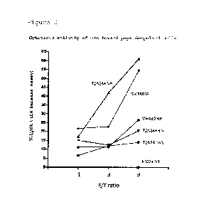
cytotoxic activity of an effector cell (OIL) induced from PBMC
toward T2-A24 cells pulsed with various peptides and unpulsed
SW480-SP cells. The
effector cell used was one induced by
means of ASB4 peptide IV9 represented by SEQ ID No: 3. K562
cells lacking MHC class I expression were used as a negative
control.
[FIG. 11] FIG. 11
shows the results of evaluation of in vivo
OIL inducibility of the peptide represented by SEQ ID No: 4
(As80 9) by means of an interferon-y ELISPOT assay. The
ordinate denotes the number of spots per given number of
seeded cells. 'A' denotes the result of a test using an HLA-
A*02:01 transgenic mouse, and 'B' denotes the result of a test
using an HLA-A*24:02 transgenic mouse. The
black bar
('w/peptide') and the white bar ('w/o') show the results of
restimulation culturing of peptide-treated mouse-derived
splenocytes in the presence or absence of administered peptide
12
CA 02969659 2017-06-02
respectively. That is, the differences in the figures between
the black bar and the white bar denote the number of peptide-
specific CTLs induced in the mouse living body by
administration of each of the peptides.
[FIG. 12] FIG. 12
shows the results of evaluation of in vivo
CTL inducibility of the peptide represented by SEQ ID No: 5
(As82 10) by means of an interferon-y ELISPOT assay. The
ordinate, A, B, black bar and white bar are the same as those
in FIG. 11.
[FIG. 13] FIG. 13
shows the results of evaluation of in vivo
CTL inducibility of the peptide represented by SEQ ID No: 6
(As124 10) by means of an interferon-y ELISPOT assay. The
ordinate, A, B, black bar and white bar are the same as those
in FIG. 11.
[FIG. 14] FIG. 14
shows the results of evaluation of in vivo
CTL inducibility of the peptide represented by SEQ ID No: 7
(As125 9) by means of an interferon-y ELISPOT assay. The
ordinate, A, B, black bar and white bar are the same as those
in FIG. 11.
[FIG. 15] FIG. 15
shows the results of evaluation of in vivo
CTL inducibility of the peptide represented by SEQ ID No: 8
(As184 12) by means of an interferon-y ELISPOT assay. The
ordinate, A, B, black bar and white bar are the same as those
in FIG. 11.
[FIG. 16] FIG. 16
shows the results of evaluation of in vivo
CTL inducibility of the peptide represented by SEQ ID No: 9
(As135 10) by means of an interferon-y ELISPOT assay. The
ordinate, A, B, black bar and white bar are the same as those
in FIG. 11.
[FIG. 17] FIG. 17
shows the results of evaluation of in vivo
CTL inducibility of the peptide represented by SEQ ID No: 10
(As83 10) by means of an interferon-y ELISPOT assay. The
ordinate, A, B, black bar and white bar are the same as those
in FIG. 11.
13
CA 02969659 2017-06-02
[FIG. 18] FIG. 18
shows the results of evaluation of in vivo
CTL inducibility of the peptide represented by SEQ ID No: 11
(As87 9) by means of an interferon-y ELISPOT assay. The
ordinate, A, B, black bar and white bar are the same as those
in FIG. 11.
[FIG. 19] FIG. 19
shows the results of evaluation of in vivo
CTL inducibility of the peptide represented by SEQ ID No: 12
(As307 10) by means of an interferon-y ELISPOT assay. The
ordinate, A, B, black bar and white bar are the same as those
in FIG. 11.
[FIG. 20] FIG. 20
shows the results of evaluation of in vivo
CTL inducibility of the peptide represented by SEQ ID No: 13
(As301 11) by means of an interferon-y ELISPOT assay. The
ordinate, A, B, black bar and white bar are the same as those
in FIG. 11.
[FIG. 21] FIG. 21
shows the results of evaluation of in vivo
CTL inducibility of the peptide represented by SEQ ID No: 14
(As405_9) by means of an interferon-y ELISPOT assay. The
ordinate, A, B, black bar and white bar are the same as those
in FIG. 11.
[FIG. 22] FIG. 22
shows the results of evaluation of in vivo
CTL inducibility of the peptide IV9 represented by SEQ ID No:
3 by means of an interferon-y ELISPOT assay. The ordinate, A,
B, black bar and white bar are the same as those in FIG. 11.
[FIG. 23] FIG. 23
shows the result of evaluation of in vivo
CTL inducibility of the peptide represented by SEQ ID No: 15
(As35 10) by means of an interferon-y ELISPOT assay using an
HLA-A*02:01 transgenic mouse. The
ordinate, black bar and
white bar are the same as those in FIG. 11.
[FIG. 24] FIG. 24
shows the result of evaluation of in vivo
CTL inducibility of the peptide represented by SEQ ID No: 16
(As92 10) by means of an interferon-y ELISPOT assay using an
HLA-A*02:01 transgenic mouse. The
ordinate, black bar and
white bar are the same as those in FIG. 11.
14
CA 02969659 2017-06-02
[FIG. 25] FIG. 25
shows the result of evaluation of in vivo
CTL inducibility of the peptide represented by SEQ ID No: 17
(As152 9) by means of an interferon-y ELISPOT assay using an
HLA-A*02:01 transgenic mouse. The
ordinate, black bar and
white bar are the same as those in FIG. 11.
[FIG. 26] FIG. 26
shows the result of evaluation of in vivo
CTL inducibility of the peptide represented by SEQ ID No: 18
(As186 10) by means of an interferon-y ELISPOT assay using an
HLA-A*02:01 transgenic mouse. The
ordinate, black bar and
white bar are the same as those in FIG. 11.
[FIG. 27] FIG. 27
shows the result of evaluation of in vivo
CTL inducibility of the peptide represented by SEQ ID No: 19
(As236 10) by means of an interferon-y ELISPOT assay using an
HLA-A*02:01 transgenic mouse. The
ordinate, black bar and
white bar are the same as those in FIG. 11.
[FIG. 28] FIG. 28
shows the result of evaluation of in vivo
CTL inducibility of the peptide represented by SEQ ID No: 20
(As265 10) by means of an interferon-y ELISPOT assay using an
HLA-A*02:01 transgenic mouse. The
ordinate, black bar and
white bar are the same as those in FIG. 11.
[FIG. 29] FIG. 29
shows the result of evaluation of in vivo
CTL inducibility of the peptide represented by SEQ ID No: 21
(As280 10) by means of an interferon-y ELISPOT assay using an
HLA-A*02:01 transgenic mouse. The
ordinate, black bar and
white bar are the same as those in FIG. 11.
[FIG. 30] FIG. 30
shows the result of evaluation of in vivo
CTL inducibility of the peptide represented by SEQ ID No: 22
(As383 10 5L) by means of an interferon-y ELISPOT assay using
_ _
an HLA-A*02:01 transgenic mouse. The ordinate, black bar and
white bar are the same as those in FIG. 11.
[FIG. 31] FIG. 31
shows the result of evaluation of in vivo
CTL inducibility of the peptide represented by SEQ ID No: 23
(As416 10) by means of an interferon-y ELISPOT assay using an
HLA-A*02:01 transgenic mouse. The
ordinate, black bar and
white bar are the same as those in FIG. 11.
CA 02969659 2017-06-02
[FIG. 32] FIG. 32
shows the result of evaluation of in vivo
CTL inducibility of the peptide represented by SEQ ID No: 24
(As76 10) by means of an interferon-y ELISPOT assay using an
HLA-A*24:02 transgenic mouse. The
ordinate, black bar and
white bar are the same as those in FIG. 11.
[FIG. 33] FIG. 33
shows the result of evaluation of in vivo
CTL inducibility of the peptide represented by SEQ ID No: 25
(As192 10) by means of an interferon-y ELISPOT assay using an
HLA-A*24:02 transgenic mouse. The
ordinate, black bar and
white bar are the same as those in FIG. 11.
[FIG. 34] FIG. 34
shows the result of evaluation of in vivo
CTL inducibility of the peptide represented by SEQ ID No: 26
(As211 10) by means of an interferon-y ELISPOT assay using an
HLA-A*24:02 transgenic mouse. The
ordinate, black bar and
white bar are the same as those in FIG. 11.
[FIG. 35] FIG. 35
shows the result of evaluation of in vivo
CTL inducibility of the peptide represented by SEQ ID No: 27
(As289 10) by means of an interferon-y ELISPOT assay using an
HLA-A*24:02 transgenic mouse. The
ordinate, black bar and
white bar are the same as those in FIG. 11.
[FIG. 36] FIG. 36
shows the result of evaluation of in vivo
CTL inducibility of the peptide represented by SEQ ID No: 28
(As318 10) by means of an interferon-y ELISPOT assay using an
HLA-A*24:02 transgenic mouse. The
ordinate, black bar and
white bar are the same as those in FIG. 11.
[FIG. 37] FIG. 37
shows the result of evaluation of in vivo
CTL inducibility of the peptide represented by SEQ ID No: 29
(As365 12) by means of an interferon-y ELISPOT assay using an
HLA-A*24:02 transgenic mouse. The
ordinate, black bar and
white bar are the same as those in FIG. 11.
[FIG. 38] FIG. 38
shows the result of evaluation of in vivo
CTL inducibility of the peptide represented by SEQ ID No: 30
(As365_9) by means of an interferon-y ELISPOT assay using an
HLA-A*24:02 transgenic mouse. The
ordinate, black bar and
white bar are the same as those in FIG. 11.
16
CA 02969659 2017-06-02
[FIG. 39] FIG.
39 shows the result of evaluation of in vivo
CTL inducibility of the peptide represented by SEQ ID No: 4
(As80 9) by means of an interferon-y ELISPOT assay using
peripheral blood mononuclear cells derived from a HLA-A*02:01-
positive healthy individual. The ordinate denotes the number
of spots per number of seeded cells (approximately 1 x 10⁵).
The black bar ('w/peptide') and the white bar ('w/o') show the
results of stimulation culturing in the presence or absence of
peptide, respectively. That
is, the differences in the
figures between the black bar and the white bar denote the
number of peptide-specific CTLs induced by administration of
each of the peptides.
[FIG. 40] FIG.
40 shows the result of evaluation of in vivo
CTL inducibility of the peptide represented by SEQ ID No: 5
(As82 10) by means of an interferon-y ELISPOT assay using
peripheral blood mononuclear cells derived from a HLA-A*02:01-
positive healthy individual. The
ordinate, black bar and
white bar are the same as those in FIG. 39.
[FIG. 41] FIG.
41 shows the result of evaluation of in vivo
CTL inducibility of the peptide represented by SEQ ID No: 6
(As124 10) by means of an interferon-y ELISPOT assay using
peripheral blood mononuclear cells derived from a HLA-A*02:01-
positive healthy individual. The
ordinate, black bar and
white bar are the same as those in FIG. 39.
[FIG. 42] FIG. 42
shows the result of evaluation of in vivo
CTL inducibility of the peptide represented by SEQ ID No: 8
(As184 12) by means of an interferon-y ELISPOT assay using
peripheral blood mononuclear cells derived from a HLA-A*02:01-
positive healthy individual. The
ordinate, black bar and
white bar are the same as those in FIG. 39.
[FIG. 43] FIG. 43
shows the result of evaluation of in vivo
CTL inducibility of the peptide represented by SEQ ID No: 9
(As135 10) by means of an interferon-y ELISPOT assay using
peripheral blood mononuclear cells derived from a HLA-A*02:01-
17
CA 02969659 2017-06-02
positive healthy individual. The
ordinate, black bar and
white bar are the same as those in FIG. 39.
[FIG. 44] FIG. 44
shows the result of evaluation of in vivo
CTL inducibility of the peptide represented by SEQ ID No: 5
(As82 10) by means of an interferon-y ELISPOT assay using
peripheral blood mononuclear cells derived from a HLA-A*24:02-
positive healthy individual. The
ordinate, black bar and
white bar are the same as those in FIG. 39.
[FIG. 45] FIG. 42
shows the result of evaluation of in vivo
CTL inducibility of the peptide represented by SEQ ID No: 8
(As184 12) by means of an interferon-y ELISPOT assay using
peripheral blood mononuclear cells derived from a HLA-A*24:02-
positive healthy individual. The
ordinate, black bar and
white bar are the same as those in FIG. 39.
[Modes for Carrying Out the Invention]
[0021]
The present invention is explained in detail below.
The 'epitope peptide' referred to in the present
invention means a peptide that binds to an MHC (an HLA for
humans) and is subjected to antigen presentation on the cell
surface and has antigenicity (can be recognized by a T cell).
The epitope peptide includes a CTL epitope peptide that binds
to an MHC class I, is subjected to antigen presentation, and
is recognized by a CD8-positive T cell, and a helper epitope
peptide that binds to an MHC class II, is subjected to antigen
presentation, and is recognized by a CD4-positive T cell.
[0022]
Among epitope peptides, a protein-derived peptide that is
specifically or overexpressed in a tumor cell is in particular
called a tumor antigen peptide. The
antigen presentation
referred to a phenomenon in which a peptide present within a
cell binds to an MHC and this MHC/antigen peptide complex is
localized on the cell surface. As
described above, it is
known that an antigen presented on a cell surface is
18
CA 02969659 2017-06-02
recognized by a T cell, etc. and then activates cell-mediated
immunity or humoral immunity; since an antigen presented by an
MHC class I activates cell-mediated immunity and is also
recognized by a T cell receptor of a naive T cell to thus
induce the naive T cell to become a CTL having cytotoxic
activity, a tumor antigen peptide used in immunotherapy is
preferably a peptide that binds to an MHC class I and is
subjected to antigen presentation.
[0023]
In the present invention, a 'tumor' includes a benign
tumor and a malignant tumor (cancer, malignant neoplasm). A
cancer includes a hematopoietic tumor, an epithelial malignant
tumor (carcinoma), and a nonepithelial malignant tumor
(sarcoma). In the
present invention, a 'cancer stem cell'
means a cell, among cells present in cancerous tissue, that
exhibits stem cell-like properties, and is a cell that is
thought to be a causal cell involved in the occurrence,
recurrence, and metastasis of a cancer. In
general, since
only a small amount of 'cancer stem cells' are present in
cancerous tissue, it is difficult to distinguish them from
other cells, but in the present technical field methods for
isolating/concentrating cancer stem cells are known, examples
thereof including an SP fractionation method.
Therefore, in
the present invention, a 'cancer stem cell' can mean a cell
population that has been isolated/concentrated by a known
cancer stem cell isolation/concentration method.
[0024]
In the present invention, a natural peptide of the
present invention has been isolated/identified using the
following method for enabling isolation/identification of a
natural peptide that is actually subjected to antigen
presentation on a cell surface. In the
present invention, a
'natural peptide' means a peptide that is actually subjected
to antigen presentation on a cell surface.
Furthermore, a
'natural antigen peptide' is a natural peptide that is
19
CA 02969659 2017-06-02
confirmed to have antigenicity. By
isolating this natural
antigen peptide from a cancer cell and determining the
sequence and the origin thereof, it is possible to obtain
useful findings for the targeted therapy of a cancer using
CTLs.
[0025]
The method of isolating/identifying natural peptides used
in the present invention comprises a step of lysing a cancer
stem cell presenting a natural peptide and isolating a complex
of an MHC and the natural peptide from the lysate, and a step
of separating the isolated complex into the MHC molecule and
the natural peptide to isolate the natural peptide, and a step
of identifying the isolated natural peptide.
For the isolation of a complex of an MHC and the natural
peptide, an extraction method of peptide/MHC complex by
immunoprecipitation using a specific antibody against MHC was
adopted. As the
suitable anti-MHC antibodies, antibodies
against HLA class I, such as anti-HLA-A02 antibody and anti-
HLA-A24 antibody were used.
[0026]
In the step of separating a complex into MHC molecules
and natural peptides, peptide isolation using a weak acid was
performed.
Furthermore, the sequence of the above isolated natural
peptide was analyzed using a peptide sequence analysis method
that combines liquid chromatography and tandem mass
spectrometry, and the natural peptide that is actually
subjected to antigen presentation on the cell surface was
identified.
As a method for confirming antigenicity of the natural
peptide isolated as described above, cytotoxicity test,
ELISPOT assay, assay using TCR-like antibody, etc. were
adopted.
CA 02969659 2017-06-02
[0027]
The present inventors have analyzed a natural antigen
peptide that is subjected to antigen presentation on a human
cancer stem cell by the above method. As a
result, an ASB4
protein-derived peptide (SEQ ID No: 3) has been identified as
a natural antigen peptide that is subjected to antigen
presentation on a cancer stem cell. As a
result of further
progressing research based on such a finding, it has been
found that the ASB4 gene is highly expressed specifically in
cancer stem cells and is a useful candidate gene for
molecularly targeted therapy of cancer stem cells. The
finding that ASB4 is a tumor antigen and, furthermore, the
finding that an ASB4-derived peptide binds to an HLA class I
antigen to form a complex on a tumor cell surface and is
transported to the cell surface and subjected to antigen
presentation are new findings that were hitherto completely
unknown.
[0028]
<1> The peptide of the present invention
In the present invention, a 'human ASB4 protein' means a
known protein reported in Mizuno Y, et al. Biochem Biophys Res
Commun. 2002 Feb 8; 290(5): 1499-505, and Yang CS, et al. Cell
Rep. 2014 Jul 24; 8(2): 327-37, and it specifically means a
protein having an amino acid sequence described in SEQ ID No:
2 (Genbank Accession No: NP 057200 ; ASB4 isoform a) and an
isoform and a homolog thereof.
Examples of the isoform
include a splicing variant and a variant such as an SNP based
on individual difference.
Specific examples include (1) a
protein with an amino acid sequence that has a homology of at
least 90%, preferably at least 95%, and more preferably at
least 98% with the amino acid sequence represented by SEQ ID
No: 2, and (2) a protein with an amino acid sequence for which
one or more amino acids, preferably one to several, and more
preferably 1 to 10, 1 to 5, 1 to 3, or 1 or 2 amino acids have
21
CA 02969659 2017-06-02
been replaced, deleted, added, or inserted in the amino acid
sequence described in SEQ ID No: 2.
Examples of such a
variant include an isoform (ASB4 isoform b) registered as
Genbank Accession No.: NP 665879 which is a splicing variant
of ASB4 isoform a, and SNPs such as dbSNP RefSNP No.:
rs35047380 in which the 17th amino acid has been replaced from
valine (V) to leucine (L). When simply 'ASB4 protein' is
referred to in the present specification, it means a human
ASB4 protein represented by the amino acid sequence described
in SEQ ID No: 2, unless otherwise specified.
[0029]
Preferred examples of the human ASB4 protein include a
protein comprising the amino acid sequence described in SEQ ID
No: 2, and a protein with an amino acid sequence for which 1
to 3, and preferably 1 or 2 amino acids have been replaced in
said protein. A
protein with the amino acid sequence
described in SEQ ID No: 2 can be cited as a yet more preferred
example.
In one embodiment, the peptide of the present invention
includes a human ASB4 protein partial peptide, the peptide
binding to an MHC, and in particular to an HLA; it is
preferably a peptide that is subjected to antigen presentation
by means of an MHC, in particular an HLA, and more preferably
a peptide that is subjected to antigen presentation by means
of an MHC, in particular an HLA, and can induce a CTL. There
are several types of HLA; the peptide of the present invention
preferably can bind to an HLA class I, more preferably can
bind to HLA-A02 or HLA-A24, and yet more preferably can bind
to both HLA-A02 and HLA-A24 (i.e., dual binding). The peptide
of the present invention may be subjected to a treatment such
as processing prior to binding to an MHC, and a peptide that
forms an epitope peptide as a result of such a treatment is
also included in the peptide of the present invention.
Therefore, the amino acid length of the peptide of the present
invention is not particularly limited as long as it is a
22
CA 02969659 2017-06-02
sequence including an amino acid sequence of an epitope
peptide.
However, it is preferable that the peptide of the
present invention itself is an epitope peptide, and therefore
the amino acid length is preferably on the order of about 8 to
14 amino acids, more preferably on the order of about 8 to 11
amino acids, and particularly preferably on the order of about
9 to about 11 amino acids.
[0030]
An epitope peptide that binds to an HLA class I, which is
a human MHC class I, has a length of about 8 to 14 amino acids,
and preferably a length of about 9 to 11 amino acids, and is
known to have an HLA-specific binding motif in the sequence.
For example, a peptide binding to HLA-A02 has a binding motif
in which the second amino acid from the N terminal is leucine,
isoleucine, or methionine and/or the amino acid at the C
terminal is valine, leucine, or isoleucine, and a peptide
binding to HLA-A24 has a binding motif in which the second
amino acid from the N terminal is tyrosine, phenylalanine,
methionine, or tryptophan and/or the amino acid at the C
terminal is leucine, isoleucine, or phenylalanine.
[0031]
Therefore, in a preferred embodiment, the peptide of the
present invention includes an epitope peptide that is a
partial peptide of the ASB4 protein with 8 to 14 consecutive
amino acids in the amino acid sequence of said protein, the
second amino acid from the N terminal being leucine,
isoleucine, or methionine and/or the amino acid at the C
terminal being valine, leucine, or isoleucine, and more
preferably is the epitope peptide itself. Among
them, an
epitope peptide with an amino acid sequence represented by any
of SEQ ID Nos: 4, 6, 7, 10, 14, 15, 17 to 19, 21 to 23, 26, 28,
31, 33, 36, 39, 41, 42, 45 and 46 is particularly preferable.
[0032]
Furthermore, in another preferred embodiment, the partial
peptide includes an epitope peptide having the second amino
23
CA 02969659 2017-06-02
acid from the N terminal replaced by leucine, isoleucine, or
methionine and/or the amino acid at the C terminal replaced by
valine, leucine, or isoleucine, and more preferably is the
epitope peptide itself. Among
them, an epitope peptide with
an amino acid sequence represented by any of SEQ ID Nos: 4, 6,
7, 10, 14, 15, 17 to 19, 21 to 23, 26, 28, 31, 33, 36, 39, 41,
42, 45 and 46, the second amino acid from the N terminal being
replaced by leucine, isoleucine, or methionine and/or the
amino acid at the C terminal being replaced by valine, leucine,
or isoleucine is particularly preferable.
[0033]
In another preferred embodiment, the peptide of the
present invention includes an epitope peptide that is a
partial peptide of the ASB4 protein with 8 to 14 consecutive
amino acids in the amino acid sequence of said protein, the
second amino acid from the N terminal being tyrosine,
phenylalanine, methionine, or tryptophan and/or the amino acid
at the C terminal being leucine, isoleucine, or phenylalanine,
and more preferably is the epitope peptide itself. Among them,
an epitope peptide with an amino acid sequence represented by
any of SEQ ID Nos: 9, 21, 25, 30, 32, 35 and 37 is
particularly preferable.
[0034]
Furthermore, in another preferred embodiment, the partial
peptide includes an epitope peptide, the second amino acid
from the N terminal being replaced by tyrosine, phenylalanine,
methionine, or tryptophan and/or the amino acid at the C
terminal being replaced by leucine, isoleucine, or
phenylalanine, and more preferably is the epitope peptide
itself. Among
them, an epitope peptide with an amino acid
sequence represented by any of SEQ ID Nos: 9, 21, 25, 30, 32,
35 and 37, the second amino acid from the N terminal being
replaced by tyrosine, phenylalanine, methionine, or tryptophan
and/or the amino acid at the C terminal being replaced by
24
CA 02969659 2017-06-02
leucine, isoleucine, or phenylalanine is particularly
preferable.
[0035]
In another preferred embodiment, the peptide of the
present invention is the partial peptide or the partial
peptide that has been subjected to replacement, one to several
amino acids being added to the N terminal and/or the C
terminal.
Among them, a peptide with an amino acid sequence
represented by any of SEQ ID Nos: 4, 6, 7, 10, 14, 15, 17 to
19, 21 to 23, 26, 28, 31, 33, 36, 39, 41, 42, 45 and 46, said
peptide in which the second amino acid from the N terminal is
replaced by leucine, isoleucine, or methionine and/or the
amino acid at the C terminal is replaced by valine, leucine,
or isoleucine, a peptide with an amino acid sequence
represented by any of SEQ ID Nos: 9, 21, 25, 30, 32, 35 and 37,
or said peptide in which the second amino acid from the N
terminal is replaced by tyrosine, phenylalanine, methionine,
or tryptophan and/or the amino acid at the C terminal is
replaced by leucine, isoleucine, or phenylalanine and,
furthermore, one to several amino acids are added to the N
terminal and/or the C terminal is particularly preferable.
[0036]
Therefore, in an embodiment, the peptide of the present
invention may be represented by
Yo-Xo-Zo,
wherein all of Xo, Yo, and Zo are peptides.
In such an embodiment, Xo is a peptide selected from (1)
to (4) below:
(1) a partial peptide of the ASB4 protein with 8 to 14
consecutive amino acids in the amino acid sequence of said
protein, and preferably 8 to 11 amino acids, the second amino
acid from the N terminal being leucine, isoleucine, or
methionine and/or the amino acid at the C terminal being
valine, leucine, or isoleucine;
CA 02969659 2017-06-02
(2) a peptide which, in the partial peptide defined in
(1), the second amino acid from the N terminal being replaced
by leucine, isoleucine, or methionine and/or the amino acid at
the C terminal being replaced by valine, leucine, or
isoleucine;
(3) a partial peptide of the ASB4 protein with 8 to 14
consecutive amino acids in the amino acid sequence of said
protein, and preferably 8 to 11 amino acids, the second amino
acid from the N terminal being tyrosine, phenylalanine,
methionine, or tryptophan and/or the amino acid at the C
terminal being leucine, isoleucine, or phenylalanine; or
(4) a peptide which, in the partial peptide defined in
(3), the second amino acid from the N terminal being replaced
by tyrosine, phenylalanine, methionine, or tryptophan and/or
the amino acid at the C terminal being replaced by leucine,
isoleucine, or phenylalanine. Since
(2) is a replacement
homolog of (1), and (4) is a replacement homolog of (3), Yo-Xo-
Zo for which Xo is a peptide of (2) or (4) is particularly
called an 'Xo homolog'.
[0037]
Furthermore, Yo and Zo are mutually independently any
peptide with 0 to several amino acids. With this regard, '0
to several amino acids' specifically means 0 to 5 amino acids,
examples including 0, 1, 2, 3, 4, or 5 amino acids, more
preferably 0, 1, 2, or 3 amino acids, and particularly
preferably 0 or 1 amino acids. In the present invention, when
it is stated that Yo and/or Zo are 'not present', it means a
case in which Yo and/or Zo are peptides with 0 amino acids.
The amino acids constituting Yo and/or Zo are not
particularly limited; any of 20 types of natural amino acids
constituting a protein can be cited, but preferable examples
include an amino acid that is cleavable by an enzyme present
in a living body.
Furthermore, an amino acid sequence
corresponding to an amino acid sequence on the N terminal side
26
CA 02969659 2017-06-02
and/or on the C terminal side of the above partial peptide in
the amino acid sequence of the ASB4 protein is desirable.
[0038]
Therefore, among them, a case in which X0 is either a
peptide with an amino acid sequence represented by any of SEQ
ID Nos: 4, 6, 7, 10, 14, 15, 17 to 19, 21 to 23, 26, 28, 31,
33, 36, 39, 41, 42, 45 and 46, said peptide in which the
second amino acid from the N terminal is replaced by leucine,
isoleucine, or methionine and/or the amino acid at the C
terminal is replaced by valine, leucine, or isoleucine, a
peptide with an amino acid sequence represented by any of SEQ
ID Nos: 9, 21, 25, 30, 32, 35 and 37, or said peptide in which
the second amino acid from the N terminal is replaced by
tyrosine, phenylalanine, methionine, or tryptophan and/or the
amino acid at the C terminal is replaced by leucine,
isoleucine, or phenylalanine and, furthermore, Yo and/or Zo is
one amino acid is particularly preferable, and a case in which
either one of Yo or Zo is one amino acid and the other is not
present is yet more preferable.
[0039]
Furthermore, another preferred embodiment is a case in
which X0 is any of (1) to (4) with 8 to 11 amino acids and Yo
and/or Zo are mutually independently a peptide with 0 to three
amino acids, Yo-X0-Zo forming a partial peptide of the ASB4
protein having a length of 9 to 14 amino acids in its entirety
or an X0 homolog thereof. Examples
of such an embodiment
include, but are not limited to, a case in which X0 is a
peptide with an amino acid sequence represented by any of SEQ
ID Nos: 3 to 7 and 9 to 19, 21 to 28, and 30 to 46, peptide Yo
and/or Zo with 0 to 3 amino acids are added to the N terminal
and/or C terminal of Xo, and such Yo-Xo-Zo is also a partial
peptide of the ASB4 protein.
[0040]
With regard to the peptide of the present invention, in a
preferred embodiment, X0 includes a peptide with the same amino
27
CA 02969659 2017-06-02
acid sequence as the amino acid sequence described in any of
SEQ ID Nos: 3 to 7, 9 to 28 and 30. In this embodiment, it is
more preferable that all of the peptides of the present
invention (that is, Yo-X0-Zo) are partial peptides of the ASB4
protein. In such a more preferred embodiment, examples of the
peptide of the present invention include a peptide with the
same amino acid sequence as the amino acid sequence described
in any of SEQ ID Nos: 3 to 30.
[0041]
The peptides represented by SEQ ID Nos: 3 to 30 are
peptides with the 9 amino acids corresponding to amino acid
positions 319 to 327 of the above ASB4 (SEQ ID No: 3), with
the 9 amino acids corresponding to positions 80 to 88 (SEQ ID
No: 4), with the 10 amino acids at positions 82 to 91 (SEQ ID
No: 5), with the 10 amino acids at positions 124 to 133 (SEQ
ID No: 6), with the 9 amino acids at positions 125 to 133 (SEQ
ID No: 7), with the 12 amino acids at positions 184 to 195
(SEQ ID No: 8), with the 10 amino acids at positions 135 to
144 (SEQ ID No: 9), with the 10 amino acids at positions 83 to
92 (SEQ ID No: 10), with the 9 amino acids at positions 87 to
95 (SEQ ID No: 11), with the 10 amino acids at positions 307
to 316 (SEQ ID No: 12), with the 11 amino acids at positions
301 to 311 (SEQ ID No: 13) and with the 9 amino acids at
positions 405 to 413 (SEQ ID No: 14), with the 10 amino acid
at positions 35 to 44 (SEQ ID No: 15), with the 10 amino acid
at positions 92 to 101 (SEQ ID No: 16), with the 9 amino acid
at positions 152 to 160 (SEQ ID No: 17), with the 10 amino
acid at positions 186 to 195 (SEQ ID No: 18), with the 10
amino acids at positions 236 to 245 (SEQ ID No: 19), with the
amino acids at positions 265 to 274 (SEQ ID No: 20), with
the 10 amino acids at positions 280 to 289 (SEQ ID No: 21),
with the 10 amino acids at positions 383 to 392 (SEQ ID No:
22), with the 10 amino acids at positions 416 to 425 (SEQ ID
No: 23), with the 10 amino acids at positions 76 to 85 (SEQ ID
No: 24), with the 10 amino acids at positions 192 to 201 (SEQ
28
CA 02969659 2017-06-02
ID No: 25), with the 10 amino acids at positions 211 to 220
(SEQ ID No: 26), with the 10 amino acids at positions 289 to
298 (SEQ ID No: 27), with the 10 amino acids at positions 318
to 327 (SEQ ID No: 28), with the 12 amino acids at positions
365 to 376 (SEQ ID No: 29), with the 9 amino acids at
positions 365 to 373 (SEQ ID No: 30), respectively, and the
present inventors have found that all of the peptides being
capable of binding to HLA-A02 and/or HLA-A24. In
particular
the present inventors have found that the peptides represented
by SEQ ID Nos: 3 to 23, 25, 26 and 28 to 30 also have CTL
inducibility.
[0042]
In a yet more preferred embodiment, X0 includes a peptide
with the same amino acid sequence as the amino acid sequence
described in any of SEQ ID Nos: 4 to 7, 9 to 12 and 15 to 19
and 21 to 23. In this embodiment, it is more preferable that
all of the peptides of the present invention (that is, Yo-X0-
Z0) are partial peptides of the ASB4 protein. In such a more
preferred embodiment, examples of the peptide of the present
invention include a peptide with the same amino acid sequence
as the amino acid sequence described in any of SEQ ID Nos: 4
to 12 and 15 to 23.
[0043]
The present inventors have found that all of the peptides
represented by any of SEQ ID Nos: 4 to 12 and 15 to 23 being
capable of binding to HLA-A02 and having CTL inducibility.
[0044]
In another yet more preferred embodiment, X0 includes a
peptide with the same amino acid sequence as the amino acid
sequence described in any of SEQ ID Nos: 3 to 7, 9, 13, 14, 18,
24 to 28 and 30. In this
embodiment, it is more preferable
that all of the peptides of the present invention (that is, Y0-
X0-Zo) are partial peptides of the ASB4 protein. In such
a
more preferred embodiment, examples of the peptide of the
present invention include a peptide with the same amino acid
29
CA 02969659 2017-06-02
sequence as the amino acid sequence described in any of SEQ ID
Nos: 3 to 9, 13, 14, 25, 26 and 28 to 30.
[0045]
The present inventors have found that all of the peptides
represented by any of SEQ ID Nos: 3 to 9, 13, 14, 25, 26 and
28 to 30 being capable of binding to HLA-A024 and having CTL
inducibility.
[0046]
In another yet more preferred embodiment, X0 includes a
peptide with the same amino acid sequence as the amino acid
sequence described in any of SEQ ID Nos: 4 to 7, 9 and 18. In
this embodiment, it is more preferable that all of the
peptides of the present invention (that is, Yo-X0-Z0) are
partial peptides of the ASB4 protein. In such a more
preferred embodiment, examples of the peptide of the present
invention include a peptide with the same amino acid sequence
as the amino acid sequence described in any of SEQ ID Nos: 4
to 9.
[0047]
The present inventors have found that all of the peptides
represented by any of SEQ ID Nos: 4 to 9 being capable of
binding to both HLA-A02 and HLA-A24, and having CTL
inducibility. Among them, the peptides represented by SEQ ID
Nos: 4 to 6, 8 and 9 are particularly preferable, and the
peptides represented by SEQ ID Nos: 5 and 8 are most
preferable.
[0048]
The peptide of the present invention may have its N
terminal and/or C terminal modified. Specific examples of the
modification include N-alkanoylation (for example,
acetylation), N-alkylation (for example, methylation), a C-
terminal alkyl ester (for example, an ethyl ester), and a C-
terminal amide (for example a carboxamide).
Synthesis of the peptide of the present invention may be
carried out in accordance with known methods used in normal
CA 02969659 2017-06-02
peptide chemistry. Such
known methods includes methods
described in the literature (Peptide Synthesis, Interscience,
New York, 1966; The Proteins, Vol. 2, Academic Press Inc., New
York, 1976; Peptide Synthesis, Maruzen Co., Ltd., 1975; Basics
and Experiments of Peptide Synthesis, Maruzen Co., Ltd., 1985;
Development of Pharmaceuticals Seq. Vol. 14 Peptide Synthesis,
Hirokawa Shoten Co., 1991, these publications forming part of
the present application by reference), etc.
[0049]
With regard to the peptide of the present invention, in
vivo activity can be confirmed by subjecting it to a CTL
induction method, which is described later, an assay using an
animal model for human (W002/47474, Int J. Cancer: 100, 565-
570 (2002)), etc.
[0050]
In another embodiment, the peptide of the present
invention includes a peptide that binds to HLA-A02 and/or HLA-
A24. Specifically, examples include, but are not limited to,
a peptide with the amino acid sequence represented by any of
SEQ ID Nos: 3 to 30. These peptides are not necessarily those
having a binding motif of HLA-A02 or HLA-A24, but they are the
peptides actually confirmed to bind to HLA-A02 and/or HLA-A24
by the present inventors. Among
them, a peptide with the
amino acid sequence represented by any of SEQ ID Nos: 3 to 23,
25, 26 and 28 to 30 has been confirmed to have CTL
inducibility as well, which is preferable.
[0051]
The peptide of the present invention further includes a
peptide in which a plurality of epitope peptides including at
least one of the peptides of the present invention are linked
(polyepitope peptide).
Therefore, specific examples of the
peptide of the present invention include a peptide that is the
above polyepitope peptide and has CTL-inducing activity.
The polyepitope peptide of the present invention may
specifically be defined as
31
CA 02969659 2017-06-02
(i) a peptide in which the peptide of the present invention
(epitope peptide) and any one or more CTL epitope peptides
other than the peptide of the present invention are linked
directly or via a spacer as appropriate,
(ii) a peptide in which the peptide of the present invention
and any one or more helper epitope peptides are linked
directly or via a spacer as appropriate, or
(iii) a peptide in which a polyepitope peptide described in
(i) above and further one or more helper epitope peptides are
linked directly or via a spacer as appropriate,
the peptide being subjected to processing within an
antigen-presenting cell, and the epitope peptide thus formed
being presented on the antigen-presenting cell, thus leading
to CTL-inducing activity.
[0052]
The CTL epitope peptide other than the peptide of the
present invention in (i) is not particularly limited; specific
examples include another human ASB4-derived epitope peptide
that is not included in the present invention and a human
OR7C1- or human DNAJB8-derived epitope peptide (for example, a
peptide described in W02010/050190), and a human FAM83B-
derived epitope peptide (International Patent Application
PCT/JP2014/076625), etc.
The spacer is not particularly limited as long as it does
not adversely affect processing within an antigen-presenting
cell, and is preferably a linker that is linked to each
epitope peptide via a peptide bond, examples including a
peptide linker in which several amino acids are linked and a
linker having an amino group and a carboxyl group at each end.
Specific examples include a glycine linker or a PEG
(polyethylene glycol) linker; examples of the glycine linker
include polyglycine (for example a peptide consisting of six
glycines; Cancer Sci, Vol. 103, p. 150-153), and examples of
the PEG linker include a linker derived from a compound having
an amino group and a carboxy group at each end of PEG (for
32
CA 02969659 2017-06-02
example, H2N-(CH2)2-(OCH2CH2)3-COOH; Angew. Chem. Int. Ed. 2008,
47, 7551-7556).
[0053]
With regard to the epitope peptide of the present
invention contained in the polyepitope peptide of the present
invention, one or more types may be selected. That
is, a
plurality of identical epitope peptides may be linked, or a
plurality of different epitope peptides may be linked.
Naturally, even when two or more types of epitope peptides are
selected, a plurality of one or more types of selected epitope
peptides may be linked. Similarly, with regard to the epitope
peptide other than the peptide of the present invention, a
plurality of types and/or a plurality of epitope peptides may
be linked. The
polyepitope peptide of the present invention
may be one in which 2 to 12 epitope peptides are linked, is
preferably one in which 2, 3, 4, 5, 6, 7, 8, 9, 10, 11, or 12
epitope peptides are linked, and is most preferably one in
which 2 epitope peptides are linked.
When the epitope peptide that is linked to the peptide of
the present invention is a helper epitope peptide, examples of
the helper epitope peptide used include hepatitis B virus-
derived HBVc128-140 and tetanus toxin-derived TT947-967. The
length of the helper epitope peptide is on the order of 13 to
30 amino acids, and preferably on the order of 13 to 17 amino
acids.
[0054]
Such a peptide in which a plurality of epitope peptides
are linked (polyepitope peptide) may also be produced by a
standard peptide synthesis method as described above.
Furthermore, based on information regarding the sequence of a
polynucleotide encoding such a polyepitope peptide in which a
plurality of epitope peptides are linked, it may be produced
using standard DNA synthesis and genetic engineering methods.
That is, said polynucleotide is inserted into a known
expression vector, a host cell is transformed by means of the
33
CA 02969659 2017-06-02
recombinant expression vector thus obtained to give a
transformant, the transformant is cultured, and the target
polyepitope peptide in which a plurality of epitopes are
linked can be produced by recovery from the culture. These
methods may be carried out in accordance with methods
described in the literature as described above (Molecular
Cloning, T. Maniatis et al., CSH Laboratory (1983), DNA
Cloning, D M. Glover, IRL PRESS (1985)).
[0055]
The polyepitope peptide thus produced in which a
plurality of epitope peptides are linked is subjected to the
above in vitro assay or an in vivo assay using an animal model
for human described in W002/47474 and Int J. Cancer: 100, 565-
570 (2002) (these publications forming part of the present
application by reference), etc., thus enabling CTL-inducing
activity to be confirmed.
The peptide of the present invention (including the
polyepitope peptide) is useful for the prevention and/or
therapy of a cancer, etc. as described in the present
specification, and may be an active ingredient of a
pharmaceutical composition.
Furthermore, the peptide of the
present invention may be for the prevention and/or therapy of
a cancer. Moreover, the present invention also relates to use
of the peptide of the present invention in the production of a
medicament for the prevention and/or therapy of a cancer.
[0056]
<2> Polynucleotide of the present invention
The polynucleotide of the present invention includes a
polynucleotide that encodes at least one of the peptides of
the present invention. The
polynucleotide of the present
invention may be any of cDNA, mRNA, cRNA, or synthetic DNA.
It may have either a single strand or a double strand
configuration. Specific examples include, but are not limited
to, a polynucleotide with a nucleotide sequence encoding an
amino acid sequence predicted using a binding prediction
34
CA 02969659 2017-06-02
program of MHC and peptide, such as BIMAS (http://www-
bimas.cit.nih.gov/molbio/hlabind/),
SYFPEITHI
(http://www.syfpeithi.de/) and IEDB (MHC-I
processing
predictions; http: //www.iedb.org/); and more specifically,
they include a polynucleotide with a nucleotide sequence
encoding an amino acid sequence described in SEQ ID Nos: 3-46,
and a polynucleotide with a nucleotide sequence encoding so
that it can express a polyepitope peptide in which any two or
more peptides selected from SEQ ID Nos: 3-46 are linked or a
peptide selected from SEQ ID Nos: 3-46 and a helper epitope
are linked.
[0057]
The polynucleotide of the present invention may take on
either a single strand or a double strand configuration. When
the polynucleotide of the present invention is a double strand,
a recombinant expression vector expressing the peptide of the
present invention may be produced by inserting the
polynucleotide of the present invention into an expression
vector. That
is, the scope of the polynucleotide of the
present invention includes a recombinant expression vector
produced by inserting the double strand polynucleotide of the
present invention into an expression vector.
The polynucleotide of the present invention is useful for
the prevention and/or therapy of a cancer, etc. as described
in the present specification, and may be an active ingredient
of a pharmaceutical composition.
Furthermore, the
polynucleotide of the present invention may be for the
prevention and/or therapy of a cancer. Moreover, the present
invention also relates to use of the polynucleotide of the
present invention in the production of a medicament for the
prevention and/or therapy of a cancer.
[0058]
With regard to the expression vector used in the present
invention, various types may be used according to the host
used, the intended application, etc., and a person skilled in
CA 02969659 2017-06-02
the art may select it as appropriate. Examples of expression
vectors that can be used in the present invention include a
plasmid, a phage vector, and a virus vector. For
example,
when the host is Escherichia coli, examples of the vector
include plasmid vectors such as pUC118, pUC119, pBR322, and
pCR3 and phage vectors such as AZAPII and Agt11. When
the
host is a yeast, examples of the vector include pYES2 and
pYEUra3. When the host is an insect cell, examples include
pAcSGHisNT-A. When the host is an animal cell, examples
include plasmid vectors such as pCEP4, pKCR, pCDM8, pGL2,
pcDNA3.1, pRc/RSV, and pRc/CMV and virus vectors such as a
retrovirus vector, an adenovirus vector, and an adeno-
associated virus vector.
[0059]
The vector may have as appropriate a factor such as a
promoter capable of inducing expression, a gene encoding a
signal sequence, a selection marker gene, or a terminator.
Furthermore, in order to make isolation and purification easy,
a sequence for expression as a fusion protein with thioredoxin,
a His tag, GST (glutathione S-transferase), etc. may be added.
In this case, a GST fusion protein vector (pGEX4T, etc.)
having an appropriate promoter (lac, tac, trc, trp, CMV, SV40
early promoter, etc.) that functions within a host cell, a
vector having a tag sequence such as Myc or His (pcDNA3.1/Myc-
His, etc.) and, furthermore, a vector expressing a fusion
protein with thioredoxin and a His tag (pET32a), etc. may be
used.
[0060]
Transforming a host with the expression vector prepared
as above enables a transformed cell containing the expression
vector to be prepared.
Therefore, the present invention
includes a gene transfer composition including the expression
vector.
The host used for transformation may be any cell as long
as the function of the polypeptide of the present invention is
36
CA 02969659 2017-06-02
not impaired, and examples include an Escherichia coli, a
yeast, an insect cell, and an animal cell.
Examples of the
Escherichia coil include E.coli K-12 strain HB101, 0600, JM109,
DH5u, and AD494 (DE3).
Examples of the yeast include
Saccharomyces cerevisiae. Examples of the animal cell include
L929 cells, BALB/c3T3 cells, 0127 cells, CHO cells, COS cells,
Vero cells, HeLa cells, and 293-EBNA cells.
Examples of the
insect cell include sf9.
As a method for introducing an expression vector into a
host cell, a standard introduction method suitable for the
host cell may be used.
Specific examples include a calcium
phosphate method, a DEAE-dextran method, an electroporation
method, and a method using a lipid for gene transfer
(Lipofectamine, Lipofectin; Gibco-BRL). After introduction,
culturing is carried out in a standard medium containing a
selection marker, thus enabling a transformed cell in which
the expression vector has been introduced into the host cell
to be selected.
[0061]
Continuing culturing the transformed cell thus obtained
under suitable conditions enables the peptide of the present
invention to be produced. The
peptide thus obtained may be
further isolated and purified by usual biochemical
purification means.
Examples of purification means include
salting out, ion-exchange chromatography,
adsorption
chromatography, affinity chromatography, and gel filtration
chromatography. When the peptide of the present invention is
expressed as a fusion protein with a thioredoxin, a His tag, a
GST, etc. as described above, isolation and purification may
be carried out by a purification method utilizing the
properties of the fusion protein or the tag.
The polynucleotide encoding the peptide of the present
invention may have a DNA configuration or an RNA configuration.
These polynucleotides of the present invention may be easily
produced by standard methods known in the present technical
37
CA 02969659 2017-06-02
field based on amino acid sequence information of the peptide
of the present invention and DNA sequence information encoded
thereby. Specifically, it may be produced by standard DNA
synthesis, amplification by means of PCR, etc.
The polynucleotide encoding the peptide of the present
invention includes a polynucleotide encoding the epitope
peptide.
[0062]
<3> CTL inducer/pharmaceutical composition comprising a
peptide of the present invention as active ingredient
The peptide of the present invention has CTL-inducing
activity and can be a CTL inducer as a tumor antigen peptide.
Furthermore, as described above, the present inventors have
found for the first time that the ASB4 protein is a tumor
antigen and an ASB4 protein-derived peptide binds to an HLA
class I antigen, forms a complex on the tumor cell surface, is
transported to the cell surface, and is subjected to antigen
presentation. Therefore, the ASB4 protein itself can become a
CTL inducer.
That is, peripheral blood lymphocytes are isolated from a
person who is positive for an HLA-A02 antigen or an HLA-A24
antigen, they are stimulated in vitro by adding the peptide of
the present invention and/or ASB4 protein, and CTLs that
specifically recognize an HLA-A02 antigen-positive cell or an
HLA-A24 antigen-positive cell that have been pulsed with the
peptide can be induced (J. Immunol., 154, p. 2257, 1995). The
presence or absence of CTL induction may be confirmed by
measuring for example the amount of various cytokines (for
example IFN-y) produced by CTLs when reacting with an antigen
peptide-presenting cell, by means of for example an ELISA
method, etc. It may also be confirmed by a method for
measuring CTL toxicity toward an antigen peptide-presenting
cell labeled with 53-Cr (51Cr release assay, Int. J. Cancer, 58:
p317, 1994).
38
CA 02969659 2017-06-02
Furthermore, a CTL clone may be established by a method
described in Int. J. Cancer, 39, 390-396, 1987, N. Eng. J. Med,
333, 1038-1044, 1995, etc.
[0063]
A CTL induced by the peptide and/or ASB4 protein of the
present invention has a cytotoxic action toward a cell
presenting the peptide of the present invention and/or another
ASB4 protein-derived epitope peptide as an antigen and the
ability to produce a lymphokine. Since
the peptide of the
present invention is a tumor antigen peptide as described
above, and the ASB4 protein is decomposed within a cell to
thus form a tumor antigen peptide, it can exhibit an anti-
tumor action, and preferably an anti-cancer action, via the
above functions.
Therefore, the peptide and/or ASB4 protein
of the present invention and a CTL induced thereby can be an
active ingredient of a medicament or a pharmaceutical
composition for the prevention and/or therapy of a cancer.
When a CTL inducer containing the peptide and/or ASB4
protein of the present invention as an active ingredient is
administered to a cancer patient, the peptide of the present
invention and/or the ASB4 protein-derived epitope peptide is
presented on an HLA-A02 antigen or HLA-A24 antigen of an
antigen-presenting cell, a CTL that is specific to a complex
of the HLA-A02 antigen or HLA-A24 antigen and the presented
peptide proliferates and destroys the cancer cells, and as a
result, the cancer can be prevented and/or treated. Therefore,
a CTL inducer containing the peptide and/or ASB4 protein of
the present invention as an active ingredient can preferably
be used for a subject who is positive for an HLA-A02 antigen
or HLA-A24 antigen and who has an ASB4-positive cancer.
Examples of ASB4-positive cancers include cancers (tumors)
such as colon cancer, lung cancer, breast cancer, oral cancer,
cervical cancer, thyroid cancer, testicular tumor, and ovarian
cancer, and the CTL inducer of the present invention may be
used for the prevention and/or therapy of such cancers.
39
CA 02969659 2017-06-02
[0064]
The 'prevention' of a cancer includes not only preventing
a patient from having a cancer but also prevention of
recurrence in a patient who has been subjected to surgery to
remove a primary tumor and prevention of metastasis of a tumor
that could not be completely removed by a cancer treatment
such as surgery, radiotherapy, drug therapy, etc. Furthermore,
the 'treatment' of a cancer includes not only curing and
improvement of the symptoms of a cancer that reduces the size
of the cancer but also prevention of cancer cell proliferation
or tumor enlargement, or suppression of metastasis of cancer
cells from a primary focus.
[0065]
A CTL inducer containing the peptide and/or ASB4 protein
of the present invention as an active ingredient is for
example particularly effective for an HLA-A02- or HLA-A24-
positive cancer patient who has a cancer positive for the ASB4
described in SEQ ID No: 2.
Specifically, it may be used for
the prevention or therapy of a cancer (tumor) such as for
example colon cancer, lung cancer, or ovarian cancer.
Therefore, a pharmaceutical composition containing the peptide
of the present invention and/or the ASB4 protein as an active
ingredient is also included in the present invention. Such a
pharmaceutical composition is preferably a composition for the
prevention and/or therapy of a cancer, that is, a preventive
and/or therapeutic agent for cancer.
Furthermore, since the
pharmaceutical composition of the present invention prevents
and/or treats a cancer by inducing a CTL that is specific to a
cancer cell (preferably a cancer stem cell), that is,
activating cell-mediated immunity that is specific to a cancer
cell, it is preferably a vaccine for the prevention and/or
therapy of a cancer.
[0066]
A pharmaceutical composition containing the peptide of
the present invention as an active ingredient may be one that
CA 02969659 2017-06-02
contains a single CTL epitope (the peptide of the present
invention) as an active ingredient or one that contains as an
active ingredient a polyepitope peptide having another peptide
(CTL epitope or helper epitope) linked thereto. In
recent
years, it has been shown that a polyepitope peptide having a
plurality of linked CTL epitopes (antigen peptides) has
activity in efficiently inducing CTLs in vivo. For
example,
Journal of Immunology 1998, 161: 3186-3194 (this publication
forms part of the present application by reference) describes
the induction in vivo of a CTL that is specific to each CTL
epitope by means of an approximately 30mer polyepitope peptide
in which cancer antigen protein PSA-derived HLA-A2, -A3, -All,
and -B53-restricted CTL epitopes (antigen peptides) are linked.
It is also shown that a polyepitope peptide in which a CTL
epitope and a helper epitope are linked efficiently induces a
CTL. When
administered in the configuration of such a
polyepitope peptide, the polyepitope peptide is incorporated
into an antigen-presenting cell, and after that, individual
antigen peptides that have been formed by intracellular
degradation bind to an HLA antigen to thus form a complex,
this complex is presented on the antigen-presenting cell
surface at high density, a CTL specific to this complex
proliferates efficiently in the body, and cancer cells are
destroyed. In this
way, the treatment or prevention of a
cancer is promoted.
[0067]
A pharmaceutical composition containing the peptide
and/or ASB4 protein of the present invention as an active
ingredient may be administered as a mixture with a
pharmaceutically acceptable carrier, for example an
appropriate adjuvant, or in combination therewith, so as to
establish cell-mediated immunity effectively.
[0068]
As the adjuvant, an adjuvant known in the present
technical field such as one described in the literature (for
41
CA 02969659 2017-06-02
example, Clin Infect Dis.: S266-70, 2000) may be applied, and
specific examples include a gel type such as aluminum
hydroxide, aluminum phosphate, or calcium phosphate, a
bacterial type such as CpG, monophosphoryl lipid A
(monophosphoryl lipid A; MPL), cholera toxin, Escherichia coli
heat-labile toxin, pertussis toxin, or muramyl dipeptide
(Muramyl dipeptide; MDP), an oil emulsion type (emulsion
preparation) such as Freund's incomplete adjuvant, MF59, or
SAF, a macromolecular nanoparticle type such as an
immunostimulatory complex (Immunostimulatory complex; ISCOMs),
a liposome, biodegradable microspheres (Biodegradable
microsphere), or saponin-derived QS-21, a synthetic type such
as a nonionic block copolymer, a muramyl peptide analog
(Muramyl peptide analogue), a polyphosphazene, or a synthetic
polynucleotide, and a cytokine type such as IFN-y, IL-2, or
IL-12.
Furthermore, the dosage form of a CTL
inducer/pharmaceutical composition containing the peptide
and/or ASB4 protein of the present invention as an active
ingredient is not particularly limited, and examples include
an oil emulsion (emulsion formulation), macromolecular
nanoparticles, a liposome formulation, a particulate
formulation bonded to beads having a diameter of a few pm, a
lipid-bonded formulation, a microsphere formulation, and a
microcapsule formulation.
[0069]
Examples of an administration method include any known
administration method such as intradermal administration,
subcutaneous administration, intramuscular administration, or
intravenous administration. The
dose of the peptide of the
present invention in a preparation may be adjusted as
appropriate according to the target disease to be treated, the
age and body weight of the patient, etc., but it is usually
0.0001 mg to 1000 mg, preferably 0.001 mg to 1000 mg, and more
42
CA 02969659 2017-06-02
preferably 0.1 mg to 10 mg, this being preferably administered
once in a few days to a few months.
As a method for making the peptide of the present
invention actually act as a medicament, there is an in vivo
method in which the peptide is directly introduced into the
body as well as an ex vivo method in which a specific type of
cells are collected from a person, the peptide of the present
invention is made to act thereon in vitro, and the cells are
returned into the body (Nikkei Science, April, 1994, pp. 20-45,
Gekkan Yakuji, 36 (1), 23-48 (1994), Experimental Medicine
Special Edition, 12 (15), (1994), references quoted therein,
etc., these publications forming part of the present
application by reference), and a person skilled in the art can
select a cell, an administration method, an administration
configuration, and a dose appropriate for such a method.
[0070]
<4> CTL inducer/pharmaceutical composition containing the
polynucleotide of the present invention as active ingredient
Since a cell in which the polynucleotide and/or ASB4
protein-encoding polynucleotide of the present invention is
expressed becomes a cell that presents the peptide of the
present invention and/or another ASB4 protein-derived epitope
peptide as an antigen, it has the feature that it is
recognized by a T cell via a T cell receptor. Therefore, the
polynucleotide and/or ASB4 protein-encoding polynucleotide of
the present invention can also become a CTL inducer. An
induced CTL can exhibit, in the same way as for a CTL induced
by the peptide and/or ASB4 protein of the present invention,
an anti-tumor action via a cytotoxic action or the production
of a lymphokine, and preferably an anti-cancer action.
Therefore, the polynucleotide and/or ASB4 protein-encoding
polynucleotide of the present invention can be an active
ingredient of a medicament or a pharmaceutical composition for
the therapy or prevention of a cancer. A CTL
inducer
containing the polynucleotide and/or ASB4 protein-encoding
43
CA 02969659 2017-06-02
polynucleotide of the present invention as an active
ingredient can treat and/or prevent a cancer by for example
administering the polynucleotide and/or ASB4 protein-encoding
polynucleotide of the present invention to a cancer patient
and expressing them in the cancer patient.
[0071]
For example, when the polynucleotide and/or ASB4 protein-
encoding polynucleotide of the present invention incorporated
into an expression vector is administered to a cancer patient
by the method below, a tumor antigen peptide is highly
expressed within antigen-presenting cells. The tumor antigen
peptide thus produced subsequently binds to an HLA-A02 antigen
or an HLA-A24 antigen to form a complex, this complex is
presented at high density on the antigen-presenting cell
surface, cancer-specific CTLs proliferate efficiently in the
body, and the cancer cells are destroyed. As described above,
the therapy or prevention of a cancer is achieved. Therefore,
a pharmaceutical composition containing the polynucleotide
and/or ASB4 protein-encoding polynucleotide of the present
invention is also included in the present invention. Such a
pharmaceutical composition is preferably a composition for the
prevention and/or therapy of a cancer, that is, a preventive
and/or therapeutic agent for cancer.
Furthermore, since the
pharmaceutical composition of the present invention prevents
and/or treats a cancer by inducing a CTL that is specific to a
cancer cell (preferably a cancer stem cell), that is,
activating cell-mediated immunity that is specific to a cancer
cell, it is preferably a vaccine for the prevention and/or
therapy of a cancer.
[0072]
The CTL inducer/pharmaceutical composition containing the
polynucleotide of the present invention as an active
ingredient may preferably be used for an HLA-A02 antigen- or
HLA-A24 antigen-positive subject who has an ASB4-positive
cancer. Examples of the ASB4-positive cancer include cancers
44
CA 02969659 2017-06-02
(tumors) such as colon cancer, lung cancer, breast cancer,
oral cancer, cervical cancer, thyroid cancer, testicular tumor,
and ovarian cancer, and the CTL inducer of the present
invention may be used for the prevention or therapy of these
cancers.
As a method for administering the polynucleotide and/or
ASB4 protein-encoding polynucleotide of the present invention
and incorporating it into a cell, any method such as a method
involving a virus vector and other methods (Nikkei Science,
1994, April, pp. 20-45, Gekkan Yakuji, 36 (1), 23-48 (1994),
Experimental Medicine Special Edition, 12 (15), (1994),
references quoted therein, etc., these publications forming
part of the present application by reference) may be employed.
Therefore, in an embodiment of the pharmaceutical composition
of the present invention, a vector containing the
polynucleotide and/or the ASB4 protein-encoding polynucleotide
of the present invention is contained as an active ingredient.
[0073]
Examples of a method involving a virus vector include a
method in which the DNA of the present invention is integrated
into for example a DNA virus or RNA virus such as a retrovirus,
adenovirus, adeno-associated virus, herpes virus, vaccinia
virus, poxvirus, poliovirus, or sindbis virus, and
incorporation is carried out. Among them, a method involving
a retrovirus, adenovirus, adeno-associated virus, vaccinia
virus, etc. is particularly preferable.
Examples of other methods include a method in which an
expression plasmid is directly administered intramuscularly
(DNA vaccine method), a liposome method, a lipofectin method,
a microinjection method, a calcium phosphate method, and an
electroporation method; and a DNA vaccine method and a
liposome method are particularly preferable.
[0074]
In order to make the polynucleotide and/or ASB4 protein-
encoding polynucleotide of the present invention actually act
CA 02969659 2017-06-02
as a medicament, there are an in vivo method in which the
polynucleotide is directly introduced into the body and an ex
vivo method in which a specific type of cells are collected
from a person, the polynucleotide of the present invention is
incorporated into the cells in vitro, and the cells are
returned into the body (Nikkei Science, 1994, April, pp. 20-45,
Gekkan Yakuji, 36 (1), 23-48 (1994), Experimental Medicine
Special Edition, 12 (15), (1994), references quoted therein,
etc., these publications forming part of the present
application by reference). An in
vivo method is more
preferable.
When the polynucleotide and/or ASB4 protein-encoding
polynucleotide of the present invention is administered by an
in vivo method, administration may be carried out by selecting
as appropriate an administration route and an administration
form according to the target disease to be treated, the
symptoms, etc. For example, administration may be carried out
in a form that can be injected into a vein, an artery,
subcutaneously, intradermally, intramuscularly, etc. When
administration is carried out by an in vivo method, for
example, a formulation form such as a liquid may be employed,
but it is usually made into an injection, etc. containing the
polynucleotide of the present invention, which is an active
ingredient, and a pharmaceutically acceptable carrier
(carrier) may be added as necessary. With
regard to a
liposome or a membrane fusion liposome (Sendai virus (HVJ)-
liposome, etc.) containing the polynucleotide of the present
invention, a liposome preparation such as a suspension, a
frozen agent, or a centrifugation-concentrated frozen agent
may be employed.
[0075]
The content of the polynucleotide of the present
invention in a formulation may be adjusted as appropriate
according to the target disease to be treated, the age and
body weight of the patient, etc.; it is usually 0.0001 mg to
46
CA 02969659 2017-06-02
100 mg as a polynucleotide content, and preferably 0.001 mg to
mg of the polynucleotide of the present invention, it
preferably being administered once in a few days to a few
months.
A person skilled in the art can appropriately select a
suitable cell, vector, administration method, administration
form, and dose.
[0076]
Furthermore, in recent years, it has been shown that a
polynucleotide encoding a polyepitope peptide having a
plurality of linked CTL epitopes (tumor antigen peptides) and
a polynucleotide encoding a polyepitope peptide having a CTL
epitope and a helper epitope that are linked have activity in
efficiently inducing CTLs in vivo. For
example, Journal of
Immunology 1999, 162: 3915-3925 (this publication forms part
of the present application by reference) reports that DNA
encoding an epitope peptide (minigene) having six types of
HBV-derived HLA-A2-restricted antigen peptides, three types of
HLA-All-restricted antigen peptides, and a helper epitope that
are linked has induced CTLs for each epitope in vivo
effectively.
Therefore, a CTL inducer active ingredient can
be made by incorporating into an appropriate expression vector
a polynucleotide prepared by linking one or more types of
polynucleotide encoding the peptide of the present invention,
and in some cases also linking a polynucleotide encoding
another peptide. Such a CTL inducer can also employ the same
administration method and administration form as described
above.
[0077]
<5> Antigen-presenting cell of the present invention
The peptide and polynucleotide of the present invention
described above may be utilized for example in vitro as
follows. That is, either of the peptide and polynucleotide of
the present invention and cells having antigen-presenting
ability are brought into contact with each other in vitro,
47
CA 02969659 2017-06-02
thus enabling antigen-presenting cells to be prepared.
Therefore, one embodiment of the present invention provides an
antigen-presenting cell that presents on the cell surface a
complex of an HLA-A02 antigen or an HLA-A24 antigen and the
peptide of the present invention, and a method for producing
same. As
described above, the peptide and polynucleotide of
the present invention can be utilized for the prevention
and/or therapy of a cancer. Therefore, the antigen-presenting
cell or the production method therefor of the present
embodiment preferably utilizes an isolated cell that is
derived from a cancer patient.
Specifically, an antigen-
presenting cell presenting a complex of an HLA-A02 antigen or
an HLA-A24 antigen and the peptide of the present invention on
the cell surface of a cancer patient-derived isolated cell
having antigen-presenting ability is produced by bringing the
cell into contact with either the peptide or the
polynucleotide of the present invention in vitro.
[0078]
The 'cell having antigen-presenting ability' is not
particularly limited as long as it is a cell expressing on the
cell surface an MHC, preferably an HLA, and more preferably an
HLA-A02 antigen or an HLA-A24 antigen, that can present the
peptide of the present invention, and among them it is
preferably a professional antigen-presenting cell, and
particularly preferably a dendritic cell, which is considered
to have high antigen-presenting ability.
Furthermore, with regard to a substance that is added in
order to prepare the antigen-presenting cell of the present
invention from the cell having an antigen-presenting ability,
it may be either the peptide or the polynucleotide of the
present invention.
The antigen-presenting cell of the present invention is
obtained by for example isolating cells having antigen-
presenting ability from a cancer patient, and pulsing the
cells with the peptide of the present invention in vitro so as
48
CA 02969659 2017-06-02
to make them present a complex of an HLA-A02 antigen or an
HLA-A24 antigen and the peptide of the present invention
(Cancer Immunol. Immunother., 46: 82, 1998, J. Immunol., 158,
p. 1796, 1997, Cancer Res., 59, p. 1184, 1999). When
dendritic cells are used, for example, lymphocytes are
separated from the peripheral blood of a cancer patient by the
Ficoll method, non-adherent cells are then removed, adherent
cells are cultured in the presence of GM-CSF and IL-4 to thus
induce dendritic cells, and the dendritic cells are cultured
and pulsed together with the peptide of the present invention,
thus enabling the antigen-presenting cell of the present
invention to be prepared.
[0079]
Furthermore, when the antigen-presenting cell of the
present invention is prepared by transfecting the cell having
an antigen-presenting ability with the polynucleotide of the
present invention, the polynucleotide may be in the form of a
DNA or the form of an RNA. Specifically, in the case of a DNA,
Cancer Res., 56: p. 5672, 1996 or J. Immunol., 161: p. 5607,
1998 (these publications forming part of the present
application by reference) may be referred to, and in the case
of an RNA, J. Exp. Med., 184: p. 465, 1996 (this publication
forming part of the present application by reference) may be
referred to.
[0080]
The antigen-presenting cell can be an active ingredient
of a CTL inducer. The CTL
inducer containing the antigen-
presenting cell as an active ingredient preferably contains
physiological saline, phosphate buffered physiological saline
(PBS), a medium, etc. in order to maintain the antigen-
presenting cell stably. Examples of an administration method
include intravenous administration,
subcutaneous
administration, and intradermal administration.
Returning a
CTL inducer containing such an antigen-presenting cell as an
active ingredient to the body of the patient enables a CTL
49
CA 02969659 2017-06-02
that is specific to a cancer cell presenting the peptide of
the present invention as an antigen to be efficiently induced
in the body of a patient having an ASB4-positive cancer, and
as a result an ASB4-positive cancer that subjects the peptide
of the present invention to antigen presentation can be
treated.
[0081]
<6> Cytotoxic T cell (CTL) of the present invention
The peptide and polynucleotide of the present invention
may be utilized in vitro for example as follows. That is, a
CTL may be induced by bringing either the peptide or the
polynucleotide of the present invention into contact with
peripheral blood lymphocytes in vitro.
Therefore, one
embodiment of the present invention provides a CTL that
specifically damages a cell that subjects the peptide of the
present invention to antigen presentation, and a method for
inducing same. As
described above, the peptide and
polynucleotide of the present invention can be utilized for
preventing and/or treating a cancer.
Therefore, the CTL and
the induction method therefor of the present embodiment
preferably utilize peripheral blood lymphocytes derived from a
cancer patient. Specifically, a CTL that specifically damages
a cell subjecting the peptide of the present invention to
antigen presentation is induced by bringing either the peptide
or the polynucleotide of the present invention into contact in
vitro with peripheral blood lymphocytes derived from a cancer
patient.
[0082]
In a melanoma for example, it has been confirmed that an
adoptive immunotherapy in which a large number of intratumoral
infiltrating T cells from the patient in question are cultured
in vitro and returned to the patient has a therapeutic effect
(J. Natl. Cancer. Inst., 86: 1159, 1994).
Furthermore, in a
mouse melanoma it has been confirmed that metastasis is
suppressed by stimulating splenocytes in vitro with TRP-2
CA 02969659 2017-06-02
tumor antigen peptide so as to make CTLs specific to the tumor
antigen peptide proliferate and administering the CTLs to a
melanoma transplanted mouse (J. Exp. Med., 185: 453, 1997).
This is based on the result that CTLs that specifically
recognize a complex of a tumor antigen peptide and an MHC of
an antigen-presenting cell proliferate in vitro. It is
therefore considered that a therapy in which peripheral blood
lymphocytes of a patient are stimulated in vitro using the
peptide or the polynucleotide of the present invention to thus
increase tumor-specific CTLs and the CTLs are subsequently
returned to the patient will be useful.
[0083]
The CTLs may be an active ingredient of a therapeutic
agent or a preventive agent for a cancer. The
therapeutic
agent or the preventive agent preferably contains
physiological saline, phosphate buffered physiological saline
(PBS), a medium, etc. in order to maintain the CTLs stably.
Examples of an administration method include intravenous
administration, subcutaneous administration, and intradermal
administration.
Returning the cancer therapeutic or
preventive agent containing such CTLs as an active ingredient
to the body of a patient enables the cytotoxic action of the
CTLs to cancer cells in the body of a patient having the ASB4-
positive cancer of the present invention to be promoted, and
the cancer to be treated by destroying the cancer cells.
[0084]
The CTL of the present invention can exhibit cytotoxic
activity with, as a target, a complex of an HLA and the
peptide of the present invention that is subjected to antigen
presentation on a tumor cell. That
is, a T cell receptor
(TCR) of the CTL of the present invention recognizes a complex
of an HLA and the peptide of the present invention. In recent
years, an adoptive immunotherapy has been devised in which a
TCR gene that recognizes a specific peptide-HLA complex
expressed in a CTL is cloned, this TCR gene is transferred to
51
CA 02969659 2017-06-02
a CD8+ T cell harvested from a cancer patient to thus
artificially produce a CTL, it is cultured on a large scale,
and it is then returned to the body of the patient (e.g. Ochi
et al., Blood. 2011 Aug 11; 118 (6): 1495-503, etc.). In the
present invention, when an 'artificial CTL' is referred to, it
means a CTL that is formed by transferring a gene encoding a
TCR that recognizes a complex of a peptide and an HLA to a T
cell as described above, and this can also be used in the
treatment of a cancer in the same way as for the above natural
CTL.
Therefore, such an artificial CTL is also included in
the CTL of the present invention. In such
an embodiment, a
TCR that recognizes a complex of the peptide of the present
invention and an HLA and that is genetically transferred to an
artificial CTL may be modified as appropriate in order to
increase the binding affinity toward the complex or the
cytotoxic activity.
Therefore, the 'artificial CTL' includes
a CTL that is formed by appropriately genetically modifying a
gene encoding a TCR that recognizes a complex of the peptide
of the present invention and an HLA and then transferring the
gene to a patient-derived T cell.
Preparation of an
artificial CTL may employ a method known in the present
technical field.
[0085]
<7> Tumor-specific CTL-detecting agent using the peptide of
the present invention
The peptide of the present invention is recognized by a
tumor-specific CTL, and is therefore useful as a component of
a tumor-specific CTL-detecting agent.
Therefore, the present
invention also relates to a tumor-specific CTL-detecting agent
containing the peptide of the present invention. In one
embodiment, the tumor-specific CTL-detecting agent of the
present invention contains an HLA multimer (monomer, dimer,
tetramer, pentamer, or Dextramer) containing HLA-A02 or HLA-
A24 and the peptide of the present invention.
52
CA 02969659 2017-06-02
[0086]
For example, the HLA tetramer means a tetramer formed by
biotinylating a complex (HLA monomer) in which the a chain and
the 32 microglobulin of the HLA are associated with a peptide
(epitope peptide) and binding it to avidin (Science 279: 2103-
2106 (1998), Science 274: 94-96 (1996)). At
present HLA
tetramers containing various types of antigen peptides are
commercially available (e.g. from Medical & Biological
Laboratories Co., Ltd.), and an HLA tetramer containing the
peptide of the present invention and HLA-A02 or HLA-A24 can be
easily prepared.
Furthermore, an HLA dimer and an HLA
pentamer are also based on the same principle, the HLA monomer
being formed into the dimer and the pentamer respectively.
Therefore, an HLA multimer containing the peptide of the
present invention and HLA-A02 or HLA-A24 is also one
embodiment of the present invention.
[0087]
Specific examples include an HLA tetramer containing a
peptide with an amino acid sequence described in any of SEQ ID
Nos: 3-14 and HLA-A02 or HLA-A24. The
HLA tetramer is
preferably fluorescently-labeled so that bound CTLs can be
easily screened or detected by known detection means such as
flow cytometry or a fluorescence microscope.
Specific
examples include HLA tetramers labeled with phycoerythrin (PE),
fluorescein isothiocyanate (FITC), peridinin chlorophyll
protein (PerCP), etc.
[0088]
Examples of methods for producing an HLA tetramer include
those described in the literature, such as Science 279: 2103-
2106 (1998) and Science 274: 94-96 (1996), which are described
in brief below.
First, Escherichia coli or mammalian cells that can
express a protein are transfected with an HLA-A24 or HLA-A02 a
chain expression vector and a [32 microglobulin expression
vector and expression is carried out. In this embodiment, it
53
CA 02969659 2017-06-02
is preferable to use Escherichia coli (for example, BL21).
The monomer HLA-A24 or HLA-A02 complex thus obtained and the
peptide of the present invention are mixed to thus form a
soluble HLA-peptide complex.
Subsequently, the C terminal
site sequence of the a chain of HLA-A02 or HLA-A24 in the HLA-
peptide complex is biotinylated with BirA enzyme. This
biotinylated HLA-peptide complex and fluorescently-labeled
avidin are mixed at a molar ratio of 4:1, thus preparing an
HLA tetramer. In each of the above steps, it is preferable to
carry out protein purification by means of gel filtration, etc.
[0089]
<8> Cancer stem cell-detecting agent
As described above, the present inventors have found for
the first time that ASB4 is highly expressed specifically in a
cancer stem cell. That
is, it has been found for the first
time by the present inventors that ASB4 is a gene whose
expression is not observed in a cancer cell except a cancer
stem cell or a normal somatic cell, but that is highly
expressed in a cancer stem cell. It has been found from such
a finding that ASB4 can be utilized as a marker for
identifying a cancer cell, and in particular a cancer stem
cell. Therefore, one aspect of the present invention relates
to a cancer stem cell-detecting agent that contains an ASB4-
detecting agent for detecting an expression product of ASB4.
[0090]
In the present invention, when just 'ASB4' is used, it
means an ASB4 gene unless otherwise specified. It preferably
means a human ASB4 gene but it may be a homolog thereof.
In the present invention, 'gene expression' means a
series of biological reactions initiated by gene transcription,
and an 'expression product' is a molecule produced by this
series of biological reactions, such as an mRNA or an
endogenous polypeptide. An endogenous polypeptide, which is a
gene expression product, is preferably a protein that is the
final product of gene expression.
54
CA 02969659 2017-06-02
In the present invention, an 'ASB4-detecting agent' means
an agent for qualitatively and/or quantitatively detecting an
ASB4 gene or an expression product thereof.
[0091]
The cancer stem cell-detecting agent of the present
invention contains an ASB4-detecting agent for detecting an
ASB4 expression product. When an
ASB4 expression product is
detected in a detection target, it can be determined that the
detection target has a cancer stem cell, i.e., a cancer stem
cell has been detected. The cancer stem cell-detecting agent
of the present invention can be used in vivo or in vitro, but
it is preferably used in vitro for a cell population derived
from a biological sample (detection target) harvested from a
biological individual (test subject). In this case, detection
of a cancer stem cell in a detection target which is a cell
population derived from a biological sample means that a
cancer stem cell has been detected in a test subject, i.e.,
biological individual from which a biological sample has been
harvested, that is, the biological individual has a cancer
stem cell. Therefore, as described herein below, a method for
detecting a cancer stem cell in a test subject using the
cancer stem cell-detecting agent of the present invention is
also included in the present invention.
The biological individual as a test subject may be any
biological individual as long as it is a biological individual
that can have a tumor but is preferably a human or a non-human
mammal individual (e.g. a rodent such as a mouse, a rat, a
guinea pig, or a hamster, a primate such as a chimpanzee, an
artiodactyl such as a cow, a goat, or a sheep, a perissodactyl
such as a horse, a rabbit, a dog, a cat, etc.), and more
preferably a human individual.
The cell population as a detection target can be any
biological sample-derived cell population obtained from the
test subject but is preferably a cell population derived from
a biological sample obtained from a human, and more preferably
CA 02969659 2017-06-02
a cell population containing a cell derived from one or more
biological samples selected from the group consisting of heart,
brain, placenta, lung, liver, skeletal muscle, kidney,
pancreas, spleen, thymus, prostate, testis, ovary, small
intestine, large intestine, and blood, which is confirmed that
almost no ASB4 is expressed.
[0092]
The ASB4-detecting agent contained in the cancer stem
cell-detecting agent of the present invention can be changed
depending on the expression product that is to be detected,
and a person skilled in the art can select the most suitable
one as appropriate.
Specifically, for example, when the
expression product is an mRNA, any mRNA detection method known
in the present technical field may be used, and examples
include, but are not limited to, an RT-PCR method, an in situ
hybridization method, a Northern blotting method, and real
time RT-PCR and, among them, a RT-PCR method is preferable
from the viewpoint of high detection sensitivity and ease of
experimental technique. For
example, when the expression
product is an endogenous polypeptide (preferably an ASB4
protein), examples include, but are not limited to, a Western
blotting method and immunohistochemical staining. The
ASB4-
detecting agent used can be changed depending on the
expression product that is to be detected and the detection
method employed, and a person skilled in the art can select
the most suitable one as appropriate.
Specifically, for
example, when an endogenous polypeptide is to be detected, an
ASB4-specific antibody (preferably a monoclonal antibody), etc.
can be cited, and when an mRNA is to be detected, a probe
and/or a primer that have a base sequence complementary to the
part of the base sequence described in SEQ ID No: 1 (Genbank
Accession No: NM 016116.2, positions 72 to 1352) can be cited,
but examples are not limited to the above.
Moreover, the
expression product that is to be detected may be a single
56
CA 02969659 2017-06-02
expression product or a combination of a plurality of
expression products.
[0093]
<9> Antibody that recognizes the peptide of the present
invention
As described above, the peptide of the present invention
is presented as a CTL epitope peptide on a cancer cell, and in
particular a cancer stem cell. In this
process, it is
presented on a cell surface by forming a complex with an MHC.
Therefore, it is possible to utilize the peptide of the
present invention as a tumor marker or a target of an antibody
medicament by the use of an antibody that specifically
recognizes the peptide of the present invention or the complex.
Examples of such an antibody include an antibody (preferably a
monoclonal antibody) that specifically binds to the peptide of
the present invention, and a TCR (T cell antigen receptor)-
like antibody that recognizes a complex of the peptide of the
present invention and an HLA, preferably HLA-A24 or HLA-A02.
Therefore, the present invention also relates to an antibody
that recognizes the peptide of the present invention and a T
cell antigen receptor-like antibody that recognizes a complex
of said peptide and an MHC.
In the present invention, when referring to an 'antibody',
not only immunoglobulin molecules, but also functional
fragments of antibodies such as Fab, Fab', F(ab')2, Fv, scFv,
dsFv, diabody and sc(Fv)2 are included.
Multimers (for
example, dimers, trimers, tetramers, polymers) of these
functional fragments are also included in the antibody of the
present invention.
In the present invention, the 'TCR-like antibody' is a
molecule having binding ability (antigen-recognizing ability)
similar to TOR to a complex of a fragmented antigen-derived
peptide and a major histocompatibility complex (MHC) molecule
(pMHC). For example, as reported in Eur J Immunol. 2004; 34:
2919-29, etc., a TCR-like antibody that recognizes a complex
57
CA 02969659 2017-06-02
of a tumor antigen-derived peptide and an MHC can recognize a
cancer cell that is presenting a tumor antigen peptide that
can be targeted by a CTL, a dendritic cell that has
phagocytized a cancer cell and is presenting a tumor antigen
peptide on an MHC class I, etc.
[0094]
Furthermore, the TCR-like antibody that recognizes a
complex of an MHC and a peptide derived from a virus, etc. can
quantitatively and chronologically analyze what kind of
presentation kinetics, CTL response, etc. a presented antigen
will show on an infected cell.
The TCR-like antibody may be prepared by a method
described in Eur J Immunol. 2004; 34: 2919-29, etc. For
example, immunizing an animal such as a mouse with an MHC-
peptide complex enables an antibody that is specific to the
complex to be obtained. It is
also possible to obtain a
complex-specific antibody by utilizing a phage display method.
[0095]
As described above, recognizing the peptide of the
present invention and an MHC complex presenting said peptide
enables a tumor cell that presents the MHC complex on the cell
surface to be detected. Therefore, the present invention also
relates to a tumor-detecting agent containing the above-
mentioned antibody or the TCR-like antibody.
Furthermore,
since the peptide of the present invention is similarly
presented on an antigen-presenting cell, preferably a
professional antigen-presenting cell such as a dendritic cell,
in addition to a tumor cell, the above antibodies are also
useful for detection of an antigen-presenting cell, etc.
presenting the peptide of the present invention.
In addition, as described above, since the peptide of the
present invention is presented as a CTL epitope peptide by a
cancer cell and in particular a cancer stem cell, an antibody
or a TCR-like antibody that recognizes the peptide of the
present invention or a complex of the peptide of the present
58
CA 02969659 2017-06-02
invention and an HLA, preferably HLA-A24 or HLA-A02 is also
useful as a preventive and/or therapeutic agent for cancer in
a subject. Accordingly, the present invention also relates to
a preventive and/or therapeutic agent for cancer comprising an
antibody and/or a TCR-like antibody of the present invention.
[0096]
Since the peptide of the present invention is presented
as a CTL epitope peptide by a tumor cell, an antibody or a
TOR-like antibody that recognizes the peptide of the present
invention or a complex of the peptide of the present invention
and an HLA, preferably HLA-A24 or HLA-A02 can bind to said
peptide and/or said complex present on the cell surface in a
subject. When
the antibody binds to the surface of a tumor
cell, the Fc receptor of an effector cell such as macrophage
or NK cell binds to the Fc site of the antibody, and antibody-
dependent cellular cytotoxicity (ADCC) activity that the
effector cell attacks the tumor cell is generated, thereby
enabling treatment of the tumor.
Therefore, the antibody and/or the TCR-like antibody can be
used as an active ingredient of a preventive and/or
therapeutic agent for cancer.
[0097]
In recent years, bi-specific antibodies that are modified
to have two different antigen binding sites, with each site
binding to different antigens, have been developed. Bi-
specific antibodies wherein a peptide presented as an antigen
or a cancer cell surface antigen such as a MHC-antigen peptide
complex is recognized at one antigen binding site, and a
lymphocyte surface antigen such as 0D3 is recognized at the
other antigen binding site, are able to restrict and integrate
cells having lymphocyte surface antigens such as CTL and
effector cells in the vicinity of cancer cells.
Lymphocytes
restricted in the vicinity of cancer cells themselves not only
exhibit antitumor activity such as ADCC activity, but also
activate naive immune cells in an anti-tumor manner around the
59
CA 02969659 2017-06-02
cancer cells by secretion of cytokines and the like; thus,
they can attack cancer cells by exhibiting bystander effect.
[0098]
Accordingly, the present invention also encompasses a bi-
specific antibody which specifically recognizes the peptide of
the present invention and/or a complex of the peptide and an
HLA, as well as a lymphocyte surface antigen. The lymphocyte
surface antigen that is specifically recognized is not
particularly limited as long as it is an antigen that is
specifically expressed on the surface of lymphocytes, but
preferably it includes CD3, CD16, CD64 and the like. In
particular, CD3 is a cell surface antigen involved in the
induction of cytotoxic activity of CTL, and when CD3 binds to
an antibody, CTL can be activated in a HLA-unrestricted manner,
without recognizing a HLA-cancer antigen complex; thus, the
exhibition of strong cytotoxic activity can be expected, which
is preferable.
[0099]
Furthermore, in recent years, a new immune cell therapy
has been devised, which includes, forming a chimeric antigen
receptor (CAR) by genetically engineering and modifying a part
of a monoclonal antibody specific to tumor antigen,
genetically transferring it to a patient-derived T cell,
culturing and amplifying this genetically modified T cell ex
vivo, and injecting the genetically modified T cells into the
patient (Nat Rev Immunol. 2012; 12: 269-81).
Specifically,
peripheral blood mononuclear cells harvested from a patient
are cultured in the presence of an anti-CD3 antibody and IL-2,
etc. to thus activate T cells, and a gene encoding CAR is
introduced into the T cells by the use of a transfection
vector such as a retrovirus vector or a lentivirus vector to
thus prepare genetically modified T cells.
In the present invention, the 'chimeric antigen receptor'
is a chimeric protein molecule that has been designed so as to
have at the N terminal a single chain antibody (scFv) which a
CA 02969659 2017-06-02
light chain and a heavy chain of an antibody variable region
of an antibody that recognizes a molecule present on the cell
surface of a cancer cell is tandemly linked, and have at the C
terminal a CD3 chain among molecules constituting a T cell
receptor (TCR)/CD3 complex. This
chimeric antigen receptor
recognizes a specific antigen via the scFv region, then
causing activation of a T cell via the CD3 chain. In
order
to enhance the activation of a T cell, one or more
costimulators (e.g. CD28, 4-1BB, ICOS, etc.) may be
incorporated between the scFv and the chain.
In the present
invention, as the scFv, a CAR may be prepared using the TCR-
like antibody of the present embodiment (including an antibody
molecule designed from the TCR-like antibody or a fragment
thereof). Since a
CAR that recognizes a complex of a tumor
antigen-derived peptide and an MHC can recognize a cancer cell
that is presenting a tumor antigen peptide that can be
targeted by a CTL, a dendritic cell that has phagocytized a
cancer cell and is presenting a tumor antigen peptide on an
MHC class I, etc., the genetically modified T cell into which
the CAR has been introduced is useful as a preventive and/or
therapeutic agent for cancer that is specific to the tumor
antigen, in the same way as for the artificial CTL. Therefore,
the present invention also relates to a preventive and/or
therapeutic agent for cancer containing a genetically modified
T cell or an artificial CTL into which has been introduced a
CAR that recognizes a complex of the tumor antigen-derived
peptide of the present invention and an MHC.
[0100]
<10> Tumor detection method (test method, diagnostic method)
The present invention provides a tumor detection method
(test method, diagnostic method) utilizing the CTL-detecting
agent, the cancer stem cell-detecting agent, or the tumor-
detecting agent of the present invention, which are described
above.
61
CA 02969659 2017-06-02
The detection method (diagnostic method) of the present
invention using the OIL-detecting agent of the present
invention typically involves harvesting blood from a test
subject or harvesting part of the test tissue for which a
tumor is suspected by means of a biopsy, etc., and
detecting/measuring the amount of CTLs that recognize a
complex of an HLA antigen and an ASB4-derived tumor antigen
peptide contained therein by means of the CTL-detecting agent
of the present invention, thus detecting, testing, or
diagnosing the presence or absence or the extent of an ASB4-
positive cancer (tumor) such as colon cancer, lung cancer,
kidney cancer, breast cancer, oral cancer, cervical cancer,
thyroid cancer, testicular tumor, or ovarian cancer.
[0101]
The detection method (test method, diagnostic method) of
the present invention using the cancer stem cell-detecting
agent of the present invention typically involves detecting,
testing, or diagnosing the presence or absence or the extent
of an ASB4-positive cancer (tumor) such as colon cancer, lung
cancer, breast cancer, oral cancer, cervical cancer, thyroid
cancer, testicular tumor, or ovarian cancer by harvesting
blood from a test subject or harvesting by means of biopsy,
etc. part of the test tissue for which a tumor is suspected,
and detecting/measuring the amount of ASB4 expression product
contained therein using the cancer stem cell-detecting agent
of the present invention.
The detection method (test method, diagnostic method) of
the present invention using the tumor-detecting agent of the
present invention typically involves harvesting blood from a
test subject or harvesting part of the test tissue for which a
tumor is suspected by means of a biopsy, etc., and
detecting/measuring the amount of cells presenting a complex
of an HLA antigen and an ASB4-derived tumor antigen peptide
contained therein by means of the tumor-detecting agent of the
present invention, thus detecting, testing, or diagnosing the
62
CA 02969659 2017-06-02
presence or absence or the extent of an ASB4-positive cancer
(tumor) such as colon cancer, lung cancer, breast cancer, oral
cancer, cervical cancer, thyroid cancer, testicular tumor, or
ovarian cancer.
[0102]
For example, the detection (test, diagnostic) method of
the present invention can detect (test, diagnose) the presence
or absence or the extent of improvement of a tumor when a
therapeutic drug is administered to a patient having a tumor
in order to improve the tumor.
Furthermore, the detection
(test, diagnostic) method of the present invention may be
applied to the screening of a patient to be treated to whom a
medicament containing the peptide or the polynucleotide of the
present invention as an active ingredient can be applied
effectively, and to the prediction, assessment, etc. of the
therapeutic effect of the medicament.
Moreover, in an
embodiment in which the tumor-detecting agent of the present
invention is used, it is possible to detect a cancer cell
presenting a tumor antigen peptide that can be actually
targeted by a CTL induced within the living body of a patient
by administering a cancer vaccine containing the peptide of
the present invention as an active ingredient.
[0103]
A specific embodiment of the detection (test) method of
the present invention using the CTL-detecting agent of the
present invention includes steps (a) and (b), and optionally
step (c), as follows:
(a) a step of bringing a biological sample obtained from a
test subject into contact with the CTL-detecting agent of the
present invention,
(b) a step of measuring the amount of CTLs that recognize a
complex of an HLA antigen and an ASB4-derived tumor antigen
peptide in the biological sample using the amount of cells to
which the CTL-detecting agent binds as an indicator, and
63
CA 02969659 2017-06-02
(c) a step of determining the presence of a cancer based on
the result of (b).
A specific embodiment of the diagnostic method of the
present invention using the CTL-detecting agent of the present
invention includes steps (a), (b), and (c) above.
[0104]
A specific embodiment of the detection (test) method of
the present invention using the cancer stem cell-detecting
agent of the present invention includes steps (d) and (e), and
optionally step (f), as follows:
(d) a step of bringing a biological sample obtained from a
test subject into contact with the cancer stem cell-detecting
agent of the present invention,
(e) a step of measuring the amount of ASB4 expression product
in the biological sample, and
(f) a step of determining the presence of a cancer based on
the result of (e).
A specific embodiment of the diagnostic method of the
present invention using the cancer stem cell-detecting agent
of the present invention includes steps (d), (e), and (f)
above.
An embodiment of the method for detecting a cancer stem
cell using the cancer stem cell-detecting agent of the present
invention includes steps (d) and (e) and step (f') below
instead of (f):
(f') a step of determining the presence or absence of a cancer
stem cell in a biological sample based on the result of (e).
Examples of the biological sample used here include a
sample prepared from biological tissue (a tissue for which the
presence of cancer cells is suspected, surrounding tissue
thereof or blood etc.) of a test subject. Specific
examples
include a sample containing tissue cells harvested from the
tissue.
64
CA 02969659 2017-06-02
[0105]
A specific embodiment of the detection (test) method of
the present invention using the tumor-detecting agent of the
present invention includes steps (g) and (h), and optionally
step (i), as follows:
(g) a step of bringing a biological sample obtained from a
test subject into contact with the tumor-detecting agent of
the present invention,
(h) a step of measuring the amount of cells that present a
complex of an HLA antigen and an ASB4-derived tumor antigen
peptide in the biological sample using the amount of cells to
which the tumor-detecting agent binds as an indicator, and
(i) a step of determining the presence of a cancer based on
the result of (h).
A specific embodiment of the diagnostic method of the
present invention using the tumor-detecting agent of the
present invention includes steps (g), (h), and (i) above.
Examples of the biological sample used here include a
sample prepared from biological tissue (a tissue for which the
presence of cancer cells is suspected, surrounding tissue
thereof or blood etc.) of a test subject.
Specific examples
include a sample containing tissue cells harvested from the
tissue.
[0106]
One embodiment of the detection method (test method,
diagnostic method) of the present invention using the CTL-
detecting agent of the present invention is carried out by
detecting a CTL specific to the peptide of the present
invention in a biological sample and measuring the amount
thereof. Specifically, a tetramer (HLA tetramer) of a complex
of a fluorescently-labeled HLA antigen and the peptide of the
present invention is prepared in accordance with a method
described in the literature (Science, 274: p. 94, 1996, this
publication forming part of the present application by
reference), and this can be used for quantitatively
CA 02969659 2017-06-02
determining by means of a flow cytometer the amount of antigen
peptide-specific CTLs in peripheral blood lymphocytes of a
patient for whom a cancer is suspected.
[0107]
The prediction, assessment, determination, or diagnosis
of the presence or absence of a tumor may be carried out by,
for example, measuring the amount of CTLs specific to the
peptide of the present invention in the blood or test tissue
for which a tumor is suspected of a test subject or the amount
of cells presenting the peptide of the present invention. In
this process, in some cases, the level of ASB4 gene expression,
the level of the peptide of the present invention, or the
level of CTLs, etc. in the corresponding normal tissue may be
used as a reference value, and this reference value may be
compared with the level in the sample obtained from the test
subject, the difference between the two being assessed.
The comparison of the levels between the test tissue of
the test subject and the corresponding normal tissue may be
carried out in parallel with measurement of the biological
sample of the test subject and a biological sample of a
healthy subject. When it is not carried out in parallel, the
average value or the statistical median of the amounts of CTLs
specific to the peptide of the present invention or the
amounts of cells presenting the peptide of the present
invention obtained using a plurality (at least two, preferably
at least three, and more preferably at least five) of normal
tissue pieces under uniform measurement conditions may be used
in the comparison as the value for a healthy subject, that is,
a reference value.
[0108]
A determination of whether or not a test subject has a
cancer may be carried out using as an indicator, for example,
the amount of CTLs specific to the peptide of the present
invention in tissue of the test subject or the cells
presenting the peptide of the present invention being for
66
CA 02969659 2017-06-02
example at least twice the level thereof in a healthy subject,
and preferably at least three times.
Furthermore, in a test subject to which the peptide or
the polynucleotide of the present invention is administered,
it is also possible by measuring the amount of CTLs specific
to the peptide of the present invention to assess whether or
not CTLs have actually been induced. For
example, it is
possible to assess whether the treatment with the peptide or
the polynucleotide of the present invention is effective by
using as an indicator the amount of CTLs specific to the
peptide of the present invention in the tissue of the test
subject being for example at least twice the level thereof of
a healthy subject, and preferably at least three times.
[0109]
<11> Preventive and/or therapeutic method for cancer
The present invention also relates to a method for
preventing and/or treating a cancer in a subject, the method
including a step of administering an effective amount of an
active ingredient selected from the group consisting of the
peptide, the polynucleotide, the CTL, the antigen-presenting
cell, the antibody and/or the TCR-like antibody, the
artificial CTL, and the genetically modified T cell of the
present invention to a subject requiring same.
The 'subject' in the present invention may be any
biological individual as long as it is a biological individual
who can suffer from a cancer, but is preferably a human or a
non-human mammal individual (e.g. a rodent such as a mouse, a
rat, a guinea pig, or a hamster, a primate such as a
chimpanzee, an artiodactyl such as a cow, a goat, or a sheep,
a perissodactyl such as a horse, and a rabbit, a dog, a cat,
etc.), and more preferably a human individual. In the present
invention, the subject may be healthy or may have any disease,
but when the prevention and/or therapy of a cancer is intended,
it typically means a subject having a cancer or having a risk
thereof. In one
embodiment of the present invention, the
67
CA 02969659 2017-06-02
subject is HLA-A02-positive or HLA-A24-positive. In one
embodiment of the present invention, the subject has an ASB4-
positive cancer or has a risk thereof. In one
embodiment of
the present invention, the subject is HLA-A02-positive or HLA-
A24-positive and has an ASB4-positive cancer or has a risk
thereof.
[0110]
With regard to the peptide, the polynucleotide, the CTL,
the antigen-presenting cell, the antibody and/or the TOR-like
antibody, the artificial CTL, and the genetically modified T
cell of the present invention used in the
preventive/therapeutic method of the present invention, any
one described in the present specification can be cited. The
effective amount referred to in the present invention is an
amount that for example reduces the symptoms of a cancer or
delays or halts the progress thereof, and is preferably an
amount that suppresses or cures a cancer. Furthermore, it is
preferably an amount that does not cause an adverse effect
that exceeds the benefit obtained by administration. Such an
amount may be determined as appropriate by means of an in
vitro test using cultured cells, etc. or a test in a model
animal such as a mouse or a rat, and such test methods are
well known to a person skilled in the art. The specific dose
of an active ingredient may be determined while taking into
consideration various conditions related to a subject
requiring same, for example, the seriousness of symptoms, the
general health state, age, and body weight of the subject, the
sex of the subject, diet, timing and frequency of
administration, concomitant medication, response to treatment,
dosage form, compliance with treatment, etc.
[0111]
In the case of for example the peptide of the present
invention, the specific dose is usually 0.0001 mg to 1000 mg,
preferably 0.001 mg to 1000 mg, and more preferably 0.1 mg to
mg, and this is preferably administered once in a few days
68
CA 02969659 2017-06-02
to a few months.
Furthermore, in the case of the
polynucleotide of the present invention, it is usually 0.0001
mg to 100 mg, and preferably 0.001 mg to 10 mg, and this is
preferably administered once in a few days to a few months.
In the case of the antibody and/or the TCR-like antibody of
the present invention, it is usually 0.0001 mg to 2000 mg, and
preferably 0.001 mg to 2000 mg, and this is preferably
administered once in 1 week to 4 weeks. In the
case of the
genetically modified T cell or artificial CTL of the present
invention, it is usually 1 x 10⁴ to 1 x 10⁸, and preferably 1 x
105 to 1 x 10⁷, and this is preferably administered once in 1
day to 4 weeks. As an
administration method, any known
appropriate administration method such as intradermal
administration, subcutaneous administration, intramuscular
administration, or intravenous administration may be used. It
is also possible to use an in vivo method in which the peptide
or the nucleotide of the present invention is directly
administered into the body as well as an ex vivo method in
which a specific type of cells are collected from a person,
CTLs or antigen-presenting cells are induced in vitro using
the peptide or the polynucleotide of the present invention,
and these cells are subsequently returned into the body.
[0112]
One embodiment of the preventive/therapeutic method of
the present invention further includes, prior to the
administration step, a step of selecting a subject who is HLA-
A02-positive or HLA-A24-positive as a subject for the
prevention/therapy. This embodiment of the present invention
may further include, prior to the selection step, a step of
determining the HLA type of a subject.
Determination of the
HLA type of a subject may be carried out by any known method.
Furthermore, one embodiment of the preventive/therapeutic
method of the present invention further includes, prior to the
administration step, a step of selecting a subject having an
ASB4-positive cancer as a subject for the prevention/therapy.
69
CA 02969659 2017-06-02
This embodiment of the present invention may further include,
prior to the selection step, a step of detecting an ASB4-
positive cancer in a subject.
Detection of an ASB4-positive
cancer in a subject may employ the tumor detection method
described in <9> above. One
embodiment of the
preventive/therapeutic method of the present invention further
includes, prior to the administration step, a step of
screening a subject who is HLA-A02-positive or HLA-A24-
positive and has an ASB4-positive cancer as a subject for the
prevention/therapy. This embodiment of the present invention
may further include, prior to the screening step, a step of
determining the HLA type of a subject and a step of detecting
an ASB4-positive cancer in a subject.
[0113]
<12> Method for screening cancer treatment drugs using cancer
stem cells as target
In an embodiment in which the cancer stem cell-detecting
agent of the present invention is used, the amount of ASB4
expression product expressed in a detection target is thought
to be correlated with the amount of cancer stem cells in the
detection target. Therefore, it is possible by comparing the
amounts of ASB4 expression product expressed before and after
administering a candidate compound for the cancer treatment
drug to a detection target to determine whether or not the
candidate compound administered is useful as a cancer
treatment drug targeting cancer stem cells.
[0114]
The screening method of the present invention includes
steps (I) and (II), and optionally (III):
(I) a step of measuring a detected amount A of an
expression product of the ASB4 gene in a subject before
administering a candidate compound for a cancer treatment drug
to the subject,
(II) a step of measuring a detected amount B of an
expression product of the ASB4 gene in the subject after
CA 02969659 2017-06-02
administering the candidate compound to the subject cell
population, and
(III) a step of determination of the candidate compound
as a cancer treatment drug candidate with cancer stem cells as
a target when the detected amounts A and B are compared and
the detected amount A is significantly larger than B.
A specific embodiment of the screening method of the
present invention includes steps (I) to (III) above. The step
of measuring the amount detected in step (I) and (II) includes
steps (d) and (e) in the detection (test, diagnosis) method.
[0115]
All patents, applications, and other publications
referred to in the present specification are incorporated
herein by reference in their entirety.
The present invention is specifically explained below by
reference to Examples, but the present invention should not be
construed as being limited by these Examples.
[Examples]
[0116]
Experimental Example 1: Detection and subcloning of SP
fraction of human colon cancer cells
a) Preparation of reagents
5% fetal calf serum (FCS (HyClone Laboratories))-
supplemented DMEM (Sigma-Aldrich) medium was prepared as a
medium and warmed at 37 C.
Verapamil (Sigma-Aldrich) was
adjusted to 50 mM and diluted to 5 mM using the 5% FCS-
supplemented DMEM medium. Hoechst 33342 (Lonza) was adjusted
to 250 pg/mL using the 5% FCS-supplemented DMEM medium. DNase
I (Qiagen) was adjusted to 1 mg/mL using DDW and sterilized by
filtration using a 0.2 pm filter.
[0117]
b) Preparation of cells for flow cytometry (FACS)
A human colon cancer cell line (SW480 (ATCC)) was
suspended in 4 mL of the 5% FCS-supplemented DMEM medium, and
71
CA 02969659 2017-06-02
the number of cells was counted.
Furthermore, the 5% FCS-
supplemented DMEM medium was added so as to adjust the cell
concentration to 10 x 106 cells/mL, thus giving a sample.
Using part of the sample, dispensing was carried out;
verapamil was not added to a main sample (verapamil(-) sample),
and verapamil was added to a secondary sample so as to give a
final concentration of 75 pM (verapamil(+) sample).
Subsequently, the Hoechst 33342 solution was added to the
verapamil(+) sample and the verapamil(-) sample so as to give
a Hoechst 33342 final concentration of 5.0 pM.
The two samples were cultured while shaking at 37 C for
90 minutes and then cooled on ice. Centrifuging at 1500 rpm
and 4 C was carried out for 5 minutes, and the supernatant was
removed. A
suspension in 5% FCS-supplemented lx PBS was
formed and transferred to an ice-cooled FACS tube.
Centrifuging at 1500 rpm and 4 C was again carried out for 5
minutes, the supernatant was removed, and a suspension in 5%
FCS-supplemented lx PBS was formed. The
same washing was
repeated once, and a suspension in 2 mL of 2% FCS-supplemented
lx PBS with 2 mM EDTA was then formed. 2 pL of the DNase I
solution was added and mixed, and a cell clump was then
removed using a FACS filter (Beckton Dickinson (BD)). After 2
pL of 1 mg/mL propidium iodide (PI) (Sigma-Aldrich) was added,
analysis was carried out using a BD FACS Aria II special
edition (registered trademark) (BD) as a flow cytometer at a
flow rate of 1000 to 2000 cells/sec.
[0118]
c) Flow cytometry (FACS)
FACS operation was carried out in accordance with the
instruction manual.
First, cells in the verapamil(-) sample were analyzed,
and cells in a cell group (side population (SP)) having low
emission intensity compared with a main cell group (main
population (MP)) were detected (FIG. 1). In
order to confirm
that SP cells had low Hoechst 33342 dye stainability specific
72
CA 02969659 2017-06-02
to an ABC transporter, the verapamil(+) sample was analyzed
under the same conditions, and it was confirmed that SP cells
disappeared (FIG. 1).
The SP cells were isolated, the cells were subjected to
centrifuging at 4 C and 1500 rpm for 15 minutes, the
supernatant was removed, and a suspension in 100 to 200 pL of
lx PBS was then formed.
[0119]
d) Subcloning at single cell level
SW480-derived SP cells were detected in c) above, and the
SP and MP cell fractions were each subjected to single cell
sorting to give 1 cell/well in a 96 well plate (FIG. 2-1).
Each well was previously charged with 1%
penicillin/streptomycin-containing 10% FCS-supplemented DMEM
medium.
After culturing for 2 to 3 weeks, the cell lines
proliferating in the wells were defined as an SW480-SP clone
cell line or an SW480-MP clone cell line. X and Y in 'SW480-
SP-X' or 'SW480-MP-Y' denote clone number.
When the morphology was examined using a confocal
microscope, the MP clone cell lines mainly proliferated as a
single layer, and each cell showed a spindle shape. On the
other hand, the SP clone cell lines showed a tendency for
multi-layering, and each cell showed a circular to oval shape.
Representative microscopic images of the SP clones and the MP
clones obtained are shown in FIG. 2-2.
[0120]
Experimental Example 2: Tumorigenicity experiment
In order to confirm the in vivo tumorigenicity of each of
the SW480-SP and SW480-MP clone cell lines obtained in
Experimental Example 1, the SP clone and the MP clone were
each transplanted to a NOD/SCID immunodeficient mouse
(Oriental Kobo) using three representative clones thereof.
Specifically, the same number of SP and MP clone cells
were suspended in 100 pL of lx PBS on ice and mixed with 100
73
CA 02969659 2017-06-02
pL of Matrigel (BD). 100 pL
of the cell Matrigel mixed
solution was injected subcutaneously under the dorsal skin of
a NOD/SCID mouse (Oriental Kobo) so as to give 100, 1000, and
10000 SP and MP clone cells for each group of five animals,
and tumor development was examined. The
major diameter and
the minor diameter of a tumor were measured, and the tumor
volume was calculated using the equation (volume = major
diameter x (minor diameter)2/2). A tumor
growth curve of a
mouse into which 10000 cells had been transplanted is shown in
FIG. 3.
[0121]
From the results, in the 10000 cell-transplanted groups,
8 weeks after cell inoculation in the SW480-MP clone
transplanted group tumor development could not be observed at
all. On the
other hand, in the SW480-SP clone transplanted
group tumor development was observed in all mice, and the
volume of the tumor formed was significantly higher compared
with the SW480-MP clone group (FIG. 3). This agrees with the
opinion that cancer stem cells are a significant factor in the
development of a tumor, and cancer stem cells are concentrated
in SP clone cells (Kondo T, Setoguchi T, Taga T. Persistence
of a small subpopulation of cancer stem-like cells in the C6
glioma cell line. Proc Natl Acad Sci U S A. 20: 781-786, 2004).
[0122]
Experimental Example 3: Identification of HLA-A24-binding
natural peptide in human colon cancer SP cells
Elution and sequence analysis of an HLA-A24-binding
natural peptide specifically presented only on the SP fraction
cells of the SW480 human colon cancer cell line were carried
out by the procedure below.
a) Cell line
SW480-MP and SW480-SP cell lines, which were the colon
cancer cell line-derived clones, were cultured in 10% FCS and
1% penicillin/streptomycin (Gibco)-containing DMEM medium so
as to give a cell count in the range of 1.5 x 10⁹ to 1.8 x 10⁹.
74
CA 02969659 2017-06-02
[0123]
b) Antibody
An anti-HLA-A24 antibody (C7709A2)-producing hybridoma
was donated by Dr. P. G. Coulie (de Duve Institute, Brussel).
The hybridoma was cultured in an RPMI-1640 (Sigma-Aldrich)
medium to which 10% FCS, 1% penicillin/streptomycin, 55 uM 2-
mercapto ethanol (Gibco), 1 mM sodium pyruvate (Gibco), 2 mM
L-glutamine (Sigma-Aldrich), and 20 mM HEPES (Gibco) had been
added, and a concentrated antibody was obtained from the
culture supernatant by a reverse osmosis method using a
cellulose tube and polyethylene glycol (PEG-20000). 0.03%
sodium azide and a protease inhibitor cocktail (Roche
Diagnostics) were added to the concentrated antibody, and it
was stored at 4 C.
[0124]
c) Binding of antibody and beads
30 to 40 mL of the concentrated antibody and 3 mL of
protein A Sepharose beads (GE Healthcare) were stirred at 4 C
overnight so as to bind them, and then washed with 0.1 M boric
acid and 0.2 M triethanolamine buffer (pH 8.2). The antibody
and the beads were covalently bonded by stirring in a 20 mM
dimethyl pimelimidate
dihydrochloride-containing
triethanolamine buffer (pH 8.3) at room temperature for 60 to
90 minutes.
d) Immunoprecipitation of HLA-A24-binding peptide
Cells (SW480-SP and SW480-MP) of Experimental Example 3a)
were dissolved in a buffer containing 0.5% NP-40, 50 mM Tris
HC1 (pH 8), 150 mM sodium chloride, and a protease inhibitor.
The cell solution was subjected to stepwise centrifuging (10
minutes at 2000 g, 30 minutes at 38000 g, 90 minutes at 100000
g), and the supernatant was collected. The
collected
supernatant was passed through a 0.5 mL protein A Sepharose
suspension column to thus remove components that
nonspecifically bound to protein A Sepharose, and then mixed
with the antibody-binding protein A Sepharose beads prepared
CA 02969659 2017-06-02
in Experimental Example 3c) to thus bind a complex of a
natural peptide and an HLA-A24 molecule to the antibody beads
by slowly stirring at 4 C overnight.
[0125]
Subsequently, the antibody beads were washed stepwise
with four types of buffer ([1] 0.005% NP-40, 50 mM Tris HC1
(pH 8.0), 150 mM sodium chloride, 5 mM EDTA, and protease
inhibitor; [2] 50 mM Tris HC1 (pH 8.0) and 150 mM sodium
chloride; [3] 50 mM Tris HC1 (pH 8.0) and 450 mM sodium
chloride; and [4] 50 mM Tris HC1 (pH 8.0)), and the peptide
and the HLA-A24 molecule bound to the antibody were then
eluted by treatment with 10% acetic acid. Subsequently, only
the target peptide was extracted using a 3 kDa cutoff filter
(Millipore). This peptide-containing extract was concentrated,
dried, and then redissolved using 0.1% formic acid as a
solvent, thus giving a sample.
[0126]
e) Sequence analysis of eluted peptide
The sample obtained in Experimental Example 3d) was
fractionated using a nanoflow HPLC (Kya Technologies
Corporation), spotted on a MALDI substrate, and then analyzed
using a mass spectrometer (Applied Biosystems; MDS SCIEX 4800
MALDI TOF/TOF). Mass
spectrometry analysis and peptide
sequence analysis employed Applied Biosystems 4000 Series
Explorer software (ver. 3.5.3), ProteinPilot 3.0 software
(Applied Biosystems), and the ipi.HUMAN PASTA protein database
(ver. 3.71). Among
the peptide sequences obtained and among
those specific to SW480-SP, the sequence and analytical
spectrum of a peptide derived from the ASB4 gene, which is
described in Experimental Example 4 and thereafter, are shown
in FIG. 4.
[0127]
f) Discussion
Identification of HLA-A24-binding peptides was possible
by a method in which immunoprecipitation using an anti-HLA-A24
76
CA 02969659 2017-06-02
antibody and mass spectrometry analysis were combined. These
are thought to be natural peptides presented on the surface of
colon cancer cells.
Furthermore, analysis was carried out
using the same method for the MP fraction cells, and by
comparing the two, a natural peptide with an amino acid
sequence described in SEQ ID No: 3 was identified as a natural
peptide that is specifically subjected to antigen presentation
on the SP fraction cells. [0128]
Experimental Example 4: Expression of gene encoding HLA-A24-
binding natural peptide
a) SP-specific gene expression
In Experimental Example 3e), a plurality of HLA-A24-
binding natural peptides specific to the SP fraction cells
were identified. These
peptides are thought to be largely
classified into two groups. They are a group for which a gene
encoding the peptide is specifically expressed in the SP
fraction cells and a group for which a gene encoding the
peptide is expressed in both the SP fraction cells and the MP
fraction cells, but due to differences in protein expression
level or peptide processing, it is not subjected to antigen
presentation by HLA-A24 as a natural peptide in the MP.
When, in order to classify the natural peptides
identified above for the purpose of the above classification,
mRNAs were extracted from SW480-SP and SW480-MP, and gene
expression was examined by RT-PCR, the ASB4 gene was
identified as one of genes specifically expressing in the SP
fraction cell. The
results of gene expression analysis are
shown in FIG. 5. Extraction and reverse transcription of mRNA
respectively employed TRIzol (Invitrogen) and SuperScript
(registered trademark) III Reverse Transcriptase (Invitrogen)
in accordance with the product package inserts. The
primer
and conditions for the thermal cycler used in RT-PCR are shown
in the tables below. RT-PCR
products were subjected to
electrophoresis at 100V for 25 minutes using 1.5% agarose gel.
77
CA 02969659 2017-06-02
[0129]
[Table 1]
Table 1: Primers used for RT-PCR
Primer information
G3PDH fw : 5'-accacagtccatgccatcac-3' (sEQ ID 53)
rv : 5'-tccaccaccctgttgctgta-3' ( SEQ ID 54)
(Size of the predicted amplification product: 450bp)
Asb4 fvv: 5'-ctgtcttgtttggccatgtg-3' ( SEQID55)
rv : 5'-gcgtctcctcatcttggttg-3' ( SEQ ID 56)
(Size of the predicted amplification product: 2880)
[Table 2]
Table 2: RT-PCR conditions
DreamTaq 0.1 I
10xbuffer 2 I
2mM dNTPs 2 1.11
Primer fw 1.2
Primer rv 1.2 111
Template 1 I
H20 12.5 .1
Total 20 111
[Table 3]
Table 3: Thermal cycler conditions
94 C 2 min
94 C 15 sec
63 C 30 sec - 35 cycles
72 C 30 sec
72 C 2 min
cc
[0130]
b) ASB4 gene expression in normal cells
The ASB4 identified in Experimental Example 4a) was
subjected to examination of expression in human adult normal
cells. A human adult normal tissue-derived mRNA panel was
obtained from Clontech, and RT-PCR was carried out using this.
The mRNA panel includes mRNAs derived from adult normal cells
and tissues from the heart, brain, placenta, lung, liver,
skeletal muscle, kidney, pancreas, spleen, thymus, prostate,
78
CA 02969659 2017-06-02
testis, ovary, small intestine, large intestine, and
peripheral blood mononuclear cells.
First, cDNA was synthesized from mRNA using SuperScript
(registered trademark) III reverse transcription enzyme
(Invitrogen) in accordance with the kit protocol. With regard
to the cDNA thus synthesized, ASB4 cDNA was amplified by means
of RT-PCR using a forward (Fw) primer and a reverse (Rv)
primer (Table 1). As a control, GAPDH cDNA was amplified by
the same method. The PCR conditions are shown in Tables 2 and
3. The amplification product thus amplified was subjected to
electrophoresis at 100V for 25 minutes using 1.5% agarose gel.
The results are shown in FIG. 6.
[0131]
c) ASB4 gene expression in cancer cell lines
ASB4 gene expression in three types of colon cancer cell
lines (SW480, SW620, HTC116), four types of lung cancer cell
lines (A549, LHK2, LK79, 86-9), two types of renal cell
carcinoma (Caki 1, ACHN), two types of breast cancer cell
lines (MDAMB468, MCF7), two types of ovarian cancer cell lines
(ES2, Tov21G), one type of cervical cancer cell line (HeLa),
one type of bladder cancer cell line (UMUC3), one type of
osteosarcoma (U205), and two types of oral cancer cell lines
(OSC70, HSC2) was confirmed by the same method as in
Experimental Example 4b). The results are shown in FIG. 7.
[0132]
d) Discussion
We confirmed gene expression of the ASB4 protein that is
presented with the HLA-A24 peptide specifically to SW480SP, a
stem cell of colon cancer cells (FIG. 5).
Expression of the
same gene was confirmed in epithelial malignant tumor cell
lines such as colon cancer and lung cancer, of which the
number of deaths was large domestically and worldwide, while
expression was not observed in normal cells in various organs
(FIG. 6 and FIG. 7). That is,
the ASB4 gene and the peptide
79
CA 02969659 2017-06-02
as a product thereof are thought to have ideal qualities as
cancer treatment targets.
[0133]
Experimental Example 5: Peptide binding assay
The ability of the ASB4 protein-derived peptide (IV9: SEQ
ID No: 3) obtained by mass spectrometry analysis to bind HLA-
A24 was examined. First,
T2-A24 cells were cultured at 24 C
overnight, and next day the peptide was pulsed at the
concentration range (0.3 pM, 1 pM, 3.3 pM and 10 pM) shown in
FIG. 8, and incubated at the same temperature for 3 hours and
then at 37 C for 2.5 hours. HIV584-
594 peptide (amino acid
sequence: RYLRDQQLLGI; SEQ ID No: 51) was used as a positive
control, and GK12 peptide (amino acid sequence: GYISPYFINTSK;
SEQ ID No: 52) was used as a negative control. The
supernatant was removed by centrifugation (15000 rpm, 5
minutes), and the isolated cell components were treated with
an HLA-A24 antibody (C7709A2.6) (incubated at 4 C for 1 hour).
Thereafter, it was washed with PBS, centrifuged to remove the
supernatant, and then treated (incubated at 4 C for 30
minutes) with a secondary antibody (Goat anti-Mouse IgG, FITC).
Thereafter, the cells were washed with PBS, and 1%
paraformaldehyde phosphate buffer was added to fix the cells.
FITC fluorescence intensity was measured using a flow
cytometer (FACScan), and the amount of the complex of the
synthetic peptide expressed on the cell surface and HLA-A24
was quantified. The results are shown in FIG. 8. As shown in
FIG. 8, it was found that the peptide IV9 derived from ASB4
protein has binding activity to HLA-A24.
[0134]
Experimental Example 6: Induction of cytotoxic T cell (CTL)
a) Separation of human peripheral blood mononuclear cells
(PBMC)
Peripheral blood was collected using a heparin-containing
50 mL syringe from HLA-A24-positive colon cancer patients and
HLA-A24-positive healthy controls who had given informed
CA 02969659 2017-06-02
consent. The whole blood was layered in a 50 mL tube (Falcon)
to which 13 mL of Lymphoprep (Nycomed) had been added, and
subjected to centrifugation at 2000 rpm for 30 minutes. A
PBMC layer precipitated on the Lymphoprep layer was recovered
using a pipette and washed three times with PBS, thus giving
human PBMC.
[0135]
b) Separation of CD8 positive cells (CD8") and CD8 negative
cells (CD8-)
The PBMC thus separated was suspended in 10 mL of AIM-V
culture medium (Life Technologies) and cultured in a 10 cm
plastic dish at 37 C for about 2 hours. The 10
cm dish was
gently shaken, floating cells were recovered together with the
AIM-V culture medium, and centrifugation was carried out in a
15 mL tube at 1500 rpm for 5 minutes. A pellet thus obtained
was suspended in 160 pL of 2 mM EDTA-containing 0.1% BSA-
supplemented PBS, 40 uL of CD8 micro beads (Miltenyi Biotec)
were added and mixed, culturing was then carried out at 4 C
for 15 minutes, washing with 5 mL of 2 mM EDTA-containing 0.1%
BSA-supplemented PBS was carried out, and centrifugation at
1500 rpm for 5 minutes was carried out. 1 mL
of 2 mM EDTA-
containing 0.1% BSA-supplemented PBS was added to and mixed
with the pellet, a magnet-equipped column was loaded with the
mixture, washing with 2 mM EDTA-containing 0.1% BSA-
supplemented PBS was carried out five times, the column was
then detached from the magnet, and the CD8 cells were
recovered. Cells that did not become attached to the column
were defined as CD8- cells.
[0136]
c) Stimulation of CD8 + cells with synthetic peptide
The CD8- cells and the CD8 + cells were cultured in a 10%
human AB serum (HS)-containing AIM-V culture medium. 1 mg/mL
phytohemagglutinin (PHA) (WAKO chemicals) and 100 U/mL
interleukin 2 (IL-2) (Takeda Chemical Industries, Ltd.) were
added to some of the CD8- cells, and the mixture was cultured
81
CA 02969659 2017-06-02
for 7 days, thus preparing PHA-blast cells. The PHA-
blast
cells were mixed with 20 pg/mL of a synthetic peptide IV9 (SEQ
ID No: 3) having an ASB4-derived amino acid sequence
identified in Experimental Example 3e) and cultured at room
temperature for 1 hour. The
peptide-pulsed PHA-blast cells
were irradiated with 100 Gy using an irradiation machine
(Softex), 10 mL of PBS was added, and centrifugation was then
carried out at 1500 rpm for 5 minutes. A pellet was suspended
in 1 mL of 10% HS-containing AMI-V, and the cell concentration
was calculated. 4 X 10⁵ PHA-blast cells were added to 2 X 10⁶
CD8+ cells, and the mixture was cultured in 1 mL of AIM-V
containing 10%HS for 1 week at 37 C. On the
7th day, PHA-
blast cells that had been peptide-pulsed in the same manner
were irradiated with 100 Gy of radiation and added to the CD8+
cells. On the
8th day, 20 U/mL IL-2 was added to the CD8+
cells.
Stimulation with PHA-blast cells was carried out in
the same manner on the 14th day.
[0137]
Experimental Example 7: Interferon (IFN)-y ELISPOT assay
a) Preparation of ELISPOT plate
An experiment was carried out using a Human IFNy ELISPOT
set (BD). An
ELISPOT plate was coated with anti-IFNy
antibodies, which had been diluted by 200 times, and allowed
to stand at 4 C overnight. The plate was cultured in 10% P05-
supplemented RPMI (Sigma-Aldrich) at room temperature for 2
hours and blocking was carried out, thus giving an ELISPOT
plate.
[0138]
b) Cell culturing
T2-A24 cells (donated by Dr. Kuzushima, Aichi Cancer
Center, ), which are of a cell line expressed by transferring
the HLA-A2402 gene to human lymphoblastoid T2 cells, were
pulsed with each peptide at a concentration of 20 pg/mL at
room temperature for 1 hour. With regard to the peptide pulse
groups there were three groups, that is, [1] no peptide pulse,
82
CA 02969659 2017-06-02
[2] HIV peptide pulse, and [3] ASB4 peptide pulse. PBS was
added subsequent to the peptide pulse, and centrifugation was
carried out at 1500 rpm for 5 minutes. A cell
pellet was
suspended to give 5 X 10⁵ cells/mL, and an ELISPOT plate was
plated with 5 X 10⁴ cells per well. CTLs were plated at 5 x
104 cells per well and cultured at 37 C overnight.
[0139]
c) Detection of spot
The culture medium and the cells were removed from the
ELISPOT plate that had been cultured overnight, and the
ELISPOT plate was washed twice with Milli Q water and three
times with wash buffer.
Biotinylated detection antibody
diluted by 250 times was added to each well, and culturing was
carried out at room temperature for 2 hours. After
washing
three times with wash buffer, HRP-labeled streptavidin diluted
by 100 times was added to each well, and culturing was carried
out at room temperature for 1 hour. After washing three times
with wash buffer and washing twice with PBS, a chromogenic
reagent was added to each well, and a chromogenic reaction was
carried out at room temperature for 15 to 30 minutes. After
sufficient visible spot formation was detected, washing with
Milli Q water was carried out, and the reaction was thus
completed. A nitrocellulose film was dried and then subjected
to detection and imaging by KS ELISPOT (ZEISS). As shown in
FIG. 9, an IFNy spot was detected in the ASB4 peptide pulse
group.
[0140]
Experimental Example 8: Cytotoxicity test
T2-A24 cells, SW480-SP, SW480-MP, and K562 HLA-class I-
deficient leukemia cells (obtained from ATCC) were suspended
in 10% PBS-supplemented RPMI at a cell concentration of 1 x 10⁶
cells/mL. Washing was carried out three times with 10 mL of
10% PBS-supplemented RPMI.
T2-A24 cells were pulsed with IV9 peptide and HIV584-594
peptide as a positive control for T2-A24 cell binding, at a
83
CA 02969659 2017-06-02
concentration of 20 pg/mL at room temperature for 1 hour. As
a negative control, a group without peptide pulses was used.
With regard to experimental group of T2-A24 cells, three
groups were defined: [1] no peptide pulse, [2] HIV584-594 peptide
pulse, and [3] IV9 peptide pulse. In
addition, K562 was
pulsed with IV9 peptide under the same conditions. Both
SW480-SP and SW480-MP groups were used without peptide pulses.
After the peptide pulse, the cells were washed twice with PBS.
The cells of each group were plated on each well at 1 x 10⁵
cells/100 pL.
Respective numbers of effector cells (CTLs) were plated
on each well so that the effector/target ratio (E/T ratio)
became 1, 3, and 9.
Effector cells were plated as a
spontaneous release well. To the maximum release well, 4% of
NP-40-added PBS was added to the target cells so that the
final concentration became 2%. After culturing at 37 C for 6
hours and centrifugation, 100 pl of the supernatant was
transferred to a new plate, 100 pl of LDH Cytotoxicity
Detection Kit reaction solution (Takara Bio Inc.) was added to
each well. After
10 minutes, fluorescence intensity of each
well was measured by Terra Scan.
Cytotoxic activity was
calculated using the equation below.
Cytotoxic activity - (release amount of experimental
group
spontaneous release amount of experimental
group)/(maximum release amount of experimental group -
spontaneous release amount of experimental group) x 100
As shown in FIG. 10, the CTLs showed high cytotoxic
activity toward [3] IV9 peptide pulse group compared with [1]
no peptide pulse group, [2] HIV peptide pulse group, and IV9
peptide-pulsed K562 group. This suggests that the CTLs showed
specific cytotoxic activity toward the ASB4 peptide. In
addition, the SW480-SP group showed a similar cytotoxic
activity as [3] IV9 peptide pulse group even without a peptide
pulse, while the SW480-MP group showed only a similar
cytotoxic activity as [1] no peptide pulse group and [2] HIV
84
CA 02969659 2017-06-02
peptide pulse group. This
suggests that the SW480 cell line
presents the IV9 peptide as an antigen on the cell surface
only in the SP fractionated cells.
[0141]
Experimental Example 9: ASB4-derived peptide having HLA-
A*02:01 and HLA-A*24:02 binding motif
ASB4-derived peptides (peptides described in SEQ ID Nos:
4 to 46) for which binding to HLA-A*02:01 and/or HLA-A*24:02
is predicted were extracted using BIMAS (http://www-
bimas.cit.nih.gov/molbio/hla_bind/),
SYFPEITHI
(http://www.syfpeithi.de/), and IEDB (MHC-I processing
predictions; http://www.iedb.org/), etc. which are programs
for predicting binding between an MHC and a peptide. These
peptides were chemically synthesized by the Fmoc method. The
peptides thus synthesized are shown in Table 4 below. 'Start'
denotes the amino acid position in ASB4 (SEQ ID No: 2) of the
N terminal amino acid of the synthesized peptide, and 'End'
denotes the amino acid position in ASB4 (SEQ ID No: 2) of the
C terminal amino acid of the synthesized peptide.
'Length'
denotes the number of amino acids of the synthesized peptide.
CA 02969659 2017-06-02
[0142]
[Table 4]
Synthesized peptide
SEQ
Name Start End Length Peptide
sequence
4 As80_9 80 88 9 HLSVLFGHV
As82_10 82 , 91 10 SVLFGHVECL
6 As124_10 124 133 10 KILCDRGAKL
7 As125_9 125 133 9 1LCDRGAKL
8 As184_12 , 184 195 12 HFGLSELVAFYV
9 As135_10 135 144 10 , CYSLSGHTAL
As83_10 83 92 10 VLFGHVECLL
11 As87_9 87 95 9 HVECLLVLL
12 As307_10 307 316 10 CYQLLLNHGA
13 As301_11 301 311 11 AAQPE1CYQLL
14 As405_9 405 413 9 PLLSLPLSL
As35_10 , 35 44 10 AILIQRQIDV
16 As92_10 92 101 10 LVLLDHNATI
17 As152_9 , 152 160 . 9 SILCAKQLV
,
18 As186_10 186 195 10 GLSELVAFYV
19 As236_10 236 245 10 ' RMLLDYKAEV
As265_10 265 274 10 HVLMHMMLEA
21 As280_10 280 289 10 LMDINGCAAI
22 As383_10_51 383 392 , 10 TLMHLSRCAI ,
23 As416_10 416 425 10 YLLLEPEGII
24 As76_10 76 85 10 ATGLHLSVLF
As192_10 192 201 10 AFYVEHGAIF
26 As211_10 211 220 , 10 PLAIAAYWAL ,
27 As289_10 289 298 10 IQYVLKVTSV
28 As318_10 318 327 , 10 R1YPPQFHKV ,
3 1V9 319 327 9 IYPPQFHKV
29 As365_12 365 376 12 KYWDFYHSLFTV
As365_9 365 373 9 KYWDFYHSL
31 As15_11 15 25 11 KLVKRNFLEAL
32 As29_9 29 37 9 DFGKLKAIL
33 As41_10 41 50 10 QIDVDTVFEV
34 As48_10 48 57 10 FEVEDENMVL
As63_9 63 71 9 GYWLPSYKL
36 As70 10 70 79 10 KLKSSWATGL
37 As14-5_10 145 154 10 HFCTTPSSIL
38 As157_10 157 166 10 KQLVWRGANV
39 As271_10 271 280 10 MLEAGAEANL
As290_9 290 298 9 QYVLKVTSV
41 As310 10 310 319 10 LLLNHGAARI
42 As311_9 311 319 9 LLNHGAARI
43 As340_9 340 348 9 VVVNAYEHI
44 As368_9 368 376 9 DFYHSLFTV
As403 9
_ 403 411 9 AIPLLSLPL
46 As408_10 408 417 10 SLPLSLKKYL
[0143]
Example 10: Evaluation of binding of ASB4-derived peptide to
HLA-A*02:01 or HLA-A*24:02
Evaluation of the binding of an ASB4-derived peptide to
each HLA molecule was carried out by an MHC class I expression
stabilization test. In this
test, T2-A24 cells, which are a
human lymphoblastoid cell line, were used. T2 cells
are
deficient in the transporter associated with antigen
processing (TAP), which is involved in the transport of a
peptide from the cytoplasm to the endoplasmic reticulum. It
86
CA 02969659 2017-06-02
is known that an MHC class I molecule (HLA-A*02:01 and HLA-
A*24:02) has an unstable structure in a state in which a
peptide is not bound (empty MHC class I). T2
cells can
usually only express a low level of empty MHC class I
molecules on the cell surface. However, when a peptide that
can bind to the MHC class I molecule is added, the empty MHC
class I molecule binds to the peptide and can be present on
the cell surface in a stable manner.
Therefore, the cell
surface MHC class I expression level depends on the MHC class
I binding affinity of a peptide.
[0144]
The T2-A24 cells were subcultured at 37 C under 5% CO2.
With regard to peptides, ASB4-derived peptides (the peptides
listed in Table 4), Melan A A27L peptide (amino acid sequence:
ELAGIGILTV; SEQ ID No: 47) as an HLA-A02-positive control,
HIV584-592 peptide (amino acid sequence: RYLRDQQLL; SEQ ID No:
48) as an HLA-A24-positive control, MAGE-1161-169 peptide (amino
acid sequence: EADPTGHSY; SEQ ID No: 49) as an HLA-A02-
negative control, and VSV52-59 peptide (amino acid sequence:
RGYVYQGL; SEQ ID No: 50) as an HLA-A24-negative control were
each evaluated in terms of binding at a concentration of 100
ug/mL. These
peptides were dissolved in DMSO and further
diluted by 200 times with RPMI 160 medium. A cell suspension
and the peptide solution were mixed and cultured under
conditions of 5% CO2 and 26 C for 16 to 18 hours. Co-culturing
was carried out at a temperature of 37 C for a further 3 hours,
the supernatant was then removed by centrifuging, and cells
were isolated. The
isolated cells were washed with 3% PBS-
containing PBS, an anti-HLA-A02 antibody (clone: BB7.2;
Medical & Biological Laboratories Co., Ltd.) or anti-HLA-A24
antibody (clone: 17A10; Medical & Biological Laboratories Co.,
Ltd.) fluorescently-labeled with FITC was added, and the
mixture was allowed to stand at room temperature for 30
minutes.
Subsequently, the cells were washed with 3% PBS-
containing PBS, 4% paraformaldehyde phosphate buffer was added,
87
CA 02969659 2017-06-02
and the mixture was allowed to stand at room temperature for
minutes to thus fix the cells. The fixed
cells were
subjected to measurement of FITC fluorescence intensity by a
flow cytometer (FACScan). The
mean fluorescence intensity
(MFI) was calculated as a solvent ratio.
[0145]
The results of the HLA-binding test are shown in Table 5.
As shown in Table 5, the MET of peptides described in SEQ ID
Nos: 4 to 23 with respect to HLA-A*02:01 was at least 1.5, the
MFI of peptides described in SEQ ID Nos: 3 to 14 and 24 to 30
with respect to HLA-A*24:02 was at least 1.5, and the MFI of
peptides described in SEQ ID Nos: 4 to 14 with respect to both
HLA-A*02:01 and HLA-A*24:02 was at least 1.5.
88
CA 02969659 2017-06-02
[0146]
[Table 5]
Results of HLA binding test
SEQ HLA-A*02:01 HLA-A*24:02 MFI
ID No. Positive Negative Positive Negative
HLA-A02 HLA-A24
control control control control ,
4 3.3 1.0 3.3 1.0 1.6 2.1
3.3 , 1.0 3.3 1.0 3.3 2.7
6 , 3.3 1.0 3.3 1.0 1.6 2.2
7 3.5 1.0 2.6 1.0 1.9 1.8
8 3.9 1.0 3.3 1.0 3.1 3.2
9 3.5 1.0 2.6 1.0 1.6 2.4
3.5 1.0 2.6 1.0 3.6 2.8
11 , 3.3 1.0 3.3 1.0 1.5 1.6
12 3.3 1.0 , 3.3 , 1.0 2.5 1.9
13 3.3 1.0 3.3 1.0 2.6 2.0
14 3.3 1.0 3.3 1,0 2.2 2.8
3.3 , 1.0 3.3 1.0 3.4 1.1
16 3.3 1.0 3.3 1.0 2.6 1.1
17 3.3 1.0 3.3 1.0 , 1.8 1.3
18 3.3 1.0 3.3 1.0 2.6 1.1
19 3.3 1.0 3.3 1.0 3.3 1.0
3.3 1.0 3.3 1.0 , 2.0 1.3
,
21 3.3 , 1.0 3.3 1.0 3.1 1.2
22 3.5 1.0 2.6 1.0 2.4 1.0
23 3.3 1.0 3.3 1.0 2.8 1.4 .
24 3.3 1.0 2.8 1.0 0.9 2.1
3.5 1.0 2.6 1.0 1.1 2.2
26 3.3 1.0 3.3 1.0 1.1 2.1
27 3.3 1.0 , 2.8 1.0 1.2 1.5
28 3.3 1.0 3.3 1.0 1.4 3.6
3 3.3 1.0 2.8 , 1.0 0.9 3.3
29 3.3 1.0 2.8 1.0 , 1.0 2.4 ,
3.3 1.0 3.3 1.0 1.1 2.5
31 3.3 , 1.0 3.3 1.0 1.0 1.0
32 3.5 1.0 2.6 1.0 1.0 1.0
33 3.3 1.0 3.3 1.0 1.2 1.1
34 3.3 , 1.0 3.3 1.0 1.0 1.4
3.5 , 1.0 2.6 1.0 1.1 1.2
36 3.3 1.0 3.3 1.0 1.1 1.0
37 3.5 1.0 2.6 1.0 1.0 1.2
38 3.3 1.0 3.3 1.0 , 1.2 1.0
,
39 3.3 1.0 2.8 1.0 1.0 1.2 .
3.5 1.0 2.6 1.0 1.0 1.3
41 3.3 1.0 3.3 1.0 1.3 1.2
42 3.5 1.0 2.6 1.0 , 0.9 0.9
43 3.5 1.0 2.6 1.0 0.9 1.0
44 3.5 1.0 2.6 1.0 1.0 1.1
, 3.3 1.0 2.8 1.0 0.9 1.1
,
46 _ 3.3 1.0 3.3 1.0 1.0 1.0
_
[0147]
Example 11: Evaluation of in vivo OIL inducibility using HLA-
A*02:01 transgenic mouse and HLA-A*24:02 transgenic mouse
The OIL inducibility of an ASB4-derived peptide that had
an MFI with respect to HLA-A*02:01 and/or HLA-A*24:02 of at
least 1.5 in Example 10 was evaluated by an in vivo CTL
induction test using an HLA-A*02:01 transgenic mouse and/or an
HLA-A*24:02 transgenic mouse.
89
CA 02969659 2017-06-02
An HLA-A*02:01 transgenic mouse (C57BL/6CrHLA-A2.1DR1) is
a mouse that is deficient in mouse MHC and expresses HLA-
A*02:01 and HLA-DRB1*01:01, which are human MHCs, and the use
of this mouse enables a peptide that can induce CTLs in humans
to be selected.
Furthermore, an HLA-A*24:02 transgenic mouse
is a mouse that expresses HLA-A*24:02, which is a human MHC,
and the use of this mouse enables a peptide that can induce
CTLs in a human to be selected. Whether or not each peptide
has an activity in inducing CTLs was determined by whether or
not T cells that can react with the administered peptide are
induced by administering the peptide to the mouse.
[0148]
Specifically, it was carried out as follows. First, the
peptide was dissolved in dimethyl sulfoxide at 80 mg/mL, then
diluted with water for injection, and mixed with an equal part
of incomplete Freund's adjuvant (ISA51VG), thus forming an
emulsion. The
peptide thus emulsified was administered to
the mouse tail base intradermally at two locations at a dose
of 250 pg/location. One
week after that, the mouse was
euthanized with CO2 gas, the spleen was removed, and
splenocytes were prepared. For measurement of IFNy production,
an IFNy ELISPOT assay kit (Becton, Dickinson and Company) was
used. On the day before preparing the splenocytes, an ELISPOT
plate was treated with an anti-IFNy antibody, and on the day
it was blocked with 10% FBS-containing RPMI 1640 medium. The
splenocytes prepared were plated on the blocked ELISPOT plate
at 0.25 to 1.0 x 10⁶ cells/well. The administered ASB4-derived
peptide was dissolved in DMSO at 40 mg/mL and further diluted
with 10% RPMI 1640 medium to 20 pg/mL. The
diluted peptide
was added at 50 pL/well to the splenocytes derived from the
animal to which the peptide had been administered. In
vitro
peptide restimulation was applied by culturing the splenocytes
to which the peptide was added under 5% 002 at 37 C for 16 to
18 hours. After
culturing, the supernatant was removed, and
the ELISPOT plate was subjected to color development in
CA 02969659 2017-06-02
accordance with the included protocol. The
number of color
developed spots was measured by KS-ELISPOT.
[0149]
The results of the IFNy ELISPOT assay are shown in FIGs.
11 to 38.
It can be seen from the results of this test that, by
confirming IFNy production specific to the peptide in the HLA-
A*02:01 transgenic mouse-derived splenocytes, the ASB4-derived
peptides represented by SEQ ID Nos: 4 to 12 and 15 to 23 had
CTL inducibility.
Furthermore, it can be seen that, by
confirming IFNy production specific to the peptide in the HLA-
A*24:02 transgenic mouse-derived splenocytes, the ASB4-derived
peptides represented by SEQ ID Nos: 4 to 9, 13, 14, 25, 26 and
28 to 30 had CTL inducibility.
Therefore, it was shown that
each ASB4-derived peptide described in SEQ ID Nos: 4 to 9 had
CTL inducibility in both HLA-A02 type and HLA-A24 type
subjects.
[0150]
Example 12: Evaluation of CTL inducibility using human
peripheral blood mononuclear cells
With respect to the six types of peptides represented by
SEQ ID NOs: 4 to 9, which were confirmed to have CTL
inducibility in both HLA-A02 type and HLA-A24 type subjects in
Experimental Example 11, whether or not the peptide-specific T
cells are induced from human-derived peripheral blood
mononuclear cells by stimulation of said peptides was
evaluated.
Specifically, peripheral blood mononuclear cells
(manufactured by Cellular Technology Limited) derived from
HLA-A*02:01-positive or HLA-A*24:02-positive healthy
individuals were suspended in an AIM-V medium containing 10%
human-derived serum. Subsequently, approximately 1 x 10⁵ cells
were plated in each well of a 96-well U bottom plate, and
cultured at 37 C under 5% CO2. At this time, human IL-2 and
peptide were added at 100 U/mL and 20 ug/mL, respectively.
91
CA 02969659 2017-06-02
The medium was exchanged every 3 or 4 days and an IFNy ELISPOT
assay was carried out after about 2 weeks. On the day before
the assay, ELISPOT plates were treated with anti-IFNy antibody,
and on the day of assay, they were blocked with RPMI1640
medium containing 10% fetal calf serum at room temperature for
about 2 hours. The
human peripheral blood mononuclear cells
in the culture were washed with an AIM-V medium containing 10%
human-derived serum and plated in each well of the blocked
ELISPOT plate. After culturing at 37 C under 5% CO2 for 16 to
18 hours, the supernatant was removed and the ELISPOT plates
were subjected to color development according to the attached
protocol. The
number of colored spots was measured with an
ELISPOT analyzer manufactured by Cellular Technology Limited.
The results evaluated for SEQ ID NOs: 4, 5, 6, 8 and 9
using HLA-A*02:01-positive PBMC are shown in FIGs. 39, 40, 41,
42 and 43, and the results evaluated for SEQ ID Nos: 5 and 8
using HLA-A*24:02-positive PBMC are shown in FIGs. 44 and 45.
The vertical axis shows the number of spots observed in each
well and the horizontal axis shows positive well numbers. In
addition, black bars indicate the number of spots detected
under peptide stimulation conditions, and white bars indicate
the number of spots (controls) detected under no peptide pulse
conditions. That is, the difference between the black bar and
the white bar shows peptide-specific spots.
As a result of this test, it was found that the peptides
represented by SEQ ID NOs: 4, 5, 6, 8 and 9 are able to induce
peptide-specific CTLs from peripheral blood mononuclear cells
derived from HLA-A*02:01-positive or HLA-A*24:02-positive
healthy individuals.
It was also shown that each of the peptides derived from
ASB4 represented by SEQ ID NOs: 5 and 8 has CTL inducibility
in both HLA-A02 type and HLA-A24 type subjects.
92
CA 02969659 2017-06-02
[Industrial Applicability]
[0151]
The present invention contributes to the development of a
highly effective cancer vaccine by identifying an ASB4-derived
natural peptide that is actually subjected to antigen
presentation on a cancer stem cell and the CTL induced by the
peptide vaccine surely kills cancer cells. Furthermore, since
it can be determined from an identified natural peptide
specific to a cancer stem cell that ASB4 is specifically
expressed in a cancer stem cell, it becomes possible to
identify a cancer stem cell using ASB4 as a marker. Moreover,
a natural antigen peptide derived from the gene is useful as a
preventive and/or therapeutic agent for cancer having a large
effect even with a small amount.
Furthermore, the present
invention provides an ASB4-derived tumor antigen peptide
having activity in inducing CTLs, etc. The
peptide of the
present invention is useful as a preventive and/or therapeutic
agent for cancer.
[Table of Sequences] PCT2751DN ST25.txt
93