Note: Descriptions are shown in the official language in which they were submitted.
-1 -
ROBOTIC DEVICE FOR DENTAL SURGERY
CROSS-REFERENCE TO RELATED APPLICATIONS
[0001] This application claims the benefit of U.S. Provisional
Application No.
62/170,038, filed June 2, 2015, and U.S. Provisional Application No.
62/089,580, filed
December 9, 2014.
FIELD OF THE INVENTION
[0002] The present disclosure relates generally to robotic systems and
methods of
using the same. More particularly, the present disclosure relates to using a
robotic system to
(i) automatically perform a variety of dental procedures and/or (ii) monitor a
manually
performed dental procedure, thereby generating positional data of the robotic
system that is
usable in creating a modified three-dimensional model for use in developing a
final and/or
temporary dental prosthesis (e.g., crown, abutment, etc.).
BACKGROUND OF THE INVENTION
[0003] The dental restoration of a partially or wholly edentulous
patient with
artificial dentition typically begins with an incision being made through the
patient's gingiva
to expose the underlying bone. An artificial tooth root, in the form of a
dental implant, is
placed in the jawbone for osseointegration. The dental implant generally
includes a threaded
bore configured to receive a retaining screw for holding mating components
(e.g., temporary
tooth prosthesis, permanent abutment, permanent crown, etc.) thereon. After
the dental
implant is placed, the gum tissue overlying the dental implant is sutured and
heals as the
osseointegration process continues.
[0004] Once the osseointegration process is complete, the gingival
tissue is re-
opened to expose an end of the dental implant. A healing component or healing
abutment is
fastened to the exposed end of the dental implant to allow the gingival tissue
to heal
therearound. It should be noted that the healing abutment can be placed on the
dental implant
immediately after the implant has been installed and before osseointegration,
thereby, for
some situations, combining the osseointegration step and gingival healing step
into a one-step
process.
[0005] At some point thereafter, designing of permanent components to be
attached to the dental implant begins. The permanent components are typically
referred to as
a prosthetic tooth (e.g., permanent abutment plus a permanent crown attached
thereto in the
shape of a tooth). The design and manufacture of these permanent components
requires
highly skilled individuals working with models of the mouth of the patient to
design
Date Recue/Date Received 2020-12-10
CA 02970323 2017-03-09
WO 2016/093984 PCMJS2015/058881
- 2 -
components that will look good and function properly (e.g., fit between the
adjacent teeth,
etc.).
[0006] While the models used to design the permanent components are
typically
physical models made from impressions of the mouth of the patient, in recent
years, the
designing of the permanent components to be attached to the manually installed
dental
implant has involved the use of computers and virtual three-dimensional models
of the mouth
of the patient. In order to accurately design permanent components that mate
with the
manually installed dental implant in a planned manner (i.e., with a planned
rotational
orientation and a planned vertical dimension of occlusion), precise details
about the location
and rotational orientation of the manually installed dental implant must be
known and
incorporated in to the virtual three-dimensional model.
[0007] In order to obtain this information, prior systems have used
scanning
abutments that replace the healing abutment for a short period of time and
require an intraoral
scan of the mouth of the patient to obtain the required data to create the
virtual three-
dimensional model. The replacing of the healing abutment with the scanning
abutment, even
for a short period of time, has disadvantages, such as, added discomfort to
the patient having
to have additional procedures performed, disruption to the gingival healing
process, etc.
[0008] However, some other prior systems use coded healing abutments
that have
scannable features (e.g., markers) thereon that when scanned and interpreted,
provide the
necessary information about the location and orientation of the underlying
dental implant to
create the virtual three-dimensional model without the need to remove the
healing abutment
and place a separate scanning abutment in the mouth of the patient. While
these systems do
not require the removal of the healing abutment to create the virtual three-
dimensional model,
they still do require the intraoral scanning step, which requires expensive
intraoral scanning
equipment. The present disclosure is directed to solving these and other
needs.
SUMMARY OF THE INVENTION
[0009] According to some implementations, a robotic system for use
during a
dental surgical procedure including installation of a dental implant in a
mouth of a patient
includes a base; a grounding arm having a first end and a second end, the
first end of the
grounding arm being coupled to the base, the second end of the grounding arm
being
configured to be coupled to a fixed structure within the mouth of the patient
for establishing
an origin for the robotic system relative to the mouth of the patient, the
second end of the
grounding arm having at least six degrees of freedom relative to the base; a
working arm
having a first end and a second end, the first end of the working arm
extending from the base,
CA 02970323 2017-03-09
WO 2016/093984 PCMJS2015/058881
- 3 -
the second end of the working arm being configured to be coupled with one or
more tools for
use during the dental surgical procedure, a portion of the working arm having
at least six
degrees of freedom relative to the base and being moveable to (i) form an
opening in bone
within the mouth of the patient and (ii) install the dental implant in the
formed opening; and
one or more sensors to monitor positions of the grounding arm and the working
arm, the one
or more sensors generating positional data that is used to create a post-
operative virtual three-
dimensional implant level model of at least a portion of the mouth of the
patient.
[0010] According to some implementations, a robotic system for use
during
installation of a dental implant in a mouth of a patient includes a base; a
grounding arm
extending from the base and being configured to be coupled to a fixed
structure within the
mouth of the patient for establishing an origin for the robotic system
relative to the mouth of
the patient; a working arm extending from the base and being configured to be
coupled with
one or more tools for use during the installation of the dental implant, at
least a portion of the
working arm being movable to install the dental implant in the mouth of the
patient; and one
or more sensors to monitor positions of the grounding arm and the working arm,
the one or
more sensors generating positional data that is used to create a virtual model
of at least a
portion of the mouth of the patient.
[0011] According to some implementations, a method of creating a post-
operative
virtual model of at least a portion of a mouth of a patient, where the mouth
includes a dental
implant installed using a robotic system during a dental surgical procedure,
includes attaching
a rigid grounding member to a fixed position within the mouth of the patient;
obtaining a pre-
operative virtual model of the mouth of the patient with the rigid grounding
member therein;
coupling a grounding arm of the robotic system to the rigid grounding member
in the mouth
of the patient, thereby establishing an origin for the mouth of the patient;
moving, as part of
the dental surgical procedure, at least a portion of a working aim of the
robotic system
coupled to a dental-implant-driving tool to install the dental implant in the
mouth of the
patient; monitoring, during the dental surgical procedure, a position of the
grounding arm and
the working arm to generate positional data related to the location of the
dental-implant-
driving tool relative to the established origin; and creating the post-
operative virtual model of
the at least a portion of the mouth of the patient based on the obtained pre-
operative virtual
model and the generated positional data.
[0012] According to some implementations, a method of automatically
shaving
alveolar bone in a mouth of a patient using a robotic system includes
attaching a rigid
grounding member to a fixed position within the mouth of the patient;
obtaining a pre-shaved
CA 02970323 2017-03-09
WO 2016/093984 PCMJS2015/058881
- 4 -
virtual model of the mouth of the patient with the rigid grounding member
therein; coupling a
grounding arm of the robotic system to the rigid grounding member in the mouth
of the
patient, thereby establishing an origin for the mouth of the patient;
developing a plan for
automatically moving a bone-cutting tool relative to the established origin to
shave a portion
of the alveolar bone in the mouth of the patient; attaching the bone-cutting
tool to a working
arm of the robotic system; and executing the developed plan by automatically
moving the
bone-cutting tool via the working arm of the robotic system, thereby shaving
the alveolar
bone in the mouth of the patient according to the developed plan such that an
exposed ridge
of the alveolar bone in the mouth of the patient is sufficiently widened for
drilling and
receiving a dental implant therein.
[0013] According to some implementations, a method of installing a
dental
implant in a mouth of a patient using a robotic system includes developing a
plan for
installing the dental implant in the mouth of the patient, the developed plan
including (i) a
first sub-plan for shaving an exposed portion of bone in the mouth with a
first tool, thereby
creating a sufficiently widened portion of the bone for receiving the dental
implant therein,
(ii) a second sub-plan for forming an opening in the sufficiently widened
portion of the bone
to receive the dental implant with a second tool, and (iii) a third sub-plan
for installing the
dental implant within the opening with a third tool; establishing an origin
for the mouth of the
patient by coupling a grounding arm of the robotic system to a rigid grounding
member in the
mouth of the patient; and executing the plan by: (A) coupling the first tool
to a working arm
of the robotic system and shaving the exposed portion of the bone in the mouth
of the patient
by moving at least a portion of the working arm according to the first sub-
plan; (B) coupling
the second tool to the working arm of the robotic system and forming the
opening in the bone
in the mouth of the patient by moving the at least a portion of the working
arm according to
the second sub-plan; and (C) coupling the third tool and the dental implant to
the working
ann of the robotic system and installing the dental implant into the opening
in the bone in the
mouth of the patient by moving the at least a portion of the working arm
according to the
third sub-plan.
[0014] According to some implementations, a method of shaving alveolar
bone in
a mouth of a patient using a robotic system includes establishing an origin
for the mouth of
the patient by coupling a grounding arm of the robotic system to a rigid
grounding member in
the mouth of the patient; determining an invisible boundary wall to be
established around a
pre-determined location in the mouth of the patient; coupling a bone-cutting
tool to a working
arm of the robotic system; moving at least a portion of the working arm of the
robotic system
CA 02970323 2017-03-09
WO 2016/093984 PCMJS2015/058881
- 5 -
to shave the alveolar bone in the mouth of the patient; during the moving,
automatically
enforcing the determined invisible boundary wall by preventing the working arm
of the
robotic system from being moved in a manner that would cause the bone-cutting
tool to be
moved past the determined invisible boundary wall; and monitoring, during the
moving, a
position of the grounding arm and the working arm to generate positional data
related to the
location of the bone-cutting tool relative to the established origin.
[0015] According to some implementations, a method of automatically
preparing
a tooth in a mouth of a patient to receive a custom crown using a robotic
system includes
attaching a rigid grounding member to a fixed position within the mouth of the
patient;
obtaining a pre-shaped virtual model of the mouth of the patient with the
rigid grounding
member therein; coupling a grounding arm of the robotic system to the rigid
grounding
member in the mouth of the patient, thereby establishing an origin for the
mouth of the
patient; developing a plan for automatically moving one or more tools relative
to the
established origin to shape the tooth in the mouth of the patient to receive
the custom crown;
and in response to a working arm of the robotic system being coupled with at
least one of the
one or more tools, implementing the developed plan by automatically moving at
least a
portion of the working arm according to the developed plan, thereby shaping
the tooth in the
mouth of the patient such that the tooth is substantially shaped according to
the developed
plan.
[0016] According to some implementations, a method of preparing a tooth
in a
mouth of a patient to receive a custom crown using a robotic system includes
attaching a rigid
grounding member to a fixed position within the mouth of the patient;
obtaining a pre-shaped
virtual model of the mouth of the patient with the rigid grounding member
therein;
determining an invisible boundary wall to be established around the tooth in
the mouth to be
shaped; coupling a grounding arm of the robotic system to the rigid grounding
member in the
mouth of the patient, thereby establishing an origin for the mouth of the
patient; in response
to a working arm of the robotic system being coupled with a shaping tool,
manually moving
at least a portion of the working arm to shape the tooth in the mouth of the
patient;
automatically enforcing the determined invisible boundary wall by preventing
the working
arm from being moved in a manner that would cause a cutting portion of the
shaping tool to
move outside of the determined invisible boundary wall; monitoring, during the
moving, a
position of the grounding arm and the working arm to generate positional data
related to the
location of the cutting portion of the shaping tool relative to the
established origin; and
creating a post-shaped virtual model of at least a portion of the mouth of the
patient based on
CA 02970323 2017-03-09
WO 2016/093984 PCMJS2015/058881
- 6 -
the obtained pre-shaped virtual model and the generated positional data, the
at least a portion
of the mouth including the shaped tooth.
[0017] According to some implementations, a method of preparing a tooth
in a
mouth of a patient to receive a custom crown using a robotic system includes
coupling a
grounding arm of the robotic system to the mouth of the patient, thereby
establishing an
origin for the mouth of the patient; in response to a working arm of the
robotic system being
coupled with a shaping tool, manually moving at least a portion of the working
arm to shape
the tooth in the mouth of the patient; monitoring, during the moving, a
position of the
grounding arm and the working arm to generate positional data related to the
location of a
cutting portion of the shaping tool relative to the established origin; and
creating a post-
shaped virtual model of at least a portion of the mouth of the patient based
at least in part on
the generated positional data.
[0018] According to some implementations, a method of modifying a
denture to
be coupled with a plurality of dental implants in a mouth of a patient as a
hybrid prosthesis
using a robotic system includes attaching a first rigid grounding member to a
fixed position
within the mouth of the patient and attaching a second rigid grounding member
to the
denture; obtaining a pre-operative virtual model of the mouth of the patient
with the first rigid
grounding member and the denture therein; removing the denture, with the
second rigid
grounding member attached thereto, from the mouth of the patient; establishing
an origin for
the mouth of the patient by coupling a grounding arm of the robotic system to
the first rigid
grounding member in the mouth of the patient; using a working arm of the
robotic system
coupled to a dental-implant-driving tool, installing the plurality of dental
implants in the
mouth of the patient; monitoring, during the installing, a position of the
grounding arm and
the working arm to generate positional data related to the location of the
dental-implant-
driving tool relative to the established origin; creating a post-operative
virtual model of at
least a portion of the mouth of the patient based on the obtained pre-
operative virtual model
and the generated positional data; based at least in part on the post-
operative virtual model,
developing a plan for automatically modifying the denture such that the
denture can be
coupled with the installed plurality of dental implants; coupling the
grounding arm of the
robotic system to the second rigid grounding member attached to the denture;
and using the
working arm of the robotic system coupled to a drill-bit tool, modifying the
denture by
creating a plurality of holes such that the denture can be coupled with the
installed plurality
of dental implants as the hybrid prosthesis.
CA 02970323 2017-03-09
WO 2016/093984 PCMJS2015/058881
- 7 -
[0019] According to some implementations, a method of modifying a
denture into
a hybrid prosthesis using a robotic system includes obtaining a pre-operative
virtual model of
the mouth of the patient with the denture therein; removing the denture from
the mouth of the
patient; installing a plurality of dental implants in the mouth of the patient
using a first tool
controlled by the robotic system; monitoring, during the installing, a
position of the first tool
to generate positional data; creating a post-operative virtual model of at
least a portion of the
mouth of the patient based on the obtained pre-operative virtual model and the
generated
positional data; and based at least in part on the post-operative virtual
model, modifying the
denture by creating a plurality of holes therein using a second tool
controlled by the robotic
system such that the denture can be coupled with the installed plurality of
dental implants as
the hybrid prosthesis.
[0020] According to some implementations, a method of modifying a
denture to
be coupled with a plurality of dental implants in a mouth of a patient as a
hybrid prosthesis
using a robotic system includes obtaining a first virtual model of the mouth
of the patient
with the denture therein; removing the denture from the mouth of the patient;
attaching a first
rigid grounding member to a fixed position within the mouth of the patient;
obtaining a
second virtual model of the mouth of the patient with the first rigid
grounding member
therein; attaching a second rigid grounding member to the denture outside of
the mouth of the
patient; obtaining a third virtual model of the denture with the second rigid
grounding
member attached thereto; establishing an origin for the mouth of the patient
by coupling a
grounding arm of the robotic system to the first rigid grounding member in the
mouth of the
patient; using a working arm of the robotic system coupled to a dental-implant-
driving tool,
installing the plurality of dental implants in the mouth of the patient;
monitoring, during the
installing, a position of the grounding arm and the working arm to generate
positional data
related to the location of the dental-implant-driving tool relative to the
established origin;
creating a fourth virtual model of at least a portion of the mouth of the
patient based on the
obtained second virtual model and the generated positional data; based at
least in part on the
first, the third, and the fourth virtual models, developing a plan for
automatically modifying
the denture such that the denture can be coupled with the installed plurality
of dental
implants; coupling the grounding arm of the robotic system to the second rigid
grounding
member attached to the denture; and using the working arm of the robotic
system coupled to
a drill-bit tool, modifying the denture by creating a plurality of holes such
that the denture
can be coupled with the installed plurality of dental implants as the hybrid
prosthesis.
CA 02970323 2017-03-09
WO 2016/093984 PCMJS2015/058881
- 8 -
[0021] According to some implementations, a method of manufacturing a
patient
specific temporary prosthesis (PSTP) for use in manufacturing a permanent
prosthesis for
attachment to a dental implant installed in a mouth of a patient includes
establishing an origin
for a PSTP blank by coupling a grounding arm of a robotic system to the PSTP
blank via a
fixture; using a working arm of the robotic system coupled to a sculpting
tool, modifying the
PSTP blank such that the PSTP blank is transformed into the PSTP having a
tooth-like shape
suitable for attachment to the dental implant installed in the mouth of the
patient; monitoring,
during the modifying, a position of the grounding arm and the working arm to
generate
positional data related to the location of the sculpting tool relative to the
established origin;
and based at least in part on the generated positional data, creating a
virtual model of at least
a portion of the PSTP.
[0022] According to some implementations, a method of manufacturing a
patient
specific temporary prosthesis (PSTP) for use in manufacturing a permanent
prosthesis for
attachment to a dental implant installed in a mouth of a patient includes
establishing an origin
for a PSTP blank by coupling a grounding arm of a robotic system to the PSTP
blank via a
fixture; using a working arm of the robotic system coupled to a sculpting
tool, modifying the
PSTP blank such that the PSTP blank is transformed into the PSTP having a
tooth-like shape
suitable for attachment to the dental implant installed in the mouth of the
patient; monitoring,
during the modifying, a position of the grounding arm and the working arm to
generate
positional data related to the location of the sculpting tool relative to the
established origin;
based at least in part on the generated positional data, creating a virtual
model of at least a
portion of the PSTP; attaching the PSTP to the dental implant in the mouth of
the patient;
permitting gingival tissue surrounding the PSTP to heal in the mouth of the
patient; in
response to the healed gingival tissue surrounding the PSTP in the mouth of
the patient
satisfying a threshold, manufacturing the permanent prosthesis as a replica of
the PSTP using
the created virtual model; in response to the healed gingival tissue
surrounding the PSTP in
the mouth of the patient not satisfying the threshold: (i) physically
modifying the PSTP; (ii)
scanning the modified PSTP to obtain a modified virtual model of at least a
portion of the
modified PSTP; and (iii) manufacturing the permanent prosthesis as a replica
of the modified
PSTP using the obtained modified virtual model.
[0023] A method of using a robotic system to automatically create a
socket in a
jawbone of a patient for receiving a dental implant therein including
attaching a rigid
grounding member to a fixed position within the mouth of the patient. A pre-
operative
virtual model of the mouth of the patient with the rigid grounding member
therein is
CA 02970323 2017-03-09
WO 2016/093984 PCT/US2015/058881
- 9 -
obtained. A grounding arm of the robotic system is coupled to the rigid
grounding member
in the mouth of the patient, thereby establishing an origin for the mouth of
the patient. A plan
for automatically moving two or more of a plurality of surgical tools relative
to the
established origin is developed to create the socket in the jawbone of the
patient. A first one
of the plurality of surgical tools is attached to a working arm of the robotic
system. A first
portion of the developed plan is executed by automatically moving the first
one of the
plurality of surgical tools via the working arm of the robotic system, thereby
starting to create
the socket in the jawbone of the patient according to the developed plan. Data
is received
from one or more sensors of the robotic system indicative of at least one of a
torque or a force
required to implement the first portion of the developed plan. A second
portion of the
developed plan is modified based on the received data. The modified second
portion of the
developed plan is executed by automatically moving a second one of the
plurality of surgical
tools via the working arm of the robotic system, thereby completing the socket
in the jawbone
of the patient according to the modified plan.
[0024] A method of using a robotic system includes developing a plan for
automatically moving one or more of a plurality of surgical tools relative to
an established
origin of the robotic system to perform a surgical procedure in a mouth of a
patient. A first
one of the plurality of surgical tools is attached to a working arm of the
robotic system. A
first portion of the developed plan is executed. During the execution of the
first portion of
the developed plan, data is received from one or more sensors of the robotic
system. A
second portion of the developed plan is modified based on the received data.
[0025] Additional aspects of the present disclosure will be apparent to
those of
ordinary skill in the art in view of the detailed description of various
implementations, which
is made with reference to the drawings, a brief description of which is
provided below.
BRIEF DESCRIPTION OF THE DRAWINGS
[0026] The foregoing and other advantages of the disclosure will become
apparent
upon reading the following detailed description and upon reference to the
drawings.
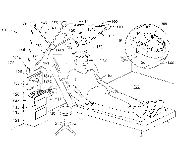
[0027] FIG. 1 is a perspective view of a robotic system having a working
arm and
a grounding arm according to some implementations of the present disclosure;
[0028] FIG. 2 is a perspective view of a portion of the working aim of
the robotic
system of FIG. 1 coupled with a surgical tool;
[0029] FIG. 3 is a perspective view of a portion of the grounding arm of
the
robotic system of FIG. 1;
CA 02970323 2017-03-09
WO 2016/093984 PCMJS2015/058881
- 10 -
[0030] FIG. 4 is a perspective view of a rigid grounding member to be
coupled
with a mouth of a patient for use with the robotic system of FIG. 1;
[0031] FIG. 5. is a perspective view of the rigid grounding member of
FIG. 4
being coupled to a mouth of a patient and a scanner for scanning the mouth of
the patient;
[0032] FIG. 6 is a perspective view of the grounding arm of the robotic
system of
FIG. 1 being coupled with the rigid grounding member that was coupled to the
mouth of the
patient;
[0033] FIG. 7 is a perspective view of the working arm and coupled
surgical tool
being used to perform a procedure in the mouth of the patient;
[0034] FIG. 8A is an illustrative perspective view of the surgical tool
coupled to
the working arm of the robotic system bumping into an invisible bather wall
according to
some implementations of the present disclosure;
[0035] FIG. 8B is the illustrative perspective view of FIG. 8A overlaid
on top of
the perspective view of FIG. 7 illustrating a use of the invisible barrier
wall during the
manual performance of a surgical procedure according to some implementations
of the
present disclosure; and
[0036] FIG. 9 illustrates a modified or post-operative virtual three-
dimensional
model of a mouth of a patient displayed on a display device of the robotic
system of FIG. 1
according to some implementations of the present disclosure.
[0037] While the present disclosure is susceptible to various
modifications and
alternative forms, specific embodiments have been shown by way of example in
the drawings
and will be described in detail herein. It should be understood, however, that
the present
disclosure is not intended to be limited to the particular forms disclosed.
Rather, the present
disclosure is to cover all modifications, equivalents, and alternatives
falling within the spirit
and scope of the present disclosure as defined by the appended claims.
DESCRIPTION OF ILLUSTRATIVE EMBODIMENTS
[0038] Referring to FIG. 1, a robotic system 100 of the present
disclosure can be
used in a variety of manners to perform a variety of surgical and/or non-
surgical procedures.
Once the robotic system 100 is registered to a patient 10 and loaded with a
pre-determined
surgical plan, the robotic system 100 is ready to automatically carry out one
or more surgical
procedures or portions thereof. By automatically, it is meant that the robotic
system can,
without interruption or input from a human (e.g., other than registering the
robotic system
100, loading the pre-determined surgical plan, and in some implementations
hitting a start
button), perform a surgical procedure or portion thereof.
CA 02970323 2017-03-09
WO 2016/093984 PCMJS2015/058881
- 11 -
[0039] Additionally, the robotic system 100 can be manually manipulated
by, for
example, an oral surgeon to be used in performing one or more surgical
procedures.
Manually performing a surgical procedure using the robotic system 100 aids the
surgeon as
compared to performing a manually procedure without the robotic system 100 as
the robotic
system 100 supports the weight of the tools (e.g., tool 155 and surgical tool-
bits 132 coupled
thereto) used by the surgeon during the often lengthy surgery. Further, the
robotic system
100 can be configured to aid the surgeon by preventing the surgeon from moving
a surgical
tool-bit 132 of the robotic system 100 in a manner that is inconsistent with a
general plan or
outline for the procedure. For example, an invisible barrier wall (e.g.,
invisible barrier wall
450 shown in FIGS. 8A and 8B) and/or area can be established prior to a
procedure being
conducted such that the robotic system 100 prevents manual manipulation of the
tool 155 and
tool-bit 132 coupled thereto in a manner that would cause the surgical tool-
bit 132 to move
past the invisible barrier wall/area 450 (e.g., and into a nerve, the wrong
tooth, the palate of
the mouth of the patient, the cheek of the patient, etc.). Further, the
robotic system 100 can
implement haptic feedback to indicate to a manual user 400 (FIGS. 7, 8A, 8B)
of the robotic
system 100 that the user 400 (e.g., the surgeon) is attempting to move the
surgical tool
outside of the accepted working space (e.g., past the invisible barrier
wall/area 450). For
example, the robotic system 100 can vibrate the tool 155 and/or make an
audible noise to
indicate that the surgeon 400 is attempting to move the tool 155 and surgical
tool-bit 132
coupled thereto past the predefined limits for the particular procedure (see
FIGS. 8A and 8B).
Further, the robotic system 100 can improve a surgeon's fidelity by increasing
the resolution
with which the surgeon can operate as compared to a surgeon not using the
robotic system
100. That is, using the robotic system 100, the surgeon is able to move
surgical tools coupled
thereto with a higher degree of accuracy and in relatively smaller increments
(i.e., higher
resolution) as compared to the accuracy and increment size the surgeon can
move surgical
tools without using the robotic system 100.
[0040] Further, during manual manipulation of the robotic system 100,
the robotic
system 100 can monitor movements of the robotic system 100 and develop a
positional data
set indicative of the performed procedure. That is, the robotic system 100 can
trace/follow a
path taken by the tool 155 and/or by the surgical tool-bit 132 coupled thereto
(or at least a tip
portion of the surgical tool-bit 132) and record data indicative of that
traced path. As such,
with knowledge of the geometry of the used surgical tool-bit(s) 132 and an
established origin,
0, (see FIGS. 4 and 8B) for the mouth 12 of the patient 10 and/or of the
robotic system 100,
the final patient situation can be determined (e.g., a post-operative virtual
three-dimensional
CA 02970323 2017-03-09
WO 2016/093984 PCMJS2015/058881
- 12 -
model 326 of at least a portion of the mouth 12 of the patient 10 as modified
during the
procedure as shown in FIG. 9). For example, if the robotic system 100 is used
to remove
bone, the positional data collected by the robotic system 100 would be
indicative of where the
bone removal tool-bit 132 physically traveled/went relative to the established
origin, 0, of the
mouth during the procedure (e.g., which is related to the bone in the mouth 12
of the patient
10) and can thereby be used to develop a post-operative virtual three-
dimensional model (
similar to the post-operative virtual three-dimensional model 326 shown in
FIG. 9) of at least
a portion of the mouth 12 of the patient 10 illustrating the removed bone.
This virtual three-
dimensional model is thus created without having to take a post-operational
intraoral scan of
the patient 10 ¨ sparing the patient 10 this added scanning step.
[0041] Additional details on the robotic system 100 and uses of the
robotic system
100 are described below in various Sections herein. While the disclosure is
broken into these
Sections including various implementations having described elements, any
portion and/or
any element contained in any Section and/or any implementation can be combined
and/or
modified with any portion and/or any element in any other Section and/or any
other
implementation described herein.
Components and General Operation Robotic System 100
[0042] As shown in FIG. 1, the robotic system 100 includes a base 120, a
working
arm 140, and a grounding arm 160. The base 120 can also be referred to as a
cabinet for
housing a multitude of elements therein, where at least some of the elements
housed therein
are coupled together to perform one or more functions/operations. The base 120
rests on a
ground surface 105 (e.g., a floor of a dental surgical room). The base 120 can
be fixed to the
ground surface 105 or moveable with respect to the ground surface 105. In the
implementations when the base 120 is moveable with respect to the ground
surface 105, a
bottom side of the base 120 includes one or more casters or rollers (not
shown).
Alternatively or additionally to the base 120 resting on and/or being coupled
to the ground
surface 105, the base can be coupled to a wall surface 107 (e.g., a wall of
the dental surgical
room). Preferably, the base 120 is mounted to the ground surface 105 and/or
the wall 107 to
aid in preventing the robotic system 100 from tipping over during use thereof.
[0043] The base 120 houses and/or is coupled to/associated with a
computer 121,
a display device 122, input devices 124a, 124b, a surgical tool tray 130, and
storage drawers
135. The computer 121 is communicatively connected with the display device
122, the input
devices 124a, 124b, the working arm 140 and the grounding arm 160. The
computer 121 can
include controllers, processors, memory devices, communication devices (e.g.,
wireless,
CA 02970323 2017-03-09
WO 2016/093984 PCMJS2015/058881
- 13 -
wired, etc.), etc. configured to run/execute one or more software programs
(e.g., robotic
system control software programs, dental surgical planning software programs,
abutment
design software programs, crown design software programs, etc.). The computer
121 is
specially programmed and improved for controlling and/or tracking/tracing the
working arm
140 and/or the grounding arm 160 of the robotic system 100. Alternatively, the
computer
121 can be a general purpose computer capable of executing specific software
to control
and/or track the working arm 140 and/or the grounding arm 160 of the robotic
system 100.
[0044] The input devices 124a, 124b are shown as being a keyboard and a
mouse
for use in operating the computer 121. Additional input devices can be used
such as, for
example, a joystick, a wireless or wired electronic pen, a touch screen
overlaid on the display
device 122, a foot pedal, etc.
[0045] The surgical tool tray 130 stores a multitude of surgical tools
or surgical
tool-bits 132 therein for use in a variety of procedures (e.g., creation of an
osteotomy, implant
placement, bone shaving, tooth-crown prepping, probing/mechanical sensing,
denture
modification, provisional restoration shaping, abutment shaping, etc.). While
only four
surgical tool-bits 132 are shown, any number of surgical tool-bits 132 can be
included in the
surgical tool tray 130 (e.g., one surgical tool-bit, two surgical tool-bits,
ten surgical tool-bits,
thirty surgical tool-bits, etc.). The surgical tool-bits 132 can include drill-
bit tools (e.g., to
create sockets and/or openings in bone), tapping tools (e.g., to create
threads in bone
sockets/openings), dental-implant-driving tools, rotating mill tools, saw
tools, probe tools,
mechanical sensing tool-bits, scalpel/knife tools, or any combination thereof.
In some
implementations, the base 120 stores a multitude of surgical tool trays 130 in
one of the
storage drawers 135. In such implementations, depending on the procedure being
conducted
using the robotic system 100, a user (e.g., oral surgeon, clinician, dental
assistant, etc.)
connects the proper one of the surgical tool trays 130 to the base 120 and
puts any extra
surgical tool trays 130 back into the storage drawer 135 for future use and/or
cleaning. The
surgical tools 132 on the surgical tool tray 130 are arranged in a known
manner such that the
robotic system 100 can cause the working arm 140 to automatically move and
pick-up a
desired one of the surgical tool-bits 132. In some implementations, to avoid
the robotic
system 100 picking-up the wrong surgical tool-bit 132, the surgical tool tray
130 is designed
such that the surgical tool tray 130 can only connect with the base 120 in one
orientation.
Further, in some implementations, each surgical tool-bit 132 is designed to
only be coupled
with one known location on the surgical tool tray 130. Yet in other
implementations, an
CA 02970323 2017-03-09
WO 2016/093984 PCMJS2015/058881
- 14 -
operator of the robotic system 100 manually couples the working arm 140 with a
desired one
of the surgical tool-bits 132.
[0046] The working arm 140 has a first end 141a that extends from the
base 120
and a second opposing end 141b that is configured to be coupled with one of
the surgical
tool-bits 132 via a tool 155 to be used in performing a procedure. Between the
first and the
second ends 141a,b, the working arm 140 includes a multitude of rigid arm
members 145
coupled together by a multitude of flexible joint members 150. Each of the
rigid arm
members 145 can have a fixed length or be capable of having a variable length,
such as, for
example, having a telescoping configuration (not shown). Such a telescoping
configuration
provides added flexibility for the robotic system 100 to reach further from
the base 120
during the performance of a procedure. As shown, the working arm 140 is
directly coupled
to the base 120 via one of the flexible joint members 150, although the
working arm 140 can
be directly coupled to the base 120 via one of the rigid arm members 145
instead. The rigid
arm members 145 can be made of a variety of materials such as, for example,
titanium,
plastic, steel, of any combination thereof.
[0047] Each of the flexible joint members 150 is configured to mate one
of the
rigid arm members 145 to another one of the rigid arm members 145 or to the
base 120. Each
of the flexible joint members 150 includes one or more motors therein for
causing relative
movement of the ones of the rigid arm members 145 mated to the flexible joint
member 150.
For example, each of the flexible joint members 150 includes an electric servo
motor, an
electric rotary motor, etc. As such, in response to a command from the
computer 121, for
example, automatically executing a preplanned surgical procedure, the flexible
joint member
150 can cause one of the rigid anu members 145 to rotate/pivot relative to
another one of the
rigid arm members 145 coupled to the same flexible joint member 150. Depending
on the
number and/or type of motor(s) in the flexible joint member 150, the relative
rotating/pivoting can be one dimensional, two dimensional, or three
dimensional. That is,
one of the rigid arm members 145 can be caused to rotate/pivot forward and
backward
relative to the other one of the rigid arm members 145 (e.g., pitching),
rotate/pivot left and
right relative to the other one of the rigid arm members 145 (e.g., swaying),
and/or
rotate/pivot side-to-side relative to the other one of the rigid arm members
145 (e.g., rolling).
[0048] As shown, the working arm 140 includes four rigid arm members 145
and
four flexible joint members 150. With the ability for each of the four
flexible joint members
150 to rotate/pivot the rigid arm members 145 mated thereto as described
above, the second
end 141b of the working arm 140 and/or the tool 155 is able to move with at
least six degrees
CA 02970323 2017-03-09
WO 2016/093984 PCMJS2015/058881
- 15 -
of freedom relative to the base 120 (e.g., three degrees of rotational freedom
and three
degrees of translational freedom). It is contemplated that the number of rigid
arm members
145 and flexible joint members 150 included in the working arm 140 can vary.
As the
number of the rigid arm members 145 and flexible joint members 150 decreases,
so does the
flexibility of the working arm 140. Similarly, as the number of the rigid arm
members 145
and flexible joint members 150 increases, so does the flexibility of the
working arm 140. For
example, additional fine-tuning-flexible joint members 150 can be included
that have a
relatively smaller range of motion to precisely place the tool 155 and the
surgical tool-bits
132 coupled thereto in the mouth 12 of the patient 10 in a desired location.
Specifically, in
some implementations, the surgical tool-bits 132 can be placed with less than
a 0.5 millimeter
margin of error from a desired target placement. In some other
implementations, the surgical
tool-bits 132 can be placed with less than a 0.1 millimeter margin of error
from a desired
target placement.
[0049] In addition to each of the flexible joint members 150 including
one or
more motors therein, each of the flexible joint members 150 includes one or
more sensors for
sensing the relative positional relationship between the rigid arm member(s)
145 coupled
thereto. That is, for example, each one of the flexible joint members 150
includes one or
more sensors that can determine the relative angular positional relationship
between the two
of the rigid arm members 145 coupled thereto relative to an X-Y-Z space having
its origin at
the center of the flexible joint member 150. Thus, in response to a user of
the robotic system
100 moving one of the rigid arm members 145, the one or more sensors in the
flexible joint
member 150 coupled to the manually moved rigid arm member 145 are configured
to sense
that movement and generate data representative of the new location of the
rigid arm member
145 relative to an origin of the flexible joint member 150 and/or relative to
the origin, 0, of
the robotic system 100 and/or of the mouth 12 of the patient 10. Such data is
transmitted
(wirelessly and/or via a wire) to the computer 121 for processing. For another
example, in
response to an operator 400 (FIG. 7) manually moving the second end 141b
and/or the tool
155 of the working arm 140 such that some or all of the rigid arm members 145
are moved
relative to the base 120, each of the flexible joint members 150 generates
data that is
transmitted to the computer 121 for processing. The computer 121 is configured
to execute a
tracing software program and/or algorithm that is able process the data from
each of the
flexible joint members 150 to determine the location of the second end 141b of
the working
arm 140 (or any portion of any tool 155 and/or surgical tool-bit 132 coupled
thereto) relative
one or more established origins, 0, for the robotic system 100 and/or for the
mouth 12 of the
CA 02970323 2017-03-09
WO 2016/093984 PCMJS2015/058881
- 16 -
patient 10. As such, the robotic system 100 is capable of tracking movement of
the working
arm 140.
[0050] The grounding arm 160 has a first end 161a that extends from the
base 120
and a second opposing end 161b that is configured to be coupled with a rigid
grounding
member 200 (FIG. 4) to establish the origin, 0, for the robotic system 100
and/or for the
mouth 12 of the patient 10. Between the first and the second ends 161a,b, the
grounding arm
160 includes a multitude of rigid arm members 165 coupled together by a
multitude of
flexible joint members 170. The rigid arm members 165 are the same as, or
similar to, the
rigid arm members 145 and the flexible joint members 170 are the same as, or
similar to, the
flexible joint members 150 described herein.
[0051] Now referring to FIG. 2, the second end 141b of the working arm
140 is
shown in detail. Specifically, a portion of two rigid arm members 145a,b
coupled by a
flexible joint member 150 is shown. The rigid arm member 145b has a first end
146a
coupled with the flexible joint member 150 and a second end 146b that is
coupled with a
coupler 147. The coupler 147 is rigidly attached to the second end 146b of the
rigid arm
member 145b and designed to removably receive a portion of the tool 155
therein. As shown
in FIG. 2, the tool 155 is a drill or driving tool for rotating a surgical
tool-bit 132a. Coupling
the tool 155 to the coupler 147 can be purely mechanical and/or electrical.
That is, the
coupling of the tool 155 with the coupler 147 can include supplying power to
the tool 155
through the coupler 147 or the tool 155 can be self-powered (e.g., via one or
more disposable
and/or rechargeable batteries). The connection between the coupler 147 and the
tool 155 can
be a press fit connection, a snap-in connection, a threaded connection, a
magnetic connection,
a friction connection, a tongue and groove connection, or any combination
thereof such that
the tool 155 is removably and securely coupled to the coupler 147 in a known
and repeatable
manner. In some implementations, the coupling of the tool 155 with the working
arm 140
creates a communicative connection where the tool 155 is able to talk with the
computer 121
through the working arm 140, such that the identity of the tool 155 is
disclosed to the
computer 121. As such, the computer learns the identity and size and shape of
the tool 155
which is needed to precisely trace the movements of the tool 155 and any
surgical tool-bit
132 coupled thereto. Alternatively, the working arm 140 is designed to only
connect with a
single tool 155 such that identification of the tool 155 is unnecessary. Yet
in further
alternative implementations, the robotic system 100 runs a check prior to
conducting a
procedure to confirm that the correct tool 155 is coupled to the working arm
140.
CA 02970323 2017-03-09
WO 2016/093984 PCMJS2015/058881
- 17 -
[0052] In some implementations, the tool 155 includes a multitude of
buttons 156.
The buttons 156 can be preprogrammed and/or programmable to start operation of
the tool
155 (e.g., a start/ON button), to end operation of the tool 155 (e.g., a
stop/OFF button), to
reverse rotation of the tool 155 (e.g., forward/backward rotation), etc. One
or more lights
(e.g., LEDs) can be included in the tool 155 and positioned adjacent to each
button 156 to
indicate whether the button 156 is activated or not. In some implementations,
the tool 155
includes a release button or mechanism 157 for use in aiding in the removal of
the surgical
tool-bit 132a from the tool 155. In other implementations, the surgical tool-
bit 132a can
simply be pulled out of the tool 155 without having to press the release
button 157.
[0053] Now referring to FIG. 3, the second end 161b of the grounding arm
160 is
shown in detail. Specifically, a portion of a rigid arm member 165a is coupled
by a flexible
joint member 170 to a grounding probe 180. The grounding probe 180 has a first
end 180a
coupled with the flexible joint member 170 and a second end 180b designed to
couple with
the rigid grounding member 200 (FIG. 4) in a removable and repeatable fashion.
As shown
in FIG. 3, the second end 180b of the grounding probe 180 includes a pair of
biased locking
bearings 182a,b. The locking bearings 182a,b are biased outward away from a
central axis,
YC, of the second end 180b of the grounding probe 180. As such, the locking
bearings
182a,b can be forced inward toward the central axis, YC, to allow the second
end 180b of the
grounding probe 180 to be slid into a receiving bore 222 of the rigid
grounding member 200
(FIG. 4) in a removable and repeatable fashion.
Registering the Robotic System 100 to the Mouth 12 of the Patient 10 Using the
Rigid
Grounding Member 200
[0054] Referring to FIG. 4, the rigid grounding member 200 is shown as
including
a body 205 and a coupling post 220. The body 205 includes a first leg 207a and
a second leg
207b coupled together by a base 209 therebetween. The base has an upper
surface 209a and a
bottom surface 209b. Protruding generally upward from the upper surface 209a
of the base
209 is the coupling post 220. The body 205 and the coupling post 220 can be
two separate
and distinct parts that are attached together or they can be integrally formed
as a monolithic
component. The coupling post 220 includes the receiving bore 222 therein for
receiving the
second end 180b of the grounding probe 180 therethrough when grounding the
grounding
arm 160 of the robotic system 100 to the mouth 12 of the patient 10 (e.g.,
when the rigid
grounding member 200 is installed in the mouth 12 of the patient 10). The
receiving bore
222 has a central axis, XC, along which the origin, 0, for the robotic system
100 and/or the
mouth 12 of the patient 10 can be established. In some implementations, the
origin, 0, is
CA 02970323 2017-03-09
WO 2016/093984 PCMJS2015/058881
- 18 -
established at a center of the receiving bore along the central axis, XC. In
some alternative
implementations, the origin, 0, is established at any point along the central
axis, XC, for
example, at either end opening of the receiving bore 222.
[0055] The first leg 207a includes a threaded throughbore 208 that
receives a
fastener 230 (e.g., surgical dental screw, dental bolt, etc.). While not
shown, the second leg
207b can also include a threaded throughbore the same as, or similar to, the
threaded
throughbore 208 for receiving the fastener 230 therein and/or another fastener
(not shown).
While the rigid grounding member 200 is shown as having a specific shape and
size, various
other shapes, sizes, and arrangements for the rigid grounding member 200 are
contemplated,
such that the rigid grounding member 200 can be attached to the mouth 12 of
the patient 10
and provide a means for coupling the grounding probe 180 thereto in a
removable and
repeatable fashion.
[0056] According to some implementations, in order to register the
robotic system
100 to the mouth 12 of the patient 10 and establish the origin, 0, for use
during a procedure
using the robotic system 100, the rigid grounding member 200 is installed into
the mouth 12
of the patient 10. As shown in FIG. 5, the mouth 12 of the patient 10 includes
teeth 14, a
jawbone 16, and soft tissue 18 (e.g., gingival tissue), where tooth 14a is
selected to be
coupled with the rigid grounding member 200. As shown in FIG. 6, the mouth 12
further
includes a surgical site 20 (e.g., location to receive a dental implant,
permanent abutment, and
permanent crown), where teeth 14b and 14c are positioned adjacent to the
surgical site 20.
[0057] While the rigid grounding member 200 is attached to the tooth
14a, the
rigid grounding member 200 can be attached to any of the other teeth 14 in the
mouth 12
and/or to the jawbone 16 of the patient 10. To attach the rigid grounding
member 200 to the
tooth 14a, the rigid grounding member 200 is placed on the tooth 14a.
Specifically, for
example, the rigid grounding member 200 is positioned in the mouth 12 of the
patient 10
such that (i) the bottom surface 209b of the base 209 is positioned generally
adjacent to an
occlusal surface of the tooth 14a, (ii) the first leg 207a is positioned
generally adjacent to a
lingual surface of the tooth 14a, and (iii) the second leg 207b is positioned
generally adjacent
to a buccal surface of the tooth 14a, as shown in FIG. 5. With the rigid
grounding member
200 so positioned, the fastener 230 is threaded into the threaded throughbore
208 of the first
leg 207a to rigidly lock the rigid grounding member 200 in place against the
tooth 14a. In
some implementations, the fastener 230 is tightened such that the fastener 230
does not
penetrate the tooth 14a, the soft tissue 18, and/or the jawbone 16. However,
in some
CA 02970323 2017-03-09
WO 2016/093984 PCMJS2015/058881
- 19 -
alternative implementations, the fastener 230 does penetrate the tooth 14a,
the soft tissue 18,
the jawbone 16, or any combination thereof.
[0058] With the rigid grounding member 200 attached to the tooth 14a, a
scan of
the mouth 12 of the patient 10 is taken using a scanner/camera 300 as shown in
FIG. 5.
Depending on the type of procedure being planned for implementation using the
robotic
system 100, the scan of the mouth 12 can be an intraoral surface scan using an
intraoral
scanner, a CT scan using a CT scanner (such as, for example, a cone-beam
computed
tomography (CBCT) scanner also known as a dental CBCT scanner), or a
combination
thereof. The scanning of the mouth 12 with the rigid grounding member 200
therein, allows
for the generation of the pre-operative three-dimensional virtual model 325
(FIG. 1) of the
mouth 12 of the patient 10 to be generated. Such a virtual model can be used
for developing
a surgical plan and/or for designing components to be mated on a dental
implant installed in
the mouth 12 of the patient 10. Specifically, the virtual model 325 includes a
virtual rigid
grounding member 200' (FIG. 1) that is used to establish the origin, 0, of a
coordinate system
to be used by the robotic system 100 when conducting a procedure.
[0059] After the scanning of the mouth 12, the robotic system 100 is
grounded to
the mouth 12 of the patient 10. Specifically, the second end 180b of the
grounding probe 180
is mated with the rigid grounding member 200 by positioning and/or sliding the
second end
180b of the grounding probe 180 into the receiving bore 222 of the coupling
post 220 of the
rigid grounding member 200. The second end 180b is slid until the locking
bearings 182a,b
are slid through and outside of the receiving bore 222 and biased outward away
from the
central axis, YC, (FIG. 3) such that the grounding probe 180 cannot readily be
removed from
the receiving bore 222. In some implementations, tactile feedback is used to
indicate that the
grounding probe 180 is fully engaged with the rigid grounding member 200 and
properly
positioned therein. For example, when the grounding probe 180 is fully
engaged, the locking
bearings 182a,b can snap into place making an audible sound and/or a vibrating
click.
[0060] By readily be removed, it is meant that to remove the grounding
probe 180
from its engagement with the rigid grounding member 200, the locking bearings
182a,b need
to be actuated by being pressed inward towards the central axis, YC, to allow
the grounding
probe 180 to be slid out of the receiving bore 222. Alternatively, the
grounding probe 180
can be removed from its engagement with the rigid grounding member 200 by
pulling the
grounding probe 180 with a sufficient amount of force to overcome the biasing
force of the
locking bearings 182a,b, thereby causing the locking bearings 182a,b to be
forced inward
CA 02970323 2017-03-09
WO 2016/093984 PCMJS2015/058881
- 20 -
towards the central axis, YC, and allow the removal of the grounding probe 180
from
engagement with the rigid grounding member 200.
[0061] As best shown in FIG. 6, a length, L180b, of the second end 180b
of the
grounding probe 180 is designed such that once the grounding probe 180 is
properly engaged
with the coupling post 220 of the rigid grounding member 200, the grounding
probe 180 does
not have any or very little translational play therein. That means that the
grounding probe
180 cannot slide and/or move laterally (e.g., translation) along the central
axis, YC, when the
grounding probe 180 is engaged with the rigid grounding member 200. However,
the
grounding probe 180 is allowed to rotate within the receiving bore 222 about
the central axis,
YC. Such a connection between the grounding probe 180 and the rigid grounding
member
200 aids the robotic system 100 in maintaining the known origin, 0, based on
the location of
the rigid grounding member 200 in the mouth 12 of the patient 10.
[0062] With the robotic system 100 grounded to the mouth 12 of the
patient 10
via the rigid grounding member 200 and the grounding probe 180 (coupled to the
grounding
arm 160 of the robotic system 100), the robotic system 100 is ready to
automatically
implement a preplanned procedure and/or to allow an operator 400 to perform a
manual
and/or a semi-manual procedure that is tracked/traced by the robotic system
100. In the case
of using the robotic system 100 to automatically implement/conduct a
preplanned procedure,
the preplanned procedure must first be planned, for example, using a dental
surgical planning
software program, as described in the following Section.
Planning a Surgical Plan for Automatic Implementation by the Robotic System
100
[0063] As discussed above, the mouth 12 of the patient 10 is scanned
with the
rigid grounding member 200 therein. The scan data generated from that scan
(e.g., intraoral
surface scan, CT scan, dental CBCT scan, X-ray scan, a combination thereof,
etc.) is
imported and/or sent to the computer 121, or another computer, that executes
and/or runs a
dental surgical planning software program. As shown in FIG. 1, the surgical
planning
software program is designed to display on the display device 122 the pre-
operative three-
dimensional virtual model 325 of at least a portion of the mouth 12 of the
patient 10. The
pre-operative three-dimensional virtual model of the virtual mouth 12'
includes virtual teeth
14', virtual soft tissue 18', a virtual surgical site 20', a virtual rigid
grounding member 200',
and in some implementations where a dental CBCT scan and/or X-ray scan is
taken, virtual
bone 16' (e.g., jawbone), which correspond with the teeth 14, the soft tissue
18, the surgical
site 20, the rigid grounding member 200, and the bone 16 in the actual mouth
12 of the
patient 10.
CA 02970323 2017-03-09
WO 2016/093984 PCMJS2015/058881
- 21 -
[0064] Depending on the type of procedure that is to be conducted in the
mouth
12 of the patient 10, the dental surgical planning software program is
designed to
automatically and/or with some input from an operator (e.g., operator 400
shown in FIG. 7),
develop a surgical plan for conducting the desired procedure automatically
using one or more
of the surgical tool-bits 132 attached to the tool 155 of the working arm 140
of the robotic
system 100. Specifically, the dental surgical planning software program is
designed to
develop a set of instructions and/or movements for the working arm 140 to make
relative to
the grounding arm 160 (e.g., relative to the second end 180b of the grounding
probe 180),
which is positioned at a known location relative to the rigid grounding member
200 in the
mouth 12 of the patient 10 (e.g., the established origin, 0 along the central
axis, XC, shown
in FIG. 4). The developed set of instructions includes directions as to what
type and/or size
of tool or tools to be used during the surgical procedure. The developed set
of instructions
also includes an order for using the tools when multiple tools are to be used.
For example, if
the desired procedure is an osteotomy, the developed instructions include
which surgical
drill-bit tool or tools 132 to use. Specifically, in an exemplary osteotomy,
the instructions
may direct the robotic system 100 to first use a first surgical drill-bit tool
132 having a first
diameter and a first length to start the procedure (e.g., to create an initial
socket/opening in
the jawbone), followed by an instruction for the robotic system 100 to switch
to a second
surgical drill-bit tool 132 having a second (e.g., relatively larger) diameter
and a second
length (e.g., the same as or different than the first length) to continue the
procedure (e.g., to
enlarge a diameter of the created socket/opening in the jawbone), etc. A
subsequent
instruction may include for the robotic system 100 to switch to a surgical
tapping tool 132 to
tap (e.g., create threads) the previously created socket/opening in the
jawbone. In some
implementations, the surgical system 100 automatically and without operator
input switches
the surgical tool-bits 132 to the tool 155; and in some alternative
implementations, an
operator manually attaches and detaches the surgical tool-bits 132 to the tool
155.
[0065] The development of the surgical plan by the dental surgical
planning
software program can be completely autonomous after receipt of the scan data
(e.g., pre-
operative virtual three-dimensional model 325), or the development can involve
some input
from an operator. For example, the dental surgical planning software program
may require
an input from the operator such as the type of procedure to be planned (e.g.,
creation of an
osteotomy, implant placement, bone shaving, etc.), the location and
orientation for a desired
central axis of the implant to be placed (e.g., to avoid a nerve in the mouth
12 and/or to avoid
CA 02970323 2017-03-09
WO 2016/093984 PCMJS2015/058881
- 22 -
an area of low density bone in the mouth 12), the manufacturer of the implant
to be placed,
the size of the implant to be placed, etc.
[0066] Once the surgical plan is developed using the dental surgical
planning
software program, the robotic system 100 is ready to conduct/implement the
surgical plan
using one or more of the surgical tool-bits 132. In some implementations, the
computer 121
that controls the working arm 140 and the grounding arm 160 also executes and
runs the
surgical planning software program. In such implementations, the developed
surgical plan is
automatically loaded and ready to be automatically implemented by the robotic
system 100,
for example, in response to an operator clicking a start button on the display
device 122. In
alternative implementations where a different computer executes/runs the
dental surgical
planning software program, the developed surgical plan needs to be sent to
and/or loaded into
the robotic system 100.
[0067] As shown in FIG. 7, after the surgical plan is initiated, the
working arm
140 moves into place (e.g., at least partially into the mouth 12 of the
patient 10) relative to
the grounding arm 160 and starts to perform the procedure automatically at the
surgical site
20 using one or more of the surgical tool-bits 132.
Defining the Invisible Barrier Wall/Area 450 for Confining Movement of the
Robotic
System 100
[0068] As discussed herein, instead of having the robotic system 100
conduct a
surgical procedure automatically, the robotic system 100 can be used manually
such that the
operator 400 (FIGS. 7, 8A, 8B) causes the working arm 140 and the attached
surgical tool-bit
132 to move relative to the patient 10. In such implementations, the invisible
barrier
wall/area 450 can be established prior to conducting the procedure. As shown
in FIG. 8A,
the invisible barrier wall/area 450 is shown without the mouth 12 of the
patient 10 for
illustrative purposes, whereas FIG. 8B illustrates the same invisible barrier
wall/area 450
overlaid in the mouth 12 of the patient 10.
[0069] The invisible barrier wall/area 450 can be established and/or
developed
using the surgical planning software program described herein as part of a
surgical plan. In
such implementations, for a semi-manual procedure to be conducted by an
operator using
(e.g., manually manipulating) the robotic system 100, the invisible barrier
wall/area 450
establishes a boundary for the working arm 140, and specifically the surgical
tool-bit 132
coupled thereto, when the surgical tool-bit 132 is near the patient 10 and/or
positioned inside
of the mouth 12 of the patient 10. As such, the invisible barrier wall/area
450 can aid in
preventing the operator (e.g., oral surgeon) from moving the tool 155 in a
manner that would
CA 02970323 2017-03-09
WO 2016/093984 PCT/US2015/058881
- 23 -
cause the surgical tool-bit 132 and/or any portion of the working arm 140
(e.g., including the
tool 155) to interfere with the mouth 12 of the patient 10.
[0070] By interfere, it is meant, for example, that working arm 140 is
bumped into
teeth 14 of the patient 10 not being addressed by the procedure being
conducted. For another
example, by interfere, it is meant that the surgical tool-bit 132 is moved
(e.g., vertically up
and/or down, side-to-side, etc.) too far into the jawbone 16 of the patient 10
when, for
example, the robotic system 100 is being used to perform a semi-manual
procedure, such as,
for example, creation of an osteotomy. As such, when the operator 400 is
manually creating
the osteotomy by manually moving the working arm 140, the robotic system 100
implementing the invisible barrier wall/area 450 allows the operator 400 to
move the surgical
tool-bit 132 as desired (e.g., vertically up and/or down, side-to-side, etc.)
within the bounds
of the invisible banier wall/area 450, but if the operator 400 attempts to
move the surgical
tool-bit 132 outside of and/or past the invisible barrier wall/area 450, then
the robotic system
100 actively prevents such a movement using, for example, the motors in the
flexible joint
members 150. For example, as illustrated in FIGS. 8A and 8B, the operator 400
is attempting
to cause the surgical tool-bit 132 to be moved in the direction of arrow A
(e.g., downward
relative to the orientation of FIG. 8B) through the invisible barrier
wall/area 450. However,
because the surgical system 100 is actively enforcing the invisible barrier
wall/area 450, the
surgical system 100 causes the working arm 140, the tool 155, the surgical
tool-bit 132, or
any combination thereof, to vibrate (e.g., tactile feedback) indicating that
the operator is
attempting to move the surgical tool 132 past the invisible barrier wall/area
450, which is not
allowed. Use of such an invisible barrier wall/area 450 can be used, for
example, to allow an
operator (e.g., dental surgeon) to perform a free-form-shaped osteotomy where
the operator is
permitted to sculpt the osteotomy (e.g., move a surgical rotating mill tool
132 vertically up
and/or down and side-to-side) - as opposed to using a set of stepped drill
bits that are only
moved vertically along a single central axis. As such, the operator can in
real-time manually
modify/update a surgical plan based on the received tactile feedback and/or
other information
(e.g., a level of torque needed to maintain a desired or preset drill speed
during the procedure,
a level of force needed to advance the surgical tool-bit 132 during the
procedure, etc.).
[0071] While the invisible barrier wall/area 450 is shown as having an
open top,
alternative invisible barrier wall/areas can have invisible walls/invisible
surfaces on all sides
such that at least a portion of the working arm 140 (e.g., a tip of the
surgical tool-bit 132) is
completely enclosed within the invisible barrier wall/area during the entire
procedure being
performed using the robotic system 100. As such, the invisible barrier
wall/area can extend
CA 02970323 2017-03-09
WO 2016/093984 PCMJS2015/058881
- 24 -
outside of the mouth 12 of the patient 10. Such an invisible barrier wall/area
can aid in
preventing a portion of the working arm 140 from being bumped into the face or
head or
chest of the patient 10, among other things.
[0072] The invisible barrier wall/area 450 is shown in FIGS. 8A and 8B
in dashed
lines to indicate that the invisible barrier wall/area 450 is not actually
visible to the operator
of the robotic system 100 in the mouth 12 of the patient 10 during the
procedure. However,
in some implementations, a virtual representation of the invisible barrier
wall/area 450 can be
displayed on the display device 122 and/or on another display device during
the procedure.
For example, a virtual representation of the invisible barrier wall/area 450
can be displayed
relative to a virtual model (e.g., the pre-operative three-dimensional virtual
model 325) of the
mouth 12 of the patient. For another example, a virtual representation of the
invisible barrier
wall/area 450 can be displayed relative to a live video feed of the mouth 12
of the patient 10.
For yet another example, a virtual representation of the invisible barrier
wall/area 450 can be
displayed relative to a picture of the mouth 12 of the patient 10. The
displaying of a virtual
representation of the invisible barrier wall/area 450 allows the operator to
visualize the
invisible barrier wall/area 450 relative to the mouth 12 of the patient 10,
which can aid in the
operator conducting and/or monitoring/supervising the procedure.
Monitoring/Tracing a Semi-Manual Procedure Using the Robotic System 100
[0073] The robotic system 100 can be used to perform a semi-manual
procedure.
By semi-manual it is meant that the robotic system 100 allows the operator 400
(FIGS. 7, 8A,
8B) of the robotic system 100 to manually manipulate/move the working arm 140
relative to
the mouth 12 of the patient 10 to perform a procedure while at the same time
maintain a level
of control. For example, the robotic system 100 maintains control of the
working arm 140
and the grounding arm 160 by supporting its own weight and/or
monitoring/sensing the
positions of the working arm 140 and the grounding arm 160 relative to the
established
origin, 0 (FIG. 8B).
[0074] As discussed herein, the working arm 140 includes a multitude of
flexible
joint members 150 and the grounding arm 160 includes a multitude of flexible
joint members
170, where each of the flexible joint members 150, 170 includes one or more
sensors for use
in determining a position of at least a portion of the working arm 140 (e.g.,
a tip of the
surgical tool-bit 132, etc.) and/or the grounding arm 160 (e.g., a tip of the
grounding probe
180). For example, as the operator 400 moves the tool 155 (FIG. 8B), the
sensors in the
flexible joint members 150, 170 record data that is transmitted to the
computer 121 for
CA 02970323 2017-03-09
WO 2016/093984 PCMJS2015/058881
- 25 -
processing. The computer 121 runs/executes robotic control software that
receives the data
from the sensors and processes that data to determine a location of at least a
portion of the
working arm 140 (e.g., a tip of the surgical tool-bit 132) relative to the
established origin, 0
(FIG. 8B).
[0075] For example, as shown in FIG. 8B, the origin, 0, for the robotic
system
100 is established as a central point along the central axis, XC, of the
receiving bore 222 of
the coupling post 220 and along the central axis, YC, of the second end 180b
of the
grounding probe 180. Alternatively, the origin, 0, can be established at any
location relative
to the rigid grounding member 200, as the rigid grounding member 200 is fixed
relative to the
rest of the mouth 12 of the patient 10 in a known and recorded fashion (e.g.,
known and
recorded during the scanning of the mouth 12 with the rigid grounding member
200 therein
as described herein) when installed in the mouth 12.
[0076] As the operator 400 conducts a semi-manual procedure, with the
robotic
system 100 tracking/tracing every movement of the working arm 140 (e.g.,
specifically
tracing every move of a tip of the surgical tool-bit 132) using the sensors in
the flexible joint
members 150, 170, data about the location of the working arm 140 (and the
surgical tool-bit
132) is generated and stored in the computer 121 or another computer and/or
memory device.
The computer 121 can execute and/or run three-dimensional modeling software
(e.g., that is
the same as the dental surgical planning software program described elsewhere
herein or a
different software program) that is programmed to create a modified or post-
operative virtual
three-dimensional model 326 (FIG. 9) of at least a portion of the mouth 12 of
the patient 10.
The post-operative virtual three-dimensional model 326 is based on (i) the pre-
operative
three-dimensional virtual model 325 (FIG. 1) of the mouth 12 and (ii) the data
collected
regarding the positions of the working arm 140 during the procedure.
Specifically, the three-
dimensional modeling software is designed to take the pre-operative three-
dimensional
virtual model 325 of the mouth 12 of the patient 10 and to modify the pre-
operative three-
dimensional virtual model 325 based on the procedure conducted using the
robotic system
100. As such, the post-operative three-dimensional virtual model 326 of the at
least a portion
of the virtual mouth 12' includes the virtual teeth 14', the virtual soft
tissue 18', the virtual
surgical site 20', the virtual rigid grounding member 200', and in some
implementations that
took a CT scan (e.g., a dental CBCT scan), virtual bone 16' (e.g., jawbone),
which are in the
pre-operative three-dimensional virtual model 325. However, depending on the
procedure
and the data collected during the same, the post-operative three-dimensional
virtual model
326 also includes modifications at the surgical site 20'.
CA 02970323 2017-03-09
WO 2016/093984 PCMJS2015/058881
- 26 -
[0077] For example, as shown in FIG. 9, if the operator 400 creates an
osteotomy
using a surgical drill-bit tool 132 while the robotic system 100 tracked the
procedure, the
tracking of the positions of the working arm 140 produces data indicative of
the location of
the surgical drill-bit tool 132 used during the procedure relative to the soft
tissue 18 and
jawbone 16 in the mouth 12 of the patient 10 relative to the rigid grounding
member 200 (and
thus the established origin, 0). As such, the three-dimensional modeling
software is able to
identify how the surgical drill-bit tool or tools 132 interfered with the pre-
operative three-
dimensional virtual model 325 and update the pre-operative three-dimensional
virtual model
325 by removing portions thereof based on the interference to result in the
post-operative
three-dimensional virtual model 326 including a virtual opening or socket 25'
in the soft
tissue 18' and/or in the bone 16'. That is, the interference of the surgical
drill-bit tool or tools
132 indicates that the surgical drill-bit tool or tools 132 were used to drill
and/or remove
material resulting in the illustrated virtual opening or socket 25'. As such,
the three-
dimensional modeling software is able to modify the pre-operative three-
dimensional virtual
model 325 by removing the material that corresponds to the material that was
actually
removed from the mouth 12 of the patient 10 during the procedure. The result
is the modified
or post-operative three-dimensional virtual model 326 of the mouth 12 of the
patient 10,
which depicts the post-operative situation in the mouth 12 of the patient 10
(e.g., with the
removed soft tissue and/or jawbone material). Thus, it is not necessary for
the operator 400
to take a post-operative scan of the mouth 12 of the patient 10 to acquire the
post-operative
patient situation. The patient 10 is spared this extra scanning step.
[0078] Further, for another example, as shown in FIG. 9, if the operator
400
placed a dental implant using an implant driving tool 132 while the robotic
system 100
tracked the procedure, the tracking of the positions of the working arm 140
produces data
indicative of the location of the implant driving tool (and thus the position
of the dental
implant coupled thereto) relative to the rigid grounding member 200 (and thus
the established
origin, 0). As such, the three-dimensional modeling software is able to
identify how the
implant driving tool (and the dental implant coupled thereto) interfered with
and/or moved
relative to the pre-operative three-dimensional virtual model 325 and update
the pre-operative
three-dimensional virtual model 325 based on the interference to result in the
post-operative
three-dimensional virtual model 326 including a virtual dental implant 30'
placed in the
virtual opening or socket 25' at a location and orientation that corresponds
with the placement
and orientation of the actual dental implant installed in the mouth 12 of the
patient 10 using
the robotic system 100. Thus, it is not necessary for the operator 400 to take
a post-operative
CA 02970323 2017-03-09
WO 2016/093984 PCMJS2015/058881
- 27 -
scan of the mouth 12 of the patient 10 to acquire the post-operative patient
situation including
the location and orientation of the dental implant installed therein. The
patient 10 is spared
this extra scanning step.
[0079] The
tracking/monitoring of the position of the working arm 140 and/or
grounding arm 160 during a semi-manual and/or manual procedure conducted using
the
robotic system 100 can be used to create a variety of modified or post-
operative virtual three-
dimensional models of at least a portion of the mouth 12 of the patient 10 in
addition to the
ones described herein and shown in FIG. 9. For example, modified or post-
operative virtual
three-dimensional models can be automatically generated by, for example, the
computer 121
running the three-dimensional modeling software after a conventional dentistry
tooth-prep is
performed using the robotic system, after a temporary prosthesis is shaped
using the robotic
system 100, after a denture is modified using the robotic system 100, etc.
Various other
manual/semi-manual procedures, such as the procedures described elsewhere
herein, can be
tracked/monitored as described above for generating positional data for use in
generating
modified or post-operative virtual three-dimensional models, thereby avoiding
post-operative
scans (e.g., post-operative intraoral surface scans, post-operative dental
CBCT scan, etc.).
Exemplary Uses of the Robotic System 100
[0080] The
robotic system 100 of the present disclosure can be used to conduct a
variety of procedures automatically, manually, or a combination thereof. One
example of a
procedure performable by the robotic system 100 is the creating of an
osteotomy. The
creating of the osteotomy is performed using one or more surgical drill-bit
tools 132 coupled
to the robotic system 100 to form a socket and/or opening in a jawbone 16 of a
patient 10 that
is suitable for receiving a dental implant therein. Additionally, in some
implementations, the
performing of the osteotomy also uses one or more surgical tapping tools 132
coupled to the
robotic system 100 to form threads in the previously formed socket/opening in
the jawbone
16 of the patient 10 that is suitable for receiving the dental implant
therein. A second
example of a procedure performable by the robotic system 100 is the installing
of a dental
implant. The installing of the dental implant is performed using an implant
driving tool 132
coupled to the robotic system 100 to install the dental implant in an opening
in a jawbone 16
of a patient 10. A third example of a procedure performable by the robotic
system 100 is the
performing of an alveolectomy. The
alveolectomy is performed using a bone
shaving/removal tool 132 coupled to the robotic system 100 to removal bone
material from a
jawbone 16 of a patient 10. A fourth example of a procedure performable by the
robotic
system 100 is the performing of an alveoloplasty. The alveoloplasty is
performed using a
CA 02970323 2017-03-09
WO 2016/093984 PCMJS2015/058881
- 28 -
surgical bone contouring tool 132 coupled to the robotic system 100 to contour
a portion of a
jawbone 16 of a patient 10. A fifth example of a procedure performable by the
robotic
system 100 is the developing of a tooth-crown-prep. The development of a tooth-
crown-prep
is performed using one or more surgical dental drill-bit tools 132 and/or
dental shaver tool-
bits 132 to remove portions of a tooth (e.g., occlusal portions) such that the
tooth is suitable
for receiving a custom crown thereon. A sixth example of a procedure
performable by the
robotic system 100 is the modifying of a denture for use as part of a hybrid
prosthesis. A
hybrid prosthesis includes a modified denture bonded with one or more
abutments or
cylinders. Typically, the modified denture is coupled to the dental implants
via the one or
more abutments or cylinders, which attach to respective ones of the dental
implants. The
robotic system 100 can be used to aid in the creation of the modified denture
by precisely
drilling holes in the denture in locations that directly correspond with
central axes of the
dental implants installed in the mouth of a patient 10, which were installed
using the robotic
system 100. As such, when the one or more abutments or cylinders are attached
to the dental
implants, the drilled holes in the denture match up with (i.e., are aligned
with) the abutments
or cylinders and can be bonded thereto to create the hybrid prosthesis. A
seventh example of
a procedure performable by the robotic system 100 is the creating of a
provisional restoration.
The creating of the provisional restoration is performed using one or more
shaver and/or
sculpting tool-bits 132 to remove portions of a tooth blank such that the
tooth blank is
sculpted to have an anatomical tooth shape for attachment to an installed
dental implant.
[0081] It is contemplated that any of the above seven examples and any
of the
following disclosed implementations, can be performed automatically by the
robotic system
100 loaded with a surgical plan with no or little input from an operator
(e.g., except for
selecting of a start procedure button), or performed manually and/or semi-
manually by an
operator 400 that manually manipulates the working arm 140 of the robotic
system 100 that is
either constrained by or free from an invisible barrier wall/area 450. It is
further
contemplated that any element and/or step from any of the following
implementations can be
removed and/or replaced with any other disclosed element in any of the other
disclosed
implementations.
[0082] According to a first implementation of the disclosed concepts of
the
present disclosure, the robotic system 100 is used to install a dental implant
in the mouth 12
of the patient 10. The robotic system 100 includes the base 120, the grounding
arm 160, the
working arm 140, and one or more sensors. The grounding arm 160 has a first
end 161a and
a second end 161b. The first end 161a of the grounding arm 160 is coupled to
the base 120
CA 02970323 2017-03-09
WO 2016/093984 PCMJS2015/058881
- 29 -
and the second end 161b of the grounding arm 160 is configured to be coupled
to a fixed
structure in the mouth 12 of the patient 10 for establishing the origin, 0,
for the robotic
system 100 relative to the mouth 12 of the patient 10. In such
implementations, the second
end 161b of the grounding arm 160 has at least six degrees of freedom relative
to the base
120. The working arm 140 has a first end 141a and a second end141b. The first
end 141a of
the working arm 140 extends from the base 120 and the second end 141b of the
working arm
140 is configured to be coupled with one or more of the surgical tools 132 for
use during
installation of the dental implant in the mouth 12 of the patient 10. A
portion of the working
arm 140 has at least six degrees of freedom relative to the base 120 and is
moveable to (i)
form an opening in the bone 16 within the mouth 12 of the patient 10 and (ii)
install the
dental implant in the formed opening. The one or more sensors (e.g., the
sensors in the
flexible joint members 150, 170) monitor the positions of the grounding arm
160 and the
working arm 140 and generate positional data that is used to create a post-
operative virtual
three-dimensional implant level model (e.g., model 326 of FIG. 9) of at least
a portion of the
mouth 12 of the patient 10.
[0083] In the first implementation, the post-operative virtual three-
dimensional
implant level model is created without use of a scanning abutment coupled to
the dental
implant installed in the mouth 12 of the patient 10. Further, the moveable
portion of the
working arm 140 is (i) manually-movable by the operator 400 of the robotic
system 100, (ii)
automatically moveable by one or more motors (e.g., motors in the flexible
joint members
150, 170) of the robotic system 100, or (iii) both. The fixed structure in the
mouth 12 of the
patient 10 is one or more teeth, jawbone, the rigid grounding member 200, or
any
combination thereof.
[0084] According to a second implementation of the disclosed concepts of
the
present disclosure, a method of creating the post-operative virtual three-
dimensional model
326 (FIG. 9) of at least a portion of the mouth 12 of the patient 10, where
the mouth 12
includes a dental implant installed using the robotic system 100 during a
dental surgical
procedure, includes attaching the rigid grounding member 200 to a fixed
position within the
mouth 12 of the patient 10. The method also includes obtaining the pre-
operative virtual
three-dimensional model 325 (FIG. 1) of the mouth 12 of the patient 10 with
the rigid
grounding member 200 therein. The method further includes, coupling the
grounding arm
160 of the robotic system 100 to the rigid grounding member 200 in the mouth
12 of the
patient 10, thereby establishing the origin, 0, (FIG. 8B) for the mouth 12 of
the patient 10.
The method also includes moving (e.g., automatically or manually, or a
combination of both),
CA 02970323 2017-03-09
WO 2016/093984 PCMJS2015/058881
- 30 -
as part of the dental surgical procedure, at least a portion of the working
arm 140 of the
robotic system 100 coupled to the dental-implant-driving tool 132 to install
the dental implant
in the mouth 12 of the patient 10. The method also includes monitoring, during
the dental
surgical procedure, a position of the grounding arm 160 and the working arm
140 to generate
positional data related to the location of the dental-implant-driving tool 132
relative to the
established origin, 0 and creating the post-operative virtual three-
dimensional model 326
(FIG. 9) of the at least a portion of the mouth 12 of the patient 10 based on
the obtained pre-
operative virtual three-dimensional model 325 (FIG. 1) and the generated
positional data.
[0085] In the second implementation, the creating the post-operative
virtual three-
dimensional model 326 (FIG. 9) occurs without use of a scanning abutment
coupled to the
installed dental implant. Further, the creating includes modifying the pre-
operative virtual
three-dimensional model 325 of the mouth 12 of the patient 10 to include at
least a portion of
a virtual model of the dental implant (e.g., virtual dental implant 30' shown
in FIG. 9)
installed in the mouth 12 of the patient 10. In some versions of the second
implementation,
the method further includes prior to the moving, determining the invisible
boundary wall/area
450 (FIGS. 8A and 8B) to be established around a pre-determined location
(e.g., the surgical
site 20 shown in FIGS. 6 and 7) for the dental implant to be installed in the
mouth 12 of the
patient 10 and during the moving, automatically enforcing the determined
invisible boundary
wall/area 450 by preventing the working arm 140 of the robotic system 100 from
being
moved in a manner that would cause the dental implant to be installed outside
of the
determined invisible boundary wall/area 450.
[0086] According to a third implementation of the disclosed concepts of
the
present disclosure, a method of automatically shaving alveolar bone in the
mouth 12 of the
patient 10 using the robotic system100 includes attaching the rigid grounding
member 200 to
a fixed position (e.g., tooth 14a shown in FIG 6) within the mouth 12 of the
patient 10. The
method further includes obtaining a pre-shaved virtual model (e.g., the same
as, or similar to,
the pre-operative three-dimensional virtual model 325 shown in FIG. 1) of the
mouth 12 of
the patient 10 with the rigid grounding member 200 therein. The method further
includes
coupling the grounding arm 160 of the robotic system 100 to the rigid
grounding member 200
in the mouth 12 of the patient 10, thereby establishing the origin, 0, (FIG.
8B) for the mouth
12 of the patient 10. The method further includes developing a plan for
automatically
moving a bone-cutting tool 132 relative to the established origin, 0, to shave
a portion of the
alveolar bone in the mouth 12 of the patient 10. The method further includes
attaching the
bone-cutting tool 132 to the working arm 140 of the robotic system 100 and
executing the
CA 02970323 2017-03-09
WO 2016/093984 PCMJS2015/058881
-31 -
developed plan by automatically moving the bone-cutting tool 132 via the
working arm 140
of the robotic system 100, thereby shaving the alveolar bone in the mouth 12
of the patient 10
according to the developed plan such that an exposed ridge of the alveolar
bone in the mouth
12 of the patient 10 is sufficiently widened for drilling and receiving a
dental implant therein.
[0087] According to a fourth implementation of the disclosed concepts of
the
present disclosure, a method of installing a dental implant in the mouth 12 of
the patient 10
using the robotic system 100 includes developing a plan for installing the
dental implant in
the mouth 12 of the patient 10. The developed plan includes (i) a first sub-
plan for shaving
an exposed portion of bone in the mouth 12 with a first tool 132 (e.g., a
surgical bone shaver),
thereby creating a sufficiently widened portion of the bone for receiving the
dental implant
therein, (ii) a second sub-plan for forming an opening or socket in the
sufficiently widened
portion of the bone to receive the dental implant with a second tool 132
(e.g., a surgical drill-
bit tool), and (iii) a third sub-plan for installing the dental implant within
the opening with a
third tool 132 (e.g., an implant driving tool). The method further includes
establishing the
origin, 0, (FIG. 8B) for the mouth 12 of the patient 10 by coupling the
grounding arm 160 of
the robotic system 100 to the rigid grounding member 200 in the mouth 12 of
the patient 10.
Then the plan is executed by (1) coupling the first tool 132 to the working
arm 140 of the
robotic system 100 and shaving the exposed portion of the bone in the mouth 12
of the
patient 10 by moving at least a portion of the working arm 140 (e.g., by
moving the tool 155
shown in FIG. 2) according to the first sub-plan, (2) coupling the second tool
132 to the
working arm 140 of the robotic system 100 and forming the opening in the bone
16 (FIG. 7)
in the mouth 12 of the patient 10 by moving the at least a portion of the
working arm 140
according to the second sub-plan, and (3) coupling the third tool 132 and the
dental implant
to the working arm 140 of the robotic system 100 and installing the dental
implant into the
opening in the bone 16 in the mouth 12 of the patient 10 by moving the at
least a portion of
the working arm 140 according to the third sub-plan.
[0088] In the fourth implementation, the method can further include
monitoring,
during the installing the dental implant, a position of the grounding aim 160
and the working
arm 140 to generate positional data related to (i) the location of the dental
implant relative to
the established origin, 0, (ii) the location of the third tool 132 relative to
the established
origin, 0, or (iii) both (i) and (ii). Further, the method can further include
creating a post-
operative virtual three-dimensional model (e.g., the same as, or similar to,
the modified or
post-operative virtual three-dimensional model 326 shown in FIG. 9) of at
least a portion of
the mouth 12 of the patient 10 based on the obtained pre-operative virtual
model 325 and the
CA 02970323 2017-03-09
WO 2016/093984 PCMJS2015/058881
- 32 -
generated positional data, where the at least a portion of the mouth 12
includes the dental
implant. In the fourth implementation, the creating the post-operative virtual
three-
dimensional model occurs without use of a scanning abutment coupled to the
installed dental
implant.
[0089] According to a fifth implementation of the disclosed concepts of
the
present disclosure, a method of shaving alveolar bone in the mouth 12 of the
patient 10 using
the robotic system 100 includes establishing the origin, 0, (FIG. 8B) for the
mouth 12 of the
patient 10 by coupling the grounding arm 160 of the robotic system 100 to the
rigid
grounding member 200 in the mouth 12 of the patient 10. The method further
includes
determining the invisible boundary wall/area 450 to be established around a
pre-determined
location (e.g., the surgical site 20 shown in FIGS. 6 and 7) in the mouth 12
of the patient 10.
The method further includes coupling a bone-cutting tool 132 to the working
arm 140 of the
robotic system 100 and moving (e.g., manually moving) at least a portion of
the working arm
140 of the robotic system 100 to shave the alveolar bone in the mouth 12 of
the patient 10.
During the moving, the method further includes automatically enforcing the
determined
invisible boundary wall/area 450 by preventing the working arm 140 of the
robotic system
100 from being moved in a manner that would cause the bone-cutting tool 132 to
be moved
past the determined invisible boundary wall/area 450. The method further
includes
monitoring, during the moving, a position of the grounding arm 160 and/or the
working arm
140 to generate positional data related to the location of the bone-cutting
tool 132 relative to
the established origin, 0.
[0090] According to a sixth implementation of the disclosed concepts of
the
present disclosure, a method of automatically preparing a tooth in the mouth
12 of the patient
to receive a custom crown using the robotic system 100 includes attaching the
rigid
grounding member 200 to a fixed position (e.g., tooth 14a shown in FIG. 5)
within the mouth
12 of the patient 10 and obtaining a pre-shaped virtual three-dimensional
model (e.g., the
same as, or similar to, the pre-operative three-dimensional virtual model 325)
of the mouth 12
of the patient 10 with the rigid grounding member 200. The method further
includes
coupling the grounding arm 160 of the robotic system 100 to the rigid
grounding member 200
in the mouth 12 of the patient 10, thereby establishing the origin, 0, (FIG.
8B) for the mouth
12 of the patient 10. The method further includes developing a plan for
automatically
moving one or more tools 132 relative to the established origin, 0, to shape
the tooth in the
mouth 12 of the patient 10 to receive the custom crown. In response to the
working arm 140
of the robotic system 100 being coupled with at least one of the one or more
tools 132, the
CA 02970323 2017-03-09
WO 2016/093984 PCMJS2015/058881
- 33 -
method further includes implementing the developed plan by automatically
moving at least a
portion of the working arm 140 (e.g., the tool 155 best shown in FIG. 2)
according to the
developed plan, thereby shaping the tooth in the mouth 12 of the patient 10
such that the
tooth is substantially shaped according to the developed plan.
[0091] In the sixth implementation, the method can further include
designing the
custom crown based on the developed plan, fabricating the designed custom
crown using a
fabrication machine, and attaching the fabricated custom crown to the prepared
tooth.
[0092] According to a seventh implementation of the disclosed concepts
of the
present disclosure, a method of preparing a tooth in the mouth 12 of the
patient 10 to receive
a custom crown using the robotic system 100 includes attaching the rigid
grounding member
200 to a fixed position (e.g., tooth 14a shown in FIG. 5) within the mouth 12
of the patient 10
and obtaining a pre-shaped virtual three-dimensional model (e.g., the same as,
or similar to,
the pre-operative three-dimensional virtual model 325) of the mouth 12 of the
patient 10 with
the rigid grounding member 200 therein. The method further includes
determining the
invisible boundary wall/area 450 to be established around the tooth in the
mouth 12 to be
shaped. The method further includes coupling the grounding arm 160 of the
robotic system
100 to the rigid grounding member 200 in the mouth 12 of the patient 10,
thereby
establishing the origin, 0, for the mouth 12 of the patient 10. In response to
the working arm
140 of the robotic system 100 being coupled with a shaping tool 132, the
method further
includes manually moving at least a portion of the working arm 140 (e.g., the
tool 155 best
shown in FIG. 2) to shape the tooth in the mouth 12 of the patient 10. The
method further
includes automatically enforcing the determined invisible boundary wall/area
450 by
preventing the working arm 140 from being moved in a manner that would cause a
cutting
portion of the shaping tool 132 to move outside of the determined invisible
boundary
wall/area 450. The method further includes monitoring, during the moving, a
position of the
grounding arm 160 and/or the working arm 140 to generate positional data
related to the
location of the cutting portion of the shaping tool 132 relative to the
established origin, 0.
The method further includes creating a post-shaped virtual three-dimensional
model (e.g.,
similar to the post-operative virtual three-dimensional model 326 shown in
FIG. 9) of at least
a portion of the mouth 12 of the patient 10 based on the obtained pre-shaped
virtual model
and the generated positional data, wherein the at least a portion of the mouth
12 includes the
shaped tooth.
[0093] In the seventh implementation, the creating the post-shaped
virtual model
occurs without scanning the mouth after the manually moving. Further, the
determined
CA 02970323 2017-03-09
WO 2016/093984 PCMJS2015/058881
- 34 -
invisible boundary wall/area 450 allows for the manual moving of the at least
a portion of the
working an-n 140 to shape the tooth while maintaining a convergent axial wall
angulation of
the tooth and allowing manual up and down movements along a long axis of the
tooth to
allow creation of a crown margin that follows a gingival margin contour in the
mouth 12 of
the patient 10. Further, the pre-shaped virtual three-dimensional model can be
derived from
scan data generated during a CT scan, scan data generated during an intraoral
surface scan,
scan data generated during a surface scan of an impression of at least a
portion of the mouth
of the patient, scan data generated during a surface scan of a physical model
of at least a
portion of the mouth 12 of the patient 10, or any combination thereof.
[0094] According to an eighth implementation of the disclosed concepts
of the
present disclosure, a method of modifying a denture to be coupled with a
plurality of dental
implants in the mouth 12 of the patient 10 (e.g., via abutments or cylinders
bonded to the
modified denture) as a hybrid prosthesis using the robotic system 100 includes
obtaining a
first virtual model of the mouth 12 of the patient 10 with the denture
therein. The method
further includes removing the denture from the mouth 12 of the patient 10 and
attaching a
first rigid grounding member (e.g., the rigid grounding member 200) to a fixed
position (e.g.,
the tooth 14c shown in FIG. 5) within the mouth 12 of the patient 10. The
method further
includes obtaining a second virtual model of the mouth 12of the patient 10
with the first rigid
grounding member therein and attaching a second rigid grounding member to the
denture
outside of the mouth 12 of the patient 10. The method further includes
obtaining a third
virtual model of the denture with the second rigid grounding member attached
thereto. The
method further includes establishing the origin, 0, (FIG. 8B) for the mouth 12
of the patient
by coupling the grounding arm 160 of the robotic system 100 to the first rigid
grounding
member (e.g., the rigid grounding member 200) in the mouth 12 of the patient
10. The
method further includes using the working arm 140 of the robotic system 100
coupled to a
dental-implant-driving tool 132 to install the plurality of dental implants in
the mouth 12 of
the patient 10 and monitoring, during the installing, a position of the
grounding arm 160
and/or the working arm 140 to generate positional data related to the location
of the dental-
implant-driving tool 132 relative to the established origin, 0. The method
further includes
creating a fourth virtual model of at least a portion of the mouth 12 of the
patient 10 based on
the obtained second virtual model and the generated positional data. Based at
least in part on
the first, the third, and the fourth virtual models, the method includes
developing a plan for
automatically modifying the denture such that the denture can be coupled with
the installed
plurality of dental implants (e.g., via a plurality of abutments or cylinders
bonded to the
- 35 -
modified denture that mate with the installed dental implants). The method
further includes
coupling the grounding arm 160 of the robotic system 100 to the second rigid
grounding
member attached to the denture. The method further includes using the working
aim 140 of
the robotic system 100, coupled to a drill-bit tool 132, to modify the denture
by creating a
plurality of holes therein. The holes are created in the denture at specific
positions such that
abutments or cylinders attached to the dental implants align with the holes
such that the
modified denture can be bonded to the abutments or cylinders and form the
hybrid prosthesis.
Additional details on hybrid prostheses, dentures, and the modification of
dentures to be used
in/as a hybrid prosthesis can be found in U.S. Published Patent Application
No. 2014-
0272797.
[0095]
According to a ninth implementation of the disclosed concepts of the
present disclosure, a method of manufacturing a patient specific temporary
prosthesis (PSTP)
for use in manufacturing a permanent prosthesis for attachment to a dental
implant installed
in a mouth of a patient includes establishing an origin for a PSTP blank by
coupling the
grounding arm 160 of the robotic system 100 to the PSTP blank via a fixture.
The method
further includes using the working arm 140 of the robotic system 100 coupled
to a sculpting
tool 132 to modify the PSTP blank such that the PSTP blank is transformed into
the PSTP
having a tooth-like shape suitable for attachment to the dental implant
installed in the mouth
12 of the patient 10. The method further includes monitoring, during the
modifying, a
position of the grounding arm 160 and/or the working arm 140 to generate
positional data
related to the location of the sculpting tool 132 relative to the established
origin. Based at
least in part on the generated positional data, the method further includes
creating a virtual
three-dimensional model of at least a portion of the PSTP and attaching the
PSTP to the
dental implant in the mouth 12 of the patient 10. The method further includes
permitting
gingival tissue (e.g., soft tissue 18 shown in FIG. 6 and 7) surrounding the
PSTP to heal in
the mouth 12 of the patient 10. In response to the healed gingival tissue
surrounding the
PSTP in the mouth 12 of the patient 10 satisfying a threshold, the method
further include
manufacturing the permanent prosthesis as a replica of the PSTP using the
created virtual
model. However, in response to the healed gingival tissue surrounding the PSTP
in the
mouth 12 of the patient 10 not satisfying the threshold, the method further
includes (i)
physically modifying the PSTP, (ii) scanning the modified PSTP to obtain a
modified virtual
model of at least a portion of the modified PSTP, and (iii) manufacturing the
permanent
prosthesis as a replica of the modified PSTP using the obtained modified
virtual model.
Additional details on PSTPs, fixtures, and the use of PSTPs for creating
and/or designing
Date Recue/Date Received 2020-12-10
- 36 -
permanent components can be found in U.S. Published Patent Application Nos.
2014-
0080095 and 2014-0080092.
[0096] According to a tenth implementation of the disclosed concepts of
the
present disclosure, the robotic system 100 is used to section (e.g., remove
and/or cut) soft
tissue (e.g., gingival tissue) in the mouth 12 of the patient 10. The
sectioning involves the
coupling of a cutting tool 132 (e.g., scalpel tool) to the working arm 140 of
the robotic system
100. Using the robotic system to automatically section the soft tissue 18 can
be result in soft
tissue modification that is more predictable and/or more reliable than free-
handing an incision
using, for example, a tissue punch or a freehand scalpel. Further, the use of
the robotic
system 100 coupled to a scalpel tool 132 to section soft tissue can result in
accelerated and/or
more aesthetic healing of the soft tissue as compared to free-handing an
incision. In some
implementations, the soft tissue can be carved out using the robotic system
100 to create a
location to place a dental implant. In some implementations, soft tissue can
be carved out
using the robotic system 100 for use in other areas in the mouth (e.g., for
use in a tissue
graft).
[0097] According to an eleventh implementation of the disclosed concepts
of the
present disclosure, after registering the robotic system 100 to a mouth of a
patient using a
rigid grounding member as described herein, the robotic system 100 is used to
track the
location of drilled sockets/openings (e.g., during the creation of an
osteotomy) in the jawbone
16 (FIG. 5) that are then filled with idealized bone grafting materials. The
grafting materials
are allowed to osseointegrate with the jawbone material and the patient is
allowed to heal
(e.g., for six months). After a sufficient amount of healing has occurred, the
patient can
return for the installation of dental implants at the locations of the
grafting materials. The
robotic system 100 is then reregistered to the mouth of the patient by, for
example,
reattaching a rigid grounding member to the mouth of the patient in the same
exact location
as previously installed. Alternatively, the rigid grounding member is left in
the mouth of the
patient during the healing phase. In such an alternative implementation, the
rigid grounding
member may have a size and shape such that the rigid grounding member does not
interfere
with the patient's mastication function. For example, such a rigid grounding
member may be
a surgical screw that partially protrudes from a jawbone of the patient in a
manner that does
not significantly impact the patient's mastication functions. As the robotic
system 100 was
used to create the sockets/openings that were filled with the grafting
materials, the robotic
system 100 knows the locations of the idealized bone grafting materials (e.g.,
by tracking the
Date Recue/Date Received 2020-12-10
- 37 -
movements of the working arm 140 during the original drilling relative to an
established
origin in the mouth of the patient) and can be preprogrammed to automatically
drill the
location of the idealized bone grafting materials to place the dental implants
therein. As it is
desirable to install the dental implants into the idealized bone graft
materials and not the
relatively soft jawbone material therearound, this eleventh implementation
aids the operator
in placement of the dental implants without having to take a CT scan of the
mouth of the
patient after the bone grafts are performed.
[0098] Portions of the present disclosure describe that a post-operative
virtual
model of a portion of a mouth of a patient can be created without taking a
post-operative scan
of the mouth of the patient. However, in some alternative implementations, a
post-operative
intraoral surface scan can be taken without a scan member (e.g., scannable
healing abutment,
scan cap, etc.) attached to an installed implant to capture soft tissue
information. This surface
scan can be merged with a pre-operative CT scan of the mouth of the patient as
modified by
data obtained the tracing of the procedure as described herein to create a
post-operative
virtual model. That is, the post-operative virtual models described herein can
further be
modified by merging with a port-operative intraoral surface scan to, for
example, improve the
accuracy of the soft tissue in the post-operative virtual model. Additional
details on creating
a virtual model with bone/hard tissue information and soft tissue information
can be found in
U.S. Patent No. 8,651,858.
[0099] Portions of the present disclosure describe using the robotic
system 100 to
shave or sculpt portions of a jawbone 16 in the mouth 12 of the patient 10. It
is important to
note that such sculpting occurs in three dimensions as the bone can be removed
in an X-Y
plane and/or in the Z-axis direction. A non-limiting example of when bone may
be removed
is to remove a narrow ridge of bone where it is desired to place a dental
implant. The
removing of such bone will result in a wider ridge (e.g., an adequate amount
of restorative
space) that is better for drilling to receive the dental implant as compared
to a narrow ridge.
Further, the shaving or sculpting of bone can be used to manipulate a vertical
dimension of
occlusion (VDO) in the mouth 12 of the patient 10 to provide an adequate
amount of
restorative space in light of the stack up height of the designed/desired
restorative
components (e.g., dental implant, abutment, crown, etc.) that will be
installed in the mouth of
the patient. Thus, in some implementations, the robotic system 100 is used to
shave bone
(e.g., alveolar ridge bone) such that the VDO is manipulated such that the
installed
components (e.g., dental implant, abutment, crown, etc.) fit properly within
the mouth 12 of
Date Recue/Date Received 2020-12-10
CA 02970323 2017-03-09
WO 2016/093984 PCMJS2015/058881
- 38 -
the patient 10. Specifically, such that the occlusal surface of the installed
crown mates with
the occlusal surface of the opposing tooth/teeth in the mouth 12 of the
patient 10.
[0100] Portions of the present disclosure describe various surgical
tools 132. In
addition to the described tools 132, various other tools 132 can be coupled
with the working
arm 140 of the robotic system 100 and used in the same, or similar, fashion as
described
herein. For example, a probe tool 132 (e.g., a tool with a metal ball or hook)
can be coupled
to the working arm 140 for use in checking for pockets between teeth and
gingival tissue.
The robotic system 100 can use the probe tool 132 to determine a length and/or
depth of such
pockets. Further, the probe tool 132 can be used as a mechanical sensing probe
to find the
location of structures (e.g., the rigid grounding member 200, teeth, gingival
tissue, palate,
etc.) in the mouth 12 of the patient 10. Such a use of the robotic system 100
can be used
when, for example, an oral surgeon performs a manual procedure without using
the working
arm 140 of the robotic system 100 and then wants to use the robotic system 100
to digitally
capture any modifications made to the mouth 12 of the patient 10. In such a
scenario, the
operator (e.g., oral surgeon) can use the robotic system 100 with a mechanical
sensing probe
coupled to the working arm 140 to sense any changes in the mouth 12. Further,
the
mechanical sensing can be used to determine hard structures in the mouth 12
like teeth and
bone and/or the mechanical sensing can be used to determine soft tissue
contours and/or
depths of soft tissue at a particular site.
[0101] Portions of the present disclosure describe using the robotic
system 100 to
create an osteotomy in the jawbone 16 of the patient 10. It is important to
note that the
surgical plan implemented by the robotic system 100 to perform an osteotomy
procedure is
based, at least in part, on information regarding the quality (e.g., density)
of the jawbone 16
in the patient 10 at the surgical site 20, which is typically derived from a
CT scan (e.g., a
dental CBCT scan) and/or an X-ray scan. This determination can be performed
automatically
by the dental surgical planning software program and/or manually by an
operator and
inputted into the dental surgical planning software program. The bone quality
at a surgical
site 20 is typically categorized as one or more of four types of bone
including Type I, Type II,
Type III, or Type IV bone, where Type I is the most dense and Type IV is the
least dense.
Thus, in a patient 10 determined to need a dental implant in a surgical site
20 having Type I
bone, the surgical plan may include instructions for the robotic system 100 to
create a right-
size socket/opening in the jawbone 16 and then to tap the socket/opening with
a surgical
tapping tool 132. By right-size socket/opening it is meant that the diameter
of the created
socket/opening and/or of the final drill-bit tool 132 used to create the
osteotomy is about
CA 02970323 2017-03-09
WO 2016/093984 PCT/US2015/058881
- 39 -
equal to a minor diameter of the dental implant to be installed therein. For
another example,
in a patient 10 determined to need a dental implant in a surgical site 20
having Type IV bone,
the surgical plan may include instructions for the robotic system 100 to
create an under-size
socket/opening in the jawbone 16 without tapping the socket/opening. By under-
size
socket/opening it is meant that the diameter of the created socket/opening
and/or of the final
drill-bit tool 132 used to create the osteotomy is smaller than a minor
diameter of the dental
implant to be installed therein.
[0102] In some implementations, the robotic system 100 includes one or
more
sensors that generate data during the performance of a desired procedure
(e.g., osteotomy)
that is used in modifying/updating the surgical plan in real-time (e.g., after
the procedure is
already underway ¨ has already started to be implemented). The sensors can
include the
sensors described elsewhere herein (e.g., the sensors in the flexible joint
member 150 coupled
to the rigid arm members 145), one or more torque sensors coupled to the tool
155, one or
more force sensors coupled to the tool 155, one or more speed sensors coupled
to the tool
155, etc. When performing a surgical plan for an osteotomy, the robotic system
100 initially
uses a first surgical drill-bit tool 132 having a first diameter to start the
socket/opening in the
jawbone 16. The robotic system 100, as it drills the jawbone 16 with the first
surgical drill-
bit tool 132, monitors the data generated by the sensors to determine if the
jawbone 16 is
more or less dense than initially determined. Based on the data and/or the
determination
during the procedure (e.g., during the drilling using the first surgical drill-
bit tool 132), the
surgical plan can be automatically modified/updated in real-time by the
robotic system 100
and/or an operator can be prompted to permit a suggested modification. For
example, if the
robotic system 100 determines that the data from the sensors (e.g., torque
sensors, force
sensors, etc.) indicates that the required torque to spin the first surgical
drill-bit tool 132 at the
planned rate (e.g., 2000 RPMs) and/or the required force to advance the first
surgical drill-bit
tool 132 into the jawbone 16 is less than anticipated (e.g., based on the
previously determined
Type I bone), the robotic system 100 may alter or propose to alter the
surgical plan because
the actual dental situation appears to be different than initially determined.
That is, the
required less than anticipated torque/force indicates that the jawbone 16
being drilled is not
Type I bone, but rather, type II bone. In such a situation, the surgical plan
can be changed by,
for example, not tapping the created socket/opening and/or by changing the
final surgical
drill-bit tool 132 to create an under-sized socket/opening as opposed to a
right-sized
socket/opening as originally planned for the Type I jawbone. The modification
of the
surgical plan can also include a change in the number of stepped surgical
drill-bit tools 132
CA 02970323 2017-03-09
WO 2016/093984 PCMJS2015/058881
-40 -
used in the creation of the osteotomy. For example, if the original surgical
plan instructions
called for three separate stepped surgical drill-bit tools 132 to be used for
drilling Type IV
bone, and during the procedure the robotic system 100 determines that the
patient actually
has Type II bone (e.g., based on the data from the torque and force sensors,
etc.), the surgical
system 100 can automatically on the fly in real-time modify/update the
surgical plan to add a
fourth intermediate surgical drill-bit tool 132. Various other modifications
can be made to the
surgical plan based on the monitored data from the torque and force sensors
and/or other
sensors. In summary, the robotic system 100 can modify/update a preplanned
surgical plan
based on real-time data with or without operator input. Examples of such
modifications
include (1) changing a preplanned amount of torque (e.g., applied to a
surgical drill-bit tool, a
surgical tapping tool, etc.) to be used by the robotic system when executing
the developed
plan; (2) changing a preplanned rotational speed (e.g., applied to a surgical
drill-bit tool, a
surgical tapping tool, etc.) to be used by the robotic system when executing
the developed
plan; (3) changing a preplanned amount of force (e.g., applied to a surgical
drill-bit tool, a
surgical tapping tool, etc.) to be used by the robotic system when executing
the developed
plan; (4) changing a preplanned number of the plurality of surgical tools
(e.g., four surgical
drill-bit tools instead of two surgical drill-bit tools) to be used by the
robotic system when
executing the developed plan. Such modifications are intended to increase the
relative initial
stability (e.g., resistance to motion from mastication forces) in installed
dental implants, and
this may promote increased reliability and overall survivability for the
dental implants and
the implant-borne prostheses.
[0103] While the present disclosure has been described with reference to
one or
more particular embodiments and implementations, those skilled in the art will
recognize that
many changes may be made thereto without departing from the spirit and scope
of the present
disclosure. Each of these embodiments and implementations and obvious
variations thereof
is contemplated as falling within the spirit and scope of the present
invention, which is set
forth in the claims that follow.
Alternative Implementations
[0104] Implementation 1. A robotic system for use during a dental
surgical
procedure including installation of a dental implant in a mouth of a patient,
the robotic system
comprising: a base; a grounding arm having a first end and a second end, the
first end of the
grounding arm being coupled to the base, the second end of the grounding aim
being
configured to be coupled to a fixed structure within the mouth of the patient
for establishing
an origin for the robotic system relative to the mouth of the patient, the
second end of the
CA 02970323 2017-03-09
WO 2016/093984 PCMJS2015/058881
- 41 -
grounding arm having at least six degrees of freedom relative to the base; a
working arm
having a first end and a second end, the first end of the working arm
extending from the base,
the second end of the working arm being configured to be coupled with one or
more tools for
use during the dental surgical procedure, a portion of the working arm having
at least six
degrees of freedom relative to the base and being moveable to (i) form an
opening in bone
within the mouth of the patient and (ii) install the dental implant in the
formed opening; and
one or more sensors to monitor positions of the grounding arm and the working
arm, the one
or more sensors generating positional data that is used to create a post-
operative virtual three-
dimensional implant level model of at least a portion of the mouth of the
patient.
[0105] Implementation 2. The robotic system of implementation 1,
wherein the post-operative virtual three-dimensional implant level model is
created without
use of a scanning abutment coupled to the dental implant installed in the
mouth of the patient.
[0106] Implementation 3. The robotic system of implementation 1, wherein
the
generated positional data is used to modify a pre-operative virtual model of
the mouth of the
patient to include at least a portion of a virtual three-dimensional model of
a dental implant
installed in the mouth of the patient during the dental surgical procedure.
[0107] Implementation 4. The robotic system of implementation 1, wherein
the
one or more tools include a drill-bit tool, a dental-implant-driving tool, a
rotating mill tool, a
saw tool, a probe tool, a scalpel tool, or any combination thereof.
[0108] Implementation 5. The robotic system of implementation 1, wherein
the
one or more sensors are electrically coupled to one or more processors of the
robotic system
and physically mounted to the grounding arm, the working arm, or both.
[0109] Implementation 6. The robotic system of implementation 1, wherein
the
moveable portion of the working arm is (i) manually-movable by an operator of
the robotic
system, (ii) automatically moveable by one or more motors of the robotic
system, or (iii)
both.
[0110] Implementation 7. The robotic system of implementation 1, wherein
the
fixed structure within the mouth of the patient is one or more teeth, jaw
bone, or both.
[0111] Implementation 8. A robotic system for use during installation of
a dental
implant in a mouth of a patient, the robotic system comprising: a base; a
grounding arm
extending from the base and being configured to be coupled to a fixed
structure within the
mouth of the patient for establishing an origin for the robotic system
relative to the mouth of
the patient; a working arm extending from the base and being configured to be
coupled with
one or more tools for use during the installation of the dental implant, at
least a portion of the
CA 02970323 2017-03-09
WO 2016/093984 PCMJS2015/058881
-42 -
working arm being movable to install the dental implant in the mouth of the
patient; and one
or more sensors to monitor positions of the grounding arm and the working arm,
the one or
more sensors generating positional data that is used to create a virtual model
of at least a
portion of the mouth of the patient.
[0112] Implementation 9. The robotic system of implementation 8,
wherein the virtual model is created without use of a scanning abutment
coupled to the dental
implant installed in the mouth of the patient.
[0113] Implementation 10. The robotic system of implementation 8,
wherein the generated positional data is used by one or more processors to
modify a virtual
model of the mouth of the patient to include at least a portion of a virtual
model of the
installed dental implant.
[0114] Implementation 11. The robotic system of implementation 8,
wherein in response to the working arm being coupled with a drill-bit tool,
the at least a
portion of the working arm is further moveable to form an opening in bone
within the mouth
of the patient to receive the dental implant therein.
[0115] Implementation 12. The robotic system of implementation 8,
wherein one or more processors of the robotic system are configured to control
movement of
the working arm to automatically install the dental implant in the mouth of
the patient
according to a pre-planned installation procedure, wherein the working arm is
coupled to a
dental-implant-driving tool during the automatic installation.
[0116] Implementation 13. The robotic system of implementation 12,
wherein at least one of the one or more processors is configured to control
movement of the
working arm to automatically remove a portion of a jaw bone of the patient,
thereby forming
a socket for receiving the dental implant, according to the pre-planned
installation procedure,
wherein the working arm is coupled to a drill-bit tool during the removal.
[0117] Implementation 14. The robotic system of implementation 8,
wherein a tip of the grounding arm has at least six degrees of freedom
relative to the base and
wherein a tip of the one or more tools coupled to the working arm has at least
six degrees of
freedom relative to the base.
[0118] Implementation 15. The robotic system of implementation 8,
wherein the at least a portion of the working arm is automatically moveable by
one or more
processors of the robotic system.
CA 02970323 2017-03-09
WO 2016/093984 PCT/US2015/058881
-43 -
[0119] Implementation 16. The robotic system of implementation 8,
wherein the one or more tools include a drill-bit tool, a dental-implant-
driving tool, a rotating
mill tool, a saw tool, a probe tool, a scalpel tool, or any combination
thereof.
[0120] Implementation 17. A method of creating a post-operative
virtual
model of at least a portion of a mouth of a patient, the mouth including a
dental implant
installed using a robotic system during a dental surgical procedure, the
method comprising:
attaching a rigid grounding member to a fixed position within the mouth of the
patient;
obtaining a pre-operative virtual model of the mouth of the patient with the
rigid grounding
member therein; coupling a grounding arm of the robotic system to the rigid
grounding
member in the mouth of the patient, thereby establishing an origin for the
mouth of the
patient; moving, as part of the dental surgical procedure, at least a portion
of a working arm
of the robotic system coupled to a dental-implant-driving tool to install the
dental implant in
the mouth of the patient; monitoring, during the dental surgical procedure, a
position of the
grounding arm and the working arm to generate positional data related to the
location of the
dental-implant-driving tool relative to the established origin; and creating
the post-operative
virtual model of the at least a portion of the mouth of the patient based on
the obtained pre-
operative virtual model and the generated positional data.
[0121] Implementation 18. The method of implementation 17, wherein
the
creating the post-operative virtual model occurs without use of a scanning
abutment coupled
to the installed dental implant.
[0122] Implementation 19. The robotic system of implementation 17,
wherein the creating includes modifying the pre-operative virtual model of the
mouth of the
patient to include at least a portion of a virtual model of the dental implant
installed in the
mouth of the patient.
[0123] Implementation 20. The method of implementation 17, wherein
the
working arm is coupled to the grounding arm.
[0124] Implementation 21. The method of implementation 17, wherein
the
working arm and the grounding arm are both electronically coupled to one or
more
processors of the robotic system.
[0125] Implementation 22. The method of implementation 17, further
comprising: prior to the moving, determining an invisible boundary wall to be
established
around a pre-determined location for the dental implant to be installed in the
mouth of the
patient; and during the moving, automatically enforcing the determined
invisible boundary
wall by preventing the working arm of the robotic system to be moved in a
manner that
CA 02970323 2017-03-09
WO 2016/093984 PCMJS2015/058881
-44 -
would cause the dental implant to be installed outside of the determined
invisible boundary
wall.
[0126] Implementation 23. A method of automatically shaving alveolar
bone in a mouth of a patient using a robotic system, the method comprising:
attaching a rigid
grounding member to a fixed position within the mouth of the patient;
obtaining a pre-shaved
virtual model of the mouth of the patient with the rigid grounding member
therein; coupling a
grounding arm of the robotic system to the rigid grounding member in the mouth
of the
patient, thereby establishing an origin for the mouth of the patient;
developing a plan for
automatically moving a bone-cutting tool relative to the established origin to
shave a portion
of the alveolar bone in the mouth of the patient; attaching the bone-cutting
tool to a working
arm of the robotic system; and executing the developed plan by automatically
moving the
bone-cutting tool via the working arm of the robotic system, thereby shaving
the alveolar
bone in the mouth of the patient according to the developed plan such that an
exposed ridge
of the alveolar bone in the mouth of the patient is sufficiently widened for
drilling and
receiving a dental implant therein.
[0127] Implementation 24. A method of installing a dental implant in
a
mouth of a patient using a robotic system, the method comprising: developing a
plan for
installing the dental implant in the mouth of the patient, the developed plan
including (i) a
first sub-plan for shaving an exposed portion of bone in the mouth with a
first tool, thereby
creating a sufficiently widened portion of the bone for receiving the dental
implant therein,
(ii) a second sub-plan for forming an opening in the sufficiently widened
portion of the bone
to receive the dental implant with a second tool, and (iii) a third sub-plan
for installing the
dental implant within the opening with a third tool; establishing an origin
for the mouth of the
patient by coupling a grounding arm of the robotic system to a rigid grounding
member in the
mouth of the patient; and executing the plan by: coupling the first tool to a
working arm of
the robotic system and shaving the exposed portion of the bone in the mouth of
the patient by
moving at least a portion of the working arm according to the first sub-plan;
coupling the
second tool to the working arm of the robotic system and forming the opening
in the bone in
the mouth of the patient by moving the at least a portion of the working arm
according to the
second sub-plan; and coupling the third tool and the dental implant to the
working arm of the
robotic system and installing the dental implant into the opening in the bone
in the mouth of
the patient by moving the at least a portion of the working arm according to
the third sub-
plan.
CA 02970323 2017-03-09
WO 2016/093984 PCMJS2015/058881
-45 -
[0128] Implementation 25. The method of implementation 24, further
comprising monitoring, during the installing the dental implant, a position of
the grounding
arm and the working arm to generate positional data related to (i) the
location of the dental
implant relative to the established origin, (ii) the location of the third
tool relative to the
established origin, or (iii) both (i) and (ii).
[0129] Implementation 26. The method of implementation 25, further
comprising creating a post-operative virtual model of at least a portion of
the mouth of the
patient based on the obtained pre-operative virtual model and the generated
positional data,
the at least a portion of the mouth including the dental implant.
[0130] Implementation 27. The method of implementation 26, wherein
the
creating the post-operative virtual model occurs without use of a scanning
abutment coupled
to the installed dental implant.
[0131] Implementation 28. The method of implementation 26, wherein
the
creating includes modifying the pre-operative virtual model of the mouth of
the patient to
include at least a portion of a virtual model of the dental implant installed
in the mouth of the
patient.
[0132] Implementation 29. The method of implementation 24, wherein
the
coupling of the first tool, the second tool, and the third tool are
automatically performed by
the robotic system without intervention by an operator.
[0133] Implementation 30. The method of implementation 24, wherein
the
shaving, the forming, and the installing are automatically performed by the
robotic system
without intervention by an operator.
[0134] Implementation 31. The method of implementation 24, wherein
one
or more of (i) the moving the at least a portion of the working arm according
to the first sub-
plan, (ii) the moving the at least a portion of the working arm according to
the second sub-
plan, and (iii) the moving the at least a portion of the working arm according
to the third sub-
plan involves manually moving the at least a portion of the working arm.
[0135] Implementation 32. The method of implementation 24, further
comprising prior to the developing: attaching the rigid grounding member to a
fixed position
within the mouth of the patient; and obtaining a pre-operative virtual model
of the mouth of
the patient with the rigid grounding member therein.
[0136] Implementation 33. A method of shaving alveolar bone in a
mouth
of a patient using a robotic system, the method comprising: establishing an
origin for the
mouth of the patient by coupling a grounding arm of the robotic system to a
rigid grounding
CA 02970323 2017-03-09
WO 2016/093984 PCMJS2015/058881
-46 -
member in the mouth of the patient; determining an invisible boundary wall to
be established
around a pre-determined location in the mouth of the patient; coupling a bone-
cutting tool to
a working arm of the robotic system; moving at least a portion of the working
aim of the
robotic system to shave the alveolar bone in the mouth of the patient; during
the moving,
automatically enforcing the determined invisible boundary wall by preventing
the working
arm of the robotic system from being moved in a manner that would cause the
bone-cutting
tool to be moved past the determined invisible boundary wall; and monitoring,
during the
moving, a position of the grounding arm and the working arm to generate
positional data
related to the location of the bone-cutting tool relative to the established
origin.
[0137] Implementation 34. The method of implementation 33, further
comprising creating a virtual model of at least a portion of the mouth of the
patient based on
the generated positional data.
[0138] Implementation 35. The method of implementation 33, wherein
the
alveolar bone is shaved such that an exposed ridge of the alveolar bone in the
mouth of the
patient is sufficiently widened for drilling and receiving a dental implant
therein.
[0139] Implementation 36. The method of implementation 33, further
comprising attaching the rigid grounding member to a fixed position within the
mouth of the
patient.
[0140] Implementation 37. The method of implementation 33, further
comprising receiving a first virtual model of the mouth of the patient with
the rigid grounding
member therein.
[0141] Implementation 38. A method of automatically preparing a tooth
in a
mouth of a patient to receive a custom crown using a robotic system, the
method comprising:
attaching a rigid grounding member to a fixed position within the mouth of the
patient;
obtaining a pre-shaped virtual model of the mouth of the patient with the
rigid grounding
member therein; coupling a grounding arm of the robotic system to the rigid
grounding
member in the mouth of the patient, thereby establishing an origin for the
mouth of the
patient; developing a plan for automatically moving one or more tools relative
to the
established origin to shape the tooth in the mouth of the patient to receive
the custom crown;
and in response to a working arm of the robotic system being coupled with at
least one of the
one or more tools, implementing the developed plan by automatically moving at
least a
portion of the working arm according to the developed plan, thereby shaping
the tooth in the
mouth of the patient such that the tooth is substantially shaped according to
the developed
plan.
CA 02970323 2017-03-09
WO 2016/093984 PCMJS2015/058881
-47 -
[0142] Implementation 39. The method of implementation 38, further
comprising designing the custom crown based on the developed plan.
[0143] Implementation 40. The method of implementation 39, further
comprising fabricating the designed custom crown using a fabrication machine.
[0144] Implementation 41. The method of implementation 40, further
comprising attaching the fabricated custom crown to the prepared tooth.
[0145] Implementation 42. The method of implementation 39, wherein
the
designing occurs prior to the implementing.
[0146] Implementation 43. The method of implementation 38, further
comprising, monitoring, during the implementing, a position of the grounding
arm and the
working arm to generate positional data related to the location of the at
least one of the one or
more tools relative to the established origin.
[0147] Implementation 44. The method of implementation 43, further
comprising creating a post-shaped virtual model of at least a portion of the
mouth of the
patient based on the obtained pre-shaped virtual model and the generated
positional data, the
at least a portion of the mouth including the shaped tooth.
[0148] Implementation 45. The method of implementation 44, further
comprising designing the custom crown based on the post-shaped virtual model
of the at least
a portion of the mouth of the patient.
[0149] Implementation 46. The method of implementation 45, further
comprising fabricating the designed custom crown using a fabrication machine.
[0150] Implementation 47. The method of implementation 46, further
comprising attaching the fabricated custom crown to the prepared tooth.
[0151] Implementation 48. The method of implementation 44, wherein
the
creating the post-shaped virtual model occurs without scanning the mouth after
the
implementing.
[0152] Implementation 49. A method of preparing a tooth in a mouth of
a
patient to receive a custom crown using a robotic system, the method
comprising: attaching a
rigid grounding member to a fixed position within the mouth of the patient;
obtaining a pre-
shaped virtual model of the mouth of the patient with the rigid grounding
member therein;
determining an invisible boundary wall to be established around the tooth in
the mouth to be
shaped; coupling a grounding arm of the robotic system to the rigid grounding
member in the
mouth of the patient, thereby establishing an origin for the mouth of the
patient; in response
to a working arm of the robotic system being coupled with a shaping tool,
manually moving
CA 02970323 2017-03-09
WO 2016/093984 PCMJS2015/058881
-48 -
at least a portion of the working arm to shape the tooth in the mouth of the
patient;
automatically enforcing the determined invisible boundary wall by preventing
the working
arm from being moved in a manner that would cause a cutting portion of the
shaping tool to
move outside of the determined invisible boundary wall; monitoring, during the
moving, a
position of the grounding arm and the working arm to generate positional data
related to the
location of the cutting portion of the shaping tool relative to the
established origin; and
creating a post-shaped virtual model of at least a portion of the mouth of the
patient based on
the obtained pre-shaped virtual model and the generated positional data, the
at least a portion
of the mouth including the shaped tooth.
[0153] Implementation 50. The method of implementation 49, wherein
the
creating the post-shaped virtual model occurs without scanning the mouth after
the manually
moving.
[0154] Implementation 51. The method of implementation 49, wherein
the
determined invisible boundary wall allows for the manual moving of the at
least a portion of
the working arm to shape the tooth while maintaining a convergent axial wall
angulation of
the tooth and allowing manual up and down movements along a long axis of the
tooth to
allow creation of a crown margin that follows a gingival margin contour in the
mouth of the
patient.
[0155] Implementation 52. The method of implementation 49, wherein
the
pre-shaped virtual model is derived from scan data generated during a dental
CBCT scan,
scan data generated during an intraoral surface scan, scan data generated
during a surface
scan of an impression of at least a portion of the mouth of the patient, scan
data generated
during a surface scan of a physical model of at least a portion of the mouth
of the patient, or
any combination thereof.
[0156] Implementation 53. A method of preparing a tooth in a mouth of
a
patient to receive a custom crown using a robotic system, the method
comprising: coupling a
grounding arm of the robotic system to the mouth of the patient, thereby
establishing an
origin for the mouth of the patient; in response to a working arm of the
robotic system being
coupled with a shaping tool, manually moving at least a portion of the working
arm to shape
the tooth in the mouth of the patient; monitoring, during the moving, a
position of the
grounding arm and the working arm to generate positional data related to the
location of a
cutting portion of the shaping tool relative to the established origin; and
creating a post-
shaped virtual model of at least a portion of the mouth of the patient based
at least in part on
the generated positional data.
CA 02970323 2017-03-09
WO 2016/093984 PCT/US2015/058881
-49 -
[0157] Implementation 54. The method of implementation 53, wherein
the
creating the post-shaped virtual model occurs without scanning the mouth after
the manually
moving.
[0158] Implementation 55. The method of implementation 53, further
comprising receiving a pre-shaped virtual model of the mouth of the patient
with the rigid
grounding member therein, the post-shaped virtual model of the at least a
portion of the
mouth of the patient further being based on the received pre-shaped virtual
model of the
mouth of the patient.
[0159] Implementation 56. A method of modifying a denture to be
coupled
with a plurality of dental implants in a mouth of a patient using a robotic
system, the method
comprising: attaching a first rigid grounding member to a fixed position
within the mouth of
the patient and attaching a second rigid grounding member to the denture;
obtaining a pre-
operative virtual model of the mouth of the patient with the first rigid
grounding member and
the denture therein; removing the denture, with the second rigid grounding
member attached
thereto, from the mouth of the patient; establishing an origin for the mouth
of the patient by
coupling a grounding arm of the robotic system to the first rigid grounding
member in the
mouth of the patient; using a working arm of the robotic system coupled to a
dental-implant-
driving tool, installing the plurality of dental implants in the mouth of the
patient; monitoring,
during the installing, a position of the grounding arm and the working arm to
generate
positional data related to the location of the dental-implant-driving tool
relative to the
established origin; creating a post-operative virtual model of at least a
portion of the mouth of
the patient based on the obtained pre-operative virtual model and the
generated positional
data; based at least in part on the post-operative virtual model, developing a
plan for
automatically modifying the denture such that the denture can be coupled with
the installed
plurality of dental implants; coupling the grounding arm of the robotic system
to the second
rigid grounding member attached to the denture; and using the working arm of
the robotic
system coupled to a drill-bit tool, modifying the denture by creating a
plurality of holes
therein such that each of the plurality of holes is aligned with a central
axis of a
corresponding one of the installed plurality of dental implants.
[0160] Implementation 57. The method of implementation 56, further
comprising attaching a cylinder to each of the installed dental implants, the
cylinders
extending from the dental implants such that in response to the modified
denture being
positioned in the mouth of the patient, a portion of each cylinder protrudes
through a
respective one of the plurality of holes in the denture.
CA 02970323 2017-03-09
WO 2016/093984 PCT/US2015/058881
- 50 -
[0161] Implementation 58. The method of implementation 57, further
comprising bonding the modified denture to the cylinders, thereby creating a
hybrid
prosthesis, wherein a diameter of each of the plurality of holes is sized such
that each of the
cylinders tightly fits in the respective one of the plurality of holes in
response to the modified
denture being installed in the mouth of the patient.
[0162] Implementation 59. A method of modifying a denture into a
hybrid
prosthesis using a robotic system, the method comprising: obtaining a pre-
operative virtual
model of the mouth of the patient with the denture therein; removing the
denture from the
mouth of the patient; installing a plurality of dental implants in the mouth
of the patient using
a first tool controlled by the robotic system; monitoring, during the
installing, a position of
the first tool to generate positional data; creating a post-operative virtual
model of at least a
portion of the mouth of the patient based on the obtained pre-operative
virtual model and the
generated positional data; and based at least in part on the post-operative
virtual model,
modifying the denture by creating a plurality of holes therein using a second
tool controlled
by the robotic system such that the denture can be coupled with the installed
plurality of
dental implants as the hybrid prosthesis.
[0163] Implementation 60. The method of implementation 59, wherein
the
first tool is a dental-implant-driving tool and wherein the second tool is a
drill-bit tool.
[0164] Implementation 61. A method of modifying a denture to be
coupled
with a plurality of dental implants in a mouth of a patient as a hybrid
prosthesis using a
robotic system, the method comprising: obtaining a first virtual model of the
mouth of the
patient with the denture therein; removing the denture from the mouth of the
patient;
attaching a first rigid grounding member to a fixed position within the mouth
of the patient;
obtaining a second virtual model of the mouth of the patient with the first
rigid grounding
member therein; attaching a second rigid grounding member to the denture
outside of the
mouth of the patient; obtaining a third virtual model of the denture with the
second rigid
grounding member attached thereto; establishing an origin for the mouth of the
patient by
coupling a grounding arm of the robotic system to the first rigid grounding
member in the
mouth of the patient; using a working arm of the robotic system coupled to a
dental-implant-
driving tool, installing the plurality of dental implants in the mouth of the
patient; monitoring,
during the installing, a position of the grounding arm and the working arm to
generate
positional data related to the location of the dental-implant-driving tool
relative to the
established origin; creating a fourth virtual model of at least a portion of
the mouth of the
patient based on the obtained second virtual model and the generated
positional data; based at
CA 02970323 2017-03-09
WO 2016/093984 PCMJS2015/058881
-51 -
least in part on the first, the third, and the fourth virtual models,
developing a plan for
automatically modifying the denture such that the denture can be coupled with
the installed
plurality of dental implants; coupling the grounding arm of the robotic system
to the second
rigid grounding member attached to the denture; and using the working arm of
the robotic
system coupled to a drill-bit tool, modifying the denture by creating a
plurality of holes such
that the denture can be coupled with the installed plurality of dental
implants as the hybrid
prosthesis.
[0165] Implementation 62. A method of manufacturing a patient
specific
temporary prosthesis (PSTP) for use in manufacturing a permanent prosthesis
for attachment
to a dental implant installed in a mouth of a patient, the method comprising:
establishing an
origin for a PSTP blank by coupling a grounding arm of a robotic system to the
PSTP blank
via a fixture; using a working arm of the robotic system coupled to a
sculpting tool,
modifying the PSTP blank such that the PSTP blank is transformed into the PSTP
having a
tooth-like shape suitable for attachment to the dental implant installed in
the mouth of the
patient; monitoring, during the modifying, a position of the grounding arm and
the working
arm to generate positional data related to the location of the sculpting tool
relative to the
established origin; and based at least in part on the generated positional
data, creating a
virtual model of at least a portion of the PSTP.
[0166] Implementation 63. The method of implementation 62, further
comprising, designing the permanent prosthesis based at least in part on the
created virtual
model of the at least a portion of the PSTP.
[0167] Implementation 64. The method of implementation 62, wherein
the
fixture includes a non-rotational feature that couples with a corresponding
non-rotational
feature of the PSTP blank.
[0168] Implementation 65. A method of manufacturing a patient
specific
temporary prosthesis (PSTP) for use in manufacturing a permanent prosthesis
for attachment
to a dental implant installed in a mouth of a patient, the method comprising:
establishing an
origin for a PSTP blank by coupling a grounding arm of a robotic system to the
PSTP blank
via a fixture; using a working arm of the robotic system coupled to a
sculpting tool,
modifying the PSTP blank such that the PSTP blank is transformed into the PSTP
having a
tooth-like shape suitable for attachment to the dental implant installed in
the mouth of the
patient; monitoring, during the modifying, a position of the grounding arm and
the working
arm to generate positional data related to the location of the sculpting tool
relative to the
established origin; based at least in part on the generated positional data,
creating a virtual
CA 02970323 2017-03-09
WO 2016/093984 PCT/US2015/058881
- 52 -
model of at least a portion of the PSTP; attaching the PSTP to the dental
implant in the mouth
of the patient; permitting gingival tissue surrounding the PSTP to heal in the
mouth of the
patient; in response to the healed gingival tissue surrounding the PSTP in the
mouth of the
patient satisfying a threshold, manufacturing the permanent prosthesis as a
replica of the
PSTP using the created virtual model; in response to the healed gingival
tissue surrounding
the PSTP in the mouth of the patient not satisfying the threshold: (i)
physically
modifying the PSTP; (ii) scanning the modified
PSTP to obtain a modified virtual model
of at least a portion of the modified PSTP; and (iii) manufacturing the
permanent prosthesis
as a replica of the modified PSTP using the obtained modified virtual model.
[0169] Implementation 66. A method
of using a robotic system to
automatically create a socket in a jawbone of a patient for receiving a dental
implant therein,
the method comprising: attaching a rigid grounding member to a fixed position
within the
mouth of the patient; obtaining a pre-operative virtual model of the mouth of
the patient with
the rigid grounding member therein; coupling a grounding arm of the robotic
system to the
rigid grounding member in the mouth of the patient, thereby establishing an
origin for the
mouth of the patient; developing a plan for automatically moving two or more
of a plurality
of surgical tools relative to the established origin to create the socket in
the jawbone of the
patient; attaching a first one of the plurality of surgical tools to a working
arm of the robotic
system; executing a first portion of the developed plan by automatically
moving the first one
of the plurality of surgical tools via the working arm of the robotic system,
thereby starting to
create the socket in the jawbone of the patient according to the developed
plan; receiving data
from one or more sensors of the robotic system indicative of at least one of a
torque or a force
required to implement the first portion of the developed plan; modifying a
second portion of
the developed plan based on the received data; executing the modified second
portion of the
developed plan by automatically moving a second one of the plurality of
surgical tools via the
working arm of the robotic system, thereby completing the socket in the
jawbone of the
patient according to the modified plan.
[0170] Implementation 67. The
method of implementation 66, wherein the
first one of the plurality of surgical tools is a first surgical drill-bit
tool having a first diameter
and the second one of the plurality of surgical tools is a second surgical
drill-bit tool having a
second diameter that is larger than the first diameter.
[0171] Implementation 68. The
method of implementation 66, wherein the
first one of the plurality of surgical tools is a surgical drill-bit tool and
the second one of the
plurality of surgical tools is a surgical tapping tool.
CA 02970323 2017-03-09
WO 2016/093984 PCMJS2015/058881
- 53 -
[0172] Implementation 69. A method of using a robotic system
comprising:
developing a plan for automatically moving one or more of a plurality of
surgical tools
relative to an established origin of the robotic system to perform a surgical
procedure in a
mouth of a patient; attaching a first one of the plurality of surgical tools
to a working arm of
the robotic system; executing a first portion of the developed plan; during
the execution of
the first portion of the developed plan, receiving data from one or more
sensors of the robotic
system; and modifying a second portion of the developed plan based on the
received data.
[0173] Implementation 70. The method of implementation 69, further
comprising: attaching a rigid grounding member to a fixed position within the
mouth of the
patient; obtaining a pre-operative virtual model of the mouth of the patient
with the rigid
grounding member therein; and coupling a grounding arm of the robotic system
to the rigid
grounding member in the mouth of the patient, thereby establishing the origin
for the mouth
of the patient.
[0174] Implementation 71. The method of implementation 69, wherein
the
executing the first portion of the developed plan includes automatically
moving the first one
of the plurality of surgical tools via the working arm of the robotic system.
[0175] Implementation 72. The method of implementation 69, wherein
the
received data is indicative of at least one of a torque or a force imparted on
the first one of the
plurality of surgical tools required to implement the first portion of the
developed plan.
[0176] Implementation 73. The method of implementation 69, further
comprising executing the modified second portion of the developed plan.
[0177] Implementation 74. The method of implementation 73, wherein
the
executing the modified second portion of the developed plan includes
automatically moving a
second one of the plurality of surgical tools via the working arm of the
robotic system.
[0178] Implementation 75. The method of implementation 74, wherein
the
first one of the plurality of surgical tools is a first surgical drill-bit
tool having a first diameter
and the second one of the plurality of surgical tools is a second surgical
drill-bit tool having a
second diameter that is larger than the first diameter.
[0179] Implementation 76. The method of implementation 69, wherein
the
modifying occurs in real-time during execution of the developed plan.
[0180] Implementation 77. The method of implementation 69, wherein
the
modifying includes changing a preplanned amount of torque to be used by the
robotic system
when executing the second portion of the developed plan.
CA 02970323 2017-03-09
WO 2016/093984 PCT/US2015/058881
- 54 -
[0181] Implementation 78. The method of implementation 69, wherein
the
modifying includes changing a preplanned rotational speed of one of the
plurality of surgical
tools to be used by the robotic system when executing the second portion of
the developed
plan.
[0182] Implementation 79. The method of implementation 69, wherein
the
modifying includes changing a preplanned amount of force to be used by the
robotic system
when executing the second portion of the developed plan.
[0183] Implementation 80. The method of implementation 69, wherein
the
modifying includes changing a preplanned number of the plurality of surgical
tools to be used
by the robotic system when executing the developed plan.