Note: Descriptions are shown in the official language in which they were submitted.
CA 02970916 2017-06-14
WO 2015/138997 PCT/US2015/020603
METHODS OF MONITORING IMMUNOSUPPRESSIVE THERAPIES IN A
TRANSPLANT RECIPIENT
CROSS-REFERENCE TO RELATED APPLICATION(S)
[0001] This application claims the benefit of U.S. Provisional Application
No. 61/953,582
filed on March 14, 2014, which is incorporated herein by reference in its
entirety.
FIELD
[0002] The present disclosure relates to methods of monitoring the status
of an allograft in a
transplant recipient, as well as to methods of monitoring and adjusting
immunosuppressive
therapies being administered to the transplant recipient.
BACKGROUND
[0003] The immune system plays a defensive role in subjects, such as a
human individual,
but can also cause diseases, disorders, and other undesirable conditions. In
the case of medically
intended transplantation of non-self (allograft) cells, tissues, or organs
into an individual, the
recipient's immune system recognizes the allograft to be foreign to the body
and activates
various mechanisms to reject the allograft. Thus, it is necessary to medically
suppress the
normal immune system responses to reject the transplant. The medical practice
of
immunosuppression in transplant recipients has evolved to include a regimen of
prophylactic
pharmacologic agents, typically beginning with induction therapies to deplete
lymphocytes,
followed by maintenance drugs intended to inhibit activation or replication of
lymphocytes such
as corticosteroids, calcineurin inhibitors (such as tacrolimus), and
additional inhibitors of
lymphocyte replication (such as mycophenolate mofetil). Changing or varying
the amount of
immunosuppressive drugs administered to a transplant recipient has largely
been guided by
empirical experience. After transplant, the dosage of immunosuppressant(s) are
reduced over
time to reduce the incidence and severity of side effects, such as increased
risk of infectious
diseases, while still avoiding immune rejection of the allograft.
[0004] After transplantation, the status of the allograft in the transplant
recipient may be
monitored for the remainder of his/her lifetime, including assessment function
of the allograft
1
CA 02970916 2017-06-14
WO 2015/138997 PCT/US2015/020603
and immune-mediated rejection of the allograft. In heart transplantation, for
example,
surveillance for rejection may include up to 15 scheduled biopsies within the
first year of the
transplant to provide specimens of the heart muscle for histologic evaluation
by a pathologist.
Each biopsy procedure is invasive (percutaneous passage of a transvenous
catheter into the right
ventricle of the heart), stressful, inconvenient, and incumbent of procedural
risks for the patient,
as well as being expensive. Moreover, the biopsy sampling is extremely
localized, so
histological abnormalities in any non-biopsied areas of the heart are missed.
The grading of
biopsies is subjective, and discordance of biopsy findings is common between
independent
pathologists. In the standard clinical care of transplant recipients, there
are a variety of clinical
laboratory diagnostics tests, in addition to periodic biopsies, that provide
some information
relating to the status of the allograft. For example, the serum trough levels
of the calcineurin
inhibitor drug are measured to estimate adequacy of intended coverage. Other
assays detect the
presence of antibodies directed against the allograft. Biopsy is primarily
used for surveillance of
transplant rejection within the first year, but this invasive method is not
well suited or established
for guiding individualized immunosuppressive therapy in the longer term (e.g
beyond one year
after transplant) maintenance care of patients. Non-invasive gene expression
methods inform on
the status of the immune system by examining the status of genes expressed in
immune cells.
AltoMap Molecular Expression Testing is an FDA-cleared test available for
heart transplant
recipients. Tests are in development for monitoring other solid organ
transplants.
[0005] In addition to existing invasive biopsy methods of monitoring
transplant status, there
are currently no specific tests with a demonstrated ability to guide
individualization (and further
minimization) of immunosuppressive drugs for long-term maintenance of a
transplant recipient.
Data, mostly derived from registry studies, have identified certain clinical
risk factors for
transplant loss or death such as recipient age, gender, and race, as well as
donor features such as
cold ischemia, time, and age. However, determining these clinical risk factors
does not supplant
the need for individualized treatment and routine surveillance of transplant
recipients.
[0006] There exists a need for improved noninvasive methods of diagnosing
and monitoring
that status of an allograft in a transplant recipient, as well as for methods
of determining the need
to adjust immunosuppressive therapy being administered to a transplant
recipient.
2
CA 02970916 2017-06-14
WO 2015/138997 PCT/US2015/020603
BRIEF SUMMARY
[0007] In one aspect, the present disclosure relates to methods of
monitoring
immunosuppressive therapy in a subject, the method including: a) providing
cell-free DNA from
a sample obtained from a subject who is the recipient of an organ transplant
from a donor, b)
sequencing a panel of single nucleotide polymorphisms (SNPs) from the cell-
free DNA, where
the panel of SNPs is suitable for differentiating between donor-derived cell-
free DNA and
recipient-derived cell-free DNA, c) assaying variance in SNP allele
distribution patterns in the
panel as compared to expected homozygous or heterozygous distribution patterns
to determine
the level of donor-derived cell-free DNA, and d) diagnosing the status of the
transplanted organ
in the subject, where a change in levels or variance of the donor-derived cell-
free DNA over a
time interval is indicative of transplanted organ status and a basis for
adjusting
immunosuppressive therapy.
[0008] In another aspect, the present disclosure relates to methods of
adjusting an
immunosuppressive therapy in a subject, the method including: a) providing
cell-free DNA from
a sample obtained from a subject who is the recipient of an organ transplant
from a donor, b)
sequencing a panel of single nucleotide polymorphisms (SNPs) from the cell-
free DNA, where
the panel of SNPs is suitable for differentiating between donor-derived cell-
free DNA and
recipient-derived cell-free DNA, c) assaying variance in SNP allele
distribution patterns in the
panel as compared to expected homozygous or heterozygous distribution patterns
to determine
the level of donor-derived cell-free DNA, d) diagnosing the status of the
transplanted organ in
the subject, where a change in levels or variance of the donor-derived cell-
free DNA over a time
interval is indicative of transplanted organ status, e) adjusting
immunosuppressive therapy being
administered to the subject.
[0009] In some embodiments that may be combined with any of the preceding
embodiments,
an increase in the levels or variance of the donor-derived cell-free DNA over
the time interval is
indicative of transplant rejection, a need for adjusting immunosuppressive
therapy, and/or a need
for further investigation of the transplanted organ status. In some
embodiments that may be
combined with any of the preceding embodiments, a decrease in the levels or
variance of the
donor-derived cell-free DNA over the time interval is indicative of transplant
tolerance, a need
for adjusting immunosuppressive therapy, and/or a need for further
investigation of the
3
CA 02970916 2017-06-14
WO 2015/138997 PCT/US2015/020603
transplanted organ status. In some embodiments that may be combined with any
of the
preceding embodiments, no change in the levels or variance of the donor-
derived cell-free DNA
over the time interval is indicative of stable transplant rejection status
and/or opportunity for
adjusting immunosuppressive therapy. In some embodiments that may be combined
with any of
the preceding embodiments, immunosuppressive therapy being administered to the
subject is
increased. In some embodiments that may be combined with any of the preceding
embodiments,
immunosuppressive therapy being administered to the subject is decreased. In
some
embodiments that may be combined with any of the preceding embodiments,
immunosuppressive therapy being administered to the subject is maintained. In
some
embodiments that may be combined with any of the preceding embodiments, the
organ
transplant is a kidney transplant. In some embodiments that may be combined
with any of the
preceding embodiments, the organ transplant is a heart transplant. In some
embodiments that
may be combined with any of the preceding embodiments, the sample is a plasma
sample. In
some embodiments that may be combined with any of the preceding embodiments,
the panel of
SNPs includes at least 20 independent SNPs. In some embodiments that may be
combined with
any of the preceding embodiments, the panel of SNPs includes independent SNPs
selected from
rs1004357, rs10092491, rs1019029, rs1027895, rs10488710, rs10500617,
rs1058083,
rs10768550, rs10773760, rs10776839, rs1109037, rs12480506, rs1294331,
rs12997453,
rs13134862, rs13182883, rs13218440, rs1336071, rs1358856, rs1410059,
rs1478829,
rs1490413, rs1498553, rs1523537, rs1554472, rs159606, rs1736442, rs1821380,
rs1872575,
rs2046361, rs2073383, rs214955, rs2175957, rs221956, rs2255301, rs2269355,
rs2270529,
rs2272998, rs2291395, rs2292972, rs2342747, rs2399332, rs2503107, rs2567608,
rs279844,
rs2811231, rs2833736, rs2920816, rs315791, rs321198, rs338882, rs3744163,
rs3780962,
rs4288409, rs430046, rs4364205, rs445251, rs4530059, rs4606077, rs464663,
rs4789798,
rs4796362, rs4847034, rs521861, rs560681, rs5746846, rs576261, rs590162,
rs6444724,
rs6591147, rs6811238, rs689512, rs6955448, rs7041158, rs7205345, rs722290,
rs7229946,
rs740598, rs7520386, rs7704770, rs8070085, rs8078417, rs891700, rs901398,
rs9546538,
rs9606186, rs985492, rs9866013, rs987640, rs9905977, rs993934, and rs9951171.
In some
embodiments that may be combined with any of the preceding embodiments,
sequencing the
panel of SNPs is performed using a multiplex sequencing platform. In some
embodiments that
may be combined with any of the preceding embodiments, the time interval is
about 12-14
4
CA 02970916 2017-06-14
WO 2015/138997 PCT/US2015/020603
months after the transplant from the donor to the recipient subject occurred.
In some
embodiments that may be combined with any of the preceding embodiments, the
method further
includes testing for viral load. In some embodiments, the testing includes
determining the
presence of a virus selected from CMV, EBV, anellovirus, and BKV. In some
embodiments that
may be combined with any of the preceding embodiments, the method further
includes
conducting one or more gene expression profiling assays. In some embodiments,
the gene
expression profiling assay is an AltoMap test.
[0010] In one aspect, the present disclosure relates to methods of
monitoring the status of a
transplanted organ in a subject, the method including: a) providing cell-free
DNA from a sample
obtained from a subject who is the recipient of an organ transplant from a
donor, b) sequencing a
panel of single nucleotide polymorphisms (SNPs) from the cell-free DNA, where
the panel of
SNPs is suitable for differentiating between donor-derived cell-free DNA and
recipient-derived
cell-free DNA, c) assaying variance in SNP allele distribution patterns in the
panel as compared
to expected homozygous or heterozygous distribution patterns to determine the
level of donor-
derived cell-free DNA, and d) diagnosing the status of the transplanted organ
in the subject,
where a change in levels or variance of the donor-derived cell-free DNA over a
time interval is
indicative of the status of the transplanted organ.
[0011] In another aspect, the present disclosure relates to methods of
monitoring
immunosuppressive therapy in a subject, the method including: a) providing
cell-free DNA from
a sample obtained from a subject who is the recipient of an organ transplant
from a donor, b)
sequencing a panel of single nucleotide polymorphisms (SNPs) from the cell-
free DNA, where
the panel of SNPs is suitable for differentiating between donor-derived cell-
free DNA and
recipient-derived cell-free DNA, c) assaying variance in SNP allele
distribution patterns in the
panel as compared to expected homozygous or heterozygous distribution patterns
to determine
the level of donor-derived cell-free DNA, and d) diagnosing the status of the
transplanted organ
in the subject, where a change in levels or variance of the donor-derived cell-
free DNA over a
time interval is indicative of transplanted organ status and a basis for
adjusting
immunosuppressive therapy.
CA 02970916 2017-06-14
WO 2015/138997 PCT/US2015/020603
[0012] In another aspect, the present disclosure relates to methods of
adjusting an
immunosuppressive therapy in a subject, the method including: a) providing
cell-free DNA from
a sample obtained from a subject who is the recipient of an organ transplant
from a donor, b)
sequencing a panel of single nucleotide polymorphisms (SNPs) from the cell-
free DNA, where
the panel of SNPs is suitable for differentiating between donor-derived cell-
free DNA and
recipient-derived cell-free DNA, c) assaying variance in SNP allele
distribution patterns in the
panel as compared to expected homozygous or heterozygous distribution patterns
to determine
the level of donor-derived cell-free DNA, d) diagnosing the status of the
transplanted organ in
the subject, where a change in levels or variance of the donor-derived cell-
free DNA over a time
interval is indicative of transplanted organ status, and e) adjusting
immunosuppressive therapy
being administered to the subject.
[0013] In some embodiments that may be combined with any of the preceding
embodiments,
an increase in the levels or variance of the donor-derived cell-free DNA over
the time interval is
indicative of transplant rejection, a need for adjusting immunosuppressive
therapy, and/or a need
for further investigation of the transplanted organ status. In some
embodiments that may be
combined with any of the preceding embodiments, a decrease in the levels or
variance of the
donor-derived cell-free DNA over the time interval is indicative of transplant
tolerance, a need
for adjusting immunosuppressive therapy, and/or a need for further
investigation of the
transplanted organ status. In some embodiments that may be combined with any
of the
preceding embodiments, no change in the levels or variance of the donor-
derived cell-free DNA
over the time interval is indicative of stable transplant rejection status
and/or opportunity for
adjusting immunosuppressive therapy. In some embodiments that may be combined
with any of
the preceding embodiments, immunosuppressive therapy being administered to the
subject is
increased. In some embodiments that may be combined with any of the preceding
embodiments,
immunosuppressive therapy being administered to the subject is decreased. In
some
embodiments that may be combined with any of the preceding embodiments,
immunosuppressive therapy being administered to the subject is maintained. In
some
embodiments that may be combined with any of the preceding embodiments, the
organ
transplant is a kidney transplant. In some embodiments that may be combined
with any of the
preceding embodiments, the organ transplant is a heart transplant. In some
embodiments that
may be combined with any of the preceding embodiments, the organ transplant is
selected from a
6
CA 02970916 2017-06-14
WO 2015/138997 PCT/US2015/020603
liver transplant, a lung transplant, and a pancreas transplant. In some
embodiments that may be
combined with any of the preceding embodiments, the sample is a plasma sample.
In some
embodiments that may be combined with any of the preceding embodiments, the
panel of SNPs
includes independent SNPs selected from rs1004357, rs10092491, rs1019029,
rs1027895,
rs10488710, rs10500617, rs1058083, rs10768550, rs10773760, rs10776839,
rs1109037,
rs12480506, rs1294331, rs12997453, rs13134862, rs13182883, rs13218440,
rs1336071,
rs1358856, rs1410059, rs1478829, rs1490413, rs1498553, rs1523537, rs1554472,
rs159606,
rs1736442, rs1821380, rs1872575, rs2046361, rs2073383, rs214955, rs2175957,
rs221956,
rs2255301, rs2269355, rs2270529, rs2272998, rs2291395, rs2292972, rs2342747,
rs2399332,
rs2503107, rs2567608, rs279844, rs2811231, rs2833736, rs2920816, rs315791,
rs321198,
rs338882, rs3744163, rs3780962, rs4288409, rs430046, rs4364205, rs445251,
rs4530059,
rs4606077, rs464663, rs4789798, rs4796362, rs4847034, rs521861, rs560681,
rs5746846,
rs576261, rs590162, rs6444724, rs6591147, rs6811238, rs689512, rs6955448,
rs7041158,
rs7205345, rs722290, rs7229946, rs740598, rs7520386, rs7704770, rs8070085,
rs8078417,
rs891700, rs901398, rs9546538, rs9606186, rs985492, rs9866013, rs987640,
rs9905977,
rs993934, and rs9951171. In some embodiments that may be combined with any of
the
preceding embodiments, the panel of SNPs includes SNPs that have an overall
population minor
allele frequency of >0.4, a target population minor allele frequency of >0.4,
the lowest
polymerase error rate of the 6 potential allele transitions or transversions,
and the genomic
distance between each independent SNP is >500kb. In some embodiments that may
be
combined with any of the preceding embodiments, the panel of SNPs includes
independent SNPs
selected from rs10488710, rs279844, rs1048290, rs1049379, rs1051614,
rs1052637, rs1055851,
rs1056033, rs1056149, rs1064074, rs1078004, rs10831567, rs6811238, rs11106,
rs11210490,
rs1126899, rs1127472, rs1127893, rs1130857, rs1049544, rs11547806, rs12237048,
rs430046,
rs12508837, rs12529, rs12717, rs13184586, rs13295990, rs13428, rs13436,
rs1374570, rs14080,
rs1411271, rs576261, rs14155, rs1151687, rs1565933, rs1600, rs1678690,
rs1881421,
rs1897820, rs1898882, rs2056844, rs20575, rs10092491, rs2070426, rs2071888,
rs2075322,
rs2180314, rs2185798, rs2227910, rs2228560, rs2229571, rs2229627, rs2245285,
rs2342747,
rs2248490, rs2253592, rs2254357, rs2275047, rs2279665, rs2279776, rs2281098,
rs2287813,
rs4364205, rs2289751, rs2289818, rs2292830, rs2294092, rs2295005, rs2296545,
rs2297236,
rs2302443, rs2306049, rs1022478, rs445251, rs230898, rs231235, rs2342767,
rs236152,
7
CA 02970916 2017-06-14
WO 2015/138997 PCT/US2015/020603
rs2362450, rs2384571, rs2455230, rs246703, rs2480345, rs248385, rs2498982,
rs2505232,
rs2509943, rs2519123, rs2523072, rs2571028, rs2657167, rs28686812, rs2946994,
rs1294331,
rs10419826, rs3088241, rs3110623, rs3173615, rs3190321, rs3205187, rs344141,
rs35596415,
rs362124, rs36657, rs1872575, rs159606, rs3731877, rs3734311, rs3735615,
rs3740199,
rs3748930, rs3751066, rs3790993, rs3802265, rs3803763, rs1004357, rs3803798,
rs3809972,
rs3810483, rs3812571, rs3813609, rs3814182, rs3816800, rs3826709, rs3829655,
rs3951216,
rs1019029, rs408600, rs41317515, rs436278, rs448012, rs475002, rs4845480,
rs4849167,
rs4865615, rs1027895, rs4890012, rs492594, rs4940019, rs4971514, rs523104,
rs528557,
rs545500, rs561930, rs57010808, rs57285449, rs10500617, rs6061243, rs609521,
rs62490396,
rs625223, rs638405, rs6459166, rs648802, rs6510057, rs6764714, rs10768550,
rs6790129,
rs6794, rs6807362, rs6838248, rs713598, rs7161563, rs726009, rs7289,
rs7301328, rs7332388,
rs10773760, rs743616, rs743852, rs745142, rs7451713, rs7526132, rs7543016,
rs7601771,
rs7785899, rs7825, rs8009219, rs10776839, rs8025851, rs8058696, rs8076632,
rs8097,
rs8103906, rs874881, rs9262, rs9289122, rs936019, rs9393728, rs1109037,
rs977070,
rs9865242, rs12480506, rs560681, rs12997453, rs13134862, rs13218440,
rs1358856,
rs1410059, rs1478829, rs1498553, rs1523537, rs4606077, rs1554472, rs1736442,
rs1821380,
rs2046361, rs214955, rs2175957, rs2255301, rs2269355, rs2270529, rs2272998,
rs2291395,
rs2292972, rs2399332, rs2503107, rs2567608, rs2811231, rs2833736, rs315791,
rs321198,
rs6955448, rs338882, rs3780962, rs4288409, rs4530059, rs464663, rs4789798,
rs4796362,
rs4847034, rs521861, rs1058083, rs5746846, rs590162, rs6444724, rs6591147,
rs689512,
rs7205345, rs722290, rs740598, rs7520386, rs221956, rs7704770, rs8070085,
rs8078417,
rs891700, rs901398, rs9546538, rs9606186, rs985492, rs9866013, rs987640,
rs13182883,
rs9905977, rs993934, rs9951171, rs10274334, rs10421285, rs1043413, rs1044010,
rs1045248,
rs1045644, and rs1047979. In some embodiments, the SNP panel includes about
195 to about
200, about 200 to about 205, about 210 to about 215, about 215 to about 220,
about 220 to about
225, about 225 to about 230, about 230 to about 235, about 235 to about 240,
about 240 to about
245, about 245 to about 250, about 250 to about 255, about 255 to about 260,
about 260 to about
265, or about 260 to about 266 of the independent SNPs. In some embodiments
that may be
combined with any of the preceding embodiments, the SNP panel includes
rs10488710,
rs279844, rs1048290, rs1049379, rs1051614, rs1052637, rs1055851, rs1056033,
rs1056149,
rs1064074, rs1078004, rs10831567, rs6811238, rs11106, rs11210490, rs1126899,
rs1127472,
8
CA 02970916 2017-06-14
WO 2015/138997 PCT/US2015/020603
rs1127893, rs1130857, rs1049544, rs11547806, rs12237048, rs430046, rs12508837,
rs12529,
rs12717, rs13184586, rs13295990, rs13428, rs13436, rs1374570, rs14080,
rs1411271, rs576261,
rs14155, rs1151687, rs1565933, rs1600, rs1678690, rs1881421, rs1897820,
rs1898882,
rs2056844, rs20575, rs10092491, rs2070426, rs2071888, rs2075322, rs2180314,
rs2185798,
rs2227910, rs2228560, rs2229571, rs2229627, rs2245285, rs2342747, rs2248490,
rs2253592,
rs2254357, rs2275047, rs2279665, rs2279776, rs2281098, rs2287813, rs4364205,
rs2289751,
rs2289818, rs2292830, rs2294092, rs2295005, rs2296545, rs2297236, rs2302443,
rs2306049,
rs1022478, rs445251, rs230898, rs231235, rs2342767, rs236152, rs2362450,
rs2384571,
rs2455230, rs246703, rs2480345, rs248385, rs2498982, rs2505232, rs2509943,
rs2519123,
rs2523072, rs2571028, rs2657167, rs28686812, rs2946994, rs1294331, rs10419826,
rs3088241,
rs3110623, rs3173615, rs3190321, rs3205187, rs344141, rs35596415, rs362124,
rs36657,
rs1872575, rs159606, rs3731877, rs3734311, rs3735615, rs3740199, rs3748930,
rs3751066,
rs3790993, rs3802265, rs3803763, rs1004357, rs3803798, rs3809972, rs3810483,
rs3812571,
rs3813609, rs3814182, rs3816800, rs3826709, rs3829655, rs3951216, rs1019029,
rs408600,
rs41317515, rs436278, rs448012, rs475002, rs4845480, rs4849167, rs4865615,
rs1027895,
rs4890012, rs492594, rs4940019, rs4971514, rs523104, rs528557, rs545500,
rs561930,
rs57010808, rs57285449, rs10500617, rs6061243, rs609521, rs62490396, rs625223,
rs638405,
rs6459166, rs648802, rs6510057, rs6764714, rs10768550, rs6790129, rs6794,
rs6807362,
rs6838248, rs713598, rs7161563, rs726009, rs7289, rs7301328, rs7332388,
rs10773760,
rs743616, rs743852, rs745142, rs7451713, rs7526132, rs7543016, rs7601771,
rs7785899,
rs7825, rs8009219, rs10776839, rs8025851, rs8058696, rs8076632, rs8097,
rs8103906,
rs874881, rs9262, rs9289122, rs936019, rs9393728, rs1109037, rs977070,
rs9865242,
rs12480506, rs560681, rs12997453, rs13134862, rs13218440, rs1358856,
rs1410059,
rs1478829, rs1498553, rs1523537, rs4606077, rs1554472, rs1736442, rs1821380,
rs2046361,
rs214955, rs2175957, rs2255301, rs2269355, rs2270529, rs2272998, rs2291395,
rs2292972,
rs2399332, rs2503107, rs2567608, rs2811231, rs2833736, rs315791, rs321198,
rs6955448,
rs338882, rs3780962, rs4288409, rs4530059, rs464663, rs4789798, rs4796362,
rs4847034,
rs521861, rs1058083, rs5746846, rs590162, rs6444724, rs6591147, rs689512,
rs7205345,
rs722290, rs740598, rs7520386, rs221956, rs7704770, rs8070085, rs8078417,
rs891700,
rs901398, rs9546538, rs9606186, rs985492, rs9866013, rs987640, rs13182883,
rs9905977,
rs993934, rs9951171, rs10274334, rs10421285, rs1043413, rs1044010, rs1045248,
rs1045644,
9
CA 02970916 2017-06-14
WO 2015/138997 PCT/US2015/020603
and rs1047979. In some embodiments that may be combined with any of the
preceding
embodiments, sequencing the panel of SNPs is performed using a multiplex
sequencing
platform. In some embodiments that may be combined with any of the preceding
embodiments,
the level of donor-derived cell-free DNA in the sample is determined without
using genotype
information. In some embodiments that may be combined with any of the
preceding
embodiments, the method further includes testing for the presence of an
infectious agent. In
some embodiments that may be combined with any of the preceding embodiments,
the infectious
agent is selected from viruses, bacteria, fungi, and parasites. In some
embodiments, the viruses
are selected from Cytomegalovirus, Epstein-Barr virus, Anelloviridae, and BK
virus. In some
embodiments that may be combined with any of the preceding embodiments, the
method further
includes conducting one or more gene expression profiling assays. In some
embodiments, a
combination score is calculated based on the results of the level of donor-
derived cell-free DNA
and the results of the gene expression profiling assay. In some embodiments
that may be
combined with any of the preceding embodiments, the gene expression profiling
assay is an
AltoMap test.
DESCRIPTION OF THE FIGURES
[0014] FIG. 1 illustrates that donor-derived cell-free DNA (dd-cfDNA) is
greater in plasma
taken from heart transplant recipients experiencing acute cellular rejection
than from those not
experiencing rejection. Data was generated using plasma from heart transplant
recipients
collected in PPT tubes. Patient samples were assigned to NR (non-rejection)
and R (rejection)
categories based on the status of endomyocardial biopsy performed at the same
patient visit.
Donor-derived cell-free DNA is expressed as a percent of the total cell-free
DNA, measured as
described herein. Groups differ by t-test (P=0.017).
[0015] FIG. 2 illustrates that dd-cfDNA is elevated prior to heart
transplant rejection. Data
was generated using plasma from heart transplant recipients collected in PPT
tubes. Patient
samples were assigned R (rejection) status based on the status of
endomyocardial biopsy
performed at the same patient visit. Visits prior to the rejection event were
grouped according to
the time prior to the rejection visit (25 for fewer days or longer than 25
days). Donor-derived
cell-free DNA is expressed as a percent of the total cell-free DNA, measured
as described herein.
CA 02970916 2017-06-14
WO 2015/138997 PCT/US2015/020603
[0016] FIG. 3 illustrates that dd-cfDNA is reduced following treatment of
heart transplant
recipients for acute cellular rejection with increased immunosuppressive
therapy. Data was
generated using plasma from heart transplant recipients collected in PPT
tubes. Patient sample
was assigned R (rejection, triangle), or NR (non-rejection, circle) status
based on the
endomyocardial biopsy performed at the same patient visit. Donor-derived cell-
free DNA is
expressed as a percent of the total cell-free DNA, measured as described
herein. This patient
received large-dose immunosuppression with prednisone based on the
endomyocardial biopsy
status of 2R rejection (index event at 225 days post-transplant).
[0017] FIG. 4 illustrates that dd-cfDNA is reduced following treatment of
kidney transplant
recipients for acute cellular rejection with increased immunosuppressive
therapy. Data was
generated using plasma from kidney transplant recipients collected as
supernatant from CPT
processing. Patient sample was assigned R (rejection, triangle) based on the
biopsy results, NR
(non-rejection, circles) based on low serum creatinine and. Donor-derived cell-
free DNA is
expressed as a percent of the total cell-free DNA, measured as described
herein.
[0018] FIG. 5 illustrates dd-cfDNA values from Streck BCT plasma collection
tubes
following heart transplantation. Patient samples were blinded to rejection
status or outcome.
Three subjects, three visits each, each a different shape symbol. Donor-
derived cell-free DNA is
expressed as a percent of the total cell-free DNA, measured as described
herein.
[0019] FIG. 6A-FIG. 6C illustrates that dd-cfDNA is a signal unique from
gene expression
profiling of blood markers, and the combination of both can better identify
rejection. FIG. 6A
illustrates dd-cfDNA levels from heart transplant recipients. cfDNA data was
generated using
plasma from heart transplant recipients collected in PPT tubes. Patient
samples were assigned R
(rejection) status based on the status of endomyocardial biopsy performed at
the same patient
visit. Donor-derived cell-free DNA is expressed as a percent of the total cell-
free DNA,
measured as described herein. FIG. 6B illustrates AlloMap data from the
transplant recipients in
FIG. 6A. Gene expression data was generated using mononuclear cells collected
using CPT
tubes. Gene expression was measured by AlloMap Molecular Expression Testing.
FIG. 6C
illustrates combined dd-cfDNA data and AlloMap data. The values for dd-cfDNA
(FIG. 6A)
and AlloMap (FIG. 6B) were scaled to the same range and additively combined.
The combined
11
CA 02970916 2017-06-14
WO 2015/138997 PCT/US2015/020603
score is better at discriminating rejection from non-rejection than either
cfDNA or gene
expression alone, as measured by the area under the curve of a receiver-
operator characteristics
plot.
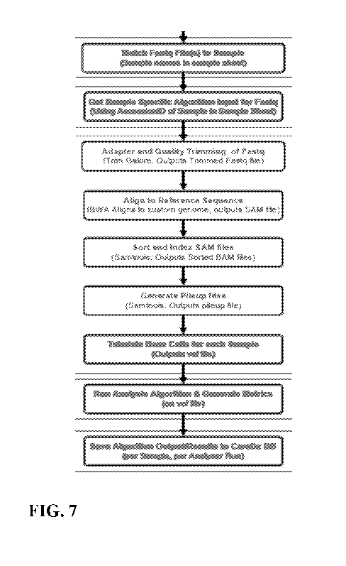
[0020] FIG. 7 illustrates an exemplary workflow of the polymorphic marker
analysis
methods described herein.
DETAILED DESCRIPTION
[0021] The following description is presented to enable a person of
ordinary skill in the art to
make and use the various embodiments. Descriptions of specific devices,
techniques, and
applications are provided only as examples. Various modifications to the
examples described
herein will be readily apparent to those of ordinary skill in the art, and the
general principles
defined herein may be applied to other examples and applications without
departing from the
spirit and scope of the various embodiments. Thus, the various embodiments are
not intended to
be limited to the examples described herein and shown, but are to be accorded
the scope
consistent with the claims.
[0022] The present disclosure relates to methods of monitoring the status
of an allograft in a
transplant recipient, as well as to methods of monitoring and adjusting
immunosuppressive
therapies being administered to the transplant recipient.
Overview
[0023] The present disclosure is based, at least in part, on Applicant's
development of
techniques for probing the status of an allograft in a transplant recipient.
Transplant recipients
contain an allograft that is foreign to the recipient's body. This triggers an
immune response
from the recipient's immune system, which may lead to acute and/or chronic
transplant rejection.
Applicant's methods involve the analysis of cell-free DNA (cfDNA) from the
transplant
recipient to diagnose the status of the transplant and inform the need to
adjust
immunosuppressive therapy being administered to the transplant recipient.
Without wishing to
be bound by theory, it is thought that transplant rejection is associated with
the death of cells in
the transplanted (donor) organ or tissue, which will release donor-derived DNA
from the dying
donor cells, thus releasing donor-derived cell-free DNA (dd-cfDNA) into the
bloodstream of the
12
CA 02970916 2017-06-14
WO 2015/138997 PCT/US2015/020603
recipient. To assay the status of the allograft in the recipient, cell-free
DNA can be extracted
from a sample from the recipient, such as a bodily fluid, and various
polymorphic markers, such
as single nucleotide polymorphism (SNP) loci, can be sequenced, where the
panel of
polymorphic markers, such as a panel of SNPs, is suitable for differentiating
between donor-
derived cell-free DNA and recipient-derived cell-free DNA (rd-cfDNA). The
specific
polymorphic markers selected to be on the panel include those that are
identified as having low
probabilities of being identical in any two individuals, thus making them
appropriate for
differentiating between recipient-derived cell free DNA and donor-derived cell-
free DNA. The
number of polymorphic markers on the panel such as, for example, the number of
SNPs on the
panel, will be sufficient to discriminate between recipient and donor alleles
even in related
individuals (excepting identical twins). The allele distribution patterns of
polymorphic markers
in the panel can be assayed to determine variance in the patterns as compared
to expected
homozygous (i.e. 0% or 100% of each allele) or heterozygous (i.e. 50% of each
allele)
distribution patterns, which can be used to determine the level of donor-
derived cell-free DNA.
Individual genotyping of the donor and the recipient to determine which allele
of the
polymorphic locus belongs to the donor and/or the recipient is not necessary,
as variance in the
polymorphic marker allele distribution pattern from expected homozygous or
heterozygous
distribution patterns informs the presence of donor-derived cell-free DNA in
the population of
cell-free DNA molecules isolated from the transplant recipient. In other
words, it is assumed
that the majority signal from the cell-free DNA sample is recipient-derived
DNA and that the
minority signal is donor-derived DNA, and this information can be used to
calculate the levels of
donor-derived DNA in the cell-free DNA sample. Changes in the levels or
variance of the
donor-derived cell-free DNA over time can be used to inform the status of the
allograft in the
transplant recipient, as well as inform a need to adjust or maintain an
immunosuppressive
therapy being administered to the subject.
[0024] Accordingly, the present disclosure provides methods of monitoring
the status of an
allograft in a transplant recipient, as well as methods of monitoring and/or
adjusting an
immunosuppressive therapy being or to be administered to a transplant
recipient. Monitoring the
status of an allograft involves analyzing various aspects which provide useful
information about
the physiological state of the allograft such as, for example, the level of
donor-derived cell-free
DNA in a sample from the transplant recipient. The methods of the present
disclosure may be
13
CA 02970916 2017-06-14
WO 2015/138997
PCT/US2015/020603
used to predict the risk of future transplant rejection such as, for example,
the risk of rejection
within the following 3-6 months after analysis of samples from the transplant
recipient. The
methods of the present disclosure may also be used to diagnose or predict the
risk of allograft
dysfunction, such as chronic renal insufficiency or cardiac allograft
vasculopathy (CAV) (e.g.
within the next 1-2 years after analysis of samples from the transplant
recipient). The methods
of the present disclosure may also be used provide an assessment of the immune
status of the
transplant recipient, which may be used to guide decisions regarding
immunosuppressive therapy
in the transplant recipient. The methods of the present disclosure may also be
used to guide
decisions related to adjustment of immunosuppressive therapies being
administered to the
transplant recipient. Additional benefits and/or uses of the methods of the
present disclosure will
be readily apparent to one of skill in the art.
Cell-Free DNA
[0025] The
methods of the present disclosure involve the analysis of cell-free DNA from a
transplant recipient to diagnose the status of the transplant and/or to inform
a need to adjust
immunosuppressive therapy being administered to the transplant recipient. Cell-
free DNA
generally refers to DNA that is present outside of a cell such as, for
example, DNA that is
present in a bodily fluid (e.g. blood, plasma, serum, etc.) of a subject. Cell-
free DNA may have
originated from various locations within a cell. Cell-free DNA may have
originated from, for
example, nuclear DNA and mitochondrial DNA. Without wishing to be bound by
theory, it is
believed that cell-free DNA is released from cells via apoptosis or necrosis
of the cells (i.e. cell
death). Accordingly, and without wishing to be bound by theory, it is believed
that during
transplant rejection, apoptosis or necrosis of transplanted (donor) cells will
result in donor-
derived cell-free DNA being released into the bodily fluid of a transplant
recipient. Transplant
recipients undergoing transplant rejection may then have a cell-free DNA
population in their
bodily fluids which includes both their own endogenous cell-free DNA
(recipient-derived cell-
free DNA) as well as cell-free DNA that originated from the donor (donor-
derived cell-free
DNA). Determining a change in the levels and/or variance in donor-derived cell-
free DNA in a
transplant recipient over time according to the methods of the present
disclosure may be used to
diagnose the status of the allograft and inform a need to adjust
immunosuppressive therapy.
14
CA 02970916 2017-06-14
WO 2015/138997 PCT/US2015/020603
[0026] Cell-free RNA may also be collected from a transplant recipient and
analyzed by
analogous methods as described above and also analysis of recipient RNA levels
from specific
marker genes to diagnose the status of the transplant and/or to inform a need
to adjust
immunosuppressive therapy being administered to the transplant recipient.
Thus, the methods of
the present disclosure generally relate to analysis of cell-free nucleic acids
from a transplant
recipient to diagnose the status of the transplant and/or to inform a need to
adjust
immunosuppressive therapy being administered to the transplant recipient.
Subjects and Samples
[0027] The methods of the present disclosure involve providing cell-free
DNA from a
sample obtained from a subject who is the recipient of an allograft from a
donor. In this sense,
the subject is a transplant recipient who contains an allograft from a donor,
and is typically a
human transplant recipient. The transplant recipient may have received one or
more of a variety
of allografts from a donor. Allografts may include transplanted organs.
Transplanted organs
may include, for example, a heart, a kidney, a lung, a liver, a pancreas, a
cornea, an organ
system, or other solid organs. The transplant received by the transplant
recipient from the donor
may also include other allografts such as, for example, a bone marrow
transplant, pancreatic islet
cells, stem cells, skin tissue, skin cells, or a xenotransplant.
[0028] The provided sample may include a bodily fluid isolated from the
transplant recipient.
Samples obtained from the transplant recipient contain cell-free DNA, and the
total cell-free
DNA present in the sample may be entirely recipient-derived cell-free DNA, or
the total cell-free
DNA present in the sample may include a mixture of recipient-derived cell-free
DNA and donor-
derived cell-free DNA. Samples may include a bodily fluid from the transplant
recipient such as,
for example, plasma, serum, whole blood, sweat, tears, saliva, ear flow fluid,
sputum, fluid from
bone marrow suspension, lymph fluid, urine, saliva, semen, vaginal flow,
cerebrospinal fluid,
brain fluid, ascites, milk, secretions of the respiratory, and intestinal or
genitourinary tract fluids.
In some embodiments where the sample is plasma, plasma derived from the venous
blood of the
transplant recipient can be obtained.
[0029] Once a sample is obtained, it can be used directly, frozen, or
otherwise stored in a
condition that maintains the integrity of the cell-free DNA for short periods
of time by
CA 02970916 2017-06-14
WO 2015/138997
PCT/US2015/020603
preventing degradation and/or contamination with genomic DNA or other nucleic
acids. The
amount of a sample that is taken at a particular time may vary, and may depend
on additional
factors, such as any need to repeat analysis of the sample. In some
embodiments, up to 50, 40,
30, 20, 10, 9, 8, 7, 6, 5, 4, 3, 2, or 1 mL of a sample is obtained. In some
embodiments, 1-50, 2-
40, 3-30, or 4-20 mL of sample is obtained. In some embodiments, more than 5,
10, 15, 20, 25,
30, 35, 40, 45, 50, 55, 60, 65, 70, 75, 80, 85, 90, 95 or 100 mL of a sample
is obtained.
[0030]
Samples may be taken from a transplant recipient over a period of time (i.e.
over a
time interval). The time at which samples are taken from the transplant
recipient following the
transplant event may vary. Samples may be taken from a transplant recipient at
various times
and over various periods of time for use in determining the status of the
allograft according to the
methods of the present disclosure. For example, samples may be taken from the
transplant
recipient within days and weeks after, about three months after, about six
months after, about
nine months after, or less than one year after the transplant event. Samples
may be taken from
the transplant recipient at various times before the one year anniversary of
the transplant event, at
the one year anniversary of the transplant event, or at various times after
the one year
anniversary of the transplant event. For example, at the one year anniversary
after a transplant,
samples may begin to be taken from the transplant recipient at month 12 (i.e.
the one year
anniversary of the transplant event) and continue to be taken for periods of
time after this. In
some embodiments, the time period for obtaining samples from a transplant
recipient is within
the first few days after the transplant from the donor to the recipient
occurred. This may be done
to monitor induction therapy. In some embodiments, the time period for
obtaining samples from
a transplant recipient is during tapering of the immunosuppressive regimen, a
period that occurs
during the first 12 months after the transplant from the donor to the
recipient occurred. In some
embodiments, the time period for obtaining samples from a transplant recipient
is during the
initial long term immunosuppressive maintenance phase, beginning about 12-14
months after the
transplant from the donor to the recipient occurred. In some embodiments, the
time period for
obtaining samples from a transplant recipient is during the entire long term
maintenance of the
immunosuppressive regimen, any time beyond 12 months after the transplant from
the donor to
the recipient occurred.
16
CA 02970916 2017-06-14
WO 2015/138997 PCT/US2015/020603
[0031] Where multiple samples are to be obtained from a transplant
recipient, the frequency
of sampling may vary. After samples have begun to be taken from a transplant
recipient,
samples may be obtained about once every week, about once every 2 weeks, about
once every 3
weeks, about once every month, about once every two months, about once every
three months,
about once every four months, about once every five months, about once every
six months, about
once every year, or about once every two years or more after the initial
sampling event.
[0032] In some embodiments, a transplant recipient has samples of bodily
fluid taken for one
to three consecutive months, starting at the one year anniversary of the
transplant event (i.e. 12
months after the transplant event), providing a total of 4-6 samples for
analysis taken over a three
month time period, with samples being collected about every two weeks. In some
embodiments,
a transplant recipient has samples of bodily fluid taken once a week for one
to three consecutive
months, starting at the one year anniversary of the transplant event (i.e. 12
months after the
transplant event), providing a total of twelve samples for analysis taken over
a three month time
period. The total duration of obtaining samples from a transplant recipient,
as well as the
frequency of obtaining such samples, may vary and will depend on a variety of
factors, such as
clinical progress. For example, a transplant recipient may have samples
obtained for analysis of
cell-free DNA for the duration of their lifetime. Appropriate timing and
frequency of sampling
will be able to be determined by one of skill in the art for a given
transplant recipient.
Analysis of Cell-Free DNA in a Transplant Recipient
[0033] The methods of the present disclosure involve the analysis of cell-
free DNA from a
transplant recipient. After cell-free DNA has been isolated from a transplant
recipient, various
methods and techniques may be used to analyze the cell-free DNA. Analysis of
cell-free DNA
according to the methods of the present disclosure involves analysis of a
panel of polymorphic
markers from the cell-free DNA. In some embodiments, the polymorphic markers
are single
nucleotide polymorphisms (SNPs). In some embodiments, SNPs are selected to be
included in
the panel at least in part on the basis that the panel of SNPs will be
sufficient to differentiate
between donor-derived cell-free DNA and recipient-derived cell-free DNA.
17
CA 02970916 2017-06-14
WO 2015/138997 PCT/US2015/020603
Panels of Polymorphic Markers
[0034] Analysis of cell-free DNA obtained from a transplant recipient
involves the analysis
of a panel of polymorphic markers from the cell-free DNA. Various polymorphic
markers may
be selected for inclusion in the panel to be analyzed as long as the
polymorphic marker panel as a
whole is suitable for differentiating between donor-derived cell-free DNA and
recipient-derived
cell-free DNA. The same polymorphic marker panel may be used for each
transplant recipient;
there is no need to customize polymorphic marker panels to individualize the
panel to different
transplant recipients.
[0035] Various types of polymorphic markers may be included in polymorphic
marker
panels. Polymorphic markers are found at a region of DNA containing a
polymorphism. A
polymorphism generally refers to the occurrence of two or more genetically
determined
alternative sequences or alleles in a population. A polymorphic marker or site
is the locus at
which divergence, or the polymorphism, occurs. A polymorphism may contain, for
example,
one or more base changes, an insertion, a repeat, or a deletion. A polymorphic
locus may be as
small as one base pair, such as a SNP. Polymorphic markers may include, for
example, single
nucleotide polymorphisms (SNPs), restriction fragment length polymorphisms
(RFLPs), short
tandem repeats (STRs), variable number of tandem repeats (VNTRs),
hypervariable regions,
minisatellites, dinucleotide repeats, trinucleotide repeats, tetranucleotide
repeats, simple
sequence repeats, and insertion elements. Polymorphic markers may contain one
or more bases
modified by methylation. Additional types of polymorphisms and polymorphic
markers will be
readily apparent to one of skill in the art. A polymorphism between two
nucleic acids can be
naturally occurring, or may be caused by exposure to or contact with
chemicals, enzymes, or
other agents, or exposure to agents that cause damage to nucleic acids such
as, for example,
ultraviolet radiation, mutagens, or carcinogens.
[0036] Various combinations of polymorphic marker types may be used in
polymorphic
marker panels. For example, the polymorphic marker panel may include both SNPs
and short
tandem repeats, or any other type of polymorphic marker. In some embodiments,
the
polymorphic marker panel is composed entirely of SNPs; thus, the polymorphic
marker panel is
18
CA 02970916 2017-06-14
WO 2015/138997 PCT/US2015/020603
a SNP panel. Additional combinations of polymorphic markers on polymorphic
marker panels
will be readily apparent to one of skill in the art.
[0037] Selection of the appropriate quantity and identity of polymorphic
markers to be
analyzed from cell-free DNA may vary, as will be appreciated by one of skill
in the art. The
panel of polymorphic markers to be analyzed may include at least 10, at least
15, at least 20, at
least 25, at least 30, at least 35, at least 40, at least 45, at least 50, at
least 55, at least 60, at least
65, at least 70, at least 75, at least 80, at least 85, at least 90, at least
95, at least 100, at least 105,
at least 110, or at least 115, at least 120, at least 150, at least 200, at
least 250, at least 300, at
least 350, at least 400, at least 450, at least 500, at least 1,000, or at
least 1,500 or more
independent polymorphic markers.
[0038] In some embodiments, the polymorphic marker panel is a panel of
SNPs. SNPs to be
included in the SNP panel, or in any other polymorphic marker panel, may be
those previously
identified as being suitable for differentiating between any two unrelated
individuals (Pakstis et
al., 2010). For example, the SNP panel may include one or more of the
following human SNPs
(named according to dbSNP numbering): rs1004357, rs10092491, rs1019029,
rs1027895,
rs10488710, rs10500617, rs1058083, rs10768550, rs10773760, rs10776839,
rs1109037,
rs12480506, rs1294331, rs12997453, rs13134862, rs13182883, rs13218440,
rs1336071,
rs1358856, rs1410059, rs1478829, rs1490413, rs1498553, rs1523537, rs1554472,
rs159606,
rs1736442, rs1821380, rs1872575, rs2046361, rs2073383, rs214955, rs2175957,
rs221956,
rs2255301, rs2269355, rs2270529, rs2272998, rs2291395, rs2292972, rs2342747,
rs2399332,
rs2503107, rs2567608, rs279844, rs2811231, rs2833736, rs2920816, rs315791,
rs321198,
rs338882, rs3744163, rs3780962, rs4288409, rs430046, rs4364205, rs445251,
rs4530059,
rs4606077, rs464663, rs4789798, rs4796362, rs4847034, rs521861, rs560681,
rs5746846,
rs576261, rs590162, rs6444724, rs6591147, rs6811238, rs689512, rs6955448,
rs7041158,
rs7205345, rs722290, rs7229946, rs740598, rs7520386, rs7704770, rs8070085,
rs8078417,
rs891700, rs901398, rs9546538, rs9606186, rs985492, rs9866013, rs987640,
rs9905977,
rs993934, and rs9951171.
[0039] SNPs may also be selected on the basis that they have, for example,
an overall
population minor allele frequency of >0.4, a target population minor allele
frequency of >0.4, the
19
CA 02970916 2017-06-14
WO 2015/138997
PCT/US2015/020603
lowest polymerase error rate (in the test system) of the 6 potential allele
transitions or
transversions, and low linkage on the genome such as, for example, >500kb
distance between
SNPs.
[0040] The SNP panel may include, for example, one or more of the following
human SNPs
(named according to dbSNP numbering): rs10488710, rs279844, rs1048290,
rs1049379,
rs1051614, rs1052637, rs1055851, rs1056033, rs1056149, rs1064074, rs1078004,
rs10831567,
rs6811238, rs11106, rs11210490, rs1126899, rs1127472, rs1127893, rs1130857,
rs1049544,
rs11547806, rs12237048, rs430046, rs12508837, rs12529, rs12717, rs13184586,
rs13295990,
rs13428, rs13436, rs1374570, rs14080, rs1411271, rs576261, rs14155, rs1151687,
rs1565933,
rs1600, rs1678690, rs1881421, rs1897820, rs1898882, rs2056844, rs20575,
rs10092491,
rs2070426, rs2071888, rs2075322, rs2180314, rs2185798, rs2227910, rs2228560,
rs2229571,
rs2229627, rs2245285, rs2342747, rs2248490, rs2253592, rs2254357, rs2275047,
rs2279665,
rs2279776, rs2281098, rs2287813, rs4364205, rs2289751, rs2289818, rs2292830,
rs2294092,
rs2295005, rs2296545, rs2297236, rs2302443, rs2306049, rs1022478, rs445251,
rs230898,
rs231235, rs2342767, rs236152, rs2362450, rs2384571, rs2455230, rs246703,
rs2480345,
rs248385, rs2498982, rs2505232, rs2509943, rs2519123, rs2523072, rs2571028,
rs2657167,
rs28686812, rs2946994, rs1294331, rs10419826, rs3088241, rs3110623, rs3173615,
rs3190321,
rs3205187, rs344141, rs35596415, rs362124, rs36657, rs1872575, rs159606,
rs3731877,
rs3734311, rs3735615, rs3740199, rs3748930, rs3751066, rs3790993, rs3802265,
rs3803763,
rs1004357, rs3803798, rs3809972, rs3810483, rs3812571, rs3813609, rs3814182,
rs3816800,
rs3826709, rs3829655, rs3951216, rs1019029, rs408600, rs41317515, rs436278,
rs448012,
rs475002, rs4845480, rs4849167, rs4865615, rs1027895, rs4890012, rs492594,
rs4940019,
rs4971514, rs523104, rs528557, rs545500, rs561930, rs57010808, rs57285449,
rs10500617,
rs6061243, rs609521, rs62490396, rs625223, rs638405, rs6459166, rs648802,
rs6510057,
rs6764714, rs10768550, rs6790129, rs6794, rs6807362, rs6838248, rs713598,
rs7161563,
rs726009, rs7289, rs7301328, rs7332388, rs10773760, rs743616, rs743852,
rs745142,
rs7451713, rs7526132, rs7543016, rs7601771, rs7785899, rs7825, rs8009219,
rs10776839,
rs8025851, rs8058696, rs8076632, rs8097, rs8103906, rs874881, rs9262,
rs9289122, rs936019,
rs9393728, rs1109037, rs977070, rs9865242, rs12480506, rs560681, rs12997453,
rs13134862,
rs13218440, rs1358856, rs1410059, rs1478829, rs1498553, rs1523537, rs4606077,
rs1554472,
rs1736442, rs1821380, rs2046361, rs214955, rs2175957, rs2255301, rs2269355,
rs2270529,
CA 02970916 2017-06-14
WO 2015/138997 PCT/US2015/020603
rs2272998, rs2291395, rs2292972, rs2399332, rs2503107, rs2567608, rs2811231,
rs2833736,
rs315791, rs321198, rs6955448, rs338882, rs3780962, rs4288409, rs4530059,
rs464663,
rs4789798, rs4796362, rs4847034, rs521861, rs1058083, rs5746846, rs590162,
rs6444724,
rs6591147, rs689512, rs7205345, rs722290, rs740598, rs7520386, rs221956,
rs7704770,
rs8070085, rs8078417, rs891700, rs901398, rs9546538, rs9606186, rs985492,
rs9866013,
rs987640, rs13182883, rs9905977, rs993934, rs9951171, rs10274334, rs10421285,
rs1043413,
rs1044010, rs1045248, rs1045644, and rs1047979. In some embodiments, each of
the 266 above
mentioned SNPs is included in the polymorphic marker panel to be analyzed from
cell-free
DNA.
[0041] The SNP panel may include, for example, at least at least 10, at
least 15, at least 20, at
least 25, at least 30, at least 35, at least 40, at least 45, at least 50, at
least 55, at least 60, at least
65, at least 70, at least 75, at least 80, at least 85, at least 90, at least
95, at least 100, at least 105,
at least 110, or at least 115, at least 120, at least 150, at least 200, at
least 205, at least 210, at
least 215, at least 220, at least 225, at least 230, at least 235, at least
240, at least 245, at least
250, at least 255, at least 260, or at least 265 of the 266 independent SNPs
identified above.
[0042] The SNP panel may include, for example, about 10 to about 20, about
20 to about 30,
about 30 to about 40, about 40 to about 50, about 50 to about 60, about 60 to
about 70, about 70
to about 80, about 80 to about 90, about 90 to about 100, about 100 to about
110, about 110 to
about 120, about 120 to about 130, about 130 to about 140, about 140 to about
150, about 150 to
about 160, about 160 to about 170, about 170 to about 180, about 180 to about
190, about 190 to
about 200, about 200 to about 210, about 210 to about 220, about 220 to about
230, about 230 to
about 240, about 240 to about 250, about 250 to about 260, or about 250 to
about 266 of the 266
independent SNPs identified above.
[0043] The SNP panel may include, for example, about 195 to about 200,
about 200 to about
205, about 210 to about 215, about 215 to about 220, about 220 to about 225,
about 225 to about
230, about 230 to about 235, about 235 to about 240, about 240 to about 245,
about 245 to about
250, about 250 to about 255, about 255 to about 260, about 260 to about 265,
or about 260 to
about 266 of the 266 independent SNPs identified above.
21
CA 02970916 2017-06-14
WO 2015/138997 PCT/US2015/020603
Amplification and Sequencing
[0044] Cell-free DNA isolated from a transplant recipient may be amplified
for downstream
techniques and analysis, such as analysis of a panel of polymorphic markers
from the cell-free
DNA. Various methods and protocols for DNA extraction are well-known in the
art and are
described herein (See e.g. Current Protocols in Molecular Biology, latest
edition). Cell-free
DNA may be extracted using the QIAamp circulating nucleic acid kit or other
appropriate
commercially available kits. Other exemplary methods of extracting cell-free
DNA are well-
known (See, e.g., Cell-Free Plasma DNA as a Predictor of Outcome in Severe
Sepsis and Septic
Shock. Clin. Chem. 2008, v. 54, p.1000-1007; Prediction of MYCN Amplification
in
Neuroblastoma Using Serum DNA and Real-Time Quantitative Polymerase Chain
Reaction.
JCO 2005, v. 23, p.5205-5210; Circulating Nucleic Acids in Blood of Healthy
Male and Female
Donors. Clin. Chem. 2005, v. 51, p.1317-1319; Use of Magnetic Beads for Plasma
Cell-free
DNA Extraction: Toward Automation of Plasma DNA Analysis for Molecular
Diagnostics. Clin.
Chem. 2003, v. 49, p.1953-1955; Chiu RWK, Poon LLM, Lau TK, Leung TN, Wong
EMC, Lo
YMD. Effects of blood-processing protocols on fetal and total DNA
quantification in maternal
plasma. Clin Chem 2001;47: 1607-1613; and Swinkels et al. Effects of Blood-
Processing
Protocols on Cell-free DNA Quantification in Plasma. Clinical Chemistry, 2003,
vol. 49, no. 3,
525-526).
[0045] Methods of amplifying DNA are similarly well-known in the art and
are described
herein. Amplification generally refers to any device, method or technique that
can generate
copies of a nucleic acid. Amplification of cell-free DNA may involve, for
example, polymerase
chain reaction (PCR) techniques such as linear amplification (cf. USPN
6,132,997), rolling circle
amplification, and the like. Cell-free DNA may be amplified for use in
downstream analysis of
the DNA by, for example, digital PCR or sequencing. The Fluidigm Access
ArrayTM System,
the RainDance Technologies RainDrop system, or other technologies for
multiplex amplification
may be used for multiplex or highly parallel simplex DNA amplification.
Amplification may
involve the use of high-fidelity polymerases such as, for example, FastStart
High Fidelity
(Roche), Expand High Fidelity (Roche), Phusion Flash II (ThermoFisher
Scientific), Phusion
Hot Start II (ThermoFisher Scientific), KAPA HiFi (Kapa BioSystems), or KAPA2G
(Kapa
Biosystems).
22
CA 02970916 2017-06-14
WO 2015/138997 PCT/US2015/020603
[0046] Amplification may include an initial PCR cycle that adds a unique
sequence to each
individual molecule, called molecular indexing. Molecular indexing allows for
quantitative
assessment of the absolute level of both alleles for each SNP amplicon and
therefore may
improve precision and accuracy of determining the percent donor-derived cell-
free DNA.
[0047] Amplified DNA may also be subjected to additional processes, such as
indexing (also
referred to as barcoding or tagging). Methods of indexing DNA are well-known
in the art and
are described herein. Indexing will allow for the use of multiplex-sequencing
platforms, which
are compatible with a variety of sequencing systems, such as Illumina HiSeq,
MiSeq, and
ThermoFisher Scientific Ion PGM and Ion Proton. Multiplex sequencing permits
the sequencing
of DNA from multiple samples at once through the use of DNA indexing to
specifically identify
the sample source of the sequenced DNA.
[0048] The amount of DNA that is used for analysis may vary. In some
embodiments, less
than 1 pg, 5 pg, 10 pg, 20 pg, 30 pg, 40 pg, 50 pg, 100 pg, 200 pg, 500 pg, 1
ng, 5 ng, 10 ng, 20
ng, 30 ng, 40 ng, 50 ng, 100 ng, 200 ng, 500 ng, 1 ug, 5 ug, 10 ug, 20 ug, 30
ug, 40 ug, 50 ug,
100 ug, 200 ug, 500 ug or 1 mg of DNA are obtained from the sample for further
genetic
analysis. In some cases, about 1-5 pg, 5-10 pg, 10-100 pg, 100 pg-1 ng, 1-5
ng, 5-10 ng, 10-100
ng, or 100 ng-1 ug of DNA are obtained from the sample for further genetic
analysis.
[0049] The methods of the present disclosure involve sequencing target loci
from cell-free
DNA, as well as analyzing sequence data. Various methods and protocols for DNA
sequencing
and analysis are well-known in the art and are described herein. For example,
DNA sequencing
may be accomplished using high-throughput DNA sequencing techniques. Examples
of next
generation and high-throughput sequencing include, for example, massively
parallel signature
sequencing, polony sequencing, 454 pyrosequencing, Illumina (Solexa)
sequencing with HiSeq,
MiSeq, and other platforms, SOLiD sequencing, ion semiconductor sequencing
(Ion Torrent),
DNA nanoball sequencing, heliscope single molecule sequencing, single molecule
real time
(SMRT) sequencing, MassARRAY , and Digital Analysis of Selected Regions
(DANSRTm).
See, e.g., Stein RA (1 September 2008). "Next-Generation Sequencing Update".
Genetic
Engineering & Biotechnology News 28 (15); Quail, Michael; Smith, Miriam E;
Coupland, Paul;
Otto, Thomas D; Harris, Simon R; Connor, Thomas R; Bertoni, Anna; Swerdlow,
Harold P; Gu,
23
CA 02970916 2017-06-14
WO 2015/138997 PCT/US2015/020603
Yong (1 January 2012). "A tale of three next generation sequencing platforms:
comparison of
Ion torrent, pacific biosciences and illumina MiSeq sequencers". BMC Genomics
13 (1): 341 ;
Liu, Lin; Li, Yinhu; Li, Siliang; Hu, Ni; He, Yimin; Pong, Ray; Lin, Danni;
Lu, Lihua; Law,
Maggie (1 January 2012). "Comparison of Next-Generation Sequencing Systems".
Journal of
Biomedicine and Biotechnology 2012: 1-11; Qualitative and quantitative
genotyping using single
base primer extension coupled with matrix-assisted laser desorption/ionization
time-of -flight
mass spectrometry (MassARRAY()). Methods Mol Biol. 2009;578:307-43; Chu T,
Bunce K,
Hogge WA, Peters DG. A novel approach toward the challenge of accurately
quantifying fetal
DNA in maternal plasma. Prenat Diagn 2010;30: 1226-9; and Suzuki N, Kamataki
A, Yamaki J,
Homma Y. Characterization of circulating DNA in healthy human plasma. Clinica
chimica acta;
international journal of clinical chemistry 2008;387:55-8). Similarly,
software programs for
primary and secondary analysis of sequence data are well-known in the art.
[0050] Where there are multiple cell-free DNA samples from a transplant
recipient to be
sequenced, such as when multiple samples are taken from the transplant
recipient over time, each
sample may be sequenced individually, or multiple samples may be sequenced
together using
multiplex sequencing.
Analyzing Polymorphic Marker Allele Distribution Patterns and Determining the
Level of Donor-Derived Cell-Free DNA
[0051] The methods of the present disclosure involve assaying variance in
polymorphic
marker allele distribution patterns in a polymorphic marker panel as compared
to expected
homozygous or heterozygous distribution patterns. Analysis of these patterns
allows for the
determination of the level of donor-derived cell-free DNA in a cell-free DNA
sample obtained
from a transplant recipient.
[0052] Generally, an individual contains DNA that is either homozygous or
heterozygous for
a given polymorphic marker, such as a SNP. An individual may be homozygous for
one allele of
a given polymorphic marker and will contain 100% of one allele of that
polymorphic marker and
will contain 0% of the other allele of that polymorphic marker (e.g. 100% of
allele A for given
polymorphic marker, 0% of allele B for given polymorphic marker). An
individual may also be
heterozygous for a given polymorphic marker, and thus will contain 50% of
allele A and 50% of
24
CA 02970916 2017-06-14
WO 2015/138997 PCT/US2015/020603
allele B of that polymorphic marker. Accordingly, if all of the DNA in a
sample originated from
a single individual, it is expected that any given polymorphic marker in the
DNA in that sample
will exhibit a homozygous distribution pattern (i.e. 100% of one allele, 0% of
the other allele) or
a heterozygous distribution pattern (i.e. 50% of one allele and 50% of the
other allele).
However, if a DNA sample contains DNA that originated from more than one
individual (e.g. a
cell-free DNA sample from a transplant recipient that contains both recipient-
derived DNA and
donor-derived DNA), then polymorphic marker allele distributions may vary, for
a given
polymorphic marker, from expected homozygous or heterozygous distribution
patterns. This is
so because two individuals may not necessarily share the same zygosity for a
given polymorphic
marker (e.g. individual 1 is homozygous for a given allele of a given
polymorphic marker, and
individual 2 is heterozygous for the alleles of that same polymorphic marker).
When this occurs,
variance in the expected allele distribution patterns as compared to expected
homozygous or
heterozygous distribution patterns may be observed. This variance can be used
to assess whether
foreign DNA is present in a DNA sample from a single individual. With respect
to the methods
of the present disclosure, variance in polymorphic marker allele distribution
patterns in the
polymorphic marker panel as compared to expected homozygous or heterozygous
distribution
patterns is used to determine the level of donor-derived cell-free DNA in the
cell-free DNA
sample obtained from a transplant recipient.
[0053] When analyzing polymorphic marker sequence data from cell-free DNA
according to
methods of the present disclosure, a majority signal from an allele of a
polymorphic marker may
be observed and a minority signal from an allele of a polymorphic marker may
be observed.
Regarding cell-free DNA isolated from a transplant recipient, as it is assumed
that the majority
of the DNA in the cell-free DNA sample from the transplant recipient
originated from the
recipient's own endogenous DNA, it is further assumed that the majority signal
represents an
allele of a polymorphic marker that originated from recipient-derived cell-
free DNA, while the
minority signal represents an allele of a polymorphic marker that originated
from donor-derived
cell-free DNA. Polymorphic markers such as SNPs, for example, with an even
distribution of
both alleles are assumed to have largely both originated from the recipient.
Deviations from the
even distribution will indicate the influence of alleles from the donor-
derived cell-free DNA.
CA 02970916 2017-06-14
WO 2015/138997 PCT/US2015/020603
[0054] Various calculations may be performed based on allele calls from the
sequence data.
For example, the methods of the present disclosure may involve calculating
various cell-free
DNA concentrations, or percents thereof, of a total amount of cell-free DNA.
Overall, as it is
assumed that the majority signal from cell-free DNA in a sample isolated from
a transplant
recipient is recipient-derived DNA and that the minority signal is donor-
derived DNA, this
information can be used to calculate a percentage of donor-derived DNA in the
cell-free DNA
sample.
[0055] As described above, individual genotyping of the donor and the
recipient to determine
which allele of the polymorphic marker belongs to the donor and/or the
recipient is not
necessary, as variance in the polymorphic marker allele distribution pattern
from expected
homozygous or heterozygous distribution patterns informs the presence of donor-
derived cell-
free DNA in the population of cell-free DNA molecules isolated from the
transplant recipient.
Accordingly, the level of donor-derived cell-free DNA in a sample obtained
from a transplant
recipient may be determined without using genotype information from the
transplant recipient,
from the transplant donor, and/or any other genotype information from any
source. Such
genotype information that need not be considered includes, for example, the
genotype across the
genome as a whole or portions thereof, or the genotype at the particular
polymorphic markers
being analyzed. In some embodiments, individual genotyping of the transplant
recipient is not
performed. In some embodiments, individual genotyping of the transplant donor
is not
performed. In some embodiments, neither the transplant recipient nor the
transplant donor are
individually genotyped. In some embodiments, genotype information from the
transplant
recipient is not considered when determining the levels of donor-derived cell-
free DNA in a
sample obtained from a transplant recipient. In some embodiments, genotype
information from
the transplant donor is not considered when determining the levels of donor-
derived cell-free
DNA in a sample obtained from a transplant recipient. In some embodiments, the
levels of
donor-derived cell-free DNA in a sample obtained from a transplant recipient
are determined
without consideration of genotype information from the transplant recipient
and without
consideration of genotype information of the transplant donor. In some
embodiments, the level
of donor-derived cell-free DNA in a sample obtained from a transplant
recipient may be
determined without using genotype information.
26
CA 02970916 2017-06-14
WO 2015/138997 PCT/US2015/020603
[0056] Improvements to the calculations may include estimating and
subtracting a level of
signal due to amplification or sequencing error to improve accuracy and
precision. For example,
a suitably chosen subset of SNPs may be used to estimate a sum, mean, median
or standard
deviation of the subset to produce a computation of the overall level of donor-
derived cell-free
DNA. Multiple samples from the same subject at a given time of sampling will
all have the
same pattern of polymorphic distributions across the SNPs, which can be used
to enhance the
estimate of donor-derived cell-free DNA in individual samples from that
subject.
[0057] The quantity of donor-derived cell-free DNA present in the cell-free
DNA sample
may be expressed in a variety of ways. In some embodiments, the amount of one
or more DNA
molecules from donor-derived cell-free DNA is determined as a percentage of
the total the DNA
molecules in the sample. In some embodiments, the amount of one or more DNA
molecules
from donor-derived cell-free DNA is determined as a ratio of the total DNA in
the sample. In
some embodiments, the amount of one or more DNA molecules from donor-derived
cell-free
DNA is determined as a ratio or percentage compared to one or more reference
DNA molecules
in the sample. For example, the total amount of donor-derived cell-free DNA
can be determined
to be 10% of the total DNA molecules in the cell-free DNA sample.
Alternatively, the total
amount of donor-derived cell-free DNA can be at a ratio of 1:10 compared to
the total DNA
molecules in the cell-free DNA sample. In some embodiments, the amount of one
or more DNA
molecules from the donor-derived cell-free DNA can be determined as a
concentration. For
example, the total amount of donor-derived cell-free DNA in the cell-free DNA
sample can be
determined to be 1 ug/mL. The values described here are merely exemplary to
illustrate various
ways to express quantities of donor-derived cell-free DNA. The percentage of
donor-derived
cell-free DNA in the cell-free DNA sample from a transplant recipient may be
extremely low
(e.g. at or below 0.5% of the total DNA content of the cell-free DNA sample).
It is noted that the
quantity of recipient-derived cell-free DNA in the cell-free DNA sample may
also be expressed
in the manners as described for donor-derived cell-free DNA. Additional
methods of expressing
the quantity of a given source, type, or sequence of DNA molecule in a cell-
free DNA sample
will be readily apparent to one of skill in the art.
[0058] The above-described embodiments of the present disclosure may be
implemented in
a variety of ways. For example, some aspects of the embodiments may be
implemented using
27
CA 02970916 2017-06-14
WO 2015/138997 PCT/US2015/020603
hardware, software or a combination thereof. When implemented in software, the
software code
can be executed on any suitable processor or collection of processors, whether
provided in a
single computer or distributed among multiple computers. It should be
appreciated that any
component or collection of components that perform the functions described
above can be
generically considered as one or more controllers that control the above -
discussed functions.
The one or more controllers can be implemented in numerous ways, such as with
dedicated
hardware, or with general-purpose hardware (e.g., one or more processors) that
is programmed
using microcode or software to perform the functions recited above.
[0059] In this respect, it should be noted that implementation of various
features of the
present disclosure may use at least one non-transitory computer-readable
storage medium (e.g., a
computer memory, a floppy disk, a compact disk, a tape, etc.) encoded with a
computer program
(i.e., a plurality of instructions), which, when executed on a processor,
performs the above -
discussed functions. The computer-readable storage medium can be transportable
such that the
program stored thereon can be loaded onto any computer resource to implement
certain aspects
of the present disclosure discussed herein. In addition, it should be noted
that the reference to a
computer program which, when executed, performs the above-discussed functions,
is not limited
to an application program running on a host computer. Rather, the term
computer program is
used herein in a generic sense to reference any type of computer code (e.g.,
software or
microcode) that can be employed to program a processor to implement certain
aspects of the
present disclosure.
Determining Status of an Allograft
[0060] The methods of the present disclosure for determining the levels of
donor-derived
cell-free DNA in a sample from a transplant recipient can be used to determine
the status of the
allograft in the transplant recipient. In general, changes in the levels or
variance of donor-
derived cell-free DNA beyond a suitable threshold value in the transplant
recipient over time are
informative with regard to the status of the allograft.
[0061] A threshold or threshold value generally refers to any predetermined
level or range of
levels that is indicative of the presence or absence of a condition or the
presence or absence of a
risk. The threshold value can take a variety of forms. It can be single cut-
off value, such as a
28
CA 02970916 2017-06-14
WO 2015/138997 PCT/US2015/020603
median or mean. As another example, a threshold value can be determined from
baseline values
before the presence of a condition or risk or after a course of treatment.
Such a baseline can be
indicative of a normal or other state in the subject not correlated with the
risk or condition that is
being tested for. For example, the baseline value may be the level of donor-
derived cell-free
DNA in samples from a transplant recipient prior to the actual transplant
event, which would be
presumably zero or negligible, but may also indicate baseline error in the
system. In some
embodiments, the threshold value can be a baseline value of the subject being
tested. The
threshold value, as it pertains to demarcating significant changes in the
levels or variance of
donor-derived cell-free DNA in a transplant recipient, may vary considerably.
One of skill in the
art would recognize appropriate parameters and means for determining
significant changes in the
levels or variance of donor-derived cell-free DNA in a transplant recipient
over time. Once
appropriate analysis parameters are selected, determining changes in the level
or variance of
donor-derived cell-free DNA in the transplant recipient over a period of time
can inform status of
the allograft.
[0062] In some embodiments, an increase in the levels or variance of the
donor-derived cell-
free DNA in the transplant recipient over time is indicative of transplant
rejection, a need for
adjusting immunosuppressive therapy, immunosuppressive treatment
nephrotoxicity, infection,
and/or a need for further investigation of the allograft status. Without
wishing to be bound by
theory, it is believed that if the level of donor-derived cell-free DNA is
increasing in a transplant
recipient over time, then the cells of the allograft are increasingly
experiencing apoptosis and/or
necrosis over time, which is indicative of transplant rejection.
[0063] In some embodiments, a decrease in the levels or variance of the
donor-derived cell-
free DNA in the transplant recipient over time is indicative of transplant
tolerance, a need for
adjusting immunosuppressive therapy, and/or a need for further investigation
of the allograft
status. Without wishing to be bound by theory, it is believed that if the
level of donor-derived
cell-free DNA is decreasing in a transplant recipient over time, then the
cells of the allograft are
decreasingly experiencing apoptosis and/or necrosis over time, which is
indicative of transplant
tolerance, overimmunosuppression, or appropriate immunosuppression.
29
CA 02970916 2017-06-14
WO 2015/138997 PCT/US2015/020603
[0064] In some embodiments, no change in the levels or variance of the
donor-derived cell-
free DNA in the transplant recipient over time is indicative of stable
transplant rejection status
and/or opportunity for adjusting immunosuppressive therapy. Without wishing to
be bound by
theory, it is believed that if the level of donor-derived cell-free DNA is not
changing in a
transplant recipient over time, then the cells of the allograft are
experiencing a steady-state level
of apoptosis over time, which is indicative of a stable transplant rejection
status. A stable
transplant rejection status informs the status of the allograft during the
time window analyzed,
but does not inform the direction, either towards rejection or tolerance, the
allograft is
progressing toward. For example, the allograft may be undergoing active
rejection in the
transplant recipient, but a stable transplant rejection status indicates that
the rate of allograft
rejection is not changing during the time it was analyzed (i.e. the rate of
transplant rejection is
not increasing or decreasing). Similarly, the allograft may be undergoing
active tolerance in the
transplant recipient, but a stable transplant rejection status indicates that
the rate of allograft
tolerance is not changing during the time it was analyzed (i.e. the rate of
transplant tolerance is
not increasing or decreasing).
Adjustment of Immunosuppressive Therapy
[0065] The methods of the present disclosure for determining the levels of
donor-derived
cell-free DNA in a sample from a transplant recipient can be used to inform
the need to adjust
immunosuppressive therapy being administered to the transplant recipient. In
general, changes
in the levels or variance of donor-derived cell-free DNA beyond a suitable
threshold value in the
transplant recipient over time are informative with regard to determining a
need to adjust
immunosuppressive therapy being administered to the transplant recipient.
[0066] Immunosuppressive therapy generally refers to the administration of
an
immunosuppressant or other therapeutic agent that suppresses immune responses
to a subject.
Exemplary immunosuppressant agents may include, for example, anticoagulents,
antimalarials,
heart drugs, non-steroidal anti-inflammatory drugs (NSAIDs), and steroids
including, for
example, Ace inhibitors, aspirin, azathioprine, B7RP-1-fc, il-blockers,
brequinar sodium,
campath-1H, celecoxib, chloroquine, corticosteroids, coumadin,
cyclophosphamide, cyclosporin
A, DHEA, deoxyspergualin, dexamethasone, diclofenac, dolobid, etodolac,
everolimus, FK778,
CA 02970916 2017-06-14
WO 2015/138997
PCT/US2015/020603
feldene, fenoprofen, flurbiprofen, heparin, hydralazine, hydroxychloroquine,
CTLA-4 or LFA3
immunoglobulin, ibuprofen, indomethacin, ISAtx-247, ketoprofen, ketorolac,
leflunomide,
meclophenamate, mefenamic acid, mepacrine, 6-mercaptopurine, meloxicam,
methotrexate,
mizoribine, mycophenolate mofetil, naproxen, oxaprozin, Plaquenil, NOX-100,
prednisone,
methyprenisone, rapamycin (sirolimus), sulindac, tacrolimus (FK506),
thymoglobulin, tolmetin,
tresperimus, U0126, and antibodies including, for example, alpha lymphocyte
antibodies,
adalimumab, anti-CD3, anti-CD25, anti-CD52 anti-IL2R, and anti-TAC antibodies,
basiliximab,
daclizumab, etanercept, hu5C8, infliximab, OKT4, and natalizumab.
[0067] In some embodiments, an increase in the levels or variance of the
donor-derived cell-
free DNA in the transplant recipient over time is indicative of a need to
increase
immunosuppressive therapy being administered to the transplant recipient. The
decision to
increase immunosuppressive therapy being administered to a transplant
recipient may be based
on additional clinical factors, such as the health of the transplant
recipient. In some
embodiments, immunosuppressive therapy being administered to the subject is
increased.
[0068] In some embodiments, a decrease in the levels or variance of the
donor-derived cell-
free DNA in the transplant recipient over time is indicative of a need to
decrease
immunosuppressive therapy being administered to the transplant recipient. The
decision to
decrease immunosuppressive therapy being administered to a transplant
recipient may be based
on additional clinical factors, such as the health of the transplant
recipient. In some
embodiments, immunosuppressive therapy being administered to the subject is
decreased.
[0069] In some embodiments, no change in the levels or variance of the
donor-derived cell-
free DNA in the transplant recipient over time is indicative of no need to
adjust
immunosuppressive therapy being administered to the transplant recipient, or
that the
immunosuppressive therapy being administered may be maintained. The decision
to maintain
immunosuppressive therapy being administered to a transplant recipient may be
based on
additional clinical factors, such as the health of the transplant recipient.
In some embodiments,
immunosuppressive therapy being administered to the subject is maintained.
[0070] In some embodiments, adjustment of immunosuppressive therapy
includes changing
the type or form of immunosuppressant or other immunosuppressive therapy being
administered
31
CA 02970916 2017-06-14
WO 2015/138997 PCT/US2015/020603
to the transplant recipient. In some embodiments where the transplant
recipient is not receiving
immunosuppressive therapy, the methods of the present disclosure may indicate
a need to begin
administering immunosuppressive therapy to the transplant recipient.
[0071] It should be noted that the levels of donor-derived cell-free DNA in
the transplant
recipient may not be the only factor taken into consideration when determining
a need or lack
thereof to adjust an immunosuppressive therapy being administered to the
transplant recipient.
For example, for a transplant recipient exhibiting both increasing levels of
donor-derived cell-
free DNA over a time interval and increasing severity of an infection, it may
not be advisable to
increase or even maintain the current immunosuppressive therapy. It should
thus be noted that
immunosuppressive therapy being administered to a transplant recipient may be
increased,
decreased, or maintained irrespective of the determined levels of donor-
derived cell-free DNA in
the transplant recipient depending on the presence or absence of other
controlling clinical factors.
Additional Analyses
[0072] The methods of the present disclosure may be performed in addition
to or in
conjunction with other analyses of samples from a transplant recipient to
diagnose the status of
the allograft and/or to inform the need to adjust immunosuppressive therapy
being administered
to the subject.
[0073] In some embodiments, the presence or levels of an infectious agent
in the transplant
recipient is tested. Infectious agents which may be tested for include, for
example, viruses;
bacteria such as Pseudomonas aeruginosa, Enterobacteriaceae, Nocardia,
Streptococcus
pneumonia, Staphyloccous aureus, and Legionella; fungi such as Candida,
Aspergillus,
Cryptococcus, Pneumocystis carinii; or parasites such as Toxoplasma gondii.
[0074] In some embodiments, the presence or levels of viral infectious
agents in the
transplant recipient is tested. Viral biomarkers may be analyzed in nucleic
acid obtained from a
sample from the transplant recipient to determine the presence or levels of
viruses in the
transplant recipient. Viruses which may be tested for include, for example,
Cytomegalovirus,
Epstein-Barr virus, Anelloviridae, and BK virus. The results of the tests for
presence or levels of
viruses may be used to classify the immune status of the transplant recipient
and to determine the
32
CA 02970916 2017-06-14
WO 2015/138997 PCT/US2015/020603
status of infection in the transplant recipient. In some embodiments,
immunosuppressive
therapies are decreased, or at least not increased, in transplant recipients
that are classified as
having a high risk of clinically significant infection. In some embodiments,
immunosuppressive
therapies are increased, or at least not decreased, in transplant recipients
that are classified as
having a low risk of clinically significant infection. It should be noted that
as other clinical
factors may inform decisions to adjust immunosuppressive therapies, a
transplant recipient may
have immunosuppressive therapy currently being administered to them increased,
decreased, or
maintained irrespective of the results of tests for presence or levels of
viruses and/or
classification for risk of clinically significant infection.
[0075] In some embodiments, the methods of the present disclosure also
involve performing
an AltoMap test to aid in determining the status of the allograft in a
transplant recipient.
AltoMap tests involve performing quantitative real-time polymerase chain
reaction (qRT-PCR)
assays using RNA that has been isolated from peripheral blood mononuclear
cells (PBMC). The
expression of a select number of genes is analyzed and this gene expression
data is used to
provide information relating to the status of an allograft in a transplant
recipient. The AltoMap
test is known in the art. Results of an AltoMap test may be used in
conjunction with the methods
of the present disclosure, with or without a method to define a single score
from the combined
tests, to determine the status of an allograft in a transplant recipient
and/or inform the need to
adjust immunosuppressive therapy being administered to the transplant
recipient.
[0076] In some embodiments, the methods of the present disclosure involve
determining a
combination score that may be used to convey the status of an allograft in a
transplant recipient.
Combination scores are generally calculated based on the results of multiple
(e.g. two or more)
assays used to probe the status of the allograft in the transplant recipient.
For example,
combination scores may be calculated based on the determined levels of donor-
derived cell-free
DNA in the transplant recipient and based on the results of a gene expression
profiling assay
such as, for example, an AltoMap test, which measures select gene expression.
Combination
scores may be calculated based on a single sample taken from a transplant
recipient, or they may
be based on samples taken from a transplant recipient over a time interval.
Combination scores
may be used to determine the status of an allograft in a transplant recipient
and/or inform the
need to adjust immunosuppressive therapy being administered to the transplant
recipient.
33
CA 02970916 2017-06-14
WO 2015/138997 PCT/US2015/020603
[0077] Additional biomarker analyses, gene expression assays, and other
assays for
diagnosing the status of an allograft in a transplant recipient and/or
determining need to adjust
immunosuppressive therapy may also be used in addition to or in conjunction
with the methods
of the present disclosure, with or without a method to define a single score
from the combined
tests, which will be readily apparent to one of skill in the art.
[0078] Additional analysis may be performed to identify markers of new,
metastatic, or
recurrent cancers in transplant recipients. Primers may be designed to amplify
regions where
genetic mutations are known to occur to provide an early detection of cancer
by identification of
known tumor-associated mutations. This may be advantageous at least in part
because transplant
recipients may be at heightened risk of developing certain malignancies due to
overimmunosuppression.
[0079] The following Examples are offered to illustrate provided
embodiments and are not
intended to limit the scope of the present disclosure.
EXAMPLES
Example 1-Analysis of Cell-Free DNA to Determine Status of Transplanted Organ
in a
Transplant Recipient and Determine Need to Adjust Immunosuppressive Therapy
[0080] This Example demonstrates analysis of samples containing cell-free
DNA from a
transplant recipient to determine the level of donor-derived cell-free DNA in
the samples.
Changes in the levels of or variance in the donor-derived cell-free DNA over
time are used to
diagnose the status of the transplanted organ in the transplant recipient, as
well as inform the
need to adjust or maintain immunosuppressive therapies being administered to
the transplant
recipient.
Subject Selection
[0081] A human patient is selected who was the subject of a kidney
transplant 12 months
prior to this assay as described in this Example. The patient is undergoing
treatment with
immunosuppressive therapy to prevent rejection of the allograft. Separate
plasma samples will
be collected from this subject on a weekly basis over the course of three
consecutive months,
34
CA 02970916 2017-06-14
WO 2015/138997 PCT/US2015/020603
starting at month 12 (the one year anniversary) after the transplant event.
Accordingly, the
transplant recipient will be analyzed during months 12, 13, and 14 after the
transplant event. The
methods described in this Example are applicable to one or more of the samples
isolated from the
transplant recipient.
Plasma Collection
[0082] Blood is extracted from the subject so that cell-free DNA can be
extracted from
plasma isolated from this blood sample. The blood sample is collected in a
cell-free DNA blood
collection tube (Streck) according to the venipuncture method as previously
described (Clinical
and Laboratory Standards Institute, 2012). The Streck tube is filled
completely with the blood
sample. The tube is removed from the adapter and is immediately mixed by
gentle inversion
about 8 to 10 times. After collection, the tubes are transported and stored
within the temperature
range of 6-37 C for up to 14 days. Upon processing of the sample, the Streck
tube containing
the blood sample is centrifuged at 1600 x g for 20 minutes at room
temperature. The resulting
plasma layer is carefully removed and is transferred to a 15 mL tube. This
plasma sample is then
centrifuged at 1600 x g for 10 minutes at room temperature. The resulting
plasma layer is
carefully removed and placed into a new tube, and the plasma sample may then
proceed to have
cell-free DNA (cf DNA) extracted. PPT plasma preparation tubes (Becton
Dickinson) and CPT
pour-off methods may also be used during plasma collection.
Cell-Free DNA (cfDNA) Extraction
[0083] Approximately 5 mLs of plasma from the plasma sample are used to
proceed with
cell-free DNA extraction. For cell-free DNA blood collection tubes (Streck),
page 26 of the
Qiagen protocol (QIAamp Circulating Nucleic Acid Handbook, 2011) may be used
with the
following modifications: at step 4 on page 28, the incubation period is 1 hour
at 60 C, and step
15 on page 29, elute with 30 [IL Buffer AVE. For PPT plasma preparation tubes
(Becton
Dickinson), page 22 of the Qiagen protocol (QIAamp Circulating Nucleic Acid
Handbook, 2011)
may be used with the following modifications: at step 15 on page 25, elute
with 21 L Buffer
AVE. The QIAsymphony method may also be used to extract cfDNA. Other DNA
extraction
methods may also include phenol/chloroform-based extraction methods.
CA 02970916 2017-06-14
WO 2015/138997 PCT/US2015/020603
SNP Selection
[0084] After extraction of cell-free DNA from the subject's plasma sample,
various SNPs
can be assayed in the cell-free DNA. A variety of SNPs are selected for
analysis. SNPs may be
selected based on those that can provide the highest possible minor allele
frequencies (nearest to
50%). The location of the SNPs in the subject's genome may vary; genetic
linkage may be
allowed, but some genetic separation of the SNPs such as, for example, >200 bp
separation may
also be desirable. The number and identity of SNPs to be used should provide
sufficient power
to accurately estimate the percentage of donor-derived cfDNA present in the
subject's plasma
sample. The SNPs to be used in this assay include those previously identified
(Pakstis et al.,
2010). To amplify the SNPs, 92 primer pairs are designed (Fluidigm).
Materials for DNA Amplification
[0085] To amplify targeted regions which include SNPs of interest in the
cell-free DNA,
various materials are assembled which will be used in the amplification
process. The FastStart
High Fidelity PCR System (Roche) will be used. 92 primer pairs are designed
and produced
(IDT or Fluidigm per Fluidgim design). ExoI and ExoI buffer (New England
BioLabs) will be
used. Methods to follow include the Fluidigm pre-amplification protocol (See
Page 152 of
Access Array System for Illumina Sequencing System). Instruments that will be
used include a
PCR machine, a plate centrifuge, and a vortexer.
DNA Amplification
[0086] The cell-free DNA is amplified according to the Fluidigm Access
Array process and
protocols (See page 63 of Access Array System for Illumina Sequencing System).
Materials to
be used for this amplification protocol include a Fluidigm Access Array, or
chip, FastStart High
Fidelity PCR system (Roche), 1X Access Array Harvest Solution (Fluidigm, PN
100-1031), 20X
Access Array Loading reagent (Fluidigm), and the 92 primer pairs designed as
described above.
Instruments to be used for this amplification protocol include two IFC
Controller AX (Fluidigm)
and one FC1 cycler (Fluidigm).
36
CA 02970916 2017-06-14
WO 2015/138997 PCT/US2015/020603
Barcoding
[0087] After cell-free DNA is amplified, the amplified DNA is barcoded.
Barcoding may be
done, for example, to uniquely identify which of the three samples any
detected amplified DNA
originated from if cell-free DNA molecules from all of the samples are to be
sequenced together.
The amplified cell-free DNA is barcoded according to the Fluidigm Access Array
process and
protocols (See page 70 of Access Array System for Illumina Sequencing System).
Materials to
be used for this barcoding protocol include a FastStart High Fidelity PCR
system (Roche), and
an Access Array Barcode Library for Illumina Sequencers (Fluidigm).
Instruments that will be
used include a PCR machine, a plate centrifuge, and a vortexer.
Sequencing
[0088] After the cell-free DNA has been barcoded, it is sequenced. The
barcoded cell-free
DNA is sequenced according to Fluidigm/Illumina sequencing protocols for
multiplex
sequencing (See page 134 of Access Array System for Illumina Sequencing
System). Materials
to be used for this sequencing protocol include FL1 and FL2 sequencing primers
(Fluidigm),
HT1 buffer (Illumina), and a MiSeq Reagent Kit v2 (Illumina). Instruments that
will be used
include a MiSeq sequencing instrument (Illumina).
Data Analysis
[0089] After the cell-free DNA is sequenced, it is analyzed to determine
the presence and/or
quantity of various SNP alleles as described above. Primary analysis involves
the generation of
FASTQ output files from the MiSeq instrument. Secondary analysis involves
alignment of the
output sequences sequenced by MiSeq to a reference sequence, which in this
case will be
sequences from the human genome. The alignment software "Bowtie" is used to
conduct the
alignment to the whole genome using default settings. If desired,
modifications can be made so
that only the intended amplicons are aligned. Allele-aware aligners may also
be used to achieve
better alignment to non-reference alleles (50% of alignments). After alignment
is complete,
variant frequencies are assigned using the "LoFreq" software program (Wilm et
al., 2012).
Tertiary analysis involves quality control aspects of the analysis. Data is
analyzed to ensure that
the minimum number of reads have been reached to achieve sufficient counting.
Data is also
37
CA 02970916 2017-06-14
WO 2015/138997 PCT/US2015/020603
analyzed to ensure that the minimum number of SNP loci above background and
below the
heterozygous call level has been reached. These lower (background) and upper
(transplant
recipient heterozygous loci) limits may vary. Further, genomic DNA may be
determined and a
cutoff assigned.
[0090] Methods used in the tertiary data will determine the levels of donor-
derived cell-free
DNA in a given sample. The analysis will include determining low and high
cutoff values,
assigning values to homozygous and heterozygous SNPs, multiplying heterozygous
SNPs by 2,
calculating the median, and calculating the confidence interval (CI).
Additional analysis
methods and/or methods to improve analysis quality include likelihood-based
determinations of
the contribution of each allele, use of control DNA in each sequencing
reaction with defined
noise levels per locus, assigning homozygous or heterozygous status based on
likelihood or 3-
distributions, and assigning calls that are potentially weighted to deal with
compression artifacts
at the low end. It is noted that each additional sample from a subject may
improve the
confidence in the accuracy of determining the percentage of donor-derived cell-
free DNA.
Determining Status of the Transplanted Organ
[0091] The data analysis methods described above are used to determine the
level of donor-
derived cell-free DNA in each of the cell-free DNA samples obtained from the
transplant
recipient. The data analysis involves comparison of the levels of donor-
derived cell-free DNA in
each of the three samples to determine if the levels of donor-derived cell-
free DNA is increasing,
decreasing, or is being maintained at relatively constant levels in cell-free
DNA isolated from the
transplant recipient over time. An increase in the levels or variance of the
donor-derived cell-
free DNA over time is indicative of transplant rejection, a need for adjusting
immunosuppressive
therapy, and/or a need for further investigation of the transplanted organ
status. A decrease in
the levels or variance of the donor-derived cell-free DNA over time is
indicative of transplant
tolerance, a need for adjusting immunosuppressive therapy, and/or a need for
further
investigation of the transplanted organ status. No change in the levels or
variance of the donor-
derived cell-free DNA over time is indicative of stable transplant rejection
status and/or an
opportunity for adjusting immunosuppressive therapy.
38
CA 02970916 2017-06-14
WO 2015/138997 PCT/US2015/020603
Example 2-Additional Analysis of Cell-Free DNA to Determine Status of
Transplanted
Organ in a Transplant Recipient and Determine Need to Adjust Immunosuppressive
Therapy
[0092] This Example demonstrates additional analysis of samples containing
cell-free DNA
from a transplant recipient to determine the level of donor-derived cell-free
DNA in the samples
that builds upon the analysis described in Example 1. Changes in the levels of
or variance in the
donor-derived cell-free DNA over time are used to diagnose the status of the
transplanted organ
in the transplant recipient, as well as inform the need to adjust or maintain
immunosuppressive
therapies being administered to the transplant recipient.
Subject Selection
[0093] A human patient is selected who was the subject of a kidney
transplant 12 months
prior to this assay as described in this Example. The patient is undergoing
treatment with
immunosuppressive therapy to prevent rejection of the allograft. Separate
plasma samples will
be collected from this subject on a weekly basis over the course of three
consecutive months,
starting at month 12 (the one year anniversary) after the transplant event.
Accordingly, the
transplant recipient will be analyzed during months 12, 13, and 14 after the
transplant event. The
methods described in this Example are applicable to one or more of the samples
isolated from the
transplant recipient.
Plasma Collection
[0094] Blood is extracted from the subject so that cell-free DNA can be
extracted from
plasma isolated from this blood sample. The blood sample is collected in a
cell-free DNA blood
collection tube (Streck cell-free DNA BCT) according to the venipuncture
method as previously
described (Clinical and Laboratory Standards Institute, 2012). The Streck tube
is filled
completely with the blood sample. The tube is removed from the adapter and is
immediately
mixed by gentle inversion about 8 to 10 times. After collection, the tubes are
transported and
stored within the temperature range of 6-37 C for up to 14 days. Upon
processing of the sample,
the Streck tube containing the blood sample is centrifuged at 1600 x g for 20
minutes at room
temperature. The resulting plasma layer is carefully removed and is
transferred to a 15 mL tube.
39
CA 02970916 2017-06-14
WO 2015/138997 PCT/US2015/020603
This plasma sample is then centrifuged at 1600 x g for 10 minutes at room
temperature, the
supernatant removed and placed into a new tube and centrifuged at 16,000xg for
10 minutes at
room temperature. The resulting plasma layer is carefully removed and placed
into a new tube,
and the plasma sample may then proceed to have cell-free DNA (cf DNA)
extracted. PPT
plasma preparation tubes (Becton Dickinson) and CPT pour-off methods may also
be used
during plasma collection.
Cell-Free DNA (cfDNA) Extraction
[0095] Approximately 5 mLs of plasma from the plasma sample are used to
proceed with
cell-free DNA extraction. For cell-free DNA blood collection tubes (Streck),
page 26 of the
Qiagen protocol (QIAamp Circulating Nucleic Acid Handbook, 2011) may be used
with the
following modifications: at step 4 on page 28, the incubation period is 1 hour
at 60 C, and step
15 on page 29, elute with 30 ittL Buffer AVE. For PPT plasma preparation tubes
(Becton
Dickinson), page 22 of the Qiagen protocol (QIAamp Circulating Nucleic Acid
Handbook, 2011)
may be used with the following modifications: at step 15 on page 25, elute
with 21 ittL Buffer
AVE. The QIAsymphony methods may also be used to extract cfDNA. Other DNA
extraction
methods may also include phenol/chloroform-based extraction methods.
SNP Selection
[0096] After extraction of cell-free DNA from the subject's plasma sample,
various SNPs
can be assayed in the cell-free DNA. A variety of SNPs are selected for
analysis. SNPs may be
selected based on those that can provide the highest possible minor allele
frequencies (nearest to
50%). The location of the SNPs in the subject's genome may vary; genetic
linkage may be
allowed, but some genetic separation of the SNPs such as, for example, >200 bp
separation may
also be desirable. The number and identity of SNPs to be used should provide
sufficient power
to accurately estimate the percentage of donor-derived cfDNA present in the
subject's plasma
sample. The SNPs to be used in this assay include those previously identified
(Pakstis et al.,
2010), as well as others selected to meet the greater than 0.4 minor allele
frequency, an
established low rate of DNA polymerase error, and low linkage. To amplify the
SNPs, 266
primer pairs are designed (Fluidigm).
CA 02970916 2017-06-14
WO 2015/138997 PCT/US2015/020603
DNA Pre-Amplification
[0097] To amplify targeted regions which include SNPs of interest in the
cell-free DNA,
various materials are assembled which will be used in the amplification
process. A high-fidelity
polymerase such as FastStart High Fidelity (Roche), Expand High Fidelity
(Roche), Phusion
Flash II (ThermoFisher Scientific), Phusion Hot Start II (ThermoFisher
Scientific), KAPA HiFi
(Kapa BioSystems), or KAPA2G (Kapa Biosystems) will be used. 266 primer pairs
are designed
and produced (IDT or Fluidigm per Fluidgim design). ExoI and ExoI buffer (New
England
BioLabs) will be used. Methods to follow include the Fluidigm pre-
amplification protocol (See
Page 152 of Access Array System for Illumina Sequencing System). Instruments
that will be
used include a PCR machine, a plate centrifuge, and a vortexer.
DNA Amplification
[0098] The cell-free DNA is amplified according to the Fluidigm Access
Array process and
protocols (See page 63 of Access Array System for Illumina Sequencing System).
Materials to
be used for this amplification protocol include a Fluidigm Access Array, or
chip, a high-fidelity
polymerase such as FastStart High Fidelity (Roche), Expand High Fidelity
(Roche), Phusion
Flash II (ThermoFisher Scientific), Phusion Hot Start II (ThermoFisher
Scientific), KAPA HiFi
(Kapa BioSystems), or KAPA2G (Kapa Biosystems), 1X Access Array Harvest
Solution
(Fluidigm, PN 100-1031), 20X Access Array Loading reagent (Fluidigm), and the
226 primer
pairs designed as described above. Instruments to be used for this
amplification protocol include
two IFC Controller AX (Fluidigm) and one FC1 cycler (Fluidigm).
Indexing (also known as barcoding)
[0099] After cell-free DNA is amplified, the amplified DNA is indexed using
index
sequences, also called barcodes or tags. Indexing may be done, for example, to
uniquely identify
which of the three samples any detected amplified DNA originated from if cell-
free DNA
molecules from all of the samples are to be sequenced together. The amplified
cell-free DNA is
indexed according to the Fluidigm Access Array process and protocols (See page
70 of Access
Array System for Illumina Sequencing System). Materials to be used for this
indexing protocol
include a high-fidelity polymerase such as FastStart High Fidelity (Roche),
Expand High Fidelity
41
CA 02970916 2017-06-14
WO 2015/138997 PCT/US2015/020603
(Roche), Phusion Flash II (ThermoFisher Scientific), Phusion Hot Start II
(ThermoFisher
Scientific), KAPA HiFi (Kapa BioSystems), or KAPA2G (Kapa Biosystems), and an
Access
Array Barcode Library for Illumina Sequencers (Fluidigm ¨ also called an index
library).
Instruments that will be used include a PCR machine, a plate centrifuge, and a
vortexer.
Sequencing
[0100] After the cell-free DNA has been amplified and indexed, it is
sequenced. The
indexed cell-free DNA is sequenced according to Fluidigm/Illumina sequencing
protocols for
multiplex sequencing (See page 134 of Access Array System for Illumina
Sequencing System).
Materials to be used for this sequencing protocol include FL1 and FL2
sequencing primers
(Fluidigm), HT1 buffer (Illumina), and a MiSeq Reagent Kit v3 (Illumina).
Instruments that will
be used include a MiSeq sequencing instrument (Illumina).
Data Analysis
[0101] After the cell-free DNA is sequenced, it is analyzed to determine
the presence and/or
quantity of various SNP alleles as described above (See e.g. FIG. 7). Primary
analysis involves
the generation of FASTQ output files from the MiSeq instrument. Secondary
analysis involves
alignment of the output sequences sequenced by MiSeq to a reference sequence,
which in this
case will be sequences from the human genome. End trimming happens using the
"Cutadapt"
and "TrimGalore" software packages. The alignment software "BWA" is used to
conduct the
alignment to the genomic regions encompassing the set of amplified amplicons.
Allele-aware
aligners may also be used to achieve better alignment to non-reference alleles
(50% of
alignments). After alignment is complete, variant frequencies are assigned
using the
"SAMtools" software program and settings customized to minimize inclusion of
sequencing
errors. Tertiary analysis involves quality control aspects of the analysis.
Data is analyzed to
ensure that the minimum number of reads have been reached to achieve
sufficient counting for
each SNP position and ensure that there are not additional alleles present in
the recipient. Data is
also analyzed to ensure that the minimum and maximum number of SNP loci above
background
and below the heterozygous call level has been reached. These lower
(background) and upper
(transplant recipient heterozygous loci) limits may vary. In addition, there
are metrics to ensure
42
CA 02970916 2017-06-14
WO 2015/138997 PCT/US2015/020603
sufficient input DNA to achieve accurate measurement by determining the
quality of the
heterozygous SNP data. Further, genomic DNA may be determined and a cutoff
assigned.
[0102] Methods used in the tertiary data will determine the levels of donor-
derived cell-free
DNA in a given sample. The analysis includes determining the set of SNPs that
are homozygous
in the recipient, determining low and high cutoff values for the allele
frequency of the recipient
homozygous SNPs to use in estimating the percent donor-derived cell-free DNA,
computing the
mean of the remaining homozygous recipient SNPs, and assessing a multiplier
based on the
relationship between the donor and recipient, and calculating the confidence
interval (CI).
Additional analysis methods and/or methods to improve analysis quality include
likelihood- or
Bayesian-based estimates of the donor or recipient allele distributions for
each SNP or of the
percent donor-derived cell free DNA, and use of control DNA in each sequencing
reaction with
defined noise levels per locus. It is noted that each additional sample from a
subject may
improve the confidence in the accuracy of determining the percentage of donor-
derived cell-free
DNA.
Determining Status of the Transplanted Organ
[0103] The data analysis methods described above are used to determine the
level of donor-
derived cell-free DNA in each of the cell-free DNA samples obtained from the
transplant
recipient. The data analysis involves comparison of the levels of donor-
derived cell-free DNA in
each of the three samples to determine if the levels of donor-derived cell-
free DNA is increasing,
decreasing, or is being maintained at relatively constant levels in cell-free
DNA isolated from the
transplant recipient over time. An increase in the levels or variance of the
donor-derived cell-
free DNA over time is indicative of transplant rejection, a need for adjusting
immunosuppressive
therapy, and/or a need for further investigation of the transplanted organ
status. A decrease in
the levels or variance of the donor-derived cell-free DNA over time is
indicative of transplant
tolerance, a need for adjusting immunosuppressive therapy, and/or a need for
further
investigation of the transplanted organ status. No change in the levels or
variance of the donor-
derived cell-free DNA over time is indicative of stable transplant rejection
status and/or an
opportunity for adjusting immunosuppressive therapy.
43
CA 02970916 2017-06-14
WO 2015/138997 PCT/US2015/020603
Example 3-Analysis of Cell-Free DNA to Determine Status of Transplanted Organ
in a
Heart Transplant Recipient and Predict Need to Adjust Immunosuppressive
Therapy
[0104] This Example demonstrates analysis of samples containing cell-free
DNA from a set
of 18 transplant recipients to determine the level of donor-derived cell-free
DNA in the samples.
Changes in the levels of donor-derived cell-free DNA over time are used to
diagnose the status
of the transplanted organ in the transplant recipient and predict future
status of the transplanted
organ, as well as inform the need to adjust or maintain immunosuppressive
therapies being
administered to the transplant recipient.
Subject Selection
[0105] Human patients were selected who were the subject of a heart
transplants prior to this
assay as described in this Example. The patients were undergoing treatment
with
immunosuppressive therapy to prevent rejection of the allograft. Separate
plasma samples were
collected from these subjects at visits dictated by the standard of care at
their respective centers
over the course of five consecutive months. The methods described in this
Example are
applicable to one or more of the samples isolated from the transplant
recipient.
Plasma Collection
[0106] Blood was extracted from the subjects so that cell-free DNA could be
extracted from
plasma isolated from the blood sample. The blood sample was collected in PPT
tubes according
to the venipuncture method as previously described (Clinical and Laboratory
Standards Institute,
2012). The PPT tube (Plasma Preparation Tube, Becton Dickinson) was filled
completely with
the blood sample. The tube was removed from the adapter and immediately mixed
by gentle
inversion. After collection, the tubes were centrifuged according to the
manufacturer's protocol
and stored at -80 C. Upon processing of the sample, the tube was thawed and
the plasma layer
was carefully removed and transferred to a clean tube. The plasma sample was
then centrifuged
at 1600 x g for 10 minutes at room temperature, the supernatant removed and
placed into a new
tube and centrifuged at 16,000 x g for 10 minutes at room temperature. The
resulting plasma
layer was carefully removed and placed into a new tube in preparation to have
cell-free DNA (cf
DNA) extracted.
44
CA 02970916 2017-06-14
WO 2015/138997 PCT/US2015/020603
Cell-Free DNA (cfDNA) Extraction
[0107] Approximately 1 mL of plasma from the plasma sample was used to
proceed with
cell-free DNA extraction. For PPT plasma preparation tubes (Becton Dickinson),
page 22 of the
Qiagen protocol (QIAamp Circulating Nucleic Acid Handbook, 2011) was used with
the
following modifications: at step 15 on page 25, elute with 21 jaL Buffer AVE.
SNP Selection
[0108] Various SNPs were selected for analysis to estimate the percentage
of donor-derived
cfDNA present in the subject's plasma sample. The SNPs selected for analysis
were
rs10488710, rs279844, rs1048290, rs1049379, rs1051614, rs1052637, rs1055851,
rs1056033,
rs1056149, rs1064074, rs1078004, rs10831567, rs6811238, rs11106, rs11210490,
rs1126899,
rs1127472, rs1127893, rs1130857, rs1049544, rs11547806, rs12237048, rs430046,
rs12508837,
rs12529, rs12717, rs13184586, rs13295990, rs13428, rs13436, rs1374570,
rs14080, rs1411271,
rs576261, rs14155, rs1151687, rs1565933, rs1600, rs1678690, rs1881421,
rs1897820,
rs1898882, rs2056844, rs20575, rs10092491, rs2070426, rs2071888, rs2075322,
rs2180314,
rs2185798, rs2227910, rs2228560, rs2229571, rs2229627, rs2245285, rs2342747,
rs2248490,
rs2253592, rs2254357, rs2275047, rs2279665, rs2279776, rs2281098, rs2287813,
rs4364205,
rs2289751, rs2289818, rs2292830, rs2294092, rs2295005, rs2296545, rs2297236,
rs2302443,
rs2306049, rs1022478, rs445251, rs230898, rs231235, rs2342767, rs236152,
rs2362450,
rs2384571, rs2455230, rs246703, rs2480345, rs248385, rs2498982, rs2505232,
rs2509943,
rs2519123, rs2523072, rs2571028, rs2657167, rs28686812, rs2946994, rs1294331,
rs10419826,
rs3088241, rs3110623, rs3173615, rs3190321, rs3205187, rs344141, rs35596415,
rs362124,
rs36657, rs1872575, rs159606, rs3731877, rs3734311, rs3735615, rs3740199,
rs3748930,
rs3751066, rs3790993, rs3802265, rs3803763, rs1004357, rs3803798, rs3809972,
rs3810483,
rs3812571, rs3813609, rs3814182, rs3816800, rs3826709, rs3829655, rs3951216,
rs1019029,
rs408600, rs41317515, rs436278, rs448012, rs475002, rs4845480, rs4849167,
rs4865615,
rs1027895, rs4890012, rs492594, rs4940019, rs4971514, rs523104, rs528557,
rs545500,
rs561930, rs57010808, rs57285449, rs10500617, rs6061243, rs609521, rs62490396,
rs625223,
rs638405, rs6459166, rs648802, rs6510057, rs6764714, rs10768550, rs6790129,
rs6794,
rs6807362, rs6838248, rs713598, rs7161563, rs726009, rs7289, rs7301328,
rs7332388,
CA 02970916 2017-06-14
WO 2015/138997 PCT/US2015/020603
rs10773760, rs743616, rs743852, rs745142, rs7451713, rs7526132, rs7543016,
rs7601771,
rs7785899, rs7825, rs8009219, rs10776839, rs8025851, rs8058696, rs8076632,
rs8097,
rs8103906, rs874881, rs9262, rs9289122, rs936019, rs9393728, rs1109037,
rs977070,
rs9865242, rs12480506, rs560681, rs12997453, rs13134862, rs13218440,
rs1358856,
rs1410059, rs1478829, rs1498553, rs1523537, rs4606077, rs1554472, rs1736442,
rs1821380,
rs2046361, rs214955, rs2175957, rs2255301, rs2269355, rs2270529, rs2272998,
rs2291395,
rs2292972, rs2399332, rs2503107, rs2567608, rs2811231, rs2833736, rs315791,
rs321198,
rs6955448, rs338882, rs3780962, rs4288409, rs4530059, rs464663, rs4789798,
rs4796362,
rs4847034, rs521861, rs1058083, rs5746846, rs590162, rs6444724, rs6591147,
rs689512,
rs7205345, rs722290, rs740598, rs7520386, rs221956, rs7704770, rs8070085,
rs8078417,
rs891700, rs901398, rs9546538, rs9606186, rs985492, rs9866013, rs987640,
rs13182883,
rs9905977, rs993934, rs9951171, rs10274334, rs10421285, rs1043413, rs1044010,
rs1045248,
rs1045644, and rs1047979. To amplify the SNPs, 266 primer pairs were designed
(Fluidigm).
DNA Pre-Amplification
[0109] To amplify targeted regions which include SNPs of interest in the
cell-free DNA,
various materials were assembled and used in the amplification process. The
Phusion Hot Start
II DNA Polymerase (ThermoFisher Scientific) was used. 266 primer pairs were
designed and
produced (IDT or Fluidigm per Fluidgim design). ExoI and ExoI buffer (New
England BioLabs)
were used. Methods followed included the Fluidigm pre-amplification protocol
(See Page 152 of
Access Array System for Illumina Sequencing System). Instruments used included
a PCR
machine, a plate centrifuge, and a vortexer.
DNA Amplification
[0110] The cell-free DNA from the pre-amplification was amplified according
to the
Fluidigm Access Array process and protocols (See page 63 of Access Array
System for Illumina
Sequencing System). Materials used for this amplification protocol included a
Fluidigm Access
Array, or chip, the high-fidelity DNA polymerase Phusion Flash II
(ThermoFisher Scientific),
lx Access Array Harvest Solution (Fluidigm, PN 100-1031), 20X Access Array
Loading reagent
(Fluidigm), and the 266 primer pairs designed as described above. Instruments
used for this
46
CA 02970916 2017-06-14
WO 2015/138997 PCT/US2015/020603
amplification protocol included two IFC Controller AX (Fluidigm) and one FC1
cycler
(Fluidigm).
Indexing (also known as barcoding)
[0111] After cell-free DNA was amplified, the amplified DNA was indexed
using index
sequences, also called barcodes or tags. Indexing may be done, for example, to
uniquely identify
which of the three samples any detected amplified DNA originated from if cell-
free DNA
molecules from all of the samples are to be sequenced together. The amplified
cell-free DNA
was indexed according to the Fluidigm Access Array process and protocols (See
page 70 of
Access Array System for Illumina Sequencing System). Materials used for this
indexing
protocol included the high-fidelity polymerase Phusion Hot Start II
(ThermoFisher Scientific),
and an Access Array Barcode Library for Illumina Sequencers (Fluidigm ¨ also
called an index
library). Instruments used included a PCR machine, a plate centrifuge, and a
vortexer.
Sequencing
[0112] After the cell-free DNA was amplified and indexed, it was sequenced.
The indexed
cell-free DNA was sequenced according to Fluidigm/Illumina sequencing
protocols for multiplex
sequencing (See page 134 of Access Array System for Illumina Sequencing
System). Materials
used for this sequencing protocol included FL1 and FL2 sequencing primers
(Fluidigm), HT1
buffer (Illumina), and a MiSeq Reagent Kit v3 (Illumina). Cell-free DNA was
sequenced using a
MiSeq sequencing instrument (Illumina).
Data Analysis
[0113] After the cell-free DNA was sequenced, it was analyzed to determine
the presence
and/or quantity of various SNP alleles (See FIG. 7 for general outline).
Primary analysis
involved the generation of FASTQ output files from the MiSeq instrument.
Secondary analysis
involved alignment of the output sequences sequenced by MiSeq to the human
genome reference
sequence. End trimming was performed using the "Cutadapt" and "TrimGalore"
software
packages. The alignment software "BWA" was used to conduct the alignment to
the genomic
regions encompassing the set of amplified amplicons. After alignment was
complete, variant
47
CA 02970916 2017-06-14
WO 2015/138997 PCT/US2015/020603
frequencies were assigned using the "SAMtools" software program and settings
customized to
minimize inclusion of sequencing errors.
[0114] Tertiary analysis of this type of data generally involves quality
control aspects of the
analysis. Data is analyzed to ensure that the minimum number of reads have
been reached to
achieve sufficient counting for each SNP position and ensure that there are
not additional alleles
present in the recipient. Data is also analyzed to ensure that the minimum and
maximum number
of SNP loci above background and below the heterozygous call level has been
reached. These
lower (background) and upper (transplant recipient heterozygous loci) limits
may vary. In
addition, there are metrics to ensure sufficient input DNA to achieve accurate
measurement by
determining the quality of the heterozygous SNP data. Further, genomic DNA may
be
determined and a cutoff assigned.
[0115] Methods used in the tertiary data determined the levels of donor-
derived cell-free
DNA in a given sample. The analysis included adjusting the minor allele
frequency of the SNPs
for sequencing or amplification errors by subtracting an empirically
determined error rate for
each transition or transversion, determining the set of SNPs that have a minor
allele frequency
lower than a cutoff between 0.1 to 0.25 as homozygous in the recipient, then
using the level of
the minor allele in these SNPs for calculation of donor contribution as a
percent of the total cell-
free DNA. SNPs with values less than 0.0008 minor allele frequency were
removed. The
median of the lower 55.4% of the remaining SNPs was doubled and averaged with
the median of
the highest 44.6% of the SNPs to estimate the donor contribution.
Determining Status of the Transplanted Organ
[0116] The data analysis methods described above were used to determine the
level of
donor-derived cell-free DNA in each of the cell-free DNA samples obtained from
the transplant
recipients. The data analysis involved comparison of the levels of donor-
derived cell-free DNA
in each of the samples to the other samples from that patient to determine if
the levels of donor-
derived cell-free DNA were increasing, decreasing, or were being maintained at
relatively
constant levels in cell-free DNA isolated from the transplant recipient over
time. An increase in
the levels or variance of the donor-derived cell-free DNA over time is
indicative of transplant
rejection as shown in FIG. 1 and FIG. 2. FIG. 1 shows the relationship between
well-
48
CA 02970916 2017-06-14
WO 2015/138997 PCT/US2015/020603
characterized rejection and high percent donor-derived cell-free DNA. FIG. 2
shows the ability
of elevated cell-free DNA to predict impending rejection within approximately
one month. This
suggests that the physician may need to adjust immunosuppressive therapy,
and/or further
investigate the transplanted organ status.
Example 4-Analysis of Cell-Free DNA to Determine Status of Transplanted Organ
in a
Transplant Recipient, Adjust Immunosuppressive Therapy, and Monitor Treatment
[0117] This Example demonstrates the analysis of samples containing cell-
free DNA from a
heart transplant recipient and determination of the level of donor-derived
cell-free DNA in the
samples. The levels of the donor-derived cell-free DNA are used to diagnose
the status of the
transplanted organ in the transplant recipient, as well as inform the need to
adjust or maintain
immunosuppressive therapies being administered to the transplant recipient.
Ongoing changes in
the levels of donor-derived cell-free DNA may be subsequently used to monitor
success of the
changes in immunosuppressive therapy.
Subject Selection
[0118] A human patient was selected who was the subject of a heart
transplant prior to this
assay as described in this Example. The patient was undergoing treatment with
immunosuppressive therapy to prevent rejection of the allograft. Separate
plasma samples were
collected at regular visits according to the standard of care within the three
months following
rejection. Accordingly, the transplant recipient was analyzed during the weeks
following a
rejection event. The methods described in this Example are applicable to one
or more of the
samples isolated from the transplant recipient.
Plasma Collection
[0119] Blood was extracted from the subject so that cell-free DNA could be
extracted from
plasma isolated from the blood sample. The blood sample was collected in PPT
tubes according
to the venipuncture method as previously described (Clinical and Laboratory
Standards Institute,
2012). The PPT tube (Plasma Preparation Tube, Becton Dickinson) was filled
completely with
the blood sample. The tube was removed from the adapter and was immediately
mixed by gentle
inversion. After collection, the tubes were centrifuged according to the
manufacturer's protocol
49
CA 02970916 2017-06-14
WO 2015/138997 PCT/US2015/020603
and stored at -80 C. Upon processing of the sample, the tube was thawed and
the plasma layer
was carefully removed and transferred to a clean tube. This plasma sample was
then centrifuged
at 1600 x g for 10 minutes at room temperature, the supernatant removed and
placed into a new
tube and centrifuged at 16,000 x g for 10 minutes at room temperature. The
resulting plasma
layer was carefully removed and placed into a new tube, and the plasma sample
proceeded to
have cell-free DNA (cf DNA) extracted.
Cell-Free DNA (cfDNA) Extraction
[0120] Approximately 1 mL of plasma from the plasma sample was used to
proceed with
cell-free DNA extraction. For PPT plasma preparation tubes (Becton Dickinson),
page 22 of the
Qiagen protocol (QIAamp Circulating Nucleic Acid Handbook, 2011) was used with
the
following modifications: at step 15 on page 25, elute with 21 jaL Buffer AVE.
SNP Selection
[0121] Various SNPs were selected for analysis to estimate the percentage
of donor-derived
cfDNA present in the subject's plasma sample. The SNPs selected for analysis
were
rs10488710, rs279844, rs1048290, rs1049379, rs1051614, rs1052637, rs1055851,
rs1056033,
rs1056149, rs1064074, rs1078004, rs10831567, rs6811238, rs11106, rs11210490,
rs1126899,
rs1127472, rs1127893, rs1130857, rs1049544, rs11547806, rs12237048, rs430046,
rs12508837,
rs12529, rs12717, rs13184586, rs13295990, rs13428, rs13436, rs1374570,
rs14080, rs1411271,
rs576261, rs14155, rs1151687, rs1565933, rs1600, rs1678690, rs1881421,
rs1897820,
rs1898882, rs2056844, rs20575, rs10092491, rs2070426, rs2071888, rs2075322,
rs2180314,
rs2185798, rs2227910, rs2228560, rs2229571, rs2229627, rs2245285, rs2342747,
rs2248490,
rs2253592, rs2254357, rs2275047, rs2279665, rs2279776, rs2281098, rs2287813,
rs4364205,
rs2289751, rs2289818, rs2292830, rs2294092, rs2295005, rs2296545, rs2297236,
rs2302443,
rs2306049, rs1022478, rs445251, rs230898, rs231235, rs2342767, rs236152,
rs2362450,
rs2384571, rs2455230, rs246703, rs2480345, rs248385, rs2498982, rs2505232,
rs2509943,
rs2519123, rs2523072, rs2571028, rs2657167, rs28686812, rs2946994, rs1294331,
rs10419826,
rs3088241, rs3110623, rs3173615, rs3190321, rs3205187, rs344141, rs35596415,
rs362124,
rs36657, rs1872575, rs159606, rs3731877, rs3734311, rs3735615, rs3740199,
rs3748930,
rs3751066, rs3790993, rs3802265, rs3803763, rs1004357, rs3803798, rs3809972,
rs3810483,
CA 02970916 2017-06-14
WO 2015/138997 PCT/US2015/020603
rs3812571, rs3813609, rs3814182, rs3816800, rs3826709, rs3829655, rs3951216,
rs1019029,
rs408600, rs41317515, rs436278, rs448012, rs475002, rs4845480, rs4849167,
rs4865615,
rs1027895, rs4890012, rs492594, rs4940019, rs4971514, rs523104, rs528557,
rs545500,
rs561930, rs57010808, rs57285449, rs10500617, rs6061243, rs609521, rs62490396,
rs625223,
rs638405, rs6459166, rs648802, rs6510057, rs6764714, rs10768550, rs6790129,
rs6794,
rs6807362, rs6838248, rs713598, rs7161563, rs726009, rs7289, rs7301328,
rs7332388,
rs10773760, rs743616, rs743852, rs745142, rs7451713, rs7526132, rs7543016,
rs7601771,
rs7785899, rs7825, rs8009219, rs10776839, rs8025851, rs8058696, rs8076632,
rs8097,
rs8103906, rs874881, rs9262, rs9289122, rs936019, rs9393728, rs1109037,
rs977070,
rs9865242, rs12480506, rs560681, rs12997453, rs13134862, rs13218440,
rs1358856,
rs1410059, rs1478829, rs1498553, rs1523537, rs4606077, rs1554472, rs1736442,
rs1821380,
rs2046361, rs214955, rs2175957, rs2255301, rs2269355, rs2270529, rs2272998,
rs2291395,
rs2292972, rs2399332, rs2503107, rs2567608, rs2811231, rs2833736, rs315791,
rs321198,
rs6955448, rs338882, rs3780962, rs4288409, rs4530059, rs464663, rs4789798,
rs4796362,
rs4847034, rs521861, rs1058083, rs5746846, rs590162, rs6444724, rs6591147,
rs689512,
rs7205345, rs722290, rs740598, rs7520386, rs221956, rs7704770, rs8070085,
rs8078417,
rs891700, rs901398, rs9546538, rs9606186, rs985492, rs9866013, rs987640,
rs13182883,
rs9905977, rs993934, rs9951171, rs10274334, rs10421285, rs1043413, rs1044010,
rs1045248,
rs1045644, and rs1047979. To amplify the SNPs, 266 primer pairs were designed
(Fluidigm).
DNA Pre-Amplification
[0122] To amplify targeted regions which include SNPs of interest in the
cell-free DNA,
various materials were assembled and used in the amplification process. The
Phusion Hot Start
II DNA Polymerase (ThermoFisher Scientific) was used. 266 primer pairs were
designed and
produced (IDT or Fluidigm per Fluidigm design). ExoI and ExoI buffer (New
England BioLabs)
were used. Methods followed included the Fluidigm pre-amplification protocol
(See Page 152 of
Access Array System for Illumina Sequencing System). Instruments used included
a PCR
machine, a plate centrifuge, and a vortexer.
51
CA 02970916 2017-06-14
WO 2015/138997 PCT/US2015/020603
DNA Amplification
[0123] The cell-free DNA from the pre-amplification was amplified according
to the
Fluidigm Access Array process and protocols (See page 63 of Access Array
System for Illumina
Sequencing System). Materials used for this amplification protocol included a
Fluidigm Access
Array, or chip, the high-fidelity DNA polymerase Phusion Flash II
(ThermoFisher Scientific),
1X Access Array Harvest Solution (Fluidigm, PN 100-1031), 20X Access Array
Loading reagent
(Fluidigm), and the 266 primer pairs designed as described above. Instruments
used for this
amplification protocol included two IFC Controller AX (Fluidigm) and one FC1
cycler
(Fluidigm).
Indexing (also known as barcoding)
[0124] After cell-free DNA was amplified, the amplified DNA was indexed
using index
sequences, also called barcodes or tags. Indexing may be done, for example, to
uniquely identify
which of the three samples any detected amplified DNA originated from if cell-
free DNA
molecules from all of the samples are to be sequenced together. The amplified
cell-free DNA
was indexed according to the Fluidigm Access Array process and protocols (See
page 70 of
Access Array System for Illumina Sequencing System). Materials used for this
indexing
protocol included the high-fidelity polymerase Phusion Hot Start II
(ThermoFisher Scientific),
and an Access Array Barcode Library for Illumina Sequencers (Fluidigm ¨ also
called an index
library). Instruments used included a PCR machine, a plate centrifuge, and a
vortexer.
Sequencing
[0125] After the cell-free DNA was amplified and indexed, it was sequenced.
The indexed
cell-free DNA was sequenced according to Fluidigm/Illumina sequencing
protocols for multiplex
sequencing (See page 134 of Access Array System for Illumina Sequencing
System). Materials
used for this sequencing protocol included FL1 and FL2 sequencing primers
(Fluidigm), HT1
buffer (Illumina), and a MiSeq Reagent Kit v3 (Illumina). Cell-free DNA was
sequenced using a
MiSeq sequencing instrument (Illumina).
52
CA 02970916 2017-06-14
WO 2015/138997 PCT/US2015/020603
Data Analysis
[0126] After the cell-free DNA was sequenced, it was analyzed to determine
the presence
and/or quantity of various SNP alleles (See FIG. 7 for general outline).
Primary analysis
involved the generation of FASTQ output files from the MiSeq instrument.
Secondary analysis
involved alignment of the output sequences sequenced by MiSeq to the human
genome reference
sequence. End trimming was performed using the "Cutadapt" and "TrimGalore"
software
packages. The alignment software "BWA" was used to conduct the alignment to
the genomic
regions encompassing the set of amplified amplicons. After alignment was
complete, variant
frequencies were assigned using the "SAMtools" software program and settings
customized to
minimize inclusion of sequencing errors.
[0127] Tertiary analysis of this type of data generally involves quality
control aspects of the
analysis. Data is analyzed to ensure that the minimum number of reads have
been reached to
achieve sufficient counting for each SNP position and ensure that there are
not additional alleles
present in the recipient. Data is also analyzed to ensure that the minimum and
maximum number
of SNP loci above background and below the heterozygous call level has been
reached. These
lower (background) and upper (transplant recipient heterozygous loci) limits
may vary. In
addition, there are metrics to ensure sufficient input DNA to achieve accurate
measurement by
determining the quality of the heterozygous SNP data. Further, genomic DNA may
be
determined and a cutoff assigned.
[0128] Methods used in the tertiary data determined the levels of donor-
derived cell-free
DNA in a given sample. The analysis included adjusting the minor allele
frequency of the SNPs
for sequencing or amplification errors by subtracting an empirically
determined error rate for
each transition or transversion, determining the set of SNPs that have a minor
allele frequency
lower than a cutoff between 0.1 to 0.25 as homozygous in the recipient, then
using the level of
the minor allele in these SNPs for calculation of donor contribution as a
percent of the total cell-
free DNA. SNPs with values less than 0.0008 minor allele frequency were
removed. The
median of the lower 55.4% of the remaining SNPs was doubled and averaged with
the median of
the highest 44.6% of the SNPs to estimate the donor contribution.
53
CA 02970916 2017-06-14
WO 2015/138997 PCT/US2015/020603
Determining Status of the Transplanted Organ
[0129] The data analysis methods described above were used to determine the
level of
donor-derived cell-free DNA in each of the cell-free DNA samples obtained from
the transplant
recipient. The data analysis involved comparison of the levels of donor-
derived cell-free DNA
in each of the samples to the other samples from that patient to determine if
the levels of donor-
derived cell-free DNA were increasing, decreasing, or were being maintained at
relatively
constant levels in cell-free DNA isolated from the transplant recipient over
time. FIG. 1 shows
the relationship between well-characterized rejection and high percent donor-
derived cell-free
DNA. In this example, as shown in FIG. 3, the patient had experienced
rejection (as determined
by endomyocardial biopsy) and also had high levels of dd-cfDNA (>5%). The
patient was given
a bolus steroid immunosuppressive treatment. Subsequent samples taken at 20
and 51 days
following treatment demonstrate that successful immunosuppressive treatment
can be monitored
by examination of dd-cfDNA levels for return to levels that indicate no
rejection (FIG. 3).
Example 5-Analysis of Cell-Free DNA to Determine Status of Kidney Transplant
in a
Transplant Recipient, Adjust Immunosuppressive Therapy, and Monitor Treatment
[0130] This Example demonstrates the analysis of samples containing cell-
free DNA from a
kidney transplant recipient to determine the level of donor-derived cell-free
DNA in the samples.
Changes in the levels of or variance in the donor-derived cell-free DNA over
time were used to
diagnose the status of the transplanted organ in the transplant recipient, as
well as inform the
need to adjust or maintain immunosuppressive therapies being administered to
the transplant
recipient.
Subject Selection
[0131] A human patient was selected who was the subject of a kidney
transplant 8 days prior
to this assay as described in this Example. The patient was undergoing
treatment with
immunosuppressive therapy to prevent rejection of the allograft. Separate
plasma samples were
collected from this subject for three consecutive months, starting at the
first rejection event 8
days post-transplant. Accordingly, the transplant recipient was analyzed
during the weeks
54
CA 02970916 2017-06-14
WO 2015/138997 PCT/US2015/020603
following a rejection event. The methods described in this Example are
applicable to one or
more of the samples isolated from the transplant recipient.
Plasma Collection
[0132] Blood was extracted from the subject so that cell-free DNA could be
extracted from
plasma isolated from the blood sample. The blood sample was collected in CPT
tubes according
to the venipuncture method as previously described (Clinical and Laboratory
Standards Institute,
2012). The CPT tube (Becton Dickinson) was filled completely with the blood
sample. The
tube was removed from the adapter and was immediately mixed by gentle
inversion. After
collection, the tubes were centrifuged according to the manufacturer's
protocol and the plasma
and mononuclear cells fraction was poured off into a tube containing 5m1 PBS.
This second tube
was centrifuged to pellet the cells and the plasma supernatant was retained
and stored at -80 C.
Upon processing of the sample, the tube was thawed and the plasma sample was
then centrifuged
at 1600 x g for 10 minutes at room temperature, the supernatant removed and
placed into a new
tube and centrifuged at 16,000 x g for 10 minutes at room temperature. The
resulting plasma
layer was carefully removed and placed into a new tube, and the plasma sample
then proceeded
to have cell-free DNA (cfDNA) extracted.
Cell-Free DNA (cfDNA) Extraction
[0133] Approximately 1-2 mLs of plasma from the plasma sample was used to
proceed with
cell-free DNA extraction. For cell-free DNA blood collection tubes (Streck),
page 26 of the
Qiagen protocol (QIAamp Circulating Nucleic Acid Handbook, 2011) was used with
the
following modifications: at step 4 on page 28, the incubation period is 1 hour
at 60 C, and step
15 on page 29, elute with 30 [IL Buffer AVE. For PPT plasma preparation tubes
(Becton
Dickinson), page 22 of the Qiagen protocol (QIAamp Circulating Nucleic Acid
Handbook, 2011)
was used with the following modifications: at step 15 on page 25, elute with
21 L Buffer AVE.
SNP Selection
[0134] Various SNPs were selected for analysis to estimate the percentage
of donor-derived
cfDNA present in the subject's plasma sample. The SNPs selected for analysis
were
rs10488710, rs279844, rs1048290, rs1049379, rs1051614, rs1052637, rs1055851,
rs1056033,
CA 02970916 2017-06-14
WO 2015/138997
PCT/US2015/020603
rs1056149, rs1064074, rs1078004, rs10831567, rs6811238, rs11106, rs11210490,
rs1126899,
rs1127472, rs1127893, rs1130857, rs1049544, rs11547806, rs12237048, rs430046,
rs12508837,
rs12529, rs12717, rs13184586, rs13295990, rs13428, rs13436, rs1374570,
rs14080, rs1411271,
rs576261, rs14155, rs1151687, rs1565933, rs1600, rs1678690, rs1881421,
rs1897820,
rs1898882, rs2056844, rs20575, rs10092491, rs2070426, rs2071888, rs2075322,
rs2180314,
rs2185798, rs2227910, rs2228560, rs2229571, rs2229627, rs2245285, rs2342747,
rs2248490,
rs2253592, rs2254357, rs2275047, rs2279665, rs2279776, rs2281098, rs2287813,
rs4364205,
rs2289751, rs2289818, rs2292830, rs2294092, rs2295005, rs2296545, rs2297236,
rs2302443,
rs2306049, rs1022478, rs445251, rs230898, rs231235, rs2342767, rs236152,
rs2362450,
rs2384571, rs2455230, rs246703, rs2480345, rs248385, rs2498982, rs2505232,
rs2509943,
rs2519123, rs2523072, rs2571028, rs2657167, rs28686812, rs2946994, rs1294331,
rs10419826,
rs3088241, rs3110623, rs3173615, rs3190321, rs3205187, rs344141, rs35596415,
rs362124,
rs36657, rs1872575, rs159606, rs3731877, rs3734311, rs3735615, rs3740199,
rs3748930,
rs3751066, rs3790993, rs3802265, rs3803763, rs1004357, rs3803798, rs3809972,
rs3810483,
rs3812571, rs3813609, rs3814182, rs3816800, rs3826709, rs3829655, rs3951216,
rs1019029,
rs408600, rs41317515, rs436278, rs448012, rs475002, rs4845480, rs4849167,
rs4865615,
rs1027895, rs4890012, rs492594, rs4940019, rs4971514, rs523104, rs528557,
rs545500,
rs561930, rs57010808, rs57285449, rs10500617, rs6061243, rs609521, rs62490396,
rs625223,
rs638405, rs6459166, rs648802, rs6510057, rs6764714, rs10768550, rs6790129,
rs6794,
rs6807362, rs6838248, rs713598, rs7161563, rs726009, rs7289, rs7301328,
rs7332388,
rs10773760, rs743616, rs743852, rs745142, rs7451713, rs7526132, rs7543016,
rs7601771,
rs7785899, rs7825, rs8009219, rs10776839, rs8025851, rs8058696, rs8076632,
rs8097,
rs8103906, rs874881, rs9262, rs9289122, rs936019, rs9393728, rs1109037,
rs977070,
rs9865242, rs12480506, rs560681, rs12997453, rs13134862, rs13218440,
rs1358856,
rs1410059, rs1478829, rs1498553, rs1523537, rs4606077, rs1554472, rs1736442,
rs1821380,
rs2046361, rs214955, rs2175957, rs2255301, rs2269355, rs2270529, rs2272998,
rs2291395,
rs2292972, rs2399332, rs2503107, rs2567608, rs2811231, rs2833736, rs315791,
rs321198,
rs6955448, rs338882, rs3780962, rs4288409, rs4530059, rs464663, rs4789798,
rs4796362,
rs4847034, rs521861, rs1058083, rs5746846, rs590162, rs6444724, rs6591147,
rs689512,
rs7205345, rs722290, rs740598, rs7520386, rs221956, rs7704770, rs8070085,
rs8078417,
rs891700, rs901398, rs9546538, rs9606186, rs985492, rs9866013, rs987640,
rs13182883,
56
CA 02970916 2017-06-14
WO 2015/138997 PCT/US2015/020603
rs9905977, rs993934, rs9951171, rs10274334, rs10421285, rs1043413, rs1044010,
rs1045248,
rs1045644, and rs1047979. To amplify the SNPs, 266 primer pairs were designed
(Fluidigm).
DNA Pre-Amplification
[0135] To amplify targeted regions which include SNPs of interest in the
cell-free DNA,
various materials were assembled and used in the amplification process. The
high-fidelity DNA
polymerase FastStart High Fidelity (Roche) was used. 266 primer pairs were
designed and
produced (IDT or Fluidigm per Fluidigm design). ExoI and ExoI buffer (New
England BioLabs)
were used. Methods followed included the Fluidigm pre-amplification protocol
(See Page 152 of
Access Array System for Illumina Sequencing System). Instruments used included
a PCR
machine, a plate centrifuge, and a vortexer.
DNA Amplification
[0136] The cell-free DNA from the pre-amplification was amplified according
to the
Fluidigm Access Array process and protocols (See page 63 of Access Array
System for Illumina
Sequencing System). Materials used for this amplification protocol included a
Fluidigm Access
Array, or chip, the high-fidelity DNA polymerase FastStart High Fidelity
(Roche), 1X Access
Array Harvest Solution (Fluidigm, PN 100-1031), 20X Access Array Loading
reagent
(Fluidigm), and the 266 primer pairs designed as described above. Instruments
used for this
amplification protocol included two IFC Controller AX (Fluidigm) and one FC1
cycler
(Fluidigm).
Indexing (also known as barcoding)
[0137] After cell-free DNA was amplified, the amplified DNA was indexed
using index
sequences, also called barcodes or tags. Indexing may be done, for example, to
uniquely identify
which of the three samples any detected amplified DNA originated from if cell-
free DNA
molecules from all of the samples are to be sequenced together. The amplified
cell-free DNA
was indexed according to the Fluidigm Access Array process and protocols (See
page 70 of
Access Array System for Illumina Sequencing System). Materials used for this
indexing
protocol included the high-fidelity DNA polymerase FastStart High Fidelity
(Roche), and an
57
CA 02970916 2017-06-14
WO 2015/138997 PCT/US2015/020603
Access Array Barcode Library for Illumina Sequencers (Fluidigm ¨ also called
an index library).
Instruments used included a PCR machine, a plate centrifuge, and a vortexer.
Sequencing
[0138] After the cell-free DNA was amplified and indexed, it was sequenced.
The indexed
cell-free DNA was sequenced according to Fluidigm/Illumina sequencing
protocols for multiplex
sequencing (See page 134 of Access Array System for Illumina Sequencing
System). Materials
used for this sequencing protocol included FL1 and FL2 sequencing primers
(Fluidigm), HT1
buffer (Illumina), and a MiSeq Reagent Kit v3 (Illumina). Cell-free DNA was
sequenced using a
MiSeq sequencing instrument (Illumina).
Data Analysis
[0139] After the cell-free DNA was sequenced, it was analyzed to determine
the presence
and/or quantity of various SNP alleles (See FIG. 7 for general outline).
Primary analysis
involved the generation of FASTQ output files from the MiSeq instrument.
Secondary analysis
involved alignment of the output sequences sequenced by MiSeq to the human
genome reference
sequence. End trimming was performed using the "Cutadapt" and "TrimGalore"
software
packages. The alignment software "BWA" was used to conduct the alignment to
the genomic
regions encompassing the set of amplified amplicons. After alignment was
complete, variant
frequencies were assigned using the "SAMtools" software program and settings
customized to
minimize inclusion of sequencing errors.
[0140] Tertiary analysis of this type of data generally involves quality
control aspects of the
analysis. Data is analyzed to ensure that the minimum number of reads have
been reached to
achieve sufficient counting for each SNP position and ensure that there are
not additional alleles
present in the recipient. Data is also analyzed to ensure that the minimum and
maximum number
of SNP loci above background and below the heterozygous call level has been
reached. These
lower (background) and upper (transplant recipient heterozygous loci) limits
may vary. In
addition, there are metrics to ensure sufficient input DNA to achieve accurate
measurement by
determining the quality of the heterozygous SNP data. Further, genomic DNA may
be
determined and a cutoff assigned.
58
CA 02970916 2017-06-14
WO 2015/138997 PCT/US2015/020603
[0141] Methods used in the tertiary data determined the levels of donor-
derived cell-free
DNA in a given sample. The analysis included adjusting the minor allele
frequency of the SNPs
for sequencing or amplification errors by subtracting an empirically
determined error rate for
each transition or transversion, determining the set of SNPs that have a minor
allele frequency
lower than a cutoff between 0.1 to 0.25 as homozygous in the recipient, then
using the level of
the minor allele for calculation of donor contribution as a percent of the
majority allele. The 5%
highest and 5% lowest values were removed and the mean of the remaining SNP
minor allele
values calculated. This value estimates the heterozygous level for the donor
contribution,
therefore is multiplied by two to determine the final estimate of donor
contribution.
Determining Status of the Transplanted Organ
[0142] The data analysis methods described above were used to determine the
level of
donor-derived cell-free DNA in each of the cell-free DNA samples obtained from
the transplant
recipient. The data analysis involved comparison of the levels of donor-
derived cell-free DNA
in each of the samples to the other samples from that patient to determine if
the levels of donor-
derived cell-free DNA were increasing, decreasing, or were being maintained at
relatively
constant levels in cell-free DNA isolated from the transplant recipient over
time. FIG. 1 shows
the relationship between well-characterized rejection and high percent donor-
derived cell-free
DNA. In this example, as shown in FIG. 4, the kidney transplant recipient
experienced rejection
(as determined by renal biopsy) and also had high levels of dd-cfDNA (>8%).
The patient was
treated by adjustment of immunosuppressive therapy. Subsequent samples taken
at 40 and 69
days following treatment demonstrate that successful immunosuppressive
treatment can be
monitored by examination of dd-cfDNA levels for return to levels that indicate
no rejection (less
than 1% dd-cfDNA, FIG. 4).
Example 6-Serial Analysis of Cell-Free DNA to Monitor Status of Transplanted
Organ in a
Transplant Recipient
[0143] This Example demonstrates the analysis of samples containing cell-
free DNA from
heart transplant recipients to determine the level of donor-derived cell-free
DNA in the samples.
Changes in the levels of or variance in the donor-derived cell-free DNA over
time were used to
59
CA 02970916 2017-06-14
WO 2015/138997 PCT/US2015/020603
diagnose the status of the transplanted organ in a transplant recipient, as
well as inform the need
to adjust or maintain immunosuppressive therapies being administered to a
transplant recipient.
Subject Selection
[0144] Human patients were selected who were the subject of a heart
transplant prior to this
assay as described in this Example. The patients were undergoing treatment
with
immunosuppressive therapy to prevent rejection of the allograft, but specific
treatment
information was blinded at the time of testing. Patient selection criteria
required that they be
stable patients without signs of rejection or other concerns regarding the
status of the
transplanted organ. Separate plasma samples were collected from these subjects
during clinical
visits according to standard of care prescribed by the physician. The methods
described in this
Example are applicable to one or more of the samples isolated from the
transplant recipient.
Plasma Collection
[0145] Blood was extracted from the subjects so that cell-free DNA could be
extracted from
plasma isolated from the blood sample. The blood sample was collected in a
cell-free DNA
blood collection tube (Streck cell-free DNA BCT) according to the venipuncture
method as
previously described (Clinical and Laboratory Standards Institute, 2012). The
Streck tube was
filled completely with the blood sample. The tube was removed from the adapter
and was
immediately mixed by gentle inversion about 8 to 10 times. After collection,
the tubes were
transported and stored within the temperature range of 6-37 C for up to 7
days. Upon processing
of the sample, the Streck tube containing the blood sample was centrifuged at
1600 x g for 20
minutes at room temperature. The resulting plasma layer was carefully removed
and was
transferred to a 15 mL tube. This plasma sample was then centrifuged at 1600 x
g for 10 minutes
at room temperature, the supernatant removed and placed into a new tube and
centrifuged at
16,000 x g for 10 minutes at room temperature. The resulting plasma layer was
carefully
removed and placed into a new tube, and the plasma sample then proceeded to
have cell-free
DNA (cfDNA) extracted.
CA 02970916 2017-06-14
WO 2015/138997
PCT/US2015/020603
Cell-Free DNA (cfDNA) Extraction
[0146] Approximately 5 mLs of plasma from the plasma sample was used to
proceed with
cell-free DNA extraction. For cell-free DNA blood collection tubes (Streck),
page 26 of the
Qiagen protocol (QIAamp Circulating Nucleic Acid Handbook, 2011) was used with
the
following modifications: at step 4 on page 28, the incubation period is 1 hour
at 60 C, and step
15 on page 29, elute with 30 jaL Buffer AVE.
SNP Selection
[0147] Various SNPs were selected for analysis to estimate the percentage
of donor-derived
cfDNA present in the subject's plasma sample. The SNPs selected for analysis
were
rs10488710, rs279844, rs1048290, rs1049379, rs1051614, rs1052637, rs1055851,
rs1056033,
rs1056149, rs1064074, rs1078004, rs10831567, rs6811238, rs11106, rs11210490,
rs1126899,
rs1127472, rs1127893, rs1130857, rs1049544, rs11547806, rs12237048, rs430046,
rs12508837,
rs12529, rs12717, rs13184586, rs13295990, rs13428, rs13436, rs1374570,
rs14080, rs1411271,
rs576261, rs14155, rs1151687, rs1565933, rs1600, rs1678690, rs1881421,
rs1897820,
rs1898882, rs2056844, rs20575, rs10092491, rs2070426, rs2071888, rs2075322,
rs2180314,
rs2185798, rs2227910, rs2228560, rs2229571, rs2229627, rs2245285, rs2342747,
rs2248490,
rs2253592, rs2254357, rs2275047, rs2279665, rs2279776, rs2281098, rs2287813,
rs4364205,
rs2289751, rs2289818, rs2292830, rs2294092, rs2295005, rs2296545, rs2297236,
rs2302443,
rs2306049, rs1022478, rs445251, rs230898, rs231235, rs2342767, rs236152,
rs2362450,
rs2384571, rs2455230, rs246703, rs2480345, rs248385, rs2498982, rs2505232,
rs2509943,
rs2519123, rs2523072, rs2571028, rs2657167, rs28686812, rs2946994, rs1294331,
rs10419826,
rs3088241, rs3110623, rs3173615, rs3190321, rs3205187, rs344141, rs35596415,
rs362124,
rs36657, rs1872575, rs159606, rs3731877, rs3734311, rs3735615, rs3740199,
rs3748930,
rs3751066, rs3790993, rs3802265, rs3803763, rs1004357, rs3803798, rs3809972,
rs3810483,
rs3812571, rs3813609, rs3814182, rs3816800, rs3826709, rs3829655, rs3951216,
rs1019029,
rs408600, rs41317515, rs436278, rs448012, rs475002, rs4845480, rs4849167,
rs4865615,
rs1027895, rs4890012, rs492594, rs4940019, rs4971514, rs523104, rs528557,
rs545500,
rs561930, rs57010808, rs57285449, rs10500617, rs6061243, rs609521, rs62490396,
rs625223,
rs638405, rs6459166, rs648802, rs6510057, rs6764714, rs10768550, rs6790129,
rs6794,
61
CA 02970916 2017-06-14
WO 2015/138997 PCT/US2015/020603
rs6807362, rs6838248, rs713598, rs7161563, rs726009, rs7289, rs7301328,
rs7332388,
rs10773760, rs743616, rs743852, rs745142, rs7451713, rs7526132, rs7543016,
rs7601771,
rs7785899, rs7825, rs8009219, rs10776839, rs8025851, rs8058696, rs8076632,
rs8097,
rs8103906, rs874881, rs9262, rs9289122, rs936019, rs9393728, rs1109037,
rs977070,
rs9865242, rs12480506, rs560681, rs12997453, rs13134862, rs13218440,
rs1358856,
rs1410059, rs1478829, rs1498553, rs1523537, rs4606077, rs1554472, rs1736442,
rs1821380,
rs2046361, rs214955, rs2175957, rs2255301, rs2269355, rs2270529, rs2272998,
rs2291395,
rs2292972, rs2399332, rs2503107, rs2567608, rs2811231, rs2833736, rs315791,
rs321198,
rs6955448, rs338882, rs3780962, rs4288409, rs4530059, rs464663, rs4789798,
rs4796362,
rs4847034, rs521861, rs1058083, rs5746846, rs590162, rs6444724, rs6591147,
rs689512,
rs7205345, rs722290, rs740598, rs7520386, rs221956, rs7704770, rs8070085,
rs8078417,
rs891700, rs901398, rs9546538, rs9606186, rs985492, rs9866013, rs987640,
rs13182883,
rs9905977, rs993934, rs9951171, rs10274334, rs10421285, rs1043413, rs1044010,
rs1045248,
rs1045644, and rs1047979. To amplify the SNPs, 266 primer pairs were designed
(Fluidigm).
DNA Pre-Amplification
[0148] To amplify targeted regions which include SNPs of interest in the
cell-free DNA,
various materials were assembled and used in the amplification process. The
Phusion Hot Start
II DNA Polymerase (ThermoFisher Scientific) was used. 266 primer pairs were
designed and
produced (IDT or Fluidigm per Fluidigm design). ExoI and ExoI buffer (New
England BioLabs)
were used. Methods followed included the Fluidigm pre-amplification protocol
(See Page 152 of
Access Array System for Illumina Sequencing System). Instruments used included
a PCR
machine, a plate centrifuge, and a vortexer.
DNA Amplification
[0149] The cell-free DNA from the pre-amplification was amplified according
to the
Fluidigm Access Array process and protocols (See page 63 of Access Array
System for Illumina
Sequencing System). Materials used for this amplification protocol included a
Fluidigm Access
Array, or chip, the high-fidelity DNA polymerase Phusion Flash II
(ThermoFisher Scientific),
lx Access Array Harvest Solution (Fluidigm, PN 100-1031), 20X Access Array
Loading reagent
(Fluidigm), and the 266 primer pairs designed as described above. Instruments
used for this
62
CA 02970916 2017-06-14
WO 2015/138997 PCT/US2015/020603
amplification protocol included two IFC Controller AX (Fluidigm) and one FC1
cycler
(Fluidigm).
Indexing (also known as barcoding)
[0150] After cell-free DNA was amplified, the amplified DNA was indexed
using index
sequences, also called barcodes or tags. Indexing may be done, for example, to
uniquely identify
which of the three samples any detected amplified DNA originated from if cell-
free DNA
molecules from all of the samples are to be sequenced together. The amplified
cell-free DNA
was indexed according to the Fluidigm Access Array process and protocols (See
page 70 of
Access Array System for Illumina Sequencing System). Materials used for this
indexing
protocol included the high-fidelity polymerase Phusion Hot Start II
(ThermoFisher Scientific),
and an Access Array Barcode Library for Illumina Sequencers (Fluidigm ¨ also
called an index
library). Instruments used included a PCR machine, a plate centrifuge, and a
vortexer.
Sequencing
[0151] After the cell-free DNA was amplified and indexed, it was sequenced.
The indexed
cell-free DNA was sequenced according to Fluidigm/Illumina sequencing
protocols for multiplex
sequencing (See page 134 of Access Array System for Illumina Sequencing
System). Materials
used for this sequencing protocol included FL1 and FL2 sequencing primers
(Fluidigm), HT1
buffer (Illumina), and a MiSeq Reagent Kit v3 (Illumina). Cell-free DNA was
sequenced using a
MiSeq sequencing instrument (Illumina).
Data Analysis
[0152] After the cell-free DNA was sequenced, it was analyzed to determine
the presence
and/or quantity of various SNP alleles (See FIG. 7 for general outline).
Primary analysis
involved the generation of FASTQ output files from the MiSeq instrument.
Secondary analysis
involved alignment of the output sequences sequenced by MiSeq to the human
genome reference
sequence. End trimming was performed using the "Cutadapt" and "TrimGalore"
software
packages. The alignment software "BWA" was used to conduct the alignment to
the genomic
regions encompassing the set of amplified amplicons. After alignment was
complete, variant
63
CA 02970916 2017-06-14
WO 2015/138997 PCT/US2015/020603
frequencies were assigned using the "SAMtools" software program and settings
customized to
minimize inclusion of sequencing errors.
[0153] Tertiary analysis of this type of data generally involves quality
control aspects of the
analysis. Data is analyzed to ensure that the minimum number of reads have
been reached to
achieve sufficient counting for each SNP position and ensure that there are
not additional alleles
present in the recipient. Data is also analyzed to ensure that the minimum and
maximum number
of SNP loci above background and below the heterozygous call level has been
reached. These
lower (background) and upper (transplant recipient heterozygous loci) limits
may vary. In
addition, there are metrics to ensure sufficient input DNA to achieve accurate
measurement by
determining the quality of the heterozygous SNP data. Further, genomic DNA may
be
determined and a cutoff assigned.
[0154] Methods used in the tertiary data determined the levels of donor-
derived cell-free
DNA in a given sample. The analysis included adjusting the minor allele
frequency of the SNPs
for sequencing or amplification errors by subtracting an empirically
determined error rate for
each transition or transversion, determining the set of SNPs that have a minor
allele frequency
lower than a cutoff between 0.1 to 0.25 as homozygous in the recipient, then
using the level of
the minor allele in these SNPs for calculation of donor contribution as a
percent of the total cell-
free DNA. SNPs with values less than 0.0008 minor allele frequency were
removed. The
median of the lower 55.4% of the remaining SNPs was doubled and averaged with
the median of
the highest 44.6% of the SNPs to estimate the donor contribution.
Determining Status of the Transplanted Organ
[0155] The data analysis methods described above were used to determine the
level of
donor-derived cell-free DNA in each of the cell-free DNA samples obtained from
the transplant
recipients. The data analysis involved comparison of the levels of donor-
derived cell-free DNA
in each of the samples to the other samples from that patient to determine if
the levels of donor-
derived cell-free DNA were increasing, decreasing, or were being maintained at
relatively
constant levels in cell-free DNA isolated from the transplant recipient over
time. An increase in
the levels or variance of the donor-derived cell-free DNA over time is
indicative of transplant
rejection as shown in FIG. 1. FIG. 5 shows the stable nature of percent donor-
derived cell-free
64
CA 02970916 2017-06-14
WO 2015/138997 PCT/US2015/020603
DNA. This suggests that the physician will not need to adjust
immunosuppressive therapy in
these stable patients.
Example 7-Analysis of Cell-Free DNA to Determine Status of Transplanted Organ
in a
Heart Transplant Recipient in Combination with a Gene Expression Test
[0156] This Example demonstrates the analysis of samples containing cell-
free DNA from a
set of 55 transplant recipients to determine the level of donor-derived cell-
free DNA in the
samples. In addition, samples containing RNA from peripheral blood mononuclear
cells were
used to determine the levels of gene expression as measured using AltoMap
Molecular
Expression Testing. Levels of donor-derived cell-free DNA and gene expression
were used to
diagnose the status of the transplanted organ in the transplant recipient and
predict future status
of the transplanted organ, as well as inform the need to adjust or maintain
immunosuppressive
therapies being administered to the transplant recipient.
Subject Selection
[0157] Human patients were selected who were the subject of a heart
transplant prior to this
assay as described in this Example. The patients were undergoing treatment
with
immunosuppressive therapy to prevent rejection of the allograft. Separate
plasma samples and
peripheral blood mononuclear cell lysates were collected from these subjects
at visits dictated by
the standard of care at their respective centers. The methods described in
this Example are
applicable to one or more of the samples isolated from the transplant
recipient.
RNA Collection, Processing, and Testing
[0158] Peripheral blood mononuclear cells (PBMC) were collected, RNA
stabilized, RNA
isolated, cDNA created, and cDNA measured by real-time quantitative PCR as
described for
AlloMap Molecular Expression Testing, an FDA-cleared gene expression profile
used to monitor
clinically stable heart transplant recipients.
Plasma Collection
[0159] Blood was extracted from the subjects so that cell-free DNA could be
extracted from
plasma isolated from the blood sample. The blood sample was collected in PPT
tubes according
CA 02970916 2017-06-14
WO 2015/138997 PCT/US2015/020603
to the venipuncture method as previously described (Clinical and Laboratory
Standards Institute,
2012). The PPT tube (Plasma Preparation Tube, Becton Dickinson) was filled
completely with
the blood sample. The tube was removed from the adapter and was immediately
mixed by gentle
inversion. After collection, the tubes were centrifuged according to the
manufacturer's protocol
and stored at -80 C. Upon processing of the sample, the tube was thawed and
the plasma layer
was carefully removed and transferred to a clean tube. This plasma sample was
then centrifuged
at 1600 x g for 10 minutes at room temperature, the supernatant removed and
placed into a new
tube and centrifuged at 16,000 x g for 10 minutes at room temperature. The
resulting plasma
layer was carefully removed and placed into a new tube, and the plasma sample
then proceeded
to have cell-free DNA (cf DNA) extracted.
Cell-Free DNA (cfDNA) Extraction
[0160] Approximately 1 mL of plasma from the plasma sample was used to
proceed with
cell-free DNA extraction. For PPT plasma preparation tubes (Becton Dickinson),
page 22 of the
Qiagen protocol (QIAamp Circulating Nucleic Acid Handbook, 2011) was used with
the
following modifications: at step 15 on page 25, elute with 21 jaL Buffer AVE.
SNP Selection
[0161] Various SNPs were selected for analysis to estimate the percentage
of donor-derived
cfDNA present in the subject's plasma sample. The SNPs selected for analysis
were
rs10488710, rs279844, rs1048290, rs1049379, rs1051614, rs1052637, rs1055851,
rs1056033,
rs1056149, rs1064074, rs1078004, rs10831567, rs6811238, rs11106, rs11210490,
rs1126899,
rs1127472, rs1127893, rs1130857, rs1049544, rs11547806, rs12237048, rs430046,
rs12508837,
rs12529, rs12717, rs13184586, rs13295990, rs13428, rs13436, rs1374570,
rs14080, rs1411271,
rs576261, rs14155, rs1151687, rs1565933, rs1600, rs1678690, rs1881421,
rs1897820,
rs1898882, rs2056844, rs20575, rs10092491, rs2070426, rs2071888, rs2075322,
rs2180314,
rs2185798, rs2227910, rs2228560, rs2229571, rs2229627, rs2245285, rs2342747,
rs2248490,
rs2253592, rs2254357, rs2275047, rs2279665, rs2279776, rs2281098, rs2287813,
rs4364205,
rs2289751, rs2289818, rs2292830, rs2294092, rs2295005, rs2296545, rs2297236,
rs2302443,
rs2306049, rs1022478, rs445251, rs230898, rs231235, rs2342767, rs236152,
rs2362450,
rs2384571, rs2455230, rs246703, rs2480345, rs248385, rs2498982, rs2505232,
rs2509943,
66
CA 02970916 2017-06-14
WO 2015/138997 PCT/US2015/020603
rs2519123, rs2523072, rs2571028, rs2657167, rs28686812, rs2946994, rs1294331,
rs10419826,
rs3088241, rs3110623, rs3173615, rs3190321, rs3205187, rs344141, rs35596415,
rs362124,
rs36657, rs1872575, rs159606, rs3731877, rs3734311, rs3735615, rs3740199,
rs3748930,
rs3751066, rs3790993, rs3802265, rs3803763, rs1004357, rs3803798, rs3809972,
rs3810483,
rs3812571, rs3813609, rs3814182, rs3816800, rs3826709, rs3829655, rs3951216,
rs1019029,
rs408600, rs41317515, rs436278, rs448012, rs475002, rs4845480, rs4849167,
rs4865615,
rs1027895, rs4890012, rs492594, rs4940019, rs4971514, rs523104, rs528557,
rs545500,
rs561930, rs57010808, rs57285449, rs10500617, rs6061243, rs609521, rs62490396,
rs625223,
rs638405, rs6459166, rs648802, rs6510057, rs6764714, rs10768550, rs6790129,
rs6794,
rs6807362, rs6838248, rs713598, rs7161563, rs726009, rs7289, rs7301328,
rs7332388,
rs10773760, rs743616, rs743852, rs745142, rs7451713, rs7526132, rs7543016,
rs7601771,
rs7785899, rs7825, rs8009219, rs10776839, rs8025851, rs8058696, rs8076632,
rs8097,
rs8103906, rs874881, rs9262, rs9289122, rs936019, rs9393728, rs1109037,
rs977070,
rs9865242, rs12480506, rs560681, rs12997453, rs13134862, rs13218440,
rs1358856,
rs1410059, rs1478829, rs1498553, rs1523537, rs4606077, rs1554472, rs1736442,
rs1821380,
rs2046361, rs214955, rs2175957, rs2255301, rs2269355, rs2270529, rs2272998,
rs2291395,
rs2292972, rs2399332, rs2503107, rs2567608, rs2811231, rs2833736, rs315791,
rs321198,
rs6955448, rs338882, rs3780962, rs4288409, rs4530059, rs464663, rs4789798,
rs4796362,
rs4847034, rs521861, rs1058083, rs5746846, rs590162, rs6444724, rs6591147,
rs689512,
rs7205345, rs722290, rs740598, rs7520386, rs221956, rs7704770, rs8070085,
rs8078417,
rs891700, rs901398, rs9546538, rs9606186, rs985492, rs9866013, rs987640,
rs13182883,
rs9905977, rs993934, rs9951171, rs10274334, rs10421285, rs1043413, rs1044010,
rs1045248,
rs1045644, and rs1047979. To amplify the SNPs, 266 primer pairs were designed
(Fluidigm).
DNA Pre-Amplification
[0162] To amplify targeted regions which include SNPs of interest in the
cell-free DNA,
various materials were assembled and used in the amplification process. The
Phusion Hot Start
II DNA Polymerase (ThermoFisher Scientific) was used. 266 primer pairs were
designed and
produced (IDT or Fluidigm per Fluidigm design). ExoI and ExoI buffer (New
England BioLabs)
were used. Methods followed included the Fluidigm pre-amplification protocol
(See Page 152 of
67
CA 02970916 2017-06-14
WO 2015/138997 PCT/US2015/020603
Access Array System for Illumina Sequencing System). Instruments used included
a PCR
machine, a plate centrifuge, and a vortexer.
DNA Amplification
[0163] The cell-free DNA from the pre-amplification was amplified according
to the
Fluidigm Access Array process and protocols (See page 63 of Access Array
System for Illumina
Sequencing System). Materials used for this amplification protocol included a
Fluidigm Access
Array, or chip, the high-fidelity DNA polymerase Phusion Flash II
(ThermoFisher Scientific),
1X Access Array Harvest Solution (Fluidigm, PN 100-1031), 20X Access Array
Loading reagent
(Fluidigm), and the 266 primer pairs designed as described above. Instruments
used for this
amplification protocol included two IFC Controller AX (Fluidigm) and one FC1
cycler
(Fluidigm).
Indexing (also known as barcoding)
[0164] After cell-free DNA was amplified, the amplified DNA was indexed
using index
sequences, also called barcodes or tags. Indexing may be done, for example, to
uniquely identify
which of the three samples any detected amplified DNA originated from if cell-
free DNA
molecules from all of the samples are to be sequenced together. The amplified
cell-free DNA
was indexed according to the Fluidigm Access Array process and protocols (See
page 70 of
Access Array System for Illumina Sequencing System). Materials used for this
indexing
protocol included the high-fidelity polymerase Phusion Hot Start II
(ThermoFisher Scientific),
and an Access Array Barcode Library for Illumina Sequencers (Fluidigm ¨ also
called an index
library). Instruments used included a PCR machine, a plate centrifuge, and a
vortexer.
Sequencing
[0165] After the cell-free DNA was amplified and indexed, it was sequenced.
The indexed
cell-free DNA was sequenced according to Fluidigm/Illumina sequencing
protocols for multiplex
sequencing (See page 134 of Access Array System for Illumina Sequencing
System). Materials
used for this sequencing protocol included FL1 and FL2 sequencing primers
(Fluidigm), HT1
buffer (Illumina), and a MiSeq Reagent Kit v3 (Illumina). Cell-free DNA was
sequenced using a
MiSeq sequencing instrument (Illumina).
68
CA 02970916 2017-06-14
WO 2015/138997 PCT/US2015/020603
cfDNA Data Analysis
[0166] After the cell-free DNA was sequenced, it was analyzed to determine
the presence
and/or quantity of various SNP alleles (See FIG. 7 for general outline).
Primary analysis
involved the generation of FASTQ output files from the MiSeq instrument.
Secondary analysis
involved alignment of the output sequences sequenced by MiSeq to the human
genome reference
sequence. End trimming was performed using the "Cutadapt" and "TrimGalore"
software
packages. The alignment software "BWA" was used to conduct the alignment to
the genomic
regions encompassing the set of amplified amplicons. After alignment was
complete, variant
frequencies were assigned using the "SAMtools" software program and settings
customized to
minimize inclusion of sequencing errors.
[0167] Tertiary analysis of this type of data generally involves quality
control aspects of the
analysis. Data is analyzed to ensure that the minimum number of reads have
been reached to
achieve sufficient counting for each SNP position and ensure that there are
not additional alleles
present in the recipient. Data is also analyzed to ensure that the minimum and
maximum number
of SNP loci above background and below the heterozygous call level has been
reached. These
lower (background) and upper (transplant recipient heterozygous loci) limits
may vary. In
addition, there are metrics to ensure sufficient input DNA to achieve accurate
measurement by
determining the quality of the heterozygous SNP data. Further, genomic DNA may
be
determined and a cutoff assigned.
[0168] Methods used in the tertiary data determined the levels of donor-
derived cell-free
DNA in a given sample. The analysis included adjusting the minor allele
frequency of the SNPs
for sequencing or amplification errors by subtracting an empirically
determined error rate for
each transition or transversion, determining the set of SNPs that have a minor
allele frequency
lower than a cutoff between 0.1 to 0.25 as homozygous in the recipient, then
using the level of
the minor allele in these SNPs for calculation of donor contribution as a
percent of the total cell-
free DNA. SNPs with values less than 0.0008 minor allele frequency were
removed. The
median of the lower 55.4% of the remaining SNPs was doubled and averaged with
the median of
the highest 44.6% of the SNPs to estimate the donor contribution.
69
CA 02970916 2017-06-14
WO 2015/138997 PCT/US2015/020603
Determining Status of the Transplanted Organ
[0169] The data analysis methods described above were used to determine the
level of
donor-derived cell-free DNA in each of the cell-free DNA samples obtained from
the transplant
recipients. The data analysis involved comparison of the levels of donor-
derived cell-free DNA
in each of the samples to the other samples from that patients to determine if
the levels of donor-
derived cell-free DNA was increasing, decreasing, or was being maintained at
relatively constant
levels in cell-free DNA isolated from the transplant recipients over time. An
increase in the
levels or variance of the donor-derived cell-free DNA over time is indicative
of transplant
rejection as shown in FIG. 1 and FIG. 6A. These two figures show the
relationship between
well-characterized rejection and high percent donor-derived cell-free DNA.
FIG. 6B shows the
relationship between well-characterized rejection and the results of AlloMap
Molecular
Expression Testing for the gene expression signature. FIG. 6C shows the
ability of a combined
result from donor-derived cell-free DNA and gene expression to better
discriminate between
rejection and non-rejection. The two values (percent dd-cfDNA and AlloMap)
were scaled to
the same range and then additively combined to create a single score. This
suggests that the
physician will have better information about the status of the transplanted
organ if both cfDNA
and gene expression are used and combined in this way or similar methods.
References
Clinical and Laboratory Standards Institute. H3-A6, (2012) Procedures for the
Collection of
Diagnostic Blood Specimens by Venipuncture; Approved Standard-Sixth Edition,
Vol. 27, No.
26.
QIAamp Circulating Nucleic Acid Handbook, (2011), Second Edition.
Pakstis AJ, Speed WC, Fang R, Hyland FC, Furtado MR, Kidd JR, Kidd KK. (2010)
SNPs for a
universal individual identification panel. Hum Genet; 127(3):315-24.
Access Array System for Illumina Sequencing System, P/N 100-3770, Rev. Gl.
Andreas Wilm, Pauline Poh Kim Aw, Denis Bertrand, Grace Hui Ting Yeo, Swee Hoe
Ong,
Chang Hua Wong, Chiea Chuen Khor, Rosemary Petric, Martin Lloyd Hibberd and
Niranj an
CA 02970916 2017-06-14
WO 2015/138997
PCT/US2015/020603
Nagarajan. (2012) LoFreq: A sequence-quality aware, ultra-sensitive variant
caller for
uncovering cell-population heterogeneity from high-throughput sequencing
datasets. Nucleic
Acids Res. 40(22):11189-201.
71