Note: Descriptions are shown in the official language in which they were submitted.
- 1 -
TRANSAPICAL DELIVERY SYSTEM FOR HEART VALVES
Field of the Invention
100011 The present invention relates to methods and systems used to deliver a
prosthetic valve to a heart. More specifically, the present invention relates
to methods and
apparatus for surgically replacing a heart valve without opening the chest
cavity and with or
without placing the patient on bypass, the latter being termed "off-pump."
Background of the Invention
1000211-leart valve replacement may be indicated when there is a narrowing of
the
native heart valve, commonly referred to as stenosis, or when the native valve
leaks or
regurgitates, such as when the leaflets are calcified. When replacing the
valve, the native
valve may be excised and replaced with either a biologic or a mechanical
valve. Mechanical
valves require lifelong anticoagulant medication to prevent blood clot
formation, and clicking
of the valve often may be heard through the chest. Biologic tissue valves
typically do not
require such medication. Tissue valves may be obtained from cadavers or may be
porcine or
bovine, and are commonly attached to cloth-covered synthetic rings and/or
leaflet support
frames that are secured to the patient's heart valve annulus.
1000.31Conventional heart valve surgery is an open-heart procedure conducted
under
general anesthesia. An incision is made through the patient's sternum
(sternotomy), and the
patient's heart is stopped while blood flow is rerouted through a heart-lung
"cardiopulmonary" bypass machine. Valve replacement surgery is a highly
invasive
operation with significant concomitant risks include bleeding, infection,
stroke, heart attack,
arrhythmia, renal failure, adverse reactions to the anesthesia medications, as
well as sudden
death. Fully 2-5% of patients die during surgery. Post-surgery, patients
temporarily may be
confused due to emboli and other factors associated with the heart-lung
machine. The first 2-
3 days following surgery are spent in an intensive care unit where heart
functions can be
closely monitored. The average hospital stay is between 1 to 2 weeks, with
several more
weeks to months required for complete recovery.
1000411n recent years, advancements in "minimally-invasive" surgery and
interventional cardiology have encouraged some investigators to pursue
percutaneous
CA 2971946 2019-07-08
- 2 -
replacement of the aortic heart valve. Percutaneous Valve Technologies
("PVT"), formerly
of Fort Lee, N.J. and now part of Edwards Lifesciences of Irvine, CA, has
developed a
plastically- or balloon-expandable stent integrated with a bioprosthetic
valve. The stent/valve
device, now called the Edwards SapienTM Heart Valve, is deployed across the
native diseased
valve to permanently hold the valve open, thereby alleviating a need to excise
the native
valve. The Edwards SapienTM Heart Valve is designed for delivery in a cardiac
catheterization laboratory under local anesthesia using fluoroscopic guidance,
thereby
avoiding general anesthesia and open-heart surgery. The SapienTM Heart Valve
may be
inserted transfemorally with the RetroFlexTM delivery system, or transapically
with the
AscendraTM delivery system. A description of the AscendraTM delivery system is
provided in
U.S. Patent Publication No. 2007-0112422 to Dehdashtian.
10005]Other prior art minimally-invasive heart valves use self-expanding
stents as
anchors. In the percutaneous/endovascular aortic valve replacement procedure,
accurate
placement of the prosthetic valve relative to the coronary ostia is critical.
Though the
proximal end of the stent is not released from the delivery system until
accurate placement is
verified by fluoroscopy, the self-expanding stent may still jump once
released. It is therefore
often difficult to know where the ends of the stent will be with respect to
the native valve and
surrounding structures.
100061U.S. Patent No. 6,425,916 to Garrison et al. describes a two-piece
device for
replacement of the aortic valve that is adapted for delivery through a
patient's aorta. A stent
is endovascularly placed across the native valve, then a replacement valve is
positioned
within the lumen of the stent and connected thereto. By separating the stent
and the valve
during delivery, a so-called "two-stage" approach, the profile of the delivery
system can be
reduced. Both the stent and a frame of the replacement valve may be balloon-
or self-
expandable.
10007] Some researchers propose implanting prosthetic heart valves at the
aortic
annulus through a ventricular approach. For instance, Christoph H. Huber of
the Brigham
and Women's Hospital of Harvard Medical School, and others, have proposed a
procedure in
which a self-expanding valve stent is implanted at the aortic position using a
direct-access
transapical approach. (E.g., Huber, et al. Direct-access valve replacement a
novel approach
CA 2971946 2019-07-08
- 3 -
for off-pump valve implantation using valved stents. J Am Coll Cardiol 2005;
46:366-70).
The clinical studies by Huber, et al. recommend use of the procedure only for
animals with
normal, noncalcified leaflets. More recently, Bergheim in U.S. Patent
Publication No.
2005/0240200 discloses another transapical approach in which either a balloon-
or self-
expanding valve may be implanted, and also proposes removing or decalcifying
stenotic
valves. Such
direct-access or "port access" techniques though less invasive than
conventional open heart surgery are not called, "minimally-invasive," as that
term is now
primarily used to refer to valves delivered using elongated catheters via the
vasculature (i.e.,
endovascularly).
100081In view of drawbacks associated with previously known techniques for
replacing a heart valve without open-heart surgery or cardiopulmonary bypass,
i.e.,
minimally-invasive procedures, improved methods and apparatuses that are more
robust and
even less invasive are needed.
Summary of the Invention
[00091 Preferred embodiments of the present invention provide a heart valve
delivery
system for delivery of a prosthetic (i.e., replacement) heart valve to a
native valve site
without an open chest procedure. The delivery system includes a valve delivery
catheter
having a steerable section to facilitate positioning of the valve.
100101In accordance with one embodiment of the present application, a medical
catheter introducer includes an elongated tubular sheath extending distally
from a proximal
housing and containing at least one introducer valve for fluidly sealing
around a catheter.
The sheath has a proximal segment with a first stiffness extending a length of
at least one half
the total length L of the sheath, and a distal section with a second stiffness
less than the first
stiffness and having a length Li. Desirably, the length Li of the distal
section ranges between
about 4-12 cm. In one embodiment, the length of the proximal segment is at
least 24 cm, and
the length L1 of the distal section ranges between about 6-9 cm. Also, the
tubular sheath may
have an inner liner and a reinforcing coil that both extend the entire length,
and at least two
sections of outer tubes in series having different durometers that create the
differing
stiffnesses of the sheath.
CA 2971946 2019-07-08
- 4 -
[0011]Another aspect disclosed herein is a medical introducer and heart valve
delivery catheter combination comprising a delivery catheter having a distal
balloon of
sufficient diameter to expand a crimped heart valve thereon. An introducer
that receives the
delivery catheter therethrough has an elongated tubular sheath extending
distally from a
proximal housing. The proximal housing contains at least one introducer valve
for fluidly
sealing around a proximal length of the delivery catheter. The sheath further
includes a
proximal segment with a first stiffness extending a length of at least one
half the total length
L of the sheath, and a distal section with a second stiffness different than
the first stiffness
and a length Li. A tubular loader defines a throughbore that receives a distal
portion of the
delivery catheter, the tubular loader having structure for engaging mating
structure on a
proximal end of the introducer housing and a distal nose that extends through
and opens the
introducer valve and facilitates passage therethrough of the balloon of the
delivery catheter.
[00121A still further feature of the present application is a medical
introducer and
heart valve delivery catheter combination, comprising a delivery catheter
having a distal
balloon of sufficient diameter to expand a crimped heart valve thereon. The
catheter includes
a marker band at a proximal end of the balloon, and a tubular valve pusher
that moves
longitudinally with respect to the balloon and has a distal marker band. An
introducer having
an elongated tubular sheath extending distally from a proximal housing
contains at least one
introducer valve for fluidly sealing around a proximal length of the delivery
catheter. The
introducer sheath has a throughbore for passage of the delivery catheter and a
marker dot
array around its distal tip to distinguish the distal tip from the marker
bands of the balloon
and the pusher.
[0013]In accordance with a still further aspect, a medical introducer and
heart valve
delivery catheter combination comprises a delivery catheter, an introducer,
and a tubular
loader therebetween. The delivery catheter has a distal balloon of sufficient
diameter to
expand a crimped heart valve thereon. The introducer has an elongated tubular
sheath
extending distally from a proximal housing which contains at least one
introducer valve for
fluidly sealing around a proximal length of the delivery catheter. Finally,
the tubular loader
includes a throughbore that receives a distal portion of the delivery
catheter, structure for
engaging mating structure on a proximal end of the introducer housing, and a
distal nose that
extends through and opens the introducer valve, facilitating passage
therethrough of the
CA 2971946 2019-07-08
- 5 -
balloon of the delivery catheter. The loader also has a proximal housing with
a seal for
fluidly sealing around the introducer sheath, and a single-handed vent for
aspirating air from
within the loader.
100141A heart valve delivery catheter of the present application includes a
catheter
tube having a distal balloon thereon of sufficient diameter to fully expand a
crimped heart
valve from within. The balloon is disposed on the end of a deflectable portion
of the catheter
tube actuated by a deflection pull wire. The delivery catheter further
includes a tubular valve
pusher that slides over the catheter tube and moves longitudinally with
respect to the balloon.
The delivery catheter also has a proximal control handle on which are mounted
both a
deflection actuator for deflecting the deflectable portion of the catheter
tube and a pusher
actuator for displacing the valve pusher. Preferably, the delivery catheter
includes a plurality
of concentric tubes extending from within the control handle, and at least one
passive seal
within the handle for sealing around one of the tubes without preventing its
movement.
[0015]Another benefit of the present application is a medical introducer and
heart
valve delivery catheter combination that comprises a delivery catheter having
a catheter tube
with a distal balloon thereon of sufficient diameter to fully expand a crimped
heart valve
from within. An introducer has an elongated tubular sheath extending distally
from a
proximal housing which contains at least one introducer valve for fluidly
sealing around a
proximal length of the delivery catheter. A tubular loader defines a
throughbore that receives
a distal portion of the delivery catheter, and includes structure for engaging
mating structure
on a proximal end of the introducer housing and a distal nose that extends
through and opens
the introducer valve and facilitates passage therethrough of the balloon of
the delivery
catheter. The loader has a proximal housing with a loader seal for fluidly
sealing around the
introducer sheath, and a single-handed vent for aspirating air from within the
loader.
[00161A heart valve delivery catheter and heart valve combination disclosed
herein
features an expandable prosthetic heart valve having a crimped configuration
and proximal
and distal ends. A delivery catheter includes a catheter tube with a distal
balloon thereon of
sufficient diameter to fully expand the crimped heart valve from within. The
balloon has a
length greater than the length of the heart valve so as to have proximal and
distal exposed
portions, and the balloon is folded in a manner that leaves only longitudinal
fold lines to
contrast with the ends of the heart valve under echocardiography.
CA 2971946 2019-07-08
- 6 -
[00171A heart valve delivery catheter of the present application a delivery
catheter
having a catheter tube with a distal balloon thereon of sufficient diameter to
fully expand the
crimped heart valve from within, the balloon being disposed on the end of a
deflection tube
actuated by a deflection pull wire, the deflectable portion comprising a
braided structure and
the deflection wire extending along its length up to a distal coil to which
the deflection wire
attaches, the deflectable portion having a dimension no greater than 8 French.
[0018]A further understanding of the nature and advantages of the present
invention
are set forth in the following description and claims, particularly when
considered in
conjunction with the accompanying drawings in which like parts bear like
reference
numerals.
Brief Description of the Drawings
[00191Features and advantages of the present invention will become appreciated
as
the same become better understood with reference to the specification, claims,
and appended
drawings wherein:
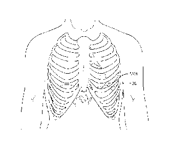
10020]Fig. 1 is a schematic frontal view of a patient showing the location of
an
intercostal incision providing access to the apex of the left ventricle of the
heart;
[0021]Figs. 2A-2B are cross-sectional views through the left side of a
patient's heart
showing a procedure for dilating a calcified aortic annulus prior to
implantation of a
prosthetic heart valve in accordance with the present invention;
100221Figs. 3A-3E are cross-sectional views through the left side of a
patient's heart
showing several steps in a procedure for implanting a prosthetic heart valve
in accordance
with the present invention;
10023]Fig. 4 is an exploded perspective view of an introducer/dilator
combination
used in the port access heart valve implantation procedure of the present
invention;
[0024]Fig. 4A is an assembled view of the introducer/dilator combination of
Fig. 4;
100251Fig. 5A and 5B are exploded perspective and elevational views of the
introducer of Figure 4;
10026] Fig. 6A is a plan view of the introducer of Figure 4;
CA 2971946 2019-07-08
- 7 -
[0027]Fig. 6B is a longitudinal sectional view of the introducer taken along
line 6B-
6B of Fig. 6A;
[0028]Fig. 6C and 6D are enlarged views portions of a variable flexibility
sheath of
the introducer of Fig. 6B;
[00291Fig. 7 is a perspective view of an exemplary balloon catheter/loader
assembly
for implanting a prosthetic heart valve as disclosed herein;
[0030]Fig. 7A is an exploded perspective view of a loader that provides an
interface
between the introducer of Figs. 4-6 and the balloon catheter of Fig. 7;
[0031]Fig. 8 is a broken elevational view of the balloon catheter of Fig. 7;
10032] Fig. 9 is a longitudinal sectional view of a proximal control handle of
the
balloon catheter of Fig. 7;
100331Fig. 10A is a perspective view of the proximal control handle of Fig. 9;
100341Fig. 10B is an exploded view of the proximal control handle of Fig. 9;
[0035]Fig. 11 is an enlarged sectional view of a distal deflecting segment of
the
balloon catheter of Fig. 7, also showing a distal balloon in a deflated state
within a protective
sheath;
10036]Fig. 12 is an enlarged sectional view of a distal balloon of the balloon
catheter
of Fig. 7 in its inflated state;
10037]Fig. 13 is an exploded view of the balloon catheter and introducer (in
section)
combination prior to coupling with a heart valve crimped onto the balloon;
[0038]Fig. 14A is an assembled view of the balloon catheter and introducer (in
section) combination after insertion of the balloon catheter through the
introducer;
10039]Figs. 14B-14E are views similar to Fig. 14A showing use of the heart
valve
delivery system disclosed herein in situ at the occurrence of a series of
steps in a valve
implant procedure;
10040] Fig. 15 is a longitudinal sectional view of a distal end of an
exemplary balloon
catheter showing a prosthetic heart valve crimped over a balloon folded in a
way that
enhances visualization of the valve during implant;
[0041]Fig. 16 is a radial section of the folded balloon of Fig. 15; and
[0042]Fig. 17 is a radial section of the balloon of Fig. 16 illustrating a
preferred
folding technique.
CA 2971946 2019-07-08
- 8 -
Detailed Description of the Preferred Embodiments
[0043]The heart is a hollow muscular organ of a somewhat conical form; it lies
between the lungs in the middle mediastinum and is enclosed in the
pericardium. The heart
rests obliquely in the chest behind the body of the sternum and adjoining
parts of the rib
cartilages, and projects farther into the left than into the right half of the
thoracic cavity so
that about one-third is situated on the right and two-thirds on the left of
the median plane.
The heart is subdivided by septa into right and left halves, and a
constriction subdivides each
half of the organ into two cavities, the upper cavity being called the atrium,
the lower the
ventricle. The heart therefore consists of four chambers; the right and left
atria, and right and
left ventricles.
[0044]As seen in Fig. 1, the left ventricular apex LVA is directed downward,
forward,
and to the left (from the perspective of the patient). The apex typically lies
behind the fifth
left intercostal space (or between the fourth and fifth), 8 to 9 cm from the
mid-sternal line,
and about 4 cm below and 2 mm to the medial side of the left mammary papilla.
Access to
the left ventricle may therefore be attained through an intercostal incision
20 as shown in
dashed line, positioned over the fifth left intercostal space. Such an
approach is often termed
a -mini-thoracotomy," and lends itself to surgical operations on the heart
carried out using
one or more short tubes or "ports" - thus, the operations are often referred
to as "port-access"
procedures.
[00451In a preferred embodiment of the present invention, a surgeon implants a
prosthetic heart valve over the existing native leaflets, which are typically
calcified. There
are procedures and devices for removing calcified leaflets, but the risks
associated therewith,
including a release of calcific material into the bloodstream, are not
insignificant. Therefore,
a heart valve replacement procedure that installs the prosthetic heart valve
directly over and
contains the native leaflets is preferred.
100461Those skilled in the art will recognize that it may be necessary to pre-
dilate the
leaflets and annulus of the stenotic aortic valve before deploying a
prosthetic valve within the
aortic valve. Figs. 2A and 2B are two snapshots of a valvuloplasty procedure
that may be
initially performed to compress the native aortic heart valve leaflets outward
against the
sinuses and ascending aorta. As mentioned above, the native aortic valve
leaflets may be
substantially calcified, and the valvuloplasty may be necessary to crack and
otherwise force
CA 2971946 2019-07-08
- 9 -
apart hardened tissue. Pre-dilation increases the flow area through the aortic
valve and
creates an opening in the leaflets of sufficient size to receive the
prosthetic valve. Pre-
dilatation is preferably achieved using an expandable member, such as a
dilatation balloon
catheter. One example of pre-dilation of a valve annulus is seen in U.S.
Patent No. 6,908,481
to Cribier, issued June 21, 2005.
[00471Fig. 2A illustrates introduction of a guidewire 30 through a pre-formed
apical
puncture 32 in the left ventricle LV. A distal tip 34 of the guidewire 30
extends through the
native aortic valve AV and into the ascending aorta AA. The distal tip 34 may
extend further
over the aortic arch, as seen in Fig. 2B, but the minimum extension is across
the aortic valve
AV.
[0048]Fig. 2B illustrates an introducer sheath 38 inserted into the LV through
the
apical puncture 32, with a balloon catheter 40 having a dilatation balloon 42
on a distal end
passed over the guidewire 30 and through the sheath. As is known, prior to
insertion of the
sheath 38, a dilator having a gradually tapered tip (not shown) may first be
inserted over the
guidewire to enlarge the apical puncture 32. It should be noted at this point
that the surgeon
installs one or more purse-string sutures 44 in the tissue of the left
ventricular apex
surrounding the puncture 32. These sutures 44 are pre-implanted prior to
formation of the
initial puncture. In a preferred embodiment, the surgeon places a first line
of purse-string
sutures generally in a first circle in one direction, and then places a second
line of purse-
string sutures generally in a circle concentric to the first circle but in an
opposite direction.
The result is two concentric circles of separate purse-string sutures 44
defining a periphery
within which the puncture is formed. The purse-string sutures 44 can therefore
be pulled to
cinch the ventricular tissue around whatever object passes through the
puncture. In
particular, the purse-string sutures 44 are tightened around both the
guidewire 30 and
introducer sheath 38. Installing the separate lines of purse-string sutures 44
in opposite
directions helps prevent tearing of the ventricular tissue and provides a more
uniform
compression about whatever elongated object passes through the puncture.
[0049] As indicated in Fig. 2B, the dilatation balloon 42 expands radially
outward into
contact with the native aortic valve leaflets. With information concerning the
size of the
particular aortic valves, the balloon 42 is chosen so that it expands outward
and nominally
compresses the aortic valve leaflets against the surrounding aortic walls.
There are various
CA 2971946 2019-07-08
- 10 -
means for assessing the size of the particular patient's aortic valve,
including ultrasound,
which will not be described herein. Suffice it to say that following the
valvuloplasty
procedure seen in Fig. 2B, the native aortic valve leaflets are compressed
outward against the
aortic wall and a substantially circular orifice results. Additional details
regarding pre-
dilatation and valve replacement can be found in Applicant's U.S. Patent No.
6,908,481 to
Cribier.
100501 With reference now to Figs. 3A-3E, a preferred method of deploying and
implanting a prosthetic heart valve of the present invention using a
transapical approach will
now be described in more detail. The devices and methods disclosed herein are
particularly
well-suited for replacing a stenotic aortic valve, and as such that the pre-
dilation procedure
seen in Figs. 2A-2B typically precedes the valve implantation so as to smooth
out the
contours of the annulus and leaflets. It should be noted, however, that the
procedure
described herein may be performed without valve pre-dilation.
[0051]Furthermore, the present procedure may be performed as a first time
valve
implant or to supplement a previous implant. A relatively large proportion of
recipients of
prosthetic heart valves are older, typically older than 60. Over time,
prosthetic heart valves
have been known to show reduced performance and even failure. Re-operating on
septegenarians and even octogenarians is problematic. However, a port access
procedure
such as disclosed herein eliminates open-heart surgery and potentially
cardiopulmonary
bypass, and is therefore more desirable for the aging patient. Therefore, the
present invention
contemplates transapical implantation of a prosthetic heart valve over an
existing prosthetic
valve implant. In such a case, a pre-dilation step is typically not necessary,
though it is
conceivable.
[0052]Prior to a discussion of the procedure itself, it should be noted that a
preferred
delivery system of the present invention will be described in greater detail
below with
reference to Figs. 4-13. The workings of the present delivery system may be
more easily
understood after an explanation of the steps taken to ultimately implant the
valve in the aortic
annulus.
100531The prosthetic heart valve implantation procedure described herein may
be
performed in conjunction with cardiopulmonary bypass, or without bypass in a
so-called off-
pump procedure. The necessity for bypass depends on a number of factors,
including the
CA 2971946 2019-07-08
- 11 -
patient's age, vulnerability to such a procedure, and viability of the native
leaflets. Ideally,
the implantation procedure is performed off-pump.
100541The surgeon or cardiologist first sizes the aortic valve using a
physical sizer, or
with echocardiography. The physician or operating room staff then crimps an
expandable
prosthetic valve 50 over the balloon 52 of a balloon catheter 54 (some of the
elements
presently described can be seen in the procedure drawings of Figs. 3A-3E,
while others can
be seen in the system drawings of the Figs. 4-13). The surgeon advances the
balloon catheter
54 over a guidewire 60 (that might be the same guidewire 30 used in a pre-
dilation
procedure), through an introducer sheath 70 that has been inserted through the
left ventricular
apex puncture 32 with the help of a dilator 74 (sometimes also referred to as
an introducer).
100551The same purse-string sutures 44 that were used for the pre-dilation
procedure
may also be used to seal the ventricular tissue around the introducer sheath
70. In the
absence of the pre-dilation procedure, the purse-string sutures 44 are pre-
implanted prior to
formation of the initial puncture. As before, the surgeon places a first line
of purse-string
sutures generally in a first circle in one direction, and then places a second
line of purse-
string sutures generally in a circle concentric to the first circle but in an
opposite direction.
The result is two concentric circles of separate purse-string sutures 44
defining a periphery
within which the puncture is formed, and which seal around the introducer
sheath 70.
10056]Furthermore, the dilator 74 that expands the inner diameter of the
puncture 32
and rides over the guidewire 60 may be inserted prior to or with the
introducer sheath 70.
Preferred dilator diameters range between 12 and 22 French. The introducer
sheath 70
comprises the distal end of an introducer that will be described below.
Introducer sheath
diameters of no greater than 24 French, and desirably 22 or 24 Fr are
preferred.
1[00571Fig. 3A shows the introducer sheath 70 passing into the left ventricle
through
the puncture 32 and over the guidewire 60 that extends upward through the
calcified aortic
valve AV. The surgeon locates a distal tip 72 of the introducer sheath 70 just
to the inflow
side of the aortic valve AV, as seen in Fig. 3A. At this point, it should be
understood by
those of skill in the art that the position of the introducer sheath 70
relative to the aortic valve
AV, as well as the position of other elements of the system, is monitored
using radiopaque
markers and fluoroscopy, or using other imaging systems such as
transesophageal echo,
transthoracic echo, intravascular ultrasound imaging (IVUS), or an injectable
dye that is
CA 2971946 2019-07-08
- 12 -
radiopaque. A specific combination of such markers for the exemplary system
will be
described below.
[00581Fig. 3B shows the advancement of the balloon catheter 54 over the
guidewire
60 and through the introducer sheath 70. Ultimately, as seen in Fig. 3C, the
prosthetic heart
valve 50 is located at the aortic annulus and between the native aortic
leaflets. Fig. 3C also
illustrates retraction of the introducer sheath 70 from its more forward
position in Fig. 3B to
permit balloon inflation/valve expansion. Radiopaque markers may be provided
on the distal
tip 72 of the introducer sheath 70 to more accurately determine its position
relative to the
valve 50 and balloon 52.
[0059]Again, the precise positioning of the prosthetic heart valve 50 may be
accomplished by locating radiopaque markers on its distal and proximal ends,
or in-between,
for example at a midpoint. Desirably, the surgeon can adjust the position of
the valve 50 by
actuating a steering or deflecting mechanism within the balloon catheter 54,
as will be
described below. Furthermore, the rotational orientation of the valve 50 can
be adjusted
relative to the cusps and commissures of the native aortic valve by twisting
the balloon
catheter 54 from its proximal end and observing specific markers on the valve
(or balloon
catheter) under fluoroscopy. One of the coronary ostia 80 opening into one of
the sinuses of
the ascending aorta is shown, and those of skill in the art will understand
that it is important
not to occlude the two coronary ostia with the prosthetic valve 50. It should
also be noted
that although the native leaflets of the aortic valve AV are shown coapting in
Fig. 3A, and
being flexibly displaced by the balloon catheter 54 in Figs. 3B and 3C, they
may actually be
compressed further outward against the aortic annulus from a pre-dilation
procedure.
[0060ifig. 3C shows the prosthetic heart valve 50 in its contracted or
unexpanded
state crimped around the balloon 52. When the surgeon is satisfied of the
proper positioning
and rotational orientation of the valve 50, the balloon 52 is expanded as seen
in Fig. 3D.
Proper size measurement of the native aortic valve AV enables the surgeon to
select an
optimum-sized valve 50 such that it expands outward into good contact with the
aortic
annulus. The term "good contact" implies sufficient contact to ensure that the
prosthetic heart
valve 50 does not migrate after implant. Excessive expansion of the valve,
however, may
damage surrounding tissue or interfere with the performance of adjacent
valves.
CA 2971946 2019-07-08
- 13 -
[0061] A number of devices are available to assist in anchoring the prosthetic
valve
50 into the aortic annulus, such as barbs and the like. A preferred
configuration of prosthetic
heart valve 50 for use with the present invention is disclosed in co-pending
U.S. Patent Serial
No. 7,993,394 to Hariton, filed June 8, 2009. Another valve is disclosed in
U.S. Patent No.
7,276,078 to Spenser, filed June 30, 2004. Of course, the valve 50 can take a
variety of
different forms but generally comprises an expandable stent portion that
supports a valve
structure. The stent portion has sufficient radial strength to hold the valve
at the treatment site
and resist recoil of the stenotic valve leaflets. Additional details regarding
preferred balloon
expandable valve embodiments can be found in U.S. Patent Nos. 6,730,118 and
6,893,460,
both to Spenser. The preferred prosthetic heart valve 50 includes sufficient
irregularity on its
outer surface such that it may be anchored in the aortic annulus without the
use of barbs or
other tissue piercing structure.
[006210nce the valve 50 is properly implanted, as seen in Fig. 3D, the surgeon
deflates the balloon 52, and withdraws the entire delivery system including
the balloon
catheter 54 over the guidewire 60. The introducer sheath 70 is then withdrawn,
followed by
the guidewire 60. Ultimately, the purse-string sutures 44 previously described
are cinched
tight and tied to close the puncture 32, as seen in Fig. 3E.
[0063] It is important to recognize that the heart valve delivery system of
the present
invention is particularly well-suited for the antegrade, left ventricular
apex, "transapical,"
approach. More particularly, the mini-thoracotomy approach requires relatively
short
instruments. Therefore, the portion of the introducer sheath 70 that extends
into the body is
desirably no more than about 8 inches (20 cm) long, and the length of the
balloon catheter 54
that may extend into the introducer sheath 70, i.e., the "working length," is
desirably no more
than about 24 inches (61 cm). Further specifics on the relatively short length
of the balloon
catheter 54 and introducer sheath 70 will be provided below. The short length
of the
prosthetic heart valve delivery system described herein is also well-suited
for other
anatomical approaches, including through the carotid or subclavian arteries.
The short length
of the system is desirable because it enhances controllability and
steerability of the distal end,
relative to longer systems, which helps improve accuracy and reduced time for
valve
positioning.
CA 2971946 2019-07-08
- 14 -10064111e delivery system of the present invention essentially comprises
an
introducer 100, the balloon catheter 54, and attendant couplers and operating
structures,
including a loader 140 between the introducer and balloon catheter as seen in
Fig. 7. The
introducer 100 is illustrated in Figs. 4-6, while the balloon catheter 54 and
loader 140 are
shown in Figs. 7-12. It should be noted that the delivery system is similar to
another system
used to percutaneously implant a prosthetic aortic valve, which is disclosed
in co-pending
U.S. Patent Publication No. 2007-0005131 to Taylor, filed June 13, 2005. The
present
system differs in several aspects that make it more suitable for a
transapical, port-access, or
direct-access approach, although some features are common.
[0065]As seen in Figs. 4 and 4A, the introducer 100 comprises the
aforementioned
distal sheath 70 coupled to an introducer housing 102 containing a series of
valves. The
exploded views of Figs. 5A and 5B shows an end cap 104 detached from the
introducer
housing 102. The end cap 104 includes a flanged nipple 105 for mating with the
loader 140,
as will be explained below. The end cap 104 threads or otherwise attaches to
the housing 102
and retains therein, in series from proximal to distal, a cross-slit valve
106, a disk valve 108,
a spacer 110, and a duck-bill valve 112. These three valves function to
provide a seal when
no instruments pass through the introducer 100, and when several different
sizes of
instruments pass therethrough. For example, the valves seal around both the
guidewire 60
and the balloon catheter 54 as previously shown. The introducer sheath 70
extends into the
body vessel, with the introducer housing 102 located outside the body vessel.
In a preferred
embodiment, the introducer sheath 70 possesses an external hydrophilic coating
and has a
length of between about 20-24 cm so that it may extend through the access
incision 20 (see
Fig. 1), into the left ventricle and reach the aortic annulus.
100661As seen best in Figs. 5 and 5A, the introducer sheath 70 attaches to the
housing
102 via a sealing extension 122 that mates with a distal nipple 124 extending
from the
housing 102. Preferably adhesive is used between these two mating sections. A
threaded nut
126 rides over the sheath 70 and couples to threading 128 provided on the
housing 102 just
proximal to the nipple 124. In this way, the various components can be
manufactured
(typically molded or extruded) separately and easily coupled together during
assembly.
Adhesive may be applied to the threading 128 prior to coupling the nut 126 for
a more secure
final assembly.
CA 2971946 2019-07-08
- 15 -
[00671A side port tube 130 extends at an angle away from the introducer
housing 102
and terminates in a three-way stopcock 132. This permits the user to infuse
medicaments or
other fluids through the lumen of the introducer 100 even if devices such as
the balloon
catheter 54 are present therein.
[00681Figs. 6A-6D show further details of the introducer 100, including a
series of
depth markings 133 on a distal section of the sheath 70. The markings 133
indicate the
distance in millimeters from the distal tip 72 so that the depth to which the
distal tip extends
into the left ventricular apex can be easily seen.
[00691Figs. 6B and 6C illustrate an advantageous construction in which the
sheath 70
has greater flexibility along a distal section than along a proximal section.
Specifically, the
sheath 70 includes a distal section 134 having a length Li that is more
flexible than a
proximal section 135, wherein the total length of the sheath 70 is L
(extending from the
threaded nut 126). Fig. 6C shows the internal construction of the sheath 70,
which includes
an inner tubular liner 136, a reinforcing coil 137, a distal exterior tube 138
and a proximal
exterior tube 139. The liner 136 and coil 137 extend the total length L of the
sheath 70, while
the exterior tubes 138, 139 abut in series. The stiffness of the proximal
exterior tube 139 is
desirably greater than that of the distal exterior tube 138 to provide the
differing flexibilitics.
Although two discrete sections each with constant stiffness are shown, the
flexibility may be
varied in more than two sections, and more gradually, with similar results.
[0070113y providing a more flexible distal section 134, movement of the heart
muscle
surrounding the introducer sheath 70 (such as in the position of Fig. 3D) is
accommodated
with less trauma to the heart tissue. That is, the preferred procedure is with
a beating heart
with the left ventricle continually contracting and relaxing, which creates a
significant
amount of tissue/introducer movement. Permitting the distal end of the
introducer to flex, or
be floppy, helps reduce damage to the heart wall. Moreover, the surgeon often
manipulates
the catheter or introducer for better implant site access, which with a
stiffer sheath may cause
trauma to the heart wall. At the same time, the stiffer proximal section 135
ensures that the
introducer 100 projects out from the operating field in a relatively straight
line, with minimal
floppiness, which is desired by surgeons. Sometimes a stabilizer at the point
of incision may
be used, which reduces the heart wall movement, though the floppy distal end
of the sheath
still provides a benefit.
CA 2971946 2019-07-08
- 16 -
[0071]The liner 136 provides a smooth inner surface through which the balloon
catheter with heart valve may pass without hindrance, and the coil 137
provides hoop
strength to the tubular structure to prevent kinking. The sheath 70 may be
fabricated using a
number of tube forming techniques, such as extrusion.
10072]In one embodiment, the total length L of the sheath 70 is between about
20-24
cm, while the distal section 134 has a length L1 of between about 4 cm and one
half the total
length L. More preferably the distal section 134 has a length Li of between
about 6-9 cm,
and most preferably about 9 cm. The length Li should be sufficient to permit
the floppy
portion of the sheath 70 to extend at least 4 cm into the heart wall.
10073]In an exemplary embodiment, the inner liner 136 and exterior tubes 138,
139
are formed of the same material for better melding, while the coil 137 is
metallic. One
particular combination is the liner 136 and exterior tubes 138, 139 made of a
nylon block
copolymer sold under the tradename PEBAX , while the coil 137 is stainless
steel. The
commercial PEBAX polymers consist of polyether blocks separated by polyamide
blocks.
The polyether blocks may be based upon polyethylene glycol, polypropylene
glycol, or
polytetramethylene ether glycol. The polyamides are usually based upon nylon-
11 but may
be based upon nylons 6 of nylon-6,6 or even a copolymer such as nylon-6/nylon-
11. The
polymers range in hardness as measured in durometer from Shore A 60 to Shore
D72, and the
proximal exterior tube 139 has a greater durometer than the distal exterior
tube 138. A
selection of PEBAX compositions and their respective physical properties are
provided on
the website, www.pebax.com, in particular under the link, "Medical
Applications."
PEBAX is a registered trademark of Arkema Inc. of Paris, France, with U.S.
Corporate
offices in Philadelphia, PA.
100741Fig. 6D also shows an advantageous visualization system for the distal
tip 72 of
the introducer sheath 70. A circular array of marker dots 73 at the distal tip
72 can be seen
under fluoroscopy, and in clear contrast to marker bands provided on the
balloon catheter 54,
as explained below.
[00751Fig. 7 illustrates in perspective the balloon catheter 54, which
comprises an
assembly of interrelated components commencing on a proximal end with a luer
fitting 142
and terminating at a distal end in a soft tip 144. The balloon catheter 54,
also shown in plan,
sectional, and exploded views in Figs. 8-12, comprises a control handle 150
having the luer
CA 2971946 2019-07-08
- 17 -
fitting 142, a balloon inflation connector 152, a deflection actuator 154, and
a pusher actuator
156. A pusher body 158 extends from the handle 150 around a balloon deflection
tube 160
having the expandable balloon 52 located just proximal to the soft tip 144.
Fig. 7 illustrates a
balloon sheath 161 covering the balloon 52 which protects the balloon during
shipping and is
removed prior to use of the system. An elongated stationary protective sleeve
162 also
extends from the handle 150 over a majority of the pusher body 158 and forms
an exterior
surface of the balloon catheter 54 along much of its length. The loader 140
shown in
perspective in Fig. 7A will be described in more detail below and provides a
coupling
between the balloon catheter 54 and the above-described introducer 100.
100761As mentioned, the present application discloses an advantageous
visualization
system for the distal tip 72 of the introducer sheath 70. Specifically, at
least one marker band
will be provided on the proximal end of the balloon 52, and also on a distal
end of the pusher
body 158. The axial proximity of the distal end of the pusher body 158 and the
proximal end
of the balloon 52 can therefore be easily seen to facilitate their engagement.
In addition, the
circular array of marker dots 73 at the distal tip 72 of the introducer sheath
70 clearly
contrasts with the marker bands on the balloon catheter 54 and the pusher body
158, and
helps the surgeon ensure that the introducer has been retracted far enough at
the time of valve
positioning and balloon expansion.
100771Prior to a detailed description of the exemplary balloon catheter 54,
its
interaction with the introducer 100 via the loader 140 will be explained. As
seen in Fig. 7A,
the loader 140 has a generally tubular body 172 and a slightly externally
tapered distal nose
174 that fits within the introducer 100, and specifically through the series
of valves 106, 110,
112 shown in Fig. 6B. The loader body 172 includes a pair of attached
cantilevered fingers
176 extending longitudinally with internally facing snap ridges for securing
the loader 140 to
a nipple on the proximal end cap 104 of the introducer 100. The loader 140
facilitates
introduction of the balloon catheter 54 into the introducer 100. As described
above, the
introducer housing 102 contains the series of valves 106, 110, 112 that in
aggregate provide
an effective fluid seal against egress of blood through the introducer 100 in
the presence or
absence of different sized medical implements. The distal nose 174 of the
loader 140 extends
through the introducer housing 102 and through these valves 106, 110, 112 (see
Fig. 14A) to
hold them open and provide a smooth internal lumen which matches the size of
the lumen of
CA 2971946 2019-07-08
- 18 -
the introducer sheath 70. In this way, the somewhat irregular contours of the
balloon catheter
54 having a prosthetic valve 50 crimped around the balloon 52 may smoothly
pass into the
introducer sheath 70.
[0078]A loader seal, seen exploded in Fig. 7A. positioned within a proximal
housing
178 comprises a pair of annular washers 180 and a resilient vent member 182.
As seen in
Fig. 7, the protective sleeve 162 passes through the loader 140, and the
loader seal prevents
fluid from escaping around the sleeve. The vent member 182 includes a pair of
lateral
buttons 184 that project through apertures in the side of the proximal housing
178. Inward
depression of one or both buttons 184 causes deformation of the vent member
182, which in
turn opens the distal space within the loader body 172 to the atmosphere. Any
air entrained
in the blood within the loader body 172 can thus easily be vented with one
hand. The one-
handed aspiration is both more convenient and also helps avoid inadvertent
misalignment of
the heart valve from unscrewing a valve cap to vent, a two-handed operation,
which is the
conventional arrangement. Moreover, eliminating the previous threaded cap
arrangement for
tightening a resilient seal with the passive loader seal means that movement
of the protective
sleeve 162 (and delivery catheter 54) is never prevented by the loader valve.
In this way,
movement of the catheter 54 is decoupled from the loader 140 and attached
introducer 100.
100791Prior to balloon expansion as seen in Fig. 12, the loader 140 couples
over the
distal extent of the balloon catheter 54, as seen in Fig. 7. The distal nose
174 inserts into the
introducer housing 102 and the cantilevered loader fingers 176 mate with the
flanged nipple
of the end cap 104 (Fig. 14A). The balloon catheter 54 is thus coupled to the
introducer 100.
Sliding the entire balloon catheter 54 distally permits the irregular contours
of the distal
extremity thereof to pass safely across the valves 106, 110, 112 and into the
introducer sheath
70. The loader 140 remains coupled to the introducer 100 during the valve
implant
procedure, and the vent member 182 can be actuated as needed to ensure no air
remains in the
system.
100801The various components of the balloon catheter 54 will now be described
with
respect to Figs. 8-12. The catheter 54 includes the proximal control handle
and a plurality of
concentric tubes that extend distally to the soft tip 144. In the exemplary
embodiment, five
concentric tubes of gradually smaller size connect to or extend into the
handle 150, as seen in
CA 2971946 2019-07-08
- 19 -
Fig. 10B. The handle 150 includes two molded halves having a plurality of
inner walls and
cavities to contain the various components.
[0081]The handle 150 includes a number of control components and is shown in
section in Fig. 9 and exploded in Figs. 10A and 10B. Specifically, the
deflection actuator 154
in the form of a trigger controls deflection of the distal tip of the balloon
deflection tube 160,
the pusher actuator 156 in the form of a slider controls longitudinal movement
of the pusher
body 158, and operation of a stopcock 190 permits infusion of fluids to flush
a space between
the introducer sheath 70 and the pusher body 158. Furthermore, a Y-port 192 at
the proximal
end of the handle 150 provides a longitudinal passage leading to the luer
fitting 142 and an
angled passage leading to the balloon inflation connector 152. An inner tube
194 (smallest)
having a throughbore extends the length of the balloon catheter 54 from the
luer fitting 142
through the distal soft tip 144 (see Fig. 12). The inner tube 194 provides a
channel for
passage of a guidewire, such as shown at 60 in Fig. 3D. The luer fitting 142
also may
provide an entry point for injection of radiographic contrast medium though
the inner tube
194, which is useful to check for perivalvular leaks after the prosthetic
valve is implanted.
[0082] Still with reference to Figs. 8-10, and in particular Fig. 9, a balloon
inflation
tube 196 (second smallest) surrounds the inner tube 194, extending from the Y-
port 192 in a
distal direction and terminating within the balloon 52. As seen in Fig. 9, the
Y-port 192
includes a stepped longitudinal bore having a larger distal portion that
sealingly receives the
balloon inflation tube 196, and a smaller middle portion that sealingly
receives the inner tube
194. The angled passage leading to the balloon inflation connector 152 fluidly
communicates
with a space outside of the inner tube 194 that opens to the lumen of the
balloon inflation
tube 196. With this configuration, fluid injected into the balloon inflation
connector 152
passes into and travels the length of the balloon inflation tube 196 until it
exits from the open
distal end 198 thereof, within the balloon 52 (as seen in Fig. 12). Additional
fluid egress
ports (not shown) may be provided in the balloon inflation tube 196 along the
length of the
balloon 52 for even inflation, and in particular ports proximal and distal to
the prosthetic
heart valve 50 are beneficial to help expand both ends of the valve at the
same rate.
10083] The balloon inflation tube 196 extends through the lumen of the balloon
deflection tube 160 (third smallest) which has a proximal end anchored by a
collar 200 fixed
within a cavity of the handle 150. The balloon deflection tube 160 has a
particular
CA 2971946 2019-07-08
- 20 -
construction that enables flexing along its length without kinking, and has a
deflectable distal
tip. More particularly, the balloon deflection tube 160 desirably includes a
braided tube
along its length to prevent kinking, a coil structure at its distal tip for
deflection, and a
deflection wire 202 that extends from the proximal end to the coil.
[0084]The deflection wire 202 also includes a plug 204 fixed on its proximal
end
acted on by a rail 206 that slides longitudinally within the handle 150.
Specifically, the
deflection wire 202 passes through an aperture of a finger 208 on the rail
206, which aperture
is smaller than the plug 204. The plug 204 is desirably cylindrical and may be
constrained
within a small guide sleeve 210 held within a cavity of the handle 150. The
rail 206 forms
part of a trigger assembly and moves with the trigger 154. Pulling the trigger
154 to the left
from its position in Figs. 8 and 9 will displace the plug 204 to the left,
also pulling the
deflection wire 202 to the left, or in a proximal direction. The deflection
wire 202 in turn
attaches to one side of the coil at a distal tip 212 of the balloon deflection
tube 160, and
pulling on the wire thus deflects the distal tip, as seen in Figs. 14D and
14E. Of course by
rotating the entire balloon catheter 54 about its axis the deflecting segment
212 may be
steered in any direction. The coil provides both flexibility and resiliency
such that release of
tension on the deflection wire 202 permits the deflecting segment 212 to
return to a straight
orientation.
10085]The construction of the deflection tube 160 enables a size reduction
from prior
designs that ultimately enables a size reduction of the valve 50 and balloon
52. In one
embodiment, the deflection tube 160 has a dimension no greater than 8 French.
The braided
proximal portion provides flexibility and column strength, while the distal
coil enables the
deflection only at the distal end. The distal tip 212 having the coil
structure desirably has a
length of about 4 cm. This construction also facilitates manufacture, as the
braided proximal
portion and coil with attached deflection wire 202 are easily combined using
welding or the
like.
[0086]The second largest tube is the pusher body 158, which is tubular until
an
outwardly flared sleeve 220 on its distal end (see Figs. 8 and 11). A proximal
end of the
pusher body 158 affixes to a threaded sleeve 222 that couples with an
internally threaded
bore of a slider cap 224, as seen in Figs. 9 and 10B. One or more passive 0-
ring seals 226
within the bore of the slider cap 224 permit relative movement of the slider
member over the
CA 2971946 2019-07-08
- 21 -
balloon deflection tube 160 while sealing against blood leakage therebetween.
Desirably,
two 0-rings 226 sandwich an annular polymer (e.g., nylon) washer 228 to help
even out the
forces on each of the 0-rings and therefore enhance the quality of the fluid
seal around the
balloon deflection tube 160. Translation of the slider 156 and attached slider
cap 224 along a
corresponding longitudinal slot in the handle 150 thus displaces the pusher
body 158 relative
to the handle and to the balloon deflection tube 160. Previous devices
included separate
handles and the seal would be positioned within a threaded cap that required
tightening. The
passive nature of the 0-ring seal eliminates the two-handed tightening
operation and also
avoids any misalignment of the heart valve 50 once positioned from inadvertent
movement of
the balloon deflection tube 160.
[0087]Moreover, the design of the handle 150 facilitates one-handed operation
of the
two primary movements of the balloon catheter 54 ¨ deflection of the distal
tip and linear
movement of the pusher body 58. The handle 150 preferably includes ergonomic
ribs 230 on
its underside, as seen in Fig. 8, which, coupled with ribs on the slider 156
assist in moving the
pusher body 158 along the catheter.
[0088]The pusher body 158 slides over the balloon deflection tube 160 as well
as
inside of the stationary protective sleeve 162 (the largest tube). As seen in
Fig. 9, the sleeve
162 affixes into a stepped bore of a housing of the stopcock 190, which in
turn attaches to a
distal end of the handle. An 0-ring seal 232 held within the stopcock housing
(or between
the housing and the handle 150) contacts and seals against the exterior of the
moving pusher
body 158 and prevents leakage of fluid from the concentric space between the
pusher body
158 and the stationary protective sleeve 162. Saline or other such fluid may
thus be infused
in through the stopcock 190 to travel down and flush the concentric space
between the pusher
body 158 and the stationary protective sleeve 162.
[00891 Fig. 11 is an elevational view of the distal end of the balloon
catheter 54
showing the balloon 52 deflated and its proximal end spaced from the pusher
sleeve 220,
while Fig. 12 shows the distal end of the balloon catheter 54 with the balloon
52 inflated.
[0090]The inner tube 194 passes through the balloon 52 and terminates at a
distal end
that is capped by the aforementioned soft tip 144. The soft tip 144
facilitates introduction of
the balloon catheter 54 and reduces trauma to surrounding tissue. This is
particularly
important in the preferred procedure of the present invention where the
catheter enters the
CA 2971946 2019-07-08
- 22 -
apex of the left ventricle and travels through the aortic valve into the
ascending aorta. As was
seen in Fig. 3D, the distal tip of the catheter may extend far enough to enter
the aortic arch,
and the soft tip 144 thus prevents rupture or other abrasion to the
surrounding vascular tissue.
Fig. 13 also illustrates the open distal end of the inner tube 194 and soft
tip 144 through
which first a guidewire 62 may be passed and then radiographic contrast medium
may be
injected to test valve sufficiency after implant.
[0091]The balloon 52 includes a first cone portion 240, a main cylindrical
portion
242, and a second cone portion 244. The prosthetic heart valve 50 desirably
crimps around
the main cylindrical portion 242 for even cylindrical expansion, such as shown
in phantom in
Fig. 12. The balloon 52 can be formed of nylon, and is rated at a burst
pressure of 6-8 atm.
In preferred embodiments, the expanded diameter of the balloon ranges from
about 20 to 28
mm, the particular size depending on the size of the heart valve 50 being
implanted.
[0092IF ig. 15 is a longitudinal sectional view of a distal end of the
exemplary balloon
catheter 54 showing a prosthetic heart valve 50 crimped over the balloon 52.
The heart valve
50 has a shorter length than the balloon 52 leaving proximal and distal
exposed portions
thereof.
[0093]The balloon 52 is folded in a way that enhances visualization of the
valve
during implant. Specifically, certain conventional folding techniques resulted
in wrinkling of
the balloon 52. For example, a common way to fold a catheter balloon is to
first form a
trifold and then wrapping the leaves of the trifold around the balloon
catheter axis. Folding
techniques like this often leave wrinkles or ripples even if done carefully.
Such irregularities
show up on echocardiography, which can interfere with precise location of the
proximal and
distal ends of the valve 50 relative to the implant site. The balloon 52 of
the present
invention on the other hand is folded in a manner that reduces if not
eliminates irregularities
that show up on echocardiography, thus enhancing the ability to properly
locate the heart
valve 50 at the aortic annulus.
100941Fig. 16 is a radial section of the folded balloon of Fig. 15, and shows
four
leaves 250 of the balloon 52 folding in a clockwise manner around the inner
tubes 194, 196,
though of course the direction that the leaves are wrapped is not critical.
Fig. 17 illustrates
the leaves 250 prior to folding. The leaves 250 extend longitudinally along
the balloon 52
and comprise even circumferential spans of the balloon 52. By careful
selection of the radial
CA 2971946 2019-07-08
- 23 -
dimension of each leaf 250, the resulting wrapped structure in Fig. 16 is
minimized for that
size of balloon, and ensuring even circumferential wrapping rates results in
longitudinal lines
in the wrapped structure. The longitudinal fold lines contrast under
fluoroscopy with the
radial ends of the valve 50, thus ensuring a clear view of the valve.
Moreover, the
longitudinal fold lines contrast with marker bands on the balloon and the
pusher, as explained
above. There may be four or more, possibly 6-8 folds or pleats pre-formed in
the balloon
which also facilitate deflation and removal through the valve and introducer.
100951In use, the present invention provides a novel and effective way for
implanting
a prosthetic heart valve 50 in the aortic annulus. The steps of the procedure
have been
described above with respect to Figs. 1-3, at least as far as the final
implantation steps. A
description of the advantageous use of the exemplary balloon catheter 54 and
introducer 100
in performing the entire procedure will now be provided with reference to
Figs. 13 and 14A-
14E, which are in situ views of the system without the valve 50.
100961First, as mentioned above, the physician determines the size of the
patient's
annulus. This can be done physically by creating the incision 20 and puncture
32 (Figs. 1 and
2A) in the left ventricular apex, and inserting a sizing tool into the aortic
annulus. However,
the puncture 32 may not be large enough to pass a conventional sizer, and an
alternative
technique such as echocardiography or other such imaging system may be
utilized.
[00971-Next, the balloon catheter 54, introducer 100, loader 140, and
prosthetic heart
valve 50 are selected, and prepared for use by removing them from any
packaging and rinsing
or sterilizing as needed. A pre-dilation step as described above with respect
to Figs. 2A-2B
may be performed to enlarge or crack existing calcification in the aortic
annulus.
[00981The process of crimping the prosthetic heart valve 50 over the balloon
52 may
be accomplished in a number of ways, and there are suitable devices on the
market for
crimping balloon-expanding stents over balloons. In a preferred embodiment, a
device
having a compressing mechanism that works like the aperture iris of a camera
is utilized. In
such a device, multiple continuous segments around the periphery of the
prosthetic heart
valve 50 close separately but in concert so that uniform inward pressure is
exerted on the
heart valve. The devices typically operate manually.
10099] Subsequently, the aforementioned pusher body 158 and flared sleeve 220
are
advanced distally over the proximal end of the balloon 52, such as seen in
Fig. 13. The
CA 2971946 2019-07-08
- 24 -
loader 140 is then secured over the distal end of the balloon catheter 54,
including the
assembly of the balloon 52 and prosthetic valve (not shown).
[0100] At this point, or at the same time as balloon catheter preparation, the
introducer
100 is positioned within the left ventricle as seen in Fig. 3A. Again, the
purse-string sutures
44 maintain a fluid tight seal around the introducer sheath 70. During the
entire procedure
the heart may continue beating. The physician inserts the distal nose 174 of
the loader 140
into the proximal end cap 104 of the introducer 100 and bottoms the loader out
such that the
cantilevered fingers 176 engage the flanged nipple 105 of the introducer, as
seen in Fig. 14A.
At this point, the balloon catheter 54 is ready for introduction in the body.
[0101] The pusher body 158 and pusher sleeve 220, as well as the stationary
protective
sleeve 162, facilitate advancement of the deflecting segment 212 and attached
balloon 52
having the valve 50 crimped thereon through the introducer sheath 70 and its
valves 106, 110,
112. In particular, the flared pusher sleeve 220 surrounds the deflecting
segment 212 and a
proximal portion of the balloon 52 during passage through the introducer
sheath 70. The
pusher sleeve 220 secures the crimped valve from movement relative to the
balloon 52.
Eventually, proximal retraction of the pusher body 158 relative to the balloon
deflection tube
160 frees the deflecting segment 212 for angled movement, and the balloon 52
for expansion.
[0102] The physician then distally advances the balloon catheter 54 with
respect to the
loader 140 and introducer 100 into a position such as that shown in Fig. 14B.
In this state,
the balloon 52 with valve may be advanced to its eventual implant position
using
echocardiography, for example.
[0103] The physician then retracts the pusher sleeve 220 from the deflecting
segment
212 and the proximal portion of the balloon 52, as seen in Fig. 14C, by simply
sliding back
the pusher actuator 156 on the handle 150. The stationary protective sleeve
162 around the
pusher body 158 serves to decouple movement of the pusher from the valves of
the
introducer, thus reducing friction on the pusher. Also, the one-handed
operation of sliding
back the pusher actuator 156 while grasping the handle 150 greatly reduces the
chance of
misalignment of the valve position.
[0104] The physician may further advance and angle the balloon 52 until it
reaches the
position shown in Fig. 3C. The entire operation is visualized using
radiographic markers and
fluoroscopy, and the precise positioning of the balloon 52 and prosthetic
valve 50 mounted
CA 2971946 2019-07-08
- 25 -
thereon is accomplished by axial movement and rotation of the catheter 54
coupled with
angular changes of the deflecting segment 212, as seen in Fig. 14D.
Specifically, as the
prosthetic valve 54 advances it is aligned as much as possible along the flow
axis of the
native aortic valve AV by gross movement of the catheter 54 and slight changes
in its angular
orientation by tensioning the deflecting wire 202 with the deflection actuator
154.
[0105] As mentioned above, the deflection wire 202 (Fig. 108) extends from the
handle 150 along the balloon deflection tube 160 and terminates at the
deflecting segment
212, and preferably at a distal end of a coil spring therein (not shown).
Pulling the deflection
wire 202 causes the deflecting segment 212 to be pulled to the side of
attachment of the wire,
thus deflecting the distal end of the catheter and balloon 52, as in Fig. 14D.
[0106] Ultimately, the valve 50 is positioned correctly as in Fig. 3C taking
care that
the valve 50 is not liable to block either of the coronary ostia 80 when
expanded. Saline
mixed with contrast is then injected through the balloon inflation connector
152 which passes
through the length of the balloon inflation tube 196 to fill the balloon 52,
as seen in Fig. 14E.
The balloon 52 is of a type that has a maximum expanded diameter which has
previously
been selected to properly expand the prosthetic heart valve 52 to its optimum
diameter in
contact with the surrounding aortic valve AV, and calcified leaflets if they
remain in place.
The step is illustrated in Fig. 3D. The balloon 52 expands the prosthetic
heart valve 50 to
implant it in the annulus, after which the balloon is deflated and removed
from within the
valve.
[0107] Subsequently, radiographic contrast medium may be injected from the
proximal luer connection 142 of the balloon catheter 54 to egress through the
distal soft tip
144 and test the efficacy of the just-implanted prosthetic valve 50. If the
valve is properly
functioning, the balloon catheter 54 is withdrawn into the introducer sheath
70, which is
removed from the puncture 32. The purse-string sutures 44 are closed up to
seal the puncture
32.
[0108] Once again, the delivery system described herein is particularly well-
suited for
an antegrade, transapical approach, partly because of its relatively short
length. With
reference to Fig. 4A, the entire length of the introducer 100 is approximately
13 inches (33
cm), while the length of the sheath 70 that may extend within the body is
between about 20-
24 cm. The portion of the balloon catheter 54 that extends into the introducer
100 (that is, the
CA 2971946 2019-07-08
- 26 -
portion of the balloon catheter from the distal soft tip 144 to approximately
the deflection
handle 154) is preferably no more than about 24 inches (61 cm), which permits
about 11
inches (28 cm) of the balloon catheter to extend beyond the introducer distal
tip 72 (see Fig.
4). It should be noted that the relatively short length of the delivery system
is unsuited for a
longer, more circuitous approach through the peripheral vasculature, such as
shown in co-
pending U.S. Patent Publication No. 2007-0005131 to Taylor. Also, the steering
mechanism
is provided on the balloon catheter 54 itself, rather than on a secondary
catheter used for
guiding the balloon catheter, as is done in U.S. Patent Publication No. 2007-
0005131. The
short length of the balloon catheter and the ability to directly manipulate it
greatly enhances
successful positioning of the prosthetic heart in the aortic annulus.
10109] While the invention has been described in its preferred embodiments, it
is to be
understood that the words which have been used are words of description and
not of
limitation. Therefore, changes may be made within the appended claims without
departing
from the true scope of the invention.
CA 2971946 2019-07-08