Note: Descriptions are shown in the official language in which they were submitted.
HUMAN PLURIPOTENT STEM CELL-BASED MODELS FOR PREDICTIVE
DEVELOPMENTAL NEURAL TOXICITY
CROSS-REFERENCE TO RELATED APPLICATIONS
[0001] This application claims the benefit of U.S. Application Serial No.
62/098,803, filed
December 31, 2014.
STATEMENT REGARDING FEDERALLY FUNDED RESEARCH OR
DEVELOPMENT
100021 This invention was made with government support under TR000506
awarded by the
National Institutes of Health. The government may have certain rights in the
invention.
BACKGROUND
100031 Pluripotent stem cells offer a potentially powerful tool for
improving in vitro
models and investigating the underlying mechanisms of development of human
neural tissue and
of neurotoxicity. Animal models have provided insight into mechanisms of
neurodevelopment,
but are of limited value for predicting developmental neurotoxicity due to
poorly understood
differences in the human brain such as an expanded cerebral cortex. Thus,
there remains a need
for models that recapitulate complex human tissues and biological processes
and that are suitable
for screening potentially hazardous compounds. Furthermore, there remains a
need in the art for
efficient, reproducible, and xenogeneic material-free methods for producing
three-dimensional
tissue constructs including neural tissue constructs having high uniformity
for standardized
quantitative and qualitative assessments and for predictive analysis of
candidate neurotoxic
agents.
SUMMARY
100041 In a first aspect, the present invention provides a method of
producing a
vascularized neural tissue construct. The method comprises or consists
essentially of (a) seeding
a three-dimensional porous biomaterial with human neural progenitor cells; (b)
culturing the
seeded biomaterial for a length of time sufficient to detect differentiation
of at least a portion of
the neural progenitor cells; (c) dispersing on or within the cultured seeded
biomaterial at least
1
Date Recue/Date Received 2021-05-19
CA 02972598 2017-06-28
WO 2016/109813 PCMJS2015/068315
one human cell type selected from the group consisting of endothelial cells,
mesenchymal cells,
primitive macrophages, and pericytes; and (d) culturing the seeded biomaterial
comprising the at
least one dispersed human cell type under culture conditions that promote cell
differentiation,
whereby a vascularized neural tissue construct comprising human neurons and
glial cells is
produced. The three-dimensional porous biomaterial can be a hydrogel. The
hydrogel can
comprise polymerized poly(ethylene glycol) (PEG) or polymerized
polysaccharide. The at least
one dispersed human cell type can be derived from a human pluripotent stem
cell. The human
pluripotent stem cell can be an embryonic stem cell or an induced pluripotent
stem cell. In some
cases, the at least one dispersed human cell type comprises human pluripotent
stem cell-derived
primitive macrophages and the 3D vascularized neural tissue construct further
comprises mature
microglia. Seeding the porous biomaterial can comprise contacting to the
porous biomaterial at
least one human neural progenitor cell.
[00051 In some cases, the method further comprises dispersing within or on
the porous
biomaterial a bioactive agent that modulates a morphological feature,
function, or differentiation
status of a cell seeded or dispersed therein. The bioactive agent can be
selected from the group
consisting of a growth factor, a cytokine, and a bioactive peptide, or a
combination thereof The
vascularized neural tissue construct can exhibit one or more properties
selected from the group
consisting of: (i) an interconnected vasculature; (ii) differentiated cells
within the neural tissue
construct mutually contact each other in three dimensions; (iii) more than one
layer of cells; and
(iv) a function or property characteristic of human neural tissue in vivo or
in situ. In some cases,
the neurons and glial cells are selected from the group consisting of
GABAergic neurons,
glutamatergic neurons, astrocytes, and oligodendrocytes. The porous
biomaterial can be
degradable. The degradable hydrogel can be selected from the group consisting
of an
enzymatically degradable hydrogel, a hydrolytically degradable hydrogel, or a
photodegradable
hydrogel. The enzymatically degradable hydrogel can be matrix
metalloproteinase (MA/1P)-
degradable.
[00061 In another aspect, provided herein is a three-dimensional (3D)
vascularized neural
tissue construct obtained according to a method described herein. The neural
tissue construct can
comprise mature microglia. The neural tissue construct can comprise stratified
layers of neurons
and glia.
2
CA 02972598 2017-06-28
WO 2016/109813 PCMJS2015/068315
[0007] In a further aspect, provided herein is a method of in vitro
screening of an agent.
The method comprises or consists essentially of (a) contacting a test agent to
a vascularized
neural tissue construct obtained according to the method of claim 1; and (b)
detecting an effect of
the agent on one or more cell types within the contacted neural tissue
construct. The agent can be
screened for toxicity to human neural tissue. In some cases, detecting
comprises detecting at least
one effect of the agent on morphology or life span of cells or tissues within
the contacted tissue
construct, whereby an agent that reduces the life span of the cells or tissues
or has a negative
impact on the morphology of the cells or tissues is identified as toxic to
human neural tissue. In
some cases, detecting comprises performing a method selected from the group
consisting of
RNA sequencing, gene expression profiling, transcriptome analysis, metabolome
analysis,
detecting reporter or sensor, protein expression profiling, Forster resonance
energy transfer
(FRET), metabolic profiling, and microdialysis. The agent can be screened for
an effect on gene
expression, and detecting can comprise assaying for differential gene
expression relative an
uncontacted tissue construct.
[0008] In some cases, the method further comprises using a predictive model
to determine
the relationship of gene expression levels of a panel of markers for the test
compound-contacted
tissue construct to gene expression levels of markers that are characteristic
of exposure to a
neurotoxic agent, where the predictive model is constructed using
transcription and metabolic
profiles obtained for each component of a panel of agents having known
neurotoxic effects as
markers of toxicity to human neural tissue.
[0009] In another aspect, provided herein is a tissue construct screening
system. The
system comprises or consists essentially of an analytical device configured to
obtain data
comprising measurements from a human vascularized neural tissue construct; a
computer
controller configured to receive the data from the analytical device; and a
machine-based
adaptive learning system trained using known gene expression data and
configured to select a
subset of features from the data using a feature selection algorithm, where
the subset of features
correspond to a change in a level of expression of at least one gene following
exposure to a
known or unknown compound. The human vascularized neural tissue construct can
be obtained
according to a method described herein. Measurements can comprise gene
expression data
obtained from microarray analysis.
3
[00010] In yet another aspect, provided herein is use of a three-
dimensional human
vascularized neural tissue construct obtained according to a method described
herein in a drug
discovery or toxicity screen.
[00011] These and other features, objects, and advantages of the present
invention will
become better understood from the description that follows. In the
description, reference is made
to the accompanying drawings, which form a part hereof and in which there is
shown by way of
illustration, not limitation, embodiments of the invention. The description of
preferred
embodiments is not intended to limit the invention to cover all modifications,
equivalents and
alternatives. Reference should therefore be made to the claims recited herein
for interpreting the
scope of the invention.
[000121
1000131 This application includes a sequence listing in computer readable
form (a "txt"
file) that is submitted herewith.
BRIEF DESCRIPTION OF THE DRAWINGS
1000141 The present invention will be better understood and features,
aspects and
advantages other than those set forth above will become apparent when
consideration is given to
the following detailed description thereof. Such detailed description makes
reference to the
following drawings, wherein:
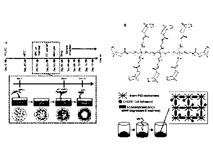
1000151 FIGS. 1A-1B present (A) a schematic representation of a strategy
for assembling a
hydrogel tissue construct. In (A), the upper timeline includes the
differentiation protocols for
obtaining neural progenitor cells (NPCs) from pluripotent stem cells, while
the lower timeline
reflects initial formation of tissue construct. Presented in (B) is a
schematic representation of the
chemistry of hydrogel formation by thiol-ene photopolymerization.
[00016] FIGS. 2A-2D are images demonstrating morphological characteristics
of neural
constructs. Human embryonic stem cell-derived precursor cells were co-cultured
on polyethylene
4
Date Recue/Date Received 2021-05-19
glycol (PEG) hydrogels in 24-well Transwell inserts. Neural progenitor cells
(NPCs) were
seeded on synthetic PEG hydrogels (day 0), followed by endothelial cells (ECs)
and
mesenchymal stem cells (MSCs) at day 9 and microglia/macrophage precursors
(MGs) at day 13.
(A and B) Maximum projection Z stack (525-um thickness) and slice views (NIS
Elements)
illustrating 13111-tubulin (green), GFAP (red), and DAPI (blue) for a day 21
neural construct. XZ
and YZ cross-sections are illustrated in the regions indicated by dashed
lines. The boxed region
in A is illustrated in B. (C and D) Volume view images (NIS Elements)
corresponding to (C) the
full neural construct shown in A (6,300 um x 6,300 um x 550 um) and (D) the
region shown in
B (1,570 um x 2,290 um x 300 um). (Scale bar in A, 1,000 um and B, 500 um.).
1000171 FIGS. 3A-3I are images (A-H) and a graph (I) demonstrating that
tunable
biophysical and biochemical properties of thiol-ene hydrogels guide cell
function. FIGS. 3A-3D
demonstrates the influence of hydrogel properties on spreading for mesenchymal
stem cells
(MSCs) cultured in PEG hydrogels formed via thiol-ene photopolymerization. The
images in
FIGS. 3A-3D illustrate PEG hydrogels that incorporate CRGDS for cellular
adhesion and M_MP-
crosslinking peptides that are derived from a native collagen sequence (ALA)
or which have
been engineered to enhance degradation rate (TRYP and LEU). Matrix remodeling
can be tuned
by controlling biological properties of the synthetic matrix. Mesenchymal stem
cell (MSC)
spreading is a function of degradation rate and adhesion ligand density. MSC
attachment and
spreading was tuned by varying adhesion ligand density (using the fibronectin
mimic CRGDS)
or the susceptibility of the crosslinker to proteolytic degradation (by
varying the 13'2 position of
the amino acid sequence). (A) MSC spreading was maximized with Tryptophan in
the P'2
position of the amino acid sequence and 1000 mM RGD. (B) MSCs remained rounded
in
hydrogels with the most degradable crosslinker (Tryptophan in the P'2
position), but without
active adhesion peptide (0 RGD condition, RGD replaced with non-bioactive RDG
scrambled
peptide). (C) Only limited spreading was observed when Tryptophan was replaced
with Ala due
to lower susceptibility to MMP degradation while (D) intermediate spreading
was observed
when Tryptophan was replaced with Leu. (E, F) Live/dead staining demonstrates
that human
umbilical vein endothelial cells (HUVECs) are viable when grown in 3D
synthetic extracellular
matrices with two different RGD concentrations which leads to differences in
3D organization.
(G) Images of human dermal fibroblasts grown in 3D synthetic matrix compared
to (H) collagen
reveal that basic cell morphologies and cytoskeletal structure are
indistinguishable between them
Date Recue/Date Received 2021-05-19
CA 02972598 2017-06-28
WO 2016/109813
PCMJS2015/068315
(where gels are matched for mechanical properties). (I) Modulus (stiffness)
can be varied across
a wide range of values by choice of monomer density (wt %), molecular weight,
and PEG
backbone molecule (4-aim or 8-arm).
[00018] FIGS. 4A-4K are confocal images demonstrating that neural tissue
constructs are
characterized by neurons with diverse morphologies and long-range order.
Immunofluorescence
imaging reveals neuronal and glial phenotypes. (A-E) Maximum projection
immunofluorescence
images illustrating 13111-tubulin (green) and DAPI (blue) expression for full
vascularized neural
construct formed within a 24-well transwell insert (top left). (F-J) Distinct
neuronal phenotypes.
(F) Calretinin (green) and Reelin (red). (G-K) (red)
coexpressed with (G) GABA,
(H) VGLUT2, (I) FOXG1, (J) Ctip2, and (K) Brn2. Scale bars: 100 pm (F-K).
[00019] FIGS. 5A-5D demonstrate vascular network formation within neural
constructs. (A
and B) Immunofluorescence for endothelial cells (CD3 1, green), glial cells
(GFAP, red), and
nuclei (DAPI, blue) for a day 21 neural construct. (B) Zoom of the boxed
region shown in A to
illustrate association and alignment for a capillary tubule and radially
oriented glial cells
(arrows). The cells in B are shown as single channel grayscale images for (C)
CD3 1 and (D)
GFAP. Scale bars in A, 250 pm and B¨D, 100 pm (shown in B).
[00020] FIGS. 6A-6B demonstrate incorporation of microglia into neural
constructs. (A)
Gene expression for neural constructs with or without microglia (Quality
Control Experiments;
ND., not detected). Statistical analysis was conducted using a Student's t
test (TPM + SD; ***P
<0.001; n = 4 replicate samples each). (B) Immunofluorescence images showing
Thal
(microglia, red) and CD31 (endothelial cells, green) expression for a day 21
neural construct.
Microglia adopt ramified morphologies (e.g., closed arrow) and associate with
capillary tubules
(e.g., open arrows). (Inset) Ibal (red) and DAPI (blue) expression for the
cell pointed out by the
closed arrow (Bottom, Right corner) and surrounding nuclei. Image is
brightened for clarity.
(Scale bar,100 pm.)
[00021] FIGS. 7A-7C demonstrate that neuronal tissue constructs exhibit
stratified layers
and radial organization of neuronal and glial cells. Maximum projection Z-
stacks show
immunofluorescence for neuronal (f3111-tubulin, green), glial (GFAP, red), and
nuclear (DAPI,
blue) markers. (A) Full neuronal construct at day 9 after NPCs were seeded
onto an MMP-
degradable PEG hydrogel. Endothelial cells and mesenchymal support cells were
added for full
tissue constructs at day 9 to mimic recruitment by neuroepithelial cells
within the neural tube.
6
CA 02972598 2017-06-28
WO 2016/109813 PCMJS2015/068315
(B, C) Higher magnification images illustrating stratification and radial
orientation of early
neuronal and glial populations. Scale bar = 250 um.
[00022] FIG. 8 is a table of gene expression data for 3D vascularized
neural constructs.
[00023] FIG. 9 is a table providing Spearman's correlation data for
replicate neuronal
constructs formed with or without microglia on days 14 and 21.
[00024] FIGS. 10A-10E present machine learning predictions. (A) A linear
support vector
machine (SVM) for a 2D problem, where an (n ¨ 1)-dimensional hyperplane
reduces to a line
that separates the classes (filled vs. open circles) and maximizes the closest
points between
classes (the support vectors, which fix the position and orientation of the
hyperplane). The xis are
the examples (points in A), the yis are their labels (filled or open in A),
and w is the weight
vector, or vector of coefficients on the features (the dimensions). The linear
SVM' s output is the
weight vector w and the other coefficient b. To make a prediction, the SVM
computes the
number w'xi ¨ b, and outputs the label 0 (nontoxic, for our application) if
this number is less than
0, and 1 otherwise. The extensions required for the soft margin version of the
SVM are
highlighted in pink in the equation, which minimizes the sum of the distances
between
incorrectly classified training points in addition to the margin, and is
used when the data are
not linearly separable (Hall M, et al. (2009) The WEKA data mining software:
an update
SIGKDD Explor News'. 11(1):10-18). (B) Performance data (averaged from day 16
(2-day
dosing) and day 21(7-day dosing) are shown in the form of receiver operating
characteristic
(ROC) curves. The ROC curve plots true positive rate on the y axis against the
false positive rate
(1 ¨ specificity) on the x axis as the threshold is varied. FIGS. 10C-10E
present additional
receiver operating characteristic (ROC) curve plots and toxins tested (E).
DETAILED DESCRIPTION
[00025] Previous in vitro studies have demonstrated the capacity for human
pluripotent stem
cell-derived neural progenitor cells to self-assemble into layered neuronal
tissues that resemble
the neocortex (Lancaster et al., Nature 501:373 (2013); Kadoshima et al.,
Proc. Natl. Acad. Sci.
U. S. A. 110:20284 (2013); Mariani et al., Proceedings of the National Academy
of Sciences
109:12770 (2012); Eiraku et al., Cell Stem Cell 3:519 (2008)), which may be
particularly
relevant to developmental neurotoxicity screening. However, prior neuronal
tissue models lacked
critical components of the developing brain such as blood vessels and
microglia. The present
7
invention is based at least in part on the Inventors' discovery that human
pluripotent stem cell-
derived precursor cells cultured in materials that are permissive towards
remodeling form highly
uniform 3D vascularized neuronal tissues that recapitulate the complexity and
organization of
human tissues. The Inventors further discovered that the 3D vascularized
tissues are useful for
screening compounds and, using global gene expression profiles from the
tissues, developed a
machine learning protocol that correctly classified greater than 90% of test
compounds. While it
was known that human pluripotent stem cell-derived neuronal tissues provide an
alternative to
animal testing for modeling human brain development, the Inventors' discovered
that it was
possible to produce complex human tissue models comprising physiologically
relevant human
cells and having the high sample uniformity necessary for large-scale,
quantitative enhanced
throughput screening applications.
[00026] Successful strategies to produce in vitro "organoid" models have
been reported for
a variety of tissues (Ader & Tanaka, Curr. Opin. Cell Biol. 31:23 (2014)), but
MatrigelTM and/or
suspension culture techniques typically used for these procedures introduce
variability that is not
well-suited for enhanced throughput quantitative analysis (Singec, Nat.
Methods 3:801(2006)).
Accordingly, the present invention relates to compositions including three-
dimensional tissue
constructs and organoids obtained using monolayer culture techniques to
assemble precursor
cells on chemically-defined bioactive substrates. The present invention also
provides methods of
using three-dimensional tissue constructs and organoids as highly uniform
models of human
tissue and for screening potentially toxic agents. Among the advantages
offered by the present
invention, three-dimensional tissue constructs and organoids of the invention
provide
biologically-relevant information about the effects of various neurotoxic
agents within the
complex environment of neural tissue. In addition, the present invention is
useful for identifying
materials and combinatorial strategies for human tissue engineering.
[00027] Compositions
[00028] Accordingly, the present invention provides a composition
comprising a three-
dimensional (3D) tissue construct. As used herein, the term "tissue construct"
refers to
engineered tissues produced in vitro that comprise complex topologies and
geometries (e.g.,
multi-layered structures, segments, sheets, tubes, sacs). The complex
topologies and geometries
of the tissue constructs recapitulate cell-to-cell interactions found within
native tissues. As used
herein, the term "three dimensional (3D) tissue construct" refers to an
engineered assemblage of
8
Date Recue/Date Received 2021-05-19
CA 02972598 2017-06-28
WO 2016/109813 PCMJS2015/068315
cells and materials that forms a three-dimensional, interconnected complex
structure to mimic in
vivo physiological conditions. By contrast, two dimensional cultures comprise
cells cultivated in
a single layer in a tissue culture dish. An engineered tissue construct of the
invention comprises
at least two layers comprising a homogeneous or heterogeneous population of
cells, wherein one
layer of the tissue construct is compositionally or architecturally distinct
from another layer. In
some cases, layers of the tissue construct comprise multiple cell types in
spatially-defined
positions relative to each other to recapitulate intercellular interactions
found within native
tissues. In exemplary embodiments, the tissue construct is a 3D neural tissue
construct that
provides a microenvironment permissive to in vitro development, in three
dimensions, to
recapitulate neural tissue in vivo. A 3D neural tissue construct of the
present invention is formed
in vitro by the addition of neural progenitor cells to layered tissue
comprising neural and glial
cell populations. An exemplary embodiment is depicted in FIGS. 1A-1B.
According to this
embodiment, a vascularized neural tissue construct is obtained by embedding
human ES/iPS
cell-derived endothelial cells, pericytes, and primitive macrophages
(microglial precursors) into a
tunable hydrogel displaying specific peptide motifs that promote capillary
network formation. To
this mesenchymal cell layer, neural and astrocyte precursors are overlayed.
The hydrogel is then
cultured for about two weeks to form a vascularized neural tissue construct
that mimics in vivo
cephalic mesenchyme-neural epithelial interactions. Neural progenitor cells
(NPCs) and/or
components derived from such progenitors are introduced by adding the
components to the top
of a three-dimensional tissue construct.
[00029] In some cases, the 3D neural tissue construct comprises layered
neural tissue
lacking either vasculature or microglia. In other cases, a 3D neural tissue
construct of the
invention further comprises vascular and/or microglia components. For example,
a 3D neural
tissue construct can comprise stratified, vascularized neural epithelium, with
or without
microglia. Preferably, a 3D vascularized neural tissue construct as described
herein has at least
one of the following properties: (i) interconnected vasculature; (ii)
differentiated cells within the
neural tissue construct mutually contact in three dimensions; (iii) having
more than one layer of
cells; and (iv) demonstrate a function or property characteristic of human
neural tissue in vivo or
in situ.
[00030] In some cases, a composition of the present invention comprises a
three-
dimensional cortical tissue construct. In such cases, a 3D cortical tissue
construct comprises
9
CA 02972598 2017-06-28
WO 2016/109813 PCMJS2015/068315
complex tissues that recapitulate the structural organization and
vascularization of human
cerebral cortex.
[00031] Naturally derived ECMs used for three-dimensional culture (e.g.,
Matrigel (BD
Biosciences, Bedford, MA), collagen gels) are not well-defined, and typically
expose cells to a
wide variety of signaling factors simultaneously. In order to optimize the
influence of a
particular type of signal on cell behavior, without interference from numerous
other signals
acting in concert, alternatives to naturally derived ECMs are preferred. In
exemplary
embodiments, a 3D tissue construct of the present invention comprises a porous
biomaterial such
as a hydrogel. The term "hydrogel" refers to a highly hydrated porous material
comprising
synthetic or biological components formed when an organic polymer (natural or
synthetic) is
cross-linked via covalent, ionic, or hydrogen bonds to create a 3D open-
lattice structure that
entraps water molecules to form a gel. Hydrogels appropriate for constructing
3D tissue
constructs of the present invention include, without limitation, synthetic
hydrogels, bioactive
hydrogels, biocompatible hydrogels, cytocompatible hydrogels, chemically
defined hydrogels,
chemically-defined synthetic hydrogels, and proteolytically degradable
hydrogels.
[00032] As used herein, "bioactive" is intended to indicate the ability to
facilitate a cellular
or tissue response, such as differentiation of a pluripotent stem cell,
induction of vasculogenesis,
neural stem cell differentiation, promotion of cellular attachment, promotion
of cell self-
assembly, and promotion of cell-cell interactions
[00033] As used herein, the term "biocompatible" refers to the ability of a
polymer or
hydrogel to perform as a substrate that will support cellular activity,
including the facilitation of
molecular and mechanical signaling systems, in order to permit proper cell
self-assembly or
cellular function such as tissue formation, production of soluble bioactive
molecules (e.g.,
growth factors), specific cell behaviors such as migration and proliferation.
In some cases,
"biocompatibility" means the absence of components having cell- or tissue-
damaging effects. As
used herein, the term "chemically defined" means that the identity and
quantity of each
component of a composition (e.g., a hydrogel) is known. An important goal in
the fields of
pluripotent stem cell culture and directed differentiation of pluripotent stem
cells is to develop
culture materials and culture media that provide improved performance
consistency and
reproducibility. In some cases, a chemically defined hydrogel for use in a
neural tissue construct
provided herein comprises a minimal number of defined components/ingredients.
CA 02972598 2017-06-28
WO 2016/109813 PCMJS2015/068315
[00034] As used herein, the tetin "cytocompatible" means the hydrogel
material is
substantially non-cytotoxic and produces no, or essentially no, cytotoxic
degradation products.
[00035] As used herein, the term "proteolytically degradable" means that
the crosslinked
backbone can be cleaved enzymatically or non-enzymatically to break down the
scaffold
network.
[00036] In some embodiments, a hydrogel appropriate for inclusion in a
neural tissue
construct as described herein is at least partially contained within a three-
dimensional structural
framework. Preferably, a structural framework comprises a three dimensional
structure prepared
from one or more polymeric materials, including biopolymers.
[00037] A hydrogel appropriate for use in a neural tissue construct of the
invention can be
prepared using various polymers including, without limitation, poly(ethylene
glycol) (PEG),
polyvinyl alcohol (PVA), polyvinyl pyrrolidone (PVP), polyacrylamides, and
polysaccharides.
PEG is a polymer having solubility in water and in many organic solvents and,
generally, lacking
toxicity, antigenicity, or immunogenicity. PEG can be activated at each
terminus to be
bifunctional. In other cases, one terminus can be modified to have a reactive
moiety. For
example, a PEG monomer can be modified to have a relatively inert methoxy
moiety (e.g.,
methoxy-PEG-OH) at one terminus while the other terminus is a hydroxyl group
that is readily
chemically modifiable Polysaccharide hydrogels are made by crosslinking
natural or semi-
synthetic polysaccharides such as alginate, carboxymethylcellulose, hyaluronic
acid, and
chitosan. The cross-linking reaction allows for the formation of a three-
dimensional network
made of covalent bonds between the polymer chains -- a network that is stable
under
physiological conditions.
[00038] In some embodiments, a hydrogel appropriate for inclusion in a
neural tissue
construct as described herein is at least partially contained within a three-
dimensional structural
framework. Preferably, a structural framework comprises a three dimensional
structure prepared
from one or more polymeric materials, including biopolymers. In other
embodiments, it may be
useful for the bioactive hydrogel matrix to have additional structure or
strength in the absence of
a framework or additives. In such cases, a bioactive hydrogel matrix is in a
stabilized,
crosslinked form.
[00039] In exemplary embodiments, hydrogels (e.g., PEG hydrogels,
polysaccharide
hydrogels) are used to produce 3D tissue constructs of the invention. Cells
can be readily
11
CA 02972598 2017-06-28
WO 2016/109813 PCMJS2015/068315
encapsulated within these gels using photo-polymerization. See Fairbanks et
al., Adv. Mater.
21:5005-5010 (2009). Proteins and cells exhibit little to no intrinsic
adhesion or interaction with
PEG hydrogels. See Drury & Mooney, Bionzaterials 24(24):4337-51 (2003); Nguyen
& West,
Biornaterials 23(22):4307-14 (2002); and Hoffman, Adv. Drug Deily. Rev.
54(1):3-12 (2002).
Thus, PEG provides an ideal "blank slate" upon which one can present specific
biological
molecules to cells in a controlled manner.
[00040] To promote self-assembly of an engineered neural construct that
recapitulates
vascularized neural epithelium, it is advantageous to use a photo-
polymerization strategy that
uses "thiol-ene" chemistry. See Fairbanks et al., Adv. Mater. 21:5005-5010
(2009). Step-growth
thiol-ene photopolymerization is based on a reaction between a thiol and a
vinyl group in the
presence of a photoinitiator -- a reaction that results in a homogeneous,
cytocompatible hydrogel.
Photopolymerization kinetics can be controlled by altering the concentration
of photoinitiator
(e.g., radical).
[00041] In some cases, a 3D neural tissue construct of the present
invention comprises a
hydrogel formed using PEG monomers functionalized with norbornene. For
example, a 3D
neural tissue construct of the present invention can be prepared using a
hydrogel comprising a 4-
arm or 8-arm PEG monomers reacted with 5-norbornene-2-carboxylic acid to form
a
norbomene-functionalized PEG solution.
[00042] In some cases, a hydrogel appropriate for neural tissue constructs
described herein
comprise a bioactive agent such as a growth factor, a cytokine, a bioactive
polypeptide or peptide
(e.g., RGD-containing peptides), or any other bioactive ligand capable of
interacting with a
biomolecule of the cells cultured on or within the hydrogel. Peptides
comprising the fibronectin-
derived RGD peptide sequence include, without limitation, RGDS (SEQ ID NO:7),
CRGDS
(SEQ ID NO:2), Ac-CRGDS (SEQ ID NO:11); CRGDS-CONH(2) (SEQ ID NO:12), Ac-
CRGDS-CONH(2) (SEQ ID NO:13), RGDSC (SEQ ID NO:8), CCRGDS (SEQ ID NO:9), and
CCCRGD (SEQ ID NO:10). The number and type of appropriate bioactive agents for
the present
invention will depend on the types of cells cultured on the hydrogel. Examples
of suitable
bioactive ligands include, without limitation, carboxyl, amine, phenol,
guanidine, thiol, indole,
imidazole, hydroxyl, sulfate, norbomene, maleimide, laminin, fibronectin,
fibrinogen, peptide
sequences, or combinations thereof. Bioactive ligands can be covalently
incorporated into PEG
hydrogels using a thiol-ene-based photo-polymerization strategy.
12
CA 02972598 2017-06-28
WO 2016/109813 PCMJS2015/068315
[00043] Other PEG formulations may be useful for methods of using the
tissue constructs
in, for example, screening applications (i.e., for an agent having a certain
activity or effect on a
cell type within the construct). In some cases, PEG formulations comprising
non-degradable
crosslinkers are used to obtain neural construct described herein. In other
cases, a hydrogel
formed using PEG monomers and comprising various concentrations of
extracellular matrix-
derived peptides or other peptides (e.g., peptides comprising the integrin-
binding sequence
CRGDS (SEQ ID NO:2)) can be used. For example, dextran hydrogels suitable for
tissue
engineering have been produced by introducing primary amine groups for
covalent
immobilization of extracellular-matrix-derived peptides (Levesque and
Shoichet, Biomaterials
27(30):5277-85 (2006)). In yet other cases, hydrogels comprise different
crosslinking densities
(i.e., altering stiffness of the hydrogel) or, in some cases, a MIMP-
degradable crosslinker.
[00044] A 3D neural tissue construct of the present invention can be
prepared by dispersing
isolated cells or an isolated cell population within or on a hydrogel. As used
herein, an "isolated
cell" is a cell that has been substantially separated or purified away from
other cell types or
biological substances. As used herein, the term "population" refers to a
collection of cells, such
as a collection of progenitor and/or differentiated cells. As used herein, the
term "differentiated"
as it relates to the cells of the present invention can refer to cells that
have developed to a point
where they are programmed to develop into a specific type of cell and/or
lineage of cells.
Similarly, "non-differentiated" or "undifferentiated" as it relates to the
cells of the present
invention can refer to progenitor cells, i.e., cells having the capacity to
develop into various types
of cells within a specified lineage. In exemplary embodiments, a 3D neural
tissue construct of
the invention is produced by dispersing one or more defined progenitor cell
populations (e.g.,
one or more isolated populations of neural progenitor cells). Preferably, as
an initial step, a
hydrogel is seeded by dispersing neural progenitor cells within or on a
hydrogel. In some cases,
the neural progenitor cells are derived from human pluripotent stem cells
including, for example,
human induced pluripotent stem cells. A hydrogel comprising dispersed neural
progenitor cells is
then cultured under conditions and for a length of time sufficient to promote
differentiation of
human neural progenitor cells dispersed therein. The hydrogel so cultured can
be further seeded
by dispersing within or on the cultured hydrogel one or more additional human
cell types.
Preferably, the hydrogel following dispersal of one or more additional human
cell types
comprises cell populations such as, for example, pericytes, microvascular
endothelial cells, glial
13
CA 02972598 2017-06-28
WO 2016/109813 PCMJS2015/068315
cells (e.g., astrocytes and oligodendrocytes), neuronal cells (e.g., GABAergic
and glutamatergic
neurons), stromal cells, Schwann cells, undifferentiated cells (e.g.,
embryonic cells, stem cells,
and progenitor cells), endoderm-derived cells, mesoderm-derived cells,
ectoderm-derived cells,
and cancer-derived cells or combinations thereof including, without
limitation, human
endothelial cells, human mesenchymal cells, human primitive macrophages, and
human
pericytes. The hydrogel comprising such dispersed human cells can be cultured
under culture
conditions that promote cell differentiation for a length of time sufficient
to be able to observe
formation of a 3D vascularized neural tissue construct comprising human
neurons and glial cells.
Upon differentiation of neural progenitor cells and the addition of cell types
such as endothelial
cells, human mesenchymal cells, human primitive macrophages, and human
pericytes, the
resulting three-dimensional neural tissue construct represents one or more
stages of human brain
development.
[00045] In some cases, a hydrogel is further seeded by dispersing within or
on the hydrogel
one or more bioactive agent that modulates a function or characteristic of a
cell. Such a bioactive
agent can be dispersed within or on the hydrogel prior to or following
dispersal of a cell type
described herein.
[00046] Advantageously, 3D neural tissue constructs of the invention
provide
physiologically relevant in vitro models of the developing human brain
including vascular
networks having characteristics of the blood brain barrier and microglia
derived from
differentiation of primitive macrophages. In exemplary embodiments, a 3D
tissue construct of
the invention comprises elements important for or involved in development of
the mammalian
(e.g., human, non-human primate) brain including, without limitation, neural
progenitor cells,
endothelial cells (e.g., human microvascular endothelial cells), mesenchymal
cells, and primitive
macrophages. Neural progenitor cells that differentiate within the construct
provide neuronal and
glial populations. Endothelial cells and mesenchymal cells contribute to an
interconnected
vasculature, and primitive macrophages differentiate to populate the construct
with microglia. In
some cases, cells populating a tissue construct of the invention are derived
from human
pluripotent stem cells, such as human embryonic stem cells (hESCs) or human
induced
pluripotent stem cells (iPSCs), under chemically defined, xenogeneic material-
free conditions. In
exemplary embodiments, human pluripotent stem cells are differentiated in
vitro under
chemically defined, xenogeneic material-free conditions to separately derive
distinct tissue
14
construct components as described in U.S. Application Serial No. 62/098,838
and U.S.
Application Serial No. 62/098,824. Such cells can self-assemble into a neural
tissue construct
that lacks vasculature or microglia, or that is subsequently seeded with
vascular cells or
microglia. In other cases, it is possible to enhance differentiation within a
3D neural tissue
construct by adding cells that are at intermediate stages such as earlier
neural progenitor cells.
1000471 In exemplary embodiments, 3D neural tissue construct is produced by
culturing
neural progenitor cells (e.g., human pluripotent stem cell-derived neural
progenitor cells) on a
bioactive synthetic hydrogel (e.g., PEG hydrogel) to promote differentiation
and self-assembly of
neuronal and glial populations. Such neural progenitor cells can be seeded on
a hydrogel at a
density between about 10,000 cells/well to about 500,000 cells/well (e.g.,
about 10,000
cells/well; 20,000 cells/well; 30,000 cells/well; 40,000 cells/well; 50,000
cells/well; 75,000
cells/well; 100,000 cells/well; 150,000 cells/well; 200,000 cells/well;
250,000 cells/well;
300,000 cells/well; 400,000 cells/well; 450,000 cells/well; 500,000
cells/well). Preferably, neural
progenitor cells are seeded at a density between about 50,000 to about 200,000
cells/well.
1000481 Subsequently, vascular cells and microglia precursors (primitive
macrophages) are
added to the hydrogel construct. The addition of vascular cells and primitive
macrophages
mimics recruitment of blood vessels and microglia after formation of the
neural tube. When
cultured on bioactive synthetic hydrogels, the precursors will self-assemble
to form complex
multilayered, highly uniform neuronal tissue-like constructs having similar
gross morphological
features between samples. Vascular cells and/or primitive macrophages can be
seeded on a
hydrogel at a density between about 10,000 cells/well to about 500,000
cells/well (e.g., about
10,000 cells/well; 20,000 cells/well; 30,000 cells/well; 40,000 cells/well;
50,000 cells/well;
75,000 cells/well; 100,000 cells/well; 150,000 cells/well; 200,000 cells/well;
250,000 cells/well;
300,000 cells/well; 400,000 cells/well; 450,000 cells/well; 500,000
cells/well). Preferably,
vascular cells and/or primitive macrophages are seeded at a density between
about 50,000 to
about 200,000 cells/well.
1000491 In exemplary embodiments, a 3D tissue construct is seeded with
progenitors of the
myeloid lineages (i.e., granulocyte, macrophage, erythroid, and megakaryocyte)
from pluripotent
stem cell-derived hematovascular mesoderm. In humans, common myeloid
progenitors (CMT's),
Date Recue/Date Received 2021-05-19
CA 02972598 2017-06-28
WO 2016/109813 PCMJS2015/068315
which are progenitor cells committed to the myeloid lineages, express CD34 and
IL-3 R alpha
(CD123). Progenitors of the myeloid lineages (i.e., granulocyte, macrophage,
erythroid, and
megakaryocyte) can be seeded on a hydrogel at a density between about 10,000
cells/well to
about 500,000 cells/well (e.g., about 10,000 cells/well; 20,000 cells/well;
30,000 cells/well;
40,000 cells/well; 50,000 cells/well; 75,000 cells/well; 100,000 cells/well;
150,000 cells/well;
200,000 cells/well; 250,000 cells/well; 300,000 cells/well; 400,000
cells/well; 450,000
cells/well; 500,000 cells/well). Preferably, progenitors of the myeloid
lineages (i.e., granulocyte,
macrophage, erythroid, and megakaryocyte) are seeded at a density between
about 50,000 to
about 200,000 cells/well.
[00050] Human hematovascular mesodermal cells can be obtained according to
a method
that comprises culturing human pluripotent stem cells for about two days in
the presence of a
serum-free, albumin-free, chemically-defined culture medium as provided herein
that is
supplemented to further comprise one or more of the following: a Rho kinase
inhibitor (ROCK
inhibitor) (e.g., Y-27632), bone morphogenetic protein 4 (BMP4), Activin A,
and lithium
chloride (LiC1). In some cases, the human pluripotent stem cells are cultured
under hypoxic (i.e.,
oxygen level lower than atmospheric) conditions. In exemplary embodiments, the
cells are
cultured as described herein in the presence of 5% 02. Methods can further
comprise obtaining
myeloid progenitors by expanding such pluripotent stem cell-derived
hematovascular
mesodermal cells under normoxic (i.e., atmospheric oxygen levels, about 20%
02) conditions in
a chemically defined, xeno-free culture medium comprising or consisting
essentially of FGF2,
VEGF, TPO, SCF, IL-6, and IL-3. The method can comprise the further step of
culturing such
cells under normoxic conditions in a myeloid differentiation culture medium.
In exemplary
embodiments, a myeloid differentiation culture medium is a chemically defined,
xeno-free
medium comprising granulocyte macrophage colony-stimulating factor (GM-CSF),
which is also
known as colony stimulating factor 2 (CSF2) and is a cytokine produced mainly
by macrophages
and activated T cells. Recombinant human GM-C SF and related products are
commercially
available.
[00051] Neural tissue constructs described herein can be modified to have
different
configurations or morphologies by seeding a construct with a larger or smaller
population of
neural progenitor cells and, consequently, altering the number, size, and
composition (e.g.,
identity) of neuron and/or glial cell populations. Likewise, any cellular
components or materials
16
used to obtain a neural tissue construct as described herein can be modified
or optimized to, for
example, tailor a screening method or other use of a neural tissue construct
provided herein, to
assay developmental aspects of human neural tissue (e.g., modify
culture/growth periods,
incorporate additional cell types, remove certain neural tissue construct
components), or to vary
material properties of a neural tissue construct (e.g., vary adhesion ligand,
crosslinking agent,
etc.).
1000521 Although human cells are preferred for use in the invention, the
cells to be used in
tissue constructs of the invention are not limited to cells from human
sources. Cells from other
mammalian species including, but not limited to, equine, canine, porcine,
bovine, feline, caprine,
murine, and ovine sources can be used. Cell donors may vary in development and
age. Cells can
be derived from donor tissues of embryos, neonates, or older individuals
including adults.
[00053] In some cases, a tissue construct of the present invention may
comprise
recombinant or genetically-modified cells in place of or in addition to
unmodified or wild-type
("normal") cells. For example, it can be advantageous in some cases to include
recombinant and
genetically-modified cells that produce recombinant cell products, growth
factors, hormones,
peptides or proteins for a continuous amount of time or as needed when
biologically, chemically,
or thermally signaled due to the conditions present in culture. Procedures for
obtaining
recombinant or genetically modified cells are generally known in the art, and
are described in
Sambrook et al, Molecular Cloning, A Laboratory Manual, Cold Spring Harbor
Press, Cold
Spring Harbor, N.Y. (1989).
[00054] In another aspect, the present invention provides 3D tissue
constructs comprising
one or more cell types derived from a particular mammalian subject (e.g., a
particular human
subject). In some cases, one or more cell types derived exhibit one or more
specific phenotypes
associated with or resulting from a particular disease or disorder of the
particular mammalian
subject. Subject-specific cells can be obtained or isolated from a target
tissue of interest by
biopsy or other tissue sampling methods. In some cases, subject-specific cells
are manipulated in
vitro prior to use in a tissue construct of the invention. For example,
subject-specific cells can be
expanded, differentiated, genetically modified, contacted to polypeptides,
nucleic acids, or other
factors, cryo-preserved, or otherwise modified prior to use in a tissue
construct of the present
invention. In some cases, subject-specific cells are differentiated prior to,
during, or after
encapsulation in a three-dimensional tissue construct of the invention. In
other cases, subject-
17
Date Recue/Date Received 2021-05-19
CA 02972598 2017-06-28
WO 2016/109813 PCMJS2015/068315
specific cells for use in a tissue construct of the invention are induced
pluripotent stem cells
obtained by reprogramming somatic cells of the subject according to methods
known in the art.
See, for example, Yu etal., Science 324(5928):797-801 (2009); Chen etal., Nat
Methods
8(5):424-9 (2011); Ebert etal., Nature 457(7227):277-80 (2009); Howden etal.,
Proc Natl Acad
Sci USA 108(16):6537-42 (2011). Human induced pluripotent stem cells allow
modeling of
drug responses in a genetically diverse population of individuals, including
those individuals
with genetic diseases. Even the safest drugs may cause adverse reactions in
certain individuals
with a specific genetic background or environmental history. Accordingly, 3D
tissue constructs
comprising cells derived from iPS cells obtained from individuals having known
susceptibilities
or resistances to various drugs or diseases will be useful in identifying
genetic factors and
epigenetic influences that contribute to variable drug responses.
[00055] In exemplary embodiments, human pluripotent stem cells (e.g., human
ESCs or iPS
cells) are cultured in the absence of a feeder layer (e.g., a fibroblast
layer) and in the presence of
a chemically defined, xenogen-free substrate. For example, human pluripotent
cells can be
cultured in the presence of a substrate comprising vitronectin, a vitronectin
fragment or variant, a
vitronectin peptide, a self-coating substrate such as Synthemax (Corning), or
combinations
thereof. In exemplary embodiments, the chemically-defined, xeno-free substrate
is a plate coated
in vitronectin peptides or polypeptides (e.g., recombinant human vitronectin).
[00056] In another aspect, the present invention provides an organoid
culture system. As
used herein, the term "organoid" refers to a tissue-like structure (i.e.,
exhibiting structural
properties of a particular tissue type) that resembles a whole organ and is
assembled in vitro by
the separate addition and self-organization of various cell types including,
but not limited to,
pluripotent stem cells, fetal neural stem cells, and isolated organ
progenitors. See, e.g., Lancaster
and Knoblich, Science 345(6194) (2014). In exemplary embodiments of the
invention, an
organoid culture system comprises a three-dimensional construct comprising
hydrogel-
encapsulated cells and provides a physiologically relevant microenvironment
for analysis or
perturbation of cell-cell interactions, cell-matrix interactions, and
morphogenesis in three-
dimensional culture. In some cases, an organoid culture system provides a
microenvironment
that at least partially recapitulates tubulogenesis (e.g., capillary
tubulogenesis) and
vasculogenesis including, for example, the formation of polarized epithelia
with lumens
surrounded by capillary-like structures having endothelial features. In
exemplary embodiments,
18
CA 02972598 2017-06-28
WO 2016/109813 PCMJS2015/068315
capillary tubulogenesis in a 3D tissue construct of the invention
recapitulates principles of both
angiogenesis, postnatal vasculogenesis, and other developmental steps that
closely resemble
embryonic neovascularization. Montano et at., Tissue Engineering Part A
16(1):269-82 (2010);
Kusuma et at., Proceedings of the National Academy of Sciences 110:12601-12606
(2013).
[00057] In some cases, a 3D tissue construct of the present invention
further comprises
isolated biological components. As used herein, an "isolated" biological
component (such as a
protein or organelle) has been substantially separated or purified away from
other biological
components in the cell of the organism in which the component naturally
occurs, such as other
chromosomal and extra-chromosomal DNA and RNA, proteins, and organelles. As
used herein,
the term "isolated protein" includes proteins purified by standard
purification methods. The term
also embraces proteins prepared by recombinant expression in a host cell, as
well as chemically
synthesized proteins, or fragments thereof.
[00058] Engineered three-dimensional tissue constructs of the present
invention can be
prepared, grown, and maintained in any suitable tissue culture vessel that
permits production,
growth, and maintenance of the constructs. Suitable vessels include
TranswellTm permeable
support devices and T-75 flasks In some cases, a 3D tissue construct of the
invention is prepared
and/or maintained in a multi-well tissue culture vessel. A multi-well vessel
is advantageous to
facilitate mechanization and large-scale or high-throughput screening of
neural construct
according to methods of the invention. For example, a 3D tissue construct of
the present
invention can be prepared or provided using a multi-well tissue culture vessel
that facilitates
high-throughput assessment of, for example, cellular interactions, in vitro
development, toxicity,
and cell proliferation upon contacting a chemical compound of interest to the
neural construct. In
some cases, a tissue culture vessel may be coated with polypeptides or
peptides that promote cell
proliferation and/or differentiation (e.g., vitronectin, fibronectin) and
placed in an incubator at
37 C prior to seeding with cells.
[00059] Any appropriate method or methods can be used to confirm uniformity
and the
presence or absence of certain components in a 3D tissue construct provided
herein. Suitable
methods for detecting the presence or absence of biological markers are well
known in the art
and include, without limitation, immunohistochemistry, qRT-PCR, RNA
sequencing, and the
like for evaluating gene expression at the RNA level. In some cases, methods
such as
immunohistochemistry are used to detect and identify cell types or
biomolecules within a 3D
19
CA 02972598 2017-06-28
WO 2016/109813 PCMJS2015/068315
tissue construct. For example, whole tissue constructs or portions thereof can
be stained for
specific differentiation markers by immunohistochemistry. In some cases, it
will be
advantageous to perform dual-label immunofluorescence to assess the relative
expression of
individual marker proteins or to detect multiple progenitor or differentiated
cell types within a
construct. Appropriate primary and secondary antibodies are known and
available to those
practicing in the art. In addition, microarray technology or nucleic acid
sequencing (e.g., RNA
sequencing) can be used to obtain gene expression profiles for 3D engineered
tissue
compositions of the invention. Myeloid markers and macrophage associated
markers include, for
example, CD14, CD16, CSFR-1, CD11b, CD206 (also known as macrophage mannose
receptor
or MMR), CD68, and CD163. Quantitative methods for evaluating expression of
markers at the
protein level in cell populations are also known in the art. For example, flow
cytometry is used to
determine the fraction of cells in a given cell population that express or do
not express biological
markers of interest. Biological markers for perivascular cells and microglia
include antibodies
having specificity to CD45, CD68, or HLA-DR complex.
[00060] Differentiation potential of progenitor cells encapsulated in a 3D
tissue construct of
the invention can be examined for changes in phenotype, organization, and the
presence of
certain proteins using, for example, magnetic sorting, flow cytometry,
immunofluorescence,
bright-field microscopy, and electron microscopy. In some cases, it will be
advantageous to fix
or freeze tissue constructs of the invention for histology or microscopy. For
example, 3D tissue
constructs of the invention can be fixed in formalin or paraformaldehyde for
plastic embedment
and sectioning using routine methods. Scanning electron microscopy (SEM) is
useful to detect
and analyze the formation of tubular structures in tissue constructs of the
invention. In particular,
SEM can be used to study cross-sectioned tissue constructs to detect blood
vessel folination
(e.g., large vessels, small capillaries). In exemplary embodiments, confocal
microscopy can
reveal the distribution of cell types and vascular structures throughout a
three-dimensional tissue
construct of the invention. In some cases, a three-dimensional assembly of
images obtained by
confocal microscopy is used to analyze the distribution and organization of
various cells and
structures.
[00061] Morphology also can be used to characterize culture components, but
cells of
different origins may share similar features and be difficult to distinguish
using morphology
CA 02972598 2017-06-28
WO 2016/109813 PCMJS2015/068315
alone. Where appropriate, excitatory and inhibitory synaptic potentials can be
analyzed using, for
example, extra- or intracellular recording techniques.
Table 1. Biological Markers of Differentiated Cell Types in
Vascularized Neural Tissue Constructs
Cell Type Marker Target Cell
Epithelial Tight junction protein (TJP1): also known Epithelial tight
junctions
as Zona occludcns protein 1 (ZO-1)
Keratin Epithelial (general)
Collagen IV Epithelial basement membrane
Mesenchymal a-SMA (alpha smooth muscle actin) Pericvtes
Vimcntin Pericytcs
PDGFR-13 (platelet derived growth factor Pericytes
receptor beta)
Endothelial PECAM-1 (Platelet-Endothelial Cell endothelial cells; blood
vessels
Adhesion Molecule-1; also known as CD-
31)
Neuronal N-CAM (neural cell adhesion molecule) Neurons (including
postmigratory
immature neurons)
A2B5 Glial progenitors;
oligodendrocyte
and astrocyte progenitors
[00062] Methods of the Invention
[00063] In another aspect, the present invention provides methods for
producing and using
heterogeneous engineered tissue constructs that mimic structural elements
important for or
involved in development of the mammalian brain. In particular, provided herein
are methods of
using 3D tissue constructs for high throughput screening of candidate
compounds and identifying
agents that are toxic to or hinder the development of one or more components
of the tissue
construct. The present invention also provides methods for screening 3D tissue
constructs
candidate therapeutic drugs, modeling a disease or pathological disorder,
assaying 3D tissue
constructs for viability and proliferative capacity of cells of the construct
under various culture
conditions, and methods using neural organoid tissues for compounds exhibiting
developmental
21
CA 02972598 2017-06-28
WO 2016/109813 PCMJS2015/068315
neurotoxicity. As described herein, the methods of the present invention are
advantageous over
standard in vitro and in vivo methodologies for toxigenicity testing (e.g., in
vivo mouse bioassays
for toxigenicity testing). In particular, the methods described herein provide
sensitive,
reproducible, and quantifiable methods for neurotoxin screening. The methods
are better
alternatives to in vivo mouse bioassays (MBA), an assay which is quantifiable
assay but error-
prone. In addition, MBA requires a large number of animals and is not easily
standardized
between laboratories or scalable for high-throughput screening. Shortcomings
of the MBA and
other animal-based assays have incited a push from regulatory agencies,
including the Food and
Drug Administration (FDA) and the United States Department of Agriculture, to
develop cell-
based models comprising more physiologically relevant human cells and having
the sensitivity
and uniformity necessary for large-scale, quantitative in vitro modeling and
screening
applications (National Institutes of Health, 2008).
[00064] In exemplary embodiments of methods of the present invention, a 3D
neural tissue
construct provided herein is used to screen test compounds for known and
unknown toxicities.
For example, a 3D neural tissue construct can be contacted to a test compound
and assayed for
any effect on any of the cell types contained therein (e.g., neuron, glial
cell, vascular cell,
microglia, other differentiated cell subtypes). In exemplary embodiments,
screening methods
comprise contacting one or more test compounds to a 3D tissue construct of the
present
invention and detecting a positive or negative change in a biological property
or activity such as,
without limitation, gene expression, protein expression, cell viability, and
cell proliferation. The
manner in which a test compound has an effect on a particular biological
activity of the
constructs of the present invention will depend on the nature of the test
compound, the
composition of the tissue construct and the particular biological activity
being assayed. However,
methods of the present invention will generally include the steps of (a)
culturing a 3D tissue
construct as provided herein with a test compound, (b) assaying a selected
biological activity of
the artificial tissue construct, and (c) comparing values determined in the
assay to the values of
the same assay performed using a 3D tissue construct having the same
composition as the
construct contacted by the test compound but cultured in the absence of the
test compound (or in
the presence of a control). Detecting a positive or negative change in a
biological property or
activity of a cell of the tissue construct can comprise detecting at least one
effect of a test
compound on morphology or life span of a cell or tissue within the contacted
tissue construct,
22
whereby a test compound that reduces the life span of the cells or tissues or
has a negative
impact on the morphology of the cells or tissues is identified as toxic to
human neural tissue. In
some cases, detecting comprises performing a method such as RNA sequencing,
gene expression
profiling, transcriptome analysis, metabolome analysis, detecting reporter or
sensor, protein
expression profiling, Forster resonance energy transfer (FRET), metabolic
profiling, and
microdialysis. Test compounds can be screened for effects on gene expression
in the contacted
tissue construct, where differential gene expression as compared to an
uncontacted tissue
construct is detected.
[00065] In exemplary embodiments, detecting and/or measuring a positive or
negative
change in a level of expression of at least one gene following exposure (e.g.,
contacting) of a 3D
neural construct to a test compound comprises whole transcriptome analysis
using, for example,
RNA sequencing. In such cases, gene expression is calculated using, for
example, data
processing software programs such as Light Cycle, RSEM (RNA-seq by Expectation-
Maximization), ExcelTM, and Prism. See Stewart et al., PLoS Comput. Biol.
9:e1002936 (2013).
Where appropriate, statistical comparisons can be made using ANOVA analyses,
analysis of
variance with Bonferroni correction, or two-tailed Student's t-test, where
values are determined
to be significant at P < 0.05. Any appropriate method can be used to isolate
RNA or protein from
neural constructs. For example, total RNA can be isolated and reverse
transcribed to obtain
cDNA for sequencing.
1000661 Test compounds that are suitable for screening according to the
methods provided
herein include any for which one wishes to determine the effect the compound
has on
development of the brain of a mammal. It will be readily apparent to the
skilled artisan that the
test compounds will include those compounds which are suspected of having one
or more
deleterious effects on cell or tissue of a 3D construct of the invention.
Ideally, test compounds
cover a range of potential cell toxicities including, without limitation,
heavy metals (e.g., lead,
cadmium) and kinase inhibitors (e.g., MEK inhibitor). Test compounds can
include FDA-
approved and non-FDA-approved drugs (including those that failed in late stage
animal testing
or in human clinical trials) having known or unknown toxicity profiles. Test
compounds can
include those included in the NIH clinical collection. Some of the toxins,
such as MEK
inhibitors may affect all or most cell types of a 3D tissue construct.
23
Date Recue/Date Received 2021-05-19
CA 02972598 2017-06-28
WO 2016/109813 PCMJS2015/068315
[00067] Any of the cell types can be targeted, including vasculature,
microglia, neurons,
glial cells, and any interactions between them. Blood brain barrier junction
properties are
another example, although we did not strictly prove we have "blood brain
barrier" function
(many of the appropriate attachments and genes were expressed, though).
[00068] Test compounds can be dissolved in a solvent such as, for example,
dimethyl
sulfoxide (DMS0) prior to contacting to an engineered tissue construct
provided herein. In some
cases, identifying agents comprises analyzing the contacted 3D tissue
construct for positive or
negative changes in biological activities including, without limitation, gene
expression, protein
expression, cell viability, and cell proliferation. For example, microarray
methods can be used to
analyze gene expression profiles of a 3D tissue construct prior to, during, or
following contacting
the plurality of test compounds to the construct. Gene expression profiles can
be obtained for
multiple time points and/or multiple 3D tissue constructs. In some cases, gene
expression
profiles do not directly reflect temporal changes during the initial formation
of vascular networks
in sECM but, instead, identify genes robustly expressed at each time point. In
some cases, a
method of the present invention further comprises additional analyses such as
metabolic assays and
protein expression profiling.
[00069] In yet another aspect, the present invention provides methods for
evaluating known
and potential environmental teratogens. As used herein, the term "teratogen"
refers to any
environmental factor that can produce a permanent abnormality in structure or
function,
restriction of growth, or death of an embryo or fetus. A method of the
invention can comprise
contacting candidate teratogens to a 3D neural tissue construct described
herein and screening
for developmental abnormalities in the construct. Development abnormalities
can include,
without limitation, vascular malformations, other defects of vascular origin,
neoplasias.
[00070] In another aspect, the present invention provides methods for in
vitro modeling of
vascular dysmorphogenesis. In particular, the present invention provides a
method in which
candidate agents are screened for antiangiogenic, neurotoxic, and/or
teratogenic effects using a
3D neural construct as provided herein. More particularly, the methods
comprise screening for
neurotoxic effects (e.g., inhibition of neuronal growth) and/or detrimental
effects on endothelial
cells or blood vessel formation (e.g., vascular dysmorphogenesis, angiogenic
outgrowth, or blood
vessel remodeling) upon exposure to known and unknown agents. Changes in cell
viability and
24
CA 02972598 2017-06-28
WO 2016/109813 PCMJS2015/068315
proliferative capacity can be detected using, for example, cell stains and 3I-
I-thymidine
incorporation.
[00071] In another aspect, the present invention provides methods for in
vitro modeling of
neurodegeneration using organoid constructs. In particular, the invention
provides an organoid
for studying biological phenomena associated with neurodegeneration and for
detecting or
measuring the expression of genes and proteins associated with
neurodegenerative disorders such
as Parkinson's disease. In addition, the organoid construct model is useful
for screening novel
drugs and growth factors and may reduce the need for invasive animal
experiments. A method
can comprise contacting a neural construct described herein to one or more
candidate agents and
screening for biological processes associated with neurodegenerative
phenotypes including,
without limitation, demyelination, axonal damage, protein aggregation, and
neurite loss.
[00072] It may be advantageous in some cases to employ a machine learning
approach for
methods that include, for example, associating characteristic profiles with
various cell types
and/or with developmental neurotoxicity. For example, in some cases, one or
more machine
learning algorithms are employed in connection with a method of the invention
to analyze data
detected and obtained by RNA sequencing or gene expression profiling of 3D
neural constructs
prior to, during, or following exposure of the constructs to known agents
having developmental
neurotoxi city. In addition, one or more machine learning algorithms can be
used to identify gene
sets that predict the neural toxicity of chemicals even in the absence of pre-
existing toxicity
information. Generally, machine learning algorithms are used to construct
models that accurately
assign class labels to examples based on the input features that describe the
example. In some
cases, machine learning algorithms apply a simple linear separator or a
(possibly weighted) vote
of individual features, or distance-based methods. See FIGS. 5A-5D and related
discussion in the
Examples section below.
[00073] In some cases, a linear support vector machine (SVM) is used to
construct a
predictive model of developmental neurotoxicity. Generally, SVMs belong to the
family of
generalized linear models and are useful to construct a predictive model for a
variable of interest
("the class") using other variables and training data in which the values of
variables including the
class are known. A linear SVM is essentially an (n-/)-dimensional hyper-plane
that separates the
instances of two classes in the n-dimensional feature space. Linear SVMs
exhibit good
CA 02972598 2017-06-28
WO 2016/109813 PCMJS2015/068315
classification performance on gene expression data. With respect to the
present invention, a
SVM can perform the following task specification:
[00074] Given: RNA-seq gene expression measurements for roughly 19K genes
on one day
or on several different days following exposure to various drugs, together
with a neural toxicity
label on each drug.
[00075] Do: Construct a model that, from the same type of expression data
on a new drug,
can accurately identify if the drug is neural toxic.
[00076] A linear SVM's output is the weight vector w and the other
coefficient b. These
are loosely analogous to the coefficients in other linear models such as
logistic regression,
although they are used somewhat differently to make predictions on new data
points. To make a
prediction, the SVM outputs the number w'xi ¨ b, and outputs the label 0 (non-
toxic) if this
number is less than 0, and 1 otherwise. While the numerical output does not
have a probabilistic
interpretation as does the output of logistic regression, a logistic
regression model can be built
with one input variable - the SVM's output - from the same training set to
output a probability of
"toxic."
[00077] In exemplary embodiments, the ability of a SVM to predict the
developmental
neural toxicities of other compounds is estimated. In some cases, an unbiased
method that
provides relatively high variance is used In other cases, a nearly unbiased
(i.e., slightly
pessimistic) method that provides lower variance is used These methods are
standards in
supervised machine learning and statistical classification. An unbiased method
comprises
collecting a set of new compounds (not included in the training set) but whose
neural toxicities
are known; generating RNA-Seq data for these compounds; and testing the
predictive model on
them after the model has been constructed. This is considered to be blinded
trial because
researchers running the SVM do not know which compounds are included or what
fraction of the
compounds are toxic. This information is revealed only after the SVM' s
predictions are made.
[00078] In some cases, a lower-variance evaluation method, such as leave-
one-out cross-
validation, is employed. Where there are N data points (compounds) in a
training set, the method
proceeds in N steps. In each step, a different data point is held out of the
training set and the
SVM is trained on the remaining data points. A prediction is made on the held-
aside data point.
Hence every data point is a test case exactly once, for a model trained
without that data point.
Results are aggregated over all the folds, or test cases, to estimate how well
the SVM model
26
CA 02972598 2017-06-28
WO 2016/109813 PCMJS2015/068315
trained on all the data will perfoim on a new data point (compound). The
method has lower
variance because it tests on more compounds--all the compounds of the training
set¨but is
slightly pessimistic because each training set is slightly smaller (one less)
than the actual training
set.
[00079] Using the above leave-one-out cross-validation methodology, numbers
of true
positive (toxic) predictions (TP), as well as false positive (FP), true
negative (non-toxic, TN),
and false negative predictions (FN) are computed. Using these numbers,
accuracy (i.e., fraction
of predictions that are correct) can be computed. In addition, one can compute
sensitivity, or true
positive rate, or recall [TP/(TP+FN)]; specificity [TN/(TN+FP)]; and
precision, or positive
predictive value [TP/(TP+FP)]; and other metrics such as F-measure and
negative predictive
value. Nevertheless, all of these metrics depend on not only the model that
produces probabilistic
predictions for toxicity but also the probability threshold at which we make
positive predictions,
such as 0.5. Hence it is common in machine learning and statistical
classification to report
"thresholdless" curves and or metrics, the most popular being the receiver
operating
characteristic (ROC) curve and the area under this curve (AUC). The ROC curve
plots true
positive rate on the y-axis against the false positive rate (1 ¨ specificity)
on the x-axis as the
threshold is varied. Random uniform guessing produces a diagonal from lower
left to upper right
corner and AUC of 0.5, while perfect prediction produces a graph that goes up
to the upper left
corner and then across and AUC of 1Ø
[00080] In a further aspect, provided herein is a tissue construct
screening system. A tissue
construct screening system can comprise an analytical device configured to
obtain data
comprising measurements from a 3D human vascularized neural tissue construct
provided
herein. The system can further comprise a computer controller configured to
receive the data
from the analytical device; and a machine-based adaptive learning system
trained using known
gene expression data and configured to select a subset of features from the
data using a feature
selection algorithm. The subset of features corresponds to a change in a level
of expression of at
least one gene following exposure to a known or unknown test compound. In some
cases, the
measurements comprise gene expression data obtained from microarrays.
[00081] Unless defined otherwise, all technical and scientific terms used
herein have the
same meaning as commonly understood by one of ordinary skill in the art to
which the invention
belongs. Although any methods and materials similar to or equivalent to those
described herein
27
can be used in the practice or testing of the present invention, the preferred
methods and
materials are described herein.
1000821 As used herein, "a medium consisting essentially of' means a medium
that contains
the specified ingredients and those that do not materially affect its basic
characteristics.
[00083] As used herein, "serum-free" means that a medium does not contain
serum or serum
replacement, or that it contains essentially no serum or serum replacement.
For example, an
essentially serum-free medium can contain less than about 1%, 0.9%, 0.8%,
0.7%, 0.6%, 0.5%,
0.4%, 0.3%, 0.2% or 0.1% serum.
[00084] As used herein, "effective amount" means an amount of an agent
sufficient to evoke
a specified cellular effect according to the present invention.
1000851 As used herein, "about" means within 5% of a stated concentration
range, density,
temperature, or time frame.
[00086] The invention will be more fully understood upon consideration of
the following
non-limiting Examples. It is specifically contemplated that the methods
disclosed are suited for
pluripotent stem cells generally.
EXAMPLES
1000871 Example 1 - Producing Vascularized Neuronal Tissue Constructs
[00067] Hydrogel polymerization: Thiol-ene photo-polymerization provides
mix and
match adaptability for customizing hydrogels, since any peptide that includes
cysteine in the
amino acid sequence can be coupled into a hydrogel. Polyethylene glycol (PEG)
hydrogels
were formed using thiol-ene photopolymerization chemistry, with modifications
from
previously a published protocol (Fairbanks et al., Adv Mater 21(48):5005-5010
(2009)). Stock
solutions of 8-arm PEG-norbornene (20000 MW, JenKem USA, 8ARM (TP)-NB-20K)
were
prepared at a final concentration of 300 mg/mL by dissolving 300 mg of solid!
0.8 mL PBS to
account for volume occupied by 8-arm PEG-norbornene solid, sterile filtered
through a 0.2 [tm
nylon syringe filter (Fisher), and stored as frozen aliquots. Matrix
metalloproteinase (MIMP)-
degradable PEG hydrogels were formed using an amino acid sequence modified
from a native
collagen sequence (Nagase et al., Biopolymers 40(4):399-416 (1996))
(KCGPQG¨IWGQCK
(SEQ ID NO:1); Active sequence in bold, cleave site = (¨); Genscript, >90%
purity, C-
28
Date Recue/Date Received 2021-05-19
CA 02972598 2017-06-28
WO 2016/109813
PCMJS2015/068315
terminus amidated), with cysteines on each end to crosslink 8-arm PEG-
norbornene molecules.
Cell adhesion was promoted by incorporating CRGDS peptide (SEQ ID NO:2) (2 mM
final
monomer solution concentration; Genscript, >90% purity, C-temiinus amidated),
an amino
acid sequence derived from fibronectin (Pierschbacher et al., Nature
309(5963):30-33 (1984)).
Stock M_MP-peptide (-75 mM peptide / 150 mM SH) and CRGDS peptide (-100 mM)
solutions were prepared and sterile filtered through a 0.22 [tin low protein
binding
polyvinylidene difluoride (PVDF) syringe filter (Millex) and the final
concentration was
verified after filtration using an Elman's assay (Thermo Scientific;
modification of
Manufacturer's protocol: PBS used to dissolve all reagents).
[00089] As shown in FIGS. 2A-2D, biophysical and biochemical properties of
thiol-ene
hydrogels are tunable and influence cell properties. For example, spreading of
mesenchymal
stem cells (MSCs) is a function of degradation rate and adhesion ligand
density. MSC
attachment and spreading was tuned by varying adhesion ligand density (using
the fibronectin
mimic CRGDS) or the susceptibility of the crosslinker to proteolytic
degradation (by varying
the P'2 position of the amino acid sequence). MSC spreading was maximized with
Tryptophan
(W) in the P'2 position of the amino acid sequence and 1000 mM RGD (FIG. 2A),
while
MSCs remained rounded in hydrogels having the most degradable crosslinker
(Tryptophan in
the P'2 position) but lacking an active adhesion peptide (FIG. 2B). Only
limited spreading was
observed when Tryptophan was replaced with Ala due to lower susceptibility to
MMP
degradation (FIG. 2C), while intermediate spreading was observed when
Tryptophan was
replaced with Leucine (FIG. 2D). Human umbilical vein endothelial cells
(HUVECs) are
viable when grown in 3D synthetic extracellular matrices, but the presence of
different RGD
concentrations affected 3D organization (FIGS. 2C-2D). Human dermal
fibroblasts grown in
3D synthetic matrix (relative to growth on collagen) demonstrate that the
basic morphology
and cytoskeletal structure of the resulting tissue constructs is
indistinguishable from natural
extracellular matrices. It was also observed that cell attachment and
spreading of human MSCs
in three dimensions are affected by choice of adhesion ligand density and
proteolytically
degradable crosslinker.
[00090] For subsequent assays, the final monomer formulation for PEG
hydrogels was 40
mg/mL 8-arm PEG-NB, 4.8 mMMMP-peptide crosslinker (9.6 mM cysteines, 60% molar
ratio relative to norbornene arms), 2 mM CRGDS (SEQ ID NO:2), and 0.05%
(wt/wt)
29
CA 02972598 2017-06-28
WO 2016/109813
PCMJS2015/068315
Irgacure 2959 photoinitiator (BASF Schweiz AG, Basel, Switzerland). Hydrogels
were
formed by pipetting 30 RL monomer into 24-well BD Transwell inserts (1 litn
pores, Fisher;
Quality control experiments) or 40 [tI, into Corning HTS Transwell-24 well
permeable support
(0.4 [tin pores, Sigma Aldrich; Toxicity experiments). After pipetting, any
gaps between the
PEG monomer solution and the edge of the insert (due to surface tension) were
removed by
tilting the insert plate and gently tapping until the solution uniformly
covered the bottom of the
transwell insert membrane. Transwell plates containing inserts and monomer
solutions were
placed on the top shelf of a UVP XX-15 lamp stand (Fisher) and exposed to ¨365
nm centered
UV light (UVP XX-15L lamp, Fisher) for 2.5 minutes. After polymerization,
hydrogels were
incubated in DF3S medium overnight to allow swelling and equilibration (5%
C07, 37 C).
[00091] Seeding Porous Bionmiterials With Pluripotent Stem Cell-Derived
Neural
Progenitor Cells: Vascularized neural tissue constructs were obtained
according to the strategy
generally depicted in FIG. 1B. Neural and astrocyte precursors were overlayed
onto the cell-
embedded PEG hydrogel and cultured for about two weeks. Specifically,
cryopreserved neural
progenitor cells (NPCs) were thawed and expanded on 6-well plates coated with
Matrigel
(BD Biosciences, 0.5 mg per plate for at least 1 hour) and cultured in neural
expansion
medium. One vial of frozen NPCs (-1.2x10⁷ cells) were thawed and plated in 3
wells of a
Matrigel coated 6-well plate (2 vials were thawed in one Matrigel coated
10 cm dish),
cultured for 2-3 days (depending on initial confluence) and passaged 1:3 using
AccutaseTM.
NPCs were passaged 1:3 after 2 days of additional culture, expanded for 2-3
more days and
used for experiments. NPCs were removed from the plate using 1 mL Accutase /
well, from
which an aliquot was removed for counting. After adding the appropriate volume
of cell
suspension to a conical vial, NPCs were pelleted at 0.2G for 4 minutes. NPCs
were
resuspended and seeded in neural expansion medium at a density of 100,000
cells / 24-well
insert. NPCs were allowed to attach overnight, and then neural expansion
medium was
exchanged on Day 1 and every 2 days for the remainder of the experiment. For
each medium
exchange, all medium under the insert was aspirated, while approximately 3/4
of the medium
was removed from the top by sliding the pipette tip down the side of the well
to avoid
damaging the developing neural tissue constructs.
[00092] As described in the following sections, the resulting vascularized
neural tissue
construct mimics in vivo cephalic mesenchyme-neural epithelial interactions,
neural progenitor
CA 02972598 2017-06-28
WO 2016/109813
PCMJS2015/068315
cells and/or components derived from such progenitors are introduced by adding
the
components to the top of a three-dimensional tissue construct.
[00093] Differentiation and growth of Pluripotent Stem Cell-Derived
Endothelial Cells
(ECs) and Mesenchymal Stem Cells (MSCs): Endothelial cells were expanded from
cryopreserved stocks on fibronectin-coated plates (Life Technologies, 100 pg
per plate) using
E7BV media, with one vial (-1x10⁶ cells) per 6 wells of a 6-well plate or a
single 10 cm dish.
ECs were split 1:3 after 2 days using Accutase, cultured for an additional 3
days, and then used
for experiments. E8BA medium: E8 supplemented with BMP4 (5 pg/L) and Activin A
(25
jtg/L). E7V medium: E8 minus TGFI31, supplemented with VEGF-A (50 ug/L).
E7B17i
medium: E7V supplemented with BMP4 (50 pg/L) and SB431542 (5 uM, TGFI3
inhibitor)
(Inman et al., Mol Pharmacol 62(1):65-74 (2002)).
[00094] At day 9, ECs and MSCs were seeded on top of the differentiating
NPC layer at a
total density of 100,000 cells /well, with a 5:1 ratio of ECs:MSCs
(83.3K:16.7K). Both ECs
and MSCs were harvested using Accutase and counted before centrifugation.
Cells were
counted and mixed in the appropriate ratio, centrifuged, and resuspended for
seeding. Neural
expansion medium was exchanged on day 11 (2 days after seeding ECs and MSCs).
At day 13,
microglia/macrophage precursors were harvested and seeded at a density of
100,000 cells /
insert. Neural expansion medium was exchanged on day 14, and then every other
day until
samples were collected for RNA, sorting, or immunofluorescence imaging
[00095] Addition of Primitive Macrophages to Neural Constructs: Primitive
macrophages
were added to hydrogel neural tissue constructs after initial vascular network
organization and
after neural progenitor cells had self-assembled into multilayered structures
with radially
organized neural and glial populations (see FIG. 8) reminiscent of the early
neuroepithelium.
[00096] The neuronal tissue constructs were characterized by several
features that
resembled the human neocortex during early development of the cortical plate.
Immunofluorescence imaging and RNA-sequencing provided evidence for diverse
neuronal
and glial phenotypes, including interneurons and projection neurons (FIGS. 3A-
3I). Radially
oriented GFAP+ and Vimentin+ cells were consistent with radial glia, and a
densely packed
cellular layer characterized by stratification of cortical neurons resembled
features of the
mammalian cortex, such as previously reported for human pluripotent stem cell-
derived 3D in
vitro neuronal tissues (Lancaster etal., Nature 501:373 (2013); Kadoshima et
al., Proc. Natl.
31
CA 02972598 2017-06-28
WO 2016/109813
PCMJS2015/068315
Acad. Sc!. US. A. 110:20284 (2013); Mariani et cll., Proc. Natl. Acad. Sci.
109:12770 (2012);
Eiraku et al., Cell Stem Cell 3:519 (2008)). For example, a reelin- layer at
the outer tissue edge
and an adjacent layer abundant with calretinin+ neurons assembled similarly to
Cajal-Rezius
neurons in the marginal zone and interneurons of the emerging human cortical
plate at ¨7-9
gestational weeks (GW).
[00097] In
summary, our neuronal constructs were characterized by cortical organization
and stratification that was consistent with 3D neuronal tissues in vitro and
features described
for the human neocortex during development. Importantly, it is our
understanding that these
neuronal tissue constructs provide the first in vitro model of human cortical
development that
was formed using a synthetic hydrogel (rather than Matrigel or suspension
culture) and that, in
some embodiments, comprises microglia derived from human pluripotent stem
cells.
Moreover, the neuronal tissue constructs described herein are believed to be
the first to
incorporate vasculature, to be formed using methods that can be easily
automated or scaled for
high throughput protocols, and, as described in the following Example, the
first in vitro three-
dimensional neural "organoids" useful for quantitative toxicity screening and
for successfully
predicting neural toxicity (in a blinded study).
[00098] The
timing for vascul arizati on of the human cerebral cortex parallels emergence
of the cortical plate, when angiogenic sprouts from the pial capillary plexus
begin penetrating
the neural tube. Endothelial cells formed extensive vascular networks by day
16 (FIGS. 4A-
4C), while capillary-like structure was more organized and extended throughout
the neuronal
constructs by day 21 (FIGS. 4D-4F). Vascular networks penetrated into the
layered regions
and extended around the circumference of the neuronal constructs (FIGS. 4E-
4F), and both
mesenchymal (FIG. 4G) and glial (FIG. 4H) cells wrapped capillary-like tubules
and larger
vessel-like structures on the periphery. Further, capillary-like tubules
aligned with radial glia
(FIG. 41), especially at the leading edge of the extending vascular network
(FIG. 4K). Glial
cells attached to capillary-like tubules through end-feet (FIGS. 4J-4K),
suggesting that the
neuronal constructs mimicked at least some aspects of the blood-brain-barrier
(BBB). By day
21, the constructs contained an extensive neural network, cells exhibiting
neural and glial
phenotypes, interconnected capillary networks, and microglia-like cells.
Notably, vascular
network formation was induced within the neuronal constructs without the
requirement for
exogenous addition of growth factors such as VEGF. Further, RNA-sequencing
demonstrated
32
CA 02972598 2017-06-28
WO 2016/109813
PCMJS2015/068315
that genes for several blood vessel-promoting growth factors were highly
expressed within the
neuronal constructs for control samples without vascular cells (e.g., VEGFA
and PDGFB).
Therefore, cellular signaling within the neuronal construct provided the
necessary cues to
induce vascularization, which is consistent with initial recruitment of
capillaries to the cerebral
cortex by the neuroepithelium.
[00099] Several microglia genes were expressed only after primitive
macrophages were
added to the neuronal constructs (e.g., AIF1/1BA1, TREM2). Further, IBA1+
(AIF1) cells with
ramified morphologies were distributed throughout the neuronal constructs by
day 21 (FIGS.
5A-5D), which is consistent with microglia in the resting state. Some IBA1+
cells also
interacted with capillary-like tubules within the neuronal constructs (FIGS.
5B-5C), which has
been observed during human development and may indicate a role for microglia
in guiding
vascular organization. Therefore, the 3D neuronal constructs provided the
necessary cues to
induce primitive macrophages to adopt a phenotype characterized by several
hallmark features
of microglia.
[000100] RNA sequencing (RNA-Seq) was used to quantitatively assess sample
uniformity
by comparing differential gene expression for replicate neural constructs
after 14 and 21 days
of differentiation on hydrogels. In addition, replicate samples were
characterized by
Spearman's correlation coefficients (p) > 099 to at least 21 days of
differentiation RNA-Seq
revealed an increase in expression of CD68, a microglial cell marker. RNA-Seq
also identified
several characteristic microglia genes that were detectable only when
primitive
macrophages/microglia precursor cells were incorporated into the neural
constructs, such as
CD11B (ITGAM), TREM2, and IBA1 (AIF1). See FIGS. 7A-7C and Tables 2 and 3. RNA-
Seq identified differentially expressed genes for day-21 neural constructs
compared to H1 ES
cells (in normal culture), and characteristic gene ontology (GO) clusters were
identified from
the resulting gene sets using the DAVID functional annotation database (Huang
et al., Nat.
Protocols 4(1):44-57 (2008); Ashburner et al., Nature Genet. 25:29-29 (2000)).
Neural
constructs were characterized by 4865 upregulated and 4669 downregulated genes
relative to
H1 ES cells (FDR < 0.005). Upregulated genes for the neural constructs were
enriched within
GO categories that included neuron differentiation (GO:0030182, 212 genes),
forebrain
development (GO:0030900, 52), hindbrain development (GO:0030902, 31), synaptic
transmission (GO:0007268, 143), vasculature development (GO:0001944, 85
genes), and
33
CA 02972598 2017-06-28
WO 2016/109813
PCMJS2015/068315
others. A wide variety of expressed genes within the neural constructs have
previously been
identified for roles in human cortical layering, including marginal zone and
layer I neurons
(GAP43, Reelin/RELN and Calretinin/CALB2), upper layer neurons (e.g., CUX1,
SATB1),
and deep layer neurons (e.g., CTIP2/BCL11B, ETV1, FOXP1, SOX5) (Bayatti et al.
, Cereb.
Cortex 18(7):1536-1548 (2008); Meyer et at., J. Neurosci. 20(5):1858-1868
(2000); Zecevic et
at., The Journal of Comparative Neurology 412(2):241-254 (1999); Saito et al.,
Cereb. Cortex
21(3):588-596 (2011); Ip et at., Cereb. Cortex 21(6):1395-1407 (2011).
Therefore, RNA-seq
identified diverse cellular phenotypes within the neural constructs and
suggested a role for
neurodevelopmental mechanisms in the emergence complexity within the tissue.
[000101] lb a-I protein expression was detected by fluorescent antibody
staining. Ibal- cells
were distributed throughout the neural constructs by day 21, and adopted
ramified
morphologies, which is a distinguishing feature for microglia in the resting
state. Ibal+ cells
associated with endothelial tubules, which has been observed during human
development and
suggests a possible role for microglia in guiding vascular organization within
the neural
constructs. Therefore, human ES cell-derived primitive macrophages exhibit
several properties
consistent with a microglia-like phenotype observed within the neural
constructs.
[000102] Human ES cell-derived neural progenitor cells alone self-assembled
into
multilayered tissue-like structures when cultured on degradable biomateri al s
such as MMP-
degradable PEG hydrogels (FIGS. 6A-6B) whereas self-organization was less
pronounced on
non-degradable hydrogels, demonstrating that remodeling of the hydrogel
components
influences the self-assembly and organization of neural progenitor cells into
three-dimensional
tissues. It is important to note that both degradable and non-degradable
hydrogel construct
formats are useful for studying the effect of altering material properties of
the construct on the
cells and tissues within the construct. In addition, the physical and chemical
properties of the
two formats may be beneficial for particular screening applications and other
uses as described
herein.
[000103] In sum, these data demonstrate that three-dimensional multilayered
neural tissue-
like constructs can be produced with remarkable uniformity when ES cell-
derived precursor
cells are cultured on bioactive hydrogels.
34
CA 02972598 2017-06-28
WO 2016/109813
PCMJS2015/068315
Table 2. Gene Expression in Neural Constructs
Average Standard Deviation
Genes Day 16 Day 21 Day 16 Day 21
AIF1/IBA1 12.1 15.6 3.3 8.8
ITGAM/CD 2.3 1.2 0.6 1.2
PTPRC 4.1 3.6 0.9 3.3
CX3CR1 3.8 3.7 0.8 4.6
CD68 17.3 23.8 4.0 18.9
CD14 1.8 3.0 1.6 1.4
Normalized expression (TPM; N = 4)
[000104] Example 2 - Methods and Materials
[000105] Human embryonic stem (ES) cell culture:
[000106] Essential 8 (E8) medium (1): DMEM/F12 HEPES (Life Technologies, 11330-
032), L-ascorbic acid-2-phosphate magnesium (64 mg/L; Sigma-Aldrich, A8960-
5G), sodium
selenite (14 pg/L; Sigma-Aldrich, S5261), NaHCO3 (543 mg/L), holo-transferrin
(10.7 mg/L;
Sigma-Aldrich, T0665-1G), insulin (20 mg/L; Sigma-Aldrich, 19278), human
recombinant
FGF2 (rhFGF2, 100 pg/L), and TGF131 (2 pg/L; R&D Systems, 240-B-001MG/CF).
[000107] H1 human embryonic stem (ES) cells were maintained in E8 medium
(1) (Life
Technologies) on Matrigel (growth factor reduced, Corning 356230) coated
culture plates and
were passaged with 0.5 mM EDTA in 1X PBS as previously described (2). Cells
were
karyotyped within 10 passages and tested negative for mycoplasma
contamination.
[000108] Human ES cell differentiation into neural progenitor cells (NPCs):
[000109] DF3S medium: DMEM/F-12, L-ascorbic acid-2-phosphate magnesium (64
mg/L), sodium selenium (14 jig/L), and NaHCO3 (543 mg/L).
[000110] Essential 6 (E6) medium: DMEM/F-12, L-ascorbic acid-2-phosphate
magnesium
(64 mg/L), sodium selenite (14 pg/L), NaHCO3 (543 mg/L), transferrin (10.7
mg/L), and
insulin (20 mg/L).
[000111] Neural expansion medium: DF3S medium supplemented with rhFGF2 (5
pg/L),
1X N2 (Life Technologies, 17502-048) and 1X B27 (Life Technologies, 17504-044)
supplements.
CA 02972598 2017-06-28
WO 2016/109813
PCMJS2015/068315
[000112] The procedure for deriving neural progenitor cells was modified
from a
previously reported protocol (3). H1 ES cells were split using 0.5 mM EDTA in
1X PBS and
cultured in E6 medium supplemented with rhFGF2 (100 pg/L) and SB431542 (TGF-r3
receptor
inhibitor, 101..iM; Sigma-Aldrich). After two days, the medium was switched to
E6 medium
supplemented with SB431542 (10 [tM) for seven days with daily media exchange
to induce
the formation of neural rosettes. The neural rosettes were then mechanically
dissociated from
the culture dish and cultured as floating aggregates in neural expansion
medium for four days.
Aggregates were then dissociated with Accutase (Life Technologies) and plated
onto Matrigel
(growth factor reduced, Corning 356230) coated plates in neural expansion
medium. Cells
were cultured for an additional 22 days and passaged when confluent, yielding
>90%
SOX1+/13III-tubulin+ neural progenitor cells ("NPCs"). NPCs were cryopreserved
at 1.2x10⁷
cells per vial. Cryopreserved neural progenitor cells were used for subsequent
expansion and
formation of 3D neural constructs to ensure a uniform cell source for all
experiments.
[000113] Human ES cell differentiation into endothelial cells (ECs):
[000114] E8BA medium: E8 medium supplemented with BMP4 (5 g/L) and Activin A
(25
Itg/10-
[000115] E7V medium: E8 medium minus TGFf31, supplemented with VEGF-A (50
pg/L).
[000116] E7BVi medium: E7V supplemented with BMP4 (50 tg/L) and 513431542
(5
TGF13 inhibitor)(4).
[000117] H1 ES cells (80-90% confluent) were dissociated using TrypLE
(Invitrogen) for
three minutes at 37 C and plated 1:3 on vitronectin-coated plates (60 jig /10
cm dish, VTN-N,
Life Technologies)(1). ES cells were first cultured for two days to 100%
confluence) in E8BA
medium, which was supplemented with 10 [iM Y-27632 for the first day to
improve cell
survival during attachment. It is critical to achieve 100% cell confluence by
day 2 to ensure
highly efficient differentiation. Cells were then cultured in E7BVi medium for
an additional
three days. Endothelial cells were then isolated with CD34 microbeads
(Miltenyi) by
autoMACS (Miltenyi) to yield purified populations of CD34+/CD31+ cells
("ECs"). The
purified endothelial cells were either cryopreserved immediately or cultured
on fibronectin-
coated plates in E7V medium for one passage before cryopreservation.
[000118] Human ES cell differentiation into mesenchymal stem cells (MSCs):
36
CA 02972598 2017-06-28
WO 2016/109813
PCMJS2015/068315
[000119] Mesenchymal serum-free expansion medium (M-SFE11/1): 50% StemLine II
serum-free HSC expansion medium (HSFEM; Sigma-Aldrich), 50% human endothelial
serum-free medium (ESFM; Invitrogen), GlutaMAX (1/100 dilution; Invitrogen),
Ex-Cyte
supplement (1/2000 dilution; Millipore), 100 mM monothioglycerol (MTG), and 10
ng/L
rhFGF2.
[000120] Mesenchymal stem cells (MSCs) were derived from H1 ES cells using
a
previously published protocol (5). Tissue culture polystyrene plates were
coated with human
fibronectin (5 mg/mL; Invitrogen) and human collagen 1(10 mg/mL; BD
Biosciences) in
phosphate buffered saline (PBS) for expansion and Accutase (StemPro) was used
for
passaging. MSCs were expanded for five passages in M-SFEM (5), followed by two
passages
in pericyte medium (ScienCell), and then cryopreserved (PDGFRB+CD13+, "MSCs").
[000121] Human ES cell differentiation into microglia/macrophage precursors
(MG):
[000122] Microglia/macrophage precursors were produced using feeder-free
conditions by
modifying a previous protocol for differentiating H1 ES cells down mesendoderm
and
hemogenic endothelium lineages (see Uenishi et al. (2014) Stem Cell Rep
3(6):1073-1084). 6-
well plates were first coated with 40 jig Tenascin C overnight at 4 C.
Tenascin C plates were
rinsed with PBS, and then seeded with singularized H1 ES cells at a density of
62,500 cells /
2
cm in E8 medium + 10 nIVI Y-27632 (ROCK inhibitor, R&D Systems). Cells were
cultured
for 24 hours under normoxic conditions.
[000123] Initiate early mesoderm differentiation. 24 hours after plating H1
ES cells, E8
media was aspirated and replaced with DM1 + 1 nM Y-27632. Cells were then
cultured under
hypoxic conditions (5% 02) for two days (do not expose cells to normoxia).
During the two
days of culture, cells detach and reattach. It is important that the culture
is not disturbed, as
cells will aggregate in the middle of the plate, affecting differentiation
efficiency.
[000124] Continue hematovascular mesoderm differentiation. On day two, the
culture was
checked for surviving cell clumps that had not fully reattached. If non-
adherent cells were
present, a 10 mL pipette tip was used to gently pull media off plate, and the
non-adherent cells
and cell clumps were centrifuged at 300 x g for five minutes to form a pellet.
DM1 was
aspirated from the pellet, and the cells were resuspended in DM2. Cells were
gently plated
back into same plate, and culture was continued in a hypoxic incubator. If
only debris was
37
CA 02972598 2017-06-28
WO 2016/109813
PCMJS2015/068315
present, DM1 was aspirated and DM2 was added slowly as to not disrupt the
adherent cells.
Culture was continued in a hypoxic incubator.
[000125] Differentiate and expand hemogenic endothelial cells into
hematopoietic
progenitor cells (HPCs). On day 4, DM2 medium was aspirated and replaced with
DM3
medium. Culture was continued under normoxic conditions. On day 6 of culture
(two days
after adding DM3 media), additional DM3 media was added without aspirating
media already
present. Culture was continued in a normoxic incubator. Cell cultures were
expanded for an
additional 3-5 days in DM3 (longer time is required when cells not fully
adherent after
hematovascular differentiation). If media color indicated a significant pH
drop, half of the
media volume was removed from the plate and placed into a low attachment dish.
An
additional volume of DM3 (1:1 mix of old and fresh media) was added to both
culture plates.
After 3-5 days, spent media containing non-adherent HIPCs was collected and
centrifuged at
300 x g for about five minutes to pellet.
[000126] Myeloid progenitor (MP) differentiation. Expansion was continued in
myeloid
progenitor medium DM4, where lx106 HIPCs / mL were to a low attachment culture
dish
under normoxic conditions. At this point, the cells could be grown in a 10 cm
dish under
normoxic conditions. Cells were expanded for 2-5 days in the DM4 medium At
least five days
in culture was required for proper transition to macrophages, but no more than
five days. DM4
was added if the culture's pH significantly dropped (half/half mixture; do not
transfer cells).
Up to 2x10⁷ cells were obtained from a 10 cm dish. During expansion in DM4
medium (2-5
days), non-adherent cells were collected for sorting to identify CD34+ and
CD45+ cells.
[000127] Microglia/macrophage precursor (MG) differentiation. After 2-5 days
of myeloid
progenitor expansion, 5x10⁵ non-adherent cells were added to macrophage
differentiation
medium DM5 in a 10 cm tissue culture treated dish. Cells were cultured for
three days, then an
equivalent volume of DM5 media was added without aspiration of the media.
After five days
(two additional days in DM5), ¨50-70% of cells had attached. When cells
reached ¨70-80%
confluence (adherent cells), remaining non-adherent cells were transferred to
a new 10 cm dish
to promote adhesion. Both adherent and non-adherent populations are CD45+, but
non-
adherent cells will be CD14 Low/Negative and adherent cells will be
CD11b+/CD14+. On days 5-
10, non-adherent cells began to attach and differentia into CD11b+ and CD14+
cells. Culture in
DM5 medium was continued.
38
[000128] For the quality control assays, RNA was collected on days 14 and
21. For the 3D
toxicity screening experiments, RNA was collected on days 16 and 21
(permitting 2 days of
chemical exposure before collecting at the first time point).
[000129] Immunofluorescence imaging: Blocking buffer: 0.25 % Triton X1O0TM
and 1%
BSA in PBS; Incubation buffer: 0.05% Triton X100TM and 1% BSA in PBS; Rinse
buffer:
0.05% Triton X100TM in PBS.
10001301 Primary Antibodies: Rabbit anti-)63-tubulin (1:500; Cell
Signaling, mAb
#5568S), mouse anti-)63-tubulin (1:500; R&D Systems, MAB1195), rabbit anti-
calretinin
(1:100-1:200: Abcam, ab137878), rabbit anti-GABA (1:200: Abcam, ab43865),
rabbit
polyclonal anti-glial fibrillary acidic protein (GFAP) (1:500; Dako, Z033401-
2), goat anti-
glial fibrillary acidic protein (GFAP) (1:100-1:200; C-19; sc-6170, Santa Cruz
Biotechnology), mouse anti-phospho-vimentin (1:200; S55 [4A4]; Abcam,
ab22651), mouse
anti-CD31 (1:200; Endothelial Cell, Clone JC70A; DAKO, M082301-2), mouse anti-
04
(1:100-1:200; clone 81; Millipore, MAB345), Chicken polyclonal anti-Tbr 1
(1:100-1:200;
Millipore, AB2261), mouse anti-SOX-2 (Cell Signaling, mAb #4900S), rabbit anti-
SOX-2
(Cell Signaling, mAb #3579S), mouse anti-M4P2, (clone AP20; Millipore,
MAB3418), mouse
anti-Reelin (1:100; clone G10, a.a. 164-496; Millipore, MAB5364), mouse anti-
Brn-2
(POU3F2) (1:200; clone 8C4.2; Millipore, MABD51), rabbit anti-Brn-2 (POU3F2)
(1:200;
Cell Signaling, mAb #12137S), rabbit anti-Ctip2 (Bc1-11b) (1:200; Cell
Signaling,
mAb #12120S), rabbit anti-VGLUT2 (1:100; Abcam), mouse anti-MAP2 (1:500; clone
AP20; Millipore, MAB3418), goat anti-Ibal (1:100; Abcam, ab5076), rabbit anti-
Tyrosine
Hydroxylase (Cell Signaling, mAb ##27925), rabbit anti-PDGFR-a (1:100; Santa
Cruz
Biotechnology, sc-338).
10001311 Secondary Antibodies: Alexa Fluor secondary antibodies were used
for all
experiments (Life Technologies): Donkey anti-goat 568 (A11057) or 647
(A21447); Donkey
anti-rabbit 488 (A21206), 568 (A10042), or 647 (A-31573); Donkey anti-mouse
488 (A-
21202), 568 (A10037), or 647 (A31571); Goat anti-chicken (A11041).
10001321 Immunostaining full neural constructs: All steps for
immunostaining were
performed within transwell inserts. Neural constructs were fixed for 60 min.
using 2%
buffered formalin and then rinsed with PBS (or stored at 4 C until
immunostaining). Neural
constructs were permeabilized and blocked in blocking buffer (at least 60
min.). For some
39
Date Recue/Date Received 2021-05-19
CA 02972598 2017-06-28
WO 2016/109813
PCMJS2015/068315
experiments, blocking buffer was used for all steps until final rinse, with
similar results.
Primary antibodies were prepared in incubation buffer, added to the neural
constructs, and
incubated overnight at 4 C. Neural constructs were then rinsed (2X with rinse
buffer, at least
60 min. / ea.) followed by a third rinse step (blocking buffer, at least 60
min.). Secondary
antibodies and 1:1000 DAPI (Sigma) were prepared in incubation buffer, added
to the neural
constructs, and incubated overnight at 4 C (or at least 4 hours at room
temperature). Neural
constructs were rinsed 2 x 60 min. in rinse buffer, followed by an overnight
rinse at 4 C in
incubation buffer. Samples were then stored in PBS until further processing
(typically at least
24 hours).
[000133] Neural constructs were removed from the transwell insert by
cutting the bottom
edge of the membrane, separated from the membrane, and mounted in aqua
polymount
solution (Polysciences, Inc.) on the bottom of a 35 mm glass bottom dish
(MatTek). To limit
bubble formation in the mounting solution, a thin layer was first added to the
glass bottom of
the 35 mm dish. The neural construct was usually placed face down into the
layer of mounting
solution (with some samples placed face up), after which a drop of mounting
solution was
added to cover the construct. A coverslip was then dropped onto the neural
construct in
mounting solution and allowed to settle, rotating the dish to ensure uniform
coverage of the
mounting solution under the coverslip. The coverslip was allowed to settle
overnight at 4 C,
and sealed around the edges with fingernail sealant. The samples remained
stable for imaging
for at least 1 month.
[000134] Immunostaining cryopreserved sections: Neural constructs were
fixed in the
transwell insert for 60 min. using 2% buffered formalin and rinsed with PBS
(overnight at
4 C). The samples were then rinsed in 15% Sucrose/PBS (at least 24 hours, 4 C)
followed by
30% Sucrose/PBS (at least 24 hours, 4 C). Neural constructs were removed from
the transwell
insert by cutting the bottom edge of the membrane, separated from the
membrane, and placed
face down into cryogel (Tissue-Tek embedding medium), and stored frozen at -80
C until
further processing. Frozen samples were equilibrated to -200 C and sectioned
(20-30 um
sections on glass slides). Glass slides containing sectioned samples were
soaked in deionized
water for at least 1 hour to remove cryogel. Samples were permeabilized and
blocked in
blocking buffer for 60 min., rinsed 2x15 min. with rinse buffer, and incubated
at room
temperature in incubation buffer for at least 60 min. Samples were then
treated primary
CA 02972598 2017-06-28
WO 2016/109813
PCMJS2015/068315
antibodies in incubation buffer at 4 C (or at least 4 hours at room
temperature). Samples were
then rinsed with wash buffer (2x15 min.) and incubation buffer (at least 60
minutes, room
temperature). Samples were then treated with secondary antibodies and 1:1000
DAPI (Sigma)
in incubation buffer overnight at 4 C (or at least 2 hours at room
temperature). Sectioned
samples were mounted in aqua polymount solution (Polysciences, Inc.), a glass
coverslip was
placed over the top, stored overnight at 4 C, and sealed around the edges with
fingernail
sealant until imaging.
[000135] Image processing: Confocal immunofluorescence images were
collected using a
Nikon MR confocal microscope. Images were processed using NIS Elements or
ImageJ
(Rasband 1997-2012, Image J, U.S. National Institutes of Health, Bethesda,
Maryland, USA,
available at imagej.nih.gov/ij/ on the World Wide Web); Schneider et al., Nat
Meth 9(7):671-
675. (2012)). Some z-stacks were aligned using the "Align Current ND Document"
(NIS
Elements) or the StackReg plugin (ImageJ) before creating maximum projection
images.
[000136] Phagocytosis by microglia/macrophage precursors. Aliquots of zymosan
A S.
cerevisiae BioParticles (Texas Red conjugate; Life Technologies) were
prepared in PBS.
¨5x10⁶ particles in 500 [IL PBS were added to each well of a 6-well plate
containing ¨400-
500K mi crogli a/macrophage precursors in DM5 media Phagocytosis was imaged
over a 24
hour time period (image capture every 10 minutes) using a Nikon Biostation CT.
[000137] Flow cytometry (FACS) analysis.
[000138] Flow cytometry analysis was performed on a BD Biosciences
FACSCanto II cell
analyzer.
[000139] Neural Progenitor Cells (NPCs). NPCs were dissociated into single
cells with
Accutase and fixed with 2% paraformaldehyde in PBS at RT for 10 minutes. Fixed
cells were
washed once with FACS buffer I (2% FBS in PBS) and permeabilized with ice cold
90%
methanol in PBS overnight at -20 C. Fixed and permeabilized cells were then
washed once
with FACS buffer I and stained with SOX1 (1:100 rabbit anti-S0X1, Cell
Signaling) and I3III-
tubulin (1:200 mouse anti-f3111-tubulin, R&D systems) primary antibodies
overnight at 4 C
followed by conjugated secondary antibodies at RT for one hour. Stained cells
were washed
once with FACS buffer I and analyzed by flow cytometry.
[000140] Endothelial cells (ECs). ECs were dissociated into single cells
with Accutase and
washed once with FACS buffer 1(2% FBS in PBS). Cells were stained with PE-CD31
(1:100;
41
CA 02972598 2017-06-28
WO 2016/109813
PCMJS2015/068315
BD Biosciences, 555446) and APC-CD34 (1:100; BD Biosciences, 555824)
antibodies in
FACS buffer I at 4 C for 30 minutes. Stained cells were washed once with FACS
buffer I and
analyzed by flow cytometry.
[000141] Mesenchymal stem cells (MSCs). MSCs were dissociated into single
cells with
Accutase and washed once with FACS buffer I (2% FBS in PBS). Cells were then
stained with
fluorescently conjugated PE-PDGFR43 and PE-Cy7-CD13 antibodies in FACS buffer
I at 4 C
for 30 minutes. Stained cells were then wash once with FACS buffer I and
analyzed by flow
cytometry.
[000142] Microglia/macrophage precursors (MG). Non-adherent cells were first
transferred to a conical vial in DM5 medium. Adherent MG were incubated in
Accutase,
gently removed from the plate using FACS buffer 11 (0.5% BSA in PBS), and
added to the
conical vial containing non-adherent cells. The cells were centrifuged (5
minutes at 300 x g)
and the cell pellet was washed once with FACS buffer II. Cells were then
centrifuged (5
minutes at 300 x g), resuspended in FACS buffer II, and incubated at 4 C for
15 minutes for
blocking. Cells were centrifuged (5 minutes at 300 x g) and resuspended in
FACS buffer II
with 1:500 PE-CD1lb (BD Biosciences, 555388), Alexa Fluor 488-CD14 (BD
Biosciences,
562689) and APC-CD45 (BD Biosciences, 555485) and then incubated at 4 C for 30
minutes
(use a shaker plate or invert tube at least three times during incubation).
Cells were then
washed twice in FACS buffer II and centrifuged (5 minutes at 300 x g).
Finally, cells were
resuspended in FACS buffer II and analyzed by flow cytometry.
[000143] Example 3 - Predictive Developmental Neurotoxicity Screening In
Vitro
[000144] For toxicity screening experiments, cells were seeded as described
above
(Examples 1 and 2), but with 65,000 cells/well for ECs + MSCs (also 5:1 ratio)
and 15,000
cells/well for microglia/macrophage precursors. Neural constructs were treated
with non-toxic
or toxic compounds starting at day 14, with media exchanged every 2 days. See
FIG. 8. The
following screening protocol was developed by the Thomson lab. Toxic chemicals
(FIG. 10E)
were chosen based on previous literature support for neurotoxicity (Adams et
al., Neurotoxicol
Teratol 15(3):193-202 (1993); Cooper et al., Science 280(5369):1603-1607
(1998); Crofton et
al., ALTEX-Altern Anim Exp 28(1):9-15 (2011); Eskes et al. (2003); Grandjean
et al., Lancet
Neurol. 13:330 (2014); Lidsky (2003); Radio et al., Neurotoxicol Teratol
32(1):25-35 (2010);
Zurich (2002)). The screen included the following experimental groups (FIG.
8): (1) construct
42
with neural progenitor cells (NPCs), endothelial cells (ECs), mesenchymal
cells (MCs), and
primitive macrophages (PMs); (2) construct lacking primitive macrophages
(quality control);
(3) neural progenitor cells only (quality control).
[000145] RNA isolation, cDNA library preparation, and next generation
sequencing: The
3D neuronal constructs were lysed directly in the insert by the addition of
RLT lysis buffer
(Qiagen) and stored at -80 C until being used for RNA isolation. When total
RNA was ready
to be extracted, the samples were thawed and 150 tl of the cell lysates in
buffer RLT were
transferred and re-arrayed to a S-block (Qiagen, Cat. No. 19585) to be mixed
with 1 volume of
70% ethanol (the rest of the lysates were stored in the -80 C). Total RNA was
then isolated
using Qiagen's RNeasyTM 96 kit beginning with step 3 of the manufacturer's
protocol (RNeasy
96 Handbook 01/2002, Using Spin Technology) and included the optional DNase
treatment.
[000146] Quality control studies: Samples used for quality control were
prepared for
RNAseq with Illumina's TruSeqTm RNA Sample Preparation Kit v2 following the
Low-
Throughput (LT) protocol (TruSeCITM RNA Sample Preparation Guide, Part #
15008136, Rev.
A) using 100 ng of total RNA as input. The cDNA libraries were pooled and run
on Illumina's
HiSeqTM 2500 with a single read of 51 bp and index read of 7 bp. FASTQ files
were generated
by CASAVA (v1.8.2). Reads were mapped to the human transcriptome (RefGene
v1.1.17)
using Bowtie (Langmead et al., Genome Biol 10(3):R25 (2009)) (v0.12.8)
allowing 2-
mismatches and a maximum of 20 multiple hits. The gene expression values
(Transcript per
Million Reads or TPM) were calculated by RSEM (Li et al., BMC Bioinformatics
12:323
(2011)) (v1.2.3).
[000147] Toxicity Screening Study: For cDNA preparation for the toxicity
screening
experiments, mRNA is isolated from purified 10Ong total RNA using Oligo-dT
beads (NEB).
Isolated mRNA is fragmented in reverse transcription buffer at 85 C for 7
minutes, and then
reverse transcribed with SmartScribeTM reverse transcriptase (Clontech) at 23
C for 10
minutes followed by a 30 minute incubation at 42 C with a random hexamer
oligo: 5'-
CCTTGGCACCCGAGAATTCCA -3' (SEQ ID NO:3).
10001481 After reverse transcription, RNA is removed by RNaseA and RNaseH
treatment.
A partial IlluminaTM 5' adaptor
(/5phos/AGATCGGAAGAGCGTCGTGTAGGGAAAGAGTGTddC) (SEQ ID NO:4) was
then ligated to the single stranded cDNA using RNA ligase l(NEB) overnight at
22 C. After
43
Date Recue/Date Received 2021-05-19
purification, ligated cDNA was amplified by 18 cycles of PCR using oligos that
contain full
IlluminaTM adaptors (5'-
AATGATACGGCGACCACCGAGATCTACACTCTTTCCCTACACGACGCTCTTCCGAT
CT-3'; SEQ ID NO:5) and index primer (5'-
CAAGCAGAAGACGGCATACGAGATnnnnnnnnnnGTGACTGGAGTTCCTTGGCACCC
GAGAATTCCA-3' (SEQ ID NO:6); nnnnnnnnnn indicates index nucleotides). The
indexed
cDNA libraries were pooled and sequenced on an IlluminaTM HiSeq2500 with a
single 51bp
read and a 10bp index read.
[000149] RNA -Seq Data Analysis: RSEM expected read counts for each gene were
determined by median normalization (utilizing the median normalization
function within EB
Seq ¨version 1.5.3) (Leng et al., Bioinformatics 29(8):1035-1043 (2013)). EB-
Seq (version
1.5.3) was used to calculate FDR for differentially expressed genes (Leng et
al.,
Bioinformatics 29(8):1035-1043 (2013)).
10001501 Gene Ontology Analysis: Gene ontology (GO) terms were identified
using the
Database for Annotation, Visualization and Integrated Discovery (DAVID) (v6.7)
functional
annotation database (Ashburner et al., Nature Genet 25(1):25-29 (2000); Huang
et al., Nat
Protocols 4(1):44-57 (2008)). GO terms were identified by analyzing
differentially expressed
genes with (FDR < 0.005) and >3-fold upregulated expression for 3D neural
constructs
relative to H1 ES cells (see Table 3). The following settings were used for
DAVID analysis
using differentially expressed genes: Gene Ontology category GOTERM BP 5;
Benjamini
corrected p-value < 0.05; Threshold options: Counts = 10, EASE = 0.05. GO
terms were also
identified genes with average TPM > 16 (N = 4, Controls from toxicity
experiment) (Dataset
S5), which were compared to a combined list of neural, vascular, and glial
terms to reduce the
total number of genes below 3000 for input into DAVID (combined list and
associated GO
categories provided in Dataset S7).
[000151] Comparisons to Allen Brain Atlas Data: Pairwise Spearman rank
correlation was
calculated for neural constructs (days 16 and 21, toxicity experiment, N = 4),
H1 ES cells (N =
4), and Allen Brain Atlas data (RNA-seq data only, samples: 8 pcw ¨ 40 yrs)
(Table 3).
Hierarchical clustering is performed by average linkage clustering on those
correlations and
the distance is 1 minus Spearman correlation.
44
Date Recue/Date Received 2021-05-19
CA 02972598 2017-06-28
WO 2016/109813
PCMJS2015/068315
[000152] Machine-Based Learning. We employed linear support vector machines
(SVMs)
to construct our predictive models (Cortes & Vapnik, Mach Learn 20(3):273-297
(1995);
Hardin et al., Stat Appl Genet Mol Biol 3(1):e10 (2004); Struyf et al., BMC
Genomics 9:531
(2008); Vapnik VN (1998) Statistical Learning Theory (Wiley, New York)), which
were
described in detail previously (Hardin et al., Stat Appl Genet Mol Biol
3(1):e10 (2004); Struyf
et al., BMC Genomics 9:531(2008)). We employed SVMs for the following task
specification:
Given: RNAseq gene expression measurements for roughly 19K genes on one day or
on
several different days following exposure to various drugs, together with a
neural toxicity label
on each drug. Do: Construct a model that, from the same type of expression
data on a new
drug, can accurately identify if the drug is neural toxic.
[000153] Evaluations of the approach, including estimates of accuracy and
receiver
operating characteristic (ROC) curves, were all by hold-out testing, either
leave-one-out cross-
validation or use of a blinded trial with a single hold-out set (Hardin et
al., Stat Appl Genet
Mol Biol 3(1):e10 (2004); Struyf et al., BMC Genomics 9:531 (2008)). A 2-
dimensional linear
support vector machine (SVM) is illustrated in the plot shown in FIG. 10A,
where the hyper-
plane reduces to a line that separates the classes (circles) and maximizes the
closest points
between classes (the support vectors that fix the position and orientation of
the hyper-plane).
The xis are the example (circles; genes for the current study), the yis are
their labels (filled or
open; toxic or non-toxic for the current study), and w is the weight vector,
or vector of
coefficients on the features (the dimensions). The red portions in the
equation are the additions
made for the soft margin version of the SVM (Cortes & Vapnik, Mach Learn
20(3).273-297
(1995)), which minimizes the incorrectly classified data points in addition to
the margin (di).
The linear SVM's output is the weight vector w and the other coefficient b. To
make a
prediction, the SVM outputs the number w'xi ¨ b, and outputs the label 0 (non-
toxic, for our
applications) if this number is less than 0, and 1 otherwise. While the
numerical output does
not have a probabilistic interpretation as does the output of logistic
regression, it is common to
build a logistic regression model with one input variable (the SVM's output)
from the same
training set to output a probability (probability of toxic), which we do here.
[000154] Leave-one-out cross-validation: Using the leave-one-out cross-
validation
methodology, we can compute the numbers of true positive (toxic) predictions
(TP), as well as
false positive (FP), true negative (non-toxic, TN), and false negative
predictions (FN). From
CA 02972598 2017-06-28
WO 2016/109813 PCMJS2015/068315
these we can compute accuracy (fraction of predictions that are correct) as
well as the
following: Sensitivity (true positive rate, or recall; TP/(TP+FN)),
specificity (TN/(TN+FP)),
and precision (or positive predictive value; TP/(TP+FP)), as well as other
metrics such as F-
measure and negative predictive value. Nevertheless, all of these metrics
depend on not only
the model that produces probabilistic predictions for toxicity but also the
probability threshold
at which we make positive predictions, such as 0.5. Hence it is common in
machine learning
and statistical classification to report "thresholdless" curves and or
metrics, the most popular
being the receiver operating characteristic (ROC) curve and the area under
this curve (AUC)
such as shown for averaged day 2 and 7 set (FIGS. 9C-9D). The ROC curve plots
true positive
rate on the y-axis against the false positive rate (1 ¨ specificity) on the x-
axis as the threshold
is varied (shown for averaged training set). Random uniform guessing produces
a diagonal
from lower left to upper right corner and AUC of 0.5, while perfect prediction
produces a
graph that goes up to the upper left corner and then across and AUC of 1Ø
[000155] For leave-one-out cross validation, there were 60 compounds in the
training set
and the method proceeds in 60 steps (FIG. 10E). In each step a different data
point is held out
of the training set, the SVM is trained on the remaining data points, and then
it makes its
prediction on the held-aside data point. Hence every data point is a test case
exactly once, for a
model trained without that data point Results are aggregated over all the
folds, or test cases, to
estimate how well the SVM model trained on all the data will perform on a new
data point
(compound). Predictions were made for both replicates for a testing compound
and averaged
together to generate the final ROC. The AUCs for the training compounds were
0.91 on day
16, 0.88 on day 21, and 0.93 for data averaged from both days. Thus, the SVM
for averaged
data from days 16 and 21 produced an estimate for future data of 0.93.
[000156] Blinded trial: In addition to constructing an SVM model, we also
aimed to estimate
how well the model predicts the developmental neural toxicities of other
compounds. Merely
reporting its accuracy on the training set would be overly-optimistic. An
unbiased "hold-out
testing" method was used to predict toxicity for a RNA-seq data set of ten
blinded compounds
that were not in the training set (5 toxins, 5 non-toxic controls) but whose
neural toxicities were
known. After construction and optimization using the training set, the
predictive model was then
tested on the unknown samples.
46
CA 02972598 2017-06-28
WO 2016/109813 PCMJS2015/068315
[000157] As a blinded trial, the assignment of toxins was unknown to
researchers generating
the SVM model until after the predictions were made. The SVM for averaged day
16 and 21 data
was chosen to generate predictive genes from the training set. The SVM
produced probabilities
which were used to rank the blinded compounds from most likely to least likely
toxic, which was
then used to produce an ROC curve and compute an AUC ("area under the curve").
In addition,
we used a threshold of 0.5 to make definitive predictions, assigning every
molecule with
probability < 0.5 as "control" and all others "toxic." The AUC generated for
the ranking of the
blinded set was 0.92, and all compounds except oleic acid were properly
assigned as toxic or
non-toxic based on the 0.5 probability cutoff. The only error in the ranking
was for oleic acid,
which was assigned a higher probability of being toxic than L-741,626 and
Ouabain. The
accuracy of the blinded prediction was 0.9 (9/10 compounds correctly
classified), with the only
error being the prediction of oleic acid (a control) as a neurotoxin (i.e., a
false positive).
[000158] References
1. D. Rice, S. Barone, Environ. Health Perspect. 108, 511 (Jun, 2000).
2. L. Smirnova, H. T. Hogberg, M. Leist, T. Hartung, ALTEX-Altern. Anim.
Exp. 31, 129
(2014).
3. P. Grandjean, P. J. Landrigan, Lancet Neurot 13, 330 (Mar, 2014).
4. L. L. Needham et al., Environ. Sci. Technol. 45, 1121 (Feb, 2011).
5. P. Grandjean, P. J. Landrigan, Lancet 368, 2167 (Dec, 2006).
6. D. C. Bellinger, EllVir011. Health Perspect. 120, 501 (Apr, 2012)
7. Z. G. Hou et al., Stem Cell Res. Ther. 4, S12 (Dec, 2013).
8. D. V. Hansen et al., Nat. Neurosci. 16, 1576 (Nov, 2013).
9. J. H. Lui, D. V. Hansen, A. R. Kriegstein, Cell 146, 18 (Jul, 2011).
10. P. Rakic, Nat. Rev. Neurosci. 10, 724 (Oct, 2009).
11. I. Bystron, C. Blakemore, P. Rakic, Nat. Rev. Neurosci. 9, 110 (Feb,
2008).
12. M. Marin-Padilla, Front. Neuroanat. 6, (Sep, 2012).
13. M. Marin-Padilla, D. S. Knopman, J. Neuropathol. Exp. Neurot 70, 1060
(Dec, 2011).
14. J. M. James, Y.-s. Mukouyama, Semin. Cell Dev. Biol. 22, 1019 (2011)
15. H. Stolp, A. Neuhaus, R. Sundramoorthi, Z. Molnar, Front. Psychiatry 3,
(2012).
16. F. Ginhoux, S. Lim, G. Hoeffel, D. Low, T. Huber, Front. Cell.
Neurosci. 7, (Apr, 2013).
17. T. Arnold, C. Betsholtz, Vascular Cell 5, 4(2013).
18. H. Kettenmann, U. K. Hanisch, M. Noda, A. Verkhratsky, Physiol. Rev.
91, 461 (Apr,
2011).
19. C. Verney, A. Monier, C. Fallet-Bianco, P. Gressens, I Anat. 217, 436
(Oct, 2010).
20. A. Monier et al., I Neuropathol. Exp. NeuroL 66, 372 (May, 2007).
21. A. Monier, P. Evrard, P. Gressens, C. Verney, J. Comp. Neurol. 499, 565
(Dec, 2006).
22. J. A. Thomson etal., Science 282, 1145 (Nov, 1998).
23. J. Y. Yu et al., Science 318, 1917 (Dec, 2007).
24. K. Takahashi et al, Cell 131, 861 (Nov, 2007).
47
CA 02972598 2017-06-28
WO 2016/109813 PCT/US2015/068315
25. S. C. Zhang, M. Wemig, I. D. Duncan, 0. Brustle, J. A. Thomson, Nat.
Biotechnol. 19,
1129 (Dec, 2001).
26. 0. Brustle et at., Science 285, 754 (Jul 30, 1999).
27. M. A. Lancaster et al., Nature 501, 373 (Sep, 2013).
28. J. Mariani et al., Proceedings of the National Academy of Sciences 109,
12770 (July 31,
2012, 2012).
29. M. Eiraku et al., Cell Stem Cell 3, 519 (Nov, 2008).
30. M. Ader, E. M. Tanaka, Curr. Op/n. Cell BioL 31, 23 (2014).
31. I. Singec etal., Nat. Methods 3, 801 (Oct, 2006).
32. B. D. Fairbanks et at., Adv. Mater. 21:5005-5010 (Dec, 2009).
33. M. Marin-Padilla, Front. NeuroanaL 6, (2012-September-13, 2012).
34. P. Rakic, Cereb. Cortex 13, 541 (Jun, 2003).
35. I. Bystron, P. Rakic, Z. Molnar, C. Blakemore, Nat Neurosci 9, 880
(2006).
36. G. Meyer, J. P. Schaaps, L. Moreau, A. M. Goffinet, J. Neurosci. 20,
1858 (Mar, 2000).
37. N. Zecevic, A. Milosevic, S. Rakic, M. Marin-Padilla, The Journal of
Comparative
Neurology 412, 241 (1999).
38. M. H. Dominguez, A. E. Ayoub, P. Rakic, Cereb. Cortex 23, 2632 (Nov,
2013).
39. B. J. Molyneaux, P. Arlotta, J. R. L. Menezes, J. D. Macklis, Nat. Rev.
Neurosci. 8, 427
(Jun, 2007).
40. J. Struyf, S. Dobrin, D. Page, BMC Genomics 9, (Nov, 2008).
41. T. R. Golub et al, Science 286, 531 (Oct, 1999).
42. V. N. Vapnik, Statistical Learning Theory. (Wiley, New York, 1998), pp.
736.
43. C. Cortes, V. Vapnik, Mach. Learn. 20, 273 (Sep, 1995).
44. T. S. Furey et al., Bioinformatics 16, 906 (Oct, 2000).
45. M. Moors et al., Environ. Health Perspect. 117, 1131 (Jul, 2009).
46. N. C. Kleinstreuer et al., Nat. BiotechnoL 32, 583 (Jun, 2014).
47. M. S. Wilson, J. R. Graham, A. J. Ball, Neurotoxicology 42, 33 (May,
2014).
48. N. V. Balmer, M. Leist, Basic Cl/n. Pharmacol. ToxicoL 115, 59 (Jul,
2014).
49. H. Olson et at., Regulatory Toxicology and Pharmacology 32, 56 (2000).
50. S. Rakic, N. Zeceyic, Cereb. Cortex 13, 1072 (Oct, 2003).
51. T. Kadoshima et al., Proc. Natl. Acad. Sci. U. S. A. 110, 20284 (Dec,
2013).
52. G. K. Chen etal., Nat. Methods 8, 424 (May, 2011).
53. X. J. Li et al., Development 136, 4055 (Dec, 2009).
54. S. M. Chambers et al., Nat. BiotechnoL 27, 275 (Mar, 2009).
55. H. Nagase, G. B. Fields, Biopolymers 40, 399 (1996).
56. M. D. Pierschbacher, E. Ruoslahti, Nature 309, 30 (1984).
57. B. Langmead, C. Trapnell, M. Pop, S. L. Salzberg, Genome Biol 10, R25
(2009).
58. B. Li, C. N. Dewey, BMC Bioinfbrmatics 12, 323 (2011).
48
Table 3. Averaged Expression Data for Markers of Vascular, Neuronal, and Glial
Cell Types
NPC = neural progenitor cells; ECs = endothelial NPCs, ECs, MCs
NPCs, ECs, MCs NPCs, ECs, MCs NPCs
0
cells; MCs = mesenchymal cells; PM = primitive w/o
with w/o with ONLY
"
=
macrophages iv/ PM PMs
PMs PMs PMs (Control) -,
a
Day Day 16 Day 21 Day 14
Day 21 S'
Gene.ID 3D Tox
Experiment QC Experiment oo
GAPDH 3678.1 4299.3
7431.1 8239.2 5675.7 5720.6 6737.1 t,.e
Awr
GFAP 149.4 131.9 100.6
173.6 143.5 161.5 48.9
SlOOB 259.7 161.5 201.5
281.3 139.2 212.3 99.4
CD44 179.9 299.2 228.5
210.7 285.3 314.5 317.3
SLC1A3 (GLAST; Astrocytcs, Bergman Glia) 635.4 455.0 315.2
294.2 339.1 369.8 274.5
PDGFRA 3.7 2.8 7.1
4.1 2.5 3.3 1.2
CLDN11 3.0 3.1 9.0
12.2 4.9 5.8 9.3 P
MAL 5.1 3.3 12.8
19.4 6.4 8.3 11.7 .
MBP 3.5 4.2 1.2
2.1 2.2 2.5 1.6 ..,
o.,
.6.
.
v: DLL1 28.7 19.5 38.8
19.4 28.5 23.2 17.5 0
FOXM1 17.8 13.5 39.9
42.1 27.3 27.3 11.0 ' ..,
,
TMPO 138.7 77.9 59.1
67.1 52.1 63.0 49.0 .
,
Microglia
.
AIF1 (IBA1) 12.1 15.6 0.3
17.3 0.0 10.2 0.0
ITGAM (Cdl lb) 2.3 1.2 0.0
0.5 0.0 0.9 0.0
PTPRC (CD45) 4.1 3.6 0.1
2.6 0.1 1.1 0.0
CD68 17.3 23.8 6.4
32.7 6.4 23.1 7.8
CD86 3.6 2.9 0.0
4.2 0.0 1.6 0.0
CD14 1.8 3.0 0.0
21.4 0.5 7.3 0.4
.0
CD4 3.8 3.2 2.0
12.4 2.1 4.8 0.9 en
-3
TREM2 (Resting MG, strong cortex) 6.7 7.0 0.0
7.9 0.0 3.8 0.0
ci)
Neural General ...............
t.)
=
MAP2 779.9 684.3 271.6
205.3 ' 293.3 268.5 310.8 -,
CDH2 777.6 665.9 338.8
256.8 300.1 298.9 235.5 =-==
a
oo
ACHE 22.6 20.2 32.0
36.6 52.4 57.0 50.9
-,
SLC18A3 (vesicular acetylcholine transporter) 1.9 1.7 0.9
0.7 1.3 0.8 1.1 ul
TPH1 1.0 0.4 1.0
0.5 0.4 0.7 0.8
TH 5.5 5.8 10.8
14.7 12.9 10.9 16.8
NEUROG2 (Ngn2) 71.7 55.6 84.7
64.5 51.8 62.4 78.9
0
ISL1 5.9 3.3 I 2.0
2.4 3.5 4.6 1 5.0 t....)
=
GABAergie
--"=-
GAD1 11.5 31.6 r 12.7
7.0 22.1 17.3 1 28.9 .
=
GAD2 1.8 2.9 2.3
3.0 3.7 4.2 4.5 oo
-,
SLC6A1 (GABA transporter 1) 9.8 12.5 5.2
2.8 13.4 10.4 9.2
CALB2 (CalRet) 111.7 156.4 171.3
147.8 214.9 219.4 204.9
ASCL 1 (MASH1) 20.8 14.3 31.4
10.1 26.6 15.5 1 11.3
..,........... ................ -------
- --------- -------- .. --------- ------- .. ------------ ..
................
Clutamaternic
,..
.. ,,, .. , , ....
, ...... .. , ...... ...... , ......
SLC32A1 (VGAT) 1.0 2.0 0.6
0.5 I 0.7 1.0 1 1.0
GLUL (glutamate-ammonia ligase) 137.7 285.3 95.4
117.7 154.2 170.2 229.7
SLC17A7 (VGLUT1) 0.6 0.8 1.4
1.1 3.4 2.2 3.9
SLC17A6 (VGLUT2) 93.5 110.0 48.0
35.9 85.8 74.9 95.9 p
SLC17A8 (VGLUT3) 10.6 14.7 6.9
5.5 9.0 8.1 5.1 0
0
0
..,
SLC1A1 (glial high affinity glutamate transporter) 4.9 7.0 4.3
2.2 4.8 3.8 3.4 0
0
.
= SLC1A2(neuronallepithelial high affinity glutamate
0
0
0
transporter) 103.2 93.4 1 37.1
31.6 1 52.0 33.7 1 39.9 .
..,
,
Tr:msporter and Vesicle proteins
, , ......
, .. ,, .... , .. , ...... , ,,,
SV2A 90.9 77.1 1 92.3
96.7 115.3 105.1 1 122.9 '
SV2B 2.2 3.4 1.1
1.0 3.4 2.3 2.7
SV2C 10.5 4.1 2.4
1.7 3.3 2.5 1.6
SNAP25 95.3 112.3 65.7
56.3 96.2 98.4 117.6
CBLN1 (Cerebellin-1) 34.7 36.7 24.0
21.7 38.5 39.9 40.9
CBLN2 14.2 13.9 7.0
7.5 8.3 6.8 7.5
CBLN3 1.2 1.3 2.3
2.6 2.3 2.9 3.3
-o
CBLN4 6.1 6.5 1.8
1.1 3.9 2.3 3.3 n
DLG4 (PSD-95) 101.2 140.7 118.4
85.2 168.7 150.3 165.6 ;:...
c.t)
SYP (synaptophysin) 44.6 54.6 64.0
63.5 100.9 112.0 113.2
=
SYN1 (synapsin I) 26.6 32.4 22.9
23.7 42.7 53.2 46.0 .
u,
SYN2 7.5 14.1 3.6
3.4 9.7 8.7 8.0
c,
00
SYN3 3.3 5.9 I 4.4
1.5 1 10.7 12.3 10.1 rd.,
Blood Vessel (EC or M(.) ,??-,
PECAM1/CD31 1.4 1.7 2.1
4.1 1.4 2.1 1.0
CD34 22.8 12.7 40.9
70.9 18.1 31.5 28.0
MCAM 15.7 23.8 36.9
33.9 39.0 42.0 51.1
0
KDR/VEGFR2/FLK1 9.2 8.7 13.0
25.8 7.8 11.5 11.4 ),)
=
VEGFA 512.8 890.2 623.5
294.7 677.6 829.1 732.8 --
-.7
,
PDGFRB 10.5 12.1 40.2
30.4 37.2 32.2 15.6 .
=
,z
PDGFB 24.8 14.2 32.7
41.4 20.9 21.9 30.0 00
ANPEP 2.1 1.5 3.6
7.1 2.1 3.8 2.4
CSPG4 1.9 1.7 11.9
12.1 12.3 10.8 8.8
ACTA2 30.8 88.9 254.4
181.2 168.2 240.3 143.4
CDH5 2.0 0.3 4.5
5.0 1.9 2.6 1.1
SLC2A1 95.0 139.6 262.0
66.2 129.7 98.3 133.7
AQPI 5.1 9.5 11.6
18.6 17.1 25.5 6.6
GPR124 1.2 1.2 17.8
13.0 6.7 8.6 1.5
AQP4 64.6 52.8 _ 29.5
31.2 32.3 29.4 39.4
P
NPC
2
NES 1275.2 763.9
620.7 895.9 562.5 642.7 625.1 ,1
o,
ul
-, VIM
5871.3 8872.8 13171.0 9312.2 15142.8 13388.9 14840.7 g
EOMES/Tbr2 1.6 2.2 3.5
3.3 3.1 3.7 4.3
,
,
PAX6 3.7 2.6 1.7
1.6 1.9 2.2 2.0 g
,
SOXI 75.2 53.9 34.4
40.6 33.2 40.1 22.4
SOX2 138.5 160.7 247.4
184.8 249.3 341.0 202.7
SOX9 169.3 153.2 106.7
62.9 110.9 133.6 87.6
NEURODI 14.0 16.6 7.6
5.5 6.1 9.1 11.9
NOTCHI 82.7 81.0 153.2
95.9 173.7 129.0 128.1
NOTCH2 51.1 40.7 46.6
38.1 53.9 40.8 45.4
NR2F1 (COUP-TF1) 51.9 48.6 40.1
35.9 42.5 43.4 32.5
-o
RELN 72.9 45.1 58.3
35.6 50.6 31.4 44.7
CUXI 54.0 54.0 57.7
46.1 60.0 65.6 52.9 u)
)J
=
CUX2 18.7 14.6 23.0
21.1 26.4 27.9 26.3 .
ul
SATBI 72.5 56.5 43.7
24.3 49.0 49.1 41.3 -I-
c,
SATB2 10.1 8.3 7.6
7.1 7.7 8.0 8.2 00
RORB 3.0 1.9 1.5
0.7 3.1 2.3 1.3 ul
BCL11B/Ctip2 37.4 34.1 11.5
8.5 12.4 9.0 11.4
SOX5 13.5 5.9 9.9
5.3 8.3 7.1 6.7
POU3F1/SCIP 7.4 6.5 7.9
10.1 7.1 8.9 7.4
FOXP1 30.1 29.6 22.8
19.5 16.8 18.4 15.7
0
FOXP2 34.5 27.3 33.7
19.2 25.2 32.7 24.1 t,)
=
ETVI 19.1 18.2 11.4
7.1 10.4 11.6 9.5 7:.=
,
MAP1B 1001.4 782.1
460.8 349.1 454.6 375.1 429.3 .
=
TLE4 43.3 48.4 47.6
33.0 37.2 36.8 44.9 00
r7J
POU3F2/Bm2 160.9 122.6 118.0
82.3 122.6 121.2 96.5
FOX01 1.6 2.0 4.3
2.6 2.5 1.9 2.3
LIX1 5.1 3.9 4.7
2.9 6.5 3.9 3.7
SYT9 35.7 22.5 19.9
17.0 25.0 25.8 18.9
S100A10 117.2 124.3 344.4
458.2 215.5 308.7 268.4
OMAI 11.3 14.8 9.8
6.9 11.6 10.5 9.0
LDB2 33.0 39.3 37.0
22.1 38.4 44.1 33.0
CRIM1 42.6 36.7 32.5
23.8 27.8 24.6 29.5
P
PCP4 5.5 8.3 5.2
3.3 11.0 9.7 12.6 2
RAC3 52.7 60.9 133.5
158.5 152.2 166.3 169.2 ..,
o.,
ul
.
l=J CRYM 4.2 4.9 2.7
3.1 1.3 2.3 3.2 0
IGFBP4 10.5 17.8 54.6
50.9 42.2 48.2 28.7 .
..,
DKK3 409.7 388.8
1003.0 803.8 1185.8 1219.9 752.7
,
SEMA3E 3.6 3.5 4.4
3.6 2.6 3.0 2.9
NR4A3 3.5 2.7 1.3
0.8 1.1 1.6 1.4
LXN 4.5 1.8 14.2
13.9 7.4 12.2 2.2
ID2 120.1 227.6 242.1
115.5 202.2 188.7 132.2
SLITRK1 12.6 14.7 5.4
6.2 5.2 5.4 6.6
LMO3 5.6 6.8 2.0
1.2 4.0 3.0 3.5
LMO4 73.0 101.2 65.5
56.9 100.2 78.7 101.1
CTGF 127.1 112.5 330.6
173.2 123.6 95.9 143.8 -0
n
Ependyma / Neuroepithelium (early)
PROM1 (CD133) 92.7 97.9 55.8
47.5 70.0 68.0 47.6 u)
t.)
ITGA6 77.1 56.3 26.6
22.6 23.0 20.7 30.8 =
ul
-I-
c,
oo
ul