Note: Descriptions are shown in the official language in which they were submitted.
DEMANDES OU BREVETS VOLUMINEUX
LA PRESENTE PARTIE DE CETTE DEMANDE OU CE BREVETS
COMPREND PLUS D'UN TOME.
CECI EST LE TOME 1 DE 2
NOTE. Pour les tomes additionels. veillez contacter le Bureau Canadien des
Brevets.
JUMBO APPLICATIONS / PATENTS
THIS SECTION OF THE APPLICATION / PATENT CONTAINS MORE
THAN ONE VOLUME.
THIS IS VOLUME 1 OF 2
NOTE: For additional volumes please contact the Canadian Patent Office.
-
84029102
SYSTEM FOR AMPLIFICATION OF A FETAL DNA SPECIES
This is a division of Canadian Patent Application Serial No. 2,887,218, filed
on
August 30, 2002, which is a division of Canadian Patent Application Serial No.
2,456,140
filed on August 30, 2002, now patented.
It is to be understood that the expression "the present invention" or the like
used in this specification encompasses not only the subject matter of this
divisional
application but that of the parent also.
BACKGROUND OF THE INVENTION
The presence of DNA originating from different individuals in bodily fluids is
a
well-known biological phenomenon in many clinical and biological scenarios.
For example,
following bone marrow transplantation, the hemopoietic system of the
transplantation recipient
will consist of varying proportions of donor's and recipient's cells. The
ascertainment of the
amount of donor's or recipient's cells has been performed by the detection of
genetic differences
between the donor and recipient, including gender (Mangioni et al., Bone
Marrow Transplant
20:969-73 (1997)) and DNA polymorphisms (Roux et al., Blood 79:2775-83
(1992)). The
corollary of this approach is that if the analysed region does not bear a
genetic difference
between the donor and recipient, then analysis by the current approach will
not be possible.
In another example, during pregnancy, detection of fetal DNA in maternal
plasma and serum has been previously demonstrated (Lo et al., Lancet 350:9076:
485-7(1997)).
This technology has demonstrated that fetal DNA isolated from maternal plasma
and serum
can be used for non-invasive prenatal diagnosis (Lo et al., N Eng J Med,
339(24): 1734-8
(1998): Faas et al., Lancet 352(9135):1196 (1998); Amicucci et al., Clin Chem
46(2):301
(2000); Chen et al., Prenat Diagn 20(4):355-7 (2000); Saito et al., Lancet
356:1170 (2000)).
The clinical application of this phenomenon has been helped by the relatively
high absolute
and relative concentrations of such circulating fetal DNA in maternal plasma
and serum
(Lo et al., Am J. Hum Genet 62:768-775 (1998)). Using this approach,
noninvasive prenatal
detection of a number of conditions has been achieved, including fetal rhesus
D status
(Lo et al., New Eng J Med 339:1734-1738 (1998)), myotonic dystrophy
1
CA 2973165 2017-07-12
1111-30D1
=
(Amicucci et al., Clin Chem M:301-302 (2000)), achondroplasia (Saito et a,
Lancet
356:1170 (2000)) and certain chromosomal trandocations (Chen at aL,,Prenat
Diag
:335-
357 (2000); Chen et aL, Clin Chem gi:937-939 (2001)). All of these current
approaches have
utilized the detection of DNA sequences inherited from the father and which
are genetically
distinguishable from those of the mother (Bianchi, Am J Hum Genet .62(4): 763
(1998).
Specifically, the detection of DNA that the fetus has inherited from the
mother in maternal
plasma or serum has been thought to be 'impossible. Similar limitations have
also been
described for the detection of fetal nucleated cells isolated from the
cellular fractionnf
maternal blood (Lo et aL, Ann N reicadScl, al.:204 (1994).
Othershave detected aberrantly methylated DNA from cancer patients. This
has beeu repotted for patients with a variety of cancers, includhig lung
(atelier, et al.,
Cancer Res 59(1);,67 (1999)) and liver cancer (Wong et aL, Cancer Res
59(1):71 (1999)).
Recently, much interest has been focused on the biology of epigenetic
phenomena, namely processes which alter the phenotype but which are not
associated with
changes in DNA sequence (Wolffe , Science 286:481-486 (1999)). One of the best
characterised epigenetic processes is DNA methyIation,(W'olffe et al., Curr
BioL 10:R463-
R465 (1999)). A method for discriminating DNA species originating from
different
individuals in biological fluids using epigenetic, rather than genetic
differences between the
DNA species would be highly valuable. For example, the epigenetic detection of
fetal DNA
in a maternal sample would provide a significant advancement enabling
additional screening
and diagnostic methods.
SUMMARY OF THE INVENTION ,
In a first aspect, the present invention features methods for differentiating
DNA species originating from different individuals in a biological sample. In
preferred
2
CA 2973165 2017-07-12
=
211-30D1
=
embodiments the methods of the present invention are used to differentiate or
detect fetal
DNA in a maternal sample or to differentiate DNA of an organ donor from DNA of
an organ
recipient.
Those of skill in the art will appreciate that the biological sample obtained
from an individual May be taken from any fluid or cell sample, however, in
preferred
embodiments the bodily fluid is plasma or serum. In preferred embodiments, the
DNA
species are differentiated by observing epigenetic differences in the DNA
species such as
differences in DNA methylation. For instance, in situations where one DNA
species comes
from a male, and one DNA species comes from a female, the epigenetic marker
may be the
inactivated X chromosome of the female individual. In such embodiments,
methylated DNA.
sequences on the inactivated X chromosome may be used to detect DNA
originating from the
female individual. In some embodiments, the epigenetic differences may be
analyzed inside
cells. Further, in some embodiments, the epigenetic differences may be
analyzed using in-
situ methylation-specific polymerase chain reaction. Additionally, the
epigenetic differences
may be used to sort or isolate cells from the respective individuals or to
purify DNA from the
respective individuals. The methods according to the present invention may be
performed
with or without Measuring the concentrations of DNA species, however, in
preferred =
embodiments, the concentrations of DNA species with the respective epigenetic
differences
are measured. Such measuring of concentrations involves measuring the
respective DNA
methylation differences in embodiments wherein DNAmethylation differences is
the
epigenetic marker_ In especially preferred embodiments, sodium bisulfite is
added to the
biological sample or to the DNA species directly to detect the DNA methylation
differences.
However, in other embodiments a rnethylation-specific polyrnerase chain
reaction, as is well
known to those skilled in the art, may be used to detect the DNA methylation
differences. In
3
CA 2973165 2017-07-12
1/71-30DI
=
yet other embodiments, DNA sequencing or primer extension may be used to
detect the
methylation differences. =
In a second aspect, the,present invention features methods of detecting
abnormalities in a fetus by detecting fetal DNA in a biological sample
obtained from a
mother. The methOds according to the present invention provide for detecting
fetal DNA in a
maternal sample by differentiating the fetal DNA from the maternal DNA based
upon
epigenetic markers such as differences in DNA methylation. Employing such
methods, fetal
DNA that is predictive of a genetic anomaly or genetically based disease may
be identified
thereby providing methods for prenatal diagnosis. These methods are applicable
to any and
all pregnancy-associated conditions for which meth.ylation changes associated
with a disease
state is identified. Exemplary diseases that may be diagnosed include, for
example,
=
preecIarnpsia, a chromosomal aneuploidy, including but not limited to trisomy
21, Prader-
. Willi Syndrome, and Angelman Syndrome.
As with the broader differentiating methods oldie first aspect of the
invention,
the biological sample obtained from the mother is preferably plasma or serum.
The
differentiation between maternal and fetal DNA may be performed with or
without
quantifying the concentration of fetal DNA in maternal plasma or serum. In
embodiments
= wherein the fetal DNA is quantified, the measured concentration may be
used to predict,
monitor or diagnose or prognosticate a pregnancy-associated disorder. In
preferred
embodiments, the particular fetus-derived epigenetic mark is associated with a
fetal disorder,
and in some embodiments an epigenetic characteristic in fetal cells in the
placenta is used as a
fetus-specific marker in maternal plasma or scrum.
In a third aspect, the present invention features methods for differentiating
DNA species originating from an organ donor from those of an organ recipient.
As with the
broader differentiating methods of the first aspect of the invention, the
biological sample
4 =
CA 2973165 2017-07-12
#1_30D1
= obtained is preferably plasma or.serum. The differentiation between DNA
from the organ
donor and organ recipient or potential organ donor and potential organ
recipient may be
= performed with or without quantifying the concentration of DNA in the
biological sample.
This embodiment is particularly useful in instances when the transplantation
is'a bone
=' 5 marrow transplantation. Such measurements may be used to predict the
clinipal progress of
the transplantation recipient especially as regards organ rejection.
In a fourth aspect, the present invention features kits for differentiating
DNA
= species originating from different individuals in a biological sample.
Such kits are useful, for
instance, for differentiating or detecting the presence of fetal DNA in a
maternal biological
sample or for differentiating DNA from an organ donor or potential organ donor
from that of
an organ. recipient or potential organ recipient. The kits according to the
present invention
comprise one or more re-agents for ascertaining the methylation status of the
maternal DNA
such as sodium bisulfite and one or more reagents for detecting the presence
of DNA such as
a gel. Additionally, such ldts may include one or more reagents for amplifying
the aniount of
DNA present in the sample such as one or more reagents for performing
polymerase chain
reaction amplification. Such reagents are well known to those of skill in the
art. Further,
such kits may include one or more apparatuses for obtaining a maternal DNA
sample. Such
= apparatuses are well known to those skilled in the art. In particular the
kits according to the
present invention may be used for diagnosing a disease caused all or in part
by a genetic
= 20 anomaly such as a mutation, substitution or deletion in all or part of
a DNA sequence present
in a. fetus. Exemplary diseases that may be diagnosed include, for example,
preeclampsia, a
chromosomal aneuploidy, including but not limited to trisomy 21, Prader-Willi
Syndrome
and Angelman Syndrome.
=
5
CA 2973165 2017-07-12
84029102
The present invention as claimed relates to a method for differentiating DNA
species originated from a pregnant woman and from a fetus the pregnant woman
is carrying,
the method comprising the steps of: (a) providing a blood sample taken from
the pregnant
woman; (b) generating a serum or plasma sample from the blood sample; (c)
detecting the
presence of a methylated version and an unmethylated version of a DNA species
in the serum
or plasma sample; and (d) analyzing the methylated or unmethylated version of
the DNA
species.
5a
CA 2973165 2017-07-12
2171-30D1
= =
BRIEF DESCRIPTION OF THE DRAWINGS
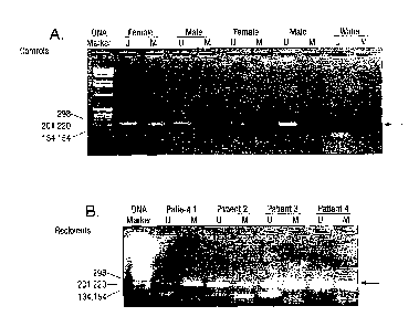
Figure 1 demonstrates the results of an assay detecting methylated and
tunnethylated
DNA. sequences of the androgen receptor gene. In total, 6 male and 11 female
healthy
subjects were recruited. Of all male control subjects, only the unmethylated
androgen
receptor gene was detected in these samples as expected (Fig, IA). By
contrast,. both
unmethylated and methylated androgen receptor gene DNA sequences were observed
in
female control subjects (Fig. 1A). The detection rates of methylated and
unmethylated
androgen receptor genes in these female subjects were 1.00% and 82%,
respectively. When
DNA samples were oraitted from the assay, no positive signal was observed
(Fig. 1A).
Interestingly, positive signals for both methylated and unmethylated DNA
sequences were
observed in all male bone marrow transplantation. recipients with female
donors, indicating
cells from female donor exist in the blood circulation of male recipients.
Figure 2 provides a schematic representation of the differentially methylated
=
region (DMR) of the hurt= IGF2-H19 region The two 450-bp repeat (Al and .A2)
and
seven 400-bp repeat (BI- B7) units are shown. The potential metitylation sites
on the upper
strand DNA of the studied region are represented by open circles. The studied
single
nucleotide polymorphism (SNP) site (A/G) is indicated by an open box. Open
arrows
represent the location of the forward (for) and reverse (rev) primers in PCR
reactions specific
for the methylated (M) and munethylated (U) alleles, respectively. Sequences
of these MSP
primer's are shown. Sequence differences between bisulfite-treated DNA and
untreated DNA
are highlighted in bold italics and sequence differences between methylated
(paternally-
.
inherited) and unmethylated (maternally-inherited) DNA are underlined in bold.
6
=
CA 2973165 2017-07-12
.1-30D1 = =
Figure 3 depicts portions of bisulfite-treated DNA sequencing profiles for the
epigenetic marker in the IGF2-H19 locus, taken from maternal and fetal sources
of a pregnant
woman, as indicated.
Panels in Figure 3a, depict samples from sources in the second trimester of
pregnancy.
Panels in Figure 3b depict samples taken in the third trimester. Maternal DNA
was isolated from sources free of fetal DNA.
Fetal DNA was isolated from amniotic fluid in the second trimester (Fig. 3a)
and cord blood in the third trimester (Fig. 3b). Postnatal maternal plasma DNA
was isolated
approximately 42 months after parturition. Labeled arrows in the maternal
plasma panels
indicate nucleotide peaks corresponding to mother and fetal markers.
Figure 4 demonstrates detection of unmethylated (maternally-inherited) fetal
DNA in maternal plasma. (a) Unmethylated DNA sequences were detected in
maternal buffy
coat (panel 1) and a third trimester maternal sample (panel 2) using direct
sequencing. The =
presence of unmethylated fetal DNA in maternal plasma is indicated by *. (b)
Unmethylated
fetal DNA (arrow) was detected in two third trimester maternal plasma samples
using the =
primer extension assay. (c) Unmethylated fetal DNA (arrow) was detected in a
second
trimester maternal plasma sample using the primer extension assay. Products
from control
reactions containing primer only, unmethylated G allele or unmethylated A
allele are shown.
The sizes (nt) of the reaction products are shown at the bottom. 0, unused
primer;
0 detected allele.
DESCRIPTION OF THE SPECIFIC EMBODIMENTS
In a first aspect, the present invention features methods for differentiating
DNA species originating from different individuals in a biological sample. In
preferred
embodiments the methods of the present invention are used to differentiate or
detect fetal
DNA in a maternal sample or to differentiate DNA of an organ donor from )NA of
an organ
recipient.
7
CA 2973165 2017-07-12
sil-30D1
Those of skill in the art will appreciate that the biological
from an individual may be taken from any fluid or cell sample, however, in
preferred
embodiments the bodily fluid is plasma or serum. In preferred embodiments, the
DNA
species are differentiated by observing epigenetic differences in the DNA
species such as
differences in DNA methylation. For instance, in situations where one
DNA.species.comes
from a male, and one DNA species comes from a female, the epigenetic marker
may be the
inactivated X chromosome of the female individual. In such embodiments,
methylated DNA
= sequences on the inactivated X chromosome may be used to detect DNA
originating from the
female individual. In some embodiments, the epigenetic differences may be
analyzed inside
cells. Further, in some embodiments, the epigenetic differences may be
analyzed using in-
situ methylation-specific polymerase chain reaction. Additionally, he
epigenetic differences
may be used to sort or isolate cells from the respective individuals or to
purify DNA from the
respective individuals. The methods according to the present invention may be
performed
with or without measuring the concentrations of DNA species, however, in
preferred
embodiments, the concentrations of DNA species with the respective epigenetic
differences
are measured. Such measuring of concentrations involves measuring the
respective DNA
methylation differences in embodiments wherein DNA methylation differences is
the
=
epigenetic marker. In especially preferred embodiments, sodium bisulflte is
added to the
biological sample or to the DNA species directly to detect the DNA methylation
differences.
However, in other embodiments a methylation-specific polymerase chain
reaction, as is well
known to those skilled in the art, may be used to detect the DNA methylation
differences. In
yet other embodiments, DNA sequencing or primer extension may be used to
detect the
=
methylation differences.
8
CA 2973165 2017-07-12
.1-30D1 =
41111
As used herein, the term "biological sample" is intended to encompass any
fluid or cellular sample or mixture thereof obtained from a living organism.
Specifically, the
term includes tissue biopsy, serum, plasma or amniotic fluid samples.
As used herein, the term "epigenetic difference is intended to encompass any
molecular or structUral difference other than the primary nucleotide sequence;
For instance,
=
this may include differences in methylation.
= As used herein, the term "DNA" is intended to encompass any sequence of
more than one nucleotide such as polynucleotides, gene fragments and complete
gene
sequences.
As used herein, the term "methylation-specific PCR" is used to describe a
method in which DNA is treated. with sodium bisulfite and then subjected to
PCR =
amplification. This technique is based on the principle that treating DNA with
bisulfite results
in conversion of unmethylated cytosine residues into uracil. Methylated
cytosine residues, on
the other hand, remain unchanged. Thus, the DNA sequences of methylated and
unmethyIated genomic regions following bisulfite conversion are different and
= distinguishable by sequence-specific PCR primers. =
The present invention utilizes the phenomenon of genomic imprinting to
overcome the limitations of the prior art. In genomic imprinting, DNA
sequences are
modified biochemically, without alteration in DNA sequence. If this process
results in
differential modification of the fetal and maternal DNA, then this difference
can be exploited
for the discrimination of fetal from maternal DNA in maternal plasma and
serum. This
phenomenon can also be used for the discrimination of fetal cells from
maternal cells in the
cellular fraction of maternal blood. In addition, this principle can also be
used to detect
maternal cells or DNA that has entered into the body of the fetus/baby (Lo, et
al., Blood
58(111:4390-5 (1996); La, et aL, Clin Chem, 46(9):1301-9 (2060); Maloney et
al., J Clin
9
CA 2973165 2017-07-12
.1-30D1
=
invest 104(1):41-7 (1999). 'this phenomenon can also be used' in many other
clinical
scenarios whereinzells or DNA sequences are found to be present inside the
body of an
individual, such as following bone marrow transplantation (Lo et 'al., Br J
Haeanatol
89(3):645-9 (1995)) or solid organ transplantation (Steal et al., Curr Opin
Nephrol
Hypertens fia):292-8 (1997); Lo et al.; .Lancet 351(9112):132930 (1998);
Zhang, CIin Chem
45(101:1741-6 (1999)).
The present invention allows development of a gender-independent and
polymorphism-independent marker for fetal DNA in maternal plasma/serum. To
develop a
gender-independent and polymorphism-independent fetal marker, one can use DNA
sequences which are preferentially and specifical/y methylated in the
trophoblasts (Ohgane
et al, Dev Genet, 22(2):132-40 (1998)). This overcomes the current limitation
which can
only easily detect the presence of DNA from a male fetus in the plasmaisenma
of the mother
(by using the Y-chromosome as the target) (Lo, et al., Am J Hum. Genet,
62(4):768 (1998).
It provides detection methods separate froth relying on sequence differences
in fetal and
maternal DNA to make such a distinction gang et aL, Clin Chem 45(11):2033-5
(1999);
Pertl et at, Hum Genet .106;45-49 (2000)).
The development of molecular detection txtethods such as the PCR has
provided many powerful tools for the monitoring of chimerism following bone
marrow
. .
transplantation (BMT). One of the roost -widely used PCR-based tests for the
detection of
post-BMT cbimerism in sex-mismatched cases is PCR for sequences on the Y
chromosome
(Lo et al., Br .1 Haematol $2: 645-9 (1995): The limitation of this strategy
is that it. can only
be used in eases wherein the donor is male and the recipient is female. The
present invention
provides a system that c.an be applied to situations when the donor is female
and the recipient
is male. The, fact that the phenomenon of Lyonization only exists in females,
can be exploited ,
to develop a female-specific marker. In this phenomenon., one of the two X
chromosomes in a
ia
CA 2973165 2017-07-12
S1-3 OD1 =
= =
female individual is inactivated at random, with methylation occurring:
inactivated genes. This therefore allows an assay for detecting female DNA in
an excess of
male DNA and which can be applied to BMT with female donors and male
recipients.
In a second aspect, the present invention features methods of detecting
abnormalities in a fetus by detecting fetal DNA in a biological sample
obtained from a
mother. The methods according to the present invention providefor detecting
fetal DNA in a
maternal sample by differentiating the fetal DNA from the maternal DNA based
upon
epigenetic markers such as differences in DNA methylation. Employing such
methods, fetal
DNA that is predictive of an anomaly or a disease may be identified thereby
providing
methods for prenatal diagnosis. These methods are applicable to any and all
pregnancy-
associated conditions for which methylation changes associated with a disease
state is
identified. Exemplary diseases that may be diagnosed include, for example,
preeclampsia, a
chromosomal aneuploidy, including but not limited to nisorny 21, Prader-Willi
Syndrome,
and Angelman Syndrome.
As with the broader differentiating methods of the first aspect of the
invention,
the biological sample obtained from the mother is preferably plasma or serum.
The
differentiation between maternal and fetal DNA may be performed with or
without
quantifying the concentration of fetal DNA in maternal plasma or sentra. In
embodiments,
wherein the fetal DNA is quantified, the measured concentration may be used to
predict,
monitor or diagnose a pregnancy-associated disorder. In preferred embodiments,
the
particular fetus-derived epigenetic mark is associated with a fetal disorder,
and in some
embodiments an epigenetic characterisitic in fetal cells in the placenta is
used as a fetus-
specific marker in maternal plasma or serum.
The present invention utilizes differentially methylated fetal DNA sequences,
which do not need to be distinguishable in terms of DNA sequence from maternal
DNA, as
=
=
11
CA 2973165 2017-07-12
.1-30D1
= .
= markers for non-invasive prenatal diagnosis. This novel approach can
convert fetus-mother
pairs who are not informative in the conventional approach, to being
informative for prenatal
diagnosis. Thus, present invention provides a platforxzi on which a new
generation anon-
'
invasive prenatal tests can be built.
The methods of the present invention are based on the detection of differently
methylated DNA of fetal origin in the plasma or serum of pregnant women.
Differentially
methylated DNA sequences, which may contain single nucleotide polymorphism,
are
preferably detected by methylation-specific polymerase chain reaction (PCR);
but in principle
any detection method for differentially methylated DNA can be used. This
approach allows
the use of conventional uninformative fetal DNA markers for prenatal
diagnosis.
The present invention allows detecting or predicting the presence of any
disorders of the fetus or the mother which. are associated with a change in
methylation status
of a DNA sequence. Examples include imprinting disorders such as Prader-Willi
syndrome
(Kubota et al., Nat Genet 16(1):16-7 (1997). The present invention provides a
new type of
test for preeclampsia which has been suggested to be an imprinting disorder
(Graves, =Reprod .
= Ferrel Dev 1 0m.:23-9 (1998). The present invention further provides a
new type of test for
chromosomal ateuploidies, including Down syndrome (trisomy 21), which may be
associated
with methyIation changes (Yu et al., Proc Natl Aced Sat USA 94(131:6862-7
(1997).
The present invention features using DNA methylation differences between
the mother and fetus thereby overcoming the limitations of the prior art in
the detection of
fetal DNA in maternal plasma.
In a third aspect, the present invention features methods for differentiating
DNA species originating flora an organ donor from those of an organ recipient.
As with the
broader differentiating methods of the first aspect of the invention, the
biological sarnple
obtained is preferably plasma or serum. The differentiation between DNA frorn
the organ
12
CA 2973165 2017-07-12
#1-30D1
donor and organ recipient or potential organ donor and potential organ
recipient may be
performed with or without quantifying the concentration of DNA in the
biological sample.
This embodiment is particularly useful in instances when the transplantation
is a bone =
=
marrow transplantation. Such measurements may be used to predict the clinical
progress of
the transplantation 'recipient especially as applied to organ rejection.
In a fourth aspect, the present invention features kits for differentiating
DNA
species originating from different individuals in a biological samPle. Such
kits are useful, for
instance, for differentiating or detecting the presence of fetal DNA in a
maternal biological
sample or for differentiating DNA from an organ donor or potential organ donor
from that of
an organ recip- lent or potential organ recipient. The kits according to the
present invention =
comprise one or more reagents for ascertaining the methylation status of the
maternal DNA
such as sodium bisulfite and one or more reagents for detecting the presence
of DNA. such as
a gel. Additionally, such kits may include one or more reagents for amplifying
the amount of ,
DNA present in the sample such as one or more reagents for performing
polymer:4e chain
reaction amplification. Such reagents are well known to those of skill in the
art. Further,
such kits may include one or more apparatuses for obtaining a maternal DNA
sample. Such
apparatuses are well known to those skilled lathe art. In particular the kits
according to the
present invention may be used for diagnosing a disease caused all or in part
by a genetic
anomaly such as a mutation, substitution or deletion or duplication in all or
part of a DNA
'sequence present in a fetus. Exemplary diseases that may be diagnosed
include, for example,
preeclampsia, a chromosomal aneuploidy, including but not limited to trisomy
21, Prader-
Willi Syndrome and &german Syndrome.
EXAMPLE 1
IS =
CA 2973165 2017-07-12
=7130D1
111
Detection of post-bone marrow transplantation cbimerism
epigenetic approach
=
Materials and Methods =
Subjects and Samples
Four male marrow transplantation recipients, who received bone marrow
from female donors, and 17 normal healthy subjects were recruited in this
stuay. Buffy coat
(BC) from all recruited EDTA-blood samples were harvested and stored at ¨20 DC
as
described (Lo *et al., Am J Hum Genet 01768-75 (1998).
DNA isolation = =
= DNA was extracted from the BC using a Nucleon DNA Extraction Kit
(Scotlabs) according to manufacturer's recommendations.
=
Bisulfite conversion
Bisulfite modification of DNA samples was performed using a CpGenome
DNA Modification Kit (Intergen) as instructed by the manufacturer. With
bisulfite
conversion, umnethylated cytosine residues are converted to uracil while
methylated cytosine
residues remain unchanged (Herman et al., Proc Nad Aced Sci USA :982l-
6(1996).23 The
sequence difference between methylated and unmethylated DNA following
bisulfite
conversion is then distinguished using different PCR primers. 1 ug of BC DNA
was used in a
bistrifite conversion reaction.
Methylation-specific PCR (MSP)
MSP assays were modified frorn the protocol as described by Herman et tti,
supra. The primers M-for (5'-GCGAGCGTAGTATITTTCGGC-3') and M-rev (5'-
.
AACCAAATAACCTATAAAACCTCTACG-3') were designed for the methylated
sequence, while the primers U-for (5'-GTIGTGAGTGTAGTA1 .t i i i. .I.GGT-3')
and U-rev
(5'-CAAATAACCTATAAAACCTCTACA-3') were designed for the unmethylated
sequence. Five pi bisulfite-treated DNA was added to a SO p.1PCR reaction
containing 5 ill
10x TaqMan buffer A (PE Applied Biosystems), 2 inM MgC12, 10 pmol dNTPs, 20
pmol
each of the corresponding MSP primers and 1.25 U AmpliTaq Gold DNA polymerase
(PE
Applied Biosystems). Reaction mixtures were thermal cycled (methylated allele:
95 C for 45
sec, 58 C for 30 sec, 72 C for 20 sec; unmethylated allele: 95 C for 45 sec,
50 C for 30
sec, 72 C for 20 sec) for 45 cycles, with an initial denaturing step of 8 min
at 95 C. PCR
products were then analyzed by agarose gel electrophoresis.
14
CA 2973165 2017-07-12
Results
This experiment provides a MSP assay to detect methylated and unmethylated
DNA sequences of the androgen receptor gene. In total, 6 male and 11 female
health siibjects
were recruited. Of all male control subjects, only the unmethylated androgen
receptor gene
was detected in these samples as expected (Fig. 1A). By contrast, both
unmethylated and
methylated androgen receptor gene DNA sequences were observed in female
control subjects
(Fig. 1A). The detection rates of methylated and unmethylated androgen recepµ
tor genes in
these female subjects were 100% and 82%, respectively. When DNA samples were
omitted
from MSP essay, no positive signal was observed (Fig. IA). Interestingly,
positive signals for
both methylated and urumethylated DNA sequences were observed in all male sex-
mismatched bone marrow transplantation recipients (100%), indicating cells
from female
donor exist in the blood circulation of male recipients.
These results demonstrate, for the first time that methylated genes on the
inactivated X chromosome from female individuals can be used as a female-
specificniarker
in. chimerism research. This assay is also applicable to the study of other
types of post-
transplantation chimerisms involving mixture of male and femile cells or DNA.
Examples
include cellular chimerism following solid organ transplantation (Starzl et
al., Curr Opin
NePplurol Hypertens 6:292-8 (1997)), post-transplantation plasma DNA chimerism
(Lo et al., =
Lancet 351;1329-30 (1998)) and urinary DNA chimerism (Zhang et al., Clin Chem
45:
1741-6 (1995)). In addition, there is also much recent interest in the passage
of cells and
DNA from the mother into the fetus during pregnancy (Lo et al., B1ood/M:4390-
5. (1996);
Maloney et al., C1Þn Invest 41-7 (1999); Lo et al.. Clin Chem 46:1301-9
(2000). The
epigenetic markers developed should also be of used in chimerism of maternal
origin in male
offsprings.
The current assay may be developed into a quantitative format, using for
example, real-time PCP._ technology (Lo et al., Cancer Res a:3899-903 (1999)).
Such
development would allow us to monitor the levels of chimerism in a particular
person.
Clinically such an assay might have a role in the monitoring of graft
acceptance in BMT. In
the ease of urinary or plasma DNA chimerism, such an assay might also be used
for the
monitoring of graft rejection.
FXAMPLE 2
CA 2973165 2017-07-12
=
.1-30D1
=
Differential DNA methylation between fetus and mother as a_strateay for
detectinz fetal DNA in maternal plasma
The present experiment demonstrates that by using a differentially methylated
region in the human IGF2-H19 locus as an epigenetic marker in maternal plasma,
detection
of an allele that the fetus has inherited from the mother is possible. These
results greatly
expand the prenatal diagnostic possibilities of fetal DNA in maternal
plasma'allowing
development of a gender- and polymorphism-independent fetal-specific marker in
maternal
=
plasma and new strategies for the prenatal diagnosis of imprinting disorders
and certain
=
chromosomal aneuploidies.
. Materials and Methods
Subjects and Samples
Samples were collected from pregnant women with informed consent. In
total; 21 and 18 women in the second trin. rester (17-21 weeks) and. third
trimester (37-42
weeks) of pregnancy, respectively, were recruited for this study. None of the
recruited
subjects had preeclarnpsia or preterm labor in the current pregnancy. EDTA
maternal blood
and fetal amniotic fluid samples were collected from the second trimester
cases as described
previously (Lo et al.. Arn Hum Genet 62:768-775 (1998)). For the.third
trimester cases, we
collected EDTA maternal blood samples at 2 to 311 before normal vaginal
delivery. EDTA
fetal cord blood samples were also collected immediately after delivery as
described (Lo et
al., Clin Chem 46:1903-1906 (2000)). Plasma and buffy coat from all recruited
blood
samples were harvested and stored at ¨20 C as described (Lo et aL, Am JHirm
Genet
.62:768-775 (1998)), except that plasma samples were rec.entrifuged at 16,000
g. Amniotic
fluid samples were stored at 4 C.
DNA isolation
DNA was extracted from plasma and amniotic fluid samples using a QTAarnp
Blood Kit (Qiagen). Typically, 800 ul of plasma or amniotic fluid was used for
DNA
extraction per column. An elution volume of 50-110 ILL was used. DNA was
extracted from
the buffy coat using a Nucleon DNA Extraction Kit (Scotlabs) according to
manufacturer's
recommendations.
Genoryping of the DMR polymorphic region
The DMIt in the human IGF2-H19 locus contains two 450-bp repeat. and
seven 400-bp repeat units (Nakagawa et aL, F'roc Nati Acad Sci USA 21:591-596
(2001))
16
CA 2973165 2017-07-12
W1l-30D1 =
=
(Fig. 2). An .A/G SNP within the DMR (Nakagawa et al., supra) was selected as
a marker in
= our investigation (Fig. 2). Polymerase chain reaction (PCR) was used to
amplify the SNP in
both maternal and fetal DNA samples. Primers were designed using the sequence
of the
Homo sapiens H19 gene (Genbank accession number AF125 183). Typically, 2 to 5
eluted
DNA, purified from maternal huffy coat, cord huffy Goat or anudOtic fluid was
added to a 25
PCR reaction containing 2.5 pi. 10x TaqMan buffer A (PE Applied Biosystems), 3
rnM
MgC12, 6.26 pmol dNTPs, 5 pmol primers (forward: 5'¨ggACGGAATTGGTTGTAGTT-3";
reverse: 5.¨AGGCAATTGTCAGITCAGTAA-3') and 0.625 U AmpliTaq Gold DNA
polymerase (PE Applied Biosystems) (95 C for 8 min followed by 35 cycles of
95 C for 1
min, 56 C for 20 sec, 72 C for 20 sec). For the forward primer, the
nucleotides in upper
case corresponded to positions 7927 to 7944 of the H19 sequence.(Genbank
accession
number AF125 183). For the reverse primer, the nucleotides were complementary
to positions
8309 to 8329 of the H19 sequence. PCR products were then analysed by agarose
gel =
electrophoresis and DNA sequencing.
Bisulfite conversion
Bisulfite modification of DNA samples was performed using a CpGenome
DNA Modification Kit (Intergen) as instructed by the manufacturer. With
bisulfite
conversion, unmethylated cytosine residues would be converted to uracil; while
methylated
cytosine residues would remain unchanged (Herman et al., Proc Natl Acad Sci
USA a:9821-
9826 (1996)). The sequence difference between methylated and unmethylated DNA
following bisulfite conversion could then be distinguished using different PCR
primers. In
general, 1 IT of buffy coat DNA from maternal or cord blood, or 941 eluted DNA
purified
from maternal plasma or amniotic fluid was used in a bisulfite conversion
reaction. Bisulfite-
treated DNA was then elutcd in 25-50 I 1 Tris-EDTA.
Methylation-specific PCR (MSP)
MSP assays were modified from the protocol as described (Herman et al.
1996). Five t1 bisulfite-treated DNA was added to a 50 gl PCR reaction
containing 5 p.1 10X
TaqMan buffer A (PE Applied Biosystems), 2.5 niM MgC12, 10 pmol dNTPs, 20 pmol
each
of the corresponding MSP primers (Fig. 2) and 1.25 U AmpliTaq Gold DNA
polymerase (PE
Applied Biosystems). The primers M-for and M-rev (Fig. 2) were designed for
the
methylated sequence, while the primers U-for and U-rev (Fig. 2) were designed
for the
unmethylated sequence. Reaction mixtures were thermal cycled (methylated
allele: 95 C for
45 sec, 55 C for 20 sec, 72 C for 20 sec; tuunethylated allele: 95 C for 45
sec, 49 C for 20 =
17
=
CA 2973165 2017-07-12
101-30D1
411
sec, 72 C for 20 sec) for 50 (buffy coat and amniotic fluid DNA) or 56
(plasma DNA)
cycles, with an initial denaturing step of 8 min at 95 it. PCR products were
then analyzed
= by agarose gel electrophoresis. Reaction products were purified using
Microspin S-300 ER
columns (Amersham Pharmacia) for DNA sequencing or the primer extension assay.
=
DNA sequencing
Purified PCR products were sequenced using an AEI Prism dRhodamine
Terminator Cycle Sequencing Ready Reaction Kit (PE Applied Biosystems) cl the'
corresponding forward primers of the PCR products. Sequencing products were
analysed
using an ABI Prism 310 Genetic Analyser (PE Applied Biosystems).
Primer extension assay
= Two al of the purified MSP product was added to a 25 jil reaction
containing
50 M ddATP (2',3--dideoxyadenine triphosphate), 50 p.M dGTP, 50 M dTTP, 0.2
Fara
Cys-5-labeled primer (5"¨GGGITATTTGGGAATAGGATATITA-31, 4 U Thermo
Sequenase (Amersham Pharmacia) and 1.43 I concentrated buffer. Reactions were
thermal
cycled for 40 cycles (95 C for 30 sec, 51 C for 20 sec, 72 C for 20 sec). The
Cys-5-labeled
primer was 25 nucleotides (nt) in length and the polymorphic site was 2 nt
away from the 3'-
end of the primer. For the A allele, the incorporation of the ddATP at this
polymorphic site
would produce chain termination, thus resulting in an extension product of 27
nt (i.e., 25+2
nt). For the G allele, chain extension would continue until the next A residue
which was 5 nt
away from the 3'-end of the primer, thus re,salting in an extension product of
30 nt (Le, 25+5
nt). Reaction products were electrophoresed using a 14% denaturing
polyacrylamide gel and
analysect using an ALF Express Sequencer (Axnersbam Pharmacia). Data were
analysed by
the AlleleLinks program (Amersham Pharmacia).
Results
Genotyping of DMR
Thirty-nine pregnant women were recruited in this study. Maternal genotype
at the SNP within the DIvIR (Fig. 2) was determined by direct sequencing of
PCR products
from the buff' coat DNA. The number of pregnant women with each of the
possible
genotypes were 17 (GG, 43.6%), 16 (AG, 41.0%) and 6 (AA, 15.4%).
Detection offetal DNA in plasma from women heterozygous for a &allelic
polymorphism
The 16 women who were heterozygous (i.e., AG) for the SNP were selected
for further examination. As this is a biallelic polymorphism, these women
would not be
18
CA 2973165 2017-07-12
11,71-30D1
=
considered informative at this polymorphic locus for the detection of fetal
DNA in maternal ,
plasma, based on previous criteria (Lo et al.. Ann N Y Acad Sci 731204-213
(1994); Bianchi
Am J Hum Genet 62:763-764 (1998)). To demonstrate that differential
methylation at this
genomic region would allow us to overcome this limitation, maternal DNA was
bisulfite-
treated and amplified by MSP using the primers shown in Fig. 2. Similarly,
fetal DNA.
isolated from amniotic fluid (2nd trimester samples) or buff' coat of cord
blood (3rd
trimester samples) was subjected to PCR and MSP to determine the imprinting
status of the
=
fetal alleles.
Amongst the 16 selected cases, the methylated (i.e., paternally-inherited)
alleles from four 3rd trimester and seven 2nd trimester fetal samples were
different froni the
methylated alleles of the respective mothers (Fig. 3a,b; compare panels 1 and
2). Tò test if =
this differential methylation between fetus and mother would allow the fetal
allele to be
detected from maternal plasma, maternal plasma DNA from thete cases was
subjected to
bisulfite conversion, followed by MSP. Interestingly, the paternally-inherited
methylated fetal
allele could be detected in two 3rd trimester and four 2nd trimester matemal
plasma samples
(Fig. 3a,b; panels 3). To exclude the possibility that these observations were
simply due to the
existence o f aberrantly methylated maternal DNA in maternal plasma, we
collected a
postnatal maternal plasma sample (-3.5 year after deliVery) from one Ellie
positive cases
for further examination. We did not observe the additional methylated allele
in this postnatal
sample (Fig. 3a, panel 4), indicating that the additional methylated allele in
maternal sample
during pregnancy was of fetal origin. In addition, no positive signal was
observed in the
plasma of non-informative cases (n--4, data not shown), thus further
demonstrating the
specificity of this MSP assay. Taken together, these data indicate that the
use of differential
rnethylation between mother and fetus allows detecting fetal DNA in maternal
plasma, even
in cases which are not considered informative with existing criteria.
Detection offetal-derived maternally-inherited DNA from maternal plasma
We then tested if the use of differential methylation between mother and fetus
=
might allow us to detect an allele that the fetus has inherited from the
mother. This type of
analysis has previously been thought to be impossible (Lo et at.. Ann N Y Aced
Sci 731:204-
213 (1994); Bianchi, Am J Hum Genet fra:763-764 (1998)). As the maternally-
ini;erited allele
was unmethylated, the primers U-for and U-rev (Fig. 2) were used to amplify
the
unmethylated allele following bisulfite conversion. Among the 16 analyzed
cases, three 3rd
trimester and Eve 2nd trimester maternal satnples were informative. In these
cases, the fetus
possessed an unmethylated allele that was different from the unmethylated
allele of the
19
CA 2973165 2017-07-12
#471*-30D1 =
mother. These results implied that in these cases, the mother had original
allele from her father and then passed on to the fetus. Of these 8 informative
cases, only a
weak positive signal was observed in one of the 3rd trimester samples on
direct sequencing
(Fig. 4a, compared panel 1 and panel 2).
We reasoned that the weak signal in this single positive case and the Iow =
detection rate of the unmethylated fetal allele from maternal plasma might be
due to the low
sensitivity of the direct sequencing method. To enhance the sensitivity of
detection, we
employed a more sensitive primer extension assay to detect the umnethylated
fetal allele from
the MSP reaction products. As the SNP was an A/G polymorphism, ddATP was used
as a
reaction substrate in the primer extension assay. Extended reaction products
from the A and
G alleles were 27 and 30 nt long, respectively. No fetal specific reaction
product was present
in the corresponding maternal buff' coat samples (Fig 4b, c; maternal BC).
Strikingly, fetal
specific extension products were observed in two 3rd trimester (Fig. 4b,
arrow) and one 2nd
trimester (Fig. 4c, arrow) maternal plasma samples, indicating the presence of
nnmethyiated
fetal DNA in maternal plasma. As controls, none of the tested non-informative
cases was
positive in this assay (n=5, data not shown). These results demonstrated, for
the first time, the
feasibility of using epigenetic markers to detect a fetal-derived maternally-
inherited DNA.
sequence from maternal plasma.
Discussion
These results demonstrate that the use of epigenetic markers overcomes the
conventional limitations of detecting.fetal DNA in maternal plasma. It is
possible to detect a
paternally-inherited fetal allele, which is genetically indistinguishable from
a maternal allele,
from the mothees plasnia, by the use of epigenetic differences between the
mother and fetus. -
Likewise, it is possible to detect a maternally-inherited fetal allele from
maternal plasma.
This novel epigenetic approach will therefore expand the repertoire of
disorders wherein fetal
DNA in maternal plasma can be used.
Even with the use of relatively insensitive methods such as direct sequencing
and primer extension, the present results demonstrate that it is passible to
detect differentially
methylated fetal DNA sequences from maternal plasma. There was a lower
sensitivity in the
detection of the unmethylated fetal DNA in maternal plasma (Fig. 4), as
compared with the
analogous assay for the methylated allele (Fig. 3). Using more sensitive
detection systems,
such as aliele-specifk PCR (Newton et al., Nucleic Acids Res 11:2503-226
(1989)) and real-
CA 2973165 2017-07-12
.71-30D1
4111
time methylation-specific PCR (Lo et aL, Cancer Res a:3899-3903 (1999); Pads
et al.,
Nucleic Acids Res 28:E32 (2000)), might enhance the sensitivity of plasma-
based epigenetic
analysis. The development of real-time methylation-specific PCR is
particularly interesting as
it opens up the possibility of quantifying fetal-specific methylation in
maternal plasma, as has
already been achieved for the detection of tumor DNA in circulation (Kawakami
et al.. J Nati
Cancer Ins! 92:1805-1811 (2000)).
The possible introduction of fetal DNA in maternal plasma as'a routine
= prenatal diagnostic tool has raised questions with regard to the need of
a generic marker for
circulating fetal DNA (La et al., Ant J Hum Genet 62:768-775 (1998); Avent et
al, Vox Sang .
21155-162 (2000)). Most proposals for such a marker have thus far focused on
the use of
genetic polymorphisms between the mother and fetus (Tang et al., Clin Chem
45:2033-2035
(1999); Peal et aL, Hum Genet n1:45-49 (2000)). The present demonstration of
the
feasibility of epigenetic markers for fetal DNA detection in maternal plasma
opens up a new
approach for the development of a gender-independent and polymorphism-
independent fetal
. 15 marker in maternal plasma. One way wherein this can be achieved is to
exploit the
phenomenon of tissue-specific methylation (Grunau et al., Hum Mal Genet 2:2651-
2663
(2000)). As the trophoblast has been suggested to be the predominant cell
population for
releasing fetal DNA into maternal plasma, the elucidation of trophoblast-
specific meeaylation
= patterns allows the development of a generic epigenetic fetal marker in
maternal plasma.
Biologically, the use of tissue-specific methylation markers may also allow
one to directly
address the question as to what fetal cell types are responsible for releasing
fetal DNA into
maternal plasma.
The epigenetic analysis of maternal plasma has obvious applications to
disorders associated with genomic imprinting, such as the Prader-Willi
syndrome (Pfeifer,
Lancet 356:1819-1820 (2000)). This strategy may also have diagnostic potential
for disorders
such as preeclampsia, wherein imprinted genes have been hypothesized to play a
role
(Graves, Reprod Feral Dev 10:23-29 (1998)).
The possible application of fetal DNA in maternal plasma for the prenatal
detection of fetal chromosomal aneuploidies is an issue that has been keenly
discussed since =
the discovery of the phenomenon (Lo et al., Lancet M:485-487 (1997); Bianchi,
Am J Hum
Genet 62:763-764 (1998)). The finding of quantitative differences between the
circulating
fetal DNA levels in aneuploid, compared with euploid pregnancies (Lo et al.,
Clin Chem
45:1747-1751 (1999); Zhong et al., Prenal Diagn 20:795-798 (2000)) offers a
method for
estimating the risk of fetal chromosomal aneuploidies from maternal plasma.
The recent
21
CA 2973165 2017-07-12
1,1-3001
=
discovery of apoptotic fetal cells in maternal plasma ("plasma-derived cells")
(Van Wijk et
aL, Clin Chem 46:729-731 (2000)) offers yet another approach for aneuploidy
detection from
maternal plasma (Poon et al., Lancet 356:1819-1820 (2000)). Interestingly, the
present data
open up yet another potential approach for the detection of fetal chromosomal
aneuploidies.
This is based on the observation that aberrant DNA methylation patterns may be
associated
with chromosomal aneuploidy (Knromitsu et aL, Mol Cell Blo111:707-712 (1997);
Yu et at,
Proc Natl Acad Sci USA 94:6862-6867 (1997)). Hence it is possible to develop
epigenetic
markers for detecting such aberrantly methylated fetal DNA sequences from
maternal
plasma. Such markers provide specificity compared with a simple quantitation
of fetal DNA
in maternal plasma (Lo et al., Clin Chem 45:1747-1751 (1999); Zhong et al.,
Prenat Diagn.
211795-798 (2000)) and better suitability to large scale application compared
with methods =
based on "plasma-derived cells" (Poon et aL, Lancet 356:1819-1820 (2000)).
Fetal epigenetic markers may also be used in the analysis of fetal cells
isolated
from the cellular fraction of maternal blood. This takes advantage of recent
data showing that
methylation analysis could be performed in an in situ manlier (Nuovo et aL,
Proc Natl Acad
Sci USA 96:12754-12759 (1999)).
= With the recent realization that fetomatenial trafficking is a
bidirectional
process (Lo et al., Blood 88:4390-4-395 (1996); Maloney et al., J Clin Invest
104:41-47
(1999)), epigenetic markers may also be used to investigate cellular and DNA
transfer from
the mother to the fetus. Such an approach naight also have applications to the
investigation of
other types of chinaerism, such as post-transplantation hemopoietie chimerism
(Starzl et al.,
Oar Opin Nephrol Hypertens 6:292-298 (1997)) and urinary DNA chimerism (Zhang
et aL,
Clin Chem 45:1741-1746 (1999)).
With our increased understanding of the human genome and the development
of high throughput array-based technologies for methylation analysis (Yan et
al.,Clin Cancer
Res 6:1432-1438 (2000)), we expect that the number of usable fetal epigenetic
markers will
rapidly increase over the next few years. Such a development will provide a
clinically
relevant panel of fetal epigenetic markers which can be used in a synergistic
manne.r with
conventional genetic markers in maternal plasma.
22
CA 2973165 2017-07-12
DEMANDES OU BREVETS VOLUMINEUX
LA PRESENTE PARTIE DE CETTE DEMANDE OU CE BREVETS
COMPREND PLUS D'UN TOME.
CECI EST LE TOME 1 DE 2
NOTE. Pour les tomes additionels. veillez contacter le Bureau Canadien des
Brevets.
JUMBO APPLICATIONS / PATENTS
THIS SECTION OF THE APPLICATION / PATENT CONTAINS MORE
THAN ONE VOLUME.
THIS IS VOLUME 1 OF 2
NOTE: For additional volumes please contact the Canadian Patent Office.
-