Note: Descriptions are shown in the official language in which they were submitted.
DIFFUSE ACOUSTIC CONFOCAL IMAGER
FIELD
The present technology relates to a diffuse acoustic confocal imager.
Additionally, this
technology relates to acoustic confocal imaging, especially with regard to
methods of
using coherent acoustic beams pulsed or continuous in the diagnosis and
treatment of
tumours and related diseases.
BACKGROUND
The use of beams of radiation to obtain information about an object by
detecting the
amplitude or phase of the beam is well known for scientific and medical
purposes. For
example, the phase information of a beam that passes through an object can
provide
information on the object's temperature, composition, magnetic field or
electrostatic field,
whereas amplitude measurements can provide information on the opaqueness or
density
of the object. The beams are comprised of waves of radiation, where a wave,
(I), can be
described as having both an amplitude, A, and phase, 0, described
mathematically as,
(I) = Aexp(0) 1)
The information obtained from the method depends on whether it is detecting
the
amplitude or both the amplitude and phase of a beam's wave. If the method
measures
only a beam's amplitude, as is the case for X-ray, only density differences in
the object
are reported. This is a limitation of the technology as it does not provide
information such
as an object's temperature, composition, elasticity, strain field, magnetic or
electrostatic
fields. An additional disadvantage of a number of imaging techniques such as X-
ray
imaging
1
Date Recue/Date Received 2023-11-10
CA 02973655 2017-07-12
WO 2016/113664
PCT/IB2016/050109
methods is the strength of radiation employed. When used in diagnosis, the
levels
employed may have the potential to damage cells in the body.
Acoustic microscopes including Ultrasound are now widely used to image the
inside of the
body such as the fetus in the womb and blood flow in arties and veins. These
microscopes
measure the intensity of the acoustic beam reflected off surfaces such as
bones and
interfaces such as the interface between the embryonic fluid and fetus. These
microscopes
cannot measure the intensity and phase of the beam passing through or
reflected from soft
tissue such as muscles or embryonic fluid. These microscopes also cannot
measure
temperature or composition as they only use the intensity of the acoustic
beams and not
the phase of the acoustic beams. Hence the images are not suitable for
providing
information other than that information that pertains to surfaces or
interfaces. A further
deficiency of these microscopes is that the image produced has a significant
amount of
background intensity caused by the diffuse scattering of beams. Taking as an
example, a
prostate gland, an ultrasound image poorly identifies the interface between
the prostate
and other tissue and can also identify the urethra) however, it cannot
identify any
abnormalities within the prostate.
Another method that measures a beam's amplitude is confocal microscopy.
Confocal
scanning laser microscopes were developed in the 1.980s for seeing, three-
dimensional
objects. Confocal scanning laser microscopy uses a laser beam passing through
an object to
create a three-dimensional amplitude image of the object by detecting the
amplitude of the
beam through a pinhole aperture placed confocal with a point on a focal plane
of the object.
Confocal microscopes have now found widespread applications in the materials,
biological,
and medical sciences. As a diagnostic tool, confocal microscopes are limited
to detecting
only thin tissue and the density differences of objects, which produce
amplitude differences
of the detected beam. The beams cannot penetrate far in to tissues and other
materials.
They do not measure the object's phase information. Hence, confocal
microscopes cannot
measure an object's composition or temperature.
2
CA 02973655 2017-07-12
WO 2016/113664
PCT/IB2016/050109
If the method measures changes in the phase of a beam, then information can be
provided
about the object's temperature and. composition. Acoustic beams can be used
for this. The
phase of acoustic beams are modified by an object's refractive index, where
the refractive
index is dependent on the object's temperature and composition and is a
measure of the
acoustic beam's speed of sound.
The absolute phase of an object can be measured using a Confocal Scanning
Holography
Microscope, as described in US Patent No. 7,639,365. This approach cannot be
used to
image the inside of the human body as laser beams do not readily pass through
the human
body.
The relative phase of an object can be measure using an Acoustic Confocal
Interferometry
Microscope, as described in US Patent No. 8,485,034. This approach requires an
interference beam and a complex arrangement of mirrors and prisms and is not
suitable for
imaging the inside of the human body because of the geometric constraints.
Standard interferometry microscopes, standard holography microscopes, and
standard
holographic interferometry microscopes have been used to measure both the
phase and the
amplitude of objects, giving important information of objects such as their
density,
composition and temperature. These microscopes create a three dimensional
amplitude
image and phase image of the object by measuring both the phase and the
amplitude. As
they are light microscopes the three-dimensional information measured from
these
microscopes comes only from the surface of the object and not at points within
the object.
In all cases, a reference beam and an object beam are used to collect data
that results in the
creation of the images. This limits the use of these microscopes to collecting
data from or
about surfaces of objects.. In medical diagnosis they would therefore be
potentially useful
for diseases of the skin, but not for diseases of internal tissues or organs.
Other means able to measure the amplitude and phase of objects using an
acoustic beam is
spatially-filtered transmission ultrasound phase imaging as disclosed in US
Patent Nos.
6,679,846, 6,436,046, 6,132,375 and 6,193,663. Spatially-filtered transmission
ultrasound
3
CA 02973655 2017-07-12
WO 2016/113664
PCT/IB2016/050109
phase imaging involves measuring the amplitude and phase of an emitted beam
and then
again measuring the amplitude and phase of the acoustic beam after it passes
through the
object upon its arrival at a detector. The difference in amplitude and phase
is attributed to
the object. From the sound source, the beam diffusely scatters outward leading
to
background scatter that is not wanted. Within or around this background
scatter will be the
image of interest. That image is representative of the interfaces of the
object being imaged.
It does not represent a three dimensional image, nor can it locate diseased
tissue within the
tissue or organ of interest. Similarly, in materials, it cannot provide a
three dimensional
image nor can it show a different material within the material or a region
having different
physical characteristics within the material, unless there is an interface,
such as the
interface between a liquid and a solid.
It would be advantageous to provide a device, system and method that can
detect both the
amplitude and phase of a beam. Such a device, system and method would be able
to
provide information on the object's density, temperature, composition,
elasticity, strain
field, magnetic or electrostatic fields. This is of great significance in the
medical field, as of
being able to obtain information on density, temperature, and composition
allows one to be
able to potentially diagnose, treat and assess effectiveness of treatments for
diseases such
as cancer. Ideally, the device would be suitable for being hand-held, with a
variety of
different shaped detector holders for application to different parts of the
body, for example,
but not limited to the prostate, breast, head, and skin.
Examples of an application where the measurement of temperature and
composition is
important include medical diagnostics aimed at understanding the function of
organs, tissue
and diseased regions in the body. Presently medical researchers do not have
good means to
non-invasively measure the internal temperature and composition of the body.
It is an
object of the present technology to provide such capabilities.
What is needed is a system that utilizes a coherent beam that can be focused
to a probe,
which then acts as a virtual source of diffusely scattered beams, which in
turn could be
detected by the detector of the system and processed into meaningful data.
Such a system
4
CA 02973655 2017-07-12
WO 2016/113664
PCT/IB2016/050109
would preferably provide the capability of detecting differences between
materials, such as
differences between healthy and diseased tissues, such as density, temperature
and
compositional differences. More preferably, the same system would allow for
treatment of
the diseased tissue. While the application of the technology would preferably
be in the
diagnosis and treatment of disease, it would also be preferably if it could be
applied more
broadly to detection of different materials or different states of materials
in a structure or
material.
SUMMARY
The present technology provides a system that utilizes a coherent beam that is
focused to a
probe. That probe then functions as a virtual source of diffusely scattered
beams that
radiate outward randomly from the probe. Some diffusely scattered beams are
detected by
a detector and the output from the detector is sent to a processor for
processing into
meaningful data using mathematical formulae. Hence, this system utilizes the
diffusely
scattered beams that are undesirable in Ultrasound and that interfere with the
clarity of the
image, to create images that identify differences between materials, such as
differences
between tumours and healthy tissue including density differences, temperature
differences,
and compositional differences. The significance of this is that a three
dimensional image
can be obtained of, for example a tumour or diseased state within a healthy
tissue.
Similarly, in non-medical applications, three dimensional images can be
obtained of any
structure within another medium, or a part of the same medium in a different
physical state
than that of the remainder of the medium.
Not only can the three dimensional image of a tumour within a tissue be
provided,
information about that tumour can also be provided. The speed of sound can be
correlated
to the type of tumour, the stage of development of the tumour and to the
temperature of
the tumour.
The same system can be used to treat the tumour or disease state by increasing
the beam
strength and dwell time either from all the emitters, or a select number of
emitters or in
pulses. Dwell time for imaging and diagnostics is short, for example, around 1
second or less
5
CA 02973655 2017-07-12
WO 2016/113664
PCT/IB2016/050109
(this would be a low dwell time), whereas dwell time for treatment could be
100 to 100s of
seconds (this would be a high dwell time), for example. Without being bound to
theory, the
increased beam strength provides a shock wave, sometimes referred to as shock
wave
lithotripsy, where an externally applied acoustic pulse is focused onto. an
internal object
such as a kidney stone to break it into tiny pieces. The increased beam
strength and dwell
time increases the temperature of the target region. An increase of 5 to 7 C
is all that is
required to kill cells. As the emitters also function as the detectors,
information about the
temperature of the tumour can then be used to direct treatment of the tumour
during the
treatment. This is because the temperature of the tumour changes as it is
broken up by the
beam and also because the temperature of the tumour changes with changes in
the health
of the tumour.
Physical disruption of the tumour allows for chemotherapy to be effective in
treatment of
tumours and other disease states where a restriction of blood flow is caused
by the tumour
or disease state. Once the tumour is broken, the blood containing the
chemicals can flow
and enter into the target tissue.
Another advantage of the system providing information about the temperature of
the
tumour or diseased tissue is that additional therapies can be employed which
are not
normally employed. For example, far infrared is able to penetrate tissues and
to heat the
target tissues, however, it presently is not used as it requires very careful
monitoring of the
temperature of the target tissue. The system of the present technology
provides that
capability, thereby allowing use of far infrared in treatments, either alone,
or in combination
with diffuse acoustic confocal imaging as an additional heat source.
As the system can report on the state of the tumour or the disease state, one
can determine
when the treatments have been successful in treating the condition.
Unlike existing technologies, the present technology can be used to first non-
invasively
image diseased tissue, diagnose type and state of diseased tissue, then
immediately treat
the disease state without having images read, a diagnosis provided, and a
treatment
6
CA 02973655 2017-07-12
WO 2016/113664
PCT/IB2016/050109
subsequently administered. The treatment can commence immediately upon
identifying
the disease state, with the equipment remaining in place on or in the patient.
Similarly, the
effectiveness of the treatment can be monitored at the same time as the
treatment is being
administered.
While the focus of the technology is the identification, characterization,
diagnosis,
treatment and monitoring of the treatments for disease states in the body, the
technology is
also suited to any application wherein differences in states and conditions
between
materials or within materials is needing to be determined.
In one embodiment, a diffuse acoustic confocal imager device for use with a
data analyzer
for providing three dimensional and state information on an object is
provided. The device
comprises a coherent acoustic source configured to produce an acoustic
confocal beam
ranging from about 0.5 megahertz to about 100 megahertz for medical imaging
purposes,
an acoustic coherent beam focuser configured to focus the acoustic coherent
beam to a
virtual source, an acoustic detector for detecting an at least one scattered
beam from the
virtual source and a vector network analyzer, the vector network analyzer in
electronic
communication with each of the coherent acoustic source and the acoustic
detector.
In the device, the acoustic beam focuser may be a curved mirror that reflects
the acoustic
confocal beam.
The device may further comprise a source actuator for moving the coherent
acoustic source.
The device may further comprise a detector actuator for moving the acoustic:
detector.
The device may further comprise a processor in electronic communication with
at least one
of the source actuator and the detector actuator.
In the device, the acoustic detector may be a one or two dimensional acoustic
area
detector.
7
CA 02973655 2017-07-12
WO 2016/113664
PCT/IB2016/050109
In the device, the one dimensional or two dimensional acoustic array detector
may include a
temporal synthetic aperture.
In the device, the acoustic detector, the curved mirror and the coherent
acoustic source
may be housed in a wand to provide a borescope or endosc:ope.
In the device, the coherent acoustic source, the acoustic coherent beam
focuser and the
acoustic detector may be integrated into a unit.
In the device, the acoustic beam focuser may be a lens.
In the device, the coherent acoustic source may be mounted between the lens
and the
acoustic detector.
In the device, the unit may be cup-shaped.
The device may further comprise a wand, the unit attached to an end of the
wand.
In another embodiment, a method of diagnosing a disease in a tissue is
provided, the
method comprising emitting an acoustic confocal beam of about 0.5 to about 100
megahertz, focusing the acoustic confocal beam to a virtual source in the
tissue, scanning
the tissue with the virtual source at a low dwell time, detecting a plurality
of diffusely
scattered beams from the virtual source to provide a plurality of data and
analyzing the
plurality of data.
In the method, a diffuse acoustic confocal imager device may emit the acoustic
confocal
beam.
The method may further comprise using the processor and a detector actuator to
assist in
detecting the plurality of scattered beams.
8
CA 02973655 2017-07-12
WO 2016/113664
PCT/IB2016/050109
The method may further comprise treating the disease in the tissue by
increasing the dwell
time to a high dwell time.
The method may further comprise using a processor and a source actuator to
control the
scanning.
In the method the acoustic confocal beam may be emitted and focused in a cup-
shaped unit
and the plurality of scattered beams are detected in the cup-shaped unit.
In the method, the disease may be prostate cancer and the cup-shaped unit may
be shaped
and sized to fit on a prostate.
In the method, the disease may be breast cancer and the cup-shaped unit may be
shaped
and sized to fit on a breast.
In another embodiment, a method of diagnosing a tumour in a tissue is
provided, the
method comprising utilizing the device described above and a data analyzerõ
The method may further comprise treating the tumour in the tissue immediately
upon
diagnosing the tumour.
In one embodiment of the technology, a Diffuse Acoustic Confocal Imager (DACI)
for use
with a suitably selected detector and a suitably selected frequency source of
about 0.5 to
about 100 megahertz for providing three dimensional information on the state
of an object
is provided. The DACI has scanning means for moving said coherent beam in a
suitably
selected pattern, and means for producing and focusing a virtual source beam
to a focal
point. The virtual source beam penetrates an object with the acoustic beam. It
is
anticipated that the frequency source may be higher or lower for different
materials, for
example as high as about 500 megahertz, the higher frequencies having shorter
wavelengths, higher spatial resolution and reduced penetration into objects.
9
CA 02973655 2017-07-12
WO 2016/113664
PCT/IB2016/050109
In another embodiment, a diffuse acoustic confocal imager device for use with
a data
analyzer for providing three dimensional and state information on an object is
provided, the
device comprising a linear array acoustic detector, the linear array acoustic
detector
including a plurality of emitting elements in the array and a detector, the
two dimensional
acoustic array detector configured to produce an acoustic confocal beam
ranging from
about 0.5 megahertz to about 100 megahertz and to detect an at least one
scattered beam
from the virtual source, an acoustic coherent beam focuser configured to
'focus the acoustic
coherent beam to a virtual source, and a vector network analyzer, the vector
network
analyzer in electronic communication with the detector of the linear array
acoustic actuator.
In the diffuse acoustic confocal imager device the acoustic coherent beam
focuser may be a
lens.
The diffuse acoustic confocal imager device may further comprise a processor,
the
processor configured to adjust a relative phase of the plurality of emitting
elements.
The diffuse acoustic confocal imager device may further comprise an at least
one laser
emitter.
In yet another embodiment, a method of diagnosing a disease in a tissue is
provided, the
method comprising utilizing the diffuse acoustic confocal imager device of any
one of claims
22 to 25, emitting an acoustic confocal beam of about 0.5 to about 100
megahertz, focusing
the acoustic confocal beam to a virtual source in the tissue, scanning the
tissue with the
virtual source at a low dwell time, detecting a plurality of diffusely
scattered beams from the
virtual source to provide a plurality of data and analyzing the plurality of
data.
The method may further comprise treating the disease in the tissue by
increasing the dwell
time of the virtual source.
The method may further comprise treating the disease in the tissue by focusing
a laser
beam from the at least one laser emitter to a virtual source on the disease.
CA 02973655 2017-07-12
WO 2016/113664
PCT/IB2016/050109
In the method, the laser beam may be an infrared laser beam.
In the method, the laser beam may be a helium-neon laser beam.
BRIEF DESCRIPTION OF THE DRAWINGS
The present technology will be described in conjunction with the drawings in
which:
Figure 1 is a Diffuse Acoustic Confocal Imager in accordance with a first
embodiment of the
technology.
Figure 2 is a Diffuse Acoustic Confocal Imager in accordance with a second
embodiment of
the technology with a one or two-dimensional area detector replacing the point
detector.
Figure 3 is a Diffuse Acoustic Confocal Imager in accordance with a third
embodiment of the
technology.
Figure 4 is a Diffuse Acoustic Confocal Imager in accordance with a fourth
embodiment of
the technology.
Figure 5 is a Diffuse Acoustic Confocal Imager in accordance with a fifth
embodiment of the
technology.
Figure 6 is a Diffuse Acoustic Confocal Imager in accordance with a sixth
embodiment of the
technology.
Figure 7 is a Diffuse Acoustic Confocal Imager in accordance with a seventh
embodiment of
the technology.
11
CA 02973655 2017-07-12
WO 2016/113664
PCT/IB2016/050109
Figure 8A shows the results obtained using the Diffuse Acoustic Confocal
Imager of the
present technology; Figure 8B shows a graphical representation of the results
for lesion 1 of
Figure 8A; Figure 8C shows a graphical representation of the results for
lesion 2 of Figure 8A.
Figure 9 shows a portion of the Diffuse Acoustic Confocal Imager in accordance
with any of
the embodiments, wherein additional beams sources are provided.
Figure 10 shows a portion of the Diffuse Acoustic Confocal Imager, wherein the
Imager is
configured for non-invasive diagnosis and treatment of prostate cancer and
other conditions
of the prostate treatable with the Imager of the present technology.
Figure 11 shows another embodiment of the present technology wherein two
acoustic
beam emitters are employed to determine functioning of an object.
DESCRIPTION
Except as otherwise expressly provided, the following rules of interpretation
apply to this
specification (written description, claims and drawings): (a) all words used
herein shall be
construed to be of such gender or number (singular or plural) as the
circumstances require;
(b) the singular terms "a", "an", and "the", as used in the specification and
the appended
claims include plural references unless the context clearly dictates
otherwise; (c) the
antecedent term "about" applied to a recited range or value denotes an
approximation
within the deviation in the range or value known or expected in the art from
the
measurements method; (d) the words "herein", "hereby", "hereof", "hereto",
"hereinbefore", and "hereinafter", and words of similar import, refer to this
specification in
its entirety and not to any particular paragraph, claim or other subdivision,
unless otherwise
specified; (e) descriptive headings are for convenience only and shall not
control or affect
the meaning or construction of any part of the specification; and (f) "or" and
"any" are not
exclusive and "include" and "including" are not limiting. Further, the terms
"comprising,"
"having," "including," and "containing" are to be construed as open ended
terms (i.e.,
meaning "including, but not limited to,") unless otherwise noted.
12
Recitation of ranges of values herein are merely intended to serve as a
shorthand method
of referring individually to each separate value falling within the range,
unless otherwise
indicated herein, and each separate value is incorporated into the
specification as if it
were individually recited herein. Where a specific range of values is
provided, it is
understood that each intervening value, to the tenth of the unit of the lower
limit unless
the context dearly dictates otherwise, between the upper and lower limit of
that range and
any other stated or intervening value in that stated range, is included
therein. All smaller
sub ranges are also included. The upper and lower limits of these smaller
ranges are also
included therein, subject to any specifically excluded limit in the stated
range.
Unless defined otherwise, all technical and scientific terms used herein have
the same
meaning as commonly understood by one of ordinary skill in the relevant art.
Although
any methods and materials similar or equivalent to those described herein can
also be
used, the acceptable methods and materials are now described.
Definitions
In the context of the present technology, "immediately" means that the device
remains in
situ while the diagnosis is made and treatment ensues. There is no need to
remove the
device, determine a diagnosis and then replace the device to conduct the
treatment. In
the context of the present technology, shock wave lithotripsy is an externally
applied
acoustic pulse that is focused onto a stone to ablate it by fracturing it into
small fragments.
Overview
A Diffuse Acoustic Confocal Imager (DACI) for obtaining an acoustic beam from
points
around, on the surfaces and inside objects that are transparent to the
acoustic beam is
provided for the three dimensional measurement of the amplitudes and phases of
the
acoustic beam intensity that is scattered from the object. A focusing lens
within the optical
system produces a convergent beam from the emitted coherent acoustic beam. The
convergent beam is focused to a point forming a virtual source. The virtual
source is
scanned around, on the surfaces and inside acoustically transparent objects. A
detector
is
13
Date Recue/Date Received 2023-11-10
CA 02973655 2017-07-12
WO 2016/113664
PCT/IB2016/050109
placed confocal to the focused virtual source. The detector detects the beams
scattered by
the object from the focused virtual source. The convergence angle of the
focused beam
onto the object defines the three-dimensional volume of the object being
measured. Each
scattered beam from the focused virtual source is equivalent to an equation,
providing the
amplitude and phase information of the scattered beam having interacted with
the part of
the object given by the focused virtual source onto the object. "N" number of
intensity
measurements of the object are taken by the DACI and they are used to solve
for "N"
number of three-dimensional points describing the three-dimensional object.
From the
phase information obtained from the intensity measurements, the refraction
index of the
object, n, can be determined, which is defined as the ratio of the speed of
sound (that is, the
speed of the acoustic beam) in air, Cdr, to the speed of sound in the object,
c, for each point
describing the three dimensional object. That is,
n = cair/c
The refractive index of the object can be used to determine the object's
state, such as its
temperature and/or composition.
Detailed Description
Figure 1 shows the illustration of the diffuse acoustic confocal Imager,
generally referred to
as 10 according to a first embodiment of the present technology. A coherent
acoustic source
12 such as a coherent acoustic emitter emits a coherent acoustic beam 14. The
coherent
acoustic source 12 can be manually moved or can be moved with a source
actuator 16 that
is in mechanical communication with the coherent acoustic source 12. The
source actuator
16 is preferably controlled by a processor 18, configured to direct the source
actuator 16 to
cause the coherent acoustic source 12 to scan the coherent acoustic beam 14
over the
tissue or organ or object material 20. The coherent acoustic source 12
provides a coherent
acoustic beam 14 with a beam frequency between and including about 0.5
megahertz and
about 100 megahertz for obtaining information including one or more of
density,
temperature, composition, elasticity, or strain field in a mammalian body.
.. The coherent acoustic beam 14 has a large cross sectional area, typically
on the order of a
centimeter or a few centimeters. The coherent acoustic beam 14 passes through
a spatial
14
CA 02973655 2017-07-12
WO 2016/113664
PCT/IB2016/050109
filter 21 to a focusing mirror or lens 22 where it is reflected by a curved
surface and focused
into a convergent beam 30 that penetrates the object medium 20 that transmits
the
convergent beam 30 into a first object, structure, medium or different
physical state of the
material or medium 32 in the object medium 20. The convergent beam 30
converges and is
.. focused to a virtual source 34 at the point of cross-over. From the virtual
source 34, the
incoming convergent beam 30 beam is scattered'in all directions three-
dimensionally. The
scattered beams 36 pass out of the first object 32 and the object medium 20
and are
detected by an acoustic detector 40. The acoustic detector 40 is focused on
the virtual
source 34. The acoustic detector 40 can move to collect scattered beams 36
having a range
of angular directions as indicated in Figure 1. A detector actuator 42 is in
mechanical
communication with the acoustic detector 40 and is under control of a
processor 44 that is
in electronic communication with the detector actuator 42. The scattered beams
36 contain
information about the object medium 20 and the first object 32 and are
commonly referred
to as the object beams. The resulting information carried by the scattered
beams 36 is
.. analyzed to determine its amplitude and phase according to techniques known
in the art.
In order for the entire first object 32 to be observed, the virtual source 34
scans outside and
inside the first object 32 by pivoting the focusing mirror 22 and the acoustic
detector 40.
Scanning of the first object 32 is also achieved by either shifting the first
object 20 or shifting
the microscope 10. By this means, a second object(s) 50 within the first
object 32 can be
imaged using the amplitude and phase information provided by the scattered
beams 36
collected by the acoustic detector 40. A first wire 52 extends between the
coherent
acoustic source 12 and a vector network analyzer 54 and a second wire 56
extends between
the vector network analyzer 54 and the acoustic detector 40, to provide an
electrical
.. communication between these components. The role of the vector network
analyzer 54 is
to measure the amplitude and phase information of the coherent acoustic beam
14 and
received scattered beams 36. It includes a built-in signal generator. To do
this, the vector
network analyzer 54 is electronically connected using the first and second
wires 52, 56 that
communicate with the coherent acoustic source 12 and the acoustic detector 40,
respectively. The vector network analyzer 54 also functions as a temporal
filter. The spatial
filter 21 and the temporal filter restrict the volume of the acoustic virtual
source 34 used for
CA 02973655 2017-07-12
WO 2016/113664
PCT/IB2016/050109
imaging, with the smaller the volume, the better the resolution for imaging.
In all
embodiments, the method of imaging does not require the spatial filter and can
rely solely
on the temporal filter, which restricts the period of time for 'collecting
intensity from the
focused virtual source 34. Without being bound to theory, the spatial filter
21 provides
higher quality images in some cases but it also reduces the intensity
collected by the
detector(s), which, for some cases, can degrade the image. Only the volume of
the beam
14 defined by either the one filter or both filters is used for detection.
The spatial resolution is set by the size of the convergent beam 30 at the
focused virtual
source 34. The object is always out-of-focus and is only observed in-focus
upon combining
all of the amplitudes and phases of the points defining the object in proper
x, y, z registry.
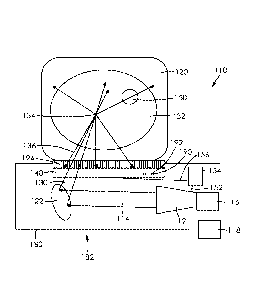
Figure 2 shows an illustration of a diffuse acoustic confocal imager 110
according to a
second embodiment of the present technology having the acoustic detector 40
replaced by
a two-dimensional acoustic array detector 140. The acoustic detector 140, the
coherent
acoustic source 112, and the focusing mirror 122 are housed in a tube 180 to
provide a
wand style acoustic borescope or endoscope, generally referred to as 182.
Again, the
coherent acoustic source 112 such as a coherent acoustic emitter emits a
coherent acoustic
beam 114. The coherent acoustic source 112 can be manually moved or can be
moved with
a source actuator 116 that is in mechanical communication with the coherent
acoustic
source 112. The source actuator 116 is preferably controlled by a processor
118, configured
to direct the source actuator 116 to cause the coherent: acoustic source 16 to
scan the
coherent acoustic beam 114 over the tissue or organ or object material 120.
The coherent
acoustic source 112 provides a coherent acoustic beam 114 with a beam
frequency between
about 0.5 megahertz and about 100 megahertz for obtaining information
including one or
more of density, temperature, composition, elasticity, or strain field in a
mammalian body.
As for the first embodiment, the coherent acoustic beam 114 has a large cross
sectional
area, typically on the order of a centimeter or centimeters. The coherent
acoustic beam 114
passes to a focusing mirror 122 where it is reflected by a curved surface and
focused into a
convergent beam 130 that penetrates the object medium 120 that transmits the
convergent
16
CA 02973655 2017-07-12
WO 2016/113664
PCT/IB2016/050109
beam 130 into a first object, structure, medium or different physical state of
the material or
medium 132 in the object medium 120. The convergent beam :130 converges and is
focused
to a virtual source 134 at the point of cross-over. From the virtual source
134, the incoming
convergent beam 130 is scattered in all directions three-dimensionally. The
scattered beams
136 pass out of the first object 132 and the object medium 120 and are
detected by a two-
dimensional acoustic array detector 140. The two-dimensional acoustic array
detector 140
need not be focused on the virtual source 134 and therefore need not move to
collect
scattered beams 136. The scattered beams 136 contain information about the
object
medium 120 and the first object 132 and are commonly referred to as the object
beams.
The resulting information carried by the scattered beams 136 is analyzed to
determine its
amplitude and phase according to techniques known in the art. Only those beams
136
reaching the one-dimensional or two-dimensional acoustic array detector 140
within a given
and set time frame corresponding to the intensity from the virtual source 134
are used.
.. In order for the entire first object 132 to be observed, the virtual source
134 scans outside
and inside the first object 132 by pivoting the focusing rnirror 122 and the
two dimensional
acoustic array detector 140. Scanning of the first object 132 is also achieved
by either
shifting the first object 120 or shifting the microscope 110. By these means,
a second
object(s) 150 within the first object 132 can be imaged using the amplitude
and phase
information provided by the scattered beams 136 collected by the acoustic
detector 1.40.
A first wire 152 extends between the coherent acoustic source 12 and a vector
network
analyzer 154 and a second wire 156 extends between the vector network analyzer
154 and
two-dimensional acoustic array detector 140, to provide an electrical
communication
between these components. More specifically, individual wires 190 are attached
to each
element 192 of the two dimensional acoustic array detector 140. Each detector
element
192 has its own spatial filter 194. The role of the vector network analyzer
1.54 is to in
measure the amplitude and phase information of the emitted and received
intensities. It
includes a built-in signal generator. It also functions as a temporal filter.
17
CA 02973655 2017-07-12
WO 2016/113664
PCT/IB2016/050109
Figure 3 shows an illustration of a diffuse acoustic confocal imager 210
according to a third
embodiment of the present technology. Again, the coherent acoustic source 212
such as a
coherent acoustic emitter emits a coherent acoustic beam 214. The coherent
acoustic
source 212 can be manually moved or can be moved with a source actuator 216
that is in
mechanical communication with the coherent acoustic source 212. The source
actuator 216
is preferably controlled by a processor 218, configured to direct the source
actuator 216 to
cause the coherent acoustic source 216 to scan the coherent acoustic beam 214
over the
tissue or organ or object material 220. The coherent acoustic source 212
provides a
coherent acoustic beam 214 with a beam frequency between and including about
0.5
megahertz and about 100 megahertz for obtaining information including one or
more of
density, temperature, composition, elasticity, or strain field in a mammalian
body.
As for the first embodiment, the coherent acoustic beam 214 has a large cross
sectional
area, typically on the order of centimeters. The coherent acoustic beam 214
passes through
the surrounding acoustically transparent medium 220 such as water and into an
acoustically
transparent object 232 (the first object). The coherent acoustic source 212
has its emission
surface 213 shaped to focus the coherent acoustic beam 214 to a virtual source
234 at the
point of cross-over. From the virtual source 234, the coherent acoustic beam
214 is
scattered in all directions three-dimensionally. The scattered beams 236 pass
out of the first
object 232 and the object medium 220 and are detected by the two-dimensional
acoustic
array detector 240. The one or two-dimensional acoustic array detector 240
need not be
focused on the virtual source 234 and therefore need not move to collect
scattered beams
236. The scattered beams 236 contain information about the object medium 220
and the
first object 232 and are commonly referred to as the object beams. The
resulting
.. information carried by the scattered beams 236 is analyzed to determine its
amplitude and
phase according to techniques known in the art.
In order for the entire first object 232 to be observed, the virtual source
234 scans outside
and inside the first object 232 by pivoting the coherent acoustic source 212.
Scanning of the
first object 232 is also achieved by either shifting the first object 220 or
shifting the
microscope 210. By these means, a second object(s) 250 within the first object
232 can be
CA 02973655 2017-07-12
WO 2016/113664
PCT/IB2016/050109
imaged using the amplitude and phase information provided by the scattered
beams 236
collected by the two dimensional acoustic array detector 240.
A first wire 252 extends between the coherent acoustic source 212 and a.
vector network
analyzer 254 and a second wire 256 extends between the vector network analyzer
254 and
two-dimensional acoustic array detector 240, to provide an electrical
communication
between these components. More specifically, individual wires 290 are attached
to each
element 292 of the two dimensional acoustic array detector 240. Each detector
element
292 has .its own spatial filter 294. The role of the vector network analyzer
254 is to in
measure the amplitude and phase information of the emitted and received
intensities. It
includes a built-in signal generator. It also functions as a temporal filter.
The spatial resolution is set by the size of the coherent acoustic beam 214 at
the focused
virtual source 234 in the third embodiment. The object is always out-of-focus
and is only
observed in-focus upon combining all of the amplitudes and phases of the
points defining
the object in proper x, y, z.registry.
Figure 4 shows an illustration of a fourth embodiment of the technology. A
coherent
acoustic source 312 such .as a coherent acoustic actuator emits a coherent
acoustic beam
314. The acoustic detector 340, the coherent acoustic source 312, and the
focusing mirror
322 (which is preferably flexible) are housed in a tube 380 to provide a wand
style acoustic
borescope, generally referred to as 382. Again, the coherent acoustic source
312 such as ,a
coherent acoustic emitter emits a coherent acoustic beam 314. The coherent
acoustic
source 312 can be manually moved or can be moved with a source actuator 316
that is in
mechanical communication with the coherent acoustic source 312. The source
actuator 316
is preferably controlled by a processor 318, configured to direct the source
actuator 316 to
cause the coherent acoustic source 316 to scan the coherent acoustic beam 314
over the
tissue or organ or object material 320. The coherent acoustic source 312
provides a
coherent acoustic beam 314 with a beam n frequency between and including about
0.5
megahertz and about 100 megahertz for obtaining information including one or
more of
19
CA 02973655 2017-07-12
WO 2016/113664
PCT/IB2016/050109
density, temperature, composition, elasticity, strain field, magnetic or
electrostatic fields in
a mammalian body.
As for the first embodiment, the coherent acoustic beam 314 has a large cross
sectional
area, typically on the order of a centimeter or centimeters. The coherent
acoustic beam 314
passes to the focusing mirror 322 where it is reflected by a curved surface
and focused into
a convergent beam 330 that penetrates the object medium 320 that transmits the
convergent beam 330 into a first object, structure, medium or different
physical state of the
material or medium 332 in the object medium 320. The convergent beam 330
converges
and is focused to a virtual source 334 at the point of cross-over. From the
virtual source 334,
the incoming convergent beam 330 beam is scattered in all directions three-
dimensionally.
The scattered beams 336 pass out of the first object 332 and the object medium
320. Only
those beams 336 within a given and set time frame corresponding to the virtual
source
intensity are used. These beams are referred to as information beams 337 and
are detected
by a one- or two-dimensional acoustic array detector 340 with a temporal
synthetic
aperture. The one- or two-dimensional acoustic array de1ector340 with the
temporal
synthetic aperture need not be focused on the virtual source 334 and therefore
need not
move to collect scattered beams 336. The information beams 337 contain
information
about the object medium 320 and the first object 332 and are commonly referred
to as the
object beams. The resulting information carried by the information beams 337
is analyzed
to determine its amplitude and phase according to techniques known in the art.
In order for the entire first object 332 to be observed, the virtual source
334 scans outside
and inside the first object 332 by pivoting the focusing mirror 322 and the
two-dimensional
acoustic array detector 340 with a temporal synthetic aperture. Scanning of
the first object
332 is also achieved by either shifting the first object 320 or shifting the
microscope 310. By
these means, a second object(s) 350 within the first object 332 can be imaged
using the
amplitude and phase information provided by the information beams 337
collected by the
two-dimensional acoustic array detector 340 with a temporal synthetic
aperture.
20
CA 02973655 2017-07-12
WO 2016/113664
PCT/IB2016/050109
A first wire 352 extends between the coherent acoustic source 332 and a vector
network
analyzer 354 and a second wire 356 extends between the vector network analyzer
354 and
two-dimensional acoustic array detector 340 with a temporal synthetic aperture
340, to
provide an electrical communication between these components. More
specifically,
individual wires 390 are attached to each element 392 of the two dimensional
acoustic array
detector 340. Each detector element 392 has its own spatial filter 394. The
temporal
synthetic aperture of the two-dimensional acoustic array detector 340 is used
to detect or
accept only the intensity emitted from the focused virtual source and to
ignore the intensity
scattered before the focused virtual source and the intensity scattered after
the focused
virtual source. The role of the vector network analyzer 354 is to in measure
the amplitude
and phase information of the emitted and received intensities. It includes a
built-in signal
generator. The temporal filter may be integral with the vector network
analyzer 354.
A fifth embodiment of the technology is shown in Figure 5. This embodiment is
specially
designed for diagnosis and treatment of prostate diseases and can also be used
as an
endoscope. The coherent acoustic source 412 and a one or two-dimensional
acoustic: array
detector 440 are integrated into a single combined unit 480 that is the shape
of a cup that
fits over the prostate and is attached to the end 478 of a wand 482. The
combined unit 480
can consist of a transparent plastic lens, made of, for example, but riot
limited to as
polymethylpentene, bonded onto a one- or two-dimensional acoustic array
detector 440.
The coherent acoustic source 412 has its emission surface 413 shaped to focus
the coherent
acoustic beam 414 to a virtual source 434 at the point of cross-over. From the
virtual source
434, the coherent acoustic beam 414 is scattered in all directions three-
dimensionally. The
scattered beams 436 pass out of the first object 432 and the object medium 420
and are
detected by the two-dimensional acoustic array detector 440. The combined unit
480 can
translate and rotate using a translational + rotational stage 484. The
translation and rotation
of the combined unit 480 is used to move the virtual source 434.
In order for the entire first object 432 to be observed, -the virtual source
434 scans outside
and inside the first object 432 by pivoting the combined unit 480 using the
translational and
rotational stage 484. By this means, a second object(s) 450 within the first
object 432 can
21
CA 02973655 2017-07-12
WO 2016/113664
PCT/IB2016/050109
be imaged using the amplitude and phase information provided by the scattered
beams 436
collected by the two dimensional acoustic array detector 440.
A first wire 452 extends between the coherent acoustic source 412 and a vector
network
analyzer 454 and a second wire 456 extends between the vector network analyzer
454 and
two-dimensional acoustic array detector 440, to provide an electrical
communication
between these components. More specifically, individual wires are attached to
each
element 492 of the two dimensional acoustic array detector 440. The role of
the vector
network analyzer 454 is to in measure the amplitude and phase information of
the emitted
and received intensities. It includes a built-in signal generator. It may also
function as a
temporal filter.
By detecting the amplitude and phase of many scattered beams 436 from many
positions of
the virtual source 434, the position and size of the object, for example a
tumour 450, within
the first object 432, for example, a prostate, can be determined. By measuring
the phase of
the scattered beams 436 the speed of sound of the second object 450 in the
first object 432
can be determined. The speed of sound of the second object 450 within the
first object 432
can be used for diagnostic purposes.
The emitter 50 and detector 52 are made out of the same material, i.e.,
piezoelectric
material. Therefore, by shaping the emission side of the emitter and detector
unit 480 like a
focusing lens, it will focus the coherent acoustic beam 414 into the object
450 (prostate) like
the focusing lens and detect the scattered beams 436 using the two dimensional
acoustic
array detector 440 on its surface. The two dimensional acoustic array detector
440
comprises many small elements 492 on its surface where each element 492
detects
independently and is a device within itself. Additionally, each detector
element 492 can
emit as well as detect. This allows for identification of a diseased region
followed
immediately by treatment of the diseased region ¨ there is no need for
equipment change,
moving of the equipment ¨ the device remains in the same location and the
intensity of the
coherent acoustic beam 414 is increased. Each detector element 492 has its own
spatial
filter 494.
22
CA 02973655 2017-07-12
WO 2016/113664
PCT/IB2016/050109
A sixth embodiment is shown in Figure 6. This embodiment is specially designed
for
diagnosis and treatment of diseases of the breast. The coherent acoustic
source 512 and a
two-dimensional acoustic array detector 540 are integrated into a single
combined unit 580
that is the shape of a cup that fits over the breast. The coherent acoustic
source 512 has its
emission surface 53.3 shaped to focus the coherent acoustic beam 514 to a
virtual source
534 at the point of cross-over. From the virtual source 534, the coherent
acoustic beam 514
is scattered in all directions three-dimensionally. The scattered beams 536
pass out of the
first object 532 and the object medium 520 and are detected by the two-
dimensional
acoustic array detector 540 with a temporal synthetic aperture that allows
only those
beams 536 within a given and set time frame corresponding to the virtual
source intensity
are used. The combined unit 580 can translate and rotate using a translational
+ rotational
stage 584. The translation and rotation of the combined unit 580 is used to
move the virtual
source 534.
In order for the entire first object 532 to be observed, the virtual source
534 scans outside
and inside the first object 532 by pivoting the combined unit 580 using the
translational and
rotational stage 584. By this means, a second object(s) 550 within the first
object 532 can
be imaged using the amplitude and phase information provided by the scattered
beams 536
collected by the two dimensional acoustic array detector 540.
A first wire 552 extends between the coherent acoustic source 512 and a vector
network
analyzer 554 and a second wire 556 extends between the vector network analyzer
554 and
two-dimensional acoustic array detector 540, to provide an electrical
communication
between these components. More specifically, individual wires are attached to
each
element 592 of the two dimensional acoustic array detector 540. Each detector
element
has its own spatial filter 594. The role of the vector network analyzer 554 is
to in measure
the amplitude and phase information of the emitted and received intensities.
It includes a
built-in signal generator. It may also function as a temporal filter.
23
CA 02973655 2017-07-12
WO 2016/113664
PCT/IB2016/050109
By detecting the amplitude and phase of many scattered beams 536 from many
positions of
the virtual source 534, the position and size of the object, for example a
tumour 550, within
the first object 532, for example, a breast, can be determined. By
measuring.the phase of
the scattered beams 536 the speed of sound of the second object 550 in the
first object 532
can be determined. The speed of sound of the second object 550 within the
first object 532
can be used for diagnostic purposes.
A seventh embodiment is shown in Figure 7, The coherent acoustic source 612,
lens 635
and a one or two-dimensional acoustic array detector 640 are integrated into a
single
combined unit 680 that is the shape of a cup. The lens 635 may be made of, for
example,
but not limited to as polymethylpentene, bonded onto the one- or two-
dimensional acoustic
array detector 640. The lens 635 is shaped to focus the coherent acoustic beam
614 to a
virtual source 634 at the point of cross-over. Note that the arrangement of
the source 612
below the detector 640 can be reversed, however, the sensitivity of detection
of the
acoustic beam will be reduced. From the virtual source 634, the coherent
acoustic beam
614 is scattered in all directions three-dimensionally. The scattered beams
636 pass out of
the first object 632 and the object medium 620 and are detected by the one or
two-
dimensional acoustic array detector 640.
In order for the entire first object 632 to be observed, the virtual source
634 scans outside
and inside the first object 632 by pivoting the combined unit 680 using the
translational and
rotational stage 684. By this means, a second object(s) 650 within the first
object 632 can
be imaged using the amplitude and phase information provided by the scattered
beams 636
collected by the one or two dimensional acoustic array detector 640.
A first wire 652 extends between the coherent acoustic source 612 and a vector
network
analyzer 654 and a second wire 656 extends between the vector network analyzer
654 and
two-dimensional acoustic array detector 640, to provide an electrical
communication
between these components. More specifically, individual wires are attached to
each
element 692 of the two dimensional acoustic array detector 640. Each element
692 has its
own spatial filter 694. The role of the vector network analyzer 654 is to in
measure the
24
CA 02973655 2017-07-12
WO 2016/113664
PCT/IB2016/050109
amplitude and phase information of the emitted and received intensities. It
includes a built-
in signal generator. The vector network analyzer and its connection to the
coherent
acoustic source and the acoustic area detector (one or two dimensional)
obviates the need
for a reference or interference beam. The vector network analyzer 654 may also
function as
.. a temporal filter.
By detecting the amplitude and phase of many scattered beams 636 from many
positions of
the virtual source 634, the position and size of the object, for example a
tumour 650, within
the first object 632, for example, a prostate, can be determined. By measuring
the phase of
the scattered beams 636 the speed of sound of the second object 650 in the
first object 632
can be determined. The speed of sound of the second object 650 within the
first object 632
can be used for diagnostic purposes. An example of this is shown in Figure 8.
The size of
lesion 1 is less than the size of lesion 2, as shown by the image. 'When this
is graphed, the
speed of sound allows for the size of the mass to be shown, with lesion 1
being smaller than
lesion 2.
As shown in Figure 9, additional beams can be focused to the same location as
the acoustic
beam 714, as the focused virtual source 734 position is independent of
wavelength. The
acoustic beam 714 is emitted from the acoustic beam emitter 712. A helium-neon
laser
emitter 782 produces a yellow beam 784 that can be used to cauterize. it can
also be used
to identify the location of the focused acoustic beam 714 on the surface of
the body. An
infrared laser emitter786 produces an infrared beam 788 that can be used to
heat-kill tissue
or ablate the tissue. The combination of the infrared beam 788 with acoustic
beam allows
for treatment of skin cancer with the infrared laser emitter 786, as the
acoustic beam can be
used to report on temperature of the skin. A mirror 780 reflects the beams
784., 788 so as
to be aligned with the acoustic beam 714 as they strike the lens 735. A second
mirror 781
may also be used to switch beam sources on the fly. Alternatively a round
mirror with a flat
surface can be used and rotated around to switch beam sources on the fly.
Without being
bound to theory, the mirror 780 can remove coma and spherical aberrations. If
a specific
and narrow wavelength of beam is used, chromatic aberrations can also be
removed. Note
CA 02973655 2017-07-12
WO 2016/113664
PCT/IB2016/050109
that only a portion of the device is shown in this figure so as to clearly
show the significant
changes. All components in the previous embodiments are found in this
embodiment.
As shown in Figure 10, the beam emitter 812 and the lens 835 work in unison.
For detection
and treatment of prostate cancer, the acoustic beam 814 is focused in the
bladder. The
focused virtual source 834 the scatters through the prostate and is detected
by the detector
840. The detector 840 is shaped to allow a patient to sit on it, Note that
only the
components that differ from the preceding embodiments are shown in this
figure. AM
components in previous embodiments are found in this embodiment.
As shown in Figure 11, two acoustic beam emitters are used in unison. The
first acoustic
beam emitter 912 emits the first acoustic beam 914, and the second acoustic
beam 916 is
emitted from the second acoustic beam emitter 918. The second acoustic beam
emitter
918 is located below the linear array detector 940. The vector network
analyzer 954 is
configured as described above. Al the components described in previous
embodiments are
found in this embodiment,. The first beam 914 is focused to provide the
virtual source 934,
which then sends the scattered beams to the object to permit imaging, as
described above.
The second acoustic beam 916 is focused on the object 932 to cause it to
function. This
allows for determining functional abnormalities. In the preferred embodiment,
the object is
the prostate.
Example 1
The effectiveness of the device and system was demonstrated using a prostate
elastography
phantom containing three randomly placed isoechoic lesions from =CSP Medical
that are
three times harder than the simulated prostate tissue, as shown in Figure 7.
Four acoustic
phase images were taken at scan depths from 10 mm to 25 mm in which the
margins of the
prostate and the margin of the urethra, bright orange to bright yellow
coloring, could be
identified. The ultrasound beams radiate out from the beam source and the
image is
collected from the diffuse beams. Regions of higher speed of sound are
indicated at A and B.
Using any of embodiments 1-4 and 6 of the present technology, two of the
lesions were
scanned. These lesions were clearly identifiable. The size, three dimensional
shape,
26
CA 02973655 2017-07-12
WO 2016/113664
PCT/IB2016/050109
position and location could be determined. Other features of disease tissue
were not
present in the prostate phantom and hence, information was limited to the
characteristics
that were different.
Example 2
The DACI can be used in medical diagnostics to non-intrusively observe the
variations in
temperature within the body such as, but not limited to, within an organ,
muscles, fatty
tissue, cancerous tissue and at the interfaces between body organs and their
surroundings.
Since the DACI focuses the beam to a virtual source, which is passed quickly
over a point, it
can be very gentle on the body by giving a low radiation dose. The power
density is
generally less than 1 watt per square centimeter and dwell times of
milliseconds to seconds
to avoid heating and cavitation effects in the object under examination. Once
the internal
body can be seen by the DACI, by increasing the intensity from tens of watts
to hundreds of
watts per square centimeter and dwell time of the beam from seconds to
hundreds of
seconds, treatments become possible, using beam heating methods and tumor
ablation
(break-up) methods such as high intensity focused ultrasound. Since the DACI
microscope
can also measure temperature by determining the speed of sound of the object
beams, the
temperature of the region of the body being treated by beam heating can be
monitored
during the treatment process to help ensure a successful treatment.
Additionally, the
treatment can be monitored by measuring scattering intensity as it decreases
with an
increase in tumour ablation/break up.
Example 3
In objects comprising of plasma, gases, liquids, and solids, there are many
unanswered
questions to simple states of matter, such as, but not limited to the 3D
temperature and the
3D composition existing within objects and at interfaces between immiscible
and miscible
fluids, a container and its contents, and within fluids having various states,
such as within a
simple flame burning fuel during combustion. The speed of sound changes as the
state of
matter changes. There are higher speeds of sound for stiffer, higher-
elasticity materials.
The application of the DACI microscope to objects transparent to acoustic
beams will
answer many of these questions.
27
CA 02973655 2017-07-12
WO 2016/113664
PCT/IB2016/050109
Example 4
Now that radiation sources, such as acoustic beams, can be obtained having
very good
beam coherence, amplitude and phase images of large objects are possible, on
the order of
many centimeters. It will be possible with the development of new optical
focusing
materials transparent or reflective to acoustic beams such as plastics that
may be able to
observe much larger and smaller objects in the future.
Example 5
.. Diagnosis and treatment of prostate cancer. Prostate cancer tumours are
hard and multi-
shaped with fine branches. Blood flow is increased around the tumour, but the
hardness of
the tumour prevents the blood reaching the tumour and eventually restricts
blood flow
around the tumour. The current state of the art is ultrasound imaging of the
prostate. This
provides information on the size of the gland, as only the interface between
the prostate
.. and surrounding tissue can be identified. At that, the images are not
highly accurate as the
diffuse scattering of the beams interferes with the image and leads to fuzzy
edges.
The present technology is provided as a wand with the emitter complex and the
detector
holder at a distal end. As noted above, the emitters may also function as the
detectors.
.. This allows for a single complex to be used with a holder that is
appropriately shaped for the
body part to be imaged. Alternatively, the emitters are housed on an emitter
complex that
is integrated into the detector holder. Again, the shape of the holder is
appropriate for the
body part being imaged.
There are diseases within the body such as within the prostate, each having
their own speed
of sound, hence the speed of sound is a signature for each disease (see
Example 6). Further,
each developmental state of the disease has a signature speed of sound.
Example 6
Speed of sound will be measured for any disease or condition of interest, and
for each
developmental stage of the disease or condition. The present technology will
be used to
28
CA 02973655 2017-07-12
WO 2016/113664
PCT/IB2016/050109
make this determination. The present technology will then be used to diagnose
or diagnose
and treat, or track progression of the disease or condition or track
progression of treatment
of the disease or condition. Change in speed of sound can be caused by changes
in one of
more of cell size, cellular granularity, tissue elasticity, blood
accumulation, increase in
temperature, inflammation and immune cell infiltration. Examples of different
speeds of
sound are 1574 m/s for smooth muscle fibres, 1610 m/s for papillary
adenocarcinoma,
1610 m/s for tubular adenocarcinoma (well differentiated), 1600 rn/s for
tubular
adenocarcinoma (moderately differentiated), 1557 m/s for tubular
adenocarcinorna (poorly
differentiated) and 1523 m/s for singlet-ring cell carcinoma. Other known
speeds of sound
.. are for breast tissue, with the speed of sound of 1422 m/s for fatty
tissue, 1487 m/s for
breast parenchyma, 1548 m/s for a malignant lesion and 1513 m/s for a benign
lesion. The
standard deviation was not more than 1.7%.
Example 7
.. The two dimensional acoustic array detector was replaced with a linear
acoustic array
detector. It was found that the focused virtual source also be created by the
linear array
actuator emission. The linear array actuator can focus the beam by a curved
surface of a
lens or it can also be focused by adjusting the relative phases of the
emitting elements in
the array of transducers (i.e., a "phased array"). Although the phased array
can't produce a
small virtual source, it can still produce a virtual source, which can be used
for imaging at a
lower spatial resolution.
Advantages of the exemplary embodiments described herein may be realized and
attained
by means of the instrumentalities and combinations particularly pointed out in
this written
description. It is to be understood that the foregoing general description and
the following
detailed description are exemplary and explanatory only and are not
restrictive of the claims
below. While example embodiments have been described in detail, the foregoing
description is in all aspects illustrative and not restrictive. it is
understood that numerous
other modifications and variations can be devised without departing from the
scope of the
example embodiment.
29
CA 02973655 2017-07-12
WO 2016/113664
PCT/IB2016/050109
While example embodiments have been described in connection with what is
presently
considered to be an example of a possible most practical and/or suitable
embodiment, it is
to be understood that the descriptions are not to be limited to the disclosed
embodiments,
but on the contrary, is intended to cover various modifications and equivalent
arrangements
included within the spirit and scope of the example embodiment. Those skilled
in the art will
recognize, or be able to ascertain using no more than routine experimentation,
many
equivalents to the specific example embodiments specifically described herein.
Such
equivalents are intended to be encompassed in the scope of the claims, if
appended hereto
or subsequently filed.
30