Note: Descriptions are shown in the official language in which they were submitted.
CA 02975362 2017-07-28
WO 2016/124702
PCT/EP2016/052408
1
Novel EGFR binding proteins
Field of the invention
The present invention relates to new EGFR binding molecules based on ubiquitin
muteins (Affilin ), preferably Affilin
molecules having a characteristic three amino acid residue motif. The
invention further refers to EGFR binding
molecules that bind to different epitopes than the anti-EGFR monoclonal
antibody Cetuximab. The invention refers to
EGFR binding proteins optionally fused or conjugated to a pharmacokinetic
moiety modulating serum half-life or to a
therapeutically or diagnostically active component. The invention further
relates to the use of these EGFR binding
proteins in medicine, preferably for use in the diagnosis or treatment of
cancer.
Background of the invention
Non-immunoglobulin based binding agents can be beneficially used in the
medical fields of diagnosis, prophylaxis
and treatment of diseases. A solution to the disadvantages resulting from
antibodies in diagnosis, prophylaxis and
treatment of diseases is to provide polypeptides with comparable or even
better affinity and specificity towards the
specific targets combined with smaller molecular size enabling an improved
tissue penetration and having thus better
biodistribution properties.
Among non-immunoglobulin-derived small proteins, molecules based on modified
ubiquitin are particularly interesting
because these molecules promise alternative therapeutic and diagnostic
possibilities compared to antibodies.
Ubiquitin is a highly conserved, small, single domain protein present in all
known eukaryotic cells and is 100%
conserved amongst all vertebrates. In addition, ubiquitin naturally occurs in
serum lowering the immunogenic
potential. This facilitates preclinical development in different species
required for toxicological and efficacy studies.
Ubiquitin muteins specific for target antigens are described in the prior art.
Such ubiquitin muteins are known as
Affilin (registered trademark of Scil Proteins GmbH) molecules. Ubiquitin
muteins and methods for producing these
muteins were described in several patents, for example, EP162698561,
EP237958161, EP2094845131, and
W02012/172055. Affilin proteins are engineered to generate de novo binding
affinity towards desired targets making
them ideal for different applications.
A key feature of the Affilin platform is its flexibility and modularity
allowing multiple functional moieties to be combined
by genetic or chemical modifications enabling tailoring of the biological,
physiological and functional properties of the
resulting Affilin molecules. Affilin molecules (ubiquitin muteins) are
designed for optimal functionality and
developability including characteristics such as high stability, affinity and
specificity. These unique features make
Affilin molecules a compelling choice for applications where antibodies have
limitations, thus broadening the
utilization of biotherapeutics.
EGFR is a receptor tyrosine kinase mediating cell proliferation and
differentiation. Increased expression of the human
epidermal growth factor receptor (EGFR) is observed for many tumors, in
particular in malignant tumors. EGFR is
known for being involved in lung cancer, head and neck cancer and colorectal
cancer amongst others. EGFR has
three characteristic domains: an extracellular ligand binding domain, a
transmembrane domain, and an intracellular
tyrosine kinase domain. Upon binding of a ligand to the extracellular ligand
binding domain, EGFR dimerizes which
activates the intracellular tyrosine kinase domain and induces cellular
processes such as proliferation, differentiation,
migration, or apoptosis. Modulating the function of EGFR is an important
approach for the development of cancer
therapeutics, and meanwhile, therapeutic anti-EGFR antibodies binding to EGFR
and thereby modulating the function
of this receptor are available for treatments of cancer, for example
colorectal cancer. One example for a monoclonal
antibody binding to EGFR is Cetuximab.
However, antibodies have major disadvantages including a complex molecular
structure, a large size and challenging
production methods. Furthermore, treatment of diseases with currently
available EGFR - binding molecules is not
CA 02975362 2017-07-28
WO 2016/124702
PCT/EP2016/052408
2
effective in all patients and may have severe side effects. Another major
disadvantage is the development of
resistances of certain tumors to Cetuximab treatment.
Cancer represents one of the leading causes for death worldwide. Needless to
say that there is a strong medical
need to effectively treat cancer with improved novel agents, in particular for
efficient tumor targeted therapeutics and
diagnostics. There is an ongoing need to substitute antibodies by smaller,
less complex molecules such as non-
immunoglobulin based binding agents which can be beneficially used in the
medical fields of diagnosis, prophylaxis
and treatment of diseases. A solution to the disadvantages resulting from
antibodies in diagnosis, prophylaxis and
treatment of diseases is to provide polypeptides with comparable affinity and
specificity towards the specific targets,
for example EGFR, combined with a less complex and smaller structure enabling
a simplified molecular engineering
as well as an improved tissue penetration and having thus better
biodistribution properties.
Further solution to the disadvantage of the development of resistances of
certain tumors to Cetuximab treatment is to
provide EGFR binding molecules that bind to a different or non-overlapping
epitope than Cetuximab.
Novel EGFR binding molecules suitable for diagnostic and therapeutic
applications should be functional and
developable and should include characteristics such as high stability,
affinity and specificity. Small, monovalent
binders would also enable improved biophysical studies. Such small binders
could also be useful for in vivo imaging
in diagnostic approaches to study EGFR localization and trafficking, in
addition to therapeutic approaches.
It is thus an objective of the present invention to provide novel molecules
for new and improved strategies in the
treatment and diagnosis of various diseases, such as cancer. In particular, it
is an objective to provide novel stable
non-immunoglobulin proteins which have high affinity and specificity to EGFR.
The invention provides small binding
proteins (Affilin) which are advantageous compared to antibodies by their
small size, simple molecular structure (one
chain compared to four chains of an antibody), and in that no
posttranslational modifications are required for full
functionality. These factors contribute to an easy handling of the molecules
including simple genetic engineering as
well as easy production and purification methods.
A major advantage of the EGFR binding molecules of the invention is that they
bind to a different (non-overlapping)
epitope of the EGFR receptor than established antibodies such as Cetuximab. A
positive effect of the different
binding site is that these novel EGFR binding molecules may overcome the
resistance of certain tumor cells to
Cetuximab. Further, the binding of the EGFR binding molecules of the invention
to a different epitope than Cetuximab
may induce different biological responses.
The present invention meets the needs presented above by providing examples
for specific EGFR binding proteins.
The above-described objectives and advantages are achieved by the subject-
matters of the enclosed independent
claims. Preferred embodiments of the invention are included in the dependent
claims as well as in the following
description, examples and figures. The above overview does not necessarily
describe all problems solved by the
present invention.
Summary of the invention
In a first aspect of the invention, the EGFR binding protein is comprising or
consisting of a ubiquitin mutein (Affilin)
with binding affinity (KD) of less than 700 nM for epidermal growth factor
receptor (EGFR) wherein the ubiquitin
mutein comprises an amino acid sequence motif wherein the amino acid in
position 64 of ubiquitin is selected from
P, V, and A, the amino acid in position 65 of ubiquitin is selected from D and
E, and the amino acid in position 66 of
ubiquitin is selected from I, V, A, M, F, Y, W, and L, and wherein the
ubiquitin mutein has 80 % to 93 % identity to
ubiquitin (SEQ ID NO: 1) or di-ubiquitin SEQ ID NO: 4. Accordingly, in a first
aspect the present invention relates to
an EGFR binding protein comprising a ubiquitin mutein that comprises an amino
acid sequence wherein three amino
acids selected from amino acids 62-66 corresponding to X62, X63, X64, X65, and
X66 of SEQ ID NO: 3 are substituted
compared to the amino acid sequence QKEST and wherein the ubiquitin mutein has
at least 90 % sequence identity
to SEQ ID NO: 3.
CA 02975362 2017-07-28
WO 2016/124702
PCT/EP2016/052408
3
In a second aspect the present invention relates to an EGFR binding ubiquitin
wherein the amino acid sequence in
positions 64, 65, and 66 is selected from amino acids P, D, and I or amino
acids V, D, and I, or amino acids A, D,
and I, or amino acids V, D, and V, or amino acids P, D, and V (õPDI motif";
including amino acid sequences PDI,
VD!, VDV, PDV, or ADO. In one aspect of the invention, the EGFR binding
protein comprises an ubiquitin mutein
wherein the amino acids in positions 62 and 63 may be any amino acid,
preferably wherein the amino acid in position
62 is selected from R, Q, H, K, G, S, T, N, V, I, and W, and wherein the amino
acid in position 63 is selected from N,
H, A, S, R, E, T, Q, and K.
In a third aspect the present invention relates to an EGFR binding protein
that binds to a different or non-overlapping
EGFR epitope than anti-EGFR monoclonal antibody Cetuximab.
A further aspect of the present invention relates to an EGFR binding protein
comprising or consisting of at least two
ubiquitin muteins of the same (e.g., homo-dimer) or a different (e.g. hetero-
dimer) target specificity and/or binding to
the same (overlapping) or a different (non-overlapping) epitope of EGFR.
A further aspect of the invention relates to an EGFR binding protein
comprising or consisting of a ubiquitin mutein
comprising an amino acid sequence selected from at least one member of the
group consisting of SEQ ID NOs: 8-73
and 90-106 and 111-112 or an amino acid sequence that exhibits at least 80 %
sequence identity to one or more of
the amino acid sequences of SEQ ID NOs: 8-73 and 90-106 and 111-112.
Another aspect the present invention relates to an EGFR binding ubiquitin
mutein further comprising at least one
additional molecule, preferably selected from at least one member of the
groups (i), (ii) and (iii) consisting of (i) a
pharmacokinetic moiety modulating serum half-life selected for example from a
polyethylene glycol, a human serum
albumin (HSA), anti-human serum albumin binding protein, albumin-binding
peptides, a polymer sequence forming a
random coil, an immunoglobulin or immunoglobulin fragments, or a
polysaccharide, and, (ii) a therapeutically active
component, optionally selected for example from a monoclonal antibody or a
fragment thereof with the binding
specificity of said monoclonal antibody, a cytokine, a chemokine, a cytotoxic
compound, an enzyme, or derivatives
thereof, or a radionuclide, and (iii) a diagnostic component, optionally
selected for example from a fluorescent
compound, a photosensitizer, or a radionuclide.
The present invention also provides, in further aspects, a nucleic acid or
nucleic acids encoding the EGFR binding
protein comprising or consisting of a ubiquitin mutein of the present
invention, as well as a vector or vectors
comprising said nucleic acid or nucleic acids, and a host cell or host cells
comprising said vector or vectors.
Another aspect relates to an EGFR binding protein comprising or consisting of
a ubiquitin mutein of the invention
binding to EGFR for use in diagnostics or medicine, preferably for use in the
diagnosis or treatment of cancer,
or a nucleic acid molecule encoding said EGFR binding protein comprising or
consisting of a ubiquitin mutein of the
invention, or to a vector comprising said EGFR binding protein comprising or
consisting of a ubiquitin mutein of the
invention, or to a host cell comprising said EGFR binding protein comprising
or consisting of a ubiquitin mutein of the
invention, or to a non-human host comprising said EGFR binding protein
comprising or consisting of a ubiquitin
mutein of the invention.
Another aspect relates to a composition comprising the EGFR binding protein of
the invention, the nucleic acid
molecule of the invention, the vector of the invention, or the host cell of
the invention, preferably for use in the
diagnosis or treatment of cancer.
Another aspect of the present invention relates to a method for the production
of an EGFR binding protein comprising
or consisting of a ubiquitin mutein (Affilin) of any of the preceding aspects
of the invention comprising culturing of host
cells under suitable conditions and optionally isolation of the EGFR binding
ubiquitin mutein produced.
This summary of the invention does not necessarily describe all features of
the present invention. Other
embodiments will become apparent from a review of the ensuing detailed
description.
Brief description of the Figures
CA 02975362 2017-07-28
WO 2016/124702
PCT/EP2016/052408
4
The Figures show:
FIG. 1 shows anti-EGFR Affilin molecules and biochemical characterization of
EGFR binding Affilin molecules.
Binding affinity (KD) or binding data for the Affilin molecules to EGFR have
been obtained from SPR (Biacore) and are
shown in the fifth column. Temperature stability is shown (DSF) in the sixth
column. Exchanges at positions 62, 63,
64, 65, and 66 of unmodified ubiquitin are shown in the last column. All
assays are further described in the Examples
section.
Affilin with one ubiquitin moiety: Six amino acids inserted in the N-terminal
loop region of ubiquitin are shown in
brackets in the third column of the table. Substitutions in positions 62-66 of
ubiquitin are shown after the bracket in
the third column of the table and are additionally shown in the last column of
the table. Further substitutions relevant
for EGFR-binding are listed for some variants (fourth column). All variants
have a characteristic amino acid residue
motif of three amino acids ("PDI motif") in positions 64, 65, and 66 of
ubiquitin. SEQ ID NOs: 8-52 are Affilin
molecules binding to EGFR and having a PDI motif at position 64, 65, and 66 of
wildtype ubiquitin.
Affilin comprising two ubiquitin moieties: Affilin molecules with SEQ ID NOs:
53-73 correspond to Affilin
molecules comprising two different Affilin moieties with substitutions in
amino acid residues 6 and 8, and in residues
62, 63, 64, 65, 66 in each moiety (as shown in the third column of the table).
A PDI motif is located in the first moiety
of the ubiquitin mutein. Two proteins without PDI motif (Affilin 139989 and
Affilin 138840) do not bind to extracellular
EGFR.
FIG. 2 shows a functional characterization of EGFR-Affilin molecules. The
figure shows binding to exogenously
EGFR expressing CHO-K1 cells as determined by FACS analysis. These cells are
used as model system for testing
EGFR binding capability. Affilin molecules show binding on CHO-K1-EGFR cells
and no activity on control cells.
EGFR binding Affilin molecules with PDI motif (or Cetuximab) are shown in
black, whereas the PBS control is shown
in grey. Cellular EGFR binding was confirmed for all binding molecules (FIG.
2B Affilin 139756 (SEQ ID NO: 50), FIG.
2C Affilin 139791 (SEQ ID NO: 49), FIG. 2D Affilin 139819 (SEQ ID NO: 39), and
FIG. 2H Affilin 142265
(SEQ ID NO: 75)). No or weak binding was observed for an Affilin without PDI
motif (Affilin 139989, SEQ ID NO: 76,
FIG. 2E). Cetuximab served as positive control for EGFR expression (FIG. 2A).
FIG. 2F shows non-binding to
negative control cells for 139819 (SEQ ID NO: 39) and FIG. 2G for Cetuximab.
FIG. 3 shows a FACS analysis of Affilin 139819 (SEQ ID NO: 39) with decreasing
Affilin concentrations. 500nM (FIG.
3a), 50 nM (FIG. 3B), 5 nM (FIG. 3c), and 0.5 nM (FIG. 3D) Affilin 139819 was
assayed for binding to CHO-K1-EGFR
cells. Even for the lowest used concentration, binding of Affilin 139819 to
cellular EGFR was detectable.
FIG. 4 shows the epitope specificity of EGFR binding molecules with or without
PDI motif. Binding analysis (SPR)
shows that the binding epitope of both Affilin molecules with PDI motif
(Affilin 139791 and Affilin 139819) is identical
or at least overlapping whereas the binding epitope of Affilin 139989 without
PDI motif is different.
FIG. 4A. SPR analysis of Affilin 139819 (PDI motif) versus Affilin 139989
FIG. 4B. SPR analysis of Affilin 139791 (PDI motif) versus Affilin 139989
FIG. 4C. SPR analysis of Affilin 139989 (no PDI) versus Affilin 139819 (PDI
motif) and Affilin 139791 (PDI motif)
FIG. 5 shows a SPR competition analysis of Affilin 139819 and Affilin 142265
with Cetuximab. Affilin 139819 (SEQ
ID NO: 39; 100nM) is shown as black line in the middle of the diagram and 10
pM is shown as medium grey line on
top, 1 pM Affilin 142265 (SEQ ID NO: 75) is shown as light grey line at the
bottom of the figure. The analysis is
described in further detail in Example 7. The Figure shows that Affilin 139819
does not compete with Cetuximab and
that Affilin 139819 uses a different or non-overlapping epitope whereas
Affilin 142265 (no PDI motif) competes with
Cetuximab. The result surprisingly shows that the Affilin binding molecules
with PDI motif bind to a different or non-
overlapping EGFR epitope than Cetuximab whereas the Affilin binding molecules
without PDI motif bind to the same
or overlapping EGFR epitope.
FIG. 6 confirms that anti-EGFR-Affilin proteins having a PDI motif bind to
tumor tissue. Shown is an
immunohistological analysis on EGFR expressing human xenograft tumor tissue
(MDA-MB-231; ATTC HTB-26),
CA 02975362 2017-07-28
WO 2016/124702
PCT/EP2016/052408
derived from metastatic site of mammary gland/breast adenocarcinoma,
epithelial cells. Different concentrations (100
nM and 500 nM) were tested of Affilin proteins with PDI motif 138819 (SEQ ID
NO: 71), Affilin 138838 (SEQ ID NO:
69), Affilin 138845 (SEQ ID NO: 73), and Affilin without PDI motif 138840 (SEQ
ID NO: 77). Positive control:
Cetuximab (not shown in this figure), negative control: unmodified ubiquitin
(SEQ ID NO: 7, clone 64156). No
5 unspecific staining was detected with unmodified ubiquitin. The results
clearly show the high specific targeting
function of EGFR binding proteins of the invention. All Affilin binding
proteins with PDI motif show strong binding to
EGFR on xenografts derived from human tumor tissue whereas the Affilin binding
molecule without PDI motif shows
only weak binding to EGFR.
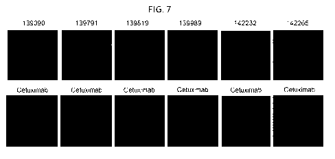
FIG. 7 confirms that EGFR-Affilin proteins bind to extracellular EGFR
expressed on tumor cells. Shown are
immunofluorescence images of EGFR expressing A431 tumor cells. The staining of
A431 tumor cells expressing
EGFR confirms binding of Affilin 139791 (SEQ ID NO: 49), Affilin139819 (SEQ ID
NO: 39), Affilin 142232 (SEQ ID
NO: 29) and Affilin 142265 (SEQ ID NO: 75). For Affilin 139989 (no PDI motif;
SEQ ID NO: 76), no binding to
extracellular EGFR on A431 tumor cells was detectable. The unmodified
ubiquitin is referred to as 139090 (SEQ ID
NO: 4) in this Figure.
FIG. 8 shows fusion proteins of EGFR binding proteins with Cetuximab. Shown
are the sensorgrams of EGFR-
monoclonal antibody Cetuximab (SEQ ID NOs: 5 and 6), control fusion of
Cetuximab with unmodified ubiquitin, and
fusion protein of Cetuximab and EGFR-Affilin 139819. The curves show different
concentrations of 15 nM (highest) to
0.0586 nM (lowest) in a 1:2 dilution. A RU results from the calculation of the
subtraction of the signals for both flow
cells (hEGFR-Fc 1578 RU; hIgG-Fc 331 RU). The analysis confirms that fusion
proteins of an anti-EGFR-Affilin to a
monoclonal antibody binds to EGFR with high affinity.
FIG. 8A shows the SPR analysis of the binding of an anti-EGFR-Affilin-
Cetuximab fusion protein to the extracellular
domain of EGFR. Shown are sensorgrams of Cetuximab, CL-ubiquitin, and CL-
139819. Anti-EGFR-Affilin fused to
the C-terminus of the light chain of Cetuximab shows higher signal intensity
to EGFR than Cetuximab.
FIG. 8B shows the SPR analysis of the binding of an anti-EGFR-Affilin-
Cetuximab fusion protein. Shown are
sensorgrams of Cetuximab, NL-ubiquitin (SEQ ID NO: 89), and NL-139819 (SEQ ID
NO: 86).
FIG. 9 shows the expression and purification of a homo-dimer of two identical
Affilin 139819 proteins. The final
product yield of the homo-dimer was 5.9 mg per liter expression.
FIG. 9A shows the polishing step after StrepTactin purification via
gelfiltration on Superdex 75 16/60, the primary axis
plots the absorption signal [mAU] against buffer volume [ml] and the secondary
axis plots the conductivity [mS/cm]
versus the buffer volume [ml].
FIG. 9B shows SDS-PAGE analysis of the gelfiltration. Lane 1, protein marker,
lane 2, pellet, lane 3 supernatant,
lane 4 flow through (StrepTactin), lane 5 fraction A3, lane 6 fraction A9,
lane 7 fraction B12, lane 8 fraction B11, lane
9 fraction B10, lane 10 fraction B9, lane 11 fraction B8, lane 12 fraction B7,
lane 13 fraction B6. The product purity is
higher than 95 % according to SDS-PAGE.
FIG. 10 shows the analysis of purity and homogeneity via SEHPLC of a homo-
dimer of two identical Affilin 139819
proteins. During the analysis only one peak could be detected. The observed
tailing is likely a result of the peptide
linker connecting both ubiquitin moieties. The primary axis plots the
absorption signal [mAU] against buffer volume
[ml] and the secondary axis plots the conductivity [mS/cm] versus the buffer
volume [ml].
FIG. 11 shows surface plasmon resonance spectroscopy (Biacore) of a homo-dimer
of two Affilin 139819 molecules
to determine the dissociation constants of binding proteins-target complexes.
Solid lines represent experimental data,
dashed lines represent fitted curve data. The Affilin proteins demonstrate a
quick association as well as a prompt
dissociation, eliciting rather high koff rates. Highest affinities to EGFR-Fc
have an KD of 0,6 nM. The affinity of the
homo-dimer (139819-139819) to extracellular EGFR is about 30fold higher than
for the monomer Affilin 139819.
FIG. 12 shows functional characterization of the binding of a homo-dimeric
EGFR Affilin.
CA 02975362 2017-07-28
WO 2016/124702
PCT/EP2016/052408
6
FIG. 12A and FIG. 12B show binding to EGFR expressing CHO-K1 cells, for
example of the homo-dimer of two
Affilin 139819 proteins (referred to as 140547 in the Figure; dissociation
constant 1,7 nM, FIG. 12A) and of Affilin
139819 (dissociation constants 8,7 nM, FIG. 12B).
FIG. 12C and FIG. 12D shows binding to EGFR expressing A549 cells, for example
of the homo-dimer of two Affilin
molecules 139819 (dissociation constant 1,7 nM, FIG. 120) and of Affilin
139819 (dissociation constants 10,9 nM,
FIG. 12D).The affinity of the homo-dimer of two Affilin molecules 139819 to
extracellular EGFR is about 10fold higher
than for the monomer Affilin 139819.
Detailed Description of the Invention
Before the present invention is described in more detail below, it is to be
understood that this invention is not limited
to the particular methodology, protocols and reagents described herein as
these may vary. It is also to be understood
that the terminology used herein is for the purpose of describing particular
embodiments only, and is not intended to
limit the scope of the present invention which will be limited only by the
appended claims. Unless defined otherwise,
all technical and scientific terms used herein have the same meanings as
commonly understood by one of ordinary
skill in the art to which this invention belongs.
Preferably, the terms used herein are defined as described in "A multilingual
glossary of biotechnological terms:
(IUPAC Recommendations)", Leuenberger, H.G.W, Nagel, B. and Kolbl, H. eds.
(1995), Helvetica Chimica Acta, CH-
4010 Basel, Switzerland).
Throughout this specification and the claims which follow, unless the context
requires otherwise, the word "comprise",
and variants such as "comprises" and "comprising", will be understood to imply
the inclusion of a stated integer or
step or group of integers or steps but not the exclusion of any other integer
or step or group of integers or steps.
Several documents (for example: patents, patent applications, scientific
publications, manufacturers specifications,
instructions, GenBank Accession Number sequence submissions etc.) are cited
throughout the text of this
application. Nothing herein is to be construed as an admission that the
invention is not entitled to antedate such
disclosure by virtue of prior invention. Some of the documents cited herein
are characterized as being Incorporated
by reference". In the event of a conflict between the definitions or teachings
of such incorporated references and
definitions or teachings recited in the present specification, the text of the
present specification takes precedence.
All sequences referred to herein are disclosed in the attached sequence
listing that, with its whole content and
disclosure, is a part of this specification.
General definitions of important terms used in the application
The terms "protein" and "polypeptide" refer to any chain of two or more amino
acids linked by peptide bonds, and
does not refer to a specific length of the product. Thus, "peptides",
"protein", "amino acid chain," or any other term
used to refer to a chain of two or more amino acids, are included within the
definition of "polypeptide," and the term
"polypeptide" may be used instead of, or interchangeably with any of these
terms. The term "polypeptide" is also
intended to refer to the products of post-translational modifications of the
polypeptide, including without limitation
glycosylation, acetylation, phosphorylation, amidation, proteolytic cleavage,
modification by non-naturally occurring
amino acids and similar modifications which are well known in the art. Thus,
binding proteins comprising two or more
protein moieties also fall under the definition of the term "protein" or
"polypeptides".
The term "ubiquitin" or õunmodified ubiquitin" refers to ubiquitin in
accordance with SEQ ID NO: 1 (wild type ubiquitin)
or to proteins with at least 95 % amino acids identity to SEQ ID NO: 1 (for
example, with point mutations in positions
W45F, G75A, G76A which do not influence binding to a target, see SEQ ID NO: 2)
and according to the following
definition. Particularly preferred are ubiquitin molecules from mammals, e.g.
humans, primates, pigs, and rodents. On
the other hand, the ubiquitin origin is not relevant since according to the
art all eukaryotic ubiquitins are highly
conserved and the mammalian ubiquitins examined up to now are even identical
with respect to their amino acid
CA 02975362 2017-07-28
WO 2016/124702
PCT/EP2016/052408
7
sequence. In addition, ubiquitin from any other eukaryotic source can be used.
For instance ubiquitin of yeast differs
only in three amino acids from the wild-type human ubiquitin (SEQ ID NO: 1).
The term "di-ubiquitin" refers to a linear protein wherein two ubiquitin
moieties are directly fused to each other in head
to tail orientation. The term "di-ubiquitin" refers to two directly linked
ubiquitin moieties of SEQ ID NO: 1 or to proteins
with at least 95 % amino acids identity to SEQ ID NO: 4 (for example, with
point mutations in positions W45F, G75A,
G76A, G151A, G152A).
The term "Affilin " (registered trademark of Scil Proteins GmbH) refers to non-
immunoglobulin derived binding
proteins based on ubiquitin muteins. The terms "modified ubiquitin" and
"ubiquitin mutein" and "Affilin" are all used
synonymously and can be exchanged. The term "modified ubiquitin" or "ubiquitin
mutein" or "Affilin" as used herein
refers to derivatives of ubiquitin (for example, derived from SEQ ID NO: 1 or
SEQ ID NO: 3) or di-ubiquitin (for
example, SEQ ID NO: 4) which differ from said unmodified ubiquitin by amino
acid exchanges, insertions, deletions
or any combination thereof, provided that the modified ubiquitin or ubiquitin
mutein has a specific binding affinity to a
target epitope or antigen which is at least 10fold lower or absent in
unmodified ubiquitin. This functional property of
an ubiquitin mutein (Affilin; modified ubiquitin) is a de novo created
function.
An Affilin is not a natural ubiquitin existing in or isolated from nature. The
scope of the invention preferably excludes
unmodified ubiquitin for example, as shown in SEQ ID NO: 1. An Affilin
molecule according to this invention
comprises or consists of either one modified ubiquitin moiety or comprises two
differently modified ubiquitin moieties
linked together in a head-to-tail fusion. A "head-to-tail fusion" is to be
understood as fusing two proteins together by
connecting them in the direction (head) N-C-N-C- (tail) (tandem molecule), as
described for example in
EP2379581B1 which is incorporated herein by reference. The head part is
designated as the first moiety and the tail
part as the second moiety. In this head-to-tail fusion, the ubiquitin moieties
may be connected directly without any
linker. Alternatively, the fusion of ubiquitin moieties can be performed via
linkers, for example, a polypeptide linker, as
described herein.
As used herein, "substitutions" are defined as exchanges of an amino acid by
another amino acid. Given the known
genetic code, and recombinant and synthetic DNA techniques, the skilled
scientist can readily construct DNAs
encoding the amino acid variants. The term "deletion" means that one or more
amino acids are taken out of the
original sequence and the amino acids originally N-terminal and C-terminal of
the deleted amino acid are now directly
connected and for a continuous amino acid sequence.
The term "insertions" comprises the addition of amino acids to the original
amino acid sequence of ubiquitin wherein
the ubiquitin remains stable without significant structural change. Naturally,
loop regions connect regular secondary
structure elements. The structure of human unmodied ubiquitin reveals six
loops at amino acid regions 8-11, 17-22,
35-40, 45-47 and 50-63 which connect secondary structure elements such as beta
sheets and alpha helix. Preferred
are ubiquitin muteins comprising a combination of insertions and
substitutions, as described in EP2721152. Preferred
ubiquitin muteins have insertions of 2-10 amino acids, preferably in the most
N-terminal loop within amino acids 8-11
or in the most C-terminal loop within amino acids 50-63. However, other
locations for insertions are possible.
Specifically, the number of amino acids to be inserted is 2, 3, 4, 5, 6, 7, 8,
9, 10, preferably 2 - 8 amino acids, most
preferred 4 - 8 amino acids.
The term "EGFR binding protein" refers to a protein which either consists of
or comprises at least one ubiquitin
mutein (Affilin), and optionally comprising other molecules or modifications.
In the present specification, the terms "target antigen", "target", "ligand"
"antigen" and "binding partner" are all used
synonymously and can be exchanged. Preferably the target is one of the targets
defined herein below. The term
"antigen", as used herein, is to be interpreted in a broad sense and includes
any target moiety that is bound by the
the binding proteins. The term õantigen" is not particularly limited in its
structure, as long as it comprises epitopes to
which antigen-binding domains present in the binding protein bind.
CA 02975362 2017-07-28
WO 2016/124702
PCT/EP2016/052408
8
The terms "protein capable of binding" or "binding protein" or "binding EGFR"
or "binding affinity for" according to this
invention refer to a protein comprising a binding capability to a defined
target antigen.
An "antigen binding site" refers to the site, i.e. one or more amino acid
residues, of an antigen binding molecule which
provide interaction with the antigen. For example, the antigen binding site of
an antibody comprises amino acid
residues from the complementarity determining regions. A native immunoglobulin
molecule typically has two antigen
binding sites, a Fab molecule typically has a single antigen binding site.
The term "antibody" as used in accordance with the present invention comprises
monoclonal antibodies having two
heavy chains and two light chains (immunoglobulin or IgG antibodies).
Furthermore, also fragments or derivatives
thereof, which still retain the binding specificity, are comprised in the term
"antibody'. The term "antibody" also
includes embodiments such as chimeric (human constant domain, non-human
variable domain), single chain and
humanized (human antibody with the exception of non-human CDRs) antibodies.
Full-length IgG antibodies
consisting of two heavy chains and two light chains are most preferred in this
invention. Heavy and light chains are
connected via non-covalent interactions and disulfide bonds. A "Fab molecule"
refers to a protein consisting of the VH
and CH domain of the heavy chain and the VL and CL domain of the light chain
of an immunoglobulin.
The term "epitope" includes any molecular determinant capable of being bound
by an EGFR binding protein. An
epitope may include specific amino acids that directly contact the EGFR
binding protein. In a conformational epitope,
amino acid residues are separated in the primary sequence, but are located
near each other on the surface of the
molecule when the polypeptide folds into the native three-dimensional
structure. A linear epitope is characterized by
two or more amino acid residues which are located adjacent in a single linear
segment of a protein chain. The
epitope may include determinants from posttranslational modifications of the
target protein such as glycosylation,
phosphorylation, sulfation, acetylation, fatty acids or others.
The term "fused" means that the components (e.g. an Affilin molecule and a
monoclonal antibody or a Fab fragment)
are linked by peptide bonds, either directly or via peptide linkers.
The term "fusion protein" relates to a protein comprising at least a first
protein joined genetically to at least a second
protein. A fusion protein is created through joining of two or more genes that
originally coded for separate proteins.
Thus, a fusion protein may comprise a multimer of different or identical
binding proteins which are expressed as a
single, linear polypeptide. It may comprise one, two, three or even more first
and/or second binding proteins. A fusion
protein as used herein comprises at least a first binding protein (e.g.
Affilin) which is fused with at least a second
binding protein, e.g. a monoclonal antibody or a fragment thereof. Such fusion
proteins may further comprise
additional domains that are not involved in binding of the target, such as but
not limited to, for example,
multimerization moieties, polypeptide tags, polypeptide linkers.
The term "conjugate" as used herein relates to a protein comprising or
essentially consisting of at least a first protein
attached chemically to other substances such as to a second protein or a non-
proteinaceous moiety. The conjugation
can be performed by means of organic synthesis or by use of enzymes including
natural processes of enzymatic
post-translational modifications. Examples for protein conjugates are
glycoproteins (conjugated protein with
carbohydrate component) or lipoproteins (conjugated protein with lipid
component). The molecule can be attached
e.g. at one or several sites through any form of a linker. Chemical coupling
can be performed by chemistry well
known to someone skilled in the art, including substitution (e.g. N-
succinimidyl chemistry), addition or cycloaddition
(e.g. maleimide chemistry or click chemistry) or oxidation chemistry (e.g.
disulfide formation). Some examples of non-
proteinaceous polymer molecules which are chemically attached to protein of
the invention are hydroxyethyl starch,
polyethylene glycol, polypropylene glycol, dendritic polymers, or
polyoxyalkylene and others.
A fusion protein or protein conjugate may further comprise one or more
reactive groups or peptidic or non-peptidic
moieties such as ligands or therapeutically or diagnostically relevant
molecules such as radionuclides or toxins. It
may also comprise small organic or non-amino acid based compounds, e.g. a
sugar, oligo- or polysaccharide, fatty
CA 02975362 2017-07-28
WO 2016/124702
PCT/EP2016/052408
9
acid, etc. Methods for attaching a protein of interest to such non-
proteinaceous components are well known in the art,
and are thus not described in further detail here.
The terms "bispecific binding molecule", "trispecific binding molecule",
"multispecific binding molecule" mean that the
antigen binding molecule is able to specifically bind two, three or multiple
different epitopes, respectively. Typically, a
bispecific antigen binding molecule comprises two antigen binding sites, each
of which is specific fora different
epitope. In certain embodiments the bispecific antigen binding molecule is
capable of simultaneously binding two
epitopes, particularly two epitopes expressed on two distinct cells. The term
"bispecific binding molecule" or
"bispecific binding protein" means that binding proteins of the present
invention are capable of specifically binding to
two different epitopes. Moreover, the bispecific binding molecule of the
present invention is capable of binding to two
different epitopes at the same time. This means that a bispecific construct is
capable of simultaneously binding to at
least one epitope "A" and at least one epitope "B", wherein A and B are not
the same. The two epitopes may be
located on the same or different target antigens which means that the fusion
molecules of the present invention can
bind one target at two different epitopes or two target antigens each with its
own epitope. Similarly, "trispecific binding
molecules" and "multispecific binding molecules" are capable of binding three
or multiple epitopes at the same time,
respectively, wherein the epitopes may be located on the same or different
antigens.
The term "multivalent binding molecule" means that the fusion protein of the
present invention comprises at least two,
three, or more binding proteins, e.g. protein "a", "f3", "y", "6" etc.. Said
binding proteins may bind specifically to the
same or overlapping epitopes on a target antigen (monospecific), e.g. the
composition of the fusion protein may be
described by (a)2, (a)3, (a)4 or (13)2, (f3)3, (f3)4etc.. In this case, the
fusion molecules are monospecific but bivalent,
trivalent, tetravalent or multivalent for the epitope A or epitope B,
respectively.
Alternatively, said binding proteins may bind to different epitopes on the
same or different target molecules and are
thus classified as bispecific, trispecific, multispecific, etc., for example
af3, f3y, a6, af3y, af3y6 binding to epitopes AB,
BC, AD, ABC or ABCD, respectively.
The term "multimeric binding molecules" refers to fusion proteins that are
multivalent and / or multispecific,
comprising two or more moieties (i.e. bivalent or multivalent) of binding
protein a, f3 and/or y etc., e.g.
aa, f31313, aaf3, aaf3f3, ayy, f313y, af3y66, etc.. For example, aaf3y is
trispecific and bivalent with respect to epitope A. For
example, the fusion proteins of anti-EGFR-Affilin and monoclonal antibodies as
described herein are at least
"bivalent" because they comprise at least two binding proteins (Affilin and
antibody).
Said binding proteins may bind specifically to the same or overlapping
epitopes on a target antigen (monospecific),
e.g. the composition of the binding protein may be described by (a)2, (a)3,
(a)4 or (13)2, (13)3, (f3)4 etc.. In this case, the
fusion molecules are monospecific but bivalent, trivalent, tetravalent, or
multivalent for the epitope A or epitope B,
respectively.
Alternatively, said binding proteins may bind to different, non-overlapping
epitopes on the same or different target
molecules and are thus classified as bispecific, trispecific, multispecific,
etc., for example af3, f3y, a6, af3y, a13y6 binding
to epitopes AB, BC, AD, ABC or ABCD, respectively. For example, the binding
proteins of the invention comprising a
Fab-fragment are bispecific.
The term õmultimeric binding molecules" refers to binding proteins that are
multivalent and / or multispecific,
comprising two or more moieties of binding protein a, 13 and/or y etc., e.g.
aa, 131313, aaf3, aaf3f3, ayy, f313y, af3y66, etc.. For
example, aaf3y is trispecific and bivalent with respect to epitope A.
The term "amino acid sequence identity" refers to a quantitative comparison of
the identity (or differences) of the
amino acid sequences of two or more proteins. "Percent ( %) amino acid
sequence identity" with respect to a
reference polypeptide sequence is defined as the percentage of amino acid
residues in a sequence that are identical
with the amino acid residues in the reference polypeptide sequence, after
aligning the sequences and introducing
gaps, if necessary, to achieve the maximum percent sequence identity.
CA 02975362 2017-07-28
WO 2016/124702
PCT/EP2016/052408
To determine the sequence identity, the sequence of a query protein is aligned
to the sequence of a reference
protein, for example, to unmodified ubiquitins as shown in SEQ ID NO: 1, SEQ
ID NO: 2, SEQ ID NO: 3, or SEQ ID
NO: 4. Methods for alignment are well known in the art. For example, for
determining the extent of an amino acid
sequence identity of an arbitrary polypeptide relative to the amino acid
sequence of SEQ ID NO: 1, SEQ ID NO: 2,
5 SEQ ID NO: 3, or SEQ ID NO: 4, the SIM Local similarity program is
preferably employed (Xiaoquin Huang and
Webb Miller (1991), Advances in Applied Mathematics, vol. 12: 337-357), that
is freely available (see also:
http://www.expasy.org/tools/sim-prot.html). For multiple alignment analysis
ClustalW is preferably used (Thompson et
al. (1994) Nucleic Acids Res., 22(22): 4673-4680).
Each amino acid of the query sequence that differs from the reference amino
acid sequence at a given position is
10 counted as one difference. An insertion or deletion in the query
sequence is also counted as one difference. For
example, an insertion of a linker between two ubiquitin moieties is counted as
one difference compared to the
reference sequence. The sum of differences is then related to the length of
the reference sequence to yield a
percentage of non-identity. The quantitative percentage of identity is
calculated as 100 minus the percentage of non-
identity. In specific cases of determining the identity of ubiquitin muteins
aligned against unmodified ubiquitin,
differences in positions 45, 75 and/or 76 are not counted, in particular,
because they are not relevant for the novel
binding capability of the ubiquitin mutein to EGFR. The ubiquitin moiety can
be modified in amino acid residues 45,
75 and/or 76 without affecting its binding capability; said modifications
might, however, be relevant for achieving
modifications in the biochemical properties of the mutein. Generally, a
ubiquitin used as starting material for the
modifications has an amino acid identity of % %at least 94 %, at least 95 %,
of at least 96 %, of at least 97 %, of at
least 98 %, or of at least an amino acid sequence identity of 99 % to SEQ ID
NO: 1, or SEQ ID NO: 2, SEQ ID NO: 3,
or to SEQ ID NO: 4. Thus, a polypeptide which is, for example, 95% "identical"
to a reference sequence may
comprise, for example, five point mutations or four point mutations and one
insertion etc, per 100 amino acids,
compared to the reference sequence.
The EGFR protein of the invention consists of or comprises a ubiquitin mutein.
The ubiquitin mutein of the invention
has an amino acid identity of at least 80 % of SEQ ID NO: 1. An ubiquitin
mutein of the invention exhibits 80 % to 93
% identity to ubiquitin (SEQ ID NO: 1) or 80 % to 93% identity to the di-
ubiquitin (SEQ ID NO: 4), most preferred 87 -
92 % identity to SEQ ID NO: 1 or SEQ ID NO: 4. Further preferred amino acid
identities are at least 83 %, at least 84
%, at least 85 %, at least 86 %, at least 87 %, at least 88 %, or at least 89
%, at least 90 %, at least 91%, at least 92
%, at least 93%, to SEQ ID NO: 1 or SEQ ID NO: 2. In SEQ ID NO: 3, the amino
acid residues 62 -66 corresponding
to amino acids QKEST of SEQ ID NO: 1 are substituted by placeholders X62 to
X66, which may be exchanged by 1, 2,
3, 4, or 5 arbitrarily chosen amino acids. In preferred embodiments, these
amino acids are selected from those
specified in the following paragraphs, wherein the "PDI motif" is one
particularly preferred combination of amino
acids. An ubiquitin mutein of the invention is at least 90 %, at least 91 %,
at least 92 %, at least 93%, at least 94 %,
at least 95 %, at least 96 %, at least 97 %, at least 98 % identical to SEQ ID
NO: 3 wherein amino acids X62 to X66
are excluded from the determination of amino acid identity. An ubiquitin
mutein of the invention exhibits 90 % to 98 %
identity to ubiquitin of SEQ ID NO: 3. In other words, considering an identity
of, for example, 97% to SEQ ID NO: 3,
two further amino acids are modified in addition to X62 to X66, Considering an
identity of, for example, 96% to SEQ ID
NO: 3, three further amino acids are modified in addition to X62 to X66,
Considering an identity of, for example, 94% to
SEQ ID NO: 3, four further amino acids are modified in addition to X62 to X66,
Considering an identity of, for example,
93% to SEQ ID NO: 3, five further amino acids are modified in addition to X62
to X66,
The term "PDI motif' comprises an amino acid residue motif. It refers herein
to a specific sequence of amino acid
residues. Preferably, the amino acid residue motif consists of three
substitutions selected from E64P, E64V, E64A,
565D, 565E, T66I, T66A, T66V, T66M, T66F, T66Y, T66W, or T66L. Preferably, the
amino acid motif consists of
three substitutions in positions E64P, 565D, and T66I corresponding to
unmodified ubiquitin; or E64V, 565D, and
T66I; or E64A, 565D, and T66I; or E64V, 565D, and T66V; or E64P, 565D, and
T66V; most preferably E64P, 565D,
CA 02975362 2017-07-28
WO 2016/124702
PCT/EP2016/052408
11
and T661. Accordingly, the term õPDI motif" comprises or consists of the three
amino acid residues PDI, VD!, ADI,
PDV, or VDV.
The term "dissociation constant" or "K0" defines the specific binding
affinity. A high affinity corresponds to a low
value of KD. Thus, the expression "a KD of at least e.g. 10-7 M" means a value
of 10-7M or lower (binding more
tightly). 1 x 10⁻⁷M corresponds to 100 nM. A value of 10-5 M and below down to
10-12 M can be considered as a
quantifiable binding affinity. Depending on the application a value of 10-7 to
10-12 M is preferred for e.g.
chromatographic applications or for e.g. diagnostic or therapeutic
applications. In accordance with the invention the
affinity of the binding protein for the target binding should be in the range
of less than 7 x 10⁻⁷M (700 nM).
The methods for determining the binding affinities are known per se and can be
selected for instance from the
following methods known in the art: Surface Plasmon Resonance (SPR) based
technology, Bio-layer interferometry
(BLI), enzyme-linked immunosorbent assay (ELISA), flow cytometry, fluorescence
spectroscopy techniques,
isothermal titration calorimetry (ITC), analytical ultracentrifugation,
radioimmunoassay (RIA or IRMA) and enhanced
chemiluminescence (ECL). Some of the methods are described in the Examples
below.
EGFR binding protein based on Ubiquitin mutein (Affilin). The EGFR binding
protein according to this invention is
comprising a ubiquitin mutein with binding affinity (KD) of less than 700 nM
for epidermal growth factor receptor
(EGFR) wherein the ubiquitin mutein exhibits 80 % to 93% identity to ubiquitin
(SEQ ID NO: 1) or 80 % to 93%
identity to the ubiquitin-dimer of SEQ ID NO: 4 and wherein the amino acid in
position 64 is selected from P, V, and
A, wherein the amino acid in position 65 is selected from D and E, and wherein
the amino acid in position 66 is
selected from I, V, A, M, F, Y, W, and L. Preferably, the amino acid sequence
in positions 64, 65, and 66 is selected
from amino acids P,D, and I, or amino acids V, D, and I, or amino acids A, D,
and I, or amino acids V, D, and V, or
amino acids P, D, and V. The EGFR binding protein according to this invention
comprising a ubiquitin mutein with
binding affinity (KD) of less than 700 nM for epidermal growth factor receptor
(EGFR) wherein the ubiquitin mutein
comprises an amino acid sequence wherein three amino acids selected from amino
acids 62-66 corresponding to X62,
X63, X64, X65, and X66 of SEQ ID NO: 3 are substituted compared to the wild-
type amino acid sequence QKEST and
wherein the ubiquitin mutein has at least 90 % sequence identity to SEQ ID NO:
3.
The degree of modification of a ubiquitin mutein according to the invention
accounts for minimally 7 % and up to a
total of about 20 % of amino acids compared to unmodified ubiquitin
(determination of identity excludes amino acids
45, 75, 76, as explained above). In other words, this corresponds to 5-15
amino acid residues in a ubiquitin moiety
which are modified in order to generate a new binding property to a target
antigen (if two ubiquitin moieties are linked,
10-30 amino acids in total are modified to generate a new binding property).
Most preferred are substitutions of less
than 15 % of all amino acids of ubiquitin to generate a novel protein with
newly created measurable binding
properties to a target antigen. In other words, this corresponds to a
modification of 6 to 11 amino acid residues to
generate a novel protein with newly created measurable binding properties to a
target antigen. Considering this,
there is a sequence identity to unmodified ubiquitin of SEQ ID NO: 1 or di-
ubiquitin of SEQ ID NO: 4 of at least 80 %,
at least 85 %, at least 86 %, at least 87 %, at least 88 %, at least 89 %, at
least 90 %, at least 91 %, at least 92 %, at
least 93 %, in particular if substitutions and insertions are generating the
novel binding property.
The derivatization of ubiquitin to generate a ubiquitin mutein that
specifically binds a particular target antigen has
been described in the art. For example, a library of different ubiquitin
muteins can be created in which the sequence
as shown in SEQ ID NO: 1 or SEQ ID NO: 2 or SEQ ID NO: 3 or SEQ ID NO: 4 has
been altered. Preferably, the
alteration is carried out at amino acids corresponding to (i) region 2-11 of
unmodified ubiquitin, or (ii) region 62-68 of
unmodified ubiquitin or (iii) in both regions simultaneously. However, further
positions not comprised by these regions
might be altered as well. Preferably, the alteration is a substitution,
insertion or deletion as described in the art. The
substitution of amino acid residues for the generation of the novel binding
proteins derived from ubiquitin can be
CA 02975362 2017-07-28
WO 2016/124702
PCT/EP2016/052408
12
performed with any desired amino acid. This is described in detail in
EP162698561, EP237958161, and EP2721152,
which are incorporated herein by reference.
The step of modification of the selected amino acids is performed according to
the invention preferably on the genetic
level by random mutagenesis of the selected amino acids. Preferably, the
modification of ubiquitin is carried out by
means of methods of genetic engineering for the alteration of a DNA belonging
to the respective protein.
One preferred method of modification of the selected amino acids is by random
mutagenesis of the multiple, selected
amino acids at the genetic level. Methods to introduce such random mutagenesis
are well known in the art. Assuming
a random distribution of the 20 natural amino acids at e.g. 8 positions
generates a pool of 20 to the power of 8 (208=
2.56 x 10¹⁰) theoretical ubiquitin muteins, each with a different amino acid
composition and potentially different
binding properties. This large pool of genes constitutes a library of
different Affilin molecules.
Subsequently, the library can be cloned into a phagemid vector (e.g. pCD87SA
(Paschke, M. and W. Hohne (2005)."
Gene 350(1): 79-88)). The library may be displayed on phage and subjected to
repeated rounds of panning against
the respective target antigen. Ubiquitin muteins from enriched phage pools are
cloned into expression vectors for
individual protein expression. Preferably, expression of the ubiquitin mutein
is then carried out in prokaryotic or
eukaryotic organism to enable screening for specific binding proteins by
established techniques, such as ELISA on
automated high-throughput screening platforms. Identified clones with desired
binding properties are then sequenced
to reveal the amino acid sequences of target-binding Affilin molecules. In
case of an Affilin with one ubiquitin mutein
moiety, the amino acid positions of the Affilin have to be aligned with the
sequence given for ubiquitin (for example,
SEQ ID NO: 1 or SEQ ID NO: 2 or SEQ ID NO: 3) in order to identify the amino
acid changes. In case of an Affilin
molecule consisting of two ubiquitin moieties, the amino acid positions of the
Affilin have to be aligned with the
sequence given for ubiquitin (SEQ ID NO: 4) in order to identify the amino
acid changes.
The identified binding protein may be subjected to further maturation steps,
e.g. by generating additional libraries
based on alterations of the identified sequences and repeated phage display,
ribosomal display, panning and
screening steps as described above.
The substitution of amino acids for the generation of the novel binding
proteins derived from ubiquitin (ubiquitin
mutein or Affilin molecules) can be performed with any desired amino acid.
This is described in detail for example in
EP1626985B1 and EP2379581131, which are incorporated herein by reference. The
identified binding protein may be
subjected to further maturation steps, e.g. by generating additional libraries
based on alterations of the identified
sequences and repeated phage display, ribosomal display, panning and screening
steps as described above.
Linker comprised in fusions or conjugates of the EGFR binding proteins of the
invention. As described above
the binding molecules of the invention can comprise one or two modified
ubiquitin subunits and/or can be genetically
fused to other functional protein moieties. In the context of such fusion
proteins of the invention the term "linker"
refers to a single amino acid or a polypeptide that joins at least two other
protein molecules covalently.
The linker is genetically fused to the first and second binding proteins or
protein moieties to generate a single, linear
polypeptide chain. The length and composition of a linker may vary between at
least one and up to about 20 amino
acids. Preferably, the linker length is between one and 20 amino acids. More
preferably, the peptide linker has a
length of between 1 and 15 amino acids; e.g. 1, 2, 3, 4, 5, 6, 7, 8, 9, 10,
11, 12, 13, 14, 15 amino acids.
It is preferred that the amino acid sequence of the peptide linker is not
immunogenic to human beings, stable against
proteases and does not form a secondary structure. An example is a linker
comprised of small amino acids such as
glycine, serine or alanine. The linkers can be glycine-rich (e.g., more than
50 % of the residues in the linker can be
glycine residues). Preferred are glycine-serine-linker of variable length
consisting of glycine and serine residues only.
In general, linkers of the structure (SGGG)n or permutations of SGGG, e.g.
(GGGS)n, can be used wherein n can be
any number between 1 and 6, preferably 1 or 2 or 3. Other linkers for the
genetic fusion of proteins are known in the
art and can be used. In one embodiment of the invention, the first binding
protein (e.g. ubiquitin mutein) and the
CA 02975362 2017-07-28
WO 2016/124702
PCT/EP2016/052408
13
second binding protein (e.g. monoclonal antibody or fragment thereof) are
linked via a (G3S)4linker. Examples for
linkers are shown in SEQ ID NOs: 78-85. Moreover, a non-peptide linker such as
polyethylene glycol or an alternative
polymer could be used.
In case of chemical conjugates of the binding proteins of the invention, the
term "linker" refers to any chemical moiety
which connects the EGFR binding protein with other proteinaceous or non-
proteinaceous moieties either covalently
or non-covalently, e.g., through hydrogen bonds, ionic or van der Weals
interactions, such as two complementary
nucleic acid molecules attached to two different moieties that hybridize to
each other. Such linkers may comprise
reactive groups which enable chemical attachment to the protein through amino
acid side chains, the N-terminal a-
amino or C-terminal carboxy-group of the protein. Such linkers and reactive
groups are well-known to those skilled in
the art and not described further.
Target antigen: EGFR. The epidermal growth factor receptor (EGFR; synonym
names are HER1 or ErbB1) is the
cell-surface receptor for members of the epidermal growth factor family (EGF-
family) (NCB! reference: NP_005219).
EGFR is known for its role in lung cancer, head and neck cancer and colorectal
cancer. The term "epidermal growth
factor receptor" or "EGFR" comprises all polypeptides which show a sequence
identity of at least 70 %, 80 %, 85 %,
90 %, 95 %, 96 % or 97 % or more, or 100 % to NP_005219 and have the
functionality of EGFR. The term "EGFR"
comprises related polypeptides, including allelic variants, splice variants,
derivative variants, substitution variants,
deletion variants, and/or insertion variants including the addition of an N-
terminal methionine, fusion polypeptides,
and interspecies homologs. For isoforms, see for example, Albitar et al.
Molecular Cancer 2010, 9: 166 which is
incorporated herein by reference. In particular, the term "EGFR" comprises the
class III variant of EGFR (EGFRvIll)
(deletion of exons 2-7, deletion of amino acids 5¨ 274, see Wikstrand et al.,
J NeuroViro 1998, 4: 148-158). The term
"EGFR" as understood herein also comprises EGFR class I, class II, class IV,
class V, class VI and class VII mutants
and variants thereof (see Wikstrand et al., supra). An EGFR polypeptide can
include terminal residues, such as tag
residues, signal peptide sequence residues, targeting residues, amino terminal
methionine residues, lysine residues.
Reference to EGFR includes variants, isoforms and species homologs of human
EGFR. The term EGFR also
comprises naturally occurring mutant forms (see for example Humphrey et al.
PNAS (USA) 87:4207-4211 (1990)).
"EGFR" may be a native sequence EGFR or an amino acid sequence variant
thereof. The extracellular part of the
mature EGFR consists of 621 amino acids and four receptor domains: Domain I
encompasses residues 1-165,
domain II residues 166-312, domain III residues 313-481 and domain IV 482-621
(see for example Cochran et al.
(2004) J. Immunol. Methods, 287, 147-158).
The involvement in many cancers validates EGFR as a useful therapeutic target
and supports the search for
improved understanding of receptor biology and the development of improved
therapies. Potential causes of the
modest efficacy of current EGFR antagonists include the inability to
effectively compete with ligand, especially in the
presence of autocrine signaling; insufficient down-regulation of receptor;
lack of inhibition of constitutively active
EGFRvIll; and mutational escape. Thus, novel binders capable of downregulation
and/or inhibition via different
modes of action would be beneficial and multivalent and/or multispecific
binders against EGFR hold the potential to
be more effective in this respect.
Description of the EGFR binding proteins of the invention. Many examples of
EGFR binding proteins with
specific 3-amino-acid sequence motif at positions 64, 65, and 66 are provided
in this invention (see, for example,
SEQ ID NOs: 8-73 and 113-114). The EGFR binding Affilin molecules of the
invention bind to the isolated
extracellular domain of EGFR with measurable binding affinity of less than 700
nM, compared to non-modified
ubiquitin that does not naturally bind to EGFR with any measurable binding
affinity. Preferred EGFR binding
molecules include ubiquitin mutein with 80 % to 93 % identity to ubiquitin
(SEQ ID NO: 1) or 80 % to 93 % identity to
the ubiquitin-dimer of SEQ ID NO: 4 and wherein the amino acid in position 64
is selected from P, V, and A, wherein
CA 02975362 2017-07-28
WO 2016/124702
PCT/EP2016/052408
14
the amino acid in position 65 is selected from D and E, and wherein the amino
acid in position 66 is selected from I,
V, A, M, F, Y, W, and L. Ubiquitin muteins with substitutions of at least 5
amino acids at the C-terminal part of the
moiety within region 62-68 wherein 3 amino acids of these amino acids
preferably show a specific "PDI motif".
Preferably, the amino acid sequence in positions 64, 65, and 66 is selected
from PDI, VD!, ADI, PDV, or VDV (e.g.
see Figure 1). Specific examples for EGFR binding proteins Affilin 139820
(binding cartridge (PWYGYD)TTVDI and
one further exchange 123T; SEQ ID NO: 113) with amino acid residues V, D, and
I, Affilin 139754 with amino acid
residues A, D, and I, Affilin 139791 with amino acid residues V, D, and V, and
Affilin 144747 (binding cartridge
(DDKGYD)QNPDV and one further exchange K6N; SEQ ID NO: 114) with amino acid
residues P, D, and V. The
EGFR binding protein comprises an ubiquitin mutein with further amino acid
modifications comprising further
substitutions and optionally an insertion of 2-10 amino acids. In one
embodiment of the invention, in order to generate
a measurable binding affinity to EGFR, a ubiquitin is at least substituted in
5 amino acids corresponding to positions
62, 63, 64, 65, 66 of SEQ ID NO: 1 in combination with an insertion of 2-10
amino acids in the loop region
corresponding to positions 8-11 of SEQ ID NO: 1. In some embodiments, the
ubiquitin mutein comprises an insertion
of amino acids within a natural loop region, preferably within the first loop
of the N-terminal part, in addition to the
substitutions in positions 64, 65, 66 and possible further modifications. A
preferred EGFR binding protein based on
ubiquitin has substitutions in amino acid region 62 - 66 of SEQ ID NO: 1 or
SEQ ID NO: 2 combined with an insertion
of 2 - 10 amino acids, preferably 4 - 8 amino acids, even more preferred 6
amino acids, in a natural loop region of
said ubiquitin muteins, preferably in region 8 - 11, more between position 9
and 10 corresponding to SEQ ID NO: 1.
The insertion of 2-10 amino acids length is preferably between positions 9-10
of SEQ ID NO: 1. Examples are given
in SEQ ID NOs: 8-52. These sequences show 80 % to 93% identity to ubiquitin
(SEQ ID NO: 1), preferably between
89 - 92 % identity to SEQ ID NO: 1. In particular, SEQ ID NOs 9-24, 26-28, 30-
32, 35-50, and 52 show 92% identity
to SEQ ID NO: 1, SEQ ID Nos 25, 29, 34, 51, and 53 exhibit 91% identity to SEQ
ID NO: 1, and SEQ ID NO. 33
exhibits 89% identity to SEQ ID NO: 1 (note that the insertion is counted as 1
difference and that modifications in
positions 45, 75, 76 are not considered according to the definitions above).
In another embodiment of the invention, two ubiquitin moieties are a least
substituted in 5 amino acids selected from
and corresponding to regions 2-11 and 62-66, in particular positions 62, 63,
64, 65, 66 of SEQ ID NO: 1 and in
positions 6 and 8, and the two ubiquitin moieties are connected directly or
via a peptide linker. Examples are given in
SEQ ID NOs: 53-73. These sequences show 80 % to 93 % identity to the ubiquitin-
dimer of SEQ ID NO: 4, preferably
between 87% and 91 % identity to SEQ ID NO: 4. Each ubiquitin moiety of the
binding protein shows 80% to 93%
identity to SEQ ID NO: 1.
Amino acid residue motif "PDI". The invention provides an EGFR binding protein
comprising a ubiquitin mutein
with binding affinity (KD) of less than 700 nM for the extracellular domain of
the epidermal growth factor receptor
(EGFR) wherein the ubiquitin mutein comprises an amino acid sequence wherein
three amino acids selected from
amino acids 62 to 66 of ubiquitin (corresponding to X62, X63, X64, X65, and
X66 of SEQ ID NO: 3) are substituted
compared to the amino acid sequence QKEST and wherein the ubiquitin mutein has
80 % to 93 % sequence identity
to ubiquitin (SEQ ID NO: 1) or to di-ubiquitin (SEQ ID NO: 4) or at least 90 %
sequence identity to SEQ ID NO: 3.
Herein, positions 62 and 63 of ubiquitin (X62 and X63) may be substituted by
any amino acid, position 64 of ubiquitin
(X64) is substituted by an amino acid selected from P, V, and A, position 65
of ubiquitin (X65) is substituted by an
amino acid selected from D and E, and position 66 of ubiquitin (X66) is
substituted by an amino acid selected from I,
V, A, M, F, Y, W, and L. Preferably, the amino acids in positions 64, 65, and
66 of ubiquitin (X64, X65, and X66) are
substituted by amino acids selected from amino acids P, D, and I, or V, D, and
I, or A, D, and I, or V, D, and V, or P,
D, and V.
CA 02975362 2017-07-28
WO 2016/124702
PCT/EP2016/052408
Thus, the EGFR binding ubiquitin mutein (Affilin) of the invention comprises a
characteristic motif, comprising an
amino acid sequence selected from positions 62-66 of SEQ ID NO: 1, i.e. from
positions X62, X63, X64, X65, and X66 of
SEQ ID NO: 3, which are substituted compared to the wild type sequence QKEST.
In particular, the invention provides a polypeptide binding to EGFR with a
characteristic amino acid residue motif in
5 amino acid positions X64, X65, and X66 of SEQ ID NO: 3 (corresponding to
positions 64, 65, 66 of SEQ ID NO: 1,
unmodified ubiquitin) wherein the amino acid motif is either proline, aspartic
acid, and isoleucine (PDI), or valine,
aspartic acid, and valine (VDV), or valine, aspartic acid, and isoleucine
(VDI), or proline, aspartic acid, and valine
(PDV), or valine, aspartic acid, and isoleucine (ADI) (all are referred to
herein as "PDI motif").
The characteristic PDI motif is found in EGFR binding proteins of the
invention. In binding proteins comprising two
10 ubiquitin moieties, the motif is found preferably in the first moiety.
Some embodiments of the invention provide substitutions in position 62 of
ubiquitin (X62 and X63) selected from R, Q,
H, K, G, S, T, N, V, I, and W; in position 63 of ubiquitin (X63) selected from
N, H, A, S, R, E, T, Q, and K; in position
64 of ubiquitin (X64) a small, unpolar amino acid selected from P, V, and A;
in position 65 of ubiquitin (X65) an acidic
amino acid selected from D and E; and in position 66 of ubiquitin (X66) is
selected from I, V, A, M, F, Y, W, and L.
15 Preferred embodiments of the invention provide substitutions in position
62 of ubiquitin (X62) selected from R, Q, G,
S, and T; in position 63 of ubiquitin (X63) selected from N, H, and A; in
position 64 of ubiquitin (X64) selected from P,
V, and A; in position 65 of ubiquitin (X65) selected from D; X66 is selected
from I and V.
In one embodiment of the invention, the EGFR binding protein comprises an
amino acid residue motif consisting of
three amino acids substitutions selected from E64P, E64V, E64A, 565D, 565E,
T66I, T66A, T66V, T66M, T66F,
T66Y, T66W, or T66L.
Some embodiments of the invention provide a five-amino acid motif in positions
62-66 of ubiquitin (X62, X63, X64, X65,
and X66) consisting of amino acid residues RNPDI, QNPDI, RHPDI, QHPDI, QAPDI,
QQPDI, GEPDI, GHPDI, IHADI,
HHPDI, RRVDV, SAPDI, SHPDI, THPDI, TSPDI, VNPDI, QSPDI, as shown in SEQ ID NO:
90-106 and in SEQ ID
NO: 111-112 and in Figure 1, particularly preferred are RX63PDI or QX63PDI,
most preferably RNPDI, RHPDI,
QNPDI, and QHPDI.
In one embodiment of the invention, the EGFR binding protein comprises a
ubiquitin mutein with binding affinity (KD)
of less than 700 nM for the extracellular domain of the epidermal growth
factor receptor (EGFR) wherein the ubiquitin
mutein comprises an amino acid residue motif at positions 64, 65, and 66
corresponding to SEQ ID NO: 1, and
wherein the ubiquitin mutein has at least 80 % to 94 % sequence identity to
SEQ ID NO: 1. In one embodiment of the
invention, the EGFR binding protein comprises a ubiquitin mutein with binding
affinity for EGFR with an KD value of at
most 700 nM wherein the amino acids corresponding to positions 62 and 63 of
SEQ ID NO: 1 may be any amino acid
sequence; the amino acid corresponding to position 64 of SEQ ID NO: 1 is
selected from proline, valine, and alanine
(P, V, and A), most preferably proline (P); the amino acid corresponding to
position 65 of SEQ ID NO: 1 is selected
from an acidic amino acid, preferably aspartic acid and glutamic acid (D and
E), most preferably aspartic acid (D);
and the amino acid corresponding to position 66 of SEQ ID NO: 1 is selected
from isoleucine, valine, alanine,
methionine, phenylalanine, tyrosine, tryptophan, and leucine (I, V, A, M, F,
Y, W, and L), most preferably isoleucine
(I). In particular, the invention provides a ubiquitin mutein binding to EGFR
wherein the amino acid residue motif is
either proline, aspartic acid, and isoleucine (PDI), or proline, aspartic
acid, and valine (PDV), or alanine, aspartic acid,
and isoleucine (AD!), or valine, aspartic acid, and valine (VDV), or valine,
aspartic acid, and isoleucine (VDI).
In another preferred embodiment of the invention, the EGFR binding protein
comprises a ubiquitin mutein wherein the
amino acids substitutions comprising an amino acid residue motif are
consisting of E64P, 565D, and T66I; or E64P,
565D, and T66V; or E64A, 565D, and T66I; or E64V, 565D, and T66V; or E64V,
565D, and T66I; most preferably
E64P, 565D, and T66I.
Preferred substitutions of the amino acid residue motif in the EGFR binding
ubiquitin mutein are:
CA 02975362 2017-07-28
WO 2016/124702
PCT/EP2016/052408
16
position 64 substituted to an amino acid selected from P, A, V, position 65
substituted to an acidic amino acid
selected from D or E, and position 66 substituted to an amino acid selected
from I, M, F, Y, W, A, V, L.
Figure 1 shows a number of modifications of ubiquitin derived EGFR binding
proteins. The column "cartridge"
denotes the amino acid substitutions. In Figure 1, in particular in SEQ ID
NOs: 8-52, the cartridge of modifications
refers to an insertion of 6 amino acids in the first N-terminal loop region of
SEQ ID NO: 1 (insertion of amino acids
between positions 9-10), shown for example as "(YNPMRY)". The following 5
amino acids in the cartridge refer to
substitutions in amino acid positions X62, X63, X64, X65, and X66
corresponding to SEQ ID NO: 3 or to substitutions in
amino acid positions 62-66 of SEQ ID NO: 1 or SEQ ID NO: 4. For example, for
Affilin 139819 (SEQ ID NO: 39) the
cartridge is (YNPMRY)RNPDI, meaning that naturally occurring N-terminal loop
region is extended by an insertion of
6 amino acids (YNPMRY) and further that there are substitutions of 5 amino
acids RNPDI at positions 62, 63, 64, 65,
66 of ubiquitin (SEQ ID NO: 1. Affilin 139819 has a characteristic PDI motif
at positions 64, 65, and 66. Affilin 139819
binds to the extracellular domain of EGFR with about 20 nM (2x10⁻⁸M) affinity.
For Affilin 139791 (SEQ ID NO: 49),
the cartridge is (PWRGYD)RRVDV, meaning that there is an insertion of 6 amino
acids in the N-terminal loop region
and substitutions of 5 amino acids at positions 62, 63, 64, 65, 66 of
ubiquitin.
Further modifications in the Affilin are possible, see column "additional
exchanges" in Figure 1. One, two, three, four
or more further substitutions can be found in the ubiquitin muteins, in
addition to the N-terminal insertion of 6 amino
acids and modifications in positions 62, 63, 64, 65, 66 or in addition to the
substitution of amino acids 6, 8, and 62,
63, 64, 65, 66 of the first moiety in an Affilin.
For example, Affilin 142232 (SEQ ID NO: 29) has one further substitutions in
position 38, Affilin 139817 (SEQ ID NO:
25) has one further substitutions in position 38, Affilin 139829 (SEQ ID NO:
34) has one further substitutions in
positions 12, Affilin 139826 (SEQ ID NO: 51) has one further substitution in
position 74, Affilin 139808 (SEQ ID NO:
33) has two further substitutions in positions 5 and 12, Affilin 139923 (SEQ
ID NO: 70) has four further substitutions
in the first moiety in positions 4, 5, 13, and 17, and one further
substitution in the second moiety in position 70. Thus,
further modifications are preferably found at least in positions 4, 5, 12, 13,
17, 38, 70, and 74 of ubiquitin. Examples
are provided in Figure 1.
In another embodiment of the invention, the cartridge of modifications refers
to a ubiquitin mutein molecule with
modifications in a first ubiquitin moiety at amino acid positions 6, 8, and
62, 63, 64, 65, 66 of ubiquitin (SEQ ID NO: 1)
(X62, X63, X64, X65, and X66 corresponding to SEQ ID NO: 3) and modifications
in a second ubiquitin moiety at positions
6, 8 and 62, 63, 64, 65, 66 of ubiquitin (SEQ ID NO: 1) (X62, X63, X64, X65,
and X66 corresponding to SEQ ID NO: 3),
wherein the moieties are linked directly or by a peptide linker of preferably
3-15 amino acids. Alternatively, as starting
sequence, SEQ ID NO: 4 could be used for modifications. SEQ ID NO: 4 is a
molecule with two unmodified ubiquitin
moieties, shown in SEQ ID NO: 4 without linker. However, in some embodiments,
a peptide linker of preferably 3-15
amino acids between the two ubiquitin moieties might be inserted between amino
acid 76 of the first moiety and
amino acid 1 of the second moiety to connect the two ubiquitin moieties. In
Figure 1, in particular in SEQ ID NOs: 53-
73 and SEQ ID NOs: 76 and 77, the cartridge refers to modifications in amino
acids 6, 8, 62-66 in the first and in the
second ubiquitin moiety of the molecule. For example, for clone 139864 (SEQ ID
NO: 58) the cartridge reads
HPRNPDI-HMGAGTM, meaning that there are substitutions in the first ubiquitin
moiety as follows: 6H, 8P, 62R, 63N,
64P, 65D, 661, and in the second ubiquitin moiety as follows: 6H, 8M, 62G,
63A, 64G, 65T, and 66M (see Figure 1).
The EGFR specific binding motif PDI is located in the N-terminal ubiquitin
moiety of the linear molecule.
Thus, the EGFR binding ubiquitin muteins of the invention comprise amino acid
sequences selected from the group
consisting of SEQ ID NOs: 8-73 and 113-114 or an amino acid sequence that
exhibits at least 80 % sequence
identity to one or more of the amino acid sequences of SEQ ID NO: 8-73 and 113-
114, and as shown in FIG. 1. In
particular, EGFR binding ubiquitin muteins comprise proteins with SEQ ID NO:
89-106 and 111-112, and ubiquitin
muteins with 80 % sequence identity to amino acids in positions 62, 63, 64,
65, 66 of SEQ ID NO: 89-106 and 111-
112.
CA 02975362 2017-07-28
WO 2016/124702
PCT/EP2016/052408
17
In one embodiment of the invention, a ubiquitin moiety is at least substituted
at 5 amino acids selected from and
corresponding to amino acids in positions 62, 63, 64, 65, 66 wherein the
substitutions in positions 64, 65, 66
comprise the PDI motif. It is preferred that additionally to these
substitutions, the ubiquitin molecule have optionally
an insertion of 2-10 amino acids, preferably 4 ¨ 8 amino acids, even more
preferred 6 amino acids, in a region
corresponding to positions 8 ¨ 11 of SEQ ID NO: 1. Examples are given in SEQ
ID NO: 8-52, as shown in Figure 1.
Thus, in some embodiments, the ubiquitin moiety comprises an insertion of
amino acids within the first loop at the N-
terminal part in addition to the PDI motif in amino acid positions 64, 65, and
66 and possible further modifications.
In some embodiments, the EGFR binding Affilin comprises two differently
modified ubiquitin moieties derived from
SEQ ID NO: 1 or SEQ ID NO: 2 or SEQ ID NO: 3 directly connected or linked to
each other and together represent
the binding entity that jointly bind one epitope. A di-ubiquitin is shown in
SEQ ID NO: 4.
In an embodiment of the invention, two ubiquitin moieties are a least
substituted at 5 amino acids selected from and
corresponding to regions 2-11 and positions 6, 8, 62, 63, 64, 65, 66 of SEQ ID
NO: 1, provided that the N-terminal
moiety comprises the PDI motif in positions 64, 65, and 66. The two ubiquitin
muteins are connected directly or via a
linker, preferably a peptide linker. Both ubiquitin muteins of this embodiment
bind to the same or an overlapping
epitope of EGFR. Examples are shown in SEQ ID NO: 53-73.
Biochemical characterization. The further characterization of EGFR binding
ubiquitin muteins can be performed in
the form of soluble proteins. The appropriate methods are known to those
skilled in the art or described in the
literature. The affinity and specificity of the variants isolated can be
detected by means of biochemical standard
methods as known to those skilled in the art and as discussed above and in the
Examples. For stability analysis, for
example spectroscopic or fluorescence-based methods in connection with
chemical or physical unfolding are known
to those skilled in the art. Exemplary methods for characterization of EGFR
binding proteins are discussed above and
outlined in the Examples section of this invention.
For example, the biochemical target binding analysis is summarized in Figure
1. Binding affinity was confirmed by
different methods known to those skilled in the art, for example by SPR
analysis (Biacore). In addition, temperature
stability was determined by differential scanning fluorimetry (DSF), as
described in further detail in the Examples and
as shown in Figure 1. In addition to results shown in Figure 1, solubility (no
aggregation) was confirmed for all EGFR
binding molecules by size exclusion chromatography. Further functional
characterization was performed using cell
EGFR binding analysis (flow cytometry) with EGFR overexpressing cells.
Different concentrations of the Affilin
molecules were tested (e.g. 500, 50, and 5 nM). EGFR cell target binding was
confirmed, as shown in Figure 2 and
Figure 3. Competitive binding experiments comparing Affilin molecules show
that the epitope that is bound by
different Affilin EGFR binding proteins with PDI motif is identical or at
least overlapping (see Figure 4). Figure 5
surprisingly shows that an EGFR binding Affilin molecule with PDI motif does
not compete with Cetuximab and thus
covers a non-overlapping epitope. Thus, Affilin binding molecules with PDI
motif bind to a different epitope on EGFR
than Cetuximab. Furthermore, Affilin binding proteins with PDI motif
surprisingly show binding to EGFR on xenograft
tumor tissue from human cancer cells. Further, cellular binding of different
Affilin molecules with PDI motif was
confirmed on tumor cells of human origin (see Figure 7). In particular and
surprisingly, Affilin molecules with PDI motif
show strong binding to EGFR.
Homo-dimers and Hetero-dimers. In further embodiments, the EGFR binding
protein of the invention comprises at
least two ubiquitin muteins (Affilin proteins) of the same or a different
target specificity and/or binding to the same or a
different epitope of EGFR. Examples are shown in Figures 9-12.
In one embodiment of the invention, EGFR binding proteins comprising two
identical ubiquitin muteins of the same
target specificity (homo-dimer) and binding to the same epitope of EGFR show
superior binding properties.
Constructs of dimeric EGFR Affilin molecules can be obtained by gene synthesis
or lab scale cloning, as known to
CA 02975362 2017-07-28
WO 2016/124702
PCT/EP2016/052408
18
those skilled in the art. For example, two identical anti-EGFR-Affilin
molecules, i.e. Affilin 139819 (SEQ ID NO: 39),
were subjected to homo-dimerization, preferably with a peptide linker. The
binding affinity of the homo-dimer to
EGFR was about 0.6 nM (Biacore, Figure 11) and about 1.5 nM to cells (FACS/CHO-
K1-EGFR cells and A549 cells,
Figure 12) which is a significant higher affinity than the binding affinity of
Affilin 139819 (Figure 9). Further, the target
binding of Affilin 139819 and homo-dimeric EGFR Affilin (139819-139819) was
compared. Neither Affilin competes
with EGF for EGFR binding. Accordingly, the PDI motif containing Affilin
molecules use different or non-overlapping
epitopes on the EGF receptor than the natural target EGF.
In another embodiment of the invention, EGFR binding proteins are comprising
two different ubiquitin muteins of the
same target specificity. It is preferred that two different anti-EGFR Affilin
proteins are linked directly or via linkers,
preferably peptide linkers. The biochemical characterization for the hetero-
dimeric Affilin comprising Affilin 139791
and Affilin 139819 is summarized in Table 2 (see Examples).
Bispecific and/or bivalent binding proteins comprising EGFR-Affilin and
monoclonal antibodies. The binding
protein of the invention may also comprise a second binding protein which
comprises or consists of a monoclonal
antibody or fragment thereof or a second Affilin molecule. In one embodiment,
the second binding protein is an
antibody with specificity for EGFR.
The function of EGFR can be inhibited by specific monoclonal EGFR antibodies
that block the binding of ligands to
the extracellular part of the receptor. For example, the monoclonal EGFR
antibody Cetuximab (heavy chain: SEQ ID
NO: 5, light chain: SEQ ID NO: 6; Tradename ErbituxO) is known from the prior
art to inhibit the function of EGFR.
Cetuximab is used for the treatment of metastatic colorectal cancer, head, and
neck cancer. The antibody is
frequently combined with chemotherapeutics and/or radionuclide approaches to
increase the therapeutic efficacy.
Another EGFR specific human monoclonal antibody is for example Panitumumab
(tradename VectibixO).
Panitumumab binds with high affinity to EGFR and is used particularly for the
treatment of metastatic colorectal
cancer.
It was surprisingly found that a bispecific binding molecule having a novel
format consisting of an anti-EGFR
monoclonal antibody and an anti-EGFR-specific Affilin with PDI motif is able
to bind with high specificities to EGFR.
This is exemplified in the Figures and Examples. In addition, such constructs
can be easily expressed in conventional
expression systems.
One embodiment of the invention shows that fusion proteins of anti-EGFR
Affilin molecules with Cetuximab show
stronger binding to EGFR than Cetuximab alone, for example, if the anti-EGFR
Affilin is fused to the C-terminus of
the light chain of Cetuximab (see Figure 8A), for example SEQ ID NO: 87
(CL139819). It was found that a bispecific
binding molecule having a novel format consisting of an anti-EGFR monoclonal
antibody and an anti-EGFR-specific
Affilin is able to bind with high specificity to EGFR.
Binding affinity was tested via Biacore, as described in further detail in the
Examples and in Figure 8, which shows
the characterization of fusion proteins with anti-EGFR-Affilin fused to light
chains of Cetuximab. Figure 8A
demonstrates significantly higher binding level of the fusion protein with
anti-EGFR-Affilin (CL-Affilin-139819)
compared to Cetuximab. The EGFR binding of the control fusion protein with
unmodified ubiquitin fused to the C-
terminus of Cetuximab (CL-ubiquitin) is comparable to Cetuximab. Comparable
results were obtained with fusion
proteins having C-terminal fusion to heavy chains. Thus, fusion of anti-EGFR
Affilin to Cetuximab enhance the
binding level of the fusion protein to EGFR compared to Cetuximab.
Figure 8B demonstrates higher binding level of NL-Affilin-139819 compared to
NL-ubiquitin. Comparable results were
obtained with fusion proteins having N-terminal fusion to heavy chains.
Fusion proteins of the invention may further comprise complex fusion proteins
of EGFR binding Affilin proteins and
Cetuximab, for example NH139791-NL139864; CH139864-NL139864; NH139864-
CH139864; CH139864-CL139864.
The complex fusion protein with anti-EGFR-Affilin proteins fused Cetuximab
enhance the binding level of the fusion
CA 02975362 2017-07-28
WO 2016/124702
PCT/EP2016/052408
19
protein to EGFR compared to Cetuximab, in particular Affilin fusions to the C-
terminus of the light chain and to the N-
terminus of the heavy chain of the monoclonal antibody.
Binding proteins and conjugates of the invention comprising further functional
moieties. One embodiment of
the invention covers an EGFR binding protein of the invention comprising the
ubiquitin mutein and further at least one
additional protein or molecule. The additional protein can be a ubiquitin
mutein (Affilin) with identical or different
specificity for an antigen as the first binding protein. In a further
embodiment, the binding protein of the invention
comprises a third binding protein wherein the third binding protein is a
ubiquitin mutein with specific binding affinity of
less than 700 nM to the same or a different epitope than the first protein and
wherein the ubiquitin muteins have a
sequence identity of at least 80 % to the amino acid sequence defined by SEQ
ID NO: 1, and wherein the third
binding protein is linked to different termini of the second binding protein
(monoclonal antibody) than the first binding
protein or to the N- or C-terminus of the first binding protein.
One embodiment of the invention covers a fusion protein or a conjugate
comprising an Affilin-antibody fusion protein
or conjugate further fused with or conjugated to a moiety preferably selected
from at least one member of the groups
(i), (ii) and (iii) consisting of (i) a pharmacokinetic moiety modulating
serum half-life selected from a polyethylene
glycol, a human serum albumin, anti-human serum albumin, albumin-binding
peptides, a polymer sequence forming a
random coil, an immunoglobulin or immunoglobulin fragments, or a
polysaccharide, and, (ii) a therapeutically active
component, optionally selected from a monoclonal antibody or a fragment
thereof with the binding specificity of said
monoclonal antibody, cytokine, a chemokine, a cytotoxic compound, an enzyme,
or derivatives thereof, or a
radionuclide, and (iii) a diagnostic component, optionally selected from a
fluorescent compound, a photosensitizer, or
a radionuclide.
The conjugate molecule can be attached e.g. at one or several sites through a
peptide linker sequence or a carrier
molecule. For example, a fusion protein of the invention could be coupled to a
carrier suitable for further multi-
toxophore conjugation. The carrier can be selected from polyethylene glycol
(PEG) or hydroxyethyl starch (HES) or
other suitable carriers.
Further conjugation with proteinaceous or non-proteinaceous moieties to
generate proteins conjugates according to
the invention can be performed applying chemical methods well-known in the
art. In particular, coupling chemistry
specific for derivatization of cysteine or lysine residues is applicable. In
case of introduction of non-natural amino
acids further routes of chemical synthesis are possible, e.g. "click
chemistry" or aldehyde specific chemistry and
others.
Conjugates thus obtained can be selected from one or more of the following
examples: conjugation of the protein via
lysine residues; conjugation of the protein via cysteine residues via
maleimide chemistry; in particular, cysteine
residues can be specifically introduced and can be located at any position
suitable for conjugation of further moieties
peptidic or proteinogenic conjugations ¨ genetic fusions (preferred C- or N-
terminal) "Tag" fusions - a protein or a
peptide located either at the C- or N- terminus of the protein. Fusion "tags"
are, e.g., poly-histidine, HA-tag, FLAG-
tag, Strep-tag, and others. These and other methods for covalently and non-
covalently attaching a protein of interest
to other functional components are well known in the art, and are thus not
described in further detail here.
A further embodiment relates to binding proteins according to the invention,
further comprising a pharmacokinetic
moiety modulating serum half-life or biodistribution, preferably selected from
polyethylene glycol (PEG), a human
serum albumin, anti-human serum albumin, albumin-binding peptides, a polymer
sequence forming a random coil, or
an immunoglobulin or immunoglobulin fragments, for example an Fc fragment.
Several techniques for producing
proteins with extended half-life are known in the art.
The invention therefore covers fusion proteins comprising an EGFR binding
protein wherein the EGFR binding
protein is genetically fused with another protein and optionally further to
non-proteinaceous moieties, or a conjugate
wherein the EGFR binding protein is chemically linked to another protein and
optionally further to non-proteinaceous
CA 02975362 2017-07-28
WO 2016/124702
PCT/EP2016/052408
moieties.
Methods of identification of Affilin molecules (mutagenesis). By way of
example, starting point for the
5 mutagenesis can be for example the cDNA or genomic DNA of ubiquitin
according to SEQ ID NOs: 1-4. Furthermore,
the gene coding for the ubiquitin protein can also be prepared synthetically.
The DNA of ubiquitin according to SEQ
ID NOs: 1-4, can be prepared, altered, and amplified by methods known to those
skilled in the art. Different
procedures known per se are available for mutagenesis, such as methods for
site-specific mutagenesis, methods for
random mutagenesis, mutagenesis using PCR or similar methods. All methods are
known to those skilled in the art.
10 In a preferred embodiment of the invention the amino acid positions to
be mutagenized are predetermined. In each
case, a library of different mutants is generally established which is
screened using methods known per se.
Generally, a pre-selection of the amino acids to be modified can be performed
based on structural information
available for the ubiquitin protein to be modified. The selection of different
sets of amino acids to be randomized
leads to different libraries.
Selection of Affilin molecules. The gene pool libraries obtained as described
above can be combined with
appropriate functional genetic elements which enable expression of proteins
for selection methods such as display
methods. The expressed proteins are contacted according to the invention with
a target molecule to enable binding of
the partners to each other if a binding affinity does exist. This process
enables identification of those ubiquitin
muteins which have a binding activity to the target molecule. See, for
example, WO 2011/073214, WO 2011/073208,
and WO 2011/073209 for more details of the selection method. The contents of
WO 2011/073214, WO 2011/073208,
and WO 2011/073209 are herewith incorporated by reference.
Contacting according to the invention is preferably performed by means of a
suitable presentation and selection
method such as the phage display, ribosomal display, mRNA display or cell
surface display, yeast surface display or
bacterial surface display methods, preferably by means of the phage display
method. For complete disclosure,
reference is made also to the following references: Hoess, Curr. Opin. Struct.
Biol. 3 (1993), 572-579; Wells and
Lowmann, Curr. Opin. Struct. Biol. 2 (1992), 597-604; Kay et al., Phage
Display of Peptides and Proteins-A
Laboratory Manual (1996), Academic Press. The methods mentioned above are
known to those skilled in the art and
can be used according to the invention including modifications thereof.
The determination whether the modified protein has a quantifiable binding
affinity with respect to a predetermined
binding partner can be performed according to the invention preferably by one
or more of the following methods:
ELISA, plasmon surface resonance spectroscopy, fluorescence spectroscopic
methods, flow cytometry, isothermal
titration calorimetry, analytical ultracentrifugation, or others.
Methods of production. EGFR binding molecules of the invention may be prepared
by any of the many conventional
and well known techniques such as plain organic synthetic strategies, solid
phase-assisted synthesis techniques or
by commercially available automated synthesizers. On the other hand, they may
also be prepared by conventional
recombinant techniques alone or in combination with conventional synthetic
techniques. Conjugates according to the
present invention may be obtained by combining compounds by chemical methods,
e.g. lysine or cysteine-based
chemistry, as described herein above.
According to another aspect of the invention, an isolated polynucleotide
encoding a binding protein of the invention is
provided. The invention also encompasses polypeptides encoded by the
polynucleotides of the invention. The
invention further provides an expression vector comprising the isolated
polynucleotide of the invention, and a host
cell comprising the isolated polynucleotide or the expression vector of the
invention.
CA 02975362 2017-07-28
WO 2016/124702
PCT/EP2016/052408
21
For example, one or more polynucleotides which encode for an EGFR binding
protein of the invention may be
expressed in a suitable host and the produced binding protein can be isolated.
Vectors comprising said
polynucleotides are covered by the invention. In a further embodiment the
invention relates to a vector comprising the
nucleic acid molecule of the invention. A vector means any molecule or entity
(e.g., nucleic acid, plasmid,
bacteriophage or virus) that can be used to transfer protein coding
information into a host cell.
The present invention furthermore relates to an isolated cell comprising the
nucleic acid molecule of the invention or
the vector of the invention. Suitable host cells include prokaryotes or
eukaryotes. Various mammalian or insect cell
culture systems can also be employed to express recombinant proteins.
The invention also relates in an embodiment to a host cell or a non-human host
carrying the vector of the invention. A
host cell is a cell that has been transformed, or is capable of being
transformed, with a nucleic acid sequence and
thereby expresses a gene of interest. The term includes the progeny of the
parent cell, whether or not the progeny is
identical in morphology or in genetic make-up to the original parent cell, so
long as the gene of interest is present.
In accordance with the present invention, the host may be a transgenic non-
human animal transfected with and/or
expressing the proteins of the present invention. In a preferred embodiment,
the transgenic animal is a non-human
mammal.
In another aspect is provided a method of producing an EGFR binding protein of
the invention, comprising the steps
of a) culturing the host cell of the invention under conditions suitable for
the expression of the binding protein and b)
isolating the produced binding protein. The invention also encompasses a
binding protein produced by the method of
the invention. Suitable conditions for culturing a prokaryotic or eukaryotic
host are well known to the person skilled in
the art.
One embodiment of the present invention is directed to a method for the
preparation of an EGFR binding protein
according to the invention as detailed above, said method comprising the
following steps:preparing a nucleic acid
encoding a fusion protein as defined above; introducing said nucleic acid into
an expression vector; introducing said
expression vector into a host cell; cultivating the host cell; subjecting the
host cell to culturing conditions under which
a fusion protein is expressed, thereby producing a fusion protein as described
above; optionally isolating the protein
produced in step (e); optionally conjugating the protein with further
functional moieties as described above.
Cultivation of cells and protein expression for the purpose of protein
production can be performed at any scale,
starting from small volume shaker flasks to large fermenters, applying
technologies well-known to any skilled in the
art.
Following the expression of the ubiquitin protein modified according to the
invention, it can be further purified and
enriched by methods known per se. The selected methods depend on several
factors known per se to those skilled in
the art, for example the expression vector used, the host organism, the
intended field of use, the size of the protein
and other factors.
In general, isolation of purified protein from the cultivation mixture can be
performed applying conventional methods
and technologies well known in the art, such as centrifugation, precipitation,
flocculation, different embodiments of
chromatography, filtration, dialysis, concentration and combinations thereof,
and others.
For simplified purification the protein modified according to the invention
can be fused to other peptide sequences
having an increased affinity to separation materials. Preferably, such fusions
are selected that do not have a
detrimental effect on the functionality of the ubiquitin mutein or can be
separated after the purification due to the
introduction of specific protease cleavage sites. Such methods are also known
to those skilled in the art.
Methods of isolation of the polypeptide produced are well-known in the art and
comprise without limitation method
steps such as ion exchange chromatography, gel filtration chromatography (size
exclusion chromatography), affinity
chromatography, high pressure liquid chromatography (HPLC), reversed phase
HPLC, disc gel electrophoresis or
immunoprecipitation. see, for example, in Sambrook J, Russell DW, (2001),
Molecular Cloning: A laboratory manual.
3rd ed, Cold Spring Harbor Laboratory Press, New York
CA 02975362 2017-07-28
WO 2016/124702
PCT/EP2016/052408
22
Methods for characterization of the binding proteins. The further
characterization of binding proteins of the
invention can be performed in the form of the isolated, soluble proteins. The
appropriate methods are known to those
skilled in the art or described in the literature. Such methods include the
determination of physical, biophysical and
functional characteristics of the proteins. The affinity and specificity of
the variants isolated can be detected by means
of biochemical standard methods such as SPR analysis or ELISA as known to
those skilled in the art and as
discussed above and in the Examples. For stability analysis, for example
spectroscopic or fluorescence-based
methods in connection with chemical or physical unfolding are known to those
skilled in the art, including e.g.
differential scanning fluorimetry (DSF). Functional characterization can be
performed in appropriate cell-based
assays or in vivo experiments. Exemplary methods for characterization of
binding proteins are discussed above and
outlined in the Examples section of this invention.
Uses of the EGFR binding proteins of the invention. In a further aspect of the
invention, an EGFR binding
ubiquitin mutein or fusion protein or conjugate is used in medicine, in
particular in a method of medical treatment or
diagnosis, preferably in cancer.
The membrane protein EGFR is known to be upregulated in tumor cells, resulting
in uncontrolled growth of tumor
cells and in the formation of metastases. New therapies for cancer patients
include an inhibition of EGFR by targeted
therapeutics such as for example the monoclonal antibodies Cetuximab or
Panitumumab.
The pharmaceutical composition comprising the EGFR binding ubiquitin mutein of
the invention, can be used for
treatment of cancer in which EGFR is relevant for the development of the
disease including but not limited to
colorectal, breast, lung, head and neck, ovarian, cervical, prostate,
pancreatic cancer.
The compositions are adapted to contain a therapeutically or diagnostically
effective dose of the EGFR binding
ubiquitin mutein of the invention. The amount of protein to be administered
depends on the organism to be treated,
the type of disease, the age and weight of the patient and further factors
known per se.
The invention covers a pharmaceutical composition containing the EGFR binding
ubiquitin mutein or conjugate or a
combination or the nucleic acid molecule of the invention, the vector of the
invention, and/or the host cell or non-
human host thereof and a pharmaceutically acceptable carrier. The invention
further covers a diagnostic agent
comprising the EGFR binding ubiquitin mutein or conjugate or the nucleic acid
molecule of the invention, the vector of
the invention, and/or the host cell or non-human host with a diagnostically
acceptable carrier. The compositions
contain a pharmaceutically or diagnostically acceptable carrier and optionally
can contain further auxiliary agents and
excipients known per se. These include for example but are not limited to
stabilizing agents, surface-active agents,
salts, buffers, coloring agents etc.
The pharmaceutical composition comprising the EGFR binding ubiquitin mutein
can be in the form of a liquid
preparation, a lyophilisate, a cream, a lotion for topical application, an
aerosol, in the form of powders, granules,
tablets, suppositories, or capsules, in the form of an emulsion or a liposomal
preparation. The compositions are
preferably sterile, non-pyrogenic and isotonic and contain the
pharmaceutically conventional and acceptable
additives known per se. Additionally, reference is made to the regulations of
the U.S. Pharmacopoeia or Remington's
Pharmaceutical Sciences, Mac Publishing Company (1990).
In the field of human and veterinary medical therapy and prophylaxis
pharmaceutically effective medicaments
containing at least one EGFR binding ubiquitin mutein in accordance with the
invention can be prepared by methods
known per se. Depending on the galenic preparation these compositions can be
administered parentally by injection
or infusion, systemically, rectally, intraperitoneally, intramuscularly,
subcutaneously, transdermally or by other
conventionally employed methods of application. The type of pharmaceutical
preparation depends on the type of
disease to be treated, the route of administration, the severity of the
disease, the patient to be treated and other
factors known to those skilled in the art of medicine.
CA 02975362 2017-07-28
WO 2016/124702
PCT/EP2016/052408
23
A pharmaceutical composition according to the invention may be present in the
form of a composition, wherein the
different active ingredients and diluents and/or carriers are admixed with
each other, or may take the form of a
combined preparation, where the active ingredients are present in partially or
totally distinct form. A suitable carrier or
excipient may be a liquid material which can serve as a vehicle or medium for
the active ingredient. An example for
such a combination or combined preparation is a kit-of-parts.
In a still further aspect the invention discloses diagnostic compositions
comprising EGFR binding ubiquitin mutein
according to the invention specifically binding specific targets/antigens or
its isoforms together with diagnostically
acceptable carriers.
Since enhanced EGFR expression is correlated with tumor malignancy, it is
desirable to develop diagnostics for non-
invasive imaging in order to gain information about EGFR expression status in
patients. Furthermore, EGFR imaging
could be useful for the assessment of the response of a patient to a
therapeutic treatment. For example, using a
protein of the invention labelled with a suitable radioisotope or fluorophore
can be used for non-invasive imaging to
determine the location of tumors and metastasis.(for review see for example
Milenic et al. 2008 Cancer Biotherapy &
Radiopharmaceuticals 23: 619-631; Hoeben et al. 2011, Int. Journal Cancer 129:
870-878). Due to their
pharmacokinetic characteristics, intact antibodies are not suitable for
routine imaging. Due to their small size and high
affinity, radiolabelled or fluorescently labelled fusion proteins of the
invention are expected to be much better suited
for use as diagnostics for imaging.
It is expected that a protein of the invention can be advantageously applied
in therapy. In particular, the molecules
are expected to show superior tumor targeting effect and desired
biodistribution and thus, reduced side effects.
Pharmaceutical compositions of the invention may be manufactured in any
conventional manner.
EXAMPLES
The following Examples are provided for further illustration of the invention.
The invention is particularly exemplified
by particular modifications of ubiquitin resulting in binding to EGFR. The
invention, however, is not limited thereto,
and the following Examples merely show the practicability of the invention on
the basis of the above description. For
a complete disclosure of the invention reference is made also to the
literature cited in the application which is
incorporated completely into the application by reference.
Example 1. Expression and purification of EGFR-binding ubiquitin muteins
(Affilin)
Target: Recombinant human EGFR-Fc Chimera was purchased from R&D Systems
(catalog no. 344-ER-050). A
DNA sequence encoding the extracellular domain (Met 1 ¨ Ser 645) of human EGFR
(NP_005219) was fused with
the Fc region of human IgG1 at the C-terminus.
Expression/Purification: Affilin molecules were subcloned to an expression
vector using standard methods known
to a skilled person, purified and analyzed as described below.
Analysis expression/purification: Further analysis included SDS-PAGE, SE-HPLC
and RP-HPLC. Protein
concentrations were determined by absorbance measurement at 280 nm. After
purification size exclusion
chromatography (SE HPLC or SEC) has been performed using a Dionex HPLC system
and a SuperdexTM 200
HiLoad 16/600 column (GE Healthcare). The column has a volume of 120 ml and
was equilibrated with 2 CV. The
samples were applied with a flow rate of 1 ml/min purification buffer B.
Fraction collection starts as the signal intensity
reaches 10 mAU. Following SDS-PAGE analysis positive fractions were pooled and
their protein concentrations were
measured. RP chromatography (RP HPLC) has been performed using a Dionex HPLC
system and a Vydac
214M554 C4 (4,6 x 250 mm, 5pm, 300 A)column (GE Healthcare). All Affilin
proteins were expressed and highly
purified by affinity chromatography and gel filtration.
Example 2. Solubility analysis of EGFR-binding Affilin proteins
CA 02975362 2017-07-28
WO 2016/124702
PCT/EP2016/052408
24
Proteins were recovered from the pellets by addition of 8 M urea. Supernatants
and resuspended pellets were
analyzed by NuPage Novex 4-12 % Bis-Tris SDS gels and stained with Coomassie.
All Affilin proteins were soluble.
For example, Affilin 139819 displayed a high solubility (100 % soluble
expression).
Example 3. Anti-EGFR Affilin proteins are stable at high temperatures
(analysis by differential scanning
fluorimetry, DSF)
Thermal stability of the binding proteins of the invention was determined by
Differential Scanning Fluorimetry. Each
probe was transferred at concentrations of 0,1 pg/pL to a MicroAmp Optical
384-well plate well plate, and SYPRO
Orange dye was added at suitable dilution. A temperature ramp from 25 to 95 C
was programmed with a heating
rate of 1 C per minute (ViiA-7 Applied Biosystems). Fluorescence was
constantly measured at an excitation
wavelength of 520 nm and the emission wavelength at 623 nm (ViiA-7, Applied
Biosystems). The midpoints of
transition for the thermal unfolding (Tm, melting points) are shown for
selected variants in Figure 1. Anti-EGFR Affilin
proteins of the invention have calculated thermal transition points for the
unfolding of the proteins (thermal stabilities)
in the temperature range of about 60 C and 80 C. Fusion proteins comprising
anti-EGFR Affilin and monoclonal
antibodies show similar melting points (between 65 - 69 C). Similar melting
points correlate to related protein
structures. All anti-EGFR Affilin proteins have similar melting temperatures.
The stability of all binding proteins is
comparable to the stability of the control proteins.
Example 4. Analysis of EGFR binding of Affilin proteins (Surface Plasmon
Resonance, SPR)
A 0M5 sensor chip (GE Healthcare) was equilibrated with SPR running buffer.
Surface-exposed carboxylic groups
were activated by passing a mixture of EDC and NHS to yield reactive ester
groups. 700-1500 RU EGFR-Fc (on-
ligand) were immobilized on a flow cell, IgG-Fc (off- ligand) was immobilized
on another flow cell at a ratio of 1:3
(hIgG-Fc:Target) to the target. Injection of ethanolamine after ligand
immobilization removes non-covalently bound
ligand. Upon ligand binding, protein analyte was accumulated on the surface
increasing the refractive index. This
change in the refractive index was measured in real time and plotted as
response or resonance units (RU) versus
time. The analytes were applied to the chip in serial dilutions with a flow
rate of 30 pl/min. The association was
performed for 30 seconds and the dissociation for 60 seconds. After each run,
the chip surface was regenerated with
pl regeneration buffer and equilibrated with running buffer. A dilution series
of Cetuximab served as positive
control, whereas a dilution series of unmodified ubiquitin represents the
negative control. The control samples were
30 applied to the matrix with a flow rate of 30p1/min, while they associate
for 60 seconds and dissociate for 120 seconds.
Regeneration and re-equilibration were performed as previously mentioned.
Binding studies were carried out by the
use of the Biacore 3000 (GE Healthcare); data evaluation was operated via the
BlAevaluation 3.0 software,
provided by the manufacturer, by the use of the Langmuir 1:1 model (RI=0).
Results of binding to EGFR are shown in
Figure 1. Evaluated dissociation constants (KD) were standardized against off-
target and indicated. All anti-EGFR
Affilin proteins tested bind with high affinity to EGFR. Clone 138840 without
PDI motif (SEQ ID NO: 77) does not
show any binding affinity to EGFR.
Example 5. Binding to cell surface expressed EGFR (FACS analysis)
Flow cytometry was used to analyze the interaction of anti-EGFR Affilin
proteins with surface-exposed EGFR. EGFR
overexpressing OHO-K1 cells and empty vector control OHO-K1 cells were used.
The anti-EGFR monoclonal
antibody Cetuximab was used as positive control. Results are summarized in
Figure 2.
Cells were detached from the culture flask bottom and diluted in pre-cooled
FACS blocking buffer and a cell
suspension dilution was prepared for cell staining. As the cell number was
determined, the cells were adjusted to
1x10⁶ cells/ml. Then, the diluted cell suspension was transferred into a 96
well plate (Greiner) in triplicate for each
cell line.
CA 02975362 2017-07-28
WO 2016/124702
PCT/EP2016/052408
Different concentrations of Affilin proteins (e.g. 500, 50, 5, or 0.5 nM) or
control (SEQ ID NO: 2 or 4) were added to
the cells and incubated. The supernatants were removed and 100 p1/well rabbit
anti-Strep antibody 1:300 diluted in
FACS blocking buffer was added. After removal of the primary antibody goat
anti-human IgG Alexa Fluor 488
antibody 1:1000 diluted was applied. Flow cytometry measurement was conducted
on the Guava easy-Cyte HT
5 device from Merck-Millipore at excitation wavelength 499 nm and emission
wavelength 520 nm. Results are shown in
Figure 3.
In the demonstrated FACS analysis, binding of proteins on cells exogenously
expressing human EGFR was
confirmed. No non-specific binding was observed on cell lines that do not
express EGFR. Further EGFR binding
proteins of the invention showed binding to cells exogenously expressing human
EGFR, for example, Affilin 139754,
10 Affilin 139791, and Affilin 139780.
Example 6. Competition Analysis that EGFR binding Affilin proteins bind to
other epitope than anti-EGFR
antibody Cetuximab
Epitopes to which anti-EGFR antibodies bind have been described, see for
example Freeman et al. 2008, Journal of
15 Clinical Oncol., 26:14536. The conformational epitope recognized by
Cetuximab covers a large surface on domain III
of the EGFR (see for example Li et al, 2005, Cancer Cell. 7:301-311 and Chao
et al., 2004, J Mol Biol. 342:539-
550). Affilin molecules that bind to different EGFR epitopes than Cetuximab
can be useful in certain medical
embodiments. To investigate whether the isolated anti-EGFR Affilin variants
can compete with the approved anti-
EGFR antibody Cetuximab, the following assay was performed: EGFR (i.e., the
extracellular domain of EGFR with
20 His-tag; Acrobiosystems) was immobilized on a CM5 Biacore chip using
NHS/EDC chemistry resulting in 1000
response units (RU). In a first experiment, all Affilin molecules (139819 and
142205) were injected at one defined
concentration (2.5 pM) at a flow rate of 30 pl/min PBST 0.005 % Tween 20
(Figure 5, solid line). In the second
experiment, the same flow channel was first pre-loaded with Cetuximab (200 nM)
until the chip surface was
saturated. After loading Cetuximab, the variants were identically applied as
in experiment 1 (2.5 pM, Figure 5, dashed
25 line). For better clarification both sensorgram traces were aligned at
the last injected Affilin.
It was demonstrated that the EGFR binding of Affilin 139819 was not influenced
by the presence of Cetuximab. Thus,
no competition was observed, meaning that Cetuximab and anti-EGFR Affilin
proteins of the invention bind to
different epitopes of EGFR. It is concluded that Affilin proteins with PDI
motif bind to different surface exposed amino
acids of EGFR than Cetuximab. EGFR-specific Affilin proteins with PDI motif
bind to an epitope not partially or
completely overlapping with the Cetuximab epitope.
Anti-EGFR Affilin proteins without PDI motif bind to an epitope in close
proximity to the Cetuximab epitope or even
partially or completely overlap with the Cetuximab epitope.
Example 7. EGFR binding Affilin proteins bind to xenograft tumor tissue
Different concentrations of binding proteins of the invention were compared
with respect to EGFR-binding on MDA-
MB231 tumor tissue slices. Slices of a thickness of 6 pm were fixed with ice
cold absolute Acetone. After blocking
with 5 % horse serum, slices were incubated with 500nM or 100 nM of Affilin
138819 (SEQ ID NO: 71), Affilin 138838
(SEQ ID NO: 69), Affilin 138840 (SEQ ID NO: 77, no PDI motif), and Affilin
138845 (SEQ ID NO: 73), and as control
unmodified ubiquitin (SEQ ID NO: 7) respectively. Binding proteins were
detected with a rabbit anti-StrepTag-
antibody and an anti-rabbit-IgG-A1exa488. All Affilin proteins bind to EGFR
expressed on human tumor tissue, as
shown in Figure 6, in particular Affilin proteins with PDI motif show strong
binding to EGFR.
Example 8. EGFR binding Affilin proteins bind to extracellular EGFR expressed
on tumor cells
(immunocytochemistry)
CA 02975362 2017-07-28
WO 2016/124702
PCT/EP2016/052408
26
A431 cells are derived from epidermoid carcinoma and are known for the high
expression levels of EGFR. A431
cells were seeded in Lab-Tek Chamber Slides (Sigma-Aldrich) and incubated for
2 days at 37 C, 5 % CO2. Cells
were fixed with 3 % paraformaldehyde for 10 minutes at RT, washed with PBS and
subsequently blocked with
blocking solution (BS, 3% BSA + 0.1 % Triton-X100) for 30 min at 4 C. Cells
were then incubated with 500 nM
binding protein (Affilin proteins 139791, 139819, 139989, 142232, 142265) or
10 nM Cetuximab as control for 1h at 4
C. Unmodified ubiquitin (referred to as 139090 in Figure 7, SEQ ID NO: 4)
served as negative control. After washing
with PBS cells were incubated with rabbit anti-Strep-antibody (1:500) for 1h
at 4 C. Cells were then incubated with
donkey anti-Rabbit A1exa488 (1:1000) or goat anti-Human Alexa 594 (1:1000)
secondary antibodies followed by
visualization of cell nuclei with DAR. Chamber slides were dissembled and the
glass slide was covered with Mowiol
and a cover glass. Cells were imaged at a Zeiss Axio Scope.A1 microscope and
images were processed using
standard software packages.
The staining of A431 tumor cells expressing EGFR confirms binding of Affilin
139791 (SEQ ID NO: 49), Affilin 139819
(SEQ ID NO: 39), Affilin 142232 (SEQ ID NO: 29) and Affilin 142265 (SEQ ID NO:
75, no PDI motif) to extracellular
EGFR. Of all Affilin molecules tested, Affilin 142232 showed the strongest
staining on A431 tumor cells. Affilin
142265 shows a significant weaker signal in comparison to Affilin 142232,
Affilin 139791, and Affilin 139819 (see
Figure 7). For Affilin 139989, no binding to extracellular EGFR on A431 tumor
cells was detectable, confirming the
results obtained by FACS analysis (see FIG. 2). Further Affilin proteins were
tested for positive staining of A431
tumor cells (for example, Affilin 139820).
Example 9: Binding analysis of fusion proteins of Affilin and Cetuximab
EGFR-Affilin 139819 (SEQ ID NO: 39) (or unmodified di-ubiquitin, SEQ ID NO: 4)
was linked to the C- or N-terminus
of the light chain or heavy chain of the anti EGFR antibody Cetuximab. The
first up to 20 amino acids of SEQ ID NO:
86-89 and 107-110 refer to a signal sequence. The cDNA encoding Cetuximab or
the fusion proteins were transiently
transfected into FreeStyleTM 293-F cells and expressed in serum-free/animal
component-free media. Expression was
confirmed by Western Blot analysis. Fusion proteins were purified from the
supernatants by Protein A affinity
chromatography (GE-Healthcare cat no 17-0402-01) with an AKTAxpress (GE
Healthcare). Further purification of
the fusion proteins was achieved by gel filtration. Further analysis included
SDS-PAGE, SE-HPLC and RP-HPLC.
Binding studies were carried out by the use of the Biacore 3000 (GE
Healthcare) as described above and as shown
in Figure 8. Further, FACS analysis of binding of the fusion proteins to human
EGFR expressed in CHO-K1 cells is
shown in Table 1.
Table 1: Affinity data for EGFR-ubiquitin-mutein-cetuximab fusion proteins of
the invention for EGFR (FACS)
Fusion protein or Cetuximab Ko [nM]
Cetuximab (control) 1.2
CH-Ubiquitin (SEQ ID NO: 107) 0.7
CH-139819 (SEQ ID NO: 108) 0.6
CL-Ubiquitin (SEQ ID NO: 88) 0.6
CL-139819 (SEQ ID NO: 87) 0.9
NH-Ubiquitin (SEQ ID NO: 109) 0.6
NH-139819 (SEQ ID NO: 110) 0.8
NL-Ubiquitin (SEQ ID NO: 89) 0.8
NL-139819 (SEQ ID NO: 86) 0.5
CA 02975362 2017-07-28
WO 2016/124702
PCT/EP2016/052408
27
Example 10: Binding analysis of homo-dimeric Affilin proteins
A homo-dimer of EGFR-Affilin 139819 (SEQ ID NO: 39) was expressed as described
in Example 1; expression was
confirmed by Western Blot analysis. The homo-dimer was purified by Protein A
affinity chromatography (GE-
Healthcare cat no 17-0402-01) with an AKTAxpress (GE Healthcare). Further
purification of the fusion proteins was
achieved by gel filtration (Superdex 75 16/600). Further analysis included SDS-
PAGE, SE-HPLC and RP-HPLC, as
described above. Binding studies were carried out by the use of the
Biacore3000 (GE Healthcare) as described
above and as shown in Figure 11. Further, FACS analysis of binding of homo-
dimers to human extracellular EGFR
expressed in CHO-K1 cells or in A549 cells is described above and shown in
Figure 12A-D. The affinity of the homo-
dimer is about 10fold better than the Affilin 139819.
Similar results were obtained upon homo-dimerization of Affilin 142265. The
affinity to EGFR is 445 nM for Affilin
142265, as measured by Biacore. However, upon homo-dimerization (Affilin
142265 x Affilin 142265), the affinity is
improved about 10fold (48 nM). Thermal stability was not influenced by the
homo-dimerization of Affilin 142265.
Example 11: Binding analysis of hetero-dimeric Affilin fusion proteins
A hetero-dimeric fusion proteins of EGFR-Affilin 139791 (SEQ ID NO: 49) and
EGFR-Affilin 139819 (SEQ ID NO: 39)
was expressed as described in Example 1; expression was confirmed by Western
Blot analysis. The homo-dimer
was purified by Protein A affinity chromatography and by gel filtration as
described above. Further analysis included
SDS-PAGE, SE-HPLC and RP-HPLC, as described above. Binding studies were
carried out by the use of the
Biacore 3000 (GE Healthcare) as described above and as shown in Table 2.
Table 2. Biochemical characterization for hetero-dimeric EGFR-binding Affilin
constructs.
SE-
Purity RP-HPLC
Affilin SDS HPLC ( KD (nM) Icon (1/Ms) koff
(1/s)
( %)
%)
4 -3
139819 >99 >99 97 16,9 6,5x10 1,1x10
-3
139791 >99 >99 256 3,98x10³ 1,02x10
139791 fused
>99 >99 >99 16,3 5,61x10⁴ 9,12x10⁴
to 139819