Note: Descriptions are shown in the official language in which they were submitted.
1
CATHETER WITH BIPOLE ELECTRODE SPACER
AND RELATED METHODS
FIELD OF INVENTION
[0001] This invention relates to an electrophysiology catheter, in
particular, a cardiac
electrophysiology catheter with an electrode configuration that provides for
more accurate and
discrete sensing.
BACKGROUND
[0002] Electrode catheters have been in common use in medical practice
for many years. They
are used to stimulate and map electrical activity in the heart and to ablate
sites of aberrant electrical
activity.
[0003] In use, the electrode catheter is inserted into a major vein or
artery, e.g., femoral artery,
and then guided into the chamber of the heart which is of concern. Once the
catheter is positioned
within the heart, the location of aberrant electrical activity within the
heart is then located.
[0004] One location technique involves an electrophysiology mapping
procedure whereby the
electrical signals emanating from the conductive endocardial tissues are
systematically monitored
and a map is created of those signals. By analyzing that map, the physician
can identify the
interfering electrical pathway. A conventional method for mapping the
electrical signals from
conductive heart tissue is to percutaneously introduce an electrophysiology
catheter (electrode
catheter) having mapping electrodes mounted on its distal extremity. The
catheter is maneuvered to
place these electrodes in contact with the endocardium. By monitoring the
electrical signals at the
endocardium, aberrant conductive tissue sites responsible for the arrhythmia
can be pinpointed.
[0005] For sensing by ring electrodes mounted on a catheter, lead
wires transmitting signals
from the ring electrodes are electrically connected to a suitable connector in
the distal end of the
-1-
CA 2976359 2017-08-14
1
catheter control handle, which is electrically connected to an ECG monitoring
system and/or a
suitable 3-D electrophysiology (EP) mapping system, for example, CARTO, CARTO
XP or
CARTO 3, available from Biosense Webster, Inc. of Irwindale, California.
[0006] Regardless of the size and number of the ring electrodes, ring
electrode pairs are evenly
spaced along the catheter. The closely-spaced electrode pairs allow for more
accurate detection of
near-field potentials versus far-field signals, which can be very important
when trying to treat
specific areas of the heart. For example, near-field pulmonary vein potentials
are smaller/weaker
signals whereas the atria, located very close to the pulmonary vein, provides
much larger/stronger
signals. Accordingly, even when the catheter is placed in the region of a
pulmonary vein, it can be
difficult for the electrophysiologist to determine whether the signal is a
small, close potential (from
the pulmonary vein) or a larger, farther potential (from the atria). Closely-
spaced bipoles permit
the physician to more accurately determine whether he is looking at a close
signal or a far signal.
Accordingly, by having closely-spaced electrodes, one is able to target
exactly the locations of
myocardial tissue that have pulmonary vein potentials and therefore allows the
clinician to deliver
therapy to the specific tissue. Moreover, the closely-spaced electrodes allow
the physician to
determine the exact anatomical location of the ostium/ostia by the electrical
signal.
[0007] However, manufacturing and assembling catheters with closely
and precisely spaced
ring electrodes pose many challenges. Where desired spacing between electrode
pairs range on the
order of millimeters or even microns, accuracy and consistency in spacing
become critical to
catheter manufacturing and assembly. Conventional methods often use adhesives
such as
polyurethane to seal each ring electrode, which creates a margin between
adjacent electrode or
electrode pairs that limits how closely the electrodes can be spaced from each
other. Spacing of 1.0
mm or larger between electrode pairs can be achieved using such conventional
methods. However,
spacing smaller, especially 0.2 or 0.1mm spacing is difficult to achieve. At
such smaller spacing,
there is the risk of two electrodes in contact due to electrode tolerance
specification or shifting of
-2-
CA 2976359 2017-08-14
1
electrodes during assembly when medical grade adhesive such as Polyurethane is
applied or when
medical epoxy is curing.
[0008] Moreover, the conventional methods of attaching a lead wire to a
ring electrode also
typically require spacing tolerances between adjacent ring electrodes. Such
attachment methods
often result in an acute angle at which the lead wire must extend to reach the
ring electrode which
can stress the lead wire and result in detachment or breakage.
[0009] Accordingly, a need exists for an electrophysiology catheter
with a ring electrode
configuration that provides very closely spaced electrodes with minimized
stress and strain to
attached lead wires. There is also a need for a method of manufacture and
assembly of such a
catheter wherein very close spacing between electrodes can be achieved readily
and consistently
with improved precision and accuracy.
1 5 SUMMARY OF THE INVENTION
[0010] The present invention is directed to an electrophysiology
catheter with electrodes
having. The catheter construction simplifies the assembly and wiring of the
ring electrodes by
employing spacer rings made of a biocompatible, electrically-nonconductive
material, whose
length is predetermined/premeasured so that manufacturing and assembly
processes are simplified
with improved accuracy and consistency.
100111 In some embodiments, an electrophysiology catheter of the
present invention comprises
an elongated body, and a distal section distal of the elongated body, the
distal section including one
spine having two electrodes, and a spacer member having at least a portion
spanning between the
two electrodes, wherein the spacer member is made of a nonconductive material
and the portion is
configured to provide a separation gap between the two electrodes. In some
embodiments, the
separation gap spans in an axial direction. In some embodiments, the
separation gap spans in a
circumferential direction.
-3-
CA 2976359 2017-08-14
1
[0012] In some embodiments, the spacer member is configured generally
as a ring with a center
axial opening configured to receive the spine therethrough.
[0013] In some embodiments, the spacer member is configured with a distal
edge and a
proximal edge, the distal edge configured to abut with a proximal end of a
distal ring electrode and
the proximal edge configured to abut with a distal end of a proximal ring
electrode.
[0014] In some embodiments, the spacer member includes an axial
extension configured to
provide a first separation gap between a first pair of electrodes in a
circumferential direction and a
circumferential extension configured to provide a second separation gap
between a second pair of
electrodes in an axial direction.
[0015] In some embodiments, the spacer member includes an axial
extension spanning between
a first pair of electrodes in the circumferential direction, and a
circumferential extension spanning
between a second pair of electrodes.
[0016] In some embodiments, the spacer member has first and second axial
extensions, and at
least one circumferential extension extending between the first and second
axial extensions.
[0017] In some embodiments, an electrophysiology catheter of the
present invention comprises
an elongated body, and a distal electrode assembly having a plurality of
spines, a spine having a
plurality of electrodes and a spacer member therebetween, the spacer member
being made of a
nonconductive material and configured with an axial through-hole, the spacer
member being
configured with recessed voids occupied by the electrodes in providing an
axial separation gap
between a first pair of adjacent electrodes and a circumferential separation
gap between a second
pair of adjacent electrodes.
[0018] In some embodiments, the spacer member has a generally hollow
cylindrical
configuration, a first circumferential extension and a first axial extension.
[0019] In some embodiments, the first circumferential extension
extends between the first axial
extension and a second axial extension.
-4-
CA 2976359 2017-08-14
1
[0020] In some embodiments, the first circumferential extension and
the first and second axial
extension define a recessed void in the spacer member.
[0021] In some embodiments, for an electrophysiology catheter having an
elongated body, and
a distal electrode assembly having a plurality of spines, a spine having a
plurality of electrodes and
a spacer member therebetween, the spacer member being made of a nonconductive
material and
configured with an axial through-hole, the spacer member being configured with
recessed voids
occupied by the electrodes in providing an axial separation gap between a
first pair of adjacent
1 0 electrodes and a circumferential separation gap between a second pair
of adjacent electrodes, a
method comprising includes inserting a distal end of the spine through the
axial through-hole of the
spacer member, sliding the spacer member along the spine to a predetermined
position on the
spine, and positioning each electrode in a respective recessed void.
[0022] In some embodiments, for an electrophysiology catheter having
an elongated body, and
1 5 a distal section distal of the elongated body, the distal section
including one spine having two
electrodes, and a spacer member having at least a portion spanning a
predetermined distance
between the two electrodes, wherein the spacer member is made of a
nonconductive material and
the spacer member having at least two recessed voids, and a respective lead
wire for each
electrode, a method of assembling comprises connecting a distal end of each
lead wire to a
20 respective ring electrode, sliding a first ring electrode on the spine,
sliding the spacer member on
the spine, sliding a second ring electrode on the spine, and abutting the
first and second ring
electrodes against the spacer member.
[0023] In some embodiments, the method further comprises connecting a
distal end of each
lead wire to a respective electrode, sliding the spacer member onto the spine,
and positioning each
25 electrode in a respective void of the spacer member.
-5-
CA 2976359 2017-08-14
1
BRIEF DESCRIPTION OF THE DRAWINGS
[0024] These and other features and advantages of the present
invention will be better
understood by reference to the following detailed description when considered
in conjunction with
the accompanying drawings. It is understood that selected structures and
features have not been
shown in certain drawings so as to provide better viewing of the remaining
structures and features.
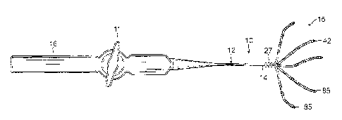
[0025] FIG. 1 is a side view of a catheter of the present invention,
in accordance with an
embodiment.
[0026] FIG. 2 is an end cross-sectional view of a catheter body of the
catheter of FIG. 1.
[0027] FIG. 3 is an end cross-sectional view of an intermediate
deflection section of the
catheter of FIG. 1.
[0028] FIG. 4 is a perspective view of a junction between the
intermediate deflection section
and a distal electrode assembly of the catheter of FIG. 1.
[0029] FIG. 5 is a perspective view of a distal electrode assembly of the
present invention, in
accordance with an embodiment.
[0030] FIG. 6 is a detailed perspective view of a spine of the distal
electrode assembly of FIG.
5.
[0031] FIG. 7 is a perspective view of a spacer of the present
invention, in accordance with an
embodiment.
[0032] FIG. 8 is a perspective view of the spine of FIG. 6, during
assembly.
[0033] FIG. 9 is a detailed perspective view of a spine of a distal
electrode assembly of the
present invention, in accordance with another embodiment.
[0034] FIG. 10 is a detailed perspective view of a spacer of the
present invention, in
accordance with another embodiment.
[0035] FIG. 11 is an end cross-section view of the spine of FIG. 9, in
contact with tissue.
-6-
CA 2976359 2017-08-14
1
DETAILED DESCRIPTION OF THE INVENTION
[0036] Referring to FIG. 1, in some embodiment of present invention, a
catheter 10 includes a
catheter body 12, an intermediate deflection section 14, a distal electrode
assembly 15, and a
control handle 16 proximal of the catheter body 12. The distal electrode
assembly 15 includes a
plurality of spines 42, each spine carrying at least one pair of closely-
spaced electrodes 13, wherein
the electrodes of a pair has a spacer defining a separation space gap distance
ranging between about
50 microns and 200 microns, and preferably between about 50 and 100 microns.
[0037] In some embodiments, the catheter body 12 comprises an elongated
tubular
construction, having a single, axial or central lumen 18, as shown in FIG. 2.
The catheter body 12
is flexible, i.e., bendable, but substantially non-compressible along its
length. The catheter body 12
can be of any suitable construction and made of any suitable material. A
presently preferred
construction comprises an outer wall 17 made of a polyurethane, or PEBAX. The
outer wall 17
comprises an imbedded braided mesh of high-strength steel, stainless steel or
the like to increase
torsional stiffness of the catheter body 12 so that, when the control handle
16 is rotated, the
deflection section 14 of the catheter 10 will rotate in a corresponding
manner.
[0038] The outer diameter of the catheter body 12 is not critical, but
is preferably no more than
about 8 french, more preferably about 7 french. Likewise the thickness of the
outer wall 17 is not
critical, but is thin enough so that the central lumen 18 can accommodate
components, including,
for example, one or more puller wires, electrode lead wires, irrigation
tubing, and any other wires
and/or cables. The inner surface of the outer wall 17 is lined with a
stiffening tube 20, which can be
made of any suitable material, such as polyimide or nylon. The stiffening tube
20, along with the
braided outer wall 17, provides improved torsional stability while at the same
time minimizing the
wall thickness of the catheter, thus maximizing the diameter of the central
lumen 18. The outer
diameter of the stiffening tube 20 is about the same as or slightly smaller
than the inner diameter of
the outer wall 17. In some embodiments, polyimide tubing is used for the
stiffening tube 20
-7-
CA 2976359 2017-08-14
=
1
because it may be very thin walled while still providing very good stiffness.
This maximizes the
diameter of the central lumen 18 without sacrificing strength and stiffness.
As would be
recognized by one skilled in the art, the catheter body construction can be
modified as desired. For
example, the stiffening tube can be eliminated.
[0039] In some embodiments, the intermediate deflection section
comprises a shorter section of
tubing 19, which as shown in FIG. 3, has multiple lumens, for example, off-
axis lumens 21, 22, 23
and 24 and on-axis lumen 25. In some embodiments, the tubing 19 is made of a
suitable non-toxic
material more flexible than the catheter body 12. A suitable material for the
tubing 19 is braided
polyurethane, i.e., polyurethane with an embedded mesh of braided high-
strength steel, stainless
steel or the like. The outer diameter of the deflection section 14 is similar
to that of the catheter
body 12. The size of the lumens is not critical and can vary depending on the
specific application.
[0040] Various components extend through the catheter 10. In some
embodiments, the
components include lead wires 30 for electrodes on the distal electrode
assembly 15, one or more
puller wires 32A and 32B for deflecting the deflection section 14, a cable 34
for an electromagnetic
position sensor 36 housed at or near a distal end of the deflection section
14, and a guidewire
tubing 38, as shown in FIG. 4. These components pass through the central lumen
18 of the catheter
body 12, as shown in FIG. 2.
[0041] In the deflection section 14, different components pass through
different lumens of the
tubing 19 as shown in FIG 3. In some embodiments, the lead wires 30 pass
through first lumen 21,
the first puller wire 32A passes through second lumen 32, the guidewire tubing
38 passes through
third lumen 23, the cable 34 passes through fourth lumen 24, and the second
puller 34B passes
through fifth lumen 25. The second and fourth lumens 22 and 24 are
diametrically opposite of each
other to provide bi-directional deflection of the intermediate deflection
section 14.
[0042] With reference to FIG. 4, distal of the deflection section 14
is the distal electrode
assembly 15 which includes a mounting stem 46 in the form of a shorter tubing
mounted on a distal
-8-
CA 2976359 2017-08-14
1
end of the tubing 19 of the intermediate deflection section 14. (In that
regard, it is understood that
where the catheter 10 is without a deflection section 14, the mounting stem 46
is mounted on a
distal end of the catheter body 12.) The stem 46 has a central lumen 48 to
house various
components. The intermediate section 14 and stem 46 are attached by glue or
the like. The stem
46 may be constructed of any suitable material, including nitinol. The stem 46
houses various
components, including the electromagnetic position sensor 36, and a distal
anchor for the puller
wires 32A and 32B.
1 0 10043] In the disclosed embodiment, the distal anchor includes one
or more washers, for
example, a distal washer 50D and a proximal washer 50P, each of which has a
plurality of through-
holes that allow passage of components between the deflection section 14 and
the stem 46 while
maintaining axial alignment of these components relative to a longitudinal
axis 40 of the catheter
10. The through-holes include holes 52 and 54 that are axially aligned with
the second and fourth
lumens 22 and 24 of the tubing 19, respectively, to receive a distal end of
puller wires 32A and
32B, respectively. It is understood that the puller wires may be formed as a
single tensile member
with a distal U-bend section that passes through the holes 52 and 54. With
tension on the washers
50D and 50P exerted by the U-bend section of the puller wires, the washers
firmly and fixedly abut
against the distal end of the tubing 19 of the deflection section 14 to
distally anchor the U-bend
section.
[0044] Each washer includes through-hole 51 which is axially aligned
with the first lumen 21
and allows passage of the lead wires 30 from the deflection section 14 and
into the lumen 48 of the
stem 46. Each washer also includes through-hole 55 which is axially aligned
with the fifth lumen
of the tubing 19 and allows passage of the sensor cable 34 from the deflection
section 14 into
25 lumen 48 of the stem 46 where the electromagnetic position sensor 36 is
housed. Each washer
further includes on-axis through-hole 53 which is axially aligned with the
third lumen 23 and
allows passage of the guidewire tubing 38 from the deflection section 14 into
the lumen 48 of the
-9-
CA 2976359 2017-08-14
1
stem 46. Marker bands or ring electrodes 27 may be carried on the outer
surface of the catheter at
or near the near the distal end of the intermediate deflection section 14, as
known in the art.
[0045] Each puller wire 32A and 32B is anchored at its proximal end in the
control handle 16
(FIG. 1). In some embodiments, the puller wires have at least sections made of
any suitable metal,
such as stainless steel or Nitinol, and are preferably coated with Teflon®
or the like. The
coating imparts lubricity to the puller wires.
[0046] A compression coil 66 is situated within the catheter body 12
in surrounding relation to
each puller wire, as shown in FIG. 2. The compression coils 66 extend from the
proximal end of
the catheter body 12 to about the proximal end of the deflection section 14.
The compression coils
are made of any suitable metal, preferably stainless steel. Each compression
coil is tightly wound
on itself to provide flexibility, i.e., bending, but to resist compression.
The inner diameter of the
compression coil is preferably slightly larger than the diameter of the puller
wire. The Teflon®
coating on the puller wire allows it to slide freely within the compression
coil. The puller wire 32A
extends through the lumen 22 of the tubing 19 and the puller wire 32B extends
through the lumen
24 of the tubing 19. Within these lumens, each puller wire extends through a
respective plastic,
preferably Teflon®, sheath 39 (see FIG. 3), which prevents the puller
wires from cutting into
the wall of the tubing 19 when the deflection section 14 is deflected.
[0047] Longitudinal movement of the puller wires relative to the catheter
body 12, which
results in deflection of the tip section 14, is accomplished by suitable
manipulation of the control
handle 16. A suitable control handle design for use with the present invention
is described in U.S.
Patent No. 8,287,532, the entire disclosure of which is incorporated herein by
reference. If desired,
the catheter can be uni-deflectional, i.e., having only one puller wire.
[0048] As shown in FIG. 4, extending from the distal end of the stem 46 are
elongated spines
42 of the distal electrode assembly 15. Each spine has a support member 43 and
a non-conductive
covering 44 that extends along the each spine 42. Each spine has a proximal
portion that extends
-10-
CA 2976359 2017-08-14
1
proximally into the lumen 48 of the stem 46. The non-conductive coverings 44
of the spines may
also extend proximally into the lumen 48. Each spine 42 may be arranged
uniformly about the
distal opening of the stem 46 in equi-radial distance from adjacent spines 42.
For example, with
five spines, each spine may be spaced apart at about 72 degrees from adjacent
spines. Suitable
adhesive, e.g., polyurethane, may be used to pot and anchor the proximal ends
of the spines 42 and
their nonconductive coverings 44. The suitable adhesive seals the distal end
of the stem 46, which
is formed to leave open the distal end of the guidewire tubing 38.
[0049] Each spine support member 43 is made of a material having shape-
memory, i.e., that
can be temporarily straightened or bent out of its original shape upon
exertion of a force and is
capable of substantially returning to its original shape in the absence or
removal of the force. One
suitable material for the support member is a nickel/titanium alloy. Such
alloys typically comprise
about 55% nickel and 45% titanium, but may comprise from about 54% to about
57% nickel with
the balance being titanium. A nickel/titanium alloy is nitinol, which has
excellent shape memory,
together with ductility, strength, corrosion resistance, electrical
resistivity and temperature stability.
The non-conductive covering 44 can be made of any suitable material, and is
preferably made of a
biocompatible plastic such as polyurethane or PEBAX.
[0050] Lead wires 30 for microelectrodes carried on the spines 42
extend through the catheter
body 12 and the deflection section 14 protected by a nonconductive sheath 60.
Toward the distal
electrode assembly 15, the lead wires 30 extend through a polytube 68, as
shown in FIG. 4. The
lead wires 30 diverge at the distal end of the polytube 68, and extend toward
their respective spine
support member 43, into their respective nonconductive covering 44 of their
respective spine 42.
[0051] With reference to FIG. 5 and FIG. 6, a plurality of bipole
electrode pairs 13 are carried
on each spine. Within each pair of bipole electrodes, a distal electrode 13D
and a proximal
electrode 13P are spaced apart and separated from each other by a
predetermined distance by a
spacer member 29 positioned therebetween. The spacer member 29 is constructed
of a
-11-
CA 2976359 2017-08-14
1
biocompatible, electrically-nonconductive material, for example, TEFLON or
PEEK, and is
sandwiched directly between the electrode pair, with edge-to-edge abutment and
contact with the
electrodes 13D and 13P to provide a physical and an electrical barrier between
these adjacent
proximal electrodes 13 of a pair. The predetermined separation gap provided by
the spacer
member 29 between adjacent electrodes 13P and 13D of a bipole pair is less
than about 1.0 mm,
preferably less than about 0.50 mm (500 microns), and more preferably about
0.05 mm (50
microns).
[0052] With reference to FIG. 7 and FIG. 8, the spacer member 29 is hollow
with an inner
diameter that is slightly greater than the outer diameter of the spine 42 so
that the spacer member
can slide onto the spine. In some embodiments, the spacer member 29 has the
same radial thickness
T between its inner surface 31 and outer surface 33 as that of the distal and
proximal ring
electrodes 13D and 13P, and the spacer member 29 has complementary or mating
adjacent edges
35 with those of the abutting ring electrodes 13D and 13P, so that the spacer
member 29 and the
electrodes 13D and 13P provide a generally seamless surface and profile on the
catheter (see FIG.
6). In the illustrated embodiments, the electrodes 13D and 13P and the spacer
member 29 are
shaped similarly, for example, each as a "ring" with a closed circular band
configuration spanning
360 degrees circumferentially, and both with similar inner and outer
diameters, the inner wherein
the spacer member 29 having a width W provides a generally equal separation
gap W between ring
electrodes 13P and 13D in the axial or longitudinal direction. The inner
diameters define axial
through-holes through which a spine is inserted during assembly of the distal
electrode assembly.
[0053] In the assembly of a spine 42, according to one embodiment,
apertures 47 are formed in
the nonconductive covering 44 at predetermined positions along the spine,
spaced at a
predetermined space gap from each other. Lead wires 30 are passed from within
the spine 42 to
exit through the respective apertures 47, whereupon distal ends of the lead
wires are further
inserted into respective ring electrodes 13P and 13D to exit through apertures
45 formed in the ring
-12-
CA 2976359 2017-08-14
1
electrodes, as shown in FIG. 8. The distal ends of the lead wires are welded
at Z, or otherwise
affixed in the apertures 45, with mechanical integrity for a fluid tight seal,
and trimmed to present a
flush surface with the outer surface of the ring electrodes.
[0054] The distal end of the spine 42 is then inserted through the
proximal ring electrode 13P,
followed by the spacer member 29, and further followed by the distal ring
electrode 13D, as shown
in FIG. 8, and the lead wires are drawn proximally or otherwise adjusted, as
needed, to fit back into
the spine 42 without leaving any excess length of lead wires outside of the
spine 42. The spacer
member and the electrodes are affixed to the outer surface of the spine by a
suitable adhesive.
100551 The electrodes 13P and 13D and the spacer member 29 are
positioned on the spine 42 so
that adjacent edges 35 of these three components are firmly abutting against
each other, and each
aperture 45 is generally concentric with its respective aperture 47. With the
spacer member 29
having a precisely-measured minimized width W, and the electrodes 13D and 13P
abutting directly
and immediately against the spacer member 29, the spacer member serves to
accurately minimize,
define, and physically set and maintain the separation gap distance between
adjacent electrodes.
The spacer member ensures consistency and repeatability while simplifying the
assembly process
by advantageously eliminating the painstaking work of measuring and affixing
electrodes at precise
locations or separation distances from each other, and traversing the
limitations of the space that
would otherwise be physically occupied by the adhesive, such as epoxy, used to
mount and seal the
electrodes. The spacer member also advantageously provides uniformity in the
separation gap
distance of each bipole electrode pair between which a spacer is positioned
such that bipole
electrode pairs of one or more spines each have the same separation gap
distance. Time and labor
for assembly are therefore greatly reduced and streamlined.
[0056] As mentioned, each electrode has an aperture 45 formed in its
sidewall. Generally
corresponding aperture 47 are formed in the nonconductive coverings 44 of the
spines 42. Each
aperture 45 in the electrode 13 is sized, shaped and configured to receive a
distal end of a
-13-
CA 2976359 2017-08-14
1
respective lead wire 30 that is passed through a respective aperture 47 from
the lumen of the
nonconductive covering 44. In that regard, the lead wire 30 is made of a
biocompatible, electrically
conductive material, for example, MP35N. The aperture 47 may be sized and
shaped in close
conformity to the size and shape of the lead wire 30.
[0057] In other embodiments, the electrodes 13 are "discrete,"
spanning less than 360 degrees
circumferentially, as shown in FIG. 9, FIG. 10 and FIG. 11, wherein spacer
member 29' provides
separation gaps between adjacent electrodes in both the axial/longitudinal
direction L and the
circumferential direction C. In that regard, the spacer member 29' has a
generally hollow
cylindrical configuration, with an outer diameter, and an inner diameter that
defines an axial
through-hole through which a distal end of a spine is inserted during assembly
of the distal
electrode assembly. The spacer member 29' spans in the axial and
circumferential directions,
wherein the spacer member has axial extensions A, center portions CA of which
are connected by
circumferential extensions C, and end portions EA of which are separated by
recessed voids V in
the spacer member. Each void V in the spacer member 29' is occupied by a
respective discrete
electrode, and each discrete electrode is surrounded generally on three sides
by two opposing axial
extensions A and one circumferential extension C. Edges E defining the
circumferential voids V
and outer peripheral edges 13E of the electrodes 13 are complementary and
provide a mating fit
with each other so that the spacer member 29' and the electrodes 13 form a
generally seamless
surface and profile on the spine 42, as shown in FIG. 9. Outer surfaces 13S of
the electrodes 13
and the outer surface 33 of the spacer member 29' together form a 360 degree
circumferential
surface that encircles and surrounds the outer surface of the spine 42.
Notably, each
circumferential void V has an open distal end or an open proximal end so that
the electrodes 13 can
advantageously slide in the axial direction (proximally or distally) into
engagement with the spacer
member 29 to occupy a respective void V.
[0058] In the illustrated embodiment of FIG. 9, FIG. 10 and FIG. 11,
the spacer member 29'
-14-
CA 2976359 2017-08-14
1
provides predetermined axial and circumferential separation gaps for four
"discrete" electrode 13a,
13b, 13c, and 13d (13d not shown). The electrodes 13a and 13c are separated in
the axial direction
(and fixedly maintained in this separation configuration) by one
circumferential extension C. The
electrodes 13b and 13d are separated in the axial direction (and fixedly
maintained in this
separation configuration) by another circumferential extension C. The
electrodes 13a and 13b are
separated in the circumferential direction (and fixedly maintained in this
separation configuration)
by one circumferential extension A. The electrodes 13c and 13d are
separated in the
circumferential direction (and fixedly maintained in this separation
configuration) by another
circumferential extension A. The spacer member 29' is configured to receive
distal electrodes 13a
and 13b in a diametrically-opposed arrangement (on opposite sides of the
spine), and proximal
electrodes 13c and 13d in a diametrically-opposed arrangement (on opposite
sides of the spine).
The spacer member 29' is also configured to position electrodes 13a and 13c in
longitudinal
alignment on the same side of the spine 42, and electrodes 13b and 13d in
longitudinal alignment
on the same and opposite side of the spine 42. The spacer member 29' is
further configured so that
each discrete electrodes spans about 90 degrees circumferentially, with each
of the axial extensions
A of the spacer member 29' spanning about 90 degrees between the electrodes
13a and 13b, and
between the electrodes 13c and 13d (better seen in FIG. 11). With the
electrodes in their discrete
and divided configuration defined by the space member 29' on the spine 42, an
operator may
activate one or more selected electrodes for sensing and/or ablation with the
remaining unselected
electrodes being unactivated. For example, the spine 42 can be placed on
target tissue surface T in
a manner, as shown in FIG. 11, so that the operator selectively activates only
the electrodes on the
same side of the spine 42 that come into contact with the target tissue
surface T, namely, electrodes
13b and 13d, for sensing electrical signals. Accordingly, electrodes on the
opposite side of the
spine 42 and not in contact with any tissue surface (namely, electrodes 13a
and 13c in FIG. 11)
may be selectively deactivated by the operator so as to avoid the detection of
noise or far-field
-15-
CA 2976359 2017-08-14
1
signals by these electrodes which may otherwise interfere with the signals
detected by the
electrodes 13b and 13d. In other embodiments, the signals received from
selected electrode(s), for
example, those not in contact with tissue, can be filtered out or otherwise
processed and separated
or distinguished from the signals received from selected electrode(s) in
contact with tissue.
[0059] As for the signals detected by the electrodes on the same side
of the spine (namely,
electrodes 13b and 13d in FIG. 11), the width of the circumferential extension
C advantageously
provides a predetermined minimized space gap between these electrodes so that
they may be used
as a bipole electrode pair in the manner, as described above, to detect
smaller/weaker signals.
[0060] It is understood that the configuration of spacer member 29'
may be varied to receive
additional electrodes greater than four. For example, the spacer 29' may
receive eight electrodes
with each distal electrode spanning about 45 degrees and each proximal
electrode spanning about
45 degrees. Morever, spacer members of different configurations may be used on
a single spine as
needed or appropriate. The shape of each discrete electrode of a spacer member
can be any
suitable shape, including circular, oval, square, rectangular, polygonal, etc.
[0061] The preceding description has been presented with reference to
presently preferred
embodiments of the invention. Workers skilled in the art and technology to
which this invention
pertains will appreciate that alterations and changes in the described
structure may be practiced
without meaningfully departing from the principal, spirit and scope of this
invention. Any feature
or structure disclosed in one embodiment may be incorporated in lieu of or in
addition to other
features of any other embodiments, as needed or appropriate. As understood by
one of ordinary
skill in the art, the drawings are not necessarily to scale. Accordingly, the
foregoing description
should not be read as pertaining only to the precise structures described and
illustrated in the
accompanying drawings, but rather should be read consistent with and as
support to the following
claims which are to have their fullest and fair scope.
-16-
CA 2976359 2017-08-14