Note: Descriptions are shown in the official language in which they were submitted.
CA 02976926 2017-08-16
WO 2016/134333
PCT/US2016/018809
ANTI-PVRIG ANTIBODIES AND METHODS OF USE
CROSS REFERENCE TO RELATED APPLICATIONS
[0001] This application claims priority under 35 U.S.C. 119 to USSN
62/118,208, filed
February 19, 2015, and to USSN 62/141,120, filed March 31, 2015, and to USSN
62/235,823, filed October 1, 2015, all of which are expressly incorporated
herein by
reference in their entirety.
BACKGROUND OF THE INVENTION
[0002] Naive T cells must receive two independent signals from antigen-
presenting cells
(APC) in order to become productively activated. The first, Signal 1, is
antigen-specific and
occurs when T cell antigen receptors encounter the appropriate antigen-MHC
complex on the
APC. The fate of the immune response is determined by a second, antigen-
independent signal
(Signal 2) which is delivered through a T cell costimulatory molecule that
engages its APC-
expressed ligand. This second signal could be either stimulatory (positive
costimulation) or
inhibitory (negative costimulation or coinhibition). In the absence of a
costimulatory signal,
or in the presence of a coinhibitory signal, T-cell activation is impaired or
aborted, which
may lead to a state of antigen-specific unresponsiveness (known as T-cell
anergy), or may
result in T-cell apoptotic death.
[0003] Costimulatory molecule pairs usually consist of ligands expressed on
APCs and their
cognate receptors expressed on T cells. The prototype ligand/receptor pairs of
costimulatory
molecules are B7/CD28 and CD40/CD4OL. The B7 family consists of structurally
related,
cell-surface protein ligands, which may provide stimulatory or inhibitory
input to an immune
response. Members of the B7 family are structurally related, with the
extracellular domain
containing at least one variable or constant immunoglobulin domain.
[0004] Both positive and negative costimulatory signals play critical roles in
the regulation of
cell-mediated immune responses, and molecules that mediate these signals have
proven to be
effective targets for immunomodulation. Based on this knowledge, several
therapeutic
approaches that involve targeting of costimulatory molecules have been
developed, and were
shown to be useful for prevention and treatment of cancer by turning on, or
preventing the
turning off, of immune responses in cancer patients and for prevention and
treatment of
autoimmune diseases and inflammatory diseases, as well as rejection of
allogenic
1
CA 02976926 2017-08-16
WO 2016/134333
PCT/US2016/018809
transplantation, each by turning off uncontrolled immune responses, or by
induction of "off
signal" by negative costimulation (or coinhibition) in subjects with these
pathological
conditions.
[0005] Manipulation of the signals delivered by B7 ligands has shown potential
in the
treatment of autoimmunity, inflammatory diseases, and transplant rejection.
Therapeutic
strategies include blocking of costimulation using monoclonal antibodies to
the ligand or to
the receptor of a costimulatory pair, or using soluble fusion proteins
composed of the
costimulatory receptor that may bind and block its appropriate ligand. Another
approach is
induction of co-inhibition using soluble fusion protein of an inhibitory
ligand. These
approaches rely, at least partially, on the eventual deletion of auto- or allo-
reactive T cells
(which are responsible for the pathogenic processes in autoimmune diseases or
transplantation, respectively), presumably because in the absence of
costimulation (which
induces cell survival genes) T cells become highly susceptible to induction of
apoptosis.
Thus, novel agents that are capable of modulating costimulatory signals,
without
compromising the immune system's ability to defend against pathogens, are
highly
advantageous for treatment and prevention of such pathological conditions.
[0006] Costimulatory pathways play an important role in tumor development.
Interestingly,
tumors have been shown to evade immune destruction by impeding T cell
activation through
inhibition of co-stimulatory factors in the B7-CD28 and TNF families, as well
as by
attracting regulatory T cells, which inhibit anti-tumor T cell responses (see
Wang (2006),
"Immune Suppression by Tumor Specific CD4+ Regulatory T cells in Cancer",
Semin.
Cancer. Biol. 16:73-79; Greenwald, et al. (2005), "The B7 Family Revisited",
Ann. Rev.
Immunol. 23:515-48; Watts (2005), "TNF/TNFR Family Members in Co-stimulation
of T
Cell Responses", Ann. Rev. Immunol. 23:23-68; Sadum, et al., (2007) "Immune
Signatures of
Murine and Human Cancers Reveal Unique Mechanisms of Tumor Escape and New
Targets
for Cancer Immunotherapy", Clin. Canc. Res. 13(13): 4016-4025). Such tumor
expressed co-
stimulatory molecules have become attractive cancer biomarkers and may serve
as tumor-
associated antigens (TAAs). Furthermore, costimulatory pathways have been
identified as
immunologic checkpoints that attenuate T cell dependent immune responses, both
at the level
of initiation and effector function within tumor metastases. As engineered
cancer vaccines
continue to improve, it is becoming clear that such immunologic checkpoints
are a major
barrier to the vaccines' ability to induce therapeutic anti-tumor responses.
In that regard,
2
CA 02976926 2017-08-16
WO 2016/134333
PCT/US2016/018809
costimulatory molecules can serve as adjuvants for active (vaccination) and
passive
(antibody-mediated) cancer immunotherapy, providing strategies to thwart
immune tolerance
and stimulate the immune system.
[0007] Over the past decade, agonists and/or antagonists to various
costimulatory proteins
have been developed for treating autoimmune diseases, graft rejection, allergy
and cancer.
For example, CTLA4-Ig (Abatacept, Orencia0) is approved for treatment of RA,
mutated
CTLA4-Ig (Belatacept, Nulojix0) for prevention of acute kidney transplant
rejection and by
the anti-CTLA4 antibody (Ipilimumab, Yervoy0), recently approved for the
treatment of
melanoma. Other costimulation regulators have been approved, such as the anti-
PD-1
antibodies of Merck (Keytruda0) and BMS (Opdivo0), have been approved for
cancer
treatments and are in testing for viral infections as well.
[0008] Accordingly, it is an object of the invention to provide PVRIG
immunomodulatory
antibodies.
BRIEF SUMMARY OF THE INVENTION
[0009] Accordingly, it is an object of the invention to provide methods of
activating
cytotoxic T cells (CTLs) of a patient comprising administering an anti-PVRIG
antibody to
the patient, wherein a subset of the CTLs of the patient are activated.
[0010] It is a further object of the invention to provide methods of
activating NK cells of a
patient comprising administering an anti-PVRIG antibody to the patient,
wherein a subset of
the NK cells of the patient are activated.
[0011] It is an additional object of the invention to provide methods of
activating y6 T cells
of a patient comprising administering an anti-PVRIG antibody to the patient,
wherein a
subset of the y6 T cells of the patient are activated.
[0012] It is a further object of the invention to provide methods of
activating Thl cells of a
patient comprising administering an anti-PVRIG antibody to the patient,
wherein a subset of
the Thl cells of the patient are activated.
[0013] It is an additional object of the invention to provide methods of
inhibiting the
interaction of PVRIG and PVLR2 in a patient having a condition associated with
this
interaction comprising administering an anti-PVRIG antibody to the patient.
3
CA 02976926 2017-08-16
WO 2016/134333
PCT/US2016/018809
[0014] It is a further object of the invention to provide methods of treating
cancer in a
patient, comprising administering an anti-PVRIG antibody to the patient,
wherein said cancer
is treated.
[0015] It is an additional object of the invention to provide methods as
outlined above
wherein the anti-PVRIG antibody comprises the vhCDR1, vhCDR2, vhCDR3, v1CDR1,
v1CDR2 and v1CDR3 sequences from an antibody selected from the group
consisting of
CPA.7.001, CPA.7.003, CPA.7.004, CPA.7.006, CPA.7.008, CPA.7.009, CPA.7.010,
CPA.7.011, CPA.7.012, CPA.7.013, CPA.7.014, CPA.7.015, CPA.7.017, CPA.7.018,
CPA.7.019, CPA.7.021, CPA.7.022, CPA.7.023, CPA.7.024, CPA.7.033, CPA.7.034,
CPA.7.036, CPA.7.040, CPA.7.046, CPA.7.047, CPA.7.049, and CPA.7.050.
[0016] It is an additional object of the invention to provide methods as
outlined above
wherein the anti-PVRIG antibody competes for binding with an antibody
comprising the
vhCDR1, vhCDR2, vhCDR3, v1CDR1, v1CDR2 and v1CDR3 sequences from an antibody
selected from the group consisting of CPA.7.001, CPA.7.003, CPA.7.004,
CPA.7.006,
CPA.7.008, CPA.7.009, CPA.7.010, CPA.7.011, CPA.7.012, CPA.7.013, CPA.7.014,
CPA.7.015, CPA.7.017, CPA.7.018, CPA.7.019, CPA.7.021, CPA.7.022, CPA.7.023,
CPA.7.024, CPA.7.033, CPA.7.034, CPA.7.036, CPA.7.040, CPA.7.046, CPA.7.047,
CPA.7.049, and CPA.7.050.
[0017] It is a further object of the invention to provide methods as outlined
above wherein
the anti-PVRIG antibody is selected from the group consisting of CPA.7.001,
CPA.7.003,
CPA.7.004, CPA.7.006, CPA.7.008, CPA.7.009, CPA.7.010, CPA.7.011, CPA.7.012,
CPA.7.013, CPA.7.014, CPA.7.015, CPA.7.017, CPA.7.018, CPA.7.019, CPA.7.021,
CPA.7.022, CPA.7.023, CPA.7.024, CPA.7.033, CPA.7.034, CPA.7.036, CPA.7.040,
CPA.7.046, CPA.7.047, CPA.7.049, and CPA.7.050.
[0018] It is an additional object of the invention to provide methods as
outlined above
wherein the anti-PVRIG antibody competes for binding with an antibody selected
from the
group consisting of CPA.7.001, CPA.7.003, CPA.7.004, CPA.7.006, CPA.7.008,
CPA.7.009, CPA.7.010, CPA.7.011, CPA.7.012, CPA.7.013, CPA.7.014, CPA.7.015,
CPA.7.017, CPA.7.018, CPA.7.019, CPA.7.021, CPA.7.022, CPA.7.023, CPA.7.024,
CPA.7.033, CPA.7.034, CPA.7.036, CPA.7.040, CPA.7.046, CPA.7.047, CPA.7.049,
and
CPA.7.050.
4
CA 02976926 2017-08-16
WO 2016/134333
PCT/US2016/018809
[0019] It is a further object of the invention to provide methods as outlined
above wherein
the anti-PVRIG antibody comprises the vhCDR1, vhCDR2, vhCDR3, v1CDR1, v1CDR2
and
v1CDR3 sequences from an antibody selected from the group consisting of
CHA.7.502,
CHA.7.503, CHA.7.506, CHA.7.508, CHA.7.510, CHA.7.512, CHA.7.514, CHA.7.516,
CHA.7.518, CHA.7.520.1, CHA.7.520.2, CHA.7.522, CHA.7.524, CHA.7.526,
CHA.7.527, CHA.7.528, CHA.7.530, CHA.7.534, CHA.7.535, CHA.7.537,
CHA.7.538.1, CHA.7.538.2, CHA.7.543, CHA.7.544, CHA.7.545, CHA.7.546,
CHA.7.547, CHA.7.548, CHA.7.549, CHA.7.550, CPA.7.001, CPA.7.003, CPA.7.004,
CPA.7.006, CPA.7.008, CPA.7.009, CPA.7.010, CPA.7.011, CPA.7.012, CPA.7.013,
CPA.7.014, CPA.7.015, CPA.7.017, CPA.7.018, CPA.7.019, CPA.7.021, CPA.7.022,
CPA.7.023, CPA.7.024, CPA.7.033, CPA.7.034, CPA.7.036, CPA.7.040, CPA.7.046,
CPA.7.047, CPA.7.049, and CPA.7.050.
[0020] It is an additional object of the invention to provide methods as
outlined above
wherein said the-PVRIG antibody competes for binding with an antibody selected
from the
group consisting of an anti-PVRIG antibody comprising the vhCDR1, vhCDR2,
vhCDR3,
v1CDR1, v1CDR2 and v1CDR3 sequences from an antibody selected from the group
consisting of CHA.7.502, CHA.7.503, CHA.7.506, CHA.7.508, CHA.7.510,
CHA.7.512,
CHA.7.514, CHA.7.516, CHA.7.518, CHA.7.520.1, CHA.7.520.2, CHA.7.522,
CHA.7.524,
CHA.7.526, CHA.7.527, CHA.7.528, CHA.7.530, CHA.7.534, CHA.7.535, CHA.7.537,
CHA.7.538.1, CHA.7.538.2, CHA.7.543, CHA.7.544, CHA.7.545, CHA.7.546,
CHA.7.547, CHA.7.548, CHA.7.549, CHA.7.550, CPA.7.001, CPA.7.003, CPA.7.004,
CPA.7.006, CPA.7.008, CPA.7.009, CPA.7.010, CPA.7.011, CPA.7.012, CPA.7.013,
CPA.7.014, CPA.7.015, CPA.7.017, CPA.7.018, CPA.7.019, CPA.7.021, CPA.7.022,
CPA.7.023, CPA.7.024, CPA.7.033, CPA.7.034, CPA.7.036, CPA.7.040, CPA.7.046,
CPA.7.047, CPA.7.049, and CPA.7.050.
[0021] It is a further object of the invention to provide methods of
diagnosing cancer
comprising a) contacting a tissue from a patient with an anti-PVRIG antibody;
and b)
determining the presence of over-expression of PVRIG in the tissue as an
indication of the
presence of cancer. The anti-PVRIG antibody can be as described herein and as
outlined
above.
[0022] It is an additional object of the invention to provide antigen binding
domains,
including antibodies, which are anti-PVRIG antibodies, comprising the vhCDR1,
vhCDR2,
CA 02976926 2017-08-16
WO 2016/134333
PCT/US2016/018809
vhCDR3, v1CDR1, v1CDR2 and v1CDR3 sequences from an antibody selected from the
group consisting of CPA.7.001, CPA. 7.003, CPA. 7.004, CPA.7.006, CPA. 7.008,
CPA.7.009,
CPA.7.010, CPA.7.011, CPA.7.012, CPA.7.013, CPA.7.014, CPA.7.015, CPA.7.017,
CPA.7.018, CPA.7.019, CPA.7.021, CPA.7.022, CPA.7.023, CPA.7.024, CPA.7.033,
CPA.7.034, CPA.7.036, CPA.7.040, CPA.7.046, CPA.7.047, CPA.7.049, and
CPA.7.050.
[0023] It is a further object of the invention to provide anti-PVRIG antigen
binding domains
(including antibodies) compositions that are anti-PVRIG antibodies, selected
from the group
consisting of CPA. 7.001, CPA. 7.003, CPA. 7.004, CPA.7.006, CPA.7.008, CPA.
7.009,
CPA.7.010, CPA.7.011, CPA.7.012, CPA.7.013, CPA.7.014, CPA.7.015, CPA.7.017,
CPA.7.018, CPA.7.019, CPA.7.021, CPA.7.022, CPA.7.023, CPA.7.024, CPA.7.033,
CPA.7.034, CPA.7.036, CPA.7.040, CPA.7.046, CPA.7.047, CPA.7.049, and
CPA.7.050.
[0024] It is a further object of the invention to provide anti-PVRIG antigen
binding domains
(including antibodies) compositions that are anti-PVRIG antibodies, selected
from the group
consisting of h518-1, h518-2, h518-3, h518-4, h518-5, h524-1, h524-2, h524-3,
h524-4,
h530-1, h530-2, h530-3, h530-4, h530-5, h538.1-1, h538.1-2, h538.1-3, h538.1-
4, h538.2-1,
h538.2-2, and h538.2-3 (as depicted in Figure 90).
[0025] It is an additional object of the invention to provide antigen binding
domains,
including antibodies, which are anti-PVRIG antibodies, comprising the vhCDR1,
vhCDR2,
vhCDR3, v1CDR1, v1CDR2 and v1CDR3 sequences from an antibody selected from the
group consisting of CHA.7.502, CHA.7.503, CHA.7.506, CHA.7.508, CHA.7.510,
CHA.7.512, CHA.7.514, CHA.7.516, CHA.7.518, CHA.7.520.1, CHA.7.520.2,
CHA.7.522, CHA.7.524, CHA.7.526, CHA.7.527, CHA.7.528, CHA.7.530, CHA.7.534,
CHA.7.535, CHA.7.537, CHA.7.538.1, CHA.7.538.2, CHA.7.543, CHA.7.544,
CHA.7.545, CHA.7.546, CHA.7.547, CHA.7.548, CHA.7.549, CHA.7.550, CPA.7.001,
CPA.7.003, CPA.7.004, CPA.7.006, CPA.7.008, CPA.7.009, CPA.7.010, CPA.7.011,
CPA.7.012, CPA.7.013, CPA.7.014, CPA.7.015, CPA.7.017, CPA.7.018, CPA.7.019,
CPA.7.021, CPA.7.022, CPA.7.023, CPA.7.024, CPA.7.033, CPA.7.034, CPA.7.036,
CPA.7.040, CPA.7.046, CPA.7.047, CPA.7.049, and CPA.7.050.
[0026] It is a further object of the invention to provide nucleic acid
compositions comprising:
a) a first nucleic acid encoding the a heavy chain variable domain comprising
the vhCDR1,
vhCDR2 and vhCDR3 from an antibody; and b) a second nucleic acid encoding a
light chain
variable domain comprising v1CDR1, v1CDR2 and and v1CDR3 from an antibody. The
6
CA 02976926 2017-08-16
WO 2016/134333
PCT/US2016/018809
antibody is selected from the group consisting of CPA.7.001, CPA.7.003,
CPA.7.004,
CPA.7.006, CPA.7.008, CPA.7.009, CPA.7.010, CPA.7.011, CPA.7.012, CPA.7.013,
CPA.7.014, CPA.7.015, CPA.7.017, CPA.7.018, CPA.7.019, CPA.7.021, CPA.7.022,
CPA.7.023, CPA.7.024, CPA.7.033, CPA.7.034, CPA.7.036, CPA.7.040, CPA.7.046,
CPA.7.047, CPA.7.049, CPA.7.050, CHA.7.502, CHA.7.503, CHA.7.506, CHA.7.508,
CHA.7.510, CHA.7.512, CHA.7.514, CHA.7.516, CHA.7.518, CHA.7.520.1,
CHA.7.520.2,
CHA.7.522, CHA.7.524, CHA.7.526, CHA.7.527, CHA.7.528, CHA.7.530, CHA.7.534,
CHA.7.535, CHA.7.537, CHA.7.538.1, CHA.7.538.2, CHA.7.543, CHA.7.544,
CHA.7.545, CHA.7.546, CHA.7.547, CHA.7.548, CHA.7.549, CHA.7.550, CPA.7.001,
CPA.7.003, CPA.7.004, CPA.7.006, CPA.7.008, CPA.7.009, CPA.7.010, CPA.7.011,
CPA.7.012, CPA.7.013, CPA.7.014, CPA.7.015, CPA.7.017, CPA.7.018, CPA.7.019,
CPA.7.021, CPA.7.022, CPA.7.023, CPA.7.024, CPA.7.033, CPA.7.034, CPA.7.036,
CPA.7.040, CPA.7.046, CPA.7.047, CPA.7.049, and CPA.7.050.
[0027] It is an additional object of the invention to provide expression
vector compositions
comprising the first and second nucleic acids as outlined herein and above.
[0028] It is a further object of the invention to provide host cells
comprising the expression
vector compositions, either as single expression vectors or two expression
vectors.
[0029] It is an additional object of the invention to provide methods of
making an anti-
PVRIG antibody comprising a) culturing a host cell of the invention with
expression
vector(s) under conditions wherein the antibody is produced; and b) recovering
the antibody.
BRIEF DESCRIPTION OF THE DRAWINGS
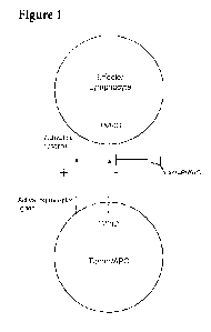
[0030] Figure 1 Schematic presentation of the mechanisms of action of the
invention.
[0031] Figure 2 presents mRNA Expression of PVRIG in various normal human
tissues.
[0032] Figure 3 presents mRNA expression of PVRIG in various immune population
derived
from peripheral blood and bone marrow (based on G5E49910).
[0033] Figure 4 presents mRNA expression of PVRIG in various CD3+ lymphocyte
population (based on G5E47855).
[0034] Figure 5 A, 5B and 5C presents mRNA expression of PVRIG in specific
cell
populations. Figure 5A resents mRNA expression of PVRIG in specific cell
populations
obtained by laser capture microscopy (based on G5E39397). Figure 5B presents
mRNA
7
CA 02976926 2017-08-16
WO 2016/134333
PCT/US2016/018809
expression of PVRIG in CD4 T-cells from normal and cancer patient as well as
expression
form CD4 T-cell expression from draining lymph nodes and TILs form breast
cancer patients
(based on GSE36765). Figure 5C presents mRNA expression of PVRIG from CD8 and
CD4
T-cells derived from follicular lymphoma tumor and tonsil (based on GSE27928).
[0035] Figure 6 presents PVRIG expression in normal tissues based on GTEx.
Expression
levels are shown in log2(RPKM) values (fragments identified per million reads
per kilobase).
Values above 1 are considered high expression. Tissues are ranked from top to
bottom by the
median expression. Each dot on the plot represent a single sample.
[0036] Figure 7 presents PVRIG expression in cancerous tissues based on TCGA.
Expression levels are shown in log2(RPKM) values (fragments identified per
million reads
per kilobase). Values above 1 are considered high expression. Tissues are
ranked from top to
bottom by the median expression. Each dot on the plot represent a single
sample
[0037] Figure 8 shows a heatmap representation of the enrichment analysis
results in three
categories: protein interactions, pathways and disease associations. Results
are ranked from
top to bottom by average p-value per row. Only the top 10 results from each
category are
shown. Gray squares indicate p-values<0.05. Each column in the heatmap
corresponds to a
normal or cancer tissue from which a list of highly correlated genes was
derived (r>0.55
using at least 50 samples). As shown in the heatmap, PVRIG correlates with a T
cell gene
expression signature which is strongly associated with the immune response and
immune
diseases.
[0038] Figure 9 presents PVRIG expression in normal skin vs. melanoma (GTEx
and TCGA
analysis). Such over-expression was observed in additional solid tumors and
results from
infiltrating lymphocytes and NK cells in the tumor microenvironment. In normal
condictions,
no infiltrating immune cells are present and therefore PVRIG expression levels
are very low.
[0039] Figure 10 presents the correlations of PVRIG and PD1 in melanoma from
TCGA
samples, with several T cell makers in lung adenocarcinoma, colon
adenocarcinoma and
melanoma. The marker CD3 is a general markers for T cells and is also
expressed on NKT
cells. CD4 and CD8 markers are used to characterized subpopulation of T cells.
[0040] Figure 11 shows expression of PVRIG on human PBLs. Human PBLs derived
from
two donors were evaluated for PVRIG expression. Both donor 61 and donor 40
showed
significant staining with anti-PVRIG specific Ab.
8
CA 02976926 2017-08-16
WO 2016/134333
PCT/US2016/018809
[0041] Figure 12 shows PVRIG-Ig exhibits strong binding to all four human
melanoma cell
lines MEL-23, Mel-624 and Me1-624.38 and me1-888 tested. Binding is not
affected by co-
culture with engineered melanoma specific T cells. Grey line corresponds to
isotype control,
solid black line corresponds to PVRIG-ECD-Ig.
[0042] Figure 13 Correlation of PVRIG with T cells and subpopulations of T
cells. CD3G is
component of the T cell receptor complex, CD4 is a maker for T helper cells
and CD8A is
component of CD8 protein used to identify cytotoxic T cells. PVRIG highly
correlated with T
cells in many types of tumors including lung adenocarcinoma, colon
adenocarcinoma and
melanoma which are shown here.
[0043] Figure 14 presents representative images from the
Confirmation/Specificity screen.
All hits from the Primary screen, and EGFR-expressing vector (negative
control), were re-
arrayed/expressed in duplicate and probed with PVRIG at 2Oug/ml. A specific
hit with strong
intensity is shown in green (PVRL2). Non-specific hits are shown in black.
Another weak hit
(MAG) was later shown to bind also other ligands, thus suggesting that it is
not specific.
[0044] Figure 15A-15E presents effect of various PVRIG-ECD-Ig M:M proteins on
mouse
CD4 T cell activation. Plates were coated with anti-CD3 mAb (2 g/mL) in the
presence of
10ug/m1PVRIG-ECD Ig (batch #198) or control mIgG2a as described in materials
and
methods. Wells were plated with lx105 CD4+CD25- mouse T cells per well in the
presence
of 2ug/m1 of soluble anti-CD28. (A) The expression of CD69 was analyzed by
flow
cytometry at 48h post-stimulation, representative histograms are shown. Each
bar is the mean
of duplicate cultures, the error bars indicating the standard deviation. (B-C)
Culture
supernatants were collected at 48 h post-stimulation and mouse IL-2 and IFNy
levels were
analyzed by ELISA. Results are shown as Mean Standard errors of duplicate
samples. (D)
Dose response effect of immobilized PVRIG-ECD Ig (Figure 92BB on surface CD69
(D) and
IFNy secretion (E) is presented. Each bar is the mean of triplicate cultures,
the error bars
indicating the standard errors.
[0045] Figure 16 presents FACS analysis on PVRIG transduced PBLs using a
specific
antibody. The percent of cells staining positive (relative to empty vector
transduced) for the
protein is provided.
[0046] Figure 17 presents FACS analysis on PVRIG (either co-expressed with F4
TCR or in
a bi-cystronic vector with F4 TCR and NGFR transduced PBLs using a specific
antibody.
9
CA 02976926 2017-08-16
WO 2016/134333
PCT/US2016/018809
The percent of cells staining positive (relative to empty vector transduced)
for the protein is
provided.
[0047] Figure 18A-18B presents FACS analysis performed on TCR transduced
stimulated
PBLs for experiment 1 (Figure 18A) and in experiment 2 (Figure 18B) using a
specific
monoclonal antibody that recognizes the extra-cellular domain of the beta-
chain from the
transduced specific TCR. The percentage of cells staining positive is
provided.
[0048] Figure 19 shows expression of PVRIG on F4 expressing PBLs causes a
reduction of
IFNy secretion upon co-culture with SK-MEL23, MEL-624 and MEL-624.38 in
comparison
to expression of an empty vector.
[0049] Figure 20A-20B shows expression of PVRIG and F4 in PBLs by co-
transduction
(Figure 20A) does not affect IFNy secretion in co-culture with melanoma cell
lines.
Expression of PVRIG and F4 in PBLs using a bi-cystronic vector (Figure 20B)
causes a
reduction of IFNy secretion upon co-culture with SK-MEL23, MEL-624 and MEL-
624.38 in
comparison to expression of an empty vector.
[0050] Figure 21 shows expression of PVRIG and F4 in PBLs using a bi-cystronic
vector
causes a reduction in T cell mediated cytotoxicity upon co-culture with
melanoma cell lines.
[0051] Figure 22 shows PVRIG expression in 3 subgroups of low, no change and
high levels
of exhausted T cells. Exhausted T cells were selected based on high level
expression of 4
markers: CD8A, PD-1, TIM-3 and TIGIT. Low expressing samples are not shown
since none
had any detectable levels of PVRIG.
[0052] Figure 23A-23B: Western blot analysis of ectopically expressed human
PVRIG
protein. Whole cell extracts of HEK293 cell pools, previously transfected with
expression
construct encoding human PVRIG-flag (lane 2) or with empty vector (lane 1)
were analyzed
by WB using an anti-flag antibody (23A) or anti-PVRIG antibodies (23B).
[0053] Figure 24: Cell surface expression of HEK293 cells ectopically
expressed human
PVRIG-flag protein by FACS analysis. Anti-PVRIG pAb (Abnova) was used to
analyze
HEK293 cells stably expressing the human PVRIG-flag protein. Cells expressing
the empty
vector were used as negative control. Detection was carried out by Goat Anti-
mouse PE-
conjugated secondary Ab and analyzed by FACS.
CA 02976926 2017-08-16
WO 2016/134333
PCT/US2016/018809
[0054] Figure 25 depicts the full length sequence of human PVRIG (showing two
different
methionine starting points) and the PVRIG Fc fusion protein used in the
Examples. The
signal peptide is underlined, the ECD is double underlined, and the Fc domain
is the dotted
underlining.
[0055] Figure 26 depicts the sequence of the human Poliovirus receptor-related
2 protein
(PVLR2, also known as nectin-2, CD112 or herpesvirus entry mediator B,
(HVEB)), the
binding partner of PVRIG as shown in Example 5. PVLR2 is a human plasma
membrane
glycoprotein.
[0056] Figure 27 PVRIG antibody specificity towards HEK cells engineered to
overexpress
PVRIG. Data shows absolute geometric MFI (gMFI) measurements as a function of
increasing antibody concentration. The broken black line with squares shows
staining of
HEK hPVRIG cells with a representative anti-human PVRIG antibody (CPA.7.021),
and the
solid black line with circles shows staining of HEK parental cells with the
same antibody.
[0057] Figure 28 PVRIG RNA was assessed in various cancer cell lines by qPCR.
Data
shown is relative expression of PVRIG RNA in cell lines as fold change over
levels in expi
cells as assessed by the 2"Act) method.
[0058] Figure 29 PVRIG RNA was assessed in sorted PBMC subsets by qPCR. Data
shown
is relative expression of PVRIG RNA in each subset as fold change over levels
in HEK GFP
cells as assessed by the 2"Act) method. D47-D49 denote three individual
donors. CD4
denotes CD4 T cells, CD8 denotes CD8 T cells, CD14 denotes monocytes, and CD56
denotes
NK cells.
[0059] Figure 30A-30B. Figure 30A: PVRIG RNA was assessed in sorted CD4 T
cells
(CD4) and NK cells (NK) under naive and activated conditions by qPCR. CD4 T
cells were
stimulated with human T cell stimulator dynabeads and 50U/m1 IL-2 for 3 days.
NK cells
were stimulated in 50U/m1 IL-2 for 3 days. Data shown is relative expression
of PVRIG RNA
in each subset as fold change over levels in expi cells as assessed by the
2"Act) method.
Jurkat is included as a positive control. D47-D49 denote three individual
donors. Figure 30B
PVRIG RNA was assessed in sorted CD8 T cells under naive and activated
conditions by
qPCR. CD8 T cells were stimulated with human T cell stimulator dynabeads and
100U/m1
IL-2 for 3 days. Data shown is relative expression of PVRIG RNA in each subset
as fold
11
CA 02976926 2017-08-16
WO 2016/134333
PCT/US2016/018809
change over levels in expi cells as assessed by the 2(-AAct) method. Jurkat is
included as a
positive control. D49, 70, and 71 indicate three individual donors.
[0060] Figure 31A-31B PVRIG binding characteristics to HEK hPVRIG engineered
cell
lines, HEK parental cells, CA46 cells, and Jurkat cells. HEK OE denotes HEK
hPVRIG cells,
HEK par denotes HEK parental cells. For Jurkat and CA46 data, gMFIr indicates
the fold
difference in geometric MFI of PVRIG antibody staining relative to their
controls.
Concentration indicates that at which the gMFIr was calculated. Not reliable
fit indicates
antibody binding characteristics do meet appropriate mathematical fitting
requirements. Some
antibodies were not tested in some conditions due to poor binding
characteristics, specificity,
or manufacturability.
[0061] Figure 32A-32B PVRIG binding characteristics to primary human PBMC,
cyno
transient over-expressing cells, and cyno primary PBMC. Expi cyno OE denotes
expi cells
transiently transfected with cPVRIG, expi par denotes expi parental cells.
gMFIr indicates the
fold difference in geometric MFI of PVRIG antibody staining relative to their
controls.
Concentration indicates that at which the gMFIr was calculated. Some
antibodies were not
tested in some conditions due to poor binding characteristics, specificity, or
manufacturability
as in Figure 31. Additionally, select antibodies were triaged for screening on
cyno PBMC
subsets based on their ability to bind cPVRIG transient cells or
functionality. Expression of
PVRIG on CD4 T cells is similar to that described in the table for CD8 T
cells.
[0062] Figure 33 PVRIG antibody specificity towards CA46 and Jurkat cells.
Data shows
absolute geometric MFI (gMFI) measurements by FACS as a function of increasing
antibody
concentration. The solid black line with triangles shows staining of CA46
cells with anti-
human PVRIG antibody (CPA.7.021) and the solid black line with squares shows
staining of
Jurkat cells. OV-90 (broken line with upside down triangles) and NCI-H4411
(broken line
with diamonds) are shown as negative controls.
[0063] Figure 34A-34D PVRIG antibody cross-reactivity towards cPVRIG transient
cells.
Data shows an example of an antibody that is a negative binder (a-b,
CPA.7.021) and a
positive binder (c-d, CPA.7.024) on cPVRIG transient cells. Solid grey
histograms indicate
control antibody, open black histograms indicate the antibody of interest.
Cells were stained
with each antibody at a concentration of 5ug/ml.
12
CA 02976926 2017-08-16
WO 2016/134333
PCT/US2016/018809
[0064] Figure 35 cPVRIG RNA was assessed in sorted cyno PBMC subsets by qPCR.
Data
shown is the average Ct values from three cyno donors as detected by two
primer sets
directed at two distinct areas of the cPVRIG gene.
[0065] Figure 36A-36C cPVRIG protein was assessed on a) CD16+ lymphocytes (NK
cells),
b) CD14+ CD56+ myeloid cells (monocytes), and c) CD3+ lymphocytes (T cells) by
FACS.
Data is shown as absolute geometric MFI, with the solid black line indicating
background
fluorescence levels. Data is representative of a sample of our panel of anti-
human PVRIG
antibodies tested in three cyno donors.
[0066] Figure 37A-37B shows the CDR sequences for Fabs that were determined to
successfully block interaction of the PVRIG with its counterpart PVRL2, as
described in
Example 5.
[0067] Figure 38A-38AA shows the amino acid sequences of the variable heavy
and light
domains, the full length heavy and light chains, and the variable heavy and
variable light
CDRs for the enumerated human CPA anti-PVRIG sequences of the invention that
both bind
PVRIG and block binding of PVRIG and PVLR2.
[0068] Figure 39A-39H depicts the amino acid sequences of the variable heavy
and light
domains, the full length heavy and light chains, and the variable heavy and
variable light
CDRs for eight human CPA anti-PVRIG sequences of the invention that bind PVRIG
and but
do not block binding of PVRIG and PVLR2.
[0069] Figure 40A-40D depicts the CDRs for all CPA anti-PVRIG antibody
sequences that
were generated that bind PVRIG, including those that do not block binding of
PVRIG and
PVLR2.
[0070] Figure 41A to 41DD depicts the variable heavy and light chains as well
as the
vhCDR1, vhCDR2, vhCDR3, v1CDR1, v1CDR2 and v1CDR3 sequences of each of the
enumerated CHA antibodies of the invention, CHA.7.502, CHA.7.503, CHA.7.506,
CHA.7.508, CHA.7.510, CHA.7.512, CHA.7.514, CHA.7.516, CHA.7.518, CHA.7.520.1,
CHA.7.520.2, CHA.7.522, CHA.7.524, CHA.7.526, CHA.7.527, CHA.7.528, CHA.7.530,
CHA.7.534, CHA.7.535, CHA.7.537, CHA.7.538.1, CHA.7.538.2, CHA.7.543,
CHA.7.544, CHA.7.545, CHA.7.546, CHA.7.547, CHA.7.548, CHA.7.549 and
CHA.7.550 (these include the variable heavy and light sequences from mouse
sequences
(from Hybridomas).
13
CA 02976926 2017-08-16
WO 2016/134333
PCT/US2016/018809
[0071] Figure 42 depicts the binning results from Example 11. Not binned:
CPA.7.029 and
CPA.7.026 (no binding to the antigen).
[0072] Figure 43 Binary matrix of pair-wise blocking ("0", red box) or
sandwiching ("1",
green box) of antigen for 35 anti-PVRIG mAbs. MAbs listed vertically on the
left of the
matrix are mAbs covalently immobilized to the ProteOn array. MAbs listed
horizontally
across the top of the matrix were analytes injected with pre-mixed antigen.
Clone CPA.7.041
was studied only as an analyte. The black boxes outline four epitope bins
according to the
vertical blocking patterns of the mAbs.
[0073] Figure 44 Hierarchical clustering dendrogram of the vertical binding
patterns of each
mAb in the binary matrix in Figure 43. There are four bins of mAbs with
identical epitope
blocking patterns within each group. The only difference between bins 1 and 2
is mAbs in
bin 1 block antigen binding to clone CPA.7.039 while mAbs in bin 2 can
sandwich the
antigen with CPA.7.039. Clone CPA.7.050 can sandwich the antigen with all
other clones.
[0074] Figure 45A-45JJ Sensorgrams indicating the antigen blocking pattern for
CPA.7.036
with all other immobilized mAbs, which are representative data for Bin #1.
Each panel
represents a different ProteOn chip array spot having a different immobilized
mAb. Blue
responses are antigen-only controls. Black responses are pre-mixed solutions
of CPA.7.036
in molar excess of antigen. Gray responses are mAb-only control injections.
CPA.7.36
blocks antigen binding to all other mAbs except for CPA.7.050 (JJ).
[0075] Figure 46A-46JJ Sensorgrams indicating the antigen blocking pattern for
CPA.7.034
with all other immobilized mAbs, which are representative data for Bin #2.
Each panel
represents a different ProteOn chip array spot having a different immobilized
mAb. Blue
responses are antigen-only controls. Black responses are pre-mixed solutions
of CPA.7.34 in
molar excess of antigen. Gray responses are mAb-only control injections.
CPA.7.34 blocks
antigen binding to all other mAbs except for CPA.7.039 (DD) and CPA.7.050
(JJ).
[0076] Figure 47A-47JJ Sensorgrams indicating the antigen blocking pattern for
CPA.7.039
with all other immobilized mAbs. CPA.7.039 is the only mAb in Bin #3. Each
panel
represents a different ProteOn chip array spot having a different immobilized
mAb. Blue
responses are antigen-only controls. Black responses are pre-mixed solutions
of CPA.7.039
in molar excess of antigen. Gray responses are mAb-only control injections.
Panels C, F, H,
J, L, N, R, S, Z, EE, GG, HH, II, and JJ show sandwiching of the antigen.
14
CA 02976926 2017-08-16
WO 2016/134333
PCT/US2016/018809
[0077] Figure 48A-48JJ Sensorgrams indicating the antigen blocking pattern for
CPA.7.050
with all other immobilized mAbs. CPA.7.050 is the only mAb in Bin #4. Each
panel
represents a different ProteOn chip array spot having a different immobilized
mAb. Blue
responses are antigen-only controls. Black responses are pre-mixed solutions
of CPA.7.50 in
molar excess of antigen. Gray responses are mAb-only control injections. Only
panel JJ
shows antigen blocking which is where CPA.7.050 was injected w/antigen over
itself
[0078] Figure 49 show the results of the SPR experiments of Example 12.
[0079] Figure 50A-50Q SPR sensorgram data of multiple concentrations of anti
PVRIG fabs
in supernatant injected over captured human PVRIG fusion protein (black
lines). The red
lines show the 1:1 global kinetic fit to multiple concentrations of the fabs
to estimate the ka
and kd of the interactions. Letters indicate the clone listed in Table 1,
which also lists the
resulting rate constants and calculated KD
[0080] Figure 51A-51C SPR sensorgrams for clones CPA.7.009 (A), CPA.7.003 (B),
and
CPA.7.014 (C) binding to captured human PVRIG fusion protein. These are
examples where
the sensorgrams showed complex, multi-phasic kinetics and therefore the rate
constants could
not be reliably estimated.
[0081] Figure 52A-52B shows the results of the blocking studies from
"Additional
Validation Study 4" in Example 5.
[0082] Figure 53 shows that following allo-activation, the expression of PVRIG
was
upregulated on CD4+ T cells as well as on CD8+ T cells and double negative
gamma delta T
cells. This upregulation was observed in PBMCs of one out of two donors
tested.
[0083] Figure 54 shows the human cell lines tested in Example 1G.
[0084] Figure 55 shows the mouse cell lines tested in Example 1G.
[0085] Figure 56A-56C. Transcript expression of human PVRIG in various Human
cancer
cell lines. Verification of the human transcript in several cell lines was
performed by qRT-
PCR using TaqMan probe. Column diagram represents data observed using TaqMan
probe
Hs04189293 gl. Ct values are detailed in the table. Analysis indicating high
transcript in
Jurkat, HUT78 and HL60, and lower levels in THP1 and RPMI8226 cell lines.
[0086] Figure 57A-57B Transcript expression of mouse PVRIG in various mouse
cell lines.
Verification of the mouse transcript in several cell lines was performed by
qRT-PCR using
CA 02976926 2017-08-16
WO 2016/134333
PCT/US2016/018809
TaqMan probe. Column diagram represents data observed using TaqMan probe
CC70L8H.
Ct values are detailed in the table. Analysis indicating high transcript in
NIH/3T3, Renca,
SaI/N and J774A.1, and lower levels in CT26 and B104-1-1 cell lines.
[0087] Figure 58 Endogenous expression of PVRIG protein was analyzed by WB
with the
commercial anti-human PVRIG rabbit polyclonal antibody (Sigma, cat#
HPA047497), using
whole cell extracts of various cell lines. Extracts of HEK293 cells
ectopically over-
expressing human PVRIG (lane 2) or cells transfected with empty vector (lane
1), were used
as positive and negative controls, respectively.
[0088] Figure 59 qRT-PCR analysis of human PVRIG transcript in Jurkat cell
line
transfected with PVRIG siRNA. Jurkat human cancer cell line, transfected with
human
PVRIG siRNA or with scrambled siRNA were analyzed by qRT-PCR using human PVRIG
TaqMan probe # Hs04189293_gl, and was normalized with geo-mean of two
housekeeping
genes indicated in table above. Ct values are detailed in the table. Standard
deviation of
technical triplicates of the PCR reaction are indicated.
[0089] Figure 60 Membrane expression of human PVRIG protein in Jurkat human
cell line
transfected with human PVRIG siRNA. Jurkat cells transfected with Human PVRIG
siRNA
were stained with monoclonal anti-PVRIG Ab Inc, CPA.7.021 (left panel, green
line) or with
IgG2 isotype control antibody (left panel, blue line) and with Sigma Ab (right
panel, red line)
or with IgG (right panel, blue line). Cells transfected with Scrambled siRNA
were stained
with the same anti-PVRIG (orange) or isotype control (left panel red line for
mAb staining;
right panel green line for Sigma Ab). Following cell washing, PE-Goat anti-
mouse secondary
conjugated Ab was added to Sigma Ab only.
[0090] Figure 61 indicates the summary of the findings described in this
report, highlighting
the cell lines showing correlation between qPCR and FACS, confirmed by knock
down,
HSKG- housekeeping gene, +- Positive, NT-Not Tested, X-negative, KD-knockdown.
[0091] Figure 62 indicates the summary of the findings described in this
report, highlighting
the cell lines showing correlation between qPCR and FACS, confirmed by knock
down.
HSKG- housekeeping gene, +- Positive, NT-Not Tested, X-negative, KD-knockdown.
[0092] Figure 63A-63D depicts the vhCDR1, vhCDR2, vhCDR3, v1CDR1, v1CDR2 and
v1CDR3 sequences of each of the enumerated CPA antibodies of the invention,
CPA.7.001 to
CPA.7.050 are human sequences (from Phage display).
16
CA 02976926 2017-08-16
WO 2016/134333
PCT/US2016/018809
[0093] Figure 64A-64B shows the results of the screening in Example 1B.
[0094] Figure 65 Antibodies specifics and staining concentration used in
Example 11.
[0095] Figure 66A-66C depicts the sequences of human IgGl, IgG2, IgG3 and
IgG4.
[0096] Figure 67 depicts a number of human PVRIG ECD fragments.
[0097] Figure 68 depicts the binding curve for CPA.7.021 as shown in EXAMPLE
13.
[0098] Figure 69A-69C Detection of CD137 and PD-1 surface expression. CD8+ T
cells,
CD4+ T cells and TILs were activated and monitored over time at 4 time-points
as described
in M&M. Resting or activated cells were first gated for lymphocytes (FSC-A vs.
SSC-A),
followed by live cells gate, further gated for singlets (FSC-H vs. FSC-A),
CD4/CD8 positive
cells and further gated for CD137 and PD1. Surface expression of PD-1 (left)
and CD137
(right) on (A) CD8+ T cells (B) CD4+ T cells and (C) TILs at different time-
points
normalized to isotype control over the time course of activation.
[0099] Figure 70A-70C PVRIG expression on resting and activated CD4+ T and
CD8+ T
cells. CD4+ and CD8+ T cells were activated and monitored over time at 4 time-
points as
described in M&M. Cells were stained with viability dye, then incubated with
anti-PVRIG
and isotype control (7.50 g/m1), and evaluated by flow cytometry. (A)
Expression on CD4+ T
cells. Expression of PVRIG on live resting (time 0) and activated CD4+ cells
following
singlet gating for 24, 48, 72h and 144h compared to isotype control. (B)
Expression on CD8+
T cells. Expression of PVRIG on live resting (time 0) and activated CD8+ cells
following
singlet gating for 24, 48, 72h and 144h compared to isotype control. Shown are
the
Geometric Mean of the fluorescent intensity values obtained. (C) Normalization
of fold
induction staining with anti-PVRIG-CPA.7.021 ab compared to human IgG2 isotype
over the
time course of activation.
[00100] Figure 71A-71C PVRIG expression on resting and activated TILs. TILs
Marti
and 209 were activated and monitored over time at 4 time-points as described
in M&M. Cells
were stained with viability dye, then incubated with anti-PVRIG and isotype
control
(7.50 g/m1), and evaluated by flow cytometry. (A) Expression on TIL Marti.
Expression of
PVRIG on live resting (time 0) and activated TIL following singlet gating for
24, 48, 72h and
144h compared to isotype control. (B) Expression on TIL 209. Expression of
PVRIG on live
resting (time 0) and activated TIL following singlet gating for 24, 48, 72h
and 144h
compared to isotype control. Shown are the Geometric Mean of the fluorescent
intensity
17
CA 02976926 2017-08-16
WO 2016/134333
PCT/US2016/018809
values obtained. (C) Normalization of fold induction staining with anti PVRIG-
CPA.7.021 ab
compared with human IgG2 isotype control over the time course of activation.
[00101] Figure 72 Expression of PVRL2 on monocyte-derived DC. PVRL2
expression
(triangles with broken line) as a function of time (days) relative to isotype
control (circles
with solid line) is shown. Day after differentiation indicates time after
addition of GM-CSF
and IL-4 to monocytes.
[00102] Figure 73A-73B Expression of PVRIG on CD4 and CD8 T cells in the
MLR.
The expression of PVRIG on proliferating (CFSE low) and non-proliferating T
cells (CFSE
high) is shown. Data is derived from three individual CD3 T cell donors and
from a range of
PVRIG antibodies. CFSE is measured on the X axis and PVRIG expression is
measured on
the Y axis. The top 3 series of scatter plots indicates PVRIG expression on
CD4 T cells, and
the bottom 3 series indicates expression on CD8 T cells.
[00103] Figure 74A-74B Normalised expression of PVRIG on CD4 and CD8 T
cells in
the MLR. The expression of PVRIG relative to mIgG1 isotype control is shown
from three
individual CD3 T cell donors across all antibodies analysed.
[00104] Figure 75A-75B PVRIG antibodies increase T cell proliferation in
the MLR.
The percentages of CFSE low cells are shown from MLR assays treated with the
indicated
PVRIG antibodies. Each graph represents one individual CD3 T cell donor.
[00105] Figure 76 FACS-based epitope analysis of PVRIG antibodies on T
cells. The
level of binding of conjugated CPA.7.021 (derived from phage campaign) is
indicated after
pre-incubation of T cells with unconjugated PVRIG antibodies derived from our
hybridoma
campaign, as well as relevant controls. Analysis was performed on CFSE low T
cells derived
from the MLR.
[00106] Figure 77 PVRIG antibody specificity towards HEK cells engineered
to
overexpress PVRIG. Data shows absolute geometric MFI (gMFI) measurements as a
function
of increasing antibody concentration. The broken black line with squares shows
staining of
HEK hPVRIG cells with a representative anti-human PVRIG antibody (CHA.7.518),
and the
solid black line with circles shows staining of HEK parental cells with the
same antibody.
[00107] Figure 78 PVRIG antibodies show specificity towards Jurkat cells.
Data shows
absolute geometric MFI (gMFI) measurements by FACS as a function of increasing
antibody
concentration. The broken black line with squares shows staining of Jurkat
cells with anti-
18
CA 02976926 2017-08-16
WO 2016/134333
PCT/US2016/018809
human PVRIG antibody (CHA.7.518) and the solid black line with circles shows
staining
with an mIgG1 control antibody.
[00108] Figure 79A-79B PVRIG hybridoma antibody binding characteristics to
HEK
hPVRIG engineered cell lines, HEK parental cells, and Jurkat cells. HEK OE
denotes HEK
hPVRIG cells, HEK par denotes HEK parental cells. For Jurkat data, gMFIr
indicates the fold
difference in geometric MFI of PVRIG antibody staining relative to their
controls.
Concentration indicates that at which the gMFIr was calculated. No binding
indicates
antibody does not bind to the tested cell line. Highlighted antibodies are the
'top four'
antibodies of interest.
[00109] Figure 80A-80B PVRIG hybridoma antibody binding characteristics to
primary human PBMC, cyno over-expressing cells, and cyno primary PBMC. Expi
cyno OE
denotes expi cells transiently transfected with cPVRIG, expi par denotes expi
parental cells.
gMFIr indicates the fold difference in geometric MFI of PVRIG antibody
staining relative to
their controls. Concentrations indicate that at which the gMFIr was
calculated. Not tested
indicates antibodies that were not tested due to an absence of binding to
human HEK
hPVRIG, expi cPVRIG cells, or not meeting binding requirements to PBMC
subsets.
Highlighted antibodies are the 'top four' antibodies of interest.
[00110] Figure 81A-81B Summary of blocking capacity of PVRIG antibodies in
the
FACS-based competition assay. The IC50 of inhibition is indicated. No IC50
indicates that
these antibodies are non-blockers. Highlighted antibodies are the 'top four'
antibodies of
interest.
[00111] Figure 82 KD validation performed in TILs 24hr post-electroporation
with
siRNA. TILs were stained with anti PVRIG or anti PD-1 analyzed by FACS.
Percentage of
the KD population is calculated relative to SCR stained with the relevant Ab.
[00112] Figure 83A-83C KD TILs (MART-1 specific) were co-cultured with
melanoma cells 624 in 1:1 E:T for 18hr and stained with anti CD8a antibody as
well as anti
CD137 antibody and analyzed by FACS. Geometric mean fluorescence intensity are
plotted
(A). Co-culture supernatant was collected as well and tested in Thl Th2 Th17
cytometric
bead array assay to detect secreted cytokines. IFNy and TNF levels were
detected (B,C). The
percentage effect of a treatment is calculated by comparing each treatment to
SCR control.
19
CA 02976926 2017-08-16
WO 2016/134333
PCT/US2016/018809
The figure shows representative data of 2 independent experiments. Treatments
were
compared by Student's t-test (*P < 0.05, **P < 0.01) of triplicate samples.
[00113] Figure 84A-84B KD TILs (F4 gp100 specific) were co-cultured with
melanoma cells 624 in 1:3 E:T for 18hr and stained with anti CD8a antibody as
well as anti
CD137 antibody and analyzed by FACS. Geometric mean fluorescence intensity are
plotted
(A). Co-culture supernatant was collected as well and tested in Thl Th2 Th17
cytometric
bead array assay to detect secreted cytokines. IFNy levels were detected (B).
Percentage of
the effect a treatment has is calculated by comparing each treatment to SCR
control. Figure
shows representative data of 2 independent experiments. Treatments were
compared by
Student's t-test (*P < 0.05, **P < 0.01) of triplicate samples.
[00114] Figure 85A-85B TILs from were co-cultured with melanoma cells 624
at 1:1
E:T for 18hr in the presence of anti-PVRIG Ab (CPA.7.021; lOug/m1) , anti-
TIGIT (10A7
clone; lOug/m1) or in combination. Supernatant was collected and tested in Thl
Th2 Th17
cytometric bead array assay to detect secreted cytokines. IFNy (A) and TNF (B)
levels were
detected. Treatments were compared by Student's t-test (*P < 0.05, **P < 0.01)
of triplicate
samples.
[00115] Figure 86A-86F MART-1 or 209 TILs were co-cultured with melanoma
cells
624 at 1:1 E:T for 18hr in the presence of anti-PVRIG Ab (CPA.7.021; lOug/m1)
, anti-
DNAM1 (DX11 clone; lOug/m1) or in in combination. Supernatant was collected
and tested
in Thl Th2 Th17 cytometric bead array assay to detect secreted cytokines. IFNy
(A,D) and
TNF (B,E) levels were detected. TILs were stained for surface expression of
CD137 (C,F).
[00116] Figure 87A-87B TILs (F4) were co-cultured with melanoma cells 624
at 1:3
E:T for 18hr in the presence of anti-PVRIG Ab (CPA.7.021; lOug/m1) , anti-
TIGIT (10A7
clone; lOug/m1), anti-PD1 (mAb 1B8, Merck; lOug/m1) or in combination.
Supernatant was
collected and tested in Thl Th2 Th17 cytometric bead array assay to detect
secreted
cytokines. IFNy (A) and TNF (B) levels were detected.
[00117] Figures 88A-88I I depict four humanized sequences for each of
CHA.7.518,
CHA.7.524, CHA.7.530, CHA.7.538 1 and CHA.7.538 2. Note that the light chain
for
CHA.7.538 2 is the same as for CHA.7.538 1. The "Hl" of each is a "CDR swap"
with no
changes to the human framework. Subsequent sequences alter framework changes
shown in
larger bold font. CDR sequences are noted in bold. CDR definitions are AbM
from website
CA 02976926 2017-08-16
WO 2016/134333
PCT/US2016/018809
ww.bioinf. Or /abS/. Human germline and joining sequences from IMGTO the
international ImMunoGeneTics0 information system www.imgt.org (founder and
director:
Marie-Paule Lefranc, Montpellier, France). Residue numbering shown as
sequential (seq) or
according to Chothia from website www.bioinf. org.uk/abs/ (AbM). "b" notes
buried
sidechain; "p" notes partially buried; "i" notes sidechain at interface
between VH and VL
domains. Sequence differences between human and murine germlines noted by
asterisk (*).
Potential additional mutations in frameworks are noted below sequence.
Potential changes in
CDR sequences noted below each CDR sequence as noted on the figure (#
deamidation
substitutions: Q/S/A; these may prevent asparagine (N) deamidation. @
tryptophan oxidation
substitutions: Y/F/H; these may prevent tryptophan oxidation; A methionine
oxidation
substitutions: L/F/A).
[00118] Figures 89A-E depicts a collation of the humanized sequences of
five CHA
antibodies.
[00119] Figure 90 depicts schemes for combining the humanized VH and VL CHA
antibodies of Figures 88 and Figures 89. The "chimVH" and "chimVL" are the
mouse
variable heavy and light sequences attached to a human IgG constant domain.
[00120] Figure 91 PVRIG hybridoma antibody binding characteristics to
primary
human PBMC, cyno over-expressing cells, and cyno primary PBMC. Expi cyno OE
denotes
expi cells transiently transfected with cPVRIG, expi par denotes expi parental
cells. gMFIr
indicates the fold difference in geometric MFI of PVRIG antibody staining
relative to their
controls. Concentrations indicate that at which the gMFIr was calculated. Not
tested indicates
antibodies that were not tested due to an absence of binding to human HEK
hPVRIG, expi
cPVRIG cells, or not meeting binding requirements to PBMC subsets. Highlighted
antibodies
are four antibodies for which humanization was done (See Figure 90).
[00121] Figure 92 Summary of blocking capacity of PVRIG antibodies in the
FACS-
based competition assay. The IC50 of inhibition is indicated. No IC50
indicates that these
antibodies are non-blockers. Highlighted antibodies are four antibodies for
which
humanization was done (See Figure 90).
[00122] Figure 93A-93C Effect of PVRIG antibodies in blocking the
interaction
between PVRIG and PVRL2. (a-b) Data shows changes in absolute gMFI
representing
changes in binding of soluble PVRIG to HEK cells when four PVRIG antibodies
are added to
21
CA 02976926 2017-08-16
WO 2016/134333
PCT/US2016/018809
disrupt the interaction. Also indicated are the IC50 values of each antibody
in each assay. A)
Data shows disruption of soluble PVRIG with HEK cells when the antibodies are
pre-
incubated with antigen. B) Data shows disruption of soluble PVRIG with HEK
cells when the
antibodies are added concomitantly with antigen. C) Data shows changes in
absolute gMFI
representing changes in binding of soluble PVRL2 Fc to HEK hPVRIG cells when
four
PVRIG antibodies are added to disrupt the interaction. IC50 values of each
antibody are
indicated. ND denotes not determined.
[00123] Figure 94A-94H NK cell receptor and ligand expression on Reh cells.
Expression of NK cell receptors such as a) PVRIG, b) DNAM-1, c) TIGIT are
shown.
Expression of NK receptor ligands such as d) PVR, e) PVRL2, f) ULBP2/5/6, g)
ULBP3, and
h) MICA/B are shown. Solid grey histograms represent isotype controls and open
black
histograms represent the antibody of interest.
[00124] Figure 95 Effect of PVRIG antibodies on enhancing NK cell-mediated
cytotoxicity against Reh cells. The effect of 5ug/m1 CPA.7.002 (a), CPA.7.005
(b),
CPA.7.021 (a-c), and CPA.7.050 (c) was examined in NK cell cytotoxicity assays
against
Reh cells where the number of NK cells was titrated against a constant number
of Reh cells.
d) The effect of varying the concentration of CPA.7.002 and CPA.7.021 on NK
cell-mediated
cytotoxicity with a constant number of NK to Reh cells (5:1) was examined.
DNAM-1 (e)
and TIGIT (0 were examined in assays with conditions as outlined in panels a-
c.
[00125] Figure 96A-96H NK cell receptor and ligand expression on MOLM-13
cells.
Expression of NK cell receptors such as a) PVRIG, b) DNAM-1, c) TIGIT are
shown.
Expression of NK receptor ligands such as d) PVR, e) PVRL2, f) ULBP2/5/6, g)
ULBP3, and
h) MICA/B are shown. Solid grey histograms represent isotype controls and open
black
histograms represent the antibody of interest.
[00126] Figure 97A-97B Effect of PVRIG antibodies on enhancing NK cell-
mediated
cytotoxicity against MOLM-13 cells. a) The effect of 5ug/m1 CPA.7.002,
CPA.7.005, and
CPA.7.021 was examined in NK cell cytotoxicity assays against MOLM-13 cells
where the
number of NK cells was titrated against a constant number of MOLM-13 cells. b)
TIGIT was
examined similar to panel a.
[00127] Figure 98 Summary of blocking capacity of PVRIG antibodies in the
cellular
biochemical assay. Assay permutation and orientation, and the IC50 of
inhibition are
22
CA 02976926 2017-08-16
WO 2016/134333
PCT/US2016/018809
indicated. (P) indicates the assay permutation where PVRIG antibodies are pre-
incubated
with PVRIG antigen prior to addition to HEK cells. (NP) indicates the
concomitant addition
of PVRIG antibodies and PVRIG antigen to HEK cells. Increased binding
indicates that
PVRL2 Fc binding to HEK hPVRIG cells was enhanced, rather than inhibited.
[00128] Figure 99: Summary of the activity of select PVRIG antibodies in NK
cell
cytotoxicity assays against Reh and MOLM-13 cells. Fold change in cytotoxicity
relative to
control was calculated by dividing the absolute level of killing (%) in the
condition with
PVRIG antibody, by the absolute level of killing (%) with control antibody.
Fold change is
calculated from the 5:1 effector to target ratio.
[00129] Figure 100 Sequence alignment of PVRIG orthologs. Aligned sequences
of
the human, cynomolgus, marmoset, and rhesus PVRIG extra-cellular domain. The
differences
between human and cynomolgus are highlighted in yellow.
[00130] Figure 101 Binding of anti human PVRIG antibodies to cyno, human,
cyno/human hybrid PVRIG variants. Binding of antibodies to wild type cyno
PVRIG (0),
H61R cyno PVRIG (M), P67S cyno PVRIG (A), L95R/T97I cyno PVRIG (v), and wild
type
human PVRIG (*) are shown. The ELISA signals are plotted as a function of
antibody
concentration.
[00131] Figure 102 Correlation of epitope group and cyno cross-reactivity
of anti-
human PVRIG antibodies.
[00132] Figure 103A-103BX shows a number of sequences of use in the
invention.
DETAILED DESCRIPTION OF THE INVENTION
I. Introduction
[00133] Cancer can be considered as an inability of the patient to
recognize and
eliminate cancerous cells. In many instances, these transformed (e.g.
cancerous) cells
counteract immunosurveillance. There are natural control mechanisms that limit
T-cell
activation in the body to prevent unrestrained T-cell activity, which can be
exploited by
cancerous cells to evade or suppress the immune response. Restoring the
capacity of immune
effector cells¨especially T cells¨to recognize and eliminate cancer is the
goal of
immunotherapy. The field of immuno-oncology, sometimes referred to as
"immunotherapy"
is rapidly evolving, with several recent approvals of T cell checkpoint
inhibitory antibodies
23
CA 02976926 2017-08-16
WO 2016/134333
PCT/US2016/018809
such as Yervoy, Keytruda and Opdivo. These antibodies are generally referred
to as
"checkpoint inhibitors" because they block normally negative regulators of T
cell immunity.
It is generally understood that a variety of immunomodulatory signals, both
costimulatory
and coinhibitory, can be used to orchestrate an optimal antigen-specific
immune response.
Generally, these antibodies bind to checkpoint inhibitor proteins such as CTLA-
4 and PD-1,
which under normal circumstances prevent or suppress activation of cytotoxic T
cells
(CTLs). By inhibiting the checkpoint protein, for example through the use of
antibodies that
bind these proteins, an increased T cell response against tumors can be
achieved. That is,
these cancer checkpoint proteins suppress the immune response; when the
proteins are
blocked, for example using antibodies to the checkpoint protein, the immune
system is
activated, leading to immune stimulation, resulting in treatment of conditions
such as cancer
and infectious disease.
[00134] The present invention is directed to the use of antibodies to human
Poliovirus
Receptor Related Immunoglobulin Domain Containing Protein, or "PVRIG",
sometimes also
referred to herein as "PV protein". PVRIG is expressed on the cell surface of
NK and T-cells
and shares several similarities to other known immune checkpoints.
[00135] Computational algorithms were used to analyze the human genome in
order to
identify novel immune checkpoints. Genes were identified that are predicted to
be cell
surface proteins, have an Ig domain and are expressed on immune cells within
the tumor
microenvironment, specifically on tumor infiltrating lymphocytes (TILs), which
are
presumed to be receptors. Proteins that have a single IgV domain and have an
intracellular
ITIM-like motif were identified, which suggests that they are acting as immune
checkpoint
and have an inhibitory effect on T cells and/or NK cells. Once identified
computationally,
various validation experiments were done, including: expression studies
demonstrating that
PVRIG is expressed on lymphocytes and on lymphocytes within the tumor
microenvironment
and has an inhibitory effect on NK and T cells (demonstrated both with
knockdown
experiments and with antibodies directed at PVRIG). PVRL2 was
identified/confirmed to be
the counterpart of PVRIG. Antibodies that bind to PVRIG were generated, and
then a subset
of those were identified that both bind to PVRIG and block the interaction of
PVRIG and
PVLR2.
[00136] Accordingly, when PVRIG is bound by its ligand (PVRL2), an
inhibitory
signal is elicited which acts to attenuate the immune response of NK and T-
cells against a
24
CA 02976926 2017-08-16
WO 2016/134333
PCT/US2016/018809
target cell (i.e. analogous to PD-1/PDL1). Blocking the binding of PVRL2 to
PVRIG shuts-
off this inhibitory signal of PVRIG and as a result modulates the immune
response of NK and
T-cells. Utilizing an antibody against PVRIG that blocks binding to PVRL2 is a
therapeutic
approach that could enhance the killing of cancer cells by NK and T-cells.
Blocking
antibodies have been generated which bind PVRIG and block the binding of its
ligand,
PVRL2.
[00137] As shown in the Example section, the expression of PVRIG has been
positively correlated to expression of PD-1, a known immune checkpoint
protein.
Additionally, introduction of PVRIG (as a extracellular domain (ECD) fusion
protein) was
shown to inhibit the activation of T cells, and thus the use of anti-PVRIG
antibodies leads to
T cell activation. Accordingly, anti-PVRIG antibodies can be used to treat
conditions for
which T cell or NK cell activation is desired such as cancer.
[00138] Functional effects of PVRIG blocking antibodies on NK and T-cells
can be
assessed in vitro (and in some cases in vivo, as described more fully below)
by measuring
changes in the following parameters: proliferation, cytokine release and cell-
surface makers.
For NK cells, increases in cell proliferation, cytotoxicity (ability to kill
target cells as
measured by increases in CD107a, granzyme, and perforin expression, or by
directly
measuring target cells killing), cytokine production (e.g. IFN-y and TNF), and
cell surface
receptor expression (e.g. CD25) is indicative of immune modulation, e.g.
enhanced killing of
cancer cells. For T-cells, increases in proliferation, increases in expression
of cell surface
markers of activation (e.g. CD25, CD69, CD137, and PD1), cytotoxicity (ability
to kill target
cells), and cytokine production (e.g. IL-2, IL-4, IL-6, IFNy, TNF-a, IL-10, IL-
17A) are
indicative of immune modulation, e.g. enhanced killing of cancer cells.
[00139] Accordingly, the present invention provides antibodies, including
antigen
binding domains, that bind to human PVRIG pps and methods of activating T
cells and/or
NK cells to treat diseases such as cancer and infectious diseases, and other
conditions where
increased immune activity results in treatment.
PVRIG Proteins
[00140] The present invention provides antibodies that specifically bind to
PVRIG
proteins. "Protein" in this context is used interchangeably with
"polypeptide", and includes
peptides as well. The present invention provides antibodies that specifically
bind to PVRIG
proteins. PVRIG is a transmembrane domain protein of 326 amino acids in
length, with a
CA 02976926 2017-08-16
WO 2016/134333
PCT/US2016/018809
signal peptide (spanning from amino acid 1 to 40) , an extracellular domain
(spanning from
amino acid 41 to 171), a transmembrane domain (spanning from amino acid 172 to
190) and
a cytoplasmic domain (spanning from amino acid 191 to 326). The full length
human PVRIG
protein is shown in Figure 25. There are two methionines that can be start
codons, but the
mature proteins are identical.
[00141] Accordingly, as used herein, the term "PVRIG" or "PVRIG protein" or
"PVRIG polypeptide" may optionally include any such protein, or variants,
conjugates, or
fragments thereof, including but not limited to known or wild type PVRIG, as
described
herein, as well as any naturally occurring splice variants, amino acid
variants or isoforms, and
in particular the ECD fragment of PVRIG. The term "soluble" form of PVRIG is
also used
interchangeably with the terms "soluble ectodomain (ECD)" or "ectodomain" or
"extracellular domain (ECD) as well as "fragments of PVRIG polypeptides",
which may
refer broadly to one or more of the following optional polypeptides:
[00142] The PVRIG proteins contain an immunoglobulin (Ig) domain within the
extracellular domain, which is a PVR-like Ig fold domain. The PVR-like Ig fold
domain may
be responsible for functional counterpart binding, by analogy to the other B7
family
members. The PVR-like Ig fold domain of the extracellular domain includes one
disulfide
bond formed between intra domain cysteine residues, as is typical for this
fold and may be
important for structure-function. These cysteines are located at residues 22
and 93 (or 94). In
one embodiment, there is provided a soluble fragment of PVRIG that can be used
in testing
of PVRIG antibodies.
[00143] Included within the definition of PVRIG proteins are PVRIG ECD
fragments.
Optionally, the PVRIG ECD fragments refer also to any one of the polypeptide
sequences
listed in Figure 67, which are reasonably expected to comprise functional
regions of the
PVRIG protein. This expectation is based on a systematic analysis of a set of
protein
complexes with solved 3D structures, which contained complexes of Ig proteins
(for example
PDB ID 1i85 which describe the complex of CTLA4 AND CD86). The intermolecular
contact residues from each "co-structure" from each PDB were collected and
projected on
the sequence of PVRIG. Several regions with clusters of interacting residues
supported by
several contact maps were identified and synthesized as a series of peptides
and are
reasonably expected to mimic the structure of the intact full length protein
and thereby
modulate one or more of the effects of PVRIG on immunity and on specific
immune cell
26
CA 02976926 2017-08-16
WO 2016/134333
PCT/US2016/018809
types. According to at least some embodiments of the invention, the PVRIG ECD
fragments
represented by polypeptide sequences listed in Figure 67, are located as
follows (as compared
to human PVRIG ECD of Figure 25, counting from the first amino acid of the
ECD): PVRIG
Fragment A is located at positions 46 to 66; PVRIG Fragment B is located at
positions 46 to
79; PVRIG Fragment C is located at positions 63 to 79; PVRIG Fragment D is
located at
positions 91 to 106; PVRIG Fragment E is located at positions 91 to 114; PVRIG
Fragment F
is located at positions 11 to 25; PVRIG Fragment G is located at positions 3
to 24; PVRIG
Fragment H is located at positions 18 to 36; PVRIG Fragment I is located at
positions 29 to
52; PVRIG Fragment J is located at positions 73-98.
[00144] As noted herein and more fully described below, anti-PVRIG
antibodies
(including antigen-binding fragments) that both bind to PVRIG and prevent
activation by
PVRL2 (e.g. most commonly by blocking the interaction of PVRIG and PVLR2), are
used to
enhance T cell and/or NK cell activation and be used in treating diseases such
as cancer and
pathogen infection.
III. Antibodies
[00145] Accordingly, the invention provides anti-PVRIG antibodies. PVRIG,
also
called Poliovirus Receptor Related Immunoglobulin Domain Containing Protein,
Q6DKI7 or
C7orf15, relates to amino acid and nucleic acid sequences shown in RefSeq
accession
identifier NP 076975, shown in Figure 25. The antibodies of the invention are
specific for
the PVRIG extracellular domain as more fully outlined herein.
[00146] As is discussed below, the term "antibody" is used generally.
Antibodies that
find use in the present invention can take on a number of formats as described
herein,
including traditional antibodies as well as antibody derivatives, fragments
and mimetics,
described below. In general, the term "antibody" includes any polypeptide that
includes at
least one antigen binding domain, as more fully described below. Antibodies
may be
polyclonal, monoclonal, xenogeneic, allogeneic, syngeneic, or modified forms
thereof, as
described herein, with monoclonal antibodies finding particular use in many
embodiments.
In some embodiments, antibodies of the invention bind specifically or
substantially
specifically to PVRIG molecules. The terms "monoclonal antibodies" and
"monoclonal
antibody composition", as used herein, refer to a population of antibody
molecules that
contain only one species of an antigen-binding site capable of immunoreacting
with a
particular epitope of an antigen, whereas the term "polyclonal antibodies" and
"polyclonal
27
CA 02976926 2017-08-16
WO 2016/134333
PCT/US2016/018809
antibody composition" refer to a population of antibody molecules that contain
multiple
species of antigen-binding sites capable of interacting with a particular
antigen. A
monoclonal antibody composition, typically displays a single binding affinity
for a particular
antigen with which it immunoreacts.
[00147] Traditional full length antibody structural units typically
comprise a tetramer.
Each tetramer is typically composed of two identical pairs of polypeptide
chains, each pair
having one "light" (typically having a molecular weight of about 25 kDa) and
one "heavy"
chain (typically having a molecular weight of about 50-70 kDa). Human light
chains are
classified as kappa and lambda light chains. The present invention is directed
to the IgG
class, which has several subclasses, including, but not limited to IgGl, IgG2,
IgG3, and IgG4.
Thus, "isotype" as used herein is meant any of the subclasses of
immunoglobulins defined by
the chemical and antigenic characteristics of their constant regions. While
the exemplary
antibodies herein designated "CPA" are based on IgG1 heavy constant regions,
as shown in
Figure 38, the anti-PVRIG antibodies of the invention include those using
IgG2, IgG3 and
IgG4 sequences, or combinations thereof For example, as is known in the art,
different IgG
isotypes have different effector functions which may or may not be desirable.
Accordingly,
the CPA antibodies of the invention can also swap out the IgG1 constant
domains for IgG2,
IgG3 or IgG4 constant domains (depicted in Figure 66), with IgG2 and IgG4
finding
particular use in a number of situations, for example for ease of manufacture
or when reduced
effector function is desired, the latter being desired in some situations.
[00148] For the enumerated antibodies of the CHA designation, these are
murine
antibodies generated in hybridomas (the "H" designation), and thus in general
they are
humanized as is known in the art, generally in the framework regions (F1 to F4
for each of
the heavy and light variable regions), and then grafted onto human IgGl, IgG2,
IgG3 or IgG4
constant heavy and light domains (depicted in Figure 66), again with IgG4
finding particular
use, as is more fully described below.
[00149] The amino-terminal portion of each chain includes a variable region
of about
100 to 110 or more amino acids primarily responsible for antigen recognition,
generally
referred to in the art and herein as the "Fv domain" or "Fv region". In the
variable region,
three loops are gathered for each of the V domains of the heavy chain and
light chain to form
an antigen-binding site. Each of the loops is referred to as a complementarity-
determining
region (hereinafter referred to as a "CDR"), in which the variation in the
amino acid sequence
28
CA 02976926 2017-08-16
WO 2016/134333
PCT/US2016/018809
is most significant. "Variable" refers to the fact that certain segments of
the variable region
differ extensively in sequence among antibodies. Variability within the
variable region is not
evenly distributed. Instead, the V regions consist of relatively invariant
stretches called
framework regions (FRs) of 15-30 amino acids separated by shorter regions of
extreme
variability called "hypervariable regions".
[00150] Each VH and VL is composed of three hypervariable regions
("complementary determining regions," "CDRs") and four FRs, arranged from
amino-
terminus to carboxy-terminus in the following order: FR1-CDR1-FR2-CDR2-FR3-
CDR3-
FR4.
[00151] The hypervariable region generally encompasses amino acid residues
from
about amino acid residues 24-34 (LCDR1; "L" denotes light chain), 50-56
(LCDR2) and 89-
97 (LCDR3) in the light chain variable region and around about 31-35B (HCDR1;
"H"
denotes heavy chain), 50-65 (HCDR2), and 95-102 (HCDR3) in the heavy chain
variable
region, although sometimes the numbering is shifted slightly as will be
appreciated by those
in the art; Kabat et al., SEQUENCES OF PROTEINS OF IMMUNOLOGICAL INTEREST,
th Ed. Public Health Service, National Institutes of Health, Bethesda, Md.
(1991) and/or
those residues forming a hypervariable loop (e.g. residues 26-32 (LCDR1), 50-
52 (LCDR2)
and 91-96 (LCDR3) in the light chain variable region and 26-32 (HCDR1), 53-55
(HCDR2)
and 96-101 (HCDR3) in the heavy chain variable region; Chothia and Lesk (1987)
J. Mol.
Biol. 196:901-917. Specific CDRs of the invention are described below and
shown in Figure
40.
[00152] The carboxy-terminal portion of each chain defines a constant
region primarily
responsible for effector function. Kabat et al. collected numerous primary
sequences of the
variable regions of heavy chains and light chains. Based on the degree of
conservation of the
sequences, they classified individual primary sequences into the CDR and the
framework and
made a list thereof (see SEQUENCES OF IMMUNOLOGICAL INTEREST, 5 th edition,
NIH publication, No. 91-3242, E. A. Kabat et al., entirely incorporated by
reference).
[00153] In the IgG subclass of immunoglobulins, there are several
immunoglobulin
domains in the heavy chain. By "immunoglobulin (Ig) domain" herein is meant a
region of an
immunoglobulin having a distinct tertiary structure. Of interest in the
present invention are
the heavy chain domains, including, the constant heavy (CH) domains and the
hinge domains.
In the context of IgG antibodies, the IgG isotypes each have three CH regions.
Accordingly,
29
CA 02976926 2017-08-16
WO 2016/134333
PCT/US2016/018809
"CH" domains in the context of IgG are as follows: "CH1" refers to positions
118-220
according to the EU index as in Kabat. "CH2" refers to positions 237-340
according to the
EU index as in Kabat, and "CH3" refers to positions 341-447 according to the
EU index as in
Kabat.
[00154] Accordingly, the invention provides variable heavy domains,
variable light
domains, heavy constant domains, light constant domains and Fc domains to be
used as
outlined herein. By "variable region" as used herein is meant the region of an
immunoglobulin that comprises one or more Ig domains substantially encoded by
any of the
Vic or V2\,, and/or VH genes that make up the kappa, lambda, and heavy chain
immunoglobulin genetic loci respectively. Accordingly, the variable heavy
domain
comprises vhFR1-vhCDR1-vhFR2-vhCDR2-vhFR3-vhCDR3-vhFR4, and the variable light
domain comprises v1FR1-v1CDR1-v1FR2-v1CDR2-v1FR3-v1CDR3-v1FR4. By "heavy
constant region" herein is meant the CH1-hinge-CH2-CH3 portion of an antibody.
By "Fe"
or "Fe region" or "Fc domain" as used herein is meant the polypeptide
comprising the
constant region of an antibody excluding the first constant region
immunoglobulin domain
and in some cases, part of the hinge. Thus Fc refers to the last two constant
region
immunoglobulin domains of IgA, IgD, and IgG, the last three constant region
immunoglobulin domains of IgE and IgM, and the flexible hinge N-terminal to
these
domains. For IgA and IgM, Fc may include the J chain. For IgG, the Fc domain
comprises
immunoglobulin domains Cy2 and Cy3 (Cy2 and Cy3) and the lower hinge region
between
Cyl (Cyl) and Cy2 (Cy2). Although the boundaries of the Fc region may vary,
the human
IgG heavy chain Fc region is usually defined to include residues C226 or P230
to its
carboxyl-terminus, wherein the numbering is according to the EU index as in
Kabat. In some
embodiments, as is more fully described below, amino acid modifications are
made to the Fc
region, for example to alter binding to one or more FcyR receptors or to the
FcRn receptor.
[00155] Thus, "Fc variant" or "variant Fc" as used herein is meant a
protein comprising
an amino acid modification in an Fc domain. The Fc variants of the present
invention are
defined according to the amino acid modifications that compose them. Thus, for
example,
N434S or 434S is an Fc variant with the substitution serine at position 434
relative to the
parent Fc polypeptide, wherein the numbering is according to the EU index.
Likewise,
M428L/N434S defines an Fc variant with the substitutions M428L and N434S
relative to the
parent Fc polypeptide. The identity of the WT amino acid may be unspecified,
in which case
CA 02976926 2017-08-16
WO 2016/134333
PCT/US2016/018809
the aforementioned variant is referred to as 428L/434S. It is noted that the
order in which
substitutions are provided is arbitrary, that is to say that, for example,
428L/434S is the same
Fc variant as M428L/N434S, and so on. For all positions discussed in the
present invention
that relate to antibodies, unless otherwise noted, amino acid position
numbering is according
to the EU index.
[00156] By "Fab" or "Fab region" as used herein is meant the polypeptide
that
comprises the VH, CH1, VL, and CL immunoglobulin domains. Fab may refer to
this region
in isolation, or this region in the context of a full length antibody,
antibody fragment or Fab
fusion protein. By "Fv" or "Fv fragment" or "Fv region" as used herein is
meant a
polypeptide that comprises the VL and VH domains of a single antibody. As will
be
appreciated by those in the art, these generally are made up of two chains.
[00157] Throughout the present specification, either the IMTG numbering
system or
the Kabat numbering system is generally used when referring to a residue in
the variable
domain (approximately, residues 1-107 of the light chain variable region and
residues 1-113
of the heavy chain variable region) (e.g, Kabat et al., supra (1991)). EU
numbering as in
Kabat is generally used for constant domains and/or the Fc domains.
[00158] The CDRs contribute to the formation of the antigen-binding, or
more
specifically, epitope binding site of antibodies. "Epitope" refers to a
determinant that interacts
with a specific antigen binding site in the variable region of an antibody
molecule known as a
paratope. Epitopes are groupings of molecules such as amino acids or sugar
side chains and
usually have specific structural characteristics, as well as specific charge
characteristics. A
single antigen may have more than one epitope.
[00159] The epitope may comprise amino acid residues directly involved in
the
binding (also called immunodominant component of the epitope) and other amino
acid
residues, which are not directly involved in the binding, such as amino acid
residues which
are effectively blocked by the specifically antigen binding peptide; in other
words, the amino
acid residue is within the footprint of the specifically antigen binding
peptide.
[00160] Epitopes may be either conformational or linear. A conformational
epitope is
produced by spatially juxtaposed amino acids from different segments of the
linear
polypeptide chain. A linear epitope is one produced by adjacent amino acid
residues in a
31
CA 02976926 2017-08-16
WO 2016/134333
PCT/US2016/018809
polypeptide chain. Conformational and nonconformational epitopes may be
distinguished in
that the binding to the former but not the latter is lost in the presence of
denaturing solvents.
[00161] An epitope typically includes at least 3, and more usually, at
least 5 or 8-10
amino acids in a unique spatial conformation. Antibodies that recognize the
same epitope can
be verified in a simple immunoassay showing the ability of one antibody to
block the binding
of another antibody to a target antigen, for example "binning". Specific bins
are described
below.
[00162] Included within the definition of "antibody" is an "antigen-binding
portion" of
an antibody (also used interchangeably with "antigen-binding fragment",
"antibody
fragment" and "antibody derivative"). That is, for the purposes of the
invention, an antibody
of the invention has a minimum functional requirement that it bind to a PVRIG
antigen. As
will be appreciated by those in the art, there are a large number of antigen
fragments and
derivatives that retain the ability to bind an antigen and yet have
alternative structures,
including, but not limited to, (i) the Fab fragment consisting of VL, VH, CL
and CH1
domains, (ii) the Fd fragment consisting of the VH and CH1 domains, (iii)
F(ab')2 fragments,
a bivalent fragment comprising two linked Fab fragments (vii) single chain Fv
molecules
(scFv), wherein a VH domain and a VL domain are linked by a peptide linker
which allows
the two domains to associate to form an antigen binding site (Bird et al.,
1988, Science
242:423-426, Huston et al., 1988, Proc. Natl. Acad. Sci. U.S.A. 85:5879-5883,
entirely
incorporated by reference), (iv) "diabodies" or "triabodies", multivalent or
multispecific
fragments constructed by gene fusion (Tomlinson et. al., 2000, Methods
Enzymol. 326:461-
479; W094/13804; Holliger et al., 1993, Proc. Natl. Acad. Sci. U.S.A. 90:6444-
6448, all
entirely incorporated by reference), (v) "domain antibodies" or "dAb"
(sometimes referred to
as an "immunoglobulin single variable domain", including single antibody
variable domains
from other species such as rodent (for example, as disclosed in WO 00/29004),
nurse shark
and Camelid V-HH dAbs, (vi) SMIPs (small molecule immunopharmaceuticals),
camelbodies, nanobodies and IgNAR.
[00163] Still further, an antibody or antigen-binding portion thereof
(antigen-binding
fragment, antibody fragment, antibody portion) may be part of a larger
immunoadhesion
molecules (sometimes also referred to as "fusion proteins"), formed by
covalent or
noncovalent association of the antibody or antibody portion with one or more
other proteins
or peptides. Examples of immunoadhesion molecules include use of the
streptavidin core
32
CA 02976926 2017-08-16
WO 2016/134333
PCT/US2016/018809
region to make a tetrameric scFv molecule and use of a cysteine residue, a
marker peptide
and a C-terminal polyhistidine tag to make bivalent and biotinylated scFv
molecules.
Antibody portions, such as Fab and F(ab1)2 fragments, can be prepared from
whole antibodies
using conventional techniques, such as papain or pepsin digestion,
respectively, of whole
antibodies. Moreover, antibodies, antibody portions and immunoadhesion
molecules can be
obtained using standard recombinant DNA techniques, as described herein.
[00164] In general, the anti-PVRIG antibodies of the invention are
recombinant.
"Recombinant" as used herein, refers broadly with reference to a product,
e.g., to a cell, or
nucleic acid, protein, or vector, indicates that the cell, nucleic acid,
protein or vector, has
been modified by the introduction of a heterologous nucleic acid or protein or
the alteration
of a native nucleic acid or protein, or that the cell is derived from a cell
so modified. Thus, for
example, recombinant cells express genes that are not found within the native
(non-
recombinant) form of the cell or express native genes that are otherwise
abnormally
expressed, under expressed or not expressed at all.
[00165] The term "recombinant antibody", as used herein, includes all
antibodies that
are prepared, expressed, created or isolated by recombinant means, such as (a)
antibodies
isolated from an animal (e.g., a mouse) that is transgenic or transchromosomal
for human
immunoglobulin genes or a hybridoma prepared therefrom (described further
below), (b)
antibodies isolated from a host cell transformed to express the human
antibody, e.g., from a
transfectoma, (c) antibodies isolated from a recombinant, combinatorial human
antibody
library, and (d) antibodies prepared, expressed, created or isolated by any
other means that
involve splicing of human immunoglobulin gene sequences to other DNA
sequences. Such
recombinant human antibodies have variable regions in which the framework and
CDR
regions are derived from human germline immunoglobulin sequences. In certain
embodiments, however, such recombinant human antibodies can be subjected to in
vitro
mutagenesis (or, when an animal transgenic for human Ig sequences is used, in
vivo somatic
mutagenesis) and thus the amino acid sequences of the VH and VL regions of the
recombinant
antibodies are sequences that, while derived from and related to human
germline VH and VL
sequences, may not naturally exist within the human antibody germline
repertoire in vivo.
A. Optional Antibody Engineering
[00166] The antibodies of the invention can be modified, or engineered, to
alter the
amino acid sequences by amino acid substitutions.
33
CA 02976926 2017-08-16
WO 2016/134333
PCT/US2016/018809
[00167] By "amino acid substitution" or "substitution" herein is meant the
replacement
of an amino acid at a particular position in a parent polypeptide sequence
with a different
amino acid. In particular, in some embodiments, the substitution is to an
amino acid that is
not naturally occurring at the particular position, either not naturally
occurring within the
organism or in any organism. For example, the substitution E272Y refers to a
variant
polypeptide, in this case an Fc variant, in which the glutamic acid at
position 272 is replaced
with tyrosine. For clarity, a protein which has been engineered to change the
nucleic acid
coding sequence but not change the starting amino acid (for example exchanging
CGG
(encoding arginine) to CGA (still encoding arginine) to increase host organism
expression
levels) is not an "amino acid substitution"; that is, despite the creation of
a new gene
encoding the same protein, if the protein has the same amino acid at the
particular position
that it started with, it is not an amino acid substitution.
[00168] As discussed herein, amino acid substitutions can be made to alter
the affinity
of the CDRs for the PVRIG protein (including both increasing and decreasing
binding, as is
more fully outlined below), as well as to alter additional functional
properties of the
antibodies. For example, the antibodies may be engineered to include
modifications within
the Fc region, typically to alter one or more functional properties of the
antibody, such as
serum half-life, complement fixation, Fc receptor binding, and/or antigen-
dependent cellular
cytotoxicity. Furthermore, an antibody according to at least some embodiments
of the
invention may be chemically modified (e.g., one or more chemical moieties can
be attached
to the antibody) or be modified to alter its glycosylation, again to alter one
or more functional
properties of the antibody. Such embodiments are described further below. The
numbering of
residues in the Fc region is that of the EU index of Kabat.
[00169] In one embodiment, the hinge region of CHi is modified such that
the number
of cysteine residues in the hinge region is altered, e.g., increased or
decreased. This approach
is described further in U.S. Pat. No. 5,677,425 by Bodmer et al. The number of
cysteine
residues in the hinge region of CHI is altered to, for example, facilitate
assembly of the light
and heavy chains or to increase or decrease the stability of the antibody.
[00170] In another embodiment, the Fc hinge region of an antibody is
mutated to
decrease the biological half-life of the antibody. More specifically, one or
more amino acid
mutations are introduced into the CH2-CH3 domain interface region of the Fc-
hinge fragment
such that the antibody has impaired Staphylococcyl protein A (SpA) binding
relative to native
34
CA 02976926 2017-08-16
WO 2016/134333
PCT/US2016/018809
Fc-hinge domain SpA binding. This approach is described in further detail in
U.S. Pat. No.
6,165,745 by Ward et al.
[00171] In some embodiments, amino acid substitutions can be made in the Fc
region,
in general for altering binding to FcyR receptors. By "Fc gamma receptor",
"FcyR" or
"FcgammaR" as used herein is meant any member of the family of proteins that
bind the IgG
antibody Fc region and is encoded by an FcyR gene. In humans this family
includes but is not
limited to FcyRI (CD64), including isoforms FcyRIa, FcyRIb, and FcyRIc; FcyRII
(CD32),
including isoforms FcyRIIa (including allotypes H131 and R131), FcyRIIb
(including
FcyRIIb-1 and FcyRIIb-2), and FcyRIIc; and FcyRIII (CD16), including isoforms
FcyRIIIa
(including allotypes V158 and F158) and FcyRIIIb (including allotypes FcyRIIIb-
NA1 and
FcyRIIIb-NA2) (Jefferis et al., 2002, Immunol Lett 82:57-65, entirely
incorporated by
reference), as well as any undiscovered human FcyRs or FcyR isoforms or
allotypes. An
FcyR may be from any organism, including but not limited to humans, mice,
rats, rabbits, and
monkeys. Mouse FcyRs include but are not limited to FcyRI (CD64), FcyRII
(CD32),
FcyRIII-1 (CD16), and FcyRIII-2 (CD16-2), as well as any undiscovered mouse
FcyRs or
FcyR isoforms or allotypes.
[00172] There are a number of useful Fc substitutions that can be made to
alter binding
to one or more of the FcyR receptors. Substitutions that result in increased
binding as well as
decreased binding can be useful. For example, it is known that increased
binding to FcyRIIIa
generally results in increased ADCC (antibody dependent cell-mediated
cytotoxicity; the cell-
mediated reaction wherein nonspecific cytotoxic cells that express FcyRs
recognize bound
antibody on a target cell and subsequently cause lysis of the target cell.
Similarly, decreased
binding to FcyRIIb (an inhibitory receptor) can be beneficial as well in some
circumstances.
Amino acid substitutions that find use in the present invention include those
listed in U.S.
Ser. Nos. 11/124,620 (particularly FIG. 41) and U.S. Patent No. 6,737,056,
both of which are
expressly incorporated herein by reference in their entirety and specifically
for the variants
disclosed therein. Particular variants that find use include, but are not
limited to, 236A, 239D,
239E, 332E, 332D, 239D/332E, 267D, 267E, 328F, 267E/328F, 236A/332E,
239D/332E/330Y, 239D, 332E/330L, 299T and 297N.
[00173] In addition, the antibodies of the invention are modified to
increase its
biological half-life. Various approaches are possible. For example, one or
more of the
following mutations can be introduced: T252L, T2545, T256F, as described in
U.S. Pat. No.
CA 02976926 2017-08-16
WO 2016/134333
PCT/US2016/018809
6,277,375 to Ward. Alternatively, to increase the biological half-life, the
antibody can be
altered within the CH1 or CL region to contain a salvage receptor binding
epitope taken from
two loops of a CH2 domain of an Fc region of an IgG, as described in U.S. Pat.
Nos.
5,869,046 and 6,121,022 by Presta et al. Additional mutations to increase
serum half life are
disclosed in U.S. Patent Nos. 8,883,973, 6,737,056 and 7,371,826, and include
428L, 434A,
434S, and 428L/434S.
[00174] In yet other embodiments, the Fc region is altered by replacing at
least one
amino acid residue with a different amino acid residue to alter the effector
functions of the
antibody. For example, one or more amino acids selected from amino acid
residues 234, 235,
236, 237, 297, 318, 320 and 322 can be replaced with a different amino acid
residue such that
the antibody has an altered affinity for an effector ligand but retains the
antigen-binding
ability of the parent antibody. The effector ligand to which affinity is
altered can be, for
example, an Fc receptor or the Cl component of complement. This approach is
described in
further detail in U.S. Pat. Nos. 5,624,821 and 5,648,260, both by Winter et
al.
[00175] In another example, one or more amino acids selected from amino
acid
residues 329, 331 and 322 can be replaced with a different amino acid residue
such that the
antibody has altered Clq binding and/or reduced or abolished complement
dependent
cytotoxicity (CDC). This approach is described in further detail in U.S. Pat.
Nos. 6,194,551
by Idusogie et al.
[00176] In another example, one or more amino acid residues within amino
acid
positions 231 and 239 are altered to thereby alter the ability of the antibody
to fix
complement. This approach is described further in PCT Publication WO 94/29351
by
Bodmer et al.
[00177] In yet another example, the Fc region is modified to increase the
ability of the
antibody to mediate antibody dependent cellular cytotoxicity (ADCC) and/or to
increase the
affinity of the antibody for an Fcy receptor by modifying one or more amino
acids at the
following positions: 238, 239, 248, 249, 252, 254, 255, 256, 258, 265, 267,
268, 269, 270,
272, 276, 278, 280, 283, 285, 286, 289, 290, 292, 293, 294, 295, 296, 298,
301, 303, 305,
307, 309, 312, 315, 320, 322, 324, 326, 327, 329, 330, 331, 333, 334, 335,
337, 338, 340,
360, 373, 376, 378, 382, 388, 389, 398, 414, 416, 419, 430, 434, 435, 437, 438
or 439. This
approach is described further in PCT Publication WO 00/42072 by Presta.
Moreover, the
binding sites on human IgG1 for FcyRI, FcyRII, FcyRIII and FcRn have been
mapped and
36
CA 02976926 2017-08-16
WO 2016/134333
PCT/US2016/018809
variants with improved binding have been described (see Shields, R. L. et al.
(2001)1 Biol.
Chem. 276:6591-6604). Specific mutations at positions 256, 290, 298, 333, 334
and 339 are
shown to improve binding to FcyRIII. Additionally, the following combination
mutants are
shown to improve FcyRIII binding: T256A/5298A, 5298A/E333A, 5298A/K224A and
5298A/E333A/K334A. Furthermore, mutations such as M252Y/5254T/T256E or
M428L/N4345 improve binding to FcRn and increase antibody circulation half-
life (see Chan
CA and Carter PJ (2010) Nature Rev Immunol 10:301-316).
[00178] In still another embodiment, the antibody can be modified to
abrogate in vivo
Fab arm exchange. Specifically, this process involves the exchange of IgG4
half-molecules
(one heavy chain plus one light chain) between other IgG4 antibodies that
effectively results
in bispecific antibodies which are functionally monovalent. Mutations to the
hinge region
and constant domains of the heavy chain can abrogate this exchange (see
Aalberse, RC,
Schuurman J., 2002, Immunology 105:9-19).
[00179] In still another embodiment, the glycosylation of an antibody is
modified. For
example, an aglycosylated antibody can be made (i.e., the antibody lacks
glycosylation).
Glycosylation can be altered to, for example, increase the affinity of the
antibody for antigen
or reduce effector function such as ADCC. Such carbohydrate modifications can
be
accomplished by, for example, altering one or more sites of glycosylation
within the antibody
sequence, for example N297. For example, one or more amino acid substitutions
can be made
that result in elimination of one or more variable region framework
glycosylation sites to
thereby eliminate glycosylation at that site.
[00180] Additionally or alternatively, an antibody can be made that has an
altered type
of glycosylation, such as a hypofucosylated antibody having reduced amounts of
fucosyl
residues or an antibody having increased bisecting GlcNac structures. Such
altered
glycosylation patterns have been demonstrated to increase the ADCC ability of
antibodies.
Such carbohydrate modifications can be accomplished by, for example,
expressing the
antibody in a host cell with altered glycosylation machinery. Cells with
altered glycosylation
machinery have been described in the art and can be used as host cells in
which to express
recombinant antibodies according to at least some embodiments of the invention
to thereby
produce an antibody with altered glycosylation. For example, the cell lines
Ms704, Ms705,
and Ms709 lack the fucosyltransferase gene, FUT8 (a (1,6) fucosyltransferase),
such that
antibodies expressed in the Ms704, Ms705, and Ms709 cell lines lack fucose on
their
37
CA 02976926 2017-08-16
WO 2016/134333
PCT/US2016/018809
carbohydrates. The Ms704, Ms705, and Ms709 FUT8 cell lines are created by the
targeted
disruption of the FUT8 gene in CHO/DG44 cells using two replacement vectors
(see U.S.
Patent Publication No. 20040110704 by Yamane et al. and Yamane-Ohnuki et al.
(2004)
Biotechnol Bioeng 87:614-22). As another example, EP 1,176,195 by Hanai et al.
describes a
cell line with a functionally disrupted FUT8 gene, which encodes a fucosyl
transferase, such
that antibodies expressed in such a cell line exhibit hypofucosylation by
reducing or
eliminating the a 1,6 bond-related enzyme. Hanai et al. also describe cell
lines which have a
low enzyme activity for adding fucose to the N-acetylglucosamine that binds to
the Fc region
of the antibody or does not have the enzyme activity, for example the rat
myeloma cell line
YB2/0 (ATCC CRL 1662). PCT Publication WO 03/035835 by Presta describes a
variant
CHO cell line, Lec13 cells, with reduced ability to attach fucose to Asn(297)-
linked
carbohydrates, also resulting in hypofucosylation of antibodies expressed in
that host cell (see
also Shields, R. L. et al. (2002)1 Biol. Chem. 277:26733-26740). PCT
Publication WO
99/54342 by Umana et al. describes cell lines engineered to express
glycoprotein-modifying
glycosyl transferases (e.g., r3(1,4)-N-acetylglucosaminyltransferase III
(GnTIII)) such that
antibodies expressed in the engineered cell lines exhibit increased bisecting
GlcNac
structures which results in increased ADCC activity of the antibodies (see
also Umana et al.
(1999) Nat. Biotech. 17:176-180). Alternatively, the fucose residues of the
antibody may be
cleaved off using a fucosidase enzyme. For example, the fucosidase a-L-
fucosidase removes
fucosyl residues from antibodies (Tarentino, A. L. et al. (1975) Biochem.
14:5516-23).
[00181] Another modification of the antibodies herein that is contemplated
by the
invention is pegylation or the addition of other water soluble moieties,
typically polymers,
e.g., in order to enhance half-life. An antibody can be pegylated to, for
example, increase the
biological (e.g., serum) half-life of the antibody. To pegylate an antibody,
the antibody, or
fragment thereof, typically is reacted with polyethylene glycol (PEG), such as
a reactive ester
or aldehyde derivative of PEG, under conditions in which one or more PEG
groups become
attached to the antibody or antibody fragment. Preferably, the pegylation is
carried out via an
acylation reaction or an alkylation reaction with a reactive PEG molecule (or
an analogous
reactive water-soluble polymer). As used herein, the term "polyethylene
glycol" is intended
to encompass any of the forms of PEG that have been used to derivatize other
proteins, such
as mono (C1-C10) alkoxy- or aryloxy-polyethylene glycol or polyethylene glycol-
maleimide.
In certain embodiments, the antibody to be pegylated is an aglycosylated
antibody. Methods
38
CA 02976926 2017-08-16
WO 2016/134333
PCT/US2016/018809
for pegylating proteins are known in the art and can be applied to the
antibodies according to
at least some embodiments of the invention. See for example, EP 0 154 316 by
Nishimura et
al. and EP 0 401 384 by Ishikawa et al.
[00182] In addition to substitutions made to alter binding affinity to
FcyRs and/or
FcRn and/or increase in vivo serum half life, additional antibody
modifications can be made,
as described in further detail below.
[00183] In some cases, affinity maturation is done. Amino acid
modifications in the
CDRs are sometimes referred to as "affinity maturation". An "affinity matured"
antibody is
one having one or more alteration(s) in one or more CDRs which results in an
improvement
in the affinity of the antibody for antigen, compared to a parent antibody
which does not
possess those alteration(s). In some cases, although rare, it may be desirable
to decrease the
affinity of an antibody to its antigen, but this is generally not preferred.
[00184] In some embodiments, one or more amino acid modifications are made
in one
or more of the CDRs of the VISG1 antibodies of the invention. In general, only
1 or 2 or 3-
amino acids are substituted in any single CDR, and generally no more than from
1, 2, 3. 4, 5,
6, 7, 8 9 or 10 changes are made within a set of CDRs. However, it should be
appreciated that
any combination of no substitutions, 1, 2 or 3 substitutions in any CDR can be
independently
and optionally combined with any other substitution.
[00185] Affinity maturation can be done to increase the binding affinity of
the
antibody for the PVRIG antigen by at least about 10% to 50-100-150% or more,
or from 1 to
fold as compared to the "parent" antibody. Preferred affinity matured
antibodies will have
nanomolar or even picomolar affinities for the PVRIG antigen. Affinity matured
antibodies
are produced by known procedures. See, for example, Marks et al., 1992,
Biotechnology
10:779-783 that describes affinity maturation by variable heavy chain (VH) and
variable light
chain (VL) domain shuffling. Random mutagenesis of CDR and/or framework
residues is
described in: Barbas, et al. 1994, Proc. Nat. Acad. Sci, USA 91:3809-3813;
Shier et al., 1995,
Gene 169:147-155; Yelton et al., 1995, J. Immunol. 155:1994-2004; Jackson et
al., 1995, J.
Immunol. 154(7):3310-9; and Hawkins et al, 1992, J. Mol. Biol. 226:889-896,
for example.
[00186] Alternatively, amino acid modifications can be made in one or more
of the
CDRs of the antibodies of the invention that are "silent", e.g. that do not
significantly alter the
affinity of the antibody for the antigen. These can be made for a number of
reasons, including
39
CA 02976926 2017-08-16
WO 2016/134333
PCT/US2016/018809
optimizing expression (as can be done for the nucleic acids encoding the
antibodies of the
invention).
[00187] Thus, included within the definition of the CDRs and antibodies of
the
invention are variant CDRs and antibodies; that is, the antibodies of the
invention can include
amino acid modifications in one or more of the CDRs of the enumerated
antibodies of the
invention. In addition, as outlined below, amino acid modifications can also
independently
and optionally be made in any region outside the CDRs, including framework and
constant
regions.
IV. PVRIG Antibodies
[00188] The present invention provides anti-PVRIG antibodies. (For
convenience,
"anti-PVRIG antibodies" and "PVRIG antibodies" are used interchangeably). The
anti-
PVRIG antibodies of the invention specifically bind to human PVRIG, and
preferably the
ECD of human VISG1, as depicted in Figure 25.
[00189] Specific binding for PVRIG or a PVRIG epitope can be exhibited, for
example, by an antibody having a KD of at least about 10-4M, at least about 10-
5 M, at least
about 10-6 M, at least about 10-7M, at least about 10-8 M, at least about 10-9
M, alternatively
at least about 10-10 M, at least about 10-11 M, at least about 10-12 M, or
greater, where KD
refers to a dissociation rate of a particular antibody-antigen interaction.
Typically, an
antibody that specifically binds an antigen will have a KD that is 20-, 50-,
100-, 500-, 1000-,
5,000-, 10,000- or more times greater for a control molecule relative to the
PVRIG antigen or
epitope.
[00190] However, as shown in the Examples, for optimal binding to PVRIG
expressed
on the surface of NK and T-cells, the antibodies preferably have a KD less 50
nM and most
preferably less than 1 nM, with less than 0.1 nM and less than 1 pM and 0.1 pM
finding use
in the methods of the invention.
[00191] Also, specific binding for a particular antigen or an epitope can
be exhibited,
for example, by an antibody having a KA or Ka for a PVRIG antigen or epitope
of at least
20-, 50-, 100-, 500-, 1000-, 5,000-, 10,000- or more times greater for the
epitope relative to a
control, where KA or Ka refers to an association rate of a particular antibody-
antigen
interaction.
CA 02976926 2017-08-16
WO 2016/134333
PCT/US2016/018809
[00192] In some embodiments, the anti-PVRIG antibodies of the invention
bind to
human PVRIG with a KD of 100 nM or less, 50 nM or less, 10 nM or less, or 1 nM
or less
(that is, higher binding affinity), or 1pM or less, wherein KD is determined
by known
methods, e.g. surface plasmon resonance (SPR, e.g. Biacore assays), ELISA,
KINEXA, and
most typically SPR at 25 or 37 C.
A. Specific anti-PVRIG antibodies
[00193] The invention provides antigen binding domains, including full
length
antibodies, which contain a number of specific, enumerated sets of 6 CDRs.
[00194] The antibodies described herein as labeled as follows. The
antibodies have
reference numbers, for example "CPA.7.013". This represents the combination of
the
variable heavy and variable light chains, as depicted in Figure 38 and Figure
39 for example.
"CPA.7.013.VH" refers to the variable heavy portion of CPA.7.013, while
"CPA.7.013.VL"
is the variable light chain. "CPA.7.013.vhCDR1", "CPA.7.013.vhCDR2",
"CPA.7.013.vhCDR3", "CPA.7.013.v1CDR1", "CPA.7.013.v1CDR2", and
"CPA.7.013.v1CDR3", refers to the CDRs are indicated. "CPA.7.013.HC" refers to
the entire
heavy chain (e.g. variable and constant domain) of this molecule, and
"CPA.7.013.LC" refers
to the entire light light chain (e.g. variable and constant domain) of the
same molecule.
"CPA.7.013.H1" refers to a full length antibody comprising the variable heavy
and light
domains, including the constant domain of Human IgG1 (hence, the Hl; IgGl,
IgG2, IgG3
and IgG4 sequences are shown in Figure 66). Accordingly, "CPA.7.013.H2" would
be the
CPA.7.013 variable domains linked to a Human IgG2. "CPA.7.013.H3" would be the
CPA.7.013 variable domains linked to a Human IgG3, and "CPA.7.013.H4" would be
the
CPA.7.013 variable domains linked to a Human IgG4.
[00195] The invention further provides variable heavy and light domains as
well as full
length heavy and light chains.
[00196] In many embodiments, the antibodies of the invention are human
(derived
from phage) and block binding of PVRIG and PVLR2. As shown in Figure 52, the
CPA
antibodies that both bind and block the receptor-ligand interaction are as
below, with their
components outlined as well:
[00197] CPA.7.001, CPA.7.001.VH, CPA.7.001.VL, CPA.7.001.HC, CPA.7.001.LC
and CPA.7.001.H1, CPA.7.001.H2, CPA.7.001.H3, CPA.7.001.H4; CPA.7.001.vhCDR1,
41
CA 02976926 2017-08-16
WO 2016/134333
PCT/US2016/018809
CPA.7.001.vhCDR2, CPA.7.001.vhCDR3, CPA.7.001.v1CDR1, CPA.7.001.v1CDR2, and
CPA.7.001.v1CDR3;
[00198] CPA. 7.003, CPA.7.003.VH, CPA.7.003.VL, CPA.7.003.HC, CPA.7.003.LC,
CPA.7.003.H1, CPA.7.003.H2, CPA.7.003.H3, CPA.7.003.H4; CPA.7.003.vhCDR1,
CPA.7.003.vhCDR2, CPA.7.003.vhCDR3, CPA.7.003.v1CDR1, CPA.7.003.v1CDR2, and
CPA.7.003.v1CDR3;
[00199] CPA. 7.004, CPA.7.004.VH, CPA.7.004.VL, CPA.7.004.HC, CPA.7.004.LC,
CPA.7.004.H1, CPA.7.004.H2, CPA.7.004.H3 CPA.7.004.H4; CPA.7.004.vhCDR1,
CPA.7.004.vhCDR2, CPA.7.004.vhCDR3, CPA.7.004.v1CDR1, CPA.7.004.v1CDR2, and
CPA.7.004.v1CDR3;
[00200] CPA. 7.006, CPA.7.006.VH, CPA.7.006.VL, CPA.7.006.HC, CPA.7.006.LC,
CPA.7.006.H1, CPA.7.006.H2, CPA.7.006.H3 CPA.7.006.H4; CPA.7.006.vhCDR1,
CPA.7.006.vhCDR2, CPA.7.006.vhCDR3, CPA.7.006.v1CDR1, CPA.7.006.v1CDR2, and
CPA.7.006.v1CDR3;
[00201] CPA.7.008, CPA.7.008.VH, CPA.7.008.VL, CPA.7.008.HC, CPA.7.008.LC,
CPA.7.008.H1, CPA.7.008.H2, CPA.7.008.H3 CPA.7.008.H4; CPA.7.008.vhCDR1,
CPA.7.008.vhCDR2, CPA.7.008.vhCDR3, CPA.7.008.v1CDR1, CPA.7.008.v1CDR2, and
CPA.7.008.v1CDR3;
[00202] CPA.7.009, CPA.7.009.VH, CPA.7.009.VL, CPA.7.009.HC, CPA.7.009.LC,
CPA.7.009.H1, CPA.7.009.H2, CPA.7.009.H3 CPA.7.009.H4; CPA.7.009.vhCDR1,
CPA.7.009.vhCDR2, CPA.7.009.vhCDR3, CPA.7.009.v1CDR1, CPA.7.009.v1CDR2, and
CPA.7.009.v1CDR3;
[00203] CPA.7.010, CPA.7.010.VH, CPA.7.010.VL, CPA.7.010.HC, CPA.7.010.LC,
CPA.7.010.H1, CPA.7.010.H2, CPA.7.010.H3 CPA.7.010.H4; CPA.7.010.vhCDR1,
CPA.7.010.vhCDR2, CPA.7.010.vhCDR3, CPA.7.010.v1CDR1, CPA.7.010.v1CDR2, and
CPA.7.010.v1CDR3;
[00204] CPA.7.011, CPA.7.011.VH, CPA.7.011.VL, CPA.7.011.HC, CPA.7.011.LC,
CPA.7.011.H1, CPA.7.011.H2, CPA.7.011.H3 CPA.7.011.H4; CPA.7.011.vhCDR1,
CPA.7.011.vhCDR2, CPA.7.011.vhCDR3, CPA.7.011.v1CDR1, CPA.7.011.v1CDR2, and
CPA.7.011.v1CDR3;
42
CA 02976926 2017-08-16
WO 2016/134333
PCT/US2016/018809
[00205] CPA.7.012, CPA.7.012.VH, CPA.7.012.VL, CPA.7.012.HC, CPA.7.012.LC,
CPA.7.012.H1, CPA.7.012.H2, CPA.7.012.H3 CPA.7.012.H4; CPA.7.012.vhCDR1,
CPA.7.012.vhCDR2, CPA.7.012.vhCDR3, CPA.7.012.v1CDR1, CPA.7.012.v1CDR2, and
CPA.7.012.v1CDR3;
[00206] CPA.7.013, CPA.7.013.VH, CPA.7.013.VL, CPA.7.013.HC, CPA.7.013.LC,
CPA.7.013.H1, CPA.7.013.H2, CPA.7.013.H3 CPA.7.013.H4; CPA.7.013.vhCDR1,
CPA.7.013.vhCDR2, CPA.7.013.vhCDR3, CPA.7.013.v1CDR1, CPA.7.013.v1CDR2, and
CPA.7.013.v1CDR3;
[00207] CPA.7.014, CPA.7.014.VH, CPA.7.014.VL, CPA.7.014.HC, CPA.7.014.LC,
CPA.7.014.H1, CPA.7.014.H2, CPA.7.014.H3 CPA.7.014.H4; CPA.7.014.vhCDR1,
CPA.7.014.vhCDR2, CPA.7.014.vhCDR3, CPA.7.014.v1CDR1, CPA.7.014.v1CDR2, and
CPA.7.014.v1CDR3;
[00208] CPA.7.015, CPA.7.015.VH, CPA.7.015.VL, CPA.7.015.HC, CPA.7.015.LC,
CPA.7.015.H1, CPA.7.015.H2, CPA.7.015.H3 CPA.7.015.H4; CPA.7.015.vhCDR1,
CPA.7.015.vhCDR2, CPA.7.015.vhCDR3, CPA.7.015.v1CDR1, CPA.7.015.v1CDR2, and
CPA.7.015.v1CDR3;
[00209] CPA.7.017, CPA.7.017.VH, CPA.7.017.VL, CPA.7.017.HC, CPA.7.017.LC,
CPA.7.017H1, CPA.7.017.H2, CPA.7.017.H3 CPA.7.017.H4; CPA.7.017.vhCDR1,
CPA.7.000171.vhCDR2, CPA.7.017.vhCDR3, CPA.7.017.v1CDR1, CPA.7.017.v1CDR2, and
CPA.7.017.v1CDR3;
[00210] CPA.7.018, CPA.7.018.VH, CPA.7.018.VL, CPA.7.018.HC, CPA.7.018.LC,
CPA.7.018.H1, CPA.7.018.H2, CPA.7.018.H3 CPA.7.018.H4; CPA.7.017.vhCDR1,
CPA.7.017.vhCDR2, CPA.7.017.vhCDR3, CPA.7.017.v1CDR1, CPA.7.017.v1CDR2, and
CPA.7.017.v1CDR3;
[00211] CPA.7.019, CPA.7.019.VH, CPA.7.019.VL, CPA.7.019.HC, CPA.7.019.LC,
CPA.7.019.H1, CPA.7.019.H2, CPA.7.019.H3 CPA.7.019.H4; CPA.7.019.vhCDR1,
CPA.7.019.vhCDR2, CPA.7.019.vhCDR3, CPA.7.019.v1CDR1, CPA.7.019.v1CDR2, and
CPA.7.019.v1CDR3;
[00212] CPA.7.021, CPA.7.021.VH, CPA.7.021.VL, CPA.7.021.HC, CPA.7.021.LC,
CPA.7.021.H1, CPA.7.021.H2, CPA.7.021.H3 CPA.7.021.H4; CPA.7.021.vhCDR1,
43
CA 02976926 2017-08-16
WO 2016/134333
PCT/US2016/018809
CPA.7.021.vhCDR2, CPA.7.021.vhCDR3, CPA.7.021.v1CDR1, CPA.7.021.v1CDR2, and
CPA.7.021.v1CDR3;
[00213] CPA. 7.022, CPA.7.022.VH, CPA.7.022.VL, CPA.7.022.HC, CPA.7.022.LC,
CPA.7.022.H1, CPA.7.022.H2, CPA.7.022.H3 CPA.7.022.H4; CPA.7.022.vhCDR1,
CPA.7.022.vhCDR2, CPA.7.002201.vhCDR3, CPA.7.022.v1CDR1, CPA.7.022.v1CDR2, and
CPA.7.022.v1CDR3;
[00214] CPA.7.023, CPA.7.023.VH, CPA.7.023.VL, CPA.7.023.HC, CPA.7.023.LC,
CPA.7.023.H1, CPA.7.023.H2, CPA.7.023.H3 CPA.7.023.H4; CPA.7.023.vhCDR1,
CPA.7.023.vhCDR2, CPA.7.023.vhCDR3, CPA.7.023.v1CDR1, CPA.7.023.v1CDR2, and
CPA.7.023.v1CDR3;
[00215] CPA. 7.024, CPA.7.024.VH, CPA.7.024.VL, CPA.7.024.HC, CPA.7.024.LC,
CPA.7.024.H1, CPA.7.024.H2, CPA.7.024.H3 CPA.7.024.H4; CPA.7.024.vhCDR1,
CPA.7.024.vhCDR2, CPA.7.024.vhCDR3, CPA.7.024.v1CDR1, CPA.7.024.v1CDR2, and
CPA.7.024.v1CDR3;
[00216] CPA.7.033, CPA.7.033.VH, CPA.7.033.VL, CPA.7.033.HC, CPA.7.033.LC,
CPA.7.033.H1, CPA.7.033.H2, CPA.7.033.H3 CPA.7.033.H4; CPA.7.033.vhCDR1,
CPA.7.033.vhCDR2, CPA.7.033.vhCDR3, CPA.7.033.v1CDR1, CPA.7.033.v1CDR2, and
CPA.7.033.v1CDR3;
[00217] CPA.7.034, CPA.7.034.VH, CPA.7.034.VL, CPA.7.034.HC, CPA.7.034.LC,
CPA.7.034.H1, CPA.7.034.H2, CPA.7.034.H3 CPA.7.034.H4; CPA.7.034.vhCDR1,
CPA.7.034.vhCDR2, CPA.7.034.vhCDR3, CPA.7.034.v1CDR1, CPA.7.034.v1CDR2, and
CPA.7.034.v1CDR3;
[00218] CPA.7.036, CPA.7.036.VH, CPA.7.036.VL, CPA.7.036.HC, CPA.7.036.LC,
CPA.7.036.H1, CPA.7.036.H2, CPA.7.036.H3 CPA.7.036.H4; CPA.7.036.vhCDR1,
CPA.7.036.vhCDR2, CPA.7.036.vhCDR3, CPA.7.036.v1CDR1, CPA.7.036.v1CDR2, and
CPA.7.036.v1CDR3;
[00219] CPA. 7.040, CPA.7.040.VH, CPA.7.040.VL, CPA.7.040.HC, CPA.7.040.LC,
CPA.7.040.H1, CPA.7.040.H2, CPA.7.040.H3 and CPA.7.040.H4; CPA.7.040.vhCDR1,
CPA.7.040.vhCDR2, CPA.7.040.vhCDR3, CPA.7.040.v1CDR1, CPA.7.040.v1CDR2, and
CPA.7.040.v1CDR3;
44
CA 02976926 2017-08-16
WO 2016/134333
PCT/US2016/018809
[00220] CPA. 7.046, CPA.7.046.VH, CPA.7.046.VL, CPA.7.046.HC, CPA.7.046.LC,
CPA.7.046.H1, CPA.7.046.H2, CPA.7.046.H3 CPA.7.046.H4; CPA.7.046.vhCDR1,
CPA.7.046.vhCDR2, CPA.7.046.vhCDR3, CPA.7.046.v1CDR1, CPA.7.046.v1CDR2, and
CPA.7.046.v1CDR3;
[00221] CPA.7.047, CPA.7.047.VH, CPA.7.047.VL, CPA.7.047.HC, CPA.7.047.LC,
CPA.7.047.H1, CPA.7.047.H2, CPA.7.047.H3 CPA.7.047.H4; CPA.7.047.vhCDR1,
CPA.7.047.vhCDR2, CPA.7.047.vhCDR3, CPA.7.047.v1CDR1, CPA.7.004701.v1CDR2, and
CPA.7.047.v1CDR3;
[00222] CPA. 7.049, CPA.7.049.VH, CPA.7.049.VL, CPA.7.049.HC, CPA.7.049.LC,
CPA.7.049.H1, CPA.7.049.H2, CPA.7.049.H3 CPA.7.049.H4; CPA.7.049.vhCDR1,
CPA.7.049.vhCDR2, CPA.7.049.vhCDR3, CPA.7.049.v1CDR1, CPA.7.049.v1CDR2, and
CPA.7.049.v1CDR3; and
[00223] CPA.7.050, CPA.7.050.VH, CPA.7.050.VL, CPA.7.050.HC, CPA.7.050.LC,
CPA.7.050.H1, CPA.7.050.H2, CPA.7.050.H3 CPA.7.050.H4, CPA.7.050.vhCDR1,
CPA.7.050.vhCDR2, CPA.7.050.vhCDR3, CPA.7.050.v1CDR1, CPA.7.050.v1CDR2, and
CPA.7.050.v1CDR3.
[00224] In addition, there are a number of CPA antibodies generated herein
that bound
to PVRIG but did not block the interaction of PVRIG and PVLR2 as shown in
Figure 52,
only eight of which sequences are included herein in Figure 40, the components
of which are
[00225] CPA.7.028, CPA.7.028.VH, CPA.7.028.VL, CPA.7.028.HC, CPA.7.028.LC,
CPA.7.028.H1, CPA.7.028.H2, CPA.7.028.H3 and CPA.7.028.H4; CPA.7.028.vhCDR1,
CPA.7.028.vhCDR2, CPA.7.028.vhCDR3, CPA.7.028.v1CDR1, CPA.7.028.v1CDR2, and
CPA.7.028.v1CDR3.
[00226] CPA.7.030, CPA.7.030.VH, CPA.7.030.VL, CPA.7.030.HC, CPA.7.030.LC,
CPA.7.030.H1, CPA.7.030.H2, CPA.7.030.H3 and CPA.7.030.H4; CPA.7.030.vhCDR1,
CPA.7.030.vhCDR2, CPA.7.030.vhCDR3, CPA.7.030.v1CDR1, CPA.7.030.v1CDR2, and
CPA.7.030.v1CDR3.
[00227] CPA.7.041, CPA.7.041.VH, CPA.7.041.VL, CPA.7.041.HC, CPA.7.041.LC,
CPA.7.041.H1, CPA.7.041.H2, CPA.7.041.H3 and CPA.7.041.H4; CPA.7.041.vhCDR1,
CA 02976926 2017-08-16
WO 2016/134333
PCT/US2016/018809
CPA.7.041.vhCDR2, CPA.7.041.vhCDR3, CPA.7.041.v1CDR1, CPA.7.041.v1CDR2, and
CPA.7.041.v1CDR3.
[00228] CPA.7.016, CPA.7.016.VH, CPA.7.016.VL, CPA.7.016.HC, CPA.7.016.LC,
CPA.7.016.H1, CPA.7.016.H2, CPA.7.016.H3 and CPA.7.016.H4; CPA.7.016.vhCDR1,
CPA.7.016.vhCDR2, CPA.7.016.vhCDR3, CPA.7.016.v1CDR1, CPA.7.016.v1CDR2, and
CPA.7.016.v1CDR3.
[00229] CPA. 7.020, CPA.7.020.VH, CPA.7.020.VL, CPA.7.020.HC, CPA.7.020.LC,
CPA.7.020.H1, CPA.7.020.H2, CPA.7.020.H3 and CPA.7.020.H4; CPA.7.020.vhCDR1,
CPA.7.020.vhCDR2, CPA.7.020.vhCDR3, CPA.7.020.v1CDR1, CPA.7.020.v1CDR2, and
CPA.7.020.v1CDR3.
[00230] CPA.7.038, CPA.7.038.VH, CPA.7.038.VL, CPA.7.038.HC, CPA.7.038.LC,
CPA.7.038.H1, CPA.7.038.H2, CPA.7.038.H3 and CPA.7.038.H4; CPA.7.038.vhCDR1,
CPA.7.038.vhCDR2, CPA.7.038.vhCDR3, CPA.7.038.v1CDR1, CPA.7.038.v1CDR2, and
CPA.7.038.v1CDR3.
[00231] CPA. 7.044, CPA.7.044.VH, CPA.7.044.VL, CPA.7.044.HC, CPA.7.044.LC,
CPA.7.044.H1, CPA.7.044.H2, CPA.7.044.H3 and CPA.7.044.H4; CPA.7.044.vhCDR1,
CPA.7.044.vhCDR2, CPA.7.044.vhCDR3, CPA.7.044.v1CDR1, CPA.7.044.v1CDR2, and
CPA.7.044.v1CDR3.
[00232] CPA.7.045, CPA.7.045.VH, CPA.7.045.VL, CPA.7.045.HC, CPA.7.045.LC,
CPA.7.045.H1, CPA.7.045.H2, CPA.7.045.H3 and CPA.7.045.H4; CPA.7.045.vhCDR1,
CPA.7.045.vhCDR2, CPA.7.045.vhCDR3, CPA.7.045.v1CDR1, CPA.7.045.v1CDR2, and
CPA.7.045.v1CDR3.
[00233] As discussed herein, the invention further provides variants of the
above
components, including variants in the CDRs, as outlined above. In addition,
variable heavy
chains can be 80%, 90%, 95%, 98% or 99% identical to the "VH" sequences
herein, and/or
contain from 1, 2, 3, 4, 5, 6, 7, 8, 9, 10 amino acid changes, or more, when
Fc variants are
used. Variable light chains are provided that can be 80%, 90%, 95%, 98% or 99%
identical
to the "VL" sequences herein, and/or contain from 1, 2, 3, 4, 5, 6, 7, 8, 9,
10 amino acid
changes, or more, when Fc variants are used. Similarly, heavy and light chains
are provided
that are 80%, 90%, 95%, 98% or 99% identical to the "HC" and "LC" sequences
herein,
46
CA 02976926 2017-08-16
WO 2016/134333
PCT/US2016/018809
and/or contain from 1, 2, 3, 4, 5, 6, 7, 8, 9, 10 amino acid changes, or more,
when Fc variants
are used.
[00234] Furthermore, the present invention provides a number of CHA
antibodies,
which are murine antibodies generated from hybridomas. As is well known the
art, the six
CDRs are useful when put into either human framework variable heavy and
variable light
regions or when the variable heavy and light domains are humanized.
[00235] Accordingly, the present invention provides antibodies, usually
full length or
scFv domains, that comprise the following CHA sets of CDRs, the sequences of
which are
shown in Figure 41:
[00236] CHA.7.502.vhCDR1, CHA.7.502.vhCDR2, CHA.7.502.vhCDR3,
CHA.7.502.v1CDR1, CHA.7.502.v1CDR2, and CHA.7.502.v1CDR3.
[00237] CHA.7.503.vhCDR1, CHA.7.503.vhCDR2, CHA.7.503.vhCDR3,
CHA.7.503.v1CDR1, CHA.7.503.v1CDR2, and CHA.7.503.v1CDR3.
[00238] CHA.7.506.vhCDR1, CHA.7.506.vhCDR2, CHA.7.506.vhCDR3,
CHA.7.506.v1CDR1, CHA.7.506.v1CDR2, and CHA.7.506.v1CDR3.
[00239] CHA.7.508.vhCDR1, CHA.7.508.vhCDR2, CHA.7.508.vhCDR3,
CHA.7.508.v1CDR1, CHA.7.508.v1CDR2, and CHA.7.508.v1CDR3.
[00240] CHA.7.510.vhCDR1, CHA.7.510.vhCDR2, CHA.7.510.vhCDR3,
CHA.7.510.v1CDR1, CHA.7.510.v1CDR2, and CHA.7.510.v1CDR3.
[00241] CHA.7.512.vhCDR1, CHA.7.512.vhCDR2, CHA.7.512.vhCDR3,
CHA.7.512.v1CDR1, CHA.7.512.v1CDR2, and CHA.7.512.v1CDR3.
[00242] CHA.7.514.vhCDR1, CHA.7.514.vhCDR2, CHA.7.514.vhCDR3,
CHA.7.514.v1CDR1, CHA.7.514.v1CDR2, and CHA.7.514.v1CDR3.
[00243] CHA.7.516.vhCDR1, CHA.7.516.vhCDR2, CHA.7.516.vhCDR3,
CHA.7.516.v1CDR1, CHA.7.516.v1CDR2, and CHA.7.516.v1CDR3.
[00244] CHA.7.518.vhCDR1, CHA.7.518.vhCDR2, CHA.7.518.vhCDR3,
CHA.7.518.v1CDR1, CHA.7.518.v1CDR2, and CHA.7.518.v1CDR3.
[00245] CHA.7.520 1.vhCDR1, CHA.7.520 1.vhCDR2, CHA.7.520 1.vhCDR3,
CHA.7.520 1.v1CDR1, CHA.7.520 1.v1CDR2, and CHA.7.520 1.v1CDR3.
47
CA 02976926 2017-08-16
WO 2016/134333
PCT/US2016/018809
[00246] CHA.7.520 2.vhCDR1, CHA.7.520 2.vhCDR2, CHA.7.520 2.vhCDR3,
CHA.7.520 2.v1CDR1, CHA.7.520 2.v1CDR2, and CHA.7.520 2.v1CDR3.
[00247] CHA.7.522.vhCDR1, CHA.7.522.vhCDR2, CHA.7.522.vhCDR3,
CHA.7.522.v1CDR1, CHA.7.522.v1CDR2, and CHA.7.522.v1CDR3.
[00248] CHA.7.524.vhCDR1, CHA.7.524.vhCDR2, CHA.7.524.vhCDR3,
CHA.7.524.v1CDR1, CHA.7.524.v1CDR2, and CHA.7.524.v1CDR3.
[00249] CHA.7.526.vhCDR1, CHA.7.526.vhCDR2, CHA.7.526.vhCDR3,
CHA.7.526.v1CDR1, CHA.7.526.v1CDR2, and CHA.7.526.v1CDR3.
[00250] CHA.7.527.vhCDR1, CHA.7.527.vhCDR2, CHA.7.527.vhCDR3,
CHA.7.527.v1CDR1, CHA.7.527.v1CDR2, and CHA.7.527.v1CDR3.
[00251] CHA.7.528.vhCDR1, CHA.7.528.vhCDR2, CHA.7.528.vhCDR3,
CHA.7.528.v1CDR1, CHA.7.528.v1CDR2, and CHA.7.528.v1CDR3.
[00252] CHA.7.530.vhCDR1, CHA.7.530.vhCDR2, CHA.7.530.vhCDR3,
CHA.7.530.v1CDR1, CHA.7.530.v1CDR2, and CHA.7.530.v1CDR3.
[00253] CHA.7.534.vhCDR1, CHA.7.534.vhCDR2, CHA.7.534.vhCDR3,
CHA.7.534.v1CDR1, CHA.7.534.v1CDR2, and CHA.7.534.v1CDR3.
[00254] CHA.7.535.vhCDR1, CHA.7.535.vhCDR2, CHA.7.535.vhCDR3,
CHA.7.535.v1CDR1, CHA.7.535.v1CDR2, and CHA.7.535.v1CDR3.
[00255] CHA.7.537.vhCDR1, CHA.7.537.vhCDR2, CHA.7.537.vhCDR3,
CHA.7.537.v1CDR1, CHA.7.537.v1CDR2, and CHA.7.537.v1CDR3.
[00256] CHA.7.538 1.vhCDR1, CHA.7.538 1.vhCDR2, CHA.7.538 1.vhCDR3,
CHA.7.538 1.v1CDR1, CHA.7.538 1.v1CDR2, and CHA.7.538 1.v1CDR3.
[00257] CHA.7.538 2.vhCDR1, CHA.7.538 2.vhCDR2, CHA.7.538 2.vhCDR3,
CHA.7.538 2.v1CDR1, CHA.7.538 2.v1CDR2, and CHA.7.538 2.v1CDR3.
[00258] CHA.7.543.vhCDR1, CHA.7.543.vhCDR2, CHA.7.543.vhCDR3,
CHA.7.543.v1CDR1, CHA.7.543.v1CDR2, and CHA.7.543.v1CDR3.
[00259] CHA.7.544.vhCDR1, CHA.7.544.vhCDR2, CHA.7.544.vhCDR3,
CHA.7.544.v1CDR1, CHA.7.544.v1CDR2, and CHA.7.544.v1CDR3.
48
CA 02976926 2017-08-16
WO 2016/134333
PCT/US2016/018809
[00260] CHA.7.545.vhCDR1, CHA.7.545.vhCDR2, CHA.7.545.vhCDR3,
CHA.7.545.v1CDR1, CHA.7.545.v1CDR2, and CHA.7.545.v1CDR3.
[00261] CHA.7.546.vhCDR1, CHA.7.546.vhCDR2, CHA.7.546.vhCDR3,
CHA.7.546.v1CDR1, CHA.7.546.v1CDR2, and CHA.7.546.v1CDR3.
[00262] CHA.7.547.vhCDR1, CHA.7.547.vhCDR2, CHA.7.547.vhCDR3,
CHA.7.547.v1CDR1, CHA.7.547.v1CDR2, and CHA.7.547.v1CDR3.
[00263] CHA.7.548.vhCDR1, CHA.7.548.vhCDR2, CHA.7.548.vhCDR3,
CHA.7.548.v1CDR1, CHA.7.548.v1CDR2, and CHA.7.548.v1CDR3.
[00264] CHA.7.549.vhCDR1, CHA.7.549.vhCDR2, CHA.7.549.vhCDR3,
CHA.7.549.v1CDR1, CHA.7.549.v1CDR2, and CHA.7.549.v1CDR3.
[00265] CHA.7.550.vhCDR1, CHA.7.550.vhCDR2, CHA.7.550.vhCDR3,
CHA.7.550.v1CDR1, CHA.7.550.v1CDR2, and CHA.7.550.v1CDR3.
[00266] As above, these sets of CDRs may also be amino acid variants as
described
above.
[00267] In addition, the framework regions of the variable heavy and
variable light
chains can be humanized as is known in the art (with occasional variants
generated in the
CDRs as needed), and thus humanized variants of the VH and VL chains of Figure
41 can be
generated. Furthermore, the humanized variable heavy and light domains can
then be fused
with human constant regions, such as the constant regions from IgGl, IgG2,
IgG3 and IgG4.
[00268] In particular, as is known in the art, murine VH and VL chains can
be
humanized as is known in the art, for example, using the IgBLAST program of
the NCBI
website, as outlined in Ye et al. Nucleic Acids Res. 41:W34-W40 (2013), herein
incorporated
by reference in its entirety for the humanization methods. IgBLAST takes a
murine VH
and/or VL sequence and compares it to a library of known human germline
sequences. As
shown herein, for the humanized sequences generated herein, the databases used
were IMGT
human VH genes (F+ORF, 273 germline sequences) and IMGT human VL kappa genes
(F+ORF, 74 germline sequences). An exemplary five CHA sequences were chosen:
CHA.7.518, CHA.7.530, CHA.7.538 1, CHA.7.538 2 and CHA.7.524 (see Figure 41
for the
VH and VL sequences). For this embodiment of the humanization, human germline
IGHV1-
46(allelel) was chosen for all 5 as the acceptor sequence and the human heavy
chain
49
CA 02976926 2017-08-16
WO 2016/134333
PCT/US2016/018809
IGHJ4(allelel) joining region (J gene). For three of four (CHA.7.518,
CHA.7.530,
CHA.7.538 1 and CHA.7.538 2), human germline IGKV1-39(allele 1) was chosen as
the
acceptor sequence and human light chain IGKJ2(allelel) (J gene) was chosen.
The J gene
was chosen from human joining region sequences compiled at IMGTO the
international
ImMunoGeneTics information system as yy:y:,22A.,jinc,,.i,sgg. CDRs were
defined according to
the AbM definition (see wycAv.bioinfo.orP.uk/abs/). Figures 88 depicts
humanized sequences
as well as some potential changes to optimize binding to PVRIG.
[00269] Specific humanized antibodies of CHA antibodies include those shown
in
Figures 88, Figures 89 and Figure 90. As will be appreciated by those in the
art, each
humanized variable heavy (Humanized Heavy; HH) and variable light (Humanized
Light,
HL) sequence can be combined with the constant regions of human IgGl, IgG2,
IgG3 and
IgG4. That is, CHA.7.518.HH1 is the first humanized variable heavy chain, and
CHA.7.518.HH1.1 is the full length heavy chain, comprising the "HH1" humanized
sequence
with a IgG1 constant region (CHA.7.518.HH1.2 is CHA.7.518.HH1 with IgG2,
etc,).
[00270] In some embodiments, the anti-PVRIG antibodies of the present
invention
include anti-PVRIG antibodies wherein the VH and VL sequences of different
anti-PVRIG
antibodies can be "mixed and matched" to create other anti-PVRIG antibodies.
PVRIG
binding of such "mixed and matched" antibodies can be tested using the binding
assays
described above. e.g., ELISAs). In some embodiments, when VH and VL chains are
mixed
and matched, a VH sequence from a particular VH/VL pairing is replaced with a
structurally
similar VH sequence. Likewise, in some embodiments, a VL sequence from a
particular
VH/VL pairing is replaced with a structurally similar VL sequence. For
example, the VH and
VL sequences of homologous antibodies are particularly amenable for mixing and
matching.
[00271] Accordingly, the antibodies of the invention comprise CDR amino
acid
sequences selected from the group consisting of (a) sequences as listed
herein; (b) sequences
that differ from those CDR amino acid sequences specified in (a) by 1, 2, 3,
4, 5, 6, 7, 8, 9, 10
or more amino acid substitutions; (c) amino acid sequences having 90% or
greater, 95% or
greater, 98% or greater, or 99% or greater sequence identity to the sequences
specified in (a)
or (b); (d) a polypeptide having an amino acid sequence encoded by a
polynucleotide having
a nucleic acid sequence encoding the amino acids as listed herein.
[00272] Additionally included in the definition of PVRIG antibodies are
antibodies
that share identity to the PVRIG antibodies enumerated herein. That is, in
certain
CA 02976926 2017-08-16
WO 2016/134333
PCT/US2016/018809
embodiments, an anti-PVRIG antibody according to the invention comprises heavy
and light
chain variable regions comprising amino acid sequences that are homologous to
isolated anti-
PVRIG amino acid sequences of preferred anti-PVRIG immune molecules,
respectively,
wherein the antibodies retain the desired functional properties of the parent
anti-PVRIG
antibodies. The percent identity between the two sequences is a function of
the number of
identical positions shared by the sequences (i.e., % homology=# of identical
positions/total #
of positions X 100), taking into account the number of gaps, and the length of
each gap,
which need to be introduced for optimal alignment of the two sequences. The
comparison of
sequences and determination of percent identity between two sequences can be
accomplished
using a mathematical algorithm, as described in the non-limiting examples
below.
[00273] The percent identity between two amino acid sequences can be
determined
using the algorithm of E. Meyers and W. Miller (Comput App!. Biosci., 4:11-17
(1988))
which has been incorporated into the ALIGN program (version 2.0), using a
PAM120 weight
residue table, a gap length penalty of 12 and a gap penalty of 4. In addition,
the percent
identity between two amino acid sequences can be determined using the
Needleman and
Wunsch (I Mol. Biol. 48:444-453 (1970)) algorithm which has been incorporated
into the
GAP program in the GCG software package (available commercially), using either
a
Blossum 62 matrix or a PAM250 matrix, and a gap weight of 16, 14, 12, 10, 8,
6, or 4 and a
length weight of 1, 2, 3, 4, 5, or 6.
[00274] Additionally or alternatively, the protein sequences of the present
invention
can further be used as a "query sequence" to perform a search against public
databases to, for
example, identify related sequences. Such searches can be performed using the
XBLAST
program (version 2.0) of Altschul, et al. (1990)J Mol. Biol. 215:403-10. BLAST
protein
searches can be performed with the XBLAST program, score=50, wordlength=3 to
obtain
amino acid sequences homologous to the antibody molecules according to at
least some
embodiments of the invention. To obtain gapped alignments for comparison
purposes,
Gapped BLAST can be utilized as described in Altschul et al., (1997) Nucleic
Acids Res.
25(17):3389-3402. When utilizing BLAST and Gapped BLAST programs, the default
parameters of the respective programs (e.g., XBLAST and NBLAST) can be used.
[00275] In general, the percentage identity for comparison between PVRIG
antibodies
is at least 75%, at least 80%, at least 90%, with at least about 95, 96, 97,
98 or 99% percent
identity being preferred. The percentage identity may be along the whole amino
acid
51
CA 02976926 2017-08-16
WO 2016/134333
PCT/US2016/018809
sequence, for example the entire heavy or light chain or along a portion of
the chains. For
example, included within the definition of the anti-PVRIG antibodies of the
invention are
those that share identity along the entire variable region (for example, where
the identity is 95
or 98% identical along the variable regions), or along the entire constant
region, or along just
the Fc domain.
[00276] In addition, also included are sequences that may have the
identical CDRs but
changes in the variable domain (or entire heavy or light chain). For example,
PVRIG
antibodies include those with CDRs identical to those shown in Figure 63 but
whose identity
along the variable region can be lower, for example 95 or 98% percent
identical.
B. PVRIG Antibodies that Compete for binding with Enumerated Antibodies
[00277] The present invention provides not only the enumerated antibodies
but
additional antibodies that compete with the enumerated antibodies (the CPA and
CHA
numbers enumerated herein that specifically bind to PVRIG) to specifically
bind to the
PVRIG molecule. As is shown in Example 11, the PVRIG antibodies of the
invention "bin"
into different epitope bins. There are four separate bins outlined herein; 1)
the epitope bin
into which CPA.7.002, CPA.7.003, CPA.7.005, CPA.7.007, CPA.7.010, CPA.7.012,
CPA.7.015, CPA.7.016, CPA.7.017, CPA.7.019, CPA.7.020, CPA.7.021, CPA.7.024,
CPA.7.028, CPA.7.032, CPA.7.033, CPA.7.036, CPA.7.037, CPA.7.038, CPA.7.043,
CPA.7.046 and CPA.7.041 all fall into; 2) the epitope bin into which
CPA.7.004, CPA.7.009,
CPA.7.011, CPA.7.014, CPA.7.018, CPA.7.022, CPA.7.023, CPA.7.034, CPA.7.040,
CPA.7.045 and CPA.7.047 all fall; 3) CPA.7.039, which defines the distinction
between bin 1
and bin 2, in that bin 1 blocks CPA.7.039 binding and bin 2 sandwiches the
ligand with
CPA.7.039, and bin 4) with CPA.7.050.
[00278] Thus, the invention provides anti-PVRIG antibodies that compete for
binding
with antibodies that are in bin 1, with antibodies that are in bin 2, with
antibodies that are
inbin 3 and/or with antibodies that are in bin 4.
[00279] Additional antibodies that compete with the enumerated antibodies
are
generated, as is known in the art and generally outlined below. Competitive
binding studies
can be done as is known in the art, generally using SPR/Biacore0 binding
assays, as well as
ELISA and cell-based assays.
C. Generation of Additional Antibodies
52
CA 02976926 2017-08-16
WO 2016/134333
PCT/US2016/018809
[00280] Additional antibodies to human PVRIG can be done as is well known
in the
art, using well known methods such as those outlined in the examples. Thus,
additional anti-
PVRIG antibodies can be generated by traditional methods such as immunizing
mice
(sometimes using DNA immunization, for example, such as is used by Aldevron),
followed
by screening against human PVRIG protein and hybridoma generation, with
antibody
purification and recovery.
V. Nucleic Acid Compositions
[00281] Nucleic acid compositions encoding the anti-PVRIG antibodies of the
invention are also provided, as well as expression vectors containing the
nucleic acids and
host cells transformed with the nucleic acid and/or expression vector
compositions. As will
be appreciated by those in the art, the protein sequences depicted herein can
be encoded by
any number of possible nucleic acid sequences, due to the degeneracy of the
genetic code.
[00282] The nucleic acid compositions that encode the PVRIG antibodies will
depend
on the format of the antibody. For traditional, tetrameric antibodies
containing two heavy
chains and two light chains are encoded by two different nucleic acids, one
encoding the
heavy chain and one encoding the light chain. These can be put into a single
expression
vector or two expression vectors, as is known in the art, transformed into
host cells, where
they are expressed to form the antibodies of the invention. In some
embodiments, for
example when scFv constructs are used, a single nucleic acid encoding the
variable heavy
chain-linker-variable light chain is generally used, which can be inserted
into an expression
vector for transformation into host cells. The nucleic acids can be put into
expression vectors
that contain the appropriate transcriptional and translational control
sequences, including, but
not limited to, signal and secretion sequences, regulatory sequences,
promoters, origins of
replication, selection genes, etc.
[00283] Preferred mammalian host cells for expressing the recombinant
antibodies
according to at least some embodiments of the invention include Chinese
Hamster Ovary
(CHO cells), PER.C6, HEK293 and others as is known in the art.
[00284] The nucleic acids may be present in whole cells, in a cell lysate,
or in a
partially purified or substantially pure form. A nucleic acid is "isolated" or
"rendered
substantially pure" when purified away from other cellular components or other
contaminants, e.g., other cellular nucleic acids or proteins, by standard
techniques, including
53
CA 02976926 2017-08-16
WO 2016/134333
PCT/US2016/018809
alkaline/SDS treatment, CsC1 banding, column chromatography, agarose gel
electrophoresis
and others well known in the art.
[00285] To create a scFv gene, the VH- and VL-encoding DNA fragments are
operatively linked to another fragment encoding a flexible linker, e.g.,
encoding the amino
acid sequence (G1y4-Ser)3, such that the VH and VL sequences can be expressed
as a
contiguous single-chain protein, with the VL and VH regions joined by the
flexible linker (see
e.g., Bird et al. (1988) Science 242:423-426; Huston et al. (1988) Proc. Natl.
Acad. Sci. USA
85:5879-5883; McCafferty et al., (1990) Nature 348:552-554).
VI. Formulations of Anti-PVRIG Antibodies
[00286] The therapeutic compositions used in the practice of the foregoing
methods
can be formulated into pharmaceutical compositions comprising a carrier
suitable for the
desired delivery method. Suitable carriers include any material that when
combined with the
therapeutic composition retains the anti-tumor function of the therapeutic
composition and is
generally non-reactive with the patient's immune system. Examples include, but
are not
limited to, any of a number of standard pharmaceutical carriers such as
sterile phosphate
buffered saline solutions, bacteriostatic water, and the like (see, generally,
Remington's
Pharmaceutical Sciences 16th Edition, A. Osal., Ed., 1980). Acceptable
carriers, excipients, or
stabilizers are nontoxic to recipients at the dosages and concentrations
employed, and include
buffers such as phosphate, citrate, acetate, and other organic acids;
antioxidants including
ascorbic acid and methionine; preservatives (such as octadecyldimethylbenzyl
ammonium
chloride; hexamethonium chloride; benzalkonium chloride, benzethonium
chloride; phenol,
butyl orbenzyl alcohol; alkyl parabens such as methyl or propyl paraben;
catechol; resorcinol;
cyclohexanol; 3-pentanol; and m-cresol); low molecular weight (less than about
10 residues)
polypeptides; proteins, such as serum albumin, gelatin, or immunoglobulins;
hydrophilic
polymers such as polyvinylpyrrolidone; amino acids such as glycine, glutamine,
asparagine,
histidine, arginine, or lysine; monosaccharides, disaccharides, and other
carbohydrates
including glucose, mannose, or dextrins; chelating agents such as EDTA; sugars
such as
sucrose, mannitol, trehalose or sorbitol; sweeteners and other flavoring
agents; fillers such as
microcrystalline cellulose, lactose, corn and other starches; binding agents;
additives;
coloring agents; salt-forming counter-ions such as sodium; metal complexes
(e.g. Zn-protein
complexes); and/or non-ionic surfactants such as TWEEN.TM., PLURONICS.TM. or
polyethylene glycol (PEG).
54
CA 02976926 2017-08-16
WO 2016/134333
PCT/US2016/018809
[00287] In a preferred embodiment, the pharmaceutical composition that
comprises the
antibodies of the invention may be in a water-soluble form, such as being
present as
pharmaceutically acceptable salts, which is meant to include both acid and
base addition
salts. "Pharmaceutically acceptable acid addition salt" refers to those salts
that retain the
biological effectiveness of the free bases and that are not biologically or
otherwise
undesirable, formed with inorganic acids such as hydrochloric acid,
hydrobromic acid,
sulfuric acid, nitric acid, phosphoric acid and the like, and organic acids
such as acetic acid,
propionic acid, glycolic acid, pyruvic acid, oxalic acid, maleic acid, malonic
acid, succinic
acid, fumaric acid, tartaric acid, citric acid, benzoic acid, cinnamic acid,
mandelic acid,
methanesulfonic acid, ethanesulfonic acid, p-toluenesulfonic acid, salicylic
acid and the like.
"Pharmaceutically acceptable base addition salts" include those derived from
inorganic bases
such as sodium, potassium, lithium, ammonium, calcium, magnesium, iron, zinc,
copper,
manganese, aluminum salts and the like. Particularly preferred are the
ammonium, potassium,
sodium, calcium, and magnesium salts. Salts derived from pharmaceutically
acceptable
organic non-toxic bases include salts of primary, secondary, and tertiary
amines, substituted
amines including naturally occurring substituted amines, cyclic amines and
basic ion
exchange resins, such as isopropylamine, trimethylamine, diethylamine,
triethylamine,
tripropylamine, and ethanolamine. The formulations to be used for in vivo
administration are
preferrably sterile. This is readily accomplished by filtration through
sterile filtration
membranes or other methods.
[00288] Administration of the pharmaceutical composition comprising
antibodies of
the present invention, preferably in the form of a sterile aqueous solution,
may be done in a
variety of ways, including, but not limited to subcutaneously and
intravenously.
Subcutaneous administration may be preferable in some circumstances because
the patient
may self-administer the pharmaceutical composition. Many protein therapeutics
are not
sufficiently potent to allow for formulation of a therapeutically effective
dose in the
maximum acceptable volume for subcutaneous administration. This problem may be
addressed in part by the use of protein formulations comprising arginine-HC1,
histidine, and
polysorbate (see WO 04091658). Fc polypeptides of the present invention may be
more
amenable to subcutaneous administration due to, for example, increased
potency, improved
serum half-life, or enhanced solubility.
CA 02976926 2017-08-16
WO 2016/134333
PCT/US2016/018809
[00289] As is known in the art, protein therapeutics are often delivered by
IV infusion
or bolus. The antibodies of the present invention may also be delivered using
such methods.
For example, administration may venious be by intravenous infusion with 0.9%
sodium
chloride as an infusion vehicle.
[00290] In addition, any of a number of delivery systems are known in the
art and may
be used to administer the Fc variants of the present invention. Examples
include, but are not
limited to, encapsulation in liposomes, microparticles, microspheres (eg.
PLA/PGA
microspheres), and the like. Alternatively, an implant of a porous, non-
porous, or gelatinous
material, including membranes or fibers, may be used. Sustained release
systems may
comprise a polymeric material or matrix such as polyesters, hydrogels,
poly(vinylalcohol),
polylactides, copolymers of L-glutamic acid and ethyl-L-gutamate, ethylene-
vinyl acetate,
lactic acid-glycolic acid copolymers such as the LUPRON DEPOT®, and poly-D-
(-)-3-
hydroxyburyric acid. The antibodies disclosed herein may also be formulated as
immunoliposomes. A liposome is a small vesicle comprising various types of
lipids,
phospholipids and/or surfactant that is useful for delivery of a therapeutic
agent to a mammal.
Liposomes containing the antibody are prepared by methods known in the art,
such as
described in Epstein et al., 1985, Proc Natl Acad Sci USA, 82:3688; Hwang et
al., 1980, Proc
Natl Acad Sci USA, 77:4030; U.S. Pat. No. 4,485,045; U.S. Pat. No. 4,544,545;
and PCT
WO 97/38731. Liposomes with enhanced circulation time are disclosed in U.S.
Pat. No.
5,013,556. The components of the liposome are commonly arranged in a bilayer
formation,
similar to the lipid arrangement of biological membranes. Particularly useful
liposomes can
be generated by the reverse phase evaporation method with a lipid composition
comprising
phosphatidylcholine, cholesterol and PEG-derivatized phosphatidylethanolamine
(PEG-PE).
Liposomes are extruded through filters of defined pore size to yield liposomes
with the
desired diameter. A chemotherapeutic agent or other therapeutically active
agent is optionally
contained within the liposome (Gabizon et al., 1989, J National Cancer Inst
81:1484).
[00291] The antibodies may also be entrapped in microcapsules prepared by
methods
including but not limited to coacervation techniques, interfacial
polymerization (for example
using hydroxymethylcellulose or gelatin-microcapsules, or poly-
(methylmethacylate)
microcapsules), colloidal drug delivery systems (for example, liposomes,
albumin
microspheres, microemulsions, nano-particles and nanocapsules), and
macroemulsions. Such
techniques are disclosed in Remington's Pharmaceutical Sciences 16th edition,
Osol, A. Ed.,
56
CA 02976926 2017-08-16
WO 2016/134333
PCT/US2016/018809
1980. Sustained-release preparations may be prepared. Suitable examples of
sustained-release
preparations include semipermeable matrices of solid hydrophobic polymer,
which matrices
are in the form of shaped articles, e.g. films, or microcapsules. Examples of
sustained-release
matrices include polyesters, hydrogels (for example poly(2-hydroxyethyl-
methacrylate), or
poly(vinylalcohol)), polylactides (U.S. Pat. No. 3,773,919), copolymers of L-
glutamic acid
and gamma ethyl-L-glutamate, non-degradable ethylene-vinyl acetate, degradable
lactic acid-
glycolic acid copolymers such as the LUPRON DEPOT® (which are injectable
microspheres composed of lactic acid-glycolic acid copolymer and leuprolide
acetate), poly-
D-(-)-3-hydroxybutyric acid, and ProLease® (commercially available from
Alkermes),
which is a microsphere-based delivery system composed of the desired bioactive
molecule
incorporated into a matrix of poly-DL-lactide-co-glycolide (PLG).
[00292] The dosing amounts and frequencies of administration are, in a
preferred
embodiment, selected to be therapeutically or prophylactically effective. As
is known in the
art, adjustments for protein degradation, systemic versus localized delivery,
and rate of new
protease synthesis, as well as the age, body weight, general health, sex,
diet, time of
administration, drug interaction and the severity of the condition may be
necessary, and will
be ascertainable with routine experimentation by those skilled in the art.
[00293] The concentration of the antibody in the formulation may vary from
about 0.1
to 100 weight %. In a preferred embodiment, the concentration of the Fc
variant is in the
range of 0.003 to 1.0 molar. In order to treat a patient, a therapeutically
effective dose of the
Fc variant of the present invention may be administered. By "therapeutically
effective dose"
herein is meant a dose that produces the effects for which it is administered.
The exact dose
will depend on the purpose of the treatment, and will be ascertainable by one
skilled in the art
using known techniques. Dosages may range from 0.0001 to 100 mg/kg of body
weight or
greater, for example 0.1, 1, 10, or 50 mg/kg of body weight, with 1 to 10
mg/kg being
preferred.
VII. Methods of Using Anti-PVRIG Antibodies
[00294] Once made, the anti-PVRIG antibodies of the invention find use in a
number
of different applications.
A. Therapeutic Uses
[00295] The anti-PVRIG antibodies of the invention find use in treating
patients, such
as human subjects, generally with a condition associated with PVRIG. The term
"treatment"
57
CA 02976926 2017-08-16
WO 2016/134333
PCT/US2016/018809
as used herein, refers to both therapeutic treatment and prophylactic or
preventative
measures, which in this example relates to treatment of cancer; however, also
as described
below, uses of antibodies and pharmaceutical compositions are also provided
for treatment of
infectious disease, sepsis, and/or autoimmune conditions, and/or for
inhibiting an undesirable
immune activation that follows gene therapy. Those in need of treatment
include those
already with cancer as well as those in which the cancer is to be prevented.
Hence, the
mammal to be treated herein may have been diagnosed as having the cancer or
may be
predisposed or susceptible to the cancer. As used herein the term "treating"
refers to
preventing, delaying the onset of, curing, reversing, attenuating,
alleviating, minimizing,
suppressing, halting the deleterious effects or stabilizing of discernible
symptoms of the
above-described cancerous diseases, disorders or conditions. It also includes
managing the
cancer as described above. By "manage" it is meant reducing the severity of
the disease,
reducing the frequency of episodes of the disease, reducing the duration of
such episodes,
reducing the severity of such episodes, slowing/reducing cancer cell growth or
proliferation,
slowing progression of at least one symptom, amelioration of at least one
measurable
physical parameter and the like. For example, immunostimulatory anti-PVRIG
immune
molecules should promote T cell or NK or cytokine immunity against target
cells, e.g.,
cancer, infected or pathogen cells and thereby treat cancer or infectious
diseases by depleting
the cells involved in the disease condition. Conversely, immunoinhibitory anti-
PVRIG
immune molecules should reduce T cell or NK activity and/or or the secretion
of
proinflammatory cytokines which are involved in the disease pathology of some
immune
disease such as autoimmune, inflammatory or allergic conditions and thereby
treat or
ameliorate the disease pathology and tissue destruction that may be associated
with such
conditions (e.g., joint destruction associated with rheumatoid arthritis
conditions).
[00296] The PVRIG antibodies of the invention are provided in
therapeutically
effective dosages. A "therapeutically effective dosage" of an anti-PVRIG
immune molecule
according to at least some embodiments of the present invention preferably
results in a
decrease in severity of disease symptoms, an increase in frequency and
duration of disease
symptom-free periods, an increase in lifespan, disease remission, or a
prevention or reduction
of impairment or disability due to the disease affliction. For example, for
the treatment of
PVRIG positive tumors, a "therapeutically effective dosage" preferably
inhibits cell growth
or tumor growth by at least about 20%, more preferably by at least about 40%,
even more
58
CA 02976926 2017-08-16
WO 2016/134333
PCT/US2016/018809
preferably by at least about 60%, and still more preferably by at least about
80% relative to
untreated subjects. The ability of a compound to inhibit tumor growth can be
evaluated in an
animal model system predictive of efficacy in human tumors. Alternatively,
this property of a
composition can be evaluated by examining the ability of the compound to
inhibit, such
inhibition in vitro by assays known to the skilled practitioner. A
therapeutically effective
amount of a therapeutic compound can decrease tumor size, or otherwise
ameliorate
symptoms in a subject.
[00297] One of ordinary skill in the art would be able to determine a
therapeutically
effective amount based on such factors as the subject's size, the severity of
the subject's
symptoms, and the particular composition or route of administration selected.
1. Cancer Treatment
[00298] The PVRIG antibodies of the invention find particular use in the
treatment of
cancer. In general, the antibodies of the invention are immunomodulatory, in
that rather than
directly attack cancerous cells, the anti-PVRIG antibodies of the invention
stimulate the
immune system, generally by inhibiting the action of PVRIG. Thus, unlike tumor-
targeted
therapies, which are aimed at inhibiting molecular pathways that are crucial
for tumor growth
and development, and/or depleting tumor cells, cancer immunotherapy is aimed
to stimulate
the patient's own immune system to eliminate cancer cells, providing long-
lived tumor
destruction. Various approaches can be used in cancer immunotherapy, among
them are
therapeutic cancer vaccines to induce tumor-specific T cell responses, and
immunostimulatory antibodies (i.e. antagonists of inhibitory receptors =
immune
checkpoints) to remove immunosuppressive pathways.
[00299] Clinical responses with targeted therapy or conventional anti-
cancer therapies
tend to be transient as cancer cells develop resistance, and tumor recurrence
takes place.
However, the clinical use of cancer immunotherapy in the past few years has
shown that this
type of therapy can have durable clinical responses, showing dramatic impact
on long term
survival. However, although responses are long term, only a small number of
patients
respond (as opposed to conventional or targeted therapy, where a large number
of patients
respond, but responses are transient).
[00300] By the time a tumor is detected clinically, it has already evaded
the immune-
defense system by acquiring immunoresistant and immunosuppressive properties
and
59
CA 02976926 2017-08-16
WO 2016/134333
PCT/US2016/018809
creating an immunosuppressive tumor microenvironment through various
mechanisms and a
variety of immune cells.
[00301] Accordingly, the anti-PVRIG antibodies of the invention are useful
in treating
cancer. Due to the nature of an immuno-oncology mechanism of action, PVRIG
does not
necessarily need to be overexpressed on or correlated with a particular cancer
type; that is,
the goal is to have the anti-PVRIG antibodies de-suppress T cell and NK cell
activation, such
that the immune system will go after the cancers.
[00302] "Cancer," as used herein, refers broadly to any neoplastic disease
(whether
invasive or metastatic) characterized by abnormal and uncontrolled cell
division causing
malignant growth or tumor (e.g., unregulated cell growth.) The term "cancer"
or "cancerous"
as used herein should be understood to encompass any neoplastic disease
(whether invasive,
non-invasive or metastatic) which is characterized by abnormal and
uncontrolled cell division
causing malignant growth or tumor, non-limiting examples of which are
described herein.
This includes any physiological condition in mammals that is typically
characterized by
unregulated cell growth. Examples of cancer are exemplified in the working
examples and
also are described within the specification.
[00303] Non-limiting examples of cancer that can be treated using anti-
PVRIG
antibodies include, but are not limited to, carcinoma, lymphoma, blastoma,
sarcoma, and
leukemia. More particular examples of such cancers include squamous cell
cancer, lung
cancer (including small-cell lung cancer, non-small cell lung cancer,
adenocarcinoma of the
lung, and squamous carcinoma of the lung), cancer of the peritoneum,
hepatocellular cancer,
gastric or stomach cancer (including gastrointestinal cancer), pancreatic
cancer, glioblastoma,
cervical cancer, ovarian cancer, liver cancer, bladder cancer, hepatoma,
breast cancer, colon
cancer, colorectal cancer, endometrial or uterine carcinoma, salivary gland
carcinoma, kidney
or renal cancer, liver cancer, prostate cancer, vulval cancer, thyroid cancer,
hepatic carcinoma
and various types of head and neck cancer, as well as B-cell lymphoma
(including low
grade/follicular non-Hodgkin's lymphoma (NHL); small lymphocytic (SL) NHL;
intermediate grade/follicular NHL; intermediate grade diffuse NHL; high grade
immunoblastic NHL; high grade lymphoblastic NHL; high grade small non-cleaved
cell
NHL; bulky disease NHL; mantle cell lymphoma; AIDS-related lymphoma; and
Waldenstrom's Macroglobulinemia); chronic lymphocytic leukemia (CLL); acute
CA 02976926 2017-08-16
WO 2016/134333
PCT/US2016/018809
lymphoblastic leukemia (ALL); Hairy cell leukemia; chronic myeloblastic
leukemia; multiple
myeloma and post-transplant lymphoproliferative disorder (PTLD).
[00304] Other cancers amenable for treatment by the present invention
include, but are
not limited to, carcinoma, lymphoma, blastoma, sarcoma, and leukemia or
lymphoid
malignancies. More particular examples of such cancers include colorectal,
bladder, ovarian,
melanoma, squamous cell cancer, lung cancer (including small-cell lung cancer,
non-small
cell lung cancer, adenocarcinoma of the lung, and squamous carcinoma of the
lung), cancer
of the peritoneum, hepatocellular cancer, gastric or stomach cancer (including
gastrointestinal
cancer), pancreatic cancer, glioblastoma, cervical cancer, ovarian cancer,
liver cancer,
bladder cancer, hepatoma, breast cancer, colon cancer, colorectal cancer,
endometrial or
uterine carcinoma, salivary gland carcinoma, kidney or renal cancer, liver
cancer, prostate
cancer, vulval cancer, thyroid cancer, hepatic carcinoma and various types of
head and neck
cancer, as well as B-cell lymphoma (including low grade/follicular non-
Hodgkin's lymphoma
(NHL); small lymphocytic (SL) NHL; intermediate grade/follicular NHL;
intermediate grade
diffuse NHL; high grade immunoblastic NHL; high grade lymphoblastic NHL; high
grade
small non-cleaved cell NHL; bulky disease NHL; mantle cell lymphoma; AIDS-
related
lymphoma; and Waldenstrom's Macroglobulinemia); chronic lymphocytic leukemia
(CLL);
acute lymphoblastic leukemia (ALL); Hairy cell leukemia; chronic myeloblastic
leukemia;
and post-transplant lymphoproliferative disorder (PTLD), as well as abnormal
vascular
proliferation associated with phakomatoses, edema (such as that associated
with brain
tumors), and Meigs' syndrome. Preferably, the cancer is selected from the
group consisting of
colorectal cancer, breast cancer, rectal cancer, non-small cell lung cancer,
non-Hodgkin's
lymphoma (NHL), renal cell cancer, prostate cancer, liver cancer, pancreatic
cancer, soft-
tissue sarcoma, Kaposi's sarcoma, carcinoid carcinoma, head and neck cancer,
melanoma,
ovarian cancer, mesothelioma, and multiple myeloma. In an exemplary embodiment
the
cancer is an early or advanced (including metastatic) bladder, ovarian or
melanoma. In
another embodiment the cancer is colorectal cancer. The cancerous conditions
amenable for
treatment of the invention include cancers that express or do not express
PVRIG and further
include non-metastatic or non-invasive as well as invasive or metastatic
cancers wherein
PVRIG expression by immune, stromal or diseased cells suppress antitumor
responses and
anti-invasive immune responses. The method of the present invention is
particularly suitable
for the treatment of vascularized tumors.
61
CA 02976926 2017-08-16
WO 2016/134333
PCT/US2016/018809
[00305] As shown in the Examples, PVRIG is over expressed and/or correlates
with
tumor lymphocyte infiltration (as demonstrated by correlation to CD3, CD4, CD8
and PD-1
expression) in a number of different tumors of various origins, and thus is
useful in treating
any cancer, including but not limited to, prostate cancer, liver cancer (HCC),
colorectal
cancer, ovarian cancer, endometrial cancer, breast cancer, pancreatic cancer,
stomach cancer,
cervical cancer, head and neck cancer, thyroid cancer, testis cancer,
urothelial cancer, lung
cancer, melanoma, non melanoma skin cancer (squamous and basal cell
carcinoma), glioma,
renal cancer (RCC), lymphoma (non-Hodgkins' lymphoma (NHL) and Hodgkin's
lymphoma
(HD)), Acute myeloid leukemia (AML), ), T cell Acute Lymphoblastic Leukemia (T-
ALL),
Diffuse Large B cell lymphoma, testicular germ cell tumors, mesotheliorna and
esophageal
cancer
[00306] "Cancer therapy" herein refers to any method which prevents or
treats cancer
or ameliorates one or more of the symptoms of cancer. Typically such therapies
will
comprises administration of immunostimulatory anti-PVRIG antibodies (including
antigen-
binding fragments) either alone or in combination with chemotherapy or
radiotherapy or
other biologics and for enhancing the activity thereof, i.e., in individuals
wherein expression
of PVRIG suppresses antitumor responses and the efficacy of chemotherapy or
radiotherapy
or biologic efficacy.
2. Combination Therapies in Cancer
[00307] As is known in the art, combination therapies comprising a
therapeutic
antibody targeting an immunotherapy target and an additional therapeutic
agent, specific for
the disease condition, are showing great promise. For example, in the area of
immunotherapy, there are a number of promising combination therapies using a
chemotherapeutic agent (either a small molecule drug or an anti-tumor
antibody) with
immuno-oncology antibodies like anti-PD-1, and as such, the anti-PVRIG
antibodies outlined
herein can be substituted in the same way. Any chemotherapeutic agent
exhibiting anticancer
activity can be used according to the present invention; various non-limiting
examples are
described in the specification.
[00308] The underlying scientific rationale for the dramatic increased
efficacy of
combination therapy claims that immune checkpoint blockade as a monotherapy
will induce
tumor regressions only when there is pre-existing strong anti-tumor immune
response to be
'unleashed' when the pathway is blocked. However, in most patients and tumor
types the
62
CA 02976926 2017-08-16
WO 2016/134333
PCT/US2016/018809
endogenous anti-tumor immune responses are weak, and thus the induction of
anti-tumor
immunity is required for the immune checkpoint blockade to be effective, as
shown in the
Figure 1 According to at least some embodiments of the present invention,
PVRIG-specific
antibodies, antibody fragments, conjugates and compositions comprising same,
are used for
treatment of all types of cancer in cancer immunotherapy in combination
therapy.
[00309] The terms "in combination with" and "co-administration" are not
limited to the
administration of said prophylactic or therapeutic agents at exactly the same
time. Instead, it
is meant that the anti-PVRIG antibody and the other agent or agents are
administered in a
sequence and within a time interval such that they may act together to provide
a benefit that
is increased versus treatment with only either anti-PVRIG antibody of the
present invention
or the other agent or agents. It is preferred that the anti-PVRIG antibody and
the other agent
or agents act additively, and especially preferred that they act
synergistically. Such molecules
are suitably present in combination in amounts that are effective for the
purpose intended.
The skilled medical practitioner can determine empirically, or by considering
the
pharmacokinetics and modes of action of the agents, the appropriate dose or
doses of each
therapeutic agent, as well as the appropriate timings and methods of
administration.
[00310] Accordingly, the antibodies of the present invention may be
administered
concomitantly with one or more other therapeutic regimens or agents. The
additional
therapeutic regimes or agents may be used to improve the efficacy or safety of
the anti-
PVRIG antibody. Also, the additional therapeutic regimes or agents may be used
to treat the
same disease or a comorbidity rather than to alter the action of the PVRIG
antibody. For
example, a PVRIG antibody of the present invention may be administered to the
patient along
with chemotherapy, radiation therapy, or both chemotherapy and radiation
therapy.
[00311] The PVRIG antibodies of the present invention may be administered
in
combination with one or more other prophylactic or therapeutic agents,
including but not
limited to cytotoxic agents, chemotherapeutic agents, cytokines, growth
inhibitory agents,
anti-hormonal agents, kinase inhibitors, anti-angiogenic agents,
cardioprotectants,
immunostimulatory agents, immunosuppressive agents, agents that promote
proliferation of
hematological cells, angiogenesis inhibitors, protein tyrosine kinase (PTK)
inhibitors, or
other therapeutic agents.
63
CA 02976926 2017-08-16
WO 2016/134333
PCT/US2016/018809
[00312] According to at least some embodiments, the anti PVRIG immune
molecules
could be used in combination with any of the known in the art standard of care
cancer
treatment (as can be found, for example, in
http://www.cancer.gov/cancertopics).
[00313] For example, the combination therapy can include an anti PVRIG
antibody
combined with at least one other therapeutic or immune modulatory agent, other
compounds
or immunotherapies, or immunostimulatory strategy as described herein,
including, but not
limited to, tumor vaccines, adoptive T cell therapy, Treg depletion,
antibodies (e.g.
bevacizumab, Erbitux), peptides, pepti-bodies, small molecules,
chemotherapeutic agents
such as cytotoxic and cytostatic agents (e.g. paclitaxel, cisplatin,
vinorelbine, docetaxel,
gemcitabine, temozolomide, irinotecan, 5FU, carboplatin), immunological
modifiers such as
interferons and interleukins, immunostimulatory antibodies, growth hormones or
other
cytokines, folic acid, vitamins, minerals, aromatase inhibitors, RNAi, Histone
Deacetylase
Inhibitors, proteasome inhibitors, doxorubicin (Adriamycin), cisplatin
bleomycin sulfate,
carmustine, chlorambucil, and cyclophosphamide hydroxyurea which, by
themselves, are
only effective at levels which are toxic or subtoxic to a patient. Cisplatin
is intravenously
administered as a 100 mg/dose once every four weeks and Adriamycin is
intravenously
administered as a 60-75 mg/ml dose once every 21 days.
[00314] According to at least some embodiments of the present invention,
therapeutic
agents that can be used in combination with anti-PVRIG antibodies are other
potentiating
agents that enhance anti-tumor responses, e.g. other anti-immune checkpoint
antibodies or
other potentiating agents that are primarily geared to increase endogenous
anti-tumor
responses, such as Radiotherapy, Cryotherapy, Conventional/classical
chemotherapy
potentiating anti-tumor immune responses, Targeted therapy potentiating anti-
tumor immune
responses, Anti-angiogenic therapy, Therapeutic agents targeting
immunosuppressive cells
such as Tregs and MDSCs, Immunostimulatory antibodies, Cytokine therapy,
Therapeutic
cancer vaccines, Adoptive cell transfer.
[00315] In some embodiments, anti-PVRIG antibodies are used in combination
with
Bisphosphonates, especially amino- bisphosphonates (ABP), which have shown to
have anti-
cancer activity. Some of the activities associated with ABPs are on human y6T
cells that
straddle the interface of innate and adaptive immunity and have potent anti-
tumour activity.
64
CA 02976926 2017-08-16
WO 2016/134333
PCT/US2016/018809
[00316] Targeted therapies can also stimulate tumor-specific immune
response by
inducing the immunogenic death of tumor cells or by engaging immune effector
mechanisms
(Galluzzi et al, 2012, Nature Reviews ¨ Drug discovery, Volume 11, pages 215-
233).
[00317] According to at least some embodiments of the invention, targeted
therapies
used as agents for combination with anti PVRIG immune molecules for treatment
of cancer
are as described herein.
[00318] In some embodiments, anti-PVRIG antibodies are used in combination
with
therapeutic agents targeting regulatory immunosuppressive cells such as
regulatory T cells
(Tregs) and myeloid derived suppressor cells (MDSCs). A number of commonly
used
chemotherapeutics exert non-specific targeting of Tregs and reduce the number
or the
immunosuppressive capacity of Tregs or MDSCs (Facciabene A. et al 2012 Cancer
Res;
72(9) 2162-71; Byrne WL. et al 2011, Cancer Res. 71:691520; Gabrilovich DI.
and Nagaraj
S, Nature Reviews 2009 Volume 9, pages 162-174). In this regard, metronomic
therapy with
some chemotherapy drugs results in immunostimulatory rather than
immunosuppressive
effects, via modulation of regulatory cells. Thus, according to at least some
embodiments of
the present invention, anti-PVRIG immune molecule for cancer immunotherapy is
used in
combination with drugs selected from but not limited to cyclophosphamide,
gemcitabine,
mitoxantrone, fludarabine, fludarabine, docetaxel, paclitaxel, thalidomide and
thalidomide
derivatives.
[00319] In some embodiments, anti-PVRIG antibodies are used in combination
with
novel Treg-specific targeting agents including: 1) depleting or killing
antibodies that directly
target Tregs through recognition of Treg cell surface receptors such as anti-
CD25 mAbs
daclizumab, basiliximab or 2) ligand-directed toxins such as denileukin
diftitox (Ontak) - a
fusion protein of human IL-2 and diphtheria toxin, or LMB-2 ¨ a fusion between
an scFv
against CD25 and Pseudomonas exotoxin and 3) antibodies targeting Treg cell
surface
receptors such as CTLA4, PD-1, 0X40 and GITR or 4) antibodies, small molecules
or fusion
proteins targeting other NK receptors such as previously identified.
[00320] In some embodiments, anti-PVRIG antibodies are used in combination
with
any of the options described below for disrupting Treg induction and/or
function, including
TLR (toll like receptors) agonists; agents that interfere with the
adenosinergic pathway, such
as ectonucleotidase inhibitors, or inhibitors of the A2A adenosine receptor;
TGF-13 inhibitors,
such as fresolimumab, lerdelimumab, metelimumab, trabedersen, LY2157299,
LY210976;
CA 02976926 2017-08-16
WO 2016/134333
PCT/US2016/018809
blockade of Tregs recruitment to tumor tissues including chemokine receptor
inhibitors, such
as the CCR4/CCL2/CCL22 pathway.
[00321] In some embodiments, anti-PVRIG antibodies are used in combination
with
any of the options described below for inhibiting the immunosuppressive tumor
microenvironment, including inhibitors of cytokines and enzymes which exert
immunosuppressive activities, such as IDO (indoleamine-2,3-dioxygenase)
inhibitors;
inhibitors of anti-inflammatory cytokines which promote an immunosuppressive
microenvironment, such as IL-10, IL-35, IL-4 and IL-13; Bevacizumab0 which
reduces
Tregs and favors the differentiation of DCs.
[00322] In some embodiments, anti-PVRIG antibodies are used in combination
with
any of the options described below for targeting MDSCs (myeloid-derived
suppressive cells),
including promoting their differentiation into mature myeloid cells that do
not have
suppressive functions by Vitamin D3, or Vitamin A metabolites, such as
retinoic acid, all-
trans retinoic acid (ATRA); inhibition of MDSCs suppressive activity by COX2
inhibitors,
phosphodiesterase 5 inhibitors like sildenafil, ROS inhibitors such as
nitroaspirin.
[00323] In some embodiments, anti-PVRIG antibodies are used in combination
with
immunostimulatory antibodies or other agents which potentiate anti-tumor
immune responses
(Pardoll J Exp Med. 2012; 209(2): 201-209). Immunostimulatory antibodies
promote anti-
tumor immunity by directly modulating immune functions, i.e. blocking other
inhibitory
targets or enhancing immunostimulatory proteins. According to at least some
embodiments of
the present invention, anti--PVRIG immune molecules for cancer immunotherapy
is used in
combination with antagonistic antibodies targeting additional immune
checkpoints including
anti-CTLA4 mAbs, such as ipilimumab, tremelimumab; anti-PD-1 such as nivolumab
BMS-
936558/ MDX-1106/0N0-4538, AMP224, CT-011, MK-3475, anti-PDL-1 antagonists
such
as BMS-936559/ MDX-1105, MEDI4736, RG-7446/MPDL3280A; Anti-LAG-3 such as
IMP-321), anti-TIM-3, anti-BTLA, anti-B7-H4, anti-B7-H3, Anti-VISTA; Agonistic
antibodies targeting immunostimulatory proteins, including anti-CD40 mAbs such
as CP-
870,893, lucatumumab, dacetuzumab; anti-CD137 mAbs such as BMS-663513
urelumab,
PF-05082566; anti-0X40 mAbs, such as anti-0X40; anti-GITR mAbs such as TRX518;
anti-
CD27 mAbs, such as CDX-1127; and anti-ICOS mAbs.
[00324] n some embodiments, anti-PVRIG antibodies are used in combination
with
cytokines. A number of cytokines are in preclinical or clinical development as
agents
66
CA 02976926 2017-08-16
WO 2016/134333
PCT/US2016/018809
potentiating anti-tumor immune responses for cancer immunotherapy, including
among
others: IL-2, IL-7, IL-12, IL-15, IL-17, IL-18 and IL-21, IL-23, IL-27, GM-
CSF, IFNa
(interferon a), IFNO, and IFNy. However, therapeutic efficacy is often
hampered by severe
side effects and poor pharmacokinetic properties. Thus, in addition to
systemic administration
of cytokines, a variety of strategies can be employed for the delivery of
therapeutic cytokines
and their localization to the tumor site, in order to improve their
pharmacokinetics, as well as
their efficacy and/or toxicity, including antibody- cytokine fusion molecules
(immunocytokines), chemical conjugation to polyethylene glycol (PEGylation),
transgenic
expression of cytokines in autologous whole tumor cells, incorporation of
cytokine genes into
DNA vaccines, recombinant viral vectors to deliver cytokine genes, etc. In the
case of
immunocytokines, fusion of cytokines to tumor-specific antibodies or antibody
fragments
allows for targeted delivery and therefore improved efficacy and
pharmacokinetics, and
reduced side effects.
[00325] In some embodiments, anti-PVRIG antibodies are used in combination
with
cancer vaccines. Therapeutic cancer vaccines allow for improved priming of T
cells and
improved antigen presentation, and can be used as therapeutic agents for
potentiating anti-
tumor immune responses (Mellman I. et al., 2011, Nature, 480:22-29; Schlom J,
2012, J Natl
Cancer Inst;104:599-613).
[00326] Several types of therapeutic cancer vaccines are in preclinical and
clinical
development. These include for example:
[00327] 1) Whole tumor cell vaccines, in which cancer cells removed during
surgery
are treated to enhance their immunogenicity, and injected into the patient to
induce immune
responses against antigens in the tumor cells. The tumor cell vaccine can be
autologous, i.e. a
patient's own tumor, or allogeneic which typically contain two or three
established and
characterized human tumor cell lines of a given tumor type, such as the GVAX
vaccine
platforms.
[00328] 2) Tumor antigen vaccines, in which a tumor antigen (or a
combination of a
few tumor antigens), usually proteins or peptides, are administered to boost
the immune
system (possibly with an adjuvant and/or with immune modulators or attractants
of dendritic
cells such as GM-CSF). The tumor antigens may be specific for a certain type
of cancer, but
they are not made for a specific patient.
67
CA 02976926 2017-08-16
WO 2016/134333
PCT/US2016/018809
[00329] 3) Vector-based tumor antigen vaccines and DNA vaccines can be used
as a
way to provide a steady supply of antigens to stimulate an anti-tumor immune
response.
Vectors encoding for tumor antigens are injected into the patient (possibly
with
proinflammatory or other attractants such as GM-CSF), taken up by cells in
vivo to make the
specific antigens, which would then provoke the desired immune response.
Vectors may be
used to deliver more than one tumor antigen at a time, to increase the immune
response. In
addition, recombinant virus, bacteria or yeast vectors should trigger their
own immune
responses, which may also enhance the overall immune response.
[00330] 4) Oncolytic virus vaccines, such as OncoVex/T-VEC, which involves
the
intratumoral injection of replication-conditional herpes simplex virus which
preferentially
infects cancer cells. The virus, which is also engineered to express GM-CSF,
is able to
replicate inside a cancer cell causing its lysis, releasing new viruses and an
array of tumor
antigens, and secreting GM-CSF in the process. Thus, such oncolytic virus
vaccines enhance
DCs function in the tumor microenvironment to stimulate anti-tumor immune
responses.
[00331] 5) Dendritic cell vaccines (Palucka and Banchereau, 2102, Nat. Rev.
Cancer,
12(4):265-277): Dendritic cells (DCs) phagocytose tumor cells and present
tumor antigens to
tumor specific T cells. In this approach, DCs are isolated from the cancer
patient and primed
for presenting tumor-specific T cells. To this end several methods can be
used: DCs are
loaded with tumor cells or lysates; DCs are loaded with fusion proteins or
peptides of tumor
antigens; coupling of tumor antigens to DC-targeting mAbs. The DCs are treated
in the
presence of a stimulating factor (such as GM-CSF), activated and matured ex
vivo, and then
re-infused back into the patient in order provoke an immune response to the
cancer cells.
Dendritic cells can also be primed in vivo by injection of patients with
irradiated whole tumor
cells engineered to secrete stimulating cytokines (such as GM-CSF). Similar
approaches can
be carried out with monocytes. Sipuleucel-T (Provenge), a therapeutic cancer
vaccine which
has been approved for treatment of advanced prostate cancer, is an example of
a dendritic cell
vaccine.
[00332] In some embodiments, anti-PVRIG antibodies are used in combination
with
adoptive T cell therapy or adoptive cell transfer (ACT), which involves the ex
vivo
identification and expansion of autologous naturally occurring tumor specific
T cells, which
are then adoptively transferred back into the cancer patient (Restifo et al,
2013, Cancer
Immunol. Immunother .62(4):727 -36 (2013) Epub Dec 4 2012). Cells that are
infused back
68
CA 02976926 2017-08-16
WO 2016/134333
PCT/US2016/018809
into a patient after ex vivo expansion can traffic to the tumor and mediate
its destruction.
Prior to this adoptive transfer, hosts can be immunodepleted by irradiation
and/or
chemotherapy. The combination of lymphodepletion, adoptive cell transfer, and
a T cell
growth factor (such as IL-2), can lead to prolonged tumor eradication in tumor
patients. A
more novel approach involves the ex vivo genetic modification of normal
peripheral blood T
cells to confer specificity for tumor-associated antigens. For example, clones
of TCRs of T
cells with particularly good anti-tumor responses can be inserted into viral
expression vectors
and used to infect autologous T cells from the patient to be treated. Another
option is the use
of chimeric antigen receptors (CARs) which are essentially a chimeric
immunoglobulin-TCR
molecule, also known as a T-body. CARs have antibody-like specificities and
recognize
MHC-nonrestricted structures on the surface of target cells (the extracellular
target-binding
module), grafted onto the TCR intracellular domains capable of activating T
cells (Restifo et
al Cancer Immunol. Immunother.62(4):727-36 (2013) Epub Dec 4 2012; and Shi et
al, Nature
493:111-115 2013.
[00333] The PVRIG antibodies and the one or more other therapeutic agents
can be
administered in either order or simultaneously. The composition can be linked
to the agent
(as an immunocomplex) or can be administered separately from the agent. In the
latter case
(separate administration), the composition can be administered before, after
or concurrently
with the agent or can be co-administered with other known therapies, e.g., an
anti-cancer
therapy, e.g., radiation.
[00334] Co-administration of the humanized anti-PVRIG immune molecules,
according to at least some embodiments of the present invention with
chemotherapeutic
agents provides two anti-cancer agents which operate via different mechanisms
which yield a
cytotoxic effect to human tumor cells. Such co-administration can solve
problems due to
development of resistance to drugs or a change in the antigenicity of the
tumor cells which
would render them unreactive with the antibody. In other embodiments, the
subject can be
additionally treated with an agent that modulates, e.g., enhances or inhibits,
the expression or
activity of Fey or Fey receptors by, for example, treating the subject with a
cytokine.
[00335] Target-specific effector cells, e.g., effector cells linked to
compositions (e.g.,
human antibodies, multispecific and bispecific molecules) according to at
least some
embodiments of the present invention can also be used as therapeutic agents.
Effector cells
for targeting can be human leukocytes such as macrophages, neutrophils or
monocytes. Other
69
CA 02976926 2017-08-16
WO 2016/134333
PCT/US2016/018809
cells include eosinophils, natural killer cells and other IgG- or IgA-receptor
bearing cells. If
desired, effector cells can be obtained from the subject to be treated. The
target-specific
effector cells can be administered as a suspension of cells in a
physiologically acceptable
solution. The number of cells administered can be in the order of 10 -8 to 10 -
9 but will vary
depending on the therapeutic purpose. In general, the amount will be
sufficient to obtain
localization at the target cell, e.g., a tumor cell expressing PVRIG proteins,
and to effect cell
killing e.g., by, e.g., phagocytosis. Routes of administration can also vary.
[00336] Therapy with target-specific effector cells can be performed in
conjunction
with other techniques for removal of targeted cells. For example, anti-tumor
therapy using the
compositions (e.g., human antibodies, multispecific and bispecific molecules)
according to at
least some embodiments of the present invention and/or effector cells armed
with these
compositions can be used in conjunction with chemotherapy. Additionally,
combination
immunotherapy may be used to direct two distinct cytotoxic effector
populations toward
tumor cell rejection. For example, anti-PVRIG immune molecules linked to anti-
Fc-y RI or
anti-CD3 may be used in conjunction with IgG- or IgA-receptor specific binding
agents.
[00337] Bispecific and multispecific molecules according to at least some
embodiments of the present invention can also be used to modulate FcyR or FcyR
levels on
effector cells, such as by capping and elimination of receptors on the cell
surface. Mixtures of
anti-Fc receptors can also be used for this purpose.
[00338] The therapeutic compositions (e.g., human antibodies, alternative
scaffolds
multispecific and bispecific molecules and immunoconjugates) according to at
least some
embodiments of the present invention which have complement binding sites, such
as portions
from IgGl, -2, or -3 or IgM which bind complement, can also be used in the
presence of
complement. In one embodiment, ex vivo treatment of a population of cells
comprising target
cells with a binding agent according to at least some embodiments of the
present invention
and appropriate effector cells can be supplemented by the addition of
complement or serum
containing complement. Phagocytosis of target cells coated with a binding
agent according to
at least some embodiments of the present invention can be improved by binding
of
complement proteins. In another embodiment target cells coated with the
compositions (e.g.,
human antibodies, multispecific and bispecific molecules) according to at
least some
embodiments of the present invention can also be lysed by complement. In yet
another
CA 02976926 2017-08-16
WO 2016/134333
PCT/US2016/018809
embodiment, the compositions according to at least some embodiments of the
present
invention do not activate complement.
[00339] The therapeutic compositions (e.g., human antibodies, alternative
scaffolds
multispecific and bispecific molecules and immunoconjugates) according to at
least some
embodiments of the present invention can also be administered together with
complement.
Thus, according to at least some embodiments of the present invention there
are
compositions, comprising human antibodies, multispecific or bispecific
molecules and serum
or complement. These compositions are advantageous in that the complement is
located in
close proximity to the human antibodies, multispecific or bispecific
molecules. Alternatively,
the human antibodies, multispecific or bispecific molecules according to at
least some
embodiments of the present invention and the complement or serum can be
administered
separately.
[00340] The anti-PVRIG immune molecules, according to at least some
embodiments
of the present invention, can be used as neutralizing antibodies. A
neutralizing antibody
(Nabs), is an antibody that is capable of binding and neutralizing or
inhibiting a specific
antigen thereby inhibiting its biological effect. . NAbs will partially or
completely abrogate
the biological action of an agent by either blocking an important surface
molecule needed for
its activity or by interfering with the binding of the agent to its receptor
on a target cell.
[00341] According to an additional aspect of the present invention the
therapeutic
agents can be used to prevent pathologic inhibition of T cell activity, such
as that directed
against cancer cells.
[00342] Thus, according to an additional aspect of the present invention
there is
provided a method of treating cancer as recited herein, and/or for promoting
immune
stimulation by administering to a subject in need thereof an effective amount
of any one of
the therapeutic agents and/or a pharmaceutical composition comprising any of
the therapeutic
agents and further comprising a pharmaceutically acceptable diluent or
carrier.
[00343] According to at least some embodiments, immune cells, preferably T
cells, can
be contacted in vivo or ex vivo with the therapeutic agents to modulate immune
responses.
The T cells contacted with the therapeutic agents can be any cell which
expresses the T cell
receptor, including ct/r3 and y/8 T cell receptors. T-cells include all cells
which express CD3,
including T-cell subsets which also express CD4 and CDS. T-cells include both
naive and
71
CA 02976926 2017-08-16
WO 2016/134333
PCT/US2016/018809
memory cells and effector cells such as CD8+ cytotoxic T lymphocytes (CTL). T-
cells also
include cells such as Thl, Tcl, Th2, Tc2, Th3, Th9, Th17, Th22, Treg,
follicular helper cells
(TFH) and Trl cells. T-cells also include NKT-cells iNKT, a/0 NKT and y16 NKT
cells, and
similar unique classes of the T-cell lineage.
[00344] PVRIG blocking antibodies can also be used in combination with
bispecific
antibodies that target Fca or Fcy receptor-expressing effectors cells to tumor
cells (see, e.g.,
U.S. Pat. Nos. 5,922,845 and 5,837,243). Bispecific antibodies can be used to
target two
separate antigens. For example anti-Fc receptor/anti-tumor antigen (e.g., Her-
2/neu)
bispecific antibodies have been used to target macrophages to sites of tumor.
This targeting
may more effectively activate tumor specific responses. The T cell arm of
these responses
would be augmented by the use of PVRIG blockade. Alternatively, antigen may be
delivered
directly to DCs by the use of bispecific antibodies which bind to tumor
antigen and a
dendritic cell specific cell surface marker.
[00345] Tumors evade host immune surveillance by a large variety of
mechanisms.
Many of these mechanisms may be overcome by the inactivation of proteins which
are
expressed by the tumors and which are immunosuppressive. These include among
others
TGF-13 (Kehrl, J. et al. (1986) J Exp. Med. 163: 1037-1050), IL-10 (Howard, M.
& O'Garra,
A. (1992) Immunology Today 13: 198-200), and Fas ligand (Hahne, M. et al.
(1996) Science
274: 1363-1365). Antibodies to each of these entities may be used in
combination with anti-
PVRIG to counteract the effects of the immunosuppressive agent and favor tumor
immune
responses by the host.
[00346] Other antibodies which may be used to activate host immune
responsiveness
can be used in combination with anti-PVRIG. These include molecules on the
surface of
dendritic cells which activate DC function and antigen presentation. Anti-CD40
antibodies
are able to substitute effectively for T cell helper activity (Ridge, J. et
al. (1998) Nature 393:
474-478) and can be used in conjunction with PVRIG antibodies (Ito, N. et al.
(2000)
Immunobiology 201 (5) 527-40). Activating antibodies to T cell costimulatory
molecules
such as OX-40 (Weinberg, A. et al. (2000) Immunol 164: 2160-2169), 4-1BB
(Melero, I. et
al. (1997) Nature Medicine 3: 682-685 (1997), and ICOS (Hutloff, A. et al.
(1999) Nature
397: 262-266) as well as antibodies which block the activity of negative
costimulatory
molecules such as CTLA-4 (e.g., U.S. Pat. No. 5,811,097, implimumab) or BTLA
(Watanabe, N. et al. (2003) Nat Immunol 4:670-9), B7-H4 (Sica, GL et al.
(2003) Immunity
72
CA 02976926 2017-08-16
WO 2016/134333
PCT/US2016/018809
18:849-61) PD-1 (may also provide for increased levels of T cell activation.
Bone marrow transplantation is currently being used to treat a variety of
tumors of
hematopoietic origin. While graft versus host disease is a consequence of this
treatment,
therapeutic benefit may be obtained from graft vs. tumor responses.PVRIG
blockade can be
used to increase the effectiveness of the donor engrafted tumor specific T
cells.
[00347] There are also several experimental treatment protocols that
involve ex vivo
activation and expansion of antigen specific T cells and adoptive transfer of
these cells into
recipients in order to antigen-specific T cells against tumor (Greenberg, R. &
Riddell, S.
(1999) Science 285: 546-51). These methods may also be used to activate T cell
responses to
infectious agents such as CMV. Ex vivo activation in the presence of anti-
PVRIG immune
molecules may be expected to increase the frequency and activity of the
adoptively
transferred T cells.
[00348] Optionally, antibodies to PVRIG can be combined with an immunogenic
agent, such as cancerous cells, purified tumor antigens (including recombinant
proteins,
peptides, and carbohydrate molecules), cells, and cells transfected with genes
encoding
immune stimulating cytokines (He et al (2004) J Immunol. 173:4919-28). Non-
limiting
examples of tumor vaccines that can be used include peptides of MUC1 for
treatment of
colon cancer, peptides of MUC-1/CEA/TRICOM for the treatment of ovary cancer,
or tumor
cells transfected to express the cytokine GM-CSF (discussed further below).
[00349] In humans, some tumors have been shown to be immunogenic such as
RCC. It
is anticipated that by raising the threshold of T cell activation by PVRIG
blockade, we may
expect to activate tumor responses in the host.
[00350] PVRIG blockade is likely to be most effective when combined with a
vaccination protocol. Many experimental strategies for vaccination against
tumors have been
devised (see Rosenberg, S., 2000, Development of Cancer Vaccines, ASCO
Educational
Book Spring: 60-62; Logothetis, C., 2000, ASCO Educational Book Spring: 300-
302; Khayat,
D. 2000, ASCO Educational Book Spring: 414-428; Foon, K. 2000, ASCO
Educational Book
Spring: 730-738; see also Restifo, N. and Sznol, M., Cancer Vaccines, Ch. 61,
pp. 3023-3043
in DeVita, V. et al. (eds.), 1997, Cancer: Principles and Practice of
Oncology. Fifth Edition).
In one of these strategies, a vaccine is prepared using autologous or
allogeneic tumor cells.
These cellular vaccines have been shown to be most effective when the tumor
cells are
transduced to express GM-CSF. GM-CSF has been shown to be a potent activator
of antigen
73
CA 02976926 2017-08-16
WO 2016/134333
PCT/US2016/018809
presentation for tumor vaccination (Dranoff et al. (1993) Proc. Natl. Acad.
Sci USA. 90:
3539-43).
[00351] The study of gene expression and large scale gene expression
patterns in
various tumors has led to the definition of so-called tumor specific antigens
(Rosenberg, S A
(1999) Immunity 10: 281-7). In many cases, these tumor specific antigens are
differentiation
antigens expressed in the tumors and in the cell from which the tumor arose,
for example
melanocyte antigens gp100, MAGE antigens, and Trp-2. More importantly, many of
these
antigens can be shown to be the targets of tumor specific T cells found in the
host. PVRIG
blockade may be used in conjunction with a collection of recombinant proteins
and/or
peptides expressed in a tumor in order to generate an immune response to these
proteins.
These proteins are normally viewed by the immune system as self-antigens and
are therefore
tolerant to them. The tumor antigen may also include the protein telomerase,
which is
required for the synthesis of telomeres of chromosomes and which is expressed
in more than
85% of human cancers and in only a limited number of somatic tissues (Kim, N
et al. (1994)
Science 266: 2011-2013). (These somatic tissues may be protected from immune
attack by
various means). Tumor antigen may also be "neo-antigens" expressed in cancer
cells because
of somatic mutations that alter protein sequence or create fusion proteins
between two
unrelated sequences (i.e. bcr-abl in the Philadelphia chromosome), or idiotype
from B cell
tumors.
[00352] Other tumor vaccines may include the proteins from viruses
implicated in
human cancers such a Human Papilloma Viruses (HPV), Hepatitis Viruses (HBV and
HCV)
and Kaposi's Herpes Sarcoma Virus (KHSV). Another form of tumor specific
antigen which
may be used in conjunction with PVRIG blockade is purified heat shock proteins
(HSP)
isolated from the tumor tissue itself These heat shock proteins contain
fragments of proteins
from the tumor cells and these HSPs are highly efficient at delivery to
antigen presenting
cells for eliciting tumor immunity (Suot, R & Srivastava, P (1995) Science
269:1585-1588;
Tamura, Y. et al. (1997) Science 278:117-120).
[00353] Dendritic cells (DC) are potent antigen presenting cells that can
be used to
prime antigen-specific responses. DC's can be produced ex vivo and loaded with
various
protein and peptide antigens as well as tumor cell extracts (Nestle, F. et al.
(1998) Nature
Medicine 4: 328-332). DCs may also be transduced by genetic means to express
these tumor
antigens as well. DCs have also been fused directly to tumor cells for the
purposes of
74
CA 02976926 2017-08-16
WO 2016/134333
PCT/US2016/018809
immunization (Kugler, A. et al. (2000) Nature Medicine 6:332-336). As a method
of
vaccination, DC immunization may be effectively combined with PVRIG blockade
to
activate more potent anti-tumor responses.
[00354] Use of the therapeutic agents according to at least some
embodiments of the
invention as adjuvant for cancer vaccination:
[00355] Immunization against tumor-associated antigens (TAAs) is a
promising
approach for cancer therapy and prevention, but it faces several challenges
and limitations,
such as tolerance mechanisms associated with self-antigens expressed by the
tumor cells.
Costimulatory molecules such as B7.1 (CD80) and B7.2 (CD86) have improved the
efficacy
of gene-based and cell-based vaccines in animal models and are under
investigation as
adjuvant in clinical trials. This adjuvant activity can be achieved either by
enhancing the
costimulatory signal or by blocking inhibitory signal that is transmitted by
negative
costimulators expressed by tumor cells (Neighbors et al., 2008 J
Immunother.;31(7):644-55).
[00356] According to at least some embodiments of the invention, any one of
polyclonal or monoclonal antibody and/or antigen-binding fragments and/or
conjugates
containing same, and/or alternative scaffolds, specific to any one of PVRIG
proteins, can be
used as adjuvant for cancer vaccination. According to at least some
embodiments, the
invention provides methods for improving immunization against TAAs, comprising
administering to a patient an effective amount of any one of polyclonal or
monoclonal
antibody and/or antigen-binding fragments and/or conjugates containing same,
and/or
alternative scaffolds, specific to any one of PVRIG proteins.
[00357] In some embodiments the invention provides the use of PVRIG
antibodies to
perform one or more of the following in a subject in need thereof: (a)
upregulating pro-
inflammatory cytokines; (b) increasing T-cell proliferation and/or expansion;
(c) increasing
interferon-y or TNF-a production by T-cells; (d) increasing IL-2 secretion;
(e) stimulating
antibody responses; (0 inhibiting cancer cell growth; (g) promoting antigenic
specific T cell
immunity; (h) promoting CD4+ and/or CD8+ T cell activation; (i) alleviating T-
cell
suppression; (j) promoting NK cell activity; (k) promoting apoptosis or lysis
of cancer cells;
and/or (1) cytotoxic or cytostatic effect on cancer cells.
[00358] In other embodiments the invention provides the use of an
immunostimulatory
antibody, antigen-binding fragment or conjugate thereof according to at least
some
CA 02976926 2017-08-16
WO 2016/134333
PCT/US2016/018809
embodiments of the invention (optionally in a pharmaceutical composition) to
antagonize at
least one immune inhibitory effect of the PVRIG.
[00359] Such an antibody, antigen-binding fragment or conjugate thereof
optionally
and preferably mediates at least one of the following effects:
[00360] (i) increases in immune response, (ii) increases in activation of
afl and/or y6 T
cells, (iii) increases in cytotoxic T cell activity, (iv) increases in NK
and/or NKT cell activity,
(v) alleviation of afl and/or y6 T-cell suppression, (vi) increases in pro-
inflammatory cytokine
secretion, (vii) increases in IL-2 secretion; (viii) increases in interferon-y
production, (ix)
increases in Thl response, (x) decreases in Th2 response, (xi) decreases or
eliminates cell
number and/or activity of at least one of regulatory T cells (Tregs).
3. Assessment of Treatment
[00361] Generally the anti-PVRIG antibodies of the invention are
administered to
patients with cancer, and efficacy is assessed, in a number of ways as
described herein.
Thus, while standard assays of efficacy can be run, such as cancer load, size
of tumor,
evaluation of presence or extent of metastasis, etc., mmuno-oncology
treatments can be
assessed on the basis of immune status evaluations as well. This can be done
in a number of
ways, including both in vitro and in vivo assays. For example, evaluation of
changes in
immune status (e.g. presence of ICOS+ CD4+ T cells following ipi treatment)
along with
"old fashioned" measurements such as tumor burden, size, invasiveness, LN
involvement,
metastasis, etc. can be done. Thus, any or all of the following can be
evaluated: the
inhibitory effects of PVRIG on CD4+ T cell activation or proliferation, CD8+ T
(CTL) cell
activation or proliferation, CD8+ T cell-mediated cytotoxic activity and/or
CTL mediated cell
depletion, NK cell activity and NK mediated cell depletion, the potentiating
effects of PVRIG
on Treg cell differentiation and proliferation and Treg- or myeloid derived
suppressor cell
(MDSC)- mediated immunosuppression or immune tolerance, and/or the effects of
PVRIG on
proinflammatory cytokine production by immune cells, e.g., IL-2, IFN-y or TNF-
a
production by T or other immune cells.
[00362] In some embodiments, assessment of treatment is done by evaluating
immune
cell proliferation, using for example, CFSE dilution method, Ki67
intracellular staining of
immune effector cells, and 3H-Thymidine incorporation method,
76
CA 02976926 2017-08-16
WO 2016/134333
PCT/US2016/018809
[00363] In some embodiments, assessment of treatment is done by evaluating
the
increase in gene expression or increased protein levels of activation-
associated markers,
including one or more of: CD25, CD69, CD137, ICOS, PD1, GITR, 0X40, and cell
degranulation measured by surface expression of CD107A.
[00364] In general, gene expression assays are done as is known in the art.
See for
example Goodkind et al., Computers and Chem. Eng. 29(3):589 (2005), Han et
al.,
Bioinform. Biol. Insights 11/15/15 9(Suppl. 1):29-46, Campo et al., Nod.
Pathol. 2013 Jan;
26 suppl. 1:S97-S110, the gene expression measurement techniques of which are
expressly
incorporated by reference herein.
[00365] In general, protein expression measurements are also similarly done
as is
known in the art, see for example, Wang et al., Recent Advances in Capillary
Electrophoresis-Based Proteomic Techniques for Biomarker Discovery, Methods.
Mol. Biol.
2013:984:1-12; Taylor et al, BioMed Res. Volume 2014, Article ID 361590, 8
pages, Becerk
et al., Mutat. Res 2011 June 17:722(2): 171-182, the measurement techniques of
which are
expressly incorporated herein by reference.
[00366] In some embodiments, assessment of treatment is done by assessing
cytotoxic
activity measured by target cell viability detection via estimating numerous
cell parameters
such as enzyme activity (including protease activity), cell membrane
permeability, cell
adherence, ATP production, co-enzyme production, and nucleotide uptake
activity. Specific
examples of these assays include, but are not limited to, Trypan Blue or PI
staining, 51Cr or
35S release method, LDH activity, MTT and/or WST assays, Calcein-AM assay,
Luminescent
based assay, and others.
[00367] In some embodiments, assessment of treatment is done by assessing T
cell
activity measured by cytokine production, measure either intracellularly in
culture
supernatant using cytokines including, but not limited to, IFNy, TNFa, GM-CSF,
IL2, IL6,
IL4, IL5, IL10, IL13 using well known techniques.
[00368] Accordingly, assessment of treatment can be done using assays that
evaluate
one or more of the following: (i) increases in immune response, (ii) increases
in activation of
afl and/or y6 T cells, (iii) increases in cytotoxic T cell activity, (iv)
increases in NK and/or
NKT cell activity, (v) alleviation of afl and/or y6 T-cell suppression, (vi)
increases in pro-
inflammatory cytokine secretion, (vii) increases in IL-2 secretion; (viii)
increases in
77
CA 02976926 2017-08-16
WO 2016/134333
PCT/US2016/018809
interferon-y production, (ix) increases in Thl response, (x) decreases in Th2
response, (xi)
decreases or eliminates cell number and/or activity of at least one of
regulatory T cells
(Tregs.
[00369] Assays to measure efficacy
[00370] In some embodiments, T cell activation is assessed using a Mixed
Lymphocyte Reaction (MLR) assay as is described in EXAMPLE 23. An increase in
activity indicates immunostimulatory activity. Appropriate increases in
activity are outlined
below.
[00371] In one embodiment, the signaling pathway assay measures increases
or
decreases in immune response as measured for an example by phosphorylation or
de-
phosphorylation of different factors, or by measuring other post translational
modifications.
An increase in activity indicates immunostimulatory activity. Appropriate
increases in
activity are outlined below.
[00372] In one embodiment, the signaling pathway assay measures increases
or
decreases in activation of c43 and/or y6 T cells as measured for an example by
cytokine
secretion or by proliferation or by changes in expression of activation
markers like for an
example CD137, CD107a, PD1, etc. An increase in activity indicates
immunostimulatory
activity. Appropriate increases in activity are outlined below.
[00373] In one embodiment, the signaling pathway assay measures increases
or
decreases in cytotoxic T cell activity as measured for an example by direct
killing of target
cells like for an example cancer cells or by cytokine secretion or by
proliferation or by
changes in expression of activation markers like for an example CD137, CD107a,
PD1, etc.
An increase in activity indicates immunostimulatory activity. Appropriate
increases in
activity are outlined below.
[00374] In one embodiment, the signaling pathway assay measures increases
or
decreases in NK and/or NKT cell activity as measured for an example by direct
killing of
target cells like for an example cancer cells or by cytokine secretion or by
changes in
expression of activation markers like for an example CD107a, etc. An increase
in activity
indicates immunostimulatory activity. Appropriate increases in activity are
outlined below.
[00375] In one embodiment, the signaling pathway assay measures increases
or
decreases in c43 and/or y6 T-cell suppression, as measured for an example by
cytokine
78
CA 02976926 2017-08-16
WO 2016/134333
PCT/US2016/018809
secretion or by proliferation or by changes in expression of activation
markers like for an
example CD137, CD107a, PD1, etc. An increase in activity indicates
immunostimulatory
activity. Appropriate increases in activity are outlined below.
[00376] In one embodiment, the signaling pathway assay measures increases
or
decreases in pro-inflammatory cytokine secretion as measured for example by
ELISA or by
Luminex or by Multiplex bead based methods or by intracellular staining and
FACS analysis
or by Alispot etc. An increase in activity indicates immunostimulatory
activity. Appropriate
increases in activity are outlined below.
[00377] In one embodiment, the signaling pathway assay measures increases
or
decreases in IL-2 secretion as measured for example by ELISA or by Luminex or
by
Multiplex bead based methods or by intracellular staining and FACS analysis or
by Alispot
etc. An increase in activity indicates immunostimulatory activity. Appropriate
increases in
activity are outlined below.
[00378] In one embodiment, the signaling pathway assay measures increases
or
decreases in interferon-y production as measured for example by ELISA or by
Luminex or
by Multiplex bead based methods or by intracellular staining and FACS analysis
or by
Alispot etc. An increase in activity indicates immunostimulatory activity.
Appropriate
increases in activity are outlined below.
[00379] In one embodiment, the signaling pathway assay measures increases
or
decreases in Thl response as measured for an example by cytokine secretion or
by changes in
expression of activation markers. An increase in activity indicates
immunostimulatory
activity. Appropriate increases in activity are outlined below.
[00380] In one embodiment, the signaling pathway assay measures increases
or
decreases in Th2 response as measured for an example by cytokine secretion or
by changes in
expression of activation markers. An increase in activity indicates
immunostimulatory
activity. Appropriate increases in activity are outlined below.
[00381] In one embodiment, the signaling pathway assay measures increases
or
decreases cell number and/or activity of at least one of regulatory T cells
(Tregs), as
measured for example by flow cytometry or by IHC. A decrease in response
indicates
immunostimulatory activity. Appropriate decreases are the same as for
increases, outlined
below.
79
CA 02976926 2017-08-16
WO 2016/134333
PCT/US2016/018809
[00382] In one embodiment, the signaling pathway assay measures increases
or
decreases in M2 macrophages cell numbers, as measured for example by flow
cytometry or
by IHC. A decrease in response indicates immunostimulatory activity.
Appropriate decreases
are the same as for increases, outlined below.
[00383] In one embodiment, the signaling pathway assay measures increases
or
decreases in M2 macrophage pro-tumorigenic activity, as measured for an
example by
cytokine secretion or by changes in expression of activation markers. A
decrease in response
indicates immunostimulatory activity. Appropriate decreases are the same as
for increases,
outlined below.
[00384] In one embodiment, the signaling pathway assay measures increases
or
decreases in N2 neutrophils increase, as measured for example by flow
cytometry or by IHC.
A decrease in response indicates immunostimulatory activity. Appropriate
decreases are the
same as for increases, outlined below.
[00385] In one embodiment, the signaling pathway assay measures increases
or
decreases in N2 neutrophils pro-tumorigenic activity, as measured for an
example by
cytokine secretion or by changes in expression of activation markers. A
decrease in response
indicates immunostimulatory activity. Appropriate decreases are the same as
for increases,
outlined below.
[00386] In one embodiment, the signaling pathway assay measures increases
or
decreases in inhibition of T cell activation, as measured for an example by
cytokine secretion
or by proliferation or by changes in expression of activation markers like for
an example
CD137, CD107a, PD1, etc. An increase in activity indicates immunostimulatory
activity.
Appropriate increases in activity are outlined below.
[00387] In one embodiment, the signaling pathway assay measures increases
or
decreases in inhibition of CTL activation as measured for an example by direct
killing of
target cells like for an example cancer cells or by cytokine secretion or by
proliferation or by
changes in expression of activation markers like for an example CD137, CD107a,
PD1, etc.
An increase in activity indicates immunostimulatory activity. Appropriate
increases in
activity are outlined below.
[00388] In one embodiment, the signaling pathway assay measures increases
or
decreases in c43 and/or y6 T cell exhaustion as measured for an example by
changes in
CA 02976926 2017-08-16
WO 2016/134333
PCT/US2016/018809
expression of activation markers. A decrease in response indicates
immunostimulatory
activity. Appropriate decreases are the same as for increases, outlined below.
[00389] In one embodiment, the signaling pathway assay measures increases
or
decreases c43 and/or y6 T cell response as measured for an example by cytokine
secretion or
by proliferation or by changes in expression of activation markers like for an
example
CD137, CD107a, PD1, etc. An increase in activity indicates immunostimulatory
activity.
Appropriate increases in activity are outlined below.
[00390] In one embodiment, the signaling pathway assay measures increases
or
decreases in stimulation of antigen-specific memory responses as measured for
an example
by cytokine secretion or by proliferation or by changes in expression of
activation markers
like for an example CD45RA, CCR7 etc. An increase in activity indicates
immunostimulatory activity. Appropriate increases in activity are outlined
below..
[00391] In one embodiment, the signaling pathway assay measures increases
or
decreases in apoptosis or lysis of cancer cells as measured for an example by
cytotoxicity
assays such as for an example MTT, Cr release, Calcine AM, or by flow
cytometry based
assays like for an example CFSE dilution or propidium iodide staining etc. An
increase in
activity indicates immunostimulatory activity. Appropriate increases in
activity are outlined
below.
[00392] In one embodiment, the signaling pathway assay measures increases
or
decreases in stimulation of cytotoxic or cytostatic effect on cancer cells. as
measured for an
example by cytotoxicity assays such as for an example MTT, Cr release, Calcine
AM, or by
flow cytometry based assays like for an example CFSE dilution or propidium
iodide staining
etc. An increase in activity indicates immunostimulatory activity. Appropriate
increases in
activity are outlined below.
[00393] In one embodiment, the signaling pathway assay measures increases
or
decreases direct killing of cancer cells as measured for an example by
cytotoxicity assays
such as for an example MTT, Cr release, Calcine AM, or by flow cytometry based
assays like
for an example CFSE dilution or propidium iodide staining etc. An increase in
activity
indicates immunostimulatory activity. Appropriate increases in activity are
outlined below.
[00394] In one embodiment, the signaling pathway assay measures increases
or
decreases Th17 activity as measured for an example by cytokine secretion or by
proliferation
81
CA 02976926 2017-08-16
WO 2016/134333
PCT/US2016/018809
or by changes in expression of activation markers. An increase in activity
indicates
immunostimulatory activity. Appropriate increases in activity are outlined
below.
[00395] In one embodiment, the signaling pathway assay measures increases
or
decreases in induction of complement dependent cytotoxicity and/or antibody
dependent cell-
mediated cytotoxicity, as measured for an example by cytotoxicity assays such
as for an
example MTT, Cr release, Calcine AM, or by flow cytometry based assays like
for an
example CFSE dilution or propidium iodide staining etc. An increase in
activity indicates
immunostimulatory activity. Appropriate increases in activity are outlined
below.
[00396] In one embodiment, T cell activation is measured for an example by
direct
killing of target cells like for an example cancer cells or by cytokine
secretion or by
proliferation or by changes in expression of activation markers like for an
example CD137,
CD107a, PD1, etc. For T-cells, increases in proliferation, cell surface
markers of activation
(e.g. CD25, CD69, CD137, PD1), cytotoxicity (ability to kill target cells),
and cytokine
production (e.g. IL-2, IL-4, IL-6, IFNy, TNF-a, IL-10, IL-17A) would be
indicative of
immune modulation that would be consistent with enhanced killing of cancer
cells.
[00397] In one embodiment, NK cell activation is measured for example by
direct
killing of target cells like for an example cancer cells or by cytokine
secretion or by changes
in expression of activation markers like for an example CD107a, etc. For NK
cells,
increases in proliferation, cytotoxicity (ability to kill target cells and
increases CD107a,
granzyme, and perforin expression), cytokine production (e.g. IFNy and TNF ),
and cell
surface receptor expression (e.g. CD25) would be indicative of immune
modulation that
would be consistent with enhanced killing of cancer cells.
[00398] In one embodiment, y6 T cell activation is measured for example by
cytokine
secretion or by proliferation or by changes in expression of activation
markers.
[00399] In one embodiment, Thl cell activation is measured for example by
cytokine
secretion or by changes in expression of activation markers.
[00400] Appropriate increases in activity or response (or decreases, as
appropriate as
outlined above), are increases of 10%, 20%, 30%, 40%, 50%, 60%, 70%, 80%, 90%,
95% or
98 to 99% percent over the signal in either a reference sample or in control
samples, for
example test samples that do not contain an anti-PVRIG antibody of the
invention. Similarly,
82
CA 02976926 2017-08-16
WO 2016/134333
PCT/US2016/018809
increases of at least one-, two-, three-, four- or five-fold as compared to
reference or control
samples show efficacy.
4. Treatment of Pathogen Infections
[00401] According to at least some embodiments, anti-PVRIG antibodies may
optionally be used for treating infectious disease, for the same reasons that
cancer can be
treated: chronic infections are often characterized by varying degrees of
functional
impairment of virus-specific T-cell responses, and this defect is a principal
reason for the
inability of the host to eliminate the persisting pathogen. Although
functional effector T cells
are initially generated during the early stages of infection, they gradually
lose function during
the course of the chronic infection as a result of persistent exposure to
foreign antigen, giving
rise to T cell exhaustion. Exhausted T cells express high levels of multiple
co-inhibitory
receptors such as CTLA-4, PD-1, and LAG3 (Crawford et al., Curr Opin Immunol.
2009;21:179-186; Kaufmann et al., J Immunol 2009;182:5891-5897, Sharpe et al.,
Nat
Immunol 2007;8:239-245). PD-1 overexpression by exhausted T cells was observed
clinically in patients suffering from chronic viral infections including HIV,
HCV and HBV
(Crawford et al., Curr Opin Immunol 2009;21:179-186; Kaufmann et al., J
Immunol
2009;182:5891-5897, Sharpe et al., Nat Immunol 2007;8:239-245). There has been
some
investigation into this pathway in additional pathogens, including other
viruses, bacteria, and
parasites (Hofmeyer et al., J Biomed Biotechnol. Vol 2011, Art. ID 451694,
Bhadra et al.,
Proc Natl. Acad Sci. 2011;108(22):9196-201). For example, the PD-1 pathway was
shown to
be involved in controlling bacterial infection using a sepsis model induced by
the standard
cecal ligation and puncture method. The absence of PD-1 in knockout mice
protected from
sepsis-induced death in this model (Huang et al., PNAS 2009: 106; 6303-6308).
[00402] T cell exhaustion can be reversed by blocking co-inhibitory
pathways such as
PD-1 or CTLA-4 (Rivas et al., J Immunol. 2009 ;183:4284-91; Golden-Mason et
al., J Virol.
2009;83:9122-30; Hofmeyer et al., J Biomed Biotechnol. Vol 2011, Art. ID
451694), thus
allowing restoration of anti-viral immune function. The therapeutic potential
of co-inhibition
blockade for treating viral infection was extensively studied by blocking the
PD-1/PD-L1
pathway, which was shown to be efficacious in several animal models of
infection including
acute and chronic simian immunodeficiency virus (SIV) infection in rhesus
macaques (Valu
et al., Nature 2009;458:206-210) and in mouse models of chronic viral
infection, such as
lymphocytic choriomeningitis virus (LCMV) (Barber et al., Nature. 2006;439:682-
7), and
83
CA 02976926 2017-08-16
WO 2016/134333
PCT/US2016/018809
Theiler's murine encephalomyelitis virus (TMEV) model in SJL/J mice (Duncan
and Miller
PLoS One. 2011;6:e18548). In these models PD-1/PD-L1 blockade improved anti-
viral
responses and promoted clearance of the persisting viruses. In addition, PD-
1/PD-L1
blockade increased the humoral immunity manifested as elevated production of
specific anti-
virus antibodies in the plasma, which in combination with the improved
cellular responses
leads to decrease in plasma viral loads and increased survival.
[00403] As used herein the term "infectious disorder and/or disease" and/or
"infection", used interchangeably, includes any disorder, disease and/or
condition caused by
presence and/or growth of pathogenic biological agent in an individual host
organism. As
used herein the term "infection" comprises the disorder, disease and/or
condition as above,
exhibiting clinically evident illness (i.e., characteristic medical signs
and/or symptoms of
disease) and/or which is asymtomatic for much or all of it course. As used
herein the term
"infection" also comprises disorder, disease and/or condition caused by
persistence of foreign
antigen that lead to exhaustion T cell phenotype characterized by impaired
functionality
which is manifested as reduced proliferation and cytokine production. As used
herein the
term "infectious disorder and/or disease" and/or "infection", further includes
any of the
below listed infectious disorders, diseases and/or conditions, caused by a
bacterial infection,
viral infection, fungal infection and /or parasite infection.
[00404] Anti-PVRIG antibodies can be administered alone or in combination
with one
or more additional therapeutic agents used for treatment of bacterial
infections, viral
infection, fungal infections, optionally as described herein.
[00405] That is, an infected subject is administered an anti-PVRIG
antibodies that
antagonizes at least one PVRIG mediated effect on immunity, e.g., its
inhibitory effect on
cytotoxic T cells or NK activity and/or its inhibitory effect on the
production of
proinflammatory cytokines, or inhibits the stimulatory effect of PVRIG on
TRegs thereby
prompting the depletion or killing of the infected cells or the pathogen, and
potentially
resulting in disease remission based on enhanced killing of the pathogen or
infected cells by
the subject's immune cells.
5. Treatment of Sepsis
[00406] According to at least some embodiments, anti-PVRIG antibodies be
used for
treating sepsis. As used herein, the term "sepsis" or "sepsis related
condition" encompasses
84
CA 02976926 2017-08-16
WO 2016/134333
PCT/US2016/018809
Sepsis, Severe sepsis, Septic shock, Systemic inflammatory response syndrome
(SIRS),
Bacteremia, Septicemia, Toxemia, Septic syndrome.
[00407] Upregulation of inhibitory proteins has lately emerged as one of
the critical
mechanisms underlying the immunosuppression in sepsis. The PD-1/PDL-1 pathway,
for
example, appears to be a determining factor of the outcome of sepsis,
regulating the delicate
balance between effectiveness and damage by the antimicrobial immune response.
During
sepsis in an experimental model, peritoneal macrophages and blood monocytes
markedly
increased PD-1 levels, which was associated with the development of cellular
dysfunction
(Huang et al 2009 PNAS 106:6303-6308). Similarly, in patients with septic
shock the
expression of PD-1 on peripheral T cells and of PDL-1 on monocytes was
dramatically
upregulated (Zhang et al 2011 Crit. Care 15:R70). Recent animal studies have
shown that
blockade of the PD-1/PDL-1 pathway by anti-PD1 or anti-PDL1 antibodies
improved
survival in sepsis (Brahmamdam et al 2010 J. Leukoc. Biol. 88:233-240; Zhang
et al 2010
Critical Care 14:R220; Chang et al 2013 Critical Care 17:R85). Similarly,
blockade of
CTLA-4 with anti-CTLA4 antibodies improved survival in sepsis (Inoue et al
2011 Shock
36:38-44; Chang et al 2013 Critical Care 17:R85). Taken together, these
findings suggest that
blockade of inhibitory proteins, including negative costimulatory molecules,
is a potential
therapeutic approach to prevent the detrimental effects of sepsis (Goyert and
Silver, J Leuk.
Biol., 88(2): 225-226, 2010).
[00408] According to some embodiments, the invention provides treatment of
sepsis
using anti-PVRIG antibodies, either alone or in combination with known
therapeutic agent
effective for treating sepsis, such as those therapies that block the cytokine
storm in the initial
hyperinflammatory phase of sepsis, and/or with therapies that have
immunostimulatory effect
in order to overcome the sepsis-induced immunosuppression phase.
[00409] Combination with standard of care treatments for sepsis, as
recommended by
the "International Guidelines for Management of Severe Sepsis and Septic
Shock" (Dellinger
et al 2013 Intensive Care Med 39:165-228), some of which are described below.
[00410] Broad spectrum antibiotics having activity against all likely
pathogens
(bacterial and/or fungal ¨ treatment starts when sepsis is diagnosed, but
specific pathogen is
not identified) ¨ example Cefotaxime (Claforan0), Ticarcillin and clavulanate
(Timentin0),
Piperacillin and tazobactam (Zosyn0), Imipenem and cilastatin (Primaxin0),
Meropenem
(Merrem0), Clindamycin (Cleocin), Metronidazole (Flagy10), Ceftriaxone
(Rocephin0),
CA 02976926 2017-08-16
WO 2016/134333
PCT/US2016/018809
Ciprofloxacin (Cipro0), Cefepime (Maxipime0), Levofloxacin (Levaquin0),
Vancomycin
or any combination of the listed drugs.
[00411] Vasopressors: example Norepinephrine, Dopamine, Epinephrine,
vasopressin
[00412] Steroids: example: Hydrocortisone, Dexamethasone, or
Fludrocortisone,
intravenous or otherwise
[00413] Inotropic therapy: example Dobutamine for sepsis patients with
myocardial
dysfunction
[00414] Recombinant human activated protein C (rhAPC), such as drotrecogin
alfa
(activated) (DrotAA).
[00415] fl-blockers additionally reduce local and systemic inflammation.
[00416] Metabolic interventions such as pyruvate, succinate or high dose
insulin
substitutions.
[00417] Combination with novel potential therapies for sepsis:
[00418] Selective inhibitors of sPLA2-IIA (such as LY315920NA/5-5920).
Rationale:
The Group HA secretory phospholipase A2 (sPLA2-IIA), released during
inflammation, is
increased in severe sepsis, and plasma levels are inversely related to
survival.
[00419] Phospholipid emulsion (such as GR270773). Rationale: Preclinical
and ex
vivo studies show that lipoproteins bind and neutralize endotoxin, and
experimental animal
studies demonstrate protection from septic death when lipoproteins are
administered.
Endotoxin neutralization correlates with the amount of phospholipid in the
lipoprotein
particles.
[00420] anti-TNF-a antibody: Rationale: Tumor necrosis factor-a (TNF-a)
induces
many of the pathophysiological signs and symptoms observed in sepsis
[00421] anti-CD14 antibody (such as IC14). Rationale: Upstream recognition
molecules, like CD14, play key roles in the pathogenesis. Bacterial cell wall
components bind
to CD14 and co-receptors on myeloid cells, resulting in cellular activation
and production of
proinflammatory mediators. An anti-CD14 monoclonal antibody (IC14) has been
shown to
decrease lipopolysaccharide-induced responses in animal and human models of
endotoxemia.
86
CA 02976926 2017-08-16
WO 2016/134333
PCT/US2016/018809
[00422] Inhibitors of Toll-like receptors (TLRs) and their downstream
signaling
pathways. Rationale: Infecting microbes display highly conserved
macromolecules (e.g.,
lipopolysaccharides, peptidoglycans) on their surface. When these
macromolecules are
recognized by pattern-recognition receptors (called Toll-like receptors
[TLRs]) on the surface
of immune cells, the host's immune response is initiated. This may contribute
to the excess
systemic inflammatory response that characterizes sepsis. Inhibition of
several TLRs is being
evaluated as a potential therapy for sepsis, in particular TLR4, the receptor
for Gram-negative
bacteria outer membrane lipopolysaccharide or endotoxin. Various drugs
targeting TLR4
expression and pathway have a therapeutic potential in sepsis (Wittebole et al
2010 Mediators
of Inflammation Vol 10 Article ID 568396). Among these are antibodies
targeting TLR4,
soluble TLR4, Statins (such as RosuvastatinO, Simvastatin0), Ketamine,
nicotinic
analogues, eritoran (E5564), resatorvid (TAK242). In addition, antagonists of
other TLRs
such as chloroquine, inhibition of TLR-2 with a neutralizing antibody (anti-
TLR-2).
[00423] Lansoprazole through its action on SOCS1 (suppressor of cytokine
secretion)
[00424] Talactoferrin or Recombinant Human Lactoferrin. Rationale:
Lactoferrin is a
glycoprotein with anti-infective and anti-inflammatory properties found in
secretions and
immune cells. Talactoferrin alfa, a recombinant form of human lactoferrin, has
similar
properties and plays an important role in maintaining the gastrointestinal
mucosal barrier
integrity. Talactoferrin showed efficacy in animal models of sepsis, and in
clinical trials in
patients with severe sepsis (Guntupalli et al Crit Care Med. 2013;41(3):706-
716).
[00425] Milk fat globule EGF factor VIII (MFG-E8) ¨ a bridging molecule
between
apoptotic cells and phagocytes, which promotes phagocytosis of apoptotic
cells.
[00426] Agonists of the `cholinergic anti-inflammatory pathway', such as
nicotine and
analogues. Rationale: Stimulating the vagus nerve reduces the production of
cytokines, or
immune system mediators, and blocks inflammation. This nerve "circuitry",
called the
"inflammatory reflex", is carried out through the specific action of
acetylcholine, released
from the nerve endings, on the a7 subunit of the nicotinic acetylcholine
receptor (a7nAChR)
expressed on macrophages, a mechanism termed 'the cholinergic anti-
inflammatory
pathway'. Activation of this pathway via vagus nerve stimulation or
pharmacologic a7
agonists prevents tissue injury in multiple models of systemic inflammation,
shock, and
sepsis (Matsuda et al 2012 J Nippon Med Sch.79:4-18; Huston 2012 Surg. Infect.
13:187-
193).
87
CA 02976926 2017-08-16
WO 2016/134333
PCT/US2016/018809
[00427] Therapeutic agents targeting the inflammasome pathways. Rationale:
The
inflammasome pathways greatly contribute to the inflammatory response in
sepsis, and
critical elements are responsible for driving the transition from localized
inflammation to
deleterious hyperinflammatory host response (Cinel and Opal 2009 Crit. Care
Med. 37:291-
304; Matsuda et al 2012 J Nippon Med Sch.79:4-18).
[00428] Stem cell therapy. Rationale: Mesenchymal stem cells (MSCs) exhibit
multiple beneficial properties through their capacity to home to injured
tissue, activate
resident stem cells, secrete paracrine signals to limit systemic and local
inflammatory
response, beneficially modulate immune cells, promote tissue healing by
decreasing
apoptosis in threatened tissues and stimulating neoangiogenesis, and exhibit
direct
antimicrobial activity. These effects are associated with reduced organ
dysfunction and
improved survival in sepsis animal models, which have provided evidence that
MSCs may be
useful therapeutic adjuncts (Wannemuehler et al 2012 J. Surg. Res. 173:113-
26).
[00429] Combination of anti-PVRIG antibody with other immunomodulatory
agents,
such as immunostimulatory antibodies, cytokine therapy, immunomodulatory
drugs. Such
agents bring about increased immune responsiveness, especially in situations
in which
immune defenses (whether innate and/or adaptive) have been degraded, such as
in sepsis-
induced hypoinflammatory and immunosuppressive condition. Reversal of sepsis-
induced
immunoparalysis by therapeutic agents that augments host immunity may reduce
the
incidence of secondary infections and improve outcome in patients who have
documented
immune suppression (Hotchkiss et al 2013 Lancet Infect. Dis. 13:260-268; Payen
et al 2013
Crit Care. 17:118).
[00430] Immunostimulatory antibodies promote immune responses by directly
modulating immune functions, i.e. blocking other inhibitory proteins or by
enhancing
costimulatory proteins. Experimental models of sepsis have shown that
immunostimulation
by antibody blockade of inhibitory proteins, such as PD-1, PDL-1 or CTLA-4
improved
survival in sepsis (Brahmamdam et al 2010 J. Leukoc. Biol. 88:233-240; Zhang
et al 2010
Critical Care 14:R220; Chang et al 2013 Critical Care 17:R85; Inoue et al 2011
Shock 36:38-
44), pointing to such immunostimulatory agents as potential therapies for
preventing the
detrimental effects of sepsis-induced immunosuppression (Goyert and Silver J
Leuk. Biol.
88(2):225-226, 2010). Immunostimulatory antibodies include: 1) Antagonistic
antibodies
targeting inhibitory immune checkpoints include anti-CTLA4 mAbs (such as
ipilimumab,
88
CA 02976926 2017-08-16
WO 2016/134333
PCT/US2016/018809
tremelimumab), Anti-PD-1 (such as nivolumab BMS-936558/ MDX-1106/0N0-4538,
AMP224, CT-011, lambrozilumab MK-3475), Anti-PDL-1 antagonists (such as BMS-
936559/ MDX-1105, MEDI4736, RG-7446/MPDL3280A); Anti-LAG-3 such as IMP-321),
Anti-TIM-3, Anti-BTLA, Anti-B7-H4, Anti-B7-H3, anti-VISTA. 2) Agonistic
antibodies
enhancing immunostimulatory proteins include Anti-CD40 mAbs (such as CP-
870,893,
lucatumumab, dacetuzumab), Anti-CD137 mAbs (such as BMS-663513 urelumab, PF-
05082566), Anti-0X40 mAbs (such as Anti-0X40), Anti-GITR mAbs (such as
TRX518),
Anti-CD27 mAbs (such as CDX-1127), and Anti-ICOS mAbs.
[00431] Cytokines which directly stimulate immune effector cells and
enhance
immune responses can be used in combination with anti-GEN antibody for sepsis
therapy:
IL-2, IL-7, IL-12, IL-15, IL-17, IL-18 and IL-21, IL-23, IL-27, GM-CSF, IFNa
(interferon
a), IFNO, IFNy. Rationale: Cytokine-based therapies embody a direct attempt to
stimulate the
patient's own immune system. Experimental models of sepsis have shown
administration of
cytokines, such as IL-7 and IL-15, promote T cell viability and result in
improved survival in
sepsis (Unsinger et al 2010 J. Immunol. 184:3768-3779; Inoue et al 2010 J.
Immunol.
184:1401-1409). Interferon-y (IFNy) reverses sepsis-induced immunoparalysis of
monocytes
in vitro. An in vivo study showed that IFNy partially reverses immunoparalysis
in vivo in
humans. IFNy and granulocyte-macrophage colony¨stimulating factor (GM-CSF)
restore
immune competence of ex vivo stimulated leukocytes of patients with sepsis
(Mouktaroudi et
al Crit Care. 2010; 14: P17; Leentjens et al Am J Respir Crit Care Med Vol
186, pp 838-845,
2012).
[00432] Immunomodulatory drugs such as thymosin al. Rationale: Thymosin a 1
(Tai) is a naturally occurring thymic peptide which acts as an endogenous
regulator of both
the innate and adaptive immune systems. It is used worldwide for treating
diseases associated
with immune dysfunction including viral infections such as hepatitis B and C,
certain
cancers, and for vaccine enhancement. Notably, recent development in
immunomodulatory
research has indicated the beneficial effect of Tal treatment in septic
patients (Wu et al.
Critical Care 2013, 17:R8).
[00433] In the above-described sepsis therapies preferably a subject with
sepsis or at
risk of developing sepsis because of a virulent infection, e.g., one resistant
to antibiotics or
other drugs, will be administered an immunostimulatory anti-PVRIG antibody or
antigen-
binding fragment according to the invention, which antibody antagonizes at
least one PVRIG
89
CA 02976926 2017-08-16
WO 2016/134333
PCT/US2016/018809
mediated effect on immunity, e.g., its inhibitory effect on cytotoxic T cells
or NK activity
and/or its inhibitory effect on the production of proinflammatory cytokines,
or inhibits the
stimulatory effect of PVRIG on TRegs thereby promoting the depletion or
killing of the
infected cells or the pathogen, and potentially resulting in disease remission
based on
enhanced killing of the pathogen or infected cells by the subject's endogenous
immune cells.
Because sepsis may rapidly result in organ failure, in this embodiment it may
be beneficial to
administer anti-PVRIG antibody fragments such as Fabs rather than intact
antibodies as they
may reach the site of sepsis and infection quicker than intact antibodies. In
such treatment
regimens antibody half-life may be of lesser concern due to the sometimes
rapid morbidity of
this disease.
B. Diagnostic Uses
[00434] The anti-PVRIG antibodies provided also find use in the in vitro or
in vivo
diagnosis, including imaging, of tumors that over-express PVRIG. It should be
noted,
however, that as discussed herein, PVRIG, as an immuno-oncology target
protein, is not
necessarily overexpressed on cancer cells rather within the immune infiltrates
in the cancer.
In some instances it is; rather, the mechanism of action, activation of immune
cells such as T
cells and NK cells, that results in cancer diagnosis. Accordingly, anti-PVRIG
antibodies can
be used to diagnose cancer.
[00435] In particular, immune cells infiltrating the tumors that over
express PVRIG,
and thus can be diagnosed by anti-PVRIG antibodies, include, but are not
limited to, prostate
cancer, liver cancer (HCC), colorectal cancer, ovarian cancer, endometrial
cancer, breast
cancer, pancreatic cancer, stomach cancer, cervical cancer, head and neck
cancer, thyroid
cancer, testis cancer, urothelial cancer, lung cancer, melanoma, non melanoma
skin cancer
(squamous and basal cell carcinoma), glioma, renal cancer (RCC), lymphoma (NHL
or HL),
Acute myeloid leukemia (AML), T cell Acute Lymphoblastic Leukemia (T-ALL),
Diffuse
Large B cell lymphoma, testicular germ cell tumors, inesoihelioma and
esophageal cancer.
[00436] Generally, diagnosis can be done in several ways. In one
embodiment, a
tissue from a patient, such as a biopsy sample, is contacted with a PVRIG
antibody, generally
labeled, such that the antibody binds to the endogenous PVRIG. The level of
signal is
compared to that of normal non-cancerous tissue either from the same patient
or a reference
sample, to determine the presence or absence of cancer. The biopsy sample can
be from a
CA 02976926 2017-08-16
WO 2016/134333
PCT/US2016/018809
solid tumor, a blood sample (for lymphomas and leukemias such as ALL, T cell
lymphoma,
etc).
[00437] In general, in this embodiment, the anti-PVRIG is labeled, for
example with a
fluorophore or other optical label, that is detected using a fluorometer or
other optical
detection system as is well known in the art. In an alternate embodiment, a
secondary labeled
antibody is contacted with the sample, for example using an anti-human IgG
antibody from a
different mammal (mouse, rat, rabbit, goat, etc.) to form a sandwich assay as
is known in the
art. Alternatively, the anti-PVRIG mAb could be directly labeled (i.e. biotin)
and detection
can be done by a secondary Ab directed to the labeling agent in the art.
[00438] Once over-expression of PVRIG is seen, treatment can proceed with
the
administration of an anti-PVRIG antibody according to the invention as
outlined herein.
[00439] In other embodiments, in vivo diagnosis is done. Generally, in this
embodiment, the anti-PVRIG antibody (including antibody fragments) is injected
into the
patient and imaging is done. In this embodiment, for example, the antibody is
generally
labeled with an optical label or an MRI label, such as a gadolinium chelate,
radioactive
labeling of mAb (including fragments).
[00440] In some embodiments, the antibodies described herein are used for
both
diagnosis and treatment, or for diagnosis alone. When anti-PVRIG antibodies
are used for
both diagnosis and treatment, some embodiments rely on two different anti-
PVRIG
antibodies to two different epitopes, such that the diagnostic antibody does
not compete for
binding with the therapeutic antibody, although in some cases the same
antibody can be used
for both. For example, this can be done using antibodies that are in different
bins, e.g. that
bind to different epitopes on PVRIG, such as outlined herein. Thus included in
the invention
are compositions comprising a diagnostic antibody and a therapeutic antibody,
and in some
embodiments, the diagnostic antibody is labeled as described herein. In
addition, the
composition of therapeutic and diagnostic antibodies can also be co-
administered with other
drugs as outlined herein.
[00441] Particularly useful antibodies for use in diagnosis include, but
are not limited
to these enumerated antibodies, or antibodies that utilize the CDRs with
variant sequences, or
those that compete for binding with any of CPA.7.001 to CPA.7.050, and in
particular, those
that both bind PVRIG and block receptor binding, including, CPA.7.001,
CPA.7.003,
91
CA 02976926 2017-08-16
WO 2016/134333
PCT/US2016/018809
CPA.7.004, CPA.7.006, CPA.7.008, CPA.7.009, CPA.7.010, CPA.7.011, CPA.7.012,
CPA.7.013, CPA.7.014, CPA.7.015, CPA.7.017, CPA.7.018, CPA.7.019, CPA.7.021,
CPA.7.022, CPA.7.023, CPA.7.024, CPA.7.033, CPA.7.034, CPA.7.036, CPA.7.040,
CPA.7.046, CPA.7.047, CPA.7.049, and CPA.7.050. In addition, those that bind
but do not
block can also be used, including CPA.7.016, CAP.7.020, CPA.7.028, CPA.7.030,
CPA.7.038, CPA.7.044 and CPA.7.045.
[00442] As will be appreciated by those in the art, for ex vivo or in vitro
assays,
murine antibodies can be used, and thus the variable heavy and light domains
from any of the
following can be used for diagnosis, including the CDR sets in other formats,
from
CHA.7.502, CHA.7.503, CHA.7.506, CHA.7.508, CHA.7.510, CHA.7.512, CHA.7.514,
CHA.7.516, CHA.7.518, CHA.7.520.1, CHA.7.520.2, CHA.7.522, CHA.7.524,
CHA.7.526,
CHA.7.527, CHA.7.528, CHA.7.530, CHA.7.534, CHA.7.535, CHA.7.537,
CHA.7.538.1, CHA.7.538.2, CHA.7.543, CHA.7.544, CHA.7.545, CHA.7.546,
CHA.7.547, CHA.7.548, CHA.7.549 and CHA.7.550.
[00443] In many embodiments, a diagnostic antibody is labeled. By "labeled"
herein is
meant that the antibodies disclosed herein have one or more elements,
isotopes, or chemical
compounds attached to enable the detection in a screen or diagnostic
procedure. In general,
labels fall into several classes: a) immune labels, which may be an epitope
incorporated as a
fusion partner that is recognized by an antibody, b) isotopic labels, which
may be radioactive
or heavy isotopes, c) small molecule labels, which may include fluorescent and
colorimetric
dyes, or molecules such as biotin that enable other labeling methods, and d)
labels such as
particles (including bubbles for ultrasound labeling) or paramagnetic labels
that allow body
imagining. Labels may be incorporated into the antibodies at any position and
may be
incorporated in vitro or in vivo during protein expression, as is known in the
art.
[00444] Diagnosis can be done either in vivo, by administration of a
diagnostic
antibody that allows whole body imaging as described below, or in vitro, on
samples
removed from a patient. "Sample" in this context includes any number of
things, including,
but not limited to, bodily fluids (including, but not limited to, blood,
urine, serum, lymph,
saliva, anal and vaginal secretions, perspiration and semen), as well as
tissue samples such as
result from biopsies of relevant tissues.
92
CA 02976926 2017-08-16
WO 2016/134333
PCT/US2016/018809
[00445] In some embodiments, in vivo imaging is done, including but not
limited to
ultrasound, CT scans, X-rays, MRI and PET scans, as well as optical
techniques, such as
those using optical labels for tumors near the surface of the body.
[00446] In vivo imaging of diseases associated with PVRIG may be performed
by any
suitable technique. For example, 99Tc-labeling or labeling with another .beta.-
ray emitting
isotope may be used to label anti-PVRIG antibodies. Variations on this
technique may
include the use of magnetic resonance imaging (MRI) to improve imaging over
gamma
camera techniques.
[00447] In one embodiment, the present invention provides an in vivo
imaging method
wherein an anti-PVRIG antibody is conjugated to a detection-promoting agent,
the
conjugated antibody is administered to a host, such as by injection into the
bloodstream, and
the presence and location of the labeled antibody in the host is assayed.
Through this
technique and any other diagnostic method provided herein, the present
invention provides a
method for screening for the presence of disease-related cells in a human
patient or a
biological sample taken from a human patient.
[00448] For diagnostic imaging, radioisotopes may be bound to an anti-PVRIG
antibody either directly, or indirectly by using an intermediary functional
group. Useful
intermediary functional groups include chelators, such as
ethylenediaminetetraacetic acid and
diethylenetriaminepentaacetic acid (see for instance U.S. Pat. No. 5,057,313),
in such
diagnostic assays involving radioisotope-conjugated anti-PVRIG antibodies, the
dosage of
conjugated anti-PVRIG antibody delivered to the patient typically is
maintained at as low a
level as possible through the choice of isotope for the best combination of
minimum half-life,
minimum retention in the body, and minimum quantity of isotope, which will
permit
detection and accurate measurement.
[00449] In addition to radioisotopes and radio-opaque agents, diagnostic
methods may
be performed using anti-PVRIG antibodies that are conjugated to dyes (such as
with the
biotin-streptavidin complex), contrast agents, fluorescent compounds or
molecules and
enhancing agents (e.g. paramagnetic ions) for magnetic resonance imaging (MRI)
(see, e.g.,
U.S. Pat. No. 6,331,175, which describes MRI techniques and the preparation of
antibodies
conjugated to a MRI enhancing agent). Such diagnostic/detection agents may be
selected
from agents for use in magnetic resonance imaging, and fluorescent compounds.
93
CA 02976926 2017-08-16
WO 2016/134333
PCT/US2016/018809
[00450] In order to load an anti-PVRIG antibody with radioactive metals or
paramagnetic ions, it may be necessary to react it with a reagent having a
long tail to which
are attached a multiplicity of chelating groups for binding the ions. Such a
tail may be a
polymer such as a polylysine, polysaccharide, or other derivatized or
derivatizable chain
having pendant groups to which can be bound chelating groups such as, e.g.,
porphyrins,
polyamines, crown ethers, bisthiosemicarbazones, polyoximes, and like groups
known to be
useful for this purpose.
[00451] Chelates may be coupled to anti-PVRIG antibodies using standard
chemistries.
A chelate is normally linked to an anti-PVRIG antibody by a group that enables
formation of
a bond to the molecule with minimal loss of immunoreactivity and minimal
aggregation
and/or internal cross-linking.
[00452] Examples of potentially useful metal-chelate combinations include 2-
benzyl-
DTPA and its monomethyl and cyclohexyl analogs, used with diagnostic isotopes
in the
general energy range of 60 to 4,000 keV, such as 1251, 1231, 1241, 62cu, 64cu,
18F, "In, 67Ga,
99Tc, 94Tc, nc, , 13-1N 50, and 76Br, for radio-imaging.
[00453] Labels include a radionuclide, a radiological contrast agent, a
paramagnetic
ion, a metal, a fluorescent label, a chemiluminescent label, an ultrasound
contrast agent and a
photoactive agent. Such diagnostic agents are well known and any such known
diagnostic
agent may be used. Non-limiting examples of diagnostic agents may include a
radionuclide
such as 110In, 111In, 177Lu, 18F, 52Fe, 62Cu, 64Cu, 67Cu, 67Ga, 68Ga, 86Y,
90Y,
89Zr, 94mTc, 94Tc, 99mTc, 1201, 1231, 1241, 1251, 1311, 154-158Gd, 32P, 11C,
13N,
150, 186Re, 188Re, 51Mn, 52mMn, 55Co, 72As, 75Br, 76Br, 82mRb, 835r, or other
.gamma.-, .beta.-, or positron-emitters.
[00454] Paramagnetic ions of use may include chromium (III), manganese
(II), iron
(III), iron (II), cobalt (II), nickel (III), copper (III), neodymium (III),
samarium (III),
ytterbium (III), gadolinium (III), vanadium (II), terbium (III), dysprosium
(III), holmium (III)
or erbium (III), Metal contrast agents may include lanthanum (III), gold
(III), lead (II) or
bismuth (III).
[00455] Ultrasound contrast agents may comprise liposomes, such as gas
filled
liposomes. Radiopaque diagnostic agents may be selected from compounds, barium
compounds, gallium compounds, and thallium compounds.
94
CA 02976926 2017-08-16
WO 2016/134333
PCT/US2016/018809
[00456] These and similar chelates, when complexed with non-radioactive
metals,
such as manganese, iron, and gadolinium may be useful for MRI diagnostic
methods in
connection with anti-PVRIG antibodies. Macrocyclic chelates such as NOTA,
DOTA, and
TETA are of use with a variety of metals and radiometals, most particularly
with
radionuclides of gallium, yttrium, and copper, respectively. Such metal-
chelate complexes
may be made very stable by tailoring the ring size to the metal of interest.
Other ring-type
chelates such as macrocyclic polyethers, which are of interest for stably
binding nuclides,
such as 223Ra may also be suitable in diagnostic methods.
[00457] Thus, the present invention provides diagnostic anti-PVRIG antibody
conjugates, wherein the anti-PVRIG antibody conjugate is conjugated to a
contrast agent
(such as for magnetic resonance imaging, computed tomography, or ultrasound
contrast-
enhancing agent) or a radionuclide that may be, for example, a .gamma.-,
.beta.-, .alpha.-,
Auger electron-, or positron-emitting isotope.
[00458] Anti-PVRIG antibodies may also be useful in, for example, detecting
expression of an antigen of interest in specific cells, tissues, or serum. For
diagnostic
applications, the antibody typically will be labeled with a detectable moiety
for in vitro
assays. As will be appreciated by those in the art, there are a wide variety
of suitable labels
for use in in vitro testing. Suitable dyes for use in this aspect of the
invention include, but are
not limited to, fluorescent lanthanide complexes, including those of Europium
and Terbium,
fluorescein, rhodamine, tetramethylrhodamine, eosin, erythrosin, coumarin,
methyl-
coumarins, quantum dots (also referred to as "nanocrystals"; see U.S. Ser. No.
09/315,584,
hereby incorporated by reference), pyrene, Malacite green, stilbene, Lucifer
Yellow, Cascade
Blue.TM., Texas Red, Cy dyes (Cy3, Cy5, etc.), alexa dyes (including Alexa,
phycoerythin,
bodipy, and others described in the 6th Edition of the Molecular Probes
Handbook by
Richard P. Haugland, hereby expressly incorporated by reference.
[00459] Stained tissues may then be assessed for radioactivity counting as
an indicator
of the amount of PVRIG-associated peptides in the tumor. The images obtained
by the use of
such techniques may be used to assess biodistribution of PVRIG in a patient,
mammal, or
tissue, for example in the context of using PVRIG as a biomarker for the
presence of invasive
cancer cells.
CA 02976926 2017-08-16
WO 2016/134333
PCT/US2016/018809
EXAMPLES
[00460] Reference is made to USSN XX, filed February 19, 2016, entitled
"PVRIG
POLYPEPTIDES AND METHODS OF TREATMENT", claiming priority to USSN to
USSN 62/118,235, filed February 19, 2015, and to USSN 62/141,168, filed March
31, 2015,
all of which are expressly incorporated herein by reference in their entirety.
Example 1: Expression Analysis of PVRIG Proteins
[00461] Example 1A:
[00462] The GDS3113 data set
(http://www.ncbi.nlm.nih.gov/sites/GDSbrowser?acc=GDS3113) was analyzed to
identify
genes with a lymphoid organ specific pattern. PVRIG was identified as
lymphocyte specific
due to high expression in primary and secondary lymphoid organs, which include
peripheral
blood, bone marrow, spleen, lymph nodes, tonsil and thymus (Figure 2). Other
tissue types
were negative or showed expression at background levels. In order to
investigate which
specific cell types within the total population of immune cells express PVRIG,
additional data
sets form the Gene Expression Omnibus (www.ncbi.nlm.nih.gov/GEO) were
analyzed, as
described in "methodology" section herein. The analysis was performed on
immune cell
populations derived from peripheral blood and bone marrow. PVRIG was expressed
in
lymphocytes both in the B-cell linage and the T-cell linage including CD8 T-
cells naive,
effector and memory (Figure 3). In addition, PVRIG was expressed in NK cells
and had the
highest expression in the iNKT population (Figure 4). The iNKT population of
lymphocytes
act as potent activators of antitumor immunity when stimulated with a
synthetic agonist in
experimental models. However, in some settings, iNKT cells can act as
suppressors and
regulators of antitumor immunity (Clin Dev Immunol. 2012;2012:720803).
Furthermore, in
early clinical trials of iNKT cell-based immunotherapy demonstrated that the
infusion of
ligand-pulsed antigen presenting cells treatment of and/or in vitro activated
iNKT cells were
safe and well tolerated in lung cancer and head and neck cancer (Clin Immunol.
2011
Aug;140(2): 167-76.).
[00463] A key question in regards to PVRIG expression was whether Tumor
Infiltrating Lymphocytes (TILs) retain expression of PVRIG in the tumor
microenvironment.
Analyzing expression data of TILs form follicular lymphoma, breast cancer and
colon cancer
showed clear expression of PVRIG in the TILs infiltrating the tumor. In the
colon cancer
96
CA 02976926 2017-08-16
WO 2016/134333
PCT/US2016/018809
example the specificity to the immune infiltrating cells was seen as the
expression is found
only in the CD45 positive population (leukocyte specific marker), and no
expression is found
in EPCAM positive population (epithelial specific marker) or in the CD45
negative EPCAM
negative (stromal cell population). Although the CD45 is not a lymphocyte
specific marker,
the other expression description infers that it is expressed on the lymphocyte
population
(Figure 5 A colon cancer, Figure 5B breast cancer and Figure 5C follicular
lymphoma).
[00464] The mRNA expression data shown herein indicates that PVRIG is
expressed
in lymphocytes and in tumor infiltrating lymphocytes (TILs). These results
together with
PVRIG inhibitory activity propose an inhibitory role of the molecule in T-
cells, suggesting
that inhibitory antibodies to PVRIG elevates PVRIG's suppressive role on the
TILs and thus
enable the TILs to induce an immune response against cancer. As the proposed
mechanism of
action is directed to the TILs infiltrating the tumor, rather than direct
effect on the tumor
cells, any cancer with immune infiltration is candidate for treatment using
PVRIG inhibitory
antibodies.
[00465] Methodology: Raw data is downloaded from the GEO site in SOFT
format. In
cases where the raw data was in MASS format, the data was taken without
manipulation. If
the data was in Log MASS then the data was converted to linear data. If the
data was in RMA
format CEL files (raw data) were downloaded and re-analyzed using MASS. If raw
CEL files
were not available the RMA format was used.
[00466] Data was then normalized by multiplicative according to the 95th
percentile
for Affy data. Datasets analyzed: G5E49910, G5E47855, G5E39397, G5E36765,
GSE27928.
[00467] Example 1B
[00468] A transcriptome reference was generated based on UCSC know genes
models
(http://hgdownload.cse.ucsc.edu/goldenPath/hg19/database/knownGene.txt.gz).
All RNA
sequencing reads were aligned to the transcriptome sequences first. This
alignment allowed
for non-unique mapping because isoforms share many exons. Each read was then
assigned
genomic coordinates and exon junctions based on the transcriptome matching.
The remaining
unmapped reads were aligned directly to the genome by considering one or more
exon
junctions. Finally, read counts were normalized as described by Bo et al.
97
CA 02976926 2017-08-16
WO 2016/134333
PCT/US2016/018809
(Bioinformatics 2010, 26 (4): 493-500) and converted to gene expression values
as described
by Trapnell et al (Nat Biotechnol. 2010 May;28(5):511-5).
[00469] As shown in Figure 6, based on Genotype-Tissue Expression (GTEx)
data
(http://www.nature.com/ng/journal/v45/n6/full/ng.2653.html;
http://www.gtexportal.org/home/), PVRIG is expressed mainly in blood cells and
to lesser
extent in various normal tissues. The same results were observed in cancerous
tissues from
The Cancer Genome Atlas (TCGA) (http://cancergenome.nih.gov/) in which high
expression
are seen in blood cancers like B- cell lymphomas and AML (Figure 7). A gene
expression
signature was generated for a variety of cancers and normal tissues using GTEx
and TCGA
data by identifying genes with a highly correlated expression pattern to
PVRIG.
[00470] The correlation analysis was conducted per tumor type and only
correlations
where both genes were expressed above 0 RPKM with at least 50 samples in the
same tumor
type, were considered. These gene expression signatures were tested for
enrichment of
interacting proteins, pathways and disease genes. Enrichment p-values were
calculated for
each tumor type and the mean ¨log(p-value) was used to rank the scoring gene
sets. A clear
signature of lymphocytes and T- cells was observed in a variety of cancers, as
shown in
Figure 8. For instance, the top scoring gene in protein interaction was IL2,
meaning that
genes known to interact with IL2 are more correlated with PVRIG than expected
by chance
across most cancers. Further analysis showed that PVRIG expression in cancer
tissues are
higher than normal. While in Figure 5 the median expression level of PVRIG is
below 1
across most normal solid tissues, in Figure 6 it is clearly higher than 1 in
many cancers. As an
example, when compared side by side in Figure 7, melanoma PVRIG was expressed
higher
than normal skin (Figure 9). We further characterized the source of over-
expression in
cancer. PVRIG is highly expressed in T cells and is highly correlated to
markers of T cells in
cancer. In Figure 10, PVRIG correlation to CD3, CD4 and CD8 are shown as an
example in
three cancer types, namely, lung adenocarcinoma, colon adenocarcinoma and
melanoma. In
addition, PVRIG is highly correlated to PD1, a validated target for
immunotherapy in cancer
known to be expressed on T cells (Figure 10).
[00471] These gene expression signatures were tested for enrichment of
interacting
proteins, pathways and disease genes. A clear signature of lymphocytes and T-
cells was
observed in a variety of cancers, as shown in Figure 8. We further analyzed
the correlation of
PVRIG to PD1 and showed high correlation between their expression in various
tumors
98
CA 02976926 2017-08-16
WO 2016/134333
PCT/US2016/018809
including breast lung pancreas and kidney (Table 2). Both PD-1 and PVRIG are
highly
expressed on activated T cells. PVRIG showed high correlation with T cell
markers in cancer,
namely, CD8A, CD4 and CD3G (Figure 13). Taken together, these data demonstrate
that
cancer expression of PVRIG is associated with tumor infiltrating lymphocytes.
[00472]
[00473] Methods: Genes correlation: FPKM values were transformed to log2
(FPKM+0.1). Samples with value that fulfills log2 (FPKM+0.1)< log2(0.1) for at
least one of
the genes, were omitted. Pearson Correlation Coefficient (PCC) and the Least
Squared
Estimators for the regression line were computed for the 2 lists (one list per
gene). PCCs with
lower value than 0.5 were omitted as well as PCCs that failed to show
significant value when
testing the linear correlation between the expression levels of the 2 genes.
[00474] Gene Enrichment analysis: Pathway, interaction and disease data
were
obtained from GeneGo Metacore (https://portal.genego.com), Reactome
(http://www.reactome.org) and KEGG Pathways (http://www.genome.jp/kegg). To
identify
pathways and processes that were enriched within a given gene list, a hyper-
geometric-based
enrichment analysis was implemented. The hyper-geometric p-value was
calculated using the
R program (http://www.R-project.org) with the following command: phyper(x ¨ 1,
m, n¨m, k
and lower.tail = FALSE), where x is the number of genes from the gene list
that are members
of the pathway, m is the number of genes in the pathway, n is the total number
of unique
genes in all pathways, and k is the number of genes from the list that were
present in at least
one pathway. The resulting p-value is indicative of the likelihood of
enriching for a specific
pathway by chance given the size of the gene list. The same analytical
procedure was applied
to gene interactions where all genes interacting with a given gene were
treated as a pathway;
or genes associated with a disease where all associated genes were treated as
a pathway. See
Figure 64.
[00475] PVRIG expression was associated with exhausted T cells in cancer.
Cancer
samples from TCGA were chosen that have high (4th quartile) expression of the
following 4
markers: CD8, PD-1, TIM-3 and TIGIT. Cancer samples were then divided to high,
no
change and low levels of the combined expression of the 4 markers. PVRIG was
not detected
in any of the low expressing markers (low or no exhausted T cells). The vast
majority of
tumors associated with high levels of exhausted T cells expressed high levels
of PVRIG
(Figure 22).
99
CA 02976926 2017-08-16
WO 2016/134333
PCT/US2016/018809
[00476] Example 1C:
[00477] The expression of human and non-human primate PVRIG RNA and protein
in
cell lines and primary leukocytes was evaluated.
[00478] Protocols
[00479] FACS analysis of engineered over-expressing cells: The following
cell lines
were used to assess the specificity of anti-human PVRIG antibodies: HEK
parental and HEK
hPVRIG over-expressing cells. These cells were cultured in DMEM (Gibco) + 10%
fetal calf
serum (Gibco) + glutamax (Gibco). For the HEK hPVRIG over-expressing cells,
0.5ug/m1
puromycin (Gibco) was also added to the media for positive selection. For FACS
analysis, all
cell lines were harvested in log phase growth and 50,000-100,000 cells per
well were seeded
in 96 well plates. Anti- human PVRIG antibodies (human IgGl, hIgG1) and their
respective
controls were added in single point dilutions (5ug/m1), or as an 8 point
titration series starting
at 3Oug/m1 on ice for 30 mins-1 hr. The titration series were conducted as
either 1:3 or 1:3.3
fold serial dilutions. Data was acquired using a FACS Canto II (BD
Biosciences) and
analyzed using FlowJo (Treestar) and Prism (Graphpad) software.
[00480] FACS analysis of human cell lines: The following cell lines were
used to
assess the expression and specificity of anti-human PVRIG antibodies: Jurkat,
CA46, NK-92,
OV-90, HepG2, and NCI-H441. Jurkat, CA46, and NCI-H441 cells were cultured in
RPMI
media + 10% fetal calf serum, glutamax, non-essential amino acids (Gibco),
sodium pyruvate
(Gibco), and penicillin/streptomycin (Gibco). NK-92 cells were cultured in
RPMI media +
25% fetal calf serum, glutamax, non-essential amino acids, sodium pyruvate,
penicillin/streptomycin, and 500U/m1 IL-2 (R&D systems). OV-90 cells were
cultured in a
1:1 mixture of MCDB 105 media (Sigma) containing a final concentration of 1.5
g/L sodium
bicarbonate (Life Technologies) and Media 199 (Sigma) containing a final
concentration of
2.2 g/L sodium bicarbonate with a final concentration of 15% fetal calf serum.
HepG2 cells
were cultured in DMEM + 10% fetal calf serum + glutamax. For FACS analysis,
all cell lines
were harvested in log phase growth and 50,000-100,000 cells per well were
seeded in 96 well
plates. Anti- human PVRIG antibodies (hIgG1) and their respective controls
were added in
single point dilutions (5ug/m1), or as an 8 point titration series starting at
3Oug/m1 on ice for
30 mins-1 hr. The titration series were conducted as either 1:3 or 1:3.3 fold
serial dilutions.
Data was acquired using a FACS Canto II and analyzed using FlowJo and Prism
software.
100
CA 02976926 2017-08-16
WO 2016/134333
PCT/US2016/018809
[00481] FACS analysis of naive human primary leukocytes: Primary
leukocytes were
obtained by Ficoll (GE Healthcare) gradient isolation of peripheral blood
(Stanford Blood
Bank). Leukocytes as isolated peripheral blood mononuclear cells (PBMC) were
frozen down
in liquid nitrogen at a density between 1x10⁷ and 5x10⁷ cells/ml in a 10% DMSO
(Sigma),
90% fetal calf serum mixture. To assess protein expression of PVRIG on PBMC,
antibody
cocktails towards major immune subsets were designed that included human anti-
PVRIG
antibodies. Anti- human PVRIG antibodies (hIgG1) and their respective controls
were added
in single point dilutions (5ug/m1), or in some cases, as an 8 point titration
series starting at 10
or 3Oug/m1 on ice for 30 mins-1 hr.
[00482] Briefly, antibody cocktail mixtures were added to resuscitated PBMC
that
were seeded at 5x10⁵¨ 1x10⁶ cells/well upon prior Fc receptor blockade and
live/dead
staining (Aqua Live/Dead, Life Technologies). Antibody cocktails were
incubated with
PBMC for 30mins ¨ lhr on ice. PBMC were then washed and data was acquired by
FACS
using a FACS Canto II. Data was analysed using FlowJo and Prism software.
Immune
subsets that were analysed include CD56 dim NK cells, CD56 bright NK cells,
CD4+ T cells,
CD8+ T cells, non-conventional T cells (e.g. NKT cells and 0 0 OT cells), B
cells, and
monocytes.
[00483] FACS analysis of activated human effector lymphocytes: In some
cases,
expression of PVRIG was assessed on activated effector lymphocyte subsets
either isolated
from whole PBMC or in whole PBMC preparations. Effector lymphocytes were
stimulated
with combinations of cytokines, combinations of antibodies and cytokines, or
pathogenic
products. FACS analysis of PVRIG expression on activated cells was performed
analogous to
that described above for naive primary leukocytes.
[00484] To study PVRIG expression on stimulated NK cells, CD56+ cells were
isolated and cultured in various cocktails of cytokines for 1-3 days in NK
cell media (RPMI +
10% fetal calf serum, glutamax, penicillin/streptomycin, non-essential amino
acids, sodium
pyruvate, and beta-mercaptoethanol [Gibco]). NK cells were sorted either using
anti-human
CD56+ microbeads (Miltenyi Biotec) or the human NK cell isolation kit
(Miltenyi Biotec)
according to the manufacturer's instructions. Cocktails of cytokines used to
simulate NK
cells included IL-2, IL-12, IL-15, IL-2/IL-12, IL-2/IL-15, IL-12/IL-15 (R&D
systems).
[00485] To study PVRIG expression on stimulated T cells, CD4+ or CD8+ T
cells
were isolated using CD4+ or CD8+ microbeads (Miltenyi Biotec). The isolated
cells were
101
CA 02976926 2017-08-16
WO 2016/134333
PCT/US2016/018809
cultured for 3 days in the presence of various activating conditions in T cell
media (RPMI +
10% fetal calf serum, glutamax, penicillin/streptomycin, non-essential amino
acids, sodium
pyruvate). Conditions used to stimulate isolated T cells include human
dynabead stimulation
(beads coupled to CD3/CD28 antibodies, Life Technologies) with IL-2 or
cytokine cocktails
that drive T cells to certain phenotypes (e.g. Thl, Th2, Th17, and T
regulatory phenotypes).
Thl driving cytokines are recombinant IL-12 (R&D systems) and an anti-IL-4
neutralizing
antibody (Biolegend). Th2 driving conditions are recombinant IL-4 (R&D
systems) and an
anti-IFN-gamma neutralizing antibody (Biolegend). Th17 driving conditions are
recombinant
IL-6 (R&D systems), TGF-beta (R&D systems), IL-23 (R&D systems), and anti-IL-4
and
anti- IFNy neutralizing antibodies. T regulatory driving conditions are
recombinant TGF-beta
and IL-2, and anti-IL-4 and anti- IFNy neutralizing antibodies.
[00486] Alternatively, activated T cells were also analyzed in whole
stimulated PBMC
cultures with staphylococcal enterotoxin B (SEB) antigen (List Biological
Laboratories) for 3
days, or in a mixed lymphocyte reaction (MLR) where CD4+ T cells are co-
cultured with
allogeneic dendritic cells for 2 or 5 days.
[00487] FACS analysis of human polarized monocytes: PVRIG expression was
assessed on dendritic cells derived from polarized monocytes. In this
instance, CD14+ cells
were enriched using RosetteSep human monocyte enrichment according to
manufacturer's
instructions. After CD14+ cell enrichment, monocytes were polarized to
dendritic cells upon
culture with GM-CSF (R&D systems) and IL-4 (R&D systems) for 4 days in RPMI +
10%
fetal calf serum, glutamax, penicillin/streptomycin, non-essential amino
acids, sodium
pyruvate, and beta-mercaptoethanol.
[00488] RNA expression analysis of human cell lines and leukocytes by qPCR:
Cell
lines that were assessed for RNA expression by qPCR were Jurkat, CA46, Daudi,
Raji, and
expi 293 cells. Jurkat, CA46, Raji, and Daudi cells were cultured in RPMI
media + 10% fetal
calf serum, glutamax, non-essential amino acids, sodium pyruvate, and
penicillin/streptomycin. Expi 293 cells were cultured in DMEM + 10% FCS +
glutamax. OV-
90, HepG2, and NCI-H441 RNA was analysed by a bioinformatics screen of the
cancer cell
line atlas. For those cell lines that were assessed for RNA expression by
qPCR, the cells were
harvested in log phase growth and 1,000,000 cells were harvested, washed in
PBS, and lysed
in 350u1 of RLT buffer (Qiagen). Lysed cells in RLT buffer were stored at -
80oc until use.
102
CA 02976926 2017-08-16
WO 2016/134333
PCT/US2016/018809
[00489] Primary leukocytes that were assessed for RNA expression were CD56+
NK
cells, CD4+ T cells, CD8+ T cells, and CD14+ monocytes. Cell populations were
isolated
using human CD56+, CD4+, CD8+, and CD14+ positive selection kits according to
manufacturer's instructions (Miltenyi Biotec). After sorting, cells were lysed
in 350u1 of RLT
buffer and stored at -80oc until use. In some instances, activated PBMC
subsets (activation
conditions outlined above) were harvested from culture and were lysed in 350u1
of RLT
buffer and stored at -80oc until use.
[00490] Upon day of use, RNA was generated from lysed cells using the
Qiagen mini
kit according to the manufacturer's instructions. cDNA was generated using
Applied
Biosystems high capacity cDNA reverse transcription kit. qPCR using cDNA was
performed
using Taqman primers (ThermoFisher) and Applied Biosystems Taqman fast
advanced
mastermix. The PVRIG primer set used was Taqman catalogue number:
Hs04189293_g1.
Beta-actin housekeeping primer set used was Taqman catalogue number:
Hs01060665_g1.
Expression of transcript was assessed by quantifying Ct values and relative
expression was
calculated by the 2(-AACt) method. Data was acquired on an Applied Biosystems
Step One
Plus instrument.
[00491] FACS analysis of cynomolgus PVRIG engineered over-expressing cells:
The
following cell lines were used to assess the cross-reactivity of anti-human
PVRIG antibodies
with cynomolgus PVRIG (cPVRIG): expi parental and expi cPVRIG over-expressing
cells.
These cells were cultured in DMEM + 10% fetal calf serum + glutamax. expi
cPVRIG
transient over-expressing cells were generated by electroporating cPVRIG DNA
into parental
expi cells using the Neon transfection system. For FACS analysis, expi cPVRIG
cells were
used between 1-3 days post transfection. Parental expi cells were harvested
from log growth
phase. 50,000-100,000 cells of per well of each type were seeded in 96 well
plates. Anti-
human PVRIG antibodies (hIgG1) and their respective controls were added in
single point
dilutions (5ug/m1), or as an 8 point titration series starting at 10Oug/m1 on
ice for 30 mins-1
hr. The titration series were conducted as either 1:3 or 1:3.3 fold serial
dilutions. Data was
acquired using a FACS Canto II and analyzed using FlowJo and Prism software.
[00492] FACS analysis of naive primary cynomolgus monkey leukocytes:
Primary
cynomolgus monkey (cyno) leukocytes were obtained from fresh blood which was
drawn no
longer than 24 hours prior to expression analysis. Blood was sourced from
Bioreclamation.
To assess protein expression of PVRIG on cyno PBMC, antibody cocktails towards
major
103
CA 02976926 2017-08-16
WO 2016/134333
PCT/US2016/018809
immune subsets were designed that included human anti-PVRIG antibodies. Anti-
human
PVRIG antibodies (hIgG1) and their respective controls were added in single
point dilutions
(5ug/m1).
[00493] Briefly, antibody cocktail mixtures were added to PBMC that were
seeded at
5x10⁵ ¨ 1x10⁶ cells/well upon prior Fc receptor blockade and live/dead
staining. Antibody
cocktails were incubated with PBMC for 30mins ¨ lhr on ice. PBMC were then
washed and
data was acquired by FACS using a FACS Canto II. Data was analysed using Prism
software.
Immune subsets that were analysed include CD16+ lymphocytes, CD14+/CD56+
monocytes/myeloid cells, and CD3+ T cells.
[00494] RNA expression analysis of primary cynomolgus monkey leukocytes:
Primary
leukocytes that were assessed for RNA expression were CD56+, CD16+, and CD56-
/CD16-
subsets. Cell populations were isolated using non-human primate CD56 and CD16
positive
selection kits according to manufacturer's instructions (Miltenyi Biotec).
After sorting, cells
were lysed in 350u1 of RLT buffer and stored at -80oc until use.
[00495] Upon day of use, RNA was generated from lysed cells using the
Qiagen mini
kit according to the manufacturer's instructions. cDNA was generated using
Applied
Biosystems high capacity cDNA reverse transcription kit. qPCR using cDNA was
performed
using Taqman primers and Applied Biosystems Taqman fast advanced mastermix.
Two sets
of primers to detect cyno PVRIG were designed by Compugen USA, Inc and
manufactured
by Genscript. The sequence and primer codes are:
[00496] Primer set 1
[00497] Forward: CTTGTGTTCACCACCTCTGG
[00498] Reverse: TGTTCTCATCGCAGGAGGTC
[00499] Primer set 2
[00500] Forward: TTGGCTGTGGATACCTCCTT
[00501] Reverse: ATAAGGGTCGTGGAGAGCAG
[00502] Beta-actin primers were used for housekeeping and the primer set
used was
Taqman catalogue number: Mf04354341_g1. Expression of transcripts was assessed
by
quantifying Ct values and relative expression was calculated by the 2"Act)
method. Products
generated with PVRIG primers and beta-actin primers were also size analysed by
traditional
104
CA 02976926 2017-08-16
WO 2016/134333
PCT/US2016/018809
RT-PCR using a 2.5% agarose gel. qPCR data was acquired using an Applied
Biosystems
Step One Plus instrument.
[00503] Results
[00504] PVRIG antibodies recognize PVRIG on overexpressing cells: To screen
for
antibodies that were specific for PVRIG, we assessed the ability of antibodies
that were
generated from a phage campaign to bind HEK cell lines that were engineered to
overexpress
PVRIG. The majority of antibodies from this campaign upon reformatting to
human IgG1
bound to the HEK hPVRIG cells, albeit with varying affinity. Furthermore, the
majority of
these antibodies also showed low background binding to HEK parental cell lines
indicating
high specificity towards PVRIG. Figure 27 shows one example of the specificity
of PVRIG
antibodies. A summary of all binding characteristics of the antibodies towards
HEK hPVRIG
cells relative to control that were generated in this phage campaign are
displayed in Figure
31.
[00505] Human PVRIG RNA is expressed in a range of cancer cell lines: To
initially
screen for cell lines that could be used to assess PVRIG protein expression by
antibodies, we
examined the cancer cell line atlas for cell lines that were high for PVRIG
RNA as assessed
by bioinformatics. We found four cell lines that were readily accessible
commercially that
were high expressors for PVRIG RNA that we chose to validate by qPCR analysis.
These cell
lines were Jurkat, CA46, Raji, and Daudi.
[00506] When qPCR analysis was conducted, we detected PVRIG RNA in all four
cell
lines consistent with the bioinformatics analysis (Figure 28). As a negative
control we
included expi cells that had relatively low PVRIG RNA expression.
[00507] Human PVRIG RNA is expressed in T cells and NK cells: To initially
screen
PBMC for subsets likely to be positive for PVRIG protein as detected by our
antibodies, we
sorted major PBMC subsets and examined PVRIG RNA expression by qPCR. Levels of
PVRIG RNA in CD56+ NK cells, CD4+ T cells, CD8+ T cells, and CD14+ monocytes
were
compared to those in Jurkat, HEK parental, and HEK hPVRIG cell lines. As shown
in Figure
29, PVRIG RNA was detected most highly and up to 50 fold higher in CD4+ T
cells, CD8+
T cells, and CD56+ NK cells when normalized to HEK GFP cells. Similar to
Figure 28,
Jurkat cells also showed positive expression. In contrast, CD14+ monocytes did
not show
105
CA 02976926 2017-08-16
WO 2016/134333
PCT/US2016/018809
higher PVRIG expression relative to HEK GFP cells indicating very low PVRIG
RNA
expression.
[00508] In addition to analyzing naive PBMC, select populations (effector
lymphocytes) were also activated under various stimulatory conditions and
expression of
PVRIG RNA was assessed. More specifically, NK cells were activated with
various
combinations of stimulatory cytokines, whereas T cells were polyclonally
activated with
human activator dynabeads or staphylococcus enterotoxin B (SEB) with or
without polarizing
cytokines (see protocol section for details). As shown in Figure 30A and B,
PVRIG RNA
expression generally increased in both NK cells and T cells upon various
stimulation
conditions, the extent of which depended on the individual donor. More
specifically, Figure
30a shows PVRIG RNA expression in naive and activated CD4 T cells and NK
cells. Figure
30b shows PVRIG RNA expression in naive and activated CD8 T cells.
[00509] PVRIG antibodies recognize PVRIG protein on NK cells most
prominently in
naive and activated primary immune subsets: Upon confirming the RNA expression
pattern
of PVRIG RNA expression in naive and activated PBMC subsets, we used our panel
of
PVRIG antibodies to assess protein expression. We first assessed PVRIG
expression in naive
PBMC subsets. The population which displayed the highest level of PVRIG was NK
cells.
CD4+ and CD8+ T cells showed low levels of PVRIG, while B cells and monocytes
had no
detectable expression. A summary of expression on NK cells and CD8+ T cells as
detected
by our antibodies is shown in Figure 32. Other minor subsets also displayed
PVRIG
expression and included non-conventional T cells such as NKT cells and y6 T
cells. The
expression pattern on PBMC subsets was very similar across all donors we
sourced and
analyzed.
[00510] When PVRIG protein was assessed after various stimulation
conditions
(including polyclonal simulation, cytokine stimulation, and MLR), there was no
robust up-
regulation of PVRIG on any PBMC subsets, including NK cells and CD4+ and CD8+
T cells.
Furthermore, monocytes which were polarized in vitro to dendritic cells with
GM-CSF and
IL-4 did not show detectable PVRIG expression consistent with that seen on non-
polarized
monocytes.
[00511] PVRIG is detected on cell lines by a proportion of PVRIG
antibodies: In
addition to screening PBMC for PVRIG protein expression, we wanted to
understand
whether it was also expressed on cancer cell lines. Using the positive cell
lines identified by
106
CA 02976926 2017-08-16
WO 2016/134333
PCT/US2016/018809
RNA expression (Figure 28), we chose to screen our antibodies on Jurkat and
CA46 cells as
they showed the lowest absolute Ct values relative to our housekeeping gene.
We also chose
a range of negative cell lines to further validate the specificity of our
antibodies which
included OV-90, NCI-H441, and HepG2. A proportion of our antibodies did detect
PVRIG
protein expression on Jurkat and CA46 cells (Figure 31), but not the negative
cell lines. An
example of PVRIG detection on Jurkat and CA46 is shown in Figure 33 with a
representative
antibody, CPA.7.021. The expression on Jurkat and CA46 was completely in
accordance with
each other and the intensity of expression was similar across the two cell
lines.
[00512] PVRIG antibodies detect cynomolgus PVRIG transiently expressed on
expi
cells: In order to assess the pre-clinical suitability of our anti-human PVRIG
antibodies for
pharmacological studies in cynomolgus monkey, we wanted to understand whether
our
antibodies were able to cross-react with cynomolgus PVRIG (cPVRIG). A
proportion of our
antibodies were able to detect cPVRIG which was transiently transfected onto
expi cells
(Figure 29). An example of an antibody that yielded negative staining
(CPA.7.021) and one
that yielded positive staining (CPA.7.024) are shown in Figure 34.
[00513] PVRIG RNA is detected in cynomolgus PBMC: Prior to assessment of
PVRIG protein on cyno PBMC, we firstly wanted to determine the PVRIG RNA
expression
profile in cyno PBMC subsets. As no cPVRIG primers set existed, we designed
two sets that
were directed at two distinct sites on the cPVRIG gene. One primer set was
specific for the
X2 variant of cPVRIG, while the other set was able to pick up both the X1 and
X2 variant.
As shown in Figure 35, both primer sets were able to detect cPVRIG RNA at a
similar level
when compared to each other. Furthermore, unlike human PBMC where there was a
distinct
PVRIG RNA signature in effector lymphocytes (NK and T cells) compared to
monocytes,
cPVRIG RNA was expressed at a similar level across all PBMC subsets from all
donors
assessed.
[00514] PVRIG protein expression on cynomolgus PBMC is very low or
negative:
Having established a cPVRIG RNA profile for cyno PBMC, we screened for the
presence of
cPVRIG protein on cyno PBMC using a select panel of anti-human PVRIG
antibodies. The
antibodies chosen to screen PBMC were based on their ability to bind cPVRIG
transient cells
and/or functional activity. . As shown in Figure 36, we were able to detect
low level of
expression of cPVRIG on the CD16+ lymphocyte subset (NK cells) from a range of
antibodies, but not the CD3+ lymphocyte subset (T cells) nor the CD14+ CD56+
myeloid
107
CA 02976926 2017-08-16
WO 2016/134333
PCT/US2016/018809
subset (monocytes). Despite this data, those antibodies that showed positive
detection over
control (as denoted by the solid black line) did not correlate to those that
were able to bind
the cPVRIG transient cells. For example, the level of staining by CPA.7.021
was more than
CPA.7.024 despite the former not binding to cPVRIG transient cells (see Figure
36).
[00515] Summary and Conclusions: Using an antibody phage platform, we have
been
able to successfully generate monoclonal antibodies towards the human PVRIG
antigen.
Using engineered over-expressing cells as well as a suite of cancer cell
lines, we showed that
our antibodies are highly specific to the PVRIG antigen, and are able to
detect protein
expression which correlated with RNA expression. Upon analysis of human PBMC
subsets,
we showed that the PVRIG protein is most highly expressed on NK cells, with
low
expression on conventional CD3+ T cells, and not detectable on B cells and
myeloid cells.
The expression did not robustly change upon exposing these cell types to
various stimulation
conditions. We also showed that a panel of our antibodies are cross-reactive
with the
cynomolgus monkey (cyno) PVRIG antigen through assessing their binding to over-
expressing cells. However, the combination of the low level of binding of this
panel of
antibodies to cyno PBMC, the lack of protein correlation with RNA, and the
discordance of
their ability to bind to over-expressing cells (compared to PBMC) indicates
that the PVRIG
antigen on cyno PBMC may be very low/negative, or it is expressed in a
different/more
complex form compared to the over-expressing cells.
[00516] Example 1D:
[00517] Expression of PVRIG in PBMC subsets from healthy donors: The
expression
of PVRIG in PBMC subsets from healthy donors was tested (gating strategy is
shown in
Figure la). In the tested samples, PVRIG was shown to express on CD8+ T cells
(data not
shown), CD8a+ y6 T cell (data not shown), double-negative y6 T cells (data not
shown) and
to a milder extent also on CD4+ T cells (data not shown) of healthy donors
PBMCs (n=5).
[00518] Example 1E
[00519] Co-expression of PVRIG with PD1, TIGIT and HLA-DR in Ovarian Cancer
ascites, PBLs of MSS, CRC, and in resting and allo-activated healthy PBMCs:
PVRIG is co-
expressed with TIGIT on CD8+ T cells in ovarian cancer ascites (data not
shown) . In this
sample, a mixed level of PVRIG expression was observed, that overlapped with
that of PD-1
expression. Low level of HLA-DR correlated with low level of PVRIG expression.
Very low
108
CA 02976926 2017-08-16
WO 2016/134333
PCT/US2016/018809
level of PVRIG was observed on CD4+ T cells is in this specific sample,
indicating no
correlation with PD1, TIGIT and HLA-DR.
[00520] In PBLs of MSS CRC patients, PVRIG is co-expressed with TIGIT on
CD8+
T cells (data not shown). Low expression levels of PVRIG were observed in this
sample
which was in correlation with the low levels of TIGIT and HLA-DR. TILs from
this patient
had small CD8+ population that stained positive for surface PVRIG, which was
also positive
for PD1 and TIGIT (data not shown). Intracellular stain reveled prominent
PVRIG stain that
mirrored the expression pattern of PD-1, showing two distinct populations that
are PD1-
PVRIG- and PD1+PVRIG+ (data not shown). Intracellular PVRIG+ CD8+ T cells seem
to
better correlate with the HLA-DR+ and TIGIT+. PVRIG was not detectable on the
surface of
CD4+ T cells and only minority of the CD4+ cells showed positive intracellular
PVRIG stain
in the PD1+ population. Due to the very small intracellular PVRIG+ population,
it is difficult
to determine if PVRIG is co-expressed with TIGIT and HLA-DR.
[00521] In healthy PBMCs, PVRIG stain on CD8 T cells mirrored the
expression
pattern of PD-1 and TIGIT, showing distinct PD1-PVRIG- and PD1+PVRIG+
populations
and distinct TIGIT-PVRIG- and TIGIT+PVRIG+ populations (data not shown). PVRIG
was
not detected on CD4+ cells. Interestingly, following allo- activation, co-
expression of PVRIG
and PD-1 was observed on CD4+ (but no on CD8+) (data not shown).
[00522] In summary, PVRIG was shown to co-express with TIGIT in CD8+ T
cells
from ovarian cancer ascites, MSS CRC patient's PBLs and with PD-1 healthy
donor's
PBMCs and with PD1 in CD4+ T cells of allo activated PBMCs from healthy donor.
[00523] Example 1F
[00524] Expression of PVRIG on lymphocyte populations from Healthy PBMCs
Urachal cancer, colorectal cancer, ovarian cancer ascites and lung cancer:
Results: The
expression of PVRIG on CD4+ and CD8+ T cells, NK cells and on CD4+ and CD8+
NKT
cells was analyzed in healthy donors' PBMCs and tonsils and in TILs from
urachal cancer,
colorectal cancer, ovarian cancer ascites, lung cancer and melanoma.
[00525] In healthy donors' PBMCs (n=5) and in ovarian cancer ascites TILs
(n=1)
high levels of PVRIG expression was detected on NK cells (data not shown) and
CD8+NKT
cells (data not shown) and to a lower extent also on CD8+ T cells (data not
shown) and
109
CA 02976926 2017-08-16
WO 2016/134333
PCT/US2016/018809
CD4+ NKT (data not shown). CD4+ T cells also stained positively for PVRIG in
some of the
PBMCs, however the level of expression was quite low (data not shown).
[00526] In addition, PVRIG expression was detected on CD4+ T cells from two
out of
6 colorectal cancer TILs tested, and in lung cancer TILs (n=3) (data not
shown) and on NK
cells from urachal cancer TILs (n=1).
[00527] No PVRIG expression was detected in melanoma TILs due to absence of
TILs
in the tested sample.
[00528] Example 1G
[00529] Additional evaluations were done to identify addition tissues that
over express
PVRIG in human and mouse cell lines.
[00530] Reagents: Human PVRIG TaqMan probes (Life technologies)
Hs04189293 gl, Cat. # 4331182, TaqMan probe for Housekeeping gene (HSKG) (Life
technologies) human RPL19 Mm 01577060_gH, human HPRT1 Hs02800695 ml, human
SDHA Hs00417200 ml, human PBGD Hs00609296_gl, and human TATA Box
Hs00375874 gl. Mouse PVRIG TaqMan probes (Life technologies) CC70L8H, CC6RN19
Custom TaqMan probes. TaqMan probes for Housekeeping gene (HSKG) (Life
technologies)
mouse RPL19: Mm02601633_gl. ABI TaqMan Fast Advanced Master mix, part no.
4444557, Applied Biosystem. Commercial Human and Mouse cancer cell lines from
American Type Culture Collection (ATCC) and CLS (Cell line service) are
detailed in Table
1. RNA extraction from human and mouse cell lines was performed with RNAeasy
Mini Kit
(Qiagen cat # 74014). cDNA was produced using High Capacity cDNA Reverse
Transcription Kit (Applied Biosystems cat#4368814. Commercial mouse polyclonal
Anti-
PVRIG Ab MaxPab (B01), Abnova, Cat#H00079037-B01, diluted 1:200. Mouse IgGl,
Life
Technologies, Cat#MG100, diluted 1:200. Commercial mouse polyclonal Anti-PVRIG
Ab,
Sigma, Cat#SAB1407935, lOug/ml. Chrom pure Mouse IgG, whole molecule, Jackson,
Cat#015-000-003, 1Oug/ml. Goat Anti Mouse-PE, Jackson, Cat#115-116-146,
diluted 1:100.
Custom polyclonal Rat-Anti mouse PVRIG, Batch#20153456C.1, Aldevron, lOug/ml.
Custom Rat total IgG, Batch#GV20884.1, Aldevron, lOug/ml. Goat Anti Rat-PE,
Jackson,
cat# 112-116-143, diluted 1:100. Anti-human PVRIG-CPA.7.024 mIgG1 conjugated
to
AF647, lOug/ml. Anti-human PVRIG-CPA.7.050 mIgG1 conjugated to AF647, lOug/ml.
Anti-human PVRIG-CPA.7.005 mIgG1 conjugated to AF647, lOug/ml. Anti-human
PVRIG-
110
CA 02976926 2017-08-16
WO 2016/134333
PCT/US2016/018809
CPA.7.002 mIgG1 conjugated to AF647, lOug/ml. Synagis IgG1 conjugated to A647,
lOug/ml. Anti-human PVRIG-CPA.7.021 mIgG1 conjugated to AF647, lOug/ml.
Synagis
IgG2 conjugated to A647, lOug/ml. Rabbit polyclonal anti PVRIG Ab, Sigma,
Cat#HPA047497, diluted 1:300. Goat Anti Rabbit-HRP, Jackson, Cat# 111-035-003,
diluted
1:100. VioBlue, Fixable viability stain 450, BD Bioscience, cat # 562247,
diluted 1:1000.
Human Trustain FcX, Biolegend, Cat#422302 . Rat anti mouse CD16/CD32 Fc block,
BD,
Cat#553142 . Ingenio Electroporation solution, Mirus, Cat#MIR50114. ON-
TARGETplus
Human PVRIG siRNA ¨ SMARTpool, Dharmacon, Cat# L-032703-02. ON TARGET plus
non targeting siRNA, Dharmacon, Cat# D-001810-01-05. The human cell lines used
in the
study are shown in Figure 54.
[00531] Transcript expression. Quantitative RT-PCR (qRT-PCR): RNA (1-5ug)
extraction of human and mouse cell lines (detailed above in Tables 1 and 2)
was preformed
according to manufactures protocols. cDNA was prepared according to
manufactures
protocols (lug RNA diluted in 20u1 cDNA mix reaction). cDNA, prepared as
described
above, diluted 1:10 (representing 25ng RNA per reaction), was used as a
template for qRT-
PCR reactions, using a gene specific TaqMan probes (detailed in materials &
methods 1.11-
4) Detection was performed using QuantStudio 12k device. The cycle in which
the reactions
achieved a threshold level of fluorescence (Ct= Threshold Cycle) was
registered and was
used to calculate the relative transcript quantity in the RT reactions. The
absolute quantity
was calculated by using the equation Q=2 A-Ct. The resulting relative
quantities were
normalized to a relative quantities of housekeeping gene, mRPL19 or hRPL19.
[00532] Protein expression detection by Western Blot (WB): The expression
of human
PVRIG in human cell lines was analyzed by WB using whole cell extracts (45ug
for the
cancer cell lines, and 3Oug for the over expressing cell line and negative
control cell line).
Commercial rabbit polyclonal anti-human PVRIG pAb, Sigma, cat # HPA047497,
diluted
1:300 in 5% BSA/TBST followed by secondary Ab goat anti-Rabbit ¨Peroxidase
conjugated
(Jackson, cat # 111-035-003), diluted 1:20,000 in 5% milk TBST.
[00533] Protein expression analysis by Flow Cytometry (FACS): The cell
surface
expression of PVRIG protein was analyzed by FACS. Human or mouse cell lines
were
stained with VioBlue reagent diluted 1:1000 in PBS. Cells were incubated 15
min at R.T. and
then washed once with PBS. Cell lines for endogenous protein analysis were pre-
incubated
with the Fc receptor blocking solutions listed above in material section (2.5
ul/reaction of
111
CA 02976926 2017-08-16
WO 2016/134333
PCT/US2016/018809
human blocker and lul/reaction of mouse blocker was used according to the
manufactures
procedures). To detect the human PVRIG protein, cells were stained with a
commercial
polyclonal anti human PVRIG or by a custom monoclonal anti-human PVRIG mAbs
(Inc
production, detailed in materials & methods section above) diluted to a
concentration of
lOug/m1 or 1:200 (for Sigma Ab and for mAb or for Abnova Ab respectively) or
IgG1
Isotype control at the same concentration followed by Goat anti mouse PE
conjugated Ab.
[00534] To detect the mouse PVRIG protein, cells were stained with a Custom
rat
polyclonal anti-mouse PVRIG pAb (Aldevron,) diluted to a concentration of
lOug/m1 or rat
IgG whole molecule as isotypes control at the same concentration followed by
Donkey anti
Rat-PE conjugated Ab diluted 1:100.
[00535] PVRIG knock down: Knock down of endogenous human PVRIG was carried
out by transient transfection of siRNA. Transfection of 100 pmol PVRIG siRNA
pool or
scrambled siRNA performed by electroporation using Amaxa nucleofector device
and
MIRUS Ingenio electroporation solution, as listed above in materials & methods
and
according to the manufacture procedure. 48 hours post transfection, cells were
collected for
further analysis by qRT-PCR and FACS.
[00536] Results: Endogenous expression of the PVRIG transcript in human and
mouse
cell lines by qRT-PCR
[00537] Human cell lines: In order to verify the presence of the PVRIG
transcript in
human cell lines (listed in Figure 54), qRT-PCR was performed using a specific
TaqMan
probe as describe above in Material & Methods. As shown in Figure 56. human
PVRIG
transcript is observed using TaqMan probe Hs04189293_g1 with relatively high
levels in
Jurkat (A, B), HUT78 (A, B) and HL60 (13) cell lines. Lower transcript level
is observed in
THP1, RPMI8226 (B) cell lines. All other cell lines show very low to no
transcript.
[00538] Endogenous expression of the PVRIG transcript in mouse cell lines
by qRT-
PCR: In order to verify the presence of the PVRIG transcript in mouse cell
lines (listed in
Figure 55), qRT-PCR was performed using a specific TaqMan probe as describe
above in
Material & Methods. As shown in Figure 57 mouse PVRIG transcript is observed
using
TaqMan probe CC70L8H with relatively high levels in NIH/3T3, Renca, SaI/N and
J774A.1
(A), cell lines. Lower transcript level is observed in CT26 (A) and B-104-1-
1(B) cell lines.
All other cell lines show very low transcript.
112
CA 02976926 2017-08-16
WO 2016/134333
PCT/US2016/018809
[00539] Endogenous expression of the PVRIG proteins in human cell lines by
WB:
WB analysis for endogenous expression of PVRIG protein was carried out on
various human
cancer cell lines lysates as detailed in Figure 54 using commercial anti human
PVRIG pAb
(Sigma, HPA047497) as described in Materials & Methods above. As a positive
control,
whole cell extract of stable HEK293 cell pool over- expressing PVRIG was used
while cells
transfected with an empty vector served as the negative control. As shown in
Figure 58. a
protein band corresponding to ¨35kD was detected in the positive control
HEK293 over
expressing cells (lane 2), as well as in the Jurkat cell line (lane 3). No
expression of human
PVRIG was detected in the empty vector cells (lane 1) which served as a
negative control nor
in ZR75-1 human cell line (lane 4).
[00540] Endogenous expression of the PVRIG proteins in human and mouse cell
lines
by FACS:
[00541] Human cell line: To verify the cell-surface endogenous expression
of human
PVRIG, various human cell lines (detailed in Figure 54) were tested as
described in Material
& Methods above. The cell lines were stained with the commercial Ab (Abnova)
or with
Isotype control followed by a secondary goat anti mouse PE Ab. Analysis was
performed by
FACS. Binding of Abnova antibody was observed in Jurkat human cancer cell line
as
compared to isotype control binding. No binding of Abnova Ab was observed in
the other
tested cell lines: For Capan2 and ZR75-1 as compared to isotype control
binding, additional
FACS analysis was done using Sigma commercial Ab on a various human cell lines
(Jurkat,
HUT78, Karpas299 and NK-YTS), binding was observed in Jurkat cells only but no
binding
was observed to other cell lines (data not shown).
[00542] Further analysis for endogenous confirmation of human PVRIG in
Jurkat cell
line, was done by testing binding of various monoclonal antibodies of the
invention. Jurkat
cell line was stained with five anti-human PVRIG custom mAbs (CPA.7.024,
CPA.7.050,
CPA.7.005, CPA.7.002 and CPA.7.021) conjugated to AF647 or with relative
Isotype control
Ab conjugated to AF647 Analysis was performed by FACS. The expression of human
PVRIG in Jurkat human cell line was observed by CPA.7.021 and CPA.7.050 only,
as
compared to isotype control expression. No binding for human PVRIG was
observed in
Jurkat cell line by using the other three mAbs.
[00543] Mouse cell line: to verify the cell-surface endogenous expression
of mouse
PVRIG, various mouse cell lines: J774A.1, NIH/3T3, SaI/N and Renca (detailed
in Figure
113
CA 02976926 2017-08-16
WO 2016/134333
PCT/US2016/018809
55), were tested as described in Material & Methods above. The cell lines were
stained with
the custom polyclonal rat anti mouse PVRIG Ab (Aldevron), or with Isotype
control
(Aldevron) followed by a secondary goat anti rat PE Ab. Analysis was performed
by FACS.
No binding for mouse PVRIG protein was observed in either of the tested mouse
cell lines by
Aldevron polyclonal Ab (data not shown).
[00544] Knock down of human PVRIG in human cell lines: In order to further
confirm
endogenous expression of PVRIG protein in Jurkat cell line, human PVRIG siRNA
pool was
used for knock down as described in Material & Methods. 48 hours post siRNA
transfection,
cells were harvested for further analysis by qRT-PCR and by FACS.
[00545] Knock down of human PVRIG in human cell lines tested by qPCR: As
shown
in Figure 59 human PVRIG transcript level in Jurkat cells transfected with
human PVRIG
siRNA pool is significantly reduced (right histogram bar) as compared to cells
transfected
with scrambled siRNA (left histogram bar)analyzed by qRT-PCR as described in
Material &
Methods..
[00546] Knock down of human PVRIG in human cell lines tested by FACS:
Further
analysis of human PVRIG membrane expression in the same siRNA transfected
cells was
performed by FACS. As shown in Figure 60 membrane expressions of human PVRIG
protein
is reduced in cells transfected with PVRIG siRNA (green for CPA.7.021mAb or
red for
Sigma Ab) as compared to cells transfected with scrambled siRNA (orange). The
fold change
(anti PVRIG vs, Isotype control) in Jurkat cell line is decreased from 8 fold
to 3.3 fold by
using Sigma Ab, or from 15.3 fold to 2.8 fold by using CPA.7.021 mAb.
[00547] This report includes preliminary data on PVRIG endogenous
expression in cell
lines both at the RNA level and the protein level in human and mouse cell
lines.
[00548] Various human cancer cell lines were tested by qRT-PCR, WB and FACS
for
endogenous expression of PVRIG.
[00549] Cell surface expression of human PVRIG was observed in Jurkat cell
line by
using the commercial polyclonal Abs (Sigma and Abnova) and the mouse
monoclonal Abs
(Inc), as shown in Figure 4A and 4B respectively. These observations are in
correlation to
RNA transcript levels as shown in FigurelA & B, and to WB results as shown in
Figure 3.
[00550] Additional confirmation of endogenous human PVRIG in Jurkat cell
lines was
done by knock down experiment confirming clear reduction in the RNA transcript
following
114
CA 02976926 2017-08-16
WO 2016/134333
PCT/US2016/018809
PVRIG siRNA transfection, as shown in Figure 5, and also reduction was
observed in the
protein cell surface expression in Jurkat cell lines as shown in Figure 6 by
commercial Ab
and by monoclonal Ab.
[00551] Various mouse cell lines were tested by qRT-PCR and FACS for
endogenous
expression of PVRIG. In the transcript level, presence of PVRIG was observed
in J774A.1,
NIH/3T3, SaI/N and Renca cell lines as shown in Figure 2A & B. Although no
membrane
expression of mouse PVRIG was observed in these tested cell lines detected by
polyclonal
Ab (Aldevron) (data not shown). Figure 61 and Figure 62 indicate the summary
of the
findings described in this report, highlighting the cell lines showing
correlation between
qPCR and FACS, confirmed by knock down.
[00552] Example 1H
[00553] The aim of this experiments is to evaluate the expression of PVRIG
protein on
resting or activated human (Tumor infiltrating lymphocytes) TILs isolated from
human
melanoma samples and propagated in the presence of melanoma specific antigens
and IL2.
Human mAb were produced directed against the extracellular domain (ECD) of
human
PVRIG. These Abs were directly labeled with Alexa flour 647 in order to
examine the
expression of PVRIG on cells by FACS analysis.
[00554] Materials and Methods
[00555] TILs: In this experiments series three different Tumor-infiltrating
lymphocyte
(TIL) from resected metastases of three melanoma patients were used: 1) TIL-
412- HLA-A2-
Marti specific; 2) TIL-F4- HLA-A2-gp100 specific, and 3) TIL-209- HLA-A2-gp100
specific. Human TILs (>90% CD8+), were thawed 24h prior to beginning of
experiment.
Cells were thawed in 12 ml of TIL medium (IMDM + 10% human serum + 1% Glutamax
+
1% Na-Pyruvate + 1% non-essential amino acids + 1% Pen-Strep) supplemented
with 300
U/ml of rhIL2 (Biolegend 509129). Cells were left to recover from freezing for
24 hours.
[00556] Assay conditions: After recovery, TILs were tested in four
different
conditions: 1) Resting ¨ with 300U/m1 of IL2 (Biolegend cat-589106), 2) With
polyclonal
activation of T cells, using 1 pg/ml of plate bound anti CD3 antibody
(eBioscience clone
OKT3, cat-16-0037-85) + 2 pg/ml of anti CD28 ab (eBioscience clone CD28.2 cat-
16-0289-
85) + 300 U/ml of IL2. 3) Co-cultured (1:1) with Me1888 (LIMS ID: CL-216)
melanoma
115
CA 02976926 2017-08-16
WO 2016/134333
PCT/US2016/018809
cells (HLA-A2 negative) and 4) Co-cultured (1:1) with Me1624 (LIMS ID CL-218)
melanoma cells (HLA-A2 + Martl/gp100 positive).
[00557] After 12 hours of resting / activation / co-culture, cells were
tested by FACS
for PVRIG expression as well as the expression of other members of PVRIG
pathway and
other surface markers.
[00558] Staining cells: Cells were harvested after 12 hours and washed
twice with
PBS. Cells were stained in room temp for 20 minutes with PBS supplemented with
1/1000 of
fixable viability stain efluor 450 (BD horizon cat-562247). After staining,
cells were washed
twice with PBS and stained for 15 minutes on ice with FACS buffer (PBS + 0.5%
BSA + 2
mM EDTA + 0.05% Azide) supplemented with 1/25 of human Truestain FC-Block
(Biolegend, 422302). After FC-blocking, cells were stained on ice for 30
minutes with the
Abs and concentrations that are listed in table 1.
Antibodies Isotype Conjugated Manufacturer Catalog concentration
Staining
to number lug/nil concentration
Anti-human Human AF-647 Compugen - iNC CPA.7.021 0.2 5 ug/ml
PVRIG - IgG2
CPA.7.021
Human IgG2 Human AF-647 Compugen - iNC 0.2 5 ug/ml
isotype control IgG2
CD96 mIgG1 APC Biolegend 338410 0.2 4 ug/ml
PVR mIgG1 APC Biolegend 337618 0.05 1 ug/ml
PVRL2 mIgG1 APC Biolegend 337412 0.1 2 ug/ml
TIGIT mIgG1 APC eBioscience 17-9500-42 0.025 0.5
ug/ml
DNAM1 mIgG1 APC Biolegend 338312 0.1 2 ug/ml
PD1 mIgG1 AF647 Biolegend 329910 0.1 2 ug/ml
CD8 mIgG1 FITC Biolegend 300906 0.15 3 ug/ml
[00559] After staining, cells were washed once and re-suspended in FACS
buffer for
analysis. Compensation calibration was done using compensation beads (BD,
552843). One
drop of beads were stained for 30 minutes with above antibodies. Beads
staining was done
with same concentrations as cell staining. After beads staining, compensation
was performed
on MacsQuant FACS machine according to standard procedure. All samples were
acquired
on a MAC SQuant analyzer (Miltenyi) and data was analyzed using Tree Star
FlowJo
software (v10Ø8).
116
CA 02976926 2017-08-16
WO 2016/134333
PCT/US2016/018809
[00560] PVRIG is expressed on human resting TILs: Resting TILs, cultured
for 12
hours with 300 U/ml of IL2 only, were stained for PVRIG expression and
analyzed by FACS.
Gating strategy for TILs: Lymphocytes were gated first according to size and
granularity in
FCS:SSC graph, than single cells were gated according to FSC-H and FSC-A, than
live cells
were gated according to viability Dye staining in Vioblue:FSC graph, than CD8+
cells were
gated according to CD8 staining in CD8:FSC graph. Expression levels of PVRIG
was than
plotted according to PVRIG staining in histograms.
[00561] PVRIG expression on human TILs is downregulated upon activation
with anti
CD3 + anti CD28 abs: Human TILs, cultured for 12 hours with anti CD3 + anti
CD28 abs +
IL2 were stained for PVRIG expression and analyzed by FACS. PVRIG expression
on
surface of all three TILs examined is downregulated upon activation, comparing
to resting
TILs (data not shown).
[00562] PVRIG expression on human TILs is slightly downregulated upon co-
culture
with Me1888: Human TILs, co-cultured for 12 hours with Me1888 cells were
stained for
PVRIG expression and analyzed by FACS. PVRIG expression on surface of all
three TILs
examined is slightly downregulated upon co-culture with Me1888 comparing to
resting TILs.
[00563] PVRIG expression on human TILs is downregulated upon co-culture
with
Me1624: Human TILs, co-cultured for 12 hours with Me1624 cells were stained
for PVRIG
expression and analyzed by FACS.PVRIG expression on surface of all three TILs
examined
is slightly downregulated upon co-culture with Me1624 comparing to resting
TILs.
[00564] Expression of other pathway members on resting TILs: Human TILs, co-
cultured for 12 hours with IL2 only were stained for the expression of CD96,
PVR, PVRL2,
TIGIT and DNAM1 and analyzed by FACS. CD96, TIGIT and DNAM1 is expressed on
all
three examined TILs. PVR is expressed on the surface of all three TILs as well
but to
relatively low levels. PVRL2 is not detected on any of the TILs.
[00565] Expression of other pathway members on TILs activated with anti CD3
and
anti CD28 abs: Human TILs, cultured for 12 hours with anti CD3 and anti CD28
abs were
stained for the expression of CD96, PVR, PVRL2, TIGIT and DNAM1 and analyzed
by
FACS. Upon activation with anti CD3 + anti CD28 abs, CD96 is downregulated,
PVR is
slightly upregulated, TIGIT is slightly upregulated and DNAM1 is upregulated
as well.
117
CA 02976926 2017-08-16
WO 2016/134333
PCT/US2016/018809
[00566] Expression of other pathway members on TILs Co-cultured with
Me1888:
Human TILs, co-cultured for 12 hours with Me1888 cells were stained for the
expression of
CD96, PVR, PVRL2, TIGIT and DNAM1 and analyzed by FACS. Upon co-culture with
Me1888, CD96 is downregulated, PVR is highly upregulated, TIGIT and DNAM1 is
downregulated, PVRL2 is slightly induced as well.
[00567] Expression of other pathway members on TILs Co-cultured with
Me1624:
Human TILs, co-cultured for 12 hours with Me1624 cells were stained for the
expression of
CD96, PVR, PVRL2, TIGIT and DNAM1 and analyzed by FACS. Gating strategy was
done
according to figure 1. Upon co-culture with Me1624, CD96 is downregulated, PVR
is highly
upregulated, TIGIT is stable or slightly upregulated, DNAM1 is downregulated
and PVRL2
is slightly induced.
[00568] Expression of PD1 on TILs: Human TILs, cultured for 12 hours with
IL2 only
or activated with anti CD3 + anti CD28 abs or co-cultured with Me1888 or with
Me1624 cells
were stained for the expression of PD1 and analyzed by FACS. As can be seen in
figure 16
and figures 17, PD1 is expressed on resting TIL412 only. No change in PD1
expression is
noticed upon co-culture with Me1888, But, PD1 is upregulated in all three TILs
upon co-
culture with Me1624 or upon activation with anti CD3 + anti CD28 abs.
[00569] Summary and conclusions: For all TILs that were tested:
= Anti PVRIG - CPA.7.021 ab stains TILs (up to 2.6 fold)
= PVRIG expression is downregulated upon activation of 12 hours with anti
CD3 + anti
CD28 abs or upon co-culture with Me1624 (almost to background level).
= Resting TILs express CD96, TIGIT and DNAM1(up to 35, 12 and 79 fold
respectively)
= CD96 expression is downregulated upon activation (from up to 35 to ¨11
fold) or co-
culture with irrelevant (HLA-A2-) melanoma
= DNAM1 expression is upregulated upon activation with aCD3/CD28 abs (from
up to
79 to 102 fold) but strongly downregulated upon co-culture of TILs with Mels
(down
to 8 fold).
= TIGIT expression is slightly downregulated upon co-culture of TILs with
me1888 cell
line, and was stable with a slight upregulation upon co-culture with Me1624 or
activation with anti CD3 + anti CD28 abs.
= PD1 expression is upregulated upon activation (from 0 up to 18 fold)
118
CA 02976926 2017-08-16
WO 2016/134333
PCT/US2016/018809
= High levels of PVR were detected following TILs co-culture with melanomas
(from
<2 up to 18 fold).
[00570] Resting TIL-412 show positive staining for PD1. TIL-F4 is also
slightly
positive for PD1 whereas TIL-209 is negative. Summary of changes in expression
levels of
all parameters tested, in the different conditions can be seen in Table 2.
[00571] Table 2:
+IL2 -FaCD3-FaCD28+1L2 +Me1888 +Me1624
PVRIG 1.4 - 2.6 0-12 1.3 - 1.7 0 - 1.2
CD96 23 - 35 12.7 - 16 11.7 - 17.6 11.1 - 16.6
TIGIT 5.7 - 12.6 7.8- 12.5 4 - 7.3 6.1 -
12.5
DNAM1 43 - 79 56 - 100 14 - 20 17 - 25
PVR 1.6- 1.8 2.6 - 3.2 13.6 - 18 11 -
17
PVRL2 0 0 1.4 - 2.3 1.2 - 1.8
PD1 0 - 4.5 2.3 - 18.4 0 - 4.6 2- 9.3
[00572]
Example 11: EXPRESSION OF PVRIG ON RESTING AND ACTIVATED HUMAN T
CELLS AND TILS
[00573] The aim of this example was to evaluate the expression of PVRIG
protein on
resting and activated human isolated primary CD4+ and CD8+ T cells, as well as
TILs
(Tumor Infiltrating Lymphocytes) isolated from human melanoma samples and
propagated in
the presence of melanoma specific antigens and IL2. Human mAbs were produced
against the
extracellular domain (ECD) of human PVRIG. These Abs were directly labeled
with Alexa
flour 647 in order to examine the expression of PVRIG on cells by FACS
analysis.
[00574] Materials and Methods
[00575] TILs: In this series of experiments, two different TILs, from
resected
metastases of three melanoma patients, were used:
119
CA 02976926 2017-08-16
WO 2016/134333
PCT/US2016/018809
[00576] TIL-Marti- HLA-A2-Mart1 specific
[00577] TIL-209- HLA-A2-gp100 specific
[00578] Human TILs (>95% CD8+), were thawed 24h prior to beginning of
experiment. Cells were thawed in 12 ml of TIL medium (IMDM + 10% human serum +
1%
Glutamax + 1% Na-Pyruvate + 1% non-essential amino acids + 1% Pen-Strep)
supplemented
with 300 U/ml of rhIL2 (Biolegend 509129). Cells were left to recover for 24
hours.
[00579] Primary T cell: In this series of experiments two different donors
were used:
[00580] CD4+ and CD8+ from donor #147
[00581] CD4+ and CD8+ from donor #186
[00582] Human primary cells (>95% purity), were thawed 24h prior to
beginning of
experiment. Cells were thawed in RPMI complete medium (RPMI + 10% FBS + 1%
Glutamax + 1% Na-Pyruvate + 1% Pen-Strep) supplemented with 300 U/ml of rhIL2
(Biolegend 509129). Cells were left to recover for 24 hours.
[00583] Assay conditions: After recovery, cells were activated using a
polyclonal
activation of T cells, with 1 pg/ml of plate bound anti CD3 antibody (BD-
pharmingen clone
Ucht-1, cat-555329), 2 pg/ml of anti CD28 ab (eBioscience clone CD28.2 cat-16-
0289-85)
and 300 U/ml of IL2.
[00584] Activation was carried out for 24h, 48h, 72h and 144h.
[00585] Staining cells: Cells were harvested and washed with PBS. Cells
were stained
at room temprature for 10 minutes with PBS supplemented with 1/1000 of fixable
viability
stain efluor 450 (BD horizon cat-562247). After staining, cells were washed
twice with PBS
and stained with the Abs at the concentrations listed in
[00586] Figure 65 for 30 minutes on ice in FACS buffer (PBS + 0.5% BSA + 2
mM
EDTA + 0.05% Azide) and concentrations that are listed in
[00587] Figure 65. After staining, cells were washed once and re-suspended
in FACS
buffer for analysis.
[00588] Results: Human T cells from two different donors and TILs were left
untreated (resting) or polyclonal stimulated for various timepoints as
described in Materials
and Methods. Cell activation state was evaluated by detection of surface
expression of
120
CA 02976926 2017-08-16
WO 2016/134333
PCT/US2016/018809
CD137 and PD-1 at each time point compared to isotype control (FMO), as shown
for
activated CD8+, CD4+ T cells and TILs (Figure 70A, B & C respectively). As
expected, PD-
1 and CD137 expression was detected and elevated upon activation (Figure 70A,
B & C).
[00589] PVRIG expression was observed on both resting CD4+ and CD8+ T
cells,
with higher expression on CD8+ cells (6-8 fold) as compared to CD4+ cells (3
folds), and
diminished upon activation (Figure 71A, B & C). On days 3-6 of activation,
PVRIG
expression was increased on CD8+ (4-5 fold) and CD4+ (2-3 fold) T cells, as
can be seen in
Figure 71.
[00590] In addition, PVRIG expression was also observed on Marti and 209
resting
TILs, and expression was decreased apon activation (Figure 72A, B & C). On day
3-6 of
activation PVRIG expression was increased, as can be seen in Figure 72,
compared to day 1-
2 of activation.
Example 2: Generation and Characterization of PVRIG-Expressin2 Stable
Transfectant
Cell Pools
[00591] Recombinant stable pools of cell lines overexpressing PVRIG human
and
mouse proteins were generated, for use in determining the effects of PVRIG on
immunity, for
PVRIG characterization and for identifying immunoregulatory PVRIG based
therapeutic
agents.
[00592] Materials & Methods:
[00593] Reagents: DNA constructs:
[00594] Human PVRIG flag pUC57
[00595] Human PVRIG flag pCDNA3.1
[00596] Human PVRIG flag pMSCV
[00597] Recombinant cells:
[00598] HEK293 pCDNA3.1 Human PVRIG flag
[00599] HEK293 pMSCV Human PVRIG flag
[00600] Commercial antibodies:
[00601] Anti PVRIG, Sigma cat. HPA047497 ¨ Rabbit polyclonal
121
CA 02976926 2017-08-16
WO 2016/134333
PCT/US2016/018809
[00602] Anti-PVRIG, Abnova cat. H00079037-B01 -Mouse polyclonal
[00603] Full length validation of mouse PVRIG was done using PCR reactions
and
sequencing of the PCR products.
[00604] Three couples of primers were used (Table 3).
[00605] Table 3: Sequence of primers used for mouse full length validation
[00606] Primer name Sequence :
[00607] 200-554 mPVRIG F CCACCAACCTCTCGTCTTTC
[00608] 200-553 mPVRIG R TCATGCCAGAGCATACAG
[00609] 200-571 mPVRIG F CAGTGCCTCTAACTGCTGAC
[00610] 200-572 mPVRIG R TCACTGTTACCAGGGAGATGAG
[00611] 200-549 mPVRIG F CACAGGCTGCCCATGCAAC
[00612] 200-551 mPVRIG R TGCCTGGGTGCTAGTGAGAG
[00613] 200-554 mPVRIG F CCACCAACCTCTCGTCTTTC
[00614] 200-546 mPVRIG R GACCCTGTTACCTGTCATTG
[00615] As a templet for the PCR reaction, cDNA of NIH 3T3 cell line or a
mix of
three commercial cDNA panels were used:
[00616] 1. cDNA panel I, Mouse, Biochain, Cat no. C8334501 (Heart,
Brain,
Kidney,Liver).
[00617] 2. cDNA panel II, Mouse, Biochain, Cat no. C8334502 (Lung,
Pancreas,
Spleen, Skeletal Muscle).
[00618] 3. cDNA, Clontech, Cat no. 637301, (Brain, Heart, day 7 Embrio,
Testis,
Spleen).
[00619] Expression constructs
[00620] Full length cloning of human and mouse PVRIG-flag was performed by
gene
synthesis (GenScript) using codon optimized sequence in pUC57 vector for human
transcript
and non optimized for mouse transcript and subcloned into a mammalian
expression vector,
pcDNA3.1 or to pMSCV, to create the expression plasmid.
122
CA 02976926 2017-08-16
WO 2016/134333
PCT/US2016/018809
[00621] Human PVRIG sequence that was subcloned into pcDNA3.1 initiate from
the
second methionine of human PVRIG protein, whereas the human PVRIG sequence
that was
subcloned into pMSCV initiate from the first methionine of human PVRIG
protein.
[00622] -Construct encoding the Human PVRIG-flag.
[00623] Full length human PVRIG gene, synthesis by GenScript was subcloned
into
using pcDNA3.1 using BamI and NheI restriction enzymes.
[00624] Constructs encoding the mouse PVRIG proteins:
[00625] Four contracts encoding the mouse sequence were synthesize by
GenScript as
following:
[00626] 1. First Methionine no tag
[00627] 2. First Methionine with Flag
[00628] 3. Second Methionine no tag
[00629] 4. Second Methionine with Flag
[00630] The synthesize gene were subcloned into pCDNA3.1
[00631] Generation of stable transfectants over expressing PVRIG proteins
[00632] The resulting expression construct was verified by sequence and
subsequently
used for transfections and stable pool generation as described below. The
protein sequences
encoded by the expression constructs are as set forth in Figure 103.
[00633] Generation of stable transfectant pools expressing human PVRIG-flag
protein
[00634] HEK293 (ATCC, catalog number: CRL-1573) cells were transfected with
pCDNA3.1+ human PVRIG-flag plasmid or with empty vector (pCDNA3.1+ as negative
control), using FUGENE 6 Reagent (Roch, catalog number 11-988-387). Geneticin,
G418
(Gibco, catalog number: 11811-031) resistant colonies were selected for stable
pool
generation.
[00635] GP2-293 packaging cell line (Clontech cat#631458) was transfected
with
pMSCV-human PVRIG or with pMSCV empty vector using Lipofectamine 2000
transfection
reagent (Invitrogen, catalog number 11668019). 48 hours post transfection
supernatants
containing virions were collected, and directly used for infection of the
human cell line as
follows:
123
CA 02976926 2017-08-16
WO 2016/134333
PCT/US2016/018809
[00636] HEK-293 (ATCC, CRL- CRL-1573) cells was infected with virions
expressing human PVRIG or with pMSCV empty vector virions as negative control,
Puromycin (Invivogen, catalog number: 58-58-2) resistant colonies were
selected for stable
pool generation.
[00637] Expression validation
[00638] Expression validation by Western blot
[00639] Whole cell extracts of cell pool (3Oug of total protein) were
analyzed by
western blot. As negative control, whole cell extracts of stable cell pools
transfected with the
empty vector were used. For the human PVRIG-flag detection, anti-flag and anti
PVRIG
antibodies were used as follow:
[00640] = Mouse anti Flag M2-Peroxidase, Sigma, cat. A8592 diluted
1:1000 in
TTBS/5% BSA;
[00641] = Anti PVRIG, Sigma cat. HPA047497¨ Rabbit polyclonal, diluted
1:200 in TTBS/5% BSA. Followed by Goat Anti Rabbit-HRP, Jackson, Cat: 111-035-
003
diluted 1:20,000 in 5%milk/TTBS
[00642] solution.
[00643] Expression validation by Flow Cytometry (FACS)
[00644] In order to validate the cell surface expression of the human PVRIG
protein in
the recombinant stable pools, 1x10⁵ cells were stained with Fixable viability
stain 450 (BD,
562247) diluted 1:1000 in PBS, for 10 min at R.T. Mouse polyclonal anti PVRIG,
(Abnova,
Cat.H00079037-B01) diluted 1:200 or with mouse IgG1 isotype control (Life
Technologies),
were then added to cells followed by staining with Goat Anti Mouse-PE
(Jackson, cat.115-
116-146).
[00645] Results Expression validation of HEK293 Stable pool cells over
expressing
the Human PVRIG-Flag protein
[00646] To verify expression of the PVRIG protein in the stably transfected
HEK293
cells pools, whole cell extracts were analyzed by western blot using anti-flag
antibody or anti
PVRIG antibodies (Abnova), as described in Material and Methods. The results,
shown in
Figure 24, demonstrate a band corresponding to the expected protein size of
¨33kDa in the
124
CA 02976926 2017-08-16
WO 2016/134333
PCT/US2016/018809
extracts of HEK293 cell pools expressing human PVRIG, but not in the cells
transfected with
the empty vector.
[00647] In order to verify cell surface expression of the PVRIG protein,
HEK293
stably transfected cells over-expressing the PVRIG- flag pCDNA3.1 vector were
analyzed by
FACS using mouse anti-PVRIG pAb (Abnova) as described in Material and Methods.
The
results presented in Figure 25 show that the binding of mouse anti-PVRIG pAb
to cells stably
expressing the human PVRIG- flag (gray) is higher than that observed with
cells transfected
with the empty vector (light gray).
Example 3: PVRIG-ECD Ig fusion protein production
[00648] PVRIG mECD-mIg fusion protein (see Figure 103), composed of the ECD
of
mouse PVRIG fused to the Fc of mouse IgG2a, was produced at ProBioGen
(Germany) in
CHO-DG44 cells by culturing stable cell pools for 12 days, followed by Protein
A
purification of cell harvest and preparative SEC purification for aggregate
removal. The final
product was formulated in 5mM Na citrate, 5mM Na/K phosphate, 140mM NaC1,
0.01%
Tween pH5.5.
[00649] Expression vector used was ProBioGen's PBG-GPEX6. PVRIG gene is
driven
by CMV/EF1 hybrid promoter followed by polyadenylation signal pA-1. The vector
contains
puromycin N-acetyl-transferase gene that allows selection of transfected cells
using
puromycin, as well as dehydrofolate reductase gene that allows selection of
transfected cells
using methotrexate (MTX).
[00650] PVRIG hECD-hIg fusion protein (see Figure 103), composed of the ECD
of
human PVRIG fused to the Fc of human IgG1 bearing C220, C226 and C229 to S
mutations
at the hinge, was produced at GenScript (China) by transient transfection in
CH0-3E7 cells
which were cultured for 6 days, followed by protein A purification of cell
harvest. The final
product was formulated in PBS pH 7.2.
[00651] Expression vector used was Mammalian Expression Vector pTT5, in
which
PVRIG gene is driven by CMV promoter.
Example 4: Expression of PVRIG on human PBLs and binding of PVRIG-Fc to
melanoma cell lines
125
CA 02976926 2017-08-16
WO 2016/134333
PCT/US2016/018809
[00652] PVRIG is a novel immune checkpoint protein, which without wishing
to be
limited by a single theory functions as a CD28 like receptor on T cells. In
this study, the
expression of PVRIG on human peripheral blood lymphocytes and the binding of
PVRIG-
ECD-Ig (composed of the extra-cellular domain of human PVRIG fused to human
IgG1) to
melanoma cell lines was evaluated.
[00653] Materials and Methods
[00654] Three human melanoma cell lines which present the MART-1 antigen in
HLA-A2 context (SK-MEL-23, Mel-624 and Me1-624.38) were used as targets for
CTLs.
Mel-888 which does not express HLA-A2, served as a negative control.
[00655] Buffy coats from human healthy donors were obtained from Tel
Hashomer
Blood Bank. Peripheral blood mononuclear cells were stimulated with PHA and
cultured for
3 days, and subsequently transduced with MSCV-based retroviral vector
(pMSGV1).
Following transduction, cells were further grown in lymphocyte medium (Bio
target medium,
fetal bovine serum (10%), L Glutamine Penicillin/ Streptomicyn (100 units/m1),
IL-2 300 IU)
for additional 5 days.
[00656] To evaluate PVRIG expression on PBLs, cells were stained with a
specific
antibody for PVRIG (mouse poly clonal) at 5ug/m1 for 30min at 4 degrees.
Following
washing, cells were stained with FITC conjugated Goat anti mouse mAb (1:250)
(Invitrogen,
Cat# A10667) in FACS buffer in the dark for 30 minutes at 4 degrees. Following
two washes
in FACS buffer, samples were read on a BD Bioscience FACS Calibur with a Cytek
HTS.
[00657] To evaluate binding of PVRIG-Ig to the melanoma cell lines, SK-MEL-
23,
Mel-624, Me1-624.38 and me1-888, cells were co-cultured with F4 transduced or
un-
transduced (designated w/o) PBLs and subsequently stained with 2Oug/m1 of the
fusion
protein PVRIG-Ig HH batch #125. Following two washes in FACS buffer, samples
were
stained with a secondary goat anti-human PE (Jackson, cat# 109-116-098).
[00658] Results
[00659] To evaluate the endogenous expression of PVRIG on primary human
leukocytes, PBLs were stimulated with PHA and subsequently transduced with an
empty
vector and stained with an anti-PVRIG specific antibody. As shown in Figure
11, in two
different donors staining with anti-PVRIG is observed relative to an isotype
matched control.
126
CA 02976926 2017-08-16
WO 2016/134333
PCT/US2016/018809
[00660] To evaluate the endogenous expression of PVRIG on melanoma cell
lines and
to determine whether the endogenous expression is affected by co-culture with
antigen
specific T cells, 4 different melanoma cell lines (SK-MEL-23, Mel-624, Me1-
624.38 and mel-
888) cu-cultured with PBLs either expressing or not expressing the F4 (gp100
specific TCR).
Cells were subsequently stained with the fusion protein composed of the extra-
cellular
domain of human PVRIG fused the Fc portion of human IgGl. As shown in Figure
12, all 4
tested human melanoma cell lines exhibit binding to PVRIG-Ig. Binding
intensity is not
affected by T cell dependent activation following co-culture with melanoma
reactive
engineered T cells.
[00661] Summary: The results presented herein suggest that PVRIG is
expressed on
PHA activated human primary peripheral blood leukocytes (PBLs). In addition, 4
melanoma
cell lines that were tested in this study bind to the fusion protein composed
of the extra-
cellular domain of human PVRIG fused the Fc portion of human IgG1 suggesting
that these
cell lines express the counterpart for PVRIG.
Example 5: ReceptorLigand identification and Validation
[00662] A first validation study was performed using a cell microarray
technology was
used to screen for interactions of PVRIG to 3559 full-length human plasma
membrane
proteins, which were individually expressed in human HEK293 cells.
[00663] Human HEK293 cells were grown over slides spotted with expression
vectors
encoding 3559 full-length human membrane proteins. An expression vector (pIRES-
hEGFR-
IRES-ZsGreen1) was spotted in quadruplicate on every slide, and was used to
ensure that a
minimal threshold of transfection efficiency had been achieved or exceeded on
every slide.
Human HEK293 cells were used for reverse transfection/expression. A fusion
protein
composed of the ECD of PVRIG fused to a human IgG1 was added at 2Oug/m1 to
each slide
following cell fixation. Detection of binding was performed by using an
appropriate
fluorescent secondary antibody. Two replicate slide-sets were screened.
Fluorescent images
were analyzed and quantitated (for transfection efficiency) using ImageQuant
software (GE).
[00664] A protein 'hit' was defined as a duplicate spot showing a raised
signal
compared to background levels. This was achieved by visual inspection using
the images
gridded on the ImageQuant software. Hits were classified as 'strong, medium,
weak or very
weak', depending on the intensity of the duplicate spots. To confirm the hits,
all vectors
127
CA 02976926 2017-08-16
WO 2016/134333
PCT/US2016/018809
encoding the hits identified in the primary screen were arrayed on new slides.
Confirmation/Specificity screen and analyses was carried out as for primary
screening (n=2
replicate slides per sample), except that identical slides were also probed
with appropriate
negative controls. Additionally, all the vectors encoding the hits were
sequenced. Vectors
encoding every primary hit was sequenced confirming its identity.
[00665] Background screen showed negligible binding to untransfected HEK293
cells
at 2, 5 and 20 ug/ml (Figure 13). Based upon the background data, 20 ug/ml was
chosen for
full profiling. Primary screen resulted in multiple duplicate hits (clones),
with the majority
being weak or very weak intensity. All primary hits identified, and a control
EGFR-ZsGreen1
vector, were spotted and re-expressed in duplicate and probed with PVRIG at
2Oug/m1 for the
Confirmation/Specificity screen.
[00666] A single specific hit, PVRL2, with strong intensity, was identified
(Figure 14).
Another weak hit, MAG, was later shown to bind also other fusion proteins
tested (data not
shown), thus suggesting that it is not specific. These results are consistent
with the recently
published abstract https://www.yumpu.com/en/document/view/7263720/sunday-
december-4-
late-abstracts-l-molecular-biology-of-the-/133 by G. Quinones in New
Technologies &
Frontlers. PVRL2 is known to play a role as a ligand for TIGIT and DNAM1,
which are both
modulators of T cell and NK cell activation. TIGIT has been recently reported
to be a key
player in the inhibition of the immune response directed against tumor cells
(Noa Stanietsky,
journal of immunology, vol. 106 no. 42, 17858-17863; Robert J Johnston, Cancer
cell,
Volume 26, Issue 6, p923-93'7, 8 December 2014). Results presented in Example
5, showing
interaction of PVRIG with the same counterpart as TIGIT, suggests an
involvement of
PVRIG in an important regulatory pathway that regulates cancer immune
surveillance and
thus positions PVRIG as a potential target for cancer treatment.
[00667] Additional Validation Study 2
[00668] Materials and Methods
[00669] Materials
[00670] Fc fusion proteins, His-tagged proteins and control Ig: The Fc
fusion protein
PVRIG-Fc M:M was used for binding studies. Mouse IgG2a was used as isotype
control.
Other commercial mouse proteins used in the study were PVRL2-his (R&D, 3869-
N2), and
PVRL2-his (Sino Biological, 50318-MO8H).
128
CA 02976926 2017-08-16
WO 2016/134333
PCT/US2016/018809
[00671] Cells: HEK293 over-expressing (OX) mouse PVRIG and PVRIG-FLAG were
generated (RC-287 and RC-286, respectively) and binding of PVRL2 to these
cells was
compared to HEK293 cells expressing empty vector (EV) (RC-83). HEK293 OX mouse
PVRL2 splice variants 1 and 2 (svl and sv2) were generated (RC-334 and RC-335,
respectively) and binding of PVRIG to these cells was compared to HEK293 cells
expressing
EV. B16-F10 cells (CL-161, mouse skin melanoma cells endogenously expressing
mPVRL2)
were also used to study the interaction between PVRIG and PVRL2.
[00672] Antibodies: Anti-mouse PVRL2-PE Ab (R&D, FAB3869P, 25 g/ml, 1:100)
was used for detection of PVRL2. Rat IgG2A-PE (R&D, ICOO6P, 25 g/ml, 1:100)
was used
as isotype control. Anti-mouse-PE (Jackson Immunoresearch, 115-115-206,
0.5mg/ml,
1:200) and anti-his Ab (Abcam, ab72467, 0.1mg/ml, 1:300) were used to detect
binding of
recombinant proteins. Anti-DYKDDDDK Tag (anti-FLAG) Ab (BioLegend, 637302,
0.5mg/ml, 1:300) was used for detection of PVRIG expression on HEK293 OX mouse
PVRIG-FLAG.For PVRIG labeling, Alexa Fluor 647 Antibody Labeling Kit
(Molecular
Probes, A-20186) was used according to manufacturer's protocol. For
biotinylation of
PVRIG, DSB-XTM Biotin Protein Labeling Kit (Molecular Probes, D-20655) was
used
according to manufacturer's protocol. Biotinylated PVRIG was detected by
streptavidin-PE
(SA-PE) (Jackson Immunoresearch, 016-110-084, 0.5mg/ml, 1:300).
[00673] Methods
[00674] FACS analysis of mouse PVRIG-Fc binding to stable HEK293 cells over-
expressing (OX) mouse PVRL2 or to B16-F10 cells: HEK293 cells OX PVRL2 (svl or
sv2)
or B16-F10 cells were suspended to 106 cells/ml in PBS. For each lml of cells,
1111 of
viability stain stock solution (BD Horizon Fixable Viability Stain 450, cat.
562247, BD
Bioscience) was added. Cells were incubated for 10min protected from light at
room
temperature. The cells were then washed twice with PBS and suspended to 3x10⁶
cells/ml in
the presence of 1:50 human TruStain FcXTM (BioLegend 422302) in FACS buffer
(PBS
supplemented with 2% FBS and 0.5mM EDTA) at room temperature for 15min for
blocking
of Fcy-receptors. Without washing, 1x10⁵ cells/well were then plated in 96-
well V-shaped
plates (Costar #3357). Expression of PVRL2 was examined by anti-PVRL2 antibody
(see
above). Binding of PVRIG-Fc to cells was examined with various batches (see
above),
generally at 60[1g/m1 or with several concentrations. Cells were incubated
with antibodies or
PVRIG-Fc for 40min at room temperature, then washed once. Secondary antibody
(anti-
129
CA 02976926 2017-08-16
WO 2016/134333
PCT/US2016/018809
mouse-PE) was added for 15min at room temperature, cells were washed twice and
were
taken for analysis by MACSQuant0 FACS analyzers (Miltenyi Biotec), followed by
data
analysis using Flow-Jo 10 software.
[00675] FACS analysis of mouse PVRL2-his binding to stable HEK293 cells OX
mouse PVRIG: PVRIG levels were examined with anti-FLAG antibody. PVRL2-his
binding
was monitored by anti-his antibody. FACS analysis was performed as described
above.
[00676] Biophysical SPR analysis of mouse PVRIG / PVRL2 interaction by
Biacore:
The interaction between mouse PVRIG and PVRL2 was analyzed in a Biacore T100
SPR
biomolecular interaction analyzer at Bar-Ilan University. Proteins were
diluted to 100nM in
acetate buffer pH 4.0, and were covalently coupled to a unique flow cell of a
CM5 Series S
Biacore chip using standard amine coupling chemistry. Surfaces were activated
with EDC-
NHS, and later blocked by injection of 1M ethanolamine (pH 8.5). Running
buffer was
10mM Hepes pH 7.3, 150mM NaC1, 3mM EDTA and 0.05% Tween-20 (HBS-EP+). Final
immobilization levels were ¨1000RU. Proteins used as analytes were diluted to
2500nM,
500nM and 100nM. In each run one tube contained running buffer only for
reference. After
each run a regeneration step with 4M MgC12 for 30 sec at 20111/sec was
performed.
[00677] Results
[00678] Binding of mouse PVRIG to HEK293 cells OX PVRL2 svl: In order to
validate the interaction between mouse PVRIG and mouse PVRL2 we first tested
the binding
of PVRIG-Fc to cells over-expressing (OX) PVRL2. The level of PVRL2 expression
on
HEK293 OX PVRL2 svl was determined using specific anti-mouse PVRL2 antibodies.
Mouse PVRL2 expression was 10-fold higher compared to HEK293 cells expressing
empty
vector (data not shown). Four batches of PVRIG-Fc were examined for binding to
PVRL2
OX cells. All PVRIG-Fc batches showed 6-11-fold binding to cells OX PVRL2
compared to
empty vector cells (data not shown). Binding of PVRIG-Fc to PVRL2 OX cells was
also
examined using biotinylated and fluorescently labelled (Alexa Fluor 647) PVRIG
proteins.
While the biotinylated proteins displayed slightly stronger binding to PVRL2
OX cells
compared to untagged PVRIG-Fc (data not shown), fluorescently labelled PVRIG
demonstrated much lower binding (data not shown). These results show that
PVLR2 is
detected on the membrane of HEK293 cells OC PVRL2; binding of mouse PVRIG-Fc
to
PVLR2 OX cells is detected by anti-mouse IgG2A antibodies; binding of
biotinylated mouse
130
CA 02976926 2017-08-16
WO 2016/134333
PCT/US2016/018809
PVRIG-Fc to PVLR2 OX cells is detected by streptavidin-PE, and binding of
Alexa Fluor
647-labeled PVRIG-Fc to PVLR2 OX cells.
[00679] Binding of mouse PVRL2 to HEK293 cells OX PVRIG: To further
validate
the interaction between mouse PVRIG and mouse PVRL2 we tested the binding of
PVRL2 to
cells OX PVRIG with or without a FLAG-tag. Membrane expression of mouse PVRIG
on
HEK293 cells OX PVRIG with a FLAG-tag was confirmed using an anti-FLAG
antibody
(data not shown). As expected, HEK293 cells OX PVRIG without a FLAG-tag showed
no
expression using an anti-FLAG antibody. Using anti-PVRIG supernatants
(Aldeveron), these
cells demonstrated lower expression of PVRIG compared to cells OX PVRIG with a
FLAG-
tag. Commercial mouse PVRL2 recombinant protein was available only as a His-
tagged
protein. Therefore, extensive calibrations were required to obtain an
appropriate anti-His
antibody and conditions for detection. His-tagged PVRL2, from two different
sources, were
tested for binding to PVRIG OX cells at 601.tg/m1 and demonstrated 2-fold
(data not shown)
and 3-4 fold (data not shown) binding compared to HEK293 cells expressing
empty vector.
That is, his-tagged mouse PVLR2 binds HEK293 OX mouse PVRIG, and mouse PVRIG
is
expressed on membranes of HEK293 cells OX PVRIG.
[00680] Study of mouse PVRIG and mouse PVRL2 interaction using SPR-Biacore:
In
order to assess the interaction between mouse PVRIG-Fc and mouse His-tagged
PVRL2,
both proteins were immobilized to a Biacore chip. Following immobilization,
both proteins,
as well as PVRIG-Fc (data not shown) were run as analytes at three
concentrations: 2500,
500 and 100nM (PVRIG batch #480 and PVRL2 were run twice as analytes).
Interaction
between the two proteins was detected in both directions and with both batches
of PVRIG
(data not shown). Due to complex kinetics, an exact KD could not be determined
from the
Biacore results.
[00681] Dose response binding of mouse PVRIG to HEK293 cells OX PVRL2 sv2
and
B16-F10 cells: As shown above, mouse PVRL2 binding to mouse PVRIG OX cells
was
relatively low. In order to establish a method for screening anti-mouse PVRIG
antibodies
capable of blocking the interaction between mouse PVRIG and mouse PVRL2, the
binding of
PVRIG-Fc to PVRL2 OX cells was selected. First, a dose response binding curve
of mouse
IgG2A and mouse PVRIG-Fc to cells OX mouse PVRL2 was generated and compared to
cells expressing empty vector (EV). The dose response was performed in two-
fold serial
dilutions (1:2) from 501.tg/m1 to 0.1[1.g/ml. While no difference in mouse
IgG2A binding was
131
CA 02976926 2017-08-16
WO 2016/134333
PCT/US2016/018809
observed (data not shown), PVRIG-Fc demonstrated saturation of binding at 12.5
g/m1 and
reduced binding in correlation with the decrease in protein concentration
(data not shown).
Similar results were obtained also with PVRIG-Fc (data not shown). These
results suggest
that this binding assay can be considered for screening of blocking
antibodies.
[00682] In order to consider also an endogenous system for screening of
anti-mouse
PVRIG antibodies, the expression of PVRL2 on B16-F10 cells was assessed using
an anti-
PVRL2 antibody. Results show that PVRL2 is highly expressed on B16-F10 cells
(data not
shown). Therefore, a similar dose response binding curve was produced also for
binding of
mouse IgG2A and mouse PVRIG-Fc to B16-F10 cells. Similarly to the results
obtained with
HEK293 cells OX PVRL2, mouse PVRIG-Fc demonstrated dose response binding to
B16-
F10 cells reaching saturation at 12.5 g/ml, while no change in binding of
mouse IgG2A was
detected (data not shown).
[00683] Discussion and Conclusions: Human PVRIG interaction with human
PVRL2
was identified using Cell Microarray Technology at Retrogenix. To validate
this interaction
also in mouse, several approaches were taken. Among them the use of PVRIG or
PVRL2 OX
cells, and biophysical measurements using SPR-Biacore. All approaches
indicated that mouse
PVRIG interacts with mouse PVRL2. However, the binding of mouse PVRL2 to cells
OX
PVRIG was relatively low compared to the binding of PVRIG to cells OX PVRL2.
The
reason for this could be the fact that commercial PVRL2 is available only as a
monomer His-
tagged protein and not as an Fc-fused protein (as for PVRIG). To this end, a
custom Fc-fused
mouse PVRL2 was produced at GenScript. However, from preliminary data, only a
minor
increase in binding was observed with this protein (-5-fold compared to 2-3
fold with the
PVRL2-his). Therefore, some other factors might influence this relatively low
binding.
[00684] Due to the low PVRL2 binding to cells OX PVRIG, it was decided to
establish
an anti-PVRIG antibody blocking assay using PVRIG-Fc binding to cells OX
PVRL2.
According to the observed dose response curves we suggested three working
concentrations:
0.1, 0.2 and 0.4 g/ml. Following similar results obtained with binding of
PVRIG to PVRL2
endogenously expressing B16-F10 cells, we suggested to perform the antibody
blocking
assay also on these cells at the following concentrations: 0.2, 0.4, 0.8
g/ml.
[00685] PVRIG is a presumed receptor, therefore, preferably the antibody
blocking
assay should be performed with PVRL2 as a soluble protein and PVRIG expressed
on the
132
CA 02976926 2017-08-16
WO 2016/134333
PCT/US2016/018809
cells. Thus, it should be considered to examine anti-mouse PVRIG antibodies
that
demonstrate blocking activity in the current format also in this system.
[00686] Additional Validation Study 3
[00687] The objective of this study is to confirm the binding partners of
PVRIG, a
novel immuno-oncology target. Preliminary studies indicate that one of these
ligands is
PVRL2. In this study, binding of the recombinant PVRIG protein to several
potential ligands
in the PVRIG axis has been investigated by ELISA.
[00688] Protocols
[00689] List of reagents: Current literature on the PVRIG proteins suggests
that there
are three potential ligands: PVR (CD155), PVRL2 (CD112), and PVRL3 (CD113). To
investigate their ability to bind the PVRIG receptor, these three ligands were
sourced
commercially, as follows: PVR and PVRL3 from Sino Biologicals Inc. and PVRL2
from
R&D Systems and Sino Biologicals Inc. The human PVRIG recombinant protein was
generated at Compugen as the PVRIG extra-cellular domain (ECD) fused to a
human IgG1
Fc domain (PVRIGHH).
[00690] ELISA to determine receptor-ligand interaction: Commercially
sourced His-
tagged ligands, PVR, PVRL2, and PVRL3, were coated on the wells of a high
binding
EIA/RIA plate (Costar 9018) overnight at 4 C. An irrelevant His-tagged protein
was included
as a negative control. Coated plate wells were rinsed twice with PBS and
incubated with 300
[IL blocking buffer (5% skim milk powder in PBS pH 7.4) at room temperature
(RT) for 1 hr.
Blocking buffer was removed and plates were rinsed twice more with PBS. Plate-
bound
ligands were incubated with varying concentrations of PVRIGHH in solution
(linear range of
0.1 [tg/mL to 4 [tg/mL in a 50 [tL/well volume) at RT for 1 hr. Plates were
washed three
times with PBS-T (PBS 7.4, 0.05% Tween20), then three times with PBS and
504/well of a
HRP-conjugated secondary antibody was added (Human IgG Fc domain specific,
Jackson
ImmunoResearch). This was incubated at RT for lhr and plates were washed
again. ELISA
signals were developed in all wells by adding 50 [IL of Sureblue TMB substrate
(KPL Inc)
and incubating for 5-20 mins. The HRP reaction was stopped by adding 50 [IL 2N
H2504
(VWR) and absorbance signals at 450 nm were read on a SpectraMax (Molecular
Devices) or
EnVision (PerkinElmer) spectrophotometer. The data were exported to Excel
(Microsoft) and
plotted in GraphPad Prism (GraphPad Software, Inc.).
133
CA 02976926 2017-08-16
WO 2016/134333
PCT/US2016/018809
[00691] Results: PVRIG preferably binds to PVRL2: The human PVRIG Fc-fusion
protein was assayed for binding to PVR, PVRL2 and PVRL3, which were
immobilized on an
EIA/RIA plate. Varying concentrations of the receptor PVRIG in solution phase
were
incubated with the immobilized ligand. The data clearly show dose-dependent
binding of
PVRIGHH to PVRL2, but no binding to ligands PVR, PVRL3 or the negative control
protein
(data not shown). The ELISA A450 signal was plotted as a function of the
receptor
concentration using a one-site binding equation, revealing an equilibrium
binding constant
(KD) of 13 1 nM.
[00692] Summary and Conclusions: PVRIG is a novel immuno-oncology target
for
which the biology is not fully understood. In an effort to shed more light on
this biology, we
examined its binding to several potential ligands. PVRL2 was clearly
identified as the
binding partner of PVRIG. Quantitative analysis suggests that this interaction
is very strong,
with a KD of 13 1 nM. Our results also suggest that human PVRIG either does
not bind the
human PVR and PVRL3, or the binding is too weak to detect by ELISA.
[00693] Additional Validation Study 4:
[00694] In this example, PVRIG expression on PBMC cell subsets was
evaluated pre
and post allo-activation. Following allo-activation the expression of PVRIG
was upregulated
on CD4+ T cells as well as on CD8+ T cells and double negative gamma delta T
cells. This
upregulation was observed in PBMCs of one out of two donors tested (see Figure
52).
Example 6 SURFACE PLASMON RESONANCE STUDIES OF PVR, PVRL2, AND
PVRL3 BINDING TO PVRIG, DNAM, AND TIGIT
[00695] Materials and Methods
[00696] All experiments were performed using a ProteOn XPR 36 instrument at
22 C.
[00697] Step 1: A high density goat anti-human fc polyclonal antibody
surface
(Invitrogen H10500) was prepared over all six lanes of a GLC chip using a
ProteOn XPR 36
biosensor. The activation step for the anti-human fc surface occurred in the
horizontal flow
direction while the immobilization step for the high density pAb occurred in
the vertical flow
direction. The blocking step occurred in both the vertical and horizontal
positions so that the
horizontal "interspots" could be used as reference surfaces. An average of
¨4400 RU of goat
anti-human pAb was immobilized on each lane.
134
CA 02976926 2017-08-16
WO 2016/134333
PCT/US2016/018809
[00698] Step 2: For each cycle, three different lots of human PVRIG fusion
protein
(human fc, GenScript lots 451, 448, 125), human DNAM-1 fusion protein (human
fc, R&D
Systems), human TIGIT fusion protein (human fc, R&D Systems), and a control
human IgG
(Synagis) were each captured over a different vertical lane for two minutes at
a concentration
of 2 [tg/mL. PVR, two lots of PVRL2, and PVRL3 were each injected in the
horizontal flow
direction at six different concentrations over all six captured ligands at
different ligand
capture cycles. The injections were two minutes followed by 10 minutes of
dissociation at a
flow rate of 504/min. The PVR concentration range was 1.4nM-332nM in a 3-fold
dilution
series, both lots of PVRL2 were injected at a concentration range of 1.3nM-
322nM in a 3-
fold dilution series, and PVRL3 was injected at a concentration range of 1.4nM-
334nM in a
3-fold dilution series. All protein reagents were prepared in running buffer
which was
degassed PBS buffer with 0.05% Tween 20 and 0.01% BSA added. The anti-human fc
capture surfaces were regenerated with two 30-second pulses of 146 mM
phosphoric acid
after each cycle.
[00699] Step 3: Sensorgram data of the analytes binding to each captured
ligand were
processed and double-referenced using ProteOn Manager version 3.1Ø6 making
use of
interspot referencing and a pre-blank injection identical to the analyte
injections.
[00700] Results
[00701] a) PVR: Binds weakly to captured DNAM-1 and TIGIT and shows no
binding to all three lots of PVRIG and the control IgG. Not enough information
was
generated to estimate the KD of the PVR interactions with DNAM-1 and TIGIT
(data not
shown).
[00702] b) PVRL2: Both lots of PVRL2 showed binding to all three lots of
PVRIG
and to DNAM-1 but minimal or no binding to TIGIT and no binding to the control
IgG.
Sensorgrams showed complex kinetics, therefore binding constants could not be
estimated
(data not shown).
[00703] c) PVRL3: Showed minimal binding to TIGIT and did not bind the
other
proteins (data not shown).
Example 7: IN-VITRO IMMUNOMODULATORY ACTIVITIES OF PVRIG ECD-IG
ON MOUSE T CELLS
135
CA 02976926 2017-08-16
WO 2016/134333
PCT/US2016/018809
[00704] In these experiments the immunomodulatory activities of the
recombinant
fused protein PVRIG-ECD-Ig was investigated on mouse T cell activation. The
effect of
PVRIG-ECD-Ig on activation of mouse CD4 T cells was investigated using a
number of in-
vitro T cell activation readouts: cell activation markers, cytokine secretion
and proliferation.
[00705] In order to evaluate the activity of pvrig protein on t cell
activation,
recombinant protein was produced comprising the mouse extracellular domain
(ecd) of the
mouse pvrig fused to the fc of mouse igg2a (designated pvrig-ecd ig m:m) (seq
id no:29). The
effect of the fc fused protein co-immobilized with anti-cd3 on mouse cd4 t
cell functions, as
manifested by activation markers and cytokines secretion was investigated.
[00706] Materials and Methods
[00707] Fc fusion protein and control Ig: Fc fusion protein, PVRIG-ECD-Ig
(batch
#198) was tested. Mouse IgG2a (clone MOPC-173; Biolegend or C1.18.4; BioXcell)
was
used as isotype control.
[00708] Mouse CD4 T cells isolation: Untouched CD4+CD25- T cells were
isolated
from pools of spleens of BALB/C mice using a T cell isolation Kit (Miltenyi
Cat# 130-093-
227) according to the manufacturer's instructions. The purity obtained was
>90%.
[00709] Activation of mouse CD4 T cells: Anti-mouse CD3-c mAb (clone 145-
2C11;
BD Biosciences) at 21.1g/m1 together with PVRIG-ECD-Ig protein or control Ig
at various
concentrations (1, 3 or 100 g/m1), were co-immobilized for 3hr at 37 C, on 96-
well flat
bottom tissue culture plates (Sigma, Cat. # Z707910). Control Ig was added to
each well in
order to complete a total protein concentration of 121.1g/m1 per well. Wells
were washed 3
times with PBS and plated with 1x10⁵ purified CD4+CD25- T cells per well and
kept in a
humidified, 5% CO2, 37 C incubator. In some experiments, soluble anti-CD28
(clone: 37.51;
eBioscience; 1 g/m1) was added. Culture supernatants were collected at the
indicated times
post stimulation and analyzed for mouse IFNy or IL-2 secretion by ELISA kits
(R&D
Systems). The effect of PVRIG-ECD-Ig protein (see Figure 103) on the
expression of the
activation marker CD69 on mouse CD4+ T cells was analyzed by flow cytometry.
Cells were
stained 48h post stimulation with a cocktail of antibodies including PerCP-
anti-CD4 (clone
G41.5; Biolegend), FITC or PE-anti-CD69 (clone H1.2F3; Biolegend), in the
presence of
anti-CD16/32 (clone 2.4g2; BD Biosciences) for blocking of Fcy-receptors.
Cells were
136
CA 02976926 2017-08-16
WO 2016/134333
PCT/US2016/018809
evaluated using MACSQuant analyzer 9 (Miltenyi) and data analyzed using BD
CellQuest or
by MACSQuantify TM Software. Data was analyzed using Excel or Prism4 software.
[00710] Results and Summary
[00711] Effect of PVRIG-ECD Ig M:M (see Figure 103) on mouse CD4+ T cells
function: Figure 15 shows in-vitro immunomodulatory activities of PVRIG-ECD-Ig
(see
Figure 103)on isolated mouse splenic T cells (CD4+, >95%purity) stimulated
with
microplates co-immobilized with anti-CD3 (2ug/m1) alone or co-immobilized with
control Ig
(mIgG2a) or PVRIG-ECD-Ig (see Figure 103)) (10 ug/ml) in the presence of
soluble anti-
CD28 (lug/m1). PVRIG-ECD-Ig (see Figure 103) suppressed mouse CD4 T cell
activation in
a dose dependent manner, as manifested by reduced CD69 up-regulation (Figure
15A, D),
and reduction in TCR-induced cytokines (IL-2 and IFNy) secretion (Figure 15B-
C, E). The
magnitude of the inhibitory effect of PVRIG-ECD-Ig ((see Figure 103)) was in
the range of
30-100%. Inhibitory effect of PVRIG-ECD-Ig ((see Figure 103)) on IFNy
secretion was
observed in concentrations as low as 3ug/m1 (-60% inhibition vs. control Ig).
[00712] PVRIG-ECD-Ig (see Figure 103) inhibits T cell activation in a
concentration-
dependent manner when the Fc fusion protein is co-immobilized with anti-CD3 on
plates.
Maximal inhibitory effect was observed at lOug/m1 of PVRIG-ECD-Ig (see Figure
103).
[00713] The results demonstrate the inhibitory effect of PVRIG-ECD-Ig on
mouse T
cells activation, manifested by reduced cytokine secretion, and suppression of
activation
marker CD69 upregulation. This inhibition of T cell activation, supports the
therapeutic
potential of immunoinhibitory PVRIG proteins (PVRIG polypeptides and fusion
proteins)
according to the present invention in treating T cell-driven autoimmune
diseases, such as
rheumatoid arthritis, multiple sclerosis, psoriasis and inflammatory bowel
disease, as well as
for other immune related diseases and/or for reducing the undesirable immune
activation that
follows gene therapy. In addition, these results also support the therapeutic
potential of
immunostimulatory PVRIG proteins (PVRIG polypeptides and fusion proteins) that
reduce
the inhibitory activity of PVRIG for treating conditions which should benefit
from enhanced
immune responses, in particular enhanced CTL immunity and proinflammatory
cytokines
such as cancer, infectious diseases, particularly chronic infections and
sepsis wherein T cell-
mediated depletion of diseased cells is therapeutically advantageous.
137
CA 02976926 2017-08-16
WO 2016/134333
PCT/US2016/018809
Example 8: IN-VITRO IMMUNOMODULATORY ACTIVITIES OF PVRIG ON
HUMAN CYTOTOXIC T CELLS (CTLs)
[00714] The experiments described in this example evaluated the effect of
ectopic
expression of human PVRIG on different melanoma cell lines on their ability to
activate
CTLs (cytotoxic T lymphocytes) and serve as targets for killing by these
cells.
[00715] MATERIALS & METHODS:
[00716] Three human melanoma cell lines which present the MART-1 antigen in
HLA-A2 context (SK-MEL-23, Mel-624 and Me1-624.38) were used as targets for
CTLs.
Mel-888 which does not express HLA-A2, served as a negative control.
[00717] Ectopic expression of human PVRIG on cytotoxic T lymphocytes
(CTLs): In
order to express human PVRIG in peripheral blood leukocyte (PBL) cultures, the
cDNA
encoding for PVRIG was amplified using specific primers and cloned into an
MSCV-based
retroviral vector (pMSGV1) or in tripartite vectors: the CD8-dependent F4 TCR
0- and 0-
chains were linked with a P2A sequence and cloned into pMSGV1 vector, either
followed by
an internal ribosome entry site (IRES) and PVRIG. The retroviral vector
encoding for
NGFR1, as negative control or in tripartite vectors: the CD8-dependent F4 TCR
0- and 0-
chains were linked with a P2A sequence and cloned into pMSGV1 vector, either
followed by
an internal ribosome entry site (TRES) and NGFR .Verification of the cloning
was done first
using restriction enzyme digestion and subsequently by sequencing. Upon
sequence
confirmation, large amounts of the retroviral vector (Maxi-prep) were produced
for
subsequent use.
[00718] Peripheral blood leukocytes of healthy human donors were transduced
with
the retroviral constructs encoding PVRIG or with the retroviral vectors
encoding for NGFR1
or an empty vector, as negative control. Transduction was carried out using a
retronectin-
based protocol; briefly, retroviral supernatant was produced in 293GP cells (a
retroviral
packaging cell line) following transfection with the retroviral vector and an
amphotropic
envelop gene (VSV-G). The retroviral supernatant was plated on retronectin-
coated plates
prior to the transduction to enable the binding of virions to the plate, and
the PBLs were
added to the plate for 6 hours. After that, the cells were replenished in a
new culture vessel.
Transduction efficiency and expression of the protein was determined by
staining the
transduced PBLs with commercial PVRIG specific rabbit polyclonal antibody or
with
138
CA 02976926 2017-08-16
WO 2016/134333
PCT/US2016/018809
commercial anti-NGFR (Cat.No 345108; BioLegend). Rabbit IgG (Sigma Cat. No.
15006)
was used as isotype control, and as secondary antibody we used APC-conjugated
anti-rabbit
IgG (Jackson, Cat. No. 711-136-152).
[00719] Ectopic expression of the F4 T cell receptor on cytotoxic T
lymphocytes
(CTLs): In order to obtain effector lymphocytes that express the MART-1-
specific F4 TCR,
specifically recognizing MART-126-35-/HLA-A2 peptide-MHC complex, freshly
isolated
human PBLs previously transduced to express either with PVRIG, NGFR or an
empty vector
were stimulated with PHA and cultured for 5-10 days, and subsequently
transduced with in
vitro-transcribed mRNA encoding both a and 13 chains from the MART-1-specific
F4 TCR.
The transduced lymphocytes were cultured in lymphocyte medium (Bio target
medium, fetal
bovine serum (10%), L Glutamine Penicillin/ Streptomicyn (100 units/m1), IL-2
300 IU),
replenished every 2-3 days. F4 TCR expression levels were verified by FACS
staining using
a specific monoclonal antibody that recognizes the extra-cellular domain of
the beta-chain
from the transduced specific TCR. (TCR-Vb12-PE, (Cat.No IM2291; Beckman
Coulter).
[00720] Cytokine secretion from PVRIG, NGFR or an empty vector and F4-TCR
transduced lymphocytes upon co-culture with melanoma cells: PBLs expressing
PVRIG or
NGFR along with F4-TCR were co-cultured with un-manipulated melanoma cells.
105
transduced PBLs were co-cultured with 105 melanoma target cells for 16 hours.
In order to
assess the response of the effector CD8 T cells to the different tumor cell
lines, cytokine
secretion (IFNy, IL-2 and TNF-a) was measured by ELISA in culture supernatants
(IFNy
(Cat.No DY285E), IL-2 (Cat.No DY202E), TNF-a (Cat.No DY210E) R&D SYSTEMS),
diluted to be in the linear range of the ELISA assay.
[00721] Cell mediated cytotoxicity assay: This assay was performed in
order to asses
target cell killing upon co-culture. PVRIG and F4 were expressed in PBLs using
a bi-
cystronic vector and co-cultured with CFSE labeled melanoma Target cells
(labeled with 2
mM CFSE (eBioscience) for 6 min), at 37 C for 18hr, at E:T ratio of 3:1. Cells
were
collected after 18hr and and 1 mM propidium iodide (Sigma-Aldrich) was added
for
assigning the ratio of cell death. Samples were run on a CyAn-ADP flow
cytometer
(Beckman Coulter).
[00722] Results:
139
CA 02976926 2017-08-16
WO 2016/134333
PCT/US2016/018809
[00723] General design of the experimental system: In the experimental
system
described herein, PVRIG is over expressed on human PBLs which are next
manipulated to
express the MART1-specific and HLA-A2 restricted F4 TCR. Over expressing cells
are then
co-cultured with HLA-A2 positive (name them) and HLA-A2 negative (names)
melanoma
cell lines (reference). The F4 TCR was recently used in clinical trials in
terminally-ill
melanoma patients to specifically confer tumor recognition by autologous
lymphocytes from
peripheral blood by using a retrovirus encoding the TCR (Morgan et al, 2006
Science,
314:126-129). The effect of PVRIG expression on antigen-specific activation of
CD8 T cells
by co-culture with cognate melanoma cells was assessed by cytokine secretion.
[00724] Over expression of PVRIG on human PBLs ¨ experiment 1: Human PBLs
were transduced with a retroviral vector encoding the PVRIG or an empty vector
as negative
control, as described in Materials & Methods. The levels of PVRIG were
assessed by flow
cytometry at 48hrs after transduction, and compared to cells transduced with
an empty vector.
The percentage of the transgene-expressing cells was 62.4% as shown in Figure
16.
[00725] Over expression of PVRIG on human PBLs ¨ experiment 2: Human PBLs
were transduced with a retroviral vector encoding the PVRIG or NGFR or an
empty vector as
negative controls, as described in Materials & Methods. The levels of PVRIG
were assessed
by flow cytometry at 48hrs after transduction, and compared to cells
transduced with an
empty vector. The percentage of the PVRIG-expressing cells was in the range of
20%. The
expression of NGFR was of 63% as shown in Figure 17. A few additional attempts
to over
express PVRIG on PBLs were un-successful. One possibility is that the
difficulty in
expressing PVRIG in primary PBLs stems from a basal endogenous expression
level in these
cells.
[00726] Over expression of F4 TCR on human PBLs : To perform functional
assays
with human CTLs, we used PBLs engineered to express the F4 TCR, which
recognizes HLA-
A2+/MART1+ melanoma cells, as described in Materials & Methods. Figure 18A
shows
levels of F4 TCR expression obtained upon TCR transduction of leukocytes used
in
experiment 1, Figure 18B shows levels of F4 TCR expression obtained upon TCR
transduction of leukocytes used in experiment 2.
[00727] Effect of PVRIG expression on IFNy secretion ¨ experiment 1: PVRIG
or
Empty-vector and F4-transduced PBLs were co-cultured with melanoma cell lines.
The levels
of IFNy secretion were measured at 16-hours of co-culture. As shown in Figure
19, the
140
CA 02976926 2017-08-16
WO 2016/134333
PCT/US2016/018809
magnitude of inhibition of IFNy secretion due to PVRIG over-expression was
more than
90%. Co-culture with the HLA-A2 negative cell line Mel-888 which served as a
negative
control, caused only a minor activation dependent IFNy secretion from F4-
transduced
lymphocytes. PBLs not expressing the F4 TCR (designated W/O) serve as an
additional
negative control.
[00728] Effect of PVRIG expression on IFNy secretion ¨ experiment 2:
PVRIG,
NGFR or Empty-vector and F4 were transduced into PBLs in co-transduction
(Figure 20A)
or using a bi-cystronic vector (Figure 20B). Transduced PBLs were co-cultured
with
melanoma cell lines. The levels of IFNy secretion were measured at 16-hours of
co-culture.
As shown in Figure 20A, the magnitude of inhibition of cytokine secretion due
to PVRIG
over-expression was in the range of 30%. Co-culture with the HLA-A2 negative
cell line
Mel-888 which served as a negative control, caused only a minor activation
dependent IFNy
secretion from F4-transduced lymphocytes. PBLs not expressing the F4 TCR
(designated
W/O) serve as an additional negative control. As shown in Figure 20B, when
PVRIG is co-
transduced with the F4 TCR, no inhibition of IFNy was observed.
[00729] Effect of PVRIG on CTL mediated killing activity ¨ experiment 2:
PVRIG or
NGFR and F4 were transduced to PBLs using a bi-cystronic vector and co-
cultured with
CFSE labeled melanoma cell lines. As shown in Figure 21, the percentage of
propidium
Iodide positive events (reflecting intensity of killing activity) was
decreased by ¨50% by the
expression of PVRIG relative to negative control NGFR transduced cells.
Killing activity of
PVRIG expressing cells is similar to that of co-culture between melanoma and
PBLs not
expressing the F4 TCR (designated W/O).
[00730] Summary: Without wishing to be limited by a single hypothesis, the
results
presented herein indicate that overexpression on primary lymphocytes results
in reduced
cytokine secretion by CTLs, suggesting that PVRIG has an inhibitory effect on
CTLs.
Example 9: HUMAN ANTI-PVRIG ANTIBODIES
[00731] The objective of this study was to isolate human antibodies that
bind to the
PVRIG immuno-oncology target with high affinity and specificity, and block the
interaction
of PVRIG with its binding partner, PVRL2. This was achieved by panning a human
fab
fragment phage display library against a recombinant protein comprising the
human PVRIG
141
CA 02976926 2017-08-16
WO 2016/134333
PCT/US2016/018809
extracellular domain (ECD) fused to the human IgG1 Fc region, and screening
the resulting
antibodies for their ability to block the PVRIG interaction with PVRL2.
[00732] Protocols
[00733] Functional QC of reagents: The purity of the panning reagent, PVRIG
ECD
fused to human IgG1 Fc domain (PVRIGHH), was determined by Microfluidics
Capillary
Electrophoresis using a LabChip System (PerkinElmer). Activity of the panning
reagent was
validated by its ability to bind its ligand PVRL2.
[00734] ELISA to detect protein-protein interaction: His-tagged PVRL2
recombinant
protein was diluted to 2 g/mL in phosphate buffered saline (PBS) and 50 4
aliquots were
coated on the wells of a high binding EIA/RIA plate (Costar) overnight at 4 C.
Coated plate
wells were rinsed twice with PBS and incubated with 300 4 blocking buffer (5%
skim milk
powder in PBS pH 7.4) at room temperature (RT) for 1 hr. Blocking buffer was
removed and
plates were rinsed twice more with PBS. Plate-bound PVRL2 was incubated with
varying
concentrations of PVRIGHH in solution (linear range of 0.1 pg/mL to 4 pg/mL in
a 50
4/well volume) at RT for 1 hr. Plates were washed three times with PBS-T (PBS
7.4, 0.05%
Tween20), then three times with PBS and 504/well of a HRP-conjugated secondary
antibody was added (Human IgG Fc domain specific). This was incubated at RT
for lhr and
plates were washed again. ELISA signals were developed in all wells by adding
50 4 of
Sureblue TMB substrate (KPL Inc) and incubating for 5-20 mins. The HRP
reaction was
stopped by adding 50 4 2N H2504 (VWR) and absorbance signals at 450 nm were
read on
a SpectraMax (Molecular Devices) or EnVision (PerkinElmer) spectrophotometer.
[00735] Preparation of biotinylated PVRIG: To facilitate phage panning in
solution
using streptavidin-coated magnetic beads, PVRIGHH and an irrelevant human IgG1
Fc
isotype control were biotinylated using Lightning-Link Biotin kit (Innova
Biosciences).
Biotinylation reactions were performed following the manufacturer's protocol
and the
biotinylated reagents were stored at 4 C for further QC and biopanning. The
purity and
activity of the biotin-labeled proteins was assessed by LabChip and functional
ELISA, as
described in Section 2.1. In addition, the degree of biotinylation was
assessed by ELISA
using two approaches: 1) the biotinylated reagents were adsorbed on a high
binding EIA/RIA
plate and the proteins were detected using HRP-conjugated streptavidin, and 2)
the
biotinylated proteins were incubated on EIA/RIA plate pre-coated with
streptavidin and the
142
CA 02976926 2017-08-16
WO 2016/134333
PCT/US2016/018809
binding was detected using a HRP-conjugated human IgG Fc domain specific
secondary
antibody.
[00736] Phage panning of human antibody library: Panning reactions were
carried out
in solution using streptavidin-coated magnetic beads to capture the
biotinylated antigens.
Note that all washing and elution steps were conducted using a magnetic rack
to capture the
beads (Promega). All incubation steps were conducted at room temperature with
gentle
mixing on a tube rotator (BioExpress). Four panning sub-campaigns were
conducted, each
with a different combination of antigen concentrations, washes and Fc-binder
depletion steps
(Table 1).
[00737] All the panning sub-campaigns were carried out using the
biotinylated
PVRIGHH antigen. For each round of panning, the phage libraries were depleted
against 100
pmol of an irrelevant human IgG1 Fc protein in two successive steps. Following
depletion,
sub-campaigns A and B involved panning against 50 nM of the antigen in each
round, under
low and high stringency wash conditions, respectively. Sub-campaigns C and D
were
identical to sub-campaign B, except that in campaign C the library was blocked
with 10-fold
excess of the irrelevant IgG1 Fc protein in panning rounds 2 and 3. Sub-
campaign D differed
in that 5 nM antigen was used in round 3.
[00738] Table 1: Antigen and washing conditions used for phage panning
against
PVRIGHH.
[00739] Sub-campaign Round Antigen Concentration Washes Fc
Depletion
[00740] A 1 50 nM 3x PBS-T + 3x PBS 2X 100 pmol
[00741] 2 50 nM 3x PBS-T + 3x PBS 2X 100 pmol
[00742] 3 50 nM 3x PBS-T + 3x PBS 2X 100 pmol
[00743] B 1 50 nM 3x PBS-T + 3x PBS 2X 100 pmol
[00744] 2 50 nM 6x PBS-T + 6x PBS 2X 100 pmol
[00745] 3 50 nM 6x PBS-T + 6x PBS 2X 100 pmol
[00746] C 1 50 nM 3x PBS-T + 3x PBS 2X 100 pmol
143
CA 02976926 2017-08-16
WO 2016/134333
PCT/US2016/018809
[00747] 2 50 nM 6x PBS-T + 6x PBS 2X 100 pmol + block with 1
nmol
[00748] 3 50 nM 6x PBS-T + 6x PBS 2X 100 pmol
[00749] block with 1 nmol
[00750] D 1 50 nM 3x PBS-T + 3x PBS 2X 100 pmol
[00751] 2 50 nM 6x PBS-T + 6x PBS 2X 100 pmol
[00752] 3 5 nM 6x PBS-T + 6x PBS 2X 100 pmol
[00753]
[00754] 2.4. Preparation of phage library for panning: All phage panning
experiments used the X0MA031 human fab antibody phage display library (XOMA
Corporation, Berkeley, CA). Sufficient phage for a 50-fold over-representation
of the library
were blocked by mixing 1:1 with 10% skim milk powder in PBS (final skim milk
concentration 5%) and incubating for lhr.
[00755] 2.4.1. Antigen coupling to streptavidin beads: For each sub-
campaign, three
100 pi aliquots of Dynal streptavidin-coated magnetic beads (Life
Technologies) were
blocked by suspension in 1 mL of blocking buffer (5% skim milk powder in PBS)
and
incubated for 30 mins. One blocked bead aliquot was mixed with 100 pmols of
biotinylated
PVRIGHH. The other two aliquots were mixed with 100 pmols of the irrelevant
antigen for
depletion of Fc-only binders. Biotin-labeled antigens were coupled to the
beads for 30 mins
at RT. Bead suspensions were washed twice with PBS to remove free antigen and
re-
suspended in 100 [1.1_, blocking buffer.
[00756] 2.4.2. Depletion of human IgG1 Fc and streptavidin bead binders
from the
phage library: It was necessary to remove unwanted binders to streptavidin
beads and the Fc
region of PVRIGHH before phage panning could commence. To achieve this,
blocked phage
was mixed with one 100 [1.1_, aliquot of uncoupled streptavidin beads and
incubated for 45
mins. The beads (and presumably unwanted bead and human IgG1 Fc-binders) were
discarded. This step was repeated once and depleted phage library supernatants
were reserved
for panning.
[00757] 2.5. Phage panning round 1: The blocked and depleted phage
library was
mixed with biotinylated PVRIGHH coupled to magnetic beads described above.
This
144
CA 02976926 2017-08-16
WO 2016/134333
PCT/US2016/018809
suspension was incubated for lhr at RT with gentle rotation to allow binding
of PVRIGHH
specific phage. Non-specific binders were removed by washing according to the
protocol in
Table 1. After washing, bound phage were eluted by incubation with 500 pi of
100 mM
triethylamine (TEA) (EMD) for 15 mins at RT. The eluate was neutralized by
adding 500 [IL
of 1 M Tris-HC1 pH 8.0 (Teknova).
[00758] 2.5.1. Determination of phage titer: 10 pi of the initial phage
library (input
titer) or panning eluate (output titer) was serially diluted (10-fold) in PBS.
A 90 pi aliquot of
each phage dilution was mixed with 500 [IL of TG1 E. coli cells grown to an
optical density
of ¨0.5 at 600 nm (OD 600nm). Phage were allowed to infect the cells by
stationary
incubation for 30 mins, then shaking incubation (250 rpm) for 30 mins, all at
37 C. A 10 pi
aliquot of each infected cell culture was spotted on a 2YT agar plate
supplemented with 2%
glucose and 100 g/mL carbenicillin (2YTCG, Teknova). Plates were incubated
overnight at
30 C. Colonies growing from each 10 [IL spot were counted and used to
calculate input and
output titers.
[00759] 2.5.2. Phage rescue: The remaining phage eluate (-1 mL) was mixed
with 10
mL of TG1 E. coli cells grown to an OD 600 nm of 0.5. Phage were infected into
cells as
detailed in section 2.5.1. Infected cells were pelleted by centrifugation at
2500xG, re-
suspended in 750 tL 2YT medium (Teknova) and spread on 2YTCG agar plates.
These were
incubated overnight at 37 C and the resulting E. coli lawns were scraped and
re-suspended in
¨20 mL liquid 2YTCG (Teknova). A small aliquot of re-suspended cells was
inoculated into
50 mL 2YTCG to achieve an OD 600nm of 0.05, and then grown at 37 C with 250
rpm
shaking until the OD reached 0.5. The resulting culture was infected with
M13K07 helper
phage (New England Biolabs) and incubated overnight at 25 C with shaking to
allow phage
packaging. The culture supernatant containing rescued phage particles was
cleared by
centrifugation at 2500xG and 1 mL was carried over for either a) a subsequent
round of
panning or b) fab binding screens.
[00760] Phage panning rounds 2-3: Second and third rounds of panning were
conducted as per the steps above, except that the rescued phage supernatant
from the previous
round was used in place of the phage library. The washing conditions,
depletion and the
antigen concentrations used are listed in Table 1.
[00761] 2.6. Binding screens using fabs prepared from periplasmic
extracts
145
CA 02976926 2017-08-16
WO 2016/134333
PCT/US2016/018809
[00762] 2.6.1. Fab expression vectors: The X0MA031 library is based on
phagemid
constructs that also function as fab expression vectors. These vectors contain
fab heavy chain
and light chain expression cassettes, a lac promoter to drive expression of
the antibody genes,
and an ampicillin resistance gene. The antibody chains are appended with N-
terminal signal
peptides to drive their secretion into the periplasmic space. The C-terminal
of the heavy chain
carries a truncated gene III protein sequence for incorporation into phage
particles. The heavy
chain also carries hexa-histidine, c-myc and V5 affinity tags. Transformation
of these vectors
into E. coli and induction with isopropyl 0-D-1-thiogalactopyranoside (IPTG)
results in
periplasmic expression of soluble fab molecules.
[00763] 2.6.2. Fab PPE production: Eluted phage pools from panning round 3
were
diluted and infected into TG1 E. coli cells (Lucigen) so that single colonies
were generated
when spread on a 2YTCG agar plate. This resulted in each colony carrying
single fab clone.
Individual clones were inoculated into 1 mL 2YTCG starter cultures in 96-well
deep well
blocks (VWR) using a Qpix2 instrument (Molecular Devices). These starter
cultures were
grown overnight in a Multitron 3mm incubator (Infors) at 37 C with 700 rpm
shaking. For
fab expression, 204 of 1 mL starter cultures were transferred into a second
set of deep well
plates containing 1 mL 2YT with 0.1% glucose and 100 pg/mL ampicillin.
Cultures were
grown until the average OD 600nm was 0.5-1.0 and protein expression was
induced by
adding IPTG (Teknova) to a final concentration of 1 mM. Expression cultures
were incubated
overnight in the Multitron instrument at 25 C with 700 rpm shaking.
[00764] Fab proteins secreted into the E. coli periplasm were extracted for
analysis.
Cells were harvested by centrifugation at 2500xG, the supernatants were
discarded and
pellets were re-suspended in 754 ice-cold PPB buffer (Teknova). Extracts were
incubated
for 10 mins at 4 C with 1000 rpm shaking, and 2254 ice-cold ddH20 was added
and
incubated for a further lhr. The resulting periplasmic extract (PPE) was
cleared by
centrifugation at 2500xG and transferred to separate plates or tubes for ELISA
and FACS
analysis. All extraction buffers contained EDTA-free Complete Protease
Inhibitors (Roche).
[00765] Each plate of samples also included duplicate "blank PPE" wells to
serve as
negative controls. These were created by intentionally leaving two 1 mL
cultures un-
inoculated and then processing them in the same way as the fab PPEs, thereby
creating a
sample with no bacterial growth and therefore no fab expression.
146
CA 02976926 2017-08-16
WO 2016/134333
PCT/US2016/018809
[00766] 2.6.3. Primary screen by ELISA: Two 96-well plates of PPE extracts
per sub-
campaign were tested for binding to PVRIGHH by ELISA. Note that a non-
biotinylated
version of the protein was used for the ELISA screen to avoid the selection of
biotin or
streptavidin-binders. PVRIGHH recombinant protein was diluted to 2 g/mL in
phosphate
buffered saline (PBS) and 50 4 aliquots were coated on the wells of a high
binding
EIA/RIA plate (Costar) overnight at 4 C. Coated plate wells were rinsed twice
with PBS and
incubated with 300 4 blocking buffer (5% skim milk powder in PBS pH 7.4) at
room
temperature (RT) for 1 hr. Blocking buffer was removed and plates were rinsed
twice more
with PBS. Plate-bound PVRIG was incubated with the PPEs, pre-blocked with 3%
skim milk,
at RT for 1 hr. Plates were washed three times with PBS-T (PBS 7.4, 0.05%
Tween20), then
three times with PBS and 504/well HRP-conjugated, anti-human Fab secondary
antibody
(Jackson ImmunoResearch) was added at a 1:2000 dilution in 5% milk in PBS.
This was
incubated at RT for lhr and plates were washed again. ELISA signals were
developed in all
wells by adding 50 4 of Sureblue TMB substrate (KPL Inc) and incubating for 5-
20 mins.
The HRP reaction was stopped by adding 50 4 2N H2504 (VWR) and absorbance
signals
at 450 nm were read on a SpectraMax (Molecular Devices) or EnVision
(PerkinElmer)
spectrophotometer. Wells that showed signal over background (blank PPE) ratio
> 3 were
selected as positive hits.
[00767] 2.6.4. Sequence analysis of ELISA positive fabs: The positive hits
from the
ELISA screen were selected and re-arrayed into a new 96-well plate. The clones
were grown
overnight at 37 C and the plasmid DNA was sequenced using heavy chain and
light chain-
specific primers. The sequences were assembled and analyzed using Xabtracker
(XOMA)
software. The clones were deemed sequence-unique if there were more than one
non-
conservative differences in the heavy chain CDR3. Clones with same or similar
heavy chain
but significantly different light chains were labeled as siblings of the
original clone.
[00768] 2.6.5. FACS screening of fabs as PPEs: The sequence-unique ELISA-
positive fab clones were selected and analyzed for their ability to bind PVRIG
over-
expressing cells by fluorescence-activated cell sorting (FACS). Analyses were
conducted
using HEK293 cells over-expressing the human PVRIG antigen. In a parallel
experiment, un-
transfected HEK293 cells were used as a negative control for each fab sample.
[00769] The PPEs for the sequence-unique ELISA-positive fab clones were
generated
as described above. All the assays were conducted using FACS buffer (1% BSA
and 0.1%
147
CA 02976926 2017-08-16
WO 2016/134333
PCT/US2016/018809
sodium azide in PBS). The human PVRIG and un-transfected HEK293 cells were
harvested,
washed twiceand re-suspended at a density of 2x10⁶ cells/ml. A 25 ul aliquot
of cells was
mixed with 25 ul of each PPE sample and incubated for 1 hr at 4 C with gentle
shaking. Two
blank PPE controls were also included in the analysis. Plates were washed one
time in 200 ul
of FACS buffer and 50 uL of a 2 ug/mL dilution of a mouse anti-C-myc antibody
(Roche)
was added to each well. After incubation for 30 mins at 4 C, cells were washed
again and 25
ul of a 5 ug/mL dilution of goat anti mouse fab-AF647 (Jackson Immunoresearch)
was added
to each PPE and negative control well. All secondary antibodies were incubated
for 30 min at
4 C. After two washes, cells were re-suspended in a final volume of 50 ul of
fixation buffer
(2% paraformaldehyde in FACS buffer). Samples were read on an Intellicyt HTFC
screening
system, recording approximately 5000 events per well in a designated live
gate. Data was
analyzed using FlowJo (De Novo Software, CA, USA) and exported to Excel. Ratio
of Mean
Fluorescence Intensity (MFI) for the human PVRIG over-expressing HEK cells and
the un-
transfected 293 cells was calculated using Xabtracker software (XOMA).
Positive hits on
each plate were identified as those giving an MFI ratio 5-fold greater than
the averaged blank
PPE control signal.
[00770] Re-formatting of fab hits and production as human IgG molecules:
Potential
PVRIG binding fabs were converted to full length human IgGs for further
characterization.
Protein expression constructs were derived by PCR-amplification of variable
heavy, lambda
and kappa domain genes, which were sub-cloned into pFUSE-CHIg-hG1 (human IgG1
heavy
chain), pFUSE2-CLIg-hK (human kappa light chain) or pFUSE2-CLIg-hL2 (human
lambda
2 light chain) vectors, respectively (all expression vectors were sourced from
Invivogen).
[00771] Expi293 cells (Life Technologies) were seeded at 6x10⁵ cells/ml in
Expi293
medium (Life Technologies) and incubated for 72 hrs at 37 C in a humidified
atmosphere of
8% CO2 with shaking at 125 rpm. This cell stock was used to seed expression
cultures at 2.0
x106 cells/ml in Expi293 medium. These cultures were incubated as above for 24
hrs with
shaking at 135 rpm.
[00772] For transfection, cells were diluted again to 2.5x10⁶ cells/ml in
Expi293
medium. The protein expression constructs for antibody heavy chain and light
chain were
mixed at a ratio of 1:2. For every 30 mL of expression culture volume, 30 ug
of DNA and 81
uL of Expifectamine (Life Technologies) were each diluted separately to 1.5 mL
with Opti-
MEM (Life Technologies) and incubated for five minutes. Diluted DNA and
Expifectamine
148
CA 02976926 2017-08-16
WO 2016/134333
PCT/US2016/018809
were then mixed and incubated at RT for 20 mins. This was then added to the
expression
culture in a shaker flask and incubated as described above, with shaking at
125 rpm.
[00773] Approximately 20 hrs post-transfection, 150pL of ExpiFectamine 293
transfection Enhancer 1 and 1.5mL of ExpiFectamine 293 Transfection Enhancer 2
was
added to each flask. Cultures were incubated for a further five days (six days
post-
transfection in total) and supernatants were harvested by centrifugation. IgGs
were purified
from the supernatants using an AKTA Pure FPLC (GE Healthcare Bio-Sciences) and
HiTrap
MabSelect Sure affinity columns (GE Healthcare Bio-Sciences) according to
manufacturer's
instructions.
[00774] FACS screening of reformatted IgG1 antibodies: FACS screening of
the
reformatted antibodies was done similarly to the PPE based screen described
herein, except
that a dose-dependent titration of the purified antibodies was performed. The
human PVRIG
over-expressing HEK293 cells, or the un-transfected HEK293 cells, were
incubated with
varying concentrations (0 ¨ 10 g/m1) of the anti PVRIG antibodies or isotype
controls in
FACS buffer at 4 C for 60 mins. Cells were washed once in FACS buffer, re-
suspended in 50
ill of Alexa Fluor 647 conjugated anti-human IgG (Fab fragment specific)
diluted 1:200 and
incubated for 30 mins at 4 C in the dark. Cells were washed twice and re-
suspended in a final
volume of 80 ill of FACS buffer and Propidium Iodide (Biolegend cat# 421301)
diluted
1:1000. Samples were analyzed using an Intellicyt HTFC screening system
(Intellicyt). Data
was analyzed using FlowJo (DeNovo), exported to Excel (Microsoft) and plotted
in
GraphPad Prism (GraphPad Software, Inc.).
[00775] Results
[00776] Functional QC of the PVRIGHH recombinant protein: The purity of the
PVRIGHH protein was assessed by microfluidics capillary electrophoresis using
a LabChip
system. Under reducing conditions, the recombinant protein migrated at 80 kDa,
consistent
with its calculated molecular weight of 80.4 kDa, and showed 99% purity (data
not shown).
Under non-reducing conditions, one additional peak was observed which likely
resulted from
the presence of a dimeric form of the protein due to Fc-Fc interaction.
[00777] The functional integrity of the recombinant protein was assessed by
evaluating
its binding to PVRL2 (a known ligand for PVRIG) in ELISA. A dose-dependent
response
was observed for the binding of PVRIGHH to PVRL2 (data not shown). In
comparison, no
149
CA 02976926 2017-08-16
WO 2016/134333
PCT/US2016/018809
binding was observed for a irrelevant human IgG1 Fc control. Taken together,
this indicated
that the PVRIGHH recombinant protein is of high purity and is functionally
active, and thus
is suitable for biopanning.
[00778] QC of the biotinylated PVRIGHH recombinant protein: The purity of
the
biotinylated PVRIGHH protein was assessed by microfluidics capillary
electrophoresis using
LabChip system. No significant differences were observed between the non-
biotinylated and
the biotinylated recombinant proteins (data not shown). Note that an
additional 44.3 kDa
peak observed in the biotinylated protein sample. This peak may result from
the monomeric
form of the PVRIGHH protein or maybe an artifact of the quenching reaction of
the
biotinylation kit.
[00779] Successful biotinylation was confirmed by incubating the
biotinylated protein
on a streptavidin-coated ETA plate and detecting the bound protein using a HRP-
conjugated
anti human IgG1 Fc secondary antibody. The binding of biotinylated PVRIGHH to
the
streptavidin-coated ETA plate was comparable to a commercially sourced
irrelevant
biotinylated protein (data not shown).
[00780] Phage panning: The biotinylated PVRIGHH protein was used for phage
panning against the X0MA031 human fab antibody phage display library (XOMA
Corporation, Berkeley, CA). Three rounds of biopannings were performed, under
4 different
combinations of washing stringency, antigen concentration, and depletion of Fc
binders (sub-
campaigns A ¨ D). The success of each round was estimated using the phage
output titers.
Qualitative guidelines were used to define the success of the panning sub-
campaigns, such as
significant reduction in phage titers after round 1, increase or maintenance
of phage titers
after rounds 2 and 3, and decrease in phage titers upon increasing wash
stringency or
decreasing antigen concentration. All 4 sub-campaigns resulted in phage titers
in the expected
range that were consistent among the sub-campaigns (data not shown).
[00781] Screening of phage output as fab PPEs: Two 96-well plates of fab
clones (as
PPEs) for each of the four sub-campaigns were screened to evaluate the success
of
biopanning. The results are summarized in table 3 and are discussed in further
detail below.
Overall, all 4 sub-campaigns yielded significant numbers of PVRIGHH specific
fabs. A total
of 49 target-specific unique fabs were identified. The sub-campaigns B and D
showed the
highest ELISA hit rates and FACS correlation and were selected for an extended
screen.
150
CA 02976926 2017-08-16
WO 2016/134333 PCT/US2016/018809
[00782] Table 3: Summary of pilot screen of fab PPEs. For each sub-
campaign, the
total number of clones screened, ELISA hits, FACS hits and sequence uniqueness
are listed.
Open reading frames (ORFs) represent the clones that were successfully
sequenced as a full-
length fab. Specificity is based on the lack of non-specific binding to
irrelevant proteins in
ELISA. FACS correlation represents the percent of ELISA hits that were also
FACS positive
(specifically bound to PVRIG over-expressing HEK293 cells).
[00783] Sub A Sub B Sub C Sub D Overall
[00784] Clones screened 182 182 182 182 728
[00785] ELISA positive (>3 S/N) 48 51 44 68 211
[00786] ELISA Hit rate 26% 28% 24% 37% 29%
[00787] ORFs 36 (75%) 45 (88%) 35 (80%) 63 (93%) 179 (85%)
[00788] Unique sequences 25 21 17 31 73
[00789] Diversity 69% 47% 49% 49% 41%
[00790] Specificity by ELISA* 100% 100% 100% 100% 100%
[00791] FACS Binders (>5 S/N) 14 17 14 24 49**
[00792] FACS correlation 56% 81% 82% 77% 67%
[00793] *No non-specific binding to irrelevant Fc conjugates or PVRL2; **35
unique
HCs, 14 siblings
[00794] Primary fab screen (ELISA): Two 96-well plates (182 fab clones) of
PPEs for
each sub-campaign were screened by ELISA against the PVRIGHH recombinant
protein.
Note that although biotinylated protein was used for panning, the non-
biotinylated version
was used for the ELISA screen, which avoided detection of biotin or
streptavidin-specific
binders. The 4 sub-campaigns resulted in ELISA hit rates ranging from 24 ¨ 37%
when the
threshold for a 'positive' signal was set at a 3-fold ratio of target-specific
binding: blank PPE
control signal.
[00795] Secondary screen (DNA sequence analysis, ELISA and FACS) fabs: The
ELISA positive clones were sequenced to select non-redundant fabs. Seventy-
three sequence-
unique fab clones were identified. 19 clones were unique to sub-campaign A, 13
clones were
unique to sub-campaign B, 10 clones were unique to sub-campaign C, 18 clones
were unique
151
CA 02976926 2017-08-16
WO 2016/134333
PCT/US2016/018809
to sub-campaign D, while the remaining 23 clones were shared between the
campaigns.
Sequence-unique, ELISA-positive fab clones were re-expressed as PPEs and
screened for
specific binding by FACS. A total of 49 out of 73 unique clones were
identified as PVRIG
specific ELISA and FACS binders (following the criteria established in 2.6.5).
The 49 FACS
binders corresponded to 35 antibodies with unique heavy chains and 14 siblings
that have
unique light chains but share the heavy chain with one of the unique clones. A
summary of
FACS binding data is presented in Table 4.
[00796] The sequence unique fabs were also tested for non-specific binding.
All the
fab PPEs analyzed bound to the PVRIGHH recombinant protein with an assay
signal greater
than 3-fold over the blank PPE control. In a parallel assay, fab PPEs were
tested for binding
to two irrelevant proteins with the same IgG1 Fc region, as well as the PVRL2
recombinant
protein. None of the clones showed significant non-specific binding to the
controls,
suggesting that the selected fabs are specific for PVRIG.
[00797] Table 4: FACS binding summary for PVRIG fabs. All unique ELISA
positive
fabs were analyzed by FACS. The mean fluorescence intensity (MFI) was measured
for the
PVRIG over-expressing HEK293 cells as well as the un-transfected HEK293 cells.
The MFI
ratio for the target-specific vs off-target binding was calculated. Clones
with MFI ratio > 5
were selected as hits and are listed below.
[00798] fab clone MFI ratio fab clone MFI ratio
[00799] CPA.7.001 11 CPA.7.026 5.3
[00800] CPA.7.002 8.9 CPA.7.027 9.2
[00801] CPA.7.003 9.5 CPA.7.028 17
[00802] CPA.7.004 9.3 CPA.7.029 6.7
[00803] CPA.7.005 6.5 CPA.7.030 15
[00804] CPA.7.006 9.6 CPA.7.031 8.5
[00805] CPA.7.007 14 CPA.7.032 7.6
[00806] CPA.7.008 14 CPA.7.033 22
[00807] CPA.7.009 10 CPA.7.034 7.7
[00808] CPA.7.010 7.6 CPA.7.035 14
152
CA 02976926 2017-08-16
WO 2016/134333
PCT/US2016/018809
[00809] CPA.7.011 10 CPA.7.036 5
[00810] CPA.7.012 19 CPA.7.037 5.3
[00811] CPA.7.013 12 CPA.7.038 6.3
[00812] CPA.7.014 14 CPA.7.039 12
[00813] CPA.7.015 15 CPA.7.040 12
[00814] CPA.7.016 7.6 CPA.7.041 7.6
[00815] CPA.7.017 13 CPA.7.042 5.4
[00816] CPA.7.018 7.8 CPA.7.043 13
[00817] CPA.7.019 16 CPA.7.044 7.9
[00818] CPA.7.020 6.9 CPA.7.045 7.8
[00819] CPA.7.021 15 CPA.7.046 10
[00820] CPA.7.022 7.5 CPA.7.047 8.4
[00821] CPA.7.023 12 CPA.7.049 10
[00822] CPA.7.024 9.8 CPA.7.050 22
[00823] CPA.7.025 6
[00824]
[00825] Reformatting of the ELISA and FACS positive fabs into hIgGl: All
unique
ELISA and FACS binders were reformatted for expression as human IgG1 molecules
in
Expi293 cells. Out of the original 49 antibodies, 44 were successfully
expressed as full-length
antibodies. These reformatted antibodies were tested for retained binding to
PVRIG over-
expressing HEK293 cells alongside an irrelevant human IgG1 isotype control.
All antibodies
were also tested against un-transfected HEK293 cells. The resulting binding
results were used
to demonstrate the specificity of the antibodies and also plotted to calculate
the equilibrium
binding constant (KD). Nine out of the remaining 44 antibodies showed weak
binding or
significant non-specific binding. The remaining 35 antibodies were selected
for further
analysis in cell-based functional assays. The FACS-based KD of these
antibodies are listed in
Table 6. The KD values range from 0.30 nM to 96 nM, with a median of 9.4 nM,
suggesting
153
CA 02976926 2017-08-16
WO 2016/134333 PCT/US2016/018809
that most antibodies obtained from the panning campaign are very specific and
bind to
PVRIG with high affinity.
[00826] Table 5: Expression and binding summary of reformatted antibodies.
All
unique ELISA and FACS positive fabs were reformatted into the human IgG1
backbone.
FACS KD values were determined by dose titration against the PVRIG over-
expressing
HEK293 cells. Off-target binding was determined by dose titration against the
un-transfected
HEK293 cells.
[00827] Antibody FACS KD (nM) Antibody FACS KD (nM)
[00828] CPA. 7.001 No-expression CPA. 7.026 Non-binder
[00829] CPA.7.002 44.35 CPA.7.027 Non-binder
[00830] CPA. 7.003 Non-specific binding CPA. 7.028 7.14
[00831] CPA.7.004 21.71 CPA.7.029 Weak binding
[00832] CPA.7.005 95.56 CPA.7.030 No-expression
[00833] CPA. 7.006 No-expression CPA. 7.031 Non-binder
[00834] CPA.7.007 0.73 CPA.7.032 8.78
[00835] CPA.7.008 No-expression CPA.7.033 12.8
[00836] CPA.7.009 33.00 CPA.7.034 14.2
[00837] CPA.7.010 21.89 CPA.7.035 Non-binder
[00838] CPA.7.011 66.02 CPA.7.036 6.0
[00839] CPA.7.012 0.30 CPA.7.037 Non-specific binding
[00840] CPA.7.013 No-expression CPA.7.038 20.26
[00841] CPA.7.014 2.04 CPA.7.039 3.76
[00842] CPA.7.015 1.34 CPA.7.040 0.79
[00843] CPA.7.016 22.02 CPA.7.041 52.2
[00844] CPA.7.017 1.82 CPA.7.042 24.26
[00845] CPA.7.018 9.29 CPA.7.043 13.2
[00846] CPA.7.019 0.45 CPA.7.044 9.4
154
CA 02976926 2017-08-16
WO 2016/134333
PCT/US2016/018809
[00847] CPA.7.020 86.97 CPA.7.045 3.73
[00848] CPA.7.021 11.22 CPA.7.046 Non-specific binding
[00849] CPA.7.022 4.17 CPA.7.047 5.36
[00850] CPA.7.023 4.08 CPA.7.049 19.9
[00851] CPA.7.024 9.08 CPA.7.050 68.3
[00852] CPA.7.025 Non-binder
[00853] Summary and Conclusions
[00854] A phage display antibody discovery campaign was conducted to
isolate
binders against the immuno-oncology target PVRIG using a recombinant Fc-tagged
version
of the antigen. Quality control analysis showed that the panning antigen was
pure and
functionally active. The panning effort yielded 49 unique fab clones that
specifically bound to
the PVRIG target, both as a recombinant protein and on the cell surface. Of
these, 35 were
successfully produced as human IgG1 antibodies and were shown to retain
specific binding to
the PVRIG. This pool of antibodies displayed high affinities in a FACS assays,
with 18 out of
35 antibodies binding with a KD < 10 nM.
Example 10 Demonstration of the ability of the anti-human PVRIG fabs to block
the
interaction between PVRIG and PVRL2 by ELISA.
[00855] Method: The human PVRL2-His (Catalog #2229-N2-050/CF, R&D Systems),
was coated on the ELISA plate. Fab periplasmic extracts (PPEs), diluted 1:1 in
5% skim
milk, were preincubated with 1 ug/ml (final concentration) of the human PVRIG-
Fc, for 15
min at RT. The fab-receptor mixture was allowed to bind the PVRL2-His coated
on the
ELISA plate. The PVRIG-Fe PVRL2-His interaction was probed using anti-human Fc
antibody, conjugated to HRP (Jackson Immuno Research catalog #709-035-098). In
the
absence of PPE (negative wells), a strong positive signal was expected. For
blocking fabs, the
signal would be significantly reduced. The fab clones with >5-fold lower
signal than the
negative wells (>80% blocking) could be selected as blocking fabs.
[00856] Protocol:
[00857] ELISA plates (Costar 9018) were coated with 50 ul of 2 ug/ml
antigen and
were stored at 4 C overnight. The antigen-coated plates were washed 3 times
with 1X PBS.
155
CA 02976926 2017-08-16
WO 2016/134333
PCT/US2016/018809
The plate was blocked with 200 p1 of 5% skim milk in PBS and incubated 1 hr at
RT (room
temperature). Next the plate was washed with 1X PB.
[00858] After adding 50 pl/well of Fab PPEs (diluted in 5% skim milk), the
plate was
preincubated with 1 ug/ml of the human PVRIG-Fc that was added to the
respective wells.
The "no fab" control was performed with 2 wells.
[00859] The plate was incubated 1 hr at RT.
[00860] The plates were washed 3 times with 1X PBST and 3 times with 1X
PBS.
[00861] After adding 50 pl/well of the HRP-conjugated secondary antibody
(Jackson
Immuno Research, 709-035-098), diluted in 5% milk in PBS, the plate was
incubated 1 hr at
RT.
[00862] The plates were washed 3 times with 1X PBST and 3 times with 1X
PBS.
[00863] After adding 50 pl/well of the TMB substrate and waiting until the
color
develops, the reaction was stopped by adding 50 pl/well of 2N H2504.
Absorbance was
measured at 450 nm.
[00864] Results
[00865] Figure 52 shows the results of testing anti-PVRIG antibodies for
their ability
to block at least 80% of PVRL2 binding to PVRIG. As shown, a large number of
such
antibodies were able to successfully block at least 80% of the binding.
Specifically the
antibodies which blocked successfully are designated as follows:
[00866] CPA. 7.001, CPA.7.003, CPA. 7.004, CPA.7.006, CPA. 7.008,
CPA.7.009,
CPA.7.010, CPA.7.011, CPA.7.012, CPA.7.013, CPA.7.014, CPA.7.015, CPA.7.017,
CPA.7.018, CPA.7.019, CPA.7.021, CPA.7.022, CPA.7.023, CPA.7.024, CPA.7.033,
CPA.7.034, CPA.7.036, CPA.7.040, CPA.7.046, CPA.7.047, CPA.7.049, CPA.7.050,
Example 11: Surface Plasmon Resonance Study of Epitope Binning of 37 Anti
PVRIG
IgG Antibodies binding to human PVRIG fusion protein
[00867] Materials and Methods
[00868] Experiments were performed using a ProteOn XPR 36 instrument at
220C
with all samples kept at 40C during the experiment.
156
CA 02976926 2017-08-16
WO 2016/134333
PCT/US2016/018809
[00869] Step 1: The following anti-PVRIG mAbs were each diluted to ¨10 g/mL
in
10mM sodium acetate, pH 5.0 and covalently immobilized on independent spots on
a
ProteOn GLC biosensor chip using standard amine coupling:
[00870] CPA.7.002 CPA.7.017 CPA.7.033
[00871] CPA.7.003 CPA.7.018 CPA.7.034
[00872] CPA.7.004 CPA.7.019 CPA.7.036
[00873] CPA. 7.005 CPA. 7.020 CPA.7.037
[00874] CPA.7.007 CPA.7.021 CPA.7.038
[00875] CPA. 7.009 CPA. 7.022 CPA.7.039
[00876] CPA.7.010 CPA.7.023 CPA.7.040
[00877] CPA.7.011 CPA.7.024 CPA.7.043
[00878] CPA.7.012 CPA.7.026 CPA.7.045
[00879] CPA.7.014 CPA.7.028 CPA.7.046
[00880] CPA.7.015 CPA.7.029 CPA.7.047
[00881] CPA.7.016 CPA.7.032 CPA.7.050
[00882] The activation step occurred in the horizontal flow direction for
five minutes
while the immobilization step occurred in the vertical flow direction. MAbs
were injected for
four minutes after surface activation. The blocking step occurred in both the
vertical and
horizontal positions at five minutes each so that the horizontal "interspots"
could be used as
reference surfaces. MAbs were immobilized at a range of ¨450RU-5000RU. An
additional
mAb CPA.7.041 was also binned in this study, but only as an analyte in
solution. See below.
[00883] Step 2: Preliminary experiments involved several cycles of
injecting ¨20 nM
PVRIG antigen (PVRIGHH-2-1-1 #448, GenScript) over all immobilized mAbs for
three
minutes at a flow rate of 254/min followed by regeneration with a 30-second
pulse of 10
mM glycine-HC1, at either pH 2.0 or pH 2.5, depending on the horizontal row of
mAbs in the
GLC chip array. Antigen samples were prepared in degassed PBST (PBS with 0.05%
Tween
20) running buffer with 100 g/mL BSA. These preliminary experiments showed
that clones
CPA.7.026 and CPA.7.029 did not bind to the antigen and were therefore not
binned. The
remaining mAbs on the ProteOn array showed reproducible binding to the
antigen.
157
CA 02976926 2017-08-16
WO 2016/134333
PCT/US2016/018809
[00884] Step 3: A "pre-mix" epitope binning protocol was performed because
of the
bivalency of the fc-fusion PVRIG antigen. In this protocol each mAb listed in
Step 1, plus
mAb CPA.7.041, was pre-mixed with PVRIG antigen and then injected for three
minutes
over all immobilized mAbs. The molar binding site concentration of each mAb
was in excess
of the molar antigen binding site concentration. The final binding site
concentration of each
mAb was ¨400nM and the final binding site concentration of the antigen was
¨20nM. An
antigen-only control cycle was performed after very eight mAb injection cycles
to monitor
the activity of the immobilized mAbs throughout the experiment. Buffer blank
injections
were also performed after about every eight mAb injection cycles for double-
referencing.
Additional controls included each mAb injected alone over all immobilized mAbs
at
concentrations identical to the pre-mix injection cycles. All surfaces were
regenerated with a
30 second pulse of 10 mM glycine-HC1 at either pH 2.0 or pH 2.5 depending on
which row of
mAbs in the array was being regenerated, and all cycles were run at a flow
rate of 25 [tL/min.
MAb and antigen samples were prepared in degassed PBST running buffer with 100
[tg/mL
BSA.
[00885] Step 4: Sensorgram data were processed and referenced using ProteOn
Manager Version 3.1Ø6 using interspots and buffer blanks for double-
referencing. The
mAb-only control injections were used as the injection references where
significant binding
with the mAb-only injections was observed. An antibody pair was classified as
having a
shared antigen binding epitope (designated as a red "0" in the matrix inFigure
43) if no
binding was observed from the injection of mixed mAb and antigen over the
immobilized
mAb, or if binding was significantly reduced as compared to the antigen-only
control
injection over the same immobilized mAb. An antibody pair was classified as
binding to
different antigen epitopes, or "sandwiching" the antigen (designated as a
green "1" in the
matrix inFigure 43) if the injection of mixed mAb and antigen showed binding
to the
immobilized mAb similar to or greater than the antigen-only control over the
same
immobilized mAb.
[00886] Step 5: The blocking pattern for mAb CPA.7.041 (#37) was studied
only as
an analyte because the GLC chip array has only 36 spots. Therefore for
consistency,
hierarchical clustering of the binding patterns in the binary matrix for each
mAb pre-mixed
with antigen (vertical patterns inFigure 42) was performed using JMP software
version
11Ø0. The blocking patterns of the immobilized mAbs (horizontal patterns
inFigure 42)
158
CA 02976926 2017-08-16
WO 2016/134333
PCT/US2016/018809
were also clustered as a comparison to the blocking patterns of the mAbs pre-
mixed in
solution (data not shown, see Results for discussion).
[00887] Results: Figure 42 shows the binary matrix of the blocking ("0") or
sandwiching ("1") between each mAb pair where the mAbs are listed in identical
order both
vertically (mAbs on the surface ¨ "ligands") and horizontally (mAbs in
solution ¨
"analytes"). Identical "bins" of blocking patterns for all mAbs as analytes
are highlighted in
Figure 42 with a black box around each group of similar vertical patterns.
Figure 43 shows
the dendrogram of the vertical (analyte) blocking patterns in the matrix in
Figure 42. For the
strictest definition of an epitope "bin" where only those mAbs which show
identical blocking
patterns technically bin together, there are a total of 4 discrete bins.
Specifically, 33 of the 35
mAbs that were binned comprise two bins where the only difference between
these two bins
is whether a mAb sandwiches (Bin 2, see Figure 42 and Figure 43) with or
blocks (Bin 1, see
Figure 42 and Figure 43) binding to CPA.7.039. This means that CPA.7.039 is in
its own
separate bin. The fourth bin consists only of mAb CPA.7.050 which is unable to
block
antigen binding to any of the other 34 mAbs. Hierarchical clustering of the
blocking patterns
of the mAbs as ligands (horizontal patterns in Figure 42) showed mAb CPA.7.016
sandwiching antigen with mAb CPA.7.039 whereas as an analyte it blocks antigen
binding to
immobilized CPA.7.039. Hence clone CPA.7.016 would be placed in bin 2 rather
than in bin
1. The mAbs in each bind are listed in Figure 43. Processed sensorgram data
representative
of each bin are shown in Figure 44 to Figure 47.
[00888] Summary: 35 anti-PVRIG IgG mAbs were binned using SPR according to
their pair-wise blocking patterns with fc fusion human PVRIG. By the strictest
definition of
an epitope bin, there are a total of four discrete bins. 33 of the 35 mAbs
comprise two bins
which differ only by whether their respective component mAbs block or sandwich
antigen
with clone CPA.7.039.
Example 12 SURFACE PLASMON RESONANCE KINETIC SCREEN OF 50 ANTI-
PVRIG HUMAN FABS PREPARED IN PERIPLASMIC EXTRACTS
[00889] Materials and Methods
[00890] All experiments were performed using a Biacore 3000 instrument and
a
ProteOn XPR 36 instrument at 22 C.
159
CA 02976926 2017-08-16
WO 2016/134333
PCT/US2016/018809
[00891] Step 1: The molar concentration of all 52 fabs in periplasmic
extract
supernatant were quantitated using a Biacore 3000 instrument at 22 C. Each fab
was diluted
20-fold and then injected for 2 minutes at 54/min over high density anti-human
fab (GE
Healthcare 28-9583-25) surfaces prepared using standard amine coupling with a
CMS
Biacore chip (GE Healthcare). A standard human fab at a known concentration
(Bethyl P80-
115) was then injected over the anti-fab surface with the same conditions as
the fab
supernatants. Samples were prepared in the running buffer which was degassed
HBSP (0.01
M HEPES, 0.15 M NaC1, 0.005% P20, pH 7.4) with 0.01% BSA added. The
association
slopes of each SPR sensorgram from each fab supernatant was fit against the
SPR association
slope of the standard human fab of known concentration using CLAMP 3.40
software to
estimate the molar concentrations of each fab in supernatant.
[00892] Step 2: A high density goat anti-human fc polyclonal antibody
surface
(Invitrogen H10500) was prepared using standard amine coupling over two lanes
of a GLC
chip using a ProteOn XPR 36 biosensor. A high density anti-mouse fc polyclonal
antibody
surface (GE Healthcare BR-1008-38) was prepared using standard amine coupling
over two
different lanes of the same GLC chip. The activation and blocking steps for
all four capture
surfaces occurred in the vertical flow direction. Each fab in supernatant was
then injected at
three concentrations over fc-fusion human PVRIG (PVRIG-HH-2-1-1 #448,
GenScript) and
fc-fusion mouse PVRIG (PVRIG-MM-2-1-1 #198, GenScript) which were captured to
one
high density anti-human fc surface and one anti-mouse fc surface
(respectively) at an average
of ¨200RU and ¨290RU per cycle, respectively. Each fab concentration series
was injected
for two minutes followed by 10 minutes of dissociation at a flow rate of 50
[tL/min. The
starting concentration range (as determined in Step 1) was ¨20nM - ¨400nM with
two three-
fold dilutions of the highest concentration for each fab. Fabs were diluted
into the running
buffer which was degassed PBS with 0.05% Tween 20 and 0.01% BSA added. The
anti-
human fc capture surfaces were regenerated with two 30-second pulses of 146 mM
phosphoric acid after each cycle and the anti-mouse fc surfaces were
regenerated with two
30-second pulses of 10mM glycine, pH 1.7 after each cycle.
[00893] Step 3: Sensorgram data of fabs in supernatant binding to captured
PVRIG
were processed and double-referenced using ProteOn Manager version 3.1Ø6.
The
sensorgrams were double-referenced using the corresponding anti-species
capture surfaces
with no captured PVRIG as reference surfaces and a blank injection over the
captured PVRIG
160
CA 02976926 2017-08-16
WO 2016/134333
PCT/US2016/018809
under identical conditions as the injections of the fabs. Where possible, the
sensorgrams for
the three different concentrations of each fab were then globally fit to a 1:1
kinetic model
(with a term for mass transport) to estimate the association and dissociation
rate constants.
Sensorgrams which did not show simple 1:1 binding were not fit with the
kinetic model and
therefore were not assigned estimates for ka and kd.
[00894] Results
[00895] None of the fabs included in this study showed binding activity to
mouse
PVRIG (data not shown). Sensorgrams for 17 of the 50 fabs screened against the
human
PVRIG could be fit for reliable estimates of their rate constants. Twenty
eight clones showed
complex kinetics, five of the fabs did not show any binding to the captured
human PVRIG
fusion protein (CPA.7.025, CPA.7.026, CPA.7.027, CPA.7.029, CPA.7.035) and one
clone
(CPA.7.035) showed no titer when performing the concentration determination in
Step 1.
The rate constants and their corresponding sensorgrams are shown below in
Figure 49 and
Figure 50. The clones listed below showed complex kinetics. Figure 51 shows
some
examples of these data.
[00896] CPA.7.001 CPA.7.006 CPA.7.013 CPA.7.045
[00897] CPA.7.030 CPA.7.036 CPA.7.014 CPA.7.046
[00898] CPA.7.031 CPA.7.037 CPA.7.041 CPA.7.017
[00899] CPA.7.032 CPA.7.009 CPA.7.042 CPA.7.018
[00900] CPA. 7.033 CPA. 7.038 CPA. 7.043 CPA. 7.047
[00901] CPA.7.034 CPA.7.039 CPA.7.016 CPA.7.023
[00902] CPA. 7.003 CPA. 7.011 CPA. 7.044 CPA. 7.024
EXAMPLE 13 MEASURING THE BINDING AFFINITY OF IGG CLONE
CPA.7.021 TO PVRIG EXPRESSED ON HEK CELLS USING FLOW
CYTOMETRY
[00903] Materials and Methods
[00904] Flow cytometry was used to measure the affinity of CPA.7.021 IgG
binding to
human PVRIG expressed on HEK 293 cells. CPA.7.021 conjugated with Alexa 647
was
161
CA 02976926 2017-08-16
WO 2016/134333
PCT/US2016/018809
added in duplicate at a binding site concentration range of 3 pM ¨ 101 nM in a
2-fold serial
dilution to a constant number of cells (100,000 cells/well) over 17 wells in a
96-well plate.
One well contained cells without any added IgG to serve as a blank well. The
cells were
equilibrated for 4 hours with IgG at 4 C. Cells were washed twice and then the
Mean
Fluorescence Intensity (MFI) was recorded over approximately 10,000 "events"
using an
Intellicyte flow cytometer. The resulting MFI values as a function of the
CPA.7.021 IgG
binding site concentration are shown below. The KD of CPA.7.021 binding to HEK
293
cells expressing human PVRIG was estimated by fitting the MFI vs. the IgG
binding site
concentration curve with a 1:1 equilibrium model as detailed in Drake and
Klakamp, Journal
of Immunol Methods, 318 (2007) 147-152.
[00905] Results: A1exa647 labelled CPA.7.021 IgG was titrated with HEK 293
cells
expressing human PVRIG and the binding signal was measured using flow
cytometry. The
resulting binding isotherm, showing MFI in duplicate vs. the binding site
concentration of
CPA.7.021, is presented below. The red line is a 1:1 equilibrium fit of the
curve that allows
for a KD estimate of 2.5 nM 0.5 nM (95% confidence interval of the fit,
N=1).
[00906]
Example 14 Effect of PVRIG Knock down (KD) and anti-PVRIG antibody on human
Melanoma specific TILs function
[00907] The aim of these assays is to evaluate the functional capacity of
PVRIG in
human derived TILs, as measured by activation markers and cytokine secretion,
upon co-
culture with melanoma target cells. PD1 was used as a benchmark immune-
checkpoint for the
knock down (siRNA) studies. The effect of anti-PVRIG antibody (CPA.7.21),
which has
been shown to block the interaction of PVRIG and PVRL2, alone or in
combination with
other antibodies (e.g aTIGIT, DNAM1) was evaluated.
[00908] Materials and Methods
[00909] TILs
[00910] Tumor-infiltrating lymphocytes (TILs) from three melanoma patients
were
used:
[00911] 0 TIL-412- HLA-A2-Mart1 specific
[00912] 0 TIL-F4- HLA-A2-gp100 specific
162
CA 02976926 2017-08-16
WO 2016/134333
PCT/US2016/018809
[00913] 0 TIL-209- HLA-A2-gp100 specific
[00914] TILs were thawed in IMDM (BI, 01-058-1A) full medium supplemented
with
10% human serum (Sigma, H3667) + 1% Glutamax (Life technologies, 35050-038) +
1%
Na-Pyruvate (Biological Industries, 03-042-1B) + 1% non-essential amino acids
(Biological
Industries, 01-340-1B) + 1% Pen-Strep (Biological Industries, 03-031-1B) + 300
U/ml of
rhIL2 (Biolegend, 509129).
[00915] Tumor cell lines: Human melanoma cells Mel-624 express MART-1 and
gp-
100 antigens in the context of MHC-I haplotype HLA-A2. Cells were cultured in
complete
DMEM medium (Biological Industries, 01-055-1A) supplemented with 10% FBS (BI,
04-
127-1A), 25 mM HEPES buffer (BI, 03-025-1B), 1% Glutamax (Life technologies,
35050-
038), and 1% Pen-Strep (Biological Industries, 03-031-1B).
[00916] Knock down in TILs: Knock-down (1(D) of human PVRIG and human PD1
in
TILs was done using 100pmol of Dharmacon ON-TARGETplus human PVRIG siRNA -
SMARTpool (L-032703-02) or Human PD1 siRNA - SMARTpool (L-004435) or non-
targeting siRNA (D-001810-01-05). siRNA were electroporated to TILs (AMAXA,
program
X-005). Electroporation was done on resting TILs cultured in full IMDM
supplemented with
IL-2 24hr post thawing. After the electroporation TILs were seeded in 96 well
TC plate to
recover for 24hr. After 24 hr, cells were harvested and stained with viability
dye (BD
Horizon; Cat# 562247, BD biosciences), washed with PBS and stained with anti-
human
PVRIG ¨ CPA.7.021 (CPA.7.021 IgG2 A647, 7.5ug/m1) or with anti-human PD-1
(Biolegend, #329910 AF647, 5ug/m1) in room temperature for 30min. isotype
control used
are synagis (IgG2 A647, 7.5ug/m1) and mouse IgG1 (Biolegend #400130 A647,
5ug/m1)
respectively. All samples were run on a MACSQuant analyzer (Miltenyi) and data
was
analyzed using FlowJo software (v10Ø8).
[00917] Co-culture of TILs with 624 melanoma cells: siRNA electroporated
TILs were
harvested and seeded in 96 TC plate 5x10⁴/well. Mel-624 cells were harvested
as well and
seeded in 1:1 / 1:3 E:T ratios in co-culture. The plate was incubated
overnight (18hr) in 37 C,
5% CO2.
[00918] To assess the effect of anti-PVRIG antibody (CPA.7.021), anti-TIGIT
(Clone
10A7) and anti-DNAM1 (clone DX11) on melanoma specific TIL activity, TILs
(1x10⁵
cells/well) were pre-incubated with tested antibodies or relevant isotype
controls in mono-
163
CA 02976926 2017-08-16
WO 2016/134333
PCT/US2016/018809
treatment (10pg/mL) or in combination-treatment (final 10pg/mL for each) prior
to the
addition of 624 Melanoma target cells at a 1:1 Effector:target ratio. The
plate was incubated
overnight (18hr) in 37 C, 5% CO2.
[00919] Assessment of TILs activation: 16 hours post co-culture, cells were
stained
with viability dye (BD Horizon; Cat# 562247, BD biosciences), washed with PBS
and
exposed to Fc blocking solution (cat# 309804, Biolegend), followed by surface
staining with
anti-CD8a (Cat #301048, Biolegend) and anti-CD137 (Cat #309804, Biolegend) in
4 C for
30min. All samples were run on a MACSQuant analyzer (Miltenyi) and data was
analyzed
using FlowJo software (v10Ø8). Culture supernatants were collected and
analyzed for
cytokine secretion by CBA kit (Cat #560484, BD).
[00920] Results
[00921] PVRIG Knock-Down in TILs: TIL MART-1 and TIL F4 were cultured 24
hr
with IL-2. 100 pmol of ON-TARGETplus human PVRIG siRNA - SMART pool (L-032703-
02) or Human PD1 siRNA - SMARTpool (L-004435) or non-targeting siRNA (D-001810-
01-05) were electroporated to TILs (AMAXA, program X-005). Detection of PVRIG
or PD-1
was performed 24 hr post electroporation (and prior to co-culture). Cells were
stained for
viability dye followed by 30min RT incubation with anti PVRIG or anti PD-1.
The
percentage of KD population is indicated in Figure 82.
[00922] Functional assay using knocked down TILs: Human TILs, cultured for
24
hours with IL2 were electroporated with siRNA encoding for human PVRIG or PD-1
or
scrambled sequence as control. TILs were tested for PVRIG and PD-1 expression
24 hr post
electroporation. ¨80% knock down of PVRIG and ¨50% knock down of PD-1 compared
to
scrambled-electroporated TILs was observed (Figure 82).
[00923] KD TILs were cultured with Mel-624 cells in 1:1 or 1:3 E:T for 18hr
and were
stained for the expression of CD137. Elevated levels of activation marker
CD137 were shown
in TIL MART-1 electroporated with PVRIG siRNA, similarly to TILs that were
electroporated with PD-1 siRNA, compared to control scrambled siRNA (Figure
83A). Co-
culture supernatant was collected and tested for the presence of secreted
cytokines. TILs that
were electroporated with PVRIG siRNA show a significant increase in IFNy and
TNF levels
compared to control SCR siRNA. A similar effect was shown in TILs that were
electroporated with PD-1 siRNA (Figure 83B-C).
164
CA 02976926 2017-08-16
WO 2016/134333
PCT/US2016/018809
[00924] The same trend of increase in activation levels was observed in TIL
F4. Co-
culture of PVRIG siRNA electroporated TIL F4 with Mel-624 in 1:3 E:T led to
increased
levels of CD137 surface expression (Figure 84A) as well as increased secretion
of IFNy in
co-culture supernatant (Figure 84B). Similar trends were observed in TILs that
were
electroporated with PD-1 siRNA.
[00925] Functional assay using blocking Abs:
[00926] In vitro monotherapy and combo therapy of anti-PVRIG and anti-
TIGIT: 209
TILs were cultured with Mel-624 cells in 1:1 E:T for 18hr. Co-culture
supernatant was
collected and tested for the presence of secreted cytokines. Treatment with
anti TIGIT did not
affect IFNy or TNF secretion levels. However, an increase in IFNy and TNF
levels was
observed when anti TIGIT and anti PVRIG were added to co-culture in
combination (Figure
85A-B).
[00927] In vitro monotherapy and combo therapy of anti-PVRIG and anti-
TIGIT: 209
TILs were cultured with Mel-624 cells in 1:1 E:T for 18hr. TILs were stained
for surface
expression of activation marker CD137 and showed reduced level of expression
upon
treatment with anti DNAM-1. Co-culture supernatant was collected and tested
for presence of
secreted cytokines. Treatment of anti DNAM-1 mediated a trend to increase
secreted
cytokines IFNy and TNF. Treatment with anti DNAM-1 and anti PVRIG in
combination
partially reversed the effect on CD137 expression (Figure 86C) and enhanced
the effect on
cytokine secretion IFNy and TNF (Fig. 5A-B). MART-1 TILs were cultured with
Mel-624
cells in 1:1 E:T for 18hr. Co-culture supernatant was collected and tested for
the presence of
secreted cytokines. Treatment with anti DNAM-1 reduced CD137 surface
expression on TILs
and also the secreted cytokines IFNy and TNF. Treatment with anti DNAM-1 and
anti
PVRIG in combination partially reversed these effects (Figure 86D-F).
[00928] Summary and conclusions
[00929] PD1 KD improved TIL activity, as measured by IFNy and secretion in
F4 and
MART-1 TILs. An increase (-20%) of IFNy and TNF secretion was observed upon
PVRIG
KD in MART-1 TILs compared to control siRNA. The same trend was observed in
CD137
expression upon co-culture with 624 Melanoma cells on F4 TILs.
165
CA 02976926 2017-08-16
WO 2016/134333
PCT/US2016/018809
[00930] Treatment of anti-TIGIT did not affect IFNy or TNF secretion levels
from
TILs co-cultured with 624 Mels, however, an increase in IFNy and TNF levels
was observed
when anti TIGIT and anti PVRIG (CPA.7.021) were added to co-culture in
combination.
[00931] Anti DNAM-1 treatment reduced TIL-MART-1 activation manifested by
reduced CD137 and cytokine secretion and anti-PVRIG (CPA.7.21) could partially
reverse
this effect in combo treatment with DNAM-1 Ab. In TIL 209, IFNy and TNF
secretion levels
were slightly elevated (-10%) with anti DNAM-1, and an increase in IFNy and
TNF levels
(-40% and 30%, respectively) was observed when anti DNAM1 and anti PVRIG
(CPA.7.021) were added to co-culture in combination. Collectively, our results
suggest that
PVRIG is a new co-inhibitory receptor for PVRL2.
EXAMPLE 15 EFFECT OF ANTI-PVRIG ANTIBODY ON HUMAN MELANOMA
SPECIFIC TILS FUNCTION IN COMBINATION WITH ANTI-TIGIT AND ANTI-
PD! ANTIBODIES
[00932] Materials and Methods
[00933] TILs: Tumor-infiltrating lymphocytes (TILs) from three melanoma
patients
were used:
[00934] 0 TIL-412- HLA-A2-Mart1 specific
[00935] 0 TIL-F4- HLA-A2-gp100 specific
[00936] 0 TIL-209- HLA-A2-gp100 specific
[00937] TILs were thawed in IMDM 01-058-1A) full medium supplemented
with
10% human serum (Sigma, H3667) + 1% Glutamax (Life technologies, 35050-038) +
1%
Na-Pyruvate (Biological Industries, 03-042-1B) + 1% non-essential amino acids
(Biological
Industries, 01-340-1B) + 1% Pen-Strep (Biological Industries, 03-031-1B) + 300
U/ml of
rhIL2 (Biolegend, 509129).
[00938] Tumor cell lines: Human melanoma cells Mel-624 express MART-1 and
gp-
100 antigens in the context of MHC-I haplotype HLA-A2. Cells were cultured in
complete
DMEM medium (Biological Industries, 01-055-1A) supplemented with 10% FBS (BI,
04-
127-1A), 25 mM HEPES buffer (BI, 03-025-1B), 1% Glutamax (Life technologies,
35050-
038), and 1% Pen-Strep (Biological Industries, 03-031-1B).
166
CA 02976926 2017-08-16
WO 2016/134333
PCT/US2016/018809
[00939] Co-culture of TILs with 624 melanoma cells in the presense of anti-
PVRIG,
anti-TIGIT and PD1 blocking antibodies: To assess the effect of anti-PVRIG
antibody
(CPA.7.021), anti-TIGIT (Clone 10A7) and anti-PD1 (mAb 1B8, Merck) on melanoma
specific TIL activity, TILs (3x10⁴cells/well) were pre-incubated with tested
antibodies or
relevant isotype controls in mono-treatment (10pg/mL) or in combination-
treatment (final
10pg/mL for each) prior to addition of 624 Melanoma target cells at 1:3
Effector:target ratio.
Plate was incubated overnight (18hr) in 37 C, 5% CO2.
[00940] Assessment of TILs activation: Culture supernatants were collected
and
analyzed for cytokine secretion by CBA kit (Cat #560484, BD).
[00941] In vitro monotherapy anti-PVRIG and combo-therapy of with anti-
TIGIT and
PD1 blocking antibodies: F4 TILs (gp100 sepecific) were cultured with Mel-624
cells in 1:3
E:T for 18hr. Co-culture supernatant was collected and tested for presence of
secreted
cytokines. Treatment of anti-TIGIT or anti-PD1 did not affect IFNy or TNF
secretion levels.
However, an increase in IFNy and TNF levels was observed when anti TIGIT or
anti-PD1 in
combination with anti PVRIG were added to co-culture in combination (Figure
87A-B).
[00942] Treatment of anti-PVRIG, anti-TIGIT and PD1 alone did not affect
IFNy or
TNF secretion levels from TILs co-culture with 624 Mels, however, an increase
in IFNy and
TNF levels was observed when anti-TIGIT or anti-PD1 antibodies were added in
combination with anti PVRIG (CPA.7.021). The presented data suggest that there
is
synergestic effect for combinatory therapy with anti-TIGIT or anti-PD1
antibodies.
EXAMPLE 16:EFFECT OF ANTI-PVRIG ANTIBODIES ON TCR SIGNALING
USING REPORTER GENE ASSAY
[00943] A reporter assay system for TCR signaling, such as the Jurkat-NFAT-
Luc cell
line, is used to test the effect of anti-PVRIG antibodies on TCR mediated
signaling. This
Jurkat cell line derivative expresses the luciferase reporter gene under the
control of the
NFAT response element. These cells are transfected with a vector encoding full
length human
PVRIG. As negative control, cells transfected with empty vector are used.
Transfectants with
vectors encoding for costimulatory or coinhibitory reference molecules, such
as CD28 and
PD-1, serve as positive control. Transfectants are stimulated by the addition
of anti-human
CD3 (e.g. OKT3) in the absence or presence of anti-PVRIG antibodies. Isotype
control serves
as negative control. Known functional antibodies against the reference
molecules serve as
167
CA 02976926 2017-08-16
WO 2016/134333
PCT/US2016/018809
positive controls. A functional agonistic crosslinking antibody is expected to
show an
inhibitory effect on the luciferase activity.
EXAMPLE 17 EFFECT OF ANTI-PVRIG ANTIBODIES ON T CELL ACTIVATION
USING PVRL2-FC
[00944] A plate bound assay is used to test the effect of anti-PVRIG
antibodies on T
cell activation, proliferation and cytokine secretion. Purified human bulk T
cells are
stimulated using 1 ug/ml plate bound anti-human CD3 (e.g. OKT3) and 5 ug/ml
PVRL2-Fc
(recombinant fused protein composed of the ECD of PVRL2, the counterpart of
PVRIG) or
negative control. T cell activation is evaluated by expression of activation
markers, e.g.
CD137, or by cell division as evaluated by dilution of CFSE dye (T cells are
labeled with
CFSE prior to their stimulation). Cytokine production (e.g. IFNg, IL-2) is
also assessed as
additional readout of T cell activation. T cell subtype markers are used to
distinguish specific
effects on CD4 or CD8 T cells. The co-immobilized PVRL2-Fc could have a basal
stimulatory effect on T cell activation, mediated through endogenous DNAM1 - a
known
costimulatory counterpart receptor of PVRL2 on T cells. In the presence of
antagonistic anti-
PVRIG Abs, this stimulatory basal effect of PVRL2-Fc is expected to be further
enhanced,
due to their blocking of the inhibitory influence of endogenous PVRIG on T
cell activation.
Accordingly, agonistic anti-PVRIG Abs are expected to show inhibition of T
cell activation.
EXAMPLE 18: Effect of anti-PVRIG antibodies on T cell activation using PVRL2
ectopic expressing cells
[00945] A cell based assay is used to test the effect of anti-PVRIG
antibodies on T cell
activation, proliferation and cytokine secretion. Purified human bulk or CD4
or CD8 T cells
are stimulated upon co-culture with CHO stimulator cells (CHO cells expressing
membrane-
bound anti-CD3) ectopically expressing PVRL2 or empty vector. T cell
activation is
evaluated by expression of activation markers, e.g. CD137, or by cell division
as evaluated by
dilution of CFSE dye (T cells are labeled with CFSE prior to their
stimulation). Cytokine
production (e.g. IFNy, IL-2) is also assessed as additional readout of T cell
activation. T cell
subtype markers are used to distinguish specific effects on CD4 or CD8 T
cells. The PVRL2-
expressing CHO stimulators are expected to have a basal sExample 19timulatory
effect on T
cell activation, mediated through endogenous DNAM1 - a known costimulatory
counterpart
receptor of PVRL2 on T cells. In the presence of antagonistic anti-PVRIG Abs,
this
stimulatory basal effect of surface expressed PVRL2 is expected to be further
enhanced, due
168
CA 02976926 2017-08-16
WO 2016/134333
PCT/US2016/018809
to their blocking of the inhibitory influence of endogenous PVRIG on T cell
activation.
Accordingly, agonistic anti-PVRIG Abs are expected to show inhibition of T
cell activation.
EXAMPLE 20 EFFECT OF ANTI-PVRIG ANTIBODIES ON T CELL ACTIVATION
USING THE SEB ASSAY
[00946] Anti-PVRIG antibodies are tested for their effect on T cell
activity using blood
cells from healthy volunteers and SEB (Staphylococcus enterotoxin B)
superantigen to
engage and activate all T cells expressing the V133 and V138 T cell receptor
chain. Human
PBMCs are cultured in 96-well round-bottom plates and pre-incubated for 30-60
min with
the tested antibodies. SEB is then added at various concentrations ranging
from 10 ng/mL to
[tg/mL. Supernatants are collected after 2 to 4 days of culture and the amount
of cytokine
(e.g. IL-2, IFNy) produced is quantified by ELISA or using standard CBA kit.
SEB stimulates
cytokine production by whole-blood cells in a dose dependent manner.. The
effect of anti-
PVRIG mAbs on cytokine production is tested at several Ab doses. Blocking anti-
PVRIG
mAbs are expected to enhance IL-2 production over control IgG. In addition to
IL-2, the
effect of the Abs on the levels of additional cytokines such as TNFa, IL-17,
IL-7, IL-6 and
IFNy can be tested in this assay using a CBA kit.
EXAMPLE 21 Effect of anti-PVRIG antibodies in Ag-specific assays
[00947] An assay that is used to profile the functional effect of anti-
human PVRIG
antibodies on Ag specific stimulation of pre-existing memory T cells in
healthy donor blood
is the tetanus toxoid (TT) assay. To this end, freshly prepared PBMC (2 x 10⁵
cells) are
plated in 96 well round-bottom plates in complete RPMI 1640 medium (containing
5% heat
inactivated human serum), pre-incubated with tested antibodies at varying
concentration and
stimulated with TT (Astarte Biologics) at a concentration of 100 ng/mL The
cells are
incubated for 3-7 days at 37 C, after which supernatants are harvested.
Cytokine
concentrations (e.g. IL-2, IFN-y) are determined by ELISA and/or CBA kit.
Blocking anti-
PVRIG Abs are expected to enhance T cell proliferation and cytokine production
compared
to that obtained with TT antigen alone.
[00948] Similarly to the method described above, which uses TT to stimulate
human
memory T cells, we can test the effect of anti-PVRIG Abs on T cell activation
upon recall
responses to additional antigens such as CMV, EBV, influenza HIV, mumps, and
TB, using
169
CA 02976926 2017-08-16
WO 2016/134333
PCT/US2016/018809
a similar experimental setup as described above. This can also be used to test
the effect of
anti-PVRIG antibodies on stimulation of naive cells using neo-antigens such as
KLH.
[00949] In addition, the effect of anti-PVRIG Abs is tested on the antigen
specific
responses of tetramer-sorted Ag-specific CD8 T cells from peripheral blood of
patients
suffering from viral infections such as HCV and HIV. Tetramer sorted CD8 T
cells are co-
cultured with peptide-loaded autologous PBMCs for 5 days. Proliferation of CD8
Ag-specific
T cells and secretion of cytokines (e.g. IFNy, IL2, TNF-a) are evaluated. We
expect anti-
PVRIG antibodies to enhance proliferation and cytokine production, compared to
antigen
alone.
EXAMPLE 22 BINDING AND FUNCTIONAL ANALYSIS OF HYBRIDOMA-
DERIVED ANTIBODIES AGAINST PVRIG
[00950] This example shows the characterization of binding of hybridoma-
derived
antibodies (the CHA antibodies) to human and cynomolgus PVRIG protein in cell
lines and
primary leukocytes, as well as the characterization of the capacity of
hybridoma-derived
antibodies to block the interaction between PVRIG and PVRL2.
[00951] Protocols
[00952] FACS analysis of hPVRIG over-expressing cells: The following cell
lines
were used to assess the specificity of anti-human PVRIG antibodies: HEK
parental and HEK
hPVRIG over-expressing cells. These cells were cultured in DMEM (Gibco) + 10%
fetal calf
serum (Gibco) + glutamax (Gibco). For the HEK hPVRIG over-expressing cells,
0.5ug/m1
puromycin (Gibco) was also added to the media for positive selection. For FACS
analysis, all
cell lines were harvested in log phase growth and 50,000-100,000 cells per
well were seeded
in 96 well plates. Anti- human PVRIG antibodies (mIgG1 or mIgG2a) and their
respective
controls were added in single point dilutions (5ug/m1), or as an 8 point
titration series starting
at lOug/m1 on ice for 30 mins-1 hr. The titration series were conducted as
either 1:3 or 1:3.3
fold serial dilutions. Data was acquired using a FACS Canto II (BD
Biosciences) or IntelliCyt
(IntelliCyt Corporation) and analyzed using FlowJo (Treestar) and Prism
(Graphpad)
software.
[00953] FACS analysis of human cell lines for hPVRIG: The following cell
lines were
used to assess the expression and specificity of anti-human PVRIG antibodies:
Jurkat and
HepG2. Jurkat cells were cultured in RPMI media + 10% fetal calf serum,
glutamax, non-
essential amino acids (Gibco), sodium pyruvate (Gibco), and
penicillin/streptomycin (Gibco).
170
CA 02976926 2017-08-16
WO 2016/134333
PCT/US2016/018809
HepG2 cells were cultured in DMEM + 10% fetal calf serum + glutamax. For FACS
analysis,
all cell lines were harvested in log phase growth and 50,000-100,000 cells per
well were
seeded in 96 well plates. Anti- human PVRIG antibodies (mIgG1 or mIgG2a) and
their
respective controls were added in single point dilutions (5ug/m1), or as an 8
point titration
series starting at lOug/m1 on ice for 30 mins-1 hr. The titration series were
conducted as
either 1:3 or 1:3.3 fold serial dilutions. Data was acquired using a FACS
Canto II or
IntelliCyte and analyzed using FlowJo and Prism software.
[00954] FACS analysis of naive human primary leukocytes for hPVRIG:
Primary
leukocytes were obtained by Ficoll (GE Healthcare) gradient isolation of
peripheral blood
(Stanford Blood Bank). Leukocytes as isolated peripheral blood mononuclear
cells (PBMC)
were frozen down in liquid nitrogen at a density between 1x10⁷ and 5x10⁷
cells/ml in a 10%
DMSO (Sigma), 90% fetal calf serum mixture. To assess protein expression of
PVRIG on
PBMC, antibody cocktails towards major immune subsets were designed that
included
human anti-PVRIG antibodies. Anti- human PVRIG antibodies (mIgG1 or mIgG2a)
and their
respective controls were added in single point dilutions (5ug/m1), or in some
cases, as a 4
point titration series starting at lOug/m1 on ice for 30 mins-1 hr.
[00955] Briefly, antibody cocktail mixtures were added to resuscitated PBMC
that
were seeded at 5x10⁵¨ 1x10⁶ cells/well upon prior Fc receptor blockade and
live/dead
staining (Aqua Live/Dead, Life Technologies). Antibody cocktails were
incubated with
PBMC for 30mins ¨ lhr on ice. PBMC were then washed and data was acquired by
FACS
using a FACS Canto II. Data was analysed using FlowJo and Prism software.
Immune
subsets that were analysed include CD56 dim NK cells, CD56 bright NK cells,
CD4+ T cells,
CD8+ T cells, non-conventional T cells (e.g. NKT cells and yO T cells), B
cells, and
monocytes.
[00956] FACS analysis of cynomolgus PVRIG engineered over-expressing cells:
The
following cell lines were used to assess the cross-reactivity of anti-human
PVRIG antibodies
with cynomolgus PVRIG (cPVRIG): expi parental and expi cPVRIG over-expressing
cells.
These cells were cultured in DMEM + 10% fetal calf serum + glutamax. expi
cPVRIG
transient over-expressing cells were generated by electroporating cPVRIG DNA
into parental
expi cells using the Neon transfection system. For FACS analysis, expi cPVRIG
cells were
used between 1-3 days post transfection. Parental expi cells were harvested
from log growth
phase. 50,000-100,000 cells of per well of each type were seeded in 96 well
plates. Anti-
171
CA 02976926 2017-08-16
WO 2016/134333
PCT/US2016/018809
human PVRIG antibodies (mIgG1 or mIgG2a) and their respective controls were
added in
single point dilutions (5ug/m1), or as an 8 point titration series starting at
lOug/m1 on ice for
30 mins-1 hr. The titration series were conducted as either 1:3 or 1:3.3 fold
serial dilutions.
Data was acquired using a FACS Canto II or IntelliCyte and analyzed using
FlowJo and
Prism software.
[00957] FACS analysis of naive primary cynomolgus monkey leukocytes:
Primary
cynomolgus monkey (cyno) leukocytes were obtained from fresh blood which was
drawn no
longer than 24 hours prior to expression analysis. Blood was sourced from
Bioreclamation.
To assess protein expression of PVRIG on cyno PBMC, antibody cocktails towards
major
immune subsets were designed that included human anti-PVRIG antibodies. Anti-
human
PVRIG antibodies (mIgG1 or mIgG2a) and their respective controls were added in
single
point dilutions (5ug/m1), or as an 8 point titration series starting at
lOug/m1 on ice for 30
mins-1 hr.
[00958] Briefly, antibody cocktail mixtures were added to PBMC that were
seeded at
5x10⁵ ¨ 1x10⁶ cells/well upon prior Fc receptor blockade and live/dead
staining. Antibody
cocktails were incubated with PBMC for 30mins ¨ lhr on ice. PBMC were then
washed and
data was acquired by FACS using a FACS Canto II. Data was analysed using Prism
software.
Immune subsets that were analysed include CD16+ lymphocytes, CD14+/CD56+
monocytes/myeloid cells, and CD3+ T cells.
[00959] Cellular-based competition assays: The ability of PVRIG antibodies
to inhibit
the interaction of PVRIG with its ligand PVRL2 was assessed in a cellular
competition assay.
In this assay, the ligand PVRL2 is endogenously expressed on un-manipulated
HEK cells and
soluble Fc-tagged PVRIG (manufactured on demand by Genscript) is added. In
this case, the
ability of PVRIG antibodies to block soluble PVRIG binding to HEK cells were
assessed
through the concomitant addition of 33nM of soluble PVRIG protein and PVRIG
antibodies
(0.066-66 nM) to 100,000 HEK cells and incubated for 1 hour on ice. The extent
of PVRIG
Fc binding was detected by addition of anti- human Fc Alexa 647 (Jackson
Laboratories) for
20-30 minutes on ice. Cells were washed twice in PBS for acquisition using a
FACS Canto II.
Data was analyzed using FlowJo (Treestar), Excel (Microsoft) and Prism
(GraphPad).
[00960] Results
172
CA 02976926 2017-08-16
WO 2016/134333
PCT/US2016/018809
[00961] Hybridoma PVRIG antibodies recognize PVRIG on overexpressing cells:
To
screen for antibodies that were specific for PVRIG, we assessed the ability of
antibodies that
were generated from two hybridoma campaigns to bind HEK cell lines that were
engineered
to overexpress human PVRIG. The majority of antibodies from these campaigns
bound to the
HEK hPVRIG cells, albeit with varying affinity. Furthermore, the majority of
these
antibodies also showed low background binding to HEK parental cell lines
indicating high
specificity towards PVRIG. Figure 77 shows one example of the specificity of
the PVRIG
antibodies. A summary of all binding characteristics of the antibodies towards
HEK hPVRIG
cells relative to control that were generated in the hybridoma campaigns are
displayed in
Figure 79.
[00962] PVRIG antibodies recognize PVRIG protein on naive NK and T cells:
The
populations which displayed the highest level of PVRIG on naive PBMC subsets
were NK
and CD8 T cells, and the absolute level of expression between these two cell
subsets was
similar (gMFI). CD4 T cells showed lower levels of PVRIG, while B cells and
monocytes
had very low/no detectable expression. A summary of expression on naive NK
cells and CD8
T cells as detected by the antibodies is shown in Figure 91. Other minor
subsets also
displayed PVRIG expression and included non-conventional T cells such as NKT
cells and yO
T cells. The expression pattern on PBMC subsets was very similar across all
donors sourced
and analyzed.
[00963] PVRIG is detected on Jurkat cell lines by hybridoma-derived PVRIG
antibodies: In addition to screening PBMC for PVRIG protein expression, we
wanted to
understand whether it was also expressed on cancer cell lines. We chose to
screen our
antibodies on Jurkat cells given their high expression of PVRIG RNA. We also
chose HepG2
as a negative control cell line to further validate the specificity of our
antibodies. Most of the
hybridoma-derived antibodies did detect PVRIG protein expression on Jurkat
cells (Figure
79PVRIG hybridoma antibody binding characteristics to primary human PBMC, cyno
over-
expressing cells, and cyno primary PBMC. Expi cyno OE denotes expi cells
transiently
transfected with cPVRIG, expi par denotes expi parental cells. gMFIr indicates
the fold
difference in geometric MFI of PVRIG antibody staining relative to their
controls.
Concentrations indicate that at which the gMFIr was calculated. Not tested
indicates
antibodies that were not tested due to an absence of binding to human HEK
hPVRIG, expi
173
CA 02976926 2017-08-16
WO 2016/134333
PCT/US2016/018809
cPVRIG cells, or not meeting binding requirements to PBMC subsets. Highlighted
antibodies
are four antibodies for which humanization was done (See Figure 90).
[00964] Figure 92), but not the HepG2 cells (data not shown). An example of
PVRIG
detection on Jurkat is shown in Figure 78 with a representative antibody,
CHA.7.518.
[00965] Cellular-based biochemical assays: Upon screening our 29 hybridoma
antibodies in the cellular biochemical assays, we found that there were 20
clear blockers and
9 non-blockers of the PVRIG-PVRL2 interaction. All of the blocking antibodies
were able to
inhibit the interaction of PVRIG Fc with HEK cells by at least 50%, with most
of these
antibodies completely abolishing PVRIG Fc binding. The IC50 values associated
with those
antibodies that did show blocking capacity are reported in Figure 92. The
majority of ICso
values were between 20-60nM.
[00966] Summary and Conclusions
[00967] Using a hybridoma platform, we have been able to successfully
generate
monoclonal antibodies towards the human PVRIG antigen. Using engineered over-
expressing
cells as well as a suite of cancer cell lines, we showed that our antibodies
are highly specific
to the PVRIG antigen, and are able to detect protein expression which
correlated with RNA
expression. Upon analysis of human PBMC subsets, we showed that the PVRIG
protein is
most highly expressed on NK and T cells, with low/negative expression on B
cells and
myeloid cells. We also showed that a proportion of these antibodies are cross-
reactive with
the cynomolgus monkey (cyno) PVRIG antigen through assessing their binding to
over-
expressing cells. Furthermore, the expression pattern on cyno PBMC is similar
to human
PBMC. Lastly, we were able to show through a FACS-based competition assay,
that a
proportion of our hybridoma antibodies are able to inhibit the interaction of
PVRIG with its
ligand, PVRL2. The antibodies which showed the best characteristics regarding
all the
aforementioned data were CHA-7-518, CHA-7-524, CHA-7-530, and CHA-7-538.
[00968]
EXAMPLE 23. Effect of CHA anti-PVRIG antibodies in the MLR assay
[00969] An assay used to profile the functional effect of anti-human PVRIG
antibodies
on allo-antigen responses is proliferation of Human CD8+ T Cells in a Mixed
Lymphocyte
Reaction (MLR) assay. As is known in the art, MLR is an ex vivo cellular
immune assay that
provides an in vitro correlation of T cell function.
174
CA 02976926 2017-08-16
WO 2016/134333
PCT/US2016/018809
[00970] Anti-PVRIG antibodies are expected to enhance proliferation of
human CD4
and CD8 T cells in response to cells from an MHC-mismatched donor. Human T
cells are
enriched from whole blood of one donor (e.g. donor A) by using Human T cell
RosetteSep
RTM (StemCell Technologies) as per manufacturer's instructions. After
separation, cells are
fluorescently labeled with CFSE dye (Molecular Probes). To serve as allogeneic
antigen
presenting cells (APCs), mononuclear cells are first isolated from whole blood
from a MHC-
mismatched donor (e.g. donor B) and then depleted of CD3+ T cells. APCs are
then
irradiated with 2500 rads in a cesium irradiator.
[00971] In general, an MLR assay is done as follows. HumanT cells and
allogeneic
150,000 APCs are co-cultured in a 96-well flat-bottom plate with 150,000 CD8+
T cells and
APCs for 5 days with anti-PVRIG antibodies at different concentrations. On day
5, cells are
harvested, washed and stained with anti-CD8-biotin followed by streptavidin-
PerCp. Samples
are run by FACS to assess the degree of proliferation as depicted by CFSE
dilution. Functional blocking anti-PVRIG antibodies are expected to enhance T
cells
proliferation and cytokine secretion in response to cells from a MHC-
mismatched donor.
[00972] An MLR assay was used to characterize the biochemical effect of the
CHA
antibodies of the invention on resting and activated human T cells, and to
characterize the
capacity of hybridoma-derived antibodies to modulate T cell proliferation in
an MLR setting
[00973] Protocols
[00974] Mixed Lymphocyte Reaction (MLR): A mixed lymphocyte reaction was
established by co-culturing dendritic cells (DCs) and T cells derived from
distinct donors in
an allogeneic setting. DCs were generated by culturing purified monocytes with
100 ng/ml
GM-CSF (R&D systems) and 10Ong/m1 IL-4 (R&D systems) for 7 days. After 7 days,
purified CFSE-labelled CD3 T cells were combined with DCs at a 10:1 ratio and
were
cultured in X vivo-20 serum free media (Lonza) for 5 days. In some conditions,
unconjugated anti-PVRIG antibodies or isotype control antibodies were added to
the plates at
lOug/ml. Three MLR assay permutations were set up, where DCs from one donor
were co-
cultured with CD3 T cells from 3 separate allogeneic donors. All blood
products were
sourced from Stanford Blood Bank.
[00975] Expression and functional analysis: After the 5 day MLR culture,
the level and
extent of T cell activation and proliferation was assessed by CFSE dilution
and expression of
175
CA 02976926 2017-08-16
WO 2016/134333
PCT/US2016/018809
activation markers such as CD25 and PD-1. In-house anti-PVRIG antibodies from
both phage
and hybridoma campaigns were used to assess the expression of PVRIG.
Expression of the
PVRIG ligand, PVRL2, was also assessed in a kinetic fashion on DC. All data
was acquired
using flow cytometry and data analysis was performed using FlowJo (Treestar)
and Prism
(Graphpad) software.
[00976] FACS-based epitope analysis: As we tested an array of antibodies in
the MLR,
we were interested in determining whether these antibodies could be epitope
'binned' based
on FACS-based binding, and whether this 'binning' would correlate to changes
in T cell
activation and proliferation in the assay. To do this, T cells harvested from
the assay were
pre-incubated with unconjugated PVRIG antibodies, and then counter-stained
with a
conjugated PVRIG antibody of a different clone. The extent to which the
conjugated PVRIG
antibody gave a signal on T cells indicated the extent to which this antibody
had to compete
for PVRIG binding on T cells with the unconjugated antibody. A negative or low
signal
would indicate that there is high competition, indicating the two antibodies
are in the same
epitope 'bin'. A high signal would indicate low or no competition and thus the
antibodies
would be considered to be in different 'bins'.
[00977] Results
[00978] Expression of PVRL2 on monocyte-derived DC: To determine whether
PVRL2 would be expressed on DC for the MLR assay, DC were generated from
monocytes,
and PVRL2 expression was assessed in a kinetic fashion at daily intervals
after addition of
GM-CSF and IL-4. As indicated in Figure 72, PVRL2 expression increased from
Day 0 until
Day 5 where expression peaked. At Day 6, expression decreased slightly
compared to Day 5.
At Day 7, expression was similar to Day 6 indicating stabilization of PVRL2
expression at
these time points. Thus, DC expressed PVRL2 at the appropriate time point for
use in the
MLR assay.
[00979] Expression of PVRIG on T cells after MLR culture: Many T cell
receptors
than modulate function in the MLR are expressed on proliferating T cells.
Thus, we wanted
to determine whether PVRIG is also expressed. We analysed proliferating T
cells at Day 5
post MLR co-culture initiation and were characterized by their dilution of
CFSE (i.e. CFSE
low). As shown in Figure 73 and Figure 74, relative to isotype control
(mIgG1), PVRIG was
expressed on CFSE low cells as determined by multiple PVRIG antibodies on both
CD4 and
CD8 T cells across three donors analysed. FACS plots are shown in Figure 73 to
indicate
176
CA 02976926 2017-08-16
WO 2016/134333
PCT/US2016/018809
PVRIG on CFSE low cells, and bar graphs in Figure 74 indicate the level of
expression of
PVRIG relative to mIgGl.
[00980] PVRIG antibodies enhance T cell proliferation: Having shown that
PVRIG
expression is expressed on proliferating T cells in the MLR, we wanted to
determine whether
treatment with PVRIG antibodies could affect levels of T cell proliferation.
As shown in
Figure 4, addition of PVRIG antibodies into the MLR assay was able to increase
the
percentage of CFSE low cells across all the hybridoma antibodies tested
compared to control.
This was observed across all donors analysed.
[00981] PVRIG antibodies bind to multiple epitopes on PVRIG: To compare the
PVRIG antibodies for their ability to bind different epitopes on PVRIG, we
performed a
competition experiment where T cells from the MLR were cultured with unlabeled
anti-
PVRIG antibodies derived from our hybridoma campaigns for 5 days. T cells were
then
harvested at day 5 and counter-stained with a conjugated anti-PVRIG antibody
that was
derived from our phage campaign (CPA.7.021). As shown in Figure 76, complete
or near
complete reduction of CPA.7.021 binding was observed in conditions that
contained
CHA.7.516-M1, CHA.7.518-M1, CHA.7.524-M1, CHA.7.530-M1, and CHA.7.538-M1
when compared to background fluorescence levels, suggesting that these
antibodies may
overlap in epitope recognition. Partial reduction in CPA.7.021 binding was
observed with
CHA.7.537-M1, CHA.7.528-M1, and CHA.7.548-M1, suggesting partial overlap in
epitope
recognition. No reduction in CPA.7.021 binding was observed in cells pre-
cultured with
CHA.7.543-M1 suggesting an absence of epitope recognition. Collectively, this
data indicates
that the PVRIG antibodies from our campaigns, when assessed relative to
CPA.7.021, could
recognize at least 3 different epitopes on PVRIG.
[00982] Conclusions We characterized our PVRIG antibodies for their ability
to bind
to proliferating and resting T cells, as well as their functional activity in
a MLR. Binding of
multiple PVRIG antibodies was detected on proliferating T cells and was higher
on
proliferating T cells as compared to resting, especially the CD8+ subset. This
data
demonstrates that PVRIG expression is increased upon T cell activation.
Furthermore,
several PVRIG antibodies increased T cell proliferation as compared to mIgG1
isotype
indicating that they can also modulate T cell function. As above, these
antibodies all have
ability to block PVRIG with its ligand, PVRL2. Based on this, we conclude that
by blocking
the PVRIG-PVRL2 interaction, these antibodies lead to an increase in T cell
activation and
177
CA 02976926 2017-08-16
WO 2016/134333
PCT/US2016/018809
proliferation, which is a hallmark indication of a desired effect for an
immune checkpoint
inhibitor that would be used to treat cancer. Lastly, we performed competition
experiments
comparing the binding of multiple hybridoma-derived PVRIG antibodies to
activated T cells,
relative to a phage-derived antibody. From this series of experiments, we
provide evidence
for epitope diversity of our phage and hybridoma-derived antibodies.
EXAMPLE 24. EFFECT OF ANTI-PVRIG ANTIBODIES ON T CELL ACTIVATION
UPON COMBINATION WITH IMMUNE CHECKPOINT BLOCKADE
[00983] The combination of PVRIG blockade with blocking Abs of a known
immune
checkpoint (e.g. PD1, PDL-1 or TIGIT), is expected to further enhance the
stimulatory effect
on T cell activation in the assays depicted above.
EXAMPLE 25. FUNCTIONAL ANALYSIS OF PVRIG ANTIBODIES
[00984] The human PVRIG antibodies of the invention were characterized for
the
ability to inhibit the interaction of PVRIG with its ligand PVRL2, and their
ability to
modulate effector lymphocyte function in primary cell-based assays.
[00985] Protocols
[00986] Cellular-based biochemical assays
[00987] The ability of PVRIG antibodies to inhibit the interaction of PVRIG
with its
ligand PVRL2 was assessed in a cellular biochemical assay format in two
orientations.
[00988] In the first orientation, the ligand PVRL2 is endogenously
expressed on un-
manipulated HEK cells and soluble biotinylated Fc-tagged PVRIG (manufactured
on demand
by Genscript) is added. In this case, the ability of PVRIG antibodies to block
soluble PVRIG
binding to HEK cells were assessed through two permutations. In the first
permutation,
various concentrations of PVRIG antibodies (range 0.066-66nM) were pre-
incubated with
33nM of soluble PVRIG in phosphate buffered saline (PBS, Gibco) for 30 minutes
on ice.
This complex was subsequently added to 100,000 HEK cells in and incubated for
a further 1
hour on ice. After 1 hour, HEK cells were washed twice in PBS and the extent
of soluble
PVRIG bound to HEK cells was detected by addition of streptavidin conjugated
to Alexa 647
(Jackson Laboratories) for 30 minutes on ice. HEK cells were washed twice in
PBS, and
resuspended in 100u1 of PBS for acquisition on the FACS Canto II (BD
Biosciences). Data
was analysed using FlowJo (Treestar) and Prism (Graphpad) software. In the
second
178
CA 02976926 2017-08-16
WO 2016/134333
PCT/US2016/018809
permutation, 33nM of soluble PVRIG protein and PVRIG antibodies (0.066-66 nM)
were
added concomitantly to 100,000 HEK cells and incubated for 1 hour on ice.
Subsequent steps
to analysis for this permutation are equivalent to the first permutation.
[00989] In the second orientation, HEK cells were engineered to over-
express PVRIG
and soluble biotinylated Fc-tagged PVRL2 (CD Biosciences) was added. In this
case, various
concentrations of PVRIG antibodies (range 0-200nM) with 160nM soluble PVRL2
were
added concomitantly to 100,000 HEK hPVRIG or parental HEK cells, and incubated
in PBS
+ 1% BSA + 0.1% sodium azide (FACS buffer) for lhr on ice. Soluble PVRL2
binding was
detected by addition of streptavidin Alexa 647 in FACS buffer for 30 minutes
on ice. Cells
were washed twice in FACS buffer, and re-suspended in 50u1 of PBS for
acquisition on the
Intellicyt HTFC (Intellicyt). Data was analyzed using FlowJo (Treestar), Excel
(Microsoft)
and Prism (GraphPad).
[00990] Primary NK cell assay
[00991] The PBMC subset with the most robust expression profile for PVRIG
was on
NK cells. As such, we designed an NK cell-based co-culture assay with PVRL2-
expressing
tumor cells to determine whether our antibodies could modulate NK cell-
mediated
cytotoxicity towards these targets. The targets we chose were the acute B cell
lymphocytic
leukemia cell line, Reh (ATCC cell bank), and the acute myeloid leukemia cell
line, MOLM-
13 (DSMZ cell bank). Reh and MOLM-13 cells were grown in RPMI media (Gibco) +
20%
fetal calf serum (Gibco), glutamax (Gibco), penicillin/streptomycin (Gibco),
non-essential
amino acids (Gibco), sodium pyruvate (Gibco), HEPES (Gibco), and beta-
mercaptoethanol
(Gibco).
[00992] Two days prior to the co-culture assay, primary NK cells were
isolated using
the human NK cell isolation kit (Miltenyi Biotec) and cultured in RPMI media +
20% fetal
calf serum, glutamax, penicillin/streptomycin, non-essential amino acids,
sodium pyruvate,
HEPES, beta-mercaptoethanol, and 250U/m1 IL-2 (R&D systems). On the day of the
assay
NK cells were harvested, enumerated and pre-incubated with PVRIG antibodies
for 15-30
minutes at room temperature. During this incubation, target cells were
harvested from
culture, labelled with Calcein AM (Life Technologies) for 30 minutes at 37 c,
washed in
media, and enumerated for the assay. NK cell-mediated cytotoxicity assays were
set up where
a constant number of target cells (50,000) were co-cultured with increasing
concentrations of
NK cells pre-incubated with 5 ug/ml of PVRIG antibodies (thus altering the NK
cell to target
179
CA 02976926 2017-08-16
WO 2016/134333
PCT/US2016/018809
ratio). Alternatively, a fixed NK cell to target ratio was used in the assay,
but NK cells were
pre-incubated with altering concentrations of PVRIG antibody (range 3.9 ng/ml -
5 ug/ml) in
a dose titration. Upon addition of the NK cells and targets, plates were pulse
spun at 1,400
rpm for 1 minute and placed at 37 c in a 5% CO2 atmosphere for 4 hours. After
4 hours,
plates were spun at 1,400 rpm for 4 minutes, and 80u1 of supernatant was
harvested to
quantitate the release of Calcein AM from the target cells. The quantity of
Calcein AM
released from targets was assessed by a Spectramax Gemini XS fluorometer
(Molecular
Devices). As controls for Calcein AM release, total and spontaneous release
was assessed by
exposing target cells to 70% ethanol or media only for the duration of the
assay. Levels of
killing (as a percentage) by NK cells were calculated using the following
formula:
[00993] (Sample release - spontaneous release) / (total release ¨
spontaneous
release)*100
[00994] In addition to PVRIG antibodies, in some cases, other antibodies
towards NK
cell receptors such as TIGIT (Genentech, clone 10A7, Patent number:
W02009126688 A2)
and DNAM-1 (Biolegend, clone 11A8) were also added as comparators.
[00995] Results
[00996] Cellular-based biochemical assays: Upon screening a panel of our
PVRIG
antibodies in the cellular biochemical assays, we found that there was
variable levels of
inhibition across the antibodies tested, and the level of inhibition was
dependent on the
permutation and orientation of the assay (Figure 98). Four antibodies are
specifically shown
in Figure 93 to illustrate these points. The orientation and permutation of
the assay which
gave the most robust inhibitory effect relative to control, was when soluble
PVRIG pre-
incubated with PVRIG antibodies was added to HEK cells (Figure 93a). In this
permutation,
CPA.7.021 showed the best absolute blocking capacity compared to the other
three antibodies
(CPA.7.002, CPA.7.005, and CPA.7.050). Despite the differences in level of
blocking, all
antibodies in this permutation showed similar IC50 values which were in the
low nanomolar
range, and the blocking capacity plateaued at higher concentrations.
[00997] When the absolute level of inhibition invoked by the four PVRIG
antibodies
was then measured when soluble PVRIG and PVRIG antibodies were concomitantly
added to
HEK cells, more variability of blocking in the assay was observed (Figure
93b). CPA.7.021
remained the best blocking antibody. However, CPA.7.002 and CPA.7.005 showed
markedly
180
CA 02976926 2017-08-16
WO 2016/134333
PCT/US2016/018809
less ability to inhibit soluble PVRIG binding to HEK cells relative to the
control antibody.
CPA.7.050 showed an intermediate level of blocking as compared to CPA.7.021,
CPA.7.002,
and CPA.7.005. This difference in absolute level of inhibition also
corresponded to
differences in the IC50 values of each antibody. CPA.7.021 and CPA.7.050 again
showed low
nanomolar IC50 values, although they were both higher than in the first
permutation of the
assay. In contrast, the IC50 values of CPA.7.002 and CPA.7.005 increased
substantially,
CPA.7.002 by approximately 20-fold, and CPA.7.005 by approximately 30-fold.
This data
indicates that how the antibody has to compete for PVRIG binding with its
cognate ligand,
will indicate the potency with which the antibody can block this interaction.
[00998] When the orientation of the biochemical assay was reversed (i.e.
PVRL2Fc
was assessed to bind to HEK hPVRIG cells), the ability of the four PVRIG
antibodies to
block PVRL2 Fc interaction was variable (Figure 93c). Consistent with the
biochemical
assays which used HEK cells as targets (Figure 93a-b), CPA.7.021 and CPA.7.050
inhibited
PVRL2 Fc binding to HEK hPVRIG cells, and their ability to block the binding
was similar.
Surprisingly however, we saw enhancement of PVRL2 Fc binding in the presence
of
CPA.7.002 and CPA.7.005 antibodies which we did not observe when HEK cells
were used
as targets.
[00999] NK cell cytotoxicity assay with Reh cells: The first target we
investigated in
the NK cell cytotoxicity assay was the Reh line. Reh was initially selected as
it showed
robust levels of PVRL2 by flow cytometry, but a low frequency of other
activating ligands
such as NKG2D ligands, and low expression of PVR (Figure 94). Traditional NK
cell targets
were not used, such as K562, due to their expression of a high frequency of
NKG2D ligands,
and high expression of PVR, which may mask a functional effect of the PVRIG
antibodies.
Importantly, Reh cells did not express any NK cell receptors known to interact
with PVRL2
and PVR such as TIGIT, DNAM-1, and PVRIG.
[001000] Upon screening our panel of PVRIG antibodies in this assay, we
found four
antibodies that were able to modulate NK cell-mediated cytotoxicity (Figure
99). These four
antibodies were those that were discussed in the biochemical assay results
section-
CPA.7.002, CPA.7.005, CPA.7.021, and CPA.7.050. In all cases, addition of
these antibodies
enhanced NK cell-mediated cytotoxicity against Reh cells (Figure 95a-c).
Addition of
CPA.7.002 and CPA.7.005 enhanced cytotoxicity most robustly (Figure 95a-b),
followed by
CPA.7.021 and CPA.7.050 which showed similar levels of enhancement (Figure
95c). Figure
181
CA 02976926 2017-08-16
WO 2016/134333
PCT/US2016/018809
95d shows a concentration-dependent analysis of enhancement of NK cell-
mediated
cytotoxicity by CPA.7.002 and CPA.7.021. Blocking antibodies towards receptors
that have
been reported to also bind PVRL2 such as TIGIT and DNAM-1 were added to the
assay with
Reh cells as comparators. As shown in Figure 95e-f, the addition of TIGIT and
DNAM-1
antibodies did not show functional effects in this assay.
[001001] NK cell assay with MOLM-13 cells: To assess whether PVRIG
antibodies
were able to modulate NK cell-mediated cytotoxicity against a second target,
MOLM-13
cells were utilized. MOLM-13 also express PVRL2 analogous to Reh cells, but
also have
robust expression of PVR (Figure 94). Like the Reh cells, MOLM-13 did not
express any NK
cell receptors. Utilization of this cell line, in addition to Reh cells, would
indicate whether
PVRIG antibodies can modulate NK cell-mediated cytotoxicity in the context of
different
receptor-ligand interactions, particularly when PVR is expressed.
[001002] Upon screening our PVRIG antibodies in this assay, we found that
the
functional effect of CPA.7.021 was diminished and did not show significant
enhancement of
NK cell-mediated cytotoxicity above control levels (Figure 97a). In contrast,
CPA.7.002 and
CPA.7.005 were able to enhance NK cell-mediated cytotoxicity in this assay
(Figure 97a).
Using a comparator antibody, blockade of TIGIT did not show functional effects
in this assay
when compared to control (Figure 97b).
[001003] Summary and Conclusions
[001004] Using our antibody phage platform, we generated a panel of
antibodies against
the human PVRIG antigen that showed an ability to block the interaction of
PVRIG with its
ligand PVRL2, and enhance NK cell-mediated cytotoxicity against two
hematological cell
lines. The ability of the PVRIG antibodies to inhibit PVRIG and PVRL2
interaction was
influenced by the orientation of the assay as well as pre-incubation steps,
representative of
potential antibody dynamics with PVRIG in physiological settings such as
cancer. Four
antibodies showed an ability to enhance NK cell-mediated cytotoxicity against
the Reh cell
line, but only two antibodies showed an ability to enhance cytotoxicity
against MOLM-13
cells. This difference may be attributed to the alternate receptor-ligand
interactions involved
in NK cell-mediated recognition of each cell line, and/or differential
properties of the
antibodies and their potency in modulating the function of PVRIG.
182
CA 02976926 2017-08-16
WO 2016/134333
PCT/US2016/018809
EXAMPLE 26. EFFECT OF ANTI-PVRIG ANTIBODIES ON GD T CELL
ACTIVATION USING PVRL2 ECTOPIC OR NATURALLY EXPRESSING CELLS
[001005] A cell based assay is used to test the effect of anti-PVRIG
antibodies on
gamma delta T cell activation, proliferation and cytokine secretion. Purified
human gamma
delta T cells are activated with HMBPP or IPP and co-cultured with target
cells (e.g. REH,
MOLM-13) that naturally express PVRL2 or with target cells ectopically
expressing PVRL2
or empty vector (e.g. CHO, Raji, 721.221). Gamma delta T cell function is
assessed by
examining cytokine production (e.g. IFN-y, IL-17) in cultured supernatants or
cytotoxic
activity on the target cells. PVLR2 expression is expected to have a basal
stimulatory effect
on gamma delta T cell activation, mediated through endogenous DNAM1 - a known
costimulatory counterpart receptor of PVRL2 on gamma delta T cells. In the
presence of
antagonistic anti-PVRIG Abs, cytokine production or cytotoxic activity is
expected to be
further enhanced, due to their blocking of the inhibitory function of
endogenous PVRIG on
gamma delta T cell activation. Accordingly, agonistic anti-PVRIG Abs are
expected to show
inhibition of gamma delta T cell activation.
Example 27: Effect of Proteins On Human T Cells Activated Using Anti-CD3 and
Anti-
CD28 in the Presence of Autologous PBMCs
[001006] MATERIALS AND METHODS
[001007] In these experiments the effects of PVRIG on human T cells which
were
activated using anti-CD3 and anti-CD28 in the presence of autologous PBMCS is
evaluated.
Conversely, this assay can also be used to assay the effects of anti-PVRIG
antibodies on T
cell activation.
[001008] PVRIG hECD-hIg fusion protein (Figure 92BA), composed of the
ECD of human PVRIG fused to the Fc of human IgG1 bearing C220, C226 and C229
to S
mutations at the hinge, was produced at GenScript (China) by transient
transfection in CHO-
3E7 cells which were cultured for 6 days, followed by protein A purification
of cell harvest.
The final product was formulated in PBS pH 7.2. Expression vector used was
Mammalian
Expression Vector pTT5, in which PVRIG gene is driven by CMV promoter.
[001009] CD4+ Human T cell Isolation Kit II is purchased from Miltenyi
(Cat. #130-
094-131). hIgG1 control (Synagis0) is obtained from Medimmune Inc. Anti-human
CD3 Ab
(OKT3, Cat# 16-0037) and anti-human CD28 Ab (clone CD28. 2; Cat# 16-0289) are
183
CA 02976926 2017-08-16
WO 2016/134333
PCT/US2016/018809
purchased from eBioscience. Dynabeads M-450 Epoxy (Cat. # 140. 11) are
purchased from
Invitrogen. Buffy coats of human blood are obtained from LifeSource. Ficoll-
Paque Plus
(Cat. #17-1440-02), is purchased from GE HealthCare.
[001010] Isolation of PBMCs from buffy coats using Ficoll separation:
Total PBMCs
are suspended in Ex-Vivo 20 medium, and irradiated at 3000rad. Naïve CD4+ T
cells are
isolated from buffy coats of three healthy human donors' blood using CD4+
Human T cell
Isolation Kit II (Miltenyi) according to manufacturer's instructions and co-
cultured with
irradiated autologous PBMCs at a ratio of 1:1 (1. 5x10⁵ T cells with 1. 5x10⁵
irradiated
PBMCs per well). The cultures are activated with anti-CD3 (0. 5ug/m1) and anti-
CD28 (0. 5
ug/ml) antibodies. Either an anti-PVRIG antibody or a PVRIG ECD protein are
added to the
culture at the indicated concentrations. After 24 hr in culture, cells are
pulsed with H3-
thymidine. Cells are harvested after 72 hours in culture.
[001011] For the ECD experiment, the results are expected to cause a dose
dependent
inhibition of T cell proliferation and/or activation, supporting the
therapeutic potential of
immunoinhibitory PVRIG based therapeutic agents (e.g. PVRIG polypeptides or
PVRIG
fusion proteins according to at least some embodiments of the invention) for
treating T cell-
driven autoimmune diseases, such as rheumatoid arthritis, multiple sclerosis,
psoriasis and
inflammatory bowel disease, as well as for treating other immune related
diseases and/or for
reducing the undesirable immune activation that follows gene or cell therapy.
Essentially,
immunoinhibitory PVRIG proteins that agonize PVRIG should prevent or reduce
the
activation of T cells and the production of proinflammatory cytokines involved
in the disease
pathology of such conditions.
[001012] In addition, these results are also expected to support a
therapeutic potential
of immunostimulatory anti-PVRIG antibodies that reduce the inhibitory activity
of PVRIG
for treating conditions which will benefit from enhanced immune responses such
as
immunotherapy of cancer, infectious diseases, particularly chronic infections
and sepsis.
Essentially, immunostimulatory anti=PVRIG antibodies will promote the
activation of T cells
and elicit the production of proinflammatory cytokines thereby promoting the
depletion of
cancerous or infected cells or infectious agents.
Example 28: Inhibition of T cell Activation Assay.
184
CA 02976926 2017-08-16
WO 2016/134333
PCT/US2016/018809
[001013] In these experiments the effects of PVRIG ECDs or anti-PVRIG
antibodies on
T cell activation in a bead assay.
[001014] MATERIALS & METHODS
[001015] Isolation of human T Cells: Buffy coats are obtained from Stanford
Blood
Bank from healthy human donors. CD3+ T cells are isolated from buffy coats
using
RosetteSep kit (StemCell Technologies) following manufacturer's instructions.
Cells are
analyzed with anti-CD45 and anti-CD3 by flow cytometry to evaluate the % of
CD3+ cells
obtained. Viability is evaluated after thawing prior to the assay.
[001016] Bead Coating and QC: Tosyl activated beads (Invitrogen, Cat#
14013) at
500x106/m1 are coated with anti-CD3 mAb and either PVRIG ECD proteins or anti-
PVRIG
antibodies in a two-step protocol: with 5Oug/m1 human anti-CD3 clone UTCH1
(R&D
systems, Cat# mab 100) in sodium phosphate buffer at 37 C. overnight,
followed with 0-
320ug/m1 of either PVRIG ECD proteins or anti-PVRIG antibodies for another
overnight
incubation at 37 C.
[001017] The amount of PVRIG protein (either ECD or antibody) bound to the
beads is
analyzed.
[001018] Bead assay setup: 100k human CD3+ T cells are cultured with 100k
or 200k
beads coated with various concentrations of the PVRIG protein for 5 days in
complete IMDM
(Gibco, Cat #12440-053) supplemented with 2% AB human serum (Gibco, Cat# 34005-
100),
Glutmax (Gibco, Cat #35050-061), sodium pyruvate (Gibco, Cat #11360-070), MEM
Non-
Essential Amino Acids Solution (Gibco, Cat #11140-050), and 2-mercaptoethanol
(Gibco,
Cat #21985). At the end of 5 day culture, cells are stained with anti-CD25,
anti-CD4, anti-
CD8, and fixable live dead dye to determine CD25 expression levels on each
subset of cells.
Supernatants are collected and assayed for IFNy secretion by ELISA (Human INFy
duoset,
R&D systems, DY285).
[001019] In these experiments human CD3 T cells co-cultured with beads
coated with
various concentration of PVRIG-protein are analyzed for their level of
expression of CD25.
Both CD4+ and CD8+ cells are anticipated to show dose dependent inhibition by
the PVRIG-
ECD- fusion protein, or, conversely, both CD4+ and CD8+ cells are anticipated
to show dose
dependent activation by the PVRIG-antibody.
185
CA 02976926 2017-08-16
WO 2016/134333
PCT/US2016/018809
Example 29: Epitope mapping of anti-human PVRIG antibodies based on cynomolgus
cross-
reactivity
[001020] Rationale and Objectives
[001021] The objective of this study is to identify the epitopes on the
PVRIG protein
that determine cross-reactivity of anti-human PVRIG antibodies against the
cynomolgus
monkey (cyno) orthologue. Many of the lead antibodies against human PVRIG
target show
varied degrees of cyno cross-reactivity despite the fact that many of these
antibodies belong
to the same epitope bin. To shed light on the molecular basis of human/cyno
cross-reactivity
(or lack thereof), several cyno-to-human mutations of the PVRIG recombinant
proteins were
designed, expressed and purified, and tested for binding to a panel of anti-
human PVRIG
antibodies in ELISA.
[001022] Methods
[001023] Design of cyno-to-human PVRIG variants: Sequence alignment of
human and
PVRIG extracellular domains (ECDs) shows 90% sequence identity and 93%
sequence
homology between human and cyno orthologs (Figure 100). Based on the nature of
the
mutations (conserved vs non-conserved) and the secondary structure prediction
(coil vs
extended) of the mutation region, three site-directed mutants of the cyno
PVRIG were
designed to probe the cyno-cross reactivity focused epitope mapping. These
mutants include
H61R, P67S, and L95R/T97I cyno PVRIG. Wild type cyno and human PVRIG were also
generated.
[001024] Expression and purification of cyno, human, and hybrid PVRIG
variants: All
the PVRIG variants were expressed as ECD fusions with a C-terminal 6,(His tag
in
mammalian cells. The proteins were purified by affinity purification, ion-
exchange
chromatography, and size-exclusion chromatography. The purified proteins were
buffer-
exchanged into PBS buffer (pH 7.4) and stored at 4 C.
[001025] ELISA to determine PVRIG-antibody interaction: The functional
ELISA was
performed as follows: cyno, human, and cyno/human hybrid PVRIG (His-tagged)
recombinant proteins were adsorbed on an IA plate overnight at 4 C. Coated
plate wells were
rinsed twice with PBS and incubated with 300 uL blocking buffer (5% skim milk
powder in
PBS pH 7.4) at room temperature (RT) for 1 hr. Blocking buffer was removed and
plates
were rinsed twice more with PBS. Plate-bound PVRIG variants were incubated
with anti-
human PVRIG mAbs (human IgG1 isotype) in solution (linear range of 0.1 ug/mL
to 8
186
CA 02976926 2017-08-16
WO 2016/134333
PCT/US2016/018809
[tg/mL in a 50 4/well volume) at RT for 1 hr. Plates were washed three times
with PBS-T
(PBS 7.4, 0.05% Tween20), then three times with PBS and 504/well of a HRP-
conjugated
secondary antibody was added (Human IgG Fc domain specific, Jackson
ImmunoResearch).
This was incubated at RT for thr and plates were washed again. ELISA signals
were
developed in all wells by adding 50 [IL of Sureblue TMB substrate (KPL Inc)
and incubating
for 5-20 mins. The HRP reaction was stopped by adding 50 [IL 2N H2SO4 (VWR)
and
absorbance signals at 450 nm were read on a SpectraMax (Molecular Devices) or
EnVision
(PerkinElmer) spectrophotometer. The data were exported to Excel (Microsoft)
and plotted in
GraphPad Prism (GraphPad Software, Inc.).
[001026] Results
[001027] S67, R95, and 197 residues as determinants of cyno cross-
reactivity: The
binding data shown in Figure 101 clearly shows that the S67, R95, and 197
residues affect the
cyno cross-reactivity of various antibodies. While the P67S cyno-to-human
mutation
negatively impacts the binding of CPA.7.002 and CPA.7.041, the L95R/T97I cyno-
to-human
mutation significantly improves the binding of CPA.7.002, CPA.7.021,
CPA.7.028, and
CPA.7.041. On the other hand, H61R cyno-to-human mutation does not affect the
binding of
any of the antibodies tested.
[001028] Relative binding to cyno-to-human variants suggests three epitope
groups: The
relative binding of the antibodies to cyno, human and hybrid PVRIG variants
suggests 3
distinct epitope groups: Group 1 binds to R95/I97 residues (CPA.7.021 and
CPA.7.028).
Group 2 binds to S67 and R95/I97 residues (CPA.7.002 and CPA.7.041). Group 3
does not
bind to S67 or R95/I97 residues (CPA.7.024 and CPA.7.050). The epitope groups
show
strong correlation to the degree of cyno cross-reactivity of these antibodies
(
[001029] Figure 102).
[001030] Summary and Conclusions
[001031] The restricted epitope mapping based on cyno-to-human variations
in the
PVRIG ECD identified S67, R95, and 197 residues as determinants of cyno cross-
reactivity
of anti-human PVRIG antibodies. The complete restoration of binding to
L95R/T97I cyno
PVRIG for CPA.7.021 and CPA.7.028 antibodies and improved binding of CPA.7.002
to this
mutant strongly suggests that R95 and 197 residues are critical human PVRIG
epitopes for
187
CA 02976926 2017-08-16
WO 2016/134333
PCT/US2016/018809
these antibodies. These findings also suggest a possible way to predict cross-
reactivity to
non-human primate PVRIG orthologs based on their primary amino acid sequence.
188