Note: Descriptions are shown in the official language in which they were submitted.
CA 02978789 2017-09-06
WO 2016/154664 PCT/AU2016/000112
1
METHOD AND APPARATUS .FOR TREATING SOFT TISSUE INJURY
TECHNICAL FIELD
100011 The present invention relates to the treatment of injury, particularly
a soft tissue injuiy, which
may be the cause, or contributes to, acute or chronic pain, including chronic
referred pain known such as
sciatica.
PRIORITY DOCUMENT
1000211 The present application claims priority from Australian Provisional
Patent Applieation.No
2015901207 titled "Method and apparatus for treating soft tissue injuty" filed
on 2 April 2015, the content
of which. is hereby incorporated by reference in its entirety,
INCORPORATION BY REFERENCE
[0003] The following co-pending patent application is referred to in. the
following description:
International Patent Application No PCT/A0201.1,1000609 (WO 20B/10969) titled.
"Method of
diagnosis and location of a soft tissue injury" filed on 23 May ZOIL
Theeontent of this application is
hereby incorporated by reference in its entirety.
BACKGROUND
100041 Soft tissue injuries are identified as a major source of pain and
disability and occur across a wide
section of the community. Generally, soft tissue injuries arise as a result of
damage to muscles, nerves,
connective tissues, fascia, joint capsules, periosteum etc as aresult of
excessive force/stress in a given
moment, or repetitive strain placed upon these tissues over an extended period
of time. As such, soft
tissue injuries are very common in the work-place. Additionally, soft tissue
injuries can occur as a
result of trauma (eg resulting. from sporting incidents and motor vehicle
accidents); these injuries
may not always be immediately obvious at the time of the trauma, but can
become the cause of
significant chronic pain at a later date.
[00051 Soft tissue injuries can be considered to comprise one or more
fracture(s), because it involves the
local separation of tissue (egmuscle, tendon or ligament tissue) into two or
more pieces under the action
of stress. Hence, damage to soil tissue can be interchangeably referred to as
a "soft tissue stress fracture"
or "soft tissue injury'. Commonly the size of the fracture(s) may be
relatively large (eg a tear of 0.5 to
3,0 cm), but in many instances,, the fracture(s) may be of the microscopic
scale (eg a tear of 2.0 2.0 mm,
such as a tear of about 1.0,0.5 or 0.2 mm, which. may comprise one or more
individual tissue fracture
CA 02978789 2017-09-06
WO 2016/154664 PCT/AU2016/000112
2
such as one or more fractures within collagen tissue) Nevertheless, both large
and microscopic soft tissue
injuries can lead to significant acute and/or chronic pain. At. least, in
part, the pain is due to the body's
inflammatory response to the injury. That response results from a complex
cascade of events. that includes
changes to theconcentration of various chemical components within thebody,
suth.as histamines,
prostaglandins, cytokines etc along, with the stimulation and/or-proliferation
of various inflammatory
cells such as leukocytes-, fibroblasts and macrophages, and can lead to a.
range of physiological effects
mediated by an increase in inflammatory hormones and/or nerve chemicals at the
site of injury. Such
physiological effects may include swelling, hypersensitivity, neuritis,
fasciculation, involuntary
muscle contraction, heat, reduced blood flow and, critically, a -reduced
ability of the lymphatic. system: to
drain interstitial fluid (lymphoedema). All of this can lead to a vicious
cycle of pain for thepatient.
[0006.] Effectively treating pain arising from a soft tissue injury requires
identification of the site of
injury. This can be difficult since the region. of the body where the patient
perceives the pain to be present
can be at some distance or even quite remote (lei-Owed pain) to the location
of the causative soft tissue
injury _ Moreover, given their small size, soft tissue injuries can.qnite
simply be very difficult to
diagnose or pinpoint, especially with the lapse of time.
[0007] One approach to the detection of small tissue injuries is to use
Magnetic Resonance Imaging
(MRI). However, such equipment requires a detailed understanding ofthe
symptoms of the injured person,
his/her case history, and then, based on that information, very-precise and.
localised use of the
equipment to observe the injury. Moreover, the equipment used forthis form of
imagery is very
expensive and cannot, therefore, he used on a day-to-day basis by general
medical practitioners (0'4
Consequently, the use of MRI is not regarded as a practical or useful tool for
the general diagnosis of soft
tissue injuries.
1.00081 An alternative approach is to look for and detect the inflammatory
response at and around the site
of a soft tissue injury. In his previous patent application, PCTIAU2011/000609
supra, the present.
inventor has described certain methods based upon this approach. In brief,
such methods can involve
initially obtaining a thermographic image (thermal image) of an area suspected
of being associated with
the patient's pain to enable visualisation of variation in surface temperature
using an infrared imaging.
camera. This involves resolving the thermal image to reveal a "hot" area
typically no more than Pbout 2-5
cm in length or diameter that may point towards the region of inflammation (ie
causing heat). However,
since it can be difficult for a medical practitioner or therapist to match the
hot area as indicated on the
thermal image to the exact site on the patient's body and/or pinpoint the site
of injury within the location.
corresponding to the hot area, it has proven insufficient to providearcliable
diagnosis or pinpointing of the
site of injury based upon a thermal image alone. Accordingly, the methods
described in
PCT/A11201 1/000609 further involve the application of electromagnetic (EM)
energy or radiation to the
location corresponding to the hot area through the use of, for example, a
laser probe and thereafter obtaining
CA 02978789 2017-09-06
WO 2016/154664 PCT/AU2016/000112
3
the patient's feedback on the level of a. warming sensation caused by the EM
energy or radiation at each
region or point of application.. The site of the soft tissue injury
corresponds. to the region or point(s) where the
sensation is warmest but not uncomfortable (eg a sensation rating of 68: on a
scale of I to 10). Once the site
of soft tissue injury has. been identified in this way, the methods enable
effective treatment through, for
example, the further application of EM energy or radiation of suitable
wavelength and intensity (eg "eokl"
laser therapy, otherwise known as low-level laser treatment (TILT) and
photobiomodulation (PBMT),
known to be effective in the treatment of chronic pain such astbat.caused by
chronic inflammatory
conditions, wound repair and lymphoedema; Liebert AD et ar..,..Malioal
Itipothesis 82(3)175-281, 2014) by
a medical practitioner or therapist to the identified site of soft tissue
injury.
[0009.1 While the methods described in PCT/A1.1201.1 /000609 have proven to be
highly effective in.
detecting and thereby enabling the effective treatment of soft tissue injury,
there is a desire to provide an
improved methodology which is less operator-dependent and/or less reliant on
verbal feedback from the
patient being treated. The present invention is therefore directed at
providing a novel method and
apparatus for treating soft tissue injury which may address one or both of
these issues.
SUMMARY
[00101 According to a first. aspect, there is provided an apparatus for the
treatment of a soft tissue injury
comprising;
a thermal imaging arrangement to scan at least a portion. of a patient to
provide a thermal image;
a processing arrangement to review the them* image to determine a point or
region of thermal
anomaly on the patient:
a laser treatment device to provide a laser beam; and
a guidance arrangement for the laser treatment device to guide the laser beam
to the point or
region of thermal anomaly on the patient as determined by the processing
arrangement, to thereby treat
the patient.
10011.1 The apparatus is suitable for the treatment of a macroscopic or
microscopic scale soft tissue
injury occurring in one or more of soft tissue such as a muscle,. tendon,
ligament, fascia, nerve, fibrous
tissue, adipose tissue (fat), blood vessel and syno vial membranes.
00121 The apparatus preferably comprises a bed to support the patient
CA 02978789 2017-09-06
WO 2016/154664 PCT/AU2016/000112
4
100131 In apparatus comprising a bed, the guidance arrangement moves the laser
treatment device with
respect to the be.d and/or moves the bed with respect to the laser treatment
device. Further, such apparatus
may comprise a first movement arrangement to move the thermal imaging
arrangement with respect to
the bed and/or to move the bed with respect. to the thermal imaging
arrangement. Moreover, in such
apparatus, both the thermal imaging arrangement and the laser treatment device
are mounted on a single
arm which is moveable with respect to the bed or in which the bed is moveable
with respect to the single
arm or both the bed and the single arm are moveable-with respect to each
other. Alternatively, the thermal
imaging arrangement and the laser treatmentdevice are mounted on separate arms
each of which can
move with respect to the bed. The bed can be arranged to support the patient
in a vertical, prone or semi-
prone position. The bed may be contained within a pod (akin to typical sun
beds) with a hinged or
pivotably mounted cover or privacy screen to enable the pod to be opened and
closed to allow ingress and
egress of the patient, or otherwise, the bed may be provided with a cover that
may be similarly hinged or
pivotably mounted or, in an embodiment, is simply fixed at a distance above
the bed that allows the
patient to readily move onto and off the bed. The thermal imaging device,
laser treatment device and all
or part of the guidance arrangement may be mounted onto the cover or screen.
The apparatus may also be
fixed to the ceiling with a bed positional underneath,
[0014] The thermal imaging arrangement may be selected from infrared medical
imaging. cameras well
known. to those skilled in the art.
[0015] Preferably, the thermal imaging arrangement provides a digital thermal
image, preferably an
image of at least 1.25(X) pixels, more preferably at least 19000 pixels (eg
a160XJ.20 pixel array image;
19200 pixels), and even more preferably at least 50000 pixels.. Most
preferably, the thermal imaging
arrangement provides a digital thermal image of at least 75000 pixels such as
a 320x240 pixel image
(76800 pixels) or a 640x480 pixel image (307200 pixels), which- may or may not
be interpolated images
generated using well known interpolation techniques.
[00161 Preferably, the thermal imaging arrangement provides a digital thermal
image based upon
temperature. sensitivityof c 0.1"C (at 30"C) and, more preferably, < 0.05 C
(at 30 C), or at least 40 ink
(at 30 C), .more preferably at least 50 ink. (at 30 C) and, more preferably,
at least 100 mk (at 30 C).
100171 The -processing arrangement reviews the thermal image to. determine a
point or region of thermal.
anomaly on the patient. Such a. point or region on the patient preferably
corresponds or resides within a
hot spot(s)-(ie a point of greatest surface temperature on the area of the
body shown in the thermal image);
in a typical thermal image, this will be indicated by white colour and will
represent a surface temperature that
is no more than about 0.5 C warmer than the immediately surrounding area(s),
which will typically be
indicated in red, The determination of the point or region by the processing
arrangement. may involve
resolving the thermal image to reveal a hot spot(s) on the patient's body of.
30 cm. in- length or diameter, but
CA 02978789 2017-09-06
WO 2016/154664 PCT/AU2016/000112
more preferably such that hot spot(s) is 5.2.0 mm.in.length or diameter such
that the hot spot(S) is of a
microscopic size corresponding with.the location. ofa.microsc.opie soft tissue
fracture (eg a hot spot on the
patienes body of about 1.0, 0..5 or 0.2 mm in length or diameter). In any
ease, the processing arrangement
of the apparatus .preferably reviews the thermal image to determine a point or
region of thermal anomaly
on the patient image to reveal a hot spot(s) on the patient's body of 5.0 cm
in length or diameter, but more
preferably such that hot spot(s) is 2.0 mm in length or diameter.
100181 The laser treatment device preferably provides laser treatment
radiation at a selected wavelength
or a set of wavelengths, in the the range of 400 nm to 10,000 rim (which
corresponds to the visible, near-
infrared and inflated wavelength spectrums). More preferably, the laser
treatment device provides laser
treatment radiation at a wavelength in the range of 800 nm to 900 nm (eg. 808
.nm, 830 um or 850 urn).
The laser treatment device may provide (eg emit) one or more laser beams.
Suitable laser treatment
devices are well known to those skilled in the art. Such laser treatment
devices preferably provide "cold"
laser therapy (photobiomodulation; MIT) known to be effective in the treatment
of chronic pain such as
that caused by chronic inflammatory conditions, wound repair and lymphoedema.
One particular example of
a suitable laser treatment device comprises two 300mW 830nm infrared laser
beams.
100191 The guidance arrangement comprises means to direct the laser beam(s) at
a selected angle and/or
a selected distance with respect to the patient.
100201 Preferably, the guidance arrangement comprises means to bring the laser
treatment device into
contact with the skin of the patient (ie at a point or region of thermal
anomaly), such that the laser beam is
directed. immediately at the skin surface (le there is practically no distance
between. the laser beam source
or beam-focussing optics (eg collimating lens) and the skin surface). In such
embodiments, the laser
treatment device may be contacted with the skin such that. it. causes
blanching (ie a. whitish appearance
caused by the preventionfreduction of blood flow to the site). A force sensor
(eg a thin-film FlexiForee."
sensor; Tekscan, Inc., South Boston, MA,. United States of America, as
described in US Patent No
5,272,936) may be provided on part or all of a surface of the laser treatment
device thateontacts. the skin
to determine a. force tic pressure) at which skin blanching would typically be
expected. Once that force is
reached, the guidance arrangement may halt further movement of the means that
brings the laser
treatment device into skin contact. In another embodiment, a photosensor can
be used to measure the
colour of the skin as pressure is applied. In other embodiments, an oximeter
may be used to determine the
point at which movement of the laser treatment device should be halted.
[00211 in some embodiments, the apparatus farther comprises a timing
arrangement which prevents the
laser beam from being directed -to a:single point on the patient for more than
about 5-8 minutes, more
preferably for no more than about 6 minutes. The timing arrangement
.preferably comprises a timing
device such as a clock or timer. The timing arrangement may operate with. the
guidance arrangement to
CA 02978789 2017-09-06
WO 2016/154664 PCT/AU2016/000112
6
direct the. laser beam(s) at a single point on the patient for a period in the
range of 3-8 minutes, more
preferably in the range 013-6 minutes, The single point .on.the patient
preferably corresponds or resides
within a hot spot(s) (ie a point of greatest surface temperature on the region
of the body shown in the
thermal image); which, in a typical thermal image, will be indicated by white
colour and represents. a surface
temperature that is no more than about 0.5(t warmer than the surrounding
region(s). After treatment at that
single point (ie a first point) the timing arrangement may operate with the
guidance arrangementto direct
the laser beam(s) to a second point on the patient for a period in the range.
of 3-8 minutes; this second
point may preferably correspond to a surrounding and/or adjacent spot(s) that
is slightly cooler (eg <
preferably about 0..5").C., cooler than the hot spot(s)) which, in a typical
thermal image, will be indicated by
led colour; the second point may represent a site of lyrnph.oedema to be
treated . After the second period, the
laser beam(s) may be re-directed to the first point or to, for example, a
third point corresponding to another
hot spans). After any or each cycle of application of the laser beam(s) (eg
after directing the laser beam(s ) to
said first point, and then optionally after, the laser beam(s) has been
directed to said second point, etc), the
thermal imagine arrangement may again scan the relevant portion of the patient
to provide anew-thermal
image, which may then be reviewed to assess/monitor the outcome of the laser
beam(s) -application(s)
and/or, through review by the processing arrangement, to determine a fUrther
point or region, of thermal
anomaly on the patient at which to direct the laser beam(s) through opo-ation
of the guidance
arrangement.
[0022] According to a second aspect, there is provided a method of treating a
soft tissue injury in a
patient, comprising subjecting the patient to treatment with the apparatus of
the first aspect.
100231 The soft tissue injury may beof the macroscopic or microscopic scale
and may occur in one or
more soft tissues such as those mentioned above. The soft tissue injury may be
eausatiVe,, or at least
contribute -to, acute or chronic pain, including chronic referred pain (eg
sciatica) and migraine.
[0024] One particular application provides a method of treating a soft tissue
injury that is the cause, or
contributes to, chronic lower back pain, it has been found that while lower
back pain is usually suffered
as. a. large, broadband region of pain, the actual site ofthe initial and
often re-occurring injury is a
microscopic soft tissue tear or strain. The method of the invention enables
laser treatment to be accurately
and effectively applied to the point(s) or region of thermal anomaly on the
back of the patient
corresponding to inflammation associated with the injury (eg inflammation at
the site of the injury). Some
assistance or "clue" as to whereto initially "point" the thermal imaging
arrangement of the apparatus (to'
obtain a thermal image revealing the point(s) or region of thermal anomaly)
may be obtained by
interviewing the patient prior to treatment in respect of the injury's history
and possible causative event
(eg sporting or gardening accident).
CA 02978789 2017-09-06
WO 2016/154664 PCT/AU2016/000112
100251 Another particular application provides a method of treating a soft
tissue injury that is the cause,
or contributes to, chronic neck pain.. Similarly,: it has been found that
while neck pain is usually suffered
as a large, broadband region of pain, the actual site of the initial and often
re-occurring Willy is a
microscopic soft tissue tear or strain. The method. of the invention enables
laser treatment to be accurately
and. effectively applied to the point(s) or region of thermal anomaly on the
neck of the patient
corresponding to inflammation associated with the injury (eg. inflammation at
the site of the injury).. Some
assistance as to where to initially "point" the thermal imaging arrangement of
the apparatus maybe
obtained by interviewing the patient prior to treatment in respect of the
injury's history and possible
causative event (eg sporting or motor vehicle accident).
100261 Yet another particular application provides a method of treating a soft
tissueinjUry that is the
cause, or contributes to, migraine. Studies of migraines have found that they
are caused by referred nerve
pain that is commonly the result of a long-standing, chronic soft tissue
injury at the base of the patient's
skull. This injury may have been caused by a childhood fall or a long-
forgotten.sporting or motor vehicle
accident. The method of the invention enables laser treatment to be accurately
and effectively applied to
the point(s) or region of thermal anomaly on the neck of the patient
corresponding to inflammation:
associated with injury (eg inflammation at the site of the injury).
100271 The method of the present invention may also be suitable for treating a
soft tissue injury that is
the cause, or contributes to, tennis elbow, tendinitis, einnitusõ carpal
tunnel syndrome or fibromyalgia.
BRIEF DESCRIPTION. OF DRAWINGS
[0028] Embodiments of the present invention will be hereinafter discussed with
reference to the
accompanying drawings. Wherein.:
100291 Figure 1. shows a side view of an apparatus according to one embodiment
of the present
invention;
100301 Figure 2 shows an end view-of the apparatus shown in Figure I;
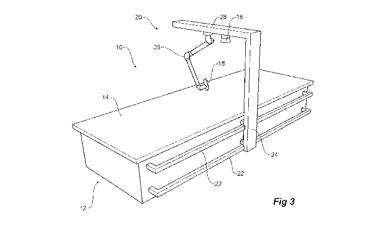
100311 Figure 3 shows a. perspective view of an alternative embodiment of the
apparatus shown in
Figure 1; and
1.00321 Figure 4 Shows a perspective view of an alternative embodiment of the
present invention.
100331 En the following description, like reference characters designate like
or corresponding parts
throughout the figures,
CA 02978789 2017-09-06
WO 2016/154664 PCT/AU2016/000112
DETAILED DESCRIPTION
[0034] Referring now to Figures I and 2, there is shown an apparatus 10 for
the treatment of soft tissue
injuries according to a first embodiment of the present invention. in this
embodiment, the apparatus 10
includes a bed 12 upon which a patient can. lie including an upper surface 14
to support the patient. For
the purposes of describing, this embodiment, it is assumed that the patient
has a soft tissue injury for
which the patient lies prone onthe surface .14. .Pillows and other forms of
support (not shown) may be
provided on the bed to hold the patient in a selected position.
100351 It will be appreciated that in other embodiments, the apparatus 10 need
not include a bed 12 but
may be used in association with a separate bed. In such embodiments, the
apparatus ID includes the
thermal imaging arrangement 16, the processing arrangement 30, the laser
treatment device 18 and the
guidance arrangement 24.
[0036] The apparatus 10 for treatment of a soft tissue injury has a thermal
imaging arrangement 16
which is used to scan a least a portion of the patient to provide a thermal
image and a laser treatment
device 18 to -provide a laser beam to provide appropriate treatment. The
thermal imaging arrangement 16
may be selected from infrared imaging cameras well known to. those skilled in
the art (cg Hug Max 76
(Flir Systems AB, Taby, Sweden), and Meditherm med2000fm IRIS (Meditherm Inc,
Fort Myers, FL,
United States of America)). The laser treatment device 18 may be. selected
from suitable laser probes,
which may emit one or more beams, well known to those skilled in the art.
Particular examples of suitable
cold laser probes include Maestro Laser Therapy Systern.(MediCom Inc, Prague,
Czech Republic), and
the Weberneedle Combi -Laser System (Weber Medical.Gmh115.Sohnreystrasse,
Germany)).
[00371 In this embodiment, both.the thermal imaging arrangement 16 and the
laser treatment device 1.8
are mounted on a single arm 20 which moves longitudinally on a rail.
arrangement 22 as shown by the
arrow 32 with respect to the length of the bed. The arm 20 has an essentially
vertical portion 24 and a
transverse portion 26 which extends over the bed. Both the thermal imaging
arrangement 16 and the laser
treatment device 18 are mounted on a housing 28 which is fixed to and moves
transversely on the
transverse portion. 26 of the arm 20 as shown by the arrow 34in Figure 2.
100381 The artn 20 can be moved longitudinally along the bed.22 and the
housing 28 moved transversely
to generally centre the thermal imaging device- over the region of the soft
tissue injury: Alternatively,
where the general region of soft tissue injury may be less clear then the
thermal imaging arrangement can
be scanned across and along the patient to find regions of thermal anomaly.
This scanning across and
along can be done automatically by the apparatus of the present invention or
by indirect control by an
operator who may define, a general area of interest such as the shoulders,
neck or lower back.
CA 02978789 2017-09-06
WO 2016/154664 PCT/AU2016/000112
9
100391 The vertical .portion.24 may be able to be.raised or lowered relative
to surface 14.
[00401 In other embodiments, the housing 28, or the individual laser treatment
device 18 or thermal
imaging arrangement 16, may be raised or lowered along vertical portion 2,4.
100411 It will be appreciated that in some embodiments, a thermal image as
such is not created, but
rather simply a dataset, which contains colour information associated with
location. This dataset can then
be processed directly to provide the guidance information for the guidance
arrangement 18.
[0042] In such an embodiment, the thermal imaging arrangement may be used, or
can be replaced by a
thermal scanner which does not necessarily generate an actual image, but
rather the dataset.
In such an embodiment, the apparatus 10 will include a thermal scanner for
scanning at least a portion of
the patient to provide a thermal dataset, a processing. arrangement to review
the thermal dataset to
determine a point or region of thermal anomaly on the patient; a laser
treatment device to provide a laser
beam, and a guidance arrangement for the laser treatment device to guide the
laser beam to the point or
region of thermal anomaly on the patient as. determined by the processing
arrangement, to thereby treat.
the patient.
[0043] A processing arrangement 30 takes information obtained by the thermal
imaging arrangement 16
and reviews the thermal image to determine a point(s) or region of thermal
anomaly on the patient. As
previously described, the single point on the patient preferably corresponds
or resides within a hot spot(s)
(ie a point of greatest surface temperature on. the region of body shown in
the thermographic image); in
a typical thermal image, this will be indicated by white colour and will
represent a surface temperaturethat is
no more than about 0.5 C warmer than the immediately surrounding region(s).
Such hot spot(s) will typically
be no more than about 5.0 cm in length or diameter, and more preferably, no
more than about 2.0 min in
length or diameter (eg a hot spot on the patient's body of about 1.0, 0.5 or
0.2 mm. in. length or diameter) A
second point for treatment may be identified by a surrounding and/or adjacent
spot(s) that is slightly cooler
(e.g < IT, preferably about 0.5T, cooler than the hot spot(s)) which, in a
typical thermal. image, will be
indicated by red colour.
100441 In the case where a thermal data set is generated in place of or as
well as an. actual image,
corresponding guidance information for use by the guidance arrangement is
generated from the dataset
Indicating where the different thermal regions are located.
100451 The information on the point(S) or region of thermal anomaly on the
patient is then transferred to
a guidance arrangement for the laser treatment device 18 to guide the laser
beam(s) to the point(s) or
region of thermal anomaly on the patient as. determined by the processing
arrangement, to thereby treat
the patient. In this illustrative embodiment, the guidance arrangement
operates to move the laser
CA 02978789 2017-09-06
WO 2016/154664 PCT/AU2016/000112
treatment device 18 to the region or point(s) by, inthis example, having arm
30 scanning or traversing
across and along the patient to the region or point(s) of thermal anomaly on
the patient. to direct the laser
beam(s) at a selected angle and/or a selected distance with respect to the
patient. In another embodiment,
the laser treatment device 1.8 maybe located generally stationary with respect
to the patient and an
associated directing and/or focusing mechanism operates to direct the laser
beam(s) to the region or
point(S) of thermal anomaly. In one particular example, the directing
mechanism may be a servo or
actuator arrangement operating to directly manipulate the laser treatment
device 18 to the correct
orientation so that the laser beam(s) is directed to the desired location.
Additionally or alternatively, the
guidance arrangement may comprise a servo or drive arrangement to lower the
vertical portion 24 relative
to the bed such that the laser treatment device I 8 is moved towards, and in
some embodiments to contact,
the skin of the patient. Inanother example; the laser treatment device 18 may
be statically mounted and
guidance of the laser beam(s) achieved by a mirror arrangement controllable to
direct the laser beam(s) to
the desired location. A timing device associated with the processing
arrangement 30 is used to determine
a suitable treatment time and then the laser beam(s) is switched on and off in
accordance with the
treatment time.
00461 It can be seen that by this arrangement, the process of determining a
treatment point(s) or region
and then applying cold laser treatment to that point(S) or region is
essentially automated.
100471 Figure 3 shows an end view of an alternative embodiment of the
apparatus shown in Figure 1
wherein the laser treatment device 18 is provided on an orientable arm 38
mountedto housing 28. The
particular orientable arm illustrated is a 4 axis robotic universal arm,
however those skilled in the art will
understand that alternative arms with a fewer or more axis points may also be
suitable,. Suitable universal
robotic arms include those available from Universal Robots AlS (Odense,
Denmark)..
[00481 Figure 4 shows a. perspective view of an alternative embodiment of the
present invention being an.
apparatus for the treatment of soft tissue injury In this embodiment, the
apparatus 50 includes a bed 52
(however, in other embodiments, the bed may not be included in the apparatus)
upon which .a patient can
lie including an upper surface 54-to support the patient. For the purposes of
describing this embodiment it
is assumed that the patient has. a. soft tissue injury tsar-which the patient
lies prone on the surface 54.
Pillows and other forms of support (not. shown) may be provided on the bed to
hold the patient in a
selected position.
100491 Over the bed 52 extend, from one side, two semicircular arms 56 and
5.8. The arm 56 is a
detection arm and carries a thermal imaging device 60 such.a.s an infrared
imaging camera. and the arm 58
is a treatment arm and carries a laser treatment device 62. Each of the arms.
56, 58 is supported
independently on a track 64 on the side of the bed 52 and each can move
longitudinally along the track 64
as shown by the arrows 68, 70, Each of the thermal imaging device 60 and the
laser treatment device 62
CA 02978789 2017-09-06
WO 2016/154664 PCT/AU2016/000112
11
can move alone the inner circumference of the semicircular arms as shown by
the arrows 66 so that they
can independently view and provide treatment to the patient.
1-00501 In use, the treatment arm 58 can be moved to one end of the track 64
so that the detection arm 56
can move along the patient to locate a point(s) or region to be treated and
then the detection arm 56 can. be
moved to one end of the track 64 so that the treatment arm 58 can. move along
the patient to enable the
laser beam to be directed to the desired point(s) or region.
[00511 From the thermal image or thermal dataset, a location for directing the
laser beam may be
determined. In one embodiment, a reference grid may be provided over the image
whichis shared with
the guidance arrangement, with the location of the determined point or region
of thermal anomaly
identified on the grid for use by the guidance arrangement.
100521 In alternative embodiments, the arms can be on opposite sides of the
apparatus and be constructed
so as not to interfere with the other arm traversing along the full length. of
a patient.
[00531 In some cases, it may be preferable to treat a patient While the
patient is standing up and the bed
can thenbe essentially vertical to provide a fixed surface .1-Or a patient to
be positioned against and both
the thermal imaging arrangement and the laser treatment deviee can be
appropriately positioned to scan
and treat a selected point(S) or region on the patient. The patient Should
remain still (ie in the same.
position) after scanning by the thermal imaging arrangement to optimise the
subsequent direction of the
laser beam .
100541 -Throughout the specification and the claims that follow, unless the
context requires otherwise, the
words "comprise" and "include" and variations such as "comprising" and
"including" will be understood
to imply the inclusion of a stated integer or group of integers, but not the
exclusion of any other integer or
group of integers.
[00551 The reference to any prior art in this specification is not, and should
not be taken as, an
acknowledgement of any form of suggestion that such prior art forms part of
the common general
knowledge.
[00561 it will be appreciated by those skilled in the art-that the invention
is not restricted in its use to the
particular application described. Neither is the present invention-
restricted, in its preferred embodiment
with regard to the particular elements and/or features described or depicted
herein. it will be appreciated
that the invention is not limited to the embodiment or embodiments disclosed,
but is capable of numerous
rearrangements, modifications and substitutions without. departing from the
scope of the invention as set
forth and defined by the following claims.