Note: Descriptions are shown in the official language in which they were submitted.
BIODEGRADABLE SOFT TISSUE FILLER
RELATED APPLICATIONS
This application claims priority to U.S. Provisional Patent Application No.
62/296146, filed
on February 17, 2017.
TECHNICAL FIELD
This patent application relates to soft tissue fillers.
BACKGROUND OF THE ART
Soft tissue fillers for connective and/or fatty soft tissues are used in both
medical and
cosmetic applications to correct various soft tissue defects or to enhance
appearance. Soft
tissue defects may be caused by various conditions such as soft tissue tumor
resection,
congenital abnormalities, trauma and aging.
Various compounds have been used as soft tissue fillers, including hyaluronic
acid,
collagen, as well as biosynthetic polymers, e.g., poly-L-lactic acid, calcium
hydroxylapatite,
and polymethylmethacrylate, in addition to implants, such as silicone-based
implants or
using a patients' own fat as a soft tissue filler. Non-limiting examples of
various injectable
dermal soft tissue fillers commercially available are. hyaluronic acid (e.g.
RestylaneTM and
Juvedermin; collagen (e.g. ZydermTm, ZyplastTm), as well as biosynthetic
polymers (e.g.
RadiesseTM (calcium hydroxylapatite); EllanséTM (Polycaprolactone); SculptraTM
(Poly-L-
lactic acid). These fillers are commonly injectable. These approaches have
various
disadvantages. Natural materials can have problems with sourcing and control
and
consistency of materials. Shaped implants must be pre-sized and do not have
the flexibility
provided by other fillers, such as e.g. injectable fillers. The use of a
patients' own tissue can
further complicate surgical procedures and may be associated with higher post-
operative
1
CA 2979429 2018-01-23
CA 02979429 2017-09-12
WO 2017/139868 PCT/CA2017/000030
complications. Additionally, where the soft tissue fillers are used to address
medical
concerns, cosmetic concerns are often not adequately addressed by these soft
tissue
fillers.
One area where poor cosmetic results are particularly problematic is treatment
following
repair of breast tissue defects arising as a result of breast cancer or the
treatment thereof.
Breast cancer is the most commonly diagnosed cancer and the second leading
cause of
cancer deaths in Canadian women. Approximately, 25,000 Canadian women were
diagnosed with breast cancer in 2015 (Canadian Cancer Society), accounting for
26% of all
new cancer cases. After several randomized controlled trials confirming the
safety and
efficacy of breast conserving surgery (BCS) with radiation, it has replaced
mastectomy as
the most common surgical procedure for breast cancer. Due to improved
treatments, most
breast cancer survivors are now expected to have a long life expectancy with a
good quality
of life. However, poor cosmesis and irregular soft tissue defects are commonly
observed in
patients that undergo BCS. While impairing the patients' aesthetic appearance,
soft tissue
defects are a main source of psychological distress, emphasizing the
increasing need for
correction/restoration techniques to address these cosmetic issues. Since
commercially-
available synthetic implants are fabricated in pre-determined sizes, they are
not suitable to
reconstruct partial breast deformities of varying sizes and are solely used
for full breast
reconstruction in post-mastectomy settings.
Several surgical techniques have been explored to address this unmet need. For
example,
there are a number of oncoplastic surgical techniques available such as local
tissue
rearrangement, contralateral breast reduction and flap procedures. However,
high rates of
complications and cost (long operative time and hospital stay) are drawbacks.
Local tissue
rearrangement, while demonstrating lower complications rates and more
cosmetically-
acceptable results, is not suitable for patients who have fatty breasts and
insufficient breast
tissue after resection. Furthermore, in order to achieve symmetry, up to 40%
of these
patients will require a contralateral breast reduction, consequently
increasing the overall
surgery time and complications for both breasts. Tissue rearrangement can also
complicate
revisions of positive surgical margins when needed. This may lead to the
decision of
performing a mastectomy due to the inability to ascertain the involved margins
accurately.
Pedicle flap procedures (e.g., latissimus dorsi flap) are recommended for
patients with
2
CA 02979429 2017-09-12
WO 2017/139868 PCT/CA2017/000030
small breasts or significant tissue loss. Advantages of this reconstruction
technique are the
lack of need for contralateral breast reduction as well as the surgeon's
ability to be more
aggressive with breast tissue resection without cosmetic detriment. However,
extensive
surgical dissection, long surgery and recovery time, donor site complications,
high costs as
well as aesthetic limitations due to potential differences in skin color and
texture are main
drawbacks of flap procedures.
Autologous fat transfer has also been used to fill the breast defect after
BCS. However, this
technique offers a temporary solution due to cytosteatonecrosis. More recent
reconstruction
methods include the use of adipose-derived regenerative cell (ADRC)-enriched
fat grafts
(Cytori Therapeutics Inc.), platelet-rich plasma (PRP) fat grafts, PRP gels or
dermal grafts
(Alloderm, LifeCell Corp.), which have shown improved cosmetic outcomes.
However,
these techniques are in their infancy.
There remains a need for improved and/or alternate methods for partial breast
reconstructions and soft tissue fillers.
BRIEF SUMMARY
The present disclosure provides a biodegradable soft tissue filler comprising
a porous
scaffold that is the reaction product of:
a) a divinyl oligomer component that comprises a carbonate-derived divinyl
oligomer that is
the reaction product of a lysine-derived diisocyanate, a vinyl coupling agent,
and a
polycarbonate and, optionally, an ether-derived divinyl oligomer, wherein the
ether-derived
divinyl oligomer is the reaction product of a lysine-derived diisocyanate, a
vinyl coupling
agent, and an ether; b) at least one anionic monomer; and c) at least one
hydrophobic
monomer. The molar ratio of (a) : (b+c) is between about 1:21 and about 1:30,
the soft
tissue filler has a porosity of > 75 %; and a compressive moduli of between
about 1kPa
and about 50kPa.
In one embodiment, the anionic monomer may be methacrylic acid and/or the
hydrophobic
monomer is methyl methacrylate.
3
CA 02979429 2017-09-12
WO 2017/139868 PCT/CA2017/000030
In one embodiment, component (a) is a carbonate-derived divinyl oligomer and
(a), (b) and
(c) are reacted in the presence of at least one porogen (d) and (a), (b) and
(c) combined
comprise between about 5 wt % and 20 wt % of the reaction mixture and (d)
comprises
between 80 and about 95 by wt % of the reaction mixture.
In another embodiment, the divinyl oligomer component comprises the carbonate-
derived
divinyl oligomer and the ether-derived divinyl oligomer. In this embodiment,
(a), (b) and (c)
may be reacted in the presence of at least one porogen (d) and (a), (b) and
(c) combined
comprise between about 5 wt % up to 25 wt % of the reaction mixture and (d)
comprises
between > 75 to about 95 by wt % of the reaction mixture. In one embodiment,
(d)
comprises between 80 and about 95 by wt % of the reaction mixture. The molar
ratio of
the carbonate-derived divinyl oligomer to ether-derived divinyl oligomer is
suitably between
about 1:100 to 50:50, preferably about 10:90.
In one embodiment, the soft tissue fillers as described above have a
compressive moduli of
between about 10 kPa and about 40 kPa.
In various embodiments, the soft tissue fillers as described above demonstrate
a swelling of
between about 100 % and about 300 %, 150% to 300%, and more preferably between
about 200 % and about 250 %.
The soft tissue fillers may include one or more additives selected from
antioxidants, cross-
linkers, plasticizers or nucleating agents.
The soft tissue fillers may be in the form of a pellet. The pellet may have a
dry volume of
between .1 mm3 and 100 mm3, preferably between 1 mm3 and 75 mm3, more
preferably 50-
60 mm3 + 10 mm3.
The soft tissue filler may further include one or more of a therapeutic agent,
a bioactive
agent and cells.
In one embodiment, the soft tissue filler is injectable.
In one embodiment, the soft tissue filler is a breast tissue filler.
4
CA 02979429 2017-09-12
WO 2017/139868 PCT/CA2017/000030
Also provided is a method of repairing a soft tissue defect in a patient in
need thereof
comprising implanting a soft tissue filler as described above at the site of
the soft tissue
defect. The method may further include hydrating the soft tissue filler prior
to implantation.
The soft tissue defect may be in connective and/or fatty and/or fibrous soft
tissue.
In one embodiment, the soft tissue defect is in the breast, and may be the
result of a
lumpectomy or breast tissue biopsy.
Also provided is a soft tissue filler comprising an amino-acid derived
biodegradable
polycarbonate-urethane scaffold having a porosity of between about 80% and
about 95%, a
compressive moduli of between about 1 kPa and about 50 kPa, a swelling
capacity of
between about 100 % and about 300 %, and a dry volume of 50 mm3 25 mm3.
BRIEF DESCRIPTION OF THE DRAWINGS
These and other features of the preferred embodiments of the invention will
become more
apparent in the following detailed description in which reference is made to
the appended
drawings wherein:
Figure 1 shows a synthesis scheme of ether-based divinyl oligomer (E-DVO) in
the
presence of Dibutyltin Dilaurate (DBDL) catalyst.
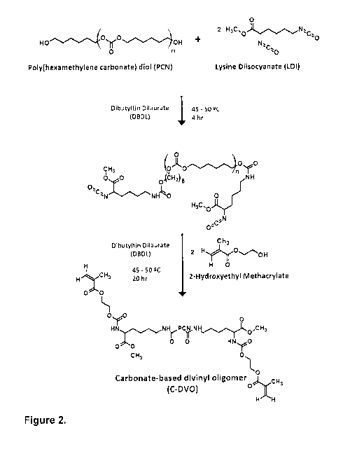
Figure 2 shows a synthesis scheme of carbonate-based divinyl oligomer (C-DVO)
in the
presence of Dibutyltin Dilaurate (DBDL) catalyst.
Figure 3 shows kinetics of the (A) C-DVO synthesis and (B) E-DVO synthesis.
Isocyanate
conversion as a function of time is represented. (A) Lysine diisocyanate (LDI)
was added to
the polycarbonate (PCN) solution and reacted in the presence of DBDL for 4 hr.
2-
hydroxyethyl methacrylate (HEMA) was added 4 hr after the start of the
reaction. (B)
Polyethylene glycol (PEG) solution was added to the LDI solution in a dropwise
manner
over 0,5 hr. HEMA was added 1 hour after the start of the reaction. Standard
deviation bars
(n=4).
CA 02979429 2017-09-12
WO 2017/139868 PCT/CA2017/000030
Figure 4 shows mechanical properties of amino-acid derived biodegradable
polycarbonate-urethane porous scaffolds following compression. Effect of E-DVO
content
on the compressive modulus of amino-acid derived biodegradable polycarbonate-
urethane
scaffolds prepared in the presence of 75 wt% (gray) and 80 wt% total porogen
concentrations is shown. Standard deviation bars (n=9).*Statistical decrease
in the
presence of more porogen for scaffolds with 0 mol% E-DVO (p<0.05).
tStatistical decrease
with respect to scaffolds with the next lowest E-DVO concentration in the
presence of 75
wt% porogen (p<0.05). tStatistical decrease with respect to scaffolds with the
next lowest
E-DVO concentration in the presence of 80 wt% porogen (p<0.05).
Figure 5 shows degree of swelling of amino-acid derived biodegradable
polycarbonate-
urethane porous scaffolds. Effect of E-DVO content on the swelling of amino-
acid derived
biodegradable polycarbonate-urethane scaffolds prepared in the presence of 75
wt% (gray)
and 80 wt% (white) total porogen concentration is shown. Standard deviation
bars (n=6).
*Statistical increase in the presence of more porogen for scaffolds with the
same E-DVO
content (p<0.05). tStatistical increase with respect to scaffolds with the
next lowest E-DVO
concentration in the presence of 75 wt% porogen (p<0.05). tStatistical
increase with
respect to scaffolds with the next lowest E-DVO concentration in the presence
of 80 wt%
porogen (p<0.05)..
Figure 6 shows pore morphology of the porous amino-acid derived biodegradable
polycarbonate-urethane scaffolds. Scanning electron micrographs of AAd-DPCU80-
E0
(formulation A, a-c) and AAd-DPCU80-E10 (formulation B, d-f) were taken at 25x
(a,d),
250x (b,e) and 2500x (c,f) original magnification.
Figure 7 shows surgery and implant site. A representative image of a pig torso
at 6 weeks
immediately following the first set of mastectomies is shown. Black arrows:
mastectomy
sites.
Figure 8 shows cell and tissue distribution in explanted breast tissue
following H&E
histological staining. Representative histology images of breast tissue
containing
formulation A (a,d,g,j), formulation B (b,e,h,k) or no amino-acid derived
biodegradable
polycarbonate-urethane (control; c,f,i,l) after 6 (a-c), 12 (d-f), 24 (g-i)
and 36 (j-l) weeks in
6
CA 02979429 2017-09-12
WO 2017/139868 PCT/CA2017/000030
vivo are shown. Arrows indicate scaffold pieces. Asterisks indicate areas high
in cells and
extracellular matrix. . Scale bars represent 500 pm.
Figure 9 shows cell and tissue distribution in explanted breast tissue
following Masson's
trichrome histological staining. Representative histology images containing
formulation A
(a,d,g,j), formulation B (b,e,h,k) or no amino-acid derived biodegradable
polycarbonate-
urethane (control; c,f,i,l) after 6 (a-c), 12 (d-f), 24 (g-i) and 36 (j-l)
weeks in vivo are shown.
Arrows indicate scaffold pieces. Asterisks indicate areas high in cells and
extracellular
matrix. Scale bars represent 500 pm.
Figure 10 angiogenesis and CD31 expression in explanted breast tissue.
Representative
immunohistochemistry images containing formulation A (a,d,g,j), formulation B
(b,e,h,k) or
no amino-acid derived biodegradable polycarbonate-urethane (control; c,f,i,l)
after 6 (a-c),
12 (d-f), 24 (g-i) and 36 (j-l) weeks in vivo are shown. Arrows indicate
scaffold pieces.
Asterisks indicate areas high in cells and extracellular matrix. Dark punctate
spots indicate
positive staining for CD31.. Scale bars represent 500 pm.
Figure 11 shows quantification of CD31 expression in vivo. The number of CD31-
positively
stained structures in explanted breast tissue containing formulation A (gray),
formulation B
(white) or no amino-acid derived biodegradable polycarbonate-urethane
(control, hashed)
was determined per image at different time-points. Standard error bars (n=4-
6). *Statistical
decrease with respect to the native breast tissue pre-surgery control
(p<0.05).
Figure 12 shows porous amino-acid derived biodegradable polycarbonate-urethane
scaffold degradation in vivo. The average size of scaffold fragments remaining
at different
time-points post-implantation was quantified for both formulation A (AAd-
DPCU80-E0, gray)
and formulation B (AAd-DPCU80-E10, white). Standard error bars (n=5-6).
*Statistical
decrease at 24 weeks when compared to 6 weeks for formulation A (p<0.05).
tStatistical
decrease at 24 weeks when compared to 6 weeks for formulation B (p<0.05).
Figure 13 shows ultrasound examination of porcine breast. Representative
ultrasound
images of the original porcine breast, prior to lumpectomy and amino-acid
derived
biodegradable polycarbonate-urethane filling are shown (a-c). Representative
images of the
breasts following lumpectomy and subsequent filling with amino-acid derived
biodegradable
7
CA 02979429 2017-09-12
WO 2017/139868 PCT/CA2017/000030
polycarbonate-urethane (formulation A (d,g,j,m) or formulation B (e,h,k,n) or
no amino-acid
derived biodegradable polycarbonate-urethane filling (control; f,i,l,o) at 6
(d-f), 12 (g-i), 24 (j-
l) and 36 weeks (m-o) are depicted.
Figure 14 shows cell and tissue distribution in explanted breast tissue
following H&E
histological staining. Representative histology images of breast tissue
containing amino-
acid derived biodegradable polycarbonate-urethane porous scaffolds of
formulation C-
DVO:MAA:MMA 1 :5 :15, in the form of 1 cm diameter by x 1 cm height (i.e. .8
cm3 or 785 5
mm3)obtained in the presence of 75 wt% porogen after 6, 12, 24 and 36 weeks in
vivo are
shown. Black arrows indicate scaffold pieces. White arrows indicate new
vascularization.
Figure 15 shows a comparison of histological staining (H&E) images comparing
two
scaffolds where one A) is made from a polycarbonate DVO of the nature
described in this
submission, MAA and MMA in a ratio of 1:5:15 respectively, with a porosity of
75%, with
size of 785 mm3; and B) is made of the same 3 monomers but in a ratio of
1:5.5:15.5, with a
porosity of 80%, with a size of approx. 50 mm3. The images compared H&E
stained
histology sections for porcine breast explants at 36 weeks. Black arrows
indicate empty
pores. White arrows indicate scaffold pieces surrounded by tissue.
DETAILED DESCRIPTION
According to medical dictionaries, soft tissues are any non-calcified tissues
in the body. In
one embodiment, soft tissues refer to connective and/or fatty and/or fibrous
soft tissues. In
one embodiment, soft tissues refer in particular to sub-epidermal fatty and/or
fibrous
tissues. Suitably, the soft tissue fillers described herein are used as
fillers for soft tissue that
do not form part of a vital organ (heart, brain, lungs, kidneys, liver etc.) .
The present disclosure provides amino-acid derived biodegradable polycarbonate-
urethane
formulations synthesized as soft tissue fillers. In one embodiment, their use
is not
particularly restricted, and may include, without being limited to, the repair
of any soft
tissue. Further, the soft tissue fillers described herein may be used for the
correction of
various soft tissue defects caused by various medical conditions such as soft
tissue tumor
8
CA 02979429 2017-09-12
WO 2017/139868 PCT/CA2017/000030
resection, congenital abnormalities, trauma and aging. The soft tissue fillers
may also be
used for cosmetic purposes, such as for the enhancement of facial features,
such as
cheeks or lips.
Soft tissue tumor resection is a common cause of soft tissue defects and, in
one
embodiment, the soft tissue fillers as described herein may be used for the
repair of any
soft tissue defect caused by tumour resection with or without a portion of the
surrounding
tissue. Soft tissue tumor resection includes treatment related to melanoma,
where a skin
graft may be used on top of a soft tissue filler described herein. Soft tissue
defects may
also be caused by biopsies. In one embodiment, the defect may be primarily to
or in sub-
epidermal fatty and/or fibrous tissue.
In one embodiment, soft tissue fillers as described herein may be used the
repair of breast
tissue defects following lumpectomy (BCS) or biopsies related to breast
cancer.
In one embodiment, the swelling and mechanical properties of the soft tissue
fillers are of
particular importance and the amino-acid derived biodegradable polycarbonate-
urethane
fillers were synthesized with swelling and mechanical properties dependent on
both the soft
segment composition, the porogen content and the size of the soft tissue
filler.
In one embodiment, the reaction product has a compressive moduli of at least
about 1kPa
but less than about 50 kPa.
In one embodiment, the fillers are highly porous (>75%, ¨ 80% to 95%, 80-90%,
or 80-85%
by volume)
Amino-acid derived biodegradable polycarbonate-urethane formulations were
fabricated
with mechanical properties comparable to that of native healthy breast tissue,
which were
capable of preserving breast shape/volume upon implantation while eliciting
minimal foreign
body reaction and integrating well within the host tissue. Due to the
segmented nature of
PUS, amino-acid derived biodegradable polycarbonate-urethane porous fillers
can be
fabricated with desirable properties, customized for this specific
application.
The soft tissue fillers were synthesized by reacting macromer divinyl
oligomers with a
hydrophobic monomer, an anionic monomer in admixture with one or more
porogens.
9
CA 02979429 2017-09-12
WO 2017/139868 PCT/CA2017/000030
In one embodiment, the reaction product is a polar non-ionic hydrophobic amino-
acid
derived degradable polycarbonate-urethane (AAd-DPCU). AAd-DPCUs are synthetic
block
copolymers characterized by the presence of a urethane linkage created in a
condensation
reaction (step-growth polymerization). Generic polyurethanes can be linear,
branched, or
cross-linked while AAd-DPCUs are specifically cross-linked. AAd-DPCUs are
copolymers
and contain two repeating segments; a hard segment of the polyurethane (the
amino-acid
derived isocyanate), which endows the material with mechanical strength and a
soft
segment (the polyol), which provides flexibility. The soft and hard segments
can
microphase separate to form soft and hard phases; these phases provide the
polymer with
both flexibility and strength. The combination of segments manifests itself in
the bulk
material composition and surface microstructure. The differences in polarity
of the hard and
soft segments affect the hydrophilic-hydrophobic balance of the material.
Furthermore, the
soft segments are mobile and will optimize their location to minimize the free
energy at the
surface of the material. The copolymer structure and the composition and the
ratio of its
monomers provide a AAd-DPCUs with its unique in vivo properties and
biocompatibility.
In one embodiment, the hard segment is derived from a lysine derived
diisocyanate and
vinyl monomers.
In one embodiment, the isocyanate is not particularly restricted. In one
embodiment, the
isocyanate has a molecular weight between about 100 and about 1000. In one
embodiment, the isocyanate component is one or more of a linear diisocyanate
e.g. L-
Lysine ethyl ester diisocyanate; Suitable isocyanates can be prepared by
methods known
to those of skill in the art and are also available from commercial sources,
including, for
example, ABI Chem, ABCR, A Chemtek, Akos Building Blocks, Alfa Aesar, Aurora
Fine
Chemicals, Bayer, CHEMOS GmbH, Chem Reagents, Chemtura, FCH Group, Fisher
Scientific, Oakwood Chemical, Perstrop, Polysciences, Inc, Sigma-Aldrich,
Suzhou
Rovathin and SynQuest.
In one embodiment, the diisocyanate is derived from lysine. In one embodiment,
the
diisocyanate is lysine diisocyanate (LDI).
In one embodiment, the vinyl coupling agent is not particularly restricted and
may be any
compound comprising a single pendant hydroxyl or primary or secondary amine
group that
CA 02979429 2017-09-12
WO 2017/139868 PCT/CA2017/000030
can react with the isocyanate group of the diisocyanate. In one embodiment,
the vinyl
coupling agent has a molecular weight between about 50 and about 500. The
vinyl
coupling agent may be, but is not limited to, a vinyl alcohol, an alkyl amine
with vinyl
groups, a vinyl amine, hydroxypropyl (meth)acrylate, 2,3-dihydroxypropyl
(meth)acrylate,
1,4-butanediol monoacrylate, (poly)ethylene glycol mono(meth)acrylate, 3-
aminopropyl
vinyl ether, and 2-hydroxyethyl methacrylate (HEMA). In one embodiment, the
vinyl
coupling agent is 2-hydroxyethyl methacrylate (HEMA).
In one embodiment, the soft segment is derived from a polyol. In one
embodiment, the
polyol is an oligomeric macromolecule containing hydroxyl or amine end groups
with low
glass transition temperatures. In one embodiment, the polyol comprises a
polyether or
polycarbonate backbone.
In various embodiments, the soft segment may be derived frompolyethylene
oxide;
polypropylene oxide; polytetramethylene oxide; polyisobutylene;
polybutadienes;
polyesters; polyethylene adipate; polyanhydrides, polyam ides,
polytetramethylene adipate;
polycaprolactone; polydimethylsiloxane; and polycarbonates.
In one embodiment, the soft segment is derived from a polycarbonate.
In one embodiment, the soft tissue filler is a scaffold comprising the
reaction product of a
carbonate-based divinyl oligomer (C-DVO), an ether-based divinyl oligomer (E-
DVO), at
least one anionic monomer and at least one hydrophobic monomer.
In one embodiment, the C-DVO is a reaction product of poly(hexamethylene
carbonate) diol
(PCN), LDI, and HEMA. In one embodiment, the E-DVO is a reaction product of
PEG, LDI,
and HEMA.
In one embodiment, the anionic component is not particularly restricted. In
one
embodiment, the anionic component has a molecular weight between about 50 and
about
1000.
In one embodiment, the anionic component is a vinyl monomer with mono acid
function
such as methacrylic acid, vinyl phosphoric acid or the like; vinyl monomers
with di-acids
11
CA 02979429 2017-09-12
WO 2017/139868 PCT/CA2017/000030
such as itaconic acid, maleic acid or the like; or vinyl monomers with tri-
acids such as
tricarballylic acid, tricarboxylic acid or the like.
In one embodiment, the anionic component comprises a methacrylic acid
derivative; 2-
(methacryloyloxy)ethyl phosphate; styrene sulphonic acid;
2(methacryloyloxy)ethyl
succinate, {3-(methacryloylamino)propylitrimethyl ammonium chloride; or 2-
(methacryloyloxy)ethyl]trimethylammonium methyl chloride. In one embodiment,
the
methacrylic acid derivative is an amino-acid derivative. In one embodiment,
the anionic
component is methacrylic acid.
In one embodiment, the hydrophobic component is not particularly restricted.
In one
embodiment, the hydrophobic component has a molecular weight between about 50
and
about 1000.
In one embodiment, the component is considered to be hydrophobic if when its
constituent
monomers are polymerized on their own, in the absence of other monomers or
additives, it
yields an advancing water contact angle measure of greater than about 50, 55,
60 or 65
degrees. In one embodiment, the advancing water contact angle measure is
greater than
about 65 degrees. Methods of measuring water contact angle are known to those
of skill in
the art.
In one embodiment, the hydrophobic compound is a non-aromatic.
In one embodiment, the hydrophobic compound does not include a pendant halogen
group,
e.g. fluorine.
In one embodiment, the hydrophobic component is an alkyl methacrylate, wherein
the alkyl
chain is linear or branched, saturated or unsaturated, and wherein the number
of carbons is
less than 12. In one embodiment, the alkyl chain is non-aromatic. In one
embodiment, the
hydrophobic component is methyl, propyl, butyl, iso-butyl or t-butyl
methacrylate. In one
embodiment, the hydrophobic compound comprises an aliphatic alkyl side chain.
In one
embodiment, the hydrophobic component is methyl methacrylate.
In one embodiment, the scaffold is a porous scaffold. While in one embodiment,
a single
porogen may be used, in other embodiments, two or more porogens may be used to
impart
12
CA 02979429 2017-09-12
WO 2017/139868 PCT/CA2017/000030
both macro-porosity and micro-porosity to the soft tissue fillers. In one
embodiment,
porogens used are not particularly restricted. In one embodiment, the porogen
system is
suitably salt particles and PEG. In one embodiment, suitable salt particles
are sodium
bicarbonate having an average particle size between about 50 and 450 pm are
used and
PEG of about 600 da to 4000 but preferably 600 to 2000 is used.
There are three main processes used to generate porosity in the scaffolds (1)
processes
using porogens, (2) processes using solid free-form or rapid prototyping
technologies and
(3) techniques using woven or non-woven fibers. In the first category, solid
materials either
in solids or dissolved in solvents, are incorporated with porogens, which
could be gases
such as carbon dioxide, liquids such as water, polyethylene glycol or the
like, or solids
such as paraffin, salts, sugar and others. Porogens are removed by
sublimation,
evaporation, dissolution or melting to leave behind a porous structure in the
scaffold.
Examples include solvent casting and particulate leaching, gas foaming, freeze-
drying and
phase separation.
Porous structures can also be manufactured by sequential delivery of material
and/or
energy needed to bond the materials to preset points in space. Some solid free-
form
fabrication technologies include laser sintering, stereolithography and 3D
printing, and
depend on precise delivery of light or heat energy in a scanner system to
points of space in
the material bed so as to bond or crosslink the materials to give solid
structures in an
otherwise soluble bed of materials.
In the third category, woven and non-woven fiber structures can be piled
together and
bonded using thermal energy or adhesives to give a porous meshwork using
techniques
such as fiber bonding, or fibers can be generated by the electrospinning
technique.
The PU scaffolds suitably have a porosity of > 75 %, ?. 80%, between 80 and
about 95 % or
between 80 and 85%. In one embodiment, the PU scaffolds of the soft tissue
filler
described herein are synthesized in the presence of > 75% by weight of
porogen, 80 %,
80-95%, still more preferably 80-85% by volume wt% by weight of porogen, by
weight of
the reaction products, to yield scaffolds having these porosities.
13
CA 02979429 2017-09-12
WO 2017/139868 PCT/CA2017/000030
In various embodiments, the PU scaffolds have a volume of between 0.1 mm3 and
100
mm3, between 1 mm3 and 75 mm3. In one embodiment, 50 mm3 25 mm3. In one
embodiment, 50 mm3 20 mm3. In one embodiment 50 mm3 10 mm3.
In one
embodiment, 50 mm3 5 mm3.
The scaffolds or particulates are biodegradable. In one embodiment the
scaffold or
particulate degrades more than 80% in less than 3 months, less than 6 months,
in less than
9 months, in less than 1 yr, or in less than 2 yrs.
Amino-acid derived biodegradable polycarbonate-urethane scaffolds were
synthesized by
reacting two types of divinyl oligomers (DV0s), a carbonate-based DVO (C-DVO)
and an
ether-based DVO (E-DVO) with methacrylate (MMA), methacrylic acid (MAA)
monomers.
Kinetic studies conducted on both the C-DVO and the E-DVO (Figure 3)
demonstrated the
full consumption (-100%) of the isocyanate groups within 24 hours. Both the C-
DVO and E-
DVO had similar hard segment chemistry through the incorporation of LDI and
HEMA. LDI
was chosen to render the polymer more biocompatible. Unlike traditional PUs,
which
produce toxic diamines upon degradation, LDI-based PUs' main degradation
product is
lysine, a naturally occurring amino acid that is abundant in biological
systems. HEMA, the
second component of the PU hard segment confers crosslinking functionality to
the DVO
and thus the potential for improved PU mechanical properties. The ester
functionality within
HEMA also rendered the scaffold more susceptible to hydrolytic degradation.
PCN and
PEG constituted the soft segment of the C-DVO and E-DVO, respectively. PUs
synthesized
with polycarbonate soft segments possess a greater tensile strength and
elastic modulus
when compared to ether-based PUs and, while demonstrating a greater oxidative
stability
when compared to poly-ether urethanes (PEUs), are susceptible to hydrolytic
degradation.
PUs with a polyether soft segment have a lower elastic modulus when compared
to PUs
with a PCN soft segment due to the greater flexibility of ether linkages.
While demonstrating
a greater hydrolytic stability, PEUs are more prone to oxidative degradation
when
compared to PCNUs. Incorporating PEG within PCNUs results in greater mass loss
due to
hydrolytic degradation with increasing PEG content, which can be attributed to
the
hydrophilic nature of PEG, which increased PU's water absorption and
accelerated the
degradation of the polymer's hydrolysable linkages. MAA and MMA methacrylate
monomers provide favorable non-specific cell adhesive chemistry.
14
CA 02979429 2017-09-12
WO 2017/139868 PCT/CA2017/000030
Amino-acid derived biodegradable polycarbonate-urethane scaffolds were
synthesized in
the presence of different porogen contents (75 wt% or 80 wt% or more, however
80% is
preferred). For formulations with the same monomer composition, increasing the
porogen
content resulted in amino-acid derived biodegradable polycarbonate-urethane
scaffolds
with a greater porosity, increased polymer swelling and a lower compressive
modulus.
In various embodiments, the scaffold has a modulus of at least 1 kPa but less
than 50 kPa.
In various embodiments, the scaffold has a modulus of at least 10 kPa and less
than 40
kPa, less than 30 kPa, or less than 20 kPa,. .
Increasing the concentration of E-DVO or the salt porogen was shown to
decrease the
compressive modulus and increase polymer swelling, resulting in the
development of PU
filler formulations with properties advantageous for use as soft tissue
fillers and, in
particular, soft tissue fillers for the repair of breast defects. Formulations
were synthesized
that exhibited a moderate degree of swelling, possessed mechanical properties
comparable
to native human breast tissue and were successfully used as soft tissue
fillers for partial
breast reconstruction in a porcine model. While capable of maintaining the
shape, volume
and natural stiffness of the breast tissue, these PU fillers were shown to
support cell, tissue
infiltration and neovascularization throughout their structure. Furthermore,
they were
observed to integrate well within the host tissue and to not elicit foreign
body giant cell and
fibrous capsule formation, suggesting the absence of chronic inflammation and
presence of
wound repair. Amino-acid derived biodegradable polycarbonate-urethane fillers
may have
one or more of the following advantages over known solutions for soft tissue
filler
applications such as partial breast reconstruction: requiring no biological
processing,
minimal surgical dissection, no prior knowledge of the defect dimension/shape
and a short
surgery time (<10 min).
Other attributes of amino-acid derived biodegradable polycarbonate-urethane
fillers as
described therein include customizability and versatility. Specifically, these
PU fillers can
be used for varying defect sizes and a prior knowledge of the exact defect
dimension/shape
is not required. Furthermore, unlike tissue rearrangement techniques, amino-
acid derived
biodegradable polycarbonate-urethane fillers are not dependent on sufficient
tissue being
available for rearrangement to fix the defect. In the context of breast
reconstruction, this can
eliminate the need for contralateral breast reduction to obtain symmetry which
is commonly
CA 02979429 2017-09-12
WO 2017/139868 PCT/CA2017/000030
performed in conjunction with tissue rearrangement procedures. Amino-acid
derived
biodegradable polycarbonate-urethane fillers may also allow surgical
oncologists to be
more aggressive in removal of tissue and allow the resection of wider surgical
margins with
less concerns regarding the cosmetic outcomes, potentially reducing the
incidence of
positive margins and the need for additional surgery. Lastly, the aesthetic
attributes of these
degradable fillers were evident in their ability in the in vivo model to
preserve breast
shape/volume while maintaining natural breast stiffness throughout the 36 week
implantation period. .
Protein adsorption occurs immediately following the implantation of a
biomaterial, or contact
of body fluids such as blood to a biomaterial. This adsorbed protein layer is
composed of
bioactive agents that can greatly influence the behavior of cells or other
body fluid elements
involved in the inflammatory, immune, and foreign body responses. While the
adsorbed
protein layer interacts with the surface of the biomaterial, the bulk of the
material does not
interface with biological tissue and so may not be a major determinant in
regulating protein
adsorption and the subsequent inflammatory response. For this reason,
biomaterials, and
particularly polymeric biomaterials, can be modified in the bulk phase by the
addition of
components that can provide stability or mechanical integrity to the material
without
influencing the implant's interactions with the proteins, cells, and tissue.
For polymeric
materials these additives include, but are not limited to, antioxidants,
fillers, cross-linkers,
plasticizers, nucleating agents, and pigments. Accordingly, in one embodiment,
the soft
tissue filler further includes one or more additives, which in one embodiment,
may be
selected from antioxidants, fillers, cross-linkers, plasticizers, nucleating
agents, and
pigments.
In various embodiments, these additives may be present in an amount of less
than 50, less
than 40, less than 30, less than 20, less than 10, less than 5 or less than 1
percent by
weight of the polymeric material.
In one embodiment, a therapeutic or bioactive agent may be added to the
material in the
bulk phase or may be impregnated or coated onto the scaffold after synthesis.
16
CA 02979429 2017-09-12
WO 2017/139868 PCT/CA2017/000030
In one embodiment, the therapeutic agent or bioactive agent may be present in
any amount
of less than 50, less than 40, less than 30, less than 20, less than 10, less
than 5 or less
than 1 percent by weight of the polymeric material.
Such therapeutics or bioactive may include, but are not limited to growth
factors, peptides,
antibodies, enzymes, platelets, glycoproteins, hormones, glycosaminoglycans,
nucleic
acids, analgesics, cytokines and combinations thereof.
In one embodiment, the soft tissue filler is used in combination with a
cytokine or growth
factor.
In one embodiment, the soft tissue fillers as disclosed herein are used in
combination with
cells, including, but not limited to, stem cells with are implanted with the
soft tissue filler at
the time of tissue repair.
The soft tissue filler as disclosed herein may further be used in combination
with both a
therapeutic agent and/or bioactive and one or more cells, including, but not
limited to stem
cells.
In one embodiment, the soft tissue filler scaffold is not coated, impregnated
and/or
otherwise used with one of the additional components described above.
In one aspect, there are no restrictions on the manner in which the reagents
are added to
each other to form soft tissue fillers disclosed herein, the temperature,
pressure or
atmosphere under which the materials are synthesized from the monomer and
macromers
or the use of the catalysts in the reaction.
In other embodiments, there is provided methods of manufacturing soft tissue
fillers as
described herein comprising combining: a) a C-DVO and, optionally, an E-DVO;
b) an
anionic monomer; c) a hydrophobic monomer; and d) a porogen, wherein (a), (b)
and (c)
combined comprise between about 5 wt% and up to 25 wt% of the reaction mixture
and (d)
comprises between 75 wt% and about 95 wt% of the reaction mixture. In one
embodiment,
the method may further include preparing the macromer C-DVO and/or E-DVO. The
method can further include curing the reaction product. The method can further
include
leaching the porogen from the reaction product. The method can further include
drying the
17
CA 02979429 2017-09-12
WO 2017/139868 PCT/CA2017/000030
porous scaffold that results from the leaching step. Methods of forming beads
or pellets
may include emulsion polymerization, precipitation methods from a solution of
organic
polymer, of freezing and pulverizing frozen scaffolds.
The ratio of divinyl oligomers to monomers (a: b+c) is at least 1:21 and less
than 1:60, less
than 1:50, less than 1:40 and preferably less than 1:30. Data from the pilot
study showed
that a ratio (a:b+c) lower than 1:21, (i.e. 1:20) resulted in scaffolds with a
stiffness higher
than the one of normal pig breast tissue and was characterised by a slow
degradation rate
and significantly different properties from the ratio of (a:b+c) of 1:21. In
one embodiment,
the ratio (a:b+c) is at least 1:21 .5. In one embodiment, the ratio (a:b+c)
is at least 1:21
.2. In one embodiment, the ratio (a:b+c) is at least 1.21 .1. In various
embodiments, the
ratio (a:b+c) is at least 1:21, at least 1:21.1, at least 1:21.2, or at least
1:21.5. Furthermore
it is known in the art that the mechanical properties of polyurethane are
dependent on the
soft segment composition. As the amount of oligomer soft segments decreases in
the
formulation, e.g., a ratio (a:b+c) higher than 1:30, the mechanical properties
of the
polyurethane go up resulting in higher modulus materials which would yield
outcomes that
are not compliant with the natural soft tissue environment being treated.
In one embodiment, the method may further include preparing the macromer C-DVO
and/or
E-DVO. The method can further include curing the reaction product. The method
may
further including freezing the reaction product. In one embodiment, the
reaction product is
cured and then pulverized to form porous particles..
AAd-DPCU can be synthesized by generating a divinyl oligomer by reaction of a
diisocyanate with an oligomeric diol and mono-vinyl monomers with pendent
hydroxyl or
amine groups. The latter is then light or heat polymerized via a free radical
polymerization
with anionic and/or hydrophobic vinyl monomers in the presence of initiators
with light or
heat activating initiators. If porogens were included in the mixture, these
are then extracted.
In the presence of a catalyst, polar non-ionic macromonomer polyurethanes are
created in
a nucleophilic addition reaction between an isocyanate and molecules
containing hydroxyl
(a polyol) or amine functional groups to create a urethane or carbamate
linkage.
18
CA 02979429 2017-09-12
WO 2017/139868 PCT/CA2017/000030
The synthesis of macromonomer polyurethane can be completed in one or two
steps. The
one-step process involves a simultaneous reaction of the isocyanate, polyol,
and vinyl
coupling agent. In the two-step prepolymer process, an excess of diisocyanate
is reacted
with the polyol to form NCO-terminated prepolymers with isocyanate
functionality as an
intermediate; this intermediate is then reacted with the vinyl coupling agent
to create the
final polar non-ionic macromonomer polyurethane. The separation of the process
into two
steps enables a greater degree of control over the polar non-ionic
macromonomer
polyurethane structure and consequently, its properties.
The synthesis of macromonomer polyurethanes is also dependent upon a catalyst,
the
selection of which depends on the final profile of the polyurethane (e.g. gel,
foam) and its
curing requirements. The two types of catalysts that can be used are metal
complexes and
amine compounds. DBDL may suitably be used in preparing the macromonomer
polyurethanes used in the soft tissue fillers described herein.
Synthesis processes will generally employ initiators and/or retarders and or
terminators. In
one embodiment, the initiator used is not particularly restricted and will be
within the
purview of a person skilled in the art. Suitable initiators can be selected
e.g. from diacyl
peroxides, peroxy esters, dialkyl peroxides, dialkyl peroxydicarbonates, tert-
alkylhydroperoxides, and ketone peroxides. Suitable free radical initiators
include e.g.
dibenzoyl peroxide, diisobutyrul peroxide, t-butyl peracetate, dicumyl
peroxide, di-sec-
butyperoxydicarbonate, methyl ethyl ketone peroxide, benzoyl peroxide (BPO)
(available
through Aldrich Chemical Co., Milwaukee, Wis.) and 1,1'-
azobis(cyclohexanecarbonitrile).
Light curing systems may be used to polymerize the vinyl resins, including but
not limited to
photopolymerizations initiated with camphorquinone (CO, initiator) and 2-
(dimethylamino)
ethyl methacrylate (DMAEM, co-initiator).
Parameter variations provide the controllable aspect in polyurethane
synthesis, which can
include modifications to the reacting molecules (e.g. chemical composition,
molecular
weight, symmetry), the processing conditions (e.g. introduction of water,
removal of carbon
dioxide, active hydrogens), or addition of additives.
Various methods can be employed in preparing scaffolds according to
embodiments of the
present invention, including nanofiber-self assembly, textile technologies,
solvent casting &
19
CA 02979429 2017-09-12
WO 2017/139868 PCT/CA2017/000030
particulate leaching (SCPL), gas foaming, emulsification/freeze-drying,
thermally induced
phase separation (TIPS), electrospinning and CAD/CAM technologies, each of
which is
briefly described below.
Nanofiber Self-Assembly: Molecular self-assembly enables the synthesis of
biomaterials
with properties similar in scale and chemistry to that of the natural in vivo
extracellular
matrix (ECM). Moreover, these hydrogel scaffolds have shown superiority in in
vivo
toxicology and biocompatibility compared to traditional macroscaffolds and
animal-derived
materials.
Textile technologies: These techniques include all the approaches that have
been
successfully employed for the preparation of non-woven meshes of different
polymers. In
particular, non-woven polyglycolide structures have been tested for tissue
engineering
applications: such fibrous structures have been found useful to grow different
types of cells.
Solvent Casting & Particulate Leaching (SCPL): This approach allows for the
preparation of
porous structures with regular porosity, but with a limited thickness. First,
the polymer is
dissolved into a suitable organic solvent, then the solution is cast into a
mold filled with
porogen particles. Such porogen can be an inorganic salt like sodium chloride,
crystals of
saccharose, gelatin spheres or paraffin spheres. The size of the porogen
particles will
affect the size of the scaffold pores, while the polymer to porogen ratio is
directly correlated
to the amount of porosity of the final structure. After the polymer solution
has been cast the
solvent is allowed to fully evaporate, then the composite structure in the
mold is immersed
in a bath of a liquid suitable for dissolving the porogen: water in the case
of sodium
chloride, saccharose and gelatin or an aliphatic solvent like hexane for use
with paraffin.
Once the porogen has been fully dissolved, a porous structure is obtained.
Gas Foaming: To overcome the need to use organic solvents and solid porogens,
a
technique using gas as a porogen has been developed. First, disc-shaped
structures made
of the desired polymer are prepared by means of compression molding using a
heated
mold. The discs are then placed in a chamber where they are exposed to high
pressure
CO2 for several days. The pressure inside the chamber is gradually restored to
atmospheric
levels. During this procedure the pores are formed by the carbon dioxide
molecules that
abandon the polymer, resulting in a sponge-like structure.
CA 02979429 2017-09-12
WO 2017/139868 PCT/CA2017/000030
Emulsification/Freeze-drying: This technique does not require the use of a
solid porogen
like SCPL. First, a synthetic polymer is dissolved into a suitable solvent
then water is added
to the polymeric solution and the two liquids are mixed in order to obtain an
emulsion .
Before the two phases can separate, the emulsion is cast into a mold and
quickly frozen by
means of immersion into liquid nitrogen. The frozen emulsion is subsequently
freeze-dried
to remove the dispersed water and the solvent, thus leaving a solidified,
porous polymeric
structure. While emulsification and freeze-drying allow for a faster
preparation when
compared to SCPL (since it does not require a time consuming leaching step),
it does
require the use of solvents. Moreover, pore size is relatively small and
porosity is often
irregular. Freeze-drying by itself is also a commonly employed technique for
the fabrication
of scaffolds.
Thermally Induced Phase Separation (TIPS): Similar to emulsification/freeze-
drying, TIPS
requires the use of a solvent with a low melting point that is easy to
sublime. For example
dioxane could be used to dissolve polylactic acid, then phase separation is
induced through
the addition of a small quantity of water: a polymer-rich and a polymer-poor
phase are
formed. Following cooling below the solvent melting point and some days of
vacuum-drying
to sublime the solvent, a porous scaffold is obtained.
Electrospinning: A highly versatile technique that can be used to produce
continuous fibers
from submicrometer to nanometer diameters. In a typical electrospinning set-
up, a solution
is fed through a spinneret and a high voltage is applied to the tip. The
buildup of
electrostatic repulsion within the charged solution, causes it to eject a thin
fibrous stream. A
mounted collector plate or rod with an opposite or grounded charge draws in
the continuous
fibers, which arrive to form a highly porous network. The primary advantages
of this
technique are its simplicity and ease of variation. At a laboratory level, a
typical
electrospinning set-up only requires a high voltage power supply (up to 30
kV), a syringe, a
flat tip needle and a conducting collector. By modifying variables such as the
distance to
collector, magnitude of applied voltage, or solution flow rate, researchers
can dramatically
change the overall scaffold architecture.
CAD/CAM Technologies: Because most of the above techniques are limited when it
comes
to the control of porosity and pore size, computer assisted design and
manufacturing
techniques have been introduced to tissue engineering. First, a three-
dimensional structure
21
CA 02979429 2017-09-12
WO 2017/139868 PCT/CA2017/000030
is designed using CAD software. The porosity can be tailored using algorithms
within the
software. The scaffold is then realized by using ink-jet printing of polymer
powders or
through Fused Deposition Modeling of a polymer melt.
The mechanical properties, specific biocompatibility, and tunable
biodegradability of the
amino-acid derived polycarbonate-urethanes described herein make them
particularly
suitable for use as soft tissue fillers.
The soft tissue fillers as described herein have the advantage of being
synthetic. Such
materials have the advantage of improved reproducibility relative to natural
biomaterials,
which in turn is associated with more reliable performance and functionality.
Amino-acid
derived polycarbonate-urethanes based biomaterials also have the advantage of
raw
material availability.
In accordance with one aspect of the present invention, amino-acid derived
polycarbonate-
urethanes undergo biodegradation in vivo due to their chemical composition and
the
presence of hydrolytic esterases in the body and their biodegradation
tendencies can be
exploited to design specific biodegradation profiles. Suitably, monomers and
other
degradation byproducts can be selected such that they are not cytotoxic.
In one embodiment, the form of the soft tissue filler of the present invention
is not
particularly restricted. In one embodiment, the soft tissue filler is provided
in the form of
pellets. In one embodiment, these pellets are injectable. Suitable pellet
sizes when un-
hydrated are between 0.1 mm3 and 100 mm3, between 1 mm3 and 75 mm3, 50 mm3 a
25
mm3, 50 mm3 a 20 mm3, 50 mm3 a 10 mm3 and 50 mm3 a 5 mm3. . In one embodiment,
the pellets are generally cylindrical pellets having a diameter of about 4 mm
and a thickness
of about 4 mm. These sizes are selected as showing beneficial rates of
degradation and
cell/tissue infiltration into the filler.
In another embodiment, there is provided a novel method for repairing soft
tissue defects
and, in particular, for the repair of breast defects, which comprises
implanting at the site of
tissue defect soft tissue filler. In one embodiment, the soft tissue filler is
provided in the form
of pellets, which are used to "fill" the soft tissue defect. The scaffolds may
be provided in a
non-hydrated form. In one embodiment, the soft tissue filler may be implanted
in a non-
22
CA 02979429 2017-09-12
hydrated form. In another embodiment, the soft tissue filler may be used in
suspension and
may be hydrated prior to use using a suitable biocompatible fluid e.g. plasma,
serum,
surgical exudates, saline, protein solution or gels, etc.
Unlike oncoplastic surgical techniques, amino-acid derived biodegradable
polycarbonate-
urethane implantation can be a simple and cost-effective procedure which does
not require
special equipment, extensive surgical dissection and a long surgery time (<10
min). This in
turn can potentially minimize the stress on the patient's body as well as
reduce the
complication rates that are associated with breast surgery. Furthermore, amino-
acid derived
biodegradable polycarbonate-urethane fillers can avoid multi-step and costly
biological
processing, such as bioactive coatings, stem cell isolation, expansion and
enrichment as
well as autologous tissue (adipose and dermal) and PRP harvesting, which have
been used
in recent studies exploring partial breast reconstruction methods.
Amino-acid derived biodegradable polycarbonate-urethane may also provide a
permanent
solution without the need for follow up procedures. Specifically, amino-acid
derived
biodegradable polycarbonate-urethane supported cell/tissue growth and
infiltration as well
as neovascularization following its implantation in an in vivo model, and no
evidence of
adipose tissue resorption or breast shape/volume change was detected within
the 36 week
time frame. This method is unlike autologous fat transplantation which has
shown adipose
tissue resorption and 40-60% graft volume reduction due to insufficient
neovascularization,
necessitating multiple procedures to achieve a desirable outcome. While
integrating well
within the surrounding breast tissue, amino-acid derived biodegradable
polycarbonate-
urethane gradually degraded in vivo, eliciting minimal foreign body reaction
and (no foreign
body giant cell and fibrous capsule formation, absence of chronic inflammation
and
presence of angiogenesis).
23
CA 02979429 2017-09-12
WO 2017/139868 PCT/CA2017/000030
The above description, the examples below and accompanying drawings should be
taken
as illustrative of the invention, and are intended to cover any variations,
uses, or
adaptations of the invention following, in general, the principles of the
invention and
including such departures from the present disclosure as come within known or
customary
practice within the art to which the invention pertains. The scope of the
invention is
therefore intended to be limited solely by the scope of the appended claims.
EXAMPLES
EXAMPLE A: Preliminary Study:
A.1 Materials
Anhydrous N,N-dimethylacetamide, benzoyl peroxide (BPO), dibutyltin dilaurate
(DBDL),
methyl methacrylate (MMA), methacrylic acid (MAA) and sodium bicarbonate
(salt, 95 wt%
of particles were in the range of 105-420 pm) were purchased from Sigma-
Aldrich Canada
and used as received. Diethyl ether (Fisher Scientific Canada) and
polyethylene glycol
(PEG, Polysciences Inc.) were used as received. Lysine diisocyanate (Arking
Pharma,
Canada) and 2-hydroxyethyl methacrylate (HEMA, Sigma-Aldrich Canada) were
distilled
under vacuum (0.05 mmHg) at 120 C and 60 C, respectively to remove residual
moisture,
low-molecular weight impurities and partially polymerized reagents from the
monomers
prior to use. Poly (hexamethylene carbonate) diol (Average Mn=1006.278 g/mol,
UBE
America) was degassed under vacuum at 50 C overnight prior to use.
A.2. Carbonate-Based Divinyl Oligomer synthesis
Carbonate-based divinyl oligomer (C-DVO) was synthesized using LDI, PCN and
HEMA
(the respective stoichiometric molar ratio was 2.00:1.00:2.01). Degassed PCN
was
dissolved in anhydrous dimethylacetamide and reacted with distilled LDI in a
controlled-
atmosphere glove box under dry nitrogen gas. After 4 hours, distilled HEMA was
added and
the reaction was allowed to progress for an additional 18 hours. The reaction
was
conducted in the presence of DBDL catalyst (281 ppm) at a temperature of
approximately
45-50 C and was stirred continuously at 300 rpm. DVO was recovered following
the
precipitation of the reaction product in a diethyl ether/distilled water
mixture (30/70 v/v%)
24
CA 02979429 2017-09-12
WO 2017/139868 PCT/CA2017/000030
and its subsequent drying under vacuum at room temperature. The synthesized C-
DVO
was characterized by proton nuclear magnetic spectroscopy (1H-NMR). 1H NMR
(CDCI3,
298 K, 300 MHz) 6 (ppm from tetramethylsilane (TMS)): 1.30-1.47 (48H, CH2-CH2-
C),
1.51-1.55 (4H, CH2-CH2-NH-000), 1.58-1.74 (44H, CH2-CH2-0C0), 1.74-1.78 (4H,
CH2-
CH-NH-COO), 1.93-1.97 (6H, CH3-C)CH2), 3.11- 3.21 (4H, CH2-NH-000), 3.73-3.76
(6H,
CH3-0C0), 4.08-4.16 (40H, CH2-0000), 4.25-4.28 (2H, 00C-CH-NH-000), 4.28-4.40
(12H, CH2-0C0), 5.57-5.61 (2H, cis-CH2)C(CH3)000), 6.12-6.15 (2H, trans-
CH2)C(CH3)C00).
A.3 Fabrication of Porous Amino-acid derived biodegradable polycarbonate-
urethane
Scaffolds
Porous amino-acid derived biodegradable polycarbonate-urethane pellets (1 cm
diameter, 1
cm thickness) were synthesized, by reacting the C-DVO with the MAA and MMA
monomers
in a final stoichiometric molar ratio of 1:5:15 (DVO:MAA:MMA). The
polymerization reaction
was carried out in the presence of BP() initiator (0.003 mol/nnol vinyl group)
at 110 C for 24
hours. A double porogen system consisting of salt particles (95 wt% of
particles are in the
range of 105-420 pm) and PEG (600 Da) was used to confer macro-porosity and
micro-
porosity to the scaffolds respectively. The polycarbonate-urethane scaffolds
were
synthesized in the presence of 75 wt% porogen (10 wt% PEG, 65 wt% salt)
resulting in a
porous amino-acid derived biodegradable polycarbonate-urethane scaffold. Upon
the
completion of the curing process, the polymeric discs underwent a porogen-
leaching
process via soxhlet extraction for 48 hours. The resulting porous scaffolds
were then dried
using an ethanol gradient. Gel fraction and the extent of polymerization was
determined
using an analytical balance. Specifically, the weight of the amino-acid
derived
biodegradable polycarbonate-urethane discs were recorded (accuracy of
0.0001g, n = 6)
before and after the porogen-leaching process to determine the amount of
extracted
unreacted monomer.
A.4 Gamma irradiation
Prior to implantation, dry, weighed scaffolds were gamma irradiated (2.5 Mrad
60Co, 12 h)
using a Gammacell 220 (performed at Southern Ontario Centre for Atmospheric
Aerosol
Research (SOCAAR) Lab, University of Toronto; manufacturer: MDS Nordion).
CA 02979429 2017-09-12
WO 2017/139868 PCT/CA2017/000030
A.5 Anesthetics and Perioperative Care
The surgical protocol was reviewed and approved by the institutional Animal
Care
Committee (ACC) at University Health Network. All work was performed in
compliance with
the standards of the Canadian Council on Animal Care (CCAC) and the Ontario
Animals for
Research Act.
Two female mature purpose-bred Yucatan Minipigs (retired breeders, age=4
years,
weight=100-120 kg) were used in this study for a duration of nine months. The
pigs were
free of unknown pathogens including including BruceIla suis, Mycoplasma
hyopneumoniae,
Leptospirosis spp., Actinobacillus pleuropneumoniae, porcine circovirus 2 (PCV-
2),
transmissible gastroenteritis virus (TGEV), pseudorabies virus (PRV), porcine
respiratory
and reproductive syndrome virus (PRRSV). The pigs were housed as a group on
the floor
with wood shavings and rubber mats, fed a standard swine diet and ad libitum
water. The
pigs were handled under the care of the veterinary staff (Animal Resource
Centre (ARC) of
University Health Network) with regular monitoring of their attitude,
activity, behavior, body
weight, vital signs, blood chemistry, and wound care. This study included a
total of five
surgical sessions at time 0, 6, 12, 24 and 36 weeks, during which the pigs
were intubated
under general aneasthesia. The induction was done using a combination of
intramuscular
midazolam (0.3 mg/kg) and ketamine (20 mg/kg) and inhalation isoflurane. The
general
anesthesia was maintained with 1-3% isoflurane. Presurgical analgesia was
provided with
0.01-0.05 mg/kg buprenorphine. The anesthesia was provided by the veterinary
staff
according to the standard practice with appropriate perioperative monitoring.
At each
surgical session, the pigs received prophylactic intravenous antibiotics
(cefazolin 20 mg/kg).
The pigs were monitored daily for 14 days and then weekly by veterinary staff
for the
parameters indicated above as well as the appearance of the incision.
Meloxicam (0.2
mg/kg) was provided orally for two days after surgery post-operative
analgesia. At week 36,
the pigs were euthanized, while under deep isoflurane anesthesia for the final
surgical
session, by rapid bolus intravenous injection of 1-2 mEq/kg KCI.
A.6 Lumpectomy and Biomaterial Implantation Surgery
The polycarbonate-urethane scaffolds were tested as potential soft tissue
fillers of breast
defects post lumpectomy procedures. Prior to surgery, the pig breasts were
labelled
26
CA 02979429 2017-09-12
WO 2017/139868 PCT/CA2017/000030
systematically according to their position on the torso and they were assigned
to one of the
two study groups: scaffold (A), and sham control (B; no biomaterial). Prior to
the
procedures, a portable ultrasound machine (Sonosite MicroMaxx HFL38/13-6 MHz)
was
used to image the breasts and to document their dimensions. The skin surface
was then
prepped and draped with a three-stage preparation using iodine-based
solutions. For each
lumpectomy, a 3 cm skin incision was made using a scalpel. The incisions were
oriented
transversely and placed immediately inferior to the nipple-areolar complex of
each breast.
The lumpectomy was carried out using electrocautery to remove the normal
breast tissue
under the skin with a diameter of approximately 2 cm, which accounted for
approximately
50% of the breast volume. Hemostasis was maintained throughout the procedures
using
electrocautery. The original excised breast tissue from each animal was placed
in 10%
buffered formalin upon retrieval and was used as histological controls. At
time 0, total of
eight lumpectomy sites (per animal) were loosely filled with saline-soaked
amino-acid
derived biodegradable polycarbonate-urethane scaffolds: four lumpectomy sites
were filled
with formulation A while four lumpectomy sites were with left empty (sham
control). For the
polycarbonate-urethane scaffolds (A) and sham control (B) per each time-point
(6, 12, 24
and 36 weeks), samples were not only placed in different pigs but also
different breast
locations. There were two repeats per time-point for A and B. All incisions
were closed
using 2-0 Polysorb interrupted and 4-0 Polysorb subcuticular running sutures
in the same
manner as in standard lumpectomies performed in clinical cases. The incisions
were then
dressed with Opsite transparent occlusive dressing for easy inspection.
A.7 Mastectomy and Biomaterial Explantation Surgery
At each time-point (6, 12, 24 and 36 weeks), the pigs underwent general
anesthesia and
ultrasound breast examination was performed as described above. A total of 6
breasts were
then excised via mastectomy: three with polycarbonate-urethane scaffold
filling and three
with no scaffold filling (sham control). For each mastectomy, an elliptical
incision was made
that included the nipple-areolar complex and the previous lumpectomy incision.
The length
of the mastectomy scars varied from 5-8 cm depending on the size of the
breast. While
keeping the seroma cavity intact within the mastectomy specimen, the entire
breast was
removed down to the underlying muscle fascia. The explanted tissue specimens
were
27
CA 02979429 2017-09-12
WO 2017/139868 PCT/CA2017/000030
placed in 10% bufferred formalin immediately upon retrieval. All incisions
were closed and
dressed in similar manner to the lumpectomy incisions performed at time zero.
A.8 Histological Staining
At each time-point (6, 12, 24 and 36 weeks), the polycarbonate-urethane
scaffold explants
were subjected to histological and immunohistochemical staining. Briefly, the
formalin-fixed
explanted tissue specimen were subjected to paraffin embedding and sectioning.
Following
their dewaxing in xylene and rehydration in gradient ethanol solutions, all
sections were
stained with hematoxylin and eosin (H&E).
A.9 Gross Observation and Cosmetic Assessment
Pigs, implanted with polycarbonate-urethane scaffolds, did not display any
abnormal
behavior and healed very well with no major complications. No observable
anesthetic and
wound complications were detected . All the blood tests (renal and liver
function tests,
blood counts and electrolytes) were normal and unchanged throughout the 36
week study
period. The polycarbonate-urethane scaffolds maintained breast shape up to 36
weeks
post-implantation while control sites (sites with no filler) flattened.
Furthermore, examination
of the implant site immediately after surgery and following 36 weeks revealed
that cavities
filled with polycarbonate-urethane scaffolds felt stiffer to the touch than
normal pig breast
tissue.
A.10 Histological Analysis
Histological analysis was carried out in order to evaluate cell and tissue
infiltration within the
amino-acid derived biodegradable polycarbonate-urethane filler resin during
the
implantation period (up to 36 weeks). Based on H&E (Figure 14; stains nuclei
purple,
cytoplasm and extracellular matrix in pink and red blood cells in deep red)),
at the early 6
week time point, cell, tissue and blood vessel (red blood cells) infiltration
were observed to
be more prominent at the at the edge of implant cavity when compared to the
scaffold
centre. At this early time-point, most cells within and around the implant
cavity appear to be
inflammatory cells. Furthermore, a greater presence of granulation tissue,
characterized by
the presence of new blood vessels and fibroblasts was observed at 6 week. At
later time
28
CA 02979429 2017-09-12
WO 2017/139868 PCT/CA2017/000030
points (12, 24 and 36 weeks) scaffolds were observed to have integrated well
within the
host tissue, displaying a very thin "reactive zone" around the material where
the collagen
fibers were aligned. Blood vessels were present right up against the interface
of the
polymeric material and native tissue and an avascular fibrous capsule was not
detected.
Furthermore, a greater density of cell, tissue (e.g. collagen) and red blood
cells was
observed to infiltrate within the pores of the scaffold centre, though some
areas were
observed that lacked tissue infiltration
H&E images were also used to assess amino-acid derived biodegradable
polycarbonate-
urethane degradation in vivo (Figure 14) . Signs of biomaterial fragmentation
were
observed and the scaffolds degraded very slowly in pigs. At the completion of
study, large
scaffold pieces were remaining (60-80% of the material remained) indicating a
very slow
degradation. Even after 36 weeks, pores were not completely infiltrated with
tissue
(indicated by black arrows (see Figure 14 and compare to Figure 15). Surgeons
also
assessed the implants at the completion of the study, for a qualitative
assessment of
stiffness (i.e. feeling for non-compliance with healthy soft tissue) The
larger particulate and
integrated tissue had a distinct stiffness that was a non-desirable clinical
outcome since it
could be confused with the presence of a tumour.
Statistical Analysis
All the other results of this work were analyzed by analysis of variance
(ANOVA). For all
analyses (SPSS 14.0), significance was assigned for p<0.05.
EXAMPLE 1. Synthesis of soft tissue fillers
1.1 Materials
Anhydrous N,N-dimethylacetamide, benzoyl peroxide (BPO), dibutyltin dilaurate
(DBDL),
methyl methacrylate (MMA), methacrylic acid (MAA) and sodium bicarbonate
(salt, 95 wt%
of particles are in the range of 105-420 pm) were purchased from Sigma-Aldrich
Canada
and used as received. Diethyl ether (Fisher Scientific Canada) and
polyethylene glycol
(PEG, Polysciences Inc.) were used as received. Lysine diisocyanate (Arking
Pharma,
Canada) and 2-hydroxyethyl methacrylate (HEMA, Sigma-Aldrich Canada) were
distilled
29
CA 02979429 2017-09-12
WO 2017/139868 PCT/CA2017/000030
under vacuum (0.05 mmHg) at 120 C and 60 C, respectively to remove residual
moisture,
low-molecular weight impurities and partially polymerized reagents from the
monomers
prior to use. Poly (hexamethylene carbonate) diol (Average Mn=1006.278 g/mol,
UBE
America) was degassed under vacuum at 50 C overnight prior to use.
Polyethylene glycol
(PEG, 1000 Da, Sigma-Aldrich Canada) was degassed under vacuum at 50 C for 48
h prior
to use.
1.2. Carbonate-Based Divinyl Oligomer synthesis
Carbonate-based divinyl oligomer (C-DVO) was synthesized using LDI, PCN and
HEMA
(the respective stoichiometric molar ratio was 2.00:1.00:2.01). Degassed PCN
was
dissolved in anhydrous dimethylacetamide and reacted with distilled LDI in a
controlled-
atmosphere glove box under dry nitrogen gas. After 4 hours, distilled HEMA was
added and
the reaction was allowed to progress for an additional 18 hours. The reaction
was
conducted in the presence of DBDL catalyst (281 ppm) at a temperature of
approximately
45-50 C and was stirred continuously at 300 rpm. DVO was recovered following
the
precipitation of the reaction product in a diethyl ether/distilled water
mixture (30/70 v/v%)
and its subsequent drying under vacuum at room temperature. The synthesized C-
DVO
was characterized by proton nuclear magnetic spectroscopy (1H-NMR).
1.3. Divinyl Oligomer synthesis
C-DVO was synthesized using LDI, PCN, and HEMA (respective stoichiometric
molar ratio
of 2.00:1.00:2.01) in a controlled atmosphere glovebox under dry nitrogen gas.
Briefly,
degassed PCN (21.49 g) was dissolved in 175 mL of anhydrous dimethylacetamide.
The
reaction flask was maintained at a temperature of approximately 45-50 C and
stirred
continuously at 300 rpm throughout the synthesis. Upon obtaining a homogeneous
solution,
distilled LDI (10.19 g) was transferred to the reaction flask. This was
followed by the
addition of the DBDL catalyst to yield a final optimal concentration of 281
ppm. The reaction
was then allowed to progress for 4 h prior to the addition of the distilled
HEMA (6.28 g).
After an additional 18 h, the DVO was recovered (-97% yield) following the
precipitation of
the reaction product in a diethyl ether/distilled water mixture (30/70 v/v%)
and its
subsequent drying under vacuum at room temperature. The synthesized DVO was
characterized by proton nuclear magnetic resonance spectroscopy (1H NMR). 1H
NMR
CA 02979429 2017-09-12
WO 2017/139868 PCT/CA2017/000030
(CDCI3, 298 K, 300 MHz) 6. (ppm from tetramethylsilane (TMS)): 1.30-1.47 (48H,
CH2-
CH2-C), 1.51-1.55 (4H, CH2-CH2-NH-000), 1.58-1.74 (44H, CH2-CH2-0C0), 1.74-
1.78
(4H, CH2-CH-NH-000), 1.93-1.97 (6H, CH3-C)CH2), 3.11- 3.21 (4H, CH2-NH-000),
3.73-3.76 (6H, CH3-0C0), 4.08-4.16 (40H, CH2-0000), 4.25-4.28 (2H, 00C-CH-NH-
COO), 4.28-4.40 (12H, CH2-0C0), 5.57-5.61 (2H, cis-CH2)C(CH3)C00), 6.12-6.15
(2H,
trans-CH2)C(CH3)C00).
A urethane-containing E-DVO was synthesized in the presence of DBDL catalyst
and in a
similar manner to C-DVO (Figure 2) as depicted in Figure 1. Ether-based
divinyl oligomer
(E-DVO) was synthesized using LDI, PEG and HEMA (respective stoichiometric
molar ratio
of 2.00:1.00:2.01) in a controlled-atmosphere glove box under dry nitrogen
gas. Briefly,
distilled LDI (10.19 g) and DBDL catalyst (0.4 mol% of total NCO groups) were
dissolved in
50 mL of anhydrous dimethylacetamide. Degassed PEG (23.49 g) was dissolved in
100 mL
of anhydrous dimethylacetamide and added in a dropwise manner to the stirring
LDI-DBDL
solution. The reaction flask was maintained at a temperature of approximately
40 C using
an oil bath and stirred continuously at 300 rpm for 1 hour, at which point,
distilled HEMA
(6.25 g in 25 mL of anhydrous dimethylacetamide) and DBDL catalyst (0.4 mol%
of total
NCO groups) were added. The oil bath was then removed from the heat source and
the
reaction mixture was allowed to passively cool to room temperature (-25 C)
while being
continuously stirred at 300 rpm. This final stage of reaction was allowed to
progress for 23
hours, resulting in a total reaction time of 24 hours. E-DVO was recovered
following the
precipitation of the reaction product in diethyl ether at 4 C. The synthesized
E-DVO was
characterized by 1H-NMR.
1.4. Divinyl Oligomer Kinetic Study
In order to monitor reaction conversion, the kinetics of E-DVO synthesis was
studied. E-
DVO was synthesized, according to the protocol outlined above and conversion
data were
collected from two reactions at specified reaction times (0.5, 1, 2, 3 and 24
hours) by
withdrawing two 1 mL samples for analysis from each of the reaction flasks.
The isocyanate
conversion for each time point was determined by titrating the free isocyanate
content in the
pre-polymer reaction mixture [per David DJ, Staley HB. Analytical Chemistry of
the
Polyurethanes: Wiley-lnterscience: New York; 1969.] The collected samples were
initially
treated with excess 2M dibutylamine/trichlorobenzene solution overnight to
react with
31
CA 02979429 2017-09-12
WO 2017/139868 PCT/CA2017/000030
residual isocyanates. This was then followed by the back-titration of the
excess
dibutylamine with 0.1N hydrochloric acid. The kinetics of C-DVO synthesis has
been
studied in previously published work.
The kinetic profile of isocyanate conversion during E-DVO synthesis in the
presence of
DBDL (0.4 mol% of total NCO groups for each step) was determined by isocyanate
back-
titration. As shown in Figure 3, the first 1 hour involved the first phase of
E-DVO synthesis
and the reaction between LDI and PEG. The isocyanate conversion at 1 hour, was
51.3 0.8%, suggesting that 50% of the isocyanate groups had been consumed. The
second phase of synthesis, which involved the LDI-HEMA reaction required an
additional 2
hours for the complete conversion of the isocyanate groups (98.9 1.8% at 3
hours).
To investigate the reaction conversion of C-DVO, C-DVO was synthesized with
varying
concentrations of DBDL (28, 141, 281, 1030 ppm). For each concentration, two
reactions
were conducted and conversion data were collected at specified reaction times
(0, 4, 18,
and 22 h) by withdrawing two 1 mL samples for analysis from each of the
reaction flasks.
The isocyanate conversion for each time point was determined by titrating the
free
isocyanate content in the prepolymer reaction mixture. The collected samples
were initially
treated with excess 2 M dibutylamine/trichlorobenzene solution overnight to
react with
residual isocyanates. This was then followed by the back-titration of the
excess
dibutylamine with 0.1 N hydrochloric acid. Upon determining the optimal
catalyst
concentration (281 ppm), a further kinetic study with more time points was
conducted to
monitor isocyanate conversion over 22 h. The kinetic study demonstrated full
consumption
(-100%) of the isocyanate groups within 24 hours.
1.5. Fabrication of Porous Amino-acid derived biodegradable polycarbonate-
urethane
Scaffolds
Porous amino-acid derived biodegradable polycarbonate-urethane pellets (4 mm
diameter,
4 mm thickness) were synthesized, by reacting the two DVOs with the MAA and
MMA
monomers in a final stoichiometric molar ratio of 1:5.5:15.5 (DVO:MAA:MMA).
The
polymerization reaction was carried out in the presence of BP0 initiator
(0.003 mol/mol
vinyl group) at 110 C for 24 hours. A double porogen system consisting of salt
particles (95
wt% of particles are in the range of 105-420 pm) and PEG (600 Da) was used to
confer
32
CA 02979429 2017-09-12
WO 2017/139868 PCT/CA2017/000030
macro-porosity and micro-porosity to the scaffolds respectively. As outlined
in Table-1,
amino-acid derived biodegradable polycarbonate-urethane scaffolds were
synthesized in
the presence of either 75 wt% porogen (10 wt% PEG, 65 wt% salt) or 80 wt%
porogen (10
wt% PEG, 70 wt% PEG), resulting in a total of 8 different porous amino-acid
derived
biodegradable polycarbonate-urethane formulations. Upon the completion of the
curing
process, the polymeric pellets underwent a porogen-leaching process via
soxhlet extraction
for 48 hours. The resulting porous scaffolds were then dried using an ethanol
gradient. Gel
fraction and the extent of polymerization was determined using an analytical
balance.
Specifically, the weight of the amino-acid derived biodegradable polycarbonate-
urethane
pellets were recorded (accuracy of 0.0001g, n = 6) before and after the
porogen-leaching
process to determine the amount of extracted unreacted monomer.
Table 1. Amino-acid derived biodegradable polycarbonate-urethane porous
scaffold
formulations.
Scaffold Porogen Content E-DVO : C-DVO DVO : MAA : MMA
(%) (Molar Ratio) (Molar Ratio)
AAd-DPCU75-E0 75 O: 100 1 : 5.5: 15.5
AAd-DPCU75-E10 75 10: 90 1 : 5.5: 15.5
AAd-DPCU75-E25 75 25 : 75 1 : 5.5: 15.5
AAd-DPCU75-E50 75 50: 50 1 : 5.5: 15.5
AAd-DPCU80-E0 80 0: 100 1 : 5.5: 15.5
AAd-DPCU80-E10 80 10: 90 1 : 5.5: 15.5
AAd-DPCU80-E25 80 25 : 75 1 : 5.5: 15.5
AAd-DPCU80-E50 80 50: 50 1 : 5.5: 15.5
EXAMPLE 2. Characterization of soft tissue fillers
33
CA 02979429 2017-09-12
WO 2017/139868 PCT/CA2017/000030
2.1 Amino-acid derived biodegradable polycarbonate-urethane Porous Scaffold
Characterization
In the current study, while maintaining a constant total DVO molar ratio with
respect to MAA
and MMA monomers (1:5.5:15.5), the C-DVO was replaced with increasing
concentrations
(0, 10, 25 and 50 mol%) of E-DVO in the presence of a total porogen content of
75 wt% and
80 wt%. The extent of polymerization for the resulting 8 formulations was
assessed.
Specifically, based on gel fraction analysis, a gel content of approximately
90 A) was
measured for all 8 formulations demonstrating no statistically significant
difference in
polymerization conversion (Table 2).
Table 2. Gel content for porous amino-acid derived biodegradable polycarbonate-
urethane
scaffolds. Data are mean standard deviation (n=6).
Gel Content (%)
Scaffold Formulation Porogen Content=75 wt% Porogen
Content=80 wt%
AAd-DPCU-E0 89 2 92 1
AAd-DPCU-E10 89 2 92 1
AAd-DPCU-E25 87 2 92 0
AAd-DPCU-E50 89 2 90 1
2.2. 1H-NMR
To confirm the structure of the synthesized C-DVO and E-DVO, 1H-NMR was
carried out
on a Varian Mercury 400 MHz spectrometer. Samples were prepared in deuterated
chloroform (30mg/m1) and were run at room temperature. The resulting peaks
were
separated relative to a TMS reference.
34
CA 02979429 2017-09-12
WO 2017/139868 PCT/CA2017/000030
2.3 Mechanical testing
To assess the mechanical properties of the porous amino-acid derived
biodegradable
polycarbonate-urethane scaffolds, the compressive modulus was calculated.
Porous amino-acid derived biodegradable polycarbonate-urethane scaffolds were
incubated in phosphate-buffered saline (PBS), supplemented with 2% penicillin-
streptomycin, for 5 days at 37 C, at which point they were subjected to
mechanical testing
using an Instron uniaxial servo-hydraulic testing machine (lnstron model 8501)
equipped
with a 10 N tension¨compression load cell. Stress¨strain data were collected
for wet
scaffolds (n = 9) at room temperature in air at a strain rate of 1 mm/min. The
compressive
modulus was calculated from the data.
As shown in Figure 4, increasing the E-DVO content (0 to 50 mol%), resulted in
a gradual
decrease in the compressive modulus in the presence of both 75wt% (compressive
modulus = 108 20, 45 13, 24 4 and 14 3 kPa, respectively) and 80wt% (45 6, 31
9. 20 7
and 11 2 kPa, respectively) porogen. Furthermore, for all 4 E-DVO
concentrations (0, 10,
25 and 50 mol%), increasing the total porogen content from 75 to 80wt%
resulted in the
decrease of the compressive modulus. However, this difference was only
statistically
significant for E-DVO free scaffolds.
2.4 Swelling studies
Gravimetric analysis was used to measure polymer swelling in aqueous
environments
based on a previously described protocol reported by Yang et al. [Yang L, Hong
J, Wang J,
Pi'liar RM, Santerre JP. Influence of anionic monomer content on the
biodegradation and
toxicity of polyvinyl-urethane carbonate-ceramic interpenetrating phase
composites.
Biomaterials 2005;26(30):5951-91 Briefly, porous amino-acid derived
biodegradable
polycarbonate-urethane scaffolds were incubated in PBS supplemented with 2%
penicillin-
streptomycin for 5 days at 37 C. Using an analytical balance, the scaffolds
were weighed
before and after immersion in media and their initial and final mass was
recorded with an
accuracy of 0.0001g (n = 6). Prior to measuring the final scaffold weight,
surface liquid
was gently removed by blotting.
CA 02979429 2017-09-12
WO 2017/139868 PCT/CA2017/000030
Based on gravimetric analysis the degree of amino-acid derived biodegradable
polycarbonate-urethane swelling was observed to be directly related to both
the E-DVO and
the total porogen content (Figure 5). Specifically, increasing E-DVO content
from 0 to 50
mol% resulted in a statistical increase in amino-acid derived biodegradable
polycarbonate-
urethane swelling (184 4 to 320 7% for 75wt% porogen and 202 6 to 381 12% for
80wt%
porogen). Also, for all 4 E-DVO concentrations, amino-acid derived
biodegradable
polycarbonate-urethane demonstrated more swelling when prepared with greater
concentrations of total porogen (80 wt% versus 75wt%).
2.5 Porosity Measurements
The degree of porosity within porous amino-acid derived biodegradable
polycarbonate-
urethane scaffolds was estimated by gravimetric analysis and by determining
the volume of
free space within each scaffold (Vvoid) with respect to the total volume of
the porous
scaffold (Vscaffold). To achieve this, the true density (ppolymer) of the
amino-acid derived
biodegradable polycarbonate-urethane polymer was determined by measuring the
mass
and volume of non-porous amino-acid derived biodegradable polycarbonate-
urethane
pellets (6 mm diameter, 0.5 mm thickness) that were synthesized according to
the protocol
in Example 1 without the addition of porogens. The mass of the non-porous
pellets mass
was recorded using an analytical balance (accuracy of 0.0001g, n = 9) and
their volume
(n=9) was estimated by measuring the height and diameter of each non-porous
pellet using
a digital caliper. The volume of polymeric material within the porous scaffold
(Vpolymer)
was then determined using the amino-acid derived biodegradable polycarbonate-
urethane
true density (ppolymer) and the total porous scaffold volume (Vscaffold),
which was also
estimated by measuring its height and diameter using a digital caliper.
Vpolymer and
Vscaffold were used according to the below equation to determine total amino-
acid derived
biodegradable polycarbonate-urethane scaffold porosity (n=6).
36
CA 02979429 2017-09-12
WO 2017/139868 PCT/CA2017/000030
Vvoid Vpolyiner
POMS'ity(%)= ___________________ X100 = 1 _____________ X100
Vsctefoid Vsce(frold
Ms-re-fold
Vpolyitier = _____________
javil.mer
The porosity of the amino-acid derived biodegradable polycarbonate-urethane
scaffolds
prepared with both porogen concentrations was found to be independent of the E-
DVO
content and measured to be ¨ 75-80% (Table 3). Furthermore, a slight but
statistically
insignificant increase in the average percent porosity for amino-acid derived
biodegradable
polycarbonate-urethane scaffolds prepared with 80 wt% porogen was observed
when
compared to those prepared with 75 wt% porogen. Based on SEM (data not shown),
the
cross-sectional morphology of amino-acid derived biodegradable polycarbonate-
urethane
scaffolds prepared with both 75 and 80wt% porogen appeared similar and
independent of
the E-DVO content. Specifically, the scanning electron micrographs (Figure 6)
of the AAd-
DPCU80-E0 and AAd-DPCU-E10 scaffolds, which were the selected formulations (A
and B,
respectively) for animal studies, did not exhibit differences in pore
morphology.
37
CA 02979429 2017-09-12
WO 2017/139868 PCT/CA2017/000030
Table 3. Percent porosity of porous amino-acid derived biodegradable
polycarbonate-
urethane scaffolds. Data are mean standard deviation (n=6).
Porosity (%)
Scaffold Formulation Porogen Content=75 wt% Porogen Content=80 wt%
AAd-DPCU-E0 75 4 78 1
AAd-DPCU-E10 78 2 81 2
AAd-DPCU-E25 77 2 80 1
AAd-DPCU-E50 78 1 80 3
2.6 SEM
To investigate the surface and cross-sectional morphology, amino-acid derived
biodegradable polycarbonate-urethane porous scaffolds were imaged using a
Hitachi 2500
scanning electron microscope (working voltage of 10 kV) after being coated
with 5 nm of
platinum using SC515 SEC Coating Unit. Prior to coating, the scaffolds were
dehydrated
using a water/ethanol gradient.
2.7 Summary of observation
Altering the amino-acid derived biodegradable polycarbonate-urethane polymer
chemistry,
by replacing C-DVO with increasing concentrations (0, 10, 25 and 50 mol%) of E-
DVO,
affected its physical properties. Specifically, for scaffolds fabricated with
the same porogen
content, increasing the E-DVO concentration decreased the compressive modulus
and
increased the polymer swelling. PU material properties have been known to be
greatly
dependent on the chemical and physical (number of hydrogen bonds between hard
and
soft segments) crosslinking density. However, in this work, since the total
DVO content
remained constant for all formulations, a difference in the total number of
vinyl groups and
consequently the chemical crosslinking density was not expected. This was
further
confirmed by the statistically similar gel content observed for all
formulations. Furthermore,
38
CA 02979429 2017-09-12
WO 2017/139868 PCT/CA2017/000030
the replacement of the PCN soft segment with increasing concentrations of a
PEG soft
segment was expected to result in a direct replacement of the C=0 proton
acceptor groups
with the C-0 proton acceptor groups, suggesting no difference in the number of
hydrogen
bonds present between and within the hard and soft segments. As a result, the
decrease in
compressive modulus of the amino-acid derived biodegradable polycarbonate-
urethane
scaffolds fabricated with more E-DVO can be attributed to the greater
concentration of the
highly flexible ether linkages and the greater mobility of the PEG soft
segment. Chemical
crosslinking density, polymer functional groups as well as ionic moieties are
important in
determining polymer swelling. As indicated above, the number of vinyl groups
remained the
same for all amino-acid derived biodegradable polycarbonate-urethane
formulations and
thus, chemical crosslinking density was expected to affect the elastic-
retractive forces,
which oppose swelling and favor solvent expulsion, in a similar manner. Also,
the
replacement of the PCN soft segment with PEG, while changing the type, it does
not alter
the number of the proton acceptor groups (0=0 versus C-0) which are capable of
forming
H-bonds with the surrounding water molecules. Thus, the greater degree of
swelling in
amino-acid derived biodegradable polycarbonate-urethane scaffolds with more E-
DVO can
be attributed to (1) the hydrophilic nature of the PEG component, which in
turn increases
the PU water absorption as well as (2) the increased mobility of the polymer
chains which in
turn reduces the elastic-retractive forces that oppose swelling. For the same
porogen
concentration, the porosity of the amino-acid derived biodegradable
polycarbonate-
urethane scaffolds was found to be independent of the E-DVO content and
measured to be
statistically similar. This suggest that despite the change in DVO composition
and polymer
mixture viscosity, a fairly homogeneous mixing of monomer-porogen was
achieved.
Furthermore, varying the amino-acid derived biodegradable polycarbonate-
urethane
formulation did not appear to hinder porogen leaching from the scaffolds
following
polymerization.
EXAMPLE 3: In Vivo Study
3.1 Introduction
Based on the amino-acid derived biodegradable polycarbonate-urethane
characterization
studies, Formulation A (0% E-DVO) and Formulation B (10% E-DVO) were chosen to
be
implanted as defect fillers post lumpectomy in a porcine model. While
possessing high
39
CA 02979429 2017-09-12
WO 2017/139868 PCT/CA2017/000030
porosity (-80%), these two formulations demonstrated mechanical properties
(elastic
modulus=45 6 kPa and 31 9 kPa) comparable to normal human breast tissue
(elastic
modulus-18-66 kPa), suggesting that a difference between the stiffness of a
native breast
versus a AAd-DPCU-filled breast would be difficult to detect. Both
formulations also had
moderate degrees of swelling (202 6% and 248 6%). While water uptake and
polymer
swelling are necessary to confer elasticity to AAd-DPCU scaffolds, excessive
swelling can
weaken the mechanical integrity of the polymeric network causing the collapse
of the
porous filler structure. .
3.2 Gamma irradiation
Prior to implantation, dry, weighed scaffolds were gamma irradiated (2.5 Mrad
60Co, 12 h)
using a Gammacell 220 (performed at Southern Ontario Centre for Atmospheric
Aerosol
Research (SOCAAR) Lab, University of Toronto; manufacturer: MDS Nordion).
3.3 Anesthetics and Perioperative Care
The surgical protocol was reviewed and approved by the institutional Animal
Care
Committee (ACC) at University Health Network. All work was performed in
compliance with
the standards of the Candian Caouncil on Animal Care (CCAC) and the Ontarion
Animals
for Research Act. Three female mature purpose-bred Yucatan Minipigs (retired
breeders,
age=4 years, weight=100-120 kg) were used in this study for a duration of nine
months.
The pigs were free of unknown pathogens including including BruceIla suis,
Mycoplasma
hyopneumoniae, Leptospirosis spp., Actinobacillus pleuropneumoniae, porcine
circovirus 2
(PCV-2), transmissible gastroenteritis virus (TGEV), pseudorabies virus (PRV),
porcine
respiratory and reproductive syndrome virus (PRRSV). The pigs were housed as a
group
on the floor with wood shavings and rubber mats, fed a standard swine diet and
ad libitum
water. The pigs were handled under the care of the veterinary staff (Animal
Resource
Centre (ARC) of University Health Network) with regular monitoring of their
attitude, activity,
behavior, body weight, vital signs, blood chemistry, and wound care. This
study included a
total of five surgical sessions at time 0, 6, 12, 24 and 36 weeks, during
which the pigs were
intubated under general anaesthesia. The induction was done using a
combination of
intramuscular midazolam (0.3 mg/kg) and ketamine (20 mg/kg) and inhalation
isoflurane.
The general anesthesia was maintained with 1-3% isoflurane. Presurgical
analgesia was
CA 02979429 2017-09-12
WO 2017/139868 PCT/CA2017/000030
provided with 0.01-0.05 mg/kg buprenorphine. The anesthesia was provided by
the
veterinary staff according to the standard practice with appropriate
perioperative monitoring.
At each surgical session, the pigs received prophylactic intravenous
antibiotics (cefazolin
20 mg/kg). The pigs were monitored daily for 14 days and then weekly by
veterinary staff
for the parameters indicated above as well as the appearance of the incision.
Meloxicam
(0.2 mg/kg) was provided orally for two days after surgery post-operative
analgesia. At
week 36, the pigs were euthanized, while under deep isoflurane anesthesia for
the final
surgical session, by rapid bolus intravenous injection of 1-2 mEq/kg KCI.
3.4 Lumpectomy and Biomaterial Implantation Surgery
Two formulations of the amino-acid derived biodegradable polycarbonate-
urethane family,
AAd-DPCU80-E0 (formulation A) and AAd-DPCU80-E10 (formulation B) were tested
as
potential soft tissue fillers of breast defects post lumpectomy procedures.
Prior to surgery,
the pig breasts were labelled systematically according to their position on
the torso and they
were assigned to one of the three study groups: formulation A, formulation B
and sham
control (C; no biomaterial). Prior to the procedures, a portable ultrasound
machine
(Sonosite MicroMaxx HFL38/13-6 MHz) was used to image the breasts and to
document
their dimensions. The skin surface was then prepped and draped with a three-
stage
preparation using iodine-based solutions. For each lumpectomy, a 3 cm skin
incision was
made using a scalpel. The incisions were oriented transversely and placed
immediately
inferior to the nipple-areolar complex of each breast. The lumpectomy was
carried out using
electrocautery to remove the normal breast tissue under the skin with a
diameter of
approximately 2 cm, which accounted for approximately 50% of the breast
volume.
Hemostasis was maintained throughout the procedures using electrocautery. The
original
excised breast tissue from each animal was placed in 10% buffered formalin
upon retrieval
and was used as histological controls. At time 0, total of eight lumpectomy
sites (per
animal) were loosely filled with saline-soaked amino-acid derived
biodegradable
polycarbonate-urethane scaffolds: four lumpectomy sites were filled with
formulation A
while four lumpectomy sites were filled with formulation B. An additional four
lumpectomy
sites (per animal) were left empty (sham control). For every AAd-DPCU
formulation (A and
B) and sham control (C) per each time-point (6, 12, 24 and 36 weeks), samples
were not
only placed in different pigs but also different breast locations. There were
three repeats per
41
CA 02979429 2017-09-12
WO 2017/139868 PCT/CA2017/000030
time-point for every formulation. All incisions were closed using 2-0 Polysorb
interrupted
and 4-0 Polysorb subcuticular running sutures in the same manner as in
standard
lumpectomies performed in clinical cases. The incisions were then dressed with
Opsite
transparent occlusive dressing for easy inspection.
3.5 Mastectomy and Biomaterial Explantation Surgery
At each time-point (6, 12, 24 and 36 weeks), the pigs underwent general
anesthesia and
ultrasound breast examination was performed as described above. A total of
nine breasts
were then excised via mastectomy: three with formulation A filling, three with
formulation B
filling and three with no AAd-DPCU filling (sham control). For each
mastectomy, an elliptical
incision was made that included the nipple-areolar complex and the previous
lumpectomy
incision. The length of the mastectomy scars varied from 5-8 cm depending on
the size of
the breast. While keeping the seroma cavity intact within the mastectomy
specimen, the
entire breast was removed down to the underlying muscle fascia. The explanted
tissue
specimens were placed in 10% bufferred formalin immediately upon retrieval.
All incisions
were closed and dressed in similar manner to the lumpectomy incisions
performed at time
zero.
3.6 Histological Staining
At each time-point (6, 12, 24 and 36 weeks), the AAd-DPCU explants were
subjected to
histological and immunohistochemical staining. Briefly, the formalin-fixed
explanted tissue
specimen were subjected to paraffin embedding and sectioning. Following their
dewaxing in
xylene and rehydration in gradient ethanol solutions, all sections were
stained with
hematoxylin and eosin (H&E) as well as Masson's trichrome staining. De-
paraffinized and
rehydrated AAd-DPCU sections were also subjected to CD31 immunohistochemical
staining. Antigen retrieval or unmasking was achieved via Heat Induced Epitope
Retrieval
(HIER), which involved the microwaving of the tissue sections in Tris-EDTA
buffer (pH 9.0)
solution. Endogenous peroxidase activity was blocked using 3% hydrogen
peroxide.
Following incubation (20 min) in normal horse blocking serum (2.5%), the
sections were
treated with anti-CD31 rabbit polyclonal antibody (Santa Cruz Biotechnology,
sc-1506,
diluted 1: 2000) for 1 hour. Color development and positive staining was
achieved using the
ImmPRESSTM HRP Anti-Rabbit IgG (Peroxidase) polymer detection kit (Vector
Labs, MP-
42
CA 02979429 2017-09-12
WO 2017/139868 PCT/CA2017/000030
7401) followed by treatment with freshly prepared diaminobenzidine (DAB)
peroxidase
substrate (DAKO, K3468). Finally, sections were counterstained lightly with
Mayer's
Hematoxylin, dehydrated and mounted with Permount mounting medium (Fisher
Scientific,
SP15-500). For all types of histological stains and each time-point, staining
was conducted
on two cross-sectional slices for every explant sample (6 slices per
formulation). Three
different regions of each cross-sectional slice were imaged (20x objective) by
two different
research associates (3 images/slice/associate). The faint staining of the
amino-acid derived
biodegradable polycarbonate-urethane PU scaffolds post exposure to
histological stains
aided in highlighting the scaffold within in the explanted tissue. As a
result, images obtained
from H&E-stained samples were used to obtain a semi-quantitative measure of
amino-acid
derived biodegradable polycarbonate-urethane degradation post-implantation.
Briefly,
Image-Pro Premier was used to measure the surface area of the scaffold
fragments present
in each image in order to obtain the size distribution of scaffold fragments
for each
formulation at different periods of implantation. Furthermore, to obtain a
semi-quantitative
measure of angiogenesis, the number of CD31-positive structures within each
anti-CD31-
stained image was also obtained using Image J (Counter Plugin).
3.7 Gross Observation and Cosmetic Assessment
Pigs, implanted with both AAd-DPCU formulations, did not display any abnormal
behavior
and healed very well with no major complications. No observable anesthetic and
wound
complications were detected except for one minor wound infection (local
redness only) in
one of the 36 breast incisions which was treated with oral antibiotics for 1
week
(amoxicillin/clavulanic acid 11-13 mg/kg daily). All the blood tests (renal
and liver function
tests, blood counts and electrolytes) were normal and unchanged throughout the
36 week
study period. Both AAd-DPCU formulations maintained breast shape up to 36
weeks post-
implantation while control sites (sites with no filler) flattened.
Furthermore, examination of
the implant site immediately after surgery and following 36 weeks revealed
that AAd-DPCU-
filled cavities felt natural to the touch and there no noticeable difference
in stiffness between
the AAd-DPCU-filled and control cavities. Refer to Figure 7.
3.8 Histological Analysis
43
CA 02979429 2017-09-12
WO 2017/139868 PCT/CA2017/000030
Histological analysis was carried out in order to evaluate cell and tissue
infiltration within the
amino-acid derived biodegradable polycarbonate-urethane filler resin during
the
implantation period (up to 36 weeks). Based on H&E (Figure 8; stains nuclei
purple,
cytoplasm and extracellular matrix in pink and red blood cells in deep red)
and Masson's
trichrome staining (Figure 9; stains nuclei black and collagen blue), at the
early 6 week time
point (Figure 8 and 9, image a-b), cell, tissue and blood vessel (red blood
cells) infiltration
were observed to be more prominent at the at the edge of implant cavity when
compared to
the scaffold centre, for both AAd-DPCU formulations. At this early time-point,
most cells
within and around the implant cavity appear to be inflammatory cells.
Furthermore, a
greater presence of granulation tissue, characterized by the presence of new
blood vessels
and fibroblasts was observed at 6 and . At later time points (12, 24 and 36
weeks, Figures 8
and 9, image d-e, g-h and j-k), both AAd-DPCU scaffolds were observed to have
integrated
well within the host tissue, displaying a very thin "reactive zone" around the
material where
the collagen fibers were aligned. Blood vessels were present right up against
the interface
of the polymeric material and native tissue and an avascular fibrous capsule
was not
detected. Furthermore, a greater density of cell, tissue (e.g. collagen) and
red blood cells
was observed to infiltrate within the pores of the scaffold centre for both
AAd-DPCU
formulations. No foreign body giant cells were detected throughout the
implantation period.
To assess blood vessel formation, the expression of CD31, a marker of
angiogenesis, was
studied using immunohistochemical staining. As observed in H&E and Masson's
trichrome
histological analysis, at the earlier time-points (6 and 12 weeks, Figure 10,
image a-b and
d-e), CD31 expression was very limited within the scaffold core and mainly
observed at the
edge of the implant cavity. However, the amino-acid derived biodegradable
polycarbonate-
urethane explants following 24 and 36 weeks showed an increased expression of
CD31 in
all areas of the implant (Figure 10, image g-h and j-k). Quantification of
CD31+ structures
using Image J confirmed the latter results (Figure 11). Specifically, AAd-DPCU-
filled
cavities showed a lower number of CD31+ structures when compared to the native
tissue.
However, at 24 weeks, more CD31+ structures were present at levels
statistically
comparable to the native tissue. These levels were maintained at 36 weeks. The
number of
CD31+ structures in the control explants, which were obtained from AAd-DPCU-
free
mastectomy sites, were statistically similar to that of native tissue (pre-
surgery).
44
CA 02979429 2017-09-12
WO 2017/139868 PCT/CA2017/000030
H&E images were also used to assess amino-acid derived biodegradable
polycarbonate-
urethane degradation in vivo. Specifically, the average size of the scaffold
fragments at
different implantation periods was determined. As shown in Figure 12, the size
of the
scaffold fragments generally decreased with implantation time, indicating
scaffold
degradation and breakdown in vivo. A slight but statistically insignificant
increased in
scaffold fragment size was observed at 36 weeks, which may be attributed to
the full
resorption of the smaller scaffold pieces while larger fragments still
remained.
3.9 Ultrasound Imaging
Prior to the procedures, a portable ultrasound machine (Sonosite MicroMazz
HFL38/13-6
MHz) was used to image the breasts and document their dimensions. The diameter
of the
original breast tissue, prior to lumpectomy and amino-acid derived
biodegradable
polycarbonate-urethane implantation, as measured by ultrasound, varied
depending on
their locations on the torso. Specifically, the average diameter was 2.54 cm
(range: 1.38-
3.47 cm). Representative images are shown in Figure 13 (a-c). Representative
ultrasound
images of one of the three pigs over the 36 week study period are also
illustrated (Figure
13). At week 6, 12, 24 and 36, the lumpectomy cavities implanted with
formulation A and B
retained their volumes. However, the control breasts, in the absence of AAd-
DPCU, initially
demonstrated a collection of fluid/dense tissue build-up and then completely
flattened and
resolved by 36 weeks. A noticeable difference in the performance of the two
AAd-DPCU
formulations was not observed.
3.10 Summary of observation
Histological analysis (H&E and Masson's trichrome) revealed an increase in
cell and tissue
presence within the centre of the implant cavity over time and by 36 weeks,
amino-acid
derived biodegradable polycarbonate-urethane was observed to have become fully
integrated within the host tissue. This suggests that despite the absence of
bioactive agents
and coatings, the amino-acid derived biodegradable polycarbonate-urethane PU
chemistry
renders it conductive to host tissue regeneration, supporting the attachment,
viability and
infiltration of various cell types. In addition to their favorable chemistry,
the amino-acid
derived biodegradable polycarbonate-urethane fillers' high porosity and pore-
interconnectivity may also play an important role in the enhanced cell and
tissue distribution
CA 02979429 2017-09-12
WO 2017/139868 PCT/CA2017/000030
observed in this work. Scaffolds with high porosity and pore-interconnectivity
that allow for
greater mass transport (nutrient and waste diffusion) may demonstrate improved
cell/tissue
infiltration throughout the 3-dimensional network. This property also has been
observed to
play a significant role in ensuring scaffold integration within the host
tissue following
implantation.
Histological assessment also demonstrated a decrease in the density of
inflammatory cells
within and around the implant cavity with time, the absence of foreign body
giant cells, the
presence of blood vessels at the polymer-host tissue interface as well as
within the
polymeric network and the absence of an avascular fibrous capsule which is
typically
observed with implanted biomaterials such as polylactic-glycolic acid (PLGA).
These
observations suggests that both amino-acid derived biodegradable polycarbonate-
urethane
filler formulations, while integrating well within the host tissue, support
wound repair and do
not elicit chronic inflammation and infection in vivo.
lmmunohistochemical analysis further confirmed the ability of both amino-acid
derived
biodegradable polycarbonate-urethane formulations to support neovessel
formation by the
observed increase of CD31 expression at the edge and centre of the implant
cavity over the
first 24 weeks of implantation to levels statistically comparable to the
native breast tissue
(Figures 10 and 11). These levels were maintained at 36 weeks. CD31 (platelet
endothelial
cell adhesion molecule-1), a type I transmembrane glycoprotein, is highly
expressed on
endothelial cells and at various levels on monocytes, granulocytes and
platelets. It has
been shown to play an important role in angiogenesis and several studies have
correlated
CD31 expression with neovascularization. AAd-DPCU's ability to support
vascularization is
important to ensure the sufficient supply of nutrients to and the survival of
the regenerated
tissue. The human breast is mainly composed of mature adipose tissue, which
has been
shown to have a high degree of vascularity. Ongoing angiogenesis is essential
to the
sustenance of adipose tissue growth and differentiation of preadipocytes.
The faint staining of the amino-acid derived biodegradable polycarbonate-
urethane PU post
exposure to the histology dyes aided in identifying the filler fragments
within the explanted
tissue and in assessing amino-acid derived biodegradable polycarbonate-
urethane
degradation in vivo. D-PHI PUs have exhibited staining following their
exposure to not only
histological dyes but also several fluorescent dyes. It should be noted that
the faint staining
46
CA 02979429 2017-09-12
WO 2017/139868 PCT/CA2017/000030
of the amino-acid derived biodegradable polycarbonate-urethane filler also
provided an
enhanced frame of reference, aiding in highlighting the position, extent of
infiltration and
general distribution of cells, tissue and blood vessels within the filler
porous structure. The
assessment of the histological images demonstrated a decrease in the average
polymer
fragment size with respect to implantation time up to 24 weeks, suggesting
amino-acid
derived biodegradable polycarbonate-urethane PU degradation and breakdown in
vivo.
The exception to this trend was at 36 weeks, where a slight increase in amino-
acid derived
biodegradable polycarbonate-urethane fragment size was observed. This may be
attributed
to the full resorption of the smaller polymer pieces, resulting in the
presence of a greater
density of larger fragments. Since both hydrolytic and oxidative mechanisms of
degradation
are present in an in vivo setting, they may both simultaneously contribute to
biomaterial
breakdown. Specifically, the presence of polycarbonate (C-DVO) and polyether
(E-DVO)
soft segments within the amino-acid derived biodegradable polycarbonate-
urethane
chemistry, render the polymer susceptible to both hydrolytic and oxidative
degradation,
respectively. The hydrophilic PEG (E-DVO) within the PU soft segment may
further
contribute to amino-acid derived biodegradable polycarbonate-urethane
breakdown, by
increasing water absorption and accelerating the degradation of the polymer's
hydrolysable
linkages. Furthermore, the high porosity and pore-interconnectivity of amino-
acid derived
biodegradable polycarbonate-urethane will lead to the availability of more
surface contact
area for polymer hydrolysis, which in turn may contribute to polymer
degradation.
Figure 15 shows a comparison of histological staining (H&E) images comparing
two
scaffolds. A) is made from a polycarbonate DVO of the nature described in
this
submission, MAA and MMA in a ratio of 1:5:15 respectively, with a porosity of
75%, with
size of 785 mm3, which is a scaffold of the type prepared and used in the
Preliminary Study
(Example A). B) is made of the same 3 monomers but in a ratio of 1:5.5:15.5,
with a
porosity of 80%, with a size of approx. 50 mm3. The images compared H&E
stained
histology sections for porcine breast explants at 36 weeks. Black arrows
indicate pores that
have not yet been infiltrated with tissue. White arrows indicate scaffold
material surrounded
by new tissue. One can note that A) still has very large material fragments
with pores in the
fragments still visible and poor tissue infiltration in the pores whereas B)
has only small
particulate (no noticible fragments with pores) remaining (most of the
material is degraded)
47
CA 02979429 2017-09-12
WO 2017/139868 PCT/CA2017/000030
with extensive tissue occupying the embodiment. This example highlights the
importance of
the correct monomer ratio, porosity and size of scaffold for achieving a
timely degradation
and generation of integrated tissue/implant material where there is mechanical
compliance
between the host tissue and the residual integrated scaffold/tissue
replacement.
EXAMPLE 4. Effect of the ratio of DVO:methacrylate on mechanical properties
and
swelling
4.1 C-DVO synthesis
C-DVO was synthesized as outlined in Example 1.1-1.3.
4.2 Fabrication of Porous Amino-acid derived biodegradable polycarbonate-
urethane
Scaffolds with varying DVO:methacrylate ratios
Porous amino-acid derived biodegradable polycarbonate-urethane pellets (4 mm
diameter,
4 mm thickness) were synthesized, by reacting C-DVO with the MAA and MMA
monomers
the stoichiometric ratios outlined in Table 4. The polymerization reaction was
carried out in
the presence of BP0 initiator (0.003 mol/mol vinyl group) at 110 C for 24
hours. A double
porogen system consisting of salt particles (95 wt% of particles are in the
range of 105-420
pm) and PEG (600 Da) was used to confer macro-porosity and micro-porosity to
the
scaffolds respectively. Upon the completion of the curing process, the
polymeric pellets
underwent a porogen-leaching process via soxhlet extraction for 48 hours. The
resulting
porous scaffolds were then dried using an ethanol gradient.
Table 4. Amino-acid derived biodegradable polycarbonate-urethane porous
scaffold
formulations with varying DVO:methacrylate ratios
Scaffold DVO:methacrylates MAA (mol /0) MMA (mol%)
(molar ratio)
ReFilx-M10-MA25 1:10 25 75
48
CA 02979429 2017-09-12
WO 2017/139868 PCT/CA2017/000030
D-PHI-M20-MA25 1:20 25 75
ReFilx-M21-MA25 1:21 25 75
ReFilx-M30-MA25 1:30 25 75
ReFilx-M40-MA25 1:40 25 75
ReFilx-M50-MA25 1:50 25 75
ReFilx-M60-MA25 1:60 25 75
ReFilx-M70-MA25 1:70 25 75
ReFilx-M80-MA25 1:80 25 75
ReFilx-M90-MA25 1:90 25 75
ReFilx-M100-MA25 1:100 25 75
4.3 Mechanical testing
To assess the mechanical properties of the porous amino-acid derived
biodegradable
polycarbonate-urethane scaffolds, the compressive modulus was calculated.
Porous amino-acid derived biodegradable polycarbonate-urethane scaffolds were
incubated in phosphate-buffered saline (PBS), supplemented with 2% penicillin-
streptomycin, for 5 days at 37 C, at which point they were subjected to
mechanical testing.
Stress¨strain data were collected for wet scaffolds (n = 5-9) at room
temperature in air at a
strain rate of 0.017 mm/min. The compressive modulus was calculated from the
data.
As shown in Table 56, increasing the amount of methacrylates relative to DVO
resulted in a
gradual decrease in the compressive modulus. Formulations ReFilx-M80-MA25,
ReFilx-
M90-MA25, and ReFilx-M100-MA25 could not be measured for compressive strength
due
to poor structural integrity. Formulation ReFilx-M10-MA25 could not be tested
since it could
not be processed due to the low viscosity of the monomer mixture.
49
CA 02979429 2017-09-12
WO 2017/139868 PCT/CA2017/000030
Table 5 Compressive modulus of ReFilx formulations with varying
methacrylates:DVO
ratios. Formulations highlighted with the red border fall within the useful
range of
mechanical properties and have not previously been disclosed.
Scaffold Compressive Modulus (kPa)
ReFilx-M10-MA25 Could not be processed
D-PHI-M20-MA25 35.1 11.7
ReFilx-M21-MA25 34.7 8.6
ReFilx-M30-MA25 15.2 3.8
ReFilx-M40-MA25 11.0 4.2
ReFilx-M50-MA25 5.0 3.1
ReFilx-M60-MA25 2.7 1.2
ReFilx-M70-MA25 6.7 5.9
ReFilx-M80-MA25 Poor structural integrity
ReFilx-M90-MA25 Poor structural integrity
ReFilx-M100-MA25 Poor structural integrity
4.4 Swelling studies
Swelling studies were performed as outlined in Example 2.2.
Based on gravimetric analysis the ratio of DVO:methacrylates was shown to be
directly
related to the amount of swelling observed (Table 6). Specifically, it was
observed that
increasing the amount of methacrylates relative to DVO resulted in greater
swelling.
Formulations ReFilx-M80-MA25, ReFilx-M90-MA25, and ReFilx-M100-MA25 could not
be
measured for swelling due to poor structural integrity. Formulation ReFilx-M10-
MA25 could
CA 02979429 2017-09-12
WO 2017/139868 PCT/CA2017/000030
not be tested since it could not be processed due to the low viscosity of the
monomer
mixture.
Table 6 Swelling of ReFilx formulations with varying methacrylates:DVO ratios.
Formulations highlighted with the red border fall within the useful range of
swelling and
have not previously been disclosed.
Scaffold Compressive Modulus (kPa)
ReFilx-M10-MA25 Could not be processed
D-PHI-M20-MA25 207 34
ReFilx-M21-MA25 216 31
ReFilx-M30-MA25 345 21
ReFilx-M40-MA25 408 21
ReFilx-M50-MA25 514 56
ReFilx-M60-MA25 675 38
ReFilx-M70-MA25 797 113
ReFilx-M80-MA25 Poor structural integrity
ReFilx-M90-MA25 Poor structural integrity
ReFilx-M100-MA25 Poor structural integrity
51