Note: Descriptions are shown in the official language in which they were submitted.
CA 02979435 2017-09-12
WO 2016/155891 PCT/EP2016/025026
1
MICRONEEDLE PATCH FOR DELIVERING AN ACTIVE INGREDIENT TO SKIN
FIELD OF INVENTION
The present invention relates to a microneedle patch composition capable of
providing
sustained release of a therapeutically active ingredient in skin. The
composition is
intended for use in the treatment of skin conditions.
BACKGROUND OF THE INVENTION
Human skin, in particular the outer layer, the stratum corneum, provides an
effective
barrier against penetration into the body of microbial pathogens and toxic
chemicals.
While this property of the skin is generally beneficial, it complicates the
dermal
administration of pharmaceuticals in that a significant quantity, if not most,
of an active
ingredient applied on the skin of a patient suffering from a dermal disease
may not
penetrate into the viable layers of the skin where it exerts its activity. One
way to obtain
increased penetration of the active ingredient into the skin is to provide
occlusion by
formulating the active ingredient in a hydrophobic vehicle such as petrolatum.
Penetration into the dermis and epidermis may be boosted by providing the
active
ingredient in a dissolved state together with a low molecular weight solvent
such as
ethanol or propylene glycol which may also act as a penetration enhancer
and/or by also
adding a penetration enhancer to the formulation. However, such measures may
not
result in adequate penetration, and in addition formulations that contain a
high
concentration of a hydrophobic excipient, e.g. petrolatum, generally have a
tacky or
greasy feel that persists for some time after application, and they are
consequently
considered to be less cosmetically acceptable.
Conventional topical formulations also have to be applied one or more times a
day. This
is considered an onerous task by many patients who would prefer less frequent
dosing
and who are therefore more likely to adhere to therapy that involves
application every 2
or 3 days or even longer.
Compositions comprising microneedles have been developed as an alternative to
transdermal patch formulations to deliver a therapeutically active ingredient
or vaccine
through skin. Compositions containing microneedles in which an active
ingredient is
incorporated have also been developed as an alternative to conventional
topical
CA 02979435 2017-09-12
WO 2016/155891 PCT/EP2016/025026
2
formulations such as ointments and creams. Microneedles are micron-scale
structures
designed to pierce the stratum corneum and permit delivery of an active
ingredient
transdermally or to the epidermis and dermis. Microneedle arrays have been
prepared
from many diverse materials such as silicon, stainless steel and biodegradable
polymers.
One example of a microneedle formulation is solid microneedles coated with a
formulation of the active ingredient which is released into the epidermis and
dermis
when the microneedles have pierced the stratum corneum. Another example of a
microneedle formulation is dissolving or biodegradable microneedles prepared
from a
polymer incorporating the active ingredient which is released gradually as the
polymer
degrades in the viable layers of the skin.
WO 02/064193 discloses arrays of microneedles composed of polymers and/or
metal, for
instance a biodegradable polymer such as polylactic acid or polyglycolic acid.
The
polymer may include a therapeutically active ingredient which is released when
the
microneedles are inserted into the skin.
WO 2008/130587 discloses arrays of microneedles containing two layers of
different
polymers, e.g. polyvinyl alcohol and polylactic co-glycolic acid,
respectively. One of the
layers may contain a therapeutically active ingredient. The other polymer
layer is cast on
top of the first layer, the solvent is removed, and the microneedle array is
removed from
the mould. WO 2008/130587 specifically discloses microneedles that comprise a
drug-
loaded tip composed of a fast dissolving polymer (e.g. polyvinylalcohol) and a
base layer
of a biodegradable polymer (polylactic co-glycolic acid).
WO 2012/153266 discloses a method of making microneedle arrays by filling
microneedle-shaped cavities in a mould with a solvent, applying a microneedle-
forming
polymer solution on the cavities to mix the solvent and polymer solution by
diffusion,
removing the solvent and removing the resulting microneedles from the mould.
WO 2012/066506 discloses a method of making microneedles by spraying a
composition
into a mould, drying the composition and removing the dried composition from
the
mould.
US 2007/0134829 discloses a method of producing microneedle arrays by wet
etching of
silicon with a potassium hydroxide solution using a masking material provided
with a
number of openings for a sufficient period of time to produce microneedles of
a specific
CA 02979435 2017-09-12
WO 2016/155891 PCT/EP2016/025026
3
shape and sharpness. The microneedle arrays may be used for medical
applications or
as masters to cast moulds for making microneedles of polymeric materials.
It is an object of the invention to provide a topical composition comprising
microneedles
of a biodegradable polymer with the aim of improving delivery of a
therapeutically active
ingredient into the viable layers of the skin, in particular the dermis and/or
epidermis. It
is a further object of the invention to provide a microneedle composition
which forms a
drug reservoir in the skin from which the active ingredient is released over a
prolonged
period of time so that the composition may be administered less frequently
than
conventional topical formulations such as creams or ointments.
SUMMARY OF THE INVENTION
In the course of research leading to the present invention, it was found
possible to
provide a microneedle composition with a layered structure that permits
insertion into
the viable layers of the skin of a microneedle comprising a layer forming a
tip and
comprising a biodegradable sustained release polymer and one or more active
ingredients . The microneedle further comprises a second layer on top of the
first layer,
the second layer comprising a polymer which dissolves shortly after insertion
of the
microneedle, thus permitting removal of a substrate on which the microneedle
is
attached. The microneedle composition exhibits desired physical and chemical
stability
and a desired rate of diffusion of the active ingredient from the polymer in
which it is
dispersed as well as a desired rate of degradation of the polymer to ensure
release of
the active ingredient over a prolonged period of time allowing less frequent
dosing than
the one or more times daily required when the composition is an ointment or
cream.
Accordingly, in one aspect the present invention relates to a microneedle
patch
composition comprising one or more microneedles each comprising
(a) a tapered tip portion containing a therapeutically active ingredient
dispersed in a
matrix of a biodegradable polymer capable of providing sustained release of
the
therapeutically active ingredient over a period of at least two days after
insertion of the
microneedle or microneedles into the skin, and
(b) a fast dissolving microneedle backing layer portion containing a water-
soluble
polymer overlayering the tip portion,
said microneedle or microneedles being attached to and extending from an
adhesive
surface of a removable substrate.
CA 02979435 2017-09-12
WO 2016/155891 PCT/EP2016/025026
4
BRIEF DESCRIPTION OF THE DRAWINGS
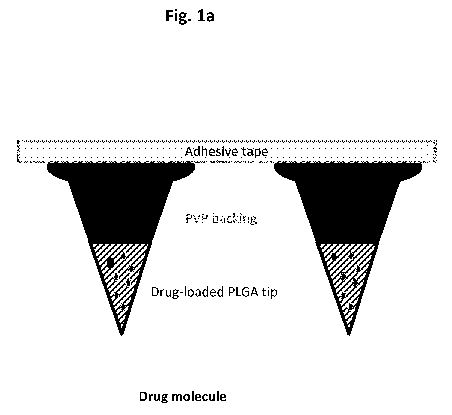
Fig. la is a graphic representation of a microneedle patch of the invention
before
insertion into the skin. The portion of the microneedle shown in dark grey
represents the
fast dissolving backing layer comprising for instance polyvinylpyrrolidone,
and the
portion of the microneedle shown as cross-hatched represents the tip
comprising the
biodegradable polymer mixed with active ingredient(s) (shown as pale grey
ovals).
Fig. lb is a graphic representation of the microneedle patch shown in Fig. la
after
insertion into the skin and after the fast dissolving backing layer has
dissolved. The tip
portion of the microneedles is present in the viable layer(s) of the skin.
Fig. lc is a graphic representation of the microneedle patch shown in Fig. lb
showing
release of active ingredient(s) into the skin.
Fig. 2a is a schematic representation of a method of preparing a microneedle
patch
composition of the invention ("DMN" is an abbreviation of dissolvable
microneedle).
Fig. 2b is a schematic representation of an alternative method of preparing a
microneedle patch composition of the invention ("DMN" is an abbreviation of
dissolvable
microneedle).
Fig. 3 is a graph showing the peak area ratio of the degradation product MC
1046 to
total concentration of calcipotriol in microneedles comprising ester-
terminated
polylactide co-glycolide (PLGA-E) in the tip portion and polyvinylpyrrolidone
(PVP) in the
backing layer in the presence or absence of the antioxidant
butylhydroxytoluene (BHT)
in samples taken after drying the microneedles for 5 hours at 65 C 2 C in a
drying
oven.
Fig. 4 is a graph showing the skin concentration (pM), 24 and 48 hours post
application,
of betamethasone-17,21-dipropionate (BDP) and betamethasone-17-propionate (B-
17-
P) in human skin explants treated with a microneedle patch composition of the
invention
compared to human skin explants treated with Daivobet gel. B-17-P is
predominantly
formed in biological matrices and is thus considered a surrogate marker of BDP
in skin.
Fig. 5a is a graph showing the mRNA levels, 24 and 48 hours post application,
of
CYP24A1 (a biomarker of calcipotriol exposure) in human skin explants treated
with a
CA 02979435 2017-09-12
WO 2016/155891 PCT/EP2016/025026
microneedle patch composition of the invention compared to human skin explants
treated with Daivobet gel.
Fig. 5b is a graph showing the mRNA levels, 24 and 48 hours post application,
of CD14
5 (a biomarker of calcipotriol exposure) in human skin explants treated
with a microneedle
patch composition of the invention compared to human skin explants treated
with
Daivobet gel.
Fig. 6a is a graph showing the mRNA levels, 24 hours and 4 days post
application, of
CYP24A1 in human skin explants treated with a microneedle patch composition of
the
invention compared to human skin explants treated with Daivobet gel.
Fig. 6b is a graph showing the skin concentration (pM), 24 hours and 4 days
post
application, of BDP and B-17-P in human skin explants treated with a
microneedle patch
composition of the invention compared to human skin explants treated with
Daivobet
gel.
Figs. 7a-7e show a series of reflectance confocal microscopy images of one
microneedle
after application of a 5x5 microneedle patch to human ex vivo skin and removal
of the
substrate (medical tape) after 45 minutes, taken by means of a Vivascope 1500
multilaser system at a wavelength of 785 nm.
DETAILED DESCRIPTION OF THE INVENTION
Definitions
In the present context, the term "calcipotriol" is intended to indicate a
vitamin D
analogue of the formula
CA 02979435 2017-09-12
WO 2016/155891
PCT/EP2016/025026
6
OH
/õ=,. -.......
V
Oa
1 HI
1
OS
OH
Calcipotriol has been found to exist in two crystalline forms, an anhydrate
and a
monohydrate. Calcipotriol monohydrate and its preparation are disclosed in WO
94/15912. The term "calcipotriol" is intended to cover any form of the
compound,
including crystalline, amorphous and dissolved forms.
The term "MC1046" is intended to indicate a compound of the formula
1 E
I
II
XHO''' oil
MC1046 is formed as a degradation product of calcpotriol under oxidative
conditions.
The term "betamethasone ester" is intended to indicate a carboxylic acid ester
of a
compound of the formula
CA 02979435 2017-09-12
WO 2016/155891 PCT/EP2016/025026
7
0
OH
H 0 0 H
-
0.0
110
0
Examples of betamethasone esters that may be included in the present
composition are
betamethasone-17-valerate or betamethasone-17,21-dipropionate. A preferred
betamethasone ester for the present purpose is betamethasone-17,21-
dipropionate
(referred to in the following as betamethasone dipropionate).
The term "dispersed" is intended to indicate that the active ingredient is
either
molecularly dispersed or present as a solid dispersion in the polymer matrix.
Results
from Raman spectroscopy of microneedle compositions of the invention suggest
that
both betamethasone dipropionate and calcipotriol are present as glass
solutions
molecularly dispersed in the polymer matrix.
The term "biodegradable" is intended to indicate that the polymer swells after
insertion
into the skin and subsequently degrades due to the hydrolysis of ester
linkages in the
presence of water.
The term "sustained release" is intended to indicate that the diffusion of
active
ingredient from the biodegradable polymer and/or the release of the active
ingredient as
a result of swelling, dissolution and/or degradation of the biodegradable
polymer takes
place over a prolonged period of time, such as at least two days, to enable
delivery of a
therapeutically effective dose of an active ingredient over the entire period,
thus
permitting less frequent dosing.
The term "fast dissolving" is intended to indicate that the backing layer
portion of the
microneedle dissolves within a period not exceeding 120 minutes, preferably
not
exceeding 60 minutes and which may be as short as about 15 minutes after
insertion
into the skin.
CA 02979435 2017-09-12
WO 2016/155891 PCT/EP2016/025026
8
The term "chemical stability" or "chemically stable" is intended to indicate
that no more
than 10%, preferably no more than 5%, of either active ingredient degrades
over the
shelf-life of the product, which may be at least 1 year, but preferably at
least 1.5 year or
more preferably at least 2 years. An approximation of chemical stability at 5
C is
obtained by subjecting the composition to accelerated stability studies at 25
C. If less
than about 6% of the substance has degraded after 3 months at 25 C, this is
usually
taken to correspond to a shelf-life of 2 years at 5 C. More specifically,
"chemical
stability" of calcipotriol is intended to mean that the calcipotriol does not
degrade
significantly over time to 24-epi calcipotriol, MC1046 or other degradation
products of
calcipotriol in the finished pharmaceutical product.
The term "physical stability" or "physically stable" is intended to mean that
the
composition retains its macroscopic and microscopic appearance and physical
properties
over the shelf-life of the product. For instance, the composition is
considered to be
physically stable when the microneedles retain their shape and sharpness over
time
along with their mechanical strength as determined by the force required to
provide
sudden discontinuities (such as fractures) in the microneedle characteristic
of sudden
structural failure.
The term "ester-terminated" is intended to mean an alkyl ester of polylactic,
polyglycolic
or polylactic co-glycolic acid. The alkyl is preferably a C1_20 alkyl, such as
methyl, ethyl,
propyl, butyl, pentyl, hexyl, heptyl, octyl, nonyl, decyl, undecyl, dodecyl,
etc.
A method of preparing the microneedle patch composition of the invention
A method of preparing a microneedle patch composition of the present invention
is
shown schematically in Fig. 2. To make a mould for casting the microneedles, a
template
is initially prepared from a suitable material such as silicon or a metal such
as steel or
titanium or a polymer such as polycarbonate or polymethacrylic acid. The
template
comprise a plurality of microneedles which have a size and shape corresponding
to the
desired shape of the microneedles in the patch composition, i.e. typically
either conical
or pyramidal with a tapering tip. The mould may then be made by casting a
liquid
polymer material such as polydimethylsiloxane over the microneedle template.
When the
material is dried and cured the mould comprises microdepressions that retain
the
negative shape of the microneedles on which it is cast.
CA 02979435 2017-09-12
WO 2016/155891 PCT/EP2016/025026
9
The length and number of microdepressions in the mould determine the length
and
number of the microneedles in the final patch. The number of microneedles per
unit area
may vary widely, typically between 2 and 100 microneedles per cm2, but is more
often
in the range of 5-75 microneedles, more usually 10-50 microneedles, preferably
15-30
microneedles such as 20-25 microneedles per cm2. Similarly, the length of the
microneedles may vary, but they should be of sufficient length to penetrate
the stratum
corneum and not so long as to penetrate into the innervated part of the skin
below the
dermis as penetration to this depth may cause a painful sensation when the
microneedle
patch is applied on the skin. The microneedles may therefore have a length of
50-1000
pm, e.g. 100-800 pm, 300-700 pm, 400-600 pm or about 500 pm. Such a length of
the
microneedles generally ensures that the drug-loaded tip portion will lodge in
the viable
layers of the skin, i.e. the dermis and epidermis where the active ingredients
exert their
effect. As indicated above, the shape of the microneedles is also determined
by the
shape of the microdepressions in the mould and may be conical or pyramidal in
shape
and with a sharp tip. Furthermore, when the shape of the microneedle is
pyramidal, it
comprises a number, typically 4-8, of longitudinally extending ridges to
facilitate the
insertion of the microneedles into the skin. The aspect ratio of the
microneedle (the
length to width at the base of the microneedle) may vary according to the
method used
to produce the mould. When the mould template is prepared by wet etching (e.g.
as
described in US 2007/0134829), the aspect ratio is typically 3:2, i.e. the
length is 1.5
times greater than the width at the base.
Microneedle patches according to the invention may then be made by filling the
microdepressions in the mould with an appropriate solvent, e.g.
dimethylformamide,
and applying a solution of the active ingredients and the biodegradable
polymer in the
same solvent on top of the microdepressions to allow mixing of the two
liquids. The
solvent is then removed, e.g. by drying in a desiccator under vacuum and/or in
a drying
oven at an appropriate temperature. A second solution of the water-soluble
polymer in
an appropriate solvent is then applied on top of the partially filled
microdepressions
followed by drying, e.g. in a desiccator under vacuum or in a drying oven at
an
appropriate temperature. In a currently preferred embodiment, the solution of
the
water-soluble polymer is applied in such a manner that the backing layer
portion when
dried overlayers the base of the tip portion in such a way that each
microneedle is
separated from the other microneedles on the patch and forms a discrete entity
when
the substrate is removed upon application of the patch on the skin.
CA 02979435 2017-09-12
WO 2016/155891 PCT/EP2016/025026
Depending on the dose of the active ingredients to be delivered from each
patch, the
drug-loaded tip portion may constitute 5-95% of the total volume of the
microneedle.
The dried microneedles may then be removed from the mould by applying adhesive
tape
5 on top of the mould and applying pressure to ensure good contact between
the tape and
the base of the microneedles followed by pulling the microneedles out of the
mould. The
tape should preferably be adhesive medical tape as this has been found to
provide good
adhesion to the base of the microneedles so that subtantially all microneedles
are
removed from the mould when the tape is pulled. The composition of the
resulting
10 microneedles is shown graphically in Fig. la. The mould may either be
cast to match the
desired size of each patch or may be made in a larger size, and individual
patches of an
appropriate size may be prepared by cutting the tape into pieces of a desired
shape and
size. The latter option may be as advantage when treating psoriasis as
psoriasis plaques
are often of different sizes and shapes.
The dried microneedle patches may be stored in a sealed airtight vial or
blister pack,
optionally together with an appropirate dessicant, to prevent absorption of
water vapour
during storage.
Further details of microneedle preparation and alternative embodiments are
disclosed in
WO 2012/153266 which is hereby incorporated by reference.
Embodiments
In the course of research leading to the present invention, a large number of
different
biodegradable polymers were tested for their suitability to form a matrix from
which the
microneedles could be made. The majority of the tested polymers were found to
be
unsuitable for the preparation of microneedles of the present invention either
because
the microneedles prepared from them did not retain their shape, in particular
their sharp
tip, i.e. they were not physically stable, or because the therapeutically
active
ingredient(s) were released unacceptably quickly from the polymer due to fast
dissolution thereof, or because the therapeutically active ingredient(s) were
found to be
chemically unstable therein. Thus, microneedles made from polyvinylpyrrolidone
and
poly(meth)acrylates or mixtures of these polymers resulted in an unacceptably
fast
release of the active ingredient(s). Microneedles made from
polyvinylpyrrolidone alone
tended to soften or melt when removed from the primary packaging.
CA 02979435 2017-09-12
WO 2016/155891 PCT/EP2016/025026
11
In the end, these various problems were solved by developing a microneedle
composition comprising a drug-loaded tip portion containing a polylactic acid
or a
derivative thereof such as an ester-terminated polylactide, polyglycolic acid
or a
derivative thereof such as an ester-terminated polyglycolide or polylactic co-
glycolic acid
(PLGA) or a derivative thereof such as an ester-terminated polylactide co-
glycolide
polymer. Satisfactory results were also obtained when a water-soluble polymer
such as
polyvinylpyrrolidone was used as the backing layer. It has been found that
when a
polylactic acid, polyglycolic acid or polylactic co-glycolic acid polymer (or
an ester-
terminated derivative of these polymers) was employed it was possible to
obtain a
composition from the active ingredient(s) are released over a prolonged period
of time
such as at least two days. When polyvinylpyrrolidone was used as the backing
layer, the
resulting microneedles were found to be physically stable, with a hard, sharp
tip.
In a particularly favoured embodiment, the biodegradable polymer is PLGA which
may
optionally be ester-terminated. The PLGA may favourably have a molecular
weight of
>5000, such as a molecular weight of 7000-17000, 24000-38000, 38000-54000,
54000-
69000 or 76000-116000, resulting in a viscosity that permits the formulation
to be
dipensed into the mould and on drying provides a satisfactory physical
stability, in
particular a sharp tip. The ratio of lactic acid to glycolic acid may
preferably vary
between 85:15 and 50:50, such as 85:15, 82:18, 75:25, 65:35 or 50:50. A
currently
preferred ratio of lactide to glycolide is 50:50.
In some cases it may be preferred to add an antioxidant to the biodegradable
polymer
matrix, e.g. butylhydroxytoluene, butylhydroxyanisole or a-tocopherol, or a
mixture
thereof, so as to reduce the formation of degradation products of the active
ingredient
under oxidative conditions. The antioxidant may suitably be present in a
concentration in
the range of 0.01-3% w/w, preferably 0.03-2% w/w such as 0.05-1% w/w of the
dry tip
portion. A currently preferred antioxidant is butylhydroxytoluene, which may
be added in
a concentration in the range of 0.03-2% by weight, e.g. 0.05% by weight, of
the dry tip
portion.
The water-soluble polymer included in the backing layer may be any polymer
that
dissolves quickly in the skin after the composition has been applied and which
is
compatible with the other components. The water-soluble polymer may for
instance be
selected from the group consisting of polyvinylpyrrolidone, a sugar such as
sucrose or
trehalose, dextran, carboxymethylcellulose or sodium alginate. A currently
preferred
water-soluble polymer is polyvinylpyrrolidone. While generally the use of
CA 02979435 2017-09-12
WO 2016/155891 PCT/EP2016/025026
12
polyvinylpyrrolidone confers favourable properties to the backing layer
portion in terms
of physical stability of the microneedles, it may be somewhat brittle and its
properties
may be improved by including a plasticizer, e.g. glycerol, polyethylene
glycol, dibutyl
sebacate, diethyl phthalate, triethyl glycerin or triethyl citrate, to reduce
the brittleness.
The concentration of the plasticizer in the backing layer portion may suitably
be in the
range of 0.5-6% by weight of the dry backing layer. A currently favoured
plasticizer to
include in the backing layer portion of the microneedles is glycerol, which
may suitably
be present in a concentration of about 2% by weight of the dry backing layer.
It should
be noted that the residual solvent remaining in the backing layer after the
composition
has been dried may also act as a plasticizer. An example of such a solvent is
ethanol
which may be present as a residue in the composition after drying.
In a specific embodiment, the present composition may comprise an active
ingredient in
the backing layer of the water-soluble polymer. This will provide immediate
(i.e. within 2
hours or preferably 1 hour) release of a portion of the active ingredient(s)
administered
to the patient. The active ingredient included in the backing layer may be the
same or
different from the active ingredient included in the tip portion of the
microneedle.
The active ingredient(s) included in the present composition may be active
ingredient(s)
that are suitable for the treatment of skin conditions and where less frequent
dosing
(less than once a day) is perceived as advantageous by patients. The active
ingredient
may suitably be selected from the group consisting of a vitamin D analogue, a
glucocorticoid receptor modulator, ingenol or an ingenol derivative, a
calcineurin
inhibitor, a JAK inhibitor, a PDE4 inhibitor, a non-steroidal anti-
inflammatory agent, an
antibiotic, an antifungal agent or a local anesthetic, or mixtures thereof.
Examples of vitamin D analogues are calcipotriol, calcitriol, maxacalcitol or
tacalcitol.
Examples of glucocorticoid receptor modulators are corticosteroids such as
amcinonide,
betamethasone, budenoside, clobetasol, clobetasone, cortisone, desonide,
desoxycortisone, desoximethasone, dexamethasone, diflucortolon, diflorasone,
flucortisone, flumethasone, flunisolide, fluocinonide, fluocinolon,
fluorometholone,
fluprednisolone, flurandrenolide, fluticasone, halcinonide, halobetasol,
hydrocortisone,
meprednisone, methylprednisone, mometasone, paramethasone, prednicarbate,
prednisone, prednisolone and triamcinolone or a pharmaceutically acceptable
ester or
acetonide thereof. The corticosteroid may preferably be selected from
betamethasone,
budenoside, clobetasol, clobetasone, desoximethasone, diflucortolon,
diflorasone,
CA 02979435 2017-09-12
WO 2016/155891 PCT/EP2016/025026
13
fluocinonide, fluocinolon, halcinonide, halobetasol, hydrocortisone,
mometasone
prednicarbate, or triamcinolone or a pharmaceutically acceptable ester
thereof. The
corticosteroid ester may for instance be betamethasone acetate, betamethasone
dipropionate, betamethasone valerate, clobetasol propionate, dexamethasone
acetate,
flumethasone pivalate, fluticasone propionate, hydrocortisone acetate,
hydrocortisone
butyrate or mometasone furoate. The acetonide may be selected from
fluocinolone
acetonide or triamcinolone acetonide.
An example of an ingenol derivative is ingenol mebutate.
Examples of calcineurin inhibitors are tacrolimus or pimecrolimus.
An example of a JAK inhibitor is tofacitinib.
Examples of PDE4 inhibitors are apremilast, roflumilast or cilomilast.
Examples of non-steroidal anti-inflammatory agents are ibuprofen, diclofenac,
naproxen,
indomethacin, dexibuprofen, ketoprofen, flurbiprofen, piroxicam, tenoxicam,
lornoxicam
or nabumeton.
Examples of antibiotics are fusidic acid or mupirocin.
Examples of local anesthetics are lidocain, bupivacain, mepivacain or
ropivacain.
Examples of antifungal agents are ketoconazole, terbinafine, miconazole,
clotrimazole,
ciclopirox, bifonazole, nystatin, econazole or amorolfine.
The therapeutically active ingredient may also be selected from
antiproliferative agents
such as methotrexate or immunosuppressants such as cyclosporin.
In another aspect, the present invention relates to a method for treating a
skin condition
comprising
(a) applying a patch composition comprising one or more microneedles as
described
herein on a surface area of the skin of a patient in need of treatment,
(b) exerting sufficient force on the patch composition to permit the
microneedles to
penetrate through the stratum corneum and into the viable layers of the skin,
and
(c) removing the adhesive substrate from the patch composition.
CA 02979435 2017-09-12
WO 2016/155891 PCT/EP2016/025026
14
To prevent the microneedles from breaking on insertion into the skin, the
mechanical
strength of the microneedles should be such that the force required to
fracture the
microneedle is significantly greater than the force required to insert the
microneedle into
the skin. Generally, the force required to insert a microneedle patch into the
skin and
have it penetrate past the stratum corneum is in the range of 0.4-8N, for
instance 2-7N,
such as 5N, per patch containing 25 microneedles per cm2. The failure force of
the
microneedle can be assessed as either a fracture force or the force required
to compress
the microneedle by a defined length. These forces can be can be determined
using a
texture analyser (e.g. a TA.XT Plus Texture Analyzer, Stable Micro Systems,
Surrey, UK)
or using a computer-controlled force-displacement station (Model 5565,
Instron,
Buckinghamshire, UK).
As indicated above, the backing layer comprising the water-soluble polymer
starts
dissolving upon insertion of the microneedles into the skin. This allows
removal of the
substrate within about 120 minutes, preferably within 60 minutes or 45 minutes
or even
as little as about 15 minutes, of application of the patch on skin. In
general, it is
preferred that at least 90% of the microneedles detach from the adhesive
surface upon
removal of the substrate within this timeframe to avoid that a substantial
number of the
microneedles are pulled out again when the substrate is peeled off.
The invention has been found able to provide delivery of the therapeutically
active
ingredients over a prolonged period of time. Thus, the therapeutically active
ingredients
may be released from the microneedles over a period of 2-21 days, preferably 2-
14 days
such as 2-7 days or 4-7 days. As shown in Example 2 below, increased mRNA
levels of
the biomarker CYP24A1 are observed 4 days after application of a microneedle
patch
containing calcipotriol indicating that calcipotriol is released from the
microneedles for at
least 4 days.
In the present method, step (b) may be carried out by applying pressure with a
finger or
by impact insertion, e.g. by using an applicator device, the latter being
preferred as it
increases insertion reproducibility (cf. van der Maaden et al., AAPS Journal
16(4), July
2014, pp. 681-684. Examples of applicator devices are disclosed in US
2002/0123675 or
WO 2008/091602.
Skin conditions to be treated using the microneedle patch composition of the
invention
may be selected from psoriasis, e.g. plaque psoriasis, inverse psoriasis, nail
psoriasis or
CA 02979435 2017-09-12
WO 2016/155891 PCT/EP2016/025026
spot psoriasis, pustulosis palmoplantaris, actinic keratosis, squamous cell
carcinoma,
basal cell carcinoma, contact dermatitis, atopic dermatitis, eczema, hand
eczema, warts,
genital warts, alopecia, acne, rosacea or skin infections.
5 Psoriasis is a chronic inflammatory skin disease that manifests as
erythematous, dry,
scaling plaques resulting from hyperkeratosis. The plaques are most often
found on the
elbows, knees and scalp, though more extensive lesions may appear on other
parts of
the body, notably the lumbosacral region. A common treatment of mild to
moderate
psoriasis involves topical application of a composition containing a
corticosteroid as the
10 active ingredient. While efficacious, application of corticosteroids has
the disadvantage of
a number of adverse effects such as skin atrophy, striae, acneiform eruptions,
perioral
dermatitis, overgrowth of skin fungus and bacteria, hypopigmentation of
pigmented skin
and rosacea.
15 Combination products for the treatment of psoriasis have been marketed
by LEO Pharma
for a number of years under the trade names Daivobet ointment and Daivobet
gel.
The product comprises calcipotriol and betamethasone dipropionate as the
active
ingredients formulated in an ointment or gel vehicle comprising
polyoxypropylene stearyl
ether as a solvent. While the efficacy of the combination products is
significantly superior
to that of either active ingredient on its own, the products need to be
applied once daily,
and many patients, in particular those with extensive psoriatic lesions, would
favour a
greater ease of application such as less frequent application. It is
considered desirable to
further improve the biological efficacy of the combination of the two active
ingredients
by providing a formulation vehicle from which delivery of the active
ingredients into the
skin is prolonged compared to the commercial product.
Thus, in a currently favoured embodiment, the present invention relates to a
microneedle patch composition comprising one or more microneedles each
comprising
(a) a tapered tip portion containing one or more therapeutically active
ingredients
selected from the group consisting of calcipotriol and betamethasone esters
dispersed in
a matrix of a biodegradable polymer selected from the group consisting of
ester-
terminated polylactide, ester-terminated polyglycolide and ester-terminated
polylactide
co-glycolide, and
(b) a fast dissolving microneedle backing layer portion containing a water-
soluble
polymer overlayering the tip portion,
said microneedle or microneedles being attached to and extending from an
adhesive
surface of a removable substrate.
CA 02979435 2017-09-12
WO 2016/155891 PCT/EP2016/025026
16
In this embodiment, the betamethasone ester may be betamethasone dipropionate
or
betamethasone valerate.The prolonged delivery is expected to be sustained with
a dose
of calcipotriol of 0.08-30 pg of calcipotriol per cm2 of patch and a dose of
betamethasone
ester of 1-60 pg of betamethasone ester per cm2 of patch. The beta methose
ester is
preferably betamethasone dipropionate.
During development of this embodiment it was found that calcipotriol was not
chemically
stable in a matrix of polylactic acid, polyglycolic acid or polylactic co-
glycolic acid,
probably due to the presence of acidic residues or impurities therein, while
calcipotriol
was chemically stable when ester-terminated polylactide, polyglycolide or
polylactide co-
glycolide were used as the biodegradable polymer.
In this embodiment, the biodegradable polymer is preferably an ester-
terminated
polylactide co-glycolide. The ester-terminated polylactide co-glycolide may
favourably
have a molecular weight of >5000, such as a molecular weight of 7000-17000,
24000-
38000, 38000-54000, 54000-69000 or 76000-116000, resulting in a viscosity that
permts the formulation to be dispensed into the mould and on drying provides a
satisfactory physical stability, in particular a sharp tip. The ratio of
lactide to glycolide
may preferably vary between 85:15 and 50:50, such as 85:15, 82:18, 75:25,
65:35 or
50:50. A currently preferred ratio of lactide to glycolide is 50:50.
In this embodiment, it may be preferred to add an antioxidant to the
biodegradable
polymer matrix, e.g. butylhydroxytoluene, butylhydroxyanisole or a-tocopherol,
or a
mixture thereof, so as to reduce the formation of MC 1046. The antioxidant may
suitably
be present in the concentration of the antioxidant is in the range of 0.03-3%
w/w,
preferably 0.05-2% w/w such as 0.05-1% w/w of the dry tip portion. A currently
preferred antioxidant is butylhydroxytoluene, which may be added in a
concentration in
the range of 0.05-2% by weight of the dry tip portion.
In this embodiment, the water-soluble polymer may for instance be selected
from the
group consisting of polyvinylpyrrolidone, a sugar such as sucrose or
trehalose, dextran,
carboxymethylcellulose and sodium alginate. A currently preferred water-
soluble
polymer is polyvinylpyrrolidone. The backing layer portion may additionally
comprise a
plasticizer, e.g. glycerol, polyethylene glycol, dibutyl sebacate, diethyl
phthalate, triethyl
glycerin or triethyl citrate, which may be included in a concentration in the
range of 0.5-
6% by weight of the dry backing layer. A currently favoured plasticizer to
include in the
CA 02979435 2017-09-12
WO 2016/155891 PCT/EP2016/025026
17
backing layer portion of the microneedles is glycerol, which may suitably be
present in a
concentration of about 2% by weight of the dry backing layer.
In a specific embodiment, the present composition may comprise calcipotriol
and/or a
beta methasone ester dispersed in the backing layer of the water-soluble
polymer.
In this embodiment, the removable substrate may suitable be composed of
adhesive
medical tape.
The invention is further described in the following examples which are not in
any way
intended to limit the scope of the invention as claimed.
EXAMPLES
Example 1
Compositions of the invention
A microneedle mould was prepared by mixing about 45 g of polydimethylsiloxane
(PDMS) elastomer base (Sylgard 184 silicone elastomer kit, part A) and about
4.5 g of
curing agent (Sylgard 184 silicone elastomer kit, part B) by hand using a
spatula and
beaker until thoroughly mixed. The resulting mixture was placed in a
desiccator under
vacuum for about 20 minutes. The PDMS was poured over a microneedle template
(patterned silicon wafer obtained from the Tyndall National Institute,
Ireland, and
prepared essentially as disclosed in US 2007/0134829) and cured in a drying
oven at
100 C for about 60 minutes. After cooling, the mould was peeled off the
microneedle
template and cut into individual moulds of 1x1 cm (containing 5x5
microdepressions).
The PDMS moulds were placed in a glass beaker and cleaned with
dimethylformamide
(DMF) under vacuum for 30 minutes and an ultrasonic bath for further 30
minutes at
room temperature. The cleaned moulds were placed in a glass beaker and the
microdepressions were prefilled with DMF under vacuum.
A solution was prepared by dissolving 300 mg ester-terminated polylactide co-
glycolide
(lactide:glycolide 50:50, Mw 7000-17000, PLGA-E) in 1 ml DMF using a vortex
mixer for
about 10 minutes until the PLGA-E was completely dissolved. In some
embodiments, 0.5
mg/ml butylhydroxytoluene (BHT) was added to the PLGA-E solution. 20 mg
calcipotriol
and 40 mg betamethasone dipropionate (BDP) were dissolved in 1 ml of the
resulting
CA 02979435 2017-09-12
WO 2016/155891 PCT/EP2016/025026
18
solution using a vortex mixer for about 10 minutes until the active
ingredients were
completely dissolved. The drug-loaded PLGA-E solution was dispensed into the
microdepressions of the PDMS moulds prefilled with DMF using a syringe pump
and
capillary tube dispenser and a flow rate of 0.5 pl/min. to a total volume of
0.15 0.03 pl
per mould.
The moulds were dried for about 18 hours in a desiccator under vacuum and
subsequently in a vacuum oven under 500 mbars for about 5 hours at 60 2 C.
A second solution was prepared by dissolving 400 mg of polyvinylpyrrolidone
(PVP,
Kollidon 17 PF) in 1 ml of ethanol 96% using a vortex mixer for about 5
minutes. 10
mg of glycerol was added and the mixture was stirred for 2-3 minutes using the
vortex
mixer. The PVP solution was dispensed into the microdepressions of the PDMS
moulds
containing the dried drug-loaded PLGA-E solution using a syringe pump and
capillary
tube dispenser at a flow rate of 1.5 pl/min. so that the dispensed volume was
0.75 0.15
pl per mould.
The filled moulds were dried in a desiccator under vacuum for about 2 hours.
Medical adhesive tape (3M) was applyied on the surface of the moulds using
finger
pressure and the microneedles were pulled out of the mould.
The patches were stored in hermetically sealed vials purged with nitrogen and
closed
with a rubber stopper, aluminium cap and crimper.
The dried microneedle patch has the following composition.
Ingredient pg/patch % w/w mg/g
Betamethasone dipropionate 6 1.66 16.60
Calcipotriol monohydrate 3 0.83 8.30
PLGA-E 45 12.45 124.48
PVP 300 82.99 829.88
Glycerol 99.5% 7.5 2.07 20.75
Total 378.82 100 1000
Physical and chemical stability of the composition appears from the following
table. It
should be noted that storage of the microneedle patches at 40 C, which is the
usual
CA 02979435 2017-09-12
WO 2016/155891 PCT/EP2016/025026
19
temperature for accelerated stability studies, was not feasible since PLGA-E
is not
physically stable at 40 C.
Storage Appearance Calcipotriol 24-epi- MC1046 BDP
temperature/ % of start calcipotriol % area % of
start
time % area
Start OK 100.0 % 0.8 % 1.2 % 100.0 %
25 C/ 1 OK 95.5 % 1.0 % 1.5 % 98.3 %
month
25 C/ 3 Not 116.4 % 0.7 % 2.1 % 118.6 %
months evaluated
40 C/ 2 Not 95.5 % 0.9 % Not 101.7 %
weeks acceptable evaluated
Microneedle patch compositions were prepared as described above with the
exception
that they contained 0%, 0.5% or 1% BHT by weight of the dry tip.
The results are shown in Fig. 3 which illustrates the percentage ratio of peak
area of the
degradation product MC 1046 to the total calcipotriol peak area for samples
without BHT
and samples with 0.5% w/w and 1% w/w BHT. The percentage peak area of MC 1046
relative to the total amount of calcipotriol was significantly reduced,
indicating that the
addition of BHT to the composition significantly reduced degradation of
calcipotriol.
Example 2
Human explant skin exposure
Two experiments were performed to investigate exposure over time in human skin
explants.
Experiment 1:
Full-thickness human skin obtained from female donors undergoing
abdominoplasty
maximally 24 hours prior to the start of the experiment was used. 22 mm punch
biopsies were placed in 24 mm Transwell inserts and placed in 6 well plates
with 1 ml
EpiLife tissue culture medium supplemented with 0.2 ng/mL human EGF, 0.2 %
bovine
pituitary extract (BPE), 5 pg/mL bovine insulin, 5 pg/mL bovine transferrin,
0.18 pg/mL
hydrocortisone and gentamycin.
CA 02979435 2017-09-12
WO 2016/155891 PCT/EP2016/025026
Compositions prepared as described in Example 1 containing 2 pg calcipotriol
and 6 pg
BDP per cm2 microneedle patch and 10 pl Daivobet gel per cm2 and Daivobet
gel
vehicle were applied topically in triplicate. The following treatment
schedules were
tested: One dose of Daivobet gel at t=Oh with skin sampling at 24h and 48h.
Two
5 doses of Daivobet gel at t=0 and 24h respectively with skin sampling at
48h, one patch
applied at t=Oh with skin sampling at 24h and 48h leaving the backing tape on
the skin
for the full duration of the experiment, and one patch applied at t=Oh with
skin sampling
at t=48h but removing the backing tape at 24h. The skin biopsies were
maintained in ex
vivo culture at 37 C with 5% CO2 for 48 hours with a change of medium at 24 h.
At the
10 end of the experiment, a 14 mm biopsy encompassing the dosed area of
each explant
was punched out and subsequently divided in two for compound analysis
(tapestripped
10 times) and biomarker analysis, respectively.
Compound analysis was performed by extracting the active compounds from the
skin
15 biopsy using an organic solvent and subsequently analysing the extract
using LC/MS-MS.
Total RNA was extracted from cells using the mirVana (Life Technologies, Grand
Island,
NY, USA) according to the instructions provided. cDNA synthesis was performed
with
the High-Capacity cDNA Reverse Transcription Kit (Applied Biosystems, Foster
City, CA,
20 USA). 2.5 pL of cDNA (equivalent to 5 ng RNA) from each sample was
amplified in a
total volume of 10 pL by quantitative realtime PCR using Taqman Gene
Expression
Assays (CYP24A1 (Hs00167999 m1), CD14 (Hs02621496 s1), PPIA (Hs99999904 m1)
GAPDH (Hs99999905 m1), TBP (Hs99999910 m1) and HMBS (Hs00609297 m1)) and
PRISM7900HT sequence detection system (SDS 2.3) from Applied Biosystems.
PPIA, GAPDH, TBP and HMBS were used for normalization.
It appears from Fig. 4 that after application BDP could reside either inside
microneedle
patch compositions or in the stratum corneum of the skin after Daivobet gel
applications and thus be unavailable for pharmacological action. B-17-P is
predominantly
formed in biological matrices and is thus considered a surrogate marker of BDP
available
for pharmacological action in skin. It appears that the amount of B-17-P
formed in the
skin increases over time for both explants treated with Daivobet gel and with
microneedle patch compositions of the invention. The skin concentrations of B-
17-P
observed after application of Daivobet gel are higher than what was observed
after
application of microneedle patch compositions of the invention, however the
increase
over time may indicate a prolonged release from the patches.
CA 02979435 2017-09-12
WO 2016/155891 PCT/EP2016/025026
21
It appears from Figs. 5(a) and 5(b) that the PD biomarkers for calcipotriol,
CYP24A1 and
CD14, are induced over time by Daivobet gel. The level of biomarker induction
elicited
by microneedle patches is lower, but increasing over time, indicating a slower
onset but
potentially a prolonged effect of calcipotriol than what is observed from
Daivobet gel.
Experiment 2:
NativeSkin Plus skin models with an available surface area of 2.5 cm2 were
acquired
from Genoskin, France and cultured according to the manufacturer's
specification.
Compositions prepared as disclosed in Example 1 containing 0.5 pg calcipotriol
and 6 pg
BDP per cm2 microneedle patch and 4.3 pl Daivobet gel per cm2 and Daivobet
gel
vehicle were applied topically in triplicate. The following treatment
schedules were
tested: One daily dose of Daivobet gel or placebo gel with skin sampling at
24h and
96h. One patch applied at t=Oh with skin sampling at 24h and 96h leaving the
backing
tape on the skin for 24h, and two patches applied at t=Oh and t=48h with skin
sampling
at t=96h. At the end of the experiment, two 4 mm biopsies were punched out and
subsequently either analysed for compound (after being tapestripped 10 times)
or the
presence of biomarker.
It appears from Fig. 6(a) that the biomarker for calcipotriol, CYP24A1, is
induced over
time by Daivobet gel applied at time 0, day 1 and day 2 of the experiment and
sampled
on day 4. The level of biomarker induction elicited by the microneedle patch
applied
once is initially lower (at day 1), but increases over time, indicating a
slower onset but
potentially a protracted effect of calcipotriol over 4 days compared to what
is observed
from Daivobet gel.
It appears from Fig. 6b that the amount of B-17-P formed in the skin increases
over time
for both explants treated with Daivobet gel and with a microneedle patch
composition
of the invention. The skin concentrations of B-17-P observed after application
of
Daivobet gel applied at time 0, day 1 and day 2 of the experiment are higher
on day 4
than concentrations observed after application once of a microneedle patch
composition
of the invention, however the increase over time may indicate a prolonged
release from
the patches.
Example 3
Reflectance confocal microscopy of a microneedle composition in human
explant skin
CA 02979435 2017-09-12
WO 2016/155891 PCT/EP2016/025026
22
A microneedle patch as described in Example 1 was applied to fresh human ex
vivo skin
prepared as described in Example 2. 45 minutes after application the medical
adhesive
tape was removed and it was conformed that none of the microneedles was left
on the
tape before reflectance confocal microscopy (RCM) imaging was conducted using
a
Vivascope 1500 multilaser system in accordance with the procedure described in
H.
Skvara et al. Dermatol Pract Concept 2(1), 2012, pp.3-12, and Calzavara-Pinton
et al.,
Photochemistry and Photobiology, 84, 2008, pp.1421-30. In this technique laser
light
with a wavelength of 785 nm is passed through a beam splitter and an optical
lens in
contact with skin. In the skin, light is focused on a small tissue spot a few
microns of
diameter. Reflection (back scattering) occurs at the boundary between two
structures
having different indexes of refraction. Light reflected from the focal point
propagates
back toward the lens through a pinhole. Light reflected from above and below
the point
in focus is masked out by the pinhole so that the detector receives light only
from the
thin plane of the specimen that is in focus. By changing the depth at which
the objective
lens focuses in the vertical plane horizontal images can be generated at
particular
depths within the skin.
The scanned field of view was 500 x 500 pm. Depth measurements were done in
steps
of 3 pm with an axial resolution of < 5 pm. The limit of detection of the RCM
is a depth
of about 150 pm.
The results appear from Fig. 7, in which
Fig. 7a shows a microneedle penetrating the stratum corneum at a depth of 12
pm. The
PVP backing layer has dissolved before the removal of the substrate and only a
thin
octagonal shell of the PLGA-E polymer reflects the light on this plane.
Fig. 7b shows a microneedle penetrating the stratum corneum at a depth of 27
pm. The
PVP backing layer has dissolved before the removal of the substrate and only
appears as
a circle in the middle of a thin octagonal shell of the PLGA-E biodegradable
polymer.
Fig. 7c shows a microneedle penetrating the stratum corneum at a depth of 45
pm. The
PLGA-E polymer microneedle tip reflects the light on this plane as does a thin
octagonal
shell of the PLGA-E polymer.
Fig. 7d shows a microneedle penetrating the epidermis at a depth of 96 pm.
Only the tip
of the needle composed of the PLGA-E biodegradable polymer is visible.
Fig. 7e shows a microneedle penetrating the epidermis at a depth of 150 pm.
Only the
tip of the needle composed of the PLGA-E biodegradable polymer is visible at
this depth.