Note: Descriptions are shown in the official language in which they were submitted.
DEVICE FOR DEFINING A FLAP GEOMETRY
Technical Field
The present disclosure relates in general to a device for defining a flap
geometry. It
relates in particular to a device for defining a flap geometry of a flap for
laser treatment
of the human eye (LASIK flap).
Background
A so-called LASIK (laser in-situ keratomileusis) technique is often used for
the
correction of defective vision of the human eye (for example, myopia,
hyperopia, or
astigmatism). In this procedure, first a small corneal cover disk (referred to
in general as
a flap) is cut from the adjacent corneal tissue, where the flap remains
adhering the
surrounding corneal tissue in the hinge region. This makes it possible to
simply fold the
flap over to expose the underlying tissue regions of the cornea and simply
folding the
flap back over after ablation of the exposed tissue regions. Removal of
material by
focused UV laser radiation in ablation results in an altered shape of the
corneal surface
after the flap has been folded back over, and therefore this alters the
refractive
properties of the cornea and consequently of the overall ophthalmic system. By
suitable
definition of the ablation profile, a vision deficiency can at least be
definitely diminished
and at best even eliminated almost completely.
To be able to correct an individual patient's faulty vision, it is necessary
to determine an
individual ablation profile for each of the patient's eyes. Furthermore, the
geometry of
the flap to be cut (size, position, orientation) must be defined for each of
the patient's
eyes, but this involves a great deal of effort on the part of the physician
operating the
cutting laser.
Summary of Exemplary Embodiments
One object of the present invention is to simplify the definition of the flap
geometry.
Certain exemplary embodiments can provide a device for defining a flap
geometry of a
flap for laser treatment of a human eye, comprising: a laser source configured
to
- 1 -
CA 2979551 2019-08-15
generate a laser beam having pulse durations in the femtosecond range; a
scanner unit
configured to control a focus of the laser beam in a transverse direction and
in a
longitudinal direction; and a control unit programmed to: evaluate ablation
profile data of
an ablation profile for a laser ablation treatment of a human cornea, the
evaluating
comprising determining a diameter of the ablation profile; store a defined
safety margin
with the ablation profile in a memory; define the flap geometry based on the
evaluation
according to: a diameter of the flap based on the diameter of the ablation
profile; and the
defined safety margin stored with the ablation profile, such that in top view
a shortest
distance between an outer edge of the ablation profile and an outer edge of
the flap at
each location amounts to at least the safety margin; and instruct the scanner
unit to
control the focus of the laser beam to cut the flap with the flap geometry.
One aspect of the present invention is a device for defining a flap geometry
of a flap for
laser treatment of a human eye, comprising a control unit, which is programmed
to
evaluate ablation profile data of an ablation profile for a laser ablation
treatment of a
human cornea and define the flap geometry based on this evaluation.
The control unit may be a program-controlled control unit, which may comprise
a
processor, a volatile memory and/or a nonvolatile memory, for example.
Programming
of the control unit may be performed by writing a corresponding
- la -
CA 2979551 2019-11-05
CA 02979551 2017-09-12
WO 2016/181237
PCT/IB2016/051888
program to a memory of the control unit, for example. A program in a memory in
the
control unit can be executed by a processor of the control unit. During
execution of
the program, the aforementioned steps can be carried out. The ablation profile
data
of the ablation profile may be present in the form of digital data, for
example, in the
form of a data file. The ablation profile data may describe the desired
ablation profile
for the ablation treatment of the eye of the patient to be treated. The
ablation profile
data may be contained in a data file, for example, which indicates, pixel-by-
pixel, a
depth value for a two-dimensional matrix of pixels. The depth value may be a
value
(in pm or nm, for example), to which the human cornea is to be ablated by an
ablation laser during the laser ablation treatment. The ablation profile data
may be in
the form of vectors. The ablation profile data can establish a spatial
relationship
between the ablation profile and the eye to be treated in. For example, a
reference
point, which corresponds to the midpoint of the pupil of the eye to be
treated, may be
defined in the ablation profile data. Furthermore, a reference axis, which
corresponds to a horizontal axis and/or a vertical axis of the eye to be
treated, can
also be defined in the ablation profile data. Evaluation of the ablation
profile data
may include a software-supported evaluation. For this evaluation, for example,
known methods may be used for the image processing and/or the image
evaluation.
The flap geometry can best be defined on the basis of the evaluation, so that
certain
result values and/or analytical data of the analytical process can be used to
define
the flap geometry. The flap geometry may be defined in such a way that
corresponding data representing the flap geometry is written to a memory of
the
control unit. Flap geometry data, which is written to a memory of the control
unit,
may be determined on the basis of the defined flap geometry.
The control unit may be programmed such that evaluating the ablation profile
data
comprises determining a diameter of the ablation profile and such that
defining the
flap geometry comprises defining a diameter of the flap based on the diameter
of the
ablation profile.
The diameter of the ablation profile can be determined with the help of known
methods of image processing, for example. The diameter of the ablation profile
may
be a diameter, which is determined in top view. "In top view," as used below,
means
that an x-y plane of the ablation profile and/or the flap geometry is
considered. The
x-y plane may essentially correspond to the surface of the human cornea, which
is
leveled by a contact element during the process of cutting a flap. The x-y
plane may
correspond to a plane that is perpendicular to a z axis. The z axis may
essentially
correspond to the incident direction of a cutting laser and/or an ablation
laser. The z
- 2 -
CA 02979551 2017-09-12
WO 2016/181237
PCT/IB2016/051888
axis may correspond to a radial direction of the eyeball, which runs through
the
midpoint of the pupil. The diameter may be, for example, the maximum diameter
of
the ablation profile. The diameter may further be, for example, the diameter
along a
predetermined axis, for example, along a horizontal or vertical axis of the
ablation
profile. The horizontal axis and/or the vertical axis of the ablation profile
may
correspond to a horizontal and/or vertical axis of the eye to be treated. The
definition
of the diameter of the flap may be such that the flap is essentially circular
in a view
from above and the diameter of the circle is defined as a function of the
specific
diameter of the ablation profile. The diameter of the circle of the flap may
be larger
by a predetermined value than the determined diameter of the ablation profile.
The control unit may be programmed such that defining the flap geometry is
performed in consideration of a defined safety margin, such that in top view a
shortest distance between an outer edge of the ablation profile and an outer
edge of
the flap at each location amounts to at least the safety margin.
The safety margin may be a length value (in pm, for example), which may be
defined
by the user of the device (for example, a physician). The safety margin may be
stored together with the ablation profile data in a data file and read out of
this data
file.
The control unit may be programmed such that evaluating the ablation profile
data
comprises determining the position of the ablation profile with respect to the
eye to
be treated and such that defining the flap geometry comprises defining a
position of
the flap with respect to the eye to be treated.
The ablation profile data may include information about the position of the
ablation
profile with respect to the eye to be treated. For example, it may include
information
about the position of the ablation profile with respect to the midpoint of the
pupil of
the eye to be treated. If the ablation profile data is available in the form
of a pixel-
based data file, for example, then a predetermined pixel of the data file may
correspond to the position of the midpoint of the pupil of the eye to be
treated. The
flap may be essentially circular or essentially oval, for example, in top
view. The flap
may have a hinge on one side. Defining the position of the flap with respect
to the
eye to be treated may include, for example, defining the position of the
midpoint of
the essentially circular flap with respect to the eye to be treated.
- 3 -
CA 02979551 2017-09-12
WO 2016/181237
PCT/IB2016/051888
The control unit may be programmed such that evaluating the ablation profile
data
comprises determining an orientation of the ablation profile with respect to
the eye to
be treated and such that defining the flap geometry comprises defining an
orientation
of the flap with respect to the eye to be treated.
The ablation profile data may include information about the orientation of the
ablation
profile with respect to the eye to be treated. For example, it may also
include
information about the orientation of the ablation profile with respect to the
horizontal
or vertical axis of the eye to be treated. This may include, for example,
angle
information or an angle value. For determining the orientation of the ablation
profile,
the ablation profile data may include a reference axis, for example, which
corresponds to the horizontal or vertical axis of the eye to be treated.
Defining the
orientation of the flap may include a definition of the orientation of
rotation of the flap
with respect to the horizontal or vertical axis of the eye to be treated. The
flap may
be essentially circular or essentially oval in a view from above. The flap may
have a
hinge on one side.
The control unit may be programmed such that defining the orientation of the
flap
comprises defining a position of a hinge of the flap with respect to the eye
to be
treated.
The position of the hinge may be defined, for example, such that a shortest
distance
from the hinge to an outer edge of the ablation profile is at its maximum in
top view.
The control unit may be programmed such that evaluating the ablation profile
data
comprises determining a diameter of the ablation profile and determining an
axis,
along which the ablation profile has the greatest diameter, and wherein
defining the
flap geometry comprises defining an orientation of the hinge of the flap
parallel to the
axis.
The control unit may be programmed such that evaluating of the ablation
profile data
comprises determining an axis of mirror symmetry of the ablation profile, and
wherein defining the flap geometry comprises defining an orientation of a
hinge of
the flap perpendicular to the axis of mirror symmetry.
The axis of mirror symmetry may be an axis with respect to which the ablation
profile
is essentially in mirror symmetry. The ablation profile may have one or two
axes of
mirror symmetry, for example. The axis of mirror symmetry may be determined so
- 4 -
CA 02979551 2017-09-12
WO 2016/181237
PCT/IB2016/051888
that it corresponds to an axis, which itself corresponds most closely to an
axis of
mirror symmetry of the ablation profile. In other words, the axis of mirror
symmetry
may be an axis with respect to which there is the greatest possible mirror
symmetry
of the ablation profile. The orientation of the hinge of the flap
perpendicular to the
axis of mirror symmetry may be carried out in such a way that mirror symmetry
of the
flap corresponds essentially to mirror symmetry of the ablation profile. The
orientation of the hinge of the flap may be defined in such a way that the
axis of
mirror symmetry of the flap corresponds to the axis of mirror symmetry of the
ablation profile.
The control unit may be programmed such that evaluating the ablation profile
data
comprises determining a depth of the ablation profile and defining the flap
geometry
comprises defining a thickness of the flap based on the depth of the ablation
profile.
The depth may be determined along the z axis (along the incident direction of
the
cutting laser and/or the ablation laser). The depth of the ablation profile
may
correspond to the thickness of the corneal tissue to be ablated by the
ablation laser.
The specific depth of the ablation profile may be, for example, the maximum
depth of
the ablation profile. In other words, it may be the depth at the deepest point
of the
ablation profile. The thickness of the flap may be the thickness along the z
direction.
The thickness of the flap may be defined in such a way that, for example, a
greater
specific depth of the ablation profile leads to a smaller defined thickness of
the flap
and vice versa.
The control unit may be programmed such that defining the flap geometry is
performed in consideration of a corneal thickness and/or at least one
curvature
radius of the cornea of the eye to be treated.
The flap geometry may be defined, for example, such that a higher value of the
corneal thickness leads to a higher value of the defined thickness of the flap
and vice
versa. Conversely the thickness of the flap may be defined so that the sum of
the
thickness of the flap, the maximum depth of the ablation profile and a
predetermined
safety distance corresponds to the thickness of the cornea.
The device may also comprise an input interface for reading in the ablation
profile
data.
- 5 -
CA 02979551 2017-09-12
WO 2016/181237
PCT/IB2016/051888
The input interface may include, for example, a network interface and/or an
interface
for reading from a memory medium. The memory medium may be, for example, a
magnetic memory medium, an optical memory medium or a semiconductor memory
medium. The network interface may be connected to the Internet, for example,
and/or to an internal network (intranet). A network interface of an ablation
laser, for
example, may be connected to the network. The ablation profile data in the
form of a
data file, for example, may be entered via the input interface. The input
interface may
comprise a network interface, which is connected to a network, and the
ablation
profile data can be retrieved from a database, which is located in a memory of
a
server or some other device connected to the network, for example, via the
network
interface.
The control unit may also be programmed to determine flap geometry data based
on
the defined flap geometry.
The flap geometry data may be present in the form of a data file and/or
individual
parameters, for example. The parameters may be written to a database, for
example. The parameters of the flap geometry data may comprise at least one of
the
following parameters, for example: flap diameter, flap thickness, position of
the
midpoint of the flap with respect to the midpoint of the pupil of the eye to
be treated
and the orientation of the flap (for example, in the form of an angle) with
respect to a
reference axis of the eye to be treated. In addition or as an alternative to
the
parameters, the entire shape of the flap (for example, its contour and/or cut
edges)
may be saved as a data file. The flap geometry data may be stored, for
example, in a
pixel-based data file or a vector-based data file. The flap profile data can
establish a
spatial relationship between the flap and the eye to be treated. For example,
a
reference point, which corresponds to the midpoint of the pupil of the eye to
be
treated, may be defined in the flap geometry data. Furthermore, a reference
axis,
which corresponds to a horizontal axis and/or a vertical axis of the eye to be
treated,
may be defined in the flap geometry data. Furthermore, the flap geometry data
can
be written to a data file together with the ablation profile data of the eye
to be treated.
The device may further comprise an output interface for outputting the flap
geometry
data.
The flap geometry data can be output via the output interface to a cutting
laser,
which then cuts a flap corresponding to the flap geometry data into the eye to
be
treated. The output interface may comprise a network interface and/or an
interface
- 6 -
CA 02979551 2017-09-12
WO 2016/181237
PCT/IB2016/051888
for reading from a memory medium, for example. The memory medium may be, for
example, a magnetic memory medium, an optical memory medium and/or a
semiconductor memory medium. The network interface may be connected, for
example, to an internal network (intranet) and/or to the Internet.
Another aspect of the present invention is a cutting laser for cutting a flap
for laser
treatment of a human eye, comprising the device described herein.
The control unit of the device may be, for example, a control unit of the
cutting laser
which is programmed accordingly. The flap geometry may be forwarded directly
to
the cutting laser in the form of flap geometry data, for example.
Brief Description of the Drawings
Additional features, advantages and components of the present invention can be
found in the following description of the accompanying drawings, in which:
Figure 1 shows a schematic block diagram of an exemplary embodiment of a
cutting laser for laser treatment of a human eye;
Figure 2 shows an exemplary embodiment of a device for defining the flap
geometry of a flap for laser treatment of the human eye;
Figure 3a shows an example of an ablation profile and a respective safety
margin;
Figure 3b shows an example of a flap geometry that has been defined on the
basis of the ablation profile shown in Figure 3a;
Figure 4a shows an example of an ablation profile and respective axes, and
Figure 4b shows an example of a flap geometry, which has been defined on
the
basis of the ablation profile shown in Figure 4a.
Detailed Description of Exemplary Embodiments
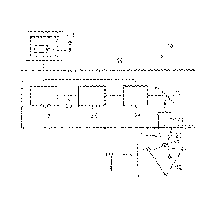
Figure 1 shows a block diagram of an exemplary embodiment of a device labeled
as
in general for laser treatment of a human eye 12. The device 10 is a cutting
laser
for laser treatment of a human eye. The device 10 comprises a control unit 14,
a
laser configuration 16 and a patient adapter 17.
- 7 -
CA 02979551 2017-09-12
WO 2016/181237
PCT/IB2016/051888
The laser configuration 16 comprises a laser source 18, which generates a
laser
beam 20 having pulse durations in the femtosecond range, for example. The
laser
beam 20 has a wavelength suitable for generating a laser-induced optical
breakdown
in the corneal tissue of the eye 12. The laser beam 20 may have a wavelength
in the
range of 300 nm (nanometers) to 1900 nm, for example, a wavelength in the
range
of 300 nm to 650 nm, 650 nm to 1050 nm, 1050 nm to 1250 nm or 1100 nm to
1900 nm. The laser beam 20 may also have a focus diameter of 5 pm or less.
A beam widening lens system 22, a scanner unit 24, a mirror 26 and a focusing
lens
system 28 are positioned behind the laser source 18 in the direction of
propagation
of the laser beam 20 (indicated by the arrows in Figure 1). The beam widening
lens
system 22 serves to increase the diameter of the laser beam 20 generated by
the
laser source 18. In the exemplary embodiment shown here, the beam widening
lens
system 22 is a Galileo telescope having a concave lens (lens with a negative
refractive power) and a convex lens (lens with a positive refractive power)
positioned
behind the concave lens in the direction of propagation of the laser beam 20.
These
may be a piano-concave lens or a piano-convex lens, which has planar sides
facing
one another. In another exemplary embodiment, the beam widening lens system
may comprise a Kepler telescope having two convex lenses, for example, as an
alternative to the Galileo telescope.
The scanner unit 24 is designed to control the position of the focus of the
laser beam
20 (beam focus) in the transverse direction and in the longitudinal direction.
The
transverse direction describes the direction transverse to the direction of
propagation
of the laser beam 20 (labeled as the x-y plane) and the longitudinal direction
describes the direction of propagation of the laser beam 20 (labeled as the z
direction). The scanner unit 24 may comprise, for example, a pair of
galvanometrically operated deflecting mirrors for transverse deflection of the
laser
beam 20; these mirrors can be tilted about mutually perpendicular axes.
Alternatively
or additionally, the scanner unit 24 may have an electro-optical crystal or
some other
components suitable for transverse deflection of the laser beam 20. The
scanner unit
24 may also comprise a longitudinally adjustable or refractive lens of a
variable
power or a deformable mirror to influence the divergence of the laser beam 20,
and
consequently, the longitudinal orientation of the beam focus. In the exemplary
embodiment shown here, the components for control of the transverse
orientation
and longitudinal orientation of the beam focus are represented as an integral
component. In another exemplary embodiment, the components may be arranged
- 8 -
CA 02979551 2017-09-12
WO 2016/181237
PCT/IB2016/051888
separately along the direction of propagation of the laser beam 20. Thus, for
example, an adjustable mirror may be arranged in the direction of propagation
upstream from the beam widening lens 22 for control of the longitudinal
orientation of
the beam focus.
The mirror 26 is a stationary deflecting mirror, which is designed to deflect
the laser
beam 20 in the direction of the focusing lens system 28. Additionally or
alternatively,
other optical mirrors and/or optical elements may also be positioned in the
beam
path for deflection and diffraction of the laser beam 20.
The focusing lens system 28 is designed to focus the laser beam 20 on the
region of
the cornea of the eye 12 to be treated. The focusing lens system 28 may be an
F-
theta lens system, for example. The focusing lens system 28 is detachably
connected to the patient adapter 17. The patient adapter 17 comprises a
conical
carrier sleeve 30, which is connected to the focusing lens system 28 by a
coupling
formation (not shown), and a contact element 32, which is mounted on the
narrower
bottom side of the carrier sleeve 30 facing the eye 12. The contact element 32
may
be attached to the carrier sleeve 30 either permanently (for example, by
adhesive
bonding) or detachably (for example, by screw connection). The contact element
32
has a bottom side which faces the eye 12 and is labeled as a contact surface
34. In
the exemplary embodiment shown here, the contact surface 34 is designed as a
planar surface. In the laser treatment of the eye 12, the contact element 32
is
pressed against the eye 12 or a vacuum is applied to the eye 12 on the contact
surface 34, such that at least the region of the cornea of the eye 12 that is
to be
treated is leveled and lies in the x-y plane.
The control unit 14 comprises a memory 36, in which at least one control
program 38
having program instructions is stored. The laser source 18 and the scanner
unit 24
are controlled by the control unit 14 in accordance with the program
instructions. The
control program 38 contains program instructions, which, when executed by the
control unit 14, cause the beam focus to move in space and time in such a way
that
a cutting pattern is created in the cornea of the eye 12 to be treated. The
cutting
pattern may comprise a LASIK flap. Data defining the shape of the cutting
pattern
may be stored in the form of flap geometry data in the memory 36 of the
control unit
14 and retrieved therefrom. The flap geometry data may have previously been
loaded into the memory 36 of the control unit 14 with the help of a network
interface
of the control unit, for example. However, the flap geometry data may also be
- 9 -
CA 02979551 2017-09-12
WO 2016/181237
PCT/IB2016/051888
entered manually via a corresponding input interface of the control unit
(using a
keyboard, for example).
Figure 2 shows a schematic block diagram of one exemplary embodiment of a
device 40 for defining the flap geometry of a flap for laser treatment of the
human
eye. The device 40 comprises a control unit 42, an input interface 44 and an
output
interface 46. Ablation profile data for evaluation by the control unit 42 can
be input
via the input interface 44. Flap geometry data generated by the control unit
42 can
be output via the output interface 46. The input interface 44 and the output
interface
46 may each comprise, for example, a network interface connected to a
conventional
network for data exchange between terminals. A server, a network memory, a
cutting
laser and/or an ablation laser, for example, may be connected to the network
to
exchange data with one another. The network may be the Internet, for example,
or
an intranet within the treatment practice. Additionally or alternatively,
however, the
input interface 44 may also have a direct input option, such as a keyboard
interface,
for example. Additionally or alternatively, the output interface 46 may have a
direct
output option, such as a screen interface, for example. Furthermore, both the
input
interface 44 and the output interface 46 may comprise an interface for reading
from
and/or writing to a memory medium. The memory medium may be a magnetic
memory medium, an optical memory medium and/or a semiconductor memory
medium.
The control unit 42 comprises a memory 48. The memory 48 comprises a volatile
memory and/or a nonvolatile memory. The memory 48 is used for temporary
storage
of calculations of the control unit 42 and can also store ablation profile
data and flap
geometry data. Furthermore, a control program, comprising commands for
evaluating ablation profile data of an ablation profile for a laser ablation
treatment of
a human cornea and for defining a flap geometry on the basis of the
evaluation, is
also stored in the memory 48.
The control unit 42 may be, for example, the control unit 14 of the cutting
laser 10
shown in Figure 1 and the memory 48 may be the memory 36 of the control unit
14.
The device 40 for defining a flap geometry may thus be provided at the cutting
laser
10, which has the advantage that the flap geometry, defined by the control
unit 42, is
directly available to the user of the cutting laser 10 (a physician) for
cutting the
respective flap. However, the control unit 42 may also be provided at an
ablation
laser, wherein the memory 48 may be, for example, a memory of the ablation
laser,
in which ablation profile data for the eyes to be treated is stored. This has
the
- 10 -
CA 02979551 2017-09-12
WO 2016/181237
PCT/IB2016/051888
advantage that the respective ablation profile data is directly available to
the control
unit 42. However, the device 40 may also be provided as an independent device,
which reads in ablation profile data via the input interface 44 and outputs
flap
geometry data via the output interface 46.
The control unit 42 also comprises a processor (not shown) for executing the
program instructions of the control program stored in the memory 48.
Ablation profile data for a laser ablation treatment of a human cornea of an
eye to be
treated is entered via the input interface 44. For example, if the control
unit 42 is
provided at the ablation laser, the ablation profile data may alternatively
also be read
directly out of the memory 48 and the input interface 44 is optional in this
case. The
ablation profile data may be present, for example, in the form of a data file
or some
other data record. For example, the ablation profile data, like a gray scale
image
data file may have a two-dimensional matrix of pixels, with a depth value
(gray scale
value) assigned to each pixel. The depth value here corresponds to the desired
depth of ablation at the respective location of the pixel, which is identified
by x-y
coordinates. Regions of the x-y plane, in which no ablation is to take place,
can also
be defined within the ablation profile data. These regions are not considered
below
as part of the ablation profile. \Mien speaking of a size and/or diameter of
the
ablation profile below, for example, only the region of the ablation profile
in which
ablation is to take place via the ablation laser is being considered.
To establish a spatial reference for the ablation profile data with respect to
the eye to
be treated, at least one fixed point and at least one reference axis may be
defined in
the ablation profile data. The fixed point may be, for example, the midpoint
of the
pupil of the eye to be treated. The reference axis may be, for example, a
horizontal
or vertical axis of the eye to be treated. For example, it is possible to
stipulate that a
certain pixel value of the x-y plane corresponds to the midpoint of the pupil
of the eye
to be treated. Furthermore, it is possible to provide that a horizontal pixel
axis, for
example, corresponds to the horizontal axis of the eye to be treated.
The ablation profile data may also be in the form of vector-based data or in
any other
data format that makes it possible to represent an ablation profile that is to
be
implemented with respect to an eye to be treated.
The control unit 42 analyzes the ablation profile data and defines a flap
geometry for
a flap to be cut by the cutting laser 10 on the basis of this evaluation.
Details of the
- 11 -
CA 02979551 2017-09-12
WO 2016/181237
PCT/IB2016/051888
evaluation and the definition are described further below with reference to
Figures 3a
to 4b. On the basis of the flap geometry, flap geometry data that is output
via the
output interface 46 is generated by the control unit 42. If the device 40 is
part of the
cutting laser, then the flap geometry data may for example only be written to
the
memory 48, from which it can be retrieved by the cutting laser 10. The output
interface 46 is optional in this case. The flap geometry data is suitable for
uniquely
defining the flap geometry to be cut by the cutting laser 10. In particular
the flap
geometry data comprises an outline of the flap in top view (in the x-y plane)
and the
thickness of the flap to be cut. The flap geometry data may be available in
the form
of a data file or parameters, for example, wherein the parameters are suitable
for
determining the flap geometry uniquely. The corresponding parameters may thus
comprise, for example, a value for the diameter of the flap in top view and an
angle
value for the orientation of the hinge of the flap.
Figure 3a shows schematically a first example of an ablation profile 50, and
Figure
3b shows the outline of a flap 52, which is defined by the control unit 42 of
the device
40 on the basis of the ablation profile 50. Figures 3a and 3b (as well as
Figures 4a
and 4b, which are described further below) show the ablation profile 50 and
the flap
52 in top view, wherein the plane of the drawing corresponds to the x-y plane
(see
also Figure 1). The depth of the ablation profile 50 in the z direction is
indicated by
depth lines (isobaths). Each of the depth lines runs along a plane extending
parallel
to the x-y plane at a constant distance. Thus each of the depth lines of the
ablation
profile 50 runs along a constant depth of the ablation profile 50. The
outermost one
of the depth lines indicates an exterior outline of the ablation profile 50.
In other
words, no ablation takes place outside of the outermost line of the ablation
profile 50,
and when speaking of the ablation profile 50 below, the region inside the
outermost
depth line of the ablation profile 50 is intended.
A horizontal line in the x direction and a vertical line in the y direction
indicate a
coordination system within the x-y plane. The position and orientation of the
ablation
profile 50 with respect to the eye to be treated can both be identified on the
basis of
the coordination system. The horizontal line in the x direction, for example,
corresponds to the horizontal axis of the eye to be treated, and the point of
intersection of the vertical line and the horizontal line identify the
midpoint of the pupil
of the eye to be treated. A patient's vision can be compensated accurately and
reliably by indicating the ablation profile 50 with respect to this coordinate
system. In
the case of astigmatism in the patient's eye in particular, it is necessary to
provide
- 12 -
CA 02979551 2017-09-12
WO 2016/181237
PCT/IB2016/051888
ablation profile data indicating the position and the orientation (rotational
orientation)
of the ablation profile 50.
Figure 3a also shows a safety zone 54, in which the flap 52 can be defined as
follows: First, the midpoint and the diameter of a circle is determined; this
is the circle
with the smallest diameter into which the ablation profile 50 fits, in top
view, without
the outer edge of the ablation profile 50 protruding beyond the circle
(internal dotted
line circle in Figure 3a). Furthermore, a value defined previously (for
example, by the
physician operating the cutting laser 10) for a safety margin is also taken
into
account. This value is added to the radius of the first circle, resulting in a
larger
second circle with the same midpoint as that of the first circle (see outer
circle,
shown with a dotted line in Figure 3a). As shown in Figure 3b, the flap 52 is
then
defined, so that the cutting edge essentially follows the second circle in top
view.
This ensures that the cutting edge of the flap 52 is at a distance from the
outer edge
of the ablation profile 50 by a corresponding safety margin 54 at all points.
In other
words, this ensures that the shortest distance between the outer edge of the
ablation
profile 50 and the outer edge of the flap 52 in top view amounts to at least
the safety
margin of the safety zone 54 at all points.
The flap geometry of the flap 52 also comprises a hinge 56, which is
represented as
a straight line in Figure 3b. The hinge 56 of the flap 52 does not represent a
cutting
edge of the flap 52, but instead is a joint of corneal tissue along which the
cutting
laser 10 does not make a cut. Providing a hinge 56 makes it possible to fold
the flap
52 over and to accurately fold the flap 52 back after the ablation treatment
so that
the flap tissue is essentially in the same position on the x-y plane before
and after
the ablation treatment. The position of the hinge 56 may be defined manually
(by
providing the proper parameters) by the user, so that it is always either in a
lower
position (see Figure 3b) or in an upper position of the flap 52, for example.
The hinge
56 may be set parallel to the horizontal axis along the x direction, for
example.
Furthermore, the position of the hinge 56 may be automatically defined by the
control
unit 42 on the basis of the ablation profile data for the ablation profile 50
(see also
the example of Figures 4a and 4b). For example, the position of the hinge 56
may be
defined in such a way that the shortest distance from the hinge 56 to the
outer edge
of the ablation profile 50 exceeds a predetermined value, so that a safety
margin is
maintained between the hinge 56 and the ablation profile 50.
Furthermore, within the context of the definition of the flap geometry, the
thickness of
the flap 52 in the z direction may be defined on the basis of the evaluation
of the
- 13 -
CA 02979551 2017-09-12
WO 2016/181237
PCT/IB2016/051888
ablation profile data. For example, the ablation profile data may be analyzed
in such
a way that the maximum depth of the ablation profile is determined. The
thickness of
the flap 52 is then defined so that the sum of the maximum depth of the
ablation
profile and the thickness of the flap 52 does not exceed a predetermined
value. It is
possible herein to ensure that the laser treatment is performed only in a
certain
region of the cornea and that the underlying tissue of the eye is not damaged.
For
the definition of the thickness, for example, a previously determined value
for the
thickness of the cornea of the eye to be treated may be taken into account.
The
thickness of the flap 52 may be defined, for example, so that the sum of the
thickness of the flap 52, the maximum depth of the ablation profile 50 and a
predetermined safety distance corresponds to the thickness of the cornea of
the eye
to be treated.
Furthermore, at least one curvature radius of the cornea of the eye to be
treated can
be taken into account in determining the flap geometry.
Figure 4a shows a second example of an ablation profile 60 and Figure 4b shows
a
flap geometry of a flap 62 defined on the basis of an evaluation of the
ablation profile
60. For Figures 4a and 4b, the same principles apply as those described
previously
in conjunction with Figures 3a and 3b. In particular a corresponding safety
margin
may be taken into account in the definition of the flap 62.
Figure 4a shows an example of an ablation profile 60 of a patient with a
severe
astigmatism (curvature of the cornea). The ablation profile 60 here is far
away from a
point symmetry, but it has two mutually perpendicular mirror symmetry axes 68
and
70. The position of the mirror symmetry axes 68 and 70 with respect to the eye
to be
treated varies from one patient to the next and is part of the individual
vision defect
to be corrected.
The orientation of the flap 62 in Figure 4b is selected so that the hinge 66
of the flap
62 is parallel to the mirror symmetry axis 68 and perpendicular to the mirror
symmetry axis 70. The mirror symmetry of the flap 62 thus corresponds to the
mirror
symmetry of the ablation profile 60 with respect to the mirror symmetry axis
70. This
has the advantage that, when cutting the flap 62 with the cutting laser 10, no
additional asymmetries are created with regard to the mirror symmetry in
relation to
the mirror symmetry axis 70.
- 14 -
CA 02979551 2017-09-12
WO 2016/181237
PCT/IB2016/051888
In evaluating the ablation profile 60, the mirror symmetry axis 68 and/or the
mirror
symmetry axis 70 of the ablation profile is/are determined. For example, the
axis 60
may be determined, so that there is a search for the axis along which the
ablation
profile 60 will have the greatest diameter (axis 68 in the example of Figure
4a). The
position of the hinge 66 of the flap 62 is then defined so that the hinge 66
runs
parallel to the axis 68.
Furthermore, the axis 68 and/or the axis 70 can be determined by considering
the
symmetry properties of the ablation profile 60. For example, it is possible to
search
for the axis with respect to which the ablation profile 60 will have the
greatest
possible mirror symmetry. It should be pointed out here that the case of
perfect
mirror symmetry, as represented in Figure 4a, occurs very rarely in reality
and there
may be minor deviations with regard to the mirror symmetry. For example, the
ablation profile may either have no preferential mirror symmetry axis at all,
just one
preferred mirror symmetry axis or two preferred mirror symmetry axes, in which
case
the first mirror symmetry axis runs essentially perpendicular to the second
mirror
symmetry axis (see Figure 4a). In evaluation of the ablation profile data of
the
ablation profile 60, for example, the mirror symmetry axis 70 can be
determined and
the flap geometry can be defined as shown in Figure 4a, so that the hinge 66
of the
flap 62 is perpendicular to the mirror symmetry axis 70. The mirror symmetry
of the
flap 62 thus corresponds essentially to the previously determined mirror
symmetry of
the ablation profile 60.
Although essentially circular flap geometries are illustrated in Figures 3b
and 4b, the
shape of the flap in top view is not limited to a circle but can also exhibit
an oval
shape or an essentially rectangular shape.
With the help of the device described herein, the flap geometry can be defined
automatically and individually on the basis of ablation profile data analyzed
automatically in advance. Valuable time can be saved here in the preparation
for the
laser ablation treatment and the flap geometry can be defined reliably and
without
error.
- 15 -