Note: Descriptions are shown in the official language in which they were submitted.
CA 02979708 2017-09-13
WO 2016/149710 PCT/US2016/023488
HIV-1 NEUTRALIZING ANTIBODIES AND USES THEREOF
[0001] This application claims the benefit of and priority to U.S. Application
Serial No.
62/135,309 filed March 19, 2015, U.S. Application Serial No. 62/222,057 filed
September
22, 2015, and U.S. Application Serial No. 62/260,100 filed November 25, 2015,
U.S.
Application Serial No. 62/191,095 filed July 10, 2015, U.S. Application Serial
No.
62/191,054 filed July 10, 2015 and U.S. Application Serial No. 62/261,233
filed November
30, 2015 the content of each application is hereby incorporated by reference
in its entirety.
[0002] This patent disclosure contains material that is subject to copyright
protection. The
copyright owner has no objection to the facsimile reproduction by anyone of
the patent
document or the patent disclosure as it appears in the U.S. Patent and
Trademark Office
patent file or records, but otherwise reserves any and all copyright rights.
[0003] All patents, patent applications and publications cited herein are
hereby incorporated
by reference in their entirety. The disclosure of these publications in their
entireties are
hereby incorporated by reference into this application in order to more fully
describe the state
of the art as known to those skilled therein as of the date of the invention
described herein.
GOVERNMENT SUPPORT
[0004] This invention was made with government support under Center for
HIV/AIDS
Vaccine Immunology-Immunogen Design grant UM1-AI100645 from the NIH, NIAID,
Division of AIDS. The government has certain rights in the invention.
FIELD OF THE INVENTION
[0005] The invention relates to the identification of monoclonal HIV-1
neutralizing
antibodies, such as, but not limited to, antibodies that bind to the membrane-
proximal region
of HIV-1 gp41, their recombinant expression and purification and uses.
BACKGROUND
[0006] A number of neutralizing monoclonal antibodies (mAbs) have been
isolated from
HIV-1 infected individuals and these mAbs define specific regions (epitopes)
on the virus
that are vulnerable to NAbs.
[0007] Broadly neutralizing antibodies have been isolated only from natural
HIV infection.
See e.g. Mascola and Haynes, Immunological Reviews (2013) Vol. 254: 225-244.
Some
CA 02979708 2017-09-13
WO 2016/149710 PCT/US2016/023488
examples of broadly neutralizing antibodies (bnAbs) that bind gp41 at gp4lbnAb
sites within
the membrane proximal region are 2F5, 4E10 and 10E8. These gp41 neutralizing
antibodies
recognize the membrane-proximal region (MPER) of the HIV-1 gp41 glycoprotein.
The
advantage of gp41 bnAbs is that they are generally quite broad in their
neutralization
coverage yet the antibodies to date, have not been developed for prevention or
treatment.
This is because 2F5 and 4E10 are quite polyreactive and autoreactive, and
while mAb 10E8
is less polyreactive, it is autoreactive and is not stable (Haynes BF et al.
Science 308: 1906-8,
2005; Yang G, et al. JEM 210: 241-56, 2013; Huang Jet al nature 491: 406-412,
2012).
Unfortunately, so far none of these antibodies have been developed for HIV
prevention or
treatment. Thus, the need exists for monoclonal broadly neutralizing
antibodies that can be
developed and used for prevention and treatment for an infectious agent, such
as HIV.
SUMMARY OF THE INVENTION
[0008] In certain aspects the invention provides an antibody or fragment
thereof with the
binding specificity of an MPER antibody as described herein. In non-limiting
embodiments
the MPER antibody from Figure 13, Figure 55, Figure 56 or Figures 30-33
(antibodies with
mutations in the DH512 or DH511 VH chain). In non-limiting embodiments,
combination
mutations in the DH512 or DH511 VHCDR3 could include VH L100dF together with
T100aW Figures 31 and 32); VH L100dW together with T100aW (Figures 31 and 32).
[0009] Non-limiting examples include antibodies comprising VH or VL chains
from DH511,
DH512, DH512 K3, DH512-L100dF, DH513, DH514, DH515, DH516, DH517, DH518,
lineage members.
[0010] In certain embodiments, the antibody or fragment thereof is fully human
and
recombinantly produced. In certain embodiments, some of the VH and/VL chains
are
isolated from human subject who have been naturally infected with HIV. In
certain
embodiments the antibody is not naturally occurring. In certain embodiments
the antibody
comprises naturally occurring pair of VH and VL chains. In certain embodiments
the
antibody comprises naturally occurring pair of VH and VL chains wherein the Fc
portion of
the antibody is not the natural isotype or portion of the naturally occurring
pair of VH and VL
chains. In certain embodiments the antibody is computationally designed, for
example based
on some naturally isolated VH and VL sequences. In certain embodiments the
antibody is
computationally designed, e.g., UCA, Intermediates in the antibody lineages.
In certain
embodiments the antibody comprises a non-naturally occurring pairing of VH and
VL chains,
2
CA 02979708 2017-09-13
WO 2016/149710 PCT/US2016/023488
wherein the VH or VL individually could be isolated from a subject. In some
embodiments,
the antibody comprises VH chain or HCDRs of a VH chain of one clonal member,
and VL or
LCDRs of another clonal member, i.e., a non-naturally occurring antibody
comprising
sequences derived from natural pairs.
[0011] In certain embodiments, the antibody or fragment thereof comprises a VH
chain that
is 90%, 91%, 92%, 93%, 94%, 95%, 96%, 97%, 98%, 99% or 100% identical to the
VH
chain of antibody DH511, DH512, DH513, DH514, DH515, DH516, DH517, DH518,
DH536, DH537, DH491 or DH493, or an antibody from Example 10, 11 or 12.
[0012] In certain embodiments, the antibody or fragment thereof comprises a VL
chain that is
90%, 91%, 92%, 93%, 94%, 95%, 96%, 97%, 98%, 99% or 100% identical to the VL
chain
of antibody DH511, DH512, DH513, DH514, DH515, DH516, DH517, DH518, DH536,
DH537, DH491 or DH493, or an antibody from Example 10, 11 or 12.
[0013] In certain embodiments, the antibody or fragment thereof comprises a VH
chain that
is 90%, 91%, 92%, 93%, 94%, 95%, 96%, 97%, 98%, 99% or 100% identical to the
VH
chain of antibody DH511, DH512, DH513, DH514, DH515, DH516, DH517, DH518,
DH536, DH537, DH491 or DH493 and further comprises a VL chain that is 90%,
91%, 92%,
93%, 94%, 95%, 96%, 97%, 98%, 99% or 100% identical to the VL chain of
antibody
DH511, DH512, DH513, DH514, DH515, DH516, DH517, DH518, DH536, DH537, DH491
or DH493, or an antibody from Example 10, 11 or 12.
[0014] In certain embodiments, the antibody or fragment thereof comprises a VH
which
comprises the HCDR1, HCDR2, and HCDR3 of antibody DH511, DH512, DH513, DH514,
DH515, DH516, DH517, DH518, DH536, DH537, DH491 or DH493, or an antibody from
Example 10, 11 or 12.
[0015] In certain embodiments, the antibody or fragment thereof comprises a VL
which
comprises the LCDR1, LCDR2, and LCDR3 of antibody DH511, DH512, DH513, DH514,
DH515, DH516, DH517, DH518, DH536, DH537, DH491 or DH493, or an antibody from
Example 10, 11 or 12.
[0016] In certain embodiments, the antibody or fragment thereof comprises a VH
which
comprises the HCDR1, HCDR2, and HCDR3 of antibody DH511, DH512, DH513, DH514,
DH515, DH516, DH517, DH518, DH536, DH537, DH491 or DH493, or an antibody from
Example 10, 11 or 12 and further comprises the complementary VL which
comprises the
LCDR1, LCDR2, LCDR3 of antibody DH511, DH512, DH513, DH514, DH515, DH516,
3
CA 02979708 2017-09-13
WO 2016/149710 PCT/US2016/023488
DH517, DH518, DH536, DH537, DH491 or DH493, or an antibody from Example 10, 11
or
12.
[0017] In certain embodiments, the antibody or fragment thereof comprises VH
and VL of
antibody DH511, DH512, DH513, DH514, DH515, DH516, DH517, DH518, DH536,
DH537, DH491 or DH493, or an antibody from Example 10, 11 or 12.
[0018] In certain embodiments, the antibody is DH511, DH512, DH513, DH514,
DH515,
DH516, DH517, DH518, DH536, DH537, DH491 or DH493, or an antibody from Example
10, 11 or 12, e.g. without limitation DH511 5a or DH511 5b, DH512 K3.
[0019] In certain aspects, the invention provides a pharmaceutical composition
comprising
anyone of the antibodies of the invention or fragments thereof or any
combination thereof.
[0020] In certain aspects, the invention provides a pharmaceutical composition
comprising
anyone of the antibodies of the invention, or a combination thereof.
[0021] In certain embodiments, the composition comprises an antibody or a
fragment thereof
which is recombinantly produced in CHO cells.
[0022] In certain aspects, the invention provides a pharmaceutical composition
comprising a
vector comprising a nucleic acid encoding anyone of inventive antibodies or
fragments. In
certain embodiments, the nucleic acids are optimized for expression in human
host cells. In
certain embodiments, the vector is suitable for gene delivery and expression.
Non-limiting
examples of such vectors include adenoviral vectors (Ads), adeno associated
virus based
vectors (AAVs), or a combination thereof.
[0023] In certain embodiments, the compositions further comprise an antibody
or a fragment
thereof comprising the VH and VL chains of antibody DH540.
[0024] In certain embodiments, the compositions further comprise an antibody
or a fragment
thereof comprising VH and VL chain of antibody CH557 or DH270 lineage
antibody, for
example without limitation DH542, DH542-QSA, DH542 L4..
[0025] In certain aspects the invention provides a bispecific antibody which
comprises gp41
NITER binding specificity. In some embodiments the NITER binding portion of
the bispecific
antibody comprises VH and/or VL chains, variants or fragments thereof
[0026] In certain aspects the invention provides methods to treat or prevent
HIV-1 infection
in a subject comprising administering to the subject the pharmaceutical
composition of any
one of the preceding claims in a therapeutically effective amount.
[0027] In certain embodiments of the methods, the pharmaceutical composition
is
administered in a therapeutically effective regimen.
4
CA 02979708 2017-09-13
WO 2016/149710 PCT/US2016/023488
BRIEF DESCRIPTION OF THE DRAWINGS
[0028] Figure 1 shows Neutralization-based Epitope Prediction (NEP) Analysis.
Neutralization-based epitope prediction analysis. The predicted relevant
prevalence of
antibody clusters [(10 epitopes targeting sites of vulnerability (CD4 binding
site, V1/V2,
MPER, glycan V3)] is shown as a heat map, with dark color intensity (higher
fractional
number) corresponding to a stronger neutralization signal. Plasma
neutralization breadth is
shown, and numbers in each row add up to 1.00. NEP algorithm reference:
[Georgiev IS et
at Science 340: 751-756].
[0029] Figure 2 shows MPR.03 Hook sequence (SEQ ID NOs: 1-2). MPR.03 is a
biotinylated peptide containing lysines at both ends for solubility
(KKKNEQELLELDKWASLWNWFDITNWLWYIRKKK-biotin) (SEQ ID NO: 463) used
to pull out gp41 antibodies from blood memory B cell sorts See Morris L. et
al. (2011) PLoS
ONE 6(9): e23532.
[0030] Figure 3 shows a representative CH0210 mper03 sort (sort #1).
[0031] Figure 4 shows V(D)J Rearrangement of MPER Antibodies Isolated from
Four HIV-1
Infected Individuals. * indicates that these mAbs neutralized the tier 1
isolate MN in TZM-bl
cells. Mutation refers to VH nucleotide sequence somatic mutation percentages
in the
variable heavy (VH) immunoglobulin (Ig) genes.
[0032] Figure 5 shows Neutralization Titers of MPER Antibodies Isolated from
Four HIV-1
Infected Individuals using a small panel of HIV-1 isolates in the TZMbl
pseudovirus
inhibition assay.
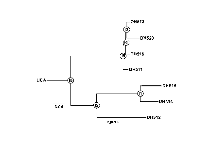
[0033] Figure 6 shows the MPER BnAb DH511 VH Phylogram of the B Cell Clonal
Lineage
Derived from Subject 0210. Antibodies in clone DH511 include the following:
DH511,
DH512, DH513, DH514, DH515, DH516 and DH520.
[0034] Figure 7 shows summary results of neutralization of gp41 antibodies
against a panel
of 30 HIV-1 tier 2 isolates in the TZMbl pseudovirus neutralization assay.
Data show that
antibodies in the DH511 B cell clonal lineage (DH511-DH516) all neutralize
100% of 30
HIV-1 isolates tested in the TZMbl Env pseudovirus neutralization assay.
[0035] Figure 8 shows Neutralizing Breadth and Potency of DH512, DH517 and
DH518
HIV-1 BnAbs compared to 10E8, VRCO1 and a mixture of CH01 and CH31 bnAbs.
DH512
neutralizes 100% of HIV strains and is as at least as potent as 10E8.
[0036] Figure 9 shows Neutralizing Breadth and Potency of various HIV-1 BnAbs
that are
candidates for being combined with DH512 or other antibodies in Figure 4 for a
potent
CA 02979708 2017-09-13
WO 2016/149710 PCT/US2016/023488
mixture of bnAbs. DH270IA1 is Ii in the DH270 lineage (See Figure 26, and US
Ser. No.
62/056,568 filed September 28, 214)
[0037] Figure 10 shows Neutralizing Breadth and Potency of some candidate
bnAbs for
single or combination use.
[0038] Figure 11 shows summary of Clone DH511 binding to the indicated
peptides (SEQ ID
NOs: 3-14) in ELISA. Clone DH511 antibodies bind at the C-terminus of the
MPER. "+"
indicates that antibodies in the Clone DH511 bind to the peptide. The summary
shows that
DH511 clone antibodies do not bind the peptides when D674 is mutated to S674.
The twelve
sequences of the peptides (without the three lysines at the N- and C- end) are
shown in SEQ
ID NOs: to . The twelve sequences of the peptides (with the three lysines
at the N- and
C- end) are shown in SEQ ID NOs: 3 to 14. Thus, antibody DH511 requires an
aspartic acid
at amino acid position 674 for binding.
[0039] Figure 12 shows nucleic acid sequences of antibodies DH511-518, DH536
and 537
(SEQ ID Nos: 15 to 34).
[0040] Figure 13 shows amino acid sequences of antibodies DH511-518, DH536 and
537.
(SEQ ID Nos: 35 to 55)
[0041] Figures 14A-B show Alignment of VH (Fig. 14A; (SEQ ID Nos: 56-61)) and
VL
(Fig. 14B (SEQ ID Nos: 62-67)) Sequences of BnAb DH511 Clonal Lineage. Bolded
is the
sequence of CDR1, underlined is the sequence of CDR2 and italicized is the
sequence of
CDR3 of the DH511 VH chain and DH511 VL chain. The CDRs of the VH and VL
sequences of the other antibodies DH512, DH513, DH514, DH515, and DH516 can be
readily determined based on the sequence alignment.
[0042] Figures 15A-B show Alignment of VH (Fig. 15A (SEQ ID Nos: 68-76)) and
VL (Fig.
15B (SEQ ID Nos: 77-85)) sequences of MPER BnAbs. Bolded is the sequence of
CDR1,
italicized is the sequence of CDR2 and underlined is the sequence of CDR3 of
VH or VL of
the listed MPER antibodies.
[0043] Figure 16 shows sequences of MPER alanine mutants (SEQ ID NOs: 86-112)
screened in ELISA. All antibodies in the DH51 clone showed weak binding to
this peptide
set. DH517 (Ab510053) strongly bound to MPER656 peptide and showed decreased
binding
to several residues (A4, A6-A13, A16-A18, A20, A23, A24, A26) using the ala
substituted
peptides in table.
[0044] Figure 17 shows Binding of DH517 (Ab510053) to alanine substituted MPER-
26
peptides. The binding studies do not conclusively map the DH517epitope.
6
CA 02979708 2017-09-13
WO 2016/149710 PCT/US2016/023488
[0045] Figure 18 shows MPER656 variants (SEQ ID NOs: 13-124) screened in
ELISA.
Residues shown in light blue (underlined) indicate positions that differ from
MPER656-
biotin.
[0046] Figure 19 shows Binding of DH511 (Ab510056) to MPER656 variants
[0047] Figure 20 shows Binding of DH512 (Ab510049) to MPER656 variants
[0048] Figure 21 shows Binding of DH513 (Ab570022) to MPER656 variants
[0049] Figure 22 shows Binding of DH514 (Ab570029) to MPER656 variants
[0050] Figure 23 shows Binding of DH515 (Ab510052) to MPER656 variants
[0051] Figure 24 shows Binding of DH516 (Ab510048) to MPER656 variants
[0052] Figure 25 shows Binding of DH518 (Ab570010) to MPER656 variants.
[0053] Figure 26 shows the amino acids sequences of VH (SEQ ID NOs: 137-148)
and VL
(SEQ ID NOs: 161-172) chains of antibodies of the DH270 lineage, and nucleic
acid
sequences (SEQ ID NOs: 125-136 (VH); SEQ ID NOs: 149-160 (VL)) encoding these
amino
acids. CDRs are highlighted and underlined in the UCA.
[0054] Figure 27A shows amino acid (SEQ ID Nos: 173 and 174) and nucleic acid
sequences
(SEQ ID Nos: 175 and 176) of CD4bs antibody CH557. Figure 27B shows amino acid
sequences of VH chains of antibodies from CH235 lineage (SEQ ID NOs:177-188).
Figure
27C shows amino acid sequences of VL chains of antibodies from CH235 lineage
(SEQ ID
NOs: 189-198).
[0055] Figure 28A shows neutralization Breadth and Potency of Plasma and
Memory B cell
(MBC)-derived MPER bnAbs. Figure 28B shows neutralization Breadth and Potency
of
chimeric MPER bnAbs (n=30 cross-clade HIV-1 isolates)
[0056] Figure 29A and B show neutralization data from TZM-bl assay (Titer in
TZM.b1 cells
(ug/ml) for DH512 K3 and other chimeric antibodies compared to DH512 and 10E8.
The
data in the first column is historic data when DH512 was run in this panel
previously.
DH512 was run at the same time as DH512 K3 but is listed as Ab510049 in this
assay;
therefore, data from columns DH512 K3 and AA&AB DH512/Ab510049 should be
compared.
[0057] Figure 30 shows positions in the VHCDR3 chain of DH511 which could be
mutated.
Amino acid positions refer to Kabat numbering. Most mutations are to changes
to W, but F,
L or possibly other substitutions can be tried.
7
CA 02979708 2017-09-13
WO 2016/149710 PCT/US2016/023488
[0058] Figure 31shows positions in the VHCDR3 chain of DH512 which could be
mutated.
Amino acid positions refer to Kabat numbering for the DH512VH chain:
QVQLVQSGGGLVKPGGSLTLSCSASGFFFDNSWMGWVRQAPGKGLEWVGRIRRLK
DGATGEYGAAVKDRFTISRDDSRNMLYLHMRTLKTEDSGTYYCTMDEGTPVTRFLE
WGYFYYYMAVWGRGTTVIVSS. Most mutations are to changes to W, but F, L or
possibly other substitutions can also be tried. Position V100 can be changed
to I. Position
L100d can be changed to F.
[0059] Figure 32 shows positions outside of VHCDR3 which could be mutated.
Most
mutations are to changes to W, but F, L or possibly other substitutions can
also be tried.
[0060] Figure 33 shows amino acid sequences (SEQ ID NOs: 199-216) of some of
the
DH512 mutants from Figure 31.
[0061] Figure 34 shows neutralization data for a set of 16 mutations from
Figure 31. In this
figure DH512 is referred to as DH512 (Ab510049 4A): its heavy chain is
H5100494 and its
light chain is K510032
[0062] Figure 35 shows summary of anti-cardiolipin activity of various
antibodies as
measured by QUANTA Lite ACA IgG III kit. Data plotted are representative of 2
independent experiments. mAb were run in duplicate in the second assay. Mean
error and
standard deviation are shown. Data were consistent between assays. Dotted line
indicates
positivity cut-off of 0.18. mAbs with OD values above 0.18 are bolded in the
figure legend
(DH514, DH518-315 HC, DH511-I6-4a through DH511 I1 4A; 4E10).
[0063] Figure 36 shows a summary of self-reactivity data of MPER antibodies.
[0064] Figure 37 shows summary results of neutralization data of DH512 and
10E8 against a
panel of HIV-1 isolates in the TZMbl pseudovirus neutralization assay. Values
represent
IC50 in g/ml. Figure 37 also shows the mean IC50 and percent of isolates
neutralized at
different IC50 values.
[0065] Figure 38 shows summary results of neutralization data of DH512 and
10E8 against a
panel of HIV-1 isolates in the TZMbl pseudovirus neutralization assay. Values
represent
IC80 in g/ml. Figure 38 also shows the mean IC80 and percent of isolates
neutralized at
different IC80 values.
[0066] Figure 39 shows Experimental Overview of Paired VH-VL Sequencing and
antibody
identification (Example 10). V gene repertoire sequencing. Identification of
individual
monoclonal antibodies requires the generation of a sample-specific database of
IgG VH
sequences constructed by next-generation sequencing of mature B cells isolated
from the
8
CA 02979708 2017-09-13
WO 2016/149710 PCT/US2016/023488
PBMCs of the donor. Reads are processed bioinformatically to obtain a database
of unique
VH sequences, which then are clustered into clonotypes according to their CDR3
sequences.
The obtained database is used to interpret the MS spectra. F(ab)2 purification
and proteomic
analysis. F(ab)2 fragments are prepared from total serum IgG and subjected to
antigen-
affinity chromatography (monomeric gp120). Proteins in the elution and flow-
through are
denatured and reduced, alkylated, trypsin-digested and analyzed by high
resolution LC-
MS/MS. Spectra are interpreted with the sample-specific VH database and
peptides uniquely
associated with a single CDR3 are used to identify full-length VH sequences.
[0067] Figure 40 shows MPER BnAb DH511 Clonal Lineage Derived from African
Individual CH0210 (the heavy chain for DH511 1A is not included).
[0068] Figure 41 shows Neutralization Activity (IC50) of MPER Antibodies
Identified by
Paired VH:VL Sequencing Technology (Example 10). Summary data of two
independent
assays.
[0069] Figure 42 shows Neutralization Activity (IC80) of MPER Antibodies
Identified by
Paired VH:VL Sequencing Technology (Example 10). Summary data of two
independent
assays.
[0070] Figure 43 shows Nucleotide Alignment of MPER Antibody Heavy Chain
Sequences
(SEQ ID NOs: 217-229).
[0071] Figure 44 shows Amino Acid Alignment of MPER Antibody Heavy Chain
Sequences
(SEQ ID NOs: 230-242).
[0072] Figure 45 shows Nucleotide Alignment of MPER Antibody Light Chain
Sequences
(SEQ ID NOs: 243-252).
[0073] Figure 46 shows Amino Acid Alignment of MPER Antibody Light Chain
Sequences
(SEQ ID NOs: 253-262).
[0074] Figure 47 shows Immunogenetic Characteristics of MPER Antibodies ¨
Original
Pairings.
[0075] Figures 48 shows epitope mapping of antibodies of Example 10. Binding
to various
MPER peptides in an ELISA assay was used to map the epitopes of these MPER
antibodies.
[0076] Figures 49 show epitope mapping of antibodies of Example 10. Binding to
various
MPER peptides in an ELISA assay was used to map the epitopes of these MPER
antibodies.
[0077] Figures 50 show epitope mapping of antibodies of Example 10. Binding to
various
MPER peptides in an ELISA assay was used to map the epitopes of these MPER
antibodies.
9
CA 02979708 2017-09-13
WO 2016/149710 PCT/US2016/023488
[0078] Figures 51 show epitope mapping of antibodies of Example 10. Binding to
various
NIPER peptides in an ELISA assay was used to map the epitopes of these NIPER
antibodies.
[0079] Figures 52 show epitope mapping of antibodies of Example 10. Binding to
various
NIPER peptides in an ELISA assay was used to map the epitopes of these NIPER
antibodies.
[0080] Figure 53 shows Poly/Autoreactivity analysis of DH511 5a. Antibody
DH511 5a
appears to be autoreactive with one protein (NUDC).
[0081] Figure 54 shows Poly/Autoreactivity analysis of DH511 5b. Antibody
DH511 5b
appears to be polyreactive.
[0082] Figure 55 shows Antibody Pairings ¨ Heavy and Light Chain Chimeric
Antibodies
from Example 11.
[0083] Figure 56A shows neutralization activity of Heavy and Light Chain
Chimeric
Antibodies chimeric pairings 1-32 (from Figure 55). Figure 56B shows
Neutralization
Activity on New Pairings in rows 33-67 (from Figure 55). Figure 56C shows
Neutralization
Activity on New Pairings in rows 68-91 (from Figure 55). Figure 56D shows that
8 chimeric
antibodies were selected for large scale expression and neutralization
activity analysis.
[0084] Figure 57 shows nucleic acid and amino acid sequences of VH and VL
sequences of
antibodies from Example 10 (SEQ ID NOs: 263-300).
[0085] Figure 58 shows sequences of DH511 5a and 5b as Fabs (SEQ ID NOs: 301-
304).
[0086] Figures 59A-F show isolation of MPER-directed broadly neutralizing
antibodies. (a)
Fluorescently-labeled NIPR.03 peptide tetramers were used to stain peripheral
blood
mononuclear cells from donor CH0210. A representative flow cytometric plot is
shown.
Square represents frequency of MPR.03 double positive memory B cells that were
single-cell
sorted for Ig gene amplification and expression. Colored dots within the
square show
individual cells that yielded MPER-specific monoclonal antibodies DH511.1-
DH511.6 as
revealed by index sorting. Memory B cells were gated as live CD16-CD14-CD3-
CD235-
CD19+IgD-CD38hi. (b) Phylogenetic tree of VHDHJH sequences of the DH511 clonal
lineage. Ancestral reconstruction of the evolutionary pathway from the
inferred unmutated
common ancestor (UCA) to the mature mAbs including 6 maturational
intermediates (circles,
I1-16) is indicated. (c) Neutralization activity of probe-identified MPER
antibodies against a
panel of 199 cross-clade HIV-1 isolates. Median and geometric mean
neutralization potency
against viruses neutralized with a median IC50/1C80<50 pg/m1 is indicated.
Percentage of
199 viruses neutralized by mAbs DH511.1-DH511.6, 10E8, and VRCO1 at IC50<50
pg/ml,
IC50<1 pg/ml, and IC50<0.1 pg/ml. (d) Neutralization potency and breadth of
DH511.2
CA 02979708 2017-09-13
WO 2016/149710 PCT/US2016/023488
compared to 10E8 and VRCO1 against a 199 HIV-1 Env pseudovirus panel displayed
as
potency-breadth curves. Percentage of isolates neutralized at IC50 (top panel)
and IC80
(bottom panel) values is plotted against mAb concentration. (e) Percent
maximum
neutralization of each isolate by DH511.2 is shown. (f) Identification of MPER-
directed
broadly neutralizing plasma antibodies by proteomics. Phylogenetic tree of
heavy chain
sequences identified in the plasma (black) and in the memory B cell
compartment (grey, see
Figure 59b). The bar on the right shows the relative abundance of the three
identified
clonotypes in serum (IV: 95%, II: 4%, III: 1%).
[0087] Figures 60A-E shows structural analysis of the DH511 lineage. (a)
Ribbon model of
crystal structures of DH511.1 and DH511.2 Fabs in complex with gp41 MPER
peptides 656-
683 and 662-683, respectively, oriented based on Ca-atom superposition of
distal MPER
residues 671-683. (b) Close-up view of antibody-peptide contacts. gp41
residues that interact
with antibody VH3-15 region residues, HCDR3 residues, or both, are shown in
cyan, red, and
brown, respectively. (c) Ribbon model of crystal structures of Fabs of plasma-
derived
variants DH511.11P and DH511.12P are shown in complex with gp41 MPER peptide
662-
683 [511.11P is placeholder here]. Residues shown in surface representation
differ in
sequence from DH511.1 or DH511.2. Of the residues that are unique to DH511.11P
and
DH511.12P, those at the interface with gp41 are colored red and are
predominantly located
within their HCDR3 loops. (d) Close-up view of DH511.11P and DH511.12P
antibody-
peptide contacts, with gp41 contacting residues colored as in b. (e) Sequence
alignment of
DH511 lineage antibodies (SEQ ID NOs: 305-310), antibody 10E8, and their
shared VH3-15
germ line gene precursor. Residues that contact gp41 are labeled with closed
circles, and
somatically-mutated residues shaded red, orange blue, and green, for 10E8,
DH511.1,
DH511.2, and DH511.11P and DH511.12P, respectively.
[0088] Figures 61A-E shows comparison with other MPER-specific antibodies. (a)
Crystal
structures of DH511.1 and DH511.2 Fab in complex with gp41 MPER peptides 656-
683 and
662-683, respectively, oriented based on Ca-atom superposition of distal MPER
residues
671-683. (b) Crystal structures of antibodies 10E8 and 4E10 in complex with
MPER peptide
epitopes, oriented as in (a). (c) Surface representations of antibodies
DH511.1, DH511.2, and
10E8, colored as in (a) and (b) and rotated by 60 . gp41 contact footprints
within the HCDR3
loops are colored red and those within the variable heavy chain VH3-15 regions
are colored
green. VH3-15 contacting residues positions that are shared by antibodies
DH511.1 and
DH511.2 and antibody 10E8 are colored cyan. (d) Angles of approach to distal
gp41 MPER
11
CA 02979708 2017-09-13
WO 2016/149710 PCT/US2016/023488
by antibodies DH511.1, DH511.2, 10E8, and 4E10. Shown is a superposition of
the
structures of antibody-bound gp41 MPER, with lines representing the
longitudinal and
latitudinal axes of antibody variable regions colored as in (a) and (b). The
longitudinal axis is
drawn to the Ca atom of gp41 residue 672 from the center of the latitudinal
axis, defined as
the point midway between heavy and light chain intra-chain disulfide bonds
(spheres).
[0089] Figures 62A-C show standard experimental mapping and neutralization-
based epitope
prediction analysis to delineate the specificities that mediate plasma
neutralization breadth.
(a) Plasma from donor CH0210 showed potent MPER-directed neutralizing activity
against
the HIV-2/HIV-1 MPER chimeric pseudovirus C1C. Neutralization titer is
reported as
median inhibitory dilution (ID50). (b) Neutralization activity adsorbed with
MPER peptide.
Anti-MPER antibodies were depleted from plasma using MPER peptide-coated
magnetic
beads. The depleted fraction was tested for neutralization activity against
the indicated
heterologous viruses. Neutralization was considerably diminished by removal of
anti-MPER
from both plasmas, indicating that MPER antibodies were largely responsible
for
neutralization breadth. ND, not determined. (c) Neutralization-based epitope
prediction
(NEP) analysis. The predicted relative prevalence of antibody clusters [(10
epitopes targeting
sites of vulnerability (CD4 binding site, V1/V2, MPER, glycan V3)] is shown as
a heat map,
with dark color intensity (higher fractional number) corresponding to a
stronger
neutralization signal. Plasma neutralization breadth is shown, and numbers in
each row add
up to 1.00. Shown below are the locations on the Env trimer of the epitopes
identified by
NEP for this donor and confirmed to be targeted by standard experimental
mapping methods.
[0090] Figures 63A-B show frequency and identity of CDR3 peptides from MPER
affinity
chromatography. (a) Representative histogram of antibody clonotype frequencies
identified
proteomically in the F(ab)'2 elution and flow through fractions following MPER
affinity
purification. Clonotypes were defined as genes with the same V- and J- gene
usage and >85%
sequence identity in the HCDR3. Frequencies of the identified clonotypes were
based on the
average peak areas of the detected CDR peptides. (b) Identified clonotypes and
gene usage
(SEQ ID NOs: 311-320).
[0091] Figure 64 shows Phylogenetic tree of VHDHJH sequences of memory B cell
and
plasma-derived DH511 clonal lineage members.
[0092] Figure 65A and 65B show Epitope mapping by alanine scanning mutagenesis
of C-
terminal MPER residues. Values listed are mean measurements from two
independent
12
CA 02979708 2017-09-13
WO 2016/149710 PCT/US2016/023488
experiments. Epitope residues were defined as residues where log AUC relative
to wild-type
(WT) for alanine mutations was reduced by 50%.
[0093] Figure 66A-C show Surface-plasmon resonance analysis of binding of the
DH511
clonal lineage to MPR.03 peptide. Figure 66C shows Association (ka) and
dissociation (kd)
rate constants and binding affinities (Kd) for each Fab.
[0094] Figure 67A-C show Surface-plasmon resonance analysis of binding of the
DH511
clonal lineage to MPER liposomes (SEQ ID NOs: 321-325).
[0095] Figure 68A-C show poly/autoreactivity analysis of MPER bNAbs.
Reactivity of
DH511 clonal lineage members with self-antigens as measured by indirect
immunofluorescence Hep-2 cell staining (b) and a multiplex bead array anti-
nuclear antibody
(ANA) assay (a) panel consisting of several autoantigens: SSA, SSB, Smith
antigen (Sm),
ribonucleoprotein (RNP), Sc1-70, Jo-1, double-stranded DNA (dsDNA), Cent B,
Histone, and
anti-cardiolipin. None of the antibodies were identified as reactive with Hep-
2 cells.
DH511.1 UCA reacted with ribonucleoprotein, and DH511 16 reacted with dsDNA.
(c)
Protein microarrays were used to assess binding to >9400 human proteins.
Autoantigens
identified: PPP1R1C (protein phosphatase 1, regulatory (inhibitor) subunit 1C)
[DH511.1];
FYN (FYN oncogene related to SRC, FGR, YES, transcription variant 1 [DH511.1,
DH511.3, DH511.6, DH511 13, DH511 I4]; NECAP endocytosis associated 1 (NECAP1)
[DH511.1, DH11.6]; STAB:BPI (fuse-binding protein-interacting repressor,
transcription
variant 1, mRNA) [DH511.1]; STUB 1 (STIP1 homology and U-box containing
protein 1)
[DH511.2, DH511.6] STIP1 (stress-induced phosphoprotein 1) [DH511 I1, DH511
I2];
OR1F1 (olfactory receptor, family 1, subfamily F, member 1) [DH511]; C6orf145
(Px-
domain containing protein) [DH511.1]; FLJ36032 [DH511 UCA];
TTC1(tetratricopeptide
repeat domain 1) [DH511 I1], nuclear distribution gene C homolog (A. nidulans)
(NUDC)
[DH511.11P, DH511.12P], Scm-like with four MBT domains protein 1 [DH511.12P].
[0096] Figure 69 shows ELISA binding of DH511 lineage members to Ul snRNP
components. The DH511 UCA bound specifically to U1-snRNPA while no binding was
observed to the other components. Results shown represent one experiment.
[0097] Figure 70 shows potential mechanistic differences in binding of 4E10
versus
DH511.2/10E8 to MPER liposomes. 4E10 bound to MPER656.1 in a biphasic
association/dissociation mode and the binding could be fit to a 2-step
conformational change
model. DH512 appears to have a different mechanistic mode and its binding
could be fit to a
1:1 Langmuir model.
13
CA 02979708 2017-09-13
WO 2016/149710 PCT/US2016/023488
[0098] Figure 71A-C show DH511.2 recognizes a transiently exposed intermediate
state of
gp41, and the lifetime of DH511.2 epitope exposure is the same as that of 10E8
and 4E10.
Time course of neutralization of tier 2 HIV-1 isolate B.BG1168 was measured by
addition of
mAbs to TZM-bl cells pre-incubated with virus. Half-life values were similar
among the
three antibodies.
[0099] Figure 72 shows Sequence Comparison of DH511, DH512, and 10E8 HCDR3
Loops
(SEQ ID NOs: 326-328). The figure shows that while HCDR3 loops of DH511 and
10E8
lineages are both encoded by D3-3 precursor, substantial differences are
observed in their
final matured lengths and sequences. One conserved sequence motif between
DH511/DH512
and 10E8 HCDR3s appears to be a hydrophobic residue doublet at the center of
the loop
(boxed).
[0100] Figure 73A-D shows Structural Comparison of DH511 (A), DH512 (B), and
10E8
(C)HCDR3 Loops. Conserved DH511/DH512 and 10E8 hydrophobic residue doublets at
apex of HCDR3 loops are spatially co-localized (D), relative to MPER.
Comparison is based
on Ca superposition of MPER residues 671-683.
[0101] Figures 74A-B shows Comparison of DH511, DH512, and 10E8 HCDR3 Loops.
(a)
Sequence alignment of HCDR3 loops of DH511, DH512, and 10E8 (SEQ ID NOs: 329-
331).
(b) Structural comparison of HCDR3 loops based on alignment of distal NIPER
gp41 residues
(that CDRH3 orientation differs from Figure 73). The HCDR3 loops of bNabs that
target the
gp41 NIPER have been shown to be critical for their capacity to neutralize the
HIV-1 virus,
largely through interactions with the viral membrane. Mutations that reduce
hydrophobicity
of the HCDR3 loops ablate virus neutralization, while mutations that augment
hydrophobicity
in turn augment neutralization potency. Given that the DH511 lineage shares a
common D3-3
gene with 10E8, we sought to compare the sequences and structures of their
respective
HCDR3 loops to assess whether common characteristics could be discerned. While
sequence
alignment of their matured amino acid sequences were quite different, as were
their lengths, a
conserved hydrophobic residue doublet at the centers of both loops was
observed. These two
residues have previously been shown to be critical for 10E8 epitope binding
and
neutralization. Remarkably, despite the overall differences in sequence and
length of the
DH511/12 and 10E8 HCDR3 loops, when they were compared structurally based on
an
alignment of MPER distal residues, the conserved hydrophobic residue doublets
at their tips
ended up spatially co-localized relative to MPER. Studies are underway to
assess the
importance of these two residues in the DH511 context, and the structures are
being utilized
14
CA 02979708 2017-09-13
WO 2016/149710 PCT/US2016/023488
to introduce additional mutations that are aimed at improving the
neutralization potency of
DH511-lineage antibodies as immunotherapeutics. 1Huang, J. et al. Broad and
potent
neutralization of HIV-1 by a gp41-specific human antibody. Nature 491, 406-
412,
doi:10.1038/nature11544 (2012).
[0102] Figure 75 shows sequence characteristics of MPER antibodies isolated
from memory
B cells (SEQ ID NOs: 332-359). Figure 75 corresponds to Supplementary Table 1
as
referenced in Example 12.
[0103] Figure 76 shows neutralization activity of MPER mAbs against a cross-
clade 30
isolate HIV-1 Env-pseudovirus panel (IC50 values). Figure 76 corresponds to
Supplementary Table 2a as referenced in Example 12.
[0104] Figure 77 shows neutralization activity of MPER mAbs against a cross-
clade 30
isolate HIV-1 Env-pseudovirus panel (IC80 values). Figure 77 corresponds to
Supplementary Table 2b as referenced in Example 12.
[0105] Figure 78 shows neutralization activity of DH511.2 against a cross-
clade 199 isolate
HIV-1 Env-pseudovirus panel. Figure 78 corresponds to Supplementary Table 3 as
referenced in Example 12.
[0106] Figure 79 shows neutralization activity of DH511.2 against a panel of
200 clade C
HIV-1 primary isolates. Figure 79 corresponds to Supplementary Table 4 as
referenced in
Example 12.
[0107] Figure 80 shows neutralization activity of 16 DH511.2 heavy chain
mutant antibodies.
Figure 80 corresponds to Supplementary Table 27 as referenced in Example 12.
[0108] Figure 81 shows sequence characteristics and pairing of plasma-derived
heavy and
light chains identified by mass spectrometry and paired VH-VL next-generation
sequencing
(SEQ ID NOs: 360-367). Figure 81 corresponds to Supplementary Table 6 as
referenced in
Example 12.
[0109] Figure 82 shows neutralization activity of 16 plasma mAbs against a 4
indicator HIV-
1 Env pseudovirus panel. Figure 82 corresponds to Supplementary Table 7 as
referenced in
Example 12.
[0110] Figure 83 shows neutralization activity of plasma mAbs DH511.11P and
DH511.12P
against a cross-clade 203 isolate HIV-1 Env-pseudovirus panel. Figure 83
corresponds to
Supplementary Table 8 as referenced in Example 12.
[0111] Figure 84 shows sequences of alanine substituted MPR.03 peptides (SEQ
ID NOs:
368-381). Figure 84 corresponds to Supplementary Table 9 as referenced in
Example 12.
CA 02979708 2017-09-13
WO 2016/149710 PCT/US2016/023488
[0112] Figure 85 shows sequences of COT6.15 MPER mutant viruses (SEQ ID NOs:
382-
403). Figure 85 corresponds to Supplementary Table 10 as referenced in Example
12.
[0113] Figure 86 shows neutralization Activity Against a series of MPER
alanine mutant
pseudoviruses in the COT6.15 Env background. Figure 86 corresponds to
Supplementary
Table 11 as referenced in Example 12.
[0114] Figure 87 shows crystallization peptides (SEQ ID NOs: 404-406). Figure
87
corresponds to Supplementary Table 12 as referenced in Example 12.
[0115] Figure 88 shows crystallographic data collection and refinement
statistics. Figure 88
corresponds to Supplementary Table 13 as referenced in Example 12.
[0116] Figure 89 shows antibody contact interfaces by CDR loop. Figure 89
corresponds to
Supplementary Table 14 as referenced in Example 12.
[0117] Figure 90 shows bonded and non-bonded contacts DH511.1-MPER. (Non-Kabat
numbering). Figure 90 corresponds to Supplementary Table 15 as referenced in
Example 12.
[0118] Figure 91 shows bonded and non-bonded contacts DH511.2-MPER. (Non-Kabat
numbering). Figure 91 corresponds to Supplementary Table 16 as referenced in
Example 12.
[0119] Figure 92 shows bonded and non-bonded contacts DH511.11P-MPER. Figure
92
corresponds to Supplementary Table 17 as referenced in Example 12.
[0120] Figure 93 shows bonded and non-bonded contacts DH511.12P-MPER. (Non-
Kabat
numbering). Figure 93 corresponds to Supplementary Table 18 as referenced in
Example 12.
[0121] Figure 94 shows neutralization of the DH511 clonal lineage against a
panel of 12
global HIV-1 reference strains. Figure 94 corresponds to Supplementary Table
19 as
referenced in Example 12.
[0122] Figures 95A-C show primers and PCR conditions for paired VH:VL NGS.
Figure
95A shows overlap extension oligonucleotides for framework region 1 (5' - 3')
(SEQ ID NOs:
407-427). Figure 95B shows overlap extension oligonucleotides for leader
peptide (5' - 3')
(SEQ ID NOs: 428-441). Figure 95C shows nested constant region
oligonucleotides (5' - 3')
(SEQ ID NOs: 442-446). Figure 95A corresponds to Supplementary Table 28 as
referenced
in Example 12. Figure 95B corresponds to Supplementary Table 29 as referenced
in Example
12. Figure 95C corresponds to Supplementary Table 30 as referenced in Example
12.
[0123] Figure 96 shows DH511 clonal lineage membrane insertion scores and
HCDR3
analysis (SEQ ID NOs: 447-455). The membrane insertion scores can be
recalculated to
exclude the C in the CDR3. HCDR3s score for the .P antibodies will be
calculated. Figure 96
corresponds to Supplementary Table 21 as referenced in Example 12.
16
CA 02979708 2017-09-13
WO 2016/149710 PCT/US2016/023488
[0124] Figure 97 shows cardiolipin reactivity of the DH511 clonal lineage.
Figure 97
corresponds to Supplementary Table 22 as referenced in Example 12.
[0125] Figure 98 shows neutralization activity of 91 chimeric MPER mAbs
against the tier 2
HIV-1 isolate B.BG1168. Figure 98 corresponds to Supplementary Table 23 as
referenced in
Example 12.
[0126] Figure 99 shows neutralization activity of chimeric mAb DH511.2 K3
against a
cross-clade 30 isolate Env-pseudovirus panel. Figure 99 corresponds to
Supplementary Table
24 as referenced in Example 12.
[0127] Figures 100A-C show primers and PCR conditions for paired VH:VL NGS.
Figure
100A shows PCR conditions for isotype specific amplification. Figure 100B
shows
oligonucleotides for isotype specific amplification (5' - 3') (SEQ ID NOs: 456-
462). Figure
100C shows PCR conditions for MiSeq Barcoding. Figure 100A corresponds to
Supplementary Table 30 as referenced in Example 12. Figure 100B corresponds to
Supplementary Table 31 as referenced in Example 12. Figure 100C corresponds to
Supplementary Table 32 as referenced in Example 12.
DETAILED DESCRIPTION
[0128] Broadly neutralizing and potent HIV envelope antibodies are now being
developed
for both prevention of HIV (Rudicell RS et al. J. Virol 88: 12669,-82, 2014)
and for
treatment of HIV infected individuals (Barouch DH, et al. Nature 503: 224-8,
2013; Shingai
M et al. Nature 503: 277-80, 2013). Thus, human recombinant antibodies either
alone or in
combinations have great prophylactic and therapeutic potential for the
prevention and
treatment of HIV. Moreover, antibodies that bind with high affinity to Env may
be useful in
eliminating the latent pool of HIV ¨infected CD4 T cells and curing HIV, when
either used to
sensitize HIV expressing target cells with bi specific bnAbs for NK or CD8 T
cell killing or
when bnAbs are conjugated with toxins or radionucleotides.
[0129] In certain aspects the invention provides fully human antibodies and
fragments that
specifically bind to and potently neutralize various isolates of HIV-1. In
some embodiments,
the antibodies bind to HIV-1 gp41. In some embodiments, the antibodies of the
invention
specifically bind the membrane-proximal extracellular region (MPER) of gp41.
[0130] In certain aspects the invention provides pharmaceutical compositions
including these
human antibodies and a pharmaceutically acceptable carrier. In certain aspects
the invention
provides antibodies for passive immunization against HIV/AIDS. Nucleic acids
encoding
17
CA 02979708 2017-09-13
WO 2016/149710 PCT/US2016/023488
these antibodies, expression cassettes and vectors including these nucleic
acids, and isolated
cells that express the nucleic acids which encode the antibodies of the
invention are also
provided.
[0131] In some embodiments, the invention provides antibodies which are clonal
variants
(See e.g., Examples 11, and 12). In some embodiments, clonal variants are
sequences that
differ by one or more nucleotides or amino acids, and have a V region with
shared mutations
compared to the germline, identical VDJ or VJ gene usage, identical the same
or similar
HCDR3 length, and the same VL and JL usage. The germline sequence (unmutated
common ancestor "UCA") is intended to be the sequence coding for the
antibody/immunoglobulin (or of any fragment thereof) deprived of mutations,
for example
somatic mutations. Antibodies in a clone that are designate as UCA and/or I
(for
"Intermediate") are typically not isolated from a biological sample, but are
derived
computationally based on VH and/or VL sequences isolated from subjects
infected with
HIV-1.
[0132] Compositions including the human antibodies of the invention, including
antibodies
specific for gp41, can be used for any purpose including but not limited to
research,
diagnostic and therapeutic purposes. In non-limiting embodiments, the human
monoclonal
antibodies disclosed herein can be used to detect HIV-1 in a biological sample
or interfere
with the HIV-1 activity, for example to diagnose or treat a subject having an
HIV-1 infection
and/or AIDS. For example, the antibodies can be used to determine HIV-1 titer
in a subject.
The antibodies disclosed herein also can be used to study the biology of the
human
immunodeficiency virus. The antibodies of the invention can be used for
therapeutic
purposes for treatment or prevention of HIV-1 infection, alone or in
combination with other
therapeutic modalities, including ART and/or combination with other HIV-1
targeting
antibodies, neutralizing antibodies and/or ADCC inducing antibodies.
[0133] In some embodiments, the disclosed MPER antibodies specifically bind to
a
polypeptide disclosed in for example but not limited to Figure 3, Figure 11,
and Figure 16,
and Example 12. The person of ordinary skill in the art will understand that
the antibodies of
the invention can also bind to gp41MPER residues extending N-terminal or C-
terminal to the
above sequences.
[0134] In some embodiments, residues believed to make contacts with the
antibodies of the
invention include resides identified in the mapping studies described in for
example but not
limited to Figures 11, 16-15. In some embodiments, the antibodies of the
invention are
18
CA 02979708 2017-09-13
WO 2016/149710 PCT/US2016/023488
expected to make contact with additional gp41 NIPER residues. In some
embodiments, the
antibodies of the invention are expected to make contact with some of the gp41
MPER
residues as previously described for the 10E8 antibody.
[0135] In some embodiments, the disclosed antibodies are referred to as 10E8-
like antibodies
because their binding to the MPER maps to a region similar to the MPER region
bound by
the 10E8 antibody previously described (See US Pub 20140348785). The 10E8
antibody
specifically binds the membrane proximal extracellular region (MPER) of gp41
at an epitope
that is designated as the 10E8 epitope. The crystal structure of the 10E8
antibody was solved
in complex with a gp41 peptide ( See 20140348785 Example 1), which allowed for
detailed
analysis of the binding of the 10E8 antibody and gp41, and describe at the
atomic level the
binding of 10E8 antibody to the 10E8 epitope. This epitope, and thus the
antibodies of this
class (10E8-like antibodies), can be distinguished from other antibodies that
bound gp41 at
other epitopes. The 10E8 epitope, e.g., KWASLWNWFDITNWLWYIR (SEQ ID NO: 464),
extends C-terminal to the 2F5 epitope (although there is some overlap) on the
gp41
ectodomain and is distinguished from the 4E10 and Z13E1 epitope by expanding
the binding
to C-terminal residues previously thought to be inaccessible (e.g. these
residues were
believed to be buried in the lipid bilayer).
[0136] In some embodiments, an NIPER antibody of the invention is not the
10E8, 4E10, 2F5
or any other NIPER antibody as previously described. Some of the difference
between
certain antibodies of the invention and the 10E8, 4E10 and 2F5 antibodies are
demonstrated
in Figure 15 (VH sequence alignment) and Figures 6, and 7 (neutralization
breadth and
potency), and for example but not limited to Figures 11, 16-25 (epitope
mapping studies),
Example 12. In certain embodiments, the inventive antibodies bind an MPER
epitope which
comprises D674 (See Figure 11). In certain embodiments, the 10E8 antibody (See
US Pub
20140348785) MPER binding is not sensitive to D6745 mutation. The DH511
lineage
antibodies (Figure 6) neutralize 100% of isolates whereas 10E8 did not (Figure
7).
[0137] In some embodiments, the antibodies of the invention are expected not
to exhibit self-
reactivity-- they do not bind or bind very weakly to self-antigens, such as
human protein. For
use as preventive or therapeutic agents, what matters is whether the mature
antibody will be
polyreactive or not (Figs. 35-36, Example 12). Various assays to determine
poly and
autoreactivity are known in the art.
[0138] The neutralization breadth of the inventive antibodies is demonstrated
by the diversity
of viruses which are neutralized in the TZMbl Env pseudovirus inhibition
assay. In certain
19
CA 02979708 2017-09-13
WO 2016/149710 PCT/US2016/023488
embodiments, the neutralization breadth and/or binding of the antibodies of
the invention can
be maintained in the presence of tolerate changes to the epitope. Comparing
the sequences of
the neutralized viruses, versus viruses that are not neutralized, a skilled
artisan can readily
determine the % virus changes, including changes in the MPER region and the
epitope, which
can be tolerated while neutralization and/or binding is maintained.
[0139] Comparing the sequences of the antibodies (e.g. Figures 4, 12, 13, 14
and 15) and
their neutralization properties (e.g. Figures 6-9), a skilled artisan can
readily determine
sequence identity, compare sequence length and determine the % sequence
identity and/or
changes, including % sequence identity and/or changes in the VH and VL
sequences,
including % sequence identity and/or changes in the CDRs, as well as the
specific positions
and types of substitutions which can be tolerated while neutralization potency
and breadth is
maintained.
[0140] Various algorithms for sequence alignment are known in the art. The
similarity
between amino acid sequences is expressed in terms of the similarity between
the sequences,
otherwise referred to as sequence identity. Sequence identity is frequently
measured in terms
of percentage identity (or similarity or homology); the higher the percentage,
the more similar
the two sequences are. Homologs or variants of a polypeptide will possess a
relatively high
degree of sequence identity when aligned using standard methods.
[0141] Methods of alignment of sequences for comparison are well known in the
art.
Various programs and alignment algorithms are described in: Smith and
Waterman, Adv.
Appl. Math. 2:482, 1981; Needleman and Wunsch, J. Mol. Biol. 48:443, 1970;
Pearson and
Lipman, Proc. Natl. Acad. Sci. U.S.A. 85:2444, 1988; Higgins and Sharp, Gene
73:237,
1988; Higgins and Sharp, CABIOS 5:151, 1989; Corpet et al., Nucleic Acids
Research
16:10881, 1988; and Pearson and Lipman, Proc. Natl. Acad. Sci. U.S.A. 85:2444,
1988.
Altschul et al., Nature Genet. 6:119, 1994, presents a detailed consideration
of sequence
alignment methods and homology calculations.
[0142] The NCBI Basic Local Alignment Search Tool (BLAST) (Altschul et al., J.
Mol. Biol.
215:403, 1990) is available from several sources, including the National
Center for
Biotechnology Information (NCBI, Bethesda, Md.) and on the interne, for use in
connection
with the sequence analysis programs blastp, blastn, blastx, tblastn and
tblastx. A description
of how to determine sequence identity using this program is available on the
NCBI website
on the internet.
CA 02979708 2017-09-13
WO 2016/149710 PCT/US2016/023488
[0143] Homologs and variants of a VL or a VH of an antibody that specifically
binds a
polypeptide are typically characterized by possession of at least about 75%,
for example at
least about 80%, 85%, 90%, 91%, 92%, 93%, 94%, 95%, 96%, 97%, 98% or 99%
sequence
identity counted over the full length alignment with the amino acid sequence
of interest.
Proteins with even greater similarity to the reference sequences will show
increasing
percentage identities when assessed by this method, such as at least 80%, at
least 85%, at
least 90%, at least 95%, at least 98%, or at least 99% sequence identity. When
less than the
entire sequence is being compared for sequence identity, homologs and variants
will typically
possess at least 80% sequence identity over short windows of 10-20 amino
acids, and may
possess sequence identities of at least 85% or at least 90% or 95% depending
on their
similarity to the reference sequence. Methods for determining sequence
identity over such
short windows are available at the NCBI web site on the internet. One of skill
in the art will
appreciate that these sequence identity ranges are provided for guidance only;
it is entirely
possible that strongly significant homologs could be obtained that fall
outside of the ranges
provided.
[0144] In certain embodiments, the invention provides antibodies which are
99%, 98%, 97%,
96%, 95%, 94%, 93%, 92%, 91%, 90%, 89%, 88%, 87%, 86%, 85%, 84%, 83%, 82%,
81%,
80% identical to the VH and VL amino acid sequences of the antibodies
described herein and
still maintain the neutralization breadth, biding and/or potency. In certain
embodiments, the
invention provides antibodies which are 99%, 98%, 97%, 96%, 95%, 94%, 93%,
92%, 91%,
90%, 89%, 88%, 87%, 86%, 85%, 84%, 83%, 82%, 81%, 80% identical to the CDR1,
2,
and/or 3 of VH and CDR1, 2, and/or 3 VL amino acid sequences of the antibodies
described
herein and still maintain the neutralization breadth, biding and/or potency.
[0145] In certain embodiments, the invention provides antibodies which can
tolerate a larger
percent variation in the sequences outside of the VH and/VL sequences of the
antibodies. In
certain embodiments, the invention provides antibodies which are 99%, 98%,
97%, 96%,
95%, 94%, 93%, 92%, 91%, 90%, 89%, 88%, 87%, 86%, 85%, 84%, 83%, 82%, 81%,
80%,
79%, 78%, 77%, 76%, 75%, 74%, 73%, 72%, 71%, 70%, 69%, 68%, 67%, 66%, 65%
identical, wherein the identity is outside of the VH or VL regions, or the
CDRs of the VH or
VL chains of the antibodies described herein.
[0146] Antibodies exist, for example as intact immunoglobulins and antigen
binding variants
or fragments e,g. as a number of well characterized produced by digestion with
various
peptidases. For instance, Fabs, Fvs, scFvs that specifically bind to gp41 or
fragments of gp41
21
CA 02979708 2017-09-13
WO 2016/149710 PCT/US2016/023488
would be gp41-specific binding agents. Binding specificity can be determined
by any
suitable assay in the art, for example but not limited competition binding
assays, epitope
mapping, etc. A scFv protein is a fusion protein in which a light chain
variable region of an
immunoglobulin and a heavy chain variable region of an immunoglobulin are
bound by a
linker, while in dsFvs, the chains have been mutated to introduce a disulfide
bond to stabilize
the association of the chains. Provided are also genetically engineered forms
such as
chimeric antibodies and heteroconjugate antibodies such as bispecific
antibodies. See also,
Pierce Catalog and Handbook, 1994-1995 (Pierce Chemical Co., Rockford, Ill.);
Kuby,
Immunology, 3<sup>rd</sup> Ed., W.H. Freeman & Co., New York, 1997.
[0147] In certain embodiments the invention provides antibody fragments, which
have the
binding specificity and/or properties of the inventive antibodies. Non-
limiting examples
include: (1) Fab, the fragment which contains a monovalent antigen-binding
fragment of an
antibody molecule produced by digestion of whole antibody with the enzyme
papain to yield
an intact light chain and a portion of one heavy chain; (2) Fab', the fragment
of an antibody
molecule obtained by treating whole antibody with pepsin, followed by
reduction, to yield an
intact light chain and a portion of the heavy chain; two Fab' fragments are
obtained per
antibody molecule; (3) (Fab')<sub>2</sub>, the fragment of the antibody obtained by
treating whole
antibody with the enzyme pepsin without subsequent reduction; (4)
F(ab')<sub>2</sub>, a dimer of
two Fab' fragments held together by two disulfide bonds; (5) Fv, a genetically
engineered
fragment containing the variable region of the light chain and the variable
region of the heavy
chain expressed as two chains; and (6) single chain antibody ("SCA"), a
genetically
engineered molecule containing the variable region of the light chain, the
variable region of
the heavy chain, linked by a suitable polypeptide linker as a genetically
fused single chain
molecule. In certain embodiments, the antibody fragments can be produces
recombinantly.
[0148] In certain embodiments, VH refers to the variable region of an
immunoglobulin heavy
chain, including but not limited to that of an antibody fragment, such as Fv,
scFv, dsFy or
Fab. In certain embodiments, VL refers to the variable region of an
immunoglobulin light
chain, including but not limited to that of an Fv, scFv, dsFy or Fab.
[0149] Any of the nucleic acids encoding any of the antibodies, or fragment
thereof can be
expressed in a recombinantly engineered cell such as bacteria, plant, yeast,
insect and
mammalian cells. The nucleic acid sequences include any sequence necessary for
expression,
including but not limited to a promoter, a leader sequence. These antibodies
can be
expressed as individual VH and/or VL chain, or can be expressed as a fusion
protein. In
22
CA 02979708 2017-09-13
WO 2016/149710 PCT/US2016/023488
certain embodiments, the antibodies can be expressed by viral vector mediated
delivery of
genes encoding the antibodies of the invention (See e.g. Yang et al. Viruses
2014, 6, 428-
447).
[0150] To create a single chain antibody, (scFv) the VH- and VL-encoding DNA
fragments
are operatively linked to another fragment encoding a flexible linker, e.g.,
encoding the
amino acid sequence (G1Y4-Ser) 3, such that the VH and VL sequences can be
expressed as a
contiguous single-chain protein, with the VH and VL domains joined by the
flexible linker
(see, e.g., Bird et al., Science 242:423-426, 1988; Huston et al., Proc. Natl.
Acad. Sci. USA
85:5879-5883, 1988; McCafferty et al., Nature 348:552-554, 1990). Optionally,
a cleavage
site can be included in a linker, such as a furin cleavage site.
[0151] In some embodiments, a single chain antibody may be monovalent, if only
a single
VH and VL are used, bivalent, if two VH and VL are used, or polyvalent, if
more than two
VH and VL are used. Bispecific or polyvalent antibodies may be generated that
bind
specifically to gp120 and to another molecule, such as gp41.
[0152] There are numerous expression systems available for expression of
proteins including
E. coli, other bacterial hosts, yeast, and various higher eukaryotic cells
such as the COS,
CHO, HeLa and myeloma cell lines.
[0153] In certain embodiments, the invention provides monoclonal antibodies.
In certain
embodiments the monoclonal antibodies are produced by a clone of B-
lymphocytes. In
certain embodiments the monoclonal antibody is a recombinant and is produced
by a host cell
into which the light and heavy chain genes of a single antibody have been
transfected. Any
suitable cell could be used for transfection and expression of the antibodies
of the invention.
Suitable cell lines include without limitation 293T cells or CHO cells.
[0154] Monoclonal antibodies are produced by any suitable method known to
those of skill
in the art. In some embodiments, monoclonal antibodies are produced by
immortalizing B-
cell expressing an antibody. Methods for immortalizing B-cells are known in
the art, for
example but not limited to using EBV transformation, treatment with various
stimulants,
and/or apoptotic inhibitors (Bonsignori et al. J. Virol. 85: 9998-10009,
2011). In some
embodiments, monoclonal antibodies are produced by making hybrid antibody-
forming cells
from a fusion of myeloma cells with immune spleen cells to make hybridomas. In
some
embodiments monoclonal antibodies are isolated from a subject, for example but
not limited
as described in Example 1 (Liao HX et al. J Virol Methods. 2009 Jun;158(1-
2):171-9). The
amino acid and nucleic acid sequences of such monoclonal antibodies can be
determined.
23
CA 02979708 2017-09-13
WO 2016/149710 PCT/US2016/023488
[0155] The antibodies described herein, or fragments thereof, may be
recombinantly
produced in prokaryotic or eukaryotic expression systems. These systems are
well described
in the art. In general, protein therapeutics are produced from mammalian
cells. The most
widely used host mammalian cells are Chinese hamster ovary (CHO) cells and
mouse
myeloma cells, including NSO and Sp2/0 cells. Two derivatives of the CHO cell
line, CHO-
K1 and CHO pro-3, gave rise to the two most commonly used cell lines in large
scale
production, DUKX-X11 and DG44. (See, e.g., Kim, J., et al., "CHO cells in
biotechnology
for production of recombinant proteins: current state and further potential,"
Appl. Microbiol.
Biotechnol., 2012, 93:917-30, which is hereby incorporated-by-reference.)
Other mammalian
cell lines for recombinant antibody expression include, but are not limited
to, COS, HeLa,
HEK293T, U20S, A549, HT1080, CAD, P19, NIH 3T3, L929, N2a, HEK 293, MCF-7,
Y79,
SO-Rb50, HepG2, J558L, and BHK. If the aim is large-scale production, the most
currently
used cells for this application are CHO cells. Guidelines to cell engineering
for mAbs
production were also reported. (Costa et al., "Guidelines to cell engineering
for monoclonal
antibody production," Eur JPharm Biopharm, 2010, 74:127-38, which is hereby
incorporated-by-reference.) Using heterologous promoters, enhancers and
amplifiable
genetic markers, the yields of antibody and antibody fragments can be
increased. Thus, in
certain embodiments, the invention provides an antibody, or antibody fragment,
that is
recombinantly produced from a mammalian cell-line, including a CHO cell-line.
In certain
embodiments, the invention provides a composition comprising an antibody, or
antibody
fragment, wherein the antibody or antibody fragment was recombinantly produced
in a
mammalian cell-line, and wherein the antibody or antibody fragment is present
in the
composition at a concentration of at least 1, 10, 100, 1000 micrograms/mL, or
at a
concentration of at least 1, 2, 3, 4, 5, 6, 7, 8, 9, 10, 20, 50, or 100
milligrams/mL.
[0156] Furthermore, large-scale production of therapeutic-grade antibodies are
much
different than those for laboratory scale. There are extreme purity
requirements for
therapeutic-grade. Large-scale production of therapeutic-grade antibodies
requires multiples
steps, including product recovery for cell-culture harvest (removal of cells
and cell debris),
one or more chromatography steps for antibody purification, and formulation
(often by
tangential filtration). Because mammalian cell culture and purification steps
can introduce
antibody variants that are unique to the recombinant production process (i.e.,
antibody
aggregates, N- and C- terminal variants, acidic variants, basic variants,
different
glycosylation profiles), there are recognized approaches in the art for
analyzing and
24
CA 02979708 2017-09-13
WO 2016/149710 PCT/US2016/023488
controlling these variants. (See, Fahrner, et al., Industrial purification of
pharmaceutical
antibodies: Development, operation, and validation of chromatography
processes, Biotech.
Gen. Eng. Rev., 2001, 18:301-327, which is hereby incorporated-by-reference.)
In certain
embodiments of the invention, the antibody composition comprises less than 1,
2, 3, 4, 5, 10,
15, 20, 25, 30, 35, 50, or 100 nanograms of host cell protein (i.e., proteins
from the cell-line
used to recombinantly produce the antibody)). In other embodiments, the
antibody
composition comprises less than 1, 2, 3, 4, 5, 6, 7, 8, 9, 10, 15, 20, or 25
ng of protein A per
milligram of antibody or antibody fragment (i.e., protein A is a standard
approach for
purifying antibodies from recombinant cell culture, but steps should be done
to limit the
amount of protein A in the composition, as it may be immunogenic). (See, e.g.,
U.S. Patent
No. 7,458,704, Reduced protein A leaching during protein A affinity
chromatography; which
is hereby incorporated-by-reference.)
[0157] The antibodies of the invention can be of any isotype. In certain
embodiments, the
antibodies of the invention can be used as IgGl, IgG2, IgG3, IgG4, whole IgG1
or IgG3s,
whole monomeric IgAs, dimeric IgAs, secretory IgAs, IgMs as monomeric,
pentameric or
other polymer forms of IgM. The class of an antibody comprising the VH and VL
chains
described herein can be specifically switched to a different class of antibody
by methods
known in the art.
[0158] In some embodiments, the nucleic acid encoding the VH and VL can encode
an Fc
domain (immunoadhesin). The Fc domain can be an IgA, IgM or IgG Fc domain. The
Fc
domain can be an optimized Fc domain, as described in U.S. Published Patent
Application
No. 20100093979, incorporated herein by reference. In one example, the
immunoadhesin is
an IgG1 Fc. In one example, the immunoadhesin is an IgG3 Fc.
[0159] In certain embodiments the antibodies comprise amino acid alterations,
or
combinations thereof, for example in the Fc region outside of epitope binding,
which
alterations can improve their properties. Various Fc modifications are known
in the art.
Amino acid numbering is according to the EU Index in Kabat. In some
embodiments, the
invention contemplates antibodies comprising mutations that affect neonatal Fc
receptor
(FcRn) binding, antibody half-life, and localization and persistence of
antibodies at mucosal
sites. See e.g. Ko SY et al., Nature 514: 642-45, 2014, at Figure la and
citations therein;
Kuo, T. and Averson, V., mAbs 3(5): 422-430, 2011, at Table 1, US Pub
20110081347 (an
aspartic acid at Kabat residue 288 and/or a lysine at Kabat residue 435), US
Pub
20150152183 for various Fc region mutation, incorporated by reference in their
entirety. In
CA 02979708 2017-09-13
WO 2016/149710 PCT/US2016/023488
certain embodiments, the antibodies comprise AAAA substitution in and around
the Fe
region of the antibody that has been reported to enhance ADCC via NK cells
(AAA
mutations) containing the Fe region aa of S298A as well as E333A and K334A
(Shields RI
et al JBC , 276: 6591-6604, 2001) and the 4th A (N434A) is to enhance FcR
neonatal
mediated transport of the IgG to mucosal sites (Shields RI et al. ibid). Other
antibody
mutations have been reported to improve antibody half-life or function or both
and can be
incorporated in sequences of the antibodies. These include the DLE set of
mutations
(Romain G, et al. Blood 124: 3241, 2014), the LS mutations M428L/N4345, alone
or in a
combination with other Fe region mutations, (Ko SY et al. Nature 514: 642-45,
2014, at
Figure la and citations therein; Zlevsky et al., Nature Biotechnology, 28(2):
157-159, 2010;
US Pub 20150152183); the YTE Fe mutations (Robbie Get al Antimicrobial Agents
and
Chemotherapy 12: 6147-53, 2013) as well as other engineered mutations to the
antibody such
as QL mutations, THE mutations (Ko SY et al. Nature 514: 642-45, 2014, at
Figure la and
relevant citations; See also Rudicell R et al. J. Virol 88: 12669-82, 201). In
some
embodiments, modifications, such as but not limited to antibody fucosylation,
may affect
interaction with Fe receptors (See e.g. Moldt, et al. JVI 86(11): 66189-6196,
2012). In some
embodiments, the antibodies can comprise modifications, for example but not
limited to
glycosylation, which reduce or eliminate polyreactivity of an antibody. See
e.g. Chuang, et
al. Protein Science 24: 1019-1030, 2015. In some embodiments the antibodies
can comprise
modifications in the Fe domain such that the Fe domain exhibits, as compared
to an
unmodified Fe domain enhanced antibody dependent cell mediated cytotoxicity
(ADCC);
increased binding to Fc.gamma.RIIA or to Fc.gamma.RIIIA; decreased binding to
Fc.gamma.RIIB; or increased binding to Fc.gamma.RIM. See e.g. US Pub
20140328836.
[0160] In certain embodiments, antibodies of the invention including but not
limited to
antibodies comprising a CDR(s) of VH and/or VL chains, or antibody fragments
of the
inventive antibodies can be used as the HIV-1 binding arm(s) of a bispecific
molecule, e.g.
DARTS, diabodies, toxin labeled HIV-1 binding molecules.
[0161] In accordance with the methods of the present invention, either the
intact antibody or
a fragment thereof can be used. Either single chain Fv, bispecific antibody
for T cell
engagement, or chimeric antigen receptors can be used (Chow et al, Adv. Exp.
Biol. Med.
746:121-41(2012)). That is, in non-limiting embodiments, intact antibody, a
Fab fragment, a
diabody, or a bispecific whole antibody can be used to inhibit HIV-1 infection
in a subject
(e.g., a human). A bispecific F(ab)2 can also be used with one arm a targeting
molecule like
26
CA 02979708 2017-09-13
WO 2016/149710 PCT/US2016/023488
CD3 to deliver it to T cells and the other arm the arm of the native antibody
(Chow et al,
Adv. Exp. Biol. Med. 746:121-41 (2012)). Toxins that can be bound to the
antibodies or
antibody fragments described herein include unbound antibody, radioisotopes,
biological
toxins, boronated dendrimers, and immunoliposomes (Chow et al, Adv. Exp. Biol.
Med.
746:121-41(2012)). Toxins (e.g., radionucleotides or other radioactive
species) can be
conjugated to the antibody or antibody fragment using methods well known in
the art (Chow
et al, Adv. Exp. Biol. Med. 746:121-41 (2012)). The invention also includes
variants of the
antibodies (and fragments) disclosed herein, including variants that retain
the ability to bind
to recombinant Env protein, the ability to bind to the surface of virus-
infected cells and/or
ADCC-mediating properties of the antibodies specifically disclosed, and
methods of using
same to, for example, reduce HIV-1 infection risk. Combinations of the
antibodies, or
fragments thereof, disclosed herein can also be used in the methods of the
invention.
[0162] Antibodies of the invention and fragments thereof can be produced
recombinantly
using nucleic acids comprising nucleotide sequences encoding VH and VL
sequences
selected from those shown in the figures and examples.
[0163] In certain embodiments the invention provides intact/whole antibodies.
In certain
embodiments the invention provides antigen binding fragments thereof.
Typically, fragments
compete with the intact antibody from which they were derived for specific
binding to the
target including separate heavy chains, light chains Fab, Fab', F(ab')<sub>2</sub>,
F(ab)c, diabodies,
Dabs, nanobodies, and Fv. Fragments can be produced by recombinant DNA
techniques, or
by enzymatic or chemical separation of intact immunoglobulins.
[0164] In certain embodiments the invention provides a bispecific antibody. A
bispecific or
bifunctional/dual targeting antibody is an artificial hybrid antibody having
two different
heavy/light chain pairs and two different binding sites (see, e.g., Romain
Rouet & Daniel
Christ "Bispecific antibodies with native chain structure" Nature
Biotechnology 32, 136-137
(2014); Garber "Bispecific antibodies rise again" Nature Reviews Drug
Discovery 13, 799-
801 (2014), Figure la; Byrne et al. "A tale of two specificities: bispecific
antibodies for
therapeutic and diagnostic applications" Trends in Biotechnology, Volume 31,
Issue 11,
November 2013, Pages 621-632 Songsivilai and Lachmann, Clin. Exp. Immunol.,
79:315-
321 (1990); Kostelny et al., J. Immunol. 148:1547-53 (1992) (and references
therein)). In
certain embodiments the bispecific antibody is a whole antibody of any
isotype. In other
embodiments it is a bispecific fragment, for example but not limited to F(ab)2
fragment. In
27
CA 02979708 2017-09-13
WO 2016/149710 PCT/US2016/023488
some embodiments, the bispecific antibodies do not include Fc portion, which
makes these
diabodies relatively small in size and easy to penetrate tissues.
[0165] In certain embodiments, the bispecific antibodies could include Fc
region. Fc bearing
diabodies, for example but not limited to Fc bearing DARTs are heavier, and
could bind
neonatal Fc receptor, increasing their circulating half-life. See Garber
"Bispecific antibodies
rise again" Nature Reviews Drug Discovery 13, 799-801 (2014), Figure la; See
US Pub
20130295121, incorporated by reference in their entirety. In certain
embodiments, the
invention encompasses diabody molecules comprising an Fc domain or portion
thereof (e.g. a
CH2 domain, or CH3 domain). The Fc domain or portion thereof may be derived
from any
immunoglobulin isotype or allotype including, but not limited to, IgA, IgD,
IgG, IgE and
IgM. In some embodiments, the Fc domain (or portion thereof) is derived from
IgG. In some
embodiments, the IgG isotype is IgGl, IgG2, IgG3 or IgG4 or an allotype
thereof In some
embodiments, the diabody molecule comprises an Fc domain, which Fc domain
comprises a
CH2 domain and CH3 domain independently selected from any immunoglobulin
isotype (i.e.
an Fc domain comprising the CH2 domain derived from IgG and the CH3 domain
derived
from IgE, or the CH2 domain derived from IgG1 and the CH3 domain derived from
IgG2,
etc.). In some embodiments, the Fc domain may be engineered into a polypeptide
chain
comprising the diabody molecule of the invention in any position relative to
other domains or
portions of the polypeptide chain (e.g., the Fc domain, or portion thereof,
may be c-terminal
to both the VL and VH domains of the polypeptide of the chain; may be n-
terminal to both
the VL and VH domains; or may be N-terminal to one domain and c-terminal to
another (i.e.,
between two domains of the polypeptide chain)).
[0166] The present invention also encompasses molecules comprising a hinge
domain. The
hinge domain be derived from any immunoglobulin isotype or allotype including
IgA, IgD,
IgG, IgE and IgM. In preferred embodiments, the hinge domain is derived from
IgG, wherein
the IgG isotype is IgGl, IgG2, IgG3 or IgG4, or an allotype thereof. The hinge
domain may
be engineered into a polypeptide chain comprising the diabody molecule
together with an Fc
domain such that the diabody molecule comprises a hinge-Fc domain. In certain
embodiments, the hinge and Fc domain are independently selected from any
immunoglobulin
isotype known in the art or exemplified herein. In other embodiments the hinge
and Fc
domain are separated by at least one other domain of the polypeptide chain,
e.g., the VL
domain. The hinge domain, or optionally the hinge-Fc domain, may be engineered
in to a
polypeptide of the invention in any position relative to other domains or
portions of the
28
CA 02979708 2017-09-13
WO 2016/149710 PCT/US2016/023488
polypeptide chain. In certain embodiments, a polypeptide chain of the
invention comprises a
hinge domain, which hinge domain is at the C-terminus of the polypeptide
chain, wherein the
polypeptide chain does not comprise an Fc domain. In yet other embodiments, a
polypeptide
chain of the invention comprises a hinge-Fc domain, which hinge-Fc domain is
at the C-
terminus of the polypeptide chain. In further embodiments, a polypeptide chain
of the
invention comprises a hinge-Fc domain, which hinge-Fc domain is at the N-
terminus of the
polypeptide chain.
[0167] In some embodiments, the invention encompasses multimers of polypeptide
chains,
each of which polypeptide chains comprise a VH and VL domain, comprising CDRs
as
described herein. In certain embodiments, the VL and VH domains comprising
each
polypeptide chain have the same specificity, and the multimer molecule is
bivalent and
monospecific. In other embodiments, the VL and VH domains comprising each
polypeptide
chain have differing specificity and the multimer is bivalent and bispecific.
In some
embodiments, the polypeptide chains in multimers further comprise an Fc
domain.
Dimerization of the Fc domains leads to formation of a diabody molecule that
exhibits
immunoglobulin-like functionality, i.e., Fc mediated function (e.g., Fc-
Fc.gamma.R
interaction, complement binding, etc.).
[0168] In yet other embodiments, diabody molecules of the invention encompass
tetramers
of polypeptide chains, each of which polypeptide chain comprises a VH and VL
domain. In
certain embodiments, two polypeptide chains of the tetramer further comprise
an Fc domain.
The tetramer is therefore comprised of two 'heavier' polypeptide chains, each
comprising a
VL, VH and Fc domain, and two 'lighter' polypeptide chains, comprising a VL
and VH
domain. Interaction of a heavier and lighter chain into a bivalent monomer
coupled with
dimerization of the monomers via the Fc domains of the heavier chains will
lead to formation
of a tetravalent immunoglobulin-like molecule. In certain aspects the monomers
are the same,
and the tetravalent diabody molecule is monospecific or bispecific. In other
aspects the
monomers are different, and the tetra valent molecule is bispecific or
tetraspecific.
[0169] Formation of a tetraspecific diabody molecule as described supra
requires the
interaction of four differing polypeptide chains. Such interactions are
difficult to achieve with
efficiency within a single cell recombinant production system, due to the many
variants of
potential chain mispairings. One solution to increase the probability of
mispairings, is to
engineer "knobs-into-holes" type mutations into the desired polypeptide chain
pairs. Such
mutations favor heterodimerization over homodimerization. For example, with
respect to Fc-
29
CA 02979708 2017-09-13
WO 2016/149710 PCT/US2016/023488
Fe-interactions, an amino acid substitution (preferably a substitution with an
amino acid
comprising a bulky side group forming a 'knob', e.g., tryptophan) can be
introduced into the
CH2 or CH3 domain such that steric interference will prevent interaction with
a similarly
mutated domain and will obligate the mutated domain to pair with a domain into
which a
complementary, or accommodating mutation has been engineered, i.e., 'the hole'
(e.g., a
substitution with glycine). Such sets of mutations can be engineered into any
pair of
polypeptides comprising the diabody molecule, and further, engineered into any
portion of
the polypeptides chains of the pair. Methods of protein engineering to favor
heterodimerization over homodimerization are well known in the art, in
particular with
respect to the engineering of immunoglobulin-like molecules, and are
encompassed herein
(see e.g., Ridgway et al. (1996) "Knobs-Into-Holes' Engineering Of Antibody
CH3 Domains
For Heavy Chain Heterodimerization," Protein Engr. 9:617-621, Atwell et al.
(1997) "Stable
Heterodimers From Remodeling The Domain Interface Of A Homodimer Using A Phage
Display Library," J. Mol. Biol. 270: 26-35, and Xie et al. (2005) "A New
Format Of
Bispecific Antibody: Highly Efficient Heterodimerization, Expression And Tumor
Cell
Lysis," J. Immunol. Methods 296:95-101; each of which is hereby incorporated
herein by
reference in its entirety).
[0170] The invention also encompasses diabody molecules comprising variant Fe
or variant
hinge-Fe domains (or portion thereof), which variant Fe domain comprises at
least one amino
acid modification (e.g. substitution, insertion deletion) relative to a
comparable wild-type Fe
domain or hinge-Fe domain (or portion thereof). Molecules comprising variant
Fe domains or
hinge-Fe domains (or portion thereof) (e.g., antibodies) normally have altered
phenotypes
relative to molecules comprising wild-type Fe domains or hinge-Fe domains or
portions
thereof. The variant phenotype may be expressed as altered serum half-life,
altered stability,
altered susceptibility to cellular enzymes or altered effector function as
assayed in an NK
dependent or macrophage dependent assay. Fe domain variants identified as
altering effector
function are known in the art. For example International Application
W004/063351, U.S.
Patent Application Publications 2005/0037000 and 2005/0064514.
[0171] The bispecific diabodies of the invention can simultaneously bind two
separate and
distinct epitopes. In certain embodiments the epitopes are from the same
antigen. In other
embodiments, the epitopes are from different antigens. In preferred
embodiments, at least one
epitope binding site is specific for a determinant expressed on an immune
effector cell (e.g.
CD3, CD16, CD32, CD64, etc.) which are expressed on T lymphocytes, natural
killer (NK)
CA 02979708 2017-09-13
WO 2016/149710 PCT/US2016/023488
cells or other mononuclear cells. In one embodiment, the diabody molecule
binds to the
effector cell determinant and also activates the effector cell. In this
regard, diabody molecules
of the invention may exhibit Ig-like functionality independent of whether they
further
comprise an Fc domain (e.g., as assayed in any effector function assay known
in the art or
exemplified herein (e.g., ADCC assay).
[0172] Non-limiting examples of bispecific antibodies can also be (1) a dual-
variable-domain
antibody (DVD-Ig), where each light chain and heavy chain contains two
variable domains in
tandem through a short peptide linkage (Wu et al., Generation and
Characterization of a Dual
Variable Domain Immunoglobulin (DVD-Ig.TM.) Molecule, In: Antibody
Engineering,
Springer Berlin Heidelberg (2010)); (2) a Tandab, which is a fusion of two
single chain
diabodies resulting in a tetravalent bispecific antibody that has two binding
sites for each of
the target antigens; (3) a flexibody, which is a combination of scFvs with a
diabody resulting
in a multivalent molecule; (4) a so called "dock and lock" molecule, based on
the
"dimerization and docking domain" in Protein Kinase A, which, when applied to
Fabs, can
yield a trivalent bispecific binding protein consisting of two identical Fab
fragments linked to
a different Fab fragment; (5) a so-called Scorpion molecule, comprising, e.g.,
two scFvs
fused to both termini of a human Fc-region. Examples of platforms useful for
preparing
bispecific antibodies include but are not limited to BiTE (Micromet), DART
(MacroGenics)
(e,g, US Patent 8,795,667; U.S. Publication Nos. 2014-0099318; 2013-0295121;
2010-
0174053 and 2009-0060910; European Patent Publication No. EP 2714079; EP
2601216; EP
2376109; EP 2158221 and PCT Publications No. WO 2015/026894; WO 2015/026892;
WO
2015/021089; WO 2014/159940; WO 2012/162068; WO 2012/018687; WO 2010/080538),
the content of each of these publications in herein incorporated by reference
in its entirety),
Fcab and Mab2 (F-star), Fc-engineered IgG1 (Xencor) or DuoBody (based on Fab
arm
exchange, Genmab).
[0173] In certain embodiments, the bispecific antibody comprises an HIV
envelope binding
fragment, for example but not limited to an HIV envelope binding fragment from
any of the
antibodies described herein. In other embodiments, the bispecific antibody
further comprises
a second antigen-interaction-site/fragment. In other embodiments, the
bispecific antibody
further comprises at least one effector domain.
[0174] In certain embodiments the bispecific antibodies engage cells for
Antibody-
Dependent Cell-mediated Cytotoxicity (ADCC). In certain embodiments the
bispecific
antibodies engage natural killer cells, neutrophil polymorphonuclear
leukocytes, monocytes
31
CA 02979708 2017-09-13
WO 2016/149710 PCT/US2016/023488
and macrophages. In certain embodiments the bispecific antibodies are T-cell
engagers. In
certain embodiments, the bispecific antibody comprises an HIV envelope binding
fragment
and CD3 binding fragment. Various CD3 antibodies are known in the art. See for
example
US Patent 8,784,821. In certain embodiments, the bispecific antibody comprises
an HIV
envelope binding fragment and CD16 binding fragment.
[0175] In certain embodiments the invention provides antibodies with dual
targeting
specificity. In certain aspects the invention provides bi-specific molecules
that are capable of
localizing an immune effector cell to an HIV-1 envelope expressing cell, so as
facilitate the
killing of the HIV-1 envelope expressing cell. In this regard, bispecific
antibodies bind with
one "arm" to a surface antigen on target cells, and with the second "arm" to
an activating,
invariant component of the T cell receptor (TCR) complex. The simultaneous
binding of
such an antibody to both of its targets will force a temporary interaction
between target cell
and T cell, causing activation of any cytotoxic T cell and subsequent lysis of
the target cell.
Hence, the immune response is re-directed to the target cells and is
independent of peptide
antigen presentation by the target cell or the specificity of the T cell as
would be relevant for
normal MHC-restricted activation of CTLs. In this context it is crucial that
CTLs are only
activated when a target cell is presenting the bispecific antibody to them,
i.e. the
immunological synapse is mimicked. Particularly desirable are bispecific
antibodies that do
not require lymphocyte preconditioning or co-stimulation in order to elicit
efficient lysis of
target cells.
[0176] Several bispecific antibody formats have been developed and their
suitability for T
cell mediated immunotherapy investigated. Out of these, the so-called BiTE
(bispecific T cell
engager) molecules have been very well characterized and already shown some
promise in
the clinic (reviewed in Nagorsen and Bauerle, Exp Cell Res 317, 1255-1260
(2011)). BiTEs
are tandem scFv molecules wherein two scFv molecules are fused by a flexible
linker.
Further bispecific formats being evaluated for T cell engagement include
diabodies (Holliger
et al., Prot Eng 9, 299-305 (1996)) and derivatives thereof, such as tandem
diabodies
(Kipriyanov et al., J Mol Biol 293, 41-66 (1999)). DART (dual affinity
retargeting)
molecules are based on the diabody format but feature a C-terminal disulfide
bridge for
additional stabilization (Moore et al., Blood 117, 4542-51 (2011)). The so-
called triomabs,
which are whole hybrid mouse/rat IgG molecules and also currently being
evaluated in
clinical trials, represent a larger sized format (reviewed in Seimetz et al.,
Cancer Treat Rev
36, 458-467 (2010)).
32
CA 02979708 2017-09-13
WO 2016/149710 PCT/US2016/023488
[0177] The invention also contemplates bispecific molecules with enhanced
pharmacokinetic
properties. In some embodiments, such molecules are expected to have increased
serum half-
life. In some embodiments, these are Fc-bearing DARTs (see supra).
[0178] In certain embodiments, such bispecific molecules comprise one portion
which targets
HIV-1 envelope and a second portion which binds a second target. In certain
embodiments,
the first portion comprises VH and VL sequences, or CDRs from the antibodies
described
herein. In certain embodiments, the second target could be, for example but
not limited to an
effector cell. In certain embodiments the second portion is a T-cell engager.
In certain
embodiments, the second portion comprises a sequence/paratope which targets
CD3. In
certain embodiments, the second portion is an antigen-binding region derived
from a CD3
antibody, optionally a known CD3 antibody. In certain embodiments, the anti-CD
antibody
induce T cell-mediated killing. In certain embodiments, the bispecific
antibodies are whole
antibodies. In other embodiments, the dual targeting antibodies consist
essentially of Fab
fragments. In other embodiments, the dual targeting antibodies comprise a
heavy chain
constant region (CH1. In certain embodiments, the bispecific antibody does not
comprise Fc
region. In certain embodiments, the bispecific antibodies have improved
effector function.
In certain embodiments, the bispecific antibodies have improved cell killing
activity. Various
methods and platforms for design of bispecific antibodies are known in the
art. See for
example US Pub. 20140206846, US Pub. 20140170149, US Pub. 20090060910, US Pub
20130295121, US Pub. 20140099318, US Pub. 20140088295 which contents are
herein
incorporated by reference in their entirety.
[0179] In certain embodiments the invention provides human, humanized and/or
chimeric
antibodies.
[0180] Pharmaceutical compositions
[0181] In certain aspects the invention provides a pharmaceutical composition
comprising an
antibody of the invention wherein the composition is used for therapeutic
purposes such as
but not limited to prophylaxis, treatments, prevention, and/or cure. In
certain aspects the
invention provides a pharmaceutical composition comprising an antibody of the
invention in
combination with any other suitable antibody. In certain embodiments, the
pharmaceutical
compositions comprise nucleic acids which encode the antibodies of the
invention. In certain
embodiments, these nucleic acids can be expressed by any suitable vector for
expression of
antibodies. Non-limiting examples include attenuated viral hosts or vectors or
bacterial
33
CA 02979708 2017-09-13
WO 2016/149710 PCT/US2016/023488
vectors, recombinant vaccinia virus, adenovirus, adeno-associated virus (AAV),
herpes virus,
retrovirus, cytomegalovirus or other viral vectors can be used to express the
antibody.
[0182] Various methods to make pharmaceutical compositions are known in the
art and are
contemplated by the invention. In some embodiments, the compositions include
excipient
suitable for a biologic molecule such as the antibodies of the invention. In
some
embodiments, the antibodies could be produced in specific cell lines and
conditions so as to
control glycosylation of the antibody. In some embodiments, the antibody
framework for
example, could comprise specific modification so as to increase stability of
the antibody.
[0183] In certain aspects, the invention provides that the antibodies, and
fragments thereof,
described herein can be formulated as a composition (e.g., a pharmaceutical
composition).
Suitable compositions can comprise an inventive antibody (or antibody
fragment) dissolved
or dispersed in a pharmaceutically acceptable carrier (e.g., an aqueous
medium). The
compositions can be sterile and can be in an injectable form (e.g. but not
limited to a form
suitable for intravenous injection, intramascular injection). The antibodies
(and fragments
thereof) can also be formulated as a composition appropriate for topical
administration to the
skin or mucosa. Such compositions can take the form of liquids, ointments,
creams, gels and
pastes. The antibodies (and fragments thereof) can also be formulated as a
composition
appropriate for intranasal administration. The antibodies (and fragments
thereof) can be
formulated so as to be administered as a post-coital douche or with a condom.
Standard
formulation techniques can be used in preparing suitable compositions.
[0184] The antibody (and fragments thereof)õ described herein have utility,
for example, in
settings including but not limited to the following:
i) in the setting of anticipated known exposure to HIV-1 infection, the
antibodies
described herein (or fragments thereof) and be administered prophylactically
(e.g., IV,
topically or intranasally) as a microbiocide,
ii) in the setting of known or suspected exposure, such as occurs in the
setting of rape
victims, or commercial sex workers, or in any homosexual or heterosexual
transmission
without condom protection, the antibodies described herein (or fragments
thereof) can be
administered as post-exposure prophylaxis, e.g., IV or topically, and
iii) in the setting of Acute HIV infection (AHI), the antibodies described
herein (or
fragments thereof) can be administered as a treatment for AHI to control the
initial viral load
or for the elimination of virus-infected CD4 T cells.
34
CA 02979708 2017-09-13
WO 2016/149710 PCT/US2016/023488
[0185] In accordance with the invention, the antibodies (or antibody
fragments) described
herein can be administered prior to contact of the subject or the subject's
immune
system/cells with HIV-1 or within about 48 hours of such contact.
Administration within this
time frame can maximize inhibition of infection of vulnerable cells of the
subject with HIV-
1.
[0186] In addition, various forms of the antibodies described herein can be
administered to
chronically or acutely infected HIV patients and used to kill remaining virus
infected cells by
virtue of these antibodies binding to the surface of virus infected cells and
being able to
deliver a toxin to these reservoir cells.
[0187] Suitable dose ranges can depend on the antibody (or fragment) and on
the nature of
the formulation and route of administration. Optimum doses can be determined
by one
skilled in the art without undue experimentation. For example, doses of
antibodies in the
range of 1-50 mg/kg of unlabeled or labeled antibody (with toxins or
radioactive moieties )
can be used. If antibody fragments, with or without toxins are used or
antibodies are used
that can be targeted to specific CD4 infected T cells, then less antibody can
be used (e.g.,
from 5 mg/kg to 0.01 mg/kg).
[0188] In certain aspects the invention provides use of the antibodies of the
invention,
including bispecific antibodies, in methods of treating and preventing HIV-1
infection in an
individual, comprising administering to said individual a therapeutically
effective amount of
a composition comprising the antibodies of the invention in a pharmaceutically
acceptable
form. In certain embodiment, the methods include a composition which includes
more than
one HIV-1 targeting antibody. In certain embodiments, the HIV-1 targeting
antibodies in
such combination bind different epitopes on the HIV-1 envelope. In certain
embodiments,
such combinations of bispecific antibodies targeting more than one HIV-1
epitope provide
increased killing of HIV-1 infected cells. In other embodiments, such
combinations of
bispecific antibodies targeting more than one HIV-1 epitope provide increased
breadth in
recognition of different HIV-1 subtypes.
[0189] In certain embodiments, the composition comprising the antibodies of
the invention
alone or in any combination can be administered via IM, subcutaneous, or IV
delivery, or
could be deposited at mucosal sites, such as the oral cavity to prevent
maternal to child
transmission, the rectal space or the vagina as a microbicide. In certain
embodiments, the
antibodies can be administered locally in the rectum, vagina, or in the oral
cavity, and can be
formulated as a microbiocide (Hladik F et al ELIFE Elife. 2015 Feb 3;4. doi:
CA 02979708 2017-09-13
WO 2016/149710 PCT/US2016/023488
10.7554/eLife.04525.; Multipurpose prevention technologies for reproductive
and sexual
health. Stone A. Reprod Health Matters. 2014 Nov;22(44):213-7. doi:
10.1016/S0968-
8080(14)44801-8). In other embodiments, antibodies can be formulated such that
the
therapeutic antibody or combination thereof is impregnated on a vaginal ring
(Chen Y et al.
Drug Des. Devel. Ther 8: 1801-15, 2014;Malcolm RK et al BJOG 121 Suppl 5: 62-
9,
2014). Antibodies can be administered alone or with anti-retroviral drugs for
a combination
microbiocide (Hladik F et al ELIFE Elife. 2015 Feb 3;4. doi:
10.7554/eLife.04525)
[0190] Alternatively they can be administered in complex with a form of HIV
Env, optimally
gp120, but also an Env trimer, to enhance Env immunogenicity. In certain
embodiments, the
antibodies can be delivered by viral vector mediated delivery of genes
encoding the
antibodies of the invention (See e.g. Yang et al. Viruses 2014, 6, 428-447).
In certain
embodiments, the antibodies can be administered in viral vector, for example
but not limited
to adenoassociated viral vector, for expression in muscle and plasma.
[0191] In certain embodiments, antibodies with different binding specificities
are combined
for use in pharmaceutical compositions and therapeutic methods. For example:
CD4 binding
site antibodies are combined with V3 antibodies, MPER antibodies and so forth.
Figures 8, 9
and 10 show a selection of potent HIV-1 neutralizing antibodies which can be
used in
pharmaceutical compositions, and therapeutic methods. Non-limiting examples of
selections
of combinations of certain antibodies include: DH542, DH542 L4, DH542 QSA,
DH429
and DH512 (or any of the DH512 variants); DH512 and CH31 (See US
Publication20140205607); DH512 (or any of the other DH512 variants) and DH540
(See
Example 8, and this antibody will be described elsewhere); DH542, DH542 L4,
DH542 QSA, DH429, DH512 and DH540; DH542, DH542 L4, DH542 QSA, DH429 and
CH557; CH557 and DH512 (or any of the DH512 variants). These combinations are
expected to give a greater overall potency and breadth. A polyclonal mixture
of Abs is
expected reduce or eliminate viral escape. It is readily understood by skilled
artisans that in
some embodiments a combination therapy envisions a composition which combines
various
antibodies. In other embodiments a combination therapy is provided wherein
antibodies are
administered as individual compositions, for example at different times, by
different means,
or at administered at different locations. In other embodiments, a combination
therapy is
provides wherein a therapeutic antibody or antibodies is combined with other
therapeutic
means, for example anti-retroviral drug cocktails, or drugs which activate
latently infected
HIV-1 cells.
36
CA 02979708 2017-09-13
WO 2016/149710 PCT/US2016/023488
[0192] In some embodiments, the disclosed antibodies or antigen binding
fragments thereof
are used to determine whether HIV-1 envelope(s) is a suitable antigen for
inclusion in a
vaccine composition. For example the antibodies can be used to determine
whether an
antigen in a vaccine composition including a gp41 immunogen assumes a
conformation
including an epitope bound by the inventive antibodies or fragments thereof
This can be
readily determined by a method which includes contacting a sample containing
the vaccine,
such as a gp120 antigen, with a disclosed antibody or antigen binding fragment
under
conditions sufficient for formation of an immune complex, and detecting the
immune
complex, to detect an HIV-1 antigen including an epitope of an inventive
antibody in the
sample. In one example, the detection of the immune complex in the sample
indicates that
vaccine component, such as a HIV-1 Env antigen assumes a conformation capable
of binding
the antibody or antigen binding fragment.
[0193] Antibodies Names correlation
[0194] Various antibodies names are used throughout the application.
Antibodies names
correlation is as follows:
[0195] Memory B cell antibodies: DH511=DH511.1; DH512=DH511.2; DH513=DH511.3;
DH514=DH511.4; DH515=DH511.5; DH516=DH511.6;
[0196] Plasma antibodies: DH511 la=DH511.7P; DH511 2a=DH511.8P;
DH511 3a=DH511.9P; DH511 4a=DH511.10P; DH511 5a=DH511.11P;
DH511 5a=DH511.12P.
[0197] Chimeric antibodies which combine a heavy and light chain from
different antibodies
are typically indicated by the designation of the heavy and light chain of
each parent
antibody.
[0198] Mutations in the VH chain are referenced with respect to Kabat
numbering of the
indicated VH chain.
[0199] The following examples are provided to illustrate particular features
of certain
embodiments, but the scope of the claims should not be limited to those
features exemplified.
Examples
Example 1: MPER antibodies
[0200] Figure 1 shows the three HIV infected individual plasma that was
evaluated for HIV
neutralizing activity and the specificities profiled by the Georgiev algorithm
(Georgiev IS et
37
CA 02979708 2017-09-13
WO 2016/149710 PCT/US2016/023488
al Science 340: 751-6, 2013). From this analysis we found three subjects
(CH0210, CH0536,
CH1244) with gp41 bnAb activity (Figure 1).
[0201] Methods to identify and isolate MPER reactive antibodies were carried
out as
described in Liao HX et al. J. Virol. Methods 158: 171-9, 2009. MPER specific
hooks were
designed to identify to antibodies which bind to HIV-1 gp41 MPER region. Using
one such
hook, the MPR.03-biotin hook tetramerized (Figure 2), with fluorophor labeled
streptavidin
in two colors (Figure 3), we sorted by flow cytometry into single wells, the
diagonally (that
reacted with both colors hooks) reactive memory B cells (Figure 3). B cells
from 10 million
PBMC were sorted and PCR was carried out according to the protocol in Liao HX
et al. J.
Virol. Methods 158: 171-9, 2009. PCR amplifications were carried out to
amplify rearranged
VH and VL fragment pairs from the diagonally sorted memory B cells (Liao et al
JVM).
Overlapping PCR was used to construct full length Ig heavy and Ig light linear
genes
comprising the rearranged VH and VL fragment pairs. RT-PCR and PCR reactions
was
carried out essentially as described in Liao HX et al. J. Virol. Methods 158:
171-9, 2009, see
for example Figure 1, Section 3.3. Sequence analysis of the VH and VL genes
was carried
out to determine the VH and VL gene usage, CDR lengths, the % mutation of
HCDR3 and
LCDR3. Based on this sequence analysis, one to two pairs of linear VH and VL
genes were
selected and made in linear cassettes (essentially as described in Liao HX et
al. J. Virol.
Methods 158: 171-9, 2009, see for example Figure 1, Section 3.3) to produce
recombinant
monoclonal antibodies by transient transfection in 293T cells.
[0202] Pairs of VH and VL genes as selected above can also be used to produce
plasmids for
stable expression of recombinant antibodies.
[0203] In certain embodiments, the plasmids or linear constructs for
recombinant antibody
expression also comprise AAAA substitution in and around the Fc region of the
antibody that
has been reported to enhance ADCC via NK cells (AAA mutations) containing the
Fc region
aa of 5298A as well as E333A and K334A (Shields RI et al JBC , 276: 6591-6604,
2001)
and the 4th A (N434A) is to enhance FcR neonatal mediated transport of the IgG
to mucosal
sites (Shields RI et al. ibid).
[0204] The antibodies of the invention were selected based on a combination of
criteria
including sequence analyses, and functional analyses including but not limited
as
neutralization breadth, and potency.
[0205] In certain embodiments, the antibodies of the invention comprise
naturally rearranged
VH and VL fragment pairs, wherein the rest of the Ig gene is not naturally
occurring with the
38
CA 02979708 2017-09-13
WO 2016/149710 PCT/US2016/023488
isolated rearranged VH and VL fragments. In certain embodiments, the
antibodies of the
invention are recombinantly produced by synth
[0206] Figure 4 and Example 12 shows a summary of some of the characteristics
of the
recombinant MPER antibodies of the invention. DH511-DH517 are antibodies with
VH and
VL chains from individual CH0210. DH518 is an antibody with VH and VL chains
from
individual CH0536. DH536 is an antibody with VH and VL chains from individual
CH1244.
CH537 is an antibody with VH and VL chains from individual CH0585. DH 511-
DH516
antibodies are all members of the same B cell clonal lineage (Figure 6).
Figure 5 shows the
neutralizing capacity of these antibodies with all but DH536 and DH537 able to
neutralize
difficult to neutralized (tier 2) HIV strains B.BG1168, C.CH505, and C.DU172).
Figure 6
shows the phylogram of the DH511 clonal lineage.
Example 2: TZMbl neutralization assay
[0207] TZMbl neutralization assay is a standard way to evaluate antibody
breadth and
potency. See Montefiori, D. Methods Mol Biol. 2009;485:395-405; HIV-1 Env-
pseudoviruses infection of TZM-bl cells. Exemplary pseudovirus neutralization
assays and
panels of HIV-1 pseudovirus are described for example, in Li et al., J Virol
79, 10108-
10125, 2005, Seaman et al, J. Virol., 84:1439-1452, 2010; Sarzotti-Kelsoe et
al., J. Immunol.
Methods, 409:131-46, 2014; and W02011/038290, each of which is incorporated by
reference herein. Various HIV-1 isolates, both Tier 1 and Tier 2 viruses can
be included in
this assay.
[0208] The TZMbl assay was conducted to determine neutralization potency and
breadth of
the various antibodies of the invention on different HIV-1 pseudoviruses.
[0209] Figure 7 shows the results of neutralization of 8 of the gp41
antibodies against a
panel of 30 HIV tier 2 isolates in the TZMbl pseudovirus neutralization assay.
The DH511
clonal lineage members all neutralized 100% (30/30) isolates while DH517
neutralized 50%
and DH518 neutralized 83%. This in contrast to 10E8 gp41 antibody that only
neutralized
29/30 isolates. Figure 8 shows the mean IC50, IC80 and percent of isolates
neutralized at an
IC50 <50ug/m1 and at an IC80 of <5ug/m1 (confirm). Thus, mAb DH512 is equally
as potent
and slightly more broad in neutralization breadth than the mAb 10E8. Figure 9
shows other
mAbs and their breadth and potency. Various figures, including without
limitation, Figures
37, 38, 28, 56 and 34, and Figures from Example 12 show neutralization data of
various
antibodies against various panels of pseudoviruses.
39
CA 02979708 2017-09-13
WO 2016/149710 PCT/US2016/023488
Example 3: Epitope mapping of MPER antibodies
[0210] Binding of antibodies to various MPER peptides in an ELISA assay was
used to map
the epitopes of the MPER antibodies.
[0211] Figure 11 shows that Antibody epitopes maps to the C-terminus of gp41
to a similar
region where 10E8 binds (Huang J et al. Nature 491 406, 2012; See US Pub
20140348785).
Figures 11, 15-25 show binding of antibodies to MPER peptide variants. These
mapping
studies show that the antibodies of the invention are 10E8 like Abs. In non-
limiting
embodiments, DH512 shows the broadest and most potent neutralization among the
antibodies tested.
[0212] Figure 12 shows an alanine substituted gp41 peptide set used to map
DH517 mab and
Figure 13 shows a summary of ala mutants to which the antibody is sensitive
for binding to
gp41. Figures 14 and 15 show the VH and VL sequences of the DH511-DH516
antibodies.
[0213] Figures 12-13 show the nucleotide and amino acid sequences of all the
certain
antibodies of the invention.
[0214] Figures 16-25 show that DH517 displayed a unique mapping pattern in
that it depends
on DKW at the N terminus and several residues at the C terminus important for
10E8 binding
and neutralization. Clone DH511 mAbs bound strongly to the majority of the
MPER656
variants, showing decreased binding to MPER656.2 and MPER656.2dYIK683R-biotin.
These data indicate that the asparagine at position 674 is critical for
binding, thus providing
evidence that these mAbs bind at the C-terminus.
[0215] All the antibodies used in the above Examples had the AAAA substitution
in and
around the Fc region of the antibody that has been reported to enhance ADCC
via NK cells
(AAA mutations) containing the Fc region aa of 5298A as well as E333A and
K334A
(Shields RI et al JBC , 276: 6591-6604, 2001) and the 4th A (N434A) is to
enhance FcR
neonatal mediated transport of the IgG to mucosal sites (Shields RI et al.
ibid).
[0216] Epitope mapping studies are also described in Example 12.
Example 4: Binding assays and Kd determination
[0217] Kd measurements of antibody binding to HIV-1 envelope, e.g. gp41 or any
other
suitable peptide for the MPER antibodies, will be determined by Surface
Plasmon Resonance
measurements, for example using Biacore, or any other suitable technology
which permits
detection of interaction between two molecules in a quantitative way.
CA 02979708 2017-09-13
WO 2016/149710 PCT/US2016/023488
Example 5: Various assays
[0218] Various assays for self-reactivity of human antibodies are known in the
art. AtheNA
Multi-Lyte ANA Plus Test System is one such assay. ELISA cardiolipin assay is
another
assay to measure autoreactivity.
[0219] The stability and properties of the antibodies, for example as
formulated in a
composition for treatment will be tested.
[0220] Animal studies (PK and PD studies) could be conducted to determine the
distribution
and half-life of the antibodies.
[0221] Various assays and experiments can be designed to analyze prevention,
treatment
and/or cure.
Example 6: Antibodies from CH235 lineage
[0222] CH557 is one example of a CD4bs broad neutralizing HIV-1 antibody, from
a series
of clonal antibodies (Figure 28) which can be used in combination with the
antibodies of the
invention.
Example 7: V3 glycan Antibodies from DH270 lineage
[0223] Antibodies from DH270 lineage are shown in Figure 26. Ii (DH270IA1),
12, 14, 13
and UCA in Figure 26 are not isolated from human subjects but are derived
computationally
based on VH and VL sequences of other observed antibodies from the clone:
DH471,
DH429, DH473, DH391 and DH270. The VH and VL sequences of DH471, DH429,
DH473, DH391 and DH270 are derived from a human subject infected with HIV-1.
[0224] The VH and VL sequences of DH471, DH429, DH473, DH391 and DH270 are
derived essentially as described in Example 1, except that cell were sorted
with a different
hook.
[0225] Neutralization data for antibodies Ii (DH270IA1) and DH429 is
summarized in
Figure 9, and Figure 10.
[0226] DH542, DH542-QSA, DH542 K3 are non-limiting examples of V3 antibodies,
which
can be used in combination with the antibodies of the invention. The
nucleotide and amino
acid sequences of the VH and VL of DH542 QSA are shown below. DH542 QSA
antibody
has the VH of DH542 and the VL called DH542-QSA
[0227] >DH542 HC nt (SEQ ID NO: 465)
41
CA 02979708 2017-09-13
WO 2016/149710 PCT/US2016/023488
CAGGTGCAGCTGGTGCAGTCTGGGGCTCAAATGAAGAACCCTGGGGCCTCAGTGAAGGTCTCCTGCGC
GCCT TCTGGATATACCTTCACCGACT TT TACATACATT GGTT GCGCCAGGCCCCTGGCCAGGGGCTTC
AGTGGATGGGAT GGAT GAACCCTCAGACTGGT CGCACAAACACT GCACGAAACT TT CAGGGGAGGGT C
AC CAT GAC CAGG GACAC G T C CAT C GG CACAGC C T ACAT GGAGTT GAGAAGCCTGACAT CT
GACGACAC
GGCCATATAT TACT GTACGACAGGGGGATGGATCAGTCTT TACTAT GATAGTAGTTAT TACCCCAACT
TT GACCACTGGGGTCAGGGAACCCTGCTCACCGTCTCCTCAG
[0228] >DH542 HC aa (SEQ ID NO: 466)
QVQLVQSGAQMKNPGASVKVSCAPSGYT FT DFY I IM7LRQAPGQGLQTA7MGTA7MNPQTGRINTARNFQGRV
TMTRDT S I GTAYMELRSLT S DDTAIY YCTIGGTA7I SLYY DS SY Y PNEDFITA7GQGTLLTVS S
[0229] >DH542 LC nt corrected (DH542 QSA) (SEQ ID NO: 467)
CAGTCTGCCCTGACTCAGCCTGCCTCCGTGTCTGGGTCTCCTGGACAGTCGATCACCATCTCCTGCAC
TGGAACCAAGTATGAT GT TGGGAGTCAT GACCTT GT CT CCTGGTACCAACAGTACCCAGGCAAAGTCC
CCAAATACAT GATT TATGAAGT CAATAAACGGCCCT CAGGAGTT TCTAAT CGCT TCTCTGGCTCCAAA
TCTGGCAACACGGCCT CCCT GACAAT CT CT GGGCTCCGGGCT GAGGACGAGGCT GACTAT TATT GCT G
TTCATTTGGAGGGAGTGCCACCGTGGTCTGCGGCGGCGGGACCAAGGTGACCGTCCTAg
[0230] >DH542 LC aa corrected (DH542 QSA) (SEQ ID NO: 468)
QSALTQ PASVSGS PGQ SIT I SCIGTKYDVGSHDLVSTNYQQYPGKVPKYMIYEVNKRPSGVSNRFSGSK
SGNTASLT I SGL RAEDEADY YCCS FGGSATVVCGGGTKVTVL
[0231] DH542-L4 is an antibody that has a VH of DH542 and VL of DH429 (Figure
26)
Example 8: DH540 antibody is described elsewhere.
[0232] DH540 antibody is described in detail in US Ser. No. 62/170,558, filed
June 3, 2015.
Example 9: TZMbl neutralization assay
[0233] TZMbl neutralization assay was conducted to determine neutralization
potency and
breadth of different HIV-1 viral species by DH512 and mAb 10E8. Figures 37 and
38 show
the results of neutralization against a panel of HIV isolates in the TZMbl
pseudovirus
neutralization assay. Figures 37 and 38 also show the mean IC50, IC80 and
percent of
isolates neutralized at different IC50 or IC80 values.
Example 10: Isolation of additional antibodies from the DH511
lineage
[0234] High throughput native VH:VL sequencing from single B cells
42
CA 02979708 2017-09-13
WO 2016/149710
PCT/US2016/023488
[0235] Additional antibodies were isolated from the individual CH0210 by high-
throughput
sequencing of the paired human immunoglobulin heavy and light chain
repertoire. See
Figure 39. For detailed methods, see DeKosky et al. Nature Biotechnology, 31,
166-169
(2013), and DeKosky et al. Nature Medicine, 21, 86-91 (2015). Briefly B cells
were isolated
from PBMCs via negative depletion. The heavy and light chain transcripts were
co-localized
on RNA binding beads, and then physically tied together using overlap
extension RT-PCR.
The paired VH:VL amplicons were then used to generate 3 libraries for
sequencing: a heavy
chain database, a light chain database, and a paired database. The necessity
for three
databases stems from the fact that MiSeq currently limits the forward and
reverse reads to
¨300 bp each (approximate read lengths are shown below as arrows). As the
heavy and light
chains are both longer than 300 nucleotides, the full length heavy and full
length light chains
were sequenced separately and the paired database was used as a key to stitch
the heavy and
light chains together by matching unique CDR3 sequences.
[0236] F(ab)2 fragments were prepared from total serum IgG and subjected to
antigen-
affinity chromatography using the MPER peptide. Proteins in the elution and
flow-through
were denatured and reduced, alkylated, trypsin-digested and analyzed by high
resolution LC-
MS/MS. Spectra were interpreted with the heavy chain database obtained from
next-
generation sequencing, and peptides uniquely associated with a single CDR
("informative
peptides") were used to identify full-length VH sequences. Clonotypes are
defined as VH
sequences having the same germline V and J and at least 85% aa identity in the
CDRH3. To
identify the MPER-binding antibodies, the focus was on the clonotypes that
contain the
identified CDR3 peptides and were highly enriched in the elution. This
identified three
clonotypes: 137, 335 and 195. All three clonotypes use the same VDJ
combination (VH3-15,
DH3-3, and JH6), which was also utilized by the DH511 series MPER lineages.
[0237] Based on VH sequences it was apparent that the antibodies pulled out by
the paired
VH:VL sequencing technology were members of the DH511 clonal lineage.
Therefore, all of
the antibodies are named starting with DH511. The numbers after the underscore
correspond
to the cluster names that were designated by the VH:VL sequencing. Antibodies
were
clustered by 96% nucleotide identity in the CDR3.
[0238] The above analysis identified additional MPER antibodies listed below:
PTID Ab ID HID K/L ID
704-01-021-0 DH511 1 a 4A DH511 lAVH 4A DH511
lAVK
43
CA 02979708 2017-09-13
WO 2016/149710
PCT/US2016/023488
704-01-021-0 DH511 lb 4A DH511 1BVH 4A DH511
lAVK
704-01-021-0 DH511 2a 4A DH511 2AVH 4A DH511
2AVK
704-01-021-0 DH511 2b 4A DH511 2BVH 4A DH511
2AVK
704-01-021-0 DH511 2c 4A DH511 2CVH 4A DH511
2AVK
704-01-021-0 DH511 3a 4A DH511 3AVH 4A DH511
3AVK
704-01-021-0 DH511 3b 4A DH511 3AVH 4A DH511
3BVK
704-01-021-0 DH511 3c 4A DH511 3AVH 4A DH511
3CVK
704-01-021-0 DH511 4a 4A DH511 4AVH 4A DH511 4A4CVK
704-01-021-0 DH511 4a 4bK 4A DH511 4AVH 4A DH511
4BVK
704-01-021-0 DH511 4b 4aK 4A DH511 4BVH 4A DH511 4A4CVK
704-01-021-0 DH511 4b 4A DH511 4BVH 4A DH511
4BVK
704-01-021-0 DH511 4c 4A DH511 4CVH 4A DH511 4A4CVK
704-01-021-0 DH511 4c 4bK 4A DH511 4CVH 4A DH511
4BVK
704-01-021-0 DH511 5a 4A DH511 5AVH 4A DH511
5AVK
704-01-021-0 DH511 5b 4A DH511 5BVH 4A DH511 5AVK
[0239] VH and VL genes were selected and made in linear cassettes (essentially
as described
in Liao HX et al. J. Virol. Methods 158: 171-9, 2009, see for example Figure
1, Section 3.3)
to produce recombinant monoclonal antibodies by transient transfection in 293T
cells. See
also Example 1 for variations in the backbone.
Example 11: Heavy and Light Chain Chimeric Antibodies; Antibodies
with changes in the amino acids of the VH chain
[0240] This example describes chimeric antibodies comprising non-natural VH
and VL chain
pairs. Naturally occurring VH or VL chain are combined in non-natural pairs as
described in
Figure 55, chimeras 1-91.
[0241] Chimeras 1-91 were recombinantly expressed and their neutralization
profile was
determined in the TZMB1 assay (Figure 56). Based on neutralization data for
chimeras 68-91
as shown in Figure 56, three antibodies DH512 K2 4A (VH: H510049 4A (DH512)
and
VL: DH511 lAVK ), DH512 K3 4A (VH: H510049 4A (DH512) and VL:
DH511 2AVK) and DH512 K4 4A (VH: H510049 4A (DH512) and VL: DH511 5AVK)
44
CA 02979708 2017-09-13
WO 2016/149710 PCT/US2016/023488
antibodies were produced large scale and will be tested for neutralization
against a larger
panel of viruses (see panels for DU512).
[0242] The invention contemplates antibodies which comprise amino acid
changes, or
combination of such changes, in the VH chains of antibodies form the DH511
lineage. Non-
limiting examples of antibodies with mutations are provided in Figures 30-33,
or any
combination thereof Most mutations are to changes to W, but can also try F, L
or possibly
other substitutions, e.g. without limitation I, V, A. Additional mutations
include without
limitation the following: T100aF; T100aL; T100aI; T100aV; T100aA; L100dW, or
any
combination thereof.
[0243] In some embodiment, such double mutants: T100aW-L100dF; T100aW-L100dW;
T100aF-L100dF; T100aL-L100dF; T100aL-L100dW.
[0244] Neutralization data for a subset of these antibodies is provided in
Figure 34. The data
show that some of the mutations abrogate neutralization while others enhance
potency. One
candidate, DH512 L100dF 4A, is more potent than 10E8 and has similar potency
to
DH512 K3.
[0245] In some embodiments, L100d could be changed to Trp.
[0246] Data in Figure 34 and 80 show that single mutant L100dF, and single
mutant T100aW
have improved neutralization. These single mutants will be tested against a
panel of
additional viruses (see panel for DH512, DH512 K3).
[0247] Contemplated are also combination mutations, for example but not
limited
combination T100aW with L100dF, combination L100dW with T100aW.
[0248] Mutated VH chain as contemplated above could be combined with VH chain
from
DH512, or with VH chain from DH512 K3 (DH511 2AVK).
Example 12: Shared Memory and Plasma Repertoires of HIV-1
Neutralizing Antibodies
[0249] Shared Memory and Plasma Repertoires of HIV-1 Neutralizing Antibodies
[0250] Understanding the relationship of the memory B cell and plasma
immunoglobulin
repertoires of HIV-1-infected individuals who develop broadly neutralizing
antibodies
(bnAbs) is important, since plasma antibody responses are required to achieve
maximum
protection from infectious agents. Using HIV-1 envelope gp41 membrane-proximal
external
region (MPER)-specific memory B cell sorting and next-generation sequencing,
coupled with
mass spectrometry analysis of plasma antibodies, we probed the memory B cell
and plasma
CA 02979708 2017-09-13
WO 2016/149710 PCT/US2016/023488
antibody repertoires of an HIV-1-infected donor with a plasma bnAb signature
that mapped
to Env gp41 distal MPER. We found potent IgG bnAbs from the same B cell clonal
lineage
in memory B cells and plasma that neutralized 99% of HIV-1 isolates.
Structural analysis
demonstrated clonal lineage antibodies from memory B cells and plasma both
recognized the
envelope gp41 epitope identically in an alpha helical conformation. Thus, a
major source of
potentially protective plasma HIV-1 bnAbs is the memory B cell pool.
[0251] Introduction
[0252] Inducing broadly reactive neutralizing antibodies (bnAbs) is critical
for developing a
protective HIV-1 vaccine. Some of the broadest bnAbs isolated are to the
envelope gp41
membrane proximal external region (MPER), with two of these, 10E8 and 4E10,
the most
broad (1, 2). Monoclonal antibody (mAb) 4E10, while extremely broad in
neutralization
breadth, is not potent, and is highly polyreactive with many non-HIV-1
proteins and
autoreactive with the human protein splicing factor 3b subunit 3 (5F3B3) (3)
as well as with
lipids (4). In contrast, mAb 10E8 is not as polyreactive as 4E10, and is both
more broad and
potent (1), although it does have a degree of lipid reactivity (5) and is
autoreactive with the
host protein family of sequence similarity 84 member A (FAM84A) (6).
[0253] To date, all HIV-1 broadly neutralizing antibodies have been isolated
from memory B
cells, either with clonal memory B cell cultures or using fluorophore-labeled
Env and flow
cytometry cell sorting. However, most correlates of protection for infectious
agents with
successful vaccines are the levels of plasma neutralizing antibodies.
Moreover, the correlate
of decreased transmission risk in the only HIV-1 vaccine trial to demonstrate
a degree of
efficacy was plasma antibodies to the second variable loop (V2) region (7).
[0254] In HIV-1 infection, 60% of HIV-1-specific antibodies derive from
abnormal B cell
subsets, that are either activated or exhausted and express Fc receptor-like-4
(FcRL4) (8, 9).
However, many of the antibodies reflected in HIV-1 memory B cells are not
expressed in
plasma (8). Similarly, many of the memory B cell specificities of antibodies
in other settings
are also not represented in plasma (10-12). Thus, it is not known if envelope-
reactive
memory B cells with bnAb B cell receptors are a major source of plasma broad
neutralizing
activity.
[0255] Here we have isolated memory B cell and plasma broad and potent
envelope gp41
bnAbs from an African donor and demonstrated broad and potent plasma gp41
bnAbs to be
in the same B cell clonal lineage as those isolated from memory B cells.
Chimeric antibodies
consisting of memory bnAb VH and plasma bnAb VL as well as engineering memory
bnAb
46
CA 02979708 2017-09-13
WO 2016/149710 PCT/US2016/023488
heavy chain complementary determining regions yield antibodies with greater
potency than
naturally paired antibodies. Thus, the class-switched memory B cell pool
contributes to
plasma bnAbs.
[0256] Results
[0257] Isolation of Memory B Cell gp41 Neutralizing Antibodies
[0258] Neutralization-based epitope prediction analysis revealed that plasma
from HIV-1
clade C-infected individual CH0210 contained C-terminal MPER bnAb activity
(13) (Figure
62). Six clonally-related mAbs, designated DH511.1-DH511.6, were isolated by
antigen-
specific single memory B cell sorting using MPER peptide fluorophore-labeled
probes (14)
(Fig. 59a, 59b, and Supplementary Table 1). The DH511 B cell clonal lineage
was
distinguished by HCDR3 loops of 24 amino acids in DH511.1, DH511.3, and
DH511.6,
while DH511.2, DH511.4, and DH511.5 antibodies had a one amino acid deletion
in the
HCDR3, resulting in a length of 23 amino acids (Supplementary Table 1). VH and
VL
somatic mutation rates were 15-22% and 14-18%, respectively. The DH511 clonal
lineage
was derived from the same heavy-chain germ line gene as previously isolated
gp41
neutralizing antibody 10E8 (VH3-15), but utilized a different VL germ line
gene (DH511:
VK1-39, 10E8: VL3-19) (1) (Supplementary Table 1). Antibody DH517, derived
from a
second clonal lineage arising from the same donor, was similarly isolated.
DH517 utilized
VH 4-34 and VL3-19 germ line genes, was 22.8% and 14.3% mutated, respectively,
and had a
long HCDR3 comprised of 24 amino acids.
[0259] DH511.1-DH511.6 and DH517 mAbs were assessed for neutralization breadth
and
potency against a panel of 30 cross-clade HIV-1 isolates. All six DH511 clonal
members
neutralized 30 of 30 isolates tested with median 50% inhibitory concentrations
(IC50) ranging
from 0.7 to 4.2 [tg/m1 (Supplementary Table 2a). DH517 had less breadth than
DH511
clone antibodies, neutralizing 15 of 30 isolates with a median IC50 of 5.7
[tg/m1
(Supplementary Table 2a). The most potent DH511 clone bnAb (DH511.2) in a
large
cross-clade panel of 199 geographically and genetically diverse HIV-1 Env
pseudoviruses,
neutralized 197/199 (99%) viruses but was less potent than 10E8 (195/200, 98%)
(median
IC50, DH511.2 = 1.1 g/m1 and 10E8 = 0.4 [tg/m1) (Fig. 59c, 59d, and
Supplementary
Table 3). Neutralization curves revealed that DH511.2 achieved >99% maximal
neutralization for 93% of the isolates (Fig. 59e), and showed similar potency
and breadth of
neutralization against a second panel of 200 clade C primary HIV-1 isolates
(Supplementary
Table 4).
47
CA 02979708 2017-09-13
WO 2016/149710 PCT/US2016/023488
[0260] Isolation of Plasma gp41 Neutralizing Antibodies
[0261] We next analyzed the MPER-specific plasma antibody repertoire from
donor CH0210
using an independent proteomics-based approach for the identification and semi-
quantitative
determination of antigen-specific antibodies in human serum (15, 16). MPER-
specific
antibodies were isolated from a 2 ml plasma sample by affinity chromatography,
processed
for proteomics (10) and subjected to liquid chromatography high-resolution
tandem mass
spectrometry (LC-MS/MS) analysis. For peptide identification, a donor-specific
VH database
comprising 98,413 unique high quality sequences was derived from a natively
paired VH:VL
repertoire from 845,000 peripheral single B cells from total PBMCs (isolated
using MACS
negative selection: CD2-CD14-CD16-CD43-CD235a-) (17-19). These VH sequences
were
then clustered into 4,428 clonotypes, using a cut-off of >85% amino acid
identity in the
HCDR3 region.
[0262] Using stringent data filtering protocols (10), high confidence peptide-
spectrum
matches (PSMs) from HCDR3 peptides were identified and their respective LC
peak
intensities were used for relative quantification. As we have shown
previously, an estimated
>80% of all HCDR3 peptides within a sample are typically identified in this
manner
(detection limit approximately 0.4 ng/ml), and peak intensities correlate well
with absolute
peptide concentrations (10, 15). Plasma Ig clonotypes were defined as VH
sequences having
the same germline V and J and 85% aa identity in the HCDR3.
[0263] We found that the MPER-specific plasma antibody repertoire consisted of
10
clonotypes, three of which used the same VDJ combination (VH3-15, DH3-3, JH6)
as the
DH511 clonal lineage (Figure 63). Clonotype IV comprised 95% of the total
intensity of
HCDR3 peptides detected in the MPER-specific antibody repertoire (i.e. in
antibodies eluted
following affinity chromatography with immobilized MPR.03 peptide); we noted
that
detection of HCDR1 and HCDR2 peptides unique to Clonotype IV provided further
unambiguous support for the prevalence of these antibodies in the CH0210
plasma (Fig. 591).
Clonotype II, which included antibodies DH511.2, DH511.4 and DH511.5 isolated
by single-
cell sorting, and Clonotype III were detected at 4% and 1% relative abundancy,
respectively
(Fig. 591). All three HCDR3 clonotypes utilized the same VDJ genes (VH3-15, DH
3-3 and
JH6), displayed similar HCDR3 lengths of 23-24 amino acids and VH gene
mutation rates of
15-20% (Supplementary Table 6). Whereas 11 VH DH511 clonal lineage members
were
found by mass spectrometry (Supplementary Table 6, Figure 64), the phylogram
was
collapsed to represent the most prevalent members (Fig. 591). It is noteworthy
that
48
CA 02979708 2017-09-13
WO 2016/149710 PCT/US2016/023488
Clonotype I (Fig. 59g), that includes DH511.1, DH511.3 and DH511.6, was
isolated by
memory B cell sorting but was not detected in the plasma; we validated that
recombinant
DH511.1, DH511.3 and DH511.6 antibodies were readily detectable by mass
spectrometry,
indicating that their absence from the CH0210 plasma was not a technical
artifact.
[0264] Using the proteomically identified HCDR3 sequences, we searched the
native VH:VL
sequence database comprising ¨200,000 heavy-light chain pairs from single B
cells to
determine the respective full-length light-chain sequence belonging to each
clonotype
(Supplementary Table 6). For clonotypes in which multiple VH:VL somatic
variants were
detected, only the two most frequent variants, as quantified by the number of
sequencing
reads, were selected for expression and characterization (Supplementary Table
6). The
light-chains belonging to these three clonotypes all shared the same V- and J-
gene identity
(IGKV1-39, IGKJ2) as the light-chains of the DH511 clonal lineage isolated by
memory B
cell single-cell sorting. Six plasma mAbs belonging to the DH511 clonal
lineage (designated
DH511.7P-DH511.12P), showed potent tier 2 neutralizing activity against a
panel of four
HIV-1 isolates (Supplementary Table 7), with mAbs DH511.11P and DH511.12P
demonstrating the most potent neutralizing activity. DH511.11P and DH511.12P
were
selected for further characterization of their neutralization breadth and
potency against a
panel of 203 cross-clade isolates and had slightly more breadth (99.5% of
isolates tested) and
greater potency than memory B cell-derived DH511.2 but were less potent than
10E8
(median IC50: 0.7 1.tg/m1 for DH511.11P and DH511.12P versus 0.4 1.tg/m1 for
10E8)
(Supplementary Table 8).
[0265] Structural Analysis of DH511 Lineage Antibodies
[0266] We used a panel of alanine substituted MPER peptides that span gp41
residues 671-
683 (Supplementary Table 9) to define the epitopes of DH511.1-DH511.12P by
enzyme
linked immunosorbent assay (ELISA). Similar to the epitopes of 4E10 and 10E8
(1),
DH511.1-DH511.12P binding was sensitive to alanine mutations at Asn671041 and
Trp672g4i, but unlike 4E10 and 10E8, was also sensitive to Asp674Alag4i, and
to a lesser
extent Leu679Alag41 mutations (Figure 63). Assessment of the neutralization
activity of
DH511.1-DH511.12P (not DH511.7-DH511.10) mAbs against clade C COT6.15 Env
pseudoviruses bearing alanine substitutions across the MPER (20, 21)
(Supplementary
Table 10) demonstrated sensitivity of neutralization to Env mutations of
Phe673Alag41,
Asp674Alag4i, and Asp674Serg4i, with the most prominent resistance observed
against the
Trp672Alag4i mutant virus (Supplementary Table 11). These data demonstrated
that the
49
CA 02979708 2017-09-13
WO 2016/149710 PCT/US2016/023488
epitope recognized by DH511 lineage antibodies was similar to but distinct
with those of
gp41 bnAbs 4E10 and 10E8, requiring the aspartic acid at position 674 for
binding and
neutralization.
[0267] Crystal structures of the antigen-binding fragments (Fab) of the
DH511.1 antibody in
complex with a peptide spanning the full gp41 MPER (residues 656-683) and of
the DH511.2
antibody in complex with gp41 peptides spanning residues 662-683 and 670-683
were
determined to 2.7 A, 2.6 A and 2.2 A resolution, respectively (Fig. 60, Figure
62 and
Supplementary Tables 12 and 13). Both DH511.1 and DH511.2 recognized an alpha-
helical conformation of the distal portion of the gp41 MPER (residues 671-683)
(Fig. 60a),
similar to the conformation recognized by neutralizing antibodies 10E8 and
4E10 (Fig. 61b).
CLI RMSDs for this region of gp41 across all four antibody-bound structures
did not exceed
0.46 A. Ordered electron density for the bound peptides was also observed
upstream of the
distal gp41 MPER helix. In the case of DH511.1, an additional 0-helix was
present between
residues 656-661, followed by an extended conformation between residues 662-
670 (Fig.
60a). DH511.2-bound MPER also adopted an extended conformation between
residues 662-
670, upstream of the distal helix, with the highest degree of overall
structural homology to
DH511.1-bound MPER occurring between gp41 residues 668-683 (CO RMSD = 0.39 A)
(Fig. 60a). Interactions between DH511.1 and DH511.2 and gp41 MPER were
mediated
exclusively by their heavy chains, with VH3-15-encoded regions accounting for
45-50% of
the antibody contact interface with gp41, and HCDR3 loops accounting for 50%
or more of
the remaining interface (Fig. 60b and 61c, Supplementary Table 14). A total of
751.1 and
681.4 A2 interactive surface area was buried on DH511.1 and DH511.2,
respectively, and
797.2 and 780.1 A2 on gp41 MPER in the two respective structures
(Supplementary Table
14). The larger interface observed for the DH511.1 complex was due to the
longer gp41
MPER peptide of that complex and the additional interface observed between its
N-terminus
and the antibody. It is likely that this additional interface is due to
crystal lattice constraints,
since alanine scan mutagenesis of N-terminal gp41 MPER residues did not result
in reduction
of antibody binding (Figure 65). Contacts between DH511.1 and DH511.2 and gp41
MPER
were highly conserved in both structures (Fig. 60c and Supplementary Tables 15-
16).
VH3-15-encoded residues of both DH511.1 and DH511.2 mediated interactions with
gp41
residues L669, W670, N671, W672, F673, and D674, while their HCDR3 loop
residues
contacted gp41 residues W672, T676, L678, W679 and R683, as well as 1675 in
the case of
DH511.2 (Fig. 60c and Supplementary Tables 15-16). The interactions observed
in the
CA 02979708 2017-09-13
WO 2016/149710 PCT/US2016/023488
structures were consistent with alanine scan analyses that revealed reduced
antibody binding
upon mutation of gp41 residues 671-674 and 679 (Figure 65). Interactions
between DH511.1
and DH511.2 and main-chain atoms of gp41, which would be difficult to detect
in alanine
scan analyses, were also observed, including between antibody residue N31 and
the carbonyl
oxygen of gp41 W670 (Fig. 60c and Supplementary Tables 15-16).
[0268] To compare atomic-level recognition of gp41 MPER by plasma-derived
versus
memory B-cell-derived antibodies, structural studies of the plasma-derived
DH511-lineage
antibodies DH511.11P and DH511.12P were undertaken in complex with gp41 MPER
peptides. Crystal structures of DH511.11P and DH511.12P Fabs were determined
in
complex with a peptide spanning gp41 MPER residues 662-683, to 2.47 and 1.88
A,
respectively (Fig. 60c and Supplementary Tables 12 and 13). The structures
revealed that
both plasma derived variants recognized a conformation of the MPER similar to
that
recognized by DH511.1 and DH511.2, adopting an 0-helix between residues 671-
683 and an
extended conformation upstream, between residues 662-670. The highest degree
of structural
homology occurred between residues 668-683. As in the case of DH511.1 and
DH511.2,
interactions between DH511.11P and DH511.12P and gp41 were mediated
exclusively by
their heavy chains (Fig. 60d and Supplementary Table 14). The plasma-derived
variants
recognized the very same gp41 residues as those recognized in common by
DH511.1 and
DH511.2, although the respective antibody residues that mediated these
contacts with gp41
differed in some cases (Fig. 60b, 60d, 60e and Supplementary Tables 15-18).
While
contacts between HCDR1 loop residues of the DH511.11P and DH511.12P and gp41
were
largely conserved relative to those of DH511.1 and DH511.2, gp41 contacts
mediated by
their HCDR2 loops diverged relative to those of DH511.1 and DH511.2 (Fig. 60).
The
substitution of DH511.1 and DH511.2 HCDR2 residue K52c with a glycine in
DH511.11P
and DH511.12P, led to the loss of a salt bridge mediated by K52c and gp41
residue D674 ¨
one that was replaced by an additional salt bridge mediated by conserved
residue R52a
(Figures 60b, 60d, 60e and Supplementary Tables 15-18). Examination of
additional
gp41-contacting residues that were unique to the plasma-derived variants
revealed that
unique residues of their HCDR3 loops, which differed from the DH511.1 and
DH511.2
HCDR3 loops at ¨7 residue positions, mediated many of these contacts (Fig. 60c
and
Supplementary Tables 17-18). Despite their overall sequence divergence from
DH511.1
and DH511.2, ¨26-28% in heavy chain variable regions, the structures of the
DH511.11P and
DH511.12P were highly homologous to those of DH511.1 and DH511.2. In sum, the
plasma-
51
CA 02979708 2017-09-13
WO 2016/149710 PCT/US2016/023488
derived variants examined here recognized a similar conformation of the gp41
MPER as that
recognized by memory B-cell derived variants, contacted a similar set of gp41
residues, and
did so through modified antibody contacts that did not significantly alter the
backbone
conformations of their paratopes or common epitope.
[0269] We next compared the structures of DH511 lineage antibodies to those of
other
antibodies that target the distal gp41 MPER (Fig. 61a and 61b). Since the
DH511 lineage
shares a common VH3-15 heavy chain precursor as the 10E8 lineage, we were
especially
interested in determining if a structural basis for usage of this precursor to
target the MPER
could be discerned. As a first step, we compared the directions of approach of
DH511
lineage antibodies to the distal MPER helix, relative to those of 10E8 and
4E10. All four
antibodies were oriented by superimposing residues 671-683 of their respective
epitopes, and
their directions of approach were defined by a line drawn from the Ca atom of
epitope
residue 672 to a point midway between the variable region intra-chain heavy
and light chain
disulfide bonds, which represented the longitudinal axis of the antibody
variable regions.
Pairwise comparison of the directions of approach of DH511.1 versus those of
DH511.2,
10E8 and 4E10 yielded differences of 4.70, 13.4 and 25.2 , respectively,
suggesting the
DH511 lineage most closely resembled 10E8 in its approach to the epitope (Fig.
61d). While
the longitudinal axes of the DH511.1 and DH511.2 variable regions and that of
10E8 were
highly similar, the orientations of their heavy and light chains relative to
this longitudinal axis
differed more substantially ¨ by ¨54 (Fig. 61d). This difference resulted in
a rotational shift
of the gp41 footprint on 10E8 relative to the footprint on DH511 lineage
antibodies (Fig.
61c). Thus, while DH511 lineage antibodies share an identical heavy chain VH3-
15 precursor
as antibody 10E8, and approached gp41 MPER from similar angles, the
orientations of their
heavy and light chains relative to the epitope differed more substantially.
[0270] To determine if a common structural basis for VH3-15 precursor usage
could
nonetheless be discerned between the two lineages, we compared VH3-15-encoded
gp41-
contacting residues in DH511.1, DH511.2 and 10E8. Of the total number of
residue
interactions that exist between the VH3-15 regions of three respective
antibodies and gp41 (8
for DH511.1, 10 for DH511.2, and 10 for 10E8), five common residue positions
were
involved interactions with gp41 in all three antibodies: 28, 31, and 33 within
the HCDR1 and
52c and 53 within the HCDR2 (Fig. 61c and 60e). Heavy chain residues 31 and 33
are
asparagine and tryptophan in all three antibodies and are un-mutated from the
germ-line
precursor. Residue 53 is aspartate in DH511.1 and DH511.2, as it is in the
germ-line
52
CA 02979708 2017-09-13
WO 2016/149710 PCT/US2016/023488
precursor, and a chemically similar glutamate in 10E8. Residue positions 28
and 52c are
somatically mutated from germ-line in all three antibodies, to disparate amino
acids (Fig.
61e). While all five residues maintain contact with gp41 in both the DH511.1
and 10E8
lineages, the rotational shift in the orientations of the heavy and light
chains between the two
lineages results in distinct modes of gp41 recognition (Fig. 60b and 61e).
Yet, the five
common VH3-15 encoded gp41-contacting residues in both lineages end up
interacting with
many of the same gp41 MPER residues, including L669, W670, N671, W672, and
F673 (Fig.
60b, 60e, and 61e). VH3-15 germ line encoded residue W33, shown in previous
studies to be
required for 10E8 recognition of gp41 (1), interacts with gp41 residues W672
and F673 in
both the DH511.1 and 10E8 lineages, although from a distinct spatial position
in each case
(Fig. 60b and 61e). Thus, despite a relative shift in heavy and light chain
orientations, a
common subset of DH511.1 and 10E8 lineage VH3-15 residues interact with the
same subset
of distal MPER residues. It remains to be determined if the observed
differences in the heavy
and light chain orientations of two lineages, relative to gp41 MPER, were
determined at
inception of naïve antibody recognition or if they were added during antibody
development
and maturation.
[0271] Origin and Development of the DH511 Clonal Lineage
[0272] A maximum likelihood phylogenetic tree was constructed from the VDJ
sequences
recovered from memory B cell sorting and was used to infer the unmutated
common ancestor
(UCA) of clone DH511 and six maturational intermediate antibodies (Fig. 59b).
A global
panel of 12 HIV-1 isolates was used to assess the development of
neutralization breadth in
the DH511 clonal lineage. None of the isolates were neutralized by the UCA or
intermediate
(I) 6 antibody that was most closely related to the DH511 UCA. Antibody 12 and
later
members of the lineage acquired the ability to neutralize 12/12 isolates
(Supplementary
Table 19). DH511 clone acquisition of breadth was associated with the
accumulation of
somatic mutations, but neutralization potency did not directly correlate with
percent VH
mutation frequency. Analysis of a panel of MPER peptides and MPER peptide
liposomes did
not reveal constructs that bound to the UCA. Binding to the MPER peptides was
acquired at
the 15 stage of maturation (Figures 66 and 67).
[0273] Polyreactivity/autoreactivity of the DH511 Clonal Lineage
[0274] The DH511 inferred UCA and intermediates 11-13 and 16 reacted with
several
autoantigens as measured by ELISA (Figures 68-69) and were found to exhibit
polyreactivity
in a protein microarray against 9,400 human proteins (3) (Figure 68). The
mature members
53
CA 02979708 2017-09-13
WO 2016/149710 PCT/US2016/023488
of the lineage were not polyreactive by ELISA, although some members
demonstrated
polyreactivity by microarray analysis (DH511.1, DH511.5, DH511.6, and
DH511.12P). All
DH511 lineage members lacked reactivity by indirect immunofluorescence human
epithelial
(HEp-2) cell staining assay. Regarding higher affinity autoreactivity with
single proteins,
mature bnAb DH511.2 reacted with the E3 ubiquitin ligase STIP1 Homology and U-
Box
Containing Protein 1 (STUB1) while both DH511.11P and DH511.12P reacted with
nuclear
distribution gene C homolog (A. nidulans) (NUDC); DH511.12 also reacted with
Scm-like
with four MBT domains protein 1 (SFMBT1) (Figure 68).
[0275] To characterize the lipid reactivity of the DH511 clonal lineage, we
first determined
propensity for lipid membrane binding/insertion of DH511.1-DH511.6 based on
HCDR3
hydrophobicity. Three or more Phe or Trp amino acid residues were contained
within the
HCDR3 sequences of each DH511 clonal lineage member, and several members were
found
to have at least one Pro, with the exception of DH511.3 and DH511.6. A
membrane insertion
score was calculated based on the Wimley-White hydrophobicity scale, which
measures the
propensity of amino acids to sit at the interface of the head and tail group
in a lipid bilayer.
Notably, membrane insertion scores were similar between the most potent
neutralizer
DH511.2 and 4E10/10E8 but differed from 2F5 (Supplementary Table 21).
[0276] To further delineate the interaction of DH511 clonal members with the
lipid bilayer
interface, we determined cardiolipin reactivity and kinetics of binding to
MPER peptide
versus MPER peptide-liposome conjugates. The UCA and members of the memory B
cell
clonal lineage did not bind cardiolipin in ELISA (Supplementary Table 22). The
binding of
gp41 bnAbs 2F5 and 4E10 to gp41-lipid complex has been proposed as a
sequential two-step
process, in which encountering the lipid membrane takes place first,
presumably to aid in
docking of the antibody with the transiently exposed gp41 intermediate
neutralizing epitope
during the virion-host cell fusion process (4, 22, 23). Surface plasmon
resonance (SPR)
analysis of DH511 lineage fragments of antigen binding (Fabs) demonstrated
that DH511.1-
DH511. 6 and intermediates 11-15 bound the
MPER peptide
(NEQELLELDKWASLWNWFDITNWLWYIR) with nanomolar affinity (Kd range: 11.1-
99.9 nm), while the inferred UCA and intermediate 6 (most closely related to
the UCA) did
not bind (Figure 66). Binding kinetics studies as show in Figure 71), support
the hypothesis
that like 2F5 and 4E10, DH511 lineage antibodies bind in a two-step
conformational change
model.
54
CA 02979708 2017-09-13
WO 2016/149710 PCT/US2016/023488
[0277] To determine the impact of timing of the gp41 intermediate epitope
exposure on HIV-
1 neutralization (24), we compared the window of time in which bnAbs DH511.2,
10E8, and
4E10 could neutralize the tier 2 HIV-1 strain B.BG1168 after virus addition to
TZM-bl cells.
The lifetime of neutralization for DH511.2 (t1/2: 26.8 2.3 min) was the same
as that for
bnAbs 10E8 (tv2: 25.6 2.5 min) and 4E10 (t1/2: 28.2 3.5 min), similar to
the published
half-life of fusion inhibition by the gp41 intermediate mimic T20 (20.2 0.5
min) (24).
These results suggest that DH511.2 recognizes a transiently exposed
intermediate state of
gp41 (25).
[0278] Engineering DH511 Clonal Lineage Members for Enhanced Potency
[0279] To identify more potent variants of the DH511 clonal lineage, we
generated 91
chimeric mAbs by swapping the heavy and light-chains of DH511.2 with those of
DH511
lineage members derived from the plasma. Of the 91 chimeric antibodies, one
variant,
DH511.2 K3 (comprised of the DH511.2 heavy-chain reconstituted with the plasma
light-
chain of DH511.8P), showed greater potency than 10E8 (Supplementary Table 24).
DH511.2 K3 neutralization data are shown in Figure 28 and 58.
[0280] Sixteen HCDR3 mutations of DH511.2 were made (Figures 30-33) to
determine
effect on DH511.2 potency. Figure 34 shows neutralization data for sixteen of
these
antibodies. Additional mutations will be made, including combinations of
mutations, from the
mutations listed in Figures 30-31.
[0281] Discussion
[0282] We have used a combination of memory B cell sorting (26, 27) and plasma
antigen-
specific antibody characterization by HCDR3 mass spectrometry sequencing to
simultaneously characterize class-switched memory B cell antibodies and plasma
antibodies
(15, 28-30). The memory B cell repertoire contains multiple specificities of
antibodies
reflective of an individual's immune history (30) whereas primary contributors
to plasma
antibodies are both long lived plasma cells as well as shorter lived plasma
cells derived from
terminally differentiated memory B cells in response to current antigens (16).
However,
evidence exists that for non-HIV-1 antigens such as influenza (11) and West
Nile virus (12),
not all of the memory B cell repertoire is found in plasma. Here we
demonstrate that class-
switched memory B cells and plasma shared the same clonal lineage members of
highly
broad and potent HIV-1 gp41 neutralizing antibodies.
[0283] In the case of HIV-1 antibody responses, the relationship of the memory
B cell and
plasma antibody pools is complicated by the damage that HIV-1 inflicts on the
B cell lineage
CA 02979708 2017-09-13
WO 2016/149710 PCT/US2016/023488
with disruption of the germinal center in the earliest stages of infection
(31), and the
accumulation of FcRL4+ memory B cells in chronic infection (8). Interestingly,
HIV-1-
specific B cell responses are enriched in the FcRL4+ memory B cell compartment
and exhibit
many features of premature exhaustion (8). Regarding antibodies that target
the Env bnAb
epitope at the CD4 binding site, it has been shown that ¨60% of this response
is contained
within the exhausted FcRL4+ memory B cell compartment, thus preventing their
progression
to plasma cells and production of secreted antibody (8, 9). In contrast,
Scheid and colleagues
studied antigen-specific memory B cell repertoires in HIV-1 infected
individuals and found
broad diversity of neutralizing antibodies (32). Moreover, analysis has
demonstrated bnAb
activity in plasma can predict isolation of bnAb variable heavy (VH) and
variable light (VI)
from memory B cells from the same individual (13, 33-38). Moreover, only a
limited number
of bnAb specificities are generally present in HIV-1-infected plasma (38, 39),
and when
bnAbs are isolated from memory B cells in clonal memory B cell cultures, the
bnAbs are the
minority of the Env specifities isolated (26, 37, 40). Thus, in spite of early
damage to B cell
follicles and accumulation of memory B cells with an exhaustion phenotype, HIV-
1 infected
individuals can make productive, albeit subdominant, bnAb responses that
progress to plasma
cell differentiation and secretion into blood plasma.
[0284] A critical question is whether memory B cells in HIV-1 infected
individuals are
differentiating into the long-lived plasma cell pool that resides in bone
marrow and is
responsible for long-lived plasma antibody responses (41). We have previously
studied the
effect of anti-retroviral treatment in HIV-1 infection on the half-lives of
Env gp120 and gp41
as well as Gag antibody responses, and demonstrated whereas Env antibody half-
life was
short for gp120 (81 weeks) and gp41 (33 weeks), antibody half-life was longer
for Gag (648
weeks). In contrast, in the same individuals, the half-life of influenza
antibodies did not
decay over the time studied (42). These data demonstrate that in chronic HIV-1-
infection, the
cells making plasma gp41 antibodies are not long-lived plasma cells.
[0285] Thus, by directly measuring the gp41 broad neutralizing repertoire in
memory B cells
and plasma, we have directly demonstrated the survival from immune damage of
memory B
cells to produce plasma broadly neutralizing antibodies. Finally, we show that
blood plasma
is a rich source for isolation of potent bnAbs for recombinant antibody
production and for
constructing chimeric memory B cell/plasma antibodies for enhancing antibody
potency and
breadth.
[0286] References
56
CA 02979708 2017-09-13
WO 2016/149710
PCT/US2016/023488
[0287] 1. Huang J, Ofek G, Laub L, Louder MK, Doria-Rose NA, Longo NS,
Imamichi
H, Bailer RT, Chakrabarti B, Sharma SK, Alam SM, Wang T, Yang Y, Zhang B,
Migueles
SA, Wyatt R, Haynes BF, Kwong PD, Mascola JR, Connors M. 2012. Broad and
potent
neutralization of HIV-1 by a gp41-specific human antibody. Nature 491:406-412.
[0288] 2. Zwick MB, Labrijn AF, Wang M, Spenlehauer C, Saphire EO, Binley
JM,
Moore JP, Stiegler G, Katinger H, Burton DR, Parren PW. 2001. Broadly
neutralizing
antibodies targeted to the membrane-proximal external region of human
immunodeficiency
virus type 1 glycoprotein gp41. Journal of virology 75:10892-10905.
[0289] 3. Yang G, Holl TM, Liu Y, Li Y, Lu X, Nicely NI, Kepler TB, Alam
SM, Liao
HX, Cain DW, Spicer L, VandeBerg JL, Haynes BF, Kelsoe G. 2013. Identification
of
autoantigens recognized by the 2F5 and 4E10 broadly neutralizing HIV-1
antibodies. The
Journal of experimental medicine 210:241-256.
[0290] 4. Alam SM, McAdams M, Boren D, Rak M, Scearce RM, Gao F, Camacho
ZT,
Gewirth D, Kelsoe G, Chen P, Haynes BF. 2007. The role of antibody
polyspecificity and
lipid reactivity in binding of broadly neutralizing anti-HIV-1 envelope human
monoclonal
antibodies 2F5 and 4E10 to glycoprotein 41 membrane proximal envelope
epitopes. Journal
of immunology 178:4424-4435.
[0291] 5. Chen J, Frey G, Peng H, Rits-Volloch S, Garrity J, Seaman MS,
Chen B.
2014. Mechanism of HIV-1 neutralization by antibodies targeting a membrane-
proximal
region of gp41. Journal of virology 88:1249-1258.
[0292] 6. Liu M, Yang G, Wiehe K, Nicely NI, Vandergrift NA, Rountree W,
Bonsignori M, Alam SM, Gao J, Haynes BF, Kelsoe G. 2015. Polyreactivity and
autoreactivity among HIV-1 antibodies. Journal of virology 89:784-798.
[0293] 7. Haynes BF, Gilbert PB, McElrath MJ, Zolla-Pazner S, Tomaras GD,
Alam
SM, Evans DT, Montefiori DC, Karnasuta C, Sutthent R, Liao HX, DeVico AL,
Lewis GK,
Williams C, Pinter A, Fong Y, Janes H, DeCamp A, Huang Y, Rao M, Billings E,
Karasavvas N, Robb ML, Ngauy V, de Souza MS, Paris R, Ferrari G, Bailer RT,
Soderberg
KA, Andrews C, Berman PW, Frahm N, De Rosa SC, Alpert MD, Yates NL, Shen X,
Koup
RA, Pitisuttithum P, Kaewkungwal J, Nitayaphan S, Rerks-Ngarm S, Michael NL,
Kim JH.
2012. Immune-correlates analysis of an HIV-1 vaccine efficacy trial. The New
England
journal of medicine 366:1275-1286.
[0294] 8. Moir S, Ho J, Malaspina A, Wang W, DiPoto AC, O'Shea MA, Roby G,
Kottilil S, Arthos J, Proschan MA, Chun TW, Fauci AS. 2008. Evidence for HIV-
associated
57
CA 02979708 2017-09-13
WO 2016/149710
PCT/US2016/023488
B cell exhaustion in a dysfunctional memory B cell compartment in HIV-infected
viremic
individuals. The Journal of experimental medicine 205:1797-1805.
[0295] 9. Kardava L, Moir S, Shah N, Wang W, Wilson R, Buckner CM, Santich
BH,
Kim LJ, Spurlin EE, Nelson AK, Wheatley AK, Harvey CJ, McDermott AB,
Wucherpfennig
KW, Chun TW, Tsang JS, Li Y, Fauci AS. 2014. Abnormal B cell memory subsets
dominate
HIV-specific responses in infected individuals. The Journal of clinical
investigation
124:3252-3262.
[0296] 10. Boutz DR, Horton AP, Wine Y, Lavinder JJ, Georgiou G, Marcotte
EM. 2014.
Proteomic identification of monoclonal antibodies from serum. Analytical
chemistry
86:4758-4766.
[0297] 11. Wrammert J, Koutsonanos D, Li GM, Edupuganti S, Sui J, Morrissey
M,
McCausland M, Skountzou I, Hornig M, Lipkin WI, Mehta A, Razavi B, Del Rio C,
Zheng
NY, Lee JH, Huang M, Ali Z, Kaur K, Andrews S, Amara RR, Wang Y, Das SR,
O'Donnell
CD, Yewdell JW, Subbarao K, Marasco WA, Mulligan MJ, Compans R, Ahmed R,
Wilson
PC. 2011. Broadly cross-reactive antibodies dominate the human B cell response
against
2009 pandemic H1N1 influenza virus infection. The Journal of experimental
medicine
208:181-193.
[0298] 12. Purtha WE, Tedder TF, Johnson S, Bhattacharya D, Diamond MS.
2011.
Memory B cells, but not long-lived plasma cells, possess antigen specificities
for viral escape
mutants. The Journal of experimental medicine 208:2599-2606.
[0299] 13. Georgiev IS, Doria-Rose NA, Zhou T, Kwon YD, Staupe RP, Moquin
S,
Chuang GY, Louder MK, Schmidt SD, Altae-Tran HR, Bailer RT, McKee K, Nason M,
O'Dell S, Ofek G, Pancera M, Srivatsan S, Shapiro L, Connors M, Migueles SA,
Morris L,
Nishimura Y, Martin MA, Mascola JR, Kwong PD. 2013. Delineating antibody
recognition
in polyclonal sera from patterns of HIV-1 isolate neutralization. Science
340:751-756.
[0300] 14. Morris L, Chen X, Alam M, Tomaras G, Zhang R, Marshall DJ, Chen
B,
Parks R, Foulger A, Jaeger F, Donathan M, Bilska M, Gray ES, Abdool Karim SS,
Kepler
TB, Whitesides J, Montefiori D, Moody MA, Liao HX, Haynes BF. 2011. Isolation
of a
human anti-HIV gp41 membrane proximal region neutralizing antibody by antigen-
specific
single B cell sorting. PloS one 6:e23532.
[0301] 15. Lavinder JJ, Wine Y, Giesecke C, Ippolito GC, Horton AP, Lungu
01, Hoi
KH, DeKosky BJ, Murrin EM, Wirth MM, Ellington AD, Dorner T, Marcotte EM,
Boutz
DR, Georgiou G. 2014. Identification and characterization of the constituent
human serum
58
CA 02979708 2017-09-13
WO 2016/149710 PCT/US2016/023488
antibodies elicited by vaccination. Proceedings of the National Academy of
Sciences of the
United States of America 111:2259-2264.
[0302] 16. Wine Y, Horton AP, Ippolito GC, Georgiou G. 2015. Serology in
the 21st
century: the molecular-level analysis of the serum antibody repertoire.
Current opinion in
immunology 35:89-97.
[0303] 17. McDaniel JR, DeKosky BJ, Tanno H, Ellington AD, Georgiou G.
2016. Ultra-
high-throughput sequencing of the immune receptor repertoire from millions of
lymphocytes.
Nature protocols 11:429-442.
[0304] 18. DeKosky BJ, Ippolito GC, Deschner RP, Lavinder JJ, Wine Y,
Rawlings BM,
Varadaraj an N, Giesecke C, Dorner T, Andrews SF, Wilson PC, Hunicke-Smith SP,
Willson
CG, Ellington AD, Georgiou G. 2013. High-throughput sequencing of the paired
human
immunoglobulin heavy and light chain repertoire. Nature biotechnology 31:166-
169.
[0305] 19. DeKosky BJ, Kojima T, Rodin A, Charab W, Ippolito GC, Ellington
AD,
Georgiou G. 2015. In-depth determination and analysis of the human paired
heavy- and light-
chain antibody repertoire. Nature medicine 21:86-91.
[0306] 20. Gray ES, Madiga MC, Moore PL, Mlisana K, Abdool Karim SS, Binley
JM,
Shaw GM, Mascola JR, Morris L. 2009. Broad neutralization of human
immunodeficiency
virus type 1 mediated by plasma antibodies against the gp41 membrane proximal
external
region. Journal of virology 83:11265-11274.
[0307] 21. Gray ES, Meyers T, Gray G, Montefiori DC, Morris L. 2006.
Insensitivity of
paediatric HIV-1 subtype C viruses to broadly neutralising monoclonal
antibodies raised
against subtype B. PLoS medicine 3:e255.
[0308] 22. Alam SM, Morelli M, Dennison SM, Liao HX, Zhang R, Xia SM, Rits-
Volloch S, Sun L, Harrison SC, Haynes BF, Chen B. 2009. Role of HIV membrane
in
neutralization by two broadly neutralizing antibodies. Proceedings of the
National Academy
of Sciences of the United States of America 106:20234-20239.
[0309] 23. Alam SM, Liao HX, Dennison SM, Jaeger F, Parks R, Anasti K,
Foulger A,
Donathan M, Lucas J, Verkoczy L, Nicely N, Tomaras GD, Kelsoe G, Chen B,
Kepler TB,
Haynes BF. 2011. Differential reactivity of germ line allelic variants of a
broadly neutralizing
HIV-1 antibody to a gp41 fusion intermediate conformation. Journal of virology
85:11725-
11731.
[0310] 24. Shen X, Dennison SM, Liu P, Gao F, Jaeger F, Montefiori DC,
Verkoczy L,
Haynes BF, Alam SM, Tomaras GD. 2010. Prolonged exposure of the HIV-1 gp41
59
CA 02979708 2017-09-13
WO 2016/149710 PCT/US2016/023488
membrane proximal region with L669S substitution. Proceedings of the National
Academy of
Sciences of the United States of America 107:5972-5977.
[0311] 25. Frey G, Chen J, Rits-Volloch S, Freeman MM, Zolla-Pazner S, Chen
B. 2010.
Distinct conformational states of HIV-1 gp41 are recognized by neutralizing
and non-
neutralizing antibodies. Nature structural & molecular biology 17:1486-1491.
[0312] 26. Liao HX, Lynch R, Zhou T, Gao F, Alam SM, Boyd SD, Fire AZ,
Roskin
KM, Schramm CA, Zhang Z, Zhu J, Shapiro L, Program NCS, Mullikin JC,
Gnanakaran S,
Hraber P, Wiehe K, Kelsoe G, Yang G, Xia SM, Montefiori DC, Parks R, Lloyd KE,
Scearce
RM, Soderberg KA, Cohen M, Kamanga G, Louder MK, Tran LM, Chen Y, Cai F, Chen
S,
Moquin S, Du X, Joyce MG, Srivatsan S, Zhang B, Zheng A, Shaw GM, Hahn BH,
Kepler
TB, Korber BT, Kwong PD, Mascola JR, Haynes BF. 2013. Co-evolution of a
broadly
neutralizing HIV-1 antibody and founder virus. Nature 496:469-476.
[0313] 27. Moody MA, Yates NIL, Amos JD, Drinker MS, Eudailey JA, Gurley
TC,
Marshall DJ, Whitesides JF, Chen X, Foulger A, Yu JS, Zhang R, Meyerhoff RR,
Parks R,
Scull JC, Wang L, Vandergrift NA, Pickeral J, Pollara J, Kelsoe G, Alam SM,
Ferrari G,
Montefiori DC, Voss G, Liao HX, Tomaras GD, Haynes BF. 2012. HIV-1 gp120
vaccine
induces affinity maturation in both new and persistent antibody clonal
lineages. Journal of
virology 86:7496-7507.
[0314] 28. Cheung WC, Beausoleil SA, Zhang X, Sato S, Schieferl SM, Wieler
JS,
Beaudet JG, Ramenani RK, Popova L, Comb MJ, Rush J, Polakiewicz RD. 2012. A
proteomics approach for the identification and cloning of monoclonal
antibodies from serum.
Nature biotechnology 30:447-452.
[0315] 29. Wine Y, Boutz DR, Lavinder JJ, Miklos AE, Hughes RA, Hoi KH,
Jung ST,
Horton AP, Murrin EM, Ellington AD, Marcotte EM, Georgiou G. 2013. Molecular
deconvolution of the monoclonal antibodies that comprise the polyclonal serum
response.
Proceedings of the National Academy of Sciences of the United States of
America 110:2993-
2998.
[0316] 30. Lavinder JJ, Horton AP, Georgiou G, Ippolito GC. 2015. Next-
generation
sequencing and protein mass spectrometry for the comprehensive analysis of
human cellular
and serum antibody repertoires. Current opinion in chemical biology 24:112-
120.
[0317] 31. Levesque MC, Moody MA, Hwang KK, Marshall DJ, Whitesides JF,
Amos
JD, Gurley TC, Allgood S, Haynes BB, Vandergrift NA, Plonk S, Parker DC, Cohen
MS,
Tomaras GD, Goepfert PA, Shaw GM, Schmitz JE, Eron JJ, Shaheen NJ, Hicks CB,
Liao
CA 02979708 2017-09-13
WO 2016/149710
PCT/US2016/023488
HX, Markowitz M, Kelsoe G, Margolis DM, Haynes BF. 2009. Polyclonal B cell
differentiation and loss of gastrointestinal tract germinal centers in the
earliest stages of HIV-
1 infection. PLoS medicine 6:e1000107.
[0318] 32. Scheid JF, Mouquet H, Feldhahn N, Seaman MS, Velinzon K,
Pietzsch J, Ott
RG, Anthony RM, Zebroski H, Hurley A, Phogat A, Chakrabarti B, Li Y, Connors
M,
Pereyra F, Walker BD, Wardemann H, Ho D, Wyatt RT, Mascola JR, Ravetch JV,
Nussenzweig MC. 2009. Broad diversity of neutralizing antibodies isolated from
memory B
cells in HIV-infected individuals. Nature 458:636-640.
[0319] 33. Tomaras GD, Binley JM, Gray ES, Crooks ET, Osawa K, Moore PL,
Tumba
N, Tong T, Shen X, Yates NIL, Decker J, Wibmer CK, Gao F, Alam SM, Easterbrook
P,
Abdool Karim S, Kamanga G, Crump JA, Cohen M, Shaw GM, Mascola JR, Haynes BF,
Montefiori DC, Morris L. 2011. Polyclonal B cell responses to conserved
neutralization
epitopes in a subset of HIV-1-infected individuals. Journal of virology
85:11502-11519.
[0320] 34. Simek MD, Rida W, Priddy FH, Pung P, Carrow E, Laufer DS,
Lehrman JK,
Boaz M, Tarragona-Fiol T, Miiro G, Birungi J, Pozniak A, McPhee DA, Manigart
0, Karita
E, Inwoley A, Jaoko W, Dehovitz J, Bekker LG, Pitisuttithum P, Paris R, Walker
LM,
Poignard P, Wrin T, Fast PE, Burton DR, Koff WC. 2009. Human immunodeficiency
virus
type 1 elite neutralizers: individuals with broad and potent neutralizing
activity identified by
using a high-throughput neutralization assay together with an analytical
selection algorithm.
Journal of virology 83:7337-7348.
[0321] 35. Walker LM, Phogat SK, Chan-Hui PY, Wagner D, Phung P, Goss JL,
Wrin T,
Simek MD, Fling S, Mitcham JL, Lehrman JK, Priddy FH, Olsen OA, Frey SM,
Hammond
PW, Protocol GPI, Kaminsky S, Zamb T, Moyle M, Koff WC, Poignard P, Burton DR.
2009.
Broad and potent neutralizing antibodies from an African donor reveal a new
HIV-1 vaccine
target. Science 326:285-289.
[0322] 36. Walker LM, Huber M, Doores KJ, Falkowska E, Pejchal R, Julien
JP, Wang
SK, Ramos A, Chan-Hui PY, Moyle M, Mitcham JL, Hammond PW, Olsen OA, Phung P,
Fling S, Wong CH, Phogat S, Wrin T, Simek MD, Protocol GPI, Koff WC, Wilson
IA,
Burton DR, Poignard P. 2011. Broad neutralization coverage of HIV by multiple
highly
potent antibodies. Nature 477:466-470.
[0323] 37. Bonsignori M, Hwang KK, Chen X, Tsao CY, Morris L, Gray E,
Marshall DJ,
Crump JA, Kapiga SH, Sam NE, Sinangil F, Pancera M, Yongping Y, Zhang B, Zhu
J,
Kwong PD, O'Dell S, Mascola JR, Wu L, Nabel GJ, Phogat S, Seaman MS,
Whitesides JF,
61
CA 02979708 2017-09-13
WO 2016/149710 PCT/US2016/023488
Moody MA, Kelsoe G, Yang X, Sodroski J, Shaw GM, Montefiori DC, Kepler TB,
Tomaras
GD, Alam SM, Liao HX, Haynes BF. 2011. Analysis of a clonal lineage of HIV-1
envelope
V2/V3 conformational epitope-specific broadly neutralizing antibodies and
their inferred
unmutated common ancestors. Journal of virology 85:9998-10009.
[0324] 38. Bonsignori M, Montefiori DC, Wu X, Chen X, Hwang KK, Tsao CY,
Kozink
DM, Parks RJ, Tomaras GD, Crump JA, Kapiga SH, Sam NE, Kwong PD, Kepler TB,
Liao
HX, Mascola JR, Haynes BF. 2012. Two distinct broadly neutralizing antibody
specificities
of different clonal lineages in a single HIV-1-infected donor: implications
for vaccine design.
Journal of virology 86:4688-4692.
[0325] 39. Walker LM, Simek MD, Priddy F, Gach JS, Wagner D, Zwick MB,
Phogat
SK, Poignard P, Burton DR. 2010. A limited number of antibody specificities
mediate broad
and potent serum neutralization in selected HIV-1 infected individuals. PLoS
pathogens
6:e1001028.
[0326] 40. Gao F, Bonsignori M, Liao HX, Kumar A, Xia SM, Lu X, Cai F,
Hwang KK,
Song H, Zhou T, Lynch RM, Alam SM, Moody MA, Ferrari G, Berrong M, Kelsoe G,
Shaw
GM, Hahn BH, Montefiori DC, Kamanga G, Cohen MS, Hraber P, Kwong PD, Korber
BT,
Mascola JR, Kepler TB, Haynes BF. 2014. Cooperation of B cell lineages in
induction of
HIV-1-broadly neutralizing antibodies. Cell 158:481-491.
[0327] 41. Amanna IJ, Carlson NE, Slifka MK. 2007. Duration of humoral
immunity to
common viral and vaccine antigens. The New England journal of medicine
357:1903-1915.
[0328] 42. Bonsignori M, Moody MA, Parks RJ, Holl TM, Kelsoe G, Hicks CB,
Vandergrift N, Tomaras GD, Haynes BF. 2009. HIV-1 envelope induces memory B
cell
responses that correlate with plasma antibody levels after envelope gp120
protein vaccination
or HIV-1 infection. Journal of immunology 183:2708-2717.
[0329] Supplementary Materials and Methods
[0330] Donor Information
[0331] Plasma and peripheral blood mononuclear cells were collected from South
African
donor CH0210, chronically infected with a clade C virus for an unknown period
at the time
of enrollment in the Center for HIV/AIDS Vaccine Immunology (CHAVI) 001
chronic HIV-
1 infection cohort (previously described in (33). Informed consent was
obtained under
clinical protocols approved by the Institutional Review Board of the Duke
University Health
System and clinical site in South Africa. The DH511 bnAb lineage was isolated
from PBMC
62
CA 02979708 2017-09-13
WO 2016/149710 PCT/US2016/023488
and plasma collected at 8 weeks post-study enrollment, where the viral load
was 5,180
copies/ml and CD4 T cell count was unknown, at which time donor CH0210 had not
initiated
anti-retroviral therapy (ART).
[0332] Epitope Mapping and Neutralization-based Epitope Prediction Analysis
[0333] Donor CH0210 plasma was screened for neutralization breadth utilizing
standard
experimental mapping and computational methods for epitope prediction (13,
43). Anti-
MPER bnAb activity was detected using two different assays: plasma
neutralization of the
HIV-2/HIV-1 MPER chimeric pseudovirus C1C and plasma adsorption with MPER
peptide
coated magnetic beads, followed by testing of adsorbed plasmas for reduction
of
neutralization activity as described previously (44). An algorithm for
Neutralization-based
Epitope Prediction (NEP) (13, 43) was used to delineate the specificities
mediating breadth
against a panel of 21 diverse HIV-1 strains. The resulting linear coefficients
on a scale of (0
to 1) from the computational procedure was used to predict the relative
prevalence of each of
the reference antibody specificities in donor CH0210 plasma.
[0334] Antigen-specific Single Memory B Cell Sorting and Antibody Expression
[0335] As previously described (14), fluorescently-labeled MPER peptide
tetramer probes
were generated using biotinylated MPR.03
peptide
(KKKNEQELLELDKWASLWNWFDITNWLWYIRKKK-biotin) (CPC Scientific Inc., San
Jose, CA) conjugated to fluorophore-labeled streptavidins, yielding a tetramer
with four
MPER epitopes for surface Ig cross-linking. Eleven and a half million PBMC
from donor
CH0210 were stained with MPR.03-A1exa647 and MPR.03-Brilliant Violet 421
peptide
tetramers and a cocktail of antibodies to identify MPER-specific memory B
cells: surface
IgM (FITC), surface IgD (phycoerythrin [PE]), CD3 (PE-Cy5), CD16 (Brilliant
Violet 570),
CD235a (PE-Cy5), and CD19 (allophycocyanin [APC]-Cy7) (BD Biosciences, San
Jose,
CA); CD14 (Brilliant Violet 605) (Invitrogen, Carlsbad, CA); CD27 (PE-Cy7),
CD38 (APC-
Alexa Fluor 700) (Beckman Coulter, Brea, CA), and CD10 (ECD) (Beckman Coulter,
Brea,
CA). Aqua blue vital dye (Invitrogen, Carlsbad, CA) was used to stain dead
cells. Using a
four laser FACS Aria cell sorter and FACSDiva software (BD Biosciences, San
Jose, CA),
MPR.03 double positive CD16-CD14-CD3-CD235-CD19+IgD-CD38hi memory B cells
were single cell sorted into individual wells of a 96-well plate containing
reverse
transcription (RT) reaction buffer (5 [EL of 5' first-strand cDNA buffer, 0.5
[EL of RNaseOUT
[Invitrogen, Carlsbad, CA], 1.25 [EL of dithiothreitol, 0.0625 [EL Igepal CA-
630 [Sigma, St.
Louis, MO], 13.25 [EL of distilled H20 [dH20; Invitrogen, Carlsbad, CA]). Data
were
63
CA 02979708 2017-09-13
WO 2016/149710 PCT/US2016/023488
further analyzed using FlowJo software (TreeStar, Ashland, OR). Plates were
stored at -80 C
until PCR could be performed.
[0336] PCR Amplification and Expression of Ig Genes
[0337] Immunoglobulin genes were amplified from RNA of isolated cells by
reverse
transcription- polymerase chain reaction (RT-PCR). For RT, 10 mM dNTPs (New
England
Biolabs, Ipswich, MA), 3 1,t1 random hexamers at 150 ng/ml (GeneLink,
Hawthorne, NY),
and 1 1,t1 SuperScript (ID III (Invitrogen, Carlsbad, CA) were added to each
well and subjected
to thermocycling under the following conditions: 42 C for 10 minutes, 25 C for
10 minutes,
50 C for 60 minutes and 94 C for 5 minutes. IgH, Igx, and Igk variable region
genes were
separately amplified from the cDNA by nested PCR, using AmpliTaq Gold (ID 360
Mastermix
(Invitrogen, Carlsbad, CA), heavy-chain (45) and light-chain gene-specific
primers as
previously described (46). PCR amplicons were purified and sequenced, and
VHDJH and
VLJL genes, mutation frequencies, and CDR3 lengths were determined using the
Clonanalyst
software (47). Clonal relatedness and inference of the unmutated common
ancestor (UCA)
and intermediate antibodies were determined by computational methods as
described in (26,
40, 48). Maximum likelihood phylogenetic trees were constructed from V(D)J
sequences
using the Phylogeny Inference Package (PHYLIP) (version 3.69; (49). Transient
small-scale
expression of antibodies was achieved by overlapping PCR assembly of variable
heavy and
light-chain gene pairs into IgH, Igx, and Igk linear expression cassettes for
production of full
length IgG1 mAbs by transfection into 293T cells as described previously (46).
Supernatants
were screened for HIV-1 Env binding by ELISA and neutralization activity in
TZM-bl cells.
For large scale antibody production, antibody variable heavy-chain and light-
chain genes
were de novo synthesized (GenScript, Township, NJ), cloned into pcDNA3.1
expression
vectors containing the constant regions of IgG1 (46), and co-transfected at
equal ratios in
Expi 293i cells using ExpiFectamine 293 transfection reagents (Thermo Fischer
Scientific,
Waltham, MA) according to the manufacturer's instructions. Culture
supernatants were
harvested and concentrated after 4-5 days incubation at 37 C and 8% CO2,
followed by
affinity purification by protein A column (Pierce, Thermo Fisher Scientific,
Waltham, MA).
Antibody purity was evaluated by SDS-Page and Coomassie Blue staining for
heavy and
light-chains of the appropriate size.
[0338] ELISA Assays
[0339] Binding of transiently transfected supernatants and mAbs to HIV-1 Env
proteins and
peptides was detected by enzyme-linked immunosorbent assay (ELISA). High-
binding 384-
64
CA 02979708 2017-09-13
WO 2016/149710 PCT/US2016/023488
well plates (Corning, Oneonta, NY) were coated overnight at 4 C or for 2 hours
at room
temperature with 2 pg/m1 HIV-1 protein or streptavidin (for detection of
binding to
biotinylated peptides) in 0.1 M sodium bicarbonate (Sigma Aldrich, St. Louis,
MO). Plates
were blocked for 1 hour at room temperature with assay diluent comprised of
phosphate
buffered saline (PBS), 4% (weight/volume) whey protein (BiPro USA, Prarie,
MN), 15%
normal goat serum (Invitrogen, Carlsbad, CA), 0.5% Tween 20, and 0.05% sodium
azide
(Sigma Aldrich, St. Louis, MO), followed by a 1 hour incubation with antibody
at a starting
concentration of 100 i.tg/ml, serially diluted 3-fold. Horseradish peroxidase-
conjugated goat
anti-human IgG Fc antibody (Jackson ImmunoResearch Laboratories, West Grove,
PA) was
added to each well and incubated for 1 hour, after which plates were washed
with PBS/0.1%
Tween 20 and developed with SureBlue Reserve TMB One Component Microwell
Peroxidase Substrate for 15 minutes (KPL, Gaithersburg, MD). Development was
stopped
with 0.1 M HC1, and plates were read at 450 nm. Experiments were performed in
duplicate,
and results were reported as logarithm area under the curve (Log AUC). For
epitope
mapping, purified mAbs were screened as listed above against a panel of MPR.03
alanine
scanned peptides. Epitope positions were defined by MPR.03 alanine scan
mutations that
reduced the Log AUC by >50% compared to the wild-type peptide.
[0340] Neutralization Assays
[0341] Neutralization assays were performed using HIV-1 Env pseudoviruses to
infect TZM-
bl cells as previously described (50, 51). A five-parameter hill slope
equation was used to fit
neutralization curves by non-linear regression and for determination of
maximum percent
inhibition (MPI) values. Titers were calculated as 50% or 80% inhibitory
concentrations
(IC50 and IC80) and reported as the concentration of antibody causing a 50% or
80% reduction
in relative luminescence units compared to virus control wells. Mapping of the
MPER
residues critical for neutralization was performed using a panel of alanine
scanned COT6.15
Env pseudoviruses as described previously (20, 21).
[0342] Poly/autoreactivity Analysis
[0343] Antibody binding to a panel of nine autoantigens, including Sjogren's
syndrome
antigen (SSA), SSB, Smith antigen (Sm), ribonucleoprotein (RNP), scleroderma
70 (Sc1-70),
Jo-1, double-stranded DNA (dsDNA), centromere B (Cent B), and histone, was
quantified by
ELISA. Anti-cardiolipin reactivity was measured using the QUANTA Lite ACA IgG
III
ELISA kit (Nova Diagnostics, San Diego, CA) per the manufacturer's
instructions as
previously described (52). Antibodies were assayed for reactivity to the human
epithelial cell
CA 02979708 2017-09-13
WO 2016/149710 PCT/US2016/023488
line (HEp-2) by indirect immunofluorescence staining using the IFA ANA/Hep-2
Test
System (Zeus Scientific, Somerville, NJ) per the manufacturer's protocol.
Antibodies were
diluted to 50 pg/m1 and 25 pg/m1 and scored negative or positive (1+ to 4+) at
each dilution.
Antibodies were also screened for binding to a panel of >9,400 human proteins
using a
Protoarray microarray (Invitrogen, Carlsbad, CA) according to the
manufacturer's
instructions and as described in (6). Briefly, the array was blocked and
incubated on ice with
2 pg/m1 HIV-1 antibody or the isotype control antibody, human myeloma protein,
151K
(Southern Biotech, Birmingham, AL) for 90 minutes. Antibody binding was
detected with 1
pg/m1 anti-human IgG-Alexa-647 secondary antibody (Invitrogen). Arrays were
scanned
using a GenePix 4000B scanner (Molecular Devices, Sunnyvale, CA) at a
wavelength of 635
nm, 10 p.m resolution, using 100% power and 650 gain. The fluorescence
intensity of
antibody binding was measured with the GenePix Pro 5.0 program (Molecular
Devices,
Sunnyvale, CA).
[0344] Surface Plasmon Resonance Affinity and Kinetics Measurements
[0345] Surface plasmon resonance analysis was performed on a Biacore 3000
instrument
(GE Healthcare, Little Chalfont, UK) at 25 C and data analyzed using the
BIAevaluation 4.1
software (BIAcore) as described previously (Alam et al. JI 2007). To determine
the affinity,
association and dissociation rate constants of the DH511 clonal lineage to
MPER,
biotinylated MPR.03 peptide was coated on streptavidin sensors at a density of
58 response
units (RUs). DH511 lineage Fabs were injected over flow cells at increasing
concentrations
at a flow and minute dissociation steps. Curves were blank surface and CH58
Fab analyte
subtracted. Peptide-liposome conjugates were generated with MPER656.1-GTH1
peptides
using an extrusion method (4) and analyzed for binding in a two-step encounter
docking
model as described previously (4).
[0346] Time Course of DH511.2 Neutralization
[0347] The time course of DH511.2 neutralization was determined using a post-
attachment
HIV-1 pseudotyped virus neutralization assay described previously (53).
Inhibitory
concentrations of DH511.2, 10E8, and 4E10 mAb were added to TZM-bl cells
incubated with
B.BG1168 virus at different time intervals after infection. Infectivity was
measured in
relative light units (RLUs).
[0348] High-throughput Paired VH:VL Sequencing of Immunoglobulin Transcripts
[0349] Material & reagents. Protein G Plus agarose, NeutrAvidin agarose,
immobilized
pepsin resin and Hypersep SpinTip C18 columns (C18-SpinTips) were acquired
from Pierce
66
CA 02979708 2017-09-13
WO 2016/149710 PCT/US2016/023488
(Thermo Fisher Scientific, Rockford, IL). TRIS hydrocholoride (Tris-HC1),
ammonium
bicarbonate (NH4HCO3), 2,2,2-trifluoroethanol (TFE), dithiothrietol (DTT), and
iodoacetamide (TAM) were obtained from Sigma-Aldrich (St. Louis, MO). LC-MS
grade
water, acetonitrile (ACN), and formic acid were purchased from EMD (Billerica,
MA).
[0350] Isolation of memory B cells. Frozen PBMCs (10 million cells in 1 mL)
were thawed
at 37 C, resuspended in 50 mL of RPMI 1640 (Lonza) supplemented with 10% Fetal
Bovine
Serum, lx non-essential amino acids, lx sodium pyruvate, lx glutamine, lx
penicillin/streptomycin, and 20 U/mL DNAse I, and recovered via centrifugation
(300 g for
min at 20 C). The cells were then resuspended in 4 mL of RPMI and allowed to
recover at
37 C for 30 min. The cells were diluted with 10 mL of cold MACS buffer (PBS
supplemented with 0.5% BSA and 2 mM EDTA), collected by centrifugation (300 g
for 10
min at 4 C), and depleted of non-B cells using the Human Memory B Cell
Isolation Kit with
an LD column (Miltenyi Biotec) as per the manufacturer's instructions. This
yielded 400,000
¨ 500,000 B cells per vial.
[0351] Amplification of the paired VH:VL repertoire. The paired VH and VL
sequences
were then determined using a custom designed axisymmetric flow focusing device
(19) that
is comprised of three concentric tubes. Total B cells were suspended in 6 mL
of cold PBS and
passed through the innermost tube at a rate of 0.5 mL/min. Oligo d(T)25
magnetic beads (1
p.m diameter at a concentration of 45 beads/mL solution; NEB) were washed,
subjected to
focused ultrasonication (Covaris) to dissociate any aggregates, resuspended in
6 mL of lysis
buffer (100 mM Tris-HC1 pH 7.5, 500 mM LiC1, 10 mM EDTA, 1% Lithium dodecyl
sulfate
(LiDS), 5 mM DTT), and passed through the middle tube at a rate of 0.5 mL/min.
The outer
tubing contained an oil phase (mineral oil containing 4.5% Span-80, 0.4% Tween-
80, and
0.05% Triton X-100; Sigma-Aldrich) flowing at 3 mL/min. The cells, beads, and
lysis buffer
were emulsified as they passed through a custom designed 120 p.m diameter
orifice, and were
subsequently collected in 2 mL microcentrifuge tubes. Each tube was inverted
several times,
incubated at 20 C for 3 minutes, and then placed on ice. Following the
collection phase,
emulsions were pooled into 50 mL conicals, and centrifuged (4,000 g for 5 min
at 4 C). The
mineral oil (upper phase) was decanted, and the emulsions (bottom phase) were
broken with
water-saturated cold diethyl ether (Fischer). Magnetic beads were recovered
following a
second centrifugation step (4,000 g for 5 min at 4 C) and resuspended in 1 mL
of cold Buffer
1 (100 mM Tris pH 7.5, 500 mM LiC1, 10 mM EDTA, 1% LiDS, 5 mM DTT). The beads
were then serially pelleted using a magnetic rack, and washed with the
following buffers: 1
67
CA 02979708 2017-09-13
WO 2016/149710 PCT/US2016/023488
mL lysis buffer, 1 mL Buffer 1, and 0.5 mL Buffer 2 (20 mM Tris pH 7.5, 50 mM
KC1, 3
mM MgC1). The beads were split into two aliquots, and each was then pelleted
one final time
and resuspended in an RT-PCR mixture (19) containing VH and VL Framework
Region 1
(FR1) linkage primers or VH and VL leader peptide (LP) linkage primers
(Supplementary
Tables 28 and 29). The RT-PCR mixtures were then added dropwise to 9 mL of
chilled oil
phase in an IKA dispersing tube (DT-20, VWR) and emulsified using an emulsion
dispersing
apparatus (Ultra-Turrax Tube Drive; IKA) for 5 min. The emulsions were
aliquoted into
96-well PCR plates (100 uL/well), and subjected to RT-PCR under the following
conditions:
30 min at 55 C followed by 2 min at 94 C; 4 cycles of 94 C for 30 s, 50 C for
30 s, 72 C for
2 min; 4 cycles of 94 C for 30 s, 55 C for 30 s, 72 C for 2 min; 32 cycles of
94 C for 30 s,
60 C for 30 s, 72 C for 2 min; 72 C for 7 min; held at 4 C.
[0352] Following RT-PCR, the emulsions were collected in 2 mL microcentrifuge
tubes and
centrifuged (16000 g for 10 min at 20 C). The mineral oil (upper phase) was
decanted, and
water-saturated ether was used to break the emulsions. The aqueous phase
(containing the
DNA) was extracted three times by sequentially adding ether, centrifuging the
samples
(16000 g for 30 s at 20 C), and removing the upper ether phase. Trace amounts
of ether were
removed using a SpeedVac for 30 min at 20 C. The DNA amplicons were purified
using a
silica spin column (Zymo-SpinTM I, Zymo Research) according to the
manufacturer's
instructions, and eluted in 40 !IL H20. The two samples were then amplified
through a nested
PCR (see Supplementary Table 30 for primers) using Platinum Taq (Life
Technologies)
under the following conditions: (FR1 primer derived sample) 2 min at 94 C, 32
cycles of
94 C for 30 s, 62 C for 30 s, 72 C for 20 s; 72 C for 7 min; held at 4 C; (LP
primer derived
sample) 2 min at 94 C, 27 cycles of 94 C for 30 s, 62 C for 30 s, 72 C for 20
s; 72 C for 7
min; held at 4 C. The amplicons, approximately 850 bp in length, were gel
purified from 1%
agarose using a gel extraction kit (Zymo Research) according to the
manufacturer's
instructions, and eluted in 20 !IL H20.
[0353] To determine the full length VH and VL reads for antibody expression
studies, the
paired amplicon was subjected to an additional PCR using NEBNext high fidelity
polymerase
(NEB) to specifically amplify the full VH chain and the full VL chain
separately in addition
to the paired chains (Note: the paired reads sequence the entire J- and D-
regions, and the
fragment of the V regions spanning FR2 to CDR3). Each sample was split into 5
reactions
and subjected to the following PCR conditions: 30 s at 98 C, X cycles of 98 C
for 10 s, 62 C
for 30 s, 72 C for Y s; 72 C for 7 min; held at 4 C (See Supplementary Table
31 for the
68
CA 02979708 2017-09-13
WO 2016/149710 PCT/US2016/023488
PCR conditions and Supplementary Table 32 for the primer sequences). Finally,
these
sequences were amplified one final time with TSBC compatible barcoding primers
following
the protocol shown in Supplementary Table 33, gel purified from 1% agarose
using a gel
purification kit according to manufacturer's instructions, and submitted for
paired-end
Illumina NGS.
[0354] Bioinformatic analysis of NGS data. Raw 2x300 MiSeq reads were quality
filtered
(minimum Phred score of 20 over half of the nucleotide sequence) and submitted
to MiXCR
(54) for CDR3 identification and gene annotation. Productive VH and VL reads
were paired
by Illumina MiSeq ID using a custom python script. Full length VH and VL reads
were
stitched together using FLAsH (55) and then quality filtered. Full length VH
and VL
constructs were designed by matching the paired CDRH3:CDRL3 nucleotide
sequences to
the respective CDR3 in the full length VH and VL libraries.
[0355] Sample preparation & LC-MSAVIS analysis. Serum IgG from donor 0210 was
purified by Protein G Plus agarose affinity chromatography, and F(ab')2
fragments were
generated by digestion with immobilized pepsin. Antigen-specific F(ab')2 was
isolated by
affinity chromatography with the biotinylated MPER peptide coupled to
NeutrAvidin agarose
and eluted in 100 mM glycine pH 2.7. The collected fractions were neutralized
and the
protein containing fractions were pooled and prepared for LC-MS/MS as
described
previously (10). Briefly, protein samples were concentrated and resuspended in
50% (v/v)
TFE, 50 mM NH4HCO3 and 2.5 mM DTT and incubated at 55 C for 45 min. The
reduced
samples were then alkylated with IAM in the dark, at room temperature for 30
min. The
reaction was quenched by addition of DTT and the samples were diluted to 5%
TFE and
digested with trypsin (trypsin/protein ration of 1:75 at 37 C for 5 h). The
digestion was
stopped by addition of formic acid to 1% (v/v). The samples were then
concentrated by
SpeedVac, resuspended in 5% ACN, 0.1% formic acid and the peptides were washed
on C18-
SpinTips according to the manufacturer's protocol. Subsequently, the peptides
were separated
by reverse phase chromatography (Dionex UltiMate 3000 RSLCnano system with
Dionex
Acclaim PepMapRSLC C18 column, Thermo Scientific) and analyzed on-line by nano-
ESI
tandem MS on an Orbitrap Velos Pro (Thermo Scientific). MS1 scans were
collected in the
orbitrap at 60,000 resolution and ions with >+1 charge were fragmented by CID
with up to 20
M52 spectra collected per MS1.
[0356] Computational interpretation of peptide mass spectra. Full length VH
and VL
sequencing data (see above) was submitted to the IMGT/HighV-Quest Tool (56)
for
69
CA 02979708 2017-09-13
WO 2016/149710 PCT/US2016/023488
annotation and unique full length VH sequences were clustered into clonotypes
according to
their CDRH3 sequences with a cut-off of 85% identity as described previously
(29). The
sample-specific target protein sequence database was constructed from the full-
length VH
and VL sequences mentioned above (>2 reads), Ensembl human protein-coding
sequences
and common contaminants (maxquant.org). The spectra were then searched against
this
database using the SEQUEST (Proteome Discoverer 1.4, Thermo Scientific) with
previously
described settings (15). The resulting PSMs were filtered with Percolator
(Proteome
Discoverer 1.4) to control false discovery rates (FDR) to <1% and the average
mass deviation
(AMD) was calculated for all high-confidence PSMs and peptides with an AMD of
<1.5 ppm
were kept for the final dataset. Informative peptides, as defined previously
(15), were
grouped by their CDRH1, 2 or 3 association and for each group the abundances
of the
corresponding clonotypes were determined by the sum of the extracted-ion
chromatograms of
the respective precursor ions.
[0357] Crystallization, Structure Determination, and Structural Analysis.
[0358] Purified DH511.1 and DH511.2 fragments of antigen binding (Fabs) were
set up in
crystallization trials in complex with a panel of gp41 MPER peptides. For each
complex, 576
initial conditions from commercially available screens (Hampton Research,
Rigaku) were set
up as vapor diffusion sitting drops robotically (TTP Labtech). Crystals of
DH511 Fab in
complex with gp41 MPER peptide 656-683 were obtained in a condition composed
of 30%
PEG 1500, while those of DH511.2 Fab in complex with peptides MPR.03.DN4 and
MPR.03.DN14, were obtained in 30% PEG 1500, 10% Isopropanol, 0.1 M CaC12, 0.1
M
Imidazole pH 6.5 and in 20% PEG 8000, 10% PEG 400, 0.5 M NaC1, 0.1 M C2H3Na02
pH
5.5, respectively. Crystal hits were hand optimized and X-ray diffraction data
extended to
2.8, 2.65, and 2.2 A, respectively. Data was processed with HKL-2000 (57) and
structures
were solved by molecular replacement using the DH514 Fab unliganded structure
as a search
model in Phaser (58). The structures were refined to Raystalafree of
21.28/25.57, 25.61/28.99,
and 19.03/22.63%, respectively, using Phenix (59) combined with iterative
model building in
Coot (60). Interactive surfaces were determined using Pisa (61) and structural
alignments
using LSQKAB (62). All graphical images were prepared with Pymol (PyMOL
Molecular
Graphics System). X-ray diffraction data was collected at SER CAT ID-22 or BM-
22
beamlines of the Advanced Photon Source (Argonne, IL), under General User
Proposal
44127 (GØ).
[0359] Supplemental References
CA 02979708 2017-09-13
WO 2016/149710
PCT/US2016/023488
[0360] 1. Huang J, Ofek G, Laub L, Louder MK, Doria-Rose NA, Longo NS,
Imamichi
H, Bailer RT, Chakrabarti B, Sharma SK, Alam SM, Wang T, Yang Y, Zhang B,
Migueles
SA, Wyatt R, Haynes BF, Kwong PD, Mascola JR, Connors M. 2012. Broad and
potent
neutralization of HIV-1 by a gp41-specific human antibody. Nature 491:406-412.
[0361] 2. Zwick MB, Labrijn AF, Wang M, Spenlehauer C, Saphire EO, Binley
JM,
Moore JP, Stiegler G, Katinger H, Burton DR, Parren PW. 2001. Broadly
neutralizing
antibodies targeted to the membrane-proximal external region of human
immunodeficiency
virus type 1 glycoprotein gp41. Journal of virology 75:10892-10905.
[0362] 3. Yang G, Holl TM, Liu Y, Li Y, Lu X, Nicely NI, Kepler TB, Alam
SM, Liao
HX, Cain DW, Spicer L, VandeBerg JL, Haynes BF, Kelsoe G. 2013. Identification
of
autoantigens recognized by the 2F5 and 4E10 broadly neutralizing HIV-1
antibodies. The
Journal of experimental medicine 210:241-256.
[0363] 4. Alam SM, McAdams M, Boren D, Rak M, Scearce RM, Gao F, Camacho
ZT,
Gewirth D, Kelsoe G, Chen P, Haynes BF. 2007. The role of antibody
polyspecificity and
lipid reactivity in binding of broadly neutralizing anti-HIV-1 envelope human
monoclonal
antibodies 2F5 and 4E10 to glycoprotein 41 membrane proximal envelope
epitopes. Journal
of immunology 178:4424-4435.
[0364] 5. Chen J, Frey G, Peng H, Rits-Volloch S, Garrity J, Seaman MS,
Chen B.
2014. Mechanism of HIV-1 neutralization by antibodies targeting a membrane-
proximal
region of gp41. Journal of virology 88:1249-1258.
[0365] 6. Liu M, Yang G, Wiehe K, Nicely NI, Vandergrift NA, Rountree W,
Bonsignori M, Alam SM, Gao J, Haynes BF, Kelsoe G. 2015. Polyreactivity and
autoreactivity among HIV-1 antibodies. Journal of virology 89:784-798.
[0366] 7. Haynes BF, Gilbert PB, McElrath MJ, Zolla-Pazner S, Tomaras GD,
Alam
SM, Evans DT, Montefiori DC, Karnasuta C, Sutthent R, Liao HX, DeVico AL,
Lewis GK,
Williams C, Pinter A, Fong Y, Janes H, DeCamp A, Huang Y, Rao M, Billings E,
Karasavvas N, Robb ML, Ngauy V, de Souza MS, Paris R, Ferrari G, Bailer RT,
Soderberg
KA, Andrews C, Berman PW, Frahm N, De Rosa SC, Alpert MD, Yates NL, Shen X,
Koup
RA, Pitisuttithum P, Kaewkungwal J, Nitayaphan S, Rerks-Ngarm S, Michael NL,
Kim JH.
2012. Immune-correlates analysis of an HIV-1 vaccine efficacy trial. The New
England
journal of medicine 366:1275-1286.
[0367] 8. Moir S, Ho J, Malaspina A, Wang W, DiPoto AC, O'Shea MA, Roby G,
Kottilil S, Arthos J, Proschan MA, Chun TW, Fauci AS. 2008. Evidence for HIV-
associated
71
CA 02979708 2017-09-13
WO 2016/149710
PCT/US2016/023488
B cell exhaustion in a dysfunctional memory B cell compartment in HIV-infected
viremic
individuals. The Journal of experimental medicine 205:1797-1805.
[0368] 9. Kardava L, Moir S, Shah N, Wang W, Wilson R, Buckner CM, Santich
BH,
Kim LJ, Spurlin EE, Nelson AK, Wheatley AK, Harvey CJ, McDermott AB,
Wucherpfennig
KW, Chun TW, Tsang JS, Li Y, Fauci AS. 2014. Abnormal B cell memory subsets
dominate
HIV-specific responses in infected individuals. The Journal of clinical
investigation
124:3252-3262.
[0369] 10. Boutz DR, Horton AP, Wine Y, Lavinder JJ, Georgiou G, Marcotte
EM. 2014.
Proteomic identification of monoclonal antibodies from serum. Analytical
chemistry
86:4758-4766.
[0370] 11. Wrammert J, Koutsonanos D, Li GM, Edupuganti S, Sui J, Morrissey
M,
McCausland M, Skountzou I, Hornig M, Lipkin WI, Mehta A, Razavi B, Del Rio C,
Zheng
NY, Lee JH, Huang M, Ali Z, Kaur K, Andrews S, Amara RR, Wang Y, Das SR,
O'Donnell
CD, Yewdell JW, Subbarao K, Marasco WA, Mulligan MJ, Compans R, Ahmed R,
Wilson
PC. 2011. Broadly cross-reactive antibodies dominate the human B cell response
against
2009 pandemic H1N1 influenza virus infection. The Journal of experimental
medicine
208:181-193.
[0371] 12. Purtha WE, Tedder TF, Johnson S, Bhattacharya D, Diamond MS.
2011.
Memory B cells, but not long-lived plasma cells, possess antigen specificities
for viral escape
mutants. The Journal of experimental medicine 208:2599-2606.
[0372] 13. Georgiev IS, Doria-Rose NA, Zhou T, Kwon YD, Staupe RP, Moquin
S,
Chuang GY, Louder MK, Schmidt SD, Altae-Tran HR, Bailer RT, McKee K, Nason M,
O'Dell S, Ofek G, Pancera M, Srivatsan S, Shapiro L, Connors M, Migueles SA,
Morris L,
Nishimura Y, Martin MA, Mascola JR, Kwong PD. 2013. Delineating antibody
recognition
in polyclonal sera from patterns of HIV-1 isolate neutralization. Science
340:751-756.
[0373] 14. Morris L, Chen X, Alam M, Tomaras G, Zhang R, Marshall DJ, Chen
B,
Parks R, Foulger A, Jaeger F, Donathan M, Bilska M, Gray ES, Abdool Karim SS,
Kepler
TB, Whitesides J, Montefiori D, Moody MA, Liao HX, Haynes BF. 2011. Isolation
of a
human anti-HIV gp41 membrane proximal region neutralizing antibody by antigen-
specific
single B cell sorting. PloS one 6:e23532.
[0374] 15. Lavinder JJ, Wine Y, Giesecke C, Ippolito GC, Horton AP, Lungu
01, Hoi
KH, DeKosky BJ, Murrin EM, Wirth MM, Ellington AD, Dorner T, Marcotte EM,
Boutz
DR, Georgiou G. 2014. Identification and characterization of the constituent
human serum
72
CA 02979708 2017-09-13
WO 2016/149710 PCT/US2016/023488
antibodies elicited by vaccination. Proceedings of the National Academy of
Sciences of the
United States of America 111:2259-2264.
[0375] 16. Wine Y, Horton AP, Ippolito GC, Georgiou G. 2015. Serology in
the 21st
century: the molecular-level analysis of the serum antibody repertoire.
Current opinion in
immunology 35:89-97.
[0376] 17. McDaniel JR, DeKosky BJ, Tanno H, Ellington AD, Georgiou G.
2016. Ultra-
high-throughput sequencing of the immune receptor repertoire from millions of
lymphocytes.
Nature protocols 11:429-442.
[0377] 18. DeKosky BJ, Ippolito GC, Deschner RP, Lavinder JJ, Wine Y,
Rawlings BM,
Varadaraj an N, Giesecke C, Dorner T, Andrews SF, Wilson PC, Hunicke-Smith SP,
Willson
CG, Ellington AD, Georgiou G. 2013. High-throughput sequencing of the paired
human
immunoglobulin heavy and light chain repertoire. Nature biotechnology 31:166-
169.
[0378] 19. DeKosky BJ, Kojima T, Rodin A, Charab W, Ippolito GC, Ellington
AD,
Georgiou G. 2015. In-depth determination and analysis of the human paired
heavy- and light-
chain antibody repertoire. Nature medicine 21:86-91.
[0379] 20. Gray ES, Madiga MC, Moore PL, Mlisana K, Abdool Karim SS, Binley
JM,
Shaw GM, Mascola JR, Morris L. 2009. Broad neutralization of human
immunodeficiency
virus type 1 mediated by plasma antibodies against the gp41 membrane proximal
external
region. Journal of virology 83:11265-11274.
[0380] 21. Gray ES, Meyers T, Gray G, Montefiori DC, Morris L. 2006.
Insensitivity of
paediatric HIV-1 subtype C viruses to broadly neutralising monoclonal
antibodies raised
against subtype B. PLoS medicine 3:e255.
[0381] 22. Alam SM, Morelli M, Dennison SM, Liao HX, Zhang R, Xia SM, Rits-
Volloch S, Sun L, Harrison SC, Haynes BF, Chen B. 2009. Role of HIV membrane
in
neutralization by two broadly neutralizing antibodies. Proceedings of the
National Academy
of Sciences of the United States of America 106:20234-20239.
[0382] 23. Alam SM, Liao HX, Dennison SM, Jaeger F, Parks R, Anasti K,
Foulger A,
Donathan M, Lucas J, Verkoczy L, Nicely N, Tomaras GD, Kelsoe G, Chen B,
Kepler TB,
Haynes BF. 2011. Differential reactivity of germ line allelic variants of a
broadly neutralizing
HIV-1 antibody to a gp41 fusion intermediate conformation. Journal of virology
85:11725-
11731.
[0383] 24. Shen X, Dennison SM, Liu P, Gao F, Jaeger F, Montefiori DC,
Verkoczy L,
Haynes BF, Alam SM, Tomaras GD. 2010. Prolonged exposure of the HIV-1 gp41
73
CA 02979708 2017-09-13
WO 2016/149710 PCT/US2016/023488
membrane proximal region with L669S substitution. Proceedings of the National
Academy of
Sciences of the United States of America 107:5972-5977.
[0384] 25. Frey G, Chen J, Rits-Volloch S, Freeman MM, Zolla-Pazner S, Chen
B. 2010.
Distinct conformational states of HIV-1 gp41 are recognized by neutralizing
and non-
neutralizing antibodies. Nature structural & molecular biology 17:1486-1491.
[0385] 26. Liao HX, Lynch R, Zhou T, Gao F, Alam SM, Boyd SD, Fire AZ,
Roskin
KM, Schramm CA, Zhang Z, Zhu J, Shapiro L, Program NCS, Mullikin JC,
Gnanakaran S,
Hraber P, Wiehe K, Kelsoe G, Yang G, Xia SM, Montefiori DC, Parks R, Lloyd KE,
Scearce
RM, Soderberg KA, Cohen M, Kamanga G, Louder MK, Tran LM, Chen Y, Cai F, Chen
S,
Moquin S, Du X, Joyce MG, Srivatsan S, Zhang B, Zheng A, Shaw GM, Hahn BH,
Kepler
TB, Korber BT, Kwong PD, Mascola JR, Haynes BF. 2013. Co-evolution of a
broadly
neutralizing HIV-1 antibody and founder virus. Nature 496:469-476.
[0386] 27. Moody MA, Yates NIL, Amos JD, Drinker MS, Eudailey JA, Gurley
TC,
Marshall DJ, Whitesides JF, Chen X, Foulger A, Yu JS, Zhang R, Meyerhoff RR,
Parks R,
Scull JC, Wang L, Vandergrift NA, Pickeral J, Pollara J, Kelsoe G, Alam SM,
Ferrari G,
Montefiori DC, Voss G, Liao HX, Tomaras GD, Haynes BF. 2012. HIV-1 gp120
vaccine
induces affinity maturation in both new and persistent antibody clonal
lineages. Journal of
virology 86:7496-7507.
[0387] 28. Cheung WC, Beausoleil SA, Zhang X, Sato S, Schieferl SM, Wieler
JS,
Beaudet JG, Ramenani RK, Popova L, Comb MJ, Rush J, Polakiewicz RD. 2012. A
proteomics approach for the identification and cloning of monoclonal
antibodies from serum.
Nature biotechnology 30:447-452.
[0388] 29. Wine Y, Boutz DR, Lavinder JJ, Miklos AE, Hughes RA, Hoi KH,
Jung ST,
Horton AP, Murrin EM, Ellington AD, Marcotte EM, Georgiou G. 2013. Molecular
deconvolution of the monoclonal antibodies that comprise the polyclonal serum
response.
Proceedings of the National Academy of Sciences of the United States of
America 110:2993-
2998.
[0389] 30. Lavinder JJ, Horton AP, Georgiou G, Ippolito GC. 2015. Next-
generation
sequencing and protein mass spectrometry for the comprehensive analysis of
human cellular
and serum antibody repertoires. Current opinion in chemical biology 24:112-
120.
[0390] 31. Levesque MC, Moody MA, Hwang KK, Marshall DJ, Whitesides JF,
Amos
JD, Gurley TC, Allgood S, Haynes BB, Vandergrift NA, Plonk S, Parker DC, Cohen
MS,
Tomaras GD, Goepfert PA, Shaw GM, Schmitz JE, Eron JJ, Shaheen NJ, Hicks CB,
Liao
74
CA 02979708 2017-09-13
WO 2016/149710
PCT/US2016/023488
HX, Markowitz M, Kelsoe G, Margolis DM, Haynes BF. 2009. Polyclonal B cell
differentiation and loss of gastrointestinal tract germinal centers in the
earliest stages of HIV-
1 infection. PLoS medicine 6:e1000107.
[0391] 32. Scheid JF, Mouquet H, Feldhahn N, Seaman MS, Velinzon K,
Pietzsch J, Ott
RG, Anthony RM, Zebroski H, Hurley A, Phogat A, Chakrabarti B, Li Y, Connors
M,
Pereyra F, Walker BD, Wardemann H, Ho D, Wyatt RT, Mascola JR, Ravetch JV,
Nussenzweig MC. 2009. Broad diversity of neutralizing antibodies isolated from
memory B
cells in HIV-infected individuals. Nature 458:636-640.
[0392] 33. Tomaras GD, Binley JM, Gray ES, Crooks ET, Osawa K, Moore PL,
Tumba
N, Tong T, Shen X, Yates NIL, Decker J, Wibmer CK, Gao F, Alam SM, Easterbrook
P,
Abdool Karim S, Kamanga G, Crump JA, Cohen M, Shaw GM, Mascola JR, Haynes BF,
Montefiori DC, Morris L. 2011. Polyclonal B cell responses to conserved
neutralization
epitopes in a subset of HIV-1-infected individuals. Journal of virology
85:11502-11519.
[0393] 34. Simek MD, Rida W, Priddy FH, Pung P, Carrow E, Laufer DS,
Lehrman JK,
Boaz M, Tarragona-Fiol T, Miiro G, Birungi J, Pozniak A, McPhee DA, Manigart
0, Karita
E, Inwoley A, Jaoko W, Dehovitz J, Bekker LG, Pitisuttithum P, Paris R, Walker
LM,
Poignard P, Wrin T, Fast PE, Burton DR, Koff WC. 2009. Human immunodeficiency
virus
type 1 elite neutralizers: individuals with broad and potent neutralizing
activity identified by
using a high-throughput neutralization assay together with an analytical
selection algorithm.
Journal of virology 83:7337-7348.
[0394] 35. Walker LM, Phogat SK, Chan-Hui PY, Wagner D, Phung P, Goss JL,
Wrin T,
Simek MD, Fling S, Mitcham JL, Lehrman JK, Priddy FH, Olsen OA, Frey SM,
Hammond
PW, Protocol GPI, Kaminsky S, Zamb T, Moyle M, Koff WC, Poignard P, Burton DR.
2009.
Broad and potent neutralizing antibodies from an African donor reveal a new
HIV-1 vaccine
target. Science 326:285-289.
[0395] 36. Walker LM, Huber M, Doores KJ, Falkowska E, Pejchal R, Julien
JP, Wang
SK, Ramos A, Chan-Hui PY, Moyle M, Mitcham JL, Hammond PW, Olsen OA, Phung P,
Fling S, Wong CH, Phogat S, Wrin T, Simek MD, Protocol GPI, Koff WC, Wilson
IA,
Burton DR, Poignard P. 2011. Broad neutralization coverage of HIV by multiple
highly
potent antibodies. Nature 477:466-470.
[0396] 37. Bonsignori M, Hwang KK, Chen X, Tsao CY, Morris L, Gray E,
Marshall DJ,
Crump JA, Kapiga SH, Sam NE, Sinangil F, Pancera M, Yongping Y, Zhang B, Zhu
J,
Kwong PD, O'Dell S, Mascola JR, Wu L, Nabel GJ, Phogat S, Seaman MS,
Whitesides JF,
CA 02979708 2017-09-13
WO 2016/149710 PCT/US2016/023488
Moody MA, Kelsoe G, Yang X, Sodroski J, Shaw GM, Montefiori DC, Kepler TB,
Tomaras
GD, Alam SM, Liao HX, Haynes BF. 2011. Analysis of a clonal lineage of HIV-1
envelope
V2/V3 conformational epitope-specific broadly neutralizing antibodies and
their inferred
unmutated common ancestors. Journal of virology 85:9998-10009.
[0397] 38. Bonsignori M, Montefiori DC, Wu X, Chen X, Hwang KK, Tsao CY,
Kozink
DM, Parks RJ, Tomaras GD, Crump JA, Kapiga SH, Sam NE, Kwong PD, Kepler TB,
Liao
HX, Mascola JR, Haynes BF. 2012. Two distinct broadly neutralizing antibody
specificities
of different clonal lineages in a single HIV-1-infected donor: implications
for vaccine design.
Journal of virology 86:4688-4692.
[0398] 39. Walker LM, Simek MD, Priddy F, Gach JS, Wagner D, Zwick MB,
Phogat
SK, Poignard P, Burton DR. 2010. A limited number of antibody specificities
mediate broad
and potent serum neutralization in selected HIV-1 infected individuals. PLoS
pathogens
6:e1001028.
[0399] 40. Gao F, Bonsignori M, Liao HX, Kumar A, Xia SM, Lu X, Cai F,
Hwang KK,
Song H, Zhou T, Lynch RM, Alam SM, Moody MA, Ferrari G, Berrong M, Kelsoe G,
Shaw
GM, Hahn BH, Montefiori DC, Kamanga G, Cohen MS, Hraber P, Kwong PD, Korber
BT,
Mascola JR, Kepler TB, Haynes BF. 2014. Cooperation of B cell lineages in
induction of
HIV-1-broadly neutralizing antibodies. Cell 158:481-491.
[0400] 41. Amanna IJ, Carlson NE, Slifka MK. 2007. Duration of humoral
immunity to
common viral and vaccine antigens. The New England journal of medicine
357:1903-1915.
[0401] 42. Bonsignori M, Moody MA, Parks RJ, Holl TM, Kelsoe G, Hicks CB,
Vandergrift N, Tomaras GD, Haynes BF. 2009. HIV-1 envelope induces memory B
cell
responses that correlate with plasma antibody levels after envelope gp120
protein vaccination
or HIV-1 infection. Journal of immunology 183:2708-2717.
[0402] 43. Chuang GY, Acharya P, Schmidt SD, Yang Y, Louder MK, Zhou T,
Kwon
YD, Pancera M, Bailer RT, Doria-Rose NA, Nussenzweig MC, Mascola JR, Kwong PD,
Georgiev IS. 2013. Residue-level prediction of HIV-1 antibody epitopes based
on
neutralization of diverse viral strains. Journal of virology 87:10047-10058.
[0403] 44. Gray ES, Taylor N, Wycuff D, Moore PL, Tomaras GD, Wibmer CK,
Puren
A, DeCamp A, Gilbert PB, Wood B, Montefiori DC, Binley JM, Shaw GM, Haynes BF,
Mascola JR, Morris L. 2009. Antibody specificities associated with
neutralization breadth in
plasma from human immunodeficiency virus type 1 subtype C-infected blood
donors. Journal
of virology 83:8925-8937.
76
CA 02979708 2017-09-13
WO 2016/149710 PCT/US2016/023488
[0404] 45. Tiller T, Meffre E, Yurasov S, Tsuiji M, Nussenzweig MC,
Wardemann H.
2008. Efficient generation of monoclonal antibodies from single human B cells
by single cell
RT-PCR and expression vector cloning. Journal of immunological methods 329:112-
124.
[0405] 46. Liao HX, Levesque MC, Nagel A, Dixon A, Zhang R, Walter E, Parks
R,
Whitesides J, Marshall DJ, Hwang KK, Yang Y, Chen X, Gao F, Munshaw S, Kepler
TB,
Denny T, Moody MA, Haynes BF. 2009. High-throughput isolation of
immunoglobulin
genes from single human B cells and expression as monoclonal antibodies.
Journal of
virological methods 158:171-179.
[0406] 47. Kepler TB. 2013. Reconstructing a B-cell clonal lineage. I.
Statistical
inference of unobserved ancestors. F1000Research 2:103.
[0407] 48. Kepler TB, Munshaw S, Wiehe K, Zhang R, Yu JS, Woods CW, Denny
TN,
Tomaras GD, Alam SM, Moody MA, Kelsoe G, Liao HX, Haynes BF. 2014.
Reconstructing
a B-Cell Clonal Lineage. II. Mutation, Selection, and Affinity Maturation.
Frontiers in
immunology 5:170.
[0408] 49. Felstein J. 2009. PHYLIP (Phylogeny Inference Package) version
3.69.
Distributed by the author. Department of Genome Sciences, University of
Washington,
Seattle.
[0409] 50. Montefiori DC. 2005. Evaluating neutralizing antibodies against
HIV, Sly,
and SHIV in luciferase reporter gene assays. Current protocols in immunology /
edited by
John E. Coligan ... [et al.] Chapter 12:Unit 12 11.
[0410] 51. Seaman MS, Janes H, Hawkins N, Grandpre LE, Devoy C, Gin i A,
Coffey RT,
Harris L, Wood B, Daniels MG, Bhattacharya T, Lapedes A, Polonis VR, McCutchan
FE,
Gilbert PB, Self SG, Korber BT, Montefiori DC, Mascola JR. 2010. Tiered
categorization of
a diverse panel of HIV-1 Env pseudoviruses for assessment of neutralizing
antibodies.
Journal of virology 84:1439-1452.
[0411] 52. Haynes BF, Fleming J, St Clair EW, Katinger H, Stiegler G,
Kunert R,
Robinson J, Scearce RM, Plonk K, Staats HF, Ortel TL, Liao HX, Alam SM. 2005.
Cardiolipin polyspecific autoreactivity in two broadly neutralizing HIV-1
antibodies. Science
308:1906-1908.
[0412] 53. Sun ZY, Oh KJ, Kim M, Yu J, Brusic V, Song L, Qiao Z, Wang JH,
Wagner
G, Reinherz EL. 2008. HIV-1 broadly neutralizing antibody extracts its epitope
from a kinked
gp41 ectodomain region on the viral membrane. Immunity 28:52-63.
77
CA 02979708 2017-09-13
WO 2016/149710 PCT/US2016/023488
[0413] 54. Bolotin DA, Poslavsky S, Mitrophanov I, Shugay M, Mamedov IZ,
Putintseva
EV, Chudakov DM. 2015. MiXCR: software for comprehensive adaptive immunity
profiling.
Nature methods 12:380-381.
[0414] 55. Magoc T, Salzberg SL. 2011. FLASH: fast length adjustment of
short reads to
improve genome assemblies. Bioinformatics 27:2957-2963.
[0415] 56. Alamyar E, Duroux P, Lefranc MP, Giudicelli V. 2012. IMGT((R))
tools for
the nucleotide analysis of immunoglobulin (IG) and T cell receptor (TR) V-(D)-
J repertoires,
polymorphisms, and IG mutations: IMGT/V-QUEST and IMGT/HighV-QUEST for NGS.
Methods in molecular biology 882:569-604.
[0416] 57. Otwinowski Z, Minor W. 1997. Processing of X-ray diffraction
data collected
in oscillation mode. Methods Enzymol. 276:307-326.
[0417] 58. Adams PD, Afonine PV, Bunkoczi G, Chen VB, Davis IW, Echols N,
Headd
JJ, Hung LW, Kapral GJ, Grosse-Kunstleve RW, McCoy AJ, Moriarty NW, Oeffner R,
Read
RJ, Richardson DC, Richardson JS, Terwilliger TC, Zwart PH. 2010. PHENIX: a
comprehensive Python-based system for macromolecular structure solution. Acta
crystallographica. Section D, Biological crystallography 66:213-221.
[0418] 59. Adams PD, Grosse-Kunstleve RW, Hung LW, Ioerger TR, McCoy AJ,
Moriarty NW, Read RJ, Sacchettini JC, Sauter NK, Terwilliger TC. 2002. PHENIX:
building
new software for automated crystallographic structure determination. Acta
Crystallogr. Sect.
D-Biol. Crystallogr. 58:1948-1954.
[0419] 60. Emsley P, Cowtan K. 2004. Coot: model-building tools for
molecular
graphics. Acta Crystallogr. Sect. D-Biol. Crystallogr. 60:2126-2132.
[0420] 61. Krissinel E, Henrick K. 2007. Inference of macromolecular
assemblies from
crystalline state. Journal of molecular biology 372:774-797.
[0421] 62. Winn MD, Ballard CC, Cowtan KD, Dodson EJ, Emsley P, Evans PR,
Keegan
RM, Krissinel EB, Leslie AG, McCoy A, McNicholas SJ, Murshudov GN, Pannu NS,
Potterton EA, Powell HR, Read RJ, Vagin A, Wilson KS. 2011. Overview of the
CCP4 suite
and current developments. Acta crystallographica. Section D, Biological
crystallography
67:235-242.
78