Note: Descriptions are shown in the official language in which they were submitted.
PRE-OPERATIVE REGISTRATION OF ANATOMICAL IMAGES WITH A
POSITION-TRACKING SYSTEM USING ULTRASOUND
FIELD OF THE INVENTION
The present invention relates generally to image
guided medical procedures, and particularly to methods
and systems for registration of an anatomical image with
a position-tracking system.
BACKGROUND OF THE INVENTION
Ultrasound (US) transducers and position-tracking
systems may be used in various medical applications, such
as image guided procedures.
For example, U.S. Patent 5,575,288, whose disclosure
is incorporated herein by reference, describes an
ultrasonic imaging system having a remote ultrasound
console and a probe connected thereto for inspecting an
interior region of a body. The ultrasonic imaging system
includes a scan-head housing disposed at a distal end of
the probe. A transducer is mounted upon a support
structure within the scan-head housing and is
electrically connected to the ultrasonic imaging system.
A magnetic position sensor is located within the scan-
head housing and coupled to the ultrasonic imaging
system.
U.S. Patent 7,751,868, whose disclosure is
incorporated herein by reference, describes an integrated
skin-mountable multifunction device for use with a
computer assisted or image guided surgical system and
methods of using the same. The multifunction device
includes a patient mountable portion that has at least
one position indicating element and at least one
imageable pattern whose geometry is known in a coordinate
1
CA 2981039 2017-10-02
system of the at least one position indicating element,
wherein the imageable pattern is visible on an imaging
modality.
U.S. Patent Application Publication 2006/0253031,
whose disclosure is incorporated herein by reference,
describes a system and method for imaging a target in a
patient's body uses a pre-acquired image of the target
and a catheter having a position sensor and an ultrasonic
imaging sensor. The catheter is placed in the patient's
body and positional information of a portion of the
catheter in the patient's body is determined using the
position sensor. The catheter is used to generate an
ultrasonic image of the target using the ultrasonic
imaging sensor. An image processor is used for
determining positional information for any pixel of the
ultrasonic image of the target and registering the pre-
acquired image with the ultrasonic image, and a display
is used for displaying the registered pre-acquired image
and ultrasonic image.
SUMMARY OF THE INVENTION
An embodiment of the present invention that is
described herein provides a method including receiving
multiple measurements, which are acquired using a
registration tool including an ultrasound (US) transducer
and a position sensor of a position-tracking system. The
measurements are acquired by attaching the registration
tool to multiple respective locations on a patient head
and acquiring respective position measurements of the
position sensor and respective US measurements of bone
tissue at the locations. First positions of the bone
tissue at the multiple locations are calculated based on
the position measurement and the US measurements obtained
2
CA 2981039 2017-10-02
using the registration tool. Second positions of the bone
tissue at the multiple locations are identified in an
anatomical image of the patient head. The anatomical
image is registered with a coordinate system of the
position tracking system, by correlating the first
positions and the second positions, so as to enable
tracking a medical instrument, which is inserted into the
patient head and includes another position sensor of the
position-tracking system, using the anatomical image
registered with the position-tracking system.
In some embodiments, the US transducer is disposed
at a fixed displacement relative to the position sensor,
and calculating the first positions includes considering
the fixed displacement in calculation of the first
positions. In other embodiments, the US transducer and
the position sensor are concentric. In yet other
embodiments, the anatomical image includes one or more
computerized tomography (CT) images.
In an embodiment, the anatomical image includes one
or more computerized tomography (CT) images. In another
embodiment, the position-tracking system includes a
magnetic position-tracking system. In yet another
embodiment, receiving the US measurements include
receiving round-trip propagation times of US pulses
traversing at each location between an external surface
of the head and the respective bone tissue.
In some embodiments, the medical instrument includes
a sinuplasty catheter. In other embodiments, the
registration tool includes a handheld wand.
There is additionally provided, in accordance with
an embodiment of the present invention, an apparatus
including a registration tool and a processor. The
3
CA 2981039 2017-10-02
registration tool includes an ultrasound (US) transducer
and a position sensor of a position-tracking system. The
US transducer is configured, when the registration tool
is attached sequentially to multiple respective locations
on a patient head, to acquire respective US measurements
of bone tissue at the locations. The position sensor is
configured to acquire respective position measurements of
the registration tool at the locations. The processor is
configured to receive the multiple US measurements and
the respective position measurements acquired by the
registration tool, calculate first positions of the bone
tissue at the multiple locations, based on the position
measurements and the US measurements, identify second
positions of the bone tissue at the multiple locations,
in an anatomical image of the patient head, and register
the anatomical image with a coordinate system of the
position tracking system, by correlating the first
positions and the second positions, so as to enable
tracking a medical instrument, which is inserted into the
patient head and includes another position sensor of the
position-tracking system, using the anatomical image
registered with the position-tracking system.
The present invention will be more fully understood
from the following detailed description of the
embodiments thereof, taken together with the drawings in
which:
BRIEF DESCRIPTION OF THE DRAWINGS
FIG. 1 is a schematic, pictorial illustration of a
sinuplasty surgical system, in accordance with an
embodiment of the present invention;
4
CA 2981039 2017-10-02
Fig. 2 is a schematic, pictorial illustration of a
registration tool, in accordance with an embodiment of
the present invention; and
Fig. 3 is a flow chart that schematically
illustrates a method for registering an anatomical image
with a coordinate system of a position tracking system,
in accordance with another embodiment of the present
invention.
DETAILED DESCRIPTION OF EMBODIMENTS
OVERVIEW
Some medical procedures such as sinuplasty require
registration of an anatomical image of relevant organs
with a coordinate system of a position tracking system.
Using the registration, a surgical tool fitted with a
position sensor is navigated to the treated organs, and
is visualized overlaid on the anatomical image.
In principle, the registration may be carried out
using some external registration tool fitted with a
position sensor of the position tracking system. Such a
tool could be attached to preselected locations on the
patient face (e.g., nose tip, and centers of the two
cheeks). The anatomical image could then be registered to
the coordinate system of the position tracking system
based on the measured positions of bone tissue at the
preselected locations.
This possible solution, however, is likely to be
inaccurate and unsuitable for sinuplasty procedures, in
which it is typically important to obtain registration of
the anatomical image at accuracy level better than 1 mm.
Since facial elements comprise soft tissue that deforms
naturally (e.g., due to changes in liquid level in the
5
CA 2981039 2017-10-02
cheeks along the day), and because of the uncontrolled
pressure applied by the registration tool thereon, the
accuracy of this hypothetical solution may become
unacceptable.
Embodiments of the present invention that are
described hereinbelow provide improved techniques for
registering an anatomical image with the coordinate
system of a position-tracking system. In the disclosed
embodiments, a registration tool comprises an ultrasound
(US) transducer coupled to a position sensor of the
position-tracking system. In order to perform
registration, an operator (e.g., physician) attaches the
registration tool to multiple predefined locations on the
patient's face. At each of the predefined locations, the
following measurements are performed:
= The position tracking system measures the position
and orientation of the position sensor fitted in the
registration tool.
= The US transducer images the bone tissue at the
respective location.
A processor then uses the above measurements to
calculate, for each of the predefined locations on the
patient's face, the position of the respective bone
tissue in the coordinate system of the position tracking
system.
For a given predefined location on the patient's
face, the output of the US transducer is indicative of
the distance between the US transducer and the bone
tissue. The relative displacement (if any) between the US
transducer and the position sensor is fixed and known.
The position of the position sensor has been measured in
the coordinate system of the position tracking system.
6
CA 2981039 2017-10-02
Therefore, the processor uses the above measurements to
calculate the exact position of the bone tissue in the
coordinate system of the position tracking system. This
position is referred to herein as the "US coordinate" of
the bone tissue for the predefined location on the
patient face. The above procedure is repeated for each of
the multiple predefined locations, to produce a set of US
coordinates.
In addition, the processor identifies the positions
of the bone tissue at the predefined multiple locations
in a pre-acquired computerized tomography (CT) image.
These positions are referred to herein as "CT
coordinates" of the bone tissue. The processor then
registers the CT image with the coordinate system of the
position tracking system, e.g., by calculating a
geometrical transformation that matches the US
coordinates with the respective CT coordinates.
Since the disclosed registration process is based on
correlation between coordinates of bone tissue, as
opposed to soft tissue, it is highly accurate and
insensitive to the impairments described above. The
proposed techniques thus enable, for example, improved
navigation of a sinuplasty surgical tool, which is
inserted into the patient head and comprises another
position sensor of the position-tracking system.
Furthermore, the disclosed techniques are not sensitive
to tissue deformation due to natural variations and due
to the pressure applied by the registration tool.
SYSTEM DESCRIPTION
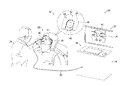
FIG. 1 is a schematic pictorial illustration of a
sinuplasty surgical system 20, in accordance with an
7
CA 2981039 2017-10-02
embodiment of the present invention. System 20 comprises
a magnetic position tracking system, which is configured
to track the position of one or more position sensors in
the head of a patient 22. The magnetic position tracking
system comprises magnetic field-generators and one or
more position sensors. The position sensors generate
position signals in response to sensed external magnetic
fields from the field generators, thereby enabling a
processor 34 to map the position of each sensor in the
coordinate system of the position tracking system as will
be described below.
This method of position sensing is implemented in
various medical applications, for example, in the CARTOTm
system, produced by Biosense Webster Inc. (Diamond Bar,
Calif.) and is described in detail in U.S. Patents
5,391,199, 6,690,963, 6,484,118, 6,239,724, 6,618,612 and
6,332,089, in PCT Patent Publication WO 96/05768, and in
U.S. Patent Application Publications 2002/0065455 Al,
2003/0120150 Al and 2004/0068178 Al, whose disclosures
are all incorporated herein by reference.
In the present example, system 20 comprises a
location pad 40, which comprises multiple field-
generators 44 fixed on a frame 46. In the exemplary
configuration shown in FIG. 1, pad 40 comprises five
field-generators 44, but any other suitable number of
generators 44 can be used. Pad 40 further comprises a
pillow 42 placed under a head 41 of patient 22, such that
generators 44 are located at fixed, known positions
external to the patient. System 20 further comprises a
console 33, which comprises a driver circuit (not shown)
configured to drive field-generators 44 with suitable
8
CA 2981039 2017-10-02
signals so as to generate magnetic fields in a predefined
working volume around head 41.
In an embodiment, processor 34 is typically a
general-purpose computer comprising suitable front end
and interface circuits for receiving data from external
sources, as well as measurements from wand 30, via a
cable 32, and for controlling other components of system
20. Console 33 further comprises input devices 39 and a
user display 36, which is configured to display the data.
In some embodiments, system 20 comprises a
registration tool, such as a handheld wand 30, which is
used by system 20 for registering the coordinate system
of the magnetic position tracking system with that of a
pre-acquired CT image. The registration tool is
configured to acquire ultrasound and position
measurements, and is depicted in detail in Fig. 2 below.
Typically, a physician 24 attaches wand 30
sequentially to multiple predefined locations on an
external surface of patient head 41. Each predefined
location is typically chosen to be an easily identifiable
feature on head 41, such as a cheek bone protrusion, a
bridge of a nose 26 (located between the eyes of patient
22), a tip of nose 26, a chin, or any other suitable
identifiable feature.
In an embodiment, processor 34 receives a
computerized tomography (CT) image 35 obtained using an
external CT system (not shown). Processor 34 uses image
to form a surface image of at least part of patient
head 41. In some embodiments, processor 34 may use
30 hounsfield units (HU) ranging between 700 and 3000 for
determining the radiodensity of bones in the patient
face, compared with HU of -1000, which is a standard
9
CA 2981039 2017-10-02
scale for air, so as to determine boundaries of the
patient face. In an alternative embodiment, HU above 500
may be used for determining the radiodensity of bones, HU
of -200 and below may be used for air, and HU ranging
between -200 and 500 may be used for muscular tissue.
Alternatively, any other suitable values can be used.
Further alternatively, processor 34 may distinguish
between different types of tissue in the CT image, and in
particular identify bone tissue, using any other suitable
criterion or technique.
In an embodiment, when placed at a predefined
location on the patient head, wand 30 is configured to
(i) acquire US measurements of bone tissue, and (ii)
generate position signals indicative of the position of
this predefined location in the coordinate system of the
magnetic position tracking system. The acquisition of the
bone tissue measurements by wand 30 is described in
detail in Fig. 2 below.
In some embodiments, processor 34 is configured to
calculate two coordinates for each predefined location on
the patient head - A "US coordinate" and a "CT
coordinate." The US coordinate is derived from the US and
position measurements of wand 30 at this predefined
location, and is indicative of the coordinate of the bone
tissue at this location in the coordinate system of the
magnetic position tracking system. The CT measurement is
indicative of the coordinate of the bone tissue at this
location, as identified in the CT image.
In an embodiment, processor 34 is configured to
correlate between the US coordinates and the CT
coordinates of the predefined locations in image 35, so
CA 2981039 2017-10-02
as to register the CT image with the coordinate system of
the position tracking system.
The registration process is typically performed
before the actual sinuplasty procedure. During the
sinuplasty procedure, physician 24 may insert into head
41 a medical device (not shown), such as a sinuplasty
catheter or other surgical tool, which comprises an
additional position sensor of the position-tracking
system. Since the CT image is already registered with the
position-tracking system, physician 24 may navigate the
medical device whose distal end is displayed on the CT
image, to a target location in head 41.
In alternative embodiments, instead of CT image 35,
processor 34 is configured to receive one or more images
acquired using another suitable anatomical imaging
technique, such as fluoroscopy or magnetic resonance
imaging (MRI), and to register these anatomical images
with the coordinate system as described above.
Fig. 1 shows only elements related to the disclosed
techniques, for the sake of simplicity and clarity.
System 20 typically comprises additional modules and
elements that are not directly related to the disclosed
techniques, and thus, intentionally omitted from Fig. 1
and from the corresponding description.
Processor 34 may be programmed in software to carry
out the functions that are used by the system, and to
store data in a memory (not shown) to be processed or
otherwise used by the software. The software may be
downloaded to the processor in electronic form, over a
network, for example, or it may be provided on non-
transitory tangible media, such as optical, magnetic or
electronic memory media. Alternatively, some or all of
11
CA 2981039 2017-10-02
the functions of processor 34 may be carried out by
dedicated or programmable digital hardware components.
REGISTERING ANATOMICAL IMAGES WITH A POSITION-TRACKING
SYSTEM USING ULTRASOUND
Fig. 2 is a schematic, pictorial illustration of
wand 30, in accordance with an embodiment of the present
invention. The figure shows wand 30 placed at one of the
multiple predefined locations on patient head 41. In some
embodiments, wand 30 comprises a housing 58, which
contains a position sensor 60 concentrically disposed
around an ultrasound (US) transducer 66. The US
transducer is configured to produce US pulses 70 into the
tissue. Fig. 2 shows example tissue structure, which
comprises a skin layer 50, an intermediate tissue 52, a
bone tissue 56 and an interface layer 54 between tissue
52 and 56.
In an embodiment, physician 24 attaches a tip 59 of
wand 30 to skin 50 at one of the predefined locations
(e.g., bridge of nose 26), and activates transducer 66 so
as to produce US pulses 70. US Pulses 70 traverse skin
layer 50 and intermediate tissue 52 toward interface
layer 54 and bone tissue 56. Pulses 70 are reflected from
interface 54 and travel through tissue 52 and skin layer
50, back to wand 30.
Wand 30 is configured to measure round-trip
propagation times of the US pulses, also denoted time-of-
flight (TOF). Based on the known speed of the US pulses
in tissue 52 and skin layer 50, processor 34 is
configured to translate the measured TOF into a thickness
of tissue 52, i.e., into the distance between transducer
66 and bone tissue 56. In an embodiment, wand 30 is
12
CA 2981039 2017-10-02
configured to transmit the position measurements obtained
from sensor 60, and the TOF measured using US transducer
66, via cable 32, to processor 34.
In some embodiments, sensor 60 is concentrically
disposed around transducer 66. Using the position of
sensor 60 and the TOF measurement of transducer 66,
processor 34 is configured to calculate the US
coordinate, i.e., the position of the bone tissue in the
coordinate system of the position tracking system.
In alternative embodiments, sensor 60 and transducer
66 may be concentrically arranged in any other suitable
arrangement. In yet alternative embodiments, sensor 60
may be fitted at a fixed, known displacement relative to
transducer 66, thus, processor 34 may take into account
the fixed displacement in calculating the US coordinate
of the bone tissue.
As depicted in Fig. 2, the pressure applied by tip
59 may deform skin layer 50 and tissue 52. Processor 34
essentially prevents such deformation from distorting the
registration, since the registration is based on bone
tissue correlation.
Fig. 3 is a flow chart that schematically
illustrates a method for registering CT image 35 with the
coordinate system of the position tracking system, in
accordance with another embodiment of the present
invention. The method begins with a CT image acquisition
step 100, in which processor 34 receives one or more CT
images that capture bone tissue (referred to herein as CT
bone images) of head 41.
At a registration tool attachment step 102,
physician 24 attaches wand 30 sequentially to the
multiple predefined locations in head 41. The subsequent
13
CA 2981039 2017-10-02
steps (104-110) are performed at each of these predefined
locations.
At a position calculation step 104, processor 34
receives the position measurement from sensor 60 and
calculates the position of transducer 66 in the
coordinate system of the position tracking system. At an
US measurements step 106, system 20 activates transducer
66 so as to acquire US measurements. At a tissue position
calculation step 108, processor 34 receives the position
signals from transducer 66 and the TOF measurement from
transducer 66, and calculates the US coordinate, i.e.,
the position of the bone tissue in the coordinate system
of the position tracking system. At a CT tissue
identification step 110, processor 34 identifies the
corresponding CT coordinate, i.e., the position of the
bone tissue in CT image 35, using any suitable technique
known in the art. As noted above, steps 104-110 are
repeated per each predefined location.
At a registration step 112, processor 34 registers
CT image 35 with the coordinate system of the position
tracking system by correlating between the US bone tissue
coordinates and the corresponding CT bone tissue
coordinates at the predefined locations in head 41.
Although the embodiments described herein mainly
address sinuplasty procedures, the methods and systems
described herein can also be used in other applications,
such as in orthopedic procedures, in which physician 24
may attach wand 30 to any suitable deformable feature on
a human body.
It will thus be appreciated that the embodiments
described above are cited by way of example, and that the
present invention is not limited to what has been
14
CA 2981039 2017-10-02
particularly shown and described hereinabove. Rather, the
scope of the present invention includes both combinations
and sub-combinations of the various features described
hereinabove, as well as variations and modifications
thereof which would occur to persons skilled in the art
upon reading the foregoing description and which are not
disclosed in the prior art. Documents incorporated by
reference in the present patent application are to be
considered an integral part of the application except
that to the extent any terms are defined in these
incorporated documents in a manner that conflicts with
the definitions made explicitly or implicitly in the
present specification, only the definitions in the
present specification should be considered.
15
CA 2981039 2017-10-02