Note: Descriptions are shown in the official language in which they were submitted.
METHOD AND SYSTEM FOR ASSESSING STAIN QUALITY FOR IN-SITU
HYBRIDIZATION AND IMMUNOHISTOCHEMISTRY
[0000]
BACKGROUND OF THE DISCLOSURE
1100011 Immunohistochemistry (IHC) refers to the processes of detecting,
localizing,
and/or quantifying antigens, such as a protein, in a biological sample using
antibodies specific to
the particular antigens. LEIC provides the substantial advantage of
identifying exactly where a
particular protein is located within the tissue sample. It is also an
effective way to examine the
tissues themselves. In situ hybridization (ISH) refers to the process of
detecting, localizing, and
quantifying nucleic acids. Both IHC and ISH can be performed on various
biological samples,
such as tissue (e.g. fresh frozen, formalin fixed, paraffin embedded) and
cytological samples.
Recognition of the targets can be detected using various labels (e.g.,
chromogenic, fluorescent,
luminescent, radiometric), irrespective of whether the target is a nucleic
acid or an antigen.
[0002] To robustly detect, locate, and quantify targets in a clinical
setting, amplification
of the recognition event is desirable as the ability to confidently detect
cellular markers of low
abundance becomes increasingly important for diagnostic purposes. For example,
depositing at
the marker's site hundreds or thousands of label molecules in response to a
single antigen
detection event enhances, through amplification, the ability to detect that
recognition event.
Adverse events often accompany amplification, such as non-specific signals
that are apparent as
an increased background signal. An increased background signal may interfere
with the clinical
analysis by obscuring faint signals that may be associated with low, but
clinically significant,
expressions.
[0003] Despite efforts to restrict labeling to the target sequence of
interest in ISH,
anomalous non-specific localization of the detection reagent or DNA probe may
occur due to a
Date recu/Date Received 2020-04-20
CA 02981155 2017-09-27
WO 2016/189065 PCT/EP2016/061859
variety of causes, such as inconsistent performance of staining instruments,
instability of
reagents and loss of specific binding activity or aggregation, inappropriate
staining protocols,
and contamination of the slide. The same issues may apply to samples stained
with chromogens,
fluorophores, and/or quantum dots.
[0004] For commercial in-vitro diagnostic ISH assays, the staining
performance, e.g.
stain uniformity, stain intensity, or background staining, must be validated
to deliver sensitive
and specific staining to the sequences of interest, with a high degree of
repeatability. For this
reason, specifications that define the various factors that influence
acceptable performance of
ISH assays must be developed and documented.
[0005] Current methods for NH performance specifications are based
primarily on
subjective impressions of various factors recorded on a scale that is
developed to aid in statistical
measurement over a large number of slides. Such subjective scoring is
generally achieved
through direct inspection of slides through the oculars of a brightfield or
fluorescent microscope.
Human perception is largely comparative, and the subjective scoring methods in
use cannot be
defined to precise standards because of the influence of experience, visual
acuity, and
understanding of the scaling system being used in a particular context.
100061 In current practice, there are at least three subjective
measurements that are
weighed in determining overall stain quality assessment for ISH: (i) intensity
(or contrast) of the
stain localizations, (ii) coverage (the percentage of potential available
hybridization sites that
exhibit signal), and (iii) background (non-specific localization of signal,
due to adherence of
either the reporter (quantum dot and conjugated antibody) or adherence of the
DNA probe itself.
Thus, it is difficult to compare slide-to-slide variation and nearly
impossible to define concrete
specifications for staining performance.
100071 The aim of the present invention is therefore to provide a means for
assessing the stain
quality in the fields of in-situ hybridization and immunohistochemistry and to
provide a means
for establishing objective criteria for assessing the stain quality for in-
situ hybridization and
immunohistoehemistry.
SUMMARY OF THE DISCLOSURE
2
CA 02981155 2017-09-27
WO 2016/189065 PCT/EP2016/061859
100081 Immunohistochemistry (IHC) and in situ hybridization (1SH) have the
aim of
detecting, localizing and quantifying certain analytes for diagnostic
purposes. The quality of the
stains which are analyzed may deviate for various reasons. The present
invention provides a
method and system for assessing the stain quality and for establishing
objective criteria for
assessing the stain quality for application in the fields of in-situ
hybridization and
immunohistochem istry.
[0009] An embodiment of the invention comprises the steps of unmixing multi-
spectral image
data of a tissue specimen to obtain analyte intensity images, each analyte
intensity image
comprising signals from a single stain, computing metrics based on the analyte
intensity images,
wherein the metrics are uniformity, distribution and/or dispersion of pixel
intensity values in the
analyte intensity images and assessing a stain quality of a slide by comparing
the computed
metrics to pre-determined cutoff values regarding uniformity, distribution
and/or dispersion of
pixel intensity, wherein the stain quality of the slide is assessed as
acceptable if the computed
metrics meet or exceed the pre-determined cutoff values, and wherein the stain
quality of the
slide is assessed as unacceptable if the computed metrics do not meet the pre-
determined cutoff
values. in order to establish objective criteria for assessing stain quality,
in one possible
embodiment, the method and system includes the step of deriving cut-off values
regarding
uniformity, distribution and/or dispersion of pixel intensity by combining the
computed metrics
based on the analyte intensity images with pre-established rating data
quantifying the stain
quality.
[0010] In accordance with embodiments of the invention there is provided a
method for
assessing the stain quality for application in in-situ hybridization and
immunohistochemistry. In
one possible embodiment, this method comprises the steps of unmixing multi-
spectral image
data of a tissue specimen to obtain analyte intensity images, each analyte
intensity image
comprising signals from a single stain, computing metrics based on the analyte
intensity images,
wherein the metrics are uniformity, distribution and/or dispersion of pixel
intensity values in the
analyte intensity images and assessing a stain quality of a slide by comparing
the computed
metrics to pre-determined cutoff values regarding uniformity, distribution
and/or dispersion of
pixel intensity. Herein, the stain quality of the slide is assessed as
acceptable if the computed
3
CA 02981155 2017-09-27
WO 2016/189065 PCT/EP2016/061859
=
metrics meet or exceed the pre-determined cutoff values, and the stain quality
of the slide is
assessed as unacceptable if the computed metrics do not meet the pre-
determined cutoff values.
[0011] According to an embodiment of the present invention, in a first step,
the multi-spectral
image data are unmixed. In general, the unmixing process extracts stain-
specific or analyte
specific channels to determine local concentrations of individual stains using
reference spectra
that are well known for standard types of tissue and stain combinations.
Therefore, via the
unmixing step, intensity images may be derived for different types of analytes
which are
simultaneously present in the analysed slide. In a further step, various
computing metrics are
applied to the analyte intensity image. These metrics pertain to the
uniformity, distribution
and/or dispersion of pixel intensity. As has been found, these three metrics
are suitable for
determining the stain quality of a slide. According to the present invention,
it is preferable if all
three metrics are applied for assessing stain quality.
[0012] The computed metrics are compared to predetermined cutoff values. These
cutoff values
may have been determined manually. These cutoff values may also have been
determined in an
automated manner, which is also a subject of the present invention. If the
computed metrics meet
or exceed the predetermined cutoff values, the stain quality of the slide is
acceptable. This
indicates that the slide is suitable for diagnostic work.
[0013] According to an embodiment of the present invention, it is preferable
that the metrics of
uniformity and distribution of pixel intensity values in the analyte intensity
images are derived
via an entropy calculation. In addition, it is preferable that the metric of
dispersion of pixel
intensity value in the analyte intensity images is derived via calculation of
mean-variance values.
These methods have been found highly suitable for providing accurate results
regarding the
uniformity, distribution and dispersion to be found in analyte intensity
images.
[0014] Preferably, the entropy calculation is performed as follows. In a first
step, image
histograms of intensity values are derived from each of the analyte intensity
images. In a second
step, a probability that a pixel sampled from an analyte intensity image has a
particular value in
the respective histogram is calculated. For this purpose, intensity values may
be derived by
sorting pixels from each analyte intensity images into bins.
[0015] According to an embodiment, the probability that a pixel sampled from
an analyte
intensity image has a particular value in the respective histogram is
calculated by summing a
total number of pixels in all bins of the derived histogram to provide a total
number of pixels in
4
CA 02981155 2017-09-27
WO 2016/189065 PCT/EP2016/061859
that derived histogram, dividing a number of pixels in each bin of the
histogram by the total
number of pixels in the histogram to provide a probability of a pixel
belonging within a
particular bin, multiplying each probability of a pixel belonging within a
particular bin by the
logarithm to the base of 2 of the probability to provide a value in bits, and
summing the values in
bits. Thus, the entropy value may be derived.
100161 Besides the calculation of the entropy value, the calculation of the
mean-variance ratio
values is highly important, as it may be used for determining the dispersion
of the pixel intensity
values in the analyte intensity images. The mean-variance ratio values are
computed by deriving
image histograms of intensity values from each of the analyte intensity images
and calculating a
ratio of a measured intensity mode value and a measured intensity variance
value from the
derived histograms.
[00171 The method as described may have applications in imnaunohistoehemistry
as well as in
situ hybridization. The stains may be selected from the group consisting of
quantum dots,
fluorophores, enzyme-deposited fluorophores and chromogens.
[0018] According to an embodiment of the present invention, the multi-spectral
image data may
be derived from a spectral cube (also referred to as an image cube or
hyperspectral cube to those
of ordinary skill in the art). A spectral cube contains the image data along
two axes, and the
wavelength data at a third axis. 'therefore, the spectral cube represents all
the relevant input data
required for the process of unmixing the image data.
100191 The underlying problem which necessitates unmixing is that different
stains may cause an
increase in intensity at overlapping wavelengths. Therefore, unmixing aims to
retrieve the
intensity of each separate stains, accounting for the fact that the intensity
of certain wave lengths
may have been elevated by multiple stains. According to the present invention,
this problem
should preferably be solved by applying a linear least squares algorithm,
which is known to those
of ordinary skill in the art.
[0020] In one embodiment of the present invention, the analyte intensity
images are thresholded
prior to computing the entropy values and mean-variance ration values. This
operation restricts
analysis of signals to a consistent part of the dynamic range of the data
acquired and avoids the
inclusion of pixel values that are not relevant to the signal localization.
[00211 The aim of the present invention is also achieved by a system for
assessing the stain
quality for application in in-situ hybridization and immunohistochemistry,
which is configured
CA 02981155 2017-09-27
WO 2016/189065 PCT/EP2016/061859
for executing the steps as previously described. In one possible embodiment,
this system
comprises a computer or workstation for performing the relevant calculations
on image data.
Preferably, the system comprises an imaging apparatus. According to a
preferred embodiment,
this imaging apparatus is a multi-spectral imaging system.
[00221 In one possible embodiment, the aim of the present invention is
achieved by a method for
establishing objective criteria for assessing the stain quality for
application in in-situ
hybridization and immunohistochemistry, comprising the steps of selecting a
set of reference
slides and obtaining multi-spectral image data for each reference slide,
wherein each reference
slide is annotated with rating data quantifying the stain quality, umnixing
multi-spectral image
data of a tissue specimen to obtain analyte intensity images, each analyte
intensity image
comprising signals from a single stain, computing metrics based on the analyte
intensity images,
wherein the metrics are uniformity, distribution and dispersion of pixel
intensity values in the
analyte intensity images and deriving cutoff values for uniformity,
distribution and/or dispersion
of pixel intensity for assessing the stain quality by combining the computed
metrics based on the
analyte intensity images and the rating data quantifying the stain quality.
[00231 This implies that according to this embodiment, image data is obtained
from multiple
reference slides and subsequently, the steps of unmixing and application of
the relevant metrics
are performed. Therein, it is preferable that the metrics of uniformity and
distribution of pixel
intensity values in the analyte intensity images are derived via an entropy
calculation. In
addition, it is preferable if the metric of dispersion of pixel intensity
value in the analyte intensity
images is derived via calculation of mean-variance values. These methods have
been found
highly suitable for providing accurate results regarding the uniformity,
distribution and
dispersion to be found in analyte intensity images.
[00241 After these metrics have been computed, cutoff values values regarding
uniformity,
distribution and/or dispersion of pixel intensity are automatically derived.
In order to be able to
establish these cutoff values, rating data for each reference slide is taken
into account. This rating
data has been established before application of the present method. For
instance, each slide may
have been identified as having acceptable or unacceptable stain quality as
judged by a
pathologist or other medical professional.
6
CA 02981155 2017-09-27
WO 2016/189065 PCT/EP2016/061859
[0025] Preferably, the cutoff values are established by methods of machine
learning. According
to one particular embodiment, the cutoff values are derived by applying a
support vector
machine, lower discriminant analysis and/or a logistic regression.
[0026] In another embodiment, the invention also includes a system for
establishing objective
criteria for assessing the stain quality for in-situ hybridization and
irnmunohistochcmistry,
wherein the system is configured to execute the method for establishing
objective criteria for
assessing the stain quality for in-situ hybridization and
immunohistochemistry, as previously
described. The system may include a personal computer and/or a workstation.
The system may
also include an imaging apparatus, which is preferably a multi-spectral
imaging system.
[0027] In one aspect of the present disclosure is a computer device and method
for stain
assessment. In general, the computer device is configured to receive image
data from a tissue
specimen having one or more detectable stains or analytes therein; unmix the
image data to
obtain analyte intensity images, each analyte intensity image corresponding to
a different image
channel (e.g. an image channel corresponding to a particular stain or
analyte); compute metrics
from the analyte intensity images; and evaluate the computed metrics to (i)
establish objective
criteria for staining performance (e.g. cutoff values for a particular assay
or set of stains);
(ii) assess a stain quality of a slide given predetermined cutoff values; or
(iii) qualify staining
inconsistencies (e.g. "root cause analysis"). In some embodiments, the image
data is multi-
spectral image data and the tissue specimen was stained in a multiplex assay.
[0028] In another aspect of the present disclosure is a computer device for
objective stain
assessment comprising one or more processors and at least one memory, the at
least one memory
storing non-transitory computer-readable instructions for execution by the one
or more
processors to cause the one or more processors to: (i) unmix multi-spectral
image data of a tissue
specimen to obtain analyte intensity images, each analyte intensity image
comprising signals
from a single stain (e.g. the stains may be chrornophores, fluorophores,
quantum dots, etc. and
the tissue may have been stained in an 11-IC and/or ISH assay); (ii) compute
metrics based on the
analyte intensity images, wherein the metrics are entropy values and mean-
variance ratio values;
and (iii) assess a stain quality of a slide by comparing the computed metrics
to pre-determined
cutoff values (e.g. predetermined entropy cutoff values, predetermined mean-
variance ratio
cutoff values, etc.), wherein the stain quality of the slide is assessed as
acceptable if the
computed metrics meet or exceed the pre-determined cutoff values, and wherein
the stain quality
7
CA 02981155 2017-09-27
WO 2016/189065 PCT/EP2016/061859
of the slide is assessed as unacceptable if the computed metrics do not meet
the pre-determined
cutoff values (where the terms 'acceptable' and 'unacceptable' are as defined
herein). In some
embodiments, the predetermined cutoff values arc specific to a particular
assay or to particular
detectable stains or analytes. In embodiments where the stain quality is
determined to be
unacceptable, instructions are provided to determine the root cause of any
staining
inconsistencies (e,g, to determine whether the root cause was due to a
deficiency in scanning or
due to a deficiency in staining). In some embodiments, the staining quality is
an assessment of
the uniformity of a stain. In some embodiments, the staining quality is an
assessment of
anomalous background staining. In some embodiments, the staining quality is an
assessment of
the uniformity of a stain and an assessment of an amount of anomalous
backgrounds staining.
100291 In some embodiments, the entropy values are computed by (i) deriving
image
histograms of intensity values from each of the analyte intensity images, and
(ii) calculating a
probability that a pixel sampled from an analyte intensity image has a
particular value in the
respective histogram. In some embodiments, the image histograms of intensity
values are derived
by sorting pixels from each analyte intensity image into bins. In some
embodiments, the
probability that a pixel sampled from an analyte intensity image has a
particular value in the
respective histogram is calculated by (i) summing a total number of pixels in
all bins of the
derived histogram to provide a total number of pixels in that derived
histogram; (ii) dividing a
number of pixels in each bin of the histogram by the total number of pixels in
the histogram to
provide a probability of a pixel belonging within a particular bin; (iii)
multiplying each
probability of a pixel belonging within a particular bin by the logarithm to
the base of 2 of the
probability to provide a value in bits; and (iv) summing the values in bits.
[0030] In some embodiments, the mean-variance ratio values are derived by
(i) deriving
image histograms of pixel intensity values from each of the analyte intensity
images, and (ii)
calculating a ratio of a measured pixel intensity mode value and a measured
pixel intensity
variance value from the derived histograms. In some embodiments, the pixel
intensity mode
value represents the pixel intensity value that occurs most often in a
particular histogram. In
some embodiments, the pixel intensity variance value represents a value of how
spread apart
certain pixel intensities are in a histogram.
[0031] In some embodiments, the one or more detectable analytes are
selected from the
group consisting of quantum dots, fl uoroph ores, enzyme-deposited
fluorophores and
8
CA 02981155 2017-09-27
WO 2016/189065 PCT/EP2016/061859
chromogens. in some embodiments, the tissue samples are stained in a
fluorescence in situ
hybridization assay (FISH'). In some embodiments, the tissue sample is stained
in a FISH assay
with one or more quantum dots. in some embodiments, the tissue sample is
stained in a FISH
assay to detect a chromosomal breakpoint. in some embodiments, the multi-
spectral image data
is unmixed by applying a linear least squares algorithm. In some embodiments,
the analyte
intensity images are thresholded prior to metric computation.
[0032] In some embodiments, the multi-spectral image data comprises scanned
images of
a stained tissue specimen, e.g. specimens mounted on a slide. In some
embodiments, spectral
images of the tissue sample are taken at several axial positions. In some
embodiments, the
scanned images are combined into a spectral cube (also referred to as an image
cube or
hyperspectral cube to those of ordinary skill in the art). In some
embodiments, the tissue samples
were stained in a multiplex LHC and/or ISH assay for the detection of
biomarkers therein.
[0033] In another aspect of the present disclosure is a computer device for
objective stain
assessment comprising one or more processors and at least one memory, the at
least one memory
storing non-transitory computer-readable instructions for execution by the one
or more
processors to cause the one or more processors to: unmix a multi-spectral
image of a tissue
specimen to obtain analyte intensity images; compute metrics based on the
analyte intensity
images, wherein a first metric is a numerical descriptor of the uniformity and
distribution of pixel
intensity values in the analyte intensity images, and wherein a second metric
is a numerical
descriptor of the dispersion of pixel intensity values in the analyte
intensity images, the pixel
intensity values corresponding to signals from a detectable stain in each
analyte intensity image;
assess a stain quality of a slide by comparing the computed metrics to pre-
determined cutoff
values, wherein the stain quality of the slide is assessed as acceptable if
the computed metrics
meet or exceed the pre-determined cutoff values, and wherein the stain quality
of the slide is
assessed as unacceptable if the computed metrics do not meet the pre-
determined cutoff values.
In embodiments where the stain quality is determined to be unacceptable,
instructions are
provided to determine the root cause of any staining inconsistencies (e.g. to
determine whether
the root cause was due to a deficiency in scanning or due to a deficiency in
staining). In some
embodiments, images deemed unacceptable are re-scanned and stain quality is
again assessed. In
some embodiments, the staining quality is an assessment of the uniformity of a
stain. In some
embodiments, the staining quality is an assessment of anomalous background
staining. In some
9
CA 02981155 2017-09-27
WO 2016/189065 PCT/EP2016/061859
embodiments, the staining quality is an assessment of the uniformity of a
stain and an assessment
of an amount of anomalous backgrounds staining.
03 41 In some embodiments, the entropy values are computed by (i) deriving
image
histograms of intensity values from each of the analyte intensity images, and
(ii) calculating a
probability that a pixel sampled from an analyte intensity image has a
particular value in the
respective histogram. In some embodiments, the image histograms of intensity
values are derived
by sorting pixels from each analyte intensity images into bins. In some
embodiments, the
probability that a pixel sampled from an analyte intensity image has a
particular value in the
respective histogram is calculated by (i) summing a total number of pixels in
all bins of the
derived histogram to provide a total number of pixels in that derived
histogram; (ii) dividing a
number of pixels in each bin of the histogram by the total number of pixels in
the histogram to
provide a probability of a pixel belonging within a particular bin; (iii)
multiplying each
probability of a pixel belonging within a particular bin by the logarithm to
the base of 2 of the
probability to provide a value in bits; and (iv) summing the values in bits.
[0 03 5] In some embodiments, the mean-variance ratio values are derived by
(i) deriving
image histograms of intensity values from each of the analyte intensity
images, and (ii)
calculating a ratio of a measured pixel intensity mode value and a measured
pixel intensity
variance value from the derived histograms. In some embodiments, the one or
more detectable
analytes are selected from the group consisting of quantum dots, fluorophores,
enzyme-deposited
fluorophores and chromogens. In some embodiments, the pre-determined cutoff
values are stored
in the non-transitory memory. In some embodiments, a database stores
predetermined cutoff
values for different assays and/or different detectable analytes. In some
embodiments, the
analytes being detected comprise one or more quantum dots, fluorophores,
enzyme-deposited
fluorophores or chrornogenie stains, or any combination thereof.
[0036] In another aspect of the present disclosure is a computer device for
objective stain
assessment comprising one or more processors and at least one memory, the at
least one memory
storing non-transitory computer-readable instructions for execution by the one
or more
processors to cause the one or more processors to: unmix multi-spectral image
data of a tissue
specimen stained in an immunohistochernical assay or an in situ hybridization
assay for the
presence of a particular biomarker to obtain analyte intensity images, each
analyte intensity
image comprising signals from a single stain; compute entropy values for each
of the analyte
CA 02981155 2017-09-27
WO 2016/189065 PCT/EP2016/061859
intensity images by (i) deriving image histograms of intensity values from
each of the analyte
intensity images, and (ii) calculating a probability that a pixel sampled from
an analyte intensity
image has a particular value in the respective histogram; compute mean-
variance ratios for each
of the analyte intensity images by (i) deriving image histograms of intensity
values from each of
the analyte intensity images, and (ii) calculating a ratio of a measured
intensity mode value and a
measured intensity variance value from the intensity histograms, and assess a
uniformity of a
stain and/or the presence of anomalous background staining by comparing the
computed entropy
and mean-variance ratio values to pre-determined entropy cutoff values and
mean-variance ratio
cutoff values, wherein the uniformity of the stain and/or the presence of
anomalous background
staining is assessed as acceptable if the computed entropy and mean-variance
ratio values meet
or exceed the pre-determined cutoff values, and wherein the uniformity of the
stain and/or the
presence of anomalous background staining is assessed as unacceptable if the
computed entropy
and mean-variance ratio values do not meet the pre-determined cutoff values.
(0037] In another aspect of the present disclosure is a system (e.g. an
analyzer)
comprising an imaging apparatus (e.g. a multi-spectral imaging system) and a
computer device
for stain assessment, as descrihed herein.
(NA Tn another aspect of the present disclosure is a computer-
implemented method of
stain assessment comprising: unmixing multi-spectral image data to obtain
analyte intensity
images, the analyte intensity images each comprising a single image channel
corresponding to
signals from a particular stain; computing entropy values for each of the
analyte intensity
images; computing mean-variance ratios for each of the analyte intensity
images; and assessing a
stain quality of a slide by comparing the computed entropy and mean-variance
ratio values to
pre-determined entropy cutoff values and mean-variance ratio cutoff values,
wherein a stain
quality is assessed as acceptable if the computed entropy and mean-variance
ratio values meet or
exceed the pre-determined cutoff values, and wherein the stain quality is
assessed as
unacceptable if the computed entropy and mean-variance ratio values do not
meet the pre-
determined cutoff values.
[0039] In some embodiments, the entropy values are computed by (i) deriving
image
histograms of intensity values from each of the analyte intensity images, and
(ii) calculating a
probability that a pixel sampled from an analyte intensity image has a
particular value in the
respective histogram. In sonic embodiments, the image histograms of intensity
values are derived
11
CA 02981155 2017-09-27
WO 2016/189065 PCT/EP2016/061859
by sorting pixels from each analyte intensity image into bins. In some
embodiments, the
probability that a pixel sampled from an analyte intensity image has a
particular value in the
respective histogram is calculated by (i) summing a total number of pixels in
all bins of the
derived histogram to provide a total number of pixels in that derived
histogram; (ii) dividing a
number of pixels in each bin of the histogram by the total number of pixels in
the histogram to
provide a probability of a pixel belonging within a particular bin; (iii)
multiplying each
probability of a pixel belonging within a particular bin by the logarithm to
the base of 2, of the
probability to provide a value in bits; and (iv) summing the values in bits.
[0040] In some embodiments, the mean-variance ratio values are derived by
(i) deriving
image histograms of intensity values from each of the analyte intensity
images, and (ii)
calculating a ratio of a measured pixel intensity mode value and a measured
pixel intensity
variance value from the derived histograms. In some embodiments, the one or
more detectable
analytes are selected from the group consisting of quantum dots, fluorophores,
enzyme-deposited
fluorophores and chromogenic.
[0041] In another aspect of the present disclosure is a computer device for
stain
assessment comprising one or more processors and at least one memory, the at
least one memory
storing non-transitory computer-readable instructions for execution by the one
or more
processors to cause the one or more processors to (i) run an image processing
module to obtain
multi-spectral image data (e.g. scanned images) of a tissue specimen having
one or more
detectable stains analytes; (ii) run an unmixing module to unmix the multi-
spectral images into
analyte intensity images; (ii) run a metric computation module to derive
metrics based on the
analyte intensity images; and (iii) run an evaluation module to assess a stain
quality by
comparing the computed metrics to pre-determined cutoff values, wherein a
stain quality is
assessed as acceptable if the computed metrics meet or exceed the pre-
determined cutoff values,
and wherein a stain quality is assessed as unacceptable if the computed
metrics do not meet the
pre-determined cutoff values.
[0042] In some embodiments, the entropy values are computed by (i) deriving
image
histograms of intensity values from each of the analyte intensity images, and
(ii) calculating a
probability that a pixel sampled from an analyte intensity image has a
particular value in the
respective histogram. In some embodiments, the image histograms of intensity
values are derived
by sorting pixels from each analyte intensity images into bins. In some
embodiments, the
12
CA 02981155 2017-09-27
WO 2016/189065 PCT/EP2016/061859
probability that a pixel sampled from an analyte intensity image has a
particular value in the
respective histogram is calculated by (i) summing a total number of pixels in
all bins of the
derived histogram to provide a total number of pixels in that derived
histogram; (ii) dividing a
number of pixels in each bin of the histogram by the total number of pixels in
the histogram to
provide a probability of a pixel belonging within a particular bin; (iii)
multiplying each
probability of a pixel belonging within a particular bin by the logarithm to
the base of 2 of the
probability to provide a value in bits; and (iv) summing the values in bits.
[0043] In some embodiments, the mean-variance ratio values are derived by
(i) deriving
image histograms of intensity values from each of the analyte intensity
images, and (ii)
calculating a ratio of a measured pixel intensity mode value and a measured
pixel intensity
variance value from the derived histograms.
100441 In another aspect of the present disclosure is a computer device for
establishing
objective criteria for stain assessment comprising one or more processors and
at least one
memory, the at least one memory storing non-transitory computer-readable
instructions for
execution by the one or more processors to cause the one or more processors
to: (i) unmix multi-
spectral image data of a tissue specimen stained in an immunohistochernical
assay or an in situ
hybridization assay for the presence of a particular biomarker to obtain
analyte intensity images,
each analyte intensity image comprising signals from a single stain, and
wherein the multi-
spectral image data is from a plurality of reference slides that have been
identified as acceptable
or unacceptable (e.g. known reference slides that have been identified as
having acceptable or
unacceptable stain quality as judged by a pathologist or other medical
professional); (ii) compute
entropy values and mean-variance ratio values based on each of the obtained
analyte intensity
images (e.g. to derive entropy and mean-variance ratios for slides having
acceptable stain quality
and slide having unacceptable stain quality); and (iii) derive cutoff values
for staining quality
assessment (e.g. cutoff values that may be used in subsequent stain assessment
processes) based
on the computed entropy and mean-variance ratio values, wherein the cutoff
values for staining
quality assessment correspond to the stains used in the immunohistochemical
assay and/or the in
situ hybridization assay.
[00451 in some embodiments, the entropy values are computed by (i) deriving
image
histograms of intensity values from each of the analyte intensity images, and
(ii) calculating a
probability that a pixel sampled from an analyte intensity image has a
particular value in the
13
CA 02981155 2017-09-27
WO 2016/189065 PCT/EP2016/061859
respective histogram. In some embodiments, the image histograms of intensity
values are derived
by sorting pixels from each analyte intensity images into bins. In some
embodiments, the
probability that a pixel sampled from an analyte intensity image has a
particular value in the
respective histogram is calculated by (i) summing a total number of pixels in
all bins of the
derived histogram to provide a total number of pixels in that derived
histogram; (ii) dividing a
number of pixels in each bin of the histogram by the total number of pixels in
the histogram to
provide a probability of a pixel belonging within a particular bin; (iii)
multiplying each
probability of a pixel belonging within a particular bin by the logarithm to
the base of 2 of the
probability to provide a value in bits; and (iv) summing the values in bits.
[0046] In some embodiments, the mean-variance ratio values are derived by
(i) deriving
image histograms of intensity values from each of the analyte intensity
images, and (ii)
calculating a ratio of a measured pixel intensity mode value and a measured
pixel intensity
variance value from the derived histograms. In some embodiments, the one or
more detectable
analytes are selected from the group consisting of quantum dots, fluorophores,
enzyme-deposited
fluorophores and chrotnogens. In some embodiments, the pre-determined cutoff
values arc stored
in the non-transitory memory. lin some embodiments, the analytes being
detected comprise one
or more quantum dots, fluorophores, enzyme-deposited fluorophores or
chromogenie stains, or
any combination thereof. In some embodiments, the cutoff values are derived
with a support
vector machine. A support vector machine is a method for pattern analysis and
machine learning.
It is suitable for the derivation of the desired cutoff values. However, any
other suitable machine
learning method may be applied to derive cutoff values, as well.
100471 In another aspect of the present disclosure is a computer-
implemented method for
establishing objective criteria for stain assessment comprising: unmixing
multi-spectral image
data of a tissue specimen from a plurality of reference slides to obtain a
series of analyte
intensity images, wherein each analyte intensity image comprises signals from
a single stain, and
wherein the reference slides have each been identified as acceptable or
unacceptable; computing
entropy values and mean-variance ratio values based on each of the obtained
analyte intensity
images; and deriving cutoff values for staining quality assessment based on
the computed
entropy and mean-variance ratio values, wherein the cutoff values for staining
quality assessment
correspond to the stains used in the itnmunohistochemical assay and/or the in
situ hybridization
assay.
14
CA 02981155 2017-09-27
WO 2016/189065 PCT/EP2016/061859
[0048] In some embodiments, the entropy values are computed by (i) deriving
image
histograms of intensity values from each of the analyte intensity images, and
(ii) calculating a
probability that a pixel sampled from an analyte intensity image has a
particular value in the
respective histogram. In some embodiments, the image histograms of intensity
values are derived
by sorting pixels from each analyte intensity images into bins. In some
embodiments, the
probability that a pixel sampled from an analyte intensity image has a
particular value in the
respective histogram is calculated by (i) summing a total number of pixels in
all bins of the
derived histogram to provide a total number of pixels in that derived
histogram; (ii) dividing a
number of pixels in each bin of the histogram by the total number of pixels in
the histogram to
provide a probability of a pixel belonging within a particular bin; (iii)
multiplying each
probability of a pixel belonging within a particular bin by the logarithm to
the base of 2 of the
probability to provide a value in bits; and (iv) summing the values in bits.
In some embodiments,
the mean-variance ratio values are derived by (i) deriving image histograms of
intensity values
from each of the analyte intensity images, and (ii) calculating a ratio of a
measured pixel
intensity mode value and a measured pixel intensity variance value from the
derived histograms.
[0049] The present invention provides an improved system and method of
evaluating
stain quality, as described herein. In fact, the present invention provides
processes of computing
metrics that serve (a) as a determinant for the development of objective
indicia for stain quality
assessment, and (b) as a means for objectively determining whether a
particular slide meets
staining performance standards by comparing the computed metrics to
predetermined cutoff
values for the analytes being detected. The presently disclosed methods allow
for superior
assessment methods as compared with state-of-the art techniques used by
pathologists and those
which rely solely on subjective criteria. The presently disclosed methods do
not rely on the
subjective interpretations of pathologists and the use of developed objective
criteria, specific for
a particular assay, tissue sample, detectable analyte, and/or processing
method, allows for
consistent and repeatable assessments to be made that are repeatable and less
prone to error.
Moreover, the methods disclosed herein allow (1) the reporting of stain
localization in terms of
descriptive statistics that reflect the spatial distribution of signal
intensities, (2) provide an
indication of anomalous background staining or other artifacts (e.g.
speckling), and/or (3)
distinguish differences between stain distributions and uniformity. The
present disclosure
CA 02981155 2017-09-27
WO 2016/189065 PCT/EP2016/061859
enables a method of evaluating stain quality for ISH/FISH assays and also
enables the
identification of specifications for acceptable stain distribution.
INDUSTRIAL APPLICABILITY
[0050] The present disclosure has industrial applicability in the field of
diagnostics.
BRIEF DESCRIPTION OF DRAWINGS
[0051] FIG. l shows a computer-based system for analyzing specimens in
accordance
with an embodiment of the disclosed technology;
[0052] FIG. 2A illustrates a system for analyzing specimens in accordance
with certain
embodiments of the present disclosure;
[0053] FIG. 2B provides flowchart showing an overview of the modules used
within the
computer-based system and method;
[0054] FICis. 3A and 3B illustrate differences between acceptable and
unacceptable
staining of two sets of slides;
[0055] FIG. 4A provides a flowchart illustrating the steps of image
processing;
[0056] FIG. 4B provides a representation of the combination of data from
several z-
positions into a spectral cube;
[0057] FIG. 4C provides an example of thresholding;
[0058] FIG. 4D provides a flowchart illustrating the steps for the
generation and
projection of spectral cubes;
[0059] FIG. 5A provides a flowchart illustrating the steps of computing an
entropy
metric and a mean-variance ratio metric;
[0060] FIG. 5B illustrates an example of an intensity histogram and the
number of pixels
in any particular bin;
[0061] FIG. 5C illustrates the summation of all pixels in each bin of a
histogram to
provide a total number of pixels in an analyte intensity image from which the
histogram was
derived;
16
CA 02981155 2017-09-27
WO 2016/189065 PCT/EP2016/061859
[0062] FIG. 5D illustrates the step of dividing a number of pixels in a
particular bin by a
total number of pixels in an analyte intensity image;
[0063] FiGs. 6A and 6B illustrate histograms of slides having acceptable
and
unacceptable stain quality;
[0064] FIG. 7 provides a flowchart demonstrating the steps of determining
cutoff values;
[0065] FIG. 8 illustrates a determined cutoff between acceptable and
unacceptable data
points;
[0066] FIGs. 9A and 9B provide flowcharts demonstrating the steps of
determining a
root cause of staining inconsistencies;
[0067] FIG. 10 demonstrates a comparison of image entropy for four slides
FISH labeled
for ERG 5'3';
[0068] FIG. 11 demonstrates a comparison of image mean-variance ratio for
four slides
FISH labeled for ERG 5'3'; and
100691 FIG. 12 provides a scatter plot of entropy vs. mean-variance ratio
for slides
labeled with quantum dot 655 (ERG 3') and quantum dot 565 (ERG 5'); and
[0070] FIG. 13 provides an acquisition strategy for evaluation of FISH
stain quality on
tissue cores.
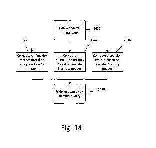
[0071] FIG. 14 provides a flowchart illustrating an embodiment of the
invention wherein
the metrics of uniformity, distribution and dispersion are calculated
separately.
DESCRIPTION OF EMBODIMENTS
[0072] In general, the present disclosure pertains to computer devices and
methods for
objectively assessing stain quality or for developing objective criteria for
assessing the staining
quality of a tissue specimen. The devices and methods described herein provide
comparatively
superior results to subjective techniques currently utilized by pathologists
and trained medical
professionals.
[0073] Definitions
[0074] As used herein, the singular terms "a," "an," and "the" include
plural referents
unless context clearly indicates otherwise. Similarly, the word "or" is
intended to include "and"
unless the context clearly indicates otherwise. The term "includes" is defined
inclusively, such
that "includes A or B" means including A, B, or A and B.
17
CA 02981155 2017-09-27
WO 2016/189065 PCT/EP2016/061859
100751 "The terms "comprising," "including," "having," and the like are
used
interchangeably and have the same meaning. Similarly, "comprises," "includes,"
"has," and the
like are used interchangeably and have the same meaning. Specifically, each of
the terms is
defined consistent with the common United States patent law definition of
"comprising" and is
therefore interpreted to be an open term meaning "at least the following," and
is also interpreted
not to exclude additional features, limitations, aspects, etc. Thus, for
example, "a device having
components a, b, and c" means that the device includes at least components a,
b and e. Similarly,
the phrase: "a method involving steps a, b, and c" means that the method
includes at least steps a,
b, and c. Moreover, while the steps and processes may be outlined herein in a
particular order,
the skilled artisan will recognize that the ordering steps and processes may
vary.
100761 As used herein, the term a "biological sample" can be any solid or
fluid sample
obtained from, excreted by or secreted by any living organism, including
without limitation,
single celled organisms, such as bacteria, yeast, protozoans, and amoebas
among others,
multicellutar organisms (such as plants or animals, including samples from a
healthy or
apparently healthy human subject or a human patient affected by a condition or
disease to be
diagnosed or investigated, such as cancer). For example, a biological sample
can be a biological
fluid obtained from, for example, blood, plasma, serum, urine, bile, ascites,
saliva, cerebrospinal
fluid, aqueous or vitreous humor, or any bodily secretion, a transudate, an
exudate (for example,
fluid obtained from an abscess or any other site of infection or
inflammation), or fluid obtained
from a joint (for example, a normal joint or a joint affected by disease). A
biological sample can
also be a sample obtained from any organ or tissue (including a biopsy or
autopsy specimen,
such as a tumor biopsy) or can include a cell (whether a primary cell or
cultured cell) or medium
conditioned by any cell, tissue or organ. In some examples, a biological
sample is a nuclear
extract. In certain examples, a sample is a quality control sample, such as
one of the disclosed
cell pellet section samples. In other examples, a sample is a test sample.
Samples can be prepared
using any method known in the art by a person of ordinary skill. The samples
can be obtained
from a subject for routine screening or from a subject that is suspected of
having a disorder, such
as a genetic abnormality, infection, or a neoplasia. The described embodiments
of the disclosed
method can also be applied to samples that do not have genetic abnormalities,
diseases,
disorders, etc., referred to as "normal" samples. Samples can include multiple
targets that can be
specifically bound by one or more detection probes.
18
CA 02981155 2017-09-27
WO 2016/189065 PCT/EP2016/061859
[0077] As used herein, the term "chromophore" refers to a molecule or a
part of a
molecule responsible for its color. Color arises when a molecule absorbs
certain wavelengths of
visible light and transmits or reflects others. A molecule having an energy
difference between
two different molecular orbitals falling within the range of the visible
spectrum may absorb
visible light and thus be aptly characterized as a chromophore. Visible light
incident on a
chromophore may be absorbed thus exciting an electron from a ground state
molecular orbital
into an excited state molecular orbital.
[0078] As used herein, the terms "multiplex," "multiplexed," or
"multiplexing" refer to
detecting multiple targets in a sample concurrently, substantially
simultaneously, or sequentially.
Multiplexing can include identifying and/or quantifying multiple distinct
nucleic acids (e.g.,
DNA, RNA, rnRNA, miRNA) and polypeptides (e.g., proteins) both individually
and in any and
all combinations.
[0079] As used herein, the term "spectral cube" refers to data aligned
along three
dimensions. Two of the dimensions are the 'x' and 'y' coordinates of an image
field of view and
the third dimension is wavelength.
[0080] As used herein, the term "target" refers to any molecule for which
the presence,
location and/or concentration is or can be determined. Examples of target
molecules include
proteins, nucleic acid sequences, and haptens, such as haptens covalently
bonded to proteins.
Target molecules are typically detected using one or more conjugates of a
specific binding
molecule and a detectable label.
[0081] An "unmixed image" as used herein encompasses a grey-value or scalar
image
obtained for one channel of a multi-channel image. By unmixing a multi-channel
image one
unmixed image per channel is obtained.
[0082] Overview
10083] The present disclosure is directed to a computer device and computer-
implemented method developed to derive metrics from analyte intensity images
(unmixed, single
channel images corresponding to a particular detectable analyte or stain),
where the derived
metrics may be used to (i) derive cutoff values to serve as objective criteria
for stain assessment
(as opposed to the subjective criteria currently used in the art); (ii) assess
a stain quality of a
particular slide by comparing derived metrics to predetermined cutoff values;
or (iii) to qualify
staining inconsistencies in slides whose stain quality has been deemed
unacceptable.
19
CA 02981155 2017-09-27
WO 2016/189065 PCT/EP2016/061859
[0084] A slide or stain quality may be referred to herein as "acceptable"
or
"unacceptable." As used herein, "acceptable" refers to a slide or stain
quality that meets
subjective performance criteria (e.g. stain uniformity/coverage, stain
intensity, and non-specific
background staining), as determined by an expert pathologist. "Acceptable"
slides possess
uniform staining and the slides do not contain anomalous background staining
to a degree that
would cause errors in any downstream processing or analysis. As used herein,
"unacceptable"
refers to a slide or stain quality that does not meet subjective performance
criteria (e.g. stain
uniformity/coverage, stain intensity, and non-specific background staining),
as determined by an
expert pathologist. Reference "acceptable" and "unacceptable" slides are used
as the basis for
developing correlations between the computed metrics so as to establish cutoff
values for use as
objective indicia for stain assessment (see FIG. 8 and the disclosures
herein). FIGs. 3A and 3B
illustrate the differences between "acceptable" and "unacceptable" slides,
where the
"unacceptable" slides contain non-specific background staining and/or other
artifacts (e.g.
resulting from amplification), which interfere with the ability of a skilled
artisan to subjectively
determine probe localizations. Slides may be determined to be "unacceptable"
if the staining for
probe localizations is not uniform in terms of the size, contrast and
coverage, as this makes the
identification of probe localizations more difficult for the skilled artisan
to discern for both
humans and for machine vision.
[00851 At least some embodiments of the technology disclosed herein relate
to computer
systems and methods for analyzing digital images or multi-spectral image data
captured from
tissue samples stained with one or more stains (e.g. stains supplied to the
tissue in an ISH and/or
1HC assay). While specific examples herein may refer to specific tissues
and/or the application
of specific stains for the detection of certain markers (and hence diseases),
the skilled artisan will
appreciate that different tissues and different stains (or probes) may be
applied to detect different
markers and different diseases. Moreover, while certain embodiments and
examples herein are
directed to 1SH or FISH assays, the skilled artisan will appreciate that the
embodiments are
equally applicable to 1HC assays. In addition, while certain examples employ
quantum dots as
detectable analytes, the skilled artisan will appreciate that other detectable
stains or analytes may
be employed in the embodiments of the present disclosure.
[0086] A computer-based specimen analyzer (10) for analyzing specimens is
shown in
FIG. 1. The computer-based specimen analyzer (10) may comprise an imaging
apparatus (12)
CA 02981155 2017-09-27
WO 2016/189065 PCT/EP2016/061859
and a computer (14), whereby the imaging apparatus (12) and computer may be
communicatively coupled together, e.g. directly, or indirectly over a network
(20). The skilled
artisan will appreciate that other computer devices or systems may be utilized
and that the
computer systems described herein may be communicatively coupled to additional
components,
e.g. specimen analyzers, scanners or imaging systems, automated slide
preparation equipment,
etc. Some of these additional components and the various computers that may be
utilized are
described further herein.
[0087] In general, the imaging apparatus (12), or other image source, can
include,
without limitation, one or more image capture devices. Image capture devices
can include,
without limitation, a camera (e.g., an analog camera, a digital camera, etc.),
optics (e.g., one or
more lenses, sensor focus lens groups, microscope objectives, etc.), imaging
sensors (e.g., a
charge-coupled device (CCD), a complimentary metal-oxide semiconductor (CMOS)
image
sensor, or the like), photographic film, or the like. In digital embodiments,
the image capture
device can include a plurality of lenses that cooperate to prove on-the-fly
focusing. An image
sensor, for example, a CCD sensor can capture a digital image of the specimen.
In some
embodiments, the imaging apparatus (12) is a brightfield imaging system, a
multispectral
imaging (MS.1) system or a fluorescent microscopy system. Additional imaging
devices and
systems are described further herein.
[0088] With reference to FIGs. 1, 2A, and 2B the computer system (14) can
include a
desktop computer, a laptop computer, a tablet, or the like, digital electronic
circuitry, firmware,
hardware, memory (110), a computer storage medium (110), a computer program
(e.g. where the
program is stored within the memory or storage medium), a processor (120),
including a
programmed processor, and/or the like. The illustrated computing system (14)
of FIG. 1 may be
a computer with a screen or display device (16) and an enclosure (18), e.g., a
system enclosed
within a tower, as depicted. The computer system can store digital images in
binary form
(locally, on a server, or other network connected device). The images can also
be divided into a
matrix of pixels. The pixels can include a digital value of one or more bits,
defined by the bit
depth.
[0089] Again, with reference to FIG. 1A, the network (20), in some
embodiments,
interconnects the imaging apparatus (12) and the computer system (14). The
network (20) may
include, without limitation, one or more gateways, routers, bridges,
combinations thereof, or the
21
CA 02981155 2017-09-27
WO 2016/189065 PCT/EP2016/061859
like. The network (20) may include one or more servers and one or more
websites that are
accessible to users and can be used to send and receive information that the
computer system
(14) can utilize. A server may include, without limitation, one or more
associated databases for
storing information (e.g., digital images, algorithms, staining protocols,
cutoff values for
comparative evaluations, or the like). The network (20) may include, but is
not limited to, data
networks using the Transmission Control Protocol (TCP). User Datagrana
Protocol (UDP),
Internet Protocol (IP) and other data protocols. In some embodiments, the
computer device or
system further comprises a display output or other means of providing
data/output to a user,
operator, or downstream instrument or process.
[0090] With reference to FIGs. 2A and 2B, the computer device or system
(14) (or
computer-implemented method) comprises one or more processors (120) and at
least one
memory (110), the at least one memory (110) storing non-transitory computer-
readable
instructions for execution by the one or more processors to cause the one or
more processors to
execute instructions to receive input images (12) (the input images of tissue
specimens stained in
an IHC and/or ISFI), run an image processing module (210) to receive and/or
capture image data
or multi-spectral image data, mu an unmixing module (240) to derive single
channel analyte
intensity images (the channels corresponding to the different detectable
analytes in the tissue
specimens), run a metric computation module (220) to derive certain numerical
descriptors of
stain quality, and run an evaluation module (230) to either evaluate stain
quality, derive objective
criteria to enable further stain quality assessments (e.g., to determine
objective stain criteria for
later assessment), or to evaluate a root cause of a staining inconsistency or
deficiency. Each of
these modules is described in greater detail herein. The skilled artisan will
recognize that any of
the instructions, algorithms, and filters described for use within each module
may be adapted or
changed based on the analytes being detected. In some embodiments, the
computer device or
system further comprises a display output or other means of providing
data/output to a user,
operator, or downstream instrument or process.
[0091] Image Acquisition and Processing
[0092] The methods described herein are developed for evaluating pure
signal
localizations as separated from colocalized signals with overlapping
wavelength distribution (e.g.
such as where multiple stains are applied in a multiplex assay). Typically,
this data is acquired
through means of spectral imaging technology, and the analyte intensity images
(i.e. images of
22
CA 02981155 2017-09-27
WO 2016/189065 PCT/EP2016/061859
the pure signal localization or images representing the analyte) are obtained
through unmixing of
the signals in the raw spectral data (e.g., linear least-squares processing
with reference data for
the spectral components known a priori).
[0093] In general, the image processing module (210) receives or captures
image data,
e.g. multi-spectral image data, as an input and, in conjunction with the
unmixing module (240),
provides analyte intensity images as output to the metric computation module
(220). FIG. 4A
provides a flowchart illustrating the various steps of "image processing"
utilizing the image
processing and unmixing modules. As described in further detail herein,
multiple fields of view
are first selected, step (410), and spectral images are acquired at several
axial (z) positions (step
(420); see also FIG. 4B). The spectral images are then projected through the z-
dimension, step
(430), unmixed with the unmixing module (240) to yield analyte intensity
images, step (440),
and optionally thresholded, step (450).
[0094] Image Processing Module
[0095] Images are acquired from a slide having a tissue specimen disposed
thereon,
wherein the tissue specimen has been stained (e.g. FISH with a fluorophore),
and the tissue
specimen thus comprises one or more detectable stains or analytes. A stain or
analyte is a
molecule or material that can produce a detectable (such as visually,
electronically or otherwise)
signal that indicates the presence and/or concentration of a label in a sample
(the label indicating
the approximate position of a target or biomarker). Examples of analytes and
methods of
"labeling" targets within a biological sample are appended herein.
100961 The process of labeling targets within a biological sample is often
spectrally
multiplexed, i.e. the tissue is stained to identify multiple targets and each
target identified by a
different stain. For example, in a multiplex assay, the tissue sample may be
stained with multiple
fluorophores and/or quantum dots fluorescing at different wavelengths. As
such, the images of
the tissue are acquired in spectral bands that aggregately span the spectral
range defined by
spectral characteristics of the analyte, e.g. quantum dot or fluorescent
dye/probe, used for
labeling. The spectral bands may have wavelengths ranging, for example, from
about 400 nm to
about 900 nm. In some embodiments, spectral images are acquired at about 96
wavelengths,
ranging from about 400 nm to about 800 nm.
[0097] 111 some embodiments, fluorescence can be measured with a
multispectral
imaging system such as those available from Ventana, Tucson, Ariz.; NuanceTM,
Cambridge
23
CA 02981155 2017-09-27
WO 2016/189065 PCT/EP2016/061859
Research & Instrumentation, Woburn, Mass.; or SpectraViewTm, Applied Spectral
Imaging,
Vista, Calif Generally, MSI equips the analysis of pathology specimens with
computerized
microscope-based imaging systems by providing access to spectral distribution
of an image at a
pixel level. While there exists a variety of multispectral imaging systems, an
operational aspect
that is common to all of these systems is a capability to form a multispectral
image. A
multispectral image is one that captures image data at specific wavelengths or
at specific spectral
bandwidths across the electromagnetic spectrum. These wavelengths may be
singled out by
optical filters or by the use of other instruments capable of selecting a pre-
determined spectral
component including electromagnetic radiation at wavelengths beyond the range
of visible light
range, such as, for example, infrared (TR).
10098] An MSI may include an optical imaging system, a portion of which
contains a
spectrally-selective system that is tunable to define a pre-determined number
N of discrete
optical bands. The optical system may be adapted to image a tissue sample,
illuminated in
transmission with a broadband light source onto an optical detector. The
optical imaging system,
which in one embodiment may include a magnifying system such as, for example,
a microscope,
has a single optical axis generally spatially aligned with a single optical
output of the optical
system. The system forms a sequence of images of the tissue as the spectrally
selective system is
being adjusted or tuned (for example with a computer processor) such as to
assure that images
are acquired in different discrete spectral bands. The apparatus may
additionally contain a
display in which appears at least one visually perceivable image of the tissue
from the sequence
of acquired images. The spectrally-selective system may include an optically-
dispersive element
such as a diffractive grating, a collection of optical filters such as thin-
film interference filters or
any other system adapted to select, in response to either a user input or a
command of the pre-
programmed processor, a particular pass-band from the spectrum of light
transmitted from the
light source through the sample towards the detector. An alternative
implementation, a spectrally
selective system defines several optical outputs corresponding to N discrete
spectral bands. This
type of system intakes the transmitted light output from the optical system
and spatially redirects
at least a portion of this light output along N spatially different optical
paths in such a way as to
image the sample in an identified spectral band onto a detector system along
an optical path
corresponding to this identified spectral band.
24
CA 02981155 2017-09-27
WO 2016/189065 PCT/EP2016/061859
1100991 In other embodiments, the imaging may be performed using the
multispectral
imaging system set forth in US Patent Publication No. 2014/0078286, to Ventana
Medical
Systems, Inc., which illustrates a multispectral imaging system and method,
the disclosure of
which is hereby incorporated herein by reference in its entirety.
1101001 Referring again to FIG. 4A, multiple fields of view (FOV) are first
acquired (step
410) from a tissue sample mounted or embedded on a slide (either automated or
manually, as
known by those of skill in the art). In some embodiments, the number of FOV
selected should be
sufficient to provide adequate sampling of the tissue such that the analysis
of the staining
provides an accurate model of the tissue staining pattern as compared to
digitizing the entire
tissue section (see Gundersen, et. al, "The Efficiency of Systematic Sampling
in Stereology and
its Prediction," J. of Microscopy, vol. 147, pt. 3, Sept. 1987, pp.229-263 and
Kayser et al.
"Theory of Sampling and its Application in Tissue Based Diagnosis," Diagnostic
Pathology,
2009, 4:6, the disclosures of which are incorporated herein by reference). In
other embodiments,
at least three FOVs are selected. In yet other embodiments, between about 10
to about 300 FOV
are selected.
101011 For each field of view, spectral images are taken of the tissue
sample at several
axial (z) positions, step (420), such as by optical sectioning. Optical
sectioning is a technique of
optically imaging different depth positions of a three-dimensional sample by
changing the focal
plane in the z-direction and taking images at each plane. See D. A. Agard,
"Optical Sectioning
Microscopy: Cellular Architecture in Three Dimensions," Annual Reviews in
Biophysics and
Bioengineering, vol. 13, pp. 191-219, 1984. S. Joshi and M. I. Miller,
"Maximum a Posteriori
Estimate with Good's Roughness for Three-Dimensional Optical-Sectioning
Microscopy,"
Journal of Optical Society of America, vol. 10, no. 5, pp. 1078-1085, 1993,
the disclosures of
which are incorporated herein by reference. In some embodiments, a camera is
operated at
multiple depths (i.e. focus settings moved in the z-plane direction). In some
embodiments, the z-
positions are spaced at about 1.8 micron in depth of field and about 0.5
micron apart, although
the skilled artisan will be able to select any appropriate spacing considering
the tissue sample
presented and the depth of focus for the optical system. Capturing multiple z-
positions (z-planes)
mitigates the effects of tissue section thickness and axial chromatic
aberration.
[0102] In some embodiments the initial focal plane is determined using a
filter associated
with a background stain signal (e.g. DAPI, 4'-6-Diamidino-2-phenyfindole). In
some
CA 02981155 2017-09-27
WO 2016/189065 PCT/EP2016/061859
embodiments, a initial focal plane coordinate in x,y,z spatial dimensions is
defined for multiple
FOV per tissue section, and the automated acquisition subsequently collects
the more lengthy
spectral acquisition at three planes for each defined region.
101031 The data from the z-positions (labeled "z" within FIG. 4A) are
combined into a
spectral cube that represents the data through 3-dimensional space, step
(430), as known to those
of ordinary skill in the art. Generally, the process of forming a spectral
cube is illustrated in FIG.
LIB. In some embodiments, the spectral cube denotes a virtual spectral data
structure obtained
through a re-sampling process of pixels in a spatial area and a spectral area.
In some
embodiments, the spectral cube includes spatial axes, i.e., spatial axis x and
spatial axis y, and a
plurality of collected images, i.e., spectral images with respect to a
wavelength X. That is, each
image in the plurality of spectral images may include a length coordinate, a
width coordinate,
and a corresponding wavelength, e.g., emission or excitation, so the plurality
of spectral images
may define a three-dimensional matrix.
[0104] First, spectral images are acquired through the z-dimension, step
(431). Each
spectral cube is compared to the next at every pixel coordinate, step (432),
and the brightest
homologous pixel is saved in a new cube, step (433). The new cube containing
the brightest
values is compared to the next z-cube, step (434) and the process is repeated
until all z-positions
have been incorporated. The resulting image cube is comprised of a maximum
projection
through the z dimension at each wavelength, and enables faster processing with
little loss of
relevant information. Alternative methods of creating an extended depth of
field, such as wavelet
transforms, may be utilized for brightfield data instead of the simple maximum
projection used
for fluorescence.
[0105] Unmixing Module
[01061 After the data is acquired in multiple z-planes (x,y data at
multiple z-positions)
and projected through the z-dimension (raw spectral cube), step (430), it is
spectrally unmixed
with the unmixing module (240), see also step (440), against reference spectra
to yield analyte
intensity images.
[0107] Methods of unmixing are well known to those of ordinary skill in the
art and any
method now known or later discovered may be used to "unrnix" the multi-
spectral image data or
images into analyte intensity images. In general, the unmixing process
extracts stain-specific or
analyte specific channels to determine local concentrations of individual
stains using reference
26
CA 02981155 2017-09-27
WO 2016/189065 PCT/EP2016/061859
spectra that are well known for standard types of tissue and stain
combinations. For example,
each pixel in an input image may comprise a mixture of component spectra
including one or
more quantum dots representing target structures, in addition to broadband
signals such as DAPI
and auto:fluorescence, as described above. The unmixing may use reference
spectra retrieved
from a control image or estimated from the image under observation. Unmixing
the component
signals of each input pixel enables retrieval and analysis of stain-specific
channels. The terms
"unmixing" and "color deconvolution" (or "deconvolution") or the like (e.g.
"deconvolving,"
"unmixed") are used interchangeably in the art.
[0108] In some embodiments, the multiplex images are unmixed using linear
unmixing.
Linear unmixing is described, for example, in 'Zimmermann "Spectral Imaging
and Linear
Unmixing in Light Microscopy" Adv Biochern Engin/Biotechnol (2005) 95:245-265
and in in C.
L. Lawson and R. J. Hanson, "Solving least squares Problems", PrenticeHall,
1974, Chapter 23,
p. 161,' the disclosures of which are incorporated herein by reference in
their entirety. In linear
stain unmixing, the measured spectrum (S(X)) at any pixel is considered a
linear mixture of stain
spectral components and equals the sum of the proportions or weights (A) of
each individual
fluorophore reference spectral signature (R(L)) that is being expressed at the
pixel
[01091 S(X) = A 1 -R1 (k) + A2-R2(k) + A3 -R3 (k) Ai-Ri(2)
[0110] which can be more generally expressed as in matrix form as
[0111] S(A.) = Ai-Ri(A) or S = R-A
[0112] If there are M channels images acquired and N individual
fluorophores, the
columns of the M x N matrix R is the known reference spectral signature of the
individual
fluorophores and the N x 1 vector A is the unknown of the proportions of
individual fluorophores
and the M x I vector S is the measured multichannel spectral vector at a
pixel. In these
equations, the signal in each pixel (S) is measured during acquisition of the
multiplex image and
the reference spectra for the known stains are usually determined in an
independent offline
method from fluorescent specimens labeled with only a single stain using
identical instrument
settings. It becomes a simple linear algebra matrix exercise to determine the
contributions of
various stains (Ai) by calculating their contribution to each point in the
measured spectrum. In
some embodiments, the solution is obtained using an inverse least squares
fitting approach that
minimizes the square difference between the measured and calculated spectra by
solving the the
following set of equations,
27
[01131 [aEj {S(Aj) 1i Ai=R ic4j)}21 / aAi 0
[0114] In this equation, j represents the number of detection channels and
i equals the
number of stains. The linear equation solution often involves allowing a
constrained unmixing to
force the weights (A) to sum to unity.
[0115] In other embodiments, unmixing is accomplished using the methods
described in
W02014/195193, entitled "Image Adaptive Physiologically Plausible Color
Separation," filed on
May 28, 2014.
in general, W02014/195193 describes a method of unmixing by separating
component signals of
the input image using iteratively optimized reference vectors. In some
embodiments, image data
from an assay is correlated with expected or ideal results specific to the
characteristics of the
assay to determine a quality metric. In the case of low quality images or poor
correlations against
ideal results, one or more reference column vectors in matrix R are adjusted,
and the unmixing is
repeated iteratively using adjusted reference vectors, until the correlation
shows a good quality
image that matches physiological and anatomical requirements. The anatomical,
physiological,
and assay information may be used to define rules that are applied to the
measured image data to
determine the quality metric. This information includes how the tissue was
stained, what
structures within the tissue were intended or not intended to be stained, and
relationships
between structures, stains, and markers specific to the assay being processed.
An iterative
process results in stain-specific vectors that can generate images that
accurately identify
structures of interest and biologically relevant information, are free from
any noisy or unwanted
spectra, and therefore fit for analysis. The reference vectors are adjusted to
within a search space.
The search space defines a range of values that a reference vector can take to
represent a stain.
The search space may be determined by scanning a variety of representative
training assays
including known or commonly occurring problems, and determining high-quality
sets of
reference vectors for the training assays.
[0116] In other embodiments, unmixing is accomplished using the methods
described in
W02015/124772, entitled ''Group Sparsity Model for Image Umnixing," filed on
February 23,
215. In
general,
W02015/124772 describes unmixing using a group sparsity framework, in which
fractions of
stain contributions from a plurality of colocation markers are modeled within
a "same group" and
fractions of stain contributions from a plurality of non-colocation markers
are modeled in
28
Date recu/Date Received 2020-04-20
CA 02981155 2017-09-27
WO 2016/189065 PCT/EP2016/061859
different groups, providing co-localization information of the plurality of
colocation markers to
the modeled group sparsity framework, solving the modeled framework using a
group lasso to
yield a least squares solution within each group, wherein the least squares
solution corresponds
to the unmixing of the colocation markers, and yielding a sparse solution
among the groups that
cot-responds to the unmixing of the non-colocation markers. Moreover,
W02015/124772
describes a method of unmixing by inputting image data obtained from the
biological tissue
sample, reading reference data from an electronic memory, the reference data
being descriptive
of the stain color of each one of the multiple stains, reading colocation data
from electronic
memory, the colocation data being descriptive of groups of the stains, each
group comprising
stains that can be collocated in the biological tissue sample, and each group
forming a group for
the group lasso criterion, at least one of the groups having a size of two or
above, and calculating
a solution of the group lasso criterion for obtaining the unmixed image using
the reference data
as a reference matrix. In some embodiments, the method for unmixing an image
may comprise
generating a group sparsity model wherein a fraction of a stain contribution
from colocalized
markers is assigned within a single group and a fraction of a stain
contribution from non-
colocalized markers is assigned within separate groups, and solving the group
sparsity model
using an unmixing algorithm to yield a least squares solution within each
group.
101171 In addition to the linear unmixing method described above, an
alternative
approach for the spectral unmixing of microscopic data may be utilized,
especially for datasets
consisting of only few spectral channels, where the approach is based on a
correlation of
intensity values of a pixel in different image channels (as can be visualized
in scatteiplots similar
to the ones used in cytofluorirnetry). The unmixing is then achieved by
finding the distribution
angles of the desired fluorophores in the scatterplot and by orthogonalizing
them into separate
channels (stretching" them onto different axes of the plot). The method in
principle does not
require a priori infottnation regarding the spectra because the main
distributions can be found by
line fitting. (See Zimmermann, supra).
[01181 In some embodiments, the unmixed images may be limited to a certain
dynamic
range constraint. For example, the dynamic range may be limited by applying a
thresholding
filter, step (450). In some embodiments, the threshold is set to cover the
entire positive spectrum
range of the unmixed images (0-100% of the brightest value present with
negative values
clamped to a value of zero). This operation restricts analysis of signals to a
consistent part of the
29
CA 02981155 2017-09-27
WO 2016/189065 PCT/EP2016/061859
dynamic range of the data acquired and avoids the inclusion of pixel values
that are not relevant
to the signal localization.
101191 In some embodiments, thresholding, step (450), is achieved by
applying an
objective thresholding computation to segment "foreground" from "background"
(see also FIG.
4C). For example, the Otsu thresholding method may be used to achieve this.
(See Nobuyuki
Otsu (1979) "A threshold selection method from gray-level histograms" IEEE
Trans. Sys, Man.,
Cyber. 9 (1): 62 66, the disclosure of which is incorporated herein by
reference in its entirety.)
In other embodiments, a maximum likelihood method may be applied such as
described by
Kurita, T., Otsu, N., Abdelmalik, N. (1992) "Maximum Likelihood Thresholding
Based On
Population Mixture Models," Pattern Recognition, 25, 10:1231-1240, the
disclosure of which is
incorporated herein by reference in its entirety. In yet other embodiments, a
threshold between
about 5% and about 95% of a brightest value is applied. Es preferable to
include all the pixel
values greater than zero (1-100) since FISH and brightfield ISH signals are
small and punctate
(so it is important to exclude values of zero in addition to excluding
negative values).
[0120] Metric Computation Module
[01211 After the analyte intensity images are derived from the image
processing module
(210), instructions are provided within the metric computation module (220) to
compute metrics
based on the derived analyte intensity images, where the computed metrics, in
some
embodiments, serve as determinants or numerical descriptors for the
development of objective
indicia for stain quality assessment. This is in contrast to processes
typically conducted in the
field of digital pathology, where the instrumentation and methods are
generally geared to the
recognition and/or analysis of morphological features found in images of
tissue samples.
[0122] In the present disclosure, metrics are computed that (i) serve as a
numerical
descriptors of the uniformity of the pixel intensity values in the analyte
intensity images; and (ii)
serve as a numerical descriptors of the distribution of the pixel intensity
values in the analyte
intensity images. Two suitable metrics which may be derived are entropy
values, and mean-
variance ratio values, each of which are described in more detail herein.
Other suitable metrics
include a measurement of the variance of pixel intensities or a measurement of
a standard
deviation of pixel intensities. Yet another metric is a ratio of a measured
mean to a measured
standard deviation of pixel intensities. The metrics computed are subsequently
output to the
evaluation module (230), described herein.
[0123] Entropy Metric
[0124] One metric of the uniformity and distribution of intensity values
(i.e., how
consistent pixel intensity values are with one another in the analyte
intensity images) is entropy.
An overview of the steps of computing an entropy metric is illustrated in FIG.
5A and described
in further detail herein.
[0125] Entropy is a metric derived from information theory that describes a
measure of
the amount of information in a one-dimensional digital signal. The calculation
of entropy is
based on the probability of gaining additional information about a signal from
repeated sampling.
In general, the concept of information entropy describes how much randomness
(or uncertainty)
there is in a signal or an image. If the uncertainty is measured before and
after imaging, the
reduction in the uncertainty, i.e., information entropy, may serve as a
quantitative measure of the
information transmitted by the image. The concept of information entropy is
discussed further by
Tsai, et. al. "Information Entropy Measure for Evaluation of Image Quality," I
Digit Imaging,
2008 Sep 21(3):338-347.
[0126] In a sense, entropy describes the degree to which the pixel
intensity values are
biased to a particular intensity. For example, and in the context of ISH or
FISH, where the signal
localizations are ideally of uniform intensity (see FIG. 6A), there is a
narrow distribution of
intensity values and thus lower entropy. Again, in the context of ISH or FISH,
where the signal is
less uniform (see FIG. 6B), there is a comparatively greater distribution of
intensity values and
thus higher entropy (the signal value is less biased toward a particular
value). The signal may
become less uniform as a result of non-specific staining by the contrast
agent, or by failure of the
contrast agent to reliably stain the appropriate targets, or by a combination
of these two
possibilities.
[0127] When assessing stain uniformity-, such as in the present disclosure,
the amount of
uncertainty that an observer loses by sampling a pixel at random depends on
the frequency of
that particular value in the histogram. The reduction of uncertainty in the
value of the next pixels
sampled in the image can be described as:
H= ¨Iptlog2(p,)
31
Date recu/Date Received 2020-04-20
CA 02981155 2017-09-27
WO 2016/189065 PCT/EP2016/061859
[0128] where H is the entropy of the image and p, represents the frequency
of pixels in a
given histogram bin. For practical purposes, the counts of pixel values in
individual histogram
bins (the histograms derived from analyte intensity images) may be used to
estimate the
probability of a newly sampled value (a pixel) from the image (analyte
intensity image) having a
value belonging to the same intensity range. For example, if all the pixels
were the same
intensity, then the histogram would contain all the pixels in one bin that
represents the intensity.
By way of example, suppose that an image is 256 pixels by 256 pixels and thus
the image has
65536 pixels. If all the pixels are the same intensity (e.g., let us assume a
value of 125) then a
histogram representing 256 intensity values (or a dynamic range of 1:256) will
have 256 bins.
Bin 125 will have a frequency of 65536 because all the pixels in the image
have a value of 125.
All the other bins will have a frequency of 0. There is a 100% probability
that a pixel sampled
from this image will have a value of 125.
101291 In some embodiments, image entropy is calculated by deriving an
image
histogram of intensity values and calculating the probability that a sampled
pixel (i.e., a pixel
sampled from an analyte intensity image) has a particular pixel intensity
value in each histogram
bin (of the histogram derived from the respective analyte intensity image).
For instance, if there
are 1000 pixels in an image, and 100 pixels within intensity bin 255, then
there is a 1/10 chance
of selecting a pixel that has the pixel intensity value of 255 at random. The
base 2 logarithm of
this probability is computed and weighted by the probability in order to
compute the amount of
information expected for any sampled pixel in units of bits. This
consideration permits reporting
of image entropy in terms of a defined unit, which here is bits.
[0130] More specifically, and with reference to FIG. 5A, the analyte
intensity images
(and hence corresponding stain or analyte channels) to be measured are first
selected, step (510).
Next, an image intensity histogram is derived for each of the analyte
intensity images, step (520),
where the derived histogram shows the number of pixels in each bin (see FIG.
5B). The total
number of pixels in all of the individual bins for each histogram is summed to
provide the total
number of pixels in each respective analyte intensity image step (550) (see
also FIG. 5C). The
number of pixels in any one particular bin is then divided by the total number
of pixels in an
analyte intensity image to determine the probability of that a sampled pixel
belongs within a
certain bin, step (560). This step is repeated for each bin (and for each of
the selected channels)
to provide a probability of a sampled pixel belonging in a particular bin.
Each probability is then
32
CA 02981155 2017-09-27
WO 2016/189065 PCT/EP2016/061859
multiplied by the logarithm to the base of 2 of the probability to convert the
value to a unit of
bits, step (570). The probabilities are then summed to estimate the entropy
for the analyte
channels, step (580).
101311 When working with images in which the pixel values have a large
dynamic range,
the number of bins used for computing the histogram should be taken into
consideration, given
that the entropy values change depending on the number of bins. In fact, when
dealing with
images that have a large dynamic range, the number of histogram bins may
underestimate the
variability of the image data if a histogram with relatively fewer bins is
used. Thus, to measure
small changes in entropy for a large dynamic range image, a histogram with a
large number of
bins (approximating the dynamic range of the image data) should be used. For
example, the
dynamic range may be 1:65536 (digitization of 16-bits or 65536 values). In
other examples, the
dynamic range may be 1:256. Of course, the skilled artisan will recognize that
any dynamic
range may be used and the dynamic range may be dependent upon the digitization
system
utilized.
10132] Example 1, comprising the steps of selecting the analyte intensity
to be measured
(510), derivation of a histogram (520), summing the total number of the pixels
(550), calculating
the probability value for each pixel (560), converting the probability values
to a unit of bits (570)
and summing the probabilities to estimate the entropy for the analyte
channels, as shown in Fig.
5A, provides sample pseudo-code for calculating image entropy. The skilled
artisan will
recognize, however, that any code may be substituted provided the resulting
calculation provides
the necessary entropy values for output to the evaluation module (230) such
that stain quality
may he assessed or that cutoff values may be derived and serve as objective
criteria for staining
performance.
[01331 In other embodiments, and as an alternative to determining an
entropy metric, a
measurement of the variance or standard deviation of pixel values in image
histograms derived
from analyte intensity images may be computed and serve as a numerical
descriptor of the
uniformity of the intensity values.
[9134] Mean to Variance Ratio Metric
_
[0135] In some embodiments, a metric is derived that serves as a numerical
descriptor of
the dispersion of the pixel intensity values for a spectrally unmixed image.
In some
embodiments, the metric of the dispersion of intensity values is a mean to
variance ratio ("M/V
33
CA 02981155 2017-09-27
WO 2016/189065 PCT/EP2016/061859
Ratio"), i.e., a ratio of a mean intensity value of a signal in an image
histogram to a variance
value of the signal in an image histogram. In some embodiments, the pixel
intensity mode value
represents the pixel intensity value that occurs most often in a particular
histogram (and hence in
an analyte intensity image). In some embodiments, the pixel intensity variance
value represents a
value of how spread apart certain pixel intensities arc in a histogram (and
hence in an analyte
intensity image).
101361 FIG. 5A provides an overview of the steps needed to calculate the
M/V Ratio.
First, the analyte intensity images to be measured are selected. step (510),
and histograms for
each analyte intensity image are derived, step (520). Next, the mean value and
variance of pixel
intensity are measured from each derived image intensity histogram, step
(530). A ratio of the
mean value and variance of pixel intensity is then computed, step (540), to
provide the M/V
Ratio metric.
101371 In the case of uniform staining, the mean intensity and variance of
the intensity
across an image will be constant from slide to slide (e.g., for a quantum dot
analyte with a given
quantum yield). This is due to the relationship between measured signal
intensity and the
uncertainty of measured light intensity following predictable Poisson
distribution (i.e., discrete
probability distribution that expresses the probability of a given number of
events occurring in a
fixed interval of time if these events occur with a known average rate and
independently of the
time since the last event). For example, when the contribution to variability
of signal intensity
due to non-specific deposition of a fluorophore is low, the signal
localizations are expected to be
of uniform intensity (see FIG 6A). The variance of signal distribution will,
in this case, be
proportional to the square root of the mean signal magnitude of the relative
pixel values
(representing stains) in the unmixed image. The variance of the signal will
then have a ratio to
the mean signal level that is a constant value when other factors are not
contributing to higher
variability or lower variability of the signal. For example, a few large
aggregates of quantum dot
interspersed with true signal may extend the tail of the distribution that
significantly decreases
variance in proportion to the mean signal. As another example, the region of a
slide with
quantum dot reagent that is sticking non-specifically to the surface may
increase (or decrease)
the overall variance measured compared to clean, uniformly bright signal
localizations (see FIG.
6B). Therefore, the metric provides means of discriminating slides with non-
specific reagent
sticking (FIG. 6B) from those with low background (FIG. 6A).
34
CA 02981155 2017-09-27
WO 2016/189065 PCT/EP2016/061859
[0138] For image measurement, a sensor selected and confirmed to have a
linear
response to increasing illumination is used. The sensor chosen must be of
sufficiently high
quality that the noise contribution (uncertainty of measurement) is small in
proportion to the
dynamic range of the intensities of interest in the sample. When these
conditions are met, the
major contributor to uncertainty in measurement of a uniform intensity is the
quantum
uncertainty of measuring photons. This source of uncertainty is referred to as
"Shot Noise," and
the sensor of sufficiently high quality can be referred to as "Shot Noise
Limited." ("Shot Noise"
is a type of electronic noise which can be modeled by a Poisson process, Shot
noise occurs in
photon counting in optical devices, where shot noise is associated with the
particle nature of
light). Examples of sensors include an Interline CCD such as a Sony ICX 285
sensor.
Alternatively, and in place of a CCD sensor, image measurement may be
performed by
incorporating laser scanning and photomultiplier tubes as known in the art.
[0139] Using a light-sensitive sensor under standardized conditions the
contributions of
noise not due to quantum uncertainty is expected to be relatively constant.
Therefore, changes in
the distribution of pixel values measured under stable conditions may be
attributed to the sample
when the requirement of standard imaging conditions is met.
101401 Without wishing to be bound by any particular theory, the quantum
uncertainty
associated with measuring intensity values follows a predictable Poisson
distribution. This means
that the dispersion of values that will be recorded for a constant light level
will he proportional to
the square root of the mode intensity value recorded for a sizable number of
measurements
(numerous pixels in a CCD array or multiple intensity values recorded over
time from a
photomultiplier tube). It follows that a measure of dispersion, squared,
should yield a value that
is proportional to the mode intensity for values within a range of interest.
This proportional
relationship of the intensity to the dispersion of measured is expected to be
constant when the
variability of the signal is limited by the shot noise. For practical
purposes, the mean intensity
value is sometimes used in lieu of the mode, particularly for images in which
the majority of
pixels do not have signal. In some embodiments, if the thresholding is such
that the irrelevant
pixels in an image are not disregarded (dark areas are not thresholded out of
the histogram), then
the majority of pixels (mode) may in fact be an irreverent value (0 or black).
In this case, the
mean value may more closely approximate the value of the relevant pixels
(stained signals),
which are a small percentage of the overall image in the case of ISH and FISH.
CA 02981155 2017-09-27
WO 2016/189065 PCT/EP2016/061859
[0141] The ratio of a measured pixel intensity mode value and a measured
pixel intensity
variance value is expected to be constant for a given analyte imaged under
standard conditions,
and this can be used to determine whether additional factors are influencing
the dispersion of
intensity values throughout a measured range. If the dispersion of pixel
intensity values is larger
or smaller than expected from shot noise with a uniform signal then this may
indicate that the
quality of staining uniformity is outside of specification.
[0142] Example 2 provides sample pseudo-code for calculating the WV Ratio.
The
skilled artisan will recognize, however, that any code may be substituted
provided the resulting
calculation provides the necessary MAT Ratio values for output to the
evaluation module (230)
such that stain quality may be assessed or that cutoff values may be derived
and serve as
objective criteria for staining performance.
[0143] In other embodiments, and as an alternative to the M/V Ratio metric,
a ratio of a
measured mean and a measured standard deviation may he computed from the image
histograms
derived from the analyte intensity images. For example, it is expected that
the standard deviation
will scale proportionally to the square root of the mean where the intensity
of the staining is
uniform. Here, the variance (standard deviation squared) will approximately
equal the mean
where the staining is uniform and sensor noise is low. Where there is greater
variability in the
intensity values of pixels in the image, the ratio obtained from this
measurement will depart from
the characteristic ratio of ideal uniformity.
[0144] Evaluation Module
101451 The metrics computed from the metric computation module (220) may be
used by
the evaluation module (230) such that (i) objective criteria for staining
quality or performance
may be determined, e.g., cutoff values; (ii) a slide having an unknown stain
quality can be
evaluated to determine whether it meets pre-determined objective criteria of
stain quality; and
(iii) to qualify staining inconsistencies (root cause analysis).
[0146] Establishment of criteria for staining performance
[0147] With reference to FIG. 7, the development of objective criteria
(e.g. cutoff values)
may be empirically determined by comparing and evaluating reference slides
that arc known to
be acceptable as determined by an expert pathologist, steps (710), to those
that arc known to be
36
CA 02981155 2017-09-27
WO 2016/189065 PCT/EP2016/061859
unacceptable as determined by an expert pathologist, steps (710), wherein the
same analytes are
being detected in both the acceptable and unacceptable quality slides. To
achieve this, metrics
(e.g., entropy and MN ratio) are computed for both sets of reference slides,
steps (720) and
(730), and the metrics for all reference slides are analyzed to derive cutoff
values or a decision
boundary, using one of a support vector machine, a linear discriminant
analysis, or a logistic
regression analysis, steps (740). For example, by plotting computed entropy
values and WV
ratio values for slides deemed by expert pathologists as acceptable or
unacceptable (see FIG. 8),
with each point on the graph representing an entropy and M/V value from each
reference slide
(e.g., vectors representing multivariate data points), a decision boundary may
be determined. The
cutoff values (e.g., a hyperplane from the SVM) or decision boundary are then
used as objective
criteria for stain assessment of slides whose stain quality is unknown (750).
10148] Linear discriminant analysis and logistic regression analysis are
statistical
methods known to those of ordinary skill in the art. For example, logistic
regression may be used
to classify the probability of an unknown slide as acceptable or unacceptable,
where the cutoff is
then a probability (e.g., p=0.005).
10149] A support vector machine is a classification technique, which is
based on
statistical learning theory where a nonlinear input data set is converted into
a high dimensional
linear feature space via kernels for the non-linear case. In general, support
vector machines
project a set of training data, E, that represents two different classes into
a high-dimensional
space by means of a kernel function, K. In this transformed data space,
nonlinear data are
transformed so that a flat line can be generated (a discriminating hyperplane)
to separate the
classes so as to maximize the class separation. Testing data are then
projected into the high-
dimensional space via K, and the test data are classified on the basis of
where they fall with
respect to the hyperplane. The kernel function K defines the method in which
data are projected
into the high-dimensional space.
101501 Determination of the objective criteria allows for slides to be
classified as
"acceptable" or "unacceptable" based on computed metrics rather than
subjective criteria. The
cutoff values or decision boundary established as objective criteria allows
for the determination
of acceptable or unacceptable (i) stain uniformity, (ii) anomalous background
staining, (iii)
speckling, (iv) intensity, and/or (v) other performance criteria without
reliance on subjective
standards. Indeed, the establishment of objective criteria allows for
repeatability in testing and
37
CA 02981155 2017-09-27
WO 2016/189065 PCT/EP2016/061859
also allows for even small changes in staining performance to be detected and
classified, such as
those changes resulting from the use of different reagents (e.g. reagents
supplied from various
venders), instruments, or technique (see root cause analysis below and FIG.
9). Examples 3 and 4
herein describe methods of collecting image data from acceptable and
unacceptable reference
slides, as determined by expert pathologists, tmmixing the data from the
images to yield unmixed
analyte intensity images, and computing entropy and M/V ratio metrics for
those unmixed
analyte intensity images. Example 5 then demonstrates the development of
objective criteria, i.e.
cutoff values, based on the computed metrics.
101511 Stain Quality Assessment Based on Pre-Determined Cutoff Values
[0152] In some embodiments, established cutoff values for a particular
detectable
analyte, tissue specimen, or assay are used to determine whether a slide of
unknown stain quality
meets pre-determined objective criteria for stain quality. For example,
metrics (e.g., entropy,
MN Ratio) may be computed for such a slide to determine whether the slide is
acceptable or
unacceptable by objectively comparing the computed metrics to predetermined
cutoff values. If
the computed metrics meet or exceed the cutoff values, within any degree of
error margin, the
slides may be retained for further downstream processing. If the computed
metrics do not meet
the established cutoff values, the slide may be rejected for further
downstream processing (or, for
example, the slide may still be processed, but a notation made that the slide
has unacceptable
stain quality and that any data derived from the slide could be erroneous).
The cutoff values may
be stored in a database that is accessible by the computer device or system.
Cutoff values may be
specific to a particular tissue type and assay; therefore, a library of
acceptable staining cutoff
criteria may be maintained in a computer used for slide classification. For
development of a
given staining protocol and assay, the proportion of slides falling into the
acceptable range is
compared to the proportion of slides falling into the unacceptable range with
each adjustment of
the staining conditions. This process will permit determination of staining
conditions that are
most likely to yield slides staining in the acceptable range.
[0153] In some embodiments, the established or predetermined cutoff values
are entropy
cutoff values. In other embodiments, the established or predetermined cutoff
values are mean-
variance ratio cutoff values. In yet other embodiments, the established or
predetermined cutoff
values are a value that is some numerical combination of entropy cutoff values
or mean-variance
ratio cutoff values.
38
CA 02981155 2017-09-27
WO 2016/189065 PCT/EP2016/061859
[0154] Qualification of Staining Inconsistencies (Root Cause Analysis)
[0155] Slides having a stain quality not meeting cutoff values may be
further evaluated to
determine a root cause for the stain quality deficiency or inconsistency. By
further analyzing one
or more of the computed metrics, the stain quality deficiency or inconsistency
may be qualified
(e.g., being attributable to instrumentation issues/differences, reagent
issues/differences,
preparation inconsistencies, etc.). The stain quality qualification may be
output to a user such
that the slide may be treated to correct any staining or labeling defects or
that instrument
processing parameters may be adjusted to provide slides meeting requisite
performance criteria.
In some embodiments, the device and/or method of the claimed disclosure may be
tied to a
staining apparatus such that staining may be monitored in real-time or that
tests may be run prior
to any batch processing of tissue samples. Running control slides with tissue
or xenograft
sections with known staining properties on automated staining instrumentation
provides a
reference sample that can be compared to the expected staining outcome for
such standardized
samples. In cases where the staining quality measurement differs from the
expected outcome, the
instrument may be determined to be in working order before valuable clinical
samples are run on
an instrument. There may exist a known correlation between particular
deviations in the staining
metrics and instrument failure mechanisms, for instance, poor washing may
result in high non-
specific background and high entropy measurements. This information may be
used in the
diagnosis of instrument failure and subsequent repair of the instrument.
[0156] As an example, and with reference to FIG. 9A, scored slides are
scanned, step
(910), and an evaluation is made as to whether the result is inconsistent,
step (920). To determine
whether an inconsistent result is caused by a slide staining apparatus or the
scanner, the slide is
classified on a separate imaging instrument, step (930) and if the slide meets
the staining
specification, step (940), the scanner should be checked for inconsistencies.
On the other hand, if
the slide does not meet the staining specification, step (950), a staining
apparatus should be
checked for inconsistencies. When slide staining is determined to be within
specification, the
artifacts that lead to an inconsistent result may be attributed to the imaging
process. Aspects of
the imaging process that impact slide evaluation may involve poor focus on the
specimen, dirty
optics, malfunction of the camera electronics, malfunction of the illumination
apparatus, and
problems with the specimen positioning mechanism. To narrow the cause of
imaging issues,
inspection and/or measurement of the anomalous scanned image may reveal
characteristic
39
CA 02981155 2017-09-27
WO 2016/189065 PCT/EP2016/061859
artifacts that are diagnostic of the imaging equipment. One example of an
image measurement
would be unusually low values for a derivative image implying that the edges
of structures are
out of focus.
[0157] As another example, to find the root cause of a speckling artifact,
experiments
may be performed to isolate possible causes (e.g., reagent stability, protocol
changes, or a
particular machine or instrument), see FIG. 9B. For example, metrics such as
entropy and M/V
Ratio may be measured in the context of each variable and compared to
determine whether any
of the individual variables have a large impact on the observed artifacts.
When all of the
conditions of slide staining are held constant except for one variable, such
as washing stringency,
the impact of this variable may be measured using the multiple metrics, such
as entropy and MN
Ratio. A deviation in this variable may have an impact that is biased toward
one metric over the
other, for instance, the MN Ratio may show a larger deviation than entropy or
vice versa. Some
staining perturbations may result in characteristic higher (or lower) values
for each metric. By
assembling reference datasets determined from many slides imaged under known
anomalous
conditions, patterns emerge that enable clustering of slides from unknown
staining conditions
with groups of measurements correlated to the most likely root cause.
[0158] Examples of Analytes and Components for Labeling Targets
[0159] In some embodiments, the stain or analyte being detected comprises
one or more
quantum dots, fluorophores, enzyme-deposited fluorophores or chromogenic
stains, or any
combination thereof. While some embodiments disclosed herein, including
Examples 3, 4, and 5,
may refer to tissue samples labeled with one or more quantum dots, the
disclosure is not limited
to the use of quantum dots. Exemplary labels include radioactive isotopes,
fluorophores,
quantum dots, chromophores, chromogenic brightfield agents, ligands,
chemiluminescent agents,
enzyme deposited fluorophores, haptens, and combinations thereof. Indeed, a
wide variety of
fluorophores and chromogenic stains are available for labeling in situ
hybridization probes, with
emission (or absorption) extending from the ultraviolet end of the spectrum to
the near infrared,
as described further below. In general, the most frequently used fluorophores
belong to several
common chemical classes including coumarins, fluorescents, rhodamines, and
cyanines. Silver
stains and fast red stains are examples of enzymatically deposited chromogenic
or light
absorbing stains that may be used by the skilled artisan. Methods for labeling
and guidance in
the choice of labels appropriate for various purposes are discussed, for
example, in Sambrook et
CA 02981155 2017-09-27
WO 2016/189065 PCT/EP2016/061859
al., Molecular Cloning: A Laboratory Manual, Cold Spring Harbor Laboratory
Press (1989) and
Ausubel et at., Current Protocols in Molecular Biology, Greene Publishing
Associates and
Wiley-Intersciences (1987), the disclosures of which are incorporated herein
by reference. Of
course, the skilled artisan will be able to adapt the methods employed herein
to accommodate
these and other agents such that stain quality and/or uniformity assessments
may be made.
[01601 A
detectable label can be detected directly or indirectly, and several different
detectable labels conjugated to different specific-binding molecules can be
used in combination
to detect one or more targets. For example, a first detectable label such as a
hapten conjugated to
an antibody specific to a target can be detected indirectly through the use of
a second detectable
label that is conjugated to a molecule that specifically binds the first
detectable label. Multiple
detectable labels that can be separately detected can be conjugated to
different specific binding
molecules that specifically bind different targets to provide a multiplexed
assay that can provide
simultaneous detection of the multiple targets in a sample. A detectable
signal can be generated
by any known or yet to be discovered mechanism including absorption, emission
and/or
scattering of a photon (including radio frequency, microwave frequency,
infrared frequency,
visible frequency and ultra-violet frequency photons). As described above,
detectable labels or
analytes include colored, fluorescent, phosphorescent and luminescent
molecules and materials,
catalysts (such as enzymes) that convert one substance into another substance
to provide a
detectable difference (such as by converting a colorless substance into a
colored substance or
vice versa, or by producing a precipitate or increasing sample turbidity),
haptens that can be
detected through antibody-haptcn binding interactions using additional
detectably labeled
antibody conjugates, and paramagnetic and magnetic molecules or materials.
Particular examples
of detectable labels include enzymes such as horseradish peroxidase, alkaline
phosphatase, acid
phosphatase, glucose oxidase, 13-galactosidase or 13-glucuronidase;
fluorphores such as
fluoresceins, luminophores, coumarins, BOD1PY dyes, resorufins, and rhodamines
(many
additional examples of fluorescent molecules can be found in The Handbook A
Guide to
Fluorescent Probes and Labeling Technologies, Molecular Probes, Eugene, OR);
nanoparticles
such as quantum dots (described further below); metal chelates such as DOTA
and DPTA
chelates of radioactive or paramagnetic metal ions like Gd3 ; and liposomes,
for example,
liposomes containing trapped fluorescent molecules. Where the detectable label
includes an
enzyme, a detectable substrate such as a chrotnogen, a fluorogenic compound,
or a luminogenic
41
compound can be used in combination with the enzyme to generate a detectable
signal (a wide
variety of such compounds are commercially available, for example, from
Invitrogen
Corporation, Eugene OR). Particular examples of chromogenic compounds include
diaminobenzidine (DAB), 4-nitrophenylphospate (pNPP), fast red,
bromochloroindolyl
phosphate (BCIP), nitro blue tetrazolium (NBT), BCIP/NBT, fast red, AP Orange,
AP blue,
tetramethylbenzidine (TMB), 2,2'-azino-di-[3-ethylbenzothiazoline sulphonate]
(ABTS), o ¨
di an is id ine, 4-chloronaphthol (4-CN), nitrophenyl-P-D-galactopyranoside
(ONPG), o-
phenylenediamine (OPD), 5 -bromo-
4-chloro-3 -indolyl-p¨galactopyrano sid e (X-Gal),
methylumbelliferyl-p-D-galactopyranoside (MU-Gal), p-nitrophenyl-u-D-
galactopyranoside
(PNP), 5-bromo-4-chloro-3-indolyl- P ¨D-glueuronide (X-Gluc), 3-amino-9-ethyl
earbazol
(AEC), fuchsin, iodonitrotetrazolium ([NT), tetrazolium blue and tetrazolium
violet.
Alternatively, an enzyme can be used in a metallographic detection scheme.
Metallographic
detection methods include using an enzyme such as alkaline phosphatase in
combination with a
water-soluble metal ion and a redox-inactive substrate of the enzyme. In some
embodiments, the
substrate is converted to a redox-active agent by the enzyme, and the redox-
active agent reduces
the metal ion, causing it to form a detectable precipitate. See, for example,
U.S. Patent
Application No. 11/015,646, filed December 20, 2004, PCT Publication No.
2005/003777 and
U.S. Patent Application Publication No. 2004/0265922.
Metallographic detection methods include using an oxido-reductase enzyme
(such as horseradish peroxidase) along with a water soluble metal ion, an
oxidizing agent and a
reducing agent, again to for form a detectable precipitate. See, for example,
U.S. Patent No.
6,670,113, which is incorporated by reference herein. Haptens are small
molecules that are
specifically bound by antibodies, although by themselves they will not elicit
an immune response
in an animal and must first be attached to a larger carrier molecule such as a
protein to generate
an immune response. Examples of haptens include di-nitrophenyl, biotin,
digoxigenin, and
fluorescein. Additional examples of oxazole, pyrazole, thiazole, nitroaryl,
benzofuran,
triperpene, urea, thiourea, rotenoid, counaarin and cyclolig-nan haptens are
disclosed in co-
pending U.S. Provisional Patent Application No., 60/856133, filed November 1,
2006.
[0161]
Nanocrystalline quantum dots are semiconductor nanocrystalline particles and
typically range from about 2-10 tun in size. Quantum dots are stable
fluorophores, often are
42
Date recu/Date Received 2020-04-20
resistant to photo bleaching, and have a wide range of excitation wavelengths
with a narrow
emission spectrum. As used herein, the term "quantum dot" should be read
broadly to cover
luminescent semi-conductor nanocrystals generally, including CdSe
nanoparticles as well as
CdTe or other luminescent semi-conductor nanoparticles. Such particles may
take any geometric
form, including spherical, rod, wires, or other. Gold particles may also be
used.
101621 Quantum dots have, for example, been constructed of semiconductor
materials
(e.g., cadmium selenide and lead sulfide) and from crystallites (grown via
molecular beam
epitaxy), etc. A variety of quantum dots having various surface chemistries
and fluorescence
characteristics are commercially available from Invitrogen Corporation,
Eugene, OR, or
Invitrogen Nanocrystal Technologies, Hayward, CA (see, for example, U.S.
Patent Nos.
6,815,064, 6,682596 and 6,649,138).
Quantum dots are also commercially available from Evident Technologies (Troy,
NY). Other
quantum dots include alloy quantum dots such as ZnSSe, ZnSeTe, ZnSTe, CdSSe,
CdSeTe,
SeSTe, HgSSe, HgSeTe, HgSTe, ZnCdS, ZnCdSe, ZnCdTe, ZnagS, ZnHgSe, ZnHgTe,
CdHgS,
CdflgSe, CdHgTe, ZnCdSSe, ZnilgSSe, ZnCdSeTe, ZnHgSeTe, CdHgSSe, CdflgSeTe,
InGaAs, GaAlAs, and InGaN quantum dots.
101631 Yet other commercially available quantum dots include those provided
under
brand name QdotTM from Life Technologies, Inc. Exemplary working embodiments
utilize
quantum dots, such as QdotTm 565 and QdotTM 655 nanocrystals, where the number
used in such
nomenclature refers to the approximate wavelength of the nanoparticle's
emission maximum. For
example, a QdotTM 565 nanocrystaI emits light having a wavelength of 565 nm
and produces a
light-green color. Thus, quantum dots can be selected to provide a detectable
signal at a
particular wavelength. Detection is performed through a variety of means, for
example a
fluorescent microscope, fluorometer, fluorescent scanner, etc., depending on a
given application.
[0164] Quantum dots having particular emission characteristics, such as
emissions at
particular wavelengths, can be selected such that plural different quantum
dots having plural
different emission characteristics can be used to identify plural different
targets. Quantum dot
bioconiugates are characterized by quantum yields comparable to the brightest
traditional
fluorescent dyes available. Additionally, these quantum dot-based fluorophores
absorb about 10-
1000 times more light than traditional fluorescent dyes. Emission from the
quantum dots is
narrow and symmetric, which means that overlap with other colors is minimized,
resulting in
43
Date recu/Date Received 2020-04-20
CA 02981155 2017-09-27
WO 2016/189065 PCT/EP2016/061859
minimal bleed-through into adjacent detection channels and attenuated
crosstalk, which can lead
to the simultaneous multiplexing of differentially emitting quantum dots for
detection purposes.
Symmetrical and tunable emission spectra can be varied according to the size
and material
composition of the particles, which allows flexible and close spacing of
different quantum dots
without substantial spectral overlap. In addition, their absorption spectra
are broad, which makes
it possible to excite all quantum dot color variants simultaneously using a
single excitation
wavelength, thereby minimizing sample autofluorescence.
[0165] Other Components for Practicing Embodiments of the Present
Disclosure
[0166] The computer system of the present disclosure may be tied to a
specimen
processing apparatus that can perform one or more preparation processes on the
tissue specimen.
The preparation process can include, without limitation, deparaffinizing a
specimen,
conditioning a specimen (e.g., cell conditioning), staining a specimen,
performing antigen
retrieval, performing immunohistocheinistry staining (including labeling) or
other reactions,
and/or performing in situ hybridization (e.g., SISH, FISH, etc.) staining
(including labeling) or
other reactions, as well as other processes for preparing specimens for
microscopy,
microanalyses, mass spectrometric methods, or other analytical methods.
[0167] The specimen processing apparatus can apply fixatives to the
specimen. Fixatives
can include cross-linking agents (such as aldehydes, e.g., formaldehyde,
paraformaldehyde, and
glutaraldehyde, as well as non-aldehyde cross-linking agents), oxidizing
agents (e.g., metallic
ions and complexes, such as osmium tetroxide and chromic acid), protein-
denaturing agents
(e.g., acetic acid, methanol, and ethanol). fixatives of unknown mechanism
(e.g., mercuric
chloride, acetone, and picric acid), combination reagents (e.g., Carnoy's
fixative, methacarn,
Bouin's fluid, B5 fixative, Rossman's fluid, and Gendre's fluid), microwaves,
and miscellaneous
fixatives (e.g., excluded volume fixation and vapor fixation).
[0168] If the specimen is a sample embedded in paraffin, the sample can be
deparaffinized using appropriate deparaffinizing fluid(s). After the waste
remover removes the
deparaffinizing fluid(s), any number of substances can be successively applied
to the specimen.
The substances can be for pretreatment (e.g., protein-crosslinking, expose
nucleic acids, etc.),
denaturation, hybridization, washing (e.g., stringency wash), detection (e.g.,
link a visual or
marker molecule to a probe), amplifying (e.g., amplifying proteins, genes,
etc.), counterstaining,
coverslipping, or the like.
44
[0169] The specimen processing apparatus can apply a wide range of
substances to the
specimen. The substances include, without limitation, stains, probes,
reagents, rinses, and/or
conditioners (hereinafter collectively referred to as "stains"). The
substances can be fluids (e.g.,
gases, liquids, or gas/liquid mixtures), or the like. The fluids can be
solvents (e.g., polar solvents,
non-polar solvents, etc.), solutions (e.g., aqueous solutions or other types
of solutions), or the
like. Reagents can include, without limitation, stains, wetting agents,
antibodies (e.g.,
monoclonal antibodies, polyelonal antibodies, etc.), antigen recovering fluids
(e.g., aqueous- or
non-aqueous-based antigen retrieval solutions, antigen recovering buffers,
etc.), or the like.
Probes can be an isolated nucleic acid or an isolated synthetic
oligonueleotide, attached to a
detectable label or reporter molecule. Labels can include radioactive
isotopes, enzyme substrates,
co-factors, ligands, chemiluminescent or fluorescent agents, haptens, and
enzymes.
[0170] The specimen processing apparatus can be an automated apparatus,
such as the
BENCHMARK XT instrument and SYMPHONY instrument sold by Ventana Medical
Systems,
Inc. Ventana Medical Systems, Inc. is the assignee of a number of United
States patents
disclosing systems and methods for performing automated analyses, including
U.S. Pat. Nos.
5,650,327, 5,654,200, 6,296,809, 6,352,861, 6,827,901 and 6,943,029, and U.S.
Published Patent
Application Nos. 20030211630 and 20040052685.
Alternatively, specimens can be manually processed.
[0171] After the specimens are processed, a user can transport specimen-
bearing slides to
the imaging apparatus. The imaging apparatus may be a brightfield imager slide
scanner, a
microscope associated with or including a scanner or spectral camera, or any
source that can
capture image content at a range of frequencies, enabling hyperspectral or
fluorescence imaging.
One brightfield im.ager is the iScan CoreoTM brightfield scanner sold by
Ventana Medical
Systems, Inc. In automated embodiments, the imaging apparatus is a digital
pathology device as
disclosed in International Patent Application No.: PCT/US2010/002772 (Patent
Publication No.:
WO/2011/049608) entitled IMAGING SYSTEM AND TECHNIQUES or disclosed in
International Patent Publication No.W02013/034430, filed on Sep. 9, 2011,
entitled IMAGING
SYSTEMS, CASSETTES, AND METHODS OF USING THE SAME.
In other embodiments, the imaging apparatus includes a digital camera coupled
to a microscope.
Date recu/Date Received 2020-04-20
CA 02981155 2017-09-27
WO 2016/189065 PCT/EP2016/061859
10172] Embodiments of the subject matter and the operations described in
this
specification can be implemented in digital electronic circuitry, or in
computer software,
firmware, or hardware, including the structures disclosed in this
specification and their structural
equivalents, Or in combinations of one or more of them. Embodiments of the
subject matter
described in this specification can be implemented as one or more computer
programs, i.e., one
or more modules of computer program instructions, encoded on computer storage
medium for
execution by, or to control the operation of, data processing apparatus. Any
of the modules
described herein may include logic that is executed by the processor(s).
101731 As described above, the modules include logic that is executed by
processor 105.
"Logic," as used herein and throughout this disclosure, refers to any
information having the form
of instruction signals and/or data that may be applied to affect the operation
of a processor.
Software is one example of such logic. Logic may also comprise digital and/or
analog hardware
circuits, for example, hardware circuits comprising logical AND, OR, XOR,
NAND, NOR, and
other logical operations. Logic may be formed from combinations of software
and hardware. On
a network, logic may be programmed on a server, or a complex of servers. A
particular logic unit
is not limited to a single logical location on the network.
[0174] A computer storage medium can be, or can be included in, a computer-
readable
storage device, a computer-readable storage substrate, a random or serial
access memory array or
device, or a combination of one or more of them. Moreover, while a computer
storage medium is
not a propagated signal, a computer storage medium can be a source or
destination of computer
program instructions encoded in an artificially generated propagated signal.
The computer
storage medium can also be, or can be included in, one or more separate
physical components or
media (e.g., multiple CDs, disks, or other storage devices). The operations
described in this
specification can be implemented as operations performed by a data processing
apparatus on data
stored on one or more computer-readable storage devices or received from other
sources.
[0175] The term "programmed processor" encompasses all kinds of apparatus,
devices,
and machines for processing data, including by way of example a programmable
microprocessor,
a computer, a system on a chip, or multiple ones, or combinations, of the
foregoing. The
apparatus can include special purpose logic circuitry, e.g., an FPGA (field
programmable gate
array) or an ASIC (application-specific integrated circuit). The apparatus
also can include, in
addition to hardware, code that creates an execution environment for the
computer program in
46
CA 02981155 2017-09-27
WO 2016/189065 PCT/EP2016/061859
question, e.g., code that constitutes processor firmware, a protocol stack, a
database management
system, an operating system, a cross-platform runtime environment, a virtual
machine, or a
combination of one or more of them. The apparatus and execution environment
can realize
various different computing model infrastnictures, such as web services,
distributed computing
and grid computing infrastructures.
[0176] A computer program (also known as a program, software, software
application,
script, or code) can be written in any form of programming language, including
compiled or
interpreted languages, declarative or procedural languages, and it can be
deployed in any form,
including as a stand-alone program or as a module, component, subroutine,
object, or other unit
suitable for use in a computing environment. A computer program may, but need
not, correspond
to a file in a file system. A program can be stored in a portion of a file
that holds other programs
or data (e.g., one or more scripts stored in a markup language document), in a
single file
dedicated to the program in question, or in multiple coordinated files (e.g.,
files that store one or
more modules, subprograms, or portions of code). A computer program can be
deployed to be
executed on one computer or on multiple computers that are located at one site
or distributed
across multiple sites and interconnected by a communication network.
[01771 The processes and logic flows described in this specification can be
performed by
one or more programmable processors executing one or more computer programs to
perform
actions by operating on input data and generating output. The processes and
logic flows can also
be performed by, and apparatus can also be implemented as, special purpose
logic circuitry, e.g.,
an FPGA (field programmable gate array) or an ASIC (application-specific
integrated circuit).
[01781 Processors suitable for the execution of a computer program include,
by way of
example, both general and special purpose microprocessors, and any one or more
processors of
any kind of digital computer. Generally, a processor will receive instructions
and data from a
read-only memory or a random access memory or both. The essential elements of
a computer are
a processor for performing actions in accordance with instructions and one or
more memory
devices for storing instructions and data. Generally, a computer will also
include, or be
operatively coupled to receive data from or transfer data to, or both, one or
more mass storage
devices for storing data, e.g., magnetic, magneto-optical disks, or optical
disks. However, a
computer need not have such devices. Moreover, a computer can be embedded in
another device,
e.g., a mobile telephone, a personal digital assistant (PDA), a mobile audio
or video player, a
47
CA 02981155 2017-09-27
WO 2016/189065 PCT/EP2016/061859
game console, a Global Positioning System (GPS) receiver, or a portable
storage device (e.g., a
universal serial bus (USB) flash drive), to name just a few. Devices suitable
for storing computer
program instructions and data include all forms of non-volatile memory, media
and memory
devices, including by way of example semiconductor memory devices, e.g.,
EPROM, EEPROM,
and flash memory devices; magnetic disks, e.g., internal hard disks or
removable disks; magneto-
optical disks; and CD-ROM and DVD-ROM disks. The processor and the memory can
be
supplemented by, or incorporated in, special purpose logic circuitry.
[0179] To provide for interaction with a user, embodiments of the subject
matter
described in this specification can be implemented on a computer having a
display device, e.g.,
an LCD (liquid crystal display), LED (light emitting diode) display, or OLED
(organic light
emitting diode) display, for displaying information to the user and a keyboard
and a pointing
device, e.g., a mouse or a trackball, by which the user can provide input to
the computer. In some
implementations, a touch screen can be used to display information and receive
input from a
user. Other kinds of devices can be used to provide for interaction with a
user as well; for
example, feedback provided to the user can be in any form of sensory feedback,
e.g., visual
feedback, auditory feedback, or tactile feedback; and input from the user can
be received in any
form, including acoustic, speech, or tactile input. In addition, a computer
can interact with a user
by sending documents to and receiving documents from a device that is used by
the user; for
example, by sending web pages to a web browser on a user's client device in
response to requests
received from the web browser.
[0180] Embodiments of the subject matter described in this specification
can be
implemented in a computing system that includes a back-end component, e.g., as
a data server,
or that includes a middleware component, e.g,, an application server, or that
includes a front-end
component, e.g., a client computer having a graphical user interface or a Web
browser through
which a user can interact with an implementation of the subject matter
described in this
specification, or any combination of one or more such back-end, middleware, or
front-end
components. The components of the system can be interconnected by any form or
medium of
digital data communication, e.g., a communication network. Examples of
communication
networks include a local area network ("LAN") and a wide area network ("WAN"),
an inter-
network (e.g., the Internet), and peer-to-peer networks (e.g., ad hoc peer-to-
peer networks). For
example, the network 109 of FIG. lA can include one or more local area
networks.
48
CA 02981155 2017-09-27
WO 2016/189065 PCT/EP2016/061859
[0181] The computing system can include any number of clients and servers.
A client
and server are generally remote from each other and typically interact through
a communication
network. The relationship of client and server arises by virtue of computer
programs running on
the respective computers and having a client-server relationship to each
other. In some
embodiments, a server transmits data (e.g., an HTML page) to a client device
(e.g., for purposes
of displaying data to and receiving user input from a user interacting with
the client device). Data
generated at the client device (e.g., a result of the user interaction) can be
received from the
client device at the server.
[0182] Samples and Targets
[0183] Samples include biological components and generally are suspected of
including
one or more target molecules of interest. Target molecules can be on the
surface of cells and the
cells can be in a suspension, or in a tissue section. Target molecules can
also be intracellular and
detected upon cell lysis or penetration of the cell by a probe. One of
ordinary skill in the art will
appreciate that the method of detecting target molecules in a sample will vary
depending upon
the type of sample and probe being used. Methods of collecting and preparing
samples are
known in the art.
[0184] Samples for use in the embodiments of the method and with the
composition
disclosed herein, such as a tissue or other biological sample, can be prepared
using any method
known in the art by of one of ordinary skill. The samples can be obtained from
a subject for
routine screening or from a subject that is suspected of having a disorder,
such as a genetic
abnormality, infection, or a neoplasia. The described embodiments of the
disclosed method can
also be applied to samples that do not have genetic abnormalities, diseases,
disorders, etc.,
referred to as "normal" samples. Such normal samples are useful, among other
things, as controls
for comparison to other samples. The samples can be analyzed for many
different purposes. For
example, the samples can be used in a scientific study or for the diagnosis of
a suspected malady,
or as prognostic indicators for treatment success, survival, etc.
[0185] Samples can include multiple targets that can be specifically bound
by a probe or
reporter molecule. The targets can be nucleic acid sequences or proteins.
Throughout this
disclosure when reference is made to a target protein it is understood that
the nucleic acid
sequences associated with that protein can also be used as a target. In some
examples, the target
is a protein or nucleic acid molecule from a pathogen, such as a virus,
bacteria, or intracellular
49
CA 02981155 2017-09-27
WO 2016/189065 PCT/EP2016/061859
parasite, such as from a viral genome. For example, a target protein may be
produced from a
target nucleic acid sequence associated with (e.g., correlated with, causally
implicated in, etc.) a
disease.
10186] A target nucleic acid sequence can vary substantially in size.
Without limitation,
the nucleic acid sequence can have a variable number of nucleic acid residues.
For example, a
target nucleic acid sequence can have at least about 10 nucleic acid residues,
or at least about 20,
30, 50, 100, 150, 500, 1000 residues. Similarly, a target polypeptide can vary
substantially in
size. Without limitation, the target polypeptide will include at least one
epitope that binds to a
peptide specific antibody, or fragment thereof. In some embodiments that
polypeptide can
include at least two epitopes that bind to a peptide specific antibody, or
fragment thereof.
[01871 In specific, non-limiting examples, a target protein is produced by
a target nucleic
acid sequence (e.g., genomic target nucleic acid sequence) associated with a
neoplasm (for
example, a cancer). Numerous chromosome abnormalities (including translocati
ons and other
rearrangements, amplification or deletion) have been identified in neoplastic
cells, especially in
cancer cells, such as B cell and T cell leukemias, lymphomas, breast cancer,
colon cancer,
neurological cancers and the like. Therefore, in some examples, at least a
portion of the target
molecule is produced by a nucleic acid sequence (e.g., genomic target nucleic
acid sequence)
amplified or deleted in at least a subset of cells in a sample.
[0188] Oncogenes are known to be responsible for several human
malignancies. For
example, chromosomal rearrangements involving the SYT gene located in the
breakpoint region
of chromosome 18q11.2 are common among synovial sarcoma soft tissue tumors.
The t(18q11.2)
translocation can be identified, for example, using probes with different
labels: the first probe
includes FPC nucleic acid molecules generated from a target nucleic acid
sequence that extends
distally from the SYT gene, and the second probe includes FPC nucleic acid
generated from a
target nucleic acid sequence that extends 3' or proximal to the SYT gene. When
probes
corresponding to these target nucleic acid sequences (e.g., genomic target
nucleic acid
sequences) are used in an in situ hybridization procedure, normal cells, which
lack a t(18q11.2)
in the SYT gene region, exhibit two fusion (generated by the two labels in
close proximity)
signals, reflecting the two intact copies of SYT. Abnormal cells with a
t(18q11.2) exhibit a single
fusion signal.
CA 02981155 2017-09-27
WO 2016/189065 PCT/EP2016/061859
[01891 In other
examples, a target protein produced from a nucleic acid sequence (e.g.,
genomic target nucleic acid sequence) is selected that is a tumor suppressor
gene that is deleted
(lost) in malignant cells. For example, the p16 region (including D9S1749,
D9S1747,
p16(INK4A), p14(ARF), D9S1748, p15(ENK4B), and D9S1752) located on chromosome
9p21 is
deleted in certain bladder cancers. Chromosomal deletions involving the distal
region of the short
arm of chromosome 1 (that encompasses, for example, S11GC57243, TP73, EGFL3,
ABL2,
ANGP111, and SHGC-1322), and the pericentromeric region (e.g., 19p13-19q13) of
chromosome 19 (that encompasses, for example, 1V1AN2B1, ZNF443, ZNF44, CRX,
GLTSCR2,
and GLTSCR1) are characteristic molecular features of certain types of solid
tumors of the
central nervous system.
[0190] The
aforementioned examples are provided solely for purpose of illustration and
are not intended to be limiting. Numerous other cytogenetic abnormalities that
correlate with
neoplastic transformation and/or growth are known to those of ordinary skill
in the art. Target
proteins that are produced by nucleic acid sequences (e.g., genomic target
nucleic acid
sequences), which have been correlated with neoplastic transformation and
which are useful in
the disclosed methods, also include the EGFR gene (7p12; e.g., GENBANKTM
Accession No.
NC __________________________________________________________________ 000007,
nucleotides 55054219-55242525), the C-MYC gene (8q24.21; e.g., GENBAN KTm
Accession No. NC ____________________________________________________ 000008,
nucleotides 128817498-128822856), D5S271 (5p15.2), lipoprotein
lipase (LPL) gene (8p22; e.g., GENBANKTM Accession No. NC ___________ 000008,
nucleotides
19841058-19869049), RB1 (13q14; e.g., GENBANKTM Accession No. NC-000013,
nucleotides 47775912-47954023), p53 (17p13.1; e.g., GENBANKTM Accession No. NC-
000017, complement, nucleotides 7512464-7531642)), N-MYC (2p24; e.g.,
GENBANKTM
Accession No. NC-----000002, complement, nucleotides 151835231-151854620),
CHOP (12q13;
e.g., GENBANKTM Accession No. NC ____________________________________ 000012,
complement, nucleotides 56196638-
56200567), FUS (16p11.2; e.g., GBNBANKTM Accession No. NC ___________ 000016,
nucleotides
31098954-31110601), MUIR (13p14; e.g., GEINBANKTM Accession No. NC __ 000013,
complement, nucleotides 40027817-40138734), as well as, for example: ALK
(2p23; e.g.,
GENBANKTM Accession No. NC-000002, complement, nucleotides 29269144-29997936),
1g
heavy chain, CCND1 (11q13; e.g., GENBANKTM Accession No. NC _________ 000011,
nucleotides
69165054.69178423), BCL2 (18q21.3; e.g., GENBANKTM Accession No. NC-000018,
complement, nucleotides 58941559-59137593), BCL6 (3q27; e.g., GENBANKTM
Accession No.
51
CA 02981155 2017-09-27
WO 2016/189065 PCT/EP2016/061859
NC ___________________________________________________________________ 000003,
complement, nucleotides 188921859-188946169), MAUI, AP (1p32-p31; e.g.,
GENBANKTM Accession No. NC ___________________________________________ 000001,
complement, nucleotides 59019051-59022373),
TOP2A (17q21-q22; e.g., GENBANKTM Accession No. NC ___________________ 000017,
complement, nucleotides
35798321-35827695), TMPRSS (21q22.3; e.g., GENBANKTM Accession No. NC-000021,
complement, nucleotides 41758351-41801948), ERG (21q22.3; e.g., GENBANKTM
Accession
No. NC _______________________________________________________________ 000021,
complement, nucleotides 38675671-38955488); ETV1 (7p21.3; e.g.,
GENBANKTM Accession No. NC ___________________________________________ 000007,
complement, nucleotides 13897379-13995289),
EWS (22q12.2; e.g., GENBANKTM Accession No. NC-000022, nucleotides 27994271-
28026505); FLI1 (11q24.1-q24.3; e.g., GENBANKTM Accession No. NC-000011,
nucleotides
128069199-128187521), PAX3 (2q35-q37; e.g., GENBANKTM Accession No. NC __
000002,
complement, nucleotides 222772851-222871944), PAX7 (1p36.2-p36.12; e.g.,
GENBANKTM
Accession No. NC-000001, nucleotides 18830087-18935219), PTEN (10q23.3; e.g.,
GENBANKTM Accession No. NC-000010, nucleotides 89613175-89716382), AKT2
(19q13.1-
q13.2; e.g., GENBANKTM Accession No. NC ______________________________ 000019,
complement, nucleotides 45431556-
45483036), MYCL1 (1p34.2; e.g., GENBANKTM Accession No. NC-000001, complement,
nucleotides 40133685-40140274), REL (2p13-p12; e.g., GENBANKTM Accession No.
NC
000002, nucleotides 60962256-61003682) and CS FIR (5q33-q35; e.g., GENBANKTM
Accession
No. NC __ 000005, complement, nucleotides 149413051-149473128).
101911 In
other examples, a target protein is selected from a virus or other
microorganism
associated with a disease or condition. Detection of the virus- or
microorganism-derived target
nucleic acid sequence (e.g., genomic target nucleic acid sequence) in a cell
or tissue sample is
indicative of the presence of the organism. For example, the target peptide,
polypeptide or
protein can be selected from the genome of an oncogenic or pathogenic virus, a
bacterium or an
intracellular parasite (such as Plasmodium falciparum and other Plasmodium
species,
Leishmania (sp.), Cryptosporidium parvum, Entarnoeba hi stolytica, and Giardia
lamblia, as well
as Toxoplasma, Eimeria, Theileria, and Babesia species).
10192] In some
examples, the target protein is produced from a nucleic acid sequence
(e.g., genomic target nucleic acid sequence) from a viral genome. Exemplary
viruses and
corresponding genomic sequences (GENBANKTM RefSeq Accession No. in
parentheses) include
human adenovirus A (NC-001460), human adenovirus B (NC-004001), human
adcnovirus
C(NC __ 001405), human adenovirus D (NC ______________________________
002067), human adenovirus E (NC 003266),
52
CA 02981155 2017-09-27
WO 2016/189065 PCT/EP2016/061859
human adenovirus F (NC ______________________________________________ 001454),
human astrovirus (NC 001943), human BK polyonaavirus
(V01109; GI:60851) human bocavirus (NC ______________________________ 007455),
human coronavirus 229E (NC-002645),
human coronavirus HKII1 (NC ___________________________ 006577), human
coronavirus NL63 (NC .. 005831), human
coronavirus 0C43 (NC ________________________________________________ 005147),
human enterovirus A (NC 001612), human enterovirus B
(NC-001472), human entcrovirus C(NC _________________________________ 001428),
human enterovirus D (NC .. 001430),
human crythrovirus V9 (NC ___________________________________________ 004295),
human foamy virus (NC 001736), human herpesvirus
1 (Herpes simplex virus type 1) (NC _________________________________ 001806),
human herpesvirus 2 (Herpes simplex virus type
2) (NC 001798), human herpesvirus 3 (Varicella zoster virus) (NC- -001348),
human
herpesvirus 4 type 1 (Epstein-Barr virus type 1) (NC ________________ 007605),
human herpesvirus 4 type 2
(Epstein-Barr virus type 2) (NC _____________________________________ 009334),
human herpesvirus 5 strain AD 169 (NC-001347),
human herpesvirus 5 strain Merlin Strain (NC-006273), human herpesvirus 6A (NC-
001664),
human herpesvirus 6B (NC-000898), human herpesvirus 7 (NC-001716), human
herpesvirus
8 type M (NC-003409), human herpesvirus 8 type P (NC-009333), human
immunodeficiency
virus 1 (NC-001802), human immunodeficiency virus 2 (NC-001722), human
metapneumovirus (NC-004148), human papillomavirus- 1 (NC-001356), human
papillomavirus-18 (NC-001357), human papillomavirus-2 (NC-001352), human
papillomavirus-54 (NC-001676), human papillornavirus-61 (NC-001694), human
papillomavirus-cand90 (NC-004104), human papillomavirus RTRX7 (NC-004761),
human
papillomavirus type 10 (NC-001576), human papillomavirus type 101 (NC-008189),
human
papillomavirus type 103 (NC-008188), human papillornavirus type 107 (NC-
009239), human
papillomavirus type 16 (NC ____________________________ 001526), human
papillomavirus type 24 (NC 001683), human
papillomavirus type 26 (NC ____________________________ 001583), human
papillomavirus type 32 (NC 001586), human
papillomavirus type 34 (NC ____________________________ 001587), human
papillomavirus type 4 (NC 001457), human
papillomavirus type 41 (NC-001354), human papillomavirus type 48 (NC-001690),
human
papillomavirus type 49 (NC-001591), human papillomavirus type 5 (NC-001531),
human
papillomavirus type 50 (NC-001691), human papillomavirus type 53 (NC-001593),
human
papillomavirus type 60 (NC-001693), human papillomavirus type 63 (NC-001458),
human
papillomavirus type 6b (NC-001355), human papillomavirus type 7 (NC-001595),
human
papillomavirus type 71 (NC-002644), human papillomavirus type 9 (NC-001596),
human
papillomavirus type 92 (NC-004500), human papillomavirus type 96 (NC-005134),
human
parainfluenza virus 1 (NC-003461), human parainfluenza virus 2 (NC-003443),
human
53
CA 02981155 2017-09-27
WO 2016/189065
PCT/EP2016/061859
parainfluenza virus 3 (NC-001796), human parechovirus (NC-001897), human
parvovirus 4
(NC---007018), human parvovirus B19 (NC ____________________________ 000883),
human respiratory syncytial virus (NC
001781), human rhinovirus A (NC 001617), human rhinovirus B (NC-001490),
human
spumaretrovirus (NC-001795), human T-lymphotropic virus 1 (NC-001436), human T-
lymphotropic virus 2 (NC-001488).
[0193] In certain examples, the target protein is produced from a nucleic
acid sequence
(e.g., genomic target nucleic acid sequence) from an oncogenic virus, such as
Epstein-Barr Virus
(EBV) or a Human Papilloma Virus (HPV, e.g., HPV16, HPV18). In other examples,
the target
protein produced from a nucleic acid sequence (e.g., genomic target nucleic
acid sequence) is
from a pathogenic virus, such as a Respiratory Syncytial Virus, a Hepatitis
Virus (e.g., Hepatitis
C Virus), a Coronavirus (e.g., SARS virus), an Adenovirus, a Polyomavirus, a
Cytornegalovirus
(CMV), or a Herpes Simplex Virus (HSV).
[0194]
[0195] Example 1 - Sample pseudo-code describing an algorithm for
calculating the
image entropy
[0196] Function Entropy Calculator (argument= Histogram)
[0197] Sum=0
[0198] Entropy-0
[0199] For each Bin in Histogram: Sum=Sum+ Bin Frequency
[02001 For each Bin in Histogram: Probability= Bin Frequency Sum
[0201] If Probability> 0.99/Sum:
[0202] Entropy= Entropy+ (-1 *Probability*(log(Probability)/log(2.0))
[0203] Return Entropy
[0204] Example 2 -Sample pseudo-code describing an algorithm for
calculating the
image mean/variance ratio
[0205] Pixel Mean: Variance Calculator (argument= List of Pixel Values)
[0206] Mean Pixel Value= Mean(List of Pixel Values)
[0207] Pixel Value Variance¨ (Standard Deviation (List of Pixel Values))2
[0208] Mean: Variance Ratio¨ Mean Pixel Value/Pixel Value Variance
[0209] Return Mean: Variance Ratio
[0210] Example 3 - Slide Preparation and Imaging
54
CA 02981155 2017-09-27
WO 2016/189065 PCT/EP2016/061859
[0211] DIG = 3-[(3 S,5R,8R,9S,10S,12R,13 S,14 S,17R)-3,12,14-
trihydroxy-10,13-
dimethy1-1,2,3,4,5,6,7,8,9,11,12,15,16,17-
tetradecahydrocyclopenta[a]phenanthren-17-yl] -2H-
furan-5-one;
[0212] DNP 2,4-dinitrophenyl
[0213] Four different slides with sections from the same tissue micro-array
(TMA) block
were evaluated for 5'3' ERG FISH stain quality with quantum dot 565 and
quantum dot 655. The
quantum dot reporting particles were conjugated to either anti-DNP hapten or
anti-DIG hapten
such that one wavelength quantum dot (e.g., 655 um) would be targeted to a DNP
labeled FISH
probe, and the other quantum dot (e.g. 565 nm) would be targeted to a DIG
labeled FISH probe.
In this particular example, the level of non-specific binding of quantum dot
655 (labeling the 3'
probe) was compared using quantum dot 655 conjugated to anti-DIG monoclonal
antibody and
quantum dot 655 conjugated to anti-DNP monoclonal antibody. Quantum dot 565
(5' Probe) was
conjugated to anti-DIG in the case where 3' ERG was labeled with quantum dot
655 anti-DNP.
Quantum dot 565 was conjugated to anti-DNP in the case where quantum dot 655
labels the DIG
hapten.
[0214] All slides were stained over a two-week period and the hapten
labeled probes and
quantum dot raw materials were the same for all the slides. Acquisition was
standardized and
automated to permit designation of multiple areas on the tissue cores and
acquisition through the
thickness of the section with minimal photo bleaching and standardized light
(100 naw) and
exposure (80 ins) and magnification (32x) used for all of the acquisitions.
For each slide
evaluated, three fields of view were acquired from the same VCAP xenograft
tissue core. For
each field of view, spectral images (96 wavelengths, 400 nm to 800 nm
wavelength) were taken
at 3 axial (z) positions (1.8 micron depth of field, 0.5 micron apart). The
raw data was projected
through the z dimension and unmixed using a linear least squares method
against reference
spectra to yield images representing the analytes (see Figure 13).
[0215] Example 4 - Processing of Unmixed Images
[0216] The unmixed analyte layers were subsequently analyzed for image
entropy and
the MN Ratio using the computation module disclosed herein and the code from
Examples 1
and 1 The metrics were collected into populations of measurements for
evaluation of differences
between metric values for slides determined to be "acceptable" and
"unacceptable" staining. This
resulted in three measurements per slide (serial sections of the same
xenograft block) that were
CA 02981155 2017-09-27
WO 2016/189065 PCT/EP2016/061859
used to compare file values obtained slide to slide and deteimine whether the
numerical
descriptors reflected differences between the slides.
[0217] Example 5 - Evaluation of Metrics
[0218] In order to determine how consistent the computed metrics were,
multiple
measurements from several slides were acquired and were plotted using box and
whisker plots to
depict the distributions of measurements from each slide. Using this format,
the populations of
computed metric measurements were compared to determine whether the difference
between
computed metric measurements taken from different slides were statistically
significant.
[0219] FIG. 10 provides a comparison of image entropy for four slides FISH
labeled for
ERG 53'. The data in 'A' represented a slide stained with QD565 (green)
conjugated to the DIG
hapten, and QD655 (red) conjugated to the DNP hapten. Data in 'B' and 'C' were
from slides
stained with QD565 conjugated to the DNP hapten, and QD655 conjugated to the
DIG hapten.
These slides each represented slides having "acceptable" stain quality. Each
box in FIG. 10
represented the distribution of measured values for three fields of view with
three z-positions
acquired for each field (white line indicates mean of measurements). The
relationship between
entropy for quantum dot 565 and quantum dot 655 were consistent with what
would have been
expected by swapping the anti-hapten conjugates (if the anti-hapten conjugate
influenced the
likelihood of non-specific staining more than the quantum dot material). The
dataset that
depicted the "unacceptable" background levels was depicted in slide 'D'. Slide
D was
"unacceptable" because it contained a heavy non-specific deposition of quantum
dot reporter,
which confounded the ability to interpret the slide. As can be seen from the
plots, the limits of
the distribution for image entropy measured from slide 'D' did not, in this
case, overlap at all with
the measurements taken from acceptable FISH stained slides ('A' through 'C').
[0220] FIG. 11 provides a comparison of image MN Ratio for 4 slides FISH
labeled for
ERG 513'. The data in 'A' represented a slide stained with QD565 (green)
conjugated to the DIG
hapten, and QD655 (red) conjugated to the DNP hapten. Data in 'B' and 'C' were
from slides
stained with QD565 conjugated to the DNP hapten, and QD655 conjugated to the
DIG hapten.
These slides, again, had "acceptable" quality. Each box represented the
distribution of measured
values for three fields of view with three z-positions acquired for each field
(white line indicates
mean of measurements). The dataset that depicted the unacceptable background
levels was
depicted in 'D (same slide as used above). As could be seen from the plots,
the limits of the
56
CA 02981155 2017-09-27
WO 2016/189065 PCT/EP2016/061859
distribution for image M/V Ratio measured from slide 'D' did not, in this
case, overlap at all with
the measurements taken from acceptable FISH stained slides ('A' through 'C')
in the case of the
Qdot 565 stain. The M/V Ratio was comparable to the acceptable slides in the
case of QD655.
[0221] FIG. 12 provides a scatter plot of entropy vs. M/V Ratio for
acquired regions of
interest. Each red dot represented the quantum dot 655 staining from a region
of interest acquired
and each green dot represented the quantum dot 565 staining from a region of
interest acquired.
As can be appreciated from this plot, the population of values for MN Ratio
taken from the
slides exhibiting acceptable FISH staining were grouped into the lower left
area on this plot.
When both Entropy and the MN Ratio were taken into account, the measurements
taken from
the high background Ratio staining were clearly separated. In the case of the
quantum dot 655,
entropy appeared to be the major distinction between acceptable and
unacceptable stained slides.
In the case of quantum dot 565, both the values for Entropy and MN Ratio were
consistently
significantly different in the high-background example as compared to the
regions showing
acceptable staining.
102221 It was apparent that one of the slides in this group ('D') differed
from the others in
level of non-specific background of the reporting quantum dots. The plots of
descriptive values
also conveyed this difference, though in an objective numerical form where
useful specifications
could be developed therefrom. This provided a novel mechanism to consistently
report non-
specific stain background on quantum dot labeled FISH slides. As such, these
examples
demonstrate that the combination of the measurement of multiple variables
(entropy, mean
intensity and variance) permitted the use of multiple variables to distinguish
differences between
slide stain distributions.
[0223] FIG. 14 illustrates one particular embodiment of the invention
wherein the metrics
of uniformity, distribution and dispersion are calculated separately. In a
first step, multi-spectral
image data of a tissue specimen are unmixed to obtain analyte intensity images
(1410).
Afterwards, three computations are performed. These computations are may be
performed in
series or in parallel, as is illustrated. A computation of uniformity metrics
1420, a computation of
distribution metrics 1430 and a computation of dispersity metrics 1440 are
performed. The
skilled artisan is capable of selecting suitable algorithms for the
computation of these metrics. In
a subsequent step, the stain quality is assessed 1450 by comparing the
computed metrics to pre-
determined cutoff values regarding uniformity, distribution and/or dispersion
of pixel intensity.
57
CA 02981155 2017-09-27
WO 2016/189065 PCT/EP2016/061859
[0224] Although the disclosure herein has been described with reference to
particular
embodiments, it is to be understood that these embodiments are merely
illustrative of the
principles and applications of the present disclosure. It is therefore
understood that numerous
modifications may be made to the illustrative embodiments and that other
arrangements may be
devised without departing from the spirit and scope of the present disclosure
as defined by the
appended claims.
58