Note: Descriptions are shown in the official language in which they were submitted.
TNFRSF14 / HVEM PROTEINS AND METHODS OF USE THEREOF
CROSS-REFERENCE TO RELATED APPLICATIONS
This application claims the benefit of priority of U.S. Provisional Patent
Application No.
62/142,450 filed on April 2, 2015, and U.S. Provisional Patent Application No.
62/303,980
filed on March 4, 2016.
SEQUENCE LISTING
The instant application contains a Sequence Listing which has been submitted
electronically
in ASCII format. Said ASCII copy, created on April 1, 2016, is named
MSKCC_008_WO1_SL.txt and is 33,621 bytes in size.
STATEMENT OF GOVERNMENT SUPPORT
This invention was made with government support under grant numbers
R01CA183876-01
and 1R01CA19038-01 awarded by the National Institutes of Health. The
government has
certain rights in the invention.
COPYRIGHT
A portion of the disclosure of this patent document contains material that is
subject to
copyright protection. The copyright owner has no objection to the facsimile
reproduction by
anyone of the patent document or the patent disclosure as it appears in the
Patent and
Trademark Office patent file or records, but otherwise reserves all copyright
rights
whatsoever.
1
Date Recue/Date Received 2022-09-16
CA 02981304 2017-09-28
WO 2016/161415
PCT/US2016/025840
BACKGROUND
Follicular Lymphoma (FL) is the second most common type of lymphoma and is
generally
considered incurable with the current treatment options. FL arises from
germinal center (GC)
B-cells, a highly specialized population of immune cells that is capable of
explosive growth
upon antigen encounter. It is known that FL is a disease that is highly
dependent on
interactions from other cells in the tumor microenvironment. However, which of
these
multiple interactions are important for the development and maintenance of the
disease is
presently not clear. While recent genomic studies have catalogued the most
common FL
mutations, providing new insights into the mechanisms that cause B-cell
malignancies, there
remains a need in the art for a better understanding of how FL interacts with
the tumor
microenvironment and a translation of these understandings into new and
improved methods
for treatment of follicular lymphoma, as well as other forms of cancer.
Tumor necrosis factor receptor superfamily member 14 (TNFRSF14), which is also
referred
to as herpes virus entry mediator or "HVEM", is a multi-functional tumor
suppressor in
lymphoma. It is a cell surface receptor expressed in the hematopoietic system -
specifically
on B-cells and T-cells. HVEM is frequently mutated or deleted in lymphomas,
such as
follicular lymphoma (FL) and diffuse large B-cell lymphoma (DLBCL). HVEM is
mutated
in around 44% of FL patients. Furthermore, HVEM mutation status correlates
with FL
patient survival.
SUMMARY OF THE INVENTION
Some of the main aspects of the present invention are summarized below.
Additional aspects
are described in the Detailed Description of the Invention, Examples,
Drawings, and Claims
sections of this disclosure. The description in each section of this patent
disclosure,
regardless of any heading or sub-heading titles, is intended to be read in
conjunction with all
other sections. Furthermore, the various embodiments described in each section
of this
disclosure can be combined in various different ways, and all such
combinations are intended
to fall within the scope of the present invention.
The present invention is based, in part, on certain discoveries that are
described in more detail
in the "Examples" section of this patent application. For example, it has now
been
discovered that loss of cell surface expression of TNF'RSF14 / HVEM
significantly
accelerates development of follicular lymphoma (FL) in an in vivo mouse model.
2
CA 02981304 2017-09-28
WO 2016/161415
PCT/US2016/025840
Furthermore it has now been shown that treatment with a "soluble HVEM
ectodomain
polypeptide" can inhibit the proliferation of B-cell lymphoma cell lines in
vitro and inhibit B-
cell lymphoma tumor growth in vivo in a BTLA-dependent manner. Building on
these
discoveries, the present invention provides various compositions and methods
for the
treatment of B-cell lymphomas.
In some embodiments the present invention provides a nucleic acid molecule
comprising: (a)
a nucleotide sequence encoding a chimeric antigen receptor (CAR), and (b) a
nucleotide
sequence encoding a HVEM ectodomain polypeptide, such as a soluble HVEM
ectodomain
polypeptide. In other embodiments the present invention provides a nucleic
acid molecule
.. comprising: (a) a nucleotide sequence encoding a chimeric antigen receptor
(CAR), and (b) a
nucleotide sequence encoding an antibody, wherein the antibody is an anti-HVEM
antibody
of an anti-BTLA antibody. In some such embodiments the CAR binds to a cell
surface
antigen present on the surface of B-cell lymphoma cells. In some such
embodiments the
CAR binds to a cell surface antigen selected from the group consisting of
CD19, CD20,
CD22, CD30, Igk, and ROR1. In some preferred embodiments the CAR binds to
CD19. In
some embodiments the present invention provides vectors that comprise any of
such nucleic
acid molecules ¨ such as expression vectors and cloning vectors. In some
embodiments the
present invention provides a cell that comprises any of such nucleic acid
molecules, or any
such vectors ¨ i.e. a genetically modified cell. In some such embodiments the
cell is a T cell.
In some embodiments the present invention provides genetically modified T
cells
comprising: (a) a nucleotide sequence encoding a chimeric antigen receptor
(CAR), and (b) a
nucleotide sequence encoding a HVEM ectodomain polypeptide, such as a soluble
HVEM
ectodomain polypeptide. In other embodiments the present invention provides
genetically
modified T-cells comprising: (a) a nucleotide sequence encoding a chimeric
antigen receptor
(CAR), and (b) a nucleotide sequence encoding an antibody, wherein the
antibody is either an
anti-HVEM antibody or an anti-BTLA antibody. Such genetically modified T-cells
are a
type of "CAR T cells." In some such embodiments the CAR binds to a cell
surface antigen
present on the surface of B-cell lymphoma cells. In some such embodiments the
CAR binds
to a cell surface antigen selected from the group consisting of CD19, CD20,
CD22, CD30,
.. Igk, and ROR1. In some preferred embodiments the CAR binds to CD19. In some
such
embodiments the nucleotide sequence encoding the chimeric antigen receptor
(CAR) and the
nucleotide sequence encoding either the soluble HVEM ectodomain polypeptide,
the anti-
3
CA 02981304 2017-09-28
WO 2016/161415
PCT/US2016/025840
HVEM antibody, or the anti-BTLA antibody, are within the same nucleic acid
molecule.
Conversely, in other embodiments the nucleotide sequence encoding the chimeric
antigen
receptor (CAR) and the nucleotide sequence encoding either the soluble HVEM
ectodomain
polypeptide, the anti-HVEM antibody, or the anti-BTLA antibody, are not within
the same
nucleic acid molecule (i.e. the nucleotide sequence encoding the chimeric
antigen receptor
(CAR) and the nucleotide sequence encoding either the soluble HVEM ectodomain
polypeptide, anti-HVEM antibody, or anti-BTLA antibody can be provided in
different
nucleic acid molecules, e.g. in different vectors).
In some embodiments the present invention provides certain non-CAR-based
compositions
that can be useful for the targeted delivery of HVEM ectodomain polypeptides
(such as
soluble HVEM ectodomain polypeptides), anti-HVEM antibodies, or anti-BTLA
antibodies
(i.e. "active agents") to B-cell lymphoma cells. For example, in one
embodiment the present
invention provides a composition (for example a pharmaceutical composition)
comprising (i)
an active agent, and (b) a "targeting antibody" (which term includes antigen-
binding antibody
fragments) that binds to a cell surface antigen on a B-cell lymphoma cell. In
some such
embodiments the active agent and the targeting antibody are covalently linked.
Conversely in
other embodiments the active agent and the targeting antibody are not
covalently linked. In
some embodiments the active agent and/or the targeting antibody are provided
in a delivery
particle, such as a nanoparticle, liposome, polymeric micelle, lipoprotein-
based drug carrier,
and/or dendrimer. In some such embodiments the targeting antibody binds to
CD19, CD20,
CD22, CD30, IgK or ROR1 on the surface of B-cell lymphoma cells. In some
preferred
embodiments the targeting antibody binds to CD19. In other preferred
embodiments the
targeting antibody binds to CD20. In some such embodiments the anti-CD20
antibody
rituximab, or an antigen-binding fragment thereof, is used.
In some embodiments the present invention provides various methods of
treatment of B-cell
lymphomas. In some embodiments such methods comprise administering to a
subject in need
thereof an effective amount of a HVEM ectodomain polypeptide, such as a
soluble HVEM
ectodomain polypeptide. In some embodiments such methods comprise
administering to a
subject in need thereof an effective amount of an anti-HVEM antibody or an
anti-BTLA
antibody. In certain embodiments the subject is a mammal, such as a human, a
non-human
primate, or a mouse. In preferred embodiments the subject is a human.
4
CA 02981304 2017-09-28
WO 2016/161415
PCT/US2016/025840
Some of such treatment methods involve using CAR T-cells to target the HVEM
ectodomain
polypeptide (e.g. the soluble HVEM ectodomain polypeptide), the anti-HVEM, or
the anti-
BTLA antibody (i.e. the "active agents") to tumor cells in the subject. For
example some of
such treatment methods involve administering to a subject in need thereof any
of the
genetically modified T cells described above or elsewhere in this patent
disclosure.
Conversely, some of such treatment methods involve using other means (i.e. non-
CAR T cell
based methods) to target the active agents to tumor cells in the subject. In
some such
methods the active agents are targeted to a B-cell lymphoma / lymphoma cell
using a
"targeting antibody" (which term includes antigen-binding antibody fragments)
that binds to
an antigen on the surface of a B-cell lymphoma /lymphoma cell. In some such
embodiments
the targeting antibody binds to CD19, CD20, CD22, CD30, IgIC, or ROR1 on B-
cell
lymphoma cells. In some preferred embodiments the targeting antibody binds to
CD19. In
other preferred embodiments the targeting antibody binds to CD20. In some such
embodiments the anti-CD20 antibody rituximab, or an antigen-binding fragment
thereof, is
used. In some such embodiments the active agent is covalently attached to the
targeting
antibody. In some embodiments the active agents and targeting antibody are
present in a
single fusion protein. In some embodiments the active agent need not be
covalently attached
to the targeting antibody. In some embodiments the active agent and/or the
targeting
antibody maybe provided in delivery particles, such as nanoparticles,
liposomes, polymeric
micelles, lipoprotein-based drug carriers, and/or dendrimers.
Any of the treatment methods described above, and elsewhere in this patent
disclosure, may
be combined with one more other treatment methods useful in B-cell lymphoma
therapy.
Such other treatment methods include, but are not limited to, treatment with
an anti-CD20
antibody, rituximab, ibrutinib, cyclophosphamide, doxorubicin, vincristine,
prednisone,
and/or idelalisib, and/or treatment by chemotherapy, radiation therapy,
immunotherapy, or
surgery.
In some embodiments the present invention provides compositions for use in
treating B-cell
lymphomas, wherein such compositions comprise a HVEM ectodomain polypeptide,
such as
a soluble HVEM ectodomain polypeptide. In some embodiments the present
invention
provides compositions for use in treating B-cell lymphomas, wherein such
compositions
comprise an anti-HVEM antibody or an anti-BTLA antibody. In other embodiments
the
present invention provides compositions for use in treating B-cell lymphomas,
wherein the
5
CA 02981304 2017-09-28
WO 2016/161415
PCT/US2016/025840
composition comprises a nucleotide sequence encoding a HVEM ectodomain
polypeptide,
such as a soluble HVEM ectodomain polypeptide. Similarly, in some embodiments
the
present invention provides compositions for use in treating B-cell lymphomas,
wherein the
composition comprises a nucleotide sequence encoding an anti-HVEM antibody or
an anti-
.. BTLA antibody.
In those embodiments described above, or elsewhere in this patent disclosure,
that involve
HVEM ectodomain polypeptides, such as a soluble HVEM ectodomain polypeptides,
in some
of such embodiments the polypeptide comprises, consists of, or consists
essentially of, a
HVEM CRD1 domain. In some such embodiments the polypeptide comprises a HVEM
.. CRD1 domain and a HVEM CDR2 domain. In some such embodiments the
polypeptide
comprises a HVEM CRD1 domain, a HVEM CDR2 domain, and a HVEM CDR3 domain. In
some such embodiments the polypeptide does not comprise a HVEM CDR3 domain. In
some such embodiments the polypeptide does not comprise a HVEM CRD2 domain. In
some such embodiments the polypeptide does not comprise a HVEM CRD2 and does
not
comprise a HVEM CDR3 domain. In some such embodiments the polypeptide
comprises a
HVEM CDR1 and a HVEM CDR2 domain but does not comprise a HVEM CDR3 domain.
In some such embodiments the polypeptide has one or more activities selected
from the group
consisting of: BTLA binding, BTLA activation, inhibition of proliferation of
BTLA + B-cell
lymphoma cells, inhibition of growth of a BTLA + B-cell lymphoma, stimulation
of the
activity of CD8+ T-cells, inhibition of the activation of B-cell receptors in
B-cell lymphoma
cells, inhibition of secretion of IL-21 by follicular T helper (TFH) cells,
inhibition of
secretion of IL-21 by B-cell lymphoma cells, inhibition of BCR pathway
activation, and
inhibition of BTK, SYK, and/or ERK activation in BTLA + B-cell lymphoma cells.
In some
such embodiments the polypeptide comprises SEQ ID NO: 4, 6, or 8. In some such
embodiments the polypeptide is encoded by a nucleotide sequence comprising SEQ
ID NO:
3, 5, or 7. In some such embodiments the polypeptide is encoded by a nucleic
acid molecule
that also encodes a chimeric antigen receptor (CAR), such as, for example, the
nucleic acid
molecule provided herein as SEQ ID NO: 9.
In those embodiments described above, or elsewhere in this patent disclosure,
that involve an
.. anti-HVEM antibody or an anti-BTLA antibody, in some of such embodiments
the antibody
is a human antibody, a humanized antibody, or a chimeric antibody. In some
such
embodiments the antibody is an antibody fragment, such as, for example, a Fab,
Fab', F(ab')2,
6
CA 02981304 2017-09-28
WO 2016/161415
PCT/US2016/025840
Fv, scFv, or nanobody antibody fragment. Furthermore, in some such embodiments
the
antibody has one or more activities selected from the group consisting of:
HVEM activation,
BTLA activation, inhibition of proliferation of BTLA + B-cell lymphoma cells,
inhibition of
growth of a BTLA B-cell lymphoma, stimulation of the activity of CD8+ T-
cells, inhibition
of the activation of B-cell receptors in B-cell lymphoma cells, inhibition of
secretion of IL-21
by follicular T helper (TFH) cells, inhibition of secretion of IL-21 by B-cell
lymphoma cells,
inhibition of BCR pathway activation, and inhibition of BTK, SYK, and/or ERK
activation in
BTLA + B-cell lymphoma cells.
In those embodiments described above, or elsewhere in this patent disclosure,
that involve a
B- lymphoma or a B-cell lymphoma cell, in some of such embodiments the B-cell
lymphoma
/ lymphoma cell is a Germinal Center ("GC") B-cell lymphoma / lymphoma cell.
In some of
such embodiments the B-cell lymphoma / lymphoma cell is a follicular lymphoma
(FL) or FL
cell. In some of such embodiments the B-cell lymphoma / lymphoma cell is a
diffuse large
B-cell lymphoma (DLBCL) or DLBCL cell. In some such embodiments the B-cell
lymphoma / lymphoma cell is BTLA. In some such embodiments the B-cell lymphoma
/
lymphoma cell is BTLAhi. In some such embodiments the B-cell lymphoma
/lymphoma cell
is HVEM". In some such embodiments the B-cell lymphoma / lymphoma cell
comprises a
HVEM mutation.
Some of the main embodiments of the present invention are summarized above.
Additional
aspects are provided and described in the Brief Description of the Figures,
Detailed
Description of the Invention, Examples, Claims, and Figures sections of this
patent
application. Furthermore, it should be understood that variations and
combinations of each of
the embodiments described herein are contemplated and are intended to fall
within the scope
of the present invention.
BRIEF DESCRIPTION OF THE FIGURES
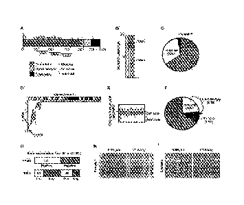
Fig. 1A-I. The HVEM ¨ BTLA interaction is disrupted in the majority of human
FLs. Fig.
1A, Summary of HVEM mutations in 141 FL samples; Fig. 1B, Distribution of copy
number
(CN) status in the 41 patients harboring a HVEM CN alteration; Fig. 1C,
Percentage of each
type of mutation found in FL patients; Fig. 1D, Chr. 1p36 deletions affect the
HVEM locus
(MSKCC cohort, n=64); Fig. 1E, GISTIC analysis indicates frequent homozygous
HVEM
deletions; Fig. 1F, Frequency of deletions by zygosity in indolent FL; Fig.
1G, quantification
7
CA 02981304 2017-09-28
WO 2016/161415
PCT/US2016/025840
of positive and negative cases represented on TMAs stained for HVEM and BTLA.
Fig. 1H
and Fig. 11, immune-histochemical staining. In the first panel (Fig. 1H)
strong staining with
an anti-HVEM antibody was observed in the malignant cell population whereas
BTLA
remained largely negative. The second panel (Fig. 1I) is negative for HVEM but
shows
strong positivity for BTLA in all tumor cells. Original magnification x400,
scale bars equal
50 pm.
Fig. 2A-G. HVEM acts as a tumor suppressor in a mouse model of FL. Fig. 2A,
schematic
representation of vavPBc12 mosaic mouse model; Fig. 2B, Kaplan-Meier analysis
of disease
free survival (Vector, n=11; shRNA against HVEM, n=19); Fig. 2C, FACS analysis
for
surface HVEM in B lymphocytes isolated from normal spleen, control lymphomas
(vavPBc12-vector), and two independent lymphomas expressing the shRNA against
HVEM
(vavPBc12-shHVEM); Fig. 2D, Quantification of HVEM FACS measurements (n=5 for
each
genotype, *p<0.01); Fig. 2E, GFP expression of shHVEM in different mouse cell
populations, HSCs (pre-injection into mouse), CD4+ , CD8+, B220+ (after
sacking mouse)
(n=5); Fig. 2F, Pathology and immunohistochemistry for the indicated markers
on murine
lymphoma comparing control lymphoma (vavPBc12-vector) to HVEM deficient
lymphomas
(vavPBc12-HVEM), scale bars = 100 pm; Fig. 2G, Immunoblot on murine control
lymphomas (vector) and HVEM deficient (HVEM) lymphomas probed as indicated.
Fig. 3A-F. BTLA deficiency recapitulates the effect of HVEM loss on lymphoma
development in vivo. Fig. 3A, Kaplan-Meier analysis of disease free survival
(vector, n=11:
shRNA against BTLA, n=16, p<0.01); Fig. 3B, qRT-PCR analysis of BTLA mRNA
expression in control (vector) and BTLA (shBTLA) lymphomas; Fig. 3C,
Pathological
analysis of shBTLA tumors stained for representative sections including H&E,
Ki67, PNA
and BCL6, scale bars = 100 pm; Fig. 3D, Quantification of Ki67 staining in
shBTLA tumors
(n=6, p<0.01); Fig. 3E, Surface analysis of vavPBc12-vector and vavPBc12-
shBTLA tumors;
Fig. 3F, Immunoblot on representative tumors probed as indicated.
Fig. 4A-E. HVEM blocks BCR signaling in a cell autonomous and BTLA dependent
manner.
Fig. 4A and Fig. 4B, Quantification of FACS analysis of phosphorylated BTK
(pBTK)
expression in BCL1 cells after stimulation with anti-IgM in the presence of
solHVEM
(10 g/m1) or Ibrutinib (10n1V1) without (Fig. 4A) or with (Fig. 4B) the
knockdown of BTLA
(shBTLA); Fig. 4C, FACS analysis of BTLA expression on purified primary human
FL B
cells distinguishes samples with high (BTLA1u) and low (BTLA1o) surface BTLA
expression
8
CA 02981304 2017-09-28
WO 2016/161415
PCT/US2016/025840
); DFACS analysis for the indicated signaling molecules in human primary FL B
cells that
were BTLAhi or BTLAlo and stimulated with anti-human IgG (3min; 10p.g/m1 and
H202
1mM) in the presence or absence of the soluble HVEM ectodomain (solHVEM; 10
pg/ml)
(right); Fig. 4E, Percentage of pSyk inhibition was calculated by comparing
the ratio of MFI
.. of pSyk +1- solHVEM and was correlated to BTLA ratio of MFI (r =0.697, p =
0.03, Purified
FL B cells, n =10, grade 1 and grade 2).
Fig. 5A-I. Abnormal activation of the lymphoid stroma in B-cell lymphomas.
Fig. 5A,
Immunohistofluorescence staining for the FDC marker CD21/35 and the FRC marker
Collagen 1 on control lymphomas (vector) and HVEM knockdown lymphomas (shHVEM)
(n=3 for each, scale bars = 1001..tm); Fig. 5B and Fig. 5C, Systematic
quantification of
CD21/35 (left) and collagen I (right) staining in control (Vector) and HVEM
deficient
(shHVEM) lymphomas based on 12 areas in the T-cell zone and 30 areas in the B-
cell zone
per mice (cumulative number for 3 mice), respectively; ** p <0.01; *** p
<0.001 by
parametric t-test; Fig. 5D and Fig. 5E, CXCL13 (Fig. 5D) and CCL19 (Fig. 5E)
expression
by qRT-PCR on control (vector) and HVEM knockdown (shHVEM) lymphomas (mean of
four replicates, error bars indicate standard deviation, * p< 0.01 ); Fig. 5F,
qRT-PCR
measurement of the LTa, LTb, and TNFa mRNA expression in B cells isolated from
the
spleens of vector and shHVEM mice (n=3); Fig. 5G-I, qRT-PCR measurement of
TNFa
(Fig. 5G), LTa (Fig. 5H), and LTb (Fig. 51) in B cell line BCL1 after 24 hrs
of treatment with
.. solHVEM (10p.g/m1 ).
Fig. 6A-I. Increased 1141 cell recruitment supports to HVEM deficient lymphoma
B cells.
Fig. 6A, FACS identification and sorting of human GC derived TFH cells based
on the
markers CD3pos, CD4pos, CD25neg, PD1hi, CXCR5hi, left: isotypic control;
right; staining
with anti-BTLA antibody; Fig. 6B and Fig. 6C, FACS measurement (Fig. 6B) and
.. quantification (Fig. 6C) of intra-tumoral TFH cells in control and HVEM
deficient murine
lymphomas; Fig. 6D and Fig. 6E, qRT-PCR measurement of IL21 (Fig. 6D), and IL4
(Fig.
6E) in sorted intra-tumoral T cells (N=?); Fig. 6F, qRT-PCR measurement of the
LTa, LTb,
and TNFa mRNA expression in T cells isolated from the spleens of vector and
shHVEM
mice * p<?; G-I, qRT-PCR measurement of TNFa (Fig. 6G), LTa (Fig. 6H), and LTb
(Fig.
.. 61) in cell sorted TFH (n=4) cultured with anti-CD3/anti-CD28 Mabs in
presence or not of
soluble HVEM (solHVEM, lops/m1), each symbol represents an independent TFH
sample.
Fig. 7A-H. The solHVEM (either Leu39-Va1202 or Pro37-Va1202) protein restores
tumor
9
CA 02981304 2017-09-28
WO 2016/161415
PCT/US2016/025840
suppressive effects of HVEM. Fig. 7A and Fig. 7B, FACS measurement of
phosphorylated
BTK (pBTK) in DOHH2 lymphoma cells that were stimulated with anti-1gG in the
presence
of absence of Pro37-Va1202 solHVEM (5 8,/m1) or the BTK inhibitor ibrutinib
(10nNI);
quantified in (B) (* indicated p <0.01); Fig. 7C, immunoblot on myc/bc12 cells
after
treatment with Leu39-Va1202 solHVEM (5 g/m1) probed as indicated; Fig. 7D,
Analysis of
cell proliferation across a panel of BTLAhi and BTLAlo lymphoma cell lines
treated with
Leu39-Va1202 solHVEM (5tig/m1); Fig. 7E, Representative picture of in vivo
treatment of
engrafted myc-bc12 murine lymphomas, Fig. 7F, In vivo treatment of engrafted
myc-bc12
murine lymphomas with either vehicle or the Leu 39-Va1202 HVEM ectodomain upon
formation of well-palpable tumors 75mm3 20u8 of Leu39-Va1202 solHVEM was
intratumoral injected every three days (indicated by arrows); Fig. 7G,
Immunoblot on lysates
from Leu39-Va1202 treated and untreated lymphomas proved as indicated; Fig.
711,
Microscopic pathology on Leu39-Va1202 treated and untreated lymphomas stained
as
indicated, scale bars = 100 gm.
Fig. 8A-G. HVEM mutations and deletions in human lymphomas. Fig. 8A, Chr. 1p36
deletions in a second series of FL (UNNIC, n=198); inset: GISTIC analysis of
DNA copy
number indicates frequent homozygous loss; Fig. 8B, Frequency of deletions by
zygosity in
transformed FLs; Fig. 8C, Distribution of the percentages of HVEM-positive
tumor cells in
FL tissue specimens arranged on a TMA. Colors represent staining intensity;
Fig. 8D,
Expression of HVEM in Human FLs samples in HVEM wt (left) and HVEM mutated or
deleted samples (right); Fig. 8E, The number of cases presenting with the
respective staining
intensities for CD272 (BTLA) in the follicular lymphoma cells are shown; Fig.
8F, BTLA
staining intensity in Human FLs in cases that are HVEM + or HVEM -; Fig. 8G,
Numbers
indicate breakdown of how individual TNIA sections scored.
Fig. 9A-E. HVEM knockdown promotes FL development in vivo. Fig. 9A,
Kaplan¨Meier
analysis of tumor onset using a second shRNA against HVEM (shHVEM-2) compared
to
empty vector (vector, n=11; shHVEM-2, n=12; p<0.01); Fig. 9B, qRT-PCR analysis
of
HVEM mRNA expression in control (vector) and HVEM (shHVEM) lymphomas; Fig. 9C,
FACS analysis for the indicated surface markers on HVEM deficient lymphomas
(shHVEM);
Fig. 9D, quantification of Ki67 in vavPBc12-vector and vavPBc12-HVEM tumors
(n=6; mean
s.d; t-test: * p<0.01); Fig. 9E, FACS analysis for the indicated surface
markers on HVEM
deficient lymphomas (shHVEM).
CA 02981304 2017-09-28
WO 2016/161415
PCT/US2016/025840
Fig. 10A-C. Analysis of variants in the VDJ region of mouse tumors. Fig. 10A,
Analysis of
jr heavy chain transcripts from three samples of shHVEM mice to evaluate
clonality and
monitor clonotypes within the samples. Table represents clones amplified above
1% (control
samples had none above 0.66%). Clones with the same VDJ junction and minimal
differences
within the V and JH segments are represented as variants in the last column;
Fig. 10B,
Evolution tree shows ongoing clonal evolution of the dominant clone by
connecting variants
observed in the CDR3 region with (VH8.12/D2.4/JH1) in shHVEM sample #2. Fig.
10C, Pie
charts represent VH family usage of the three samples (and control) analyzed
to globally
assess the B cell repertoire in each sample. Abundant clonal proliferation in
samples 2 and 3
accordingly show clear repertoire biases.
Fig. 11. Effect of HVEM on murine and human FL B cells. A FACS analysis of
BTLA
expression on purified human FL B cells distinguishes samples with high
(BTLAhi) and low
(BTLA1o) surface BTLA expression (top); FACS analysis for the indicated
signaling
molecules in human primary FL B cells that were BTLAhi or BTLAlo and
stimulated with
anti-human IgG (3min and 10min; 10pg/m1 and H202 1mM) in the presence or
absence of the
soluble HVEM ectodomain (solHVEM; 10 gimp.
Fig. 12A-F. Analysis of the lymphoid stroma in B cell lymphomas. Fig. 12A,
Immunohistofluorescence staining of CD20pos B cells, Transglutaminasepos FRCs,
and
CD21Lpos FDCs in reactive lymph nodes and two separate human follicular
lymphoma
tissue specimens; Fig. 12B, Flowchart of the image processing for FRC density
(Collagen
I); briefly, images were thresholded and transformed to binaries images, then
a watershed
algorithm was applied and number of polygons evaluated and analyzed by ImageJ
software;
Fig. 12C, Number of polygons indicates FRC density in control lymphomas
(vector) and
HVEM knockdown lymphomas showing no difference in FRC contribution. 40 areas
were
selected in the T cell zone and analyzed per mice (n=3 per each group); Fig.
12D-F, qRT-
PCR measurement of TNFa (Fig. 12D), LTa (Fig. 12E), and LTb (Fig. 12F) in
mouse B- cell
line EuMyc- Bc12.
Fig. 13A-E. Analysis of TFH cell function in HVEM deficient lymphomas. Fig.
12A and
Fig. 12B, qRT-PCR measurement of the receptors for IL21 (IL-21ra; A), and IL4
(IL4ra; B)
in purified lymphoma B cells; Fig. 12C, Viability of purified murine TFH cells
(samples:
n=4) that were cultured for 3 days with or without (UN) stimulation by anti-
CD3/anti-CD28
in the presence or absence of the soluble HVEM ectodomain (solHVEM:
10i_tg/m1); Fig. 12D
11
CA 02981304 2017-09-28
WO 2016/161415
PCT/US2016/025840
and Fig. 12E, Cell-Sorted GC-TFH cultured with anti-CD3/anti-CD28 Mabs in
presence or
not of solHVEM, production of CXCL13(Fig. 12D) and IL-21(Fig. 12E) evaluated
by
ELISA.
Fig. 14A-E. Effect of solHVEM (either Leu39-Va1202 or Pro37-Va1202) on murine
and
human FL B cells. Fig. 14 and Fig. 14B, Quantification of pSYK levels in DOHH2
lymphoma cells that were stimulated with anti-IgG in the presence or absence
of Pro37-
Va1202 solHVEM (5 g/m1) (* indicated p <0.01); representative FACS measurement
in (Fig.
14B) Fig. 14C, FACS analysis of BTLA expression in a panel of lymphoma lines
including
murine myc/bc12 lymphomas and human lines (DOHH2, Su-DHL6, Granta, Lyi 0);
Fig. 14D,
representative pictures of tumors from mice; Fig. 14E, tumor weight of mouse
tumors (n=3,
p<0.01).
Fig. 15A-B. sTNFRSF14 opposes B cell receptor signaling in lymphoma B cells by
decreasing P-BTK. A B-cell lymphoma cell line (DOHH2) was pre-treated for one
hour with
the soluble ectodomain of TNFRSF14 (sTNFRSF14) Pro 37-Val 202 (5ug/m1) or the
BTK
inhibitor Ibrutinib (10nmM) and then stimulated for 5 mins at 37 C with anti-
IgG molecule.
The cells were subsequently fixed and permeabilized and probed for pBTK
expression using
phospo-flow antibodies and analyzed on BD Fortessa. Fig. 15A, Representative
FACS plots.
Fig. 15B, Quantification of mean fluorescence intensity of phospho-BTK after
treatment with
vehicle or drug.
Fig. 16A-B. sTNFRSF14 opposes B-cell receptor signaling in lymphoma B cells by
decreasing P-SYK. A B-cell lymphoma cell line (DOHH2) was pre-treated for one
hour with
the soluble ectodomain of TNFRSF14 (sTNFRSF14) Pro37-Val 202 (5ug/m1) or the
BTK
inhibitor Ibrutinib (10nmM) and then stimulated for 5 mins at 37oC with anti-
IgG molecule.
The cells were subsequently fixed and permeabilized and probed for pSYK
expression using
phospo-flow antibodies and analyzed on a BD Fortessa. Fig. 16A, Representative
FACS
plots. Fig. 16B, Quantification of mean fluorescence intentisty of phospho-SYK
after
treatment with vehicle or drug.
Fig. 17. sTNFRSF14 inhibits the growth of lymphoma cell lines in vitro. Three
lymphoma
cell lines (Myc-Bc12, LY-10, Granta) were plated at 1 x 10⁵ cells/ml and were
treated with
sTNFRSF14 (5ug/m1) or vehicle each day for 72 hours. After 72 hours cells were
counted
using a hemocytometer. Each bar represents the average of three independent
experiments.
12
CA 02981304 2017-09-28
WO 2016/161415
PCT/US2016/025840
Fig. 18. sTNFRSF14 decreases cell viability in vitro. Cells of the myc-Bc12
lymphoma cell
line were plated at a density of 1 x10⁵ cells/m1 and they were treated with
sTNFRSF14 (5
ug/ml) or vehicle. After 24 hours of treatment cell viability was assessed
using CellTiterGlo
reagent.
Fig. 19. In vitro effect of sTNFRSF14. Immunoblots of cell lines that were
treated with 5
ug/ml of sTNFRSF14. Blots were probed as indicated.
Fig. 20, sTNFRSF14 inhibits tumor growth in vivo. Xenograft myc-bc12 lymphomas
were
grown in the flanks of mice. When the tumors reached a volume of approximately
0.5 cm3
mice were treated every other day by intra-tumoral injection in the flanks
with 20 ug/ml of
sTNFRSF14diluted in PBS. The control (vehicle) animals were treated with PBS.
Tumors
were weighed and volumes were measured twice weekly.
Fig. 21. sTNFRSF14 decreases lymphoma growth in a xenograft model. 5 million
myc-Bc12
cells were mixed with Matrigel and injected subcutaneously into the flanks of
mice J:Nu
Nude (Foxnl nu/ Foxnl nu) mice. Animals were sacrificed according to IUCAC
protocols.
Upon sacrifice tumors were weighed and measured.
Fig. 22. Exogenous administration of sTNFRSF14 suppresses mouse lymphoma
xenografts.
Animals were sacrificed on day day 11 and the xenografted tumors were excised
from the
flanks of the mice. The tumors from each flank - treated (sTNFRSF14) and
untreated
(vehicle) were weighed. Bars represent the average of n=4 mice.
Fig. 23. Molecular characterization of in vivo tumors after treatment with
sTNFRSF14.
Fig. 24. Immunohistochemical analysis of xenograft tumors. Pathological
analysis of
sTNFRSF14 treated and vehicle treated mouse lymphomas. Tumors were excised
from the
flanks of the animals and fixed in 4% paraformaldehyde overnight. The tumors
were
sectioned and stained via THC for particular tumor markers. Representative
staining for HE,
TUNEL, and Ki67 is shown.
Fig. 25A-B. Fig. 25A, Schematic illustration of delivery of soluble HVEM
polypeptides to
lymphoma cells using CD19-specific chimeric antigen receptor (CAR)-modified T
cells that
are modified to constitutively secrete soluble HVEM. Fig. 25B, Schematic
illustration of
chimeric antigen receptor (CAR) molecule comprising a soluble HVEM sequence
(HVEM
13
CA 02981304 2017-09-28
WO 2016/161415
PCT/US2016/025840
P37-V202).
Fig. 26A-B. solHVEM does not have an effect on T cell viability or activation.
Fig. 26A,
Viability of purified murine OT1 cells (n=2) that were cultured for 24 hours
with or without
stimulation by anti-CD3/anti-CD28 in the presence or absence of the soluble
HVEM
ectodomain (solHVEM: 10p.g/m1); Fig. 26B, Percentage of activated murine OT1
cells
identified by FACS, OT1 cells were culture as in Fig. 26A.
Fig. 27A-B. 19-28-HVEM-modified T cells, compared to 19-28 T cells, show
increase in
HVEM production and secretion (Fig. 27A) WB on FACS sorted CAR-T, and probed
for
HVEM (Fig. 27B) ELISA assay on HVEM shows increase in HVEM levels (p<0.1).
Fig. 28A-D. Fig. 28A, 19-28-HVEM-modified T cells exhibit enhanced in vitro
cytotoxicity
to B cells with high BTLA expression as compared to control 19-28 T cells.
DOHH2 or Raji
cells were incubated with GFP-labeled CAR-T cells at given T (target) to E
(effector T cell)
ratios. At the indicated times cells were labeled with Annexin V and DAPI, and
the
percentage of GFP- viable cells was assessed by FACS. Fig. 28B, FACS analysis
of BTLA
expression on B cell lines distinguishes samples with high and low surface
BTLA expression.
Fig. 28C-D, 19-28-HVEM-modified T cells exhibit enhanced cytotoxicity in vivo
on DOHH2
tumors as compared to control 19-28 T cells. Xenografts were generated by s.c.
injections of
5Mio DoHH2 human lymphoma cells mixed with Matrigel (BD) into flanks of
NOD/SCID
(NOD.CB17- Prkdcscid/J) mice. Upon visible tumor formation (20mm3), mice were
given a
single dose of 1 Mio anti-CD19 CAR T cells that are with or without HVEM
secretion. T
cells containing prostate-specific membrane antigen (PSMA) scFv was used as a
control
CAR. Fig. 28C, Representative tumors isolated upon mouse sacrifice. Fig. 28D,
Quantification of tumor size.
DETAILED DESCRIPTION
The sub-headings provided below, and throughout this patent disclosure, are
not intended to
denote limitations of the various aspects or embodiments of the invention,
which are to be
understood by reference to the specification as a whole. For example, this
Detailed
Description is intended to read in conjunction with, and to expand upon, the
description
provided in the Summary of the Invention section of this application.
1. Definitions & Abbreviations
14
CA 02981304 2017-09-28
WO 2016/161415
PCT/US2016/025840
As used in this specification and the appended claims, the singular forms "a,"
"an," and "the"
include plural referents, unless the context clearly dictates otherwise. The
terms "a" (or "an")
as well as the terms "one or more" and "at least one" can be used
interchangeably.
Furthermore, "and/or" is to be taken as specific disclosure of each of the two
specified
features or components with or without the other. Thus, the term "and/or" as
used in a phrase
such as "A and/or B" is intended to include A and B, A or B, A (alone), and B
(alone).
Likewise, the term "and/or" as used in a phrase such as "A, B, and/or C" is
intended to
include A, B, and C; A, B, or C; A or B; A or C; B or C; A and B; A and C; B
and C; A
(alone); B (alone); and C (alone).
Units, prefixes, and symbols are denoted in their Systeme International de
Unites (SI)
accepted form. Numeric ranges provided herein are inclusive of the numbers
defining the
range. Where a numeric term is preceded by "about," the term includes the
stated number
and values +10% of the stated number.
An "active agent" is an agent (e.g. a molecule or a cell) as described and/or
claimed herein
that is, or that comprises, a soluble HVEM ectodomain polypeptide, an anti-
HVEM
antibody, or an anti-BTLA antibody, or a nucleotide sequence that encodes any
of such
agents. Active agents include, but are not limited to, cells (such as T
cells),
polypeptides/proteins, and nucleic acid molecules.
The terms "inhibit," "block," "reduce," and "suppress" are used
interchangeably and refer to
any statistically significant decrease in biological activity, including ¨ but
not limited to - full
blocking of the activity.
"TNFRSF14" refers to "tumor necrosis factor receptor superfamily member 14."
"HVEM" refers to "herpes virus entry mediator."
TNFRSF14 and HVEM are one and the same. Accordingly, the terms TNFRSF14 and
HVEM are used interchangeably throughout this patent disclosure. In some
instances these
proteins may be referred to herein as "TNFRSF14 / HVEM."
"BTLA" refers to "B and T lymphocyte attenuator."
CA 02981304 2017-09-28
WO 2016/161415
PCT/US2016/025840
The terms "BTLA-positive" and "BTLA" are used interchangeably herein to refer
to tumors
or cells that express (or express detectable levels of) BTLA.
The terms "BTLA-negative" and "BTLA"' are also used interchangeably herein and
refer to
tumors or cells that do not express (or do not express detectable levels of)
BTLA.
The term "BTLA'" refers to tumors or cells that express high levels of BTLA.
The term "BTLAki" refers to tumors or cells that express low levels of BTLA.
The terms BTLA, B ILK, BTLAhi, and BTLA1 are all used to denote expression
levels of
BTLA in relative terms. For example a cell or a tumor may be classified as
BTLA + as
opposed to BTLA". Similarly, a cell or a tumor may be classified as BTLA' i as
opposed to
BTLA1 . The usage of such relative terms to denote expression levels, for
example using "+"
versus "-" and "hi" versus "lo" terminology, is standard in the art and the
meaning of such
terms will be clear to those of ordinary skill in the art. For example, one of
skill in the art
will understand that a cell or tumor may be designated as BTLA + based on
determination of
BTLA expression levels in comparison with suitable positive (i.e. BTLA
expressing) and/or
.. negative (i.e. non-BTLA expressing) controls. Similarly, one of skill in
the art will
understand that a cell or tumor may be designated as BTLAhi based on
determination of
BTLA expression levels in comparison with suitable highly expressing and/or
weakly
expressing controls. Suitable assays for making such comparative
determinations are
provided in Example 1, and include, but are not limited to,
immunohistochemistry and flow
cytometry or FACS-based assays. Similarly, suitable control cell types for
making such
comparative determinations are provided in Example 1.
"CAR" refers to a "chimeric antigen receptor."
"CAR T cells" refers to genetically modified T cells that have been engineered
to express a
CAR.
Various other terms are defined elsewhere in this patent disclosure, where
used. Furthermore,
terms that are not specifically defined herein may be more fully understood in
the context in
which the terms are used and/or by reference to the specification in its
entirety. Where no
explicit definition is provided all technical and scientific terms used herein
have the meanings
commonly understood by those of ordinary skill in the art to which this
invention pertains.
16
CA 02981304 2017-09-28
WO 2016/161415
PCT/US2016/025840
2. TNFRSF14 / HVEM Polypeptides
TNFRSF14 was originally identified as a mediator of the entry herpes simplex
virus-1 into
human and mouse cells for (Montgomery, Warner et al. 1996). The TNFRSF14
receptor is
one of 29 currently known receptors within the TNF receptor superfamily. The
TNFRSF14
receptor gene is located on chromosome 1p36 in humans - a site that has been
frequently
reported to harbor tumor suppressors due to its frequent deletion in multiple
cancers (Bagchi
and Mills 2008). TNFRS14 is expressed throughout the major human tissues but
exhibits its
highest levels of expression in cells of the hematopoietic system. TNFRSF14 is
an insoluble
trans-membrane protein comprising an intracellular domain, a trans-membrane
domain, and
an extracellular domain or "ectodomain." The extracellular domain of TNFRSF14
comprises
3 cysteine rich domains or "CRDs" ¨ referred to as CRD1, CRD2, and TNFRSF14
can
interact with multiple different ligands, which bind to TNFRSF14 via its CRD
domains.
Some such ligands deliver co-stimulatory signals: such as the ligands
"Iymphotoxin-like,
inducible expression, competes with herpes simplex virus glycoprotein D for
HVEM, a
receptor expressed by T lymphocytes" (or "LIGHT"), and LTa. Other ligands
deliver co-
inhibitory signals: such as CD160, glycoprotein D (gD), and "B and T
lymphocyte
attenuator" or "BTLA" (Murphy and Murphy 2010).
A full length human TNFRS14 / HVEM protein sequence is provided in Figure 29
and SEQ
ID NO. 2. A nucleotide sequence that encodes the protein of SEQ ID NO. 2 (i.e.
the full
length human TNFRS14 / HVEM protein) is provided in Figure 29 and SEQ ID NO.
1. A
further nucleotide sequence that encodes a full length human TNFRS14 / HVEM
protein is
provided as SEQ ID NO. 10 (NCBI Reference Sequence: NM 003820.3). A nucleotide
sequence that encodes a full length mouse TNFRS14 / HVEM protein is provided
as SEQ ID
NO. 11 (NCBI Reference Sequence: NM 178931.2). A nucleotide sequence that
encodes a
full length rat TNFRS14 / HVEM protein is provided as SEQ ID NO. 12 (NCBI
Reference
Sequence: NM 001015034.1). A nucleotide sequence that encodes a full length
monkey
TNFRS14 / HVEM protein sequence is provided as SEQ ID NO. 13 (NCBI Reference
Sequence: 001043357.1). Other full-length TNFRS14 / HVEM protein sequences,
and
nucleotide sequences that encode such protein sequences, are also known in the
art. Some
embodiments of the present invention involve these full-length HVEM sequences.
However, most of the embodiments of the present invention involve non-
naturally occurring
soluble fragments of the full-length insoluble HVEM protein referred to herein
as "soluble
17
HVEM ectodomain polypeptides." As discussed in the Examples section of this
patent
application, it has now been demonstrated that soluble HVEM ectodomain
polypeptides
inhibit B-cell tumor growth and that this activity involves binding to BTLA.
It is already
known that within the HVEM ectodomain, the CRD1 domain is the essential
binding site for
BTLA and that deletion of the CRD1 domain blocks the inhibitory activity of
HVEM, and
there is also evidence that the CRD2 domain of HVEM provides structural
support of CRD1
binding ligands such as BTLA (see M.L.del Rio, 2010, Gonzales 2004, and
Bjordahl 2013).
Thus, the "soluble HVEM ectodomain polypeptides" of the present invention
comprise at
least a CRD1 domain (and may, optionally, comprise the CRD2 and/or CRD3 and/or
other
HVEM ectodomain regions), and do not comprise the HVEM trans-membrane or
intracellular
domains. Furthermore, the "soluble HVEM ectodomain polypeptides" of the
present
invention exhibit one or more of the following functional properties: tumor
suppressor
activity in BTLA:Ihi B-cell lymphomas (e.g. ability to inhibit B-cell lymphoma
cell growth
in vitro and/or tumor growth in vivo in BTLA+/hi B-cell lymphomas), ability to
increase/stimulate the activity of CD8+ T-cells, ability to inhibit/reduce
activation of B-cell
receptors in lymphoma cells, ability to inhibit/reduce the secretion of IL-
21by follicular T
helper MID cells or lymphoma B cells, ability to inhibit BCR pathway
activation in a
BTLA-dependent manner, and ability to inhibit BTK, SYK, and/or ERK activation
in
BTLA/hi lymphoma cells (e.g. DOHH2 cells). Suitable assays for assessing such
functional
properties are provided in the Examples section of this patent application.
The sequences of several exemplary soluble HVEM ectodomain polypeptides are
provided
herein - as summarized in Table 1, below. The amino acid numbering of all of
the soluble
HVEM ectodomain polypeptides described herein is based on SEQ ID NO. 2 (i.e.
SEQ ID
NO. 4 is amino acids 29-202 of SEQ ID NO. 2, SEQ ID NO. 6 is amino acids 37-
202 of
SEQ ID NO. 2, and SEQ ID NO. 8 is amino acids 39-202 of SEQ ID NO. 2, etc.).
Amino
acid residues Cys42-Cys75 of SEQ ID NO. 2 form the CRD1 domain of HVEM. Amino
acid
residues Cys78-Cys119 of SEQ ID NO. 2 form the CRD2 domain of HVEM. Amino acid
residues Cys121-Cys162 of SEQ ID NO. 2 form the CRD3 domain of HVEM. The
Examples section of this patent application describes experiments performed
using some of
such exemplary soluble HVEM ectodomain polypeptides.
18
Date Recue/Date Received 2022-09-16
CA 02981304 2017-09-28
WO 2016/161415
PCT/US2016/025840
Table 1 - Sequences of Exemplary Soluble HVEM Ectodomain Polypeptides
Soluble HVEM Ectodomain Polypeptide Nucleotide Amino Acid
Sequence Sequence
Gln29-Va1202 SEQ ID NO. 3 SEQ ID NO. 4
Pro37-Va1202 SEQ ID NO. 5 SEQ ID NO. 6
Leu39-Va1202 SEQ ID NO. 7 SEQ ID NO. 8
In some embodiments the soluble HVEM ectodomain polypeptides of the invention
comprise, or consist of, or consist essentially of, a CRD1 domain of an HVEM
protein (e.g.
amino acid residues Cys42-Cys75 of SEQ ID NO. 2, or amino acid residues that
correspond
thereto).
In some embodiments the soluble HVEM ectodomain polypeptides of the invention
comprise, or consist of, or consist essentially of a CRD1 domain and a CRD2
domain of an
HVEM protein (e.g. amino acid residues Cys42-Cys75 of SEQ ID NO. 2 and amino
acid
residues Cys78-Cys119 of SEQ ID NO. 2, or amino acid residues that correspond
thereto).
In some embodiments the soluble HVEM ectodomain polypeptides of the invention
comprise, or consist of, or consist essentially of a CRD1 domain, a
CRD2domain, and a
CDR3 domain of an HVEM protein (e.g. amino acid residues Cys42-Cys75 of SEQ ID
NO. 2
and amino acid residues Cys78-Cys119 of SEQ ID NO. 2 and amino acid residues
Cys121-
Cys162 of SEQ ID NO. 2, or amino acid residues that correspond thereto).
In some embodiments the soluble HVEM ectodomain polypeptides of the invention
do not
comprise a CRD2 domain.
In some embodiments the soluble HVEM ectodomain polypeptides of the invention
do not
comprise a CRD3 domain.
In some embodiments the soluble HVEM ectodomain polypeptides of the invention
do not
comprise a CRD2 or CRD3 domain.
In some embodiments the soluble HVEM ectodomain polypeptides of the invention
comprise, or consist of, or consist essentially of, the amino acid sequence of
SEQ ID NO. 4,
19
CA 02981304 2017-09-28
WO 2016/161415
PCT/US2016/025840
SEQ IDNO. 6, or SEQ ID NO. 8, or amino acid sequences that correspond thereto.
In some embodiments the soluble HVEM ectodomain polypeptides of the invention
comprise, or consist of, or consist essentially of, an amino acid sequence
starting at amino
acid position 19, 20, 21, 22, 23, 24, 25, 26, 27, 28, 29, 30, 31, 32, 33, 34,
35, 36, 37, 38,
.. 39,40, 41, or 42 of SEQ ID NO. 2, or amino acid residues that correspond
thereto.
In some embodiments the soluble HVEM ectodomain polypeptides of the invention
comprise, or consist of, or consist essentially of, an amino acid sequence
starting at amino
acid position 19, 20, 21, 22, 23, 24, 25, 26, 27, 28, 29, 30, 31, 32, 33, 34,
35, 36, 37, 38,
39,40, 41, or 42 of SEQ ID NO. 2, and ending at amino acid 75, 76, or 77 of
SEQ ID NO. 2,
or amino acid residues that correspond thereto (i.e. comprising a CDR1
domain).
In some embodiments the soluble HVEM ectodomain polypeptides of the invention
comprise, or consist of, or consist essentially of, an amino acid sequence
starting at amino
acid position 19, 20, 21, 22, 23, 24, 25, 26, 27, 28, 29, 30, 31, 32, 33, 34,
35, 36, 37, 38,
39,40, 41, or 42 of SEQ ID NO. 2, and ending at amino acid 119 or 120 of SEQ
ID NO. 2, or
amino acid residues that correspond thereto (i.e. comprising a CRD1 and CRD2
domain).
In some embodiments the soluble HVEM ectodomain polypeptides of the invention
comprise, or consist of, or consist essentially of, an amino acid sequence
starting at amino
acid position 19, 20, 21, 22, 23, 24, 25, 26, 27, 28, 29, 30, 31, 32, 33, 34,
35, 36, 37, 38,
39,40, 41, or 42 of SEQ ID NO. 2, and ending at amino acid 162, 163, 164, 165,
166, 167,
168, 169, 170, 171, 172, 173, 174, 175, 176, 177, 178, 179, 180, 181, 182,
183, 184, 185,
186, 187, 188, 189, 190, 191, 192, 193, 194, 195, 196, 197, 198, 199, 200, 201
202, 203, 204,
205, 206, 207, 208, or 209 of SEQ ID NO. 2, or amino acid residues that
correspond thereto
(i.e. comprising a CRD, CRD2, and CRD3 domain).
It should be noted that one of skill in the art can readily determine and/or
identify amino acid
positions in other sequences that "correspond" to any of the specific amino
acid residues
defined herein, regardless of whether those other sequences utilize a
different numbering
scheme or are present in a different HVEM sequences (such as in an HVEM
sequence from a
different species), for example by performing a sequence alignment to the
sequence of SEQ
ID NO. 2. It should also be noted that for all of the numbered sequences or
numbered amino
acid residues provided herein sequences and amino acid residues that
"correspond" to such
sequences/residues are also contemplated and encompassed herein.
CA 02981304 2017-09-28
WO 2016/161415
PCT/US2016/025840
Variants of any of the specific soluble HVEM ectodomain polypeptide sequences
provided
above and elsewhere in this patent disclosure are also contemplated and are
intended to fall
within the scope of the present invention. For example, in some embodiments
variants of the
specific sequences disclosed herein from other species (orthologs) may be
used. Similarly, in
other embodiments variants that comprise fragments of any of the specific
sequences
disclosed herein may be used. Likewise, in some embodiments variants of the
specific
sequences disclosed herein that comprise one or more amino acid substitutions,
additions,
deletions, or other mutations may be used. In some embodiments the variant
amino acid
sequences have at least about 40% or 50% or 60% or 65% or 70% or 75% or 80% or
85% or
90% or 95% or 98% or 99% identity with the specific soluble HVEM ectodomain
polypeptides described herein. In all such cases, all variant soluble HVEM
ectodomain
polypeptides should comprise a CRD1 domain, or a portion thereof that is
sufficient for
binding to BTLA, and they should exhibit one or more of the following
functional properties:
HVEM activation, BTLA activation, inhibition of proliferation of BTLA + B-cell
lymphoma
.. cells, inhibition of growth of a BTLA + B-cell lymphoma, stimulation of the
activity of CD8+
T-cells, inhibition of the activation of B-cell receptors in B-cell lymphoma
cells, inhibition of
secretion of IL-21 by follicular T helper (TFH) cells, inhibition of secretion
of II,-21 by B-
cell lymphoma cells, inhibition of BCR pathway activation, and inhibition of
BTK, SYK,
and/or ERK activation in BTLA + B-cell lymphoma cells. Suitable assays for
assessing such
functional properties are provided in the Examples section of this patent
application.
It should be noted that all of the soluble HVEM ectodomain polypeptides
contemplated by or
described in the present patent disclosure may, in some embodiments, comprise
a secretion
signal sequence, or may be expressed via a precursor form that comprises a
secretion signal
sequence. In some embodiments an IgG Kappa secretion signal is used. In other
embodiments an interleukin 2 (IL2) secretion signal is used. However, any
suitable secretion
signal sequence known in the art may be used.
In addition to providing amino acid sequences, the present invention also
provides nucleic
acid sequences. For example, in some embodiments the present invention
provides
nucleotide sequences that encode soluble HVEM ectodomain polypeptides,
including, but not
limited to, those that comprise, or consist of, or consist essentially of, the
nucleotide
sequences of SEQ ID NO. 3, SEQ IDNO. 5, or SEQ ID NO. 7. The present invention
contemplates and provides nucleotide sequences that encode all of the soluble
HVEM
21
CA 02981304 2017-09-28
WO 2016/161415
PCT/US2016/025840
ectodomain polypeptides described herein ¨ including those for which specific
sequences are
disclosed and the various variants of such sequences described herein. The
present invention
also provides DNA constructs (e.g. vectors and plasmids) comprising any of the
nucleic acid
molecules and/or nucleotide sequences described herein, or encoding any of the
soluble
HVEM ectodomain polypeptides described herein.
The present invention also provides genetically modified cells comprising any
of the nucleic
acid molecules and/or nucleotide sequences described herein, or encoding any
of the soluble
HVEM ectodomain polypeptides described herein.
It should be noted that, while the present invention is directed primarily to
use of soluble
.. HVEM ectodomain polypeptides, in some instances it may be possible to use
insoluble (i.e.
membrane-bound) proteins that comprise the sequences present in such soluble
HVEM
ectodomain polypeptides. For example, in those embodiments that involve CAR T-
cells that
express (and secrete) soluble HVEM ectodomain polypeptides, it may, in some
instances, be
possible to use a CAR T-cell that expresses an insoluble (i.e. membrane-bound)
version of
the HVEM ectodomain polypeptide, wherein rather than being secreted by the T-
cell the
HVEM ectodomain polypeptide sequences are membrane bound and are presented on
the
surface of the T-cell. Such embodiments are intended to fall within the scope
of the present
invention. Thus, unless stated otherwise, all of those embodiments of the
present invention
that involve a soluble HVEM ectodomain polypeptide can be performed using
insoluble
.. variants of such polypeptides that comprise the sequences present soluble
HVEM ectodomain
polypeptide as well as other sequences that result in presentation of such
sequences in a cell
membrane (e.g. on the surface of a cell).
3. Antibodies (including anti-HVEM and anti-BTLA antibodies)
Several embodiments of the present invention involve antibodies. As used
herein, the term
"antibody" encompasses intact polyclonal antibodies, intact monoclonal
antibodies, antibody
fragments (such as Fab, Fab', F(ab')2, and Fv, and single chain Fv (scFv)
fragments, single-
domain antibodies (sdAb or nanobodies)), fusion proteins comprising an antigen
determination portion of an antibody, bispecific antibodies generated from at
least two intact
antibodies, chimeric antibodies, humanized antibodies, human antibodies, and
any other
modified immunoglobulin molecule(s) comprising an antigen recognition site -
so long as the
antibodies comprise an antigen recognition site and exhibit the desired
biological activity.
22
Various different types of antibody fragments, and methods of making and using
such
antibody fragments, are known in the art. See, for example, Fridy et al.,
Nature Methods.
2014 Dec;11(12):1253-60 for a description of the production of nanobody
repertoires multi-
specific antibodies. An antibody can be of any the five major classes of
immunoglobulins:
IgA, IgD, IgE, IgG, and IgM, or subclasses (isotypes) thereof (e.g. IgGl,
IgG2, IgG3, IgG4,
IgAl and IgA2), based on the identity of their heavy-chain constant domains
referred to as
alpha, delta, epsilon, gamma, and mu, respectively. The different classes of
immunoglobulins have different and well-known subunit structures and three-
dimensional
configurations. Antibodies can be naked, or conjugated to other molecules such
as toxins,
radioisotopes, or any of the other specific molecules recited herein.
In some embodiments the present invention involves antibodies against BTLA
and/or
antibodies against HVEM. In some embodiments such antibodies may be any
suitable type
of anti-BTLA antibody or anti-HVEM antibody. In certain preferred embodiments
an
antibody fragment that binds to BTLA or HVEM is used. For example, in certain
embodiments a Fab, Fab', F(ab)2, Fv, scFv, or sdAb (nanobody) fragment is
used. In certain
preferred embodiments the antibody fragment is a scFv fragment. In other
preferred
embodiments the antibody fragment is a nanobody. In certain embodiments such
antibodies
(including antibody fragments) bind to their respective target antigens (i.e.
BTLA or HVEM)
with high affinity and/or high specificity. In certain preferred embodiments
such antibodies
(including antibody fragments) both bind to and activate their respective
target antigens (i.e.
BTLA or HVEM) on the surface of B-cells ¨ i.e. they act as agonists for their
respective
target antigens. For example such aztivating/agonist antibodies may mimic the
biological
activity of one or more natural ligands of their respective target antigens
(i.e. BTLA or
HVEM). Examples of antibodies (including antibody fragments) that are specific
for BTLA
are described in WO 2010106051 Al, and that are specific for HVEM are
described in Park
et al., Cancer Immunol. Immunother. 2012 Feb;61(2):203-14. However, any other
suitable
antibodies (including antibody fragments) may be used.
The term "humanized antibody" refers to an antibody derived from a non-human
(e.g.,
murine) immunoglobulin, which has been engineered to contain minimal non-human
(e.g.,
murine) sequences. Typically, humanized antibodies are human immunoglobulins
in which
residues from the complementary determining region (CDR) are replaced by
residues from
23
Date Recue/Date Received 2022-09-16
CA 02981304 2017-09-28
WO 2016/161415
PCT/US2016/025840
the CDR of a non-human species (e.g., mouse, rat, rabbit, or hamster) that
have the desired
specificity, affinity, and capability (Jones et al., 1986, Nature, 321:522-
525; Riechmann et al.,
1988, Nature, 332:323-327; Verhoeyen et al., 1988, Science, 239:1534-1536). In
some
instances, the Fv framework region (FW) residues of a human immunoglobulin are
replaced
with the corresponding residues in an antibody from a non-human species that
has the desired
specificity, affinity, and capability.
Humanized antibodies can be further modified by the substitution of additional
residues
either in the Fv framework region and/or within the replaced non-human
residues to refine
and optimize antibody specificity, affinity, and/or capability. In general,
humanized
antibodies will comprise substantially all of at least one, and typically two
or three, variable
domains containing all or substantially all of the CDR regions that correspond
to the non-
human immunoglobulin whereas all or substantially all of the FR regions are
those of a
human immunoglobulin consensus sequence. Humanized antibody can also comprise
at least
a portion of an immunoglobulin constant region or domain (Fc), typically that
of a human
immunoglobulin. Examples of methods used to generate humanized antibodies are
described
in U.S. Pat. Nos. 5,225,539 or 5,639,641.
The term "human antibody" means an antibody produced by a human or an antibody
having
an amino acid sequence corresponding to an antibody produced by a human made
using any
technique known in the art. This definition of a human antibody includes
intact or full-length
antibodies, fragments thereof, and/or antibodies comprising at least one human
heavy and/or
light chain polypeptide such as, for example, an antibody comprising murine
light chain and
human heavy chain polypeptides.
The term "chimeric antibodies" refers to antibodies wherein the amino acid
sequence of the
immunoglobulin molecule is derived from two or more distinct sources,
typically two or
more distinct species. Typically, the variable region of both light and heavy
chains
corresponds to the variable region of antibodies derived from one species of
mammals (e.g.,
mouse, rat, rabbit, etc.) with the desired specificity, affinity, and
capability while the constant
regions are homologous to the sequences in antibodies derived from another
(usually human)
to avoid eliciting an immune response in that species.
A "monoclonal antibody" (mAb) refers to a homogeneous antibody population
involved in
the highly specific recognition and binding of a single antigenic determinant,
or epitope. This
24
CA 02981304 2017-09-28
WO 2016/161415
PCT/US2016/025840
is in contrast to "polyclonal antibodies" that typically include different
antibodies directed
against different antigenic determinants.
Furthermore, "monoclonal antibody" refers to such antibodies made in any
number of ways
including, but not limited to, by hybridoma, phage selection, recombinant
expression, and
transgenic animals.
In particular, monoclonal antibodies can be prepared using hybridoma methods,
such as those
described by Kohler and Milstein (1975) Nature 256:495. Using the hybridoma
method, a
mouse, hamster, or other appropriate host animal, is immunized as described
above to elicit
the production by lymphocytes of antibodies that will specifically bind to an
immunizing
antigen. Lymphocytes can also be immunized in vitro. Following immunization,
the
lymphocytes are isolated and fused with a suitable myeloma cell line using,
for example,
polyethylene glycol, to form hybridoma cells that can then be selected away
from unfused
lymphocytes and myeloma cells. Hybridomas that produce monoclonal antibodies
directed
specifically against a chosen antigen as determined by immunoprecipitation,
immunoblotting,
or by an in vitro binding assay (e.g. radioimmunoassay (RIA); enzyme-linked
immunosorbent assay (ELISA)) can then be propagated either in in vitro culture
using
standard methods (Goding, Monoclonal Antibodies: Principles and Practice,
Academic Press,
1986) or in vivo as ascites tumors in an animal. The monoclonal antibodies can
then be
purified from the culture medium or ascites fluid.
Alternatively, monoclonal antibodies can be made using recombinant DNA
methods, as
described in U.S. Patent No. 4,816,567. The polynucleotides encoding a
monoclonal
antibody are isolated from mature B-cells or hybridoma cells, such as by RT-
PCR using
oligonucleotide primers that specifically amplify the genes encoding the heavy
and light
chains of the antibody, and their sequence is determined using conventional
procedures. The
isolated polynucleotides encoding the heavy and light chains are then cloned
into suitable
expression vectors, which when transfected into host cells such as E. coli
cells, simian COS
cells, Chinese hamster ovary (CHO) cells, or myeloma cells that do not
otherwise produce
immunoglobulin protein, monoclonal antibodies are generated by the host cells.
Also,
recombinant monoclonal antibodies or antigen-binding fragments thereof of the
desired
species can be isolated from phage display libraries expressing CDRs of the
desired species
as described (McCafferty et al., 1990, Nature, 348:552-554; Clackson et al.,
1991, Nature,
352:624-628; and Marks et al., 1991, J. MoL Biol., 222:581-597).
Polyclonal antibodies can be produced by various procedures well known in the
art. For
example, a host animal such as a rabbit, mouse, rat, etc. can be immunized by
injection with
an antigen to induce the production of sera containing polyclonal antibodies
specific for the
antigen. The antigen can include a natural, synthesized, or expressed protein,
or a derivative
(e.g., fragment) thereof. Various adjuvants may be used to increase the
immunological
response, depending on the host species, and include, but are not limited to,
Freund's
(complete and incomplete), mineral gels such as aluminum hydroxide, surface
active
substances such as lysolecithin, pluronic polyols, polyanions, peptides, oil
emulsions,
keyhole limpet hemocyanins, dinitrophenol, and potentially useful human
adjuvants such as
BCG (bacille Cahnette-Guerin) and corynebacterium parvum. Such adjuvants are
also well
known in the art. Antibodies can be purified from the host's serum.
4. Compositions & Methods Involving CAR T Cells
Cancer immunotherapy involves engineering patients' own immune cells to
recognize and
attack their tumors ¨ an approach that is frequently referred to as adoptive
cell transfer
(ACT). Such methods have yielded promising results in clinical trials so far,
including those
for treatment of lymphoma. hi ACT T cells collected from a patient's own blood
are
genetically engineered to produce recombinant receptors on their surface
called chimeric
antigen receptors or "CARs." CARs contain an antigen-binding domain designed
to
recognize and bind to a specific cell surface antigen on the patient's tumor
cells. The
engineered CAR T cells are expanded in vitro and then infused into the
patient. After the
infusion, the T cells multiply in the patient's body and can recognize and
kill cancer cells in
the patient that express the cell surface antigen. There are several CAR T
cell clinical trials
ongoing, including several for lymphoma. Several of the lymphoma trials
involve the use of
CAR T cells expressing a CAR designed to bind to the antigen CD19 (i.e. CD19-
specific
CARs) - as CD19 is frequently expressed on the surface of lymphoma cells.
There are also
lymphoma trials ongoing that utilize CD20-specific, CD22-specific, or CD30-
specific CAR T
cells. See Brentjens, Riviere et al. 2011, Brentjens, Davila et al. 2013,
Sadelain 2015,
Jackson, Rafiq et al. 2016, Ramos, Heslop et al. 2016 for additional
description regarding
CART cell therapy and clinical trials, including CD19- CART cell therapy for
lymphoma.
Several of the embodiments of the present invention involve CARs, CART cells,
and CART
26
Date Recue/Date Received 2022-09-16
cell therapy / ACT for the treatment of lymphoma. For example, in some
embodiments the
present invention provides vectors and nucleotide sequences that comprise both
CAR-
encoding nucleotide sequences and nucleotide sequences that encode soluble
HVEM
ectodomain polypeptides. Similarly, in other embodiments the present invention
provides
vectors and nucleotide sequences that comprise both CAR-encoding nucleotide
sequences
and nucleotide sequences that encode antibodies (such as antibody fragments)
that bind to
HVEM or BTLA. Transduction of T-cells with such vectors result in the
production of CAR
T cells that express the desired chimeric antigen receptor and also express -
and secrete - the
desired active agents described herein (e.g. soluble HVEM ectodomain
polypeptides, HVEM
antibodies, or BTLA antibodies). In some embodiments the present invention
provides CAR
T cells that express both a CAR and a soluble HVEM ectodomain polypeptide,
whether
following transduction with one of the specific modified vectors described
herein that contain
CAR and HVEM sequences within the same nucleic acid molecule, or following
transduction
with separate CAR-encoding and soluble HVEM ectodomain polypeptide-encoding
nucleic
acid molecules / vectors). Similarly in some embodiments the present invention
provides
CAR T cells that express both a CAR and an HVEM antibody or a BTLA antibody,
whether
following transduction with one of the specific modified vectors described
herein that contain
CAR and antibody sequences within the same nucleic acid molecule, or following
transduction with separate CAR-encoding and antibody-encoding nucleic acid
molecules /
vectors). The present invention also provides methods of treatment that
utilize such CAR T
cells. In such embodiments the CAR can be one that binds to any suitable cell
surface
receptor expressed on the surface of the cells of interest, i.e. B-cell
lymphoma cells, including
BTLA/hi B-cell lymphoma cells. For example, in some embodiments the CAR may be
a
CD19-specific CAR, a CD20-specific CAR, a CD22-specific CAR, a CD30-specific
CAR, an
Igk-specific, a ROR1-specific CAR, or a CAR that binds to any other suitable
cell surface
receptor.
Methods of making and using CARs and CAR T cells are known in the art, and the
compositions and methods of the present invention can be made and used with
reference to
the existing literature regarding CAR T-cell generation and use - including
that literature that
teaches how to generate and use CD19-specific CAR T cells. For example,
reference is made
herein to the following references: (Brentjens, Santos et al. 2007, Pegram,
Purdon et at.
2015). The present invention provides certain modifications of current CAR T
cell schemes,
including known CD19-
27
Date Recue/Date Received 2022-09-16
specific CAR T cell schemes. For example the compositions and methods of the
present
invention can be used to enable the targeted treatment of B-cell lymphomas
with a soluble
HVEM ectodomain polypeptide that is secreted from CAR T cells. A schematic
illustration
of this approach is provided in Fig. 25 ¨ where CD19-specific CARs are shown
as an
example. Similarly, the compositions and methods of the present invention can
be used to
enable the targeted treatment of B-cell lymphomas with an anti-HVEM or anti-
BTLA
antibody that is secreted from CAR T cells. This could be achieved, for
example, by
replacing the soluble HVEM ectodomain polypeptide sequences shown the
schematic of Fig.
25 with sequences that encode an anti-HVEM or anti-BTLA antibody.
In one embodiment the present invention provides certain novel vectors for CAR
T cell
generation. In one embodiment the present invention provides a nucleic acid
molecule
comprising: (a) a nucleotide sequence encoding a chimeric antigen receptor
(CAR), and (b) a
nucleotide sequence encoding a soluble HVEM ectodomain polypeptide. In another
embodiment the present invention provides a nucleic acid molecule comprising:
(a) a
nucleotide sequence encoding a chimeric antigen receptor (CAR), and (b) a
nucleotide
sequence encoding an anti-HVEM antibody or an anti-BTLA antibody. In some such
embodiments the nucleic acid molecule of also optionally comprises a
nucleotide sequence
encoding a reporter protein, such as green fluorescent protein (GFP). The
nucleotide
sequence encoding the chimeric antigen receptor (CAR) can be any suitable
sequence that
encodes a CAR of the desired specificity that is known in the art. For
example, in one
embodiment the sequence may be that from a SFG-1928z vector encoding a CD19-
specific
CAR. Such SFG-1928z vectors are known the in art. See, for example, the
disclosure of WO
2014134165. However, sequences of other CD19-specific CARs, and CARs having
different
specificities, are known in the art and can be used herein. The nucleotide
sequence encoding
the soluble HVEM ectodomain polypeptide can be any nucleotide sequence that
encodes a
soluble HVEM ectodomain polypeptide ¨ as described and defined herein. The
nucleotide
sequences encoding the anti-HVEM or anti-BTLA antibodies can be any suitable
nucleotide
sequence ¨ for example as further described and defined herein. In preferred
embodiments a
secretion signal is included upstream of the nucleotide sequence encoding the
soluble HVEM
ectodomain polypeptide or the nucleotide sequence encoding the antibodies. The
arrangement
of the CAR-encoding nucleotide sequence relative to the nucleotide sequence
encoding the
soluble HVEM ectodomain polypeptide (or the anti-HVEM or anti-BTLA antibody)
in the
28
Date Recue/Date Received 2022-09-16
CA 02981304 2017-09-28
WO 2016/161415
PCT/US2016/025840
nucleic acid molecule/vector can be varied. Figure 25 provides one exemplary
arrangement
for expression of a soluble HVEM ectodomain polypeptide. However, other
arrangements
that enable expression of both the CAR molecule and the soluble HVEM
ectodomain
polypeptide (or the anti-HVEM or anti-BTLA antibody) from the same vector can
be
employed ¨ for example using internal ribosome entry sites, proteolytic
cleavage sites, or any
other suitable means. In some embodiments, including that shown in Figure 25,
the soluble
HVEM ectodomain polypeptide is initially expressed as a GFP fusion, and the
GFP and
HVEM components are then proteolytically cleaved ¨ for example as a result of
inclusion of
a P2A proteolytic cleavage/recognition sequence. This enables GFP expression
to be used as
surrogate to monitor expression of the soluble HVEM ectodomain polypeptide. In
some
embodiments different expression reporters/markers may be used in place of
GFP.
Alternatively, in other embodiments an expression reporter need not be used.
5. Non CAR T Cell-Based Compositions and Methods for Targeted Delivery
In some embodiments the present invention provides certain non-CAR-based
compositions
and methods useful for the targeted delivery of the active agents described
herein (such as
soluble HVEM ectodomain polypeptides and anti-HVEM or anti-BTLA antibodies) to
lymphoma cells. Such compositions and methods involve using a suitable
"targeting agent"
that can bind to a molecule expressed on, or in the vicinity of, lymphoma
cells, e.g. in a
subject's tumor. In some such embodiments the targeting agent may be an
antibody, or
antigen-binding domain of an antibody. For example, in some embodiments the
present
invention provides a composition that comprises both (a) a soluble HVEM
ectodomain
polypeptide (or an anti-HVEM or anti-BTLA antibody), and (b) an antibody, or
antigen-
binding domain thereof that is specific for a cell surface antigen on a B-cell
lymphoma cell
(for example a BTLA+ lymphoma cell). In some such embodiments the composition
is, or
comprises, a fusion protein wherein the fusion protein comprises both (a) a
soluble HVEM
ectodomain polypeptide (or an anti-HVEM or anti-BTLA antibody), and (b) an
antibody, or
antigen-binding domain thereof that is specific for a cell surface antigen on
a B-cell
lymphoma cell (for example a BTLA+ lymphoma cell). However, in other
embodiments that
composition may comprise both components separately, such as in a
nanoparticle, a
liposome, a polymeric micelle, a lipoprotein-based drug carrier, a dendrimer,
or in any other
suitable vehicle by which the antibody component of the composition can be
used to deliver
the active agent specifically to the desired lymphoma cells. In some
embodiments the cell
29
CA 02981304 2017-09-28
WO 2016/161415
PCT/US2016/025840
surface antigen may be selected from the group consisting of CD19, CD20, CD22,
CD30,
BTLA, Igk, and ROR1. In some embodiments the targeting agent may be rituximab
(a
CD20-specific antibody), or the antigen-binding domain thereof
6. Methods of Treatment
Several of the embodiments of the present invention involve methods of
treatment. As used
herein, the terms "treat," "treating," and "treatment," refer to therapeutic
measures that result
in a detectable improvement in one or more clinical indicators or symptoms of
a B-cell
lymphoma in a subject. For example, such terms encompass either transiently or
permanently improving, alleviating, abating, ameliorating, relieving,
reducing, inhibiting,
preventing, or slowing at least one clinical indicator or symptom, preventing
additional
clinical indicators or symptoms, reducing or slowing the progression of one or
more clinical
indicators or symptoms, causing regression of one or more clinical indicators
or symptoms,
and the like. For example, "treating" a B-cell lymphoma according to the
present invention
includes, but is not limited to, reducing the rate of growth of B-cell
lymphoma (or of B-cell
lymphoma cells), halting the growth of a B-cell lymphoma (or of B-cell
lymphoma cells),
causing regression of a B-cell lymphoma (or of B-cell lymphoma cells),
reducing the size of a
B-cell lymphoma tumor (for example as measured in terms of tumor volume or
tumor mass),
reducing the grade of a B-cell lymphoma tumor, eliminating a B-cell lymphoma
tumor (or B-
cell lymphoma tumor cells), preventing, delaying, or slowing recurrence
(rebound) of a B-
cell lymphoma tumor, improving symptoms associated with a B-cell lymphoma,
improving
survival timed for a B-cell lymphoma patient, inhibiting or reducing spreading
of a B-cell
lymphoma (e.g. metastases), and the like. Similarly, "treating" a B-cell
lymphoma can
include, but is not limited to, reducing activation of B-cell receptors,
reducing activity of IL-
21-secreting follicular T helper cells, and/or increasing activity of CD8+ T-
cells, in a
patient's tumor.
In some embodiments the methods of treatment described herein may be performed
in
combination with other methods of treatment useful for the treatment of B-cell
lymphomas,
including, but not limited to, administration of other agents (including, but
not limited to,
DNA damaging agents, an anti-CD20 antibody, rituximab, ibrutinib,
cyclophosphamide,
doxorubicin, vincristine, prednisone, and idelalisib), surgical methods (e.g.
for tumor
resection), radiation therapy methods, treatment with chemotherapeutic agents,
radiation
therapy, immunotherapy, adoptive cell transfer (ACT), targeted delivery of
EphA7 tumor
CA 02981304 2017-09-28
WO 2016/161415
PCT/US2016/025840
suppressor proteins, treatment with an or any other suitable method.
Similarly, in certain
embodiments the methods of treatment provided herein may be employed together
with
procedures used to monitor disease status/progression, such as biopsy methods
and diagnostic
methods (e.g. MRI methods or other imaging methods).
6.1 Subjects
The terms "subject," "individual," and "patient" - which are used
interchangeably herein,
are intended to refer to any subject, preferably a mammalian subject, and more
preferably still
a human subject, for whom diagnosis, prognosis, or therapy is desired.
Mammalian subjects
include humans, domestic animals, farm animals, sports animals, and zoo
animals including,
e.g., humans, non-human primates, dogs, cats, mice, rats, guinea pigs, and the
like.
In most of the embodiments of the present invention the subject has, or is
suspected of
having, a B-cell lymphoma. In some such embodiments the B-cell lymphoma is
follicular
lymphoma (FL). In other embodiments the B-cell lymphoma is diffuse large B-
cell
lymphoma (DLBCL).
In some embodiments the subject has a B-cell lymphoma that is, or has a B-cell
lymphoma
that comprises lymphoma cells that are, BTLA + (i.e. that express detectable
levels of BTLA)
or BTLAhi (i.e. that express high levels of BTLA). In some embodiments the
subject has a B-
cell lymphoma that is not BTLA- or has a B-cell lymphoma that comprises
lymphoma cells
that are not BTLA- (i.e. that do not express detectable levels of BTLA).
In some embodiments the subject has a B-cell lymphoma that is, or has a B-cell
lymphoma
that comprises lymphoma cells that are, HVEM- (i.e. that do not express
detectable levels of
HVEM), or HVEMlo (i.e. that express low levels of HVEM), or that comprise one
or more
HVEM mutations, such as mutations that inhibit or prevent the normal tumor
suppressor
function of HVEM or that are associated with poor outcomes in B-cell lymphoma
patients.
Many such HVEM mutations are known in the art.
6.2 Administration Routes
The various different "active agents" provided herein can be administered to a
subject via any
suitable route, including by systemic administration or by local
administration. "Systemic
administration" means that the active agent is administered such that it
enters the circulatory
31
CA 02981304 2017-09-28
WO 2016/161415
PCT/US2016/025840
system, for example, via enteral, parenteral, inhalational, or transdermal
routes. Enteral routes
of administration involve the gastrointestinal tract and include, without
limitation, oral,
sublingual, buccal, and rectal delivery. Parenteral routes of administration
involve routes
other than the gastrointestinal tract and include, without limitation,
intravenous,
intramuscular, intraperitoneal, intrathecal, and subcutaneous. Preferably
parenteral
administration is used. More preferably still, intravenous parenteral
administration is used.
"Local administration" means that a pharmaceutical composition is administered
directly to
where its action is desired (e.g., at or near the site of a B-cell lymphoma),
for example via
direct intratumoral injection. It is within the skill of one of ordinary skill
in the art to select
an appropriate route of administration taking into account the nature of the
specific active
agent being used and nature of the specific B-cell lymphoma to be treated.
6.3 Effective Amounts
An "effective amount" of any active agent, composition, or pharmaceutical
composition
disclosed herein is an amount sufficient to sufficient to achieve, or
contribute towards
achieving, one or more outcomes described in the "treatment" definition above.
An
appropriate "effective" amount in any individual case may be determined using
standard
techniques known in the art, such as dose escalation studies, and may be
determined taking
into account such factors as the nature of the active agent, the desired route
of administration,
the desired frequency of dosing, the specific B-cell lymphoma being treated,
the subjects,
age, sex, and/or weight, etc. Furthermore, an "effective amount" may be
determined in the
context of any other treatment to be used. For example, in those situations
where an active
agent as described herein is to be administered or used in conjunction with
other treatment
methods or other agents, then the effective amount may be less than it would
be where no
such additional treatment method is used.
7. Methods for Determining Whether a Subject is a Candidate for Treatment
In some embodiments the present invention provides methods for determining
whether a
subject is a candidate for treatment using any of the compositions or methods
provided
herein. In some embodiments such methods involve determining or measuring or
detecting
decreased or absent expression or activity of HVEM, or presence of HVEM
mutations, in a
B-cell lymphoma or in B-cell lymphoma cells of the subject, whereby if the
subject's B-cell
lymphoma or B-cell lymphoma cells express decreased or absent expression or
activity of
32
CA 02981304 2017-09-28
WO 2016/161415
PCT/US2016/025840
HVEM, or presence of HVEM mutations, then the subject may be a candidate for
treatment.
Similarly in other embodiments such methods involve determining or measuring
or detecting
expression of, or high levels of expression of, BTLA in a B-cell lymphoma or
in B-cell
lymphoma cells of the subject, whereby if the subject's B-cell lymphoma or B-
cell lymphoma
cells express detectable levels of BTLA (i.e. are BTLA) or express high levels
of BTLA (i.e.
are BTLAhi) then the subject may be a candidate for treatment. Furthermore, in
other
embodiments such methods involve a combination of these two approaches ¨ i.e.
determining
or measuring or detecting both (a) decreased or absent expression or activity
of HVEM, or
presence of HVEM mutations, and (b) expression of, or high levels of
expression of, BTLA
in a B-cell lymphoma or in B-cell lymphoma cells of the subject.
8. Compositions
Several of the embodiments of the present invention involve compositions, for
example
pharmaceutical compositions. The term "composition" refers to a composition
comprising at
least one of the "active agents" described herein, and one or more additional
components -
such as diluents, buffers, saline (such as phosphate buffered saline), cell
culture media, and
the like. Where such "compositions" are "pharmaceutical compositions" the one
or more
additional components must be components that are suitable for delivery to a
living subject,
such as diluents, buffers, saline (such as phosphate buffered saline),
carriers, stabilizers,
dispersing agents, suspending agents, and the like.
The term "pharmaceutical composition" refers to a preparation that is in such
form as to
permit the biological activity of the active ingredient to be effective, and
which contains no
additional components that are unacceptably toxic to a subject to which the
composition
would be administered. Pharmaceutical compositions can be in numerous dosage
forms, for
example, tablet, capsule, liquid, solution, soft-gel, suspension, emulsion,
syrup, elixir,
tincture, film, powder, hydrogel, ointment, paste, cream, lotion, gel, mousse,
foam, lacquer,
spray, aerosol, inhaler, nebulizer, ophthalmic drops, patch, suppository,
and/or enema.
Pharmaceutical compositions typically comprise a pharmaceutically acceptable
carrier, and
can comprise one or more of a buffer (e.g. acetate, phosphate or citrate
buffer), a surfactant
(e.g. polysorbate), a stabilizing agent (e.g. human albumin), a preservative
(e.g. benzyl
alcohol), a penetration enhancer, an absorption promoter to enhance
bioavailability and/or
other conventional solubilizing or dispersing agents. Choice of dosage form
and excipients
33
CA 02981304 2017-09-28
WO 2016/161415
PCT/US2016/025840
depends upon the active agent to be delivered and the disease or disorder to
be treated or
prevented, and is routine to one of ordinary skill in the art.
EXAMPLES
The invention is further described in the following non-limiting Examples.
Numbers in
parentheses in these Examples refer to the numbered references in the
reference list that
follows this Examples section.
Example 1
Role of HVEM Inactivation in B Cell Lymphomas, and
In vitro and In vivo Effects of Treatment with Soluble HVEM Polypeptides
The results presented in this Example demonstrate that the HVEM-BTLA
interaction has a
tumor suppressive function in B-cell lymphomas, and, importantly, demonstrate
that
administration of a soluble HVEM ectodomain protein reverses these effects and
blocks
lymphoma growth in vivo.
Unless specifically stated otherwise any reference to "solHVEM" in Example 1
or in Figure
1-14, refers to the Leu39-Va1202 soluble HVEM ectodomain polypeptide of SEQ ID
NO. 8
(as encoded by the nucleotide sequence of SEQ ID NO. 7).
The germinal center (GC) microenvironment has been implicated in the
pathogenesis of B
cell lymphomas. However, a precise mechanism linking the genetic pathogenesis
of
lymphoma and the microenvironment has not been defined. The HVEM (TNFRSF14)
receptor gene is among the most frequently mutated genes in GC lymphomas. Loss
of
HVEM leads to the cell autonomous activation of proliferation signals and
drives the
development of GC lymphomas in vivo. In addition, HVEM deficient lymphoma B
cells
shape a tumor permissive microenvironment marked by an exacerbated lymphoid
stroma
activation and increased recruitment of T follicular helper ("ITH) cells. Most
of these
changes result from disruption of inhibitory cell-cell interactions between
HVEM and the
BTLA (B and T Lymphocyte Attenuator) receptors. Importantly, it has now been
found that
exogenous administration of an HVEM ectodomain protein fragment (either
solHVEM L39-
V202 or P37-V202) impairs proliferative signals, normalizes cytokine
production, and blocks
lymphoma growth in vivo. Hence, loss of HVEM promotes lymphoma development
through
34
CA 02981304 2017-09-28
WO 2016/161415
PCT/US2016/025840
dual effects on B cells and their microenvironment that are directly amendable
to exogenous
intervention.
Introduction & Background for Example 1
Most human lymphomas arise from germinal center (GC) B-cells. These include
diffuse
large B cell lymphomas (DLBCL) and follicular lymphomas (FL) which continue to
pose a
significant health challenge. Recent genomic studies have yielded important
new insight into
lymphoma pathogenesis and have catalogued recurrent genomic lesions (Challa-
Malladi et
al., 2011; Cheung etal., 2010; Lohr et at., 2012; Morin et al., 2011; Okosun
et al., 2014;
Oricchio et al., 2011; Pasqualucci et at., 2014). In addition, the germinal
center (GC)
microenvironment has been discussed as a key factor in lymphoma development
(Ame-
Thomas et at., 2007; Amin et al., 2015; Mourcin et at., 2012; Pangault et al.,
2010).
However, precise mechanisms linking the GC microenvironment to the emergence
of GC
lymphomas are unknown.
The GC microenvironment is critical for most aspects of B cell function and
likely
contributes to lymphoma development and maintenance. GCs are dynamic
structures that are
composed of multiple hematopoietic and stromal cell types (Chang and Turley,
2015; De
Silva and Klein, 2015). For example, the main lymphoid stromal cell subtypes,
fibroblastic
reticular cells (FRCs) and follicular dendritic cells (FDCs), contribute to B-
cell recruitment,
survival, and differentiation (Aguzzi et at., 2014; Fletcher et al., 2015). In
turn, activated B
cells produce TNF family cytokines TNFa and LTa1b2 that stimulate FRCs and
FDCs
(Roozendaal and Mebius, 2011). CXCL13 derived from these stromal cells is the
major
attractant for TFH cells that in turn support B cells through CD4OL and
secretion of cytokines
IL-4 and IL-21 (Crotty, 2014). Notably, follicular lymphoma (FL) B cells
retain a strong
dependence on the GC microenvironment, which is thought to form a permissive
niche for
lymphomagenesis as a result of the crosstalk with malignant B cells (Ame-
Thomas and Tarte,
2014; Mourcin et al., 2012; Rehm et al., 2011).
Cancer specific gene alternations can shed light on the underlying tumor
biology. For
example, somatic mutations in the HVEM (Herpes Virus Entry Mediator; TNFRSF14)
receptor gene are among the most frequent genetic lesions in GC lymphomas and
have been
variably associated with prognosis (Cheung et at., 2010; Launay et al., 2012;
Lohr et at.,
2012). Exactly how HVEM mutations contribute to the biology of GC lymphomas is
not
CA 02981304 2017-09-28
WO 2016/161415
PCT/US2016/025840
known.
Studies of the HVEM receptor in T lymphocytes inform our current knowledge of
this
receptor's function. In T lymphocytes HVEM engages in stimulating cell-cell
interactions by
binding to LIGHT or CD160 receptors, whereas HVEM binding to the BTLA receptor
(B and
T Lymphocyte Attenuator) results in an inhibitory signal (Bjordahl et al.,
2013; Cai and
Freeman, 2009; Pasero et al., 2012; Steinberg et al., 2011). Expression of
HVEM and its
partner receptors is lineage restricted. For example, normal B cells variably
express HVEM
and BTLA depending on their differentiation and activation stage but they lack
LIGHT and
CD160, whereas follicular T helper (TFH) cells are characterized by their high
BTLA
expression (M'Hidi etal., 2009; Murphy et al., 2006)
The studies presented herein examine the function of HVEM in GC
lymphomagenesis using
a genetically and pathologically accurate mouse model. Furthermore, the
studies presented
herein also demonstrate that a soluble form of the HVEM receptor (solHVEM
Leu39-
Va1202) can repair the effects of HVEM loss in lymphoma.
Results
Unless specifically stated otherwise any reference to "solHVEM" in Example 1
refers to the
Leu39-Va1202 soluble HVEM ectodomain polypeptide of SEQ ID NO. 8 (as encoded
by the
nucleotide sequence of SEQ ID NO. 7).
The interaction between the HVEM and BTLA receptors is lost in most human FLs
In a large collection (n 141) of human FLs it was found that HVEM mutations
were present
in 28% (n = 40) of samples, and that one third (35%) of these were homozygous
mutations
(Figure 1A- C)(Cheung et al., 2010; Launay et al., 2012; Lohr et al., 2012;
Ross et al., 2007).
HVEM mutations target the receptor's ectodomain and include missense (65%),
nonsense
(32.5%), and frame shift mutations (2.5%). Moreover, HVEM localizes to minimal
common
region of the chromosome 1p36 deletion ¨ a region that is commonly lost across
B cell
malignancies (Cheung et al., 2010; Fitzgibbon et al., 2007). Meta-analysis of
two separate
series of array CGH data (MSKCC: n = 64 (Oricchio et al., 2011); UNMC cohort:
n = 198
(Bouska et al., 2014)) shows that loss of the HVEM locus occurs in 34% of
indolent FL
samples (n = 262), and 37% of transformed FLs (n = 67) (Figure 1D-F, Fig. 8A
and B).
GISTIC (Genomic Identification of Significant Targets in Cancer) analysis
indicates that 22-
36
CA 02981304 2017-09-28
WO 2016/161415
PCT/US2016/025840
24% of these lesions are homozygous losses in both indolent and transformed
samples
(Figure lE and Fig. 8A). Hence, the genomic evidence indicates a powerful
selection against
the HVEM receptor gene during FL development.
In the present study HVEM protein expression in human FLs was examined. Tissue
microarrays comprising 198 FL samples were evaluated for HVEM protein
expression by
immunohistochemistry. Samples were scored as HVEM positive when at least 20%
of tumor
cells showed specific staining. Using this cut-off, 62 samples (31.3%) were
HVEM negative
and 136 samples (68.7%) classified as HVEM positive (Figure 1G, Fig. 8C). This
proportion
is consistent with the genomic data and reduced or absent protein expression
was confirmed
in HVEM mutated or deleted samples for samples (n = 14) with available genomic
and
protein data (Fig. 8D).
BTLA is a known HVEM binding partner and the only HVEM receptor expressed on B
cells
(Murphy et al., 2006). Therefore BTLA expression was evaluated across the
lymphoma
tissue arrays. For a positive BTLA score (i.e. BTLA+) it was required that
tumor cells
showed a stronger stain than reactive GC B cells, which are weakly positive
and were used as
on-slide controls. Using this cut-off for BTLA, 102 samples were negative
(51.2%) and 95
samples (48.2%) scored as positive (Figure 1G, Fig. 8E). Together, 146 of 198
samples
(74%) were negative for either HVEM or BTLA. Their association was tested
using the chi-
squared test and it was found that there was a significant negative (mutual
exclusive)
association such that HVEM positive tumors were more likely to lose BTLA than
would be
expected by chance (OR = 0.254; 95%CI 0.126 - 0.511; p < 0.0001) (Figure 1G-I,
Fig. 8F
and 1G). Mutations or deletions of BTLA were not observed and were most likely
silenced
transcriptionally. In this regard, it is noted that BTLA expression is
controlled by the
KMT2D (MLL2) methyltransferase in FL (Ortega-Molina et al., 2015). Hence, it
appears
that the interaction between HVEM and BTLA receptors is disrupted in the
majority of
human FLs, indicating that this is a potentially important tumor suppressor
pathway.
HVEM acts as a tumor suppressor in a mouse model of follicular lymphoma
To elucidate the role of HVEM inactivation in FL development the vavPBc12
model that
recapitulates key aspects of the genetics and pathology of human BCL2-positive
FLs (Egle et
al., 2004; Oricchio et al., 2011) was used. Briefly, vavPBc12 hematopoietic
progenitor cells
(HPCs) isolated from fetal livers were transduced with retroviruses expressing
short hairpin
37
CA 02981304 2017-09-28
WO 2016/161415
PCT/US2016/025840
RNAs (shRNAs) against HVEM or empty vector controls. These cells were then
transplanted into lethally irradiated mice, and the recipients were monitored
for lymphoma
development (Figure 2A). Knockdown of HVEM (red, n = 19) caused a significant
acceleration and increased penetrance of lymphoma development compared to
controls (blue,
n = 11). Ninety percent of control animals remained tumor free for > 100 days
compared to
only 10% of animals receiving the shHVEM (p <0.01) (Figure 2B). These results
were
confirmed with a second shRNA against HVEM (Fig. 9A). Knockdown of the HVEM
mRNA was also confirmed and decreased HVEM surface expression was observed by
FACS
(Figure 2C and D, Fig. 9B). To test whether the HVEM knockdown in the B cell
compartment was responsible for lymphoma development, the expression of the
shHVEM
co-expressed with the GFP reporter was tracked from the initial HPC infection
into the
derived hematopoietic compartments. The initial transduction efficiency of the
HPCs was
¨15% and enrichment was found only in the FACS sorted lymphoma B cells - where
over
80% expressed the shHVEM and GFP (Figure 2E). Hence, these studies demonstrate
that
loss of HVEM leads to a B cell autonomous expansion and lymphoma development
in vivo.
Pathological analysis of murine HVEM wild type and HVEM deficient lymphomas
shows
typical hallmarks of GC derived FLs. Typical follicular architecture, and
expression of GC
markers PNA, BCL6, and GL7, was found by immunohistochemistry and FACS
analysis
(Figure 2F, Fig. 9C). Immunohistochemistry further showed increased Ki67
staining in
HVEM deficient lymphomas consistent with increased proliferation and reduced
latency (Fig.
9D). FACS analysis showed that all lymphomas were largely composed of small
B220+ and
CD19+ B cells and HVEM deficient tumors showed a modest reduction in
infiltrating CD3+
T cells (Fig. 9E). A detailed, deep sequencing-based B cell receptor (BCR)
analysis further
revealed an oligoclonal disease and associated repertoire bias, with somatic
hypermutation
(SHM) yielding intraclonal diversity. This likely reflected ongoing clonal
evolution of a GC-
driven disease (Fig 10). A survey of signaling molecules further indicated
activation and
phosphorylation of signaling molecules related to the B cell receptor pathway
(BCR) such as
SYK, BTK, and also ERK activation in HVEM deficient compared to control
lymphomas
(Figure 2G).
In human FL samples a mutually exclusive pattern of HVEM and BTLA expression
was
noticed. Studies in T cells have indicated that HVEM and BTLA can directly
interact on the
same cell - in cis (Cheung et al., 2009). These findings raise the possibility
that loss of
38
CA 02981304 2017-09-28
WO 2016/161415
PCT/US2016/025840
BTLA may similarly promote lymphoma development (Figure 1G-I). Therefore, the
effect of
BTLA knockdown was tested in the same vavBc12 mouse lymphoma model described
above.
Briefly, BTLA knockdown caused a significant acceleration of lymphoma
development (n --
11 vector, n = 16 for BTLA, p <0.01) (Figure 3A and 3B). Tumor pathology
revealed
follicular structures, composition of predominant B220+ and CD19+ B cells, and
BTLA
deficient lymphomas had higher Ki67 than controls and expressed the GC markers
PNA and
BCL6 (Figure 3C-E). Similar to HVEM deficient lymphomas, activation of
mitogenic
signals such as increased ERK phosphorylation was observed by immunoblot
(Figure 3F).
Hence, these studies demonstrate that loss of either HVEM or BTLA can
cooperate with Bc12
and promote lymphoma development in vivo.
HVEM controls mitogenic signals in a cell autonomous and BTLA dependent manner
Loss of HVEM and BTLA leads to BCR activation in murine lymphomas (Figures 2G
and
3F). Activation of the BCR signal could be a direct effect related to BTLA's
ability to bind
CD79 or alternatively it could be secondary to changes in local cytokine
levels (Vendel et al.,
2009). In order to directly test whether HVEM has a direct, cell autonomous,
and BTLA-
dependent effect on signaling, isolated lymphoma B cells were treated with a
purified soluble
HVEM ectodomain protein fragment (solHVEM: Leu39-Va1202) that retains HVEM's
binding properties (Cheung et al., 2005; del Rio et al., 2010). Briefly, the
BCR signaling
pathway in BCL1 mouse lymphoma cells was stimulated with IgM in the presence
or absence
of solHVEM (10 g/m1) or the pharmacological BTK inhibitor ibrutinib (10nM) and
BTK
phosphorylation was measured as an indicator of BCR pathway activation by flow
cytometry.
The addition of solHVEM blocked BTK phosphorylation and activation similar to
the
pharmacological inhibitor (Figure 4A). The ability of solHVEM to block the BCR
signal
transduction required BTLA and knockdown of BTLA prevented BTK inhibition in
BCL1
cells (Figure 4B). Similar observations were made in primary human FL B cells.
BTLA
expression was analyzed across ten samples of purified human FL B cells by
FACS, and the
samples were divided into BTLA'' and BTLA10 groups (Figure 4C). The B cells
were
stimulated with anti-IgG in the presence or absence of solHVEM (101.1g/m1) and
inhibition of
SYK and ERK was observed in BTLAbi cells whereas solHVEM had little effect in
the
BTLA10 cells (Figure 4D, Fig. 11). Cumulative analysis of all ten primary
human FL B cells
confirmed a significant relationship between the ability of solHVEM to block
SYK
phosphorylation and BTLA surface expression (r = 0.697, p = 0.03) (Figure 4E).
39
CA 02981304 2017-09-28
WO 2016/161415
PCT/US2016/025840
HVEM deficient lymphomas have an excessive activation of the tumor stroma
In human FLs the malignant B cells are admixed with lymphoid stroma that
provides support
to the malignant B cells (Mourcin et al., 2012). These non-hematopoietic
lymphoma
components include in particular CD21Lpos follicular dendritic cells (FDCs)
and
transglutaminasepos fibroblast reticular cells (FRCs) (Fig. 12A). In the mouse
lymphomas
we observed an activation of the tumor stroma in the absence of any
immunization and this
was significantly more pronounced in the HVEM deficient lymphomas (Figure 5A).
Quantitative analysis of microscopic images showed a significant (p <0.05)
increase of the
CD21/CD35pos FDC network within follicles in HVEM deficient tumors compared to
control tumors (n = 3 for each) (Figure 5B). Similarly, type I collagen
density in
perifollicular areas was significantly (p <0.05) increased in HVEM deficient
lymphomas
indicating activation of FRCs in the absence of a cellular expansion of
ERTR7pos FRC
network (Figure 5B, Fig. 12B and 12C). Consistent with these microscopic
observations,
significantly elevated expression of FDC and FRC derived cytokines CXCL13 and
CCL19
was found in the HVEM deficient tumors compared to controls (n = 5, p <0.01)
(Mueller and
Germain, 2009) (Figure 5D and 5E).
The TNF family cytokines TNFa and LTa and LTb are essential and non-redundant
activators of stromal FRCs and FDCs (Roozendaal and Mebius, 2011). Therefore,
expression
of these cytokines in murine lymphomas was examined. Significantly increased
production
of all three factors was observed in purified B220+ B cells from HVEM
deficient lymphoma
compared to control lymphomas (Figure SF n=5, p< 0.05). Moreover, treatment of
two
different mouse lymphoma lines (BCL1 and Myc/Bc12) with the HVEM ectodomain
(solHVEM; 10 g/m1) readily decreased the expression of LTa and LTb but did not
reduce
TNFa (Figure 5G-I, Fig. 12D-F). Hence, HVEM deficient lymphoma B cells show
aberrant
production of stroma inducing TNF family cytokines.
Increased Follicular T helper (TFH) cells in HVEM deficient lymphomas
The stroma-derived cytokine CXCL13 is the main chemo-attractant for CXCR5pos
follicular
T helper cells (TFH) (Crotty, 2014). Consistent with the increased CXCL13
production in
HVEM deficient lymphomas (Figure 5D) a significant increase in the abundance
of TFH
cells was observed in the HVEM deficient tumors compared to control tumors (n
= 3 for
each; p <0.01) (Figure 6A and 6B). This increase in TFH cell numbers is
associated with an
CA 02981304 2017-09-28
WO 2016/161415
PCT/US2016/025840
elevated expression of the TFH derived cytokines. Specifically, increased
expression of
IL21, IL4, and the stroma activating cytokines TNFa, LTa, and LTb was observed
in FACS
purified CD3+ T cells from HVEM deficient versus control lymphomas (n for each
genotype
= 5, p < 0.01) (Figure 6C - 6E). Further, it was observed that the increased
production of
IL21 and IL4 by TFH cells was matched with an elevated expression of the IL21
and IL4
receptors on FACS purified lymphoma B cells from HVEM deficient lymphomas (p <
0.01)
(Fig. 13A and 13B). Human TFH cells are characterized by high-level expression
of the
BTLA receptor (Figure 13C) and experiments were performed to test whether HVEM
directly affected these tumor infiltrating T cells. In order to test the
direct effect of HVEM on
TFH cells purified human TFH cells were isolated and treated with solHVEM as
before in the
presence or absence of stimulation with anti-CD3 and anti-CD28. SolHVEM did
not affect
TFH cell numbers or viability, and reductions in the expression of LTa and
LTb, but not of
TNFa, IL21, or CXCL13, were observed (Figure 6F - H, Fig. 13D - F). Hence,
HVEM
deficient lymphomas recruit increased numbers of TFH cells that contribute to
stroma
activation and support B cell growth through IL4 and IL21 production.
The HVEM ectodomain protein counters lymphoma growth in vitro and in vivo
It has been demonstrated herein that the solHVEM protein can inhibit BCR
pathway
activation in a BTLA-dependent manner and reverse, at least in part the
aberrant cytokine
production in lymphoma B cells and TFH cells. Building on these findings,
experiments
were performed to test whether solHVEM would have any single agent activity
against
lymphomas. First, the expression of the B ILA receptor was characterized
across a panel of
human and mouse lymphoma (mostly DLBCL) cell lines. Consistent with the
findings in
human FL samples and primary FL B cells (Figures 1 and 4), it was found that
cell lines fell
into BTLAhI (DOHH2, SU-DHL6, murine MYC/BCL2) and BTLA1 (Granta, Su-DHL10)
(Fig. 14C) groups. SolHVEM (101.tg/m1) readily blocked BTK, SYK, and ERK
activation in
DOHH2 cells that are BTLAhI and that have a homozygous deletion of HVEM (not
shown)
(Figure 7A-7C ¨ Figure 7A and 7B data was generated using SolHVEM Pro37-
Va1202, i.e.
SEQ ID No. 6). Across the full panel, solHVEM caused a significant growth
inhibition in all
BTLA lymphoma cells, whereas BTLAI cells showed overall higher baseline
growth rates
and were not affected by solHVEM (Figure 7D). Next, experiments were performed
to test if
solHVEM had any effect on established BTLAhI lymphomas in vivo. Briefly,
aggressive
MYC/BCL2 double positive murine lymphoma cells that express BTLA (BTLAhi) were
41
CA 02981304 2017-09-28
WO 2016/161415
PCT/US2016/025840
transplanted into the flanks of J:Nu nude mice and mice were treated with 20 g
of solHVEM
or vehicle (PBS) every three days for a total four times once the engrafted
tumors reached a
volume of ¨50mm3. Treatment with the solHVEM protein prevented any further
tumor
growth, whereas the vehicle treated tumors expanded rapidly (n= 4 for vehicle
and
solHVEM; p <0.01) (Figure 7E and 7F, Fig. 14D and 14E). The effect of solHVEM
was not
merely cytostatic and TUNEL stains showed abundant apoptosis and immunoblots
indicate
ERK inhibition in vivo (Figure 7G and 7H). Hence, solHVEM has significant
single agent
activity against lymphomas in vivo, Similar results were obtained both in
vitro and in vivo
with a different soluble HVEM molecule consisting of the extracellular region
from amino
acids Pro37 to Va1202 (SEQ ID NO. 6). These results are summarized in Example
2.
Discussion
Dual function of the HVEM-BTLA tumor suppressor axis in lymphoma
The GC is the origin of most human B cell lymphomas and the data presented
herein provides
new insight into their pathogenesis. It has now been shown that the HVEM ¨
BTLA
interaction is disrupted in 75% of GC B cell lymphomas - indicating that it is
a critical barrier
to lymphoma development. The HVEM receptor gene is among the most frequent
genetic
targets in lymphoma and somatic mutations and chromosomal deletions result in
complete
inactivation in a large fraction of GC lymphomas including FLs and DLBCLs.
BTLA is the
only HVEM interacting receptor expressed in B cells and lymphomas that retain
wild type
HVEM are likely to silence expression of the BTLA receptor gene. However, BTLA
is not a
target of mutations or deletions. Instead BTLA is a target of the KMT2D (MLL2)
histone
methyltransferase and KMT2D inactivation in lymphomas may contribute to
reduced BTLA
expression (Ortega-Molina et al., 2015).
HVEM loss has dual effects on lymphoma B cells and also reshapes the local
microenvironment. First, loss of HVEM stimulates BCR signaling and B cell
growth in a cell
autonomous and BTLA-dependent manner. The inhibitory BTLA receptor has two
ITIM
domains that can interact with B cell receptor signaling molecules (CD79,
SHP1/2) (Gavrieli
et al., 2003; Vendel et al., 2009; Watanabe et al., 2003). Stimulation of BTLA
by cell surface
HVEM or soluble HVEM leads to inhibition of BCR signaling molecules and blocks
lymphoma cell proliferation. In T cells this interaction has been shown to
occur in cis on the
same cell (Cheung et al., 2009). A similar cis interaction in B cells leads to
a cell
42
CA 02981304 2017-09-28
WO 2016/161415
PCT/US2016/025840
autonomous growth advantage and is likely a key factor driving the genetic
HVEM
inactivation.
In addition to its cell autonomous effects on B cell growth, HVEM is also a
key driver of the
lymphoma niche. HVEM-deficient B lymphocytes produce increased amounts of TNF
family cytokines (TNFa, LTa, LTb) that are the key activators of lymphoid
stroma cells such
as FDCs and FRCs (Ame-Thomas et al., 2007; Guilloton et al., 2012; Roozendaal
and
Mebius, 2011). The activated lymphoid stroma in HVEM deficient mouse lymphomas
closely resembles the abnormal stroma activation seen in human FLs (Mourcin et
al., 2012).
Human FL cells depend on their stroma which supports FL B cells, at least in
part, through
increased CCL19 and CXCL13 mediated recruitment of IL4, IL21, and CD4OL
producing
TFH cells (Ame-Thomas et al., 2015{Pangault, 2010 #1807; Ame-Thomas et al.,
2012).
HVEM deficiency is sufficient to trigger these exact changes in cytokine
production and
cellular composition that together contribute to a lymphoma permissive niche
in vivo.
HVEM produces direct effects through BTLA interactions and also indirect
effects secondary
to altered cytokine production. For example, lymphoid stromal cells do not
express BTLA
(not shown) and effects on the lymphoid stroma are mostly secondary to
increased production
of TNF family cytokines. On the other hand, BTLA is present at very high
levels on TFH
cells. Accordingly, in the present study it was noted that TFH cells are
subject to both
increased CXCL13 mediated recruitment and also direct effects of HVEM on TFH
cells.
Similarly, HVEM directly engages BTLA on lymphoma B cells and in addition TFH
derived
cytokines such as IIA and11,21 provide further support B cell growth. HVEM may
have
additional direct and secondary effects. The results presented herein show
that loss of
HVEM disrupts a critical node that controls B cell growth and maintains a
balanced GC
environment.
Restoring the HVEM ¨ BTLA interaction for therapy
HVEM is one of the most frequently mutated genes in FL and DLBCL. Accordingly,
a
therapeutic strategy tailored to HVEM deficient lymphomas would be highly
beneficial.
Notably, the interactions between the tumor suppressive HVEM and BTLA
receptors occur at
the cell surface and are therefore directly accessible. In the present study a
soluble HVEM
ectodomain was able to bind BTLA and induce significant single agent effects
on BCR
signaling, cytokine production, and tumor growth in vivo. These therapeutic
effects of
43
solHVEM depend on BTLA expression, indicating that alternate strategies may be
needed to
treat BTLA deficient lymphomas, and suggesting that BTLA expression can be a
predictor of
solHVEM response_ The results presented herein provide proof-of-concept for
therapeutic
strategies aimed at restoring, at least in part, the tumor suppressive
functions of HVEM in GC
lymphomas. Enhanced ligands based on HVEM or BTLA activating antibodies, and
improved vehicles for tumor specific HVEM delivery, could also produce tumor
suppressive
functions effects in GC lymphomas.
Materials & Methods
Statistical methods
Sample sizes for comparisons between cell types, or between mouse genotypes,
followed
Mead's recommendations (Festing, 2002). Samples were allocated to their
experimental
groups according to their pre-determined type (i.e mouse genotype) and
therefore there was
no randomization. Investigators were not blinded to the experimental groups.
In the
experiments for which data is provided in Figures 2B and 3A, only mice that
developed
lymphomas were considered; mice that didn't develop lymphomas were censored
and
indicated with ticks in the Kaplan¨Meier curves. Quantitative PCR data were
obtained from
independent biological replicates (n values indicated in the corresponding
Figure legends).
Normal distribution and equal variance was confirmed in the large majority of
data and,
therefore, normality and equal variance was assumed for all samples. The
Student's t-test
(two-tailed, unpaired) was used to estimate statistical significance. Survival
in mouse
experiments was represented with Kaplan-Meier curves, and significance was
estimated with
the log-rank test. For association analysis between HVEM and BTLA expression
in human
FL tissue biopsies, a Chi-square test was used.
Exon sequencing of HVEM in FL
Cases were analyzed as described previously (Li et al., 2014; Yildiz et al.,
2015). Briefly,
primers to amplify and sequence all coding exons and adjacent intronic
sequences of HVEM
were designed using the primer 3 program and sequence information generated
using direct
sequencing as described. Mutations were confirmed to be somatically acquired
using
unamplified lymphoma cell-derived DNA and paired CD3 cell-derived DNA from
sorted
cells as templates.
44
Date Recue/Date Received 2022-09-16
Deep coverage massively parallel re-sequencing of HVEM
A customized multiplexed primer panel (Qiagen GeneReadTm Panel) was used to
amplify all
coding exons of HVEM. PCR products were pooled and sequencing libraries
prepared using
barcoded adapters. Sequencing was done on a HiSeqTm2000 sequencer.
Bioinformatics
nomination of sequence variants was performed using a custom algorithm. Fastq
files were
uploaded to the Qiagen GeneReadm data portal to trim primer regions from the
reads and to
align to the human genome (build hg19) using bowtie26. The aligned barn files
were
individually downloaded from the Qiagen portal and submitted to VarScan
(2.3.6) for variant
calling with default parameters. SnpEff (3.4B) was used to annotate the
variants with gene
names and predicted impact on amino acid sequence; dbNSFP (2.1) was used to
annotate
predicted functional impact based on standard tools (Sift, Polyphen,
MutationTaster).
Variants found in 1000 Genomes phase 2 were excluded. Jacquard, a custom tool
developed
by the UM Bioinfonnatics Core, was used to combine all sample VCFs into a
single matrix
of variants by samples. All sequence variants with VAF > 15% were validated in
stock T and
paired N DNA using Sanger sequencing.
Array comparative genomic hybridization/ Gistic analysis
DNA from fresh frozen or OCT-embedded tissue was isolated and processed as
previously
described (Bouska et al., 2014; Oricchio et al., 2011, Bouska, 2014 #43). In
short, labeling
and hybridization was done according to protocols perfoimed by Agilent
Technologies. Data
are available on GEO under accession no. G5E40989. Copy Number Data from the
second
dataset that consisted of 197 follicular lymphoma patients (UNMC dataset) has
been
generated using GeneChiplm Human Mapping 250K Nsp SNP array (Affymetrix) as
described in (Bouska et al., 2014). To identify significantly amplified and
deleted regions the
Gistic 2.0 R package implementing the GISTIC algorithm (Beroukhim et al.,
2010) was used.
GISTIC has been run on segmented copy number data generated for each dataset
using the
DNAcopy R package from Bioconductor (Olshen et al., 2004).
Mouse Model of FL
The vavPBc12 mouse model, as adapted for adoptive transfer to retrovirally
transduced HPCs,
was used (see Egle et al., 2004) and Wendel et al., 2004). In short, vavPBc12
transgenic fetal
liver cells were isolated from vavPBc12 heterozygous animals at embryonic day
14.5 (E14.5).
The HPCs were grown in vitro for 4 days in a specially adapted growth medium
and
Date Regue/Date Received 2022-10-24
retrovirally transduced with MSCV vectors directing the expression of shRNAs
of interest.
The HPCs were transplanted into lethally irradiated wild type recipients and
disease onset
monitored once weekly by palpation. Data were analyzed in Kaplan-Meier format
using log-
rank (Mantel-Cox) test for statistical significance.
Immunohistochemical and TMA methods
Immunohistochemistry (IHC) was applied to a tissue microarray (TMA)
encompassing 1.5
mm duplicate cores of 199 formalin-fixed, paraffin-embedded (FFPE) tissue
specimens from
186 patients diagnosed with FL (Kridel et al., 2015). 41.1,m sections were cut
and IHC was
performed on a Ventana BenchMark XT platform (Ventana, AZ) using a mouse
monoclonal
antibody against HVEM (dilution 1:50; clone 2G6-2C7; Abnova, Walnut, CA) and a
rabbit
polyclonal antibody against CD272/BTLA (dilution 1:100; Epitomics, cat. #
S2379; Toronto,
ON). Slides were evaluated by two hematopathologists for the percentage of
positive tumor
cells (in 10% increments) and staining intensity (0=negative, 1=weak,
2=moderate,
3=strong). Representative images were acquired with a Nikon" DS-Fil camera
connected
to a Nikon Eclipse E600 microscope. Spleens were collected for histology and
immunochemistry analysis. Sections were stained with HE, PNA, BCL6, TUNEL,
Ki67 as
previously described (Oricchio et al., 2011). Ki67 positive cells were
quantified using
MetamorphTM software.
Flow Cytometry on FL mouse models
Flow cytometry analyses of cell suspensions obtained after mechanical
dissociation were
performed on a BD LSRFortessa" (Becton Dickinson, Franldin Lakes, NJ). Tumor
cell
suspensions of representative tumors of each genotype were stained as
described (Wendel et
al., 2004). The following antibodies used in staining were obtained from BD
Biosciences:
CD8 (clone RA3-6B2), CD4 (clone 1D3), FAS (clone Jo2), T and B cell activation
antigen
(GL7), IgG (A85-1), IgM (R6-60.2), CD3 (clone 17A2) CXCR5 (clone RF8B2), or
from
ebiosciences: PD-1 (clone JH3), CD44 (clone IM7), CD62L (clone MEL14), or from
Biolegend: HVEM (clone HMHV-1B18), BTLA (clone 6A6).
Purification and analysis of B and T cells from FL mouse models
B and T cells were isolated from the spleens of mice using bead cell
separation. Whole cell
lysates were subject to separation using either the Pan T Cell Isolation Kit
or the B Cell
Isolation Kit (Miltenyl Biotec) and isolated subject to manufacturer's
instructions.
46
Date Regue/Date Received 2022-10-24
Total RNA was extracted from tumors, sorted T cells, and sorted B cells using
the QiagenTm
RNA extraction kit. Reverse transcription was performed on 1tg of total RNA
using the M-
MulV reverse transcriptase (New England BioLabs). qRT-PCR analysis was
performed by
the AACt method as described (Mavrakis et al., 2008) using TaqManTm Universal
master mix
on an ABI Prism Tm 7000 Sequence Detection System (Applied Biosystems).
TaqmanTm
Gene Expression assays from Applied Biosystems were used for: Gusb, IL-21, IL-
4, IL-21ra,
IL-4ra, HVEM, BTLA, p21, and CXCL13.
Immunohistofluorescence on stromal cells
Mouse spleens and human lymph nodes were snap frozen in OCT (Tissue-Tek OCT
Compound). Twenty-micrometer sections were fixed in 4% PFA for 15min at room
temperature. Sections were incubated for 1 hour with a blocking solution (PBS,
10% BSA,
10% Donkey serum, 0.1% Saponin) then incubated in a humidified chamber
overnight at 4 C
with the following primary antibodies: CD21/CD35 (Rat IgG2b, dilution 1/50, BD
Biosciences) and collagen I (Rabbit polyclonal, dilution 1/100, Abeam) for
mouse spleens;
and CD21L (Mouse IgM, dilution 1/100, Dako), Transglutaminase-2 (Mouse IgGl,
dilution
1/50, Abeam), and CD20 (Polyclonal Rabbit, dilution 1/50, Abacam) for human
lymph
nodes. After washes, slides were incubated with the corresponding secondary
antibodies
(Jackson ImmunoResearch) and were finally mounted in Mowiol anti-fade reagent
containing
SytoxTM Blue (dilution 1/500, Invitrogen) and analyzed by confocal microscopy
on a SP8
(Leica Microsystems). ImageJ software was used for image analysis.
Human cell samples
Subjects were recruited under institutional review board approval and infonned
consent
processes. Samples comprised lymph nodes (LN) obtained from patients with
follicular
lymphoma (FL) and tonsils collected from children undergoing routine
tonsillectomy.
Tissues were cut into pieces and flushed using syringes and needles. Tonsil
TFH were sorted
using a FACSAriaTM (Becton Dickinson) as CD3posCD4posCXCR5hiICOShiCD25neg
cells
with a purity greater than 98% as described (Mourcin et al., 2012){Pangault,
2010 #2203}.
Primary FL B cells were purified using the B-cell isolation kit II (Miltenyi
Biotech).
Antibodies used in staining were: Miltenyi CD3 (clone BW264/56), Beckman
Coulter CD4
(clone 13B8.2), eBiosciences (clone J105), and BD Biosciences CD25 (clone M-
A251),
CXCR5 (RF8B2), and BTLA (clone J168-540).
47
Date Regue/Date Received 2022-10-24
'ITH stimulation
Purified 'IT11 were cultured in IMDM 10% FCS with or without anti-CD3 (0.6
g/mL) and
anti-CD28 (0.6 g/mL, Pelicluster Sanquin) MAbs in the presence or not of
solHVEM
(10pg/mL). After 3 days of culture, the number of viable TFH was evaluated by
flow
cytometry using count beads (Flow Count, Beckman Coulter) and To-prom4-3
staining
(Invitrogen). CXCL13 was quantified in culture supernatants by ELISA (R&D
Systems)
according to manufacturer's instructions.
Analysis of BCR signaling in human FL
Purified IgGpos FL B cells were stimulated using FITC-conjugated goat anti-
human IgG
(Invitrogen, 10 mg/mL) in the presence of H202 (1 mM) with or without solHVEM
(10
ptg/mL). The reaction was stopped by adding PFA at 4% final concentration for
15 min at
room temperature. Fixed cells were permeabilized with methanol 80% for 20 min
at -20 C in
dark before washing and rehydratation with PBS-1% BSA. Phosphoprotein
activation was
quantified using Alexa 647-conjugated anti-pSyk (clone 17A/p-ZAP70), anti-
pBLNK (clone
j117-1278), or anti-pERK1/2 (clone 20A, BD Biosciences) and analyzed on B
cells
expressing clonal heavy and light chain gated using the anti-IgG FITC Ab and a
PE-
conjugated anti-kappa Ab (Southern Biotech).
Phospho Flow Cytometry in mouse cells
For phospho-BTK, phospho-Syk staining, cells were pretreated for 60 min with
either
51.1g/mL of sHVEM (R&D Systems) or lOng/mL Ibrutinib (ChemieTek PCI-32765) at
37 C.
Cells were fixed by adding equal volume of formaldehyde directly to the cells.
Cells were
incubated for 10 minutes at room temperature, washed 2x in PBS and the
residual cells were
permeabilized in lmL of ice cold methanol (100%) for 30 min on ice. Cells were
then
washed twice and stained with the phospho-BTK (Bd Biosciences clone N35-88)
and
phospho-Syk (Bd Biosciences clone 17 A/P-Zap70) and analyzed on BD
LSRFortessaTM.
Sequencing of VDJ regions
RNA was prepared from potentially tumoral lymphoid tissues and from a normal
mouse
spleen as control. Expressed VDJ regions from t heavy chain transcripts were
sequenced
through a next generation method. This strategy combined 5' RACE PCR,
pyrosequencing
48
Date Regue/Date Received 2022-10-24
and precise repertoire analysis with quantification of the most frequent
clonotypes using
1MGT/High-V-QuestmRNA and associated tools available on IMGT (the
International
ImMunoGeneTics information website). RACE-PCR started with a reverse primer
hybridizing within the It CH1 exon.
Cell culture, and cellular proliferation assays
Lymphoma cell lines DoHH2, Ly-10, Granta, Su-DHL-6 were maintained in RPMI
1640
with 10% fetal bovine serum, 1% L-Glutamine and 1% penicillin/streptomycin.
Mouse
lymphoma cell line myc-bc12 was maintained in IMDM-DMEM (50:50) with 10% fetal
bovine serum, 1% L-Glutamine, and 1% penicillin/streptomycin. Cell lines were
seeded at 5
x 105 /mL and were treated with 5 g/m1 of sHVEM. After 24 hours cell number
was
counted using hemocytometer for a total of 72 hours after treatment.
In vivo growth and treatment studies
Transplant and treatment studies were generated as previously described
(Schatz et al., 2011).
In summary, subcutaneous injection of one million myc-bc12 mouse lymphoma
cells
combined with Matrigellrm (BD) in the right and left flanks of mice J:Nu Nude
(Foxnl nu/
Foxnl nu). Once tumors reached 75-mm3 mice were treated every three days by
intra tumor
injection with 20 g of sHVEM diluted in PBS (right flank) or with vehicle
control (left
flank). Tumor sizes were measured and recorded every three days. Tumors were
weighed
after the animals were sacrificed and tumors excised.
Immunoblots
Immunoblots were performed using whole cell lysates or supernatants as
previously
described (Wendel et al., 2004). In brief, 30tig protein/sample was resolved
on SDS-PAGE
gels and transferred to ImmobilonTm-P membranes (Millipore). Antibodies were
against,
pSyk (Cell Signaling Technologies #2712), Syk (Cell Signaling Technologies
#2710), pBTK
(Cell Signaling Technologies # 5082), BTK (Cell Signaling Technologies # 3533)
pERK
(Cell Signaling Technologies #9102), ERK (Cell Signaling Technologies #4370)
and Tubulin
(Sigma-Aldrich). Enhanced chemiluminescence was used for detection (ECL; GE
Healthcare).
Example 2
49
Date Recue/Date Received 2022-09-16
CA 02981304 2017-09-28
WO 2016/161415
PCT/US2016/025840
In vitro and In vivo Effects of Treatment with Additional Soluble HVEM
Polypeptides
Several of the experiments described in Example 1, above, involved use of a
L39-V202
soluble HVEM polypeptide (having the sequence provided in SEQ ID NO. 8, which
consists
of amino acids L39-V202 of the full-length HVEM amino acid sequence (SEQ ID
NO. 2)).
Comparable results were also obtained using other soluble HVEM protein
sequences. The
results presented in this Example were obtained with a Pro37-VaI202 soluble
HVEM
polypeptide (encoded by the nucleotide sequence of SEQ ID NO. 5, and having
the amino
acid sequence provided in SEQ ID NO. 6, which consists of amino acids Pro37-
Va1202 of the
full-length HVEM amino acid sequence of SEQ ID NO. 2). Unless specifically
stated
otherwise any reference to "solHVEM" in Example 2 or in Figures 15-24, refers
to the Pro37-
Va1202 soluble HVEM ectodomain polypeptide of SEQ ID NO. 6 (as encoded by the
nucleotide sequence of SEQ ID NO. 5).
Some experiments were performed using DOHH2 cells - a cell line that expresses
BTLA.
Human DOHH2 cells were stimulated with anti-immunoglobulin G (anti-IgG) either
alone, in
conjunction with sTNFRSF14 (Pro37-Va1202), or with the BTK ibrutinib. Anti-IgG
treatment caused an ibrutinib-sensitive activation (phosphorylation) of BTK,
which was
effectively blocked by pre-incubating the DOHH2 cells with sTNFRSF14 for one
hour before
stimulating the cells (Fig. 15A-B). This inhibiting effect was also seen
upstream of BTK in
the BCR pathway - levels of phosphorylated SYK were also inhibited when pre-
treated with
sTNFRSF14 before activation with anti-IgG (Fig. 16A-B).
Experiments were performed to determine if this inhibition of signaling in
vitro was seen in
other cell lines. Cell lines that either expressed high amounts of BTLA or did
not express
BTLA were exposed to 5ug of sTNFRSF14 and cell growth was monitored over a
three day
time period. Strikingly, the cell lines in which the largest effect on growth
were observed
were those that expressed the highest levels of BTLA (Myc-Bc12 cell line),
whereas in cell
lines that did not express B ________________________________________________
FLA sTNFRSF14 did not inhibit cell growth (Fig. 17). In vitro
treatment caused a modest decrease in cell viability but clearly reduced the
ERK
phosphorylation levels in the cell lines that expressed high levels of BTLA
(Fig. 18, Fig. 19).
To study the effects of the sTNFRSF14 polypeptide in vivo, five (5) million
myc-bc12 cells
were injected into both the right and left flanks of nude mice. Upon formation
of palpable
tumors treatment was commenced. The treatment comprise injecting mice intra-
tumorally
CA 02981304 2017-09-28
WO 2016/161415
PCT/US2016/025840
with either 20 ug of sTNFRSF14 on the right flank or vehicle on the left
flank. Striking
single agent effects were observed with near complete growth delay in
sTNFRSF14-injected
tumors (Fig. 20). Vehicle treated tumors grew significantly faster and to a
larger size when
compared to sTNFRSF14-treated tumors (Fig. 21). sTNFRSF14 treated tumors
averaged a
weight of only 0.75 grams while vehicle treated tumors weighed on average 3
grams 11 days
after treatment initiation (Fig. 22). Tumors treated with sTNFRSF14 exhibited
reduced levels
of phosphorylated ERK as compared to vehicle-treated tumors (Fig. 23).
sTNFRSF14-treated
tumors also exhibited higher levels of TUNEL staining and a decrease in the
proliferation
marker Ki67 (Fig. 24). Taken together these results further confirm the
utility of HVEM as a
therapeutic target and the utility of soluble HVEM polypeptides as therapeutic
agents, for
example in Bc12-positive follicular lymphomas.
Example 3
Targeted Delivery of Soluble HVEM Polypeptides to Tumors Using CAR T-Cells
It has recently emerged that CD19+ B cell malignancies are sensitive to immune
modulatory
therapies including re-introduction of engineered chimeric antigen receptor
(CAR) T cells
(Brentj ens, Riviere et al. 2011, Kalos, Levine et al. 2011, Kochenderfer,
Dudley et al. 2012,
Brentj ens, Davila et al. 2013). These T cells express a CAR that allows for
the generation of
tumor targeted T cells that are capable of non-major histocompatibility tumor
recognition and
eradication. In addition, these T cells can be engineered to secrete
additional factors, such as
IL12, that increase the survival of mice with CD19+ tumors (Pegram, Purdon et
al. 2015). As
described herein, this scheme has now been modified to enable the treatment of
CD19+ B cell
malignancies, such as FL, using soluble TNFRSF14 / HVEM polypeptides. A
schematic
illustration of this approach is provided in Fig. 25.
Experiments were first performed to determine if the soluble HVEM polypeptides
have any
effect on T-cell viability. Fig. 26A shows the viability of purified murine
OT1 cells (n=2)
cultured for 24 hours with or without stimulation by anti-CD3/anti-CD28 in the
presence or
absence of the soluble HVEM polypeptide (solHVEM: lOgg/m1); Fig. 26B shows the
percentage of activated murine OT1 cells identified by FACS. These results
demonstrated
that soluble HVEM polypeptide expression did not have an effect on T cell
viability or
activation.
Next, a modified chimeric antigen receptor (CAR) construct was generated to
allow for
51
CA 02981304 2017-09-28
WO 2016/161415
PCT/US2016/025840
expression of both a CAR molecule and a soluble HVEM ectodomain polypeptide
(as well as
GFP) from the same construct/vector. The SFG-1928z vector was modified to
include a
nucleotide sequence encoding a human soluble HVEM polypeptide (HVEM P37-V202)
downstream of a P2A proteolytic cleavage site and an IgG Kappa secretion
signal, as
illustrated in Fig. 25B. A nucleotide sequence encoding green fluorescent
protein (GFP) was
also included in the construct - downstream of the 1928z sequence - with an
internal
ribosomal entry site (IRES) between the GFP and 1928z sequences, as shown in
Fig. 25B. A
schematic representation of the resulting 1928-GFP-HVEM construct is shown in
Fig. 25B.
The nucleotide sequence of the resulting 1928-GFP-HVEM construct is provided
in as SEQ
ID NO. 9.
Next, human T cells were isolated from human PBMCs by density centrifugation,
and
activated and expanded by culturing with CD3/CD28 Dynobeads (Invitrogen) in
the presence
of IL2 (Peprotech) and phytohemagglutinin (Sigma). Transduction of T cells
with the 1928-
GFP-HVEM construct (or control constructs) was performed on rectronectin
(Takara)
covered plates. Upon T cell transduction, GFP+ cells were sorted and further
expanded using
CD3/Cd28 beads.
HVEM expression was assessed via western blot analysis of T cells containing
either a 1928-
GFP control construct (no HVEM) the 1928-GFP-HVEM construct (see Fig. 27A).
HVEM
secretion was confirmed by ELISA assay of cell culture supernatant using the
Origene
Human HVEM ELISA kit (see Fig. 27B). As shown in Fig. 27, the 1928-GFP-HVEM-
modified T cells exhibited increased HVEM production and secretion as compared
to control
1928 T cells.
The cytolytic capacity of the transduced T cells was determined by co-
culturing target and
effector cells at particular cell ratios. The target cells included DOHH2 and
Raji cell lines,
with high and low BTLA expression, respectively. After 4 or 24 hours of co-
culture, cells
were harvested and stained for DAPI and Annexin V and assayed by flow
cytometry to detect
residual GFP-negative viable cells. The results are provided in Fig. 28A-B.
Xenografts were generated by subcutaneous injection (s.c.) of 5 Mio DoHH2
human
lymphoma cells mixed with Matrigel (BD) into the flanks of NOD/SC1D (NOD.CB17-
Prkdeid/J) mice. Upon visible tumor formation (approximate volume 20mm3), mice
were
given a single dose oft Mio anti-CD19 CART cells with or without the HVEM
secretion
52
CA 02981304 2017-09-28
WO 2016/161415
PCT/US2016/025840
modification. T cells containing prostate-specific membrane antigen (PSMA)
scFv were used
as control CAR T cells. Tumor volumes were measured twice weekly. As
demonstrated in
Fig. 28C and Fig. 28D, the HVEM secreting CD19 CAR T-cells inhibited in vivo
tumor
growth to a greater degree than was observed with non-HVEM secreting CD19 CAR
T-cells
or with the control PS/vIA CAR T cells.
Example 4
Targeted Delivery of a Soluble HVEM Ectodomain PoIN/peptide Using an Anti-CD20
Antibody
Soluble HVEM ectodomain polypeptides can be linked to any suitable tumor-
targeting agent,
such those agents that target B-cell lymphomas specifically. For example, in
the present
example soluble HVEM ectodomain polypeptides are covalently linked to the anti-
CD20
antibody rituximab and then administered to subjects having a B-cell lymphoma.
A similar
targeting approach has already been shown to work with another extracellular
tumor
suppressor in FL (Oricchio, Nanjangud et al. 2011, Oricchio and Wendel 2012).
Notably,
this type of approach has benefits over current therapies including the
reduction of off-target
effects and the potential for use of soluble soluble HVEM ectodomain
polypeptides at very
low doses.
While the foregoing invention has been described in some detail for purposes
of clarity and
understanding, it will be clear to one skilled in the art from a reading of
this disclosure that
various changes in form and detail can be made without departing from the true
spirit and
scope of the invention. The invention may also be further defined in terms of
the following
claims.
References
Aguzzi etal. (2014). Follicular dendritic cells: origin, phenotype, and
function in health and
disease. Trends Immunol 35, 105-113.
Ame-Thomas et al. (2015). CD10 delineates a subset of human IL-4 producing
follicular
helper T cells involved in the survival of follicular lymphoma B cells. Blood
125, 2381-2385.
Ame-Thomas etal. (2012). Characterization of intratumoral follicular helper T
cells in
follicular lymphoma: role in the survival of malignant B cells. Leukemia 26,
1053-1063.
53
CA 02981304 2017-09-28
WO 2016/161415
PCT/US2016/025840
Ame-Thomas et al. (2007). Human mesenchymal stem cells isolated from bone
marrow and
lymphoid organs support tumor B-cell growth: role of stromal cells in
follicular lymphoma
pathogenesis. Blood 109, 693-702.
Ame-Thomas etal. (2014). The yin and the yang of follicular lymphoma cell
niches: role of
microenvironment heterogeneity and plasticity. Semin Cancer Biol 24, 23-32.
Amin et al. (2015). DC-SIGN-expressing macrophages trigger activation of
mannosylated
IgM B-cell receptor in follicular lymphoma. Blood 126, 1911-1920.
Bagchi et al. (2008). "The quest for the 1p36 tumor suppressor." Cancer Res
68(8): 2551-
2556.
Beroukhim et al. (2010). The landscape of somatic copy-number alteration
across human
cancers. Nature 463, 899-905.
Bjordahl et al. (2013). Lymphotoxin network pathways shape the tumor
microenvironment.
Current opinion in immunology 25, 222-229.
Bouska et al. (2014). Genome-wide copy-number analyses reveal genomic
abnormalities
involved in transformation of follicular lymphoma. Blood 123, 1681-1690.
Brentj ens et al. (2013). "CD19-targeted T cells rapidly induce molecular
remissions in adults
with chemotherapy-refractory acute lymphoblastic leukemia." Sci Transl Med
5(177):
177ra138.
Brentj ens etal. (2011). "Safety and persistence of adoptively transferred
autologous CD19-
targeted T cells in patients with relapsed or chemotherapy refractory B-cell
leukemias."
Blood 118(18): 4817-4828.
Brentj ens et al. (2007). "Genetically targeted T cells eradicate systemic
acute lymphoblastic
leukemia xenografts." Clin Cancer Res 13(18 Pt 1): 5426-5435.
Cai etal. (2009). The CD160, BTLA, LIGHT/HVEM pathway: a bidirectional switch
regulating T-cell activation. Immunological reviews 229, 244-258.
Challa-Malladi etal. (2011). Combined Genetic Inactivation of 32-Microglobulin
and CD58
Reveals Frequent Escape from Immune Recognition in Diffuse Large B Cell
Lymphoma.
54
CA 02981304 2017-09-28
WO 2016/161415
PCT/US2016/025840
Cancer Cell 20, 728-740.
Chang etal. (2015). Stromal infrastructure of the lymph node and coordination
of immunity.
Trends Immunol 36, 30-39.
Cheung etal. (2010). Acquired TNFRSF14 mutations in follicular lymphoma are
associated
with worse prognosis. Cancer Res 70, 9166-9174.
Cheung et al. (2005). Evolutionarily divergent herpesviruses modulate T cell
activation by
targeting the heipesvirus entry mediator cosignaling pathway. Proc Natl Acad
Sci U S A 102,
13218-13223.
Cheung et al. (2009). T cell intrinsic heterodimeric complexes between HVEM
and BTLA
determine receptivity to the surrounding microenvironment. J Immunol 183, 7286-
7296.
Crotty, S. (2014). T follicular helper cell differentiation, function, and
roles in disease.
Immunity 41, 529-542.
De Silva et al. (2015). Dynamics of B cells in germinal centres. Nature
reviews Immunology
15, 137-148.
del Rio etal. (2010). HVEM/LIGHT/BTLA/CD160 cosignaling pathways as targets
for
immune regulation. J Leukoc Biol 87, 223-235.
Egle et al. (2004). VavP-Bc12 transgenic mice develop follicular lymphoma
preceded by
germinal center hyperplasia. Blood 103, 2276-2283.
Festing, M.F. (2002). The design and statistical analysis of animal
experiments. ILAR J 43,
191-193.
Fitzgibbon et al. (2007). Genome-wide detection of recurring sites of
uniparental disomy in
follicular and transformed follicular lymphoma. Leukemia 21, 1514-1520.
Fletcher et al. (2015). Lymph node fibroblastic reticular cells in health and
disease. Nature
reviews Immunology 15, 350-361.
Fridy etal. (2014). A robust pipeline for rapid production of versatile
nanobody repertoires.
Nature Methods 11(12):1253-60.
CA 02981304 2017-09-28
WO 2016/161415
PCT/US2016/025840
Gavrieli et al. (2003). Characterization of phosphotyrosine binding motifs in
the cytoplasmic
domain of B and T lymphocyte attenuator required for association with protein
tyrosine
phosphatases SHP-1 and SHP-2. Biochemical and biophysical research
communications 312,
1236-1243.
Guilloton et al. (2012). Mesenchymal stromal cells orchestrate follicular
lymphoma cell
niche through the CCL2-dependent recruitment and polarization of monocytes.
Blood 119,
2556-2567.
Jackson et al. (2016). "Driving CAR T-cells forward." Nat Rev Clin Oncol.
Kalos et al. (2011). "T cells with chimeric antigen receptors have potent
antitumor effects
and can establish memory in patients with advanced leukemia." Sci Transl Med
3(95):
95ra73.
Kochenderfer etal. (2012). "B-cell depletion and remissions of malignancy
along with
cytokine-associated toxicity in a clinical trial of anti-CD19 chimeric-antigen-
receptor-
transduced T cells." Blood 119(12): 2709-2720.
Launay et al. (2012). High rate of TNFRSF14 gene alterations related to 1p36
region in de
novo follicular lymphoma and impact on prognosis. Leukemia 26, 559-562.
Li etal. (2014). Mutations in linker histone genes HIST1H1 B, C, D, and E;
OCT2
(POU2F2); 1RF8; and AR1D1A underlying the pathogenesis of follicular lymphoma.
Blood
123, 1487-1498.
Lohr et al. (2012). Discovery and prioritization of somatic mutations in
diffuse large B-cell
lymphoma (DLBCL) by whole-exome sequencing. Proc Natl Acad Sci U S A 109, 3879-
3884.
M'Hidi et al. (2009). High expression of the inhibitory receptor BTLA in T-
follicular helper
cells and in B-cell small lymphocytic lymphoma/chronic lymphocytic leukemia.
American
journal of clinical pathology 132, 589-596.
Mavrakis etal. (2008). Tumorigenic activity and therapeutic inhibition of Rheb
GTPase.
Genes Dev 22, 2178-2188.
Montgomery etal. (1996). "Herpes simplex virus-1 entry into cells mediated by
a novel
56
CA 02981304 2017-09-28
WO 2016/161415
PCT/US2016/025840
member of the TNF/NGF receptor family." Cell 87(3): 427-436.
Morin etal. (2011). Frequent mutation of histone-modifying genes in non-
Hodgkin
lymphoma. Nature 476, 298-303.
Mourcin etal. (2012). Stromal cell contribution to human follicular lymphoma
pathogenesis.
Frontiers in immunology 3, 280.
Mueller et al. (2009). Stromal cell contributions to the homeostasis and
functionality of the
immune system. Nature reviews Immunology 9, 618-629.
Murphy et al. (2006). Balancing co-stimulation and inhibition with BTLA and
HVEM.
Nature reviews Immunology 6, 671-681.
Murphy et aL (2010). "Slow down and survive: Enigmatic immunoregulation by
BTLA and
HVEM." Annu Rev Immunol 28: 389-411.
Okosun etal. (2014). Integrated genomic analysis identifies recurrent
mutations and
evolution patterns driving the initiation and progression of follicular
lymphoma. Nat Genet
46, 176-181.
Olshen et al. (2004). Circular binary segmentation for the analysis of array-
based DNA copy
number data. Biostatistics 5, 557-572.
Oricchio et al. (2011). The Eph-receptor A7 is a soluble tumor suppressor for
follicular
lymphoma. Cell 147, 554-564.
Oricchio & Wendel (2012). "Mining the cancer genome uncovers therapeutic
activity of
EphA7 against lymphoma." Cell Cycle 11(6): 1076-1080.
Ortega-Molina et al. (2015). The histone lysine methyltransferase KMT2D
sustains a gene
expression program that represses B cell lymphoma development. Nat Med 21,
1199-1208.
Pangault et al. (2010). Follicular lymphoma cell niche: identification of a
preeminent IL-4-
dependent T(FH)-B cell axis. Leukemia 24, 2080-2089.
.. Park et al. (2012). Expression of anti-HVEM single-chain antibody on tumor
cells induces
tumor-specific immunity with long-term memory. Cancer Immunol. Immunother.
61(2), 203-
14.
57
CA 02981304 2017-09-28
WO 2016/161415
PCT/US2016/025840
Pasero et al. (2012). The HVEM network: new directions in targeting novel
costimulatory/co-
inhibitory molecules for cancer therapy. Current opinion in pharmacology 12,
478-485.
Pasqualucci et aL (2014). Genetics of follicular lymphoma transformation. Cell
reports 6,
130-140.
Pegram et aL (2015). "IL-12-secreting CD19-targeted cord blood-derived T cells
for the
immunotherapy of B-cell acute lymphoblastic leukemia." Leukemia 29(2): 415-
422.
Ramos et al. (2016). "CAR-T Cell Therapy for Lymphoma." Annu Rev Med 67: 165-
183.
Rehm et aL (2011). Cooperative function of CCR7 and lymphotoxin in the
formation of a
lymphoma-permissive niche within murine secondary lymphoid organs. Blood 118,
1020-
1033.
Roozendaal & Mebius (2011). Stromal cell-immune cell interactions. Annual
review of
immunology 29, 23-43.
Ross et aL (2007). Comprehensive analysis of copy number and allele status
identifies
multiple chromosome defects underlying follicular lymphoma pathogenesis. Clin
Cancer Res
13,4777-4785.
Sadelain (2015). "CAR therapy: the CD19 paradigm." J Clin Invest 125(9): 3392-
3400.
Steinberg et al. (2011). The signaling networks of the herpesvirus entry
mediator
(TNFRSF14) in immune regulation. Immunological reviews 244, 169-187.
Vendel et aL (2009). B and T lymphocyte attenuator regulates B cell receptor
signaling by
targeting Syk and BLNK. J Immunol 182, 1509-1517.
Watanabe et al. (2003). BTLA is a lymphocyte inhibitory receptor with
similarities to CTLA-
4 and PD-1. Nature immunology 4, 670-679.
Wendel et al. (2004). Survival signalling by Akt and e1F4E in oncogenesis and
cancer
therapy. Nature 428, 332-337.
Yildiz et al. (2015). Activating STAT6 mutations in follicular lymphoma. Blood
125, 668-
679
58