Note: Descriptions are shown in the official language in which they were submitted.
CA 02981350 2017-09-29
1
TRACKING SYSTEM
TECHNICAL FIELD
[0001] The present
disclosure relates to the field of medical
simulation. More specifically, the present disclosure relates to a tracking
system.
BACKGROUND
[0002] Medical
simulations are used to practice complex medical
procedures, for the purposes of training medical staff, rehearsing a
particular
medical procedure in a simulation environment before performing it on a real
patient, etc.
[0003] A specific type of
complex medical procedure consists in
inserting a medical instrument (e.g. a guide wire, a catheter, a cannula,
etc.)
inside a body channel (e.g. in a trachea while performing a tracheotomy, in a
channel of the intestine such as the large intestine or the small intestine
while
performing an intervention on the digestion system, etc.). The intervention
may
involve insertion of a single medical instrument in the channel.
Alternatively, a
more complex intervention may involve insertion of a plurality of medical
instruments in the channel (e.g. a guide wire inserted inside a catheter
inserted
inside a cannula inserted inside the channel).
[0004] Devices for
simulating medical procedures involving mock
medical instruments have been developed for practicing the medical
procedures without interfering with a real patient. The device simulates a
particular body region, for instance a body cavity comprising a channel, and
allows insertion of the mock medical instrument(s) inside the simulated body
region. Some of these devices further include a dedicated mechanism for
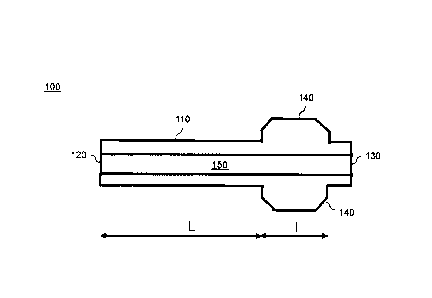
tracking the progress of the mock medical instrument(s) inside the simulated
CA 02981350 2017-09-29
2
body region.
[0005] However, such devices are usually bulky, and their size
reduces their mobility. There is therefore a need for a new tracking system.
SUMMARY
[0006] According to a first aspect, the present disclosure provides a
system for simulating medical procedures. The system comprises a body
cavity simulator and at least one camera. The body cavity simulator comprises
a channel, the channel has a proximal end, a distal end, and an inner
longitudinal passage extending between the proximal end and the distal end.
The channel is partially made of a material comprised of one of the following:
a
transparent material, a translucent material, a semi-transparent material. The
channel is adapted for receiving at least one simulated medical instrument
through the proximal end. The at least one camera is adapted for capturing
through the material of the channel a pattern of a tracking device of the at
least
one simulated medical instrument inserted in the channel. The camera is
adapted for transmitting data corresponding to the captured pattern of the
tracking device to a processing unit.
[0007] According to a second aspect, the present disclosure provides
a method for simulating medical procedures. The method comprises inserting
at least one simulated medical instrument inside a channel of a body cavity
simulator. The method further comprises capturing with at least one camera a
pattern of a tracking device of the at least one simulated medical instrument
inserted inside the channel. The method pursues with transmitting by the
camera data corresponding to the captured pattern of the tracking device to a
processing unit. The method also receives the data corresponding to the
captured pattern at the processing unit. The method further comprises
analyzing by the processing unit the captured pattern for the at least one
simulated medical instrument to determine at least one of the following: an
identification of the at least one simulated medical instrument, a translation
of
CA 02981350 2017-09-29
3
the at least one simulated medical instrument inside the channel, and an
orientation of the at least one simulated medical instrument inside the
channel.
BRIEF DESCRIPTION OF THE DRAWINGS
[0008] Embodiments of the disclosure will be described by way of
example only with reference to the accompanying drawings, in which:
[0009] Figure 1A illustrates a side cross-sectional view of a simulated
medical instrument, according to a first embodiment;
[0010] Figure 1B illustrates a front view of the simulated medical
instrument of Figure 1A;
[0011] Figure 1C illustrates a rear view of the simulated medical
instrument of Figure 1A;
[0012] Figure 2 represents two instances of the simulated medical
instrument of Figure 1A having a tracking device with a particular combination
of shape and color pattern;
[0013] Figure 3A illustrates a side cross-sectional view of a simulated
medical instrument, according to a second embodiment;
[0014] Figure 3B illustrates a front view of the simulated medical
instrument of Figure 3A;
[0015] Figure 3C illustrates a rear view of the simulated medical
instrument of Figure 3A;
[0016] Figure 4 illustrates the simulated medical instrument of Figure
3A being inserted inside the simulated medical instrument of Figure 1A;
[0017] Figure 5A illustrates a side cross-sectional view of a simulated
medical instrument, according to a third embodiment;
[0018] Figure 5B illustrates a front view of the simulated medical
instrument of Figure 5A;
[0019] Figure 5C illustrates a rear view of the simulated medical
CA 02981350 2017-09-29
4
instrument of Figure 5A;
[0020] Figure 6A illustrates a side cross-sectional view of a simulated
medical instrument, according to a fourth embodiment;
[0021] Figure 6B illustrates a front view of the simulated medical
instrument of Figure 6A;
[0022] Figure 6C illustrates a rear view of the simulated medical
instrument of Figure 6A;
[0023] Figure 7 illustrates the simulated medical instrument of Figure
6A being inserted inside the simulated medical instrument of Figure 5A;
[0024] Figure 8A illustrates a schematic cross-sectional perspective
view of a body cavity simulator of a system for simulating medical procedures;
[0025] Figure 8B illustrates a side view of the body cavity simulator of
Figure 8A;
[0026] Figure 8C illustrates a top view of the body cavity simulator of
Figure 8A;
[0027] Figure 9 illustrates the system for simulating medical
procedures of Figure 8A comprising a control station;
[0028] Figure 10 illustrates an analysis performed by a processing
unit of the control station of Figure 9;
[0029] Figures 11A, 11B, 11C, 11D, 11E and 11F illustrate an
exemplary determination of an orientation of a simulated medical instrument;
[0030] Figure 12 illustrates a schematic cross-sectional perspective
view of another embodiment of the body cavity simulator of Figure 8A;
[0031] Figures 13A and 13B illustrate the integration of the body
cavity simulator of Figure 8A with a simulation mannequin;
[0032] Figure 13C illustrates the integration of the body cavity
simulator of Figure 12 with a simulation mannequin;
CA 02981350 2017-09-29
[0033] Figures 14A, 14B and 14C illustrate a body cavity simulator
adapted for providing dynamic haptic interactions; and
[0034] Figure 15 illustrates a method for simulating medical
procedures.
DETAILED DESCRIPTION
[0035] The foregoing and other features will become more apparent
upon reading of the following non-restrictive description of illustrative
embodiments thereof, given by way of example only with reference to the
accompanying drawings. Like numerals represent like features on the various
drawings.
[0036] Various aspects of the present disclosure generally address
one or more of the problems related to medical simulation.
[0037] Reference is now made concurrently to Figures 1A, 1B, 1C, 2,
3A, 3B 30, and 4, which represent a simulated medical instrument 100 for
insertion in a channel of a body cavity simulator, according to a first aspect
of
the present disclosure.
[0038] Figures 1A, 1B and 1C represent a first embodiment of the
simulated medical instrument 100, where Figure 1A is a side cross-sectional
view, Figure 1B is a front view and Figure 1C is a rear view.
[0039] The simulated medical instrument 100 comprises a tube 110.
The tube 110 has a proximal end 120 and a distal end 130. The tube 110 is
sized and shaped for insertion in a channel of a body cavity simulator, which
will be described later in the description. The tube 110 illustrated in
Figures 1A,
1B and 1C has a cylindrical shape, but may have another shape based on the
type of medical instrument simulated. Furthermore, the length and diameter of
the tube 110 also varies based on the type of medical instrument simulated.
The tube 110 can be made in various materials, but is preferably made of a
flexible material, for allowing insertion in a channel of a body cavity
simulator
CA 02981350 2017-09-29
6
which does not have a linear shape, as will be illustrated later in the
description.
[0040] The simulated medical instrument 100 comprises at least one
tracking device 140 located in proximity of the distal end 130 of the tube
110.
In Figures 1A, 1B and 1C, two tracking devices 140 have been represented for
illustration purposes. The two tracking devices 140 are aligned with one
another and define a 180 degree angle between them. However, any number
of tracking devices 140 may be present, which may be aligned or not with one
another, and define various angles between themselves. For instance, the
simulated medical instrument 100 can also have a single tracking device 140,
three tracking devices 140 aligned with one another and defining a 120 degree
angle between them, four tracking devices 140 aligned with one another and
defining a 90 degree angle between them, etc. Furthermore, the tracking
devices 140 of a particular simulated medical instrument 100 can all have a
similar shape, or may have different shapes.
[0041] The length L of the tube 110 illustrated in Figure 1A is not
meant to be limitative in terms of a ratio between the length L of the tube
110
and the length I of the tracking devices 140. The tube 110 has been
represented with a relatively short length L for simplification purposes.
However, the tube 110 may have any length that represents a realistic
intervention (e.g. one meter). With respect to a diameter of the tube 110, it
depends on the type of medical instrument being simulated (e.g. catheters of
various diameters), and varies accordingly.
[0042] Each tracking device 140 has a pattern detectable via camera.
The pattern may consist of a specific shape, a specific color pattern, a
combination of a specific shape and a specific color pattern, etc. The
specific
color pattern can be a different uniform color for each tracking device 140,
or a
combination of colors forming a different color pattern for each tracking
device
140. Figure 2 represents two simulated medical instruments 100 with a single
tracking device 140, each tracking device 140 having a combination of shape
CA 02981350 2017-09-29
7
and color pattern different from the other tracking device 140. In a
particular
embodiment, the tracking device 140 projects radially away from the tube 110
along an external surface of the tube 110. For example, the tracking device is
a flag, etc. In another particular embodiment, the tracking device 140 is a
marking on the tube 110. For example, the tracking device consists of a tag, a
line, a barcode, etc. The tracking device 140 may be removably secured to the
tube 110 (e.g. glued to a surface of the tube 110, inserted in a securing
mechanism of the tube 100 such as a notch, etc.). Alternatively, it is
integral to
the tube 110.
[0043] Figures 1A, 1B and 1C represent a first embodiment of the
simulated medical instrument 100, where the tube 110 comprises an inner
longitudinal passage 150 extending between the proximal end 120 and the
distal end 130, for receiving another simulated medical instrument in the
inner
longitudinal passage 150. The passage 150 illustrated in Figures 1A, 1B and
1C has a cylindrical shape (generally circular cross-section), but may have
another shape (e.g. generally elliptical cross-section) based on the type of
medical instrument simulated. Furthermore, the diameter of the passage 150
also varies based on the type of medical instrument simulated. Although the
passage 150 illustrated in Figures 1A, 1B and 1C is centered within the tube
110, it may also not be centered. Additionally, the tube 110 may include more
than one passage 150. In this first embodiment, the simulated medical
instrument 100 can simulate a cannula, a catheter, a catheter equipped with a
balloon, etc.
[0044] The tube 110 may be at least partially made of a transparent
material for allowing detection by a camera of a pattern of a tracking device
140 of another simulated medical instrument 100 inserted inside the tube 110.
[0045] The simulated medical instrument 100 replicates a real
medical instrument (e.g. a real cannula or a real catheter), but includes the
tracking device(s) for detection purposes. However, real medical instruments
could also be used, but need to be adapted to include the tracking device(s)
CA 02981350 2017-09-29
8
140.
[0046] Figures 3A, 3B and 3C represent a second embodiment of the
simulated medical instrument 100, where Figure 3A is a side cross-sectional
view, Figure 3B is a front view and Figure 30 is a rear view.
[0047] This second embodiment is similar to the first embodiment
represented in Figures 1A, 1B and 1C, except that the tube 110 is solid
between the proximal end 120 and the distal end 130 (does not comprise an
inner longitudinal passage). Consequently, it does not allow insertion of
another simulated medical instrument in a passage. In this second
embodiment, the simulated medical instrument 100 can simulate a guiding
wire, etc.
[0048] Figure 4 represents two simulated medical instruments 101
and 102, both having two tracking devices 140 forming a 180 degrees angle
with one another. The simulated medical instrument 101 corresponds to the
first embodiment represented in Figure 1A, and has an inner longitudinal
passage for inserting the simulated medical instrument 102. The simulated
medical instrument 102 corresponds to the second embodiment represented in
Figure 3A, and has no inner longitudinal passage. For example, the tube 110
of the simulated medical instruments 102 simulates a guide wire, and the
simulated medical instruments 101 is a catheter. The size and shape of the
tracking devices 140 of the simulated medical instrument 102 may be adapted
for allowing insertion of the simulated medical instrument 102 in the inner
passage 150 of the simulated medical instrument 101. Alternatively, the
tracking devices 140 of the simulated medical instrument 102 are made of a
flexible material for allowing insertion of the simulated medical instrument
102
in the inner passage 150 of the simulated medical instrument 101. In still
another alternative, the tracking devices 140 of the simulated medical
instrument 102 are not adapted for allowing insertion of the simulated medical
instrument 102 in the inner passage 150 of the simulated medical instrument
101; and the simulated medical instrument 102 must be inserted before (and
CA 02981350 2017-09-29
9
extracted after) the simulated medical instrument 101. in the channel of the
body cavity simulator. Although not represented in Figure 4 for simplification
purposes, the simulated medical instrument 101 may also be inserted in the
inner longitudinal passage of a third simulated medical instrument (e.g. a
cannula or another catheter having a larger diameter).
[0049] The tracking device(s) 140 of the simulated medical instrument
100 represented in Figures 1A-C, 3A-C and 4 (e.g. with tracking device(s) 140
in the form of flags, tags, barcodes, etc.) only allows static haptic
interactions
with the channel of the body cavity simulator, as will be detailed later in
the
description.
[0050] Reference is now made concurrently to Figures 5A, 5B, 5C,
6A, 6B, 6C, and 7, which represent a simulated medical instrument 200 for
insertion in a channel of a body cavity simulator, according to a second
aspect
of the present disclosure. The simulated medical instrument 200 is similar to
the aforementioned simulated medical instrument 100, except for its tracking
device.
[0051] Figures 5A, 5B and 5C represent a first embodiment of the
simulated medical instrument 200, where Figure 5A is a side cross-sectional
view, Figure 5B is a front view and Figure 5C is a rear view.
[0052] The simulated medical instrument 200 comprises a tube 210.
The tube 210 has a proximal end 220 and a distal end 230. The tune 210 is
sized and shaped for insertion in a channel of a body cavity simulator. As
illustrated in Figure 5A, the tube 210 has a length L, and the distal end 230
is
separated by the distance L from the proximal end 220. The characteristics of
the tube 210 are similar to the characteristics of the tube 110 represented in
Figure 1A.
[0053] As mentioned previously with respect to the tube 110
illustrated in Figure 1A, the length L of the tube 210 illustrated in Figure
5A is
not meant to be !imitative in terms of a ratio between the length L of the
tube
CA 02981350 2017-09-29
210 and the length I of the tracking device 240. The tube 210 has been
represented with a relatively short length L for simplification purposes.
[0054] The simulated medical instrument 200 comprises a tracking
device 240 positioned at the distal end 230 of the tube 210. The tracking
device 240 of the simulated medical instrument 200 allows static haptic
interactions with the channel of the body cavity simulator. However, the
tracking device 240 is further adapted for receiving friction caused by a
dynamic haptic mechanism positioned along at least a section of the channel
of the body cavity simulator, as will be detailed later in the description.
[0055] Similarly to the tracking device 140 represented in Figure 1A,
the tracking device 240 has a pattern detectable via camera. The pattern may
consist of a specific shape, a specific color pattern, a combination of a
specific
shape and a specific color pattern, etc.
[0056] The tracking device 240 may be a sphere, or another object
allowing a dynamic haptic mechanism of the body cavity simulator to exert a
friction against a surface of the tracking device 240. In a particular
embodiment, a diameter of the tracking device 240 (e.g. a sphere) is
substantially equal to a diameter of the tube 210. In another particular
embodiment, a diameter of the tracking device 240 (e.g. a sphere) is
substantially greater than a diameter of the tube 210, for increasing the
friction
exerted by the dynamic haptic mechanism of the body cavity simulator.
[0057] The tracking device 240 can be removably secured to the tube
210 (e.g. glued to the distal end 230 of the tube 210, etc.), or it can be
integral
to the tube 210.
[0058] Figures 5A, 5B and 5C represent a first embodiment of the
simulated medical instrument 200, where the tube 210 comprises an inner
longitudinal passage 250 extending between the proximal end 220 and the
distal end 230, for receiving another simulated medical instrument in the
inner
longitudinal passage 250. The characteristics of the passage 250 are similar
to
CA 02981350 2017-09-29
11
the characteristics of the passage 150 represented in Figure 1A. The tracking
device 240 also comprises an inner longitudinal passage 260 for receiving the
other simulated medical instrument in the inner longitudinal passage 260. The
passage 250 of the tube 210 is aligned with the passage 260 of the tracking
device 240. The characteristics of the passage 260 are generally similar to
the
characteristics of the passage 250, although the respective diameters and
shapes of the passages 250 and 260 may be different, as long as they both
allow insertion of the other simulated medical instrument. As mentioned
previously, in this first embodiment, the simulated medical instrument 200 can
simulate a cannula, a catheter, a catheter equipped with a balloon, etc.
[0059] The tube 210 may be at least partially made of a transparent
material for allowing detection by a camera of a pattern of a tracking device
240 of another simulated medical instrument 200 inserted inside the tube 210.
[0060] Figures 6A, 6B and 6C represent a second embodiment of the
simulated medical instrument 200, where Figure 6A is a side cross-sectional
view, Figure 6B is a front view and Figure 6C is a rear view.
[0061] This second embodiment is similar to the first embodiment
represented in Figures 5A, 56 and 5C, except that the tube 210 is solid
between the proximal end 220 and the distal end 230 (does not comprise an
inner longitudinal passage). Consequently, it does not allow insertion of
another simulated medical instrument in a passage. The tracking device 240 is
also solid (does not comprise an inner longitudinal passage). As mentioned
previously, in this second embodiment, the simulated medical instrument 200
can simulate a guiding wire, etc.
[0062] Figure 7 represents two simulated medical instruments 201
and 202, both having a tracking device 240 in the form of a sphere. The
simulated medical instrument 201 corresponds to the first embodiment
represented in Figure 5A, and has two inner longitudinal passages respectively
in its tube 210 and tracking device 240 for inserting the simulated medical
instrument 202. The simulated medical instrument 202 corresponds to the
CA 02981350 2017-09-29
12
second embodiment represented in Figure 6A, and has no inner longitudinal
passages. For example, the tube 210 of the simulated medical instruments 202
simulates a guide wire, and the simulated medical instruments 201 is a
catheter. The diameter of the tracking device 240 of the simulated medical
instrument 202 may be adapted for allowing insertion of the simulated medical
instrument 202 in the inner passage 250 of the simulated medical instrument
201. Alternatively, the tracking device 240 of the simulated medical
instrument
202 is made of a flexible material for allowing insertion of the simulated
medical instrument 202 in the inner passage 250 of the simulated medical
instrument 201. In still another alternative, the tracking device 240 of the
simulated medical instrument 202 is not adapted for allowing insertion of the
simulated medical instrument 202 in the inner passage 250 of the simulated
medical instrument 201; and the simulated medical instrument 202 must be
inserted before (and extracted after) the simulated medical instrument 201 in
the channel of the body cavity simulator. Although not represented in Figure 7
for simplification purposes, the simulated medical instrument 201 may also be
inserted in the inner longitudinal passages of respectively the tube and
tracking
device of a third simulated medical instrument (e.g. a cannula or another
catheter having a larger diameter).
[0063] Although not represented in the Figures, a simulated medical
instrument 100 with a tracking device 140 in the form of a flag, tag, barcode,
etc. (as illustrated in Figures 1A and 3A) may be inserted in a simulated
medical instrument 200 with a tracking device 240 in the form of a sphere,
etc.
(as illustrated in Figure 5A).
[0064] Reference is now made concurrently to Figures 8A, 86, 80
and 9, which represent a system for simulating medical procedures, according
to a third aspect of the present disclosure.
[0065] Figures 8A, 8B and 8C represent a first embodiment of the
system for simulating medical procedures, where Figure 8A is a schematic
cross-sectional perspective view, Figure 86 is a side view and Figure 8C is a
CA 02981350 2017-09-29
13
top view.
[0066] The system for simulating medical procedures comprises a
body cavity simulator 300. The body cavity simulator 300 comprises a channel
310. The channel 310 can be shaped like a vase to avoid dead points. In a
particular embodiment, the channel 310 has any shape or path allowing easy
insertion of the body cavity simulator 300 in a suitcase, and the body cavity
simulator 300 is thus portable. The channel 310 has a proximal end 312, a
distal end 314, and an inner longitudinal passage 316. The passage 316
extends between the proximal end 312 and the distal end 314. The channel
310 simulates a body channel, such as a trachea, an artery, a channel of the
intestine such as the large intestine or the small intestine, etc. The channel
310
is adapted for receiving at least one of the aforementioned simulated medical
instruments 100 (illustrated in Figure 1A or 3A) or 200 (illustrated in Figure
5A
or 6A) through the proximal end 312. In addition to the channel 310, the body
cavity simulator 300 may also include a simulator of a body part enclosing the
channel 310. For instance, in the case of an artery, the body cavity simulator
300 only includes the channel 310 for simulating the artery, or includes a
simulation of a body part such as an arm or a leg with the channel 310
enclosed in the simulated body part. Similarly, in the case of a large
intestine,
the body cavity simulator 300 only includes the channel 310 for simulating the
large intestine, or includes a simulation of a body part such as a portion of
the
digestion system with the channel 310 enclosed in the simulated body part.
Furthermore, the body cavity simulator 300 is generally adapted for simulating
medical procedures on humans, but could also be adapted for simulating
medical procedures on animals. Furthermore, the mechanism of the body
cavity simulator 300 for receiving a simulated medical instrument is not
limited
to a channel 310, but may consist in any other mechanism allowing a realistic
simulation of insertion of the simulated medical instrument in a simulated
body
cavity.
[0067] The passage 316 illustrated in Figure 8A has a cylindrical
CA 02981350 2017-09-29
14
shape (generally circular cross-section), but may have another shape (e.g.
generally elliptical cross-section) based on the type of simulated body
channel.
Furthermore, the diameter of the passage 316 also varies based on the type of
simulated body channel.
[0068] In the embodiment illustrated in Figure 8A, the channel 310 is
spirally wound and defines a circular body cavity simulator 300.
[0069] The body cavity simulator 300 may also include a frame 320
as illustrated in Figures 8B and 8C. The channel 310 is enclosed within the
frame 320, except for its proximal end 312. The frame 320 can play several
roles, such as protecting the channel 310, hiding a particular geometry of the
channel 310, allowing attachment of the body cavity simulator 300 to another
device via a dedicated attachment part 322 of the frame 320, etc. The shape
and size of the frame 320 can vary significantly, as long as the channel 310
can be enclosed within the frame 320.
[0070] The system further comprises a camera 400 for capturing a
pattern of a tracking device of the at least one simulated medical instrument
inserted in the channel 310. For instance, the pattern of a tracking device
140
of the simulated medical instrument 100 illustrated in Figures 1A or 3A; or
the
pattern of the tracking device 240 of the simulated medical instrument 200
illustrated in Figures 5A or 6A. The camera 400 transmits data corresponding
to the captured pattern of the tracking device to a processing unit 510
represented in Figure 9, where the data are further processed. The further
processing will be detailed later in the description. In a particular
embodiment,
the system comprises a plurality of cameras 400, as illustrated in Figure 12.
[0071] Figure 8A illustrates a system with a camera 400 centrally
positioned with respect to the circular body cavity simulator 300. The camera
400 has an ultra wide angle, allowing capture of pattern(s) of any simulated
medical instrument inserted in the channel 310. For example, the tracking
devices 240 of three simulated medical instruments 200 (illustrated in Figure
5A or 6A) have been represented in Figure 8A. Only the three tracking devices
CA 02981350 2017-09-29
240 have been represented in Figure 8A for simplification purposes, but all
three simulated medical instruments have been inserted in the channel 310 via
its proximal end 312. Each of the three simulated medical instruments extends
up to its respective tracking device 240 within the passage 316 of the channel
310. The camera 400 is capable of capturing the patterns of the three tracking
devices 240.
[0072] The channel 310 is partially made of a transparent material for
allowing the camera 400 to capture the pattern(s) through the transparent
material of the channel 310. For example, if the camera 400 is positioned on
top of the body cavity simulator 300 as illustrated in Figure 8A, at least an
upper section of the channel 310 is made of the transparent material.
[0073] The upper section of the channel 310 can be made of a semi-
transparent, translucent or any type of material that allows for the tracking
devices to be viewed and captured by the camera.
[0074] In the embodiment illustrated in Figures 8A and 8B, the
camera 400 is located within an upper section of the frame 320 on top of the
body cavity simulator 300, and it is secured to the frame 320 by proper
securing means.
[0075] Reference is now made concurrently to Figures 9, 10, 11A,
11B, 110, 11D, 11E, 11F, which illustrate the processing by the processing
unit 510 of the data captured by the camera 400.
[0076] The information captured by the camera 400 may comprise
any surgical object used in the context of the medical simulation performed
with the body cavity simulator 300. The captured information is not limited to
the pattern(s) of the tracking device(s) of the simulated medical
instrument(s)
inserted in the body cavity simulator 300.
[0077] The processing unit 510 may be part of a control station 500.
The processing unit 510 has one or more processors (not represented in
Figure 9 for simplification purposes) capable of executing instructions of
CA 02981350 2017-09-29
16
computer program(s). Each processor may further have one or several cores.
The control station 500 also comprises memory 520 for storing instructions of
the computer program(s) executed by the processing unit 510, data generated
by the execution of the computer program(s), data received via a
communication interface 530 of the control station 500, etc. The control
station
500 may comprise several types of memories, including volatile memory, non-
volatile memory, etc. The control station 500 further comprises the
communication interface 530 (e.g. Wi-Fi interface, Ethernet interface,
cellular
interface, etc.). The communication interface 530 is used for exchanging data
with other entities, such as the camera 400 via communication links 450. Such
communication links 450 may include wired (e.g. a fixed broadband network)
and wireless communication links (e.g. a cellular network or a Wi-Fi network).
The control station 500 may further comprise a display 540 (e.g. a regular
screen or a tactile screen) for displaying data generated by the processing
unit
510, and a user interface 550 (e.g. a mouse, a keyboard, a trackpad, a
touchscreen, etc.) for allowing a user to interact with the control station
500.
The control station 500 may consist of a computer, a laptop, a mobile device
(e.g. smartphone, tablet, etc.), a dedicated control station for medical
simulations, a dedicated control station for operational medical procedures,
etc.
[0078] The camera 400 includes a communication interface
supporting a communication protocol (e.g. USB, Wi-Fi, cellular, etc.) for
transmitting data captured by the camera 400 to the processing unit 510 via
the communication interface 530 through the communication links 450.
[0079] The processing unit 510 receives the data comprising the
captured pattern(s) transmitted by the camera 400, and analyses the captured
pattern(s). The analysis comprises determining at least one of the following:
an
identification of at least one simulated medical instrument inserted in the
channel 310, a translation of the at least one simulated medical instrument
inside the channel 310, and an orientation of the at least one simulated
CA 02981350 2017-09-29
17
medical instrument inside the channel 310. The determination is based on the
analysis of the captured pattern(s) for the at least one simulated medical
instrument.
[0080] Figure 10 illustrates the analysis performed by the processing
unit 510 for the two simulated medical instruments 101 and 102 previously
represented in Figure 4, each having two tracking devices 140 forming a 180
degrees angle with one another. The two simulated medical instruments 101
and 102 are inserted inside the channel 310. Furthermore, the simulated
medical instrument 101 (e.g. a cannula) corresponds to the embodiment
represented in Figure 1A, and has an inner longitudinal passage for inserting
the simulated medical instrument 102 (e.g. a guide wire) corresponding to the
embodiment represented in Figure 3A. Each of the simulated medical
instruments 101 and 102 perform a translation 600 within channel 310, and
can also perform a rotation 610 around their longitudinal axis.
[0081] Each tracking device 140 is a flag having two opposite sides,
each side having a unique color pattern. The unique color pattern may be
simply a unique uniform color, or may be a unique combination of several
colors (in order to be able to handle (with a limited number of colors) a
plurality
of simulated medical instruments respectively having a plurality of tracking
devices). Thus, the aforementioned pattern of each tracking device 140
consists of the combination of the unique color pattern of each of its
opposite
sides.
[0082] At any time, the camera 400 is at least capable of capturing
the unique color pattern of one side of one of the two flags 140 for each
simulated medical instruments 101 and 102, based on their respective
orientation with respect to the camera 400.
[0083] Based on the captured unique color pattern(s) for each
simulated medical instruments 101 and 102, the processing unit 510 can
identify the two simulated medical instruments 101 and 102 inserted in the
channel 310.
CA 02981350 2017-09-29
18
[0084] Furthermore, the data captured by the camera 400 may
comprise an image of the channel 310. Thus, by analyzing the captured unique
color pattern(s) for each simulated medical instruments 101 and 102 with
respect to the image of the channel 310, a position of each simulated medical
instruments 101 and 102 within the channel 310 can be determined. Based on
the particular geometry of the channel 310, a translation for each simulated
medical instrument 101 and 102 can be further determined based on the
determined position. The determined translation can for example indicate how
far from the proximal end 312 of the channel 310 each simulated medical
instrument 101 and 102 has been inserted.
[0085] Alternatively, the camera 400 can be configured during an
initial phase to take a picture comprising the channel 310, this picture being
correlated with the geometry of the channel 310. During the operational phase
when the unique color pattern(s) are captured by the camera 400, by analyzing
the position of the colors patterns within the image captured by the camera
400, the position of the color patterns within the channel 310 can be
determined.
[0086] Additionally, by analyzing the captured color pattern for each
simulated medical instruments 101 and 102, an orientation of each simulated
medical instruments 101 and 102 within the channel 310 can be determined.
[0087] Figures 11A, 11B, 11C, 11D, 11E and 11F illustrate an
example of determination of the orientation of the simulated medical
instrument
102 represented in Figures 10 and 3B. The first flag 141 of the simulated
medical instruments 102 has two patterns 650 and 651 on its respective
opposite sides. The second flag 142 of the simulated medical instruments 102
has two patterns 652 and 653 on its respective opposite sides.
[0088] In the configuration represented in Figure 11A, the pattern 650
is detected by the camera 400. Thus the flags 141 and 142 are substantially
vertical, the flag 141 being on top and the flag 142 being below. Furthermore,
the flag 141 is on the right with respect to a reference vertical axis 660,
while
CA 02981350 2017-09-29
19
the flag 142 is on the left with respect to the reference vertical axis 660.
[0089] In the configuration represented in Figure 11B, the pattern 651
is detected by the camera 400. Thus the flags 141 and 142 are substantially
vertical, the flag 141 being on top and the flag 142 being below. Furthermore,
the flag 141 is on the left with respect to the reference vertical axis 660,
while
the flag 142 is on the right with respect to the reference vertical axis 660.
[0090] In the configuration represented in Figure 11C, the pattern 652
is detected by the camera 400. Thus the flags 141 and 142 are substantially
vertical, the flag 142 being on top and the flag 141 being below. Furthermore,
the flag 142 is on the right with respect to the reference vertical axis 660,
while
the flag 141 is on the left with respect to the reference vertical axis 660.
[0091] In the configuration represented in Figure 11D, the pattern 653
is detected by the camera 400. Thus the flags 141 and 142 are substantially
vertical, the flag 142 being on top and the flag 141 being below. Furthermore,
the flag 142 is on the left with respect to the reference vertical axis 660,
while
the flag 141 is on the right with respect to the reference vertical axis 660.
[0092] In the configuration represented in Figure 11E, the patterns
650 and 653 are detected by the camera 400. Thus the flags 141 and 142 are
substantially horizontal. Furthermore, the flag 141 is on the right with
respect to
the reference vertical axis 660, while the flag 142 is on the left with
respect to
the reference vertical axis 660.
[0093] In the configuration represented in Figure 11F, the patterns
651 and 652 are detected by the camera 400. Thus the flags 141 and 142 are
substantially horizontal. Furthermore, the flag 142 is on the right with
respect to
the reference vertical axis 660, while the flag 141 is on the left with
respect to
the reference vertical axis 660.
[0094] Although the determination by the processing unit 510 of the
identification, translation and orientation of simulated medical instrument(s)
inserted inside the channel 310 has been illustrated in Figure 10 for two
CA 02981350 2017-09-29
simulated medical instruments 101 and 102, it can be generalized for one, two,
three or more simulated medical instruments simultaneously inserted inside
the channel 310. Furthermore, as mentioned previously, the patterns used for
determining the identification, translation and orientation of the simulated
medical instrument(s) are not limited to specific color patterns, but may also
include specific shapes, or a combination of specific color patterns and
specific
shapes, as long as they can be detected by the camera 400. Furthermore, the
two simulated medical instruments 101 and 102 represented in Figure 10
respectively correspond to the embodiments represented in Figure 1A and
Figure 3A, with two tracking devices 140. However, the determination of the
identification, translation and orientation can be generalized for simulated
medical instruments 100 having one, two, three, four or more tracking devices
140. In particular, a larger number of tracking devices 140 on the simulated
medical instruments 100 improves the accuracy of the determination of the
orientation. The determination of the identification, translation and
orientation
can also be generalized for simulated medical instruments 200 corresponding
to the embodiments represented in Figure 5A and Figure 6A. For example, the
tracking device 240 of a simulated medical instrument 200 can be a sphere
having an external surface covered with a unique color pattern detectable by
the camera 400, the unique color pattern allowing a determination of the
orientation of the sphere.
[0095] Figure 12 represents a second embodiment of the system for
simulating medical procedures, Figure 12 being a schematic cross-sectional
perspective view.
[0096] This second embodiment is similar to the first embodiment
represented in Figures 8A and 9, except that the channel 310 is linear and
defines a linear body cavity simulator 300. Furthermore, the system may
comprise a plurality of cameras 400 for covering the entire length of the
channel 310. The system represented in Figure 12 comprises three cameras
400 for illustration purposes, but may comprise more or less cameras 400. The
CA 02981350 2017-09-29
21
number of cameras 400 is adapted for covering the entire length of the channel
310, and depends on the extent of the area which can be covered by a single
camera 400. The data captured by each camera 400 are combined by the
processing unit 510 represented in Figure 9, for the determination of the
identification, translation and orientation of simulated medical instrument(s)
inserted inside the channel 310.
[0097] The system may comprise a frame (not represented in Figure
12) for enclosing and supporting the linear body cavity simulator 300 and the
camera(s) 400. Alternatively, the system does not comprise a frame, and the
linear body cavity simulator 300 and the camera(s) 400 are independently
affixed to a supporting entity.
[0098] Reference is now made concurrently to Figures 9, 13A, 136
and 13C, where Figures 13A, 13B and 130 illustrate the integration of the body
cavity simulator 300 with a simulation mannequin 700.
[0099] The simulation mannequin 700 is a realistic representation of a
human body and is positioned on a table 710. The body cavity simulator 300
represented in Figures 13A and 13B corresponds to the embodiment
represented in Figure 8B of a circular body cavity simulator 300 with a
spirally
wounded channel 310.
[00100] The simulation mannequin 700 comprises a plurality of
securing mechanisms 720 (projecting downwardly through a horizontal surface
of the table 710), for securing the body cavity simulator 300 thereto (e.g.
via
the dedicated attachment part 322 represented in Figure 80). A particular
securing mechanism 720 is selected among the plurality of securing
mechanisms 720 for attaching the body cavity simulator 300, so that the body
cavity simulated with the body cavity simulator 300 is positioned
substantially
below its counterpart in the simulation mannequin 700.
[00101] In Figures 13A and 13B, the body cavity 300 is located below
the surface of the table 710. In an alternative embodiment, the body cavity
CA 02981350 2017-09-29
22
simulator 300 is located above the surface of the table 710, and positioned
below the mannequin 700 or besides the mannequin 700. The body cavity
simulator 300 can also be located inside the table 710.
[00102] Alternatively, the body cavity simulator 300 may be integrated
into the simulation mannequin 700, and positioned within the simulation
mannequin 700 at a position corresponding to the simulated body cavity, with
the proximal end 312 (represented in Figure 8A) projecting away from a
surface of the simulation mannequin 700. In another alternative, a patient may
be positioned on the table 710 in place of the simulation mannequin 700.
[00103] The use of a simulation mannequin 700 or a patient in
combination with the body cavity simulator 300 allows a trainee (or an
experimented professional) to simulate and practice an operation involving
insertion of medical instruments inside a body channel (e.g. a trachea, an
artery, a channel of the intestine such as the large intestine or the small
intestine, etc.) in a more realistic manner, compared to the use of the body
cavity simulator 300 alone.
[00104] The body cavity simulator 300 represented in Figure 13C
corresponds to the embodiment represented in Figure 12 of a linear body
cavity simulator 300 with a linear channel 310. The body cavity simulator 300
is substantially aligned with a simulation mannequin 700 or a patient, for
instance to simulate an artery of an arm.
[00105] In the case of a patient being positioned on the table 710, a
medical imaging system (not represented in Figures 13A, 13B and 13C) may
take (2D or 3D) images of the body cavity of the patient simulated by the body
cavity simulator 300. As mentioned previously, the processing unit 510
represented in Figure 9 determines (based on the data captured and
transmitted by the camera 400) the following: an identification of at least
one
simulated medical instrument inserted the channel 310 of the body cavity
simulator 300, the translation of the at least one simulated medical
instrument
inside the channel 310, and the orientation of the at least one simulated
CA 02981350 2017-09-29
23
medical instrument inside the channel 310. The determined identification,
translation and orientation can be combined with the images taken by the
medical imaging system. The combination is performed by the processing unit
510 (or another processing unit of another computing device). The combination
is further displayed on a screen 730 for showing a progression of the at least
one simulated medical instrument inside the simulated body cavity.
[00106] The simulated medical instrument 100 represented in Figures
1A-C, 3A-C and 4 (e.g. with tracking devices 140 in the form of flags) only
allows static haptic interactions with the channel 310 of the body cavity
simulator 300 represented in Figures 8A or 12. The static haptic interactions
consist of frictions of the flags 140 against the internal surface of the
channel
310 defining the inner longitudinal passage 316. The static haptic friction
increases with a deeper penetration of the simulated medical instrument 100
inside the inner longitudinal passage 316 of the channel 310. Furthermore, the
circular body cavity simulator 300 represented in Figure 8A offers more
friction
than the linear body cavity simulator 300 represented in Figure 12.
[00107] The simulated medical instrument 200 represented in Figures
5A-C, 6A-C and 7 (e.g. with a tracking device 140 in the form of a sphere)
also
allows dynamic haptic interactions with the channel 310 of the body cavity
simulator 300 represented in Figures 8A or 12, when the body cavity simulator
300 is adapted for this purpose, as detailed in the following.
[00108] Reference is now made concurrently to Figures 14A, 14B and
14C, which represent a body cavity simulator 800 adapted for providing
dynamic haptic interactions, according to a fourth aspect of the present
disclosure.
[00109] The body cavity simulator 800 is similar to the body cavity
simulators 300 represented in Figures 8A or 12, except for the channel 810.
This new design can be applied to both a circular body cavity simulator as
illustrated in Figure 8A and to a linear circular body cavity simulator as
represented in Figure 12.
CA 02981350 2017-09-29
24
[00110] For illustration purposes, Figures 14A and 14C represent two
simulated medical instruments 201 and 202 (corresponding to those
represented in Figure 7), both having a tracking device 240 in the form of a
sphere, being inserted inside the inner longitudinal passage 816 of the
channel
810. The tracking devices 240 of simulated medical instruments 201 and 202
are both rigid for allowing haptic interactions with the body cavity simulator
800. The simulated medical instrument 202 has been inserted before the
simulated medical instruments 201. Furthermore, the diameters of the tracking
devices 240 of simulated medical instruments 201 and 202 are substantially
the same for allowing simultaneous haptic interactions with both simulated
medical instruments. In an alternative not represented in the Figures, a
single
simulated medical instrument 201 with a rigid tracking device 240 in the form
of
a sphere can be inserted for allowing haptic interactions with the body cavity
simulator 800. In still another alternative represented in Figure 14B, a first
simulated medical instrument 201 with a rigid tracking device 240 in the form
of
a sphere can be inserted for allowing haptic interactions with the body cavity
simulator 800. A second simulated medical instrument 102 (corresponding to
the embodiment represented in Figure 3A) with a tracking device 140 in the
form of a flag, tag, barcode, etc. is also inserted, but does not allow haptic
interactions with the body cavity simulator 800. However, the second simulated
medical instrument 102 can be easily inserted / removed through the first
simulated medical instrument 201. In yet another alternative, a simulated
medical instrument simulating a catheter equipped with a balloon can be
inserted in the body cavity simulator 800, and provides haptic interactions
with
the balloon. There is no limitations on the simulated medical instrument
inserted in the body cavity simulator 800, as long as it includes a component
providing haptic interactions.
[00111] As previously mentioned, at least the upper section of the
channel 810 is made of a transparent material for allowing detection by a
camera of the patterns of the tracking devices 240 of the simulated medical
instruments 201 and 202.
CA 02981350 2017-09-29
[00112] A dynamic haptic mechanism is used for exerting a pressure
causing a friction against the tracking devices 240 of the simulated medical
instruments 201 and 202. For example, as illustrated in Figure 14A, an
actuator pushes the interior wall 811 of the entire lower section of the
channel
810 towards the simulated medical instruments 201 and 202. Alternatively, a
bladder or any device capable of exerting a pressure by pushing the interior
wall 811 can be used. When the interior wall 811 reaches the tracking devices
240, it exerts a pressure causing a friction against these tracking devices
240.
In another embodiment illustrated in Figure 14C, a plurality of devices
capable
of exerting a pressure (e.g. actuators, bladders, etc.) can be activated
independently for pushing the interior wall (812 or 813) of a specific zone of
the
lower section of the channel 810 towards at least one simulated medical
instrument. For instance, actuation of zone 1 pushes the corresponding
interior
wall 812 towards the simulated medical instruments 201 for exerting a
pressure causing a friction against the tracking device 240 of the simulated
medical instruments 201. Similarly, actuation of zone 2 pushes the
corresponding interior wall 813 towards the simulated medical instruments 202
for exerting a pressure causing a friction against the tracking device 240 of
the
simulated medical instruments 202.
[00113] The interior walls 811, 812 and 813 for exerting a pressure
causing a friction against the tracking devices may consist of a brush, a
bladder, a fabric, a material, etc. The interior walls 811, 812 and 813 can be
made in silicone, plastic, etc. They can also be covered by an abrasive paint
that causes friction. The resulting friction is a combination of the material
/
geometry of the tracking device and the surface finish / material of the
interior
walls.
[00114] The dynamic haptic mechanism may be activated manually by
a user of the body cavity simulator 800. Alternatively, the dynamic haptic
mechanism is automatically activated when the presence of a particular
simulated medical instrument detected. The automatic activation may also
CA 02981350 2017-09-29
26
depend on the position and / or orientation of the simulated medical
instrument
in the channel 810 of the body cavity simulator 800. For example, the
processing unit 510 represented in Figure 9 controls the dynamic activation of
the dynamic haptic mechanism, based on the determination of the
identification, translation and orientation of the simulated medical
instrument.
[00115] The friction generated by the dynamic haptic mechanism
simulates the friction experienced when a real medical instrument (e.g. a
catheter) hits an interior wall of a real body channel.
[00116] According to another embodiment, the channel 810 of the
body cavity simulator 800 also includes at least one pressure sensor (not
represented in the Figures), for measuring a pressure exerted by a tracking
device 240 against the interior walls of the channel 810.
[00117] According to still another embodiment, the body cavity
simulator 800 is configurable. For example, actuators are used for dynamically
modifying the diameter of a particular section of the channel 810, for
dynamically modifying the shape of a particular section of the channel 810,
for
dynamically modifying the orientation of a particular section of the channel
810,
etc. The body cavity simulator 800 can also be provided with opening doors.
The opening doors are controlled by a software, which is configured to take
into account different possible channel 810 in the body cavity simulator 800.
A
particular software configuration provides a small, medium or long channel
810, depending on a particular medical procedure to be simulated. The body
cavity simulator 800 can be seen as a configurable labyrinth providing a
variety
of paths based on its configuration.
[00118] Reference is now made to Figure 15, which represents a
method 900 for simulating medical procedures, according to a fifth aspect of
the present disclosure.
[00119] The method 900 comprises the step 910 of inserting at least
one simulated medical instrument inside a channel of a body cavity simulator.
CA 02981350 2017-09-29
27
[00120] The method 900 comprises the step 920 of capturing with a
camera a pattern of a tracking device of the at least one simulated medical
instrument inserted inside the channel of the body cavity simulator.
[00121] The method 900 comprises the step 930 of transmitting by the
camera data corresponding to the captured pattern of the tracking device to a
processing unit.
[00122] The method 900 comprises the step 935 of receiving the data
corresponding to the captured pattern at the processing unit.
[00123] The method 900 comprises the step 940 of analyzing by the
processing unit the captured pattern for the at least one simulated medical
instrument, to determine at least one of the following: an identification of
the at
least one simulated medical instrument, a translation of the at least one
simulated medical instrument inside the channel of the body cavity simulator,
and an orientation of the at least one simulated medical instrument inside the
channel of the body cavity simulator.
[00124] In a particular aspect, the method 900 further comprises the
step 950 of exerting a pressure for causing a friction against the tracking
device of at least one of the simulated medical instrument inserted inside the
channel of the body cavity simulator, by means of a dynamic haptic
mechanism. As mentioned previously, step 950 can only be performed for a
simulated medical instrument having a tracking device (e.g. a sphere, but not
a
flag, a tag, etc.) adapted for supporting the pressure / friction exerted by
the
dynamic haptic mechanism. Step 950 can be performed concurrently with
steps 920, 930 and 940.
[00125] A simulated medical instrument with a tracking device which
does not provide dynamic haptic interactions (e.g. a flag, a tag, a line, a
barcode, etc.) is generally used for the entry procedure in a channel of a
body
cavity simulator. A simulated medical instrument with a tracking device
providing dynamic haptic interactions (e.g. a sphere, etc.) is generally used
CA 02981350 2017-09-29
28
thereafter for monitoring the progression towards the distal end of the
channel.
[00126] Although the
present disclosure has been described
hereinabove by way of non-restrictive, illustrative embodiments thereof, these
embodiments may be modified at will within the scope of the appended claims
without departing from the spirit and nature of the present disclosure.