Note: Descriptions are shown in the official language in which they were submitted.
CA 02981811 2017-10-03
WO 2016/172224
PCT/US2016/028459
VECTORS AND METHODS FOR REGENERATIVE THERAPY
[0001] This application claims benefit of United States provisional patent
application number
62/150,159, filed April 20, 2015, the entire contents of which are
incorporated by reference into
this application.
REFERENCE TO A SEQUENCE LISTING SUBMITTED VIA EFS-WEB
[0002] The content of the ASCII text the of the sequence listing named
"UW57WOUl_ST25",
which is 15 kb in size was created on April 19, 2016, and electronically
submitted via EFS-Web
with this application is incorporated herein by reference in its entirety.
TECHNICAL HELD OF THE INVENTION
[0003] The present invention relate..s to nucleic acid molecules, vectors,
ce..11s, and related
compositions and their use for inducing proliferation of quiescent cells and
in methods of
regenerative therapy.
BACKGROUND OF THE INVENTION
[0004] The vast majority of mammalian cardiac i-nyocytes (CM) stop
proliferating soon after
birth and subsequent heart growth predominately comes from hypertrophy, an
increase in cell
size, instead of hyperplasia, an increase in cell number. Because CM
proliferation is required for
the heart regeneration seen in lower vertebrates and neonatal mammalian injury
models, there
is great interest in understanding the mechanisms regulating CM cell cycle
exit and whether this
cell cycle withdrawal can be reversed,
[0005] Ischemic heart disease leading to heart failurel, 2 is the leading
cause of death in the
word. Although adult human hearts are unable to replace lost CMs after injury,
substantial
cardiac regeneration is seen in lower vertebrate and mammalian models. Adult
ze..brafish4 and
neonatal i-nice5 are able to regenerate their hearts after 5% has been
amputated. Models of
myocardial infarction (MI) in newborn mice offer a more clinically relevant
injury model to
demonstrate heart regeneration capacity in mammals'', 7. A common finding in
these studies was
the mechanism by which cardiac regeneration occurred. Blood clot formation,
inflammation, and
collagen deposition were seen in response to the injuries, but ultimately new
CMs repopulated
the lost tissue. Fate mapping studies revealed that the new cardiac myocytes
came from
dedifferentiation and proliferation of pre-existing cardiac rhyocytes, in
contrast to cardiac
progenitor or stem cells4-5. However, when cardiac injury was induced in mice
at a later time-
point, postnatal day 7 (P7), the regenerative response was lost leading to
fibrotic scarring5,6
similar to what is seen with human M1s1.2. Thus, mammalian hearts lose their
regenerative
capacity early in life, a process that requires CM proliferation.
1
CA 02981811 2017-10-03
WO 2016/172224
PCT/US2016/028459
[0006] The poor regenerative response seen in adult hearts highlights the
question of whether
there is any turnover of CMs in adults. Though there has been controversy over
the extent of
ACM proliferation, elegant studies have estimated a 0.8% annual renewal rate
in rnarnmals8. 9.
But this very iimited source of new ACMs was demonstrated to come from pre-
existing ACMs8,
The rate of ACM hyperplasia increased slightly after MI, though most DNA-
synthesis activity
resulte..d in polyploidization and multi-nucleation, rather than complete cell
division8. Consistent
with very rare ACM cell division, gene expression analysis reveals a dramatic
downregulation of
cell cycle progression genes in ACMs compared with embryonic CMs10. Pressure-
overload
trans-aortic-constriction (TAO) models stimulate ACM expression of Gl/S-phase
promoting
genes, but genes that promote mitosis and cytokinesis remain silenced10. As
Gl1S-phase genes
are required for CM hypertrophyil, 12, the gene expression results are
consistent with the
hypertrophy-restricted growth and increased DNA-content displayed in ACMs
after TAC or MI8,
13. Thus, ACMs have cell growth that is uncoupled from cell division14.
Interestingly, CM
switching to hypertrophic growth coincides with the postnatal loss of
regeneration capacity 5,8,13.
[0007] The stable silencing of G2/M and cytokinesis genes represents part of
the change in
gene expression profile that occurs when CMs undergo terminal
differentiation13. Recent studies
suggest epigenetic mechanisms, such as post-translational modifications of
histone proteins,
DNA rnethylation, and non-coding RNAs, direct the changes in gene expression
that occur
during cardiac development and disease6, 15-19. Modulating epigenetic
mechanisms can delay
CM loss of proliferative and regenerative potential until adolescence, but
regeneration in adult
hearts remains elusive20. Simplistically, there are two types of
epigenetically-defined chromatin
structure and function: accessible and actively transcribed euchromatin, and,
in contrast,
condensed and transcriptionally silenced heterochromatin21, 22. Each chromatin
type is
associated with distinct sets of histone modifications and chromatin-
associated proteins 22-24.
Histone modifications are thought to establish different states of chromatin
by physically altering
its structure25-27, as well as recruiting other effector proteins which
possess modification-
specific-binding domains21, 22. In general, euchromatin is enriched with
histone acetylations,
H3K4me3, and H3K36me3, which recruit transcriptional machinery22. In contrast,
heterochromatin is enriched with H3K9me3, H3K27rne3, and H4K2Orne3: repressive
methylations that recruit heterochromatin-protein-1 (H P1) family members,
Polycornb proteins,
and other repressive effectors21, 22, Interestingly, cells that have
permanently exited the cell
cycle show a striking difference in the organization of chromatin within the
nucleus. in
proliferating fetal CMs, there is limited heterochromatin that is organized
into many small foci
within the nucleus, while in ACM, these foci accumulate into few, large foci
with additional
heterochromatin at the nuclear laminal . Similar patterns are observed in
other non-proliferative
cells; accumulation of heterochromatin coincides with terminal differentiation
and cell cycle-
exiting28-30.
2
CA 02981811 2017-10-03
WO 2016/172224
PCT/US2016/028459
[0008] E2F and Retinoblastoma family members (Rb, p107, p130) are at the
interface of cell
cycle gene and chromatin structural regulation31-34. In proliferating cells,
E2F family proteins
bind to a consensus sequence found in the promoters of many cell cycle
progression genes,
acting as master regulators of cell division34, 35.When hypophosphorylated Rb
family members
bind to E2Fs, they inhibit cell cycle gene expression and cell proliferation.
However, mitogenic
stimulation can lead to phosphorylation of Rb proteins, freeing E2Fs to
activate ceU cycle gene
expression34. In contrast to quiescent cells, terminally differentiated
skeletal and cardiac
myocytes do not proliferate in response to rnitogenic stimuii36,37, This
permanent cell cycle exit
is mediated by Rb-dependent recruitment of H3K9me3- and H3K27me3-associated
proteins to
E2F-dependent gene promotersla, 32' 38' 39. H3K9me3 and H3K27me3 are highly
enriched on cell
cycle gene promoters in ACMs compared to embryonic CMs, with H3K9me3 showing
preferential enrichment on G2IM and cytokinesis gene promoters10. ACM-specific
Rb knock out
(KO), combined with germline deletion of p130, abrogated the heterochromatin
formation of cell
cycle genes in ACMs' . ACMs in these mice upregulate..d cell cycle genes,
including G2/1\11 and
cytokinesis genes, which resulted in ACM proliferation. The Rb/p130 KO mice
develop heart
failure, though it is unclear if it is a result of ACM proliferation, or due
to more broad changes in
gene expression profile and loss of global heterochromatin-organizationw. Rb-
family proteins
interact with many chromatin-modifiers and transcription factors that also
govern gene
expression outside of cell-cycle33, 34, making it difficult to attribute
changes in the Rb/p130 KO
hearts to a single factor or pathway. Specific perturbation of H3K9me3 in
vitro by knockdown of
H3K9me3 rnethyltransferase Suv39h1 resulted in global reduction of H3K9me3,
accompanied
with specific re-induction of G2/M and cytokinesis genes in ACMs, but this was
not seen in
vivoi . Knockdown of HPly also specifically re-induced late cell cycle genes
in ACMs,
demonstrating that H3K9me3 and its downstream effector are required for the
silencing of these
genes in vitro 1(), but its physiological role in vivo remains uncertain.
[0009] There remains a need to understand the mechanisms regulating CM cell
cycle exit and
provide means by which this cell cycle withdrawal can be reversed. There
further remains a
need for methods of treating ischemic heart disease to reduce the incidence of
heart failure and
related deaths.
SUMMARY OF THE INVENTION
[0010] The invention provides an expression vector capable of disrupting the
silencing of cell
cycle genes in adult cells, such as adult cardiac myocytes and other quiescent
cells in terminally
differentiated tissues. Other examples of quiescent cells and terminally
differentiated tissues in
which vectors and methods of the invention can be used to induce proliferation
include, but are
not limited to, skeletal muscle, neurons, pancreatic islet cells, and
hepatocytes. These vectors
and methods provide tools for regenerative therapy and tissue repair.
3
CA 02981811 2017-10-03
WO 2016/172224
PCT/US2016/028459
[0011] In one embodiment, the expression vector comprises: (a) a nucleic acid
sequence
encoding lysine-specific demethylase 4D (KDM4D); (b) a promoter that induces
or effects
overexpression of KDM4D, wherein the promoter is operably linked to the
nucleic add
sequence: and (c) a regulatory element that inducibly represses the
overexpression of KDM4D.
Optionally, the vector further comprises (d) a tissue-specific promoter
operably linked to the
nucleic add sequence. Alternatively, tissue-specific overexpression of KDM4D
can be achieved
through selection of a tissue-specific promoter in (b). The KDM4D is capable
of specifically
removing the histone modification H3K9me3 by dernethyiating the lysine residue
at position 9
(H3K9) of heterochromatin protein 1 (HP1).
[0012] In one embodiment, the promoter of (b) is a tiss.Ae-specific promoter.
In another
embodiment, separate promoters serve the functions described in (b) and (d)
above.
Representative examples of tissue-specific promoters include, but are not
limited to, promoters
specific to cardiac tissue, skeletal muscle, neurons, pancreatic islet cells,
or hepatocytes. A
promoter that is tissue-specific promotes expression of the gene encoded by
the nucleic add
sequence predoi-ninantly in the particular tissue. In one embodiment, the
tissue-specific
promoter is specific to cardiac tissue. An a-myosin heavy chain (aMHC)
promoter is one
example of a cardiac-specific promoter. hi another embodiment, the tissue-
specific promoter is
specific to liver tissue, or hepatocytes. A CBA promoter is one example of a
iiver-specific
promoter. Other examples of tissue-specific promoters known in the art include
the neuron-
specific enolase (NSE) and tubulin al promoters for neurons, a1-antitrypsin
and albumin (ALB)
promoters for hepatocytes, and troponin, cmv, or myosin light chain-2 (MLC2)
for cardiac
rhyocytes.
[0013] Representative exarnpies of a regulatory element capable of inducibly
repressing
expression (or overexpression) include, but are not limited to, tetracycline
responsive elements.
Those skilled in the art will appreciate alternative methods of controlled
gene expression that
can be adapted for use in a sii-nilar manner to regulate the expression of
KDM4D, both
temporally and histologically. For example, in one embodiment, the reguiatory
eiernent enables
positive regulation of KDM4D expression, while in another embodiment, the
regulatory element
enables negative regulation of KDM4D expression. in another example, the
regulatory element
enables tissue-specific and/or condition-specific regulation of KDM4D
expression.
[0014] Vectors for use in the methods described herein include viral vectors,
as well as non-
viral vectors, .virus-like particles, bacterial vectors, bacteriophage
vectors, and other vectors
known in the art. In one embodiment, the vector is a viral vector. in a
particular embodiment, the
viral vector is an adeno-associated virus (AAV) vector, or other vector suited
for infecting
quiescent cells. Representative exai-nples of an AAV vector include, but are
not limited to, AAV6
and AAV9.
4
CA 02981811 2017-10-03
WO 2016/172224
PCT/US2016/028459
[0015] The invention also provides a method for inducing proliferation in a
mammalian cell by
reducing H3K9me3 levels in the cell via KDM4D. In one embodiment, the
invention provides a
method for inducing tissue-specific hyperplasia in a mammal comprising
administering an
expression vector as described herein to the mammal. Also provided is a method
for inducing
cardiac myacyte (CM) hyperplasia in a mammal comprising administering an
expression vector
of the invention to the mami-nal. The invention further provides a method for
inducing cardiac
i-nyocyte (CM) hyperplasia in a mammal. The method comprises grafting CMs to
the heart of the
mammal, wherein the CMs contain an expression vector of the invention.
[0016] The invention additionally provides a method for inducing CM
hyperplasia comprising
administering KDM4D to CMs, The KDM4D can be administered using a modification
of the
peptide and/or a delivery means that protects the activity of KDM4D.
Administration can be oral,
intravenous, subcutaneous, or transdermal.
[0017] In one embodiment, the invention provides a method of improving organ
function in a
mammal comprising grafting cells genetically modified with an expression
vector of the invention
to the organ. The organ can be, for example, heart, muscle, brain, pancreas,
or liver. In one
embodiment, the invention provides a method of improving cardiac function in a
mammal
comprising grafting CMs to the heart of the mammal, wherein the CMs contain an
expression
vector of the invention. In another embodiment, the invention provides a
method of improving
cardiac function in a mammal comprising administering an expression vector of
the invention to
the mammal. Also provided is a method of improving cardiac function in a
mammal comprising
administering KDM4D to the mammal.
[0018] The invention further provides a method of proliferating CM comprising
culturing CM with
KDM4D under conditions effective to induce CM hyperplasia In one embodiment,
the CM are
adult CM (ACM). In addition, the invention provides a method of promoting
cardiac regeneration
comprising reducing lysine 9 of histone H3 (H3K9me3) levels in CMs. In one
embodiment, the
reducing comprises administering an expression vector of the invention to a
subject in need of
cardiac regeneration. In a particular embodiment, the expression vector is
administered by
administering CMs that contain the expression vector. In another embodiment,
the reducing
comprises administering KDM4D,
[0019] The methods of the invention can involve administration to the subject
by any of a
variety of means understood by those skilled in the art to be suitable for
particular
circumstances. In some embodiments, the administration is systemic. In other
embodiments,
the administration is intravenous. In some embodiments, the administration is
by intra-
myocardial injection. The subject is typically a mammal. In one embodiment,
the mammal is
human. In other embodiments, the mammal is a veterinary subject. Examples of
veterinary
subjects include, but are not limited to, equine, canine, bovine, porcine,
ovine, and feline
subjects.
5
CA 02981811 2017-10-03
WO 2016/172224
PCT/US2016/028459
DESCRIPTION OF THE DRAWINGS
[0020] FIGS 1A-1F, Characterization of histone demethylases in development and
disease.
(1A-1D) El 5.5, P3, P7, and 10 week (adult) CM expression levels of (1A)
cardiac myocyte
genes, (1B) cell cycle progression genes, (1C) cell cycle regulators, and (ID)
KDM4 H3K9rne3-
dernethylase family members through development. (1E) Gene expression of HDMs
in
dedifferentiated mouse ACMs. (IF) KDM4D expression in human ischemic
cardiomyopathy
sample (IHD), expression normalized to GAPDH. Sample Number: (A-D) P0=3, P3=2,
P7=4, 10
week,-,3. (1E) ACM=3, Dedif. ACM=3. (1F) Normal,=2, IHD=3. Statistics: (1A-1D)
One-way
ANOVA/Tukey's test, *P<0.05 vs E15.5, t P<0.05 vs P3, t P<0.05 vs P7. (1E-1F)
Two-tailed T-
test, * P<0.05.
[0021] FIGS 2A-2D. Generation of cardiac myocyte-specific KDM4D model. (2A)
Schematic
showing breeding strategy resulting in BiTg mice, and KDM4D induction in BiTg
CMs. (2B)
KDM4D transgene expression is robustly induced in BiTg ACMs and P14 hearts,
fold induction
vs. tet control. (20) BiTg mice display nuclear KDM4D (FLAG-tag) localization
specifically in
OMs, (2D) KDM4D protein induction and global levels of specific histone
methylations in 9-week
ACMs. Sample Number: (2B-2D) Each assay had animals per group. Statistics:
One-way
ANOVA/Tukey's test, * P<0.05 vs NonTg, t P<0.05 vs tet, P<0.05 vs tTA.
[0022] FIGS 3A-3C. Gene expression in KDM4D-overexpressing ACMs. (3A) Gene
Ontology
Enrichment scores for "Cellular Process"; and (3B) "Cell Cycle Process". GO
enrichment scores
were generated from lists containing all genes with >3 fold increase in BiTg
ACMs at 9 weeks
compared controls. (30) Expression of CM and cell cycle genes in 9-week ACMs,
fold induction
vs. NonTg. Sample Number: (3A-3B) Control=2, BiTg=2. (30) NonTg=3, tet=6,
tTA=3, BiTg=5.
Statistics: (3A-3B) One-way ANOVA was used to identify genes with
significantly altered
expression (P<0.05), Fisher's exact test was used to identify GO terms with
significant
enrichment scores (P<0.05). (30) One-way ANOVA/Tukey's test, * P<0.05 vs
NonTg, t P<0,05
vs tet, t P<0.05 vs tTA.
[0023] FIGS 4A-4E. Heart mass is increased in KDM4D induced mice. (4A)
Representative
image showing PFA-fixed BiTg and control hearts at 9 weeks, tick i-narks=lmm.
(4B)
Quantification of HW/BW at 9 weeks showing cardiac growth phenotype is
specific to BiTg mice.
(40) Quantification of HWIPAI in different ages of mice, normalized to
controls for each time
point. (4D) H&E staining in 9-week NonTg and BiTg hearts. (4E) VVGA staining
in 9-week
NonTg and BiTg hearts, and 4 weeks post-TAO surgery in NonTg mice resulting in
visible
fibrosis. Sample Number: (4A-4B) NonTg=6, tet=11, tTA=9, BiTo=10. (40) P0,
Control=,-22,
BiTg=9; P14, Control=14, BiTg=6; 9wk Control=26, BiTg=10; 7mo Control=19,
BiTg=10. (D-E)
Representative images from N1?-3 for each group. Statistics: (4B) One-way
ANOVA/Tukey's test,
* P<0.05 vs NonTg, t P<0.05 vs tet, t P<0.05 vs tTA. (40) Two-tailed T-test,
control vs. BiTg,
P<0,05.
6
CA 02981811 2017-10-03
WO 2016/172224
PCT/US2016/028459
[0024] FIGS 5A-5E. Cardiac myocyte number is increased in BiTg mice. (5A)
Left, VVGA
staining in 9-week NonTg and BiTg PEA-fix hearts, bar=201.irn. Right,
quantification of ACM
transverse area. (5B) Quantification of longitudinal area and (5C) length
measured in dispersed,
isolated 9-wk CMs, with representative images below. (5D) Calculated ACM
volume and (5E)
ACM number at 9-,wks of age. Sample Number: (5A-5E) Each assay had ?.3 animals
per group.
Statistics: (5A) One-way ANOVA/Tukey's test, * P<0.05 vs NonTg, t P<0.05 vs
tet, f P<0.05 vs
tTA. (5B,5C) Two-tailed T-test, control vs. BiTg; P<0.05. (5D-5E) The
Bootstrap method was
used to compute standard error and Permutation test was used to compute p-
value, P<0.05.
[0025] FIGS 6A-6D. Persistent low level cardiac myocyte cell cycle activity in
adult BiTg hearts.
Mitotic marker phospho-H3 (pH3) staining in NonTg and BiTg heart sections (6A)
at P14 and
(6B) 9 weeks, bar=40pm. (6A) White arrows point to pl---13+ non-CM nuclei,
yellow arrowheads
point to pH3+ CM nuclei. (6B) Right; high magnification of boxed region,
bar=lOurn. (60) Cell
cycling marker Ki67 in 9-week BiTg hearts, bar=lOurn. (6D) Quantification of
nuclei number in 7
month old ACMs. Sample Number: (6A-6D) Each assay had animals per group.
Statistics=
(6D) Two-tailed T-test, control vs. BiTg, * P<0.05.
[0026] FIGS 7A-7D, KDM4D expression induces hyperplastic growth in adult BiTg
hearts. (7A)
Schematic showing usage of doxycycline for temporal control of CM-specific
KDM4D
expression in BiTg mice. (7B) Timeline showing protocol for development-
restricted KDM4D
expression. (70) KDM4D expression in 9 week or 3 week ventricles of
doxycycline (dox) treated
mice, fold induction compared to tet control. (7D) HW/BW at 9-weeks in mouse
models where
CM-specific KDM4D expression is un-induced (Dox E0-9w), turned off at P14 (Dox
2w-9w), and
constitutively expressed (no dox). Sample Number DoxE0-9w, Control=17, BiTg=4;
Dox2w-9w;
Control=11; BiTg=8.; No dox, Control=-26, BiTg=10, Statistics: Two-way
ANOVA/Tukey's test, *
P<0.05 vs DoxE0-9w control and BiTg, Dox2,,,v-9w control and BiTg, and no dox
control.
[0027] FIGS 8A-8C, Hemodynamic load stimulates hyperplastic growth in BiTg
hearts. (BA)
Representative images of methanol-fixed hearts and (88) HVV/BW quantification
of control and
BiTg hearts at 10 days post-operation, bar=2rnm. (80) Representative Masson
Trichrome
staining of operated mice. Sample Number Sham; Control=4, BiTg=4 ; TAO,
Control=9;
BiTg=8, Statistics= (88) Two-way ANOVA/Tukey's test, * P<0.05 vs Sham-Control;
t P<0.05 vs
Sham-BiTg, f P<0.05 vs TAO-Control.
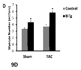
[0028] FIGS 9A-9D. Pressure overload stimulates ACM mitotic activity in BiTg
mice. (9A) Low
and high magnification images of TAO hearts. Bar=40um (top) or 20um (bottom);
white arrows
point to 013+ non-CM nuclei, yellow arrowheads point to pH3+ ACM nuclei. (98)
Quantification
of ACM mitotic activity in control and BiTg hearts, 10 days post-operation.
(9C) Quantification of
ACM transverse area in methanol-fixed hearts; 10 days post-operation. (9D)
Estimated myocyte
cell number. Sample Number (9A-9D) Sham, Control=3, BiTg=3; TAO, Control=8,
BiTg=7.
Statistics: (9B,9C) Two-way ANOVA/Tukey's test, * P<0,05 vs Sham-Control; t
P<0.05 vs
7
CA 02981811 2017-10-03
WO 2016/172224
PCT/US2016/028459
Sham-BiTg, P<0.05 vs TAO-Control. (9D) The Bootstrap method was used to
compute
standard error and Permutation test was used to compute p-value, * P<0.05 vs
control.
[0029] FIGS 10A-10C. KDM4A demethylates H3K9me3 and H3K36me3 in ACMs. (10A)
Timeline showing adenovirus-mediated KDM4A overexpression protocol in cultured
WT ACMs.
(10E3) p-galactosidase staining in (top) uninfected and (bottom) lacZ-infected
ACMs, showing
>30% infection efficiency. (100) Irnmunoblot showing KDM4A-expressing ACMs
have global
reductions in H3K9me3 and H3K36me3, but not in H3K27rne3 (Millipore 07449).
Larnin A/C
(Cell Signaling 47775) and H3 were used as loading controls. Sample Number:
Nõ?_:3 for each
group.
[0030] FIGS 11A-11B. CM-specific KDM4D transoene expression. (11A) KDM4D
transgene
expression in various BiTg tissue samples at 9 weeks of age, normalized to
expression levels in
BiTg hearts. (11B) Exogenous KDM4D (FLAG-tag) irnrnunostaining showing lack of
expression
in non-CM cardiac cells.
[0031] FIGS 12A-12B. Cell Cycle Regulators in BiTg ACM. (12A) Gene expression
(RNA-sea
RPKM, fold induction vs, control) of E2F family members in 9 week ACMs. (128)
qRT-PCR of
cell cycle regulators in 9 week ACMs, fold induction vs. NonTo, Sample Number:
(12A) N=2 per
group, (128) NonTg=3, tet=6, tTA=3, BiTg=5. Statistics: (12A) Two-tailed T-
test, P<0.05. (12B)
One-way ANOVA/Tukey's test, P<0.05 vs NonTg, t P<0.05 vs tet, f P<0.05 vs tTA.
[0032] FIGS 13A-13B. Apoptotic cells are not detected 10 days post-operation.
(13A)
Representative images of TUNEL staining in vibratorne sections. (138) DNAsel-
treated heart
sections of adult non-operated mice give robust nuclear-specific signal,
showing our assay is
able to detect TUNEL staining. Sample Number: N=2 for each group.
[0033] FIGS 14A-140, BiTg hearts have increased myocardium and dilated LV
chambers.
(14A) Representative images of mid-papillary vibratome sections, bar=2rnm.
Quantification of
(148) myocardium area and (140) LV chamber area. Sample Number: N=3 per group,
Statistics= Two-way ANOVA/Tukey's test, * P<0.05 vs Sham-Control. t P<0.05 vs
Sham-BiTg, f
P<0.05 vs TAO-Control,
[0034] FIGS 15A-15B. Unique chromatin structure in proliferative CMs. (15A)
immunostaining
in embryonic and postnatal wildtype heart sections showing anti-localization
of heterochrornatin
marker H3K9me3 (Active Motif, 39161) with euchromatin marker H3K36me3
(Diagenode,
015200183); the change in chromatin organization during postnatal development
is also seen,
bar=5pm. (158) In BiTg heart sections, pH3+ ACM nuclei (arrowheads) display
heterochromatin
organization that resembles embryonic OMs, in contrast to the typical ACM
chromatin
organization (arrows),
[0035] FIG 16. Neonatal mouse regeneration model. Schematic in upper panel
illustrates
timeline for creating BiTG mice in which Ml occurs at P7 and sacrifice at
POD21 for histology
8
CA 02981811 2017-10-03
WO 2016/172224
PCT/US2016/028459
and genotyping. Scar detection uses Sirius Red 4- Fast Green staining at 21
days after MI.
Fibrotic area is analyzed as a percentage taken from (the sum of fibrotic area
at L600 and L800
/ sum of myocardial area in the LV at L600 and L800) x 100.
[0036] FIG 17. KDM4 overexpressing mice have enhanced regeneration post-Ml.
Histological
sections in left panels show Non-BiTG and BiTG samples taken at indicated LO
to L1000. Bar
graphs on right panels show average and maximum percent fibrotic area for the
two groups,
[0037] FIGS 18A-18C. Adult CM-specific KDM4D Expression is sufficient to
induce late cell
cycle gene expression in ACMs, (18A) Schematic illustration of doxycycline
administration
through P21, and later KDM4D overexpression. (18B) Fold-induction of KDM4D
plotted for both
tet and BiTg subjects, with doxycycline treatment at E0-P21, or without
doxycycline treatment.
(180) Fold-induction of indicated genes for Non-BiTg and BiTg subjects.
[0038] FIG, 19. Preliminary data showing adult CM-specific KDM4D expression
and cell cycle
activity post-Ml. Doxycycline chow was administered from E0-P28. MI occurred
at 10 weeks;
during period of KDM4D overexpression, and at 14 weeks (30 days post-Ml),
tissue was
examined for phospho-H3, phalloidin, WGA, and Hoechst, comparing control (left
panel) and
BiTg (right panel),
DETAILED DESCRIPTION OF THE INVENTION
[0039] The invention is based on the unexpected finding that terminally
differentiated cells can
be induced to proliferate via epigenetic manipulation. The invention thus
provides materials and
methods for reversibly inducing proliferation in quiescent cells based on
discovery of the role of
H3K9me3 demethylases in regulating ACM cell cycle gene silencing,
[0040] Before the discovery of histone demethylases (HDMs)4(). H3K9me3, and
histone
rnethylation in general, was thought to be a permanent i-nark41. However; the
dynamic nature of
histone rnethylation is beginning to be appreciated42-44, though little is
known about the functions
of HDIVis in the heart. Interestingly, members of the KDM4 family of H3K9me3
demethylases
are upreg .Alated in several forms of cancer and are thought to promote cell
proliferation and
survival45-43. A member of the KDM4 family. KDM4A, has been studied in the
heart17,49. CM-
specific overexpression of KDM4A in mice exacerbated TAO-induced hypertrophy
and fetal CM
gene expression, while CM-specific KDM4A deletion diminished the effects of
pressure-
overload; though neither manipulation had an effect at baselines'. Mechanistic
studies
demonstrated KDM4A knockdown in neonatal CMs increases H3K9me3 levels at the
ANP
promoter and modestly downregulates ANP expression17. H3K9me3 and HP1
enrichment on
the ANP promoter was reduced in an isolated-working heart model of elevated
preload that
induces ANP expression17. However; KDM4A expression and enrichment on the ANP
gene
promoter were not changed in this model17. Thus, it is not clear how KDM4A
regulates fetal CM
9
CA 02981811 2017-10-03
WO 2016/172224
PCT/US2016/028459
gene expression in ACMs. Complicating the interpretation of these results
further is the fact that
KDM4A has dual-substrate specificity; KDM4A can dei-nethylate repressive
H3K9rne3, but also
activating H3K36me32. We also found that global levels of both these
modifications were
reduced in ACMs with adenovirus-mediated KDM4A overexpression. One KDM4 family
member, KDM4D, has robust and specific H3K9-demethylase activity5 ..52, giving
it particular
usefulness as an experimental tool to study H3K9rne3 specifically. Until this
study, KDM4D has
not been explored.
Definitions
[0041] All scientific and technical terms used in this application have
meanings commonly used
in the art unless otherwise specified. As used in this application, the
following words or phrases
have the meanings specified.
[0042] As used herein, "lysine-specific dernethylase 4D" or "KDM4D" means a
specific member
of the KDM4 family of lysine-specific demethylases that exhibits demethylase
activity specific to
the methylated lysine residue at position 9 (H3K9) of heterochromatin protein
1 (HP1). In one
embodiment, the KDM4D has the amino acid sequence shown in SEQ ID NO: 1. The
amino
acid sequence optionally further includes tags, such as, for example, a myc
tag and/or a FLAG
tag, as shown in SEQ ID NO: 2,
[0043] As used herein, "inducibly represses" or "inducible repression" refers
to regulation of
gene expression whereby expression of the gene can be repressed upon
introduction of an
inducing condition. The inducing condition can be administration of or contact
with an agent that
effects the repression. The agent can be a corepressor, such as is found in
repressible gene
regulation wherein expression is on except when the corepressor is present to
suppress gene
expression. Alternatively, the agent can be an inducer, such as is found in
inducible gene
regulation wherein expression is off except when the inducer is present to
allow for gene
expression.
[0044] As used herein, a "regulatory element" refers to an element that
regulates gene
expression. The regulatory element may induce or repress gene expression in
response to the
presence or absence of a condition.
[0045] As used herein, a "tetracycline responsive element" refers to a
regulatory element that
reduces expression from a tet-inducible promoter in the presence of
tetracycline or a derivative
thereof, e.g., doxycycline. One example of a tetracycline responsive element
is a tetracycline-
controlled transactivator (tTA), created by fusion of the tetracycline
repressor (tetR) with a
transcriptional activation domain, such as the C-terminal domain of VP16 of
herpes simplex
virus (HSV).
[0046] The term "nucleic acid" or "polynucleotide" or "oligonucleotide" refers
to a sequence of
nucleotides, a deoxyribonucleotide or ribonucleotide polymer in either single-
or double-
CA 02981811 2017-10-03
WO 2016/172224
PCT/US2016/028459
stranded form, and unless otherwise limited, encompasses known analogs of
natural
nucleotides that hybridize to nucleic acids in a manner similar to naturally
occurring nucleotides.
[0047] The term "primer," as used herein, means an oligonucleotide designed to
flank a region
of DNA to be a-nplified. In a primer pair, one primer is complementary to
nucleotides present on
the sense strand at one end of a polynucleotide fragment to be amplified and
another primer is
complementary to nucleotides present on the antisense strand at the other end
of the
polynucleotide fragment to be amplified. A primer can have at least about 11
nucleotides, and
preferably, at least about 16 nucleotides and no more than about 35
nucleotides. Typically, a
primer has at least about 80% sequence identity, preferably at least about 90%
sequence
identity with a target polynucleotide to which the primer hybridizes,
[0048] As used herein, the term "probe" refers to an oligonucleotide,
naturally or synthetically
produced, via recombinant methods or by PCR amplification, that hybridizes to
at least part of
another oligonucleotide of interest. A probe can be single-stranded or double-
stranded.
[0049] As used herein, the term "active fragment" refers to a substantial
portion of an
oligonucleotide that is capable of performing the same function of
specifically hybridizing to a
target polynucleotide,
[0050] As used herein, "hybridizes," "hybridizing," and "hybridization" means
that the
oligonucleotide forms a noncovalent interaction with the target DNA molecule
under standard
conditions. Standard hybridizing conditions are those conditions that allow an
oligonucleotide
probe or primer to hybridize to a target DNA molecule. Such conditions are
readily determined
for an oligonucleotide probe or primer and the target DNA molecule using
techniques well
known to those skilled in the art. The nucleotide sequence of a target
polynucleotide is generally
a sequence complementary to the oligonucleotide primer or probe. The
hybridizing
oligonucleotide may contain nonhybridizing nucleotides that do not interfere
with forming the
noncovalent interaction. The nonhybridizing nucleotides of an oligonucleotide
primer or probe
may be located at an end of the hybridizing oligonucleotide or within the
hybridizing
oligonucleotide. Thus, an oligonucleotide probe or primer does not have to be
complementary to
all the nucleotides of the target sequence as long as there is hybridization
under standard
hybridization conditions,
[0051] The term "complement" and "complementary" as used herein, refers to the
ability of two
DNA molecules to base pair with each other, where an adenine on one DNA
molecule will base
pair to a guanine on a second DNA molecule and a cytosine on one DNA molecule
will base
pair to a thymine on a second DNA molecule. Two DNA molecules are
complementary to each
other when a nucleotide sequence in one DNA molecule can base pair with a
nucleotide
sequence in a second DNA molecule, For instance, the two DNA molecules 5LATGC
and 5'-
GOAT are complementary, and the complement of the DNA molecule 5-ATGC is 5'-
GOAT. The
term complement and complementary also encompasses t w o DNA molecules where
one DNA
11
CA 02981811 2017-10-03
WO 2016/172224
PCT/US2016/028459
molecule contains at least one nucleotide that will not base pair to at least
one nucleotide
present on a second DNA molecule. For instance the third nucleotide of each of
the two DNA
molecules 5'-ATTGC and 5-GCTAT will not base pair, but these two DNA molecules
are
complementary as defined herein. Typically two DNA molecules are complementary
if they
hybridize under the standard conditions referred to above. Typically, two DNA
molecules are
complementary if they have at least about 80% sequence identity, preferably at
least about 90%
sequence identity.
[0052] As used herein, "a" or "an" means at least one, unless clearly
indicated otherwise.
[0053] As used herein, to "prevent" or "protect against" a condition or
disease means to hinder,
reduce or delay the onset or progression of the condition or disease.
[0054] As used herein, the term "isolated" means that a naturally occurring
DNA fragment, DNA
molecule, coding sequence, or oligonucleotide is removed from its natural
environment, or is a
synthetic molecule or cloned product. Preferably, the DNA fragment, DNA
molecule, coding
sequence, or oligonucleotide is purified, i.e., essentially free from any
other DNA fragment, DNA
molecule, coding sequence, or oligonucleotide and associated cellular products
or other
impurities.
Vectors
[0055] In one embodiment, the expression vector comprises: (a) a nucleic acid
sequence
encoding lysine-specific demethylase 4D (KDM4D); (b) a promoter that induces
or effects
overexpression of KDM4D, wherein the promoter is operably linked to the
nucleic acid
sequence; and (c) a regulatory element that inducibly represses the
overexpression of KDM4D.
Optionally, the vector further comprises (d) a tissue-specific promoter
operably linked to the
nucleic acid sequence. In some embodiments, the tissue-specific overexpression
of KDM4D is
be achieved through selection of a tissue-specific promoter in (b). In some
embodiments, tissue-
specific expression is provided through both (b) and an additional promoter
(d). The KDM4D is
capable of specifically removing the histone modification H3K9me3 by
demethylating the lysine
residue at position 9 of histone 3 (H3K9).
[0056] While the promoter of (b) can be a tissue-specific promoter, and in
some embodiments,
separate promoters serve the functions described in (b) and (d) above, the
selection of a tissue-
specific promoter is designed to optimize preferential expression in the
target tissue while
minimizing unintended expression elsewhere. Representative examples of tissue-
specific
promoters include, but are not limited to, promoters specific to cardiac
tissue (myosin heavy
chain, troponin I or T), skeletal muscle (myogenein, MyoD, muscle creatine
kinase), neurons,
pancreatic islet cells, or hepatocytes. A promoter that is tissue-specific
promotes expression of
the gene encoded by the nucleic acid sequence predominantly in the particular
tissue. In one
embodiment, the tissue-specific promoter is specific to cardiac tissue. An a-
myosin heavy chain
12
CA 02981811 2017-10-03
WO 2016/172224
PCT/US2016/028459
(aMHC) promoter is one example of a cardiac-specific promoter. in another
embodiment, the
tissue-specific promoter is specific to liver tissue, or hepatocyte.s. A CBA
promoter is one
example of a liver-specific promoter. Other examples of tissue-specific
promoters known in the
art include the neuron-specific enolase (NSE) and tubulin al promoters for
neurons, al-
__ antitrypsin and albumin (ALB) promoters for hepatocytes, and troponin, CMV,
or myosin light
chain-2 (r,õ1 LC2) for cardiac myocytes.
[0057] Representative examples of a regulatory element capable of inducibly
repressing
expression (or overexpression) include, but are not limited to, tetracycline
responsive elements
and hormone responsive proteins. Those skilled in the art will appreciated
alternative methods
__ of controlled gene expression that can be adapted for use in a similar
manner to regulate the
expression of KDM4D, both temporally and histologically. For example, in one
embodiment, the
regulatory element enables positive regulation of KDM4D expression, while in
another
embodiment, the regulatory element enables negative regulation of KDM4D
expression. In
another example, the regulatory element enables tissue-specific and/or
condition-specific
__ regulation of KDM4D expression. While the ability to turn off expression of
KDM4D is desirable,
it is not essential to all embodiments. In one embodiment, the invention
provides a vector
comprising a nucleic acid sequence encoding lysine-specific dernethylase 4D
(KDM4D) and a
promoter that induces or effects overexpression of KDM4D, wherein the promoter
is operably
linked to the nucleic acid sequence.
__ [0058] Vectors for use in the methods described herein include viral
vectors, as well as non-
viral vectors, virus-like particles, bacterial vectors, bacteriophage vectors,
and other vectors
known in the art. In one embodiment, the vector is a viral vector. In a
particular embodiment, the
viral vector is an adeno-associated virus (AAV) vector, or other vector suited
for infecting
quiescent cells. Representative examples of an AAV vector include, but are not
limited to, AAV6
__ and AAV9.
[0059] KDM4D amino acid sequence (SEQ ID NO: 1):
[0060] MetETMetKSKANCAQNPNCNIMetIFHPTKEEFNDFDKYIAYMet
ESQGAHRAGLAKIIPPKEWKARETYDNISEILIATPLQQVASGRAG
/FTQYHKKKKAMetTVGEYRHLANSKKYOTPPHQNFEDLERKYWK
NR1YNSPIYGADISGSLFDENTKOWNLGHLGTIQDLLEKECGVVIE
GVNTPYLYFGMetWKTTFAWHTEDMetDLYSINYLHLGEPKTWYVV
PPEHGQRLERLARELFPGSSRGCGAFLRHKVALISPTVLKENGIP
FNRITQEAGEFMetVTFPYGYHAGFNHGFNCAEAINFATPRWIDYG
KMetASQCSCGEARVTFSMetDAFVRILQPERYDLWKRGQDRAVVD
HMetEPRVPAS0ELST0KEVQLPRRAALGLROLPSHWARHSPWP
MetAARSGTRCHTLVCSSLPRQSAVSGTATQPRAAAVHSSKKPSS
TPSSTPGPSAQIIHPSNGRRGRGRPPOKLRAQELTLQTPAKRPLL.,
13
CA 02981811 2017-10-03
WO 2016/172224
PCT/US2016/028459
ACTTCTASCPEPEPLPEDCALMetDKPVPLSPCLQHPVKASGCSW
APVP
[0061] Optional additional amino acid sequence with rnyc (underlined) and flag
(shaded) tags
(SEQ ID NO: 2):
[0062] TRTLPLEOKLISEEDLAAND1L annattin V Stop
Compositions & Kits
[0063] The invention provides compositions, which can be provided as kits
and/or used for the
methods described herein. Compositions of the invention comprise vectors,
nucleic acid
molecules, and cells as described herein. Compositions and kits of the
invention can include
additional containers, agents, and materials to fa.cilitate practice of the
invention.
Methods of the Invention
[0064] The invention provides methods for inducing tissue-specific hyperplasia
in a mammal
comprising administering an expression vector as described herein to the
mammal. The method
can be tailored to any organ or tissue in which proliferation or regeneration
of quiescent cells is
of interest. Examples of tissues in which regeneration or proliferation may be
of interest include,
but art not limited to, heart, muscle, brain, nervous system, pancreas and
liver. Also provided is
a method for inducing cardiac myocyte (CM) hyperplasia in a mammal comprising
administering
an expression vector of the invention to the mammal. The invention further
provides a method
for inducing cardiac myocyte (CM) hyperplasia in a mammal. The method
comprises grafting
CMs to the heart of the mammal, wherein the CMs contain an expression vector
of the
invention,
[0065] The invention additionally provides a method for inducing CM
hyperplasia comprising
administering KOM4D to CMs. The KDIV14D can be administered using a
modification of the
peptide and/or a delivery means that protects the activity of KDM4D.
Administration can be
systemic, localized, oral, intravenous, subcutaneous, or transdermal.
[0066] In one embodiment, the invention provides a method of improving organ
function in a
mammal comprising grafting cells genetically modified with an expression
vector of the invention
to the organ. The organ can be, for example, heart, muscle, brain, pancreas,
or liver. In one
embodiment, the invention provides a method of improving cardiac function in a
mammal
comprising grafting CMs to the heart of the mammal, wherein the CMs contain an
expression
vector of the invention. In another embodiment, the invention provides a
method of improving
cardiac function in a mammal comprising administering an expression vector of
the invention to
the mammal. Also provided is a method of improving cardiac function in a
mammal comprising
administering KDIVI4D to the mammal.
14
CA 02981811 2017-10-03
WO 2016/172224
PCT/US2016/028459
[0067] The invention further provides a method of proliferating CM comprising
culturing CM with
KDM4D under conditions effective to induce CM hyperplasia. In one embodiment,
the CM are
adult CM (ACM). In addition, the invention provides a method of promoting
cardiac regeneration
comprising reducing lysine 9 of histone H3 (H3K9me3) levels in CMs. In one
embodiment, the
reducing comprises administering an expression vector of the invention to a
subject in need of
cardiac regeneration. In a particular embodiment, the expression vector is
administered by
administering CMs that contain the expression vector. In another embodiment,
the reducing
comprises administering KDM4D.
[0068] The methods of the invention can involve administration to the subject
by any of a
variety of means understood by those skilled in the art to be suitable for
particular
circumstances. In some embodiments, the administration is systemic. In other
embodiments,
the administration is intravenous. In some embodiments, the administration is
by intra-
myocardial injection. The subject is typically a mammal. In one embodiment,
the mammal is
human. In other embodiments, the mammal is a veterinary subject. Examples of
veterinary
subjects include, but are not limited to, equine, canine, bovine, porcine,
ovine, and feline
subjects.
EXAMPLES
[0069] The following examples are presented to illustrate the present
invention and to assist
one of ordinary skill in making and using the same. The examples are not
intended in any way
to otherwise limit the scope of the invention.
Example 1: Epidenetic Regulation of Cardiac Myacyte Cell Cycle Arrest
[0070] This example demonstrates that trirnethylation of Lysine 9 of Histone
H3 (H3K9me3), a
histone modification associated with heterochromatin, is required for the
silencing of cell cycle
genes in adult CMs (ACMs). To test this, we developed a transgenic (BiTg)
mouse model where
H3K9me3 is specifically removed by histone demethylase KDM4D in CMs. Loss of
H3K9me3 in
CMs disrupts ACM cell cycle gene silencing preferentially and results in
increased CM cycling.
Normalized heart mass was increased by postnatal day 14 (P14) and continued to
increase until
9-weeks of age. ACM number, but not size, was significantly increased in BiTg
hearts,
suggesting CM hyperplasia accounts for the increased heart mass. Challenging
H3K9me3-
depleted hearts with a hypertrophic growth signal stimulated ACM mitotic
activity. Thus, we
demonstrated that H3K9me3 is required for cell cycle gene silencing in ACMs
and depletion of
H3K9me3 allows hyperplastic growth in viva.
Methods
[0071] Mouse Studies. The aMHC-tTA mice used to control transgene expression
was
generated by the Robbins lab (60). We used the previously published responder
construct,
CA 02981811 2017-10-03
WO 2016/172224
PCT/US2016/028459
which possesses a tetracycline responsive element upstream of an attenuated
aMHC promoter
to drive KDM4D expression (60). Plasmid containing FLAG- and MYC-tagged human
KDM4D
cDNA (origene RC212600) had a Nati restriction site present in the cDNA
sequence, which we
destroyed by inducing a silent mutation (Agilent 200521). The resulting cDNA
was subcloned
into the responder construct, then freed of vector backbone, purified, and
injected into mouse
pronuclei (University of Washington transgenic core facility). The resulting
tet transgenic was
bred to the gMHC-tTA line to generate the CM-specific KDM4D induction model.
Litteri-nate
controls were used for all experiments involving transgenic mice. TAO and sham
operations
were performed on 10 to 12 week old littermates from breeders backcrossed
;,,==-8 generations to
the C57/B6 strain. Mice were anesthetized using ketamine (130 mg/kg i.p.) and
xylazine (8.8
mg/kg i,p,) and subjected to transverse aortic constriction using a 26-gauge
needle as described
(103).
[0072] CM cell isolation and culture. Heparinized mice were euthanized with
isoflurane and
hearts were extracted and arrested in KB buffer (mmol/L: KC! 20, KH2PO4 10, K+-
glutamate
70, MgC12 1, glucose 25, taurine 20, EGTA 0.5, HEPES 10, 0.1% albumin, pH 7.4
with KOH).
For purified ACM preparations, the aorta was cannulated and the heart was
washed with
Tyrodes solution (pH 7.4, supplemented with 25uM Blebbistatin -/-) and
digested for 7 minutes
with collagenase II (Worthington 4176) and Protease Streptomyces griseus XIV
(Sigma P5147)
using Langendorf perfusion. Ventricles were dissociated and the resulting cell
suspension was
filtered through a 100um mesh. Three rounds of low speed centrifugation, where
ACMs are
loosely pelleted and non-CMs in suspension are aspirated, density purify the
ACM population,
resulting in >90% rod-shaped ACMs. For embryonic and postnatal CM
preparations, hearts
were washed in Ads buffer (rnmoilL: NaCI 116, HEPES 20, NAH2PO4 10.8, glucose
5.5, KCI
5.4, MgSO4 0.83) and incubated with enzyme solution (Collagenase II,
Pancreatin (Sigma
P3292)) with rotation. Freed cells were collected into serum (stopping
digestion) every 20
minutes, resulting in dissociation of the entire heart within 2 hours, The
resulting cell
suspension was fractionated using a percoll (Sigma P4937) gradient, and the CM
layer and
non-CM layer were each collected. Quality and purity of CM preparations were
verified by
immunostaining, flow cytometry, and RNA expression of cell-type-specific
markers.
[0073] Control ACM and dedifferentiated ACM cDNA was generated as described
(57). In brief,
ACMs were plated on laminin-coated dishes and cultured with growth factors for
10-14 days,
resulting in a loss of sarcornere organization and increased CC activity.
[0074] RNA isolation and analysis. RNA was isolated from cells and tissue
using TRASOL
(Sigma T9424) phenol/chloroform purification, followed by column purification
with DNase
treatment (Qiagen 74004). For human gene expression studies, normal human
heart sample
was obtained from commercial vendors (Clontech 636532, lot 1206518A; and
Agilent 540011,
16
CA 02981811 2017-10-03
WO 2016/172224
PCT/US2016/028459
lot 6151000). Ischemic heart disease samples came from consenting male
subjects in their 60's
that underwent placement of a left ventricular assist device.
[0075] cDNA was synthesized as described in the manufacturers guidelines
(Roche
04896866001). oPCR was performed using SYBR green (Life Technologies 4472908)
on a real-
time PCR machine (AB1 7900HT). Primers were validated by standard PCR with
electrophoresis
to confirm specific target band and lack of primer dimers. oPCR dissociation
curves were
consistent with a single specific product. Ct values were assigned using ABI's
SDS 2.4 software
with automated thresholding and baselines. The standard curve method or dCt
method was
used to quantify expression, and expression of each gene was normalized by
GAPDH,
However, in Figure 1 A-D, we present ¨log(Ct) values. Finding a suitable
control gene that is
stably expressed at different stages in CM development is not trivial (104).
Gapdh was the most
stably expressed control across all samples compared to 326 and Rolp0; but
compared to
normalization by input RNA, Gapdh normalization resulted in E15.5 CM gene
expression being
underestimated by ¨2.5 fold, as Gapdh expression decreases in P3 CMs, then
remains stable;
consistent with the high glycolytic activity in fetal hearts (105). Standard
Curves were generated
using tissue or cells that highly express the indicated gene, resulting in
cIPCR efficiencies
ranging from 88-97%. The sequences of primers used are:
[0076] Mouse (SEQ ID NOs: 3-50, respectively; individual SEQ ID Nos in
parentheses below):
[0077] Gapdh F-CCAATGTGTCCGTCGTGGATCT (3), R-GTTGAAGTCGCAGGAGACAACC (4);
[00M] ANP F-AGGATTGGAGCCCAGAGTGGA (5), R-TGATAGATGAAGGCAGGAAGC (6);
[0079] blV1HC F-GCGACTCAAAAAGAAGGACTTTG (7), R-GGCTTGCTCATCCTCAATCC (8);
[0080] arsAHC F-AGAAGCCCAGCGCTCCCTCA (9), R-GGGCGTTCTTGGCCTTGCCT (10);
[0081] cTN1F-,-GCAGCCCAGAGGAAACCCAACC (11), R-AGCCGCATCGCTGCTCTCATC (12);
[0082] Cclid1 F-TGCTGCAAATGGA,ACTGCTTCTGG (13), R-TACCATGGAGGGTGGGTTGGAA,AT
(14):
[0083] Ccnel F-GCTTCGGGTCTGAGTTCCAA (15), R-GGATGAAGAGCAGGGGTCC (16);
[0084] Cdk4 F-GGGACCTGAAGCCAGAGAAC (17), R-CCACAGAAGAGAGGCTTCCG (18);
[0085] Ccnbl F-GCCTCACAAAGCACATGACTG (19), R-TCGACAACTTCCGTTAGCCT (20);
[0086] Cdki F-GGCGAGTTCTTCACAGAGACTTG (21), R-CCCTATACTCCAGATGTCAACCGG (22);
[0087] AurkB F-GCACCTGAAACATCCCAACAT (23), R-GGTCCGACTCTTCTGCAGTT (24);
[0088] RIM F- GTATTCCCAAGCACATCAA (25), R-GTAGCCAGAAGTGAAGAAC (26),
[0089] E2F1 F-TGCCAAGAAGTCCAAGAATCA (27), R-CTGCTGCTCACTCTCCTG (28);
[0090] E2F4 F-TGTCCTTGGCAGCACTCA (29), R-TTCACCACTGTCCTTGTTCTCA (30);
[0091] Rb F-CCTGATAACCTTGAACCTGCTTGT ($1), R-GCTGAGGCTGCTTGTGTCT (32);
17
CA 02981811 2017-10-03
WO 2016/172224
PCT/US2016/028459
[0092] p130 F-CACCGAACTTATGATGGACAG (33), R-ATGGCTTCTGCTCTCACT (34);
[0093] p107 F- GCAGAGGAGGAGATTGGAACA (35), R-GCTACAGGCGTGGTGACT (36);
[0094] p21 F-GCAGACCAGCCTGACAGATTT (37), R-CTGACCCACAGCAGAAGAGG (38);
[0095] p53 F-CAGTGGGAACCTTCTGGGAC (39), R-CGCGGATCTTGAGGGTGAAA (40);
[0096] KDM4A F-CTGCTAGGGCTTTAGGCTCC (41), R-TTTGGGAGGAACGACCTTGG (42);
[0097] KDM4B F-CAGAGAGCATCACGAGCAGA (43), R-CTCTTGGGCAGCTCCTCTTC (44);
[0098] KDM4C F-GCGGGTTCATGCAAGTTGTT (45), R-GTTTCAGAGCACCTCCCCTC (46);
[0099] KDM4D (endogenous) F-TCTGAGTCTGCCTTCTTCTG (47), R-
GCCAGGGTTCACAAGTCCTGAG (48);
[0100] KDM4D (transgene) F-TTGATGGACAAGCCTGTACC (49), R-TCATTTGCTGCCAGATCCTC
(50).
[0101] Mouse: TaqMan (Life Technologies) reagents were used for the following
genes:
KDM2B (Mm01194587m1), KDM4A (Mm00605000_m1), KDM6B (Mm01332680m1), KDM8
(Mm00513079_m1), GAPDH (Mm99999915_g1).
[0102] Human (SEQ ID NOs: 51-54, respectively; individual SEC) ID Nos in
parentheses below):
[0103] GAPDH F-CCTCAACGACCACTTTGTCA (51), R-TTACTCCTTGGAGGCCATGT (52);
[0104] KDM4D F- AAGCCCAGCTCAACTCCATC (53), R-TGTCCATCAAAGCCCCATCC (54).
[0105] RNAseq library construction (Illui-nina Tru-Seq) and paired-end RNA-
sequencing
(ABI3730XL) was performed by the Stai-n Lab's University of Washington core
facility. Read
alignment was performed using Bowtie/Cufflinks package. Partek Genornic Suites
was used for
mRNA quantification, differential expression analysis, and gene ontology.
[0106] 2-D Echocardiography. Under 0,5% isoflurane, mice EKG and heart
function was
assessed using Visual Sonics Vevo 2100. Parasteznal short axis images at the
plane of the
papillary muscle were collected in B- and M-Modes. images were collected with
heart rates
ranging from 400-500 BPMs. Imaging and analysis was performed by a single
operator who
was blinded to the genotypes. Quantification of images was performed using
Vevo Labs 1,7.0,
according to the manufacturer's guidelines.
[0107] Protein extraction and Western Blotting. Isolated ACMs were pelleted
and resuspended
in iysis buffer (0.5% NP-40, 25mM KC, 5rriM MgCl2, 10mM Tris-HCI, pH 8.0) and
homogenized
(Wheaton 358103), releasing soluble cytoplasmic proteins. Nuclear-enriched
pellets were
processed to release chromatin-associated proteins from DNA; including
sonication, MNase
treatment, and addition of 1% SDS, 600rrim NaCl, and 20mM pME. The nuclear
proteins were
quantified using a BOA assay (Thermo Scientific 23252) and were loaded on
polyacrylamide
gels for electrophoresis, subsequently transferred onto PVDF membrane, and
probed with the
indicated antibodies: KDM4D (Abcam ab93694), H3K9me3 (Abcam ab8898), pan H3
(Millipore
18
CA 02981811 2017-10-03
WO 2016/172224
PCT/US2016/028459
05-928), H3K36me3 (Active Motif 61101), H3K9me2 (Millipore 07-441), and
H4K2Ome3 (Abcam
ab9053). HRP-conjugated secondary antibodies (Santa Cruz) and ECL-detection
(Thermo
Scientific 34095) were used.
[0108] Histological studies and quantification CM dimensions and CM number.
For histological
analysis, arrested P14 or 9 week hearts were fixed with 4% PFA. Paraffin
sections were stained
with H&E, Masson Trichrome, or immunostained using standard protocol with FLAG
(Sigma
F7425) o-actinin (Sigma A7811), cardiac Troponin T (Thermo Scientific MS-295-
P), Ki67
(Abcam abl 5580) and phospho-H3 (Abcam ab5176) antibodies, and Hoechst (Life
Technologies H3570) to visualize nuclei. Images were acquired with confocal
microscopy
(Nikon AIR). To assess ACM transverse area, sections were stained with Wheat
Germ
Agglutinin (WGA; Life Technologies W6748), a marker for plasma membrane.
Stitched-images
of the whole left ventricle were acquired on a Nikon Ti-E scope. We chose
several regions in
each section at random, though we excluded large vessels, epicardiurn and
endocardium, and
>1000 cells per animal were analyzed using Image J's "analyze particle"
function (negative
image of WGA stain); resulting in direct measurement of transverse area. For
ACM longitudinal
area and length measurements, isolated ACMs were fixed with 4% PFA and imaged.
The area
and long-axis of hundreds of ACMs for each animal were manually traced and
quantified using
Image J's "measure" function. We calculated CM volume using the formula: (mean
ACM length
x mean ACM transverse area). CM number was estimated from the following
formula: [mean
Heart Volume (Heart mass/1.06, the density of muscle tissue(106)) / mean ACM
volume x 0.75
(the proportion of adult rnurine heart volume occupied by CMs(106))] The
number of nuclei per
ACM was counted manually for >100 cells per animal.
[0109] Imaging of thick sections. For unambiguous determination of cell type
in our phospho-H3
staining assays in operated mice, we developed a method for generating,
staining, and imaging
100um-thick heart sections, which will be described in detail in a
methodologies article. Briefly,
hearts were arrested in KB buffer, perfused with KB, then perfusion fixed with
methanol cooled
to -200 C. The hearts were rehydrated in IViethanoi:PBS gradients (100:0,
80:20, 60:40), then
washed with PBS and mounted in 5% low-melt agarose. 100um-thick sections were
cut from a
Leica 1200s vibratome and were stained in suspension, with reagents listed
above as well as
with Phalloidin (ThermoFisher Scientific A22287). The stained sections were
mounted to glass
coverslips coated with 0.01% poly-L-lysine. To increase the transparency of
the sections, which
is needed to view the interior of the thick sections, they were cleared:
sections were incubated
in an isopropanol series (70%, 85%, 95%, 100%) followed by incubations in a
1:2 solution of
benzyl alcohol and benzyl benzoate. The samples were prepared, imaged with
confocal
microscopy, and analyzed by a single operator blinded to the genotypes. We
calculated the
number of pH3+ ACMs using the formula: RpH3+ ACM
nucleiimm3)/(nuclei#LACM)/(ACM/mm3)]
We note that transverse area measurements in the sham-operated hearts were
19.2% less than
at baseline, which we attribute to differences in fixation procedure,
consistent with other reports
19
CA 02981811 2017-10-03
WO 2016/172224
PCT/US2016/028459
comparing cell shrinkage after formaldehyde or alcohol fixation (107). Because
of this,
transverse area in all groups was corrected by multiplying by a constant
factor of 1.192, when
calculating ACM number in the post-operation methanol-fixed samples: [mean
Heart Volume
(Heart mass/1,06, the density of muscle tissue(106)) / mean ACM volume (mean
ACM baseline
length x mean ACM post-operation transverse area) x 0,75 (the proportion of
adult murine heart
volume occupied by CMs (106))].
[0110] Statistics. All results are displayed as mean standard error of
means. Graphpad Prism
was used for one-way-ANOVAs and Tukey's post hoc tests performed on studies
comparing
more than two groups, Graphpad Prism was used for two-way-ANOVAs and Tukey's
post hoc
tests performed on studies with two independent variables, Microsoft Excel F-
test and two-tailed
T-test functions were used to analyze studies comparing two groups. For
outcomes where
different basic measurements were combined for calculations (Figure 5, D and
E, and Figure
9D) we used the bootstrap method (10,000 bootstrap samples) to compute
standard error and
the Permutation test (100,000 Monte-Carlo samples) was used to compute p-
value; with the
assumption that ACM transverse area, ACM length, and heart volume are
independent
variables. For RNA-seg analysis, Partek Genoi-nic Suites was used to perform
statistics.
[0111] Study approval. All animal studies were performed in accordance with an
approved
Institutional Animal Care and Use Committee (IACUC protocol #4290-01), the
University of
Washington institutional guidelines, and the National Institute of Health
Guide for the Care and
Use of Laboratory Animals, Human ischemic heart disease samples came from
participants that
gave written and informed consent; the use of human samples was approved by
the University
of Washington's Institutional Review Board (IRB# 35358).
[0112] Adenoviral studies. Adenoviruses for KDIVI4A and LacZ were generated
according to
manufacturers guidelines (Agilent 240082), isolated ACMs were plated on
arninin coated wells
in M199 medium supplemented with lx ITS, lx PS, 5mM Taurine, 1mM Na-pyruvate,
5mM
Cre..atine, 2mM L-camitine, and 25mM Blebistatin, with the presence of 5% FBS.
After 1 hour,
media was changed to 2% FBS containing media and 150 rnoi of viruses were
added. ACMs
were maintained in media containing 2% FBS until harvesting. Beta-
galactosidase staining was
performed on 4% PFA-fixed ACMs that were incubated in 5mM K+ fern-cyanide, 5mM
K+ ferro-
cyanide, 2rnM MgCl2, and 1mg/mL X-gal for 4 hours.
[0113] Temporally-controlled KDM4D induction. Doxycycline-containing chow
(Harlan
TD.00502) was administered ad lib for the indicated times. Note that the Dox
2weeks-9weeks
group includes mice that received dox ranging from P14-9w to P18-9w.
[0114] Myocardium and LV area quantification. Vibratorne sections were cut
from the mid-
papillary muscle plane of hearts and imaged. Myocardium area and LV chamber
area were
manually traced in IrnageJ and area was calculated using the "measure" tool.
CA 02981811 2017-10-03
WO 2016/172224
PCT/US2016/028459
[0115] Quantification of aboptosis. Apoptosis was visualized in vibratome
sections by using a
TUNEL staining kit (Life Technologies 010618) according to the manufacturers
guidelines.
Following TUNEL labeling, we stained for WGA, Hoechst, and phalloidin, and
imaged as
described in the procedures for vibratome sections.
Results
Characterization of H3K9me3 Histone Demethyiase Expression in CMs.
[0116] To better understand the role of H3K9me3 in regulating cell cycle gene
expression, we
characterized the relationship between cell cycle and H3K9me3-HDM gene
expression in CMs
through cardiac development (Figure 1). Developmental changes in CM-specific
and cell cycle
gene expression included switching of myosin isoforms and dramatic
dovvnregulation of G2/M
and cytokinesis genes in ACMs (Figure 1, A and B), consistent with prior
studies (9,53,54). Cell
cycle transcription factor E2F1 was downregulated 167-fold in ACMs (P<0.0001,
vs. E15.5
CMs), while expression of repressive E2F4 remained high (Figure 10).
Consistent with prior
studies in skeletal muscle (35) and OMs (9), we found p107 was the Rb-family-
member that was
expressed specifically in proliferative rhyocyte..s, in contrast to Rb and
p130 (Figure 1C).
Expression of KDM4 family members followed a similar, though less dramatic,
pattern of
expression as fetal CM, G2/M, and cytokinesis genes, and was moderately
downregulated after
P7 (Figure 1D), coinciding with loss of CM regenerative potential (2,3).
Downregulation of
H3K9me3-HDMs in ACMs is consistent with the increase of global H3K9me3 levels
in ACMs
compared to embryonic CMs (9). The low basal level of KDM4D in ACMs is
consistent with
other reports of KDM4D expression in tissues with limited proliferative
potential (55,56).
[0117] To screen for HDMs that might be involved in CM proliferation, we
looked for HDMs that
were upregulated during CM dedifferentiation, as dedifferentiation appears to
be a requisite for
CM proliferation in the zebrafish and neonatal mouse heart regeneration models
(1-3).
Dedifferentiation of mammalian ACMs can be achieved in vitro by long-term
culture with growth
factors, resulting in disassembly of sarcorneres and restoration of
proliferative potential (57).
From a panel of diverse HDMs, KDM4D was the most highly upregulated (401-fold)
during
dedifferentiation (Figure 1E; P<0.03). Because KDM4A expression is elevated in
human
hypertrophic cardiornyopathy samples and CM-specific KDM4A overexpression
exacerbated
hypertrophic growth in mice (49), we wondered if KDM4D was upregulated in
human ischernic
myocardium. KDM4D expression was unchanged in hearts of subjects with
ischernic
cardiomyopathy (Figure 1F), consistent with the exceedingly low CM hyperplasia
in this setting
(58).
21
CA 02981811 2017-10-03
WO 2016/172224
PCT/US2016/028459
Generation of a transgenic mouse model to deplete H3K9me3 specifically in CM
[0118] To explore the role of H3K9me3 in ACM cell cycle gene silencing in
vivo, we chose to
overexpress KDM family member 4D because: 1) KDM4D is the most specific
H3K9me3
demethylase (50-52) (Figure 10), 2) it is expressed in proliferative CMs and
elevated in
dedifferentiated ACMs (Figure 1, D and E), 3) it is not expressed in
cardiornyopathy samples
where hypertrophic growth would predominate (Figure 1F), 4) it promotes
proliferation and
survival in non-CMs (46-48), and 5) gain of function experiments are less
subject to
compensation by redundant factors (59). We used a previously characterized
tetracycline
inducible (tet-off) overexpression model where the tetracycline transactivator
(tTA) is expressed
.specifically in CMs. (60), We generated a CM-specific transgenic mouse line
containing a MYC-
and FLAG-tagged KDM4D cDNA downstream of a tetracycline responsive promoter,
which
contains tTA-binding sequence in the context of an attenuated-aMHC promoter
(60). Breeding
heterozygous tTA mice with heterozygous tet-responsive KDM4D (te0 mice yields
bi-transgenic
(BiTg) mice that constitutively express KDM4D specifically in CMs (Figure 2A)
as well as single-
transgenic (tet or tTA) and non-transgenic (NonTg) controls. In BiTg CMs, the
tTA protein is
expressed and binds to the tet-responsive element upstream of KDM4D, inducing
KDM4D
transgene expression (Figure 2A). We confirmed that KDM4D expression was
robustly induced
in BiTg hearts at P14 and 9 weeks (Figure 2B), KDM4D transgene expression was
not
detectable in other organs in BiTg mice or non-CM cardiac cells (Figure 11, A
and B), with the
exception that low levels could be detected in BiTg lungs, consistent with
previous reports using
the dMHC promoter (60). li-nmunofluorescence imaging in heart sections showed
exogenous
KDM4D protein was specifically expressed and localized in the nuclei of BiTg
CMs (Figure 2C),
Western blot analysis confirmed KDM4D protein expression and showed global
H3K9me3
levels were depleted in BiTg ACMs (Figure 2D). We also confirmed that in
contrast to other
KDM4 family members (Figure 10), KDM4D dernethylase activity is specific to
H3K9me3 (50-
52) and did not demethylate H3K9rne2 or H3K36rne3 in ACMs (Figure 2D).
H4K2Orne3, which
has been implicated as a repressive mark that is downstream of H3K9me3 and HP1
(61-63)
was unchanged (Figure 2D); although this does not rule out changes in
methylation levels at
specific gene loci.
H3K9me3 is required for ACM cell cycle gene silencing in vivo.
[0119] To assess the impact of depleting H3K9me3 on global gene expression in
vivo we
performed RNA-sequencing on 9-week ACMs. Control ACM samples were grouped
since
NonTg and single transgenic mice showed no differences in gene expression,
with the
unconstrained slope correlation test showing R2=0.9764 when comparing the
whole-genome
transcriptome. RNA-seq analysis revealed that BiTg ACMs had increased
expression of genes
involved in 16 of 138 cellular processes and 16 of 142 cell cycle processes
(Figure 3, A and B).
Strikingly, cell processes involved in cell cycling were preferentially
increased (Figure 3A),
22
CA 02981811 2017-10-03
WO 2016/172224
PCT/US2016/028459
Within cell cycle processes, categories involved in the later phases of cell
cycle, particularly
mitosis showed increased gene expression (Figure 3B). We confirmed increases
in G2/M and
cytokinesis genes by gRT-PCR (5.8- to 21.4-fold, P<0.01) and fetal CM genes
were also
increased (Figure 3C). Although the expression of fetal CM genes is frequently
associated with
a pathologic state, it should be noted that expression of less mature CM-
specific genes could
also be consistent with proliferation-competent OMs in fetal and neonatal
hearts (9,64) (Figure
1, A and B). We also examined cell cycle-gene transcriptional regulators
(Figure 12, A and B)
and found that positive regulators of cell cycle progression, E2F1 and E2F2,
were highly
expressed in BiTg ACMs compared to control ACMs (>12-fold, P<0.03). The
repressive E2F
members, E2F4-6, were unchanged. Interestingly, p107 was also increased in
BiTg ACMs
(Figure 128), consistent with the E2F/Rb-family expression in proliferative
rnyocytes (Figure 1,
B and C).
CM-specific H3K9me3 depletion promotes CM hyperplasia without altering cardiac
function.
[0120] BiTg mice had visibly larger hearts (Figure 4A) with a 20.8% increase
in heart weight to
body weight ratio (1-1WIBM at 9 weeks (Figure 4B; P<0.0001). This increase in
HW/BW first
became apparent in BiTg mice at P14 (Figure 4C; 12,9% increase, P<0.001);
suggesting
KDM4D overexpression specifically promoted postnatal cardiac growth. This
cardiac
enlargement was not associated with sarcornere disarray, fibrosis or
alteration of vasculature
(Figure 4, D and E) and there was no increase in extracelluiar matrix (65)
(Figure 4E).
Quantification of ACM transverse area or direct measurements of isolated ACM
longitudinal
area and length did not reveal differences in dimensions or calculated volumes
in BiTg ACMs
compared to controls (Figure 5, A-D). Calculated myocyte., number suggested
BiTg hearts had
22% more ACMs compared to controls (Figure 5E; P<0.03). To determine the
longterm effect
of H3K9me3-depletion on heart function; we performed echocardiography on 7
month old BiTg
and control mice: ejection fractions, fractional shortening, cardiac output,
and left ventricle
chamber size were similar in all groups (Table 1). No significant differences
in cardiac function
or morphology were seen.
Table 1. Normal cardiac function and morphology in BiTg mice at 7 months.
[0121] Echocardiography results in 7 month old mice. HR: Heart Rate, EF:
Ejection Fraction,
CO: Cardiac Output, LVEDD: Left Ventricular End-Diastolic Dimension, LV Mass:
Left
Ventricular Mass. Mean and SEM values are shown, Sample Number: N=3 for each
genotype,
Statistics: One-way ANOVAITukey's test, * P<0.05 vs NonTg, t P<0.05 vs tet,
P<0.05 vs tTA.
NonTg tet tTA BiTg
HR (BPM) 440 10 461 16 452 6 404 3tt
EF (%) 74.5 3.7 80.9 1.6 83.8 2
80.2 4.6
FS (%) 42.7 3.2 48.9 1.6 52.3 2.2
48.8 4.9
23
CA 02981811 2017-10-03
WO 2016/172224
PCT/US2016/028459
CO (ml/min) 16.4 1.4 20.5 1 17.4
1.5 19.1 0.7
LVEDD(rnm) 3.47 0.06 3.61 0.02 3.35
0.15 3.73 0.07
[0122] To assess cell cycle activity, we immunostained P14 and 9 week
myocardial sections for
phosphorylated histone H3 serine-10 (pH3), a marker of mitosis. We observed
pH3+ CMs only
in BiTg mice at both time points (Figure 6, A and B). Similar trends were seen
for the general
cell cycle activity marker Ki67 in 9 week hearts (Figure 60). Quantification
of the number of
nuclei per ACM at seven months revealed there was an increase of mononucleated
and a
decrease in binucleated ACMs in BiTg hearts (Figure 6D). These findings are
consistent with a
model where the increased heart mass in BiTg mice was secondary to CM
hyperplasia.
[0123] Cardiac mass increased up to 9-weeks of age in BiTg mice (Figure 4C).
Cell cycle
activity in 9-week BiTg ACMs, though elevated compared to controls (Figure 6,
B and C), was
rare (<2 pH3+ CM/ 200x field). To determine if the increased mass was related
to the normal
post-natal hypertrophic growth of an increased number of CMs in BiTg hearts or
whether there
was ongoing CM hyperplasia we utilized doxycycline (Dox) to shut-off KDM4D
expression
(Figure 7A). \Ale examine heart mass in BiTg mice where KDM4D expression was
never
induced or was induced in utero but suppressed at P14 (Figure 7B). Dox
treatment reduced
KDM4D expression in BiTg ventricles to control levels within one week (Figure
70). Adult BiTg
mice that had KDM4D expression suppressed since conception displayed
Frv"V/BV\is that were
indistinguishable from controls (Figure 7D). BiTg hearts with constitutive
KDM4D expression
were larger at P14 (Figure 40; 12.9% increase vs. control; P<0.001). However,
when KDM4D
expression was turned off at 2 weeks, heart size at 9 weeks was less compared
to mice with
constitutive KDM4D expression (Figure 7D; 7.9% vs 20.8%; P<0.05), These
findings suggest
that KDM4D overexpression continues to promote additional CM hyperplasia
between weeks 2
through 9.
Hypertrophic signals stimulate proliferation of H3K9me3-depleted ACMs.
[0124] Although highly upregulated compared to controls, late cell cycle gene
expression in
BiTg ACMs is much less than in wildtype embryonic and postnatal CMs (compare
Figure 1B
with Figure 30). Also the absolute number of cycling CMs, while increased in
BiTg hearts, was
low and normalized cardiac mass did not increase further between 9 weeks and 7
months
(Figure 40) suggesting CM proliferation is very limited at baseline in adult
BiTg hearts. Since
there is robust activation of numerous growth factor signaling pathways post
TAC that typically
result in hypertrophic growth (9,66), we thought this might be an excellent
model to test
KDM4D's ability to promote ACM proliferation in vivo, This allowed us to
determine whether a
hypertrophic growth signal such as TAO would induce hyperplasia in H3K9me3-
depleted ACMs
or whether they would undergo hypertrophy similar to control hearts. BiTg and
control
littermates underwent sham or TAO surgeries at 11 weeks of age and were
examined 10-days
24
CA 02981811 2017-10-03
WO 2016/172224
PCT/US2016/028459
after surgery. Similar to un-operated mice sham-operated BiTg hearts were
visibly larger than
controls (Figure 8A). TAO induced a 48.2% increase in HVV/BW in BiTg mice
compared to a
20.9% in control mice (Figure 88; P<0.0001). To determine if the TAC-induced
heart growth
occurred through ACM hypertrophy or hyperplasia, we measured ACM transverse
area Sham
BiTg ACMs were similar to sham controls (Figure 5A); however, with TAO BiTg
ACM transverse
area was 12.5% smaller compared to TAO control ACMs (Figure 9, A and C;
P<0.05). Since
ACM transverse area, but not length, is increased after TAO (67), we estimated
CM number and
found a 56.6% increase in ACMs in BiTg hearts compared to controls (Figure 9D,
P<0,001). To
confirm that BiTg ACMs were cycling after TAO we assayed for phospho-H3 and
observed a 41-
fold increase in pH3+ ACMs in BiTg mice compared to control mice (0.77% vs
0.019%;
P<0.0001) (Figure 9, A and B). This cell cycle activity was not associated
with fibrosis (Figure
80 and Figure 9A) or increased apoptosis (Figure 13). To determine if
reinduction of CM cell
cycling negatively impacted cardiac function, 2-D echoes were performed 9 days
post-surgery
(Table 2). Ejection fraction was decreased in BiTg mice after TAO along with
and an increase in
LVEDD. However, the calculated cardiac output was unchanged, presumably due to
the
chamber size in BiTg hearts (Figure 14).
Table 2. Cardiac function and morphology in TAO-operated mice.
[0125] Echocardiography results in 12 week old mice, 10 days post-operation.
HR: Heart Rate,
EF: Ejection Fraction, CO: Cardiac Output, LVEDD: Left Ventricular End-
Diastolic Dimension,
LV Mass: Left Ventricular Mass. Mean and SEM values are shown. Sample Number
Sham,
Contro1=-4, 8iTg=4 ; TAO, Control=9, BiTg=8, Statistics= Two-tailed T-test,
control vs. BiTg,
P<0.05.
Sham TAC
Control BiTg Control BiTg
HR (BPM) 428 17 416 7.6 430 11 406
10
EF (%) 711 4.2 72.4 3.7 75.5 4.4 51.2
3.7.
FS (%) 39.5 3.5 40.8 3.4 43.7 3.5 25.9
2.3*
CO (mL/min) 11.6 2.4 12.4 1.5 9.8
0.9 10.9 0.7
LVEDD(mm) 3.04 0.14 3.19 0.11 2.83 0.09 3.57
0,10*
Discussion
[0126] Since the discovery that neonatal mammalian hearts can regenerate by CM
proliferation
(2,3) and that ACMs retain some, though very limited, capacity to divide (7),
interest in the
regulation of ACM cell cycle has been reignited (4,69-71). Many strategies
that can promote
proliferation in mammalian neonatal CMs have been ineffective in ACMs (72,73)
highlighting the
fact that strong barriers to proliferation exist in ACMs, Recently, epicardial
paracrine factor
FSTL1 (70), rniR-15 inhibition (3), and the NRG1 co-receptor Erbb2 (74) were
suggested to
CA 02981811 2017-10-03
WO 2016/172224
PCT/US2016/028459
promote ACM proliferation, though the molecular mechanisms and relevant
targets in ACMs
remain elusive. We previously found that the heterochrornatin marker H3K9me3
was enriched
on G211µ11 and cytokinesis genes in ACMs compared to fetal CMs implicating
this mark as a
barrier to ACM proliferation (9). This example characterizes the expression of
H3K9me3-
demethylases in development, dedifferentiation, and disease and shows that
KDM4D was
expressed in fetal CMs and was the primary H3K9me3-specific demethylase
upregulated in
dedifferentiating ACMs. CM-specific KDM4D overexpression depleted H3K9me3
specifically
and led to increases in ACM expression of G2/M and cytokinesis genes, cardiac
mass, ACM
number, and ACM mitotic activity.
[0127] The renewed interest in CM proliferation has also called attention to
the need of
improved methodologies for detecting ACM proliferation (75). The standard
indicator of ACM
proliferation has been indirect in situ histology of phase-specific or general
cell cycle markers.
However, it has been suggested these results are often equivocal because it
can be difficult to
determine if the marker is present in a CM or non-CM (75,76). Most studies are
performed in
cardiac sections less than lOpm thick, which is significantly thinner than
even the shortest axis
of mammalian ACMs, and are further confounded by the dense myocardial
vasculature and
non-CMs that are the majority of cardiac cells (77). We addressed this
limitation by developing
a novel sample preparation and imaging technique that yields high-resolution
3D image
reconstructions of cleared thick sections with several layers of whole ACMs,
allowing
unambiguous identification of mitotic ACMs in KDM4D overexpressing mice
(Figure 9). Our
methods corroborate findings from in vivo cumulative-proliferation-labeling in
CM lineage-tracing
models (multi-isotope-mass-spectrometry (7), mosaic analysis with double
markers (78), and
multi-color clonal assays(76)) and supports the emerging consensus that ACM
proliferation is
rare and difficult to stimulate (75). Despite the fact that late cell cycle
gene expression was
markedly increased at 9 weeks in H3K9me3-depleted CMs (Figure 30) their
expression levels
were much lower when compared to neonatal CMs (Figure 18). As well, the vast
majority of
BiTg ACMs appeared to exit the cell cycle by P14 (Figure 6A) and the
difference in HW/BW
compared to controls did not appear to increase further after 9 weeks (Figure
40), This
suggests mechanisms, in addition to H3K9me3, prevent proliferation in ACMs.
[0128] This example confirms that H3K9me3 is required for ACM cell cycle gene
silencing in
vivo but that H3K9me3 is not sufficient by itself to explain the stable
silencing of cell cycle genes
in ACMs since it was maintained on G2/M and cytokinesis genes even after they
were
derepressed by Rb/p130 double knockout (9). In that study, HPly, whose
chromodomain
specifically binds H3K9me3, was displaced from late cell cycle gene promoters.
This study
suggested that stable silencing of cell cycle genes in ACMs required
recruitment of HPly and
that H3K9me3, though required, was not sufficient to silence genes (9), Since
Rb and
H3K9me3 are both required for HP1 binding in many systems it is possible that
KDM4D
overexpression derepressed late cell cycle genes in ACMs by removing HPly's
binding-
26
CA 02981811 2017-10-03
WO 2016/172224
PCT/US2016/028459
substrate from chromatin. Regardless, since Rb and H3K9me3 are present on
numerous genes
it rei-nains unclear why late cell cycle genes are preferentially derepressed.
It may perhaps be
related to particular combinations of E2F- and Rb- family members forming
distinct complexes
and targeting specific subsets of genes (33,34). In BiTg ACMs, this example
showed that the
E2F/Rb-family members most increased by H3K9me3-depletion were E2F1 and p107
(F)gure
12), which are the same E2F/Rb-family members that are specifically expressed
in proliferative
embryonic and neonatal CMs (Figure 10) (9). Consistent with HP1 losing the
ability to target
E2F-dependent genes in BiTg ACMs, p107, in contrast to Rb, has not been shown
to bind HP1
(39). In agreement, Rb has additional protein-binding and phospho-regulated
domains not
found in p107, and deletion of p107 has not shown the hyperplasia phenotype
seen with Rb loss
of function (79;80). Thus, selective increases in E2F/Rb-family expression
levels and differential
recruitment of HP1 may explain the preferential increase of late cell cycle
genes in BiTg ACMs.
[0129] This example demonstrated that ACMs can tolerate moderate levels of
G2IM and
cytokinesis gene expression vvithout deleterious effects on heart function
(Figure 3 and Table 1).
This is similar to other models of limited ACM cycling (76), but contrasts
with our previous
findings where disrupting heterochromatin formation and inducing cell cycle
reentry in ACMs
was associated with decreased heart function (9). A fundamental difference
between the
KDM4D mice and that model is that KDM4D overexpression specifically targets
one methylation
pathway; whereas Rb/p130 KO likely disrupted multiple epigenetic modifications
(H3K9me3,
H3K27i-ne3, and H4K2Ome3) (9,32,33). This may explain why genes involved in
promoting all
phases of cell cycle were upregulated in Rbip130 KO, while H3K9me3- or HP1 y-
specific
disruption leads to preferential increase of G2i'M and cytokinesis gene
expression(9) (Figure 3).
Consistent with this notion, H3K9me3-depleted chromatin in KDM4D mice
maintained its global
structure including heterochromatin unlike the Rb/p130 model with the
exception of cycling
pH3+ ACMs (Figure 15). Thus, repressive methylations have overlapping roles in
maintaining
global chromatin structure in ACMs consistent with reports of H3K9me3-
depletion in other cell
types (27,81,82),
[0130] We have suggested that there is a transition in heterochromatin
structure during
postnatal differentiation in ACMs (9) from limited heterochromatin organized
into many small
foci within the nucleus of embryonic CM, to few, large foci with additional
heterochromatin at the
nuclear lamina in postnatal CM nuclei (Figure 15A). We found that the pH3+
ACMs in BiTg
mice had a gross chromatin structure similar to that of proliferative
embryonic CMs (Figure
15B). The relationship between higher-order chromatin structure and gene-
specific regulation is
not clear, though studies suggest they act independently (27,82).
Interestingly, H3K9me3 and
H3K9me3-associated proteins also regulate cell cycle via transcription-
independent
mechanisms involving changes in global chromatin structure required for DNA
synthesis and
mitosis (42,83). Not surprisingly, pH3, the hallmark of mitotic activity, is
inhibited by
trimethylation of the adjacent amino acid residue H3K9 and double knockout of
H3K9me3
27
CA 02981811 2017-10-03
WO 2016/172224
PCT/US2016/028459
methyltransferases Suv39h1/2 resulted in increased pH3+ mouse embryonic
fibroblasts (84,85),
suggesting that global H3K9me3-depletion facilitates mitotic activity. Though
several
mechanisms may contribute to KDM4D-rnediated ACM cell cycle activity, we have
provided
evidence that supports a model where cell cycle gene silencing is prevented by
depleting
H3K9me3, but additional repressors appear to prevent robust cell cycle
activation in the
absence of growth stimulation.
[0131] The results can be compared to findings in studies of another major
cell proliferation
regulation signaling pathway, the organ-size-controlling Hippo/Yap pathway.
This pathway has
been intensely studied with several CM-specific loss of function and gain of
function mouse
models through CM development and adulthood (71,93). Though Yapl gain of
function in adults
increased ACM proliferation, the levels were 20-fold less than NonTg neonatal
CMs (76). The
authors postulated Yap activation alone is insufficient to overcome the
multiple barriers blocking
ACM proliferation. In several other systems Yap signaling fads to drive
proliferation in the
absence of E2F signaling (94-96). Interestingly, informatics and chromatin
irnmunoprecipitation
sequencing approaches found E2F- and Yap- binding sites neighbor each other on
many cell
cycle gene promoters (95-97), which suggests E2F and Yap might be parallel
pathways.
Indeed, in liver regeneration models enhanced E2F activation by triple
knockout of the Rb-family
members resulted in cell proliferation; however the increased proliferation
declined over time
due to dampening of Yap signaling (97). Forced Yapl activation or reducing
liver size by partial
hepatectorny allowed the E2F-mediated increases in proliferation to persist.
This suggests that
the intrinsic Hippo/Yap pathway has a remarkable ability to sense and regulate
normal organ
size and that E2F-mediated increases in proliferation can be augmented by
growth stimulation
or Yap signaling. This is consistent with our finding that hemodynamic load,
which increases
active Yap levels (98) stimulated dramatic proliferation even in quiescent
adult KDM4D hearts.
Interestingly, endogenous Yapl protein is expressed highly in postnatal CMs
and is maintained
at 8-weeks but lost by 20-weeks (99), which may account for the increases in
HVVIE3W leveling
out by 9-weeks in our model. The synergism of E2F and Yap activation is also
consistent with a
model of multiple blocks to ACM proliferation. Future studies examining the
connectivity of the
E2F/Rb-family and Hippo/Yap signaling pathways in CM cell cycle may clarify if
H3K9me3
regulation of cell cycle genes is strictly E2F dependent. The relative
importance of these
pathways is speculative but growth signals that typically induce ACM
hypertrophy caused re-
induction of ACM proliferation in both H3K9me3-depleted ACMs and previously
reported Yapl
mice (76). Although NonTg ACMs in the present example and these YAP-activated
models had
similar, very limited mitotic activity after growth stimulus (-0.02% (76) vs
0.019% Figure 98) we
found substantially more mitotic BiTg ACMs after TAC than seen in Yapl-
activated ACMs after
Ml (0.77% Figure 98 vs 0.07% (76)). Whether all BiTg ACMs retained
proliferative potential or
there is a subset of highly proliferative ACMs is unclear but if pH3 signal is
present for 3 hours
28
CA 02981811 2017-10-03
WO 2016/172224
PCT/US2016/028459
of a 22 hour cell cycle (102) it vvould suggest that 5.7% of BiTg ACMs were
actively cycling 10
days after TAO.
[0132] In conclusion, CM-specific KDM4D induction and the subsequent H3K9rne3-
depletion is
sufficient to maintain proliferation competence in ACIVis. These results
further the
understanding of how cardiac growth is regulated and identify a new role for
H3K9me3 and
common effector pathway for regulation of CM cell cycling. The fact that KDM4D
overexpression did not affect normal heart function but allowed hyperplasia in
response to
hypertrophic signals supports the use of KDM4D to improve the regenerative
response in
clinical settings. This strategy is very amenable to gene therapies with
localized and ternporally
controlled KDM4D expression and provides regenerative cardiac therapies.
Example 2: Neonatal Mouse Regeneration Model
[0133] An animal model for regeneration of heart tissue is illustrated in
Figure 16. Using this
model, mice overexpressing KDM4D show enhanced regeneration after myocardial
infarct (MI)
per histological analysis, as illustrated in Figure 17. A statistically
significant (p<0.05) reduction
in both average fibrotic area and maximum fibrotic area was observed in KDM4D
overexpressing mice compared to control mice at 21 days after MI. These data
demonstrate the
utility of KDM4D for regenerative therapy of cardiac tissues.
Example a Gene Therapy for Heart Regeneration
[0134] Vectors can be constructed for implementation and assessment of cardiac
regeneration
using gene therapy. The vectors can be constructed using AAV viruses, such as
AAV6 or AAV9
or other hybrid AAV serotypes offering specificity for cardiac myocytes. A
Troponin promoter is
used for CM-specific expression. The AAV-KDM4D vector (expressing SEQ ID NO: 1
and
optionally SEQ D NO: 2) is injected into cardiac tissue at, or shortly after,
the time of
myocardial infarct. Assessment of heart function by echocardiography (ECHO)
and cardiac
magnetic resonance (CMR) is performed at subsequent time intervals to monitor
recovery and
regeneration. At the conclusion of the study, infarct size and ACM
proliferation are assessed.
[0135] In one version of the construct, KDM4D can be shut off after a few
weeks of
regeneration to return KDM4D levels back to baseline, once lost CMs have been
repopulated.
Those skilled in the art will appreciate adjustments to the protocol that can
be made to allow for
alternative vectors and/or promoters that can achieve the same effect, as well
as to allow for
appropriate timing of the period of treatment before returning KDM4D levels
back to baseline,
taking into account individual circumstances such as the extent of damage
and/or patient
responsiveness.
29
CA 02981811 2017-10-03
WO 2016/172224
PCT/US2016/028459
References:
[0136] 1. Jopling,C., et al, 2010. Nature 464:606-609,
[0137] 2. Porrello,E.R., et al, 2011. Science 331:1078-1080.
[0138] 3. Porrello,E.R., et al, 2013. Proc. Natl. Acad. Sci. U. S. A
110:187-192.
[0139] 4. Xin,M,, et al 2013, Proc, Natl. Acad. Sci. U. S. A 110:13839-
13844.
[0140] 5. Laflamme,M,A.; and Murry,C.E, 2011, Nature 473:326-335,
[0141] 6. LiA.1-1,, Liu,P.P.; et al, 2014. Circ, Res. 1141916-927.
[0142] 7. Senyo,S,E., et a 2013. Nature 493:433-436.
[0143] 8. Bergmann,0,, et al 2009. Science 324:98-102,
[0144] 9. Sdek,P,, et al. 2011. J. Cell Biol. 194:407-423,
[0145] 10. Angells,E., et al. 2008. Circ. Res. 102:1222-1229.
[0146] 11. Zhong,W., et al. 2006. EMBO J. 2513869-3879.
[0147] 12. Ahuja,P., et al, 2007, Physic! Rey. 87:521-544.
[0148] 13. Neufeld,T.P., and Edgar,B.A. 1998. Cum Opin. Cell Biol. 10:784-
.790.
[0149] 14 hle,A., et al 2012, Circ. Res. 110:406-415.
[0150] 15. OyamaX.; et al, 2014. Front Genet. 5:375,
[0151] 16. Hohl,M., et al 2013. J. Clin. Invest 123:1359-1370.
[0152] 17. Lee,S., et al, 2012, Bev. Cell 22:25-37.
[0153] 18. Movassagh,M,, et al 2011. Circulation 124:2411-2422.
[0154] 19. Eulalio,A,, et al. 2012. Nature 492:376-381.
[0155] 20. Beisel,C., and Paro,R. 2011. Nat. Rev. Genet. 12:123-135.
[0156] 21. Musselman,C.A., et al. 2012. Nat. Struct. Mol, Biol. 19:1218-1227.
[0157] 22. Kouzarides,T, 2007, Cell 128:693-705,
[0158] 23. Filion,G.J., et al 2010. Cell 143:212-224.
[0159] 24. Margueron,R., et al. 2008. Mol. Cell 32:503-518.
[0160] 25. Tessarz,P., and Kouzarides,T. 2014. Net. Rev. Mol. Cell Biol.
15:703-708,
[0161] 26. Simon,M,, et al. 2011. Proc. Nati, Acad, Sci. U. S. A 108:12711-
12716,
[0162] 27. Chandra,T., et al 2012. Moir Cell 47:203-214.
[0163] 28. SoloveLl., et al, 2009. Cell 137:356-368.
CA 02981811 2017-10-03
WO 2016/172224
PCT/US2016/028459
[0164] 29. Zhang,R., et al, 2007, Mol. Cell Biol. 27:2343-2358,
[0165] 30. Giacinti,C., and Giordano,A, 2006, Oncogene 25:5220-5227.
[0166] 31. Blais,A., and Dynlacht,B.D. 2007. Curr, Opin, Cell Biol. 19:658-
662.
[0167] 32. Gonzalo,S., et al 2005, Nat, Cell Biol. 7:420-428.
[0168] 33. Macaluso,M., et al, 2006. Oncogene 25:5263-5267.
[0169] 34. Bracken,A.P., et al, 2004. Trends Blochem. Sci. 29:409-417,
[0170] 35. Blais,A., et al, 2007. J. Cell Biol, 179:1399-1412.
[0171] 36. Xiao,G,, et al. 2001. Circ, Res. 89:1122-1129.
[0172] 37. Dahiya,A., et al. 2001. Mol. Cell 8:557-569,
[0173] 38. Narita,M,, et al. 2003. Cell 113:703-716.
[0174] 39. Nielsen,S,J., et al 2001. Nature 412:561-565.
[0175] 40. Whetstine,J.R,, et al 2006. Cell 125:467-481.
[0176] 41. Bannister,A,J., and Kouzarides,T. 2005, Nature 436:1103-1106.
[0177] 42. McManus,K.J., et al. 2006. J. Biol. Chem, 281:8888-8897.
[0178] 43. Mosammaparast,N., and Shi,Y. 2010. Anru..i, Rev, Blochem. 79:155-
179,
[0179] 44. Shi,Y., and Whetstine,J,R. 2007. Mol, Cell 25:1-14,
[0180] 45. Berry,W,L, et al. 2012. J. Oncol. 41:1701-1706,
[0181] 46. Berry,W,L, and Janknecht,R. 2013. Cancer Res, 73:2936-2942.
[0182] 47. Kim,T.D,, et al. 2012. PLoS, One, 7:e34618.
[0183] 48. Shin,S., and Janknecht,R, 2007, Biochem. Biophys, Res. Commun.
359:742-746,
[0184] 49. Zhang,Q.J., et al. 2011. J. Olin. Invest 121:2447-2456.
[0185] 50. Hillringhaus,L, et al. 2011. J. Biol. Chem. 286:41616-41625,
[0186] 51. Krishnan,S., and Trievel,R.C, 2013, Structure. 21:98-108.
[0187] 52. Shin,S., and Janknecht,R, 2007, Biochem. Biophys, Res. Commun.
353:973-977,
[0188] 53. Zhang,Y,, et al. 2013. J. Regen. Med. 2:2.
[0189] 54. Siedner,S., et al. 2003, J. Physiol 548:493-505.
[0190] 55. lwamori,N., et al. 2011. Biol. Reprod. 84:1225-1234,
[0191] 56. Zhu,Y., et al. 2012. Mol. Cell 46:408-423.
[0192] 57. Zhang,Y,, et al. 2010. PLoS. One. 5:e12559.
31
CA 02981811 2017-10-03
WO 2016/172224
PCT/US2016/028459
[0193] 58. Kajstura,J,, et al. 1908. Proc. Natl. Acad, Sc, U. S. A 95:8801-
8805.
[0194] 59. Peters,A.I-1,, et al 2001. Cell 107:323-337.
[0195] 60. Sanbe,A., et al, 2003, et al Circ. Res, 92:609-616.
[0196] 61. Schoeftner,S., and Blasco,M,A. 2009. EMBO J. 28:2323-2336.
[0197] 62. Lange,U,C., et al. 2013. Nat. Commun. 4:2233.
[0198] 63. Schotta,G,, et al. 2004 Genes Dev. 18:1251-1262.
[0199] 64. Kikuchi,K, et al, 2010. Nature 464:601-605,
[0200] 65. Emde,B., et al. 2014. Eur. J. Histochem. 58:2448.
[0201] 66. Esposito,G., et al 2010. Hypertension 55:137-143,
[0202] 67. Qu,J., et a 2007, J, Mal. Cell Cardiol. 43:319-326.
[0203] 68. Okada,K., et al 2004. Circulation 110:705-712.
[0204] 69. Mahmoud,A.I., et a 2013. Nature 497:249-253.
[0205] 70. Wel,K., et al 2015. Nature 525:479-485.
[0206] 71. Zhau,Q., et a 2015, Circ. Res. 116:1431-1447.
[0207] 72. Novoyatleva,T,, et al. 2010. Cardiovasc. Res. 85:681-690,
[0208] 73. Oh,H., et al. 2001. Proc. Natl. Acad, Sc, U. S. A 98:10308-10313.
[0209] 74. D'Uva,G,, et al 2015, Nat, Cell Biol. 17:627-638.
[0210] 75. van Berlo,J,H., and Nilolkentiri,J.D. 2014. Nat. Med. 20:1386-1393.
[0211] 76. Lin,Z., et al 2014, Circ. Res. 115:354-363.
[0212] 77. Zhau,P,, and Pu,W.T, 2016. Circ. Res. 118:368-370,
[0213] 78. Ali,S.R,, et al. 2014. Proc. Natl. Acad, Sci. U. S. A 111:8850-
8855.
[0214] 79. Khidr,L., and Chen,P.L. 2006. Oncogene 25:5210-5219.
[0215] 80. Knudsen,E.S., and Wang,J.Y. 1998. Oncogene 16:1655-1663,
[0216] 81. Muramatsu,a, et al. 2013. J. Biol, Chem. 288:25285-25296.
[0217] 82. Peters,A.I-1,, et al 2003, Mal. Cell 12:1577-1589.
[0218] 83. Black,J,C., et al 2010. MOL Cell 40:736-748.
[0219] 84. Rea,S., et al 2000. Nature 406:593-509.
[0220] 85. Xu,D., et al. 2009. Cell Cycle 8:3688-3604.
[0221] 86. Ponnaluri,V,K., et al, 2009. Biochem. Biophys. Res. Commun. 300:280-
284.
32
CA 02981811 2017-10-03
WO 2016/172224
PCT/US2016/028459
[0222] 87. Yang,L, et al. 2011. Cell 147:773-788,
[0223] 88. Munro,S,, et al. 2010. Oncogene 29:2357-2367.
[0224] 89. Saddic,L.A., et al. 2010. J. Biol. Chem, 285:37733-37740,
[0225] 90. Khoury-Haddad,H., et al, 2014. Proc. Natl. Acad. Sci. U. S. A
111:E728-E737.
[0226] 91. Khoury-Haddad,H., et al, 2015. Cell Cycle 14:950-958.
[0227] 92. Puente,B,N., et al 2014. Cell 157:565-579.
[0228] 93. Lin,Z., and Pu,W,T. 2014, Stem Cell Res. 13:571-581,
[0229] 94. Dick,F.A,, and Mymryk,J.S. 2011. Genes Dev. 25:889-894.
[0230] 95. Ehmer,U,, and Sage,J. 2016. Mol, Cancer Res. 14:127-140.
[0231] 96. Nicolay,B.N.; et al, 2011, Genes Dev. 25:323-335,
[0232] 97. Ehmer,U,, et al. 2014. Cell Rep. 8:371-381.
[0233] 98. Wang,P., et al 2014. Basic Res. Cardiol, 109:435.
[0234] 99. von,G.A., et al 2012, Proc, Natl. Acad. Sci. U. S. A 109:2394-2399.
[0235] 100, Dong,J., et al. 2007. Cell 130:1120-1133.
[0236] 101, Esteve,P.O., et al . 2005. Proc. Natl. Acad, Sci. U. S. A 102:1000-
1005.
[0237] 102, Choi,B.H., et al, 2015. Genes Cancer 6:371-377.
[0238] 103, Liu,Y., et al, 2010, Am. J. Physiol Heart Circ. Physiol 298:H2082-
H2092.
[0239] 104, Brattelid,T., et al. 2010, BMC, Mo. Biol, 11:22.
[0240] 105, Kolwicz,S.C,, Jr., et al. 2013, Circ. Res. 113:603-616.
[0241] 106, Anversa,P., et al. 1990. Circ. Res. 67:871-885.
[0242] 107, Schnell,U,, et al. 2012. Net. Methods 9:152-158,
[0243] Throughout this application various publications are referenced, The
disclos.Ares of
these publications in their entireties are hereby incorporated by reference
into this application in
order to describe more fully the state of the art to which this invention
pertains.
[0244] Those skilled in the art will appre.ciate that the conceptions and
specific embodiments
disclosed in the foregoing description may be readily utilized as a basis for
modifying or
designing other embodiments for carrying out the same purposes of the present
invention.
Those skilled in the art will also appreciate that such equivalent embodiments
do not depart
from the spirit and scope of the invention as set forth in the appended dams.
33