Note: Descriptions are shown in the official language in which they were submitted.
- -
VALIDATION TECHNIQUES FOR FLUID DELIVERY SYSTEMS
TECHNICAL FIELD
[0002] This disclosure relates to medical fluid containers and, more
particularly, to
medical fluid containers for medical fluid delivery systems.
BACKGROUND
[0003] Various medical procedures require that one or more medical fluids be
injected
into a patient. For example, medical imaging procedures oftentimes involve the
injection
of contrast media into a patient, possibly along with saline and/or other
fluids. Contrast
media can highlight features that would otherwise be less distinguishable from
nearby
tissue to help a clinician diagnose and treat a patient's medical condition. A
patient is
typically injected with contrast media before or during an imaging procedure
and then
exposed to radiation or electromagnetic energy to generate an image of the
patient's body.
[0004] When used, contrast media is usually injected into a patient by an
automated
injection system. The automated injection system may include a pump, syringe,
or other
fluid delivery device operatively connected to a catheter. The catheter is
placed into a
vein or artery of a patient. During operation, the fluid delivery device
operates to
pressurize the contrast media and to inject the media into the patient at a
predetermined
rate and volume.
[0005] Contrast media for an automated injection system can be supplied in a
container
sized to provide multiple doses of contrast media to multiple different
patients or a
container sized to provide a single dose of contrast media to a single
patient. For
example, a powered syringe injector may use a pre-filled syringe that is
filled with fluid at
one facility and then shipped to another facility (e.g., an imaging suit)
where it is installed
on the powered injector. In this case, the syringe is used for a single
injection on a single
CA 2982031 2017-10-11
-2 -
patient. Any contrast media remaining in the syringe after this single
injection cannot be
used for another patient and is thereby wasted.
[0006] Alternatively, a powered syringe injector may receive an empty syringe
(e.g., in
an imaging suite) that is filled with fluid from a multi-dose container in
preparation for
subsequent injection into a patient. The syringe in this application may or
may not still
only be used for a single injection on a single patient. However, the multi-
dose container
supplying fluid to the syringe and tubing connecting the container to the
syringe may be
used to fill multiple syringes for multiple different patients. Ensuring that
contaminants
do not enter the fluid supplied by the multi-dose container between syringe
fillings or
during syringe filling may be beneficial for the safe and efficient operation
of the
automated injection system.
SUMMARY
[0007] In general, this disclosure is directed to systems and techniques for
evaluating the
integrity and sterility of components in a fluid delivery system (e.g., a
fluid injector
system). The fluid delivery system includes, for example, a medical fluid
container, a
fluid pressurizing unit, and a fluid transfer set. The disclosed techniques
can be used to
help validate and ensure that the components of the fluid delivery system do
not allow
ingress of pathogens; do not chemically degrade during use; and/or do not
allow cross-
contamination of fluids between patients during subsequent injection
procedures. By
following structured protocols, suppliers of fluid delivery system components
can
benchmark their compliance and determine if redesign of injector system
components is
necessary. Further, fluid delivery system validation can allow suppliers,
clinicians, and
patients to all proceed with confidence in the knowledge that the injection
system
hardware meets standards for integrity.
[0008] In one example, a method is described that includes applying pathogen
at a
connection between a medical fluid container, a fluid pressurizing unit, and a
fluid
transfer set. The fluid transfer set is configured to provide fluid
communication between
the medical fluid container and the fluid pressurizing unit. The method also
includes
determining if the pathogen enters a medical fluid in at least one of the
medical fluid
container, the fluid pressurizing unit, and the fluid transfer set.
Additionally, the method
involves holding the medical fluid in the fluid transfer set and the fluid
pressurizing unit,
CA 2982031 2017-10-11
- 3 -
and evaluating the fluid to determine if chemical degradation has caused these
components to release particles or leach chemicals into the medical fluid.
[0009] In another example, a method is described that includes applying a
bacteria to a
connection between a medical fluid container and a fluid transfer set, where
the fluid
transfer set is connected to transfer a fluid from the medical fluid container
to a fluid
pressurizing unit. The method also includes applying the bacteria to a
connection
between the fluid transfer set and the fluid pressurizing unit, and drawing
the fluid from
the medical fluid container, through the fluid transfer set, and into the
fluid pressurizing
unit. The example method further involves extracting a sample of the fluid
from the fluid
pressurizing unit, and analyzing the sample to determine a concentration level
of the
bacteria in the sample.
[0010] In another example, a method is described that includes providing a
fluid delivery
system that includes a medical fluid container, a fluid pressurizing unit, and
a fluid
transfer set, where the fluid transfer set is connected to transfer a fluid
from the medical
fluid container to the fluid pressurizing unit. The method includes drawing
the fluid from
the medical fluid container, through the fluid transfer set, and into the
fluid pressurizing
unit so that the fluid transfer set and fluid pressurizing unit are filled
with the fluid, and
holding the fluid in the fluid transfer set and the fluid pressurizing unit
for a period of
time. In addition, the method involves extracting a sample of the fluid from
at least one
of the fluid transfer set and the fluid pressurizing unit, analyzing the
sample to determine
if chemical degradation of the at least one of the fluid transfer set and the
fluid
pressurizing unit caused release of particles or leaching of chemicals into
the sample.
[0011] In another example, a method is described that includes providing a
fluid delivery
system that includes a medical fluid container, a fluid pressurizing unit, and
a fluid
transfer set, where the fluid transfer set is connected to transfer a fluid
from the medical
fluid container to the fluid pressurizing unit. The method includes drawing
the fluid from
the medical fluid container, through the fluid transfer set, and into the
fluid pressurizing
unit so that the fluid transfer set and fluid pressurizing unit are filled
with the fluid.
Additionally, the method includes placing a discharge outlet of the fluid
pressurizing unit
in fluid communication with a fluid reservoir containing a tracking fluid,
where the
tracking fluid contains a tracking agent, and where the fluid reservoir is
closed so that the
fluid pressurizing unit cannot draw the fluid from the medical fluid container
and
discharge the fluid into the fluid reservoir. The example method further
involves
CA 2982031 2017-10-11
-4-
operating the fluid pressurizing unit so as to pressurize a portion of the
fluid in the fluid
pressurizing unit, extracting a sample of the fluid from at least one of the
medical fluid
container, the fluid transfer set, and the fluid pressurizing unit, and
analyzing the sample
to determine a concentration of the tracking agent in the at least one of the
medical fluid
container, the fluid transfer set, and the fluid pressurizing unit.
[0012] In another example, a method is described that includes providing a
fluid delivery
system that includes a medical fluid container, a fluid pressurizing unit
having a discharge
outlet, a fluid transfer set, and a discharge line. The fluid transfer set is
connected to
transfer a fluid from the medical fluid container to the fluid pressurizing
unit, and the
discharge line is connected to the discharge outlet of the fluid pressurizing
unit. The
method includes filling the discharge line with a tracking agent, establishing
a positive
pressure that biases the tracking agent in the discharge line toward the fluid
pressurizing
unit, and extracting a sample of the fluid from at least one of the medical
fluid container
and the fluid transfer set. The example method also includes analyzing the
sample to
determine a concentration of the tracking agent in the at least one of the
medical fluid
container and the fluid transfer set.
[0013] In another example, a method is described that includes applying a
bacteria to a
connection located between a medical fluid container and a fluid pressurizing
unit, where
a fluid transfer set is configured to transfer a fluid from the medical fluid
container to the
fluid pressurizing unit. The method includes operating the fluid pressurizing
unit
multiple times to discharge multiple portions of fluid from the fluid
pressurizing unit and
obtaining a plurality of samples from the multiple portions of fluid
discharged from the
fluid pressurizing unit, each of the plurality of samples being obtained from
a different
portion of fluid. The method also includes analyzing the plurality of samples
to
determine a concentration level of the bacteria in the plurality of samples.
[0014] Products validated using one or more method according to the disclosure
are also
described. For example, a validated kit may include a validated medical fluid
container, a
validated fluid transfer set, and/or a validated fluid pressurizing unit. The
products may
be validated for resistance to bacterial entry into a medical fluid held in
the medical fluid
container and transferred through the fluid transfer set via the fluid
pressurizing unit. The
products may additionally or alternatively be validated for chemical
compatibility with a
medical fluid. In one example, the medical fluid is a contrast medium.
CA 2982031 2017-10-11
- 5 -
BRIEF DESCRIPTION OF DRAWINGS
100151 FIG. 1 is a function block diagram illustrating components of an
example fluid
delivery system.
[00161 FIG. 2 is an illustration of an example configuration of a fluid
transfer set that
may be used in the example fluid delivery system of FIG, I.
[00171 FIG. 3 is an illustration of another example configuration of a fluid
transfer set
that may be used in the example fluid delivery system of FIG. 1,
[00181 FIG. 4 is a cross-sectional illustration of an example mechanical
connector that
can be used in the example fluid delivery system of FIG. 1.
100191 FIGS. 5A, 5B, 6, 7A, and 713 are flow diagrams illustrating example
techniques
that may be performed to validate the integrity and sterility of the example
fluid delivery
system of FIG. I.
[00201 FIGS. 8A and 83 are perspective drawings of an example peristaltic pump
that
has a fluid seal and may be used as a fluid pressurizing unit.
[00211 FIG. 9 is a perspective drawing of the peristaltic pump of FIGS. 8A and
8B
illustrating a discharge line filled with a tracking agent.
DETAILED DESCRIPTION
100221 The following detailed description is exemplary in nature and is not
intended to
limit the scope, applicability, or configuration of the invention in any way.
Rather, the
following description provides practical illustrations for implementing
exemplary
embodiments of the present invention. Examples of constructions, materials,
dimensions,
and manufacturing processes may be provided for selected elements, and all
other
elements employ that which is known to those of skill in the field of the
invention. Those
skilled in the art will recognize that many of the examples provided have
suitable
alternatives that can be utilized.
CA 2982031 2019-03-19
-6-
[0023] A powered medical fluid injector may be used to inject a medical fluid
such as
contrast media into the body of a patient during a diagnostic imaging
procedure. To
perform an injection, the medical fluid injector is supplied with one or more
desired
medical fluids. The medical fluid injector pressurizes the medical fluid and
discharges
the pressurized fluid into a catheter inserted into the patient. By
controlling the type, rate,
and volume of medical fluid delivered to the patient, a clinician can control
the visual
contrast of structures or fluids within the patient to help the clinician
diagnose and treat
the patient's medical condition.
[0024] A medical fluid injector can be supplied with medical fluid from a
number of
different sources. Depending on the configuration of the injector and type of
fluid
intended to be injected, the injector can be supplied with a single dose of
fluid that is used
only for a single patient. For example, when the medical fluid injector is
configured as a
syringe injector, a syringe prefilled with medical fluid by a medical fluid
manufacturer or
supplier may be loaded into the injector. After injecting the fluid from the
syringe, the
syringe may be removed and replaced with another prefilled syringe for a
different
patient. The empty syringe can be discarded or sent back to the medical fluid
manufacturer or supplier for refilling and sterilization, as required.
[0025] Alternatively, rather than send a facility housing a medical fluid
injector a syringe
prefilled with fluid, a medical fluid manufacturer or supplier may instead
send the facility
a bulk container holding enough medical fluid for multiple patients. At the
facility,
personnel may connect the bulk container directly to the medical fluid
injector or may
instead connect the bulk fluid container to an injector reservoir (e.g., an
empty syringe)
that is filled and then loaded into the medical fluid injector, In either
case, the bulk fluid
container can supply enough medical fluid to inject multiple different
patients with the
fluid during different imaging procedures.
[0026] When a medical fluid injector is configured to receive fluid from a
bulk medical
fluid container, the injector can be connected to a multi-use tubing set that
transfers the
medical fluid from the container to the injector and a patient-specific tubing
set that
transfers the medical fluid from the injector to a specific patient. The multi-
use tubing set
may be used during injection procedures for multiple patients, although the
tubing set
may nevertheless be replaced on a periodic basis (for example, once per day or
one per
shift). The patient-specific tubing set, by contrast, may be replaced between
patient
injection procedures so that there is a new tubing set for each new patient.
CA 2982031 2019-03-19
-7-
100271 Components used multiple times in a medical fluid injector system with
different
patients cannot become contaminated or lose sterility during any one injection
procedure.
This is because components contaminated or that have lost sterility during one
injection
procedure may cause cross-contamination between patients, compromising the
integrity
of the injection system. For example, if contaminants enter a bulk medical
fluid container
during an injection of one patient, the contaminants may remain in the fluid
during
injection of subsequent patients.
100281 To help ensure that components used in a medical fluid injector system
during
multiple different injection procedures do not present a risk of cross-
contamination
between patients, the injector system and constituent components can be tested
to validate
their ability to resist cross-contamination and loss of sterility. For
example, the injector
system and constituent components may be tested prior to any patient injection
procedures to validate that the system and components will not lose safety or
integrity
during the course of multiple different injection procedures.
100291 In accordance with some examples of the present disclosure, systems and
techniques are described for testing multi-use medical fluid injector system
hardware to
validate that the hardware does not become contaminated or otherwise lose
chemical or
biological safety or integrity during the course of injecting multiple
different patients with
medical fluid. The testing may validate that the multi-use hardware does not
degrade
during the course of multiple injection procedures and/or does not provide a
pathway that
can allow contaminants to enter the system and to transfer from one patient to
another
patient during expected use.
100301 Example techniques for validating the safety and integrity of injector
systems and
their constituent components will be described in greater detail with
reference to FIGS. 5-
9. Further, example components that may be included in a medical fluid
injector system
will be described with reference to FIGS. 2-4. However, an example medical
fluid
delivery system will first be described with reference to FIG. 1.
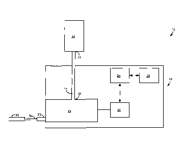
[00311 FIG. I is a function block diagram illustrating components of a fluid
delivery
system 10, which includes a powered fluid injector 12, a medical fluid
container 14
(hereinafter "container 14"), and a fluid transfer set 16 fluidly connecting
powered fluid
injector 12 to medical fluid container 14. Powered fluid injector 12 includes
a fluid
pressurizing unit 18, a motor 22, a processor 24, and a memory 26. Motor 22 is
operatively coupled to fluid pressurizing unit 18 and configured to drive the
fluid
CA 2982031 2019-03-19
- 8 -
pressurizing unit to draw a medical fluid from container 14 and pressurize the
fluid for
discharge into a patient during an imaging procedure. Processor 24 is
communicatively
coupled to motor 22 and memory 26. In the example of FIG. 1, fluid
pressurizing unit 18
defines a discharge outlet 28 that in fluid communication with a patient
catheter 32 via a
patient line or extension tube 30.
100321 Fluid delivery system 10 may include one or more multi-use components
that are
used repeatedly during the course of multiple patient injections. For example,
container
14 and fluid transfer set 16 may be used during the course of multiple patient
injections
and may only be replaced on a periodic basis. By contrast, one or more other
components
of fluid delivery system 10 may be patient-specific, single use components
that are
replaced for each patient injection procedure. For example, patient line 30
and catheter
32 may be replaced for each new patient receiving an injection using powered
fluid
injector 12. Fluid pressurizing unit 18 in powered fluid injector 12 may or
may not also
be replaced for each new patient.
[00331 In instances in which fluid delivery system 10 includes one or more
multi-use
components, the multi-use components cannot lose their integrity or provide
pathways for
contamination during their service life in the fluid delivery system. Testing
the
components in fluid delivery system 10 can validate the safety and integrity
of the
components for extended service during multiple injection procedures for
multiple
patients. Although different tests can be performed, in one example as will be
described
in greater detail below, the components are tested by challenging the
connection or joints
between components with a pathogen (e.g., bacteria and/or virus) and then
evaluating
whether the pathogen is able to enter a medical fluid in fluid delivery system
10 at the
connection or joints. In another example, the components of fluid delivery
system 10,
including fluid transfer set 16 and pressurizing unit 18, are filled with a
medical fluid that
is then allowed to reside in the components for a period longer than
components would be
filled during a single patient injection. The medical fluid and/or components
are then
evaluated to determine if the components degrade and release particles or
leach chemicals
into the medical fluid. In yet another example, pressurizing unit 18 is
operated to
discharge medical fluid against a blocked fluid outlet containing a tracking
agent. Such
an operation may simulate injecting medical fluid into a patient with a
blocked catheter.
Additionally or alternatively, a discharge line connected to pressurizing unit
18 may be
filled with a tracking agent and placed under a pressure that tends to force
the tracking
CA 2982031 2019-03-19
- 9 -
agent back into the pressurizing unit, In either example, a medical fluid in
fluid
communication with pressurizing unit 18 during testing with the tracking agent
can be
evaluated to determine if the tracking agent is present in the medical fluid,
which may
indicate backflow of fluid from a patient-specific line into a multi-use
component. In this
way the operational integrity of fluid delivery system 10 may be analyzed and
validated.
[00341 During operation of powered fluid injector 12, pressurizing unit 18
receives a
medical fluid from container 14, pressurizes the medical fluid, and discharges
the
pressurized medical fluid through discharge outlet 28 and into catheter 32.
Pressurizing
unit 18 can be any mechanism configured to increase the pressure of a liquid
medical
fluid for injection into a patient. Depending on the configuration of
pressurizing unit 18,
the unit may pressurize the medical fluid so it discharges through discharge
outlet 28 at a
pressure greater than 50 pounds per square inch (psi) such as, e.g., a
pressure greater than
200 psi, a pressure greater than 500 psi, or even a pressure greater than 1000
psi.
100351 In one example, pressurizing unit 18 is implemented as a syringe. The
syringe
may include a syringe barrel that receives and holds medical fluid from
container 14 and
a plunger that is disposed within and moveable relative to the syringe barrel.
To fill the
syringe, the syringe may be .fluidly coupled to container 14 and the syringe
plunger driven
to its furthest forward position adjacent discharge outlet 28. This will expel
the majority
of the air that is located within the syringe. Thereafter, the plunger is
retracted within the
syringe barrel, creating a vacuum within the syringe barrel that draws medical
fluid from
container 14 and into the syringe barrel. To subsequently discharge the
medical fluid,
fluid communication between the syringe barrel and container 14 is closed, and
the
plunger is advanced forward in the syringe barrel to pressurize and discharge
the medical
fluid in the syringe barrel.
100361 In another example, pressurizing unit 18 is implemented as a pump. The
pump
may draw fluid from container 14 and discharge the fluid under an increased
pressure out
of discharge outlet 28. When pressurizing unit 18 is implemented as a pump,
the pump
may be an axial pump, a centrifugal pump, a pusher plate pump, a piston-driven
pump, or
other pumping device. In one such example (e.g., FIGS. 8A, 813, and 9), the
pump is a
squeeze pump that squeezes a compressible fluid tube (e.g., a plastic tube) in
a controlled
manner, e.g., such as a peristaltic pump, to progressively pressurize and move
medical
fluid through the tube.
CA 2982031 2019-03-19
- 10 -
[0037] While powered fluid injector 12 in the example of FIG. 1 is illustrated
as having
only a single pressurizing unit 18, in other examples, the powered injector
system may
have multiple pressurizing units. For example, in addition to pressurizing
unit 18
receiving fluid from container 14, powered fluid injector 12 may include one
or more
additional pressurizing units that can receive fluid from container 14 or a
different
medical fluid container (not illustrated). For instance, powered fluid
injector 12 may
include pressurizing unit 18 that receives fluid from container 14 holding one
type of
medical fluid and another pressurizing unit that receives fluid from a
different container
holding a different type of medical fluid. When powered fluid injector 12
includes
multiple pressurizing units, each pressurizing unit may be the same type
(e.g., each
pressurizing unit is a syringe or pump) or the pressurizing units may be of
different types.
[0038] Motor 22 is operatively coupled to pressurizing unit 18 and may provide
mechanical energy that causes the pressurizing unit to draw medical fluid from
container
14 and to pressurize the medical fluid for discharge out through discharge
outlet 28. In
one example, motor 22 is a DC motor that is configured to advance and retract
a plunger
through a syringe barrel. In another example, motor 22 is a DC motor that is
configured
to drive a pump head. Regardless, motor 22 may or may not be a variable speed
motor
that can ramp up speed and ramp down speed to control the rate at which
pressurizing
unit 18 delivers medical fluid to a patient.
[0039] During operation, powered fluid injector 12 receives medical fluid from
container
14. Container 14 may be a bottle, a bag, or any other suitable container that
is configured
to hold and store a liquid fluid. Container 14 is typically formed from
plastic or glass,
although any suitable materials can be used to fabricate container 14.
Depending on the
application, container 14 may he sized to hold enough liquid to inject only a
single dose
of the liquid into a single patient or enough liquid to inject multiple doses
of the liquid
into multiple different patients. When container 14 is sized to hold only a
single dose of
liquid for a single patient, the container may, for example, hold a volume
less than
approximately 100 milliliters (m1). By contrast, a container sized to hold
enough liquid to
inject multiple doses of the liquid into multiple different patients may hold
more liquid
that fluid pressurizing unit 18 can hold when fully tilled. In some examples
when
container 14 is sized to hold enough liquid to inject multiple doses, the
container may
hold greater than approximately 100 ml such as, e.g., greater than or equal to
200 ml,
greater than or equal to 300 ml, or greater than or equal to 500 ml. The
foregoing
CA 2982031 2019-03-19
- -
volumes are merely examples, and it should be appreciated that the disclosure
is not
limited in this respect.
10040] Container 14 can contain a wide variety of different fluids such as
contrast media,
flushing agents (e.g., saline), and fluid medications, among others. Contrast
media is a
liquid that can be injected into a patient to highlight selected areas of the
patient while the
patient is being scanned, e.g., radiographically. Contrast media typically has
a viscosity
ranging from approximately 1 centipoise to approximately 50 centipoise and, in
some
examples, may have an organically (i.e., non-ionic) or non-organically (i.e.,
ionic) bound
molecule that functions to provide contrast, such as organically or non-
organically bound
iodine. Examples of iodine-based contrast media include diatrizoate (llypaque
TM 50),
metrizoate (lsopaque 370), ioxaglate (Hexabrixt), iopamidol (Isovue 300,
Isovue
370), iohexol (OrnnipaqueTM 350), ioxilan (Oxilane 350), iopromidc (UltravistO
370),
and iodixanol (VisipaqueTM 320). Other example contrast media agents include
barium-
based agents such as barium sulfate. In still other examples, contrast media
may include
gadolinium for MR imaging, radioisotopes for nuclear medicine, micro-spheres
for
ultrasound, or the like.
100411 Although fluid delivery system 10 is only illustrated as including a
single
container 14 of medical fluid, fluid delivery system 10 may include multiple
containers
that can each house the same medical fluid or that can house different medical
fluids. In
one example, fluid delivery system 10 includes at least two containers that
each house the
same contrast medium, increasing the amount of fluid connected to pressurizing
unit 18
for injecting into patients as compared to when there is only a single
reservoir. In another
example, fluid delivery system 10 includes at least two containers where one
container
houses a contrast medium and another container houses a flushing media such as
saline.
Powered fluid injector 12 may inject alternating doses of the contrast medium
and the
saline into a patient to control the patient's response to the contrast medium
during
imaging.
[0042] To transfer medical fluid from container 14 to pressurizing unit 18,
fluid delivery
system 10 includes fluid transfer set 16. Fluid transfer set 16 may provide a
fluid
communication pathway between container 14 and pressurizing unit 18. Fluid
transfer set
16 may include a segment of tubing (e.g., flexible polymeric tubing) or duct
that allows
fluid to be conveyed from container 14 to fluid pressurizing unit 18. In the
illustrated
example, fluid transfer set 16 extends from a proximal end 34 that connects to
container
CA 2982031 2019-03-19
- 12 -
14 to a distal end 36 that connects to pressurizing unit 18. In such an
example, fluid
transfer set 16 may define at least one connection between the fluid transfer
set and
container 14 and another connection between the fluid transfer set and
pressurizing unit
18. The connections may be locations where one component (e.g., container 14)
is joined
to another component (e.g., a flexible tube) to form a junction. The specific
number of
connections between container 14 and pressurizing unit 18 may vary depending
on the
specific configuration of fluid transfer set 16. Further, depending on the
configuration,
each of the connections may be detachable connections rather than permanent
connections to allow an operator to exchange and replace components. In
addition,
depending upon the design of fluid injector 12, transfer set 16 may interface
with an
ultrasonic or electro-optic sensor to detect fluid presence in the tube. This
can serve the
dual purpose of preventing air entry into the pressurizing unit by allowing
the operator to
have an automatic container 14 empty detection.
[0043] To connect proximal end 34 of fluid transfer set 16 to container 14,
the fluid
transfer set may have a mechanical connector positioned at proximal end 34.
The
mechanical connector may be a threaded male or female connector that is
configured to
mate with a corresponding connector on container 14. For example. fluid
transfer set 16
may have a female or male luer lock fitting positioned at proximal end 34 that
is
configured to engage with a corresponding luer lock fitting on container 14
for creating a
fluid tight connection between the components. Alternatively, as described
with respect
to FIG. 4, fluid transfer set 16 may have a bottle spike positioned at
proximal end 34 for
piercing a seal on container 14 when placing the container in service.
[0044] Distal end 36 of fluid transfer set 16 may also have a mechanical
connector for
connecting to pressurizing unit 18. For example, as with the connector on
proximal end
34, the mechanical connector on distal end 36 may be a threaded male or female
connector that is configured to mate with a corresponding connector on
container 14. In
one example, fluid transfer set 16 has a female or male luer lock fitting
positioned at
distal end 36 that is configured to engage with a corresponding luer lock
fitting on
pressurizing unit 18 for creating a fluid tight connection between the
components. In
addition, although distal end 36 of fluid transfer set 16 is described as
connecting to
pressurizing unit 18, it should be appreciated that the fluid transfer set may
not connect to
the pressurizing unit directly but may instead connect through intermediary
structures.
For example, distal end 36 of fluid transfer set 16 may connect to a valve
assembly that
CA 2982031 2019-03-19
- 13 -
controls fluid communication between container 14 and pressurizing unit 18
which, in
turn, is in fluid communication with the pressurizing unit.
100451 In the example of FIG. 1, pressurizing unit 18 is simultaneously
connected to
container 14 and catheter 32 through separate fluid ports. In other examples,
pressurizing
unit 18 may have a single fluid port that is connected at separate times to
container 14 and
catheter 32. For example, during a fill operation, pressurizing unit 18 may be
connected
to container 14. Once pressurizing unit 18 has been filled with a suitable
amount of fluid,
the pressurizing unit may be disconnected from container 14 and connected to
catheter
32, thereby allowing a single fluid port to function as both a fluid filling
inlet and a fluid
discharge outlet.
[0046] During operation of powered fluid injector 12, processor 24 may control
the
filling of medical fluid to and discharge of medical fluid from pressurizing
unit 18 with
the aid of instructions associated with program information stored in memory
26.
Processor 24 may also control the filling of medical fluid to and discharge of
medical
fluid from pressurizing unit 18 based on instructions received from a user,
e.g., via a user
interface. Instructions executed by processor 24 may, for example, define
fluid delivery
programs that specify the quantity, rate, and/or pressure with which medical
fluid is to be
delivered from pressurizing unit 18 through discharge outlet 28 during a
diagnostic
imaging procedure and/or during operational testing of powered fluid injector
12.
Instructions executed by processor 24 may also control the opening and closing
of valves
within fluid delivery system 10 (not illustrated) to fill pressurizing unit 18
with medical
fluid and to discharge the fluid from the unit.
10047] Processor 24 may include one or more processors, such as one or more
microprocessors, digital signal processors (DSPs), application specific
integrated circuits
(ASICs), field programmable gate arrays (FPGAs), programmable logic circuitry,
or the
like, either alone or in any suitable combination. In general, processor 24
may receive
electrical signals from input devices such as a user interface and provide
electrical signals
to output devices such as motor 22. For example, processor 24 may provide
signals to
motor 22 to control the advancing and retracting of a plunger in a syringe
barrel and/or
the movement of a pump head. Memory 26 may store instructions and related data
that,
when executed by processor 24, cause powered fluid injector 12 and processor
24 to
perform the functions attributed to them in this disclosure. Typically,
powered fluid
CA 2982031 2019-03-19
-14-
injector 12 uses electrical energy to drive pressurizing unit 18, although
hydraulic,
pneumatic, or other suitable power sources may also be used.
[0048] In the example of FIG. 1, fluid pressurizing unit 18 defines a
discharge outlet 28
that is in fluid communication with a patient catheter 32 via a patient line
30. Patient line
30 may also be referred to as a discharge line, e.g., when the line is not
connected to
catheter 32 outside of a patient injection procedure. Discharge outlet 28 may
be an
opening in fluid pressurizing unit 18 through which high pressure fluid is
discharged and
may or may not include a length of tubing (e.g., patient line 30 or another
line) connected
to the outlet. Patient line 30 may be a length of tubing that traverses from
powered fluid
injector 12 to catheter 32 and can comprise a unitary tube or a plurality of
tube segments
connected together to form an overall length of tube. In other examples,
catheter 32 may
be coupled directly to fluid pressurizing unit 18 without the aid of
intermediate tubing or
extensions.
[0049] Fluid delivery system 10 can be used in any appropriate application
where
delivery of one or more medical fluids is desired including, for example,
during any type
of medical imaging procedure. Example imaging procedures in which fluid
delivery
system 10 can be used include, but are not limited to, X-ray, computed
tomography (CT),
nuclear magnetic resonance (NMR) / magnetic resonance (MR), ultrasound,
fluoroscopy,
and positron emission tomography (PET). When used in these applications,
powered
fluid injector 12 may be communicatively coupled to an imaging system (e.g., a
CT
scanner) and may send and receive electrical signals between the imaging
system for
controlling the operation of the fluid delivery device.
[0050] As discussed above, fluid delivery system 10 can have a variety of
different
configurations to transfer fluid from container 14 to fluid pressurizing unit
18 and,
ultimately, catheter 32. FIG. 2 illustrates one example configuration of a
fluid transfer set
40 that may be used as fluid transfer set 16 in fluid delivery system 10.
Fluid transfer set
40 includes a length of flexible polymeric tubing 42 that extends from a
proximal end 44
to a distal end 45. A mechanical connector 46 is located at proximal end 44
and is
configured to mate with container 14 so as to create a fluid tight connection
between the
container and fluid transfer set 40. Mechanical connector 46 includes a base
48 that is
configured to receive and mate with a rim of container 14 that extends around
an opening
through which medical fluid is withdrawn from the container. Mechanical
connector 46
also includes a spike 50 that projects proximally away from base 48. As
described in
CA 2982031 2019-03-19
-15-
greater detail with respect to FIG. 4, spike 50 is configured to be inserted
into container
14 and to pierce a seal on the container so as to place the container in fluid
communication with fluid transfer set 40.
[0051] Fluid transfer set 40 in the example of FIG. 2 also includes a
mechanical
connector 52 located at distal end 45 of tubing 42. Mechanical connector 52 in
this
example is a luer lock fitting that is configured to mate with a corresponding
luer lock
fitting on fluid pressurizing unit 18 (FIG. 1) so as to create a fluid tight
connection
between the fluid pressurizing unit and fluid transfer set 40. In some
applications in
accordance with this example, the fluid pressurizing unit is a syringe.
[0052] To place fluid transfer set 40 in service, an operator may insert
bottle spike 50 into
container 14 and secure the container to base 48 so that there is a connection
between the
container and proximal end 44 of the fluid transfer set. The operator may
further engage
the luer lock fitting 52 with a corresponding luer lock fitting on
pressurizing unit 18 so
that there is a connection between the pressurizing unit and distal end 45 of
the fluid
transfer set. In this manner, fluid communication can be established between
container
14 and fluid pressurizing unit 18 using a fluid transfer set that defines two
connection
locations. Container 14, fluid transfer set 40 and, in some examples,
pressurizing unit 18
may be used repeatedly during multiple injection procedures to transfer
medical fluid
from a multi-dose container to a pressurizing unit.
[0053] FIG. 3 is an illustration of another example configuration of a fluid
transfer set 60
that may be used in fluid delivery system 10. Fluid transfer set 60 is
configured to fluidly
connect at least one container 14 (FIG. 1) to fluid pressurizing unit 18. In
the illustrated
example of FIG. 3, fluid transfer set 60 is configured to connect three
containers to a fluid
pressurizing unit 62 that is shown as a peristaltic pump. Fluid transfer set
60 includes a
first length of flexible polymeric tubing 64 that extends from a proximal end
66 to a distal
end 68, a second length of flexible polymeric tubing 70 that extends from a
proximal end
72 to a distal end 74, and a third length of flexible polymeric tubing 76 that
extends from
a proximal end 78o a distal end 80. The first and second lengths of tubing 64
and 70
may each fluidly connect a container of contrast media to pump 62. The third
length of
tubing 76 may fluidly connect a container of saline to pump 62.
[0054] In the example of fluid transfer set 60, proximal end 66 of first
tubing 64 and
proximal end 72 of second tubing 70 are each connected to a mechanical
connector 82
that is configured to mate with a container so as to create a fluid tight
connection between
CA 2982031 2019-03-19
- 16 -
the container and the fluid tubing. Third tubing 76 also has a mechanical
connector 84
that is configured to mate with a container holding saline so as to create a
fluid tight
connection between the container arid fluid tubing. At the opposite end, first
tubing 64
and third tubing 76 are each connected at their distal ends to a fluid
pressurizing unit inlet
connector 86 (e.g. a pump inlet connector). Fluid pressurizing unit inlet
connector 86 is
configured to mate with a fluid pressurizing unit (e.g., pump 62) so as to
create a fluid
tight connection between the connector and the pump. Second fluid tubing 70 is
connected directly to pump 62 and, in different examples, may be connected
upstream of
the pump so that fluid from the tubing is pressurized within the pump or
downstream of
the pump so that fluid from the tubing bypasses pressurization within the
pump.
[00551 To place fluid transfer set 60 in service, an operator may connect
mechanical
connectors 82 to first and second tubing 64 and 70 and further connect
mechanical
connectors 82 and 84 to corresponding containers filled with medical fluid(s).
The
operator may further connect fluid pressurizing inlet connector 86 to an inlet
of pump 62,
thereby establishing fluid communication between the first and third tubing 64
and 76 and
pump 62. Second tubing 70 may be connected to fluid pressurizing inlet
connector 86 or
may have a separate mechanical connector that an operator separately connects
to pump
62. When assembled, fluid communication may be established between two
containers
holding contrast media, one container holding saline, and pump 62. Fluid
transfer set 60
may define connections at least between mechanical connectors 82 and first and
second
tubing lines 64 and 70, a connection between fluid pressurizing unit inlet
connector 86
and pump 62, and a connection between second tubing 70 and pump 62. First
tubing line
64, second tubing line 70, and third tubing line 76 along with the containers
to which the
tubing is connected may be used repeatedly during multiple injection
procedures to
transfer medical fluid from the containers to pump 62. Pump 62 and a patient
line or
discharge line 30 to which a discharge outlet of the pump is connected may be
replaced
for each patient and/or each injection procedure.
[0056] FIG. 4 is a cross-sectional illustration of an example mechanical
connector 100
that can be used in fluid delivery system 10 (FIG. 1) to connect a tubing line
to container
14 housing a medical fluid. Mechanical connector 100 defines a base 102 that
is
configured to be positioned around rim 104 of the container 14 so that fluid
does not leak
out between the connector and the container. Mechanical connector 100 also
includes a
spike 106 that is inserted into an aperture 108 defined by rim 104. Spike 106
may pierce
CA 2982031 2019-03-19
-17-
a seal that extends over aperture 108 to close and hermetically seal the
container, e.g., for
shipping and storage prior to use. In the illustrated example, spike 106
pierces a seal that
includes a septum 110 and a foil or collar 112. When spike 106 pierces septum
110 and
foil / collar 112 to access an interior of container 14, tubing 114 is placed
in fluid
communication with the contents of the container and can receive and convey
the
contents, e.g., to fluid pressurizing unit 18.
100571 To help ensure that the various components of fluid delivery system 10
(FIG. 1)
do not lose their physical integrity or provide pathways that allow
contaminants to enter a
sterile medical fluid during the course of use, fluid delivery system 10 may
be tested to
evaluate and validate the integrity of the system. For example, if fluid
delivery system 10
were to be used to transfer medical fluid from container 14 to fluid
pressurizing unit 18 in
a non-sterile environment (e.g., in an imaging suite), the fluid delivery
system may be
validated to help ensure the system will be safe and sterile during the course
of service.
[0058] FIGS. SA-5B, 6, and 7A-7B, are flow diagrams illustrating example
techniques
that may be performed to validate the integrity and sterility of a medical
fluid delivery
system including, e.g., components of the system that may be used multiple
times during
multiple different patient injection procedures. For ease of description, the
techniques of
FIGS. 5-7 will generally be described with reference to fluid delivery system
10 in FIG.
1. The techniques can be performed on fluid delivery systems having other
configurations, as described herein, and it should be appreciated that the
techniques are
not limited to the example fluid delivery system of FIG. 1.
[0059] In addition, in practice, the techniques of FIGS. 5A-5B, 6, and 7A-7B
can be
executed in a number of different environments. In one example, the techniques
are
performed in a cleanroom to help prevent external contaminants from entering
medical
fluids during testing. In another example, the techniques are performed under
a laminar
flow air hood, again to help prevent external contaminants from entering
medical fluids
during testing. Other locations for performing the techniques are also
possible.
[0060] With reference to FIG. 5A, the example technique includes applying one
or more
pathogens (e.g., one or more viruses and/or bacteria) to one or more
components in fluid
delivery system 10 (200). For example, a user may apply the pathogen by
rubbing or
brushing a culture containing the pathogen on the one or more components or by
immersing the components in a culture containing the pathogen. By applying the
pathogen to the one or more components, a user may determine the ability of
fluid
CA 2982031 2019-03-19
-18-
delivery system 10 to resist the passage of microorganisms into fluid pathways
that
convey medical fluid from container 14 to a patient during an injection
procedure.
[0061) In some examples, the pathogen is applied at a connection between
different
components in fluid delivery system 10. The connection, which is where
different
components are detachably joined, may provide the most likely pathway through
which
the pathogen could enter a medical fluid in the system. For example, the
pathogen may
be applied at a connection (e.g., all connections) between container 14 and
fluid transfer
set 16 and/or a connection (e.g., all connections) between fluid transfer set
16 and fluid
pressurizing unit 18. In different examples, the pathogen is applied after the
components
are joined together to test whether external contamination of joined
components can enter
a medical fluid or before the components are joined together to test whether
external
contamination of components before joining can allow the contamination to
enter the
medical fluid.
[0062] When fluid transfer set 16 is configured in the example of FIG. 2, for
instance, the
pathogen may be applied to mechanical connector 46 and/or container 14 (e.g.,
a seal
covering the container) before the components are joined together. The
pathogen may be
applied to external surfaces of mechanical connector 46 and/or container 14
that would be
touched by an operator during normal use. A user may subsequently insert spike
50 of
mechanical connector 46 into container 14 to fluidly connect the fluid
transfer set to the
container. Alternatively, mechanical connector 46 may be mated with container
14 to
define a fluid tight connection between the two components and, thereafter,
the pathogen
applied at the junction where the two components mate.
[0063] In addition to or in lieu of applying the pathogen to mechanical
connector 46
and/or container 14, the pathogen can be applied to mechanical connector 52
and/or fluid
pressurizing unit 18. In one example, the pathogen is applied to external
surfaces of
mechanical connector 52 and/or fluid pressurizing unit 18 that would be
touched by an
operator during normal use. For example, the pathogen may be applied around
the
external surface of a luer lock fitting and/or at an inlet of a syringe barrel
or fluid pump.
A user may subsequently mate mechanical connector 52 with a corresponding
connector
on fluid pressurizing unit 18 to fluidly connect the fluid transfer set to the
fluid
pressurizing unit.
CA 2982031 2019-03-19
-19-
[0064] As another example, specifically when fluid transfer set 16 is
configured as shown
in the example of FIG. 3, the pathogen may be applied to mechanical connectors
82, 84
and/or the containers to which the connectors join, as described above with
respect to the
fluid transfer set of FIG. 2. The pathogen may be applied to mechanical
connectors 82,
84 and/or the containers to which the connectors join before mating the
connectors with
the containers or after mating the connectors with the containers. In addition
to or in lieu
of applying the pathogen to the mechanical connector and/or containers, the
pathogen can
be applied at one or more connections where tubing mates with pump 62. For
example,
the pathogen may be applied on external surfaces of fluid pressurizing unit
inlet
connector 86 and/or an inlet of pump 62 to which the connector mates before or
after the
components are mated together.
[0065] The type and amount of pathogen applied at connection locations and/or
to
components within fluid delivery system 10 may vary, e.g., based on the
severity and
parameters of testing. When bacteria is used as the pathogen, example bacteria
that may
be applied include, but are not limited to, Staphylococcus aureus,
Staphylococcus
epidermidis, Pseudomanas aeruginosa, Klebsiella pneumonia, Eschcrichia coli,
Candida
albicans, and Aspergillus niger. In some examples, multiple types of bacteria
are applied
to fluid delivery system 10, for example either simultaneously together or by
conducting
serial tests using one type of bacteria and then another type of bacteria, to
evaluate the
ability of fluid delivery system 10 to resist the passage of different types
of
microorganisms. In one example, at least 100 colony forming units/milliliter
(CFU/ml) of
bacteria are applied to each component or Connection location during the
technique of
FIG. 5A such as, e.g., at least 500 CFU/ml, at least 1000 CFU/ml, or at least
5000
Mimi. Bacteria applied to fluid delivery system 10 may be in an organism
diluent,
such as Mile's Test Soil or Tryptic Soy Broth.
[00661 In applications where the pathogen is applied to the components of
fluid delivery
system 10 prior to assembly, the components may subsequently be disinfected
(201) and
assembled (202) to place the components in fluid communication with one
another.
Disinfecting the components of fluid delivery system 10 prior to assembly may
remove
surface pathogens from the components so that the pathogens are not
deliberately
introduced into medical fluid during assembly of the components. For example,
by
applying the pathogen to one or more components of fluid delivery system 10
and then
disinfecting the surfaces of the components, the technique of FIG. 5A may be
used to
CA 2982031 2019-03-19
- 20 -
determine whether the pathogen bypassed a seal or barrier of the components
(e.g., a seal
covering a medial fluid container) or otherwise invaded the components such
that surface
disinfection does not remove the pathogen.
[0067] To disinfect the one or niore components of fluid delivery system 10
(201), a
disinfectant designed to kill and/or remove the pathogen can be applied to
surfaces of the
components where the pathogen was originally applied. An example disinfectant
is an
isopropyl alcohol solution (e.g., containing greater than 60% isopropyl
alcohol such as
approximately 70% isopropyl alcohol), although other disinfectants can be
used. The
disinfectant can be applied to or impregnated in a cloth that is then wiped
over the
surfaces of the components. In some examples, the cloth is wiped over a
surface of a
component so that the cloth is in contact with the component for a period of
time greater
than 5 seconds such as, e.g., a period greater than 20 seconds, a period
greater than 30
seconds, or a period of time ranging from approximately 25 seconds and
approximately
30 seconds.
(0068] When fluid transfer set 16 is configured in the example of FIG. 2, for
instance,
mechanical connector 46 and/or container 14 (e.g., a seal covering the
container) may be
disinfected by wiping a cloth containing a disinfectant over the surfaces of
the mechanical
connector and/or container to which the pathogen was applied. As another
example,
when fluid transfer set 16 is configured as shown in the example of FIG. 3,
mechanical
connectors 82, 84 and/or the containers to which the connectors join may be
disinfected
by wiping a cloth containing a disinfectant over the surfaces of the
mechanical connectors
and/or containers to which the pathogen was applied.
[0069] In addition to or in lieu of disinfecting the one or more components of
fluid
delivery system 10 (201) after applying the pathogen (200) as described above,
the one or
more components of fluid delivery system 10 may be disinfected prior to
applying the
pathogen (200). For example, a disinfectant designed to kill and/or remove the
pathogen
can be applied to surfaces of the components where the pathogen is to be
applied.
Disinfecting the surfaces of the components where the pathogen is to be
applied can clean
and sterilize the components. This can help ensure that any pathogenic ingress
subsequently identified in fluid delivery system 10 is attributable to the
controlled
application of the pathogen according to the technique of FIG. 5A and not
external
sources. When disinfected prior to applying the pathogen, the one or more
components of
CA 2982031 2019-03-19
-21 -
fluid delivery system 10 can be disinfected, e.g., using the techniques
described above for
disinfecting the one or more components after application of the pathogen.
100701 Independent of whether the one or more components of fluid delivery
system 10
are disinfected, the components may be assembled (202) to place the components
in fluid
communication with one another. When the one or more components are
disinfected
prior to assembly (201), the components may first be allowed to dry for a
period of time
prior to assembly such as a period of greater than 10 seconds, greater than 30
seconds, or
greater than approximately 1 minute. The components of fluid delivery system
10 can be
assembled in accordance with fluid delivery system use instructions. To
assemble fluid
transfer set 16 (FIG. 1) in fluid delivery system 10, for example, an operator
can mate a
mechanical connector positioned at a proximal end 34 of the fluid transfer set
with
container 14, As the mechanical connector is mated with container 14, the
connector may
pierce a seal on the container, allowing fluid to flow from the container into
the fluid
transfer set. The operator may also mate a mechanical connector at distal end
36 of the
fluid transfer set with fluid pressurizing unit 18 so as to place the fluid
transfer set in fluid
communication with the pressurizing unit.
100711 With further reference to FIG. 5A, the example technique also includes
drawing
fluid from container 14 through fluid transfer set 16 and into fluid
pressurizing unit 18
(203). Subsequent to applying the pathogen to the one or more components of
fluid
delivery system 10 (200) and disinfecting (201) and assembling (202) the
components,
fluid is drawn through the system to evaluate if the pathogen will enter the
fluid during
typical filling and injection operations. Fluid may be drawn from container 14
through
fluid transfer set 16 and into fluid pressurizing unit 18 immediately after
applying the
pathogen or after the pathogen has been applied for a certain amount of time.
For
example, fluid may be drawn through fluid delivery system 10 after the
pathogen has
been applied and allowed to reside in or on the components of the system for a
period of
at least 1 hour such as, e.g., a period greater than or equal to 4 hours, a
period greater than
or equal to 8 hours, or a period greater than or equal to 10 hours. Of course,
the fluid
delivery components may first be disinfected (201), allowed to dry, and
assembled after
the pathogen is allowed to reside on the components for any of the foregoing
periods of
time.
CA 2982031 2019-03-19
- 22 -
[00721 The technique of FIG. 5A also includes extracting a sample of medical
fluid from
within fluid delivery system 10 (204). The fluid sample may be extracted by
operating
fluid pressurizing unit 18 to discharge pressurized medical fluid through
discharge outlet
28. The sample may be collected at the discharge outlet, e.g., from discharge
line 30.
Additionally or alternatively, a fluid sample may be extracted by
disconnecting
detachably connected components in fluid delivery system 10 and extracting a
sample of
fluid from within the components. For example, fluid transfer set 16 may be
detached
from container 14 and/or fluid pressurizing unit 18 and a sample of fluid
taken from
within container 14, from within the fluid transfer set, and/or from within
fluid
pressurizing unit 18.
100731 Independent of the specific technique used to extract a sample from
fluid delivery
system 10 (204), the sample is subsequently analyzed (206) to determine a
concentration
level of the pathogen applied to the fluid delivery system in the fluid
sample. The
determined pathogen level may be compared to a concentration level of the
pathogen in
the medical fluid within container 14 before the container was connected to
fluid delivery
system 10 and challenged with the pathogen. For example, a concentration level
of the
pathogen in the medical fluid within container 14 before the container was
connected to
fluid delivery system 10 and challenged with the pathogen may be zero. If the
extracted
sample is determined to also have a pathogen concentration level of zero,
fluid delivery
system 10 may be validated as successfully resisting the passage of
microorganisms into
fluid pathways. Different tolerance levels may be established depending on the
requirements of a particular application.
[00741 FIG. 5B is a flow diagram of an example implementation of the technique
of FIG.
5A, where like process steps described above with respect to FIG. 5A are
designated with
like reference numerals. As shown in FIG. 5B, the example technique includes
applying
one or more pathogens (e.g., one or more bacteria and/or viruses) to the one
or more
components of fluid delivery system 10 (200), such as a portion of a component
or
portions of components that join together to form a connection. Subsequent to
applying
the pathogen to the one or more components of fluid delivery system 10 (200),
the
components may be disinfected (201) and assembled (202) to place the
components in
fluid communication with one another.
CA 2982031 2019-03-19
- 23 -
[0075] Once the pathogen challenged components are assembled, fluid is drawn
through
the system to evaluate if the pathogen will enter the fluid during typical
filling and
injection operations (203). Fluid can be drawn from container 14 through fluid
transfer
set 16 and into fluid pressurizing unit 18 by operating (e.g., activating) the
fluid
pressurizing unit. In the technique of FIG. 5B, fluid pressurizing unit 18 is
operated
multiple times (300) to discharge multiple portions of fluid from the fluid
pressurizing
unit 18 via discharge outlet 28. For example, fluid pressurizing unit 18 may
be activated
a first time to draw fluid from container 14 through fluid transfer set 16 and
then
discharge a first portion of pressurized fluid out through discharge outlet
28. After
dispensing a suitable volume of fluid, fluid pressurizing unit 18 may cease
operation so
that no fluid is being dispensed from discharge outlet 28. Fluid pressurizing
unit 18 may
subsequently be activated a second time to draw additional fluid from
container 14
through fluid transfer set 16 and discharge a second portion of pressurized
fluid out
through discharge outlet 28. After discharging a suitable volume of fluid,
fluid
pressurizing unit 18 may again cease operation so that no fluid is being
dispensed from
discharge outlet 28. The process of activating fluid pressurizing unit 18 and
ceasing
operation of the unit can be repeated any additional number of times, such as
one, two,
three, or more times, e.g., to convey a certain volume of fluid and/or
generate a certain
number of discharged fluid portions.
[0076] Operating fluid pressurizing unit 18 multiple times to generate
multiple portions
of fluid (300) may be useful to simulate real-world operation of fluid
delivery system 10
when the system is used to inject multiple patients with fluid from container
14 during
multiple sequential patient injection procedures. During each patent injection
procedure,
fluid pressurizing unit 18 is operated to draw fluid from container 14 and
discharge the
fluid under pressure into catheter 32 connected to a patient. After each
patient injection
procedure, fluid pressurizing unit 18 ceases operation and, in some examples,
is replaced
with a new, sterile fluid pressurizing unit. The fluid pressurizing unit can
then be
operated during a subsequent injection procedure to inject a new patient with
pressurized
medical fluid. The process can be repeated for additional patient injection
procedures.
[0077] By operating fluid pressurizing unit 18 multiple times to discharge
multiple
portions of fluid (300) during validation testing, fluid delivery system 10
can be evaluated
for resistance to pathogenic ingress during a normal course of operation.
Fluid
pressurizing unit 18 can be operated any desired number of times to generate
any desired
CA 2982031 2019-03-19
- 24 -
number of portions or volumes of fluid during the performance of the method of
FIG. 5B.
In some examples, fluid pressurizing unit 18 is operated at least twice (e.g.,
three, four, or
more times) to provide at least two portions of fluid (e.g., three, four, or
more portions of
fluid) that are discharged from the fluid pressurizing unit during operation.
Fluid
pressurizing unit 18 may cease operation for a given period of time between
each cycle in
which the unit is operated to discharge fluid. For example, the fluid
pressurizing unit 18
may remain inactive for a period of at least 5 minutes between each cycle of
operation,
such as a period of at least 20 minutes, a period of at least one hour, a
period of at least 2
hours, a period ranging from 5 minutes to 5 hours, or a period ranging from 10
minutes to
2 hours. As described in greater detail below, a fluid sample can be extracted
from one
more of the portions of fluid discharged from fluid pressurizing unit 18 for
subsequent
analysis (204).
100781 The volume of fluid discharged from fluid pressurizing unit 18 during
the
performance of the technique of FIG. 513 can vary, e.g., depending on the
capacity of
container 14, the discharge rate of the fluid pressurizing unit, and the
amount of time the
fluid pressurizing unit is operated during each cycle. Moreover, when
attempting to
simulate real-world operation of fluid delivery system 10, the fluid delivery
system can,
in different operating environments, be operated in a low volume throughput
scenario in
which only a few patients would be injected during a day of operation or a
high volume
throughput scenario in which many patients would be injected during a day of
operation.
[0079] In a comparatively low volume throughput environment, fluid delivery
system 10
may be connected to a single container 14 (e.g., contrast, saline) or single
set of
containers (e.g., a container of contrast and a container of saline) that are
used throughout
a single day without replacement. Accordingly, to simulate comparatively low
volume
operation, fluid pressurizing unit 18 may be operated so that each portion of -
fluid
discharged from the fluid pressurizing unit is drawn from the same container
or set of
containers, e.g., without replacing a container between operating cycles of
the fluid
pressurizing unit. In such an application, each sample of fluid extracted from
fluid
delivery system 10 (204) and analyzed for the pathogen (206) may originate
from the
same container or set of containers. In some cases, each sample of fluid may
be obtained
from a discharged portion of fluid without disassembling fluid system 10
(e.g.,
disconnecting container 14, fluid transfer set 16, and/or fluid pressurizing
unit 18), which
may otherwise introduce contamination into the system.
CA 2982031 2019-03-19
- 25 -
100801 As one example of a low volume throughput simulation, specifically when
fluid
transfer set 16 is configured as shown in the example of FIG. 3, connector 82
may be
attached to a container of contrast sized to provide multiple doses of fluid
to multiple
different patients (e.g., 500 milliliters) and connector 84 may be attached to
a container of
saline sized to provide multiple doses of fluid to multiple different patients
(e.g., 500
milliliters). Fluid pressurizing unit 18 can then be periodically operated to
dispense a
portion of fluid that is drawn from the container of contrast and/or the
container of saline.
For example, to simulate a patient dose, fluid pressurizing unit 18 may be
operated to
dispense 100 milliliters of contrast followed by 30 milliliters of saline,
thereby dispensing
a first portion of fluid that is 130 milliliters. Fluid pressurizing unit 18
may be operated
to subsequently dispense additional portions of fluid that arc each composed
of 100
milliliters of contrast followed by 30 milliliters of saline. For example,
fluid pressurizing
unit 18 may he operated to dispense a first portion of fluid upon initial
assembly of fluid
delivery system 10, a second portion of fluid four hours after assembly, a
third portion ten
hours after assembly, and a fourth portion twelve and a half hours after
assembly. The
container of contrast and container of saline in such an example would have a
capacity
sufficient to allow all four portions of fluid to be drawn from the same set
of containers.
100811 In contrast to a low volume throughput environment, in a comparatively
high
volume throughput environment, the container 14 (e.g., contrast, saline) or a
set of
containers (e.g., a container of contrast and a container of saline) connected
to fluid
delivery system 10 may be replaced throughout a day of operation as the
contents of the
containers are exhausted. Accordingly, to simulate comparatively high volume
operation,
fluid pressurizing unit 18 may be operated a sufficient number of times to
empty the
container or set of containers. Upon emptying the containers, the container or
set of
containers to which fluid pressurizing unit 18 is fluidly connected may be
replaced with a
replacement container or set of containers filled with medical fluid (302).
After
replacement, fluid pressurizing unit 18 may again be operated to dispense
portions of
fluid from the replacement containers.
100821 As one example of a high volume throughput simulation, specifically
when fluid
transfer set 16 is configured as shown in the example of FIG. 3. connector 82
may be
attached to a container of contrast sized to provide a dose of fluid to
multiple different
patients (e.g., 200 milliliters) and connector 84 may be attached to a
container of saline
sized to provide a dose of fluid to multiple different patients (e.g., 500
milliliters). Fluid
CA 2982031 2019-03-19
- 26 -
pressurizing unit 18 can then be periodically operated to dispense a portion
of fluid that is
drawn from the container of contrast and/or the container of saline. For
example, to
simulate a patient dose, fluid pressurizing unit 18 may be operated to
dispense 100
milliliters of contrast followed by 30 milliliters of saline, thereby
dispensing a first
portion of fluid that is 130 milliliters. Fluid pressurizing unit 18 may be
operated
additional times at a frequency sufficient to consume multiple containers of
contrast
and/or multiple containers of saline over a given period of time. For example,
fluid
pressurizing unit 18 may be operated at a frequency sufficient to consume
twenty
containers of contrast and four containers of saline over a twelve and a hail
hour period
by dispensing discrete 130 milliliter portions of contrast and saline. The
containers of
contrast and saline may be replaced with full replacement containers as the in-
service
containers connected to fluid delivery system 10 become exhausted. In such an
application, different samples of fluid extracted from fluid delivery system
10 (204) and
analyzed for the pathogen (206) may originate from different containers or
different sets
of containers. Such an application may be useful to evaluate the tendency of
the pathogen
to invade fluid system 10 during the course of high volume operation when
medical fluid
containers are being replaced multiple times per day.
100831 Independent of whether fluid delivery system 10 is operated to simulate
low
volume throughput, a high volume throughput, or both low and high volume
throughputs,
the technique of FIG. 5B includes applying the pathogen to one or more
components in
fluid delivery system 10 (200). For example, when fluid transfer set 16 is
configured as
shown in the example of FIG, 3, the pathogen may he applied to connector 82
(e.g., a
connection between a container such as container 14 and connector 82), a
proximal end
66 of the first length of tubing 64 and connector 82 (e.g., a connection
between the
components), at connector 84 (e.g., a connection between a container and
connector 84),
and/or at a connection between fluid pressurizing unit inlet connector 86 and
pump 62.
After applying the pathogen to the components (200), the components may be
disinfected
(201) and assembled (202), as described with respect to FIG. 5A.
[0084] In instances where a fluid container or set of fluid containers is
replaced during
performance of the method of FIG. 5B (302), the pathogen may or may not be
reapplied
to some or all of the connection locations where the pathogen was applied
during initial
assembly of fluid delivery system 10. Reapplying the pathogen may be useful to
evaluate
the tendency of the pathogen to invade fluid system 10 during the course of
high volume
CA 2982031 2019-03-19
- 27
operation when Medical fluid containers are being replaced multiple times per
day. When
the pathogen is reapplied, the components may again be disinfected (201) and
then
reassembled (202).
[0085] After or while operating fluid pressurizing unit 18 multiple times over
a given
period to draw fluid through fluid delivery system 10 and dispense multiple
portions of
fluid from the unit (203), a plurality of fluid samples of fluid are extracted
for analysis
(204). Each sample of fluid may be from a different portion of fluid dispensed
during a
different cycle of operation of fluid pressurizing unit 18. In one example, a
sample of
fluid is obtained from each portion of fluid discharged from -fluid
pressurizing unit 18. In
another example, a sample of fluid is obtained from some but not all portions
of fluid
discharged from fluid pressurizing unit 18. For example, an operator may
extract a
sample from a first portion of fluid dispensed from fluid pressurizing unit 18
upon initial
assembly of fluid delivery system 10 and/or a sample from a last portion of
fluid
dispensed from the fluid pressurizing unit during a final operation. An
operator may
extract additional or different samples. For instance, in addition to
extracting a sample
from a first portion of fluid and a sample from a last portion of fluid, the
operator may
extract one or more (e.g., two, three, or more) additional samples from
portions of fluid
dispensed between the first portion of fluid and the last portion of fluid.
[0086] The technique of FIG. 5B also includes analyzing the plurality of
samples (206) to
determine a concentration level of the pathogen applied to the fluid delivery
system in the
plurality of fluid samples. In some examples, each of the plurality of samples
obtained
from the multiple portions of fluid are combined together to form a pooled
sample. In
such applications, the pooled sample may be analyzed to determine a
concentration level
of the pathogen in the pooled sample. In other examples, each of the plurality
of samples
is separately analyzed to determine a concentration level of the pathogen in
each
respective sample. In either case, if a sample is determined to have a low
concentration
level (e.g., zero) for the pathogen or combination of pathogens originally
applied to fluid
delivery system 10, the fluid delivery system may be validated as successfully
resisting
the passage of microorganisms into fluid pathways,
[0087] In some examples, the technique of FIG. 513 is repeated under both high
volume
throughput conditions and low volume throughput conditions to validate fluid
delivery
system 10, or a component thereof For example, the technique may be performed
once
under high throughput conditions in which multiple containers of contrast
and/or saline
CA 2982031 2019-03-19
- 28 -
are consumed to generate samples of discharged fluid that originated from
different
containers or sets of containers. The samples obtained during high throughput
testing can
be pooled together and analyzed to determine a concentration level of the
pathogen in the
pooled sample. In addition, the technique may be performed again under low
throughput
conditions. In low throughput conditions, only a single container or set of
containers of
contrast and/or saline may be consumed to generate samples of discharged fluid
that all
originated from the same container or set of containers. The samples obtained
during low
throughput testing can also be pooled together and analyzed to determine a
concentration
level of the pathogen in the pooled sample. In some applications, fluid
delivery system
may be validated as successfully resisting the passage of microorganisms into
fluid
pathways if both the pooled sample from high throughput testing and the pooled
sample
from low throughput testing are determined to have a sufficiently low
concentration level
(e.g., zero) of the pathogen.
100881 FIG. 6 is a flow diagram illustrating another example technique that
may be used
to validate the integrity and sterility of a medical fluid delivery system.
The components
of the medical fluid delivery system may be assembled together (e.g., so that
a fluid
transfer set is in fluid communication with both a container of medical fluid
and a fluid
pressurizing unit). Once assembled, the example technique includes drawing
fluid from
container 14 through -fluid transfer set 16 and into fluid pressurizing unit
18 so as to fill
fluid holding areas in fluid delivery system 10 with medical fluid (208).
Although fluid
delivery system 10 can be filled with any medical fluid as described herein,
in some
examples, the medical fluid is contrast media. By filling fluid delivery
system 10 with
contrast media, a user may determine the ability of the components of fluid
delivery
system 10 to resist chemical degradation, including multi-use components that
may be
used during the course of multiple patient injections.
100891 Subsequent to filling fluid transfer set 16 and fluid pressurizing unit
18 with
medical fluid, the medical fluid is held in the fluid transfer set and fluid
pressurizing unit
for a period of time (210). The components of fluid delivery system 10 may be
held full
of fluid for a period of time so that the fluid contacts interior surfaces of
the components
that would normally be fluid-wet during filling and/or discharge of powered
fluid injector
12. In some examples, the fluid is static (e.g., not moving) within fluid
delivery system
10 as the components of the system are held full of fluid. In other examples.
the fluid
CA 2982031 2019-03-19
- 29 -
may be moving through fluid delivery system 10 during the period of time in
which the
components are held full of fluid.
[0099] The components of fluid delivery system 10 can be held full of fluid
for any
period of time suitable for evaluating the ability of the components to resist
chemical
degradation (210). In applications where a multi-use component is intended to
remain in
service during the course of multiple fluid injections, the component may be
exposed to
fluid continuously or intermittently for an extended period of time such as an
entire shift
or entire day. Accordingly, in some examples, the components of fluid delivery
system
may be held full of fluid for a period of time greater than or equal to 1 hour
such as,
e.g., a period of time greater than or equal to 2 hours, a period of time
greater than or
equal to 4 hours, or a period of time greater than or equal to 8 hours. For
instance, the
components of fluid delivery system 10 may be held full of fluid for a period
of time
ranging from approximately 1 hour to approximately 48 hours such as, e.g.,
from
approximately 4 hours to approximately 24 hours, or approximately 8 hours to
approximately 16 hours.
100911 After holding the components of fluid delivery system 10 (e.g., fluid
transfer set
16 and fluid pressurizing unit 18) full of medical fluid, a sample of the
medical fluid can
be extracted for analysis (212). The fluid sample may be extracted by
operating fluid
pressurizing unit 18 to discharge pressurized medical fluid through discharge
outlet 28.
The sample may be collected at the discharge outlet. Additionally or
alternatively, a fluid
sample may be extracted by disconnecting detachably connected components in
fluid
delivery system 10 arid extracting a sample of fluid from within the
components. For
example, fluid transfer set 16 may be detached from container 14 and/or fluid
pressurizing unit 18 and a sample of fluid taken from within container 14,
from within the
fluid transfer set, and/or from within fluid pressurizing unit 18.
[0092] The extracted sample is analyzed to determine if any components of the
fluid
delivery system have chemically degraded with exposure to the medical fluid
(214). The
fluid may be analyzed to determine if a material(s) used to fabricate
components of fluid
delivery system 10 (e.g., a material used to fabricate fluid transfer set 16
and/or fluid
pressurizing unit 18) have entered the medical fluid held within the
components. In one
example, the fluid is analyzed to determine if there are any particles in the
fluid greater
than a certain size such as, e.g., greater than 10 micrometers, greater than
100
micrometers, or greater than 1 millimeter. Such particles may be pieces of a
component
CA 2982031 2019-03-19
- 30 -
of fluid delivery system 10 that have detached from the component. If the
extracted
sample is determined to not have particles greater than a certain size or not
have a certain
number of particles greater than the certain size, fluid delivery system 10
may be
validated as being chemically compatible and maintaining chemical integrity
with the
medical fluid (e.g., the class of medical fluids).
[0093] In addition to or in lieu of analyzing the extracted sample for
particles, the
extracted sample may be analyzed to determine if a chemical present in the
material(s)
used to fabricate components of fluid delivery system 10 (e.g., a material
used to fabricate
fluid transfer set 16 andior fluid pressurizing unit 18) has leached into the
medical fluid
held within the components. As examples, the extracted fluid sample may be
analyzed to
determine if one or more of the following chemical compounds are present in
the fluid:
cyclohexanone, 2-ethyl-l-hexanol, di(2-ethylliexyl)phthalate (DEHP),
epoxidized
soybean oil, tris (nonylphenyl) phosphate (TNPP), stearic acid, zinc or other
heavy
metals, The extracted sample may be analyzed using gas chromatography, high-
performance liquid chromatography, inductively coupled plasma mass
spectrometry
(ICP-MS), or any other suitable techniques. A determined concentration level
of the
chemical compound(s) may be compared to a concentration level in the medical
fluid
within container 14 before the container was connected to fluid delivery
system 10 and
exposed to the components in the system. For example, a concentration level of
the
chemical compound(s) in the medical fluid within container 14 before the
container was
connected to fluid delivery system 10 and drawn through the system may be
zero. If the
extracted sample is determined to also have a concentration level of zero for
the chemical
compound(s), fluid delivery system 10 may be validated as being chemically
compatible
and not leaching chemical compound(s) into the medical fluid. Different
tolerance levels
may be established depending on the requirements of a particular application.
[0094] FIG. 7A is a flow diagram illustrating another example technique that
may be
used to validate the integrity and sterility of a medical fluid delivery
system. The
technique may be used to confirm that cross-contamination of fluids between
patients will
not occur when using fluid delivery system 10 by having fluid from a patient-
specific
tube (e.g., downstream of fluid pressurizing unit 18) mix with fluid in a
multi-use tube
(e.g., upstream of fluid pressurizing unit 18). The components of the medical
fluid
delivery system may be assembled together (e.g., so that a fluid transfer set
is in fluid
communication with both a container of medical fluid and a fluid pressurizing
unit).
CA 2982031 2019-03-19
-31-
Once assembled, the example technique includes drawing fluid from container 14
through
fluid transfer set 16 and into fluid pressurizing unit 18 so as to fill fluid
holding areas in
fluid delivery system 10 with medical fluid (216). By drawing medical fluid
from
container 14 through fluid transfer set 16 and into fluid pressurizing unit
18, the fluid
holding regions of fluid delivery system 10 upstream of fluid pressurizing
unit 18 may be
filled with medical fluid.
100951 In addition, in the technique of FIG. 7A, discharge outlet 28 of fluid
delivery
system 10 is placed in fluid communication with a fluid reservoir containing a
tracking
fluid (218). The fluid reservoir may be a bottle, bag, pouch, syringe, or tube
filled with
fluid, or any other suitable reservoir. The tracking fluid may contain a
tracking agent not
present in the medical fluid in container 14. The tracking agent may be
tracked to
determine if fluid downstream of discharge outlet 28 migrates into fluid
pressurizing unit
18 and/or upstream of fluid pressurizing unit 18. For example, the tracking
agent may
simulate the movement of blood borne pathogens, were discharge outlet 28 in
fluid
communication with a catheter inserted into a patient. Example tracking agents
may
include, but are not limited to, bacteria, viruses, dyes, radioactive
isotopes, and electro-
magnetic markers.
[0096] To simulate cross-contamination conditions, the fluid reservoir
containing the
tracking agent is blocked so that fluid pressurizing unit 18 cannot draw
medical fluid
from container 14 and inject the fluid into the fluid reservoir containing the
tracking
agent. Configuring the fluid reservoir containing thc tracking agent as a
closed reservoir
may simulate conditions in which a patient's catheter 32 is blocked and fluid
pressurizing
unit 18 is attempting to inject fluid into a blocked or partially occluded
catheter. Were
the fluid reservoir containing the tracking agent not closed, fluid
pressurizing unit 18
could draw fluid from container 14 and inject the fluid into the reservoir,
preventing the
tracking agent from migrating upstream in fluid delivery system 10, By
contrast, when
fluid pressurizing unit 18 discharges medical fluid through discharge outlet
28 against a
closed reservoir of tracking fluid, a generally static interface may be
created where
discharged medical fluid meets tracking fluid, potentially resulting in mixing
and
upstream migration of the tracking fluid into the upstream medical fluid.
100971 Accordingly, after placing discharge outlet 28 of fluid delivery system
10 in fluid
communication with a fluid reservoir containing a tracking fluid (218), fluid
pressurizing
unit 18 is operated to try and discharge pressurized medical fluid into the
tracking fluid
CA 2982031 2019-03-19
- 32 -
(220). For example, when fluid pressurizing unit is a pump, the pump may
operate
continuously -for a period of time even though the pump may not necessarily be
conveying fluid because the fluid path downstream of the pump is blocked or
restricted.
Fluid pressurizing unit 18 may operate for any suitable period such as, e.g.,
greater than 1
minute, greater than 15 minutes, greater than 30 minutes, or greater than 1
hour.
[0098] After operating fluid pressurizing unit 18 for a period of time (220),
a sample of
medical fluid is extracted from fluid delivery system 10 for analysis (222).
The fluid
sample may be extracted by disconnecting detachably connected components in
fluid
delivery system 10 and extracting a sample of fluid from within the
components. For
example, fluid transfer set 16 may be detached from container 14 and/or fluid
pressurizing unit 18 and a sample of fluid taken from within container 14,
from within the
fluid transfer set, and/or from within fluid pressurizing unit 18.
[0099] The extracted sample is analyzed to determine a concentration level of
the
tracking agent in the sample of medical fluid (224). The determined
concentration level
may be compared to a concentration level of the tracking agent in the medical
fluid within
container 14 before the container was connected to fluid delivery system 10.
For
example, a concentration level of the tracking agent in the medical fluid
within container
14 and/or transfer set 16 before the container was connected to fluid delivery
system 10
may be zero. If the extracted sample is determined to also have a
concentration level of
zero, fluid delivery system 10 may be validated as successfully preventing
cross-
contamination of fluid between a patient-specific fluid line and a multi-use
fluid line.
Different tolerance levels may also be established depending on the
requirements of a
particular application.
[00100] FIG. 7B is a flow diagram illustrating another example technique that
may be
used to confirm that cross-contamination of fluids between patients will not
occur when
using fluid delivery system 10, where like process steps described above with
respect to
FIG. 7A are designated with like reference numerals. In the technique of FIG.
7B, the
components of the medical fluid delivery system may be assembled together
(e.g., so that
fluid transfer set 16 is in fluid communication with both container 14 and
fluid
pressurizing unit 18). Once assembled, the example technique includes filling
fluid
transfer set 16 and fluid pressurizing unit 18 with medical fluid (310). The
components
can be filled with fluid by activating fluid pressurizing unit 18 to draw
fluid from
container 14 through -fluid transfer set 16 and into the fluid pressurizing
unit. This can
CA 2982031 2019-03-19
- 33 -
prime fluid pressurizing unit 18 and/or fill fluid holding areas in fluid
delivery system 10
with medical fluid, thus providing filled fluid pathway(s) to evaluate whether
a tracking
agent will travel through the pathways, potentially indicating a risk of cross-
contamination. In some examples, fluid transfer set 16 and fluid pressurizing
unit 18 are
filled with a comparatively low viscosity medical fluid, such as saline, which
may be
more prone to permit flow of a tracking agent than a comparatively higher
viscosity fluid,
such as contrast.
[00101] In addition, in the technique of FIG. 7B, a discharge line (e.g.,
discharge line 30
in FIG. 3) connected to discharge outlet 28 of fluid pressurizing unit 18 is
filled with a
tracking agent (312). The tracking agent may be a tracking fluid containing a
tracking
agent and, in different examples as described above with respect to FIG. 7A,
can be a
bacteria, virus, dye, radioactive isotope, and/or eleetro-magnetic marker. The
discharge
line may be filled by introducing the tracking agent through a distal outlet
of the
discharge line (e.g., opposite fluid pressurizing unit 18) and allowing the
tracking agent to
flow down the discharge line toward the fluid pressurizing unit. Upon
initially filling the
discharge line with tracking agent, the tracking agent may be positioned
between
discharge outlet 28 of fluid pressurizing unit 18 and a distal end of
discharge line 30
extending away from discharge outlet 28.
[00102] When filling a discharge line with tracking agent (312), the tracking
agent can
be introduced into the discharge line until it is positioned any suitable
distance from fluid
pressurizing unit 18. In general, the distance between the tracking agent in
the discharge
line and the fluid pressurizing unit can be controlled by controlling the
amount of medical
fluid in the discharge line before introduction of the tracking agent. For
example, if fluid
pressurizing unit 18 was primed with medical fluid and the discharge line was
filled with
a column of medical fluid extending approximately 10 centimeters away from the
fluid
pressurizing unit, the tracking agent may initially be positioned
approximately 10
centimeters away from the fluid pressurizing unit, when introduced into the
discharge
line. In such an example, the column medical fluid that does not contain
tracking agent
may function to initially separate the tracking agent :from the fluid
pressurizing unit.
[00103] In some examples, fluid pressurizing unit 18 provides a fluid seal
adjacent
discharge outlet 28 and the discharge line is filled with tracking agent until
the tracking
agent is positioned adjacent the fluid seal. FIGS. 8A and 8B are perspective
drawings of
an example peristaltic pump 400 that has a fluid seal and may be used as fluid
CA 2982031 2019-03-19
- 34 -
pressurizing unit 18. FIG. 8A illustrates peristaltic pump 400 outside of and
insertable
into a pump housing 402, while FIG. 8B illustrates the peristaltic pump
inserted into the
pump housing.
[00104] As shown in the examples of FIGS. 8A and 8B, peristaltic pump 400 has
a
plurality of rollers 404 that are configured to squeeze (e.g., compress) a
compressible tube
406. For example, when peristaltic pump 400 is inserted into pump housing 402
as
illustrated in FIG. 8B, rollers 404 may push radially outwards to compress
compressible
tube 406 between each of the rollers and an opposite wall surface of the pump.
Rotation
of the plurality of rollers 404 pressurizes and moves medical fluid through
the tube. In
addition, locations where each of the plurality of rollers 404 impinges upon
the tube may
define a fluid seal, such as fluid seal 408. Fluid seal 408 can be a location
where the
cross-sectional flow area of compressible tube 406 is minimized compared to
other areas
of the tube and/or completely closed due to the compressive action of the
rollers.
[00105J Filling a -fluid pressurizing unit, such as peristaltic pump 400, with
tracking
agent so the tracking agent is positioned adjacent to fluid seal 408 may be
useful to
simulate a worst case cross-contamination scenario in which a simulated
contaminant
(tracking agent) is best positioned to cross from a single-patient discharge
line and/or
single-patient fluid pressurizing unit back into a multi-patient fluid
transfer set. FIG. 9 is
a perspective drawing of peristaltic pump 400 illustrating a discharge line
410 filled with
tracking agent. The discharge line is filled with tracking agent so the
tracking agent is
positioned adjacent to fluid seal 408. In particular, in the illustrated
example, the tracking
agent impinges on fluid seal 408. When so configured, tracking agent may
extend into a
region of compressible tube 406 where the cross-sectional flow area of the
compressible
tube is minimized compared to other areas of the tube and/or completely closed
due to the
compressive action of the rollers pressing on the tube.
1001061 With further reference to FIG. 7B, the example technique also includes
establishing a positive pressure that biases the tracking agent in the
discharge line toward
the fluid pressurizing unit (314). To simulate cross-contamination conditions,
a pressure
may be applied to the tracking agent in the discharge line that attempts to
force the
tracking agent back through fluid pressurizing unit 18 and into fluid transfer
set 16. The
ability of fluid pressurizing unit 18 to resist migration of tracking agent
hack into fluid
transfer set 16 may indicate cross-contamination resistance capabilities of
the system.
CA 2982031 2019-03-19
- 35 -
[001071 Any suitable technique can be used to establish a positive pressure
that acts on
the tracking agent in the discharge line. In one example, a positive pressure
source (e.g.,
a pressurized liquid or gas) is connected to a distal end of the discharge
line, thereby
establishing a positive pressure that biases fluid in the discharge line
toward fluid
pressurizing unit 18. In another example, the discharge line is oriented
vertically with a
distal end of the line open to ambient atmosphere. In such an example, a fluid
head
pressure provided by the weight of the fluid in the discharge line and gravity
acting on the
fluid can provide positive pressure that biases the tracking agent toward
fluid pressurizing
unit 18. For example, peristaltic pump 400 is illustrated in FIG. 9 with
discharge line 410
extending vertically upward with respect to ground to provide a positive
pressure that
biases the tracking agent toward the pump.
1001081 Independent of the specific technique used to establish a positive
pressure, any
suitable magnitude of pressure may act on the tracking agent to bias the
tracking agent
back toward the fluid pressurizing unit. In some examples, the tracking agent
is biased
with a positive pressure greater than 0.05 pounds per square inch gauge
(psig), such as
greater than 0.1 psig, greater than 0.25 psig, greater than 0.5 psig, or
greater than I psig.
For example, the positive pressure acting on the tracking agent at the
proximal end of the
discharge tube immediately adjacent fluid pressurizing unit 18 may range from
0.05 psig
to 5 psig, such as from 0.1 psig to 2 psig, or from 0.25 psig to 1 psig. In
one example, the
pressure is greater than or equal to an average peripheral venous pressure of
a human,
which is typically reported as approximately 0.3 pounds per square inch. Fluid
pressurizing unit 18 will typically not be operating while the positive
pressure is acting on
the tracking agent.
1001091 The technique of FIG. 7B also includes extracting a sample of medical
fluid
from fluid delivery system 10 (222) and analyzing the sample to determine a
concentration level of the tracking agent in the sample (224), as discussed
above with
respect to FIG. 7A. Medical fluid may be extracted from fluid delivery system
10 (222)
after the established positive pressure (314) is allowed to act on the
tracking agent for a
given period of time. In general, the longer the tracking agent is held under
pressure and
biased against fluid pressurizing unit 18, the more likely the tracking agent
is to bypass
the pressurizing unit and enter medical fluid in fluid transfer set 16. In
different
examples, the tracking agent is held under positive pressure for a period of
at least 5
minutes, such as at least 15 minutes, at least 30 minutes, at least 1 hour, at
least 8 hours,
CA 2982031 2019-03-19
- 36 -
or at least I day. For example, the tracking agent may be held with a positive
pressure
biasing the agent toward fluid pressurizing unit 18 for a period ranging from
5 minutes to
4 hours, such as a period ranging from 30 minutes to 2 hours.
100110] While the example techniques of FIGS. 5A, 5B, 6, 7A, and 7B have been
described as discrete techniques for validating the integrity and sterility of
a medical fluid
delivery system, it should be appreciated that any two of the techniques or
all three of the
techniques may be performed on a single fluid delivery system to validate
different
aspects of the system.
[00111] The following examples may provide additional details about validation
techniques and validated components in accordance with this disclosure.
EXAMPLES
Example 1: Chemical Compatibility
1001121 A chemical compatibility study was performed to verify the chemical
compatibility of the materials composing a Bracco transfer set [part no.
100115] similar
to that shown in FIG. 2 with Isovue-370 contrast media as well as to check for
the
presence of sub-visible and visible particulates and potential leachable
compounds in
Isovue-370, which was subjected to contact with the transfer set during
simulated use in
accordance with the validation testing. The 13racco transfer set was
configured as a
disposable component intended to be used to fill injector syringes, such as
injector
syringes of the Bracco Empower CFA and Medrad Stellantt injectors, with
Isovue-370
contrast media from multi-dose, multi-patient containers.
[00113] To perform the chemical compatibility testing, a container closure of
a 500 mL =
bottle of Isovue-370 was pierced with the Bracco transfer set and an injector
was used to
draw samples from the bottle through the transfer set into sterile, single use
only injector
syringe. Using a new syringe each time, l 00 mt. samples were dispensed at 0,
4, 10 and
14 hours by connecting the syringe to a tube and performing an injection of
100 mL into a
chemically clean container. Each sample was subsequently analyzed along with a
sample
of the remaining contrast in the bottle at the end of the 14 hour testing
protocol.
CA 2982031 2019-03-19
-37 -
[00114] Each sample was evaluated to determine if any sub-visible or visible
particles of
material were released into the fluid. In addition, each sample was evaluated
to determine
if the following potentially leachable compounds leached into the fluid: di(2-
ethylhexyl)
phthalate (DEHP) and Oetadecyl 3,5-Di-(tert)-butyl-4-hydroxyhydrocinnamate
(Irganox
1076). The results of the particle analysis are provided in Table 1 below and
the results
of the leachable compounds analysis is provided in Table 2 below.
Table 1
Injector Empower CTA.10 Medrad Stellante
Particles Visible >10 A >25p , Visible >10 A >25 A
Spec. None <25 particles -<3 particles per None <25
particles <3 particles per
per ml ml per ml ml
Time point
0 hours None 10.13 0.27 I None 6.53 0.40
4 hours None 8.27 0.93 None 3.60 1.07
hours None 13.33 0.53 None 12.40 0.53
14 hours None 16.80 __ 0.40
None 2.80 0.40
Remaining None 9.33 0.1.3 None 15.47 ' 1.73
Contents
of the
Bottle at
14 Hrs.
Table 2
Injector _____ Empower CTA _____ Medrad Stellant(04 Controls -
Time point DEHP Irganox DEHP Irganox DEHP Irganox
( g/mL) 1076 (ItgjrnI) 1076 Otg/m1.4 1076
______________________ ,Apg/mL) (pg/mL) (yg/mL)
0 hours <1.0 <2.5 <1.0 <2.5 <1.0 <2.5
_..
4 hours <1.0 <2.5 <1.0 <2.5 ___ <1.0 <2.5
14 hours <1.0 <2.5 <1.0 <2.5 <1.0 <2.5
Remaining <1.0 <2.5 <1.0 <2.5 <1.0 <2.5
Contents of
the Bottle
at 14 Hrs.
100115] The results demonstrated that the chemical integrity of the Bracco
transfer set
was maintained when Isovue 370 was transferred via the transfer set to empty
sterile,
single use only syringes on automated power injectors. Chemical integrity was
maintained throughout an extended hold period for the bottle of Isovue once
the container
closure was penetrated. Confirmation of chemical integrity included
demonstration that
CA 2982031 2019-03-19
-38-
the fluid samples exhibited sub-visible and visible particles within tolerance
limits.
Confirmation of chemical integrity also included a demonstration that the
fluid samples
lacked leachable compounds at levels of potential toxicological concern.
Example 2: Microbial Ingress Resistance
[00116] A microbial ingress resistance study was performed to verify the
ability of a
Bracco transfer set [part no. 100115] similar to that shown in FIG. 2 and a
multi-dose,
multi-patient Isovue contrast media container to resist microbial ingress into
fluid
pathways under simulated operating conditions. The Bracco transfer set was
configured
as a disposable component intended to be used to fill injector syringes, such
as injector
syringes of the Bracco Empower CIA and Medrad Stellant injectors, with
Isovue
contrast media from the multi-dose, multi-patient containers.
[0(11171 To perform the testing, the injection systems were set up and
operated using
disposables that were surface contaminated (e.g., challenged) at specified
locations (e.g.,
contact points) with a high concentration of viable microorganisms (10 ul of a
>1,000
colony forming units per milliliter [CFU/mL]) and allowed to dry (<90
minutes). In
particular, the disposables were challenged by applying a high concentration
microorganism to each of the following contact points: a center of a septum of
the multi-
dose, multi-patient Isovue container; a side surface of a spike guard of the
transfer set,
around the base of the bottle spike; a Luer connection of the transfer set;
and an exterior
base of syringe tip. Using new, sterile disposables each time, individual
tests were
performed using each of the following bacteria: Staphylococcus aureus,
Staphylococcus
epiderrnidis, Pseudornanas aeruginosa, and Klehsiella pneumonia.
[00118] After allowing the bacteria to dry, the challenged contact points of
the transfer
set and the Isovue container septa were decontaminated with an alcohol wipe.
The
injectors were then set up and operated in accordance with the operator's
manual for each
injector system. Aliquots of the fluid that would normally be injected into a
patient were
collected from a distal end of a discharge line (e.g., patient line) attached
to the injector
syringe. Injection samples were dispensed at 0, 4, 1.0 and 14 hours after
connection and
assayed for sterility. In addition, a sample of the remaining contrast in the
bottle at the
end of the 14 hour testing protocol was collected and evaluated for sterility.
[001191 The results of the sterility testing are provided in Tables 3 and 4
below.
CA 2982031 2019-03-19
- 39 -
Table 3
------
Challenge Site Staphylococcus Klebsiella
pnetunoniae 1
ephlermidis
i
Site Description # Tests Results # Tests Results
4 replicates
;enter of septum of the bottle cap of each set: No 4 replicates of
No
h set:ac
T=0 hr. Growth in e Growth
1=4 hr. 20/ 1=0 hr.
20 for 20/20
'T=4 T hr.
-10 hr. samples samples
-
T-14 hr. T10 hr.
Bottle T=14 hr.
For a total Bottle
of 20 For a total of
samples 20 samples
4 replicates
replicates of
Side surface of the spike guard of the of each set: No 4 rep
No
transfer set, around the base of the T each set:
-0 hr. Growth for Growth
bottle spike T-4 hr. 20/20 1=0 hr. for 20/20
T-10 hr. samples T-4 hr. samples
T-10 hr,
T-14 hr.
Bottle 1-14 hr.
For a total Bottle
of 20 For a total of
samples 20 samples
--,-
4 replicates
The Luer connection of the transfer of each set: No 4
replicates of No
set T each set:
T=4 hr. Growth for Growth
T=4 hr. 20/20 1=0 hr. for 20/20
1=10 hr. samples 1-4 hr. samples
T-14 hr. T-10 hr,
Bottle T-14 hr.
For a total Bottle
()f20 For a total of
samples 20 samples
4 replicates
he exterior base of syringe tip of each set: No 4
replicates ofNo
T each set:=0 hr. Growth for
Growth
1-4 hr. 20 1-0 hr.
/20 for 19/19
hr.
T 1-4 -10 hr. samples
samples
T-10 hr.
T=I4 hr.
Bottle 1.--14 hr.
For a total Bottle
of 20 For a total of
19 samples
samples
_ ... __________________________________________________________ .
OVERALL: No growth
No growth in 80 of 80 No growth in
79 of 79
in 319
samples including 80 samples samples, including 20 samples,
including 20
from the Isovue multidose, samples from fro the !some samples from
the !so cue
multidose, multipatient multidose,
mullipatient
multipatient bottle at the end of 14
hours, bottle at the end of 14 bottle at the
end of 14
hours. hours.
CA 2982031 2019-03-19
- 40 -
Table 4
_________________________________________________________________ <
IChallenge Site Staphylococcus aureus Pseudomonas
aeruglnosa
_________________________________________________________________ !
Site Description
# Tests Results 4 Tests Results
4 replicates of 4 replicates of
Center of septum of the bottle cap No No
each set: each set:
Growth Growth
1=0 hr. for 20/20 1=0 hr. fc)r 20/20
1-4 hr. T-4 , hr
samples samples
1-10 hr. T-I 0 hr.
T-14 hr. '1.-14 hr.
Bottle Bottle
For a total of For a total of
20 samples 20 samples
4 replicates of 4 replicates of
Side surface of the spike guard of the No No
each set: each set:
transfer set, around the base of the
T=0 hr: Growth
T=0 hr. Growth
bottle spike for 20/20 for 20/20
TrA hr, T-4 hr,
samples samples
T=10 hr. T=10 hr.
T-14 hr. 1-14 hr.
Bottle Bottle
For a total of For a total of
20 samples 20 samples
4 replicates of 4 replicates of
No No
The Luer connection of the transfer
each set: each set:
set Growth Growth
T=0 hr. for 20/20 T=0 hr. for 20/20
T-4 hr. samples T-4 hr. samples
T-10 hr. T10 hr,
T=I4 hr. 1-14 hr.
Bottle Bottle
For a total c?f For a total of
20 samples 20 samples
4 replicates of 4 replicates of
The exterior base of the syringe tip No No
each set: each set:
Growth Growth
T=0 hr. T-1:1 hr. for 20/20
for 20/20
T=4 hr. T-4 hr.
samples samples
T-10 hr, T-10 hr.
T-14 hr. T=14 hr.
Bottle Bottle
For a total of For a total of
20 samples 20 samples
No growth in 80 of 80 No growth in 80 of 80
OVERALL: No growth in 319
samples including 80 samples samples, including 20 samples, in chiding
20
from the Isovue multidose, samples from the home santp1es
front the Isovue
ntultidose, multipatient multidose, multipatient
multipatiem bottle at the end of 14
bottle at the end of 14 bottle at the end of 14
hours.
hours. hours,
[001201 The result demonstrated that the Isovue multi-dose, multi-patient
container,
when used with the Bracco transfer set to fill empty sterile syringes on
syringe-based
injectors, effectively resisted microbial ingress into the fluidic pathway.
CA 2982031 2019-03-19
-41 -
Example 3: Cross-Contamination
[001211 A cross-contamination study was performed to verify the ability of a
Bracco
transfer set similar to that shown in FIG. 3 to avoid cross-contamination
between the
patient-specific fluid transfer components (identified as the "patient set" in
FIG. 3) and
the multi-use fluid transfer components (identified as the "day set" in FIG.
3). The
Bracco transfer set was configured to transfer medical fluid from multi-dose,
multi-
patient containers using a fluid pressurization system, such as the Bracco CT
ExprêsTM
system.
1001221 To perform the testing, a Bracco CT ExprèsTM system was set-up as
outlined in
the operator's manual. This involved attaching a day set and a patient set to
the system as
well as installing a multi-dose container of saline. The fluid transfer
components of the
system, including the day set and patient set, were then primed with saline
from the multi-
dose container by operating the peristaltic pump of the system to draw fluid
from the
container and discharge the fluid through the day set and the patient set.
[00123] After priming the fluid transfer components, the patient set (PS 41)
was ejected
from the system and a new patient set (PS 42) was installed and manually
primed with
saline just past the cassette rollers of the peristaltic pump. The patient set
(PS 42) was
then clamped adjacent the peristaltic pump and a syringe needle inserted into
the patient
set tubing, filling the patient set tubing with red no. 40 dye. The distal end
of the patient
set (PS #2) tubing was then raised to a height 21 cm above the rest of the
patient set. The
clamp was then opened and the red no. 40 dye allowed to sit in the tubing for
40 minutes,
open to atmospheric pressure at the distal end and the peristaltic pump at the
opposite
end.
[001241 After the 40 minute hold time, the patient set tubing was double
clamped once
just past the peristaltic pump and once approximately 5 cm away from the first
clamp.
The patient set (PS #2) was then removed from the system and a new patient set
(PS 43)
installed onto the system. The peristaltic pump was then operated to eject
approximately
4 milliliters of solution from each of a first (left side) bottle of contrast,
a second (right
side) bottle of contrast, and a bag of saline. The samples were collected from
the distal
end of the patient set (PS #3). In addition, a fourth sample was collected by
cutting the
CA 2982031 2019-03-19
- 42.
removed patient set (PS #2) between the two clamps and extracting fluid from
the portion
of tube between the clamps.
[001251 The samples were analyzed in triplicate at 506 tun to determine if any
red no. 40
dye was present in the samples. The results of the cross-contamination testing
are
provided in Tables 5 and 6 below.
Table 5
Mean Absorbance of Samples
Mange is across 9 readings (3 tests (left
Sample ID - contrast bottle, right contrast bottle, and
saline bag) with 3 readings per test]
0.000
Replicate 1
[-0.002 -- 0.001]
-0.001
Replicate 2
0.000
Replicate 3
__________________________________________________ [-0.001 ¨ 0.001]
0.000
Replicate 4
[-0.001 ¨ 0.0011
0.000
Replicate 5
[0.000 ¨ 0.000]
0.000
Replicate 6
__________________________________________________ (0.000 ¨ 0.000]
-0.001
Replicate 7
[-0.001 ¨ 0.0001
-0.001
Replicate 8
1-0.001 ¨ 0.0001
0.000
Replicate 9
[0.000 ¨ 0.001]
0.000
Replicate 10
[0.000 ¨ 0.000]
0.001
Replicate 11
__________________________________________________ [0.000 ¨ 0.002]
0.001
Replicate 12
(0.000 ¨ 0.002]
0.
Replicate 13
10.001 ¨001 0.0011
0.000
Replicate 14
[0.000 ¨ 0.000]
CA 2982031 2019-03-19
- -
Table 6
Wan Absorbance of I Calculated Concentration
Challenged Red No. 40 Dye
. .
- =I
[Concentrated dye was :71
Sample ID
(Range is across 3 readings diluted
100,000 fold] -;
(1 test in triplicate)1
0.056
Replicate 1 n 248
[0.055 - 0.057]
0.057
Replicate 2 0.253
[0.0556 - 0.058]
0.054
Replicate 3 0.240
[0.053 - 0.0551
0.058
Replicate 4 0.257
[0.058 - 0.0591
0.059
Replicate 5 0.262
[0.059 - 0.059]
0.047
Replicate 6 0.208
[0.047 - 0.047]
0.053
Replicate 7 0.235
[0.052 - 0.053]
0.054
Replicate 8 0.240
[0.054 - 0.0551
0.050
Replicate 9 0.222
[0.050 - 0.0501
0.055
Replicate 10 0.244
[0.055 - 0.0561
0.056
Replicate 11 0.248
[0.056 - 0.0571
0.058
Replicate 12 0.257
[0.058 - 0.0591
0.053
Replicate 13 0.235
[0.053 -0.0531
0.041
Replicate 14 0.182
[0.041 - 0.042]
Average 0.054 0,239
(00126j The result demonstrated that the Bracco transfer set, when used to
dispense fluid
from multi-dose, multi-patient containers using the Bracco CT ExprèsTM system,
effectively resisted cross-contamination between the patient set and the day
set.
CA 2982031 2019-03-19