Note: Descriptions are shown in the official language in which they were submitted.
ANNULOPLASTY TECHNOLOGIES
FIELD OF THE INVENTION
The present invention relates in general to valve repair, and more
specifically to repair
of an atrioventricular valve of a subject.
BACKGROUND
Ischemic heart disease causes mitral regurgitation by the combination of
ischemic
dysfunction of the papillary muscles, and the dilatation of the left ventricle
that is present in
ischemic heart disease, with the subsequent displacement of the papillary
muscles and the
dilatation of the mitral valve annulus.
Dilation of the annulus of the mitral valve prevents the valve leaflets from
fully
coapting when the valve is closed. Mitral regurgitation of blood from the left
ventricle into
the left atrium results in increased total stroke volume and decreased cardiac
output, and
ultimate weakening of the left ventricle secondary to a volume overload and a
pressure
overload of the left atrium.
SUMMARY OF THE INVENTION
In some applications of the present invention, a multi-component tubular
system is
provided for accessing a heart of a subject. The system comprises one or more
steerable
guiding catheters configured for directing the passage of devices therethrough
into the heart.
The multi-component tubular system is configured to deliver an implant in a
desired
orientation to an annulus of a cardiac valve of the subject and to facilitate
anchoring of the
implant to the annulus. For some applications of the present invention, the
guiding system is
advanced transluminally or transthoracically accessing an atrium of the heart.
Typically, the
system comprises two or more steerable catheters. A first catheter has a
distal portion that is
steerable to a first desired spatial orientation. A second catheter is
disposed within the first
catheter and has a distal portion that is steerable to a second desired
spatial orientation. The
system provides techniques and relative-spatial-orientation-controlling
devices for controlling
the orientation of the distal portion of the second catheter with respect to
the first catheter
without substantially distorting the first spatial orientation of the distal
portion of the first
catheter.
1
Date Recue/Date Received 2022-11-15
For some applications, an implant is advanced via the multi-component catheter
system, and is anchored to tissue of the subject by driving one or more tissue
anchors through
a channel using an anchor driver. For some applications, the anchor driver is
used to provide
a reference force to a recently-anchored anchor, while the implant is further
exposed from the
catheter system. For some applications, a first tissue anchor has a tissue-
coupling element
that is wider than the tissue-coupling element of subsequent anchors, and is
wider than the
channel. For some applications, a lance is used to control anchoring of the
tissue anchors.
For some applications, the implant has a contraction member that extends from
an
adjustment mechanism, along the implant, and back again.
For some applications, a system is provided for repeatedly docking with and
adjusting
an adjustment mechanism of the implant.
For some applications, the multi-component catheter system comprises a force
gauge
for testing the anchoring strength of individual anchors subsequent to their
anchoring.
Other embodiments are also described.
There is therefore provided, in accordance with an application of the present
invention, apparatus, for use with a tissue of a subject, the apparatus
including:
an anchor, including:
an anchor head, and
a tissue-engaging member, coupled to the anchor head, extending distally
away from the anchor head until a distal tip of the tissue-engaging member,
and
configured to anchor the anchor to the tissue;
an anchor driver, including:
a longitudinal shaft, having a flexible distal portion and a distal end,
a deployment element at the distal end of the shaft, reversibly lockable to
the
anchor head, and reversibly movable between (i) a locked state that retains
locking
between the deployment element and the anchor head, and (ii) an unlocked state
that
unlocks the deployment element from the anchor head, and
a tissue-piercing lance, reversibly movable between:
an extended state in which (i) the lance extends distally from the shaft,
(ii) while the deployment element is locked to the anchor head, the lance
2
Date Recue/Date Received 2022-11-15
extends distally past the distal tip of the anchor, and (iii) the lance
retains the
deployment element in the locked state, and
a retracted state in which the deployment element automatically moves
into the unlocked state.
In an application, in the retracted state, the lance does not extend distally
past the
distal tip of the anchor.
In an application, in the retracted state, the lance does not extend distally
from the
shaft.
There is further provided, in accordance with an application of the present
invention,
apparatus, for use with a tissue of a subject, the apparatus including:
a percutaneous catheter;
an implant, dimensioned to be advanced into the subject via the catheter;
an anchor-delivery channel, shaped to define a lumen therethrough, the lumen
having
a diameter, and the channel being dimensioned to be disposable within the
catheter;
at least one anchor, including an anchor head coupled to a tissue-coupling
element, the
anchor head defining an aperture therethrough, and
an anchor driver:
including a stem, and a driver head coupled to the distal end of the stem, the
driver head being reversibly couplable to the anchor head,
configured to advance the anchor through the lumen of the channel while the
driver head is coupled to the anchor head,
further including a lance that is reversibly extendable with respect to the
driver
head, such that when the driver head is coupled to the anchor head, extension
of the
lance causes the lance to slide through the aperture such that a tip of the
lance
becomes disposed distally beyond a distal tip of the tissue-engaging element,
and
configured to drive the tip of the lance through a portion of the implant and
into the tissue of the subject, and to drive the tissue-coupling element of
the anchor
through the portion of the implant and into the tissue of the subject,
independently of
the driving of the tip of the lance.
3
Date Recue/Date Received 2022-11-15
There is further provided, in accordance with an application of the present
invention,
apparatus, for use with a tissue of a subject, the apparatus including:
an anchor, including:
an anchor head, having a proximal side and a distal side, and defining an
aperture from the proximal side to the distal side,
a tissue-engaging member, coupled to the anchor head, extending distally
away from the anchor head until a distal tip of the tissue-engaging member,
and
configured to anchor the anchor to the tissue;
an anchor driver, including:
a longitudinal shaft, having a flexible distal portion and a distal end,
a tissue-piercing lance, reversibly extendible distally from the shaft,
a deployment element coupled to the distal end of the shaft, and reversibly
couplable to the anchor head in a position in which extension of the lance
distally
from the shaft moves the lance through the aperture and past the distal tip of
the
anchor; and
a catheter system, including:
a catheter:
through which the anchor driver is intracorporeally advanceable (i)
while the deployment element is coupled to the anchor head, and (ii) such that
the distal portion of the shaft extends distally out of the catheter, and
having a distal segment that is intracorporeally deflectable with respect
to another segment of the catheter immediately proximal to the distal segment,
and
an extracorporeal controller configured, while the distal portion of the shaft
is
extended distally out of the catheter, and the lance is extended distally from
the shaft
and is disposed in the tissue, to cause deflection of the distal segment with
respect to
the other segment, such that the distal portion of the shaft deflects with
respect to
another portion of the shaft immediately proximal to the distal portion,
the anchor driver being configured to drive the tissue-engaging member into
the tissue while
the distal portion of the shaft is deflected with respect to the other portion
of the shaft.
4
Date Recue/Date Received 2022-11-15
There is further provided, in accordance with an application of the present
invention, a
method, including:
advancing a distal end of an anchor driver through a catheter and toward a
tissue of a
subject, the anchor driver including a shaft, a tissue-piercing lance, and a
deployment
element;
subsequently, piercing the tissue with the lance;
deflecting a distal portion of the shaft with respect to another portion of
the shaft
immediately proximal to the distal portion, by moving a distal segment of the
catheter while
at least some of the lance is disposed within the tissue; and
while (i) the distal portion of the shaft is deflected with respect to the
other portion of
the shaft, and (ii) the deployment element is locked to a head of an anchor,
driving a tissue-
engaging member of the anchor into the tissue using the anchor driver.
There is further provided, in accordance with an application of the present
invention, a
method for use with an implant, the method including:
using an implant-manipulating handle, coupled to the implant, to
percutaneously
advance the implant through a catheter toward an implant site of a subject;
by applying a first force to the implant-manipulating handle, sliding the
implant with
respect to the catheter without causing the implant to apply force to tissue
at the implant site;
measuring a magnitude of the first force;
subsequently, anchoring the implant to tissue at the implant site;
subsequently, by applying a second force to the implant-manipulating handle,
causing
the implant to apply a third force to tissue at the implant site via the
anchoring of the implant;
measuring a magnitude of the second force; and
determining a magnitude of the third force at least in part responsively to a
difference
between the magnitude of the first force and the magnitude of the second
force.
In an application, sliding the implant by applying the first force to the
implant-
manipulating handle includes sliding the implant proximally with respect to
the catheter by
applying the first force to the implant-manipulating handle.
In an application:
measuring the magnitude of the first force includes measuring the magnitude of
the
first force using a force gauge,
5
Date Recue/Date Received 2022-11-15
measuring the magnitude of the second force includes measuring the magnitude
of the
second force using the force gauge, and
the method further includes, subsequently to measuring the magnitude of the
first
force and prior to causing the implant to apply the third force, zeroing the
force gauge to the
magnitude of the first force.
In an application:
the anchor-manipulator handle includes a force gauge,
measuring the magnitude of the first force includes measuring the magnitude of
the
first force using the force gauge, and
measuring the magnitude of the second force includes measuring the magnitude
of the
second force using the force gauge.
In an application, anchoring the implant includes anchoring the implant by
driving a
tissue anchor into tissue at the implant site.
In an application, causing the implant to apply the third force by applying
the second
force to the implant-manipulating handle includes, by applying the second
force to the
implant-manipulating handle, causing the implant to apply the third force via
the tissue
anchor.
There is further provided, in accordance with an application of the present
invention,
apparatus, including:
a percutaneously-implantable implant;
an adjustment device, including:
an adjustment mechanism, coupled to the implant, and configured to change a
dimension of the implant upon actuation of the adjustment mechanism; and
a lock:
having a locked state in which the lock inhibits actuation of the
adjustment mechanism,
having an unlocked state in which the adjustment mechanism is
actuatable, and
reversibly movable between the locked state and the unlocked state;
a longitudinal guide member; and
6
Date Recue/Date Received 2022-11-15
an adapter:
coupled to the guide member,
including a fastener that couples the adapter to the adjustment device, and is
intracorporeally decouplable from the adjustment device,
configured to be percutaneously delivered while coupled to the adjustment
device, and
including an unlocking mechanism, configured such that, while the adapter is
coupled to the adjustment device, actuation of the unlocking mechanism moves
the
lock between the locked state and the unlocked state.
In an application, the actuation of the unlocking mechanism moves the lock
from the
locked state to the unlocked state by the unlocking mechanism pressing on a
depressible
portion of the lock.
In an application, the unlocking mechanism includes a pin disposed in a
channel, and
the actuation of the unlocking mechanism that moves the lock from the locked
state to the
unlocked state includes sliding of the pin within the channel.
In an application, the fastener is shaped to define at least part of the
channel.
In an application:
the adjustment device is shaped to define a first screw thread, and
the fastener (i) is shaped to define a second screw thread that couples the
fastener to
the adjustment device by engaging the first screw thread, and (ii) is
intracorporeally
decouplable from the adjustment device by the second screw thread being
unscrewed from the
first screw thread.
In an application, the lock is biased to be in the locked state in the absence
of the
pressing of the depressible portion.
In an application, the apparatus further includes an adjustment tool, and the
adjustment tool:
is percutaneously aelvanceable along the guide member to the adapter,
subsequently to
implantation of the implant,
includes an adjustment-mechanism interface, dimensioned to interface with the
adjustment mechanism,
7
Date Recue/Date Received 2022-11-15
includes an adapter interface, dimensioned to interface with the adapter, and
including
an force applicator, and
is configured:
to move the lock into the unlocked state by, while the adapter is coupled to
the
adjustment device, actuating the unlocking mechanism by applying, with the
force
applicator, a force to the unlocking mechanism, and
to actuate the adjustment mechanism via the interface between the adjustment-
mechanism interface and the adjustment mechanism.
In an application, the tool is configured to decouple the adapter from the
adjustment
device.
In an application, the adjustment-mechanism interface and the adapter
interface are
independently controllable.
In an application, the tool is configured to decouple the adapter from the
adjustment
device independently of actuating the unlocking mechanism.
In an application, the force applicator is axially slidable with respect to
the adapter,
and is configured to actuate the unlocking mechanism by applying an axial
force to the
unlocking mechanism.
In an application:
the adapter includes a trunk that is shaped to define a channel,
the unlocking mechanism includes the channel, and a pin disposed and slidable
within
the channel, and
the force applicator is configured to actuate the unlocking mechanism by
sliding the
pin within the channel by applying an axial force to the pin.
In an application, the trunk is shaped to define a lateral opening, the pin
includes an
appendage that protrudes laterally out of the opening, and the adapter
interface is
dimensioned to be slidable over a proximal portion of the trunk to a
sufficient extent that the
force applicator reaches the appendage.
In an application, a transverse cross-section of the proximal portion of the
trunk has an
external shape that is non-circular, and the tool is configured to decouple
the adapter from the
adjustment device by applying torque to the trunk via the adapter interface.
8
Date Recue/Date Received 2022-11-15
In an application, a distal portion of the adapter interface is angled such
that, in
response to sliding of the adapter interface axially over the proximal portion
of the trunk, the
adapter interface automatically assumes a pre-determined rotational
orientation with respect
to the trunk.
In an application, the distal portion of the adapter interface is angled such
that in the
pre-determined rotational orientation the force applicator is aligned with the
appendage.
In an application, the force applicator is angled such that, in response to
sliding of the
adapter interface axially over the proximal portion of the trunk, the adapter
interface
automatically assumes a pre-determined rotational orientation with respect to
the trunk.
In an application, the distal portion of the adapter interface is angled such
that in the
pre-determined rotational orientation the force applicator is aligned with the
appendage.
In an applicationõ while the adapter interface assumes the pre-determined
rotational
orientation in which the force applicator is aligned with the appendage, the
non-circular shape
of the proximal portion of the trunk inhibits the adapter interface from
rotating further in
response to further sliding of the adapter interface axially over the trunk.
In an application, the trunk is shaped to define one or more shoulders that
are angled
such that, in response to sliding of the adapter interface axially over the
shoulders, the adapter
interface automatically assumes a pre-determined rotational orientation with
respect to the
trunk.
In an application, the distal portion of the adapter interface is angled such
that in the
pre-determined rotational orientation the force applicator is aligned with the
appendage.
There is further provided, in accordance with an application of the present
invention,
apparatus, for use with a tissue of a subject, the apparatus including an
annuloplasty structure,
the annuloplasty structure including:
a sleeve, having a first end and a second end, a bearing site, and including a
lateral
wall that defines a lumen from the first end to the second end,
an adjustment mechanism, and
a contraction member:
having a first end coupled to the adjustment mechanism,
9
Date Recue/Date Received 2022-11-15
having a first portion that extends from the adjustment mechanism along the
sleeve toward the second end, until the bearing site, and
having a second portion that extends from the bearing site back toward the
adjustment mechanism and the first end,
the adjustment mechanism being configured to reduce a length of the sleeve
between the first
end and the second end by pulling on the first portion of the contraction
member such that the
second portion of the contraction member progressively slides past the bearing
site.
In an application, the first portion weaves through the lateral wall of the
sleeve.
In an application, the second portion weaves through the lateral wall of the
sleeve.
In an application, the first portion passes along the lumen.
In an application, the second portion passes along the lumen.
In an application, the contraction member has a second end that is fixedly
coupled to
the sleeve.
In an application, the sleeve has a hole therein, the hole defining the
bearing site, the
contraction member being slidable through the hole.
There is further provided, in accordance with an application of the present
invention, a
method, including:
percutaneously advancing toward a tissue of a subject an implant including a
sleeve
that defines a tubular lateral wall and a lumen, while a distal portion of an
anchor-delivery
channel is disposed within the lumen, such that a distal opening of the
channel is disposed at a
first portion of the sleeve;
anchoring the first portion of the sleeve to a first tissue site by using an
anchor driver
to drive a tissue-coupling element of a first anchor through the distal
opening of the channel,
through the lateral wall at the first portion of the sleeve, and into the
first tissue site;
pressing a second portion of the sleeve against a second tissue site; and
anchoring the second portion of the sleeve to a second tissue site by driving
a tissue-
coupling element of a second anchor from outside the lumen, through opposing
sides of the
lateral wall at the second portion of the sleeve, and into the second tissue
site.
Date Recue/Date Received 2022-11-15
In an application, pressing the second portion of the sleeve against the
second tissue
site includes pressing the second portion of the sleeve against the second
tissue site such that
the opposing sides of the lateral wall at the second portion of the sleeve
contact each other.
In an application:
the implant includes an annuloplasty structure that includes the sleeve,
anchoring the first portion of the sleeve to the first tissue site includes
anchoring the
first portion of the sleeve to an annulus of an atrioventricular valve of a
heart of a subject, and
anchoring the second portion of the sleeve to the second tissue site includes
anchoring
the second portion of the sleeve to a wall of an atrium of the heart of the
subject.
There is further provided, in accordance with an application of the present
invention, a
method, including:
percutaneously advancing toward a tissue of a subject an implant including a
sleeve,
while a distal portion of an anchor-delivery channel is disposed within a
lumen defined by the
sleeve, such that a distal opening of the channel is disposed at a first
portion of the sleeve;
anchoring the first portion of the sleeve to the tissue by using an anchor
driver to drive
a tissue-coupling element of a first anchor through the distal opening of the
channel, through
the sleeve, and into the tissue;
subsequently, while providing a distally-directed reference force to the first
anchor via
the driver, proximally withdrawing the distal portion of the channel such that
the distal
opening of the channel is disposed at a second portion of the sleeve;
subsequently, proximally withdrawing the driver through the channel; and
subsequently, anchoring the second portion of the sleeve to the tissue by
driving a
tissue-coupling element of a second anchor through the distal opening of the
channel, through
the sleeve, and into the tissue.
There is further provided, in accordance with an application of the present
invention,
apparatus, for use with a tissue of a subject, the apparatus including:
a percutaneous catheter;
an implant, dimensioned to be advanced into the subject via the catheter;
an anchor-delivery channel, shaped to define a lumen therethrough, the lumen
having
a diameter, and the channel being dimensioned to be disposable within the
catheter;
11
Date Recue/Date Received 2022-11-15
at least one small anchor, including a small-anchor anchor head coupled to a
small-
anchor tissue-coupling element, and having a central longitudinal axis from
the small-anchor
anchor head to the small-anchor tissue-coupling element, a greatest transverse
width of the
small anchor being smaller than the diameter of the lumen of the channel;
at least one large anchor, including a large-anchor anchor head coupled to a
large-
anchor tissue-coupling element, and having a central longitudinal axis from
the large-anchor
anchor head to the large-anchor tissue-coupling element, a greatest transverse
width of the
large anchor being greater than the diameter of the lumen of the channel; and
an anchor driver, including a driver head that is reversibly couplable to the
large-
anchor anchor head, and a stem that is dimensioned to extend, while the driver
head is
coupled to the large-anchor anchor head, from the driver head, through the
lumen of the
channel, and out of a proximal end of the channel.
In an application:
the large anchor is disposed at a distal portion of the channel, with at least
the large-
anchor tissue-coupling element outside of the lumen of the channel,
the driver head is coupled to the large-anchor anchor head,
the stem extends from the driver head, proximally through the lumen of the
channel,
and out of the proximal end of the channel,
the implant is shaped to define a lumen,
the distal portion of the channel and the large-anchor tissue-coupling element
are
disposed within the lumen of the implant, and are slidable through the
catheter with the
implant while within the lumen of the implant.
In an application, the diameter of the lumen of the channel is 2-3 mm.
In an application, the greatest transverse width of the large anchor is 3-4
mm.
In an application, the large-anchor tissue-engaging element is shaped to
define a helix
having a transverse width of 3-4 mm.
In an application, the large-anchor anchor head has a greatest transverse
width of 2-3
mm.
In an application, the small-anchor tissue-engaging element is shaped to
define a helix
having a transverse width of 2-3 mm.
12
Date Recue/Date Received 2022-11-15
In an application, the greatest transverse width of the large anchor is a
greatest
transverse width of the large-anchor tissue-coupling element.
In an application, the large-anchor anchor head has a greatest transverse
width that is
smaller than the diameter of the lumen of the channel.
In an application, the large-anchor anchor head has a greatest transverse
width that is
greater than the diameter of the lumen of the channel.
There is additionally provided, in accordance with some applications of the
present
invention, an implant having a body portion, the implant including:
a contraction member;
an actuatable adjustment mechanism, coupled to the contraction member, and
configured to, when actuated, adjust a dimension of the body portion of the
implant by
applying tension to the contraction member; and
an adjustment indicator, coupled to the contraction member and directly
coupled to the
body portion of the implant, and configured to change shape according to a
degree of tension
of the contraction member.
In some applications of the present invention, the implant includes an
annuloplasty
ring structure.
In some applications of the present invention, the body portion includes a
sleeve.
In some applications of the present invention, the adjustment indicator is
directly
coupled to an external surface of the body portion of the implant.
In some applications of the present invention, the adjustment indicator
includes a
radiopaque element.
In some applications of the present invention, the implant includes an
annuloplasty
structure, and the contraction member is coupled to the annuloplasty structure
via the
radiopaque element.
In some applications of the present invention:
the radiopaque element includes:
a receptacle; and
a plug shaped so as to fit within the receptacle,
13
Date Recue/Date Received 2022-11-15
the contraction member is coupled to the radiopaque element by being coupled
to the
plug such that an increase in the degree of tension of the contraction member
changes the
shape of the radiopaque element by positioning the plug within the receptacle.
In some applications of the present invention, the radiopaque element is
disposed
adjacent to the adjustment mechanism.
In some applications of the present invention, the adjustment mechanism is
coupled to
the contraction member at a first end portion of the contraction member, and
the radiopaque
element is coupled to the contraction member at a second end portion of the
contraction
member.
In some applications of the present invention, contraction member is threaded
through
the radiopaque element.
In some applications of the present invention, the implant includes an
annuloplasty
structure, and the radiopaque element is coupled to the contraction member
such that an
increase in the degree of tension of the contraction member changes the shape
of the
radiopaque element by pressing the radiopaque element against the annuloplasty
structure.
In some applications of the present invention, the radiopaque element includes
a band.
In some applications of the present invention, the band has a width of 1-3 mm.
In some applications of the present invention:
when tension is not applied to the contraction member, a shape of the band in
an
unpressed state has an unpressed longitudinal length of 4-6 mm measured along
a
longitudinal axis of the band, and
in response to an increase in the degree of tension of the contraction member,
at least a
portion of the band is pressed against the implant assuming a pressed state,
and has a pressed
longitudinal length of 7-10 mm measured along the longitudinal axis of the
band.
In some applications of the present invention, the radiopaque element includes
a tube
surrounding a portion of the contraction member.
In some applications of the present invention, the radiopaque element is
coupled to the
contraction member such that an increase in the degree of tension of the
contraction member
changes the shape of the radiopaque element by compressing the tube.
14
Date Recue/Date Received 2022-11-15
In some applications of the present invention, the radiopaque element includes
a
spring.
In some applications of the present invention, the radiopaque element is
coupled to the
contraction member such that an increase in the degree of tension of the
contraction member
changes the shape of the radiopaque element by expanding the spring.
In some applications of the present invention, the spring includes a volute
spring.
In some applications of the present invention, the spring includes a
telescoping spring
surrounding a portion of the contraction member.
In some applications of the present invention, the radiopaque element is
coupled to the
contraction member such that an increase in the degree of tension of the
contraction member
changes the shape of the radiopaque element by compressing the spring.
In some applications of the present invention:
the radiopaque element is shaped so as to define at least first and second
arms, and
the contraction member is coupled to the radiopaque element by being coupled
to each
of the first and second aims such that an increase in the degree of tension of
the contraction
member changes the shape of the radiopaque element by changing a distance
between the first
and second arms.
In some applications of the present invention, in response to the increase in
the degree
of tension of the contraction member, the first and second arms are drawn
toward each other.
In some applications of the present invention, the contraction member is
threaded
through respective portions of the first and second arms.
There is yet additionally provided, in accordance with some applications of
the present
invention, an implant, the implant including:
an annuloplasty structure having a primary body portion;
a contraction member, extending along at least a contracting portion of the
annuloplasty structure;
an actuatable adjustment mechanism, coupled to the contraction member, and
configured to, when actuated, adjust a length of the annuloplasty structure by
applying tension
to the contraction member; and
Date Recue/Date Received 2022-11-15
a contraction-member-protecting element, having a first end coupled to the
primary
body portion of the annuloplasty structure, and a second end coupled to the
adjustment
mechanism,
the contraction member extends from the adjustment mechanism via the
contraction-
member-protecting element to the primary body portion of the annuloplasty
structure.
In some applications of the present invention, the first end of the
contraction-
member-protecting element is connected to the annuloplasty structure at a
connection point
that is at least 10 mm from any end of the annuloplasty structure.
In some applications of the present invention, the annuloplasty structure
includes a
primary sleeve that includes a tubular lateral wall that defines a primary
lumen through the
primary sleeve, the contraction-member-protecting element includes a secondary
sleeve that
defines a secondary lumen through the secondary sleeve, and a portion of the
contraction
member is disposed within secondary lumen.
In some applications of the present invention, the contraction-member-
protecting
element includes a band, and the contraction member is threaded through the
band.
In some applications of the present invention, the band has a width of 3-5 mm.
In some applications of the present invention, the band has a band width that
is 10
times greater than a width of the contraction member.
In some applications of the present invention, the contraction-member-
protecting
element includes a spring, and the contraction member is disposed within a
lumen of the
spring.
In some applications of the present invention:
the first end of the contraction-member-protecting element is connected to the
annuloplasty structure at a connection point,
the annuloplasty structure defines a central longitudinal axis,
the implant has a delivery state in which:
the implant is percutaneously advanceable through the catheter to an implant
site, and
the adjustment mechanism is disposed on the central longitudinal axis, distal
to
the annuloplasty structure, and the contraction-member-protecting element
extends
16
Date Recue/Date Received 2022-11-15
from the connection point, alongside the annuloplasty structure, to the
adjustment
mechanism,
the implant has a deployed state in which:
the adjustment mechanism is disposed laterally to the central longitudinal
axis,
and
tensioning of the contraction member by the adjustment mechanism moves the
adjustment mechanism closer to the connection point, and compresses the
contraction-
member-protecting element.
In some applications of the present invention, the contraction-member-
protecting
element has a longitudinal length of 10-15 mm prior to the tensioning of the
contraction
member when measured along the central longitudinal axis of the contraction-
member-
protecting element.
In some applications of the present invention, the apparatus further includes
a plurality
of tissue anchors:
the annuloplasty structure has a distal end, and a distal portion that extends
between
the connection point and the distal end,
the plurality of tissue anchors includes (i) at least three tissue anchors
disposed at the
distal portion of the annuloplasty structure, and (ii) at least one tissue
anchor disposed in the
contracting portion of the annuloplasty structure.
There is further provided, in accordance with some applications of the present
invention, apparatus, including an implant, the implant including:
an annuloplasty structure including a primary sleeve that includes a tubular
lateral
wall that defines a primary lumen through the primary sleeve;
a contraction member, having a first portion extending along at least a
contracting
portion of the primary sleeve of the annuloplasty structure, the contraction
member exiting
the primary lumen at an exit point of the primary lumen;
an actuatable adjustment mechanism, coupled to the contraction member at an
end
portion of the contraction member, and configured to, when actuated, adjust a
length of the
annuloplasty structure by applying tension to the contraction member; and
a secondary sleeve coupled to the primary sleeve at the exit point of the
contraction
member from the primary lumen, the secondary sleeve:
17
Date Recue/Date Received 2022-11-15
defining a secondary lumen through the secondary sleeve, a second portion of
the contraction member is disposed within secondary lumen and extends to the
adjustment mechanism, and
coupling the adjustment mechanism to the primary sleeve.
There is yet further provided, in accordance with some applications of the
present
invention, apparatus for use with a subject, the apparatus including:
a catheter, transluminally advanceable into the subject; and
an implant advanceable through the catheter, the implant including a flexible
sleeve
that defines a lumen having a proximal end, a distal end, and a central
longitudinal axis
therebetween, the implant being twisted about the longitudinal axis of the
sleeve and being
longitudinally slidable through the catheter while the sleeve is twisted about
the longitudinal
axis of the sleeve.
In some applications of the present invention, an angle of twist between a
proximal
end and a distal end of the sleeve that is 170-190 degrees.
In some applications of the present invention, the apparatus further includes
a channel
longitudinally slidable through the catheter, the flexible sleeve of the
implant encases a distal
portion of the channel while the sleeve is twisted about the axis of the
sleeve, and the implant
is longitudinally slidable through the catheter with the channel, while the
sleeve encases the
distal portion of the channel while the sleeve is twisted about the axis of
the sleeve.
In some applications of the present invention, the apparatus further includes:
a contraction member that extends longitudinally along the sleeve; and
an actuatable adjustment mechanism, coupled to the contraction member, and
configured to, when actuated, adjust a dimension of the implant by applying
tension to the
contraction member.
In some applications of the present invention, the contraction member has a
first end
portion that is coupled to the adjustment mechanism, and a second end portion
that is coupled
to the sleeve of the implant, while the sleeve is twisted about the axis of
the sleeve, the
adjustment mechanism is twisted from the second end portion of the contraction
member at
an angle of twist between 155 and 175 degrees.
18
Date Recue/Date Received 2022-11-15
In some applications of the present invention, the apparatus further includes
a channel
longitudinally slidable through the catheter, the flexible sleeve of the
implant encases a distal
portion of the channel while twisted about the axis of the sleeve, and the
implant is
longitudinally slidable through the catheter with the channel, while the
sleeve encases the
distal portion of the channel while twisted about the axis of the sleeve.
In some applications of the present invention, when the sleeve encases the
distal
portion of the channel while twisted about the axis of the sleeve, the implant
is rotated around
a central longitudinal axis of the channel.
In some applications of the present invention, the contraction member has a
first end
portion that is coupled to the adjustment mechanism, and a second end portion
that is coupled
to a portion of the sleeve of the implant, and the contraction member defines:
a first longitudinal portion that extends from the adjustment mechanism along
a first
longitudinal path,
a second longitudinal portion that extends to the portion of the sleeve of the
implant
along a second longitudinal path that is offset with respect to the first
longitudinal path, and
an offsetting portion which offsets the first and second longitudinal portions
of the
contraction member.
In some applications of the present invention, the offsetting portion extends
along a
stepped path.
In some applications of the present invention, the offsetting portion extends
along a
helical path.
In some applications of the present invention, the sleeve of the implant is
tubular and
the first and second longitudinal portions are offset by a distance of 0.3-0.7
radians.
In some applications of the present invention, the first and second
longitudinal
portions are offset by a distance of 0.8-1.2 mm.
There is additionally provided, in accordance with some applications of the
present
invention, apparatus, including an implant, the implant including:
an annuloplasty structure having a body portion;
a contraction member, extending along at least a contracting portion of the
annuloplasty structure; and
19
Date Recue/Date Received 2022-11-15
an actuatable adjustment mechanism, coupled to the contraction member, and
configured to, when actuated, adjust a length of the annuloplasty structure by
applying tension
to the contraction member,
the contraction member has a first end portion that is coupled to the
adjustment
mechanism, and a second end portion that is coupled to a portion of the body
portion of the
implant, the contraction member defining:
a first longitudinal portion that extends from the adjustment mechanism along
a first longitudinal path,
a second longitudinal portion that extends to the portion of the sleeve of the
implant along a second longitudinal path that is offset with respect to the
first
longitudinal path, and
an offsetting portion which offsets the first and second longitudinal portions
of
the contraction member.
There is additionally provided, in accordance with some applications of the
present
invention, apparatus for use with a subject, the apparatus including:
a flexible sleeve, transluminally advanceable into the subject, and including
a tubular
lateral wall that (i) circumscribes a central longitudinal axis of the sleeve,
and (ii) defines a
lumen having a distal end, a proximal end, and a length therebetween; and
a longitudinal contraction member:
coupled to the flexible sleeve such that tensioning the contraction member
reduces the length of the lumen, and
coupled to the lateral wall such that, in an absence of torsion of the sleeve
around the longitudinal axis, at least part of the contraction member is
disposed
helically around the longitudinal axis.
In some applications of the present invention, the contraction member is woven
through the lateral wall.
In some applications of the present invention, the contraction member extends
along at
least a contracting portion of the sleeve.
Date Recue/Date Received 2022-11-15
In some applications of the present invention, the contraction member extends
along at
least the contracting portion of the sleeve at an angle of twist between a
proximal end and a
distal end of the sleeve that is 170-190 degrees.
In some applications of the present invention, further including an actuatable
adjustment mechanism coupled to the contraction member, and configured to,
when actuated,
adjust a dimension of the sleeve by applying tension to the contraction
member.
In some applications of the present invention, the contraction member has a
first end
portion that is coupled to the adjustment mechanism, and a second end portion
that is coupled
to the sleeve of the implant, while the contraction member is disposed
helically about the axis
of the sleeve, the adjustment mechanism is twisted from the second end portion
of the
contraction member at an angle of twist between 140-180 degrees.
There is yet additionally provided, in accordance with some applications of
the present
invention, apparatus for use with a subject, the apparatus including:
a primary body portion, transluminally advanceable into the subject that has a
distal
end, a proximal end, and a length therebetween measured along a longitudinal
axis of the
primary body portion; and
a longitudinal contraction member:
coupled to the primary body portion such that tensioning the contraction
member reduces the length of the primary body portion, and
coupled to the primary body portion such that, in an absence of torsion of the
primary body portion around the longitudinal axis, at least part of the
contraction
member is disposed helically around the longitudinal axis.
There is yet further provided, in accordance with some applications of the
present
invention, apparatus for use with a subject, the apparatus including:
an annuloplasty structure having a primary body portion, the annuloplasty
structure
being transluminally advanceable into the subject; and
a longitudinal contraction member:
coupled to the annuloplasty structure such that tensioning the contraction
member reduces a length of the primary body portion of the annuloplasty
structure,
and
21
Date Recue/Date Received 2022-11-15
woven a plurality of times through the primary body portion,
the primary body portion of the annuloplasty structure defines first and
second
holes, a portion of the contraction member exiting away from the primary body
portion through the first hole and reengaging the primary body portion through
the
second hole.
In some applications of the present invention:
the primary body portion includes a sleeve defining a lumen therethrough,
the contraction member is woven in and out of the lumen of the sleeve, and
the sleeve defines first and second holes, a portion of the contraction member
exiting
away from the sleeve through the first hole and reentering the lumen of the
sleeve through the
second hole.
In some applications of the present invention, the second hole is disposed at
a distance
of 16-22 mm from an end of the primary body portion.
In some applications of the present invention, the primary body portion
defines a
contraction-member-free section of the primary body portion that is between
the first and
second holes has a degree of friction that is less than sections of the
primary body portion that
are adjacent to the first and second holes and to the contraction-member-free
section.
In some applications of the present invention, the apparatus further includes
an
actuatable adjustment mechanism coupled to the contraction member, and
configured to,
when actuated, adjust a dimension of the primary body portion of the
annuloplasty structure
by applying tension to the contraction member.
In some applications of the present invention, the contraction member has a
first end
portion that is coupled to the adjustment mechanism, and a second end portion
that is coupled
to the primary body portion of the annuloplasty structure.
In some applications of the present invention, the apparatus further includes
a
contraction-member-protecting element, having a first end coupled to the
primary body
portion of the annuloplasty structure, and a second end coupled to the
adjustment mechanism,
the contraction member extends from the adjustment mechanism via the
contraction-member-
protecting element to the primary body portion of the annuloplasty structure.
22
Date Recue/Date Received 2022-11-15
In some applications of the present invention, the first end of the
contraction-member-
protecting element is connected to the annuloplasty structure at a connection
point that is at
least 10 mm from any end of the annuloplasty structure.
In some applications of the present invention, the first and second holes are
disposed
in a vicinity of the connection point.
There is also provided, in accordance with some applications of the present
invention,
apparatus for use with a subject, the apparatus including:
an annuloplasty structure having a primary body portion, the annuloplasty
structure
being transluminally advanceable into the subject; and
a longitudinal contraction member coupled to the annuloplasty structure such
that
tensioning the contraction member reduces a length of the primary body portion
of the
annuloplasty structure,
the annuloplasty structure defines a first portion having a first degree of
friction
between the primary body portion and a first portion of the contraction
member, and
the annuloplasty structure defines a second portion having a second degree of
friction
between the primary body portion and a second portion of the contraction
member, the second
degree of tension being less than the first degree of tension.
In some applications of the present invention:
the first portion of the contraction member is woven a plurality of times
through the
primary body portion in the first portion of the annuloplasty structure, and
the second portion of the annuloplasty structure defines first and second
holes in the
primary body portion of the annuloplasty structure, the second portion of the
contraction
member exiting away from the primary body portion through the first hole and
reengaging the
primary body portion through the second hole.
There is also provided, in accordance with some applications of the present
invention,
apparatus, including:
a tube having a distal end configured for advancement into a heart of a
patient;
an implant moveable at least in part through a lumen of the tube, the implant
including:
an annuloplasty structure having a body portion;
23
Date Recue/Date Received 2022-11-15
a contraction member, extending along at least a contracting portion of the
ammloplasty structure and until 10-15 mm from an end of the body portion; and
an actuatable adjustment mechanism, coupled to the contraction member, and
configured to, when actuated, adjust a length of the annuloplasty structure by
applying
tension to the contraction member,
a portion of the adjustment mechanism is disposed distally to the distal end
of the tube
while the contraction member is disposed entirely within the tube.
In some applications of the present invention, the adjustment mechanism is
movable
with respect to the primary body portion.
There is also provided, in accordance with some applications of the present
invention,
apparatus, including:
a tube having a distal end configured for advancement into a heart of a
patient;
an implant moveable at least in part through a lumen of the tube, the implant
including:
an annuloplasty structure having a body portion;
a contraction member, extending along at least a contracting portion of the
annuloplasty structure and until 10-15 mm from an end of the body portion; and
an actuatable adjustment mechanism, coupled to the contraction member, and
configured to, when actuated, adjust a length of the annuloplasty structure by
applying
tension to the contraction member,
a distal-most portion of the contraction member is disposed distally to the
distal end of
the tube at a first distance from the distal end of the tube and a portion of
the adjustment
mechanism is disposed distally to the contraction member at a second distance
from the distal
end of the tube that is greater than the first distance.
There is also provided, in accordance with some applications of the present
invention,
the following inventive concepts:
1. A method, comprising:
advancing a distal end of an anchor driver through a catheter and toward a
tissue of a
subject, the anchor driver including a shaft, a tissue-piercing lance, and a
deployment
element;
subsequently, piercing the tissue with the lance;
24
Date Recue/Date Received 2022-11-15
deflecting a distal portion of the shaft with respect to another portion of
the shaft
immediately proximal to the distal portion, by moving a distal segment of the
catheter while
at least some of the lance is disposed within the tissue; and
while (i) the distal portion of the shaft is deflected with respect to the
other portion of
.. the shaft, and (ii) the deployment element is locked to a head of an
anchor, driving a tissue-
engaging member of the anchor into the tissue using the anchor driver.
2. A method for use with an implant, the method comprising:
using an implant-manipulating handle, coupled to the implant, to
percutaneously
advance the implant through a catheter toward an implant site of a subject;
by applying a first force to the implant-manipulating handle, sliding the
implant with
respect to the catheter without causing the implant to apply force to tissue
at the implant site;
measuring a magnitude of the first force;
subsequently, anchoring the implant to tissue at the implant site;
subsequently, by applying a second force to the implant-manipulating handle,
causing
.. the implant to apply a third force to tissue at the implant site via the
anchoring of the implant;
measuring a magnitude of the second force; and
determining a magnitude of the third force at least in part responsively to a
difference
between the magnitude of the first force and the magnitude of the second
force.
3. The method according to inventive concept 2, sliding the implant by
applying the first
force to the implant-manipulating handle comprises sliding the implant
proximally with
respect to the catheter by applying the first force to the implant-
manipulating handle.
4. The method according to inventive concept 2, wherein:
measuring the magnitude of the first force comprises measuring the magnitude
of the
first force using a force gauge,
measuring the magnitude of the second force comprises measuring the magnitude
of
the second force using the force gauge, and
the method further comprises, subsequently to measuring the magnitude of the
first
force and prior to causing the implant to apply the third force, zeroing the
force gauge to the
magnitude of the first force.
5. The method according to inventive concept 2, wherein:
Date Recue/Date Received 2022-11-15
the anchor-manipulator handle includes a force gauge,
measuring the magnitude of the first force comprises measuring the magnitude
of the
first force using the force gauge, and
measuring the magnitude of the second force comprises measuring the magnitude
of
the second force using the force gauge.
6. The method according to any one of inventive concepts 2-5, anchoring the
implant
comprises anchoring the implant by driving a tissue anchor into tissue at the
implant site.
7. The method according to inventive concept 6, causing the implant to
apply the third
force by applying the second force to the implant-manipulating handle
comprises, by applying
the second force to the implant-manipulating handle, causing the implant to
apply the third
force via the tissue anchor.
8. A method for using an adjustment tool with an implant, the method
comprising:
transluminally implanting the implant in a heart of a subject, such that a
guide wire
extends from an adjustment device of the implant, the adjustment device
including an
adjustment mechanism and a lock, the adjustment mechanism configured to change
a
dimension of the implant upon actuation of the adjustment mechanism, and the
lock having (i)
a locked state in which the lock inhibits actuation of the adjustment
mechanism, and (ii) an
unlocked state in which the adjustment mechanism is actuatable;
subsequently, advancing the adjustment tool along and over the guide wire to
the
adjustment device;
subsequently, using the tool, actuating the adjustment mechanism while the
lock is in
the unlocked state;
subsequently, unlocking the lock using the tool, and withdrawing the tool
along and
over the guide wire away from the adjustment device, leaving the lock in the
locked state;
while the tool remains withdrawn, and is coupled to the adjustment device only
by the
guide wire, observing a function of the heart;
subsequently, returning the adjustment tool along and over the guide wire to
the
adjustment device, and using the tool: unlocking the lock, and actuating the
adjustment
mechanism; and
subsequently, (i) using the tool: locking the lock, and decoupling the guide
wire from
the locking device, and (ii) withdrawing the guide wire and the tool from the
subject.
26
Date Recue/Date Received 2022-11-15
9. A method, comprising:
percutaneously advancing toward a tissue of a subject an implant including a
sleeve
that defines a tubular lateral wall and a lumen, while a distal portion of an
anchor-delivery
channel is disposed within the lumen, such that a distal opening of the
channel is disposed at a
first portion of the sleeve;
anchoring the first portion of the sleeve to a first tissue site by using an
anchor driver
to drive a tissue-coupling element of a first anchor through the distal
opening of the channel,
through the lateral wall at the first portion of the sleeve, and into the
first tissue site;
pressing a second portion of the sleeve against a second tissue site; and
anchoring the second portion of the sleeve to a second tissue site by driving
a tissue-
coupling element of a second anchor from outside the lumen, through opposing
sides of the
lateral wall at the second portion of the sleeve, and into the second tissue
site.
10. The method according to inventive concept 9, pressing the second
portion of the
sleeve against the second tissue site comprises pressing the second portion of
the sleeve
against the second tissue site such that the opposing sides of the lateral
wall at the second
portion of the sleeve contact each other.
11. The method according to inventive concept 9, wherein:
the implant includes an annuloplasty structure that includes the sleeve,
anchoring the first portion of the sleeve to the first tissue site comprises
anchoring the
first portion of the sleeve to an annulus of an atrioventricular valve of a
heart of a subject, and
anchoring the second portion of the sleeve to the second tissue site comprises
anchoring the second portion of the sleeve to a wall of an atrium of the heart
of the subject.
12. A method, comprising:
percutaneously advancing toward a tissue of a subject an implant comprising a
sleeve,
while a distal portion of an anchor-delivery channel is disposed within a
lumen defined by the
sleeve, such that a distal opening of the channel is disposed at a first
portion of the sleeve;
anchoring the first portion of the sleeve to the tissue by using an anchor
driver to drive
a tissue-coupling element of a first anchor through the distal opening of the
channel, through
the sleeve, and into the tissue;
27
Date Recue/Date Received 2022-11-15
subsequently, while providing a distally-directed reference force to the first
anchor via
the driver, proximally withdrawing the distal portion of the channel such that
the distal
opening of the channel is disposed at a second portion of the sleeve;
subsequently, proximally withdrawing the driver through the channel; and
subsequently, anchoring the second portion of the sleeve to the tissue by
driving a
tissue-coupling element of a second anchor through the distal opening of the
channel, through
the sleeve, and into the tissue.
13. A method, comprising:
providing and implant including:
an annuloplasty structure having a body portion;
a contraction member having (1) a first portion extending along at least a
contracting portion of the annuloplasty structure, and (2) a second portion
that extends
away from the body portion of the annuloplasty structure; and
an actuatable adjustment mechanism, coupled to the second portion of the
contraction member, the second portion of the contraction member extending
away
from the body portion and to the adjustment mechanism, the adjustment
mechanism
configured to, when actuated, adjust a length of the body portion of the
annuloplasty
structure by applying tension to the contraction member; and
delivering the implant to a chamber of a heart of a subject through a catheter
in a
manner in which the adjustment mechanism is disposed distally to the body
portion of the
annuloplasty structure;
deploying a portion of the annuloplasty structure in the chamber such that the
adjustment mechanism is distanced from the body portion of the annuloplasty
structure by a
distance of 10-15 mm via the second portion of the contraction member; and
subsequently to the deploying, reducing the distance between the adjustment
mechanism and the body portion by actuating the adjustment mechanism.
14. A method, comprising:
tTansluminally advancing a catheter into a subject;
providing an implant, the implant including a flexible sleeve that defines a
lumen
having a proximal end, a distal end, and a central longitudinal axis
therebetween; and
28
Date Recue/Date Received 2022-11-15
advancing the implant through the catheter, while the flexible sleeve encases
the distal
portion of the channel while twisted about the axis of the sleeve.
15. The method according to inventive concept 14, providing the implant
further
comprises providing a channel, the flexible sleeve encases a distal portion of
the channel
while twisted about the axis.
16. The method according to inventive concept 14, an angle of twist between
the proximal
end and the distal end is 170-190 degrees.
17. The method according to inventive concept 14, further comprising,
subsequently to
the advancing, progressively releasing successive portions of the sleeve off
of the channel,
and anchoring the successive portions to tissue of the subject, such that an
angle of twist of
the sleeve becomes reduced.
18. The method according to inventive concept 14, the providing the implant
comprises
providing the implant including:
a contraction member that extends longitudinally along the sleeve; and
an actuatable adjustment mechanism, coupled to the contraction member, and
configured to, when actuated, adjust a dimension of the implant by applying
tension to the
contraction member.
19. The method according to inventive concept 18, the contraction member
has a first end
portion that is coupled to the adjustment mechanism, and a second end portion
that is coupled
to the sleeve of the implant, while the sleeve is twisted about the axis of
the sleeve, the
adjustment mechanism is twisted from the second end portion of the contraction
member at
an angle of twist between 170-190 degrees.
20. The method according to inventive concept 18, providing the implant
further
comprises providing a channel, the flexible sleeve encases a distal portion of
the channel
while twisted about the axis of the sleeve.
21. The method according to inventive concept 20, further comprising, prior
to the
advancing of the implant, when the sleeve encases the distal portion of the
channel while
twisted about the axis of the sleeve, rotating the implant in a first
rotational direction around a
central longitudinal axis of the channel.
22. A method for use with a heart of a subject, the method comprising:
29
Date Recue/Date Received 2022-11-15
using an implantation assembly, advancing, to a site in the heart, an implant
that
includes an implant-adjustment mechanism to which is coupled a flexible wire
of the
implantation assembly, the implantation assembly further including an
adjustment tool that is
slidable along and over the flexible wire, and is reversibly-engageable with
the implant-
adjustment mechanism;
securing the implant at the site in the heart, such that the flexible wire
extends from
the implant-adjustment mechanism out of the heart;
subsequently, actuating the implant-adjustment mechanism using the adjustment
tool
while the adjustment tool is disposed over the flexible wire, and is engaged
with the implant-
adjustment mechanism;
subsequently, disengaging and withdrawing the adjustment tool from the implant-
adjustment mechanism by moving the adjustment tool along and over the flexible
wire while
the flexible wire remains coupled to the implant-adjustment mechanism;
subsequently, while (i) the adjustment tool remains withdrawn from the implant-
adjustment mechanism, and (ii) the flexible wire remains coupled to the
implant-adjustment
mechanism, detecting a parameter of the heart;
subsequently, reengaging the adjustment tool with the implant-adjustment
mechanism
by moving the adjustment tool along and over the flexible wire toward the
implant-adjustment
mechanism while the flexible wire remains coupled to the implant-adjustment
mechanism;
and
subsequently, re-actuating the implant-adjustment mechanism using the
adjustment
tool while the adjustment tool is disposed over the flexible wire, and is
engaged with the
implant-adjustment mechanism.
23. The method according to inventive concept 22, detecting the parameter of
the heart
comprises detecting the parameter of the heart while the flexible wire is the
only part of the
implantation assembly that is in contact with the implant.
24. The method according to inventive concept 22, further comprising:
locking the implant-adjustment mechanism after actuating the implant-
adjustment
mechanism, and before withdrawing the adjustment tool; and
unlocking the adjustment mechanism after moving the adjustment tool along and
over
the flexible wire toward the implant-adjustment mechanism, and before re-
actuating the
implant-adjustment mechanism.
Date Recue/Date Received 2022-11-15
25. The method according to inventive concept 24, locking the implant-
adjustment
mechanism comprises locking the implant-adjustment mechanism using the
implantation
assembly, and unlocking the implant-adjustment mechanism comprises unlocking
the
implant-adjustment mechanism using the implantation assembly.
26. The method according to inventive concept 22, further comprising, after re-
actuating the
implant-adjustment mechanism, decoupling the flexible wire from the adjustment
mechanism.
27. The method according to inventive concept 26, decoupling the flexible wire
from the
implant-adjustment mechanism comprises using the implantation assembly to
decouple the
flexible wire from the implant-adjustment mechanism.
28. The method according to inventive concept 26, further comprising re-
locking the
implant-adjustment mechanism before decoupling the flexible wire from the
adjustment
mechanism.
29. The method according to inventive concept 22, detecting the parameter of
the heart
comprises detecting the parameter of the heart using echocardiography.
The present invention will be more fully understood from the following
detailed
description of embodiments thereof, taken together with the drawings, in
which:
BRIEF DESCRIPTION OF THE DRAWINGS
Fig. 1 is a schematic illustration of an annuloplasty ring structure,
comprising a sleeve
and an adjustment mechanism, in accordance with some applications of the
invention;
Fig. 2 is a schematic illustration of a multi-component tubular system for
delivering
and anchoring an implant and for controlling a relative spatial orientation of
components of
the catheter system, in accordance with some applications of the present
invention;
Figs. 3A-G are schematic illustrations of steps in the implantation of an
annuloplasty
ring structure to repair a mitral valve, in accordance with some applications
of the invention;
Figs. 4A and 4B are schematic illustrations that show steps between the state
shown in
Fig. 3C and the state shown in Fig. 3D, in accordance with respective
applications of the
invention;
Figs. 5A-B are schematic illustrations of techniques for use with an excess
portion of
sleeve, in accordance with some applications of the invention;
31
Date Recue/Date Received 2022-11-15
Figs. 6A-B and 7A-B are schematic illustrations of steering of catheters, in
accordance
with respective applications of the invention;
Figs. 8A-B, 9, 10A-C, 11, and 12A-B are schematic illustrations of tissue
anchors, and
the use of the tissue anchors for implantation of an implant, in accordance
with some
applications of the invention;
Figs. 13A-D and 14A-F are schematic illustrations of a system, comprising a
tissue
anchor, an anchor driver, and a lance, and techniques for use with the system,
in accordance
with some applications of the invention;
Figs. 15A-B are schematic illustrations of implants that comprise a
contracting wire,
in accordance with some applications of the invention;
Figs. 16A-B, 17A-C and 18A-K are schematic illustrations of a system for
docking
with and adjusting an adjustment mechanism of a percutaneously-implantable
implant, and
techniques for use therewith, in accordance with some applications of the
invention;
Figs. 19A-F are schematic illustrations of a force gauge, and techniques for
use
thereof, in accordance with some applications of the invention;
Fig. 20 is a schematic illustration of an annuloplasty structure in a delivery
and
implanted state, in accordance with some applications of the invention;
Fig. 21 is a schematic illustration of an annuloplasty structure in a delivery
and
implanted state showing a contraction member having an offsetting region, in
accordance
with some applications of the invention;
Figs. 22A-C are schematic illustrations of an annuloplasty structure in
respective
twisted and/or rotated states of delivery, in accordance with some
applications of the
invention;
Figs. 23A-B are schematic illustrations of an annuloplasty structure
comprising a
primary and secondary sleeve, in accordance with some applications of the
invention;
Figs. 24A-B are schematic illustrations of the annuloplasty structure of Figs.
23A-B
comprising a volute spring, in accordance with some applications of the
invention;
Figs. 25A-B are schematic illustrations of an annuloplasty structure
comprising a
contraction-member protecting band, in accordance with some applications of
the invention;
32
Date Recue/Date Received 2022-11-15
Figs. 26A-B, 27A-B, 28A-B, and 29A-B are schematic illustrations of an
annuloplasty
structure comprising respective adjustment indicators, in accordance with
respective
applications of the invention;
Fig. 30 is a schematic illustration of an annuloplasty structure in and a
contraction
member disposed helically with respect to the sleeve of the annuloplasty
structure, in
accordance with some applications of the invention; and
Figs. 31A-C are schematic illustrations of an annuloplasty structure in being
shaped so
as to define holes, in accordance with some applications of the invention.
DETAILED DESCRIPTION OF EMBODIMENTS
Reference is now made to Figs. 1-2, which are schematic illustrations of a
multi-
component tubular system 10 providing one or more rotationally-controlled
steering catheters
configured for delivering an implant to a heart of a subject, in accordance
with some
applications of the present invention.
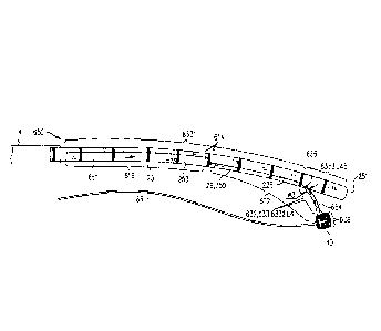
Fig. 1 shows a distal portion of an implant that comprises an annuloplasty
ring
structure 222 (i.e., an implant, e.g., an annuloplasty band) comprising a
flexible sleeve 26
(shown in the exploded view of Fig. 2). Sleeve 26 typically comprises a
braided fabric mesh,
e.g., comprising polyethylene terephthalate (such as Dacron (TM)). Sleeve 26
is typically
configured to be placed only partially around a cardiac valve annulus (i.e.,
to assume a C-
shape), and, once anchored in place, to be contracted so as to
circumferentially tighten the
valve annulus. Alternatively, the ring structure is configured to be placed
entirely around the
valve annulus.
Sleeve 26 has (a) a tubular lateral wall 253 that (i) circumscribes a central
longitudinal
axis of the sleeve, and (ii) defines the lumen of the sleeve, and (a) at least
one end wall 251
(e.g., a distal end wall) having a surface that is substantially transverse to
a lateral surface of
tubular wall 253. Typically, end wall 251 defines an end wall of annuloplasty
ring structure
222.
In order to tighten the annulus, annuloplasty ring structure 222 comprises a
flexible
elongated contraction member 226 that extends along sleeve 26. Elongated
contraction
member 226 comprises a wire, a ribbon, a rope, or a band, which typically
comprises a
flexible and/or superelastic material, e.g., nitinol, polyester, stainless
steel, or cobalt chrome.
33
Date Recue/Date Received 2022-11-15
For some applications, the wire comprises a radiopaque material. For some
applications,
contraction member 226 comprises a braided polyester suture (e.g., Ticron).
For some
applications, contraction member 226 is coated with polytetrafluoroethylene
(PTFE). For
some applications, contraction member 226 comprises a plurality of wires that
are intertwined
to form a rope structure.
Annuloplasty ring structure 222 further comprises an adjustment mechanism 40,
which facilitates contracting and expanding of annuloplasty ring structure 222
so as to
facilitate adjusting of a perimeter of the annulus and leaflets of the cardiac
valve. Adjustment
mechanism 40 is described in more detail hereinbelow. Adjustment mechanism 40
comprises
a rotatable structure (e.g., a spool, as described hereinbelow) that is
disposed within a housing
44. For some applications of the present invention, adjustment mechanism 40
comprises the
housing 44. Adjustment mechanism 40 may be surrounded by a braided mesh,
coupled (e.g.,
by being sutured or otherwise coupled) to the braided mesh of sleeve 26. For
some
applications, adjustment mechanism 40 is coupled to an outer, lateral surface
of sleeve 26.
Reference is now made to Fig. 2, which shows the concentric relationship
between
components of tubular system 10 (in an exploded view on the left side of Fig.
2). System 10
comprises an implant-delivery tool. Typically, system 10 comprises a first,
outer catheter 12
comprising a sheath configured for transluminal advancement through
vasculature of a
subject. For some applications of the present invention, outer catheter 12
comprises a sheath
configured for advancement through a femoral artery toward an interatrial
septum of a heart
of a subject. A distal end portion 112 of outer catheter 12 is configured to
pass through the
transatrial septum of the subject, and to be oriented in a desired spatial
orientation within the
left atrium. System 10 comprises a second catheter, or guide catheter 14,
comprising a distal
end portion 114 that is configured to pass through catheter 12 (i.e., a
primary lumen thereof),
to become disposed outside of a distal end 102 of the outer catheter, and to
be oriented in a
desired spatial orientation within the left atrium.
Distal end portion 112 of outer catheter 12 is steerable. That is, distal end
portion 112
is deflectable with respect to an immediately more proximal portion of
catheter 12 (e.g., by
using extracorporeal elements of system 10). Distal end portion 112 comprises
a pull ring 11
that is coupled to two or more pull wires 29a and 29b, that are disposed
within respective
secondary lumens within a lateral wall of catheter 12 (as shown in section A-A
of Fig. 2). As
34
Date Recue/Date Received 2022-11-15
shown in the exploded view, guide catheter 14 is configured to be
concentrically disposed
within the lumen of catheter 12. Distal end portion 114 of inner catheter 14
is steerable. That
is, distal end portion 114 is deflectable with respect to an immediately more
proximal portion
of catheter 14 (e.g., by using extracorporeal elements of system 10). Distal
end portion 114
comprises a pull ring 13 that is coupled to two or more pull wires 31a and
31b, that are
disposed within respective secondary lumens within a wall of catheter 14 (as
shown in
sections A-A and B-B).
Guide catheter 14 is steerable to a desired spatial orientation in order to
facilitate
advancing and implantation of an implant in a body cavity of the subject.
For applications in which system 10 is used to deliver an implant to the
mitral valve of
the subject, typically, outer catheter 12 is configured for initial
advancement through
vasculature of the subject until a distal end 102 of catheter 12 is positioned
in the left atrium.
The distal steerable end portion of catheter 12 is then steered such that
distal end 102 of
catheter 12 is positioned in a desired spatial orientation within the left
atrium. The steering
procedure is typically performed with the aid of imaging, such as fluoroscopy,
transesophageal echo, and/or echocardiography. Following the steering of the
distal end
portion of catheter 12, guide catheter 14 (which houses annuloplasty ring
structure 222) is
advanced through catheter 12 in order to facilitate delivery and implantation
of structure 222
along the annulus of the mitral valve. During the delivery, at least a portion
of steerable distal
end portion 114 is exposed from distal end 102 of catheter 12 and is thus free
for steering
toward the annulus of the mitral valve, as is described hereinbelow.
During delivery of sleeve 26 to the annulus of the cardiac valve, sleeve 26
and
mechanism 40 are disposed within a lumen of catheter 14 and are typically
aligned
longitudinally with a longitudinal axis of catheter 14. Mechanism 40 is
coupled to sleeve 26
in a manner that allows mechanism 40 to move (e.g., to translate) from a state
in which it is in
line with the longitudinal axis of catheter 14 (Fig. 2) to a state in which it
is disposed
alongside sleeve 26 (Fig. 1). For example, adjustment mechanism 40 may be
coupled to
sleeve 26 via one or more connectors 27, such as sutures, which provide
flexible and/or
articulated coupling. For some applications, the positioning of adjustment
mechanism 40
alongside a portion of sleeve 26 exposes a driving interface of the rotational
structure (e.g., a
Date Recue/Date Received 2022-11-15
driving interface 476, Fig. 16A), providing access to the interface for an
adjustment tool that
is subsequently guided toward adjustment mechanism 40 via a guide member 86.
Reference is again made to Fig. 1. A flexible, longitudinal guide member 86
(e.g., a
wire) is coupled to a portion of adjustment mechanism 40 (e.g., a portion of
the rotatable
structure, as described hereinbelow). Guide member 86 has a thickness of 0.35-
0.45 mm,
e.g., 0.4 mm. Guide member 86 is configured to facilitate guiding of an
adjustment tool via
guide member 86 and toward the rotatable structure of adjustment mechanism 40.
Typically,
the adjustment tool is configured to engage the rotatable structure of
adjustment mechanism
40 following implantation of sleeve 26 along the annulus of the cardiac valve.
Guide member
86 extends from adjustment mechanism 40, alongside a portion of distal end
portion 114 of
guide catheter 14, and into a secondary lumen in the wall of guide catheter 14
via an opening
in guide catheter 14. Guide member 86 extends through the secondary lumen of
guide
catheter 14 (as shown in sections A-A and B-B in Fig. 2) and has a proximal
end that is
accessible from outside the body of the subject. The secondary lumen in the
wall of guide
15 catheter 14 facilitates passage of guide member 86 through system 10
without interfering with
the other concentrically-disposed elongate tubular members that pass
concentrically through
the lumen of guide catheter 14.
Reference is again made to Fig. 2. In addition, system 10 comprises a
plurality of
anchors 32, typically between about 5 and about 20 anchors, such as about 10
or about 16
anchors. Each anchor 32 comprises a tissue-coupling element 60 (e.g., a
helical tissue-
coupling element), and a tool-engaging head 62 (e.g., a non-helically-shaped
portion), fixed to
one end of the tissue-coupling element. Only one anchor 32 is shown in Fig. 2
as being
reversibly coupled to a deployment element 38 of an anchor driver 36 of an
anchor
deployment manipulator 61. However, each of anchors 32 is reversibly couplable
to a
deployment element 38 of one or more anchor drivers 36. When sleeve 26 is
disposed along
the annulus of the cardiac valve, deployment manipulator 61 is configured to
advance within
a lumen of sleeve 26 and deploy each anchor 32 from within sleeve 26 through a
wall of
sleeve 26 and into cardiac tissue, thereby anchoring sleeve 26 around a
portion of the valve
annulus. The insertion of the anchors into the sleeve and deployment of the
anchors into
cardiac tissue is described in detail hereinbelow.
36
Date Recue/Date Received 2022-11-15
Typically, but not necessarily, anchors 32 comprise a biocompatible material
such as
stainless steel 316 LVM. For some applications, anchors 32 comprise nifinol.
For some
applications, anchors 32 are coated fully or partially with a non-conductive
material.
Deployment manipulator 61 comprises anchor driver 36 and deployment element
38.
For some applications, deployment manipulator 61 comprises channel 18.
As shown in the exploded view of Fig. 2, sleeve 26 is disposed within a lumen
of
guide catheter 14. Forces are applicable to a proximal end of sleeve 26 via a
reference-force
tube 19, a distal end of which is coupled to the proximal end of the sleeve.
As shown, an
implant-decoupling channel 18 is advanceable within a lumen of reference-force
tube 19 and
within a lumen of sleeve 26. As shown in the enlarged image of Fig. 1, a
distal end 17 of
implant-decoupling channel 18 is placeable in contact with an inner wall of
sleeve 26, e.g., at
a distal end thereof. The distal end portion of channel 18 may comprise a
radiopaque marker
1018. As shown, tube 19 and sleeve 26 are longitudinally and coaxially
disposed with respect
to each other.
For some applications, channel 18 is steerable.
Typically, manipulator 61 advances within channel 18. For some applications,
system
10 comprises a plurality of anchor drivers 36 of manipulator 61, each driver
36 being coupled
to a respective anchor 32. Each driver 36 is advanced within channel 18 in
order to advance
and implant anchor 32 in tissue. Following implantation of anchor 32, anchor
32 is
decoupled from driver 36, as described herein, and driver 36 is removed from
within channel
18. A subsequent anchor 32 is then advanced within channel 18 while coupled to
a driver 36
(e.g., a new driver).
As will be described hereinbelow, a first one of anchors 32 is configured to
be
deployed through end wall 251 of sleeve 26 into cardiac tissue, when sleeve 26
is positioned
along the annulus of the valve. Following the deployment of the first tissue
anchor, a distal
portion of sleeve 26 is slid distally off a portion of implant-decoupling
channel 18. In order
to decouple sleeve 26 distally from a portion of outer surface of channel 18,
(1) a proximal
force is applied to channel 18, while (2) reference-force tube 19 is
maintained in place in a
manner in which a distal end of tube 19 provides a reference force to sleeve
26, thereby
facilitating freeing of a successive portion of sleeve 26 from around channel
18. Channel 18
37
Date Recue/Date Received 2022-11-15
is then positioned at a successive location within the lumen of sleeve 26
while tube 19 and/or
catheter 14 is steered toward a successive location along the annulus of the
valve (as will be
described hereinbelow). Consequently, the successive portion of sleeve 26
provides a free
lumen for advancement of a successive anchor 32 and deployment of the anchor
through the
.. wall of the sleeve at the successive portion thereof. Such freeing of the
successive portion of
sleeve 26 creates a distance between successive anchors deployed from within
the lumen of
sleeve 26.
For some applications, sleeve 26 comprises a plurality of radiopaque markers
25,
which are positioned along the sleeve at respective longitudinal sites. The
markers may
provide an indication in a radiographic image (such as a fluoroscopy image) of
how much of
the sleeve has been deployed at any given point during an implantation
procedure, in order to
enable setting a desired distance between anchors 32 along the sleeve. For
some applications,
the markers comprise a radiopaque ink.
Typically, at least some (e.g., at least three, such as all) of the
longitudinal sites are
.. longitudinally spaced at a constant interval. Typically, the longitudinal
distance between the
distal edges of adjacent/consecutive markers, and/or the distance between the
proximal edges
of adjacent markers, is set equal to the desired distance between adjacent
anchors. For
example, the markers may comprise first, second, and third markers, which
first and second
markers are adjacent, and which second and third markers are adjacent, and the
distance
between the proximal and/or distal edges of the first and second markers equal
the
corresponding distance between the proximal and/or distal edges of the second
and third
markers. For example, the distance may be between 3 and 15 mm, such as 6 mm,
and the
longitudinal length of each marker may be between 0.1 and 14 mm, such as 2 mm.
(If, for
example, the distance were 6 mm and the length were 2 mm, the longitudinal
gaps between
adjacent markers would have lengths of 4 mm.)
Each anchor 32 is coupled to deployment element 38 of an anchor driver 36.
Anchor
driver 36 typically comprises an elongate and flexible shaft (which is
typically tubular)
having at least a flexible distal end portion. The elongate shaft of driver 36
extends within a
lumen of channel 18, through system 10 toward a proximal end of a proximal
handle portion
101 of system 10. The tube of anchor driver 36 provides a lumen for slidable
advancement
therethrough of an elongate rod 130. Rod 130 facilitates the locking and
unlocking of anchor
38
Date Recue/Date Received 2022-11-15
32 to deployment element 38. As shown in Section E-E of Fig. 2, a proximal end
of rod 130
is coupled to a component of an anchor-release mechanism 28 at a proximal end
of system 10.
Mechanism 28 comprises a housing 135 and a finger-engager 131 that is coupled
to the
proximal end of rod 130. Finger-engager 131 is coupled to a housing 135 via a
spring 133
(section E-E of Fig. 2). A proximal end of the tube of anchor driver 36 is
coupled to housing
135. The physician releases anchor 32 from deployment element 38 when finger-
engager 131
is pulled proximally, thereby pulling rod 130 proximally.
Proximal handle portion 101 is supported by a stand having support legs 91 and
a
handle-sliding track 90. Handle portion 101 comprises an outer-catheter handle
22, a guide-
catheter handle 24, an implant-manipulating handle 126, and anchor-release
mechanism 28.
Handle 22 is coupled to a proximal end of outer catheter 12. Handle 24 is
coupled to a
proximal portion of guide catheter 14. Handle 126 is coupled to a proximal
portion of
reference-force tube 19, and linear movement of handle 126 with respect to
handle 24 moves
reference-force tube 19 (and thereby typically structure 222) through catheter
14. As
described hereinabove, housing 135 of anchor-release mechanism 28 is coupled
to a proximal
portion of the tube of anchor driver 36. The relative positioning of each of
the concentrically-
disposed components of system 10 is shown in the exploded view and sections A-
A, B-B, C-
C, and D-D of Fig. 2.
The stand supporting proximal handle portion 101 may be moved distally and
.. proximally to control a position of the entire multi-component system 10,
particularly so as to
adjust a distance of distal end 102 of catheter 12 from the interatrial
septum. Handle 22
comprises a steering knob 210 that is coupled to steering wires 29a and 29b
disposed within
respective secondary lumens in the wall of outer catheter 12. Rotation of knob
210 adjusts a
degree of tension of wires 29a and 29b which, in turn, apply a force to pull
ring 11 at the
distal end portion of outer catheter 12. Such force steers the distal end
portion of catheter 12
within the atrium of the heart of the subject in a manner in which the distal
end portion of
catheter 12 is steered in a first steering plane that is typically parallel
with the plane of the
annulus of the valve (e.g., in a direction from the interatrial septum toward
surrounding walls
of the atrium). For some applications of the present invention, the distal end
portion of
catheter 12 may be pre-shaped so as to point downward toward the valve. For
other
applications, the distal end portion of catheter 12 may be pulled to assume an
orientation in
39
Date Recue/Date Received 2022-11-15
which the distal end portion points downward toward the valve. For yet other
applications of
the present invention, the distal end portion of catheter 12 is not made to
point downward
toward the valve.
Handle 24 is coupled to track 90 via a first mount 92. Mount 92 is slidable
proximally
and distally along track 90 in order to control an axial position of guide
catheter 14 with
respect to outer catheter 12. Mount 92 is slidable via a control knob 216. For
example,
control knob 216 of mount 92 controls the proximal and distal axial movement
of the distal
steerable portion of guide catheter 14 with respect to distal end 102 of outer
catheter 12.
Handle 24 comprises a steering knob 214 that is coupled to steering wires 31a
and 31b
disposed within respective secondary lumens in the wall of guide catheter 14.
Rotation of
knob 214 adjusts a degree of tension of wires 31a and 31b which, in turn,
apply a force to pull
ring 13 at the distal end portion of guide catheter 14. Such force steers the
distal end portion
of catheter 14 in a second steering plane within the atrium of the heart of
the subject, typically
downward and toward the annulus of the cardiac valve. Typically, as described
hereinbelow,
the distal end portion of guide catheter 14 is steered in the second plane
that is substantially
perpendicular with respect to the first plane in which the distal end portion
of outer catheter
12 is steered.
The combined steering of the respective distal end portions of catheters 12
and 14
directs sleeve 26 down toward the annulus (e.g., via the steering of the
distal end portion of
catheter 14) and along the perimeter of annulus (e.g., from the posterior
section of the valve to
the anterior section of the valve, and vice versa), via the steering of the
distal end portion of
catheter 12.
For some applications, handle 22 may be tilted by the operating physician, in
order to
further adjust a position of the distal end of catheter 12.
Handle 126 is slidably coupled to track 90 via a second mount 93. Mount 93 is
slidable proximally and distally along track 90, in order to control an axial
position of
reference-force tube 19 and at least a proximal portion of sleeve 26 with
respect to guide
catheter 14. For some applications, mount 93 comprises a control knob 95. For
some such
applications, control knob reversibly locks mount 93 to track 90, thereby
reversibly inhibiting
sliding of the mount along the track. Alternatively or additionally, turning
of control knob 95
may cause sliding of mount 93 along track 90 (e.g., acting like a rack and
pinion). For some
Date Recue/Date Received 2022-11-15
applications, friction between (i) reference-force tube 19 and (ii) catheter
14 and/or handle 24
reduces a likelihood of inadvertent sliding of tube 19 through catheter 14,
and thereby
obviates the need for locking of mount 93 to track 90. Taken together with the
steering of the
distal end portion of guide catheter 14, such movement of tube 19 and at least
the proximal
portion sleeve 26 moves the proximal portion of sleeve 26 toward a desired
portion of tissue
of the annulus of the valve during deployment of anchors 32 from within the
lumen of sleeve
26, as is described hereinbelow.
As is described hereinabove, in order to decouple sleeve 26 from a portion of
an outer
surface of channel 18, (1) channel 18 is pulled proximally, while (2)
reference-force tube 19
is maintained in place. A proximal end of channel 18 is coupled to a knob 94
which adjusts
an axial position of channel 18 proximally and distally with respect to
reference-force tube 19
and sleeve 26.
Typically, handle portion 101 comprises a release-decision-facilitation member
127,
such as a latch or button, that automatically engages when a given length of
sleeve 26 has
advanced off channel 18 (e.g., when channel 18 is at a given position with
respect to tube 19);
typically just before sleeve 26 becomes completely decoupled from channel 18.
Engagement
of member 127 inhibits proximal movement of channel 18 with respect to tube
19, thereby
reducing a likelihood of (e.g., preventing) inadvertent release of sleeve 26.
In order to release
sleeve 26 (e.g., to decouple channel 18 from the sleeve), the operating
physician must
disengage member 127, such as by pushing the button, before continuing to
withdraw channel
18 proximally. Typically, when engaged, member 127 also inhibits distal
movement of
channel 18 with respect to tube 19.
Handle portion 101 (comprising handles 22, 24, and 126 and anchor-release
mechanism 28) has a length Li of between 65 and 85 cm, e.g., 76 cm. Typically,
as shown, a
majority of the body portion of outer-catheter handle 22 is disposed at a non-
zero angle with
respect to a longitudinal axis 7 of the multiple components of system 10. The
steering
mechanism provided by handle 22 in order to steer the distal end portion of
catheter 12 is
disposed within the portion of handle 22 that is disposed at the non-zero
angle with respect to
axis 7. Handle 22 comprises an in-line tubular portion which is longitudinally
disposed in-
line along axis 7 and coaxially with respect to handles 24 and 126 and release
mechanism 28.
The in-line tubular portion is shaped so as to define a lumen for inserting
guide catheter 14
41
Date Recue/Date Received 2022-11-15
therethrough and subsequently into the lumen of outer catheter 12. The in-line
tubular portion
has a length L24 of between 7 and 11 cm, e.g., 7 cm. Such spatial orientation
of the majority
of handle 22 at an angle with respect to axis 7 reduces an overall functional
length of handle
portion 101.
Typically, but not necessarily, a guidewire 2244 extends alongside sleeve 26
to
facilitate positioning of sleeve 26 along the annulus.
Reference is made to Figs. 3A-G, and 4A-B, which are schematic illustrations
of steps
in the implantation of an annuloplasty ring structure to repair a mitral
valve, in accordance
with some applications of the invention. This procedure is one exemplary
procedure that can
be performed using system 10.
Anchor deployment manipulator 61 is advanced into a lumen of sleeve 26, and,
from
within the lumen, deploys the anchors through a wall of the sleeve and into
cardiac tissue,
thereby anchoring the sleeve around a portion of the valve annulus. For some
application,
annuloplasty ring structure 222 is implemented using techniques described in
US Application
12/437,103, filed May 7, 2009 which published as US 2010/0286767, and/or US
Application
12/689,635, filed January 19, 2010 which published as US 2010/0280604. As
described
hereinabove, annuloplasty ring structure 222 comprises adjustment mechanism
40. The
adjustment mechanism comprises a rotatable structure, such as a spool,
arranged such that
rotation of the rotatable structure contracts the implant structure. The
implant further
comprises a longitudinal member, such as a wire, which is coupled to the
adjustment
mechanism. An adjustment tool is provided for rotating the rotatable
structure. The tool is
configured to be guided along (e.g., over, alongside, or through) the
longitudinal member, to
engage the rotatable structure, and to rotate the rotatable structure in
response to a rotational
force applied to the tool.
The procedure typically begins by advancing a semi-rigid guidewire into a
right
atrium 220 of the subject. The procedure is typically performed with the aid
of imaging, such
as fluoroscopy, trans esophageal echo, and/or echocardiography.
The guidewire provides a guide for the subsequent advancement of outer
catheter 12
therealong and into the right atrium. Once a distal portion of catheter 12 has
entered the right
atrium, the guidewire is retracted from the subject's body. Catheter 12
typically comprises a
42
Date Recue/Date Received 2022-11-15
14-24 F sheath, although the size may be selected as appropriate for a given
subject. Catheter
12 is advanced through vasculature into the right atrium using a suitable
point of origin
typically determined for a given subject. For example:
= catheter 12 may be introduced into the femoral vein of the subject,
through an
inferior vena cava 223, into right atrium 220, and into a left atrium 224
transseptally, typically
through the fossa ovalis;
= catheter 12 may be introduced into the basilic vein, through the
subclavian
vein to the superior vena cava, into right atrium 220, and into left atrium
224 transseptally,
typically through the fossa ovalis; or
= catheter 12 may be introduced into the external jugular vein, through
the
subclavian vein to the superior vena cava, into right atrium 220, and into
left atrium 224
transseptally, typically through the fossa ovalis.
For some applications of the present invention, catheter 12 is advanced
through
inferior vena cava 223 of the subject (as shown) and into right atrium 220
using a suitable
point of origin typically determined for a given subject.
Catheter 12 is advanced distally until the sheath reaches the interatrial
septum, and the
guidewire is withdrawn.
A resilient needle and a dilator are advanced through catheter 12 and into the
heart. In
order to advance catheter 12 transseptally into left atrium 224, the dilator
is advanced to the
septum, and the needle is pushed from within the dilator and is allowed to
puncture the
septum to create an opening that facilitates passage of the dilator and
subsequently catheter 12
therethrough and into left atrium 224. The dilator is passed through the hole
in the septum
created by the needle. Typically, the dilator is shaped to define a hollow
shaft for passage
along the needle, and the hollow shaft is shaped to define a tapered distal
end. This tapered
distal end is first advanced through the hole created by the needle. The hole
is enlarged when
the gradually increasing diameter of the distal end of the dilator is pushed
through the hole in
the septum. A distal-most end 102 of catheter 12 is tapered so as to
facilitate passage of at
least part of distal portion 112 of catheter 12 through the opening in the
septum.
The advancement of catheter 12 through the septum and into the left atrium is
followed by the extraction of the dilator and the needle from within catheter
12. Once distal
43
Date Recue/Date Received 2022-11-15
portion 112 of catheter 12 is disposed within atrium 224, portion 112 is
steered (i.e.,
deflected) in a first steering plane, typically parallel to a plane of the
annulus of mitral valve
230. The steering of the distal portion of catheter 12 is performed via
steering knob 210 of
handle 22 in handle portion 101 (in Fig. 2).
As shown in Fig. 3A, catheter 14, containing annuloplasty ring structure 222
(with a
distal portion of channel 18 disposed within sleeve 26 thereof), is advanced
through catheter
12 into left atrium 224. For some applications, soon before implantation
(e.g., within the
operating theater or in an adjacent room) the distal portion of channel 18 is
loaded into sleeve
26, and structure 222 is loaded into catheter 14. Distal end portion 114 of
catheter 14 extends
beyond distal end 102 of catheter 12. Distal end portion 114 is then steered
(i.e., deflected) in
a second steering plane, typically perpendicular with respect to the steering
plane of catheter
12, and further typically toward the annulus of valve 230. The steering of the
distal portion of
catheter 14 is performed via steering knob 214 of handle 24 in handle portion
101 (in Fig. 2).
Fig. 3A shows annuloplasty ring structure 222, comprising sleeve 26 and
adjustment
mechanism 40, having been advanced, via catheter 14, to a mitral valve 230. As
shown in
Fig. 3A, and as described hereinabove, during advancement of structure 222,
adjustment
mechanism 40 is disposed distal to (i.e., in front of) sleeve 26. In this way,
adjustment
mechanism 40 is disposed on the longitudinal axis of sleeve 26 (e.g.,
collinearly with the
sleeve), so as to advantageously maintain a small cross-sectional diameter of
the implant for
transluminal delivery. A proximal end of connector 27 is disposed proximally
to mechanism
40 (e.g., by being fixed to a portion of sleeve 26 proximal to mechanism 40 or
by being
accessible outside the body of the subject). A distal end of connector 27 is
coupled (e.g., by
being fixedly coupled by a knot or other mechanical coupling) to mechanism 40.
Guide
member 86, described hereinabove, typically extends distally from catheter 14,
between end
wall 251 of sleeve 26 and adjustment mechanism 40, and there is coupled to the
adjustment
mechanism. For some applications it is advantageous to (1) advance the
structure to the
mitral valve while mechanism 40 is disposed on the longitudinal axis of sleeve
26 (e.g.,
collinearly with the sleeve), so as to maintain a small cross-sectional
diameter of the structure
for transluminal delivery; and (2) to subsequently move mechanism 40 away from
the
longitudinal axis, e.g., so as to allow end wall 251 of the sleeve to be
placed against the
annulus, and/or so as to allow an anchor to be driven through the end wall of
the sleeve.
44
Date Recue/Date Received 2022-11-15
Connectors 27 facilitate this technique by making mechanism 40 flexibly and/or
articulatably
coupled to sleeve 26. For some applications, connectors 27 are tensioned or
relaxed to move
mechanism 40 with respect to sleeve 26 to reposition mechanism 40. For some
applications,
guide member 86 is tensioned or relaxed in order to reposition mechanism 40.
Subsequent to exposure of at least adjustment mechanism 40 (and typically at
least
end wall 251 of sleeve 26) from catheter 14, the adjustment mechanism is moved
away from
end wall 251. Typically, this is achieved by guide member 86 being moved
proximally such
that mechanism 40 moves (e.g., translates, deflects, and/or rotates) away from
the longitudinal
axis of the sleeve, typically to become disposed laterally from sleeve 26.
Fig. 3B shows
mechanism 40 having translated to such a position. The movement of mechanism
40 away
from end wall 251 of sleeve 26 advantageously allows end wall 251 of sleeve 26
to be placed
against an atrial surface of an annulus 240, and a first one of anchors 32 to
be driven through
end wall 251 of the sleeve and into the annulus (Fig. 3C).
As shown in Fig. 3C, end wall 251 of sleeve 26 is positioned in a vicinity of
a left
fibrous trigone 242 of an annulus 240 of mitral valve 230. (It is noted that
for clarity of
illustration, distal end wall 251 of sleeve 26 is shown schematically in the
cross-sectional
view of the heart, although left trigone 242 is in reality not located in the
shown cross-
sectional plane, but rather out of the page closer to the viewer.)
Alternatively, the distal end
of sleeve 26 is positioned in a vicinity of a right fibrous trigone 244 of the
mitral valve
(configuration not shown). Further alternatively, the distal end of the sleeve
is not positioned
in the vicinity of either of the trigones, but is instead positioned elsewhere
in a vicinity of the
mitral valve, such as in a vicinity of the anterior or posterior commissure.
Once positioned at
the desired site near the selected trigone, deployment manipulator 61 deploys
the first one of
anchors 32 through the wall of sleeve 26 (by penetrating and passing through
the wall of the
sleeve (i) in a direction parallel to a central longitudinal axis of
deployment manipulator 61,
or anchor driver 36, through the distal end of channel 18, and/or (ii)
parallel to a central
longitudinal axis of tissue-coupling element 60 of anchor 32) into cardiac
tissue near the
trigone. Following the deployment of anchor 32 in the cardiac tissue,
deployment element 38
is decoupled from anchor 32.
Anchors 32 are typically deployed from a distal end of manipulator 61 while
the distal
end is positioned such that a central longitudinal axis through the distal end
of manipulator 61
Date Recue/Date Received 2022-11-15
forms an angle with a surface of the cardiac tissue of between about 20 and 90
degrees, e.g.,
between 45 and 90 degrees, such as between about 75 and 90 degrees, such as
about 90
degrees. Typically, anchors 32 are deployed from the distal end of manipulator
61 into the
atrial surface of the cardiac tissue in a direction parallel to the central
longitudinal axis
through the distal end of manipulator 61. Such an angle is typically provided
and/or
maintained by channel 18 being more rigid than sleeve 26. Distal end 17 of
channel 18 is
typically brought close to the surface of the cardiac tissue (and the wall of
sleeve 26 that is
disposed against the surface of the cardiac tissue), such that little of each
anchor 32 is exposed
from channel 18 before penetrating the sleeve and the tissue. For example,
distal end 17 of
channel 18 may be placed (e.g., pushed) against the wall of the sleeve,
sandwiching the sleeve
against the cardiac tissue.
For some applications, such placement of distal end 17 of channel 18 against
the
cardiac tissue (via the wall of the sleeve), stabilizes the distal end during
deployment and
anchoring of each anchor 32, and thereby facilitates anchoring. For some
applications,
pushing of distal end 17 against the cardiac tissue (via the wall of the
sleeve) temporarily
deforms the cardiac tissue at the site of contact. This defoimation may
facilitate identification
of the site of contact using imaging techniques (e.g., by identifying a
deformation in the
border between cardiac tissue and blood), and thereby may facilitate correct
positioning of the
anchor.
That is, typically the entire circular surface of distal end 17 of channel 18
is disposed
in contact with the wall of sleeve 26 that is disposed against the surface of
the cardiac tissue.
As shown, distal end 17 is the lower-most circular tip of channel 18 and
defines a distal
opening of channel 18. In the configuration in which channel 18 is positioned
in order to
sandwich the portion of sleeve 26 against annulus 240, the distal end 17 is
disposed in parallel
with a planar surface 255 of the tissue of the annulus.
As shown in Fig. 3C, end wall 251 aligns against the tissue of annulus 240 in
a
manner in which a surface of end wall 251 is disposed in parallel with a
planar surface of the
tissue of annulus 240. Additionally, distal end 17 of implant-decoupling
channel 18 flattens
end wall 251 against the tissue of annulus 240 in a manner in which channel 18
sandwiches
end wall 251 between (1) distal end 17 of the channel, and (2) the portion of
the tissue of
annulus 240 at the planar surface into which a first one of anchors 32 is
implanted. In such a
46
Date Recue/Date Received 2022-11-15
manner, end wall 251 lies flat against the tissue of annulus 240 in parallel
with the planar
surface, while at least a distal portion of lateral wall 253 is disposed
substantially
perpendicularly with respect to the portion of the tissue of annulus 240 at
the planar surface
into which the first one of anchors 32 is implanted.
As shown, anchor 32 is implanted using channel 18 and manipulator 61 contained
within sleeve 26 of annuloplasty structure 222 while at least a portion of
annuloplasty
structure 222 (e.g., a proximal portion) is contained within surrounding
catheter 14.
Reference is now made to Figs. 2, 3C-D, and 4A-B. Following the deployment of
the
first tissue anchor, a distal portion of sleeve 26 is decoupled from a portion
of implant-
decoupling channel 18. In order to decouple the portion of sleeve 26 from
outer surface of
channel 18, (1) channel 18 is pulled proximally, while (2) reference-force
tube 19 is
maintained in place in a manner in which a distal end of tube 19 provides a
reference force to
sleeve 26 in order to facilitate freeing of a successive portion of sleeve 26
from around
channel 18. In order to decouple sleeve 26 from the outer surface of channel
18, (1) channel
18 is pulled proximally, while (2) reference-force tube 19 is maintained in
place. An
indicator 2120 on handle 126 provides an indication of how much channel 18 is
withdrawn
from within sleeve 26 (i.e., how much the delivery tool is decoupled from
sleeve 26, and how
much the sleeve has advanced off channel 18 and against tissue). A proximal
end of channel
18 is coupled to a knob 94 (Fig. 2) which adjusts an axial position of channel
18 proximally
and distally with respect to reference-force tube 19 and sleeve 26. As shown
in Fig. 3D, once
the successive portion of sleeve 26 is freed, deployment manipulator 61 is
repositioned along
annulus 240 to another site selected for deployment of a second one of anchors
32.
Figs. 4A and 4B are schematic illustrations that show steps between the state
shown in
Fig. 3C and the state shown in Fig. 3D, in accordance with respective
applications of the
invention. Step C of each of Figs. 4A and 4B shows a state that is generally
equivalent to the
state shown in Fig. 3D.
For some applications, and as shown in Fig. 4A, anchor driver 36 is decoupled
from
anchor 32 and is retracted through channel 18 prior to retracting channel 18
through sleeve 26
and repositioning channel 18.
47
Date Recue/Date Received 2022-11-15
For some applications, and as shown in Fig. 4B, anchor driver 36 remains
coupled to
anchor 32 during the retraction of channel 18 though sleeve 26. For some such
applications,
anchor driver 36 provides a reference force (e.g., a distally-directed
reference force) that
holds in place anchor 32 and the anchored portion of sleeve 26 while channel
18 is retracted,
e.g., reducing a pulling force on anchor 32.
A method is therefore described, comprising: (1) percutaneously advancing
toward a
tissue of a subject structure 222, while a distal portion of channel 18 is
disposed within the
lumen defined by sleeve 26, such that a distal opening of the channel is
disposed at a first
portion of the sleeve; (2) anchoring the first portion of the sleeve to the
tissue by using anchor
driver 36 to drive tissue-coupling element 60 of a first anchor 32 through the
distal opening of
the channel, through the sleeve, and into the tissue; (3) subsequently, while
providing a
distally-directed reference force to the first anchor 32 via driver 36,
proximally withdrawing
the distal portion of channel 18 such that the distal opening of the channel
is disposed at a
second portion of the sleeve; (4) subsequently, proximally withdrawing driver
36 through the
channel; and (5) subsequently, anchoring the second portion of the sleeve to
the tissue by
driving tissue-coupling element 60 of a second anchor 32 through the distal
opening of the
channel, through sleeve 26, and into the tissue.
Reference is now made to Figs. 2, 3D, and 4A-B. Such repositioning of
manipulator
61 is accomplished by performing one or more of the following:
(1) steering distal end portion 112 of catheter 12 (e.g., by steering knob 210
of handle
22) in the first steering plane, in a manner that bends portion 112,
(2) steering distal end portion 114 of catheter 14 (e.g., by steering knob 214
of handle
24) in the second steering plane, in a manner that portion 112,
(3) axially moving catheter 14 with respect to catheter 12 via knob 216,
(4) axially moving the stand supporting handles 22 and 24 to move both
catheters 12
and 14,
(5) moving tube 19 and sleeve 26 axially by sliding mount 93 along track 90,
(6) by moving channel 18 relative to tube 19 by actuating knob 94.
48
Date Recue/Date Received 2022-11-15
Typically, the first tissue anchor is deployed most distally in the sleeve
(generally at or
within a few millimeters of the distal tip of the sleeve), and each subsequent
anchor is
deployed more proximally, such that the sleeve is gradually decoupled from
channel 18 of
deployment manipulator 61 in a distal direction during the anchoring procedure
(i.e., channel
18 is withdrawn from within sleeve 26, and handle 126 is moved distally so as
to retract the
tool to make the successive proximal portion sleeve 26 ready for implantation
of a subsequent
anchor). The already-deployed first one of anchors 32 holds the anchored end
of sleeve 26 in
place, so that the sleeve is drawn from the site of the first tissue anchor
towards the site of the
second tissue anchor. As sleeve 26 is drawn and decoupled from channel 18, a
distal portion
257 of sleeve 26 (i.e., the portion of the sleeve that is proximal to end wall
251) is positioned
in a vicinity of tissue of annulus 240.
Fig. 3D shows distal portion 257 of sleeve 26 (i.e., the portion of the sleeve
that is
proximal to end wall 251) having been decoupled from a portion of channel 18
by retracting
channel 18 proximally. Depending on the tension applied between the first and
second tissue
anchor sites, the portion of sleeve 26 therebetween may remain tubular in
shape, or may
become flattened.
Fig. 3E shows a second tissue anchor 32 (shown as a second tissue anchor 32b)
being
deployed through a portion of lateral wall 253 of sleeve 26. The first one of
anchors 32
deployed through end wall 251 is labeled as anchor 32a. Deployment manipulator
61 deploys
the second tissue anchor by driving the anchor to penetrate and pass through
the wall of
sleeve 26 into cardiac tissue at the second site.
As shown, anchor 32b is implanted using channel 18 and manipulator 61
contained
within sleeve 26 of annuloplasty structure 222 while at least a portion of
annuloplasty
structure 222 (e.g., a proximal portion) is contained within surrounding
catheter 14.
As described hereinabove, anchors 32a and 32b are each deployed from a distal
end of
manipulator 61 while the distal end is positioned such that a central
longitudinal axis through
the distal end of manipulator 61 forms an angle with a surface of the cardiac
tissue of between
about 20 and 90 degrees, e.g., between 45 and 90 degrees, such as between
about 75 and 90
degrees, such as about 90 degrees. Typically, anchors 32 are deployed from the
distal end of
manipulator 61 into the atrial surface of the cardiac tissue in a direction
parallel to the central
longitudinal axis through the distal end of manipulator 61. Such an angle is
typically
49
Date Recue/Date Received 2022-11-15
provided and/or maintained by channel 18 being more rigid than sleeve 26.
Distal end 17 of
channel 18 is typically brought close to the surface of the cardiac tissue
(and the wall of
sleeve 26 that is disposed against the surface of the cardiac tissue), such
that little of anchor
32b is exposed from channel 18 before penetrating the sleeve and the tissue.
For example,
.. distal end 17 of channel 18 may be placed (e.g., pushed) against the wall
of the sleeve,
sandwiching the sleeve against the cardiac tissue.
As shown in Figs. 3D-E, a portion of the lateral wall of sleeve 26 aligns
against the
tissue of in a manner in which a surface of the portion of the lateral wall is
disposed in
parallel with the planar surface of the tissue. Additionally, distal end 17 of
channel 18
flattens the portion of the lateral wall against the tissue of annulus 240 in
a manner in which
channel 18 sandwiches the portion of the lateral wall between (1) distal end
17 of implant-
decoupling channel, and (2) the portion of the tissue of annulus 240 at the
planar surface into
which second tissue anchor 32h is implanted. In such a manner, the portion of
the lateral wall
being anchored lies flat against the tissue of annulus 240 (parallel with the
planar surface
thereof), while the remaining portion of the tubular lateral wall is disposed
substantially
perpendicularly with respect to the portion of the tissue into which second
tissue anchor 32b
is implanted.
It is to be noted that first and second tissue anchors 32a and 32b extend in a
substantially same direction and into a common, substantially planar surface
of a valve
.. annulus, despite that first tissue anchor 32a is deployed through end wall
251 of sleeve 26,
and tissue anchor 32b is deployed through lateral wall 253 of the sleeve. For
some
applications, anchors 32a and 32b are disposed with respect to each other at
an angle of
between 0 and 45 degrees, e.g., between 0 and 30 degrees, e.g., between 0 and
20 degrees.
For some applications, a maximum distance L10 between first tissue anchor 32a
and a
point of anchoring of second tissue anchor 32b is provided by the length of
sleeve 26 that has
been decoupled from the portion of channel 18 (e.g., by the distance that
channel 18 has been
retracted from sleeve 26, e.g., between 3 and 15 mm, e.g., 8 mm). That is, for
some
applications, second tissue anchor 32b may be placed anywhere within a circle
having a
radius that equals L10, centered on the first tissue anchor (e.g., indicated
by arc 1928). For
some such applications, sleeve 26 thereby serves as a constraining member
(e.g., a tether) that
Date Recue/Date Received 2022-11-15
is used to facilitate positioning of second tissue anchor 32b. Distance L10 is
thereby set by
the operating physician retracting channel 18 from sleeve 26 by a particular
distance.
Fig. 3F shows the entire length of sleeve 26 having been anchored, via a
plurality of
anchors 32, to annulus 240, as described hereinabove. The deployment
manipulator (i.e.,
deployment manipulator 61 described herein but not shown in Fig. 3F) is
repositioned along
the annulus to additional sites, at which respective anchors are deployed,
until the last anchor
is deployed in a vicinity of right fibrous trigone 244 (or left fibrous
trigone 242 if the
anchoring began at the right trigone). Alternatively, the last anchor is not
deployed in the
vicinity of a trigone, but is instead deployed elsewhere in a vicinity of the
mitral valve, such
as in a vicinity of the anterior or posterior commissure. Then, system 10 is
removed, leaving
behind annuloplasty ring structure 222, and guide member 86 coupled thereto.
Fig. 3G shows an adjustment tool 87 being threaded over and advanced along
guide
member 86. Adjustment tool 87 typically comprises a rotation tool, and is
configured to
actuate (e.g., rotate) adjustment mechanism 40, so as to tension contraction
member 226, and
thereby contract sleeve 26, as described hereinabove. Typically, adjustment
mechanism 40
comprises a housing which houses a spool, i.e., a rotatable structure, to
which a first end of
contraction member 226 is coupled. Typically, the spool is configured to
adjust a perimeter of
annuloplasty ring structure 222 by adjusting a degree of tension of
contraction member 226
that is coupled at a first portion of member 226 to the spool. The contraction
member 226
extends along sleeve 26 and a second portion of contraction member 226 (i.e.,
a free end
portion) is coupled to a portion of sleeve 26 such that upon rotation of the
spool in a first
rotational direction, the contraction member is pulled toward adjustment
mechanism 40 in
order to contract annuloplasty ring structure 222. It is to be noted that the
contraction of
structure 222 is reversible. That is, rotating the spool in a second
rotational direction that
opposes the first rotational direction used to contract the annuloplasty
structure, unwinds a
portion of contraction member 226 from around the spool. Unwinding the portion
of
contraction member 226 from around the spool thus feeds the portion of
contraction member
226 back into sleeve 26 of structure 222, thereby slackening the remaining
portion of
contraction member 226 that is disposed within the sleeve. Responsively, the
annuloplasty
structure gradually relaxes and expands (i.e., with respect to its contracted
state prior to the
unwinding).
51
Date Recue/Date Received 2022-11-15
Adjustment mechanism 40 typically comprises a locking mechanism that prevents
actuation of the adjustment mechanism (e.g., rotation of the spool) after
contraction member
226 has been tightened. For example, locking techniques may be used that are
described with
reference to Fig. 4 of US Patent 8,241,351 to Cabin.
Tool 87 and is used to rotate the spool of adjustment mechanism 40 in order to
tighten
structure 222 by adjusting a degree of tension of contraction member 226 (not
shown in Fig.
3G). Once the desired level of adjustment of structure 222 is achieved, e.g.,
by monitoring
the extent of regurgitation of the valve using echocardiography (such as
Doppler
echocardiography and/or fluoroscopy), adjustment tool 87 and guide member 86
are removed
from the heart. For some applications, a distal portion of guide member 86 may
be left within
the heart of the subject and the proximal end may be accessible outside the
body, e.g., using a
port. For such applications, adjustment mechanism 40 may be accessed at a
later stage
following initial implantation and adjustment of ring structure 222 (e.g., as
described with
reference to Figs. 16A-18K).
Alternatively, annuloplasty ring structure 222 is implanted by right or left
thoracotomy, mutatis mutandis.
Reference is again made to Figs. 3A-G, and is also made to Figs. 5A-B, which
are
schematic illustrations of techniques for use with an excess portion 261 of
sleeve 26, in
accordance with some applications of the invention. For some applications of
the present
invention, following implantation of sleeve 26 along the annulus, an excess
portion 261 of
sleeve 26 may be present at the proximal portion of sleeve. For some such
applications,
excess portion 261 may be anchored to an atrial surface, such as an atrial
wall, using anchors
delivered via the lumen of sleeve 26, as described hereinabove, mutatis
mutandis, as shown in
Fig. 5A.
Alternatively or additionally, excess portion 261 may be anchored to the
atrial surface
using anchors driven from outside of sleeve 26, laterally through the sleeve,
such that each
anchor passes through the lateral wall of the sleeve twice (e.g., on opposite
sides of the lumen
of the sleeve), as shown in Fig. 5B. Therefore a method is described
comprising: (1)
percutaneously advancing toward a tissue of a subject structure 222, while a
distal portion of
a channel 18 is disposed within the lumen of sleeve 26, such that a distal
opening of the
channel is disposed at a first portion of the sleeve; (2) anchoring the first
portion of the sleeve
52
Date Recue/Date Received 2022-11-15
to a first tissue site by using anchor driver 36 to drive tissue-coupling
element 60 of a first
anchor 32 through the distal opening of the channel, through the first portion
of the sleeve
(e.g., through end wall 251 or lateral wall 253), and into the first tissue
site; (3) pressing a
second portion of the sleeve (i.e., excess portion 261) against a second
tissue site; and (4)
anchoring the second portion of the sleeve to the second tissue site by
driving tissue-coupling
element 60 of a second anchor 32 from outside the lumen, through opposing
sides of the
lateral wall at the second portion of the sleeve, and into the second tissue
site.
For some applications, when the second portion (i.e., the excess portion) of
the sleeve
is pressed against the second tissue site (e.g., the atrial wall) the opposing
sides of lateral wall
253 at the second portion of the sleeve contact each other.
Reference is again made to Figs. 3A-G. For anatomical reasons, a transluminal
(e.g.,
transfemoral) approach to the mitral valve via transseptal puncture typically
provides access
more directly and/or easily to the region of the anterior commissure (e.g.,
including left
fibrous trigone 242) than to the region of the posterior commissure (e.g.,
including right
fibrous trigone 244). It may therefore be advantageous to position and anchor
distal end wall
251 of sleeve 26 in the vicinity of the left fibrous trigone; the positioning
of the first point of
anchoring of structure 222 may be more difficult than the positioning of
subsequent points of
anchoring (e.g., due to guidance provided by sleeve 26; Fig. 3E). Due to this
same reason of
accessibility, it may also be advantageous to deliver adjustment tool 87 to
the region of the
anterior commissure (as shown in Fig. 3G).
System 10 (e.g., structure 222 thereof) is configured to facilitate
exploitation of these
two advantages: By adjustment mechanism 40 being disposed at a distal end of
sleeve 26, and
being movable away from the longitudinal axis of the sleeve, (1) the first
tissue anchor may
be driven through end wall 251 into the region of the anterior commissure,
despite the
adjustment mechanism having previously been obstructively positioned, and (2)
the
adjustment tool may be delivered to the region of the anterior commissure
because the
adjustment mechanism is disposed in that region.
Reference is now further made to Figs. 6A-B and 7A-B, which are schematic
illustrations of steering of catheters 12 and 14, in accordance with
respective applications of
the invention. As described hereinabove, distal end portion 112 of catheter 12
is steerable in
a first steering plane, and distal end portion 114 of catheter 14 is steerable
in a second steering
53
Date Recue/Date Received 2022-11-15
plane, typically perpendicular to the first steering plane. As also described
hereinabove,
typically (i) catheter 12 is steered in a steering plane that is parallel with
the plane of the
annulus of the valve (e.g., as shown in Fig. 6A), and (ii) catheter 14 is
steered downward and
toward the annulus of the valve (e.g., as shown in Fig. 6B), such that an
angle alpha_l is
formed between (i) plane 241 of the annulus, and (ii) an exit direction 105
from distal end 104
of catheter 14. (Exit direction 105 is typically collinear with the central
longitudinal axis
through the distal end of manipulator 61, and/or the central longitudinal axis
of tissue-
coupling element 60 of anchor 32.) Typically, angle alpha _i is greater than
an angle alpha _3
formed between plane 241 and an exit direction 103 from distal end 102 of
catheter 12.
Alternatively, catheter 12 may be steered in a different steering plane, such
that
catheter 14 may approach the tissue from a different angle, such that an
anchor 32 may
penetrate the tissue at a different angle of attack. For example, and as shown
in Fig. 7,
catheter 12 may be steered downward and toward the annulus of the valve, and
catheter 14
may be steered such that an angle alpha _2 (formed between the plane of the
annulus and the
central longitudinal axis through the distal end of manipulator 61) is smaller
than angle
alpha 1. For such applications, angle alpha_2 is typically smaller than an
angle alpha _4
formed between plane 241 and exit direction 103.
Reference is now made to Figs. 8A-B, 9, 10A-C, 11, and 12A-B, which are
schematic
illustrations of tissue anchors, and the use of the tissue anchors for
implantation of structure
222, in accordance with some applications of the invention.
Fig. 8A shows tissue anchor 32, described hereinabove. As shown in Fig. 8A,
tissue-
coupling element 60 is typically helical, and has a central longitudinal axis
33 (which, when
element 60 is helical, is an axis of rotation of element 60). Tool-engaging
head 62 has a
width dl, and tissue-coupling element 60 has a width (e.g., a helix diameter)
d2 that for some
applications is about the same as width dl. Width dl and width d2 are each
smaller than the
diameter of the lumen of channel 18, thereby facilitating delivery of anchor
32 through
channel 18, as described hereinabove. A greatest transverse width of anchor 32
is smaller
than the diameter of the lumen of channel 18.
Width d2 is typically between 0.1 and 0.5 cm (e.g., 0.25 cm). Element 60 has a
helix
length d7 that is typically 0.3-0.9 cm, such as 0.3-0.65 cm (e.g., 0.55 cm),
and a helix pitch d8
54
Date Recue/Date Received 2022-11-15
that is typically 0.05-0.3 cm (e.g., 0.12 cm). Typically, element 60 has a
helix wire thickness
d9 of 0.02-0.1 cm (e.g., 0.05 cm).
Fig. 8B shows a tissue anchor 332, which is typically identical to tissue
anchor 32
except where noted. Anchor 332 comprises a tool-engaging head 362, which is
typically (but
not necessarily) identical to head 62 of anchor 32. Anchor 332 further
comprises a tissue-
coupling element 360, and has a central longitudinal axis 333. A width d3 of
head 362 is
typically smaller than the diameter of the lumen of channel 18, whereas a
width d4 of tissue-
coupling element 360 is greater than the diameter of the lumen of the channel
(and is
therefore greater than width d2). For example, widths dl, d2, and d3, and the
diameter of the
lumen of channel 18 may each be 2-3 mm, and width d4 (which is typically the
greatest
transverse width of anchor 332) may be 3-4 mm (e.g., about 3.4 mm). Tissue-
coupling
element 360 therefore typically protrudes radially outward from longitudinal
axis 333 further
than does head 362, by a distance d5. Alternatively, width d3 may also be
greater than the
diameter of the lumen of channel 18.
The larger width of element 360 compared to that of element 60 provides
increased
anchoring strength. It is hypothesized that for some applications this
increased anchoring
strength is particularly useful for the first anchor used to anchor structure
222 (e.g., the anchor
that penetrates end wall 251), due to increased forces exerted on that anchor
compared to, for
example, anchors further along sleeve 26. Due to width d4 being greater than
the diameter of
the lumen of channel 18, anchor 332 cannot be advanced through channel 18 in
the same
manner as anchor 32. Figs. 9-12B show techniques for anchoring structure 222
(e.g., the
distal end of sleeve 26) using anchor 332.
Typically, tissue-coupling element 360 has a helix wire thickness that is
generally the
same as thickness d9. Tissue-coupling element 360 typically has a helix length
that is
generally the same as length d7. For some applications, a helix pitch d10 of
element 360 is
different to pitch d8. For example, pitch d10 may be smaller than pitch d8, so
as to maintain
the helix length of element 360 as generally the same as length d7. For some
applications, a
helix angle alpha 6 (the angle between the helix and its central longitudinal
axis) of element
360 is different to a helix angle alpha_5 of element 60. For example, angle
alpha_6 may be
greater than angle alpha 5, so as to maintain the helix length of element 360
as generally the
same as length d7.
Date Recue/Date Received 2022-11-15
At least tissue-coupling element 360 of anchor 332 is disposed outside of
distal end 17
of channel 18 at the time that channel 18 is loaded into the lumen of the
sleeve. For example,
deployment element 38 of anchor driver 36 may be advanced, without an anchor
coupled
thereto, through channel 18, and subsequently coupled to head 362 of anchor
332. An
assembly comprising element 38 (and optionally head 362) may then be retracted
into channel
18 before the channel, anchor 332, and driver 36 are advanced together into
sleeve 26. For
some applications, this assembly is advanced through catheter 14 (and out of
the distal end
thereof) prior to being advanced into sleeve 26. Therefore, tissue-coupling
element 360 does
not require passage through channel 18, thereby facilitating the use of anchor
332.
For some applications, and as shown in Figs. 9-10C, during advancement of
structure
222, tissue-coupling element 360 is disposed (i) outside of distal end 17 of
channel 18, and
(ii) inside the lumen of sleeve 26. This may be understood by comparing Fig. 9
with Fig. 1.
The steps shown in Figs. 10A-C generally correspond to the steps shown in
Figs. 3A-C, but
with element 360 disposed outside of distal end 17 of channel 18, and inside
the lumen of
sleeve 26. Subsequent to the anchoring of anchor 332 (Fig. 10C), a plurality
of anchors 32
are used to anchor the remainder of structure 222, as described hereinabove,
mutatis
mutandis.
For some applications, and as shown in Figs. 11-12B, during advancement of
structure
222, tissue-coupling element 360 is disposed (i) outside of distal end 17 of
channel 18, and
(ii) outside of sleeve 26, e.g., having been driven through sleeve 26 (e.g.,
end wall 251
thereof). This may be understood by comparing Fig. 11 with Fig. 9 (and/or Fig.
1). The steps
shown in Figs. 12A-B generally correspond to the steps shown in Figs. 10B-C,
but with
element 360 disposed outside of sleeve 26. Because element 360 protrudes
through sleeve
26, at the time that element 360 contacts the tissue, the sleeve is not
pressed against the tissue
before driving anchor 332, and a gap 366 exists between the tissue and the
sleeve. Tissue
anchor 332 typically has a straight and/or central stem portion 364 that
facilitates subsequent
closure of this gap by allowing free rotation of the anchor within the sleeve,
e.g., as is known
in the art for captive screws. This feature is described in more detail in WO
2014/064694 to
Sheps et al.
56
Date Recue/Date Received 2022-11-15
Reference is made to Figs. 13A-D and 14A-F, which are schematic illustrations
of a
system 400, comprising a tissue anchor 402, an anchor driver 404, and a lance
406, and
techniques for use with the system, in accordance with some applications of
the invention.
Except for where noted, anchor driver 404 is typically identical to anchor
driver 36
described herein, and is typically substitutable for anchor driver 36, mutatis
mutandis. Except
for where noted, tissue anchor 402 is typically identical to tissue anchor 32
described herein,
and is substitutable for tissue anchor 32, mutatis mutandis. Anchor driver 404
comprises an
elongate shaft 408 (which is typically tubular) and a deployment manipulator
410 coupled to
a distal end of the shaft.
System 400 is shown being used to anchor structure 222, but it is to be noted
that the
scope of the invention includes using system 400 in other situations that
require percutaneous
delivery of tissue anchors. Tissue anchor 402 comprises a tissue-coupling
element, which in
Figs. 13A-14F is shown as element 60, but which could comprise a different
tissue-coupling
element.
Lance 406 serves two functions: (1) to facilitate reversible locking of driver
404 to
anchor 402, and (2) to stabilize system 400 at the tissue prior to driving of
anchor 402 into the
tissue.
System 400 is advanced while a distal tip of lance 406 extends distally past a
distal tip
of tissue-coupling element 60 (e.g., in the state shown in Fig. 13A), such
that the lance
engages the tissue before element 60 does (Fig. 14A). Lance 406 penetrates the
tissue,
thereby stabilizing system 400 at the tissue. As illustrated by the transition
between Fig. 14A
and Fig. 14B, for some applications system 400 is used in combination with a
catheter system
that facilitates pivoting (i.e., deflection) of system 400 about the point at
which lance 406
penetrates the tissue. For example, such a catheter system may comprise
catheter 14 (as
shown), catheter 12, and/or other elements of system 10. It is hypothesized
that this
facilitates separation between (i) correctly locating anchor 402 at the anchor
site, and (ii)
correctly orienting the anchor with respect to the tissue at the anchor site.
That is, lance 406
can penetrate the tissue at the correct location but the incorrect orientation
(e.g., angle) (Fig.
14A), and system 400 can be subsequently deflected about that location (e.g.,
using the lance
as a pivot) so as to obtain the correct orientation (e.g., the correct angle
of attack for anchor
402) (Fig. 14B).
57
Date Recue/Date Received 2022-11-15
Anchor 402 is typically driven at least partway into the tissue before
partially
retracting lance 406 (Figs. 14C-D). Fig. 13B shows lance 406 in this partly
retracted position.
The presence of lance 406 within deployment manipulator 410 retains the
deployment
manipulator locked to anchor 402 (e.g., to a tool-engaging head 412 thereof).
For example,
and as shown, deployment manipulator 410 may comprise one or more detents 414
that are
held in a locking position (e.g., radially outward) by lance 406. The partial
retraction of lance
406 shown in Fig. 13B does not remove the lance from deployment manipulator
410, and so
the manipulator remains locked to anchor 402.
Fig. 14D shows anchor 402 fully anchored to the tissue, and lance 406
partially
retracted. Subsequent to the anchoring, lance 406 is retracted further,
thereby unlocking
deployment manipulator 410 from anchor 402 (Figs. 14E), e.g., due to detents
414
responsively moving radially inward, as shown in Fig. 13C. Driver 404 may then
be
decoupled from anchor 402 (Figs. 14F and 13D).
Apparatus is therefore described, comprising (1) an anchor, comprising (a) an
anchor
head, and (b) a tissue-engaging member, coupled to the anchor head, extending
distally away
from the anchor head until a distal tip of the tissue-engaging member, and
configured to
anchor the anchor to the tissue; (2) an anchor driver, comprising: (a) a
longitudinal shaft,
having a flexible distal portion and a distal end, (b) a deployment element at
the distal end of
the shaft, reversibly lockable to the anchor head, and reversibly movable
between (i) a locked
state that retains locking between the deployment element and the anchor head,
and (ii) an
unlocked state that unlocks the deployment element from the anchor head, and
(c) a tissue-
piercing lance, reversibly movable between an extended state in which (i) the
lance extends
distally from the shaft, (ii) while the deployment element is locked to the
anchor head, the
lance extends distally past the distal tip of the anchor, and (iii) the lance
retains the
deployment element in the locked state, and a retracted state in which the
deployment element
automatically moves into the unlocked state.
Apparatus is therefore also described, comprising (1) a percutaneous catheter;
(2) an
implant, dimensioned to be advanced into the subject via the catheter; (3) an
anchor-delivery
channel, shaped to define a lumen therethrough, the lumen having a diameter,
and the channel
being dimensioned to be disposable within the catheter; (4) at least one
anchor, comprising an
anchor head coupled to a tissue-coupling element, the anchor head defining an
aperture
58
Date Recue/Date Received 2022-11-15
therethrough, and (5) an anchor driver (i) comprising a stem, and a driver
head coupled to the
distal end of the stem, the driver head being reversibly couplable to the
anchor head, (ii)
configured to advance the anchor through the lumen of the channel while the
driver head is
coupled to the anchor head, (iii) further comprising a lance that is
reversibly extendable with
respect to the driver head, such that when the driver head is coupled to the
anchor head,
extension of the lance causes the lance to slide through the aperture such
that a tip of the lance
becomes disposed distally beyond a distal tip of the tissue-engaging element,
and (iv)
configured to drive the tip of the lance through a portion of the implant and
into the tissue of
the subject, and to drive the tissue-coupling element of the anchor through
the portion of the
implant and into the tissue of the subject, independently of the driving of
the tip of the lance.
Apparatus is therefore also described, comprising (1) an anchor, comprising
(i) an
anchor head, having a proximal side and a distal side, and defining an
aperture from the
proximal side to the distal side, (ii) a tissue-engaging member, coupled to
the anchor head,
extending distally away from the anchor head until a distal tip of the tissue-
engaging member,
and configured to anchor the anchor to the tissue; (2) an anchor driver,
comprising (i) a
longitudinal shaft, having a flexible distal portion and a distal end, (ii) a
tissue-piercing lance,
reversibly extendible distally from the shaft, (iii) a deployment element
coupled to the distal
end of the shaft, and reversibly couplable to the anchor head in a position in
which extension
of the lance distally from the shaft moves the lance through the aperture and
past the distal tip
of the anchor; and (3) a catheter system, comprising (i) a catheter through
which the anchor
driver is intracorporeally advanceable (a) while the deployment element is
coupled to the
anchor head, and (b) such that the distal portion of the shaft extends
distally out of the
catheter, and having a distal segment that is intracorporeally deflectable
with respect to
another segment of the catheter immediately proximal to the distal segment,
and (ii) an
extracorporeal controller configured, while the distal portion of the shaft is
extended distally
out of the catheter, and the lance is extended distally from the shaft and is
disposed in the
tissue, to cause deflection of the distal segment with respect to the other
segment, such that
the distal portion of the shaft deflects with respect to another portion of
the shaft immediately
proximal to the distal portion, the anchor driver being configured to drive
the tissue-engaging
member into the tissue while the distal portion of the shaft is deflected with
respect to the
other portion of the shaft.
59
Date Recue/Date Received 2022-11-15
A method is therefore also described, comprising (1) advancing a distal end of
an
anchor driver through a catheter and toward a tissue of a subject, the anchor
driver including a
shaft, a tissue-piercing lance, and a deployment element (2) subsequently,
piercing the tissue
with the lance; (3) deflecting a distal portion of the shaft with respect to
another portion of the
shaft immediately proximal to the distal portion, by moving a distal segment
of the catheter
while at least some of the lance is disposed within the tissue; and (4) while
(i) the distal
portion of the shaft is deflected with respect to the other portion of the
shaft, and (ii) the
deployment element is locked to a head of an anchor, driving a tissue-engaging
member of
the anchor into the tissue using the anchor driver.
Reference is made to Figs. 15A-B, which are schematic illustrations of
implants 422a
and 422b that each comprise a contracting wire, in accordance with some
applications of the
invention. Each of implants 422a and 422b comprise an annuloplasty structure
that comprises
(1) a sleeve, having a first end and a second end, a bearing site, and
comprising a lateral wall
that defines a lumen from the first end to the second end, (2) adjustment
mechanism 40, and
(3) a contraction member (a) having a first end coupled to the adjustment
mechanism, (b)
having a first portion that extends from the adjustment mechanism along the
sleeve toward
the second end, until the bearing site, and (c) having a second portion that
extends from the
bearing site back toward the adjustment mechanism and the first end, the
adjustment
mechanism being configured to reduce a length of the sleeve between the first
end and the
second end by pulling on the first portion of the contraction member such that
the second
portion of the contraction member progressively slides past the bearing site.
Typically, implants 422a and 422b are identical to structure 222, except where
noted,
and may be used, in place of structure 222, in techniques described herein.
Similarly, the
sleeve of each implant is typically identical to sleeve 26, mutatis mutandis,
and the reference
numeral 26 is also used for these sleeves.
Implant 422a comprises a contraction member 426a. A first end of contraction
member 426a is coupled to mechanism 40. A first portion 424a of contraction
member 426a
extends from mechanism 40 through the lumen of sleeve 26 toward proximal end
252 of the
sleeve, until a bearing site 430. A second portion 428a of contraction member
426a extends
from bearing site 430 back toward adjustment mechanism 40 and the distal end
of the sleeve
(e.g., end wall 251), weaving through lateral wall 253 of sleeve 26.
Date Recue/Date Received 2022-11-15
Implant 422b comprises a contraction member 426b. A first end of contraction
member 426b is coupled to mechanism 40. A first portion 424b of contraction
member 426a
extends from mechanism 40 toward proximal end 252 of sleeve 26, weaving
through lateral
wall 253, until a bearing site 430. A second portion 428b of contraction
member 426a
extends from bearing site 430 back toward adjustment mechanism 40 and the
distal end of the
sleeve (e.g., end wall 251), weaving through lateral wall 253 of sleeve 26.
Implant 422b is
typically identical to implant 422a, except that the first portion of
contraction member 426b
also weaves through lateral wall 253 of sleeve 26.
For each of implants 422a and 422b, when adjustment mechanism 40 tensions the
contraction member, the second portion of the contraction member progressively
slides past
(e.g., through) bearing site 430. (This typically occurs as bearing site 430
moves toward
adjustment mechanism 40 due to the contraction of the implant). Typically, and
as shown,
bearing site 430 is defined by a hole in sleeve 26, reinforced by an eyelet
(e.g., a metal ring,
such as a grommet). For some applications, bearing site 430 may comprise a
different
bearing, such as a wheel (e.g., a sheave). It is to be noted that for both
implant 422a and
implant 422b, both the first portion and the second portion of the contraction
member become
shortened during contraction of sleeve 26.
Typically, for both implant 422a and implant 422b, the first portion of the
contraction
member enters sleeve 26 via a hole in the sleeve, reinforced by an eyelet
(e.g., a metal ring,
such as a grommet). This hole may also serve as a bearing site 431, through
which the first
portion of the contraction member slides when adjustment mechanism 40 tensions
the
contraction member.
Typically, a second end 429 of the contraction member (i.e., the end not
coupled to
adjustment mechanism 40) is fixedly coupled to the sleeve (e.g., using a crimp
bead, as
shown).
Reference is made to Figs. 16A-B, 17A-C and 18A-K, which are schematic
illustrations of a system 440 for docking with and adjusting an adjustment
mechanism of a
percutaneously-implantable implant, and techniques for use therewith, in
accordance with
some applications of the invention.
Apparatus is described, comprising: (1) a percutaneously-implantable implant
(e.g.,
annuloplasty ring structure 222, comprising sleeve 26); (2) an adjustment
device 442,
61
Date Recue/Date Received 2022-11-15
comprising (i) an adjustment mechanism (e.g., mechanism 40), coupled to the
implant, and
configured to change a dimension of the implant upon actuation of the
adjustment
mechanism; and (ii) a lock 444, (a) having a locked state in which the lock
inhibits actuation
of the adjustment mechanism, (b) having an unlocked state in which the
adjustment
mechanism is actuatable, and (c) reversibly movable between the locked state
and the
unlocked state; (3) a longitudinal guide member (e.g., guide member 86); and
(4) an adapter
446: (i) coupled to the guide member, (ii) comprising a fastener 448 that
couples the adapter
to the adjustment device, and is intracorporeally decouplable from the
adjustment device, (iii)
configured to be percutaneously delivered while coupled to the adjustment
device, and (iv)
comprising an unlocking mechanism 450, configured such that, while the adapter
is coupled
to the adjustment device, actuation of the unlocking mechanism moves the lock
between the
locked state and the unlocked state.
Figs. 16A-B are schematic illustrations of adapter 446 coupled to adjustment
device
442, in accordance with some applications of the invention. As described
hereinbelow,
adapter 446 is typically coupled to adjustment device 442 before delivery and
implantation of
the implant (e.g., the adapter is provided pre-coupled to device 442, or is
coupled to device
442 by the physician prior to implantation), and the implant is delivered and
implanted with
adapter 446 coupled to device 442. Fig. 16A shows an exploded view of
adjustment device
442 and adapter 446, and Fig. 16B shows an assembled view, with the adapter
coupled to the
adjustment device.
Adapter 446 comprises a trunk 452 (i.e., a main body portion) that is coupled
to
fastener 448. Typically, unlocking mechanism 450 comprises a pin disposed in a
channel,
and actuation of the unlocking mechanism to unlock lock 444 of adjustment
device 442
(described hereinbelow) comprises sliding of the pin within the channel. For
some
applications, and as shown, at least part of this channel is defined by
fastener 448. For some
applications, and as shown, at least part of this channel is defined by trunk
452. Trunk 452
typically comprises a lateral opening 474 through which an appendage 451 of
the pin
protrudes.
Lock 444 comprises a depressible portion 443 that defines, or is coupled to, a
detent
445, and is unlocked by unlocking mechanism 450 pressing on the depressible
portion,
thereby moving the detent, as described hereinbelow.
62
Date Recue/Date Received 2022-11-15
Trunk 452 is shaped such that an external shape of a transverse cross-section
of at
least a proximal portion of the trunk (the upper portion as shown in the
figures) is non-
circular. This facilitates application of torque to trunk 452, so as to
decouple (e.g., unscrew)
adapter 446 from adjustment device 442, as described hereinbelow.
For some applications, and as shown, fastener 448 is shaped to define a screw
thread
that screws into a corresponding screw thread defined by adjustment device
442, and adapter
446 is decouplable from adjustment device 442 by unscrewing.
Figs. 17A-C are schematic illustrations of an adjustment tool 460, in
accordance with
some applications of the invention. Adjustment tool 460 is percutaneously
advanceable along
guide member 86 to adapter 446 subsequently to implantation of structure 222,
and comprises
(i) an adjustment-mechanism interface 462, dimensioned to interface with
(e.g., to engage)
mechanism 40, and (ii) an adapter interface 464, dimensioned to interface with
(e.g., to
engage) adapter 446, and comprising a force applicator 466. For some
applications, and as
shown, force applicator 466 is defined by a distal portion of adapter
interface 464. Tool 460
is configured (1) to move lock 444 into its unlocked state by, while adapter
446 is coupled to
adjustment device 442, actuating unlocking mechanism 450 by applying, with
force
applicator 466, a force to the unlocking mechanism, and (2) to actuate
adjustment mechanism
40 via the interface between adjustment-mechanism interface 462 and the
adjustment
mechanism. Typically, tool 460 is also configured to decouple adapter 446 from
adjustment
device 442.
For some applications, and as shown, force applicator 466 is axially slidable
with
respect to adapter 446, and is configured to actuate unlocking mechanism 450
by applying an
axial force (e.g., a distal force) to the unlocking mechanism. For such
applications, adapter
interface 464 (or at least applicator 466 thereof) is typically axially
slidable with respect to
adjustment-mechanism interface 462. Fig. 17A shows force applicator 466 slid
axially
distally with respect to interface 462, and Figs. 17B-C show (in isometric and
cutaway views,
respectively) the force applicator slid axially proximally with respect to
interface 462, e.g.,
such that the force applicator is disposed within a tubular portion 468 of
interface 462. This
sliding is typically driven via a control rod 470, coupled to adapter
interface 464 and
accessible from outside the subject, e.g., extending from the adapter
interface to outside the
subject, such as to a handle of tool 460 (not shown).
63
Date Recue/Date Received 2022-11-15
For some applications, and as shown, the slidability of tool 460 along guide
member
86 is provided by rod 470 being tubular and being slidable over rod 470. For
some
applications, and as shown, movement of adjustment-mechanism interface 462 is
facilitated
via an outer tube 472, a distal end of which may define interface 462, and
tubular portion 468.
As described hereinabove, contraction of structure 222 may be performed while
monitoring the heart (e.g., using Doppler echocardiography) so as to determine
a desired
amount of contraction (typically an amount of contraction that results in the
least
regurgitation). It is hypothesized that for some applications, contact between
the adjustment
tool and the adjustment mechanism may interfere with such monitoring, e.g., by
applying a
force that temporarily deforms the anatomy of the valve. As described
hereinbelow, system
440 provides reversible and repeatable coupling of adjustment tool 460 to
adjustment device
442, and repeatable unlocking, adjustment and relocking of the adjustment
device (e.g., of
adjustment mechanism 40 thereof), and thereby facilitates such monitoring by
allowing the
monitoring to be performed while the adjustment tool is not in contact with
the adjustment
device.
Figs. 18A-K show techniques for use with system 440, in accordance with some
applications of the invention. Fig. 18A shows structure 222 having been
implanted at valve
230. Adapter 446 is coupled to adjustment device 442; structure 222 is
typically
percutaneously advanced and implanted while the adapter is coupled to the
adjustment device.
Subsequently, tool 460 is advanced over guide member 86 toward adjustment
device
442, as described for tool 87, mutatis mutandis (Fig. 18B). Adapter interface
464 is slidable
over trunk 452 to a sufficient extent that force applicator 466 reaches
appendage 451. For
some applications, and as shown, a distal portion of adapter interface 464
(e.g., force
applicator 466) is angled such that, in response to sliding of the adapter
interface axially over
the proximal portion of trunk 452, the adapter interface automatically assumes
a pre-
determined rotational orientation with respect to the trunk; typically such
that the force
applicator aligns with appendage 451. For some applications, and as shown, a
proximal
portion of adapter 446 is angled such that, in response to sliding of adapter
interface 464
axially over the proximal portion of trunk 452, the adapter interface
automatically assumes
the pre-determined rotational orientation. For example, and as shown, trunk
452 may define
one or more shoulders 454 that are angled in this way.
64
Date Recue/Date Received 2022-11-15
This automatic rotational alignment is illustrated by Figs. 18C-E. In Fig.
18C, tool
460 arrives at adapter 446 with adapter interface 464 and force applicator 466
misaligned.
For example, adapter interface 464 is rotationally oriented such that force
applicator 466 is
not aligned with appendage 451. Fig. 18D shows that, in response to further
axial sliding of
adapter interface 464 over trunk 452, the adapter interface automatically
rotates into the pre-
determined orientation in which force applicator 466 aligns with appendage
451. As
described hereinabove, this may be facilitated by an angled portion of adapter
interface 464
and/or an angled portion of trunk 452. Fig. 18E shows further axial
advancement of tool 460,
such that adjustment-mechanism interface 462 interfaces with adjustment
mechanism 40. As
shown, at this time, lock 444 is locked, e.g., with detent 445 inhibiting
actuation (e.g.,
rotation) of adjustment mechanism 40.
Subsequently, force applicator 466 actuates unlocking mechanism 450 (e.g., by
applying an axial force thereto, such as via appendage 451), which
responsively unlocks lock
444 (Fig. 18F). For example, and as shown, the pin of unlocking mechanism 450
may slide
axially within its channel, and press on depressible portion 443 of lock 444,
thereby
disengaging detent 445 from adjustment mechanism 40. It is to be noted that,
while (i)
adapter interface 464 is disposed over trunk 452, and (ii) force applicator
466 is in contact
with unlocking mechanism 450 (e.g., appendage 451 thereof), the non-circular
shape of the
trunk inhibits the adapter interface from rotating further in response to
further sliding of the
adapter interface axially over the trunk. For example, the angle of force
applicator 466 that
causes adapter interface 464 to rotate when axially pushed over trunk 452 may
also provide a
rotational force to the adapter interface when axially pushed against
appendage 451. Such
rotational force is resisted by the non-circular shape of trunk 452, and a
corresponding (e.g.,
mating) shape of adapter interface 464.
While lock 444 is unlocked, adjustment-mechanism interface 462 actuates
adjustment
mechanism 40, thereby changing a dimension of the implant (e.g., contracting
the implant),
e.g., by adjusting tension of contraction member 226 (Fig. 18G).
After this adjustment, tool 460 is retracted along guide member 86 away from
structure 222, e.g., partially or completely into catheter 12, while the guide
member remains
coupled to the implant (e.g., coupled to adjustment device 442 via adapter
446) (Fig. 18H).
At this time, lock 444 is in a locked state, having been relocked prior to de-
interfacing (e.g.,
Date Recue/Date Received 2022-11-15
disengagement) of adjustment-mechanism interface 462 from adjustment mechanism
40. For
some applications, lock 444 is actively locked by a force applied thereto by
unlocking
mechanism 450. However, lock 444 is typically biased to be in its locked
state, and so
automatically locks upon removal of the pressing force, e.g., by retracting
force applicator
466, or by retracting tool 460 as a whole.
In this state, the anatomical and/or functional condition of valve 230 is
observed, e.g.,
using Doppler echocardiography (as illustrated by the inset schematic in Fig.
18H), or another
imaging technique, so as to determine whether a desired amount of contraction
has been
achieved. For example, regurgitation from valve 230 may be observed, so as to
determine
whether the regurgitation has been sufficiently reduced (e.g., eliminated).
Tool 460 may then
be returned to adjustment device 442, and readjustment performed (18I).
System 440 facilitates repeated cycles of engagement with, adjustment of, and
disengagement from adjustment device 442. It is hypothesized that for some
applications, in
the absence of the locking of lock 444 before (or upon) each disengagement,
structure 222
might return at least partway toward its previous shape or size. For example,
in the absence
of this locking, a tendency of the native annulus to return toward its
previous circumference
might otherwise cause contraction member 226 to unspool from adjustment
mechanism 40
each time that adjustment-mechanism interface 462 disengages from the
adjustment
mechanism. Additionally, the independence between (i) the decoupling of guide
member 86
from adjustment device 442, and (ii) the unlocking and locking of lock 444
further facilitates
repeated retraction and re-engagement of tool 460. Together, these features
facilitate (i) post-
adjustment observation of the condition of valve 230 in the absence of force
applied to the
valve and/or to structure 222 by tool 460, and (ii) subsequent readjustment of
the implant at
least in part responsively to that observation.
Once a desired amount of adjustment has been achieved, tool 460 is used to
decouple
adapter 446 (and thereby guide member 86) from adjustment device 442.
Typically, this is
achieved by (i) rotating adapter 446 (e.g., by applying torque to trunk 452)
using adapter
interface 464, while (ii) providing a reference force to adjustment device 442
(e.g., holding
driving interface 476 still) using adjustment-mechanism interface 462, thereby
decoupling
(e.g., unscrewing) fastener 448 from the adjustment device (Fig. 18J). Tool
460 and guide
member 86 are then withdrawn from the subject (Fig. 18K).
66
Date Recue/Date Received 2022-11-15
For some applications, adapter 446 (and thereby guide member 86) are not
decoupled
from adjustment device 442 at the end of the procedure, and a proximal end of
guide member
86 remains accessible from outside the body, e.g., using a port. For such
applications,
adjustment mechanism 40 may be accessed and readjusted during a subsequent
procedure.
Reference is made to Figs. 19A-F, which are schematic illustrations of a force
gauge
500, and techniques for use thereof, in accordance with some applications of
the invention. It
is hypothesized that for some applications it is advantageous to test the
anchoring strength of
individual anchors subsequent to their anchoring, and prior to anchoring of a
subsequent
anchor. For some applications this is achieved using a force gauge such as
force gauge 2800
described in PCT application publication WO 2014/064694. For some
applications, and as
described with reference to Figs. 19A-F, this is achieved using force gauge
500.
A method is described, comprising: (1) using implant-manipulating handle 126,
coupled to structure 222 (via reference-force tube 19), to percutaneously
advance structure
222 toward the implant site (e.g., as described hereinabove); (2) by applying
a first force to
the implant-manipulating handle, sliding the implant with respect to catheter
14 without
causing the implant to apply force to tissue at the implant site (Fig. 19B);
(3) measuring a
magnitude of the first force (Fig. 19B); (4) subsequently, anchoring the
implant to tissue at
the implant site (Fig. 19E); (5) subsequently, by applying a second force to
the implant-
manipulating handle, causing the implant to apply a third force to tissue at
the implant site via
the anchoring of the implant (Fig. 19F); (6) measuring a magnitude of the
second force; and
(7) determining a magnitude of the third force at least in part responsively
to a difference
between the magnitude of the first force and the magnitude of the second
force.
Force gauge 500 is provided on handle 126, and is coupled to structure 222 via
reference-force tube 19. Gauge 500 indicates the strength of a force (e.g., a
pulling force)
applied to structure 222 via the gauge. For some applications, and as shown,
gauge 500
comprises a grip 502, which facilitates applying the force to structure 222
using handle 126 in
a similar marmer to if the gauge were absent.
Fig. 19A shows a state of structure 222 being implanted at valve 230, in which
(i) a
first anchor 32 has been used to anchor the distal end of sleeve 26 to annulus
240, (ii) a
successive portion of sleeve 26 has been freed from channel 18, and (iii)
channel 18 is in
67
Date Recue/Date Received 2022-11-15
position to anchor a second anchor, sandwiching sleeve 26 against the tissue.
The state
shown in Fig. 19A is typically the same as that shown in Fig. 3D, mutatis
mutandis .
Prior to anchoring the second anchor, structure 222 is slid with respect to
catheter 14
by applying a force via grip 502, without causing the implant to apply force
to tissue at the
implant site. Fig. 19B shows grip 502 being pulled proximally such that
reference-force tube
19 (and thereby a proximal portion of sleeve 26, which is coupled thereto) and
channel 18
(disposed through tube 19) are together pulled proximally. (Tube 19 and
channel 18 do not
move with respect to each other because knob 94 remains stationary.) The
pulling is typically
stopped as soon as movement of handle 126 is observed, and the portion of
sleeve 26
disposed between channel 18 and the first anchor is typically not tensioned,
therefore
structure 222 is not caused to apply force to the tissue. The force required
to cause handle
126 to move proximally is measured using an indicator 504 of gauge 500.
Indicator 504 is
typically a peak force indicator, which continues to indicate the maximum
force experienced
when that force is no longer present. Frame (i) of Fig. 19B shows indicator
504 indicating
"0" on a scale 506, prior to pulling on grip 502, and frame (ii) shows
indicator 504 indicating
"1" on scale 506, subsequent to pulling on the grip.
Because no force is applied to the tissue (e.g., because the portion of sleeve
26
disposed between channel 18 and the first anchor is not tensioned), the
measured force is
indicative of friction between (i) tube 19 (and in some cases a proximal
portion of sleeve 26),
and (ii) catheter 14. Such friction is typically present in transcatheter
systems, and for some
applications, as described hereinabove, such friction is intentionally
provided so as to reduce
a likelihood of inadvertent sliding of tube 19 through catheter 14. Typically,
the force
required to overcome static friction (i.e., that required to initiate the
sliding of the implant) is
greater than that required to overcome kinetic friction (i.e., that required
to maintain sliding of
the implant). Therefore measurement of the force is possible even if the
pulling is stopped as
soon as movement of handle 126 is observed. This therefore facilitates
avoiding applying
force to the tissue via structure 222 (which (i) might otherwise occur if a
greater degree of
movement were required, and (ii) would interfere with measurement of friction
alone).
As described hereinabove, the force used to slide the implant without causing
the
implant to apply force to the tissue is subsequently compared with a force
used to apply force
to the tissue via the previously-implanted anchor (Fig. 19F). The result of
this comparison is
68
Date Recue/Date Received 2022-11-15
indicative of the magnitude of the net force applied to the anchor via sleeve
26 (i.e., the total
force applied, minus the force required to overcome friction). Successfully
pulling on the
anchor with a force greater than a pre-determined threshold force, without the
anchor
becoming de-anchored, is indicative of successful anchoring of the anchor.
Gauge 500
facilitates accurately identifying that the pre-determined threshold force has
been achieved,
thereby allowing the operator not to pull harder than is necessary to identify
this. Gauge 500
therefore reduces the likelihood of applying an unnecessarily strong pulling
force to the
anchor via sleeve 26.
Figs. 19B-C show an application of the invention for facilitating the
comparison of the
pre- and post-anchoring forces. For such an application, scale 506 is zeroable
(e.g.,
resettable). Once the pre-anchoring force is measured (Fig. 19B), scale 506 is
zeroed to the
value indicated by peak indicator 504. This is shown by the sliding of scale
506 in Fig. 19C,
but it to be noted that the scope of the present invention includes other
techniques for zeroing
a scale. Therefore, the indicated post-anchoring force shown in Fig. 19F is
the net force
applied to the anchor via sleeve 26, ignoring the force required to overcome
friction.
For some applications, a force gauge is alternatively or additionally provided
on a
proximal portion of anchor driver 36 (not shown). In this case, the anchor
could be pulled
directly, rather than via sleeve 26. However, for applications in which
channel 18 remains
sandwiching sleeve 26 against the tissue until after anchor driver 36 is
decoupled from anchor
32 (e.g., illustrated by Fig. 3C and step (A) of Fig. 4A, in sequence),
resistance to the pulling
may disadvantageously be provided the channel, making accurate measurement
difficult. For
some applications in which channel 18 is withdrawn prior to decoupling of
anchor driver 36
from anchor 32 (e.g., illustrated by Fig. 3C and step (A) of Fig. 4B, in
sequence), this
disadvantage may not apply.
Figs. 19A-F show the measured force as a pulling force. However for some
applications, the measured force (or at least one measured force) is a pushing
force. It is
hypothesized that for some applications the resistance (e.g., friction)
between (i) tube 19 (and
optionally part of sleeve 26) and (ii) catheter 14 is generally equal in
either axial direction.
For some such applications, the pre-anchoring measured force is a pushing
force, which is
used in the same way, mutatis mutandis, to determine the net force applied to
the anchor. It is
hypothesized that for some applications this advantageously facilitates
measurement of the
69
Date Recue/Date Received 2022-11-15
pre-anchoring force during the movement of channel 18 and sleeve 26 toward the
anchoring
site (e.g., illustrated by the transition from step B to step C of Figs. 4A
and 4B), thereby
shortening the overall procedure by eliminating the step shown in Fig. 19B.
Alternatively or additionally, such measurement of resistance to a pushing
force may
be used to confirm successful positioning of the distal end of channel 18
against tissue. For
example, distal movement of the distal end of channel 18 (and/or if handle
126) before the
applied pushing force reaches a particular threshold, may indicate that
channel 18 was placed
against weaker tissue (e.g., leaflet tissue), whereas higher resistance may
indicate that the tube
was placed against stronger tissue (e.g., annulus 240, or a fibrous trigone).
Reference is now made to Fig. 20, which is a schematic illustration of a
system 600
comprising an implant configured for delivery into a heart of a subject, in
accordance with
some applications of the present invention. The implant comprises annuloplasty
ring
structure 222 (i.e., an implant, e.g., an annuloplasty band) comprising
flexible sleeve 26 (as
described hereinabove with reference to Fig. 1). As described hereinabove,
structure 222
comprises contraction member 226 that extends along sleeve 26.
As shown in the lower image of Fig. 20, during anchoring of sleeve 26 along
the
circumference of annulus 240, with each successive deployment of anchors 32,
sleeve 26 gets
successively twisted helically around its longitudinal axis at the point of
entry of each
successive anchor 32. That is, with each successive deployment of anchor 32,
tension and
torsion of sleeve 26 increases as it is positioned circumferentially around
annulus 240. Since
contraction member 226 is threaded through sleeve 26, as the sleeve twists,
the twist and
torsion of sleeve 26 causes contraction member 226 to assume a helical path
with respect to
the tissue of annulus 240. In response to twisting of the sleeve, parts of
contraction member
226 are disposed further away from the tissue (i.e., the parts of member 226
at the parts of
sleeve 26 that are positioned first against the annulus), while other parts of
contraction
member 226 are disposed closer to the tissue of annulus 240 (i.e., the parts
of member 226 at
the parts of sleeve 26 that are positioned later against the annulus, after
the sleeve assumes a
curved shape corresponding to the curve of the annulus 240). Thus, the
twisting of some
parts of sleeve 26, for a given part of the sleeve 26, a part of contraction
member 226 is
brought in line with and against tissue of annulus 240, and in some cases, in
the path of
anchor 32 passing through that part of sleeve 26. In such a case, the part of
contraction
Date Recue/Date Received 2022-11-15
member 226 is likely to become entangled with anchor 32 passing through that
part of sleeve
26, and ultimately, compromising the smooth tensioning of contraction member
226 in
response to actuation of adjustment mechanism 40. For some applications, the
entangling of
contraction member 226 with anchor 32 increases friction between the part of
contraction
member 226 and the part of sleeve 26.
Reference is now made to Fig. 21, which is a schematic illustration of a
system 610
comprising an implant configured for delivery into a heart of a subject, in
accordance with
some applications of the present invention. The implant comprises annuloplasty
ring
structure 611 (i.e., an implant, e.g., an annuloplasty band) comprising a
flexible sleeve 26
defining a primary body portion 750 of structure 611. It is to be noted that
annuloplasty
structure 611 is similar to structure 222 as described throughout the
application and
specifically, hereinabove with reference to Fig. 1, with the exception of the
coupling of
contraction member 226 with respect to sleeve 26, as is described hereinbelow.
Contraction member 226 of structure 611 has a first end portion that is
coupled to
adjustment mechanism 40 and a second end portion that is coupled to a portion
of a body
portion 615 of structure 611. Member 226 defines a first longitudinal portion
612 extending
from the first end portion and through a contracting portion of body portion
615 of structure
611. First longitudinal portion 612 extends along a first longitudinal path.
For some
applications, the first longitudinal path is parallel with respect to a
longitudinal axis of body
portion 615 when structure 611 assumes a linear shape and when structure 611
is in a state in
which no torsion or twisting is applied to sleeve 26. For some applications of
the present
invention, and as shown in Fig. 21, body portion 615 comprises sleeve 26.
Member 226 also
defines a second longitudinal portion 616 extending through the contracting
portion of body
portion 615 of structure 611 and to the second end portion of member 226.
Second
longitudinal portion 616 extends along a second longitudinal path that is
offset with respect to
the first longitudinal path when structure 611 is in a state in which no
torsion or twisting is
applied to sleeve 26. For some applications, the second longitudinal path is
parallel with
respect to a longitudinal axis of body portion 615 when structure 611 assumes
a linear shape.
Additionally, member 226 defines an offsetting portion 614 which offsets first
and second
longitudinal portions 612 and 616 of contraction member 226.
71
Date Recue/Date Received 2022-11-15
For some applications of the present invention, offsetting portion 614 extends
along a
stepped path when structure 611 is in a state in which no torsion or twisting
is applied to
sleeve 26. For some applications of the present invention, offsetting portion
614 extends
along a helical path. For some applications of the prevent invention,
contraction member 226
is coupled to sleeve 26 in a manner in which at least a part of contraction
member 226 is
disposed helically around longitudinal axis A of sleeve 26 of structure 222.
For some
applications, portion 614 defines at least between 1-5%, e.g., between 1-2%,
of contraction
member 226 that is disposed helically around longitudinal axis A.
For some applications, and as shown in Fig. 21, body portion 615 of
annuloplasty
structure 611 comprises is tubular and first and second longitudinal portions
612 and 616 are
offset by a distance Dil of 0.3-0.7 radians, e.g., 0.5 radians. For some
applications, first and
second longitudinal portions 612 and 616 are offset by a distance of 0.8-1.2
mm, e.g., 1 mm.
For some applications of the present invention, first and second longitudinal
portions
612 and 616 and offsetting portion 614 are threaded in and out of a woven
material of sleeve
26.
Sleeve 26, for some applications, comprises a flexible tubular wall that
circumscribes
a central longitudinal axis of sleeve 26 when structure 611 assumes a linear
shape. Sleeve 26
has a lumen having a distal end (i.e., at end wall 251 of sleeve 26), a
proximal end, and a
length therebetween. Contraction member 226 is coupled to sleeve 26 such that
tensioning
contraction member 226 reduces a length of the lumen. Contraction member 226
is woven, or
threaded, through the lateral wall such that, in an absence of torsion of the
sleeve around the
longitudinal axis, as shown in the upper image of Fig. 21, at least part of
contraction member
226 is disposed helically around the longitudinal axis.
Once sleeve 26 is curved to correspond to the shape of annulus 240, sleeve 26
is
naturally twisted during the curving, as described hereinabove with reference
to Fig. 20.
Even during and following the twisting of sleeve 26, offsetting portion 614
enables second
longitudinal portion 616 of member 226 to either (1) face the atrium and/or
(2) face a center C
of structure 611 following the curving of structure 611 to correspond to the
shape of annulus
240. As such, portion 616 is positioned away from the portion of sleeve 26
that is against the
tissue of annulus 240 and away from any path of anchor 32 that passes through
the portion of
sleeve 26 that is against the tissue of annulus 240.
72
Date Recue/Date Received 2022-11-15
Additionally, since offsetting portion 614 enables second longitudinal portion
616 of
member 226 to either face center C of structure 611 following the curving of
structure 611 to
correspond to the shape of annulus 240, contraction member 226 extends more
uniformly
along the inner wall of sleeve 26 facing center C, once structure 611 is
curved to correspond
to the shape of the annulus. That is, portion 616 is not disposed helically
with respect to the
tissue, nor is any section of portion 616 disposed against tissue of annulus
240. With this
more uniform extending of member 226 along the inner wall of sleeve 26 facing
center C,
once structure 611 is curved to correspond to the shape of the annulus,
contraction member
226 is able to more unifoimly radially contract structure 611, since the force
of member 226
is distributed more evenly along the curved path of sleeve 26 from the inner
wall of sleeve 26
facing center C, once structure 611 is curved to correspond to the shape of
the annulus.
Additionally, once structure 611 is curved to correspond to the shape of the
annulus,
offsetting portion 614 enables structure to assume a configuration in which
the entire
contraction member 226 is disposed along an inner perimeter of structure 611
(i.e., facing
center C) and not along any portion of the outer perimeter of structure 611.
As such, the
configuration prevents entangling of member 226 with any anchor that is
anchored through
sleeve 26 at the outer perimeter of structure 611, as described hereinabove,
for example, with
reference to Figs. 3A-G and 4A-B.
It is to be noted that the coupling of member 226 to sleeve 26 of structure
611 in the
helical orientation such that member 226 defines portions 612, 614, and 616,
may be applied
to any annuloplasty structure 222 described herein. Additionally, it is to be
noted that any
system described herein for use with annuloplasty structure 222 may be used in
combination
with annuloplasty structure 611.
Reference is now made to Figs. 22A-C, which are schematic illustrations of a
system
620 comprising an implant configured for delivery into a heart of a subject,
in accordance
with some applications of the present invention. The implant comprises
annuloplasty ring
structure 222 (i.e., an implant, e.g., an annuloplasty band) comprising
flexible sleeve 26 (as
described hereinabove with reference to Fig. 1) defining a primary body
portion 750 of
structure 222.
Fig. 22A is similar to Fig. 20, as described hereinabove, with the exception
that sleeve
26 of structure 222 is shown as being twisted even more than sleeve 26 of Fig.
20 such that a
73
Date Recue/Date Received 2022-11-15
portion of contraction member 226 is disposed on the underside of structure
222 once
structure 222 is curved to correspond to the curved shape of annulus 240. In
such a manner,
during anchoring of anchors 32 through the portion of sleeve 26 that is
twisted, it is possible
that anchors 32 can entangle with the portion of contraction member 226 that
is disposed
adjacent to the tissue of annulus 240. Additionally, in the twisted state of
sleeve 26,
contraction member 226 is not uniformly and consistently disposed along the
inner wall of
sleeve 26 facing center C, once structure 222 is curved to correspond to the
shape of the
annulus. As will be described hereinbelow with reference to Figs. 22B-C, in
order to prevent
any portion of contraction member 226 being disposed adjacent the tissue of
the annulus
which is shown in Fig. 22A, prior to delivery of structure 222, sleeve 26 is
actively twisted
around a central longitudinal axis A of channel 18 disposed within sleeve 26
and when sleeve
26 assumes a linear shape and/or structure 222 is rotated around axis A.
Typically, second end 429 of contraction member 226 (i.e., the end not coupled
to
adjustment mechanism 40) is fixedly coupled to sleeve 26 (e.g., using a crimp
bead, as
shown). As shown in Fig. 22A, in the upper image, in a resting state of
structure 222, second
end 429 of contraction member 226 is at a distance of between 0 and 0.25
radians from a
location 624 along sleeve 26 at which contraction member 226 exits away from
sleeve 26 and
to adjustment mechanism 40. In the resting state, as shown in the upper image
of Fig. 22A,
an angle of twist between second end 429 of contraction member 226 and
location 624 is
between 0 and 10 degrees.
Fig. 22B shows structure 222 being actively twisted about longitudinal axis A
of
channel 18. In the twisted state, as shown in the upper image of Fig. 22B,
second end 429 of
contraction member 226 is at a distance of between 2.5 and 3.5 radians from
location 624
along sleeve 26 at which contraction member 226 exits away from sleeve 26 and
to
adjustment mechanism 40. In the twisted state, as shown in the upper image of
Fig. 22B, an
angle of twist between second end 429 of contraction member 226 and location
624 is
between 170 and 190 degrees, e.g., 180 degrees.
As described throughout the application, and specifically, hereinabove with
reference
to Figs. 1-2, structure 222 is advanced within catheter 14. Channel 18 is
disposed within the
lumen of structure 222. As shown in Figs. 22B-C, structure 222 comprises
flexible sleeve 26
that defines a lumen having a proximal end, a distal end, and a central
longitudinal axis
therebetween. Structure 222 is longitudinally slidable through catheter 14
while sleeve 26 is
74
Date Recue/Date Received 2022-11-15
twisted about the axis of sleeve 26 and about axis A of channel 18. Channel 18
is
longitudinally slidable through catheter 14 while flexible sleeve 26 of the
structure 222
encases a distal portion of channel 18 while twisted about the axis of the
sleeve. Structure
222 is longitudinally slidable through catheter 14 with channel 18, while
sleeve 26 encases
the distal portion of channel 18 while sleeve 26 twisted about the axis of
sleeve 26.
For some applications of the present invention, sleeve 26 is twisted such that
an angle
of twist between the proximal end and the distal end is 170-190 degrees, e.g.,
180 degrees.
That is, adjustment mechanism 40 is twisted from second end 429 of contraction
member 226
at an angle of twist between 140-180 degrees, e.g., between 155 and 175
degrees.
During the placement of sleeve 26 around annulus 240, successive portions of
sleeve
26 are progressively released off channel 18, as described hereinabove. As the
successive
portions of sleeve 26 are released off channel 18, the angle of twist of
sleeve 26 naturally and
passively becomes reduced in a manner in which contraction member 226 is
disposed facing
center C of valve 230.
Additionally, once structure 222 is curved to correspond to the shape of the
annulus,
as shown in the lower image of Fig. 22B, the active twisting of sleeve 26
prior to delivery
followed by the reducing of the angle of twist during the releasing of sleeve
26, enables
structure 222 to assume a configuration in which the entire contraction member
226 is
disposed along an inner perimeter of structure 222 (i.e., facing center C) and
not along any
portion of the outer perimeter of structure 222. As such, the configuration
prevents
entangling of member 226 with any anchor that is anchored through sleeve 26 at
the outer
perimeter of structure 222, as described hereinabove, for example, with
reference to Figs. 3A-
G and 4A-B.
Fig. 22C shows structure 222 being rotated around central longitudinal axis A
of
channel 18 while sleeve 26 is twisted, as described hereinabove with reference
to Figs. 22B.
When structure 222 is rotated around central longitudinal axis of channel 18
in a first
rotational direction as indicated by arrow 622, and when the distal end of
sleeve 26 is distal to
a distal end of catheter 14, adjustment mechanism 40 adjacent a first location
of sleeve 26
corresponding to a first point along a perimeter of a distal end of channel
18, as shown in the
upper image of Fig. 22C.
Date Recue/Date Received 2022-11-15
Additionally, once structure 222 is curved to correspond to the shape of the
annulus,
the rotating of sleeve 26 prior to delivery followed by the reducing of the
angle of twist
during the releasing of sleeve 26, enables structure 222 to assume a
configuration in which
the entire contraction member 226 is disposed along an inner perimeter of
structure 222 (i.e.,
facing center C) and not along any portion of the outer perimeter of structure
222. As such,
the configuration prevents entangling of member 226 with any anchor that is
anchored
through sleeve 26 at the outer perimeter of structure 222, as described
hereinabove, for
example, with reference to Figs. 3A-G and 4A-B.
Reference is now made to Figs. 22B-C. For some applications of the present
invention, only sleeve 26 is twisted about axis A, as shown in Fig. 22B while
structure 222 is
not rotated around axis A as shown in Fig. 22C. For some applications of the
present
invention, sleeve 26 is twisted about axis A, as shown in Fig. 22B and
structure 222 is rotated
around axis A as shown in Fig. 22C.
Reference is now made to Figs. 21 and 22A-C. It is to be noted that structure
611
described hereinabove with reference to Fig. 21 may be used in place of
structure 222 shown
in Figs. 22A-C.
Reference is now made to Figs. 23A-B, which are schematic illustrations of a
system
630 comprising an implant configured for delivery into a heart of a subject,
in accordance
with some applications of the present invention. The implant comprises
annuloplasty ring
structure 631 (i.e., an implant, e.g., an annuloplasty band) comprising a
flexible sleeve 26. It
is to be noted that annuloplasty structure 631 is similar to structure 611 as
described
hereinabove with reference to Fig. 21, with the exception of structure 631
comprising a
contraction-member-protecting element 633. Sleeve 26 defines a primary body
portion 750
of annuloplasty structure 631.
Contraction member 226 defines (1) a first portion 6331 that extends along a
contracting portion of sleeve 26 and extends away from sleeve 26 at a
connection point 635,
and (2) a second portion 6332 which extends away from sleeve 26 and to
adjustment
mechanism 40. Contraction-member-protecting element 633 protects second
portion of 6332
since portion 6332 is disposed outside of wall 253 of sleeve 26 and away from
sleeve 26.
Additionally, element 633 provides a path along which portion 6332 slides
during the
tensioning and pulling of contraction member 226. This path provided by
element 633
76
Date Recue/Date Received 2022-11-15
prevents entangling of portion 6332 during the tensioning and pulling of
contraction member
226.
Additionally, contraction-member-protecting element 633 protects second
portion of
6332 from any tool that is placed in proximity of the implant. In particular,
contraction-
member-protecting element 633 protects second portion of 6332 by at least
mostly covering
portion 6332.
As shown in Figs. 23A-B, contraction-member-protecting element 633 comprises a
contraction-member-protecting element sleeve 634 defining a lumen
therethrough, and
second portion 6332 of contraction member 226 is disposed within the secondary
lumen and
extends to adjustment mechanism 40. Thus, sleeve 26 of structure 631 defines a
primary
sleeve 637, while contraction-member-protecting element sleeve 634 defines a
secondary
sleeve 639 defining a secondary lumen therethrough, and second portion 6332 of
contraction
member 226 is disposed within the secondary lumen of secondary sleeve 639 and
extends to
adjustment mechanism 40. Secondary sleeve 639 functions as a connector to
couple
adjustment mechanism 40 to primary sleeve 637 of structure 631.
For some applications of the present invention, secondary sleeve 639 functions
as
connector 27 described hereinabove with reference to Fig. 1.
For some applications of the present invention, sleeve 634 comprises the same
material as sleeve 26, as described hereinabove with reference to Fig. 1. For
some
applications of the present invention, sleeve 634 covers adjustment mechanism
40. For some
applications of the present invention, the sleeve 26, sleeve 634, and the
fabric covering
adjustment mechanism 40 are fabricated from the same material. For some
applications of
the present invention, the sleeve 26, sleeve 634, and the fabric covering
adjustment
mechanism 40 are fabricated from a single piece (i.e., structure 631 is
entirely encased in
fabric except for the proximal opening in sleeve 26).
Contraction-member-protecting element 633 has (1) a first end that is coupled
to a
primary body portion 750 (i.e., sleeve 26) of the structure 631, and (2) a
second end that is
coupled to adjustment mechanism 40. Contraction member 226 extends from
adjustment
mechanism 40 via contraction-member-protecting element to primary body portion
750 (i.e.,
sleeve 26) of structure 631. Contraction member 226 enters sleeve 26 at
connection point
77
Date Recue/Date Received 2022-11-15
635, then continues to extend along a contracting portion of sleeve 26. That
is, structure 631
defines a contracting portion of structure 631 (i.e., the portion of sleeve
along which a first
portion 6331 of contraction member 226 extends) and a non-contracting portion
6333 (i.e.,
the part of sleeve 26 along which contraction member 226 does not extend).
Typically the
non-contracting portion of sleeve 26 comprises the portion of sleeve 26 that
is distal to
connection point 635 and extends to distal end wall 251. Typically, connection
point 635 is at
least 10 mm, e.g., at least 15 mm, from any end of structure 631, e.g.,
connection point 635 is
at least 10 mm, e.g., at least 15 mm, from end wall 251. That is, typically,
the first end of
contraction-member-protecting element 633 is connected to the annuloplasty
structure at
connection point 635 that is at least 10 mm, e.g., at least 15 mm, from any
end of the
annuloplasty structure, e.g., from end wall 251, as shown. For some
applications of the
present invention, connection point 635 is 10-15 mm from end wall 251.
Reference is now made to Figs. 3A-B and 23A-B. As shown in Fig. 3A, adjustment
mechanism 40 advances toward the annulus of the mitral valve distally to the
distal end of
sleeve 26. In this way, adjustment mechanism 40 is disposed on the
longitudinal axis of
sleeve 26 (e.g., collinearly with the sleeve), so as to advantageously
maintain a small cross-
sectional diameter of the implant for transluminal delivery. In Fig. 3B,
subsequent to
exposure of at least adjustment mechanism 40 (and typically at least end wall
251 of sleeve
26) from catheter 14, the adjustment mechanism is moved away from end wall
251. As
shown in Figs. 23A-B, contraction-member-protecting element 633 facilitates
this movement
of adjustment mechanism 40 by making mechanism 40 flexibly and/or
articulatably coupled
to sleeve 26. For some applications, element 633 is tensioned or relaxed to
move mechanism
40 with respect to sleeve 26 to reposition mechanism 40. For some
applications, guide
member 86 is tensioned or relaxed in order to reposition mechanism 40.
Element 633 is connected to sleeve 26 at connection point 635 in order to
enable
portion 6333 of sleeve 26 to be free of contraction member 226 for a length
L45 of between
10-15 mm of portion 6333. That is, during delivery of the annuloplasty
structure, mechanism
40 is disposed most-distally followed by portion 6333 which does not have any
contraction
member 226 threaded therethrough. During delivery, element 633 is disposed
alongside
portion 6333. Thus, L4 is slightly larger than L45. Once the annuloplasty
structure has been
78
Date Recue/Date Received 2022-11-15
contracted, as shown in Fig. 23B, adjustment mechanism 40 is brought adjacent
to the outer
surface of sleeve 26 at connection point 635.
Contraction-member-protecting element 633 has a longitudinal length L4 of 10-
15
mm prior to the tensioning of contraction member 226 when measured along a
central
longitudinal axis of contraction-member-protecting element 633. As described
herein above,
prior to adjusting of structure 631 by adjustment mechanism 40, structure 631
is advanced
through catheter 12. As structure 631 is advanced through catheter 12, in a
delivery state of
structure 631, adjustment mechanism 40 is disposed distal to (i.e., in front
of) sleeve 26. This
configuration is shown in Fig. 3A, which shows the distal part of sleeve 26
and adjustment
mechanism 40 disposed distal to the distal part of sleeve 26 immediately
following
deployment from catheter 12 and exposed from catheter 14, although this
configuration is
maintained throughout advancement of sleeve 26 and mechanism 40 through
catheter 12 in
the delivery state of the annuloplasty structure. In this delivery state,
contraction-member-
protecting element 633 extends from connection point 635 alongside a portion
of wall 253 of
sleeve 26 to adjustment mechanism 40 disposed distal to sleeve 26. In this
way, adjustment
mechanism 40 is disposed on the central longitudinal axis of sleeve 26 (e.g.,
collinearly with
the sleeve), so as to advantageously maintain a small cross-sectional diameter
of the implant
for transluminal delivery.
As shown in Fig. 3A, a distal-most portion of contraction member 226 is
disposed
distally to the distal end of guide catheter 14 (i.e., a tube) at a first
distance from the distal end
of the catheter 14, and a portion (i.e., a distal portion) adjustment
mechanism 40 is disposed
distally to contraction member 226 at a second distance from the distal end of
catheter 14 that
is greater than the first distance. During initial deployment of catheter 12
from within
catheter 12 (i.e., a tube), a distal-most portion of contraction member 226 is
disposed distally
to the distal end of guide catheter 12 (i.e., a tube) at a first distance from
the distal end of the
catheter 12, and a portion (i.e., a distal portion) adjustment mechanism 40 is
disposed distally
to contraction member 226 at a second distance from the distal end of catheter
12 that is
greater than the first distance. That is, during the initial deployment of
catheter 14 from
within catheter 12, a portion of adjustment mechanism 40 (e.g., a distal
portion) is disposed
distally to the distal end of catheter 12 while contraction member 226 is
disposed entirely
within catheter 12.
79
Date Recue/Date Received 2022-11-15
Reference is now made to Figs. 3B and 23A. Once a distal portion of sleeve 26
is
deployed within the atrium of the heart, adjustment mechanism 40 is movable
away from
body portion 750 and distanced from sleeve 26 (i.e., body portion 750 of
structure 631) by a
distance of 10-15 mm, e.g., 10 mm, which corresponds to longitudinal length
L4, as shown in
Fig. 23A. Typically, adjustment mechanism 40 is distanced from the sleeve via
second
portion 6332 of contraction member 226 and/or as shown, adjustment mechanism
40 is
distanced from the sleeve via contraction-member-protecting element 633.
As shown in Fig. 23B, subsequently to the deploying of the distal portion of
sleeve 26,
structure 631 assumes a deployed state in which mechanism 40 moves closer to
connection
point 635 and the distance between adjustment mechanism 40 and the body
portion 750 of
structure 631 (i.e., sleeve 26) is reduced by actuating adjustment mechanism
40 and adjusting
the tension of contraction member 226, as shown in Fig. 23B.
As adjustment mechanism 40 is actuated, tension is applied to contraction
member
226 as successive portions of member 226 are wound around the spool of
mechanism 40.
Responsively to the tensioning of member 226, successive portions of primary
sleeve 637
contract. Once primary sleeve 637 contracts, secondary sleeve 639 (i.e.,
contraction-member-
protecting element 633) contracts and changes shape as tension is applied to
second portion
6332 of contraction member 226. During tensioning of second portion 6332 of
contraction
member 226, contraction-member-protecting element 633 protects second portion
6332 of
contraction member 226.
During the reducing of the distance between adjustment mechanism 40, a length
of
contraction-member-protecting element 633 is reduced and a shape of
contraction-member-
protecting element 633 changes. As shown in Fig. 23, contraction-member-
protecting
element 633 is brought closer to wall 253 of sleeve 26 while being compressed
and/or folded,
and a portion of element 633 is pressed against wall 253 of sleeve 26 of
structure 631. That
is, at least a portion of element 633 is pressed against wall 253 of sleeve 26
and element 633
has a pressed longitudinal length L44 of 0.5-1.5 mm, e.g., 1 mm, measured
along the
longitudinal axis of element 633. As shown in Fig. 23B, the distance between
adjustment
mechanism 40 and sleeve 26 is smaller than the distance between adjustment
mechanism 40
and sleeve 26 corresponding to length L4 shown in Fig. 23A, prior to actuation
of adjustment
mechanism 40.
Date Recue/Date Received 2022-11-15
As shown in Fig. 23B, once structure 631 is in the deployed state, a plurality
of tissue
anchors 32 are used to anchor structure 631 to annulus 240. The plurality of
tissue anchors 32
comprises (i) at least three tissue anchors 32 disposed at the distal portion
of structure 631
(i.e., at non-contracting portion 6333), and (ii) at least one tissue anchor
32 (e.g., a plurality,
as shown) is disposed in the contracting portion of structure 631 (i.e., the
portion of sleeve
along which a first portion 6331 of contraction member 226 extends).
For some applications of the present invention, adjustment mechanism 40 is
surrounded by a sheath 636 that is an extension of secondary sleeve 639.
Reference is again made to Figs. 23A-B. For some applications, contraction-
member-
protecting element 633 comprises a radiopaque material which functions as an
adjustment
indicator 632 which provides an indication of the adjustment of contraction
member 226 and
of the annuloplasty structure. For some applications of the present invention,
adjustment
indicator 632 functions as a tension indicator which provides an indication of
the tension of
contraction member 226 and of the annuloplasty structure. As element 633
changes shape
according to an increase or a decrease in a degree of tension of contraction
member 226 (i.e.,
portion 6332 passing through sleeve 639), the radiopaque material in element
633 enables
element 633 to function as indicator 632. As shown in Fig. 23B, a portion of
indicator 632 is
pressed against sleeve 26 of structure 631.
Adjustment indicator 632 is typically coupled to a body portion 750 of the
implant, for
some applications of the present invention. For example, the implant comprises
an
annuloplasty ring structure having a body portion 750 (e.g., sleeve 26), and
indicator 632 is
directly coupled to the body portion 750 of the annuloplasty ring structure.
As shown,
indicator 632 is directly coupled to an external surface of the body portion
750 of the
armuloplasty ring structure.
It is to be noted that contraction-member-protecting element 633 may be used
in
combination with any annuloplasty structure described herein (e.g., structures
222, 611, or
any other annuloplasty structure described hereinbelow). It is to be noted
that for some
applications of the present invention, annuloplasty structures described
herein may be
provided with or without contraction-member-protecting element 633.
81
Date Recue/Date Received 2022-11-15
Reference is now made to Figs. 24A-B, which are schematic illustrations of a
system
640 comprising an implant configured for delivery into a heart of a subject,
in accordance
with some applications of the present invention. The implant comprises
annuloplasty ring
structure 641 (i.e., an implant, e.g., an annuloplasty band) comprising a
flexible sleeve 26. It
is to be noted that annuloplasty structure 641 is similar to structure 631 as
described
hereinabove with reference to Figs. 23A-B, with the exception of structure 641
comprising a
spring 642. Sleeve 26 defines a primary body portion 750 of annuloplasty
structure 641.
Spring 642 is shaped so as to define a lumen which surrounds second portion
6332 of
contraction member 226. For some applications, second portion 6332 extends
alongside
spring 642. Spring 642 is disposed within contraction-member-protecting
element sleeve
634. For some applications of the present invention, spring 642 comprises a
telescoping
spring, e.g., a volute spring, as shown. It is to be noted that any suitable
spring may be
positioned within contraction-member-protecting element sleeve 634. For
example, a helical
spring may be positioned within contraction-member-protecting element sleeve
634.
For some applications of the present invention, spring 642 comprises a
radiopaque
material such that contraction-member-protecting element 633 functions as
adjustment
indicator 632. Adjustment indicator 632 is typically coupled to a body portion
of the implant,
for some applications of the present invention. For example, the implant
comprises an
annuloplasty ring structure having a body portion (e.g., sleeve 26), and
indicator 632 is
directly coupled to the body portion of the nnuloplasty ring structure. As
shown, indicator
632 is directly coupled to an external surface of the body portion of the
annuloplasty ring
structure.
During the reducing of the distance between adjustment mechanism 40 and sleeve
26,
as described hereinabove with reference to Figs. 23A-B, the length of
contraction-member-
protecting element 633 is reduced and a shape of contraction-member-protecting
element 633
changes. That is, during the reducing of the distance between adjustment
mechanism 40 and
sleeve 26, spring 642 compresses, as shown in Fig. 24B. During the compressing
of spring
642, a shape of spring 642 changes, and thus, the radiopaque material of
spring 642 provides
an indication of contraction of structure 641. That is, in response to an
increase in the degree
of tension of contraction member 226 the shape of the radiopaque element of
spring 642
82
Date Recue/Date Received 2022-11-15
changes by compressing spring 642. As shown in Fig. 24B, a portion of
indicator 632 is
pressed against sleeve 26 of structure 641.
It is to be noted that contraction-member-protecting element 633 and/or spring
642
may be used in combination with any annuloplasty structure described herein
(e.g., structures
222, 611, 631, or any other annuloplasty structure described hereinbelow). It
is to be noted
that for some applications of the present invention, annuloplasty structures
described herein
may be provided with or without contraction-member-protecting element 633.
Reference is now made to Figs. 25A-B, which are schematic illustrations of a
system
640 comprising an implant configured for delivery into a heart of a subject,
in accordance
with some applications of the present invention. The implant comprises
annuloplasty ring
structure 651 (i.e., an implant, e.g., an annuloplasty band) comprising a
flexible sleeve 26. It
is to be noted that annuloplasty structure 651 is similar to structure 631 as
described
hereinabove with reference to Figs. 23A-B, with the exception of structure 651
comprising a
band 654. Sleeve 26 defines a primary body portion 750 of annuloplasty
structure 641.
Band 654 defines contraction-member-protecting element 633 since second
portion
6332 of contraction member 226 is woven, e.g., threaded, through band 654, and
thereby
band 654 protects portion 6332 of contraction member 226 and prevents
interference of
portion 6332 with actuation of adjustment mechanism 40. Fig. 25A shows
structure 651
before contraction member 226 is fully pulled tight. In such a state, second
portion 6332 is
not pulled tight and band 654 is in a relaxed state and is not pressed against
sleeve 26 (i.e., it
is in an unpressed state). In the relaxed, unpressed state, band 654 defines a
longitudinal
length L4 of 10-15 mm measured along a longitudinal axis of the band, a width
W3 of 3-5
mm, and a thickness of 0.1-0.3 mm. Typically, width W3 of band 654 is 10 times
greater
than a width of contraction member 226.
In the relaxed, unpressed state of band 654, adjustment mechanism 40 is
distanced
from sleeve 26 (i.e., the body portion of structure 631) by a distance of 10-
15 mm which
corresponds to longitudinal length L4, as shown in Fig. 25A. Typically,
adjustment
mechanism 40 is distanced from sleeve 26 via second portion 6332 of
contraction member
226 and/or as shown, adjustment mechanism 40 is distanced from sleeve 26 via
band 654 of
contraction-member-protecting element 633.
83
Date Recue/Date Received 2022-11-15
For some applications of the present invention, contraction-member-protecting
element 633 comprises a strip which functions as band 654.
When contraction member 226 is fully pulled tight by adjustment mechanism 40
(i.e.,
when there is an increase in the degree of tension of member 226), band 654
changes shape
(e.g., compresses and/or is folded, as shown in Fig. 25B), in order to bring
adjustment
mechanism 40 closer to wall 253 of sleeve 26. During the pulling of
contraction member
226, portion 6332 slides along the length of band 654 which protects and
provides a path for
the sliding of portion 6332 of contraction member 226 during the sliding of
portion 6332
along band 654. Additionally, band 654 prevents entangling of portion 6332
during the
.. pulling of contraction member 226.
During the pulling of contraction member 226, and the sliding of portion 6332
along
band 654, at least a portion of band 654 is pressed against wall 253 of sleeve
26 and band 654
has a pressed longitudinal length L44 of 0.5-1.5 mm, e.g., 1 mm, measured
along the
longitudinal axis of band 654.
For some applications of the present invention, band 654 comprises the same
material
as sleeve 26, as described hereinabove with reference to Fig. 1. For some
applications band
654 of contraction-member-protecting element 633 comprises a radiopaque
material which
functions as an adjustment indicator 632 which provides an indication of the
adjustment of
structure 651. As element 633 changes shape according to an increase or
decrease in the
degree of tension of contraction member 226 (i.e., portion 6332 passing along
band 654), the
radiopaque material in element 633 enables element 633 to function as
indicator 632. As
shown in Fig. 25B, a portion of indicator 632 is pressed against sleeve 26 of
structure 651.
Adjustment indicator 632 is typically coupled to a body portion of the
implant, for
some applications of the present invention. For example, the implant comprises
an
annuloplasty ring structure having a body portion (e.g., sleeve 26), and
indicator 632 is
directly coupled to the body portion of the annuloplasty ring structure. As
shown, indicator
632 is directly coupled to an external surface of the body portion of the
annuloplasty ring
structure.
84
Date Recue/Date Received 2022-11-15
It is to be noted that band 654 may be used in combination with any
annuloplasty
structure described herein (e.g., structures 222, 611, 631, 641, or any other
armuloplasty
structure described hereinbelow).
Reference is now made to Figs. 23A-25B, which show adjustment mechanism 40
being coupled to contraction member 226 at a first end portion of contraction
member 226,
and indicator 632 being coupled to contraction member 226 at a second portion
6332 of
contraction member 226. In Figs. 23A-25B, second portion 6332 is adjacent to
adjustment
mechanism 40 such that indicator 632 and the radiopaque material are disposed
adjacent to
adjustment mechanism 40.
Reference is now made to Figs. 26A-B, which are schematic illustrations of a
system
660 comprising an annuloplasty structure 662 comprising adjustment indicator
632 which
comprises an adjustment-indicator band 666, in accordance with some
applications of the
present invention. It is to be noted that structure 662 is similar to
structure 222 described
herein with the exception that structure 662 comprises band 666. Band 666
typically
comprises a flexible material such as polyester and radiopaque material and
provides an
indication of contraction of contraction member 226 and of structure 662 in
general. A
portion of contraction member 226 adjacent end 429 of member 226 is threaded
through band
666. That is, as shown in Figs. 26A-B, adjustment mechanism 40 is coupled to
contraction
member 226 at a first end portion of contraction member 226, and indicator 632
is coupled to
contraction member 226 at a second end portion of contraction member 226.
Sleeve 26
defines a primary body portion 750 of structure 222.
Band 666 has a width W22 of 1-5 mm, e.g., 3 mm, and a thickness of 0.1-0.5 mm.
For some applications of the present invention, adjustment-indicator band 666
comprises a strip.
Adjustment indicator 632 is typically coupled to a body portion of the
implant, for
some applications of the present invention. For example, the implant comprises
an
annuloplasty ring structure having a body portion (e.g., sleeve 26), and
indicator 632 is
directly coupled to the body portion of the annuloplasty ring structure. As
shown, indicator
632 is directly coupled to an external surface of the body portion of the
annuloplasty ring
.. structure.
Date Recue/Date Received 2022-11-15
Fig. 26A shows structure 662 before contraction member 226 is pulled tight. In
such
a state, band 666 is in a relaxed state and is not pressed against sleeve 26
(i.e., it is in an
unpressed state). In the relaxed, unpressed state, band 666 defines a
longitudinal length L2 of
4-6 mm measured along a longitudinal axis of band 666 from a first end of band
666 to a
second end of band 666. In the relaxed, unpressed state of band 666, an upper
portion of band
666, e.g., the apex of band 666, is distanced from sleeve 26 (i.e., the body
portion of structure
662) by a distance Di3 of 2-4 mm.
As shown in Fig. 26B, when contraction member 226 is fully pulled tight by
adjustment mechanism 40 (i.e., when there is an increase in the degree of
tension of member
226), band 666 changes shape. That is, band 666 is flattened and at least a
portion of band
666 is pressed closer to and against sleeve 26. During the flattening of band
666, as at least a
portion of band 666 is pressed against sleeve 26, band 666 has a flattened,
and pressed
longitudinal length L3 of 7-10 mm measured along the longitudinal axis of band
666 from a
first end of band 666 to a second end of band 666. In the flattened, pressed
state of band 666,
the upper portion of band 666, e.g., the apex of band 666, is closer to sleeve
26 (i.e., the body
portion of structure 662) by a distance Di4 of 0-1 mm.
It is to be noted that adjustment-indicator band 666 may be used in
combination with
any annuloplasty structure described herein (e.g., structures 222, 611, 631,
641, 651, 711,
721, or any other annuloplasty structure described hereinbelow).
Reference is now made to Figs. 27A-B, which are schematic illustrations of a
system
670 comprising an annuloplasty structure 672, in accordance with some
applications of the
present invention. It is to be noted that structure 672 is similar to
structure 222 described
herein with the exception that structure 672 comprises adjustment indicator
632 which
comprises a shape-deforming element 674 which comprises first and second arms
676.
Element 674 typically comprises a flexible material such as stainless steel
and comprises
radiopaque material and provides an indication of contraction of contraction
member 226 and
of structure 672 in general. A portion of contraction member 226 adjacent end
429 of
member 226 is coupled to element 674. That is respective portions of member
226 are
coupled to (e.g., threaded through) each of arms 676 of element 674. That is,
as shown in
Figs. 27A-B, adjustment mechanism 40 is coupled to contraction member 226 at a
first end
portion of contraction member 226, and indicator 632 is coupled to contraction
member 226
86
Date Recue/Date Received 2022-11-15
at a second end portion of contraction member 226. Sleeve 26 defines a primary
body portion
750 of structure 672.
Adjustment indicator 632 is typically coupled to a body portion of the
implant, for
some applications of the present invention. For example, the implant comprises
an
annuloplasty ring structure having a body portion (e.g., sleeve 26), and
indicator 632 is
directly coupled to the body portion of the annuloplasty ring structure. As
shown, indicator
632 is directly coupled to an external surface of the body portion of the
annuloplasty ring
structure.
Fig. 27A shows structure 672 before contraction member 226 is pulled tight. In
such
a state, and element 674 is in a relaxed state and arms 676 are spaced apart
from each other.
As shown in Fig. 27B, when contraction member 226 is fully pulled tight by
adjustment mechanism 40 (i.e., when there is an increase in the degree of
tension of member
226), element 674 changes shape in order to change a distance between first
and second arms
676. That is, arms 676 are pulled closer toward each other and a distance
between arms 676
is reduced. Conversely, when tension of contraction member 226 is reduced,
tension on arms
676 is reduced and arms 676 are drawn away from each other and return to their
resting state.
It is to be noted that shape-deforming element 674 may be used in combination
with
any annuloplasty structure described herein (e.g., structures 222, 611, 631,
641, 651, 711,
721, or any other annuloplasty structure described hereinbelow).
Reference is now made to Figs. 28A-B, which are schematic illustrations of a
system
680 comprising an annuloplasty structure 682, in accordance with some
applications of the
present invention. It is to be noted that structure 682 is similar to
structure 222 described
herein with the exception that structure comprises adjustment indicator 632
which comprises
a receptacle 684 and a plug 686. Receptacle 684 is coupled to an outer surface
of sleeve 26.
Receptacle 684 and plug 686 comprise radiopaque material and provides an
indication of
contraction of contraction member 226 and of structure 682 in general. A
portion of
contraction member 226 adjacent end 429 of member 226 is coupled to plug 686.
That is, as
shown in Figs. 28A-B, adjustment mechanism 40 is coupled to contraction member
226 at a
first end portion of contraction member 226, and indicator 632 is coupled to
contraction
87
Date Recue/Date Received 2022-11-15
member 226 at a second end portion of contraction member 226. Sleeve 26
defines a primary
body portion 750 of structure 682.
Fig. 28A shows structure 682 before contraction member 226 is pulled tight. In
such
a state, at least a majority of plug 686 is disposed outside of a space
defined by receptacle
684.
As shown in Fig. 28B, when contraction member 226 is fully pulled tight by
adjustment mechanism 40 (i.e., when there is an increase in the degree of
tension of member
226), indicator 632 changes shape in order to position and fit plug 686 within
the space
defined by receptacle 684. Conversely, when tension of contraction member 226
is reduced,
plug 686 is moved away from the space defined by receptacle 684.
Adjustment indicator 632 is typically coupled to a body portion of the
implant, for
some applications of the present invention. For example, the implant comprises
an
annuloplasty ring structure having a body portion (e.g., sleeve 26), and
indicator 632 (e.g., at
least receptacle 684) is directly coupled to the body portion of the
annuloplasty ring structure.
As shown, indicator 632 (e.g., at least receptacle 684) is directly coupled to
an external
surface of the body portion of the annuloplasty ring structure.
It is to be noted that receptacle 684 and plug 686 may be used in combination
with any
annuloplasty structure described herein (e.g., structures 222, 611, 631, 641,
651, 711, 721, or
any other annuloplasty structure described hereinbelow).
Reference is now made to Figs. 29A-B, which are schematic illustrations of a
system
690 comprising an annuloplasty structure 692 comprising adjustment indicator
632 which
comprises an adjustment-indicator spring 694, in accordance with some
applications of the
present invention. It is to be noted that structure 692 is similar to
structure 222 described
herein with the exception that structure 692 comprises spring 694. Spring 694
typically
comprises a flexible material such as stainless steel and radiopaque material
and provides an
indication of contraction of contraction member 226 and of structure 692 in
general. For
some applications, spring 694 comprises a folded spring with peaks and valley,
as shown in
Figs. 29A-B. For some applications, spring 694 comprises a helical spring. For
some
applications, spring 694 comprises a telescoping spring, e.g., a volute spring
or any other
telescoping spring. Sleeve 26 defines a primary body portion 750 of structure
692.
88
Date Recue/Date Received 2022-11-15
A portion of contraction member 226 adjacent end 429 of member 226 is coupled
to
one end of spring 694. A second end of spring 694 comprises a sleeve coupler
696 which
fixedly couples the second end of spring 694 to sleeve 26. As shown in Figs.
29A-B,
adjustment mechanism 40 is coupled to contraction member 226 at a first end
portion of
contraction member 226, and indicator 632 is coupled to contraction member 226
at a second
end portion of contraction member 226.
Fig. 29A shows structure 662 before contraction member 226 is pulled tight. In
such
a state, spring 694 is in a relaxed, unpulled state. In the relaxed, unpulled
state, spring 694
defines a longitudinal length L5 of 2-4 mm measured along a longitudinal axis
of spring 694
from the first end of spring 694 to a second end of spring 694 before coupler
696.
As shown in Fig. 29B, when contraction member 226 is fully pulled tight by
adjustment mechanism 40 (i.e., when there is an increase in the degree of
tension of member
226), spring 694 changes shape. That is, spring 694 is pulled longitudinally
and is stretched.
During the pulling of spring 694, spring 694 has a pulled longitudinal length
L5 of 6-8 mm
measured along the longitudinal axis of spring 694 from the first end of
spring 694 to the
second end of spring 694 before coupler 696. For some applications of the
present invention,
markers 25 are used as reference points for how far spring 694 is pulled.
Adjustment indicator 632 is typically coupled to a body portion of the
implant, for
some applications of the present invention. For example, the implant comprises
an
annuloplasty ring structure having a body portion (e.g., sleeve 26), and
indicator 632 is
directly coupled to the body portion of the annuloplasty ring structure. As
shown, indicator
632 is directly coupled to an external surface of the body portion of the
annuloplasty ring
structure.
It is to be noted that adjustment-indicator spring 694 may be used in
combination with
any annuloplasty structure described herein (e.g., structures 222, 611, 631,
641, 651, 711, or
721).
Reference is now made to Fig. 30, which is a schematic illustration of a
system 700
comprising an implant configured for delivery into a heart of a subject, in
accordance with
some applications of the present invention. The implant comprises an
annuloplasty ring
structure 711 (i.e., an implant, e.g., an annuloplasty band) comprising
flexible sleeve 26 and
89
Date Recue/Date Received 2022-11-15
an adjustment mechanism 40 (as described hereinabove with regard to structure
222, with
reference to Fig. 1).
Structure 711 is similar to structure 222 with the exception that contraction
member
226 is coupled to sleeve 26 in a manner in which at least a part of
contraction member 226 is
disposed helically around longitudinal axis A of sleeve 26 of structure 222 in
the absence of
torsion or twisting applied to sleeve 26. For some applications, at least 50%,
e.g., at least
60% of contraction member 226 is disposed helically around axis A. Sleeve 26
defines a
primary body portion 750 of structure 711. Sleeve 26 has a lateral wall
through which
contraction member 226 is woven.
As shown, contraction member 226 extends along first portion 6331 which
defines the
contracting portion of sleeve 26. Contraction member 226 extends along at
least the
contracting portion of sleeve 26 at an angle of twist between a proximal end
and a distal end
of sleeve 26 that is 170-190 degrees, e.g., 180.
As shown in the upper image of Fig. 30, second end 429 of contraction member
226 is
at a distance of between 2.5 and 3.5 radians from location 624 along sleeve 26
at which
contraction member 226 exits away from sleeve 26 and to adjustment mechanism
40. An
angle of twist between second end 429 of contraction member 226 and location
624 is
between 170 and 190 degrees, e.g., 180 degrees. That is, adjustment mechanism
40 is
positioned from second end 429 of contraction member 226 at an angle of
between 140-180
degrees, e.g., between 155 and 175 degrees from second end 429 of contraction
member 226.
Additionally, once structure 711 is curved to correspond to the shape of the
annulus,
as shown in the lower image of Fig, 30, the helical coupling of contraction
member 226 to
sleeve 26, enables structure 711 to assume a configuration in which the entire
contraction
member 226 is disposed along an inner perimeter of structure 711 (i.e., facing
center C) and
not along any portion of the outer perimeter of structure 711. As such, the
configuration
prevents entangling of member 226 with any anchor that is anchored through
sleeve 26 at the
outer perimeter of structure 711, as described hereinabove, for example, with
reference to
Figs. 3A-G and 4A-B.
Reference is now made to Figs. 31A-C, which are schematic illustrations of a
system
720 comprising an implant configured for delivery into a heart of a subject,
in accordance
Date Recue/Date Received 2022-11-15
with some applications of the present invention. The implant comprises an
annuloplasty ring
structure 721 (i.e., an implant, e.g., an annuloplasty band) comprising
flexible sleeve 26 and
an adjustment mechanism 40 (as described hereinabove with regard to structure
222, with
reference to Fig. 1). Sleeve 26 defines a primary body portion 750 of
structure 721.
Structure 711 is similar to structure 651 described hereinabove with reference
to Fig.
25, with the exception that sleeve 26 is shaped so as to define first and
second holes 723 and
725 in a vicinity of contraction-member-protecting element 633. Typically,
holes 723 and
725 are disposed in a vicinity of connection point 635. A portion of
contraction member 226
exits sleeve 26 from hole 723 and reenters the lumen of sleeve 26 through hole
725. That is,
the portion of contraction member 226 exits away from primary body portion 750
of structure
721 through first hole 723 and reengages primary body portion 750 of structure
721 through
second hole 725. Typically, second hole 725 is disposed at a distance L46 of
16-22 mm from
end wall 251 of primary body portion 750 (e.g., sleeve 26). Typically, holes
723 and 725 are
0.3-0.7 mm in diameter.
A majority of contraction member 226 is threaded through sleeve 26 and woven
in
and out of the lumen of sleeve 26 through a plurality of threading points 722.
Threading
points 722 are areas of sleeve 26 through which contraction member 226 is
threaded. In
addition to threading points 722, sleeve 26 defines holes 723 and 725 which
are larger than
the openings provided by points 722. That is there is less friction between
sleeve 26 and
contraction member 226 at holes 723 and 725 than there is at threading points
722. Thus,
structure 721 defines a first portion 726 having a first degree of friction
between primary
body portion 750 (e.g., sleeve 26) and a first portion of contraction member
226, and structure
721 defines a second portion 724 having a second degree of friction between
primary body
portion 750 (e.g., sleeve 26) and a second portion of contraction member 226.
The second
degree of friction is less than the first. Typically, when contraction member
226 is not pulled
fully tight as shown in Figs. 31A-B, second portion 724 defines a contraction-
member-free
section of primary body portion 750 (e.g., sleeve 26) that is between first
and second holes
723 and 725.
As shown in Fig. 31B, during the placement of sleeve 26 around annulus 240,
sleeve
26 is curved and sleeve 26 initially passively contracts to conform to the
shape of annulus
240. During the initial contraction (i.e., the contraction performed passively
in response to
91
Date Recue/Date Received 2022-11-15
placing sleeve 26 around annulus 240, and not by actuation of adjustment
mechanism 40), the
contracted shape of sleeve 26 does not accommodate the length of contraction
member 226 as
it had in its linear state (shown in Fig. 31A). As such, a portion of
contraction member 226 is
forced a bit out outside of primary body portion 750 (e.g., sleeve 26) through
holes 723 and
725. Holes 723 and 723 thereby accommodate and encourage the movement of the
excess
portion of contraction member 226 during the curving of sleeve 26 responsively
to the
anchoring of sleeve 26 along the annulus, and prior to actuation of adjustment
mechanism 40.
Holes 723 and 725 enable the excess portion of contraction member 226 to exit
away from a
surface of the annuloplasty structure at a location of the structure that is
along primary body
portion 750 (e.g., sleeve 26) of the annuloplasty structure and not at
adjustment mechanism
40, which is advantageous because the excess portion of contraction member 226
exiting
along primary body portion 750 of the annuloplasty structure typically does
not interfere with
the function of the adjustment mechanism 40.
Fig. 31C shows structure 721 in a fully-contracted state in which contraction
member
226 is pulled tight even at second portion 724 of structure 721 that is
between first and second
holes 723 and 725.
During the pulling of member 226 responsively to actuation of adjustment
mechanism
40, contraction member 226 slides freely and with minimal friction through
holes 723 and
725 relatively to the sliding of contraction member 226 through threading
points 722 which
occurs with more friction.
It is to be noted that system 710 may be used in combination with any
annuloplasty
structure described herein (e.g., structures 222, 611, 631, 641, 651, or 711).
It is to be noted that any of the apparatus or methods described herein may be
used in
combination with those described in PCT application publication WO
2014/064694.
92
Date Recue/Date Received 2022-11-15