Note: Descriptions are shown in the official language in which they were submitted.
CA 02982568 2017-10-12
METHOD FOR PRODUCING STEM CELL CLONE SUITABLE FOR INDUCING
DIFFERENTIATION INTO SOMATIC CELLS
Technical Field
[0001] The present invention relates to, for instance, a method for
producing a
cell population derived from a single cell, namely cloning, of stem cells or
somatic cells
composed of various populations by using reprogramming of somatic cells.
Background Art
[0002] Cloning is an important step in the development of cell lines, and
this
cloning has conventionally been carried out using the limiting dilution
method.
[0003] Although cells are converted to a desired cell type by
introducing an
exogenous gene into the cells (Patent Document 1, Non-Patent Documents 1 and
2),
the resulting cells are not uniform due to such factors as differences in the
number of
copies of the introduced exogenous gene or differences in the introduced site
in the
chromosome. Therefore, although cloning is thought to be able to be carried
out
according to the limiting dilution method, the limiting dilution method cannot
necessarily be applied to all cells.
[0004] In addition, since cells obtained by introducing an exogenous
gene have a
low probability of allowing desired cells to be obtained again even if the
cells are
attempted to be obtained by the same method, in the case of the cells
requiring gene
manipulation such as homologous recombination, there are limitations on those
cells
types that permit such manipulation.
[0005] Although replacement therapy has been proposed that involves
inducing
reconversion to T lymphocytes from iPS cells obtained by reprogramming
T-lymphocytes retaining a desired TCR type (Patent Document 2 or Non-Patent
Document 3), this therapy is not conducted for the purpose of cloning or gene
manipulation.
Citation List
Patent Documents
[0006] Patent Document 1: WO 2014/148646
1
CA 02982568 2017-10-12
Patent Document 2: WO 2011/096482
Non-Patent Documents
[0007] Non-Patent Document 1: Nakamura S, et al, Cell Stem Cell.
14:535-548,
2014
Non-Patent Document 2: Tanaka A, et al, PLoS One. 8:e61540, 2013
Non-Patent Document 3: Nishimura T, et al., Cell Stem Cell. 12(1):114-126,
2013
Summary
Technical Problem
[0008] An object of the present invention is to produce a cell population
derived
from a single cell, namely cloning, of stem cells or somatic cells composed of
various
populations.
Solution to Problem
[0009] When the inventors of the present invention isolated stem
cells having a
gene associated with inducing differentiation into somatic cells incorporated
in a
chromosome thereof from a stem cell colony prepared by one or multiple rounds
of
dedifferentiation, the isolated stem cells were found to be suitable for
inducing
differentiation into somatic cells, thereby leading to completion of the
present
invention.
[0010] Namely, the present invention provides the inventions indicated
below.
[Al] A method for producing a stem cell clone, which comprises the steps of:
(i) introducing into stem cells an exogenous gene associated with induction of
differentiation into somatic cells;
(ii) inducing differentiation of the stem cells, introduced with the exogenous
gene,
into the somatic cells;
(iii) dedifferentiating the differentiation-induced somatic cells; and
(iv) isolating stem cells having the exogenous gene incorporated into a
chromosome thereof from a colony of the stem cells formed in step (iii).
[A2] The method described in [Al], wherein the differentiation induction
efficiency of isolated stem cell clones into somatic cells is higher in
comparison with
that of stem cells prior to cloning.
2
CA 02982568 2017-10-12
[A3] The method described in [Al] or [A2], wherein the somatic cells are
hematopoietic progenitor cells, megakaryocyte progenitor cells, erythroblasts,
nerve
cells, neural stem cells, neural crest cells, myocardial cells, skeletal
muscle cells,
chondrocytes, hepatocytes or melanocytes.
[A4] The method described in [A3], wherein the somatic cells are megakaryocyte
progenitor cells, and the exogenous gene associated with induction of
differentiation is
at least one exogenous gene selected from the group consisting of oncogenes
including MYC family genes, genes (polycomb genes) inhibiting expression of
p16
gene or p19 gene including Bmil , and apoptosis suppressing genes including
BCL-XL
gene.
[A5] The method described in any of [Al] to [A4], wherein stem cells
expressing
MEG3 are isolated.
[A6] The method described in any of [Al] to [A5], wherein the exogenous gene
associated with induction of differentiation is functionally linked to a drug-
responsive
promoter.
[A7] The method described in any of [Al] to [A6], wherein the
dedifferentiation in
step (iii) is carried out by introducing a reprogramming factor selected from
the group
consisting of OCT3/4, SOX2 and KLF4.
[A8] A method for producing somatic cells, which comprises the step of:
inducing differentiation of stem cell clones produced according to the method
described in any of [Al] to [A7] into somatic cells.
[A9] A method for producing platelets, which comprises the steps of:
inducing differentiation of stem cell clones produced according to the method
described in any of [Al] to [A6] into megakaryocyte progenitor cells; and
allowing the differentiation-induced megakaryocyte progenitor cells to mature
into megakaryocytes and release platelets.
[A10] The method described in [A9], wherein the produced platelets are
deficient
in HLA.
[0011] [B1] A method for cloning somatic cells, which comprises the
steps
indicated below, wherein stem cells are produced by expressing an exogenous
gene:
(i) forming a stem cell colony by introducing a reprogramming factor into
somatic
3
CA 02982568 2017-10-12
cells having an exogenous gene functionally linked to a drug-responsive
promoter
incorporated into a chromosome thereof;
(ii) isolating the stem cell colony obtained in step (i); and
(iii) inducing stem cells contained in the stem cell colony isolated in step
(ii) to
differentiate into somatic cells by contacting cells in any stage of
differentiation from
the stem cells to the somatic cells with a corresponding drug.
[B2] The method described in [B1], wherein the somatic cells are megakaryocyte
progenitor cells, and the exogenous gene is at least one gene selected from
the group
consisting of MYC family genes, polycomb genes, and apoptosis suppressing
genes.
[B3] The method described in [B2], wherein step (iii) comprises the steps of:
(a) inducing stem cells contained in the stem cell colony isolated in step
(ii) to
differentiate into hematopoietic progenitor cells; and
(b) contacting the hematopoietic progenitor cells obtained in step (a) with a
corresponding drug.
[B4] The method described in any of [B1] to [B3], wherein the reprogramming
factor includes 0CT3/4, SOX2 and KLF4.
[B5] The method described in any of [B1] to [B4], wherein the drug-responsive
promoter is a promoter having a TRE sequence, and further expresses reverse
tetR
fusion protein at least in the cells of step (iii).
[B6] The method described in any of [B2] to [B5], wherein step (iii) further
comprises a step for selecting stem cells expressing MEG3 among stem cells
contained in the stem cell colony isolated in step (ii).
[B7] The method described in any of [B1] to [B6], wherein step (iii) further
comprises a step for causing stem cells contained in the stem cell colony
isolated in
step (ii) to be deficient in HLA.
[B8] The method described in [B7], wherein the HLA is a class I antigen.
[B9] The method described in [B8], wherein the class I antigen is
13 2-microglobulin.
[B10] A method for producing platelets, which comprises the steps of:
cloning megakaryocyte progenitor cells using the method described in any one
of [B2] to [B9]; and
4
CA 02982568 2017-10-12
allowing the cloned megakaryocyte progenitor cells to mature into
megakaryocytes and release platelets.
[B11] A method for producing HLA-deficient somatic cells, which comprises the
steps of:
(i) forming pluripotent stem cells by introducing a reprogramming factor into
somatic cells;
(ii) causing the pluripotent stem cells obtained in step (i) to be deficient
in HLA;
and
(iii) inducing the HLA-deficient pluripotent stem cells obtained in step (ii)
to
differentiate into somatic cells.
[B12] The method described in [B11], wherein the reprogramming factor includes
OCT3/4, SOX2 and KLF4.
[B13] The method described in [B11] or [B12], wherein the somatic cells used
in
step (i) are megakaryocyte progenitor cells, and the megakaryocyte progenitor
cells
are megakaryocyte progenitor cells produced by incorporating at least one
gene,
which is functionally linked to a drug-responsive promoter and selected from
the group
consisting of the MYC family genes, polycomb genes, and apoptosis suppressing
genes, in a chromosome thereof.
[B14] The method described in [B13], wherein step (iii) comprises the steps
of:
(A) inducing the HLA-deficient pluripotent stem cells obtained in step (ii) to
differentiate into hematopoietic progenitor cells; and
(B) contacting the hematopoietic progenitor cells obtained in step (A) with a
corresponding drug.
[B15] The method described in any of [B11] to [B14], wherein the HLA is a
class I
antigen.
[B16] The method described in [B15], wherein the class I antigen is
f32-microglobulin.
[B171A method for producing HLA-deficient platelets, which comprises the steps
of:
producing HLA-deficient megakaryocyte progenitor cells using the method
described in any of [B13] to [B16], and
5
CA 02982568 2017-10-12
allowing the HLA-deficient megakaryocyte progenitor cells to mature into
megakaryocytes and release platelets.
[B18] An iPS cell containing an exogenous oncogene and an exogenous gene
that suppresses expression of p16 gene or p19 gene, wherein the content ratio
of the
exogenous gene that suppresses expression of p16 gene or p19 gene to the
exogenous oncogene is 2-fold to 7-fold.
[B19] The iPS cell described in [B18], wherein the oncogene is c-Myc, and the
gene that suppresses expression of p16 gene or p19 gene is Bmi1.
[B20] A megakaryocyte progenitor cell containing an exogenous oncogene and
an exogenous gene that suppresses expression of p16 gene or p19 gene, wherein
the
content ratio of the exogenous gene that suppresses expression of p16 gene or
p19
gene to the exogenous oncogene is 2-fold to 7-fold.
[B21] The megakaryocyte progenitor cell described in [B20], wherein the
oncogene is c-Myc, and the gene that suppresses expression of p16 gene or p19
gene
is Bmi1.
[B22] A method for selecting pluripotent stem cells or hematopoietic
progenitor
cells suitable for inducing differentiation of megakaryocyte progenitor cells,
which
comprises the step of: selecting pluripotent stem cells or hematopoietic
progenitor
cells that express MEG3.
Advantageous Effects of Invention
[0012] According to the present invention, a stem cell clone can be
produced that
is suitable for inducing differentiation into somatic cells. In addition, stem
cells cloned
according to the present invention have superior proliferation potency in
comparison
with stem cells cloned according to the conventional limiting dilution method.
For
example, not only do secondary megakaryocyte progenitor cell clones prepared
in
accordance with the present invention have superior cell proliferation potency
and
ability to mature into megakaryocytes in comparison with conventional clones,
megakaryocytes prepared from these clones have a high platelet production
capacity.
Brief Description of Drawings
[0013] Fig. 1 is a graph indicating the numbers of platelets produced
derived
6
CA 02982568 2017-10-12
from 22 megakaryocyte progenitor cell clone candidates obtained by the
limiting
dilution method along with megakaryocyte progenitor cells (H+) composed of
various
original populations (Fig. 1A), and the fluorescence intensity in lieu of the
amount of
GPIlb/Illa complex formed by PMA stimulation of the platelets (Fig. 1B).
Fig. 2 is a graph indicating the results of re-measuring the number of
platelets
produced derived from five of the 22 megakaryocyte progenitor cell clone
candidates
of Fig. 1 along with megakaryocyte progenitor cells (H) composed of various
original
populations (Fig. 2A), and the fluorescence intensity in lieu of the amount of
GPIlb/Illa
complex formed by PMA stimulation of the platelets (Fig. 2B).
Fig. 3 is a graph indicating the results of re-measuring the numbers of
platelets
produced derived from the 5 megakaryocyte progenitor cell clones of Fig. 2
along with
the original megakaryocyte progenitor cells (H) (Fig. 3A), and the
fluorescence
intensity in lieu of the amount of GPIlb/Illa complex formed by PMA
stimulation of the
platelets (Fig. 3B).
Fig. 4A is a schematic diagram of the production of megakaryocyte progenitor
cells, cloning by reprogramming factors, and the production of secondary
megakaryocyte progenitor cells from secondary iPS cells. Fig. 4B is a graph
indicating the number of copies per c-MYC or BMII cell contained in the
chromosomes
of secondary iPS cell clones obtained by reprogramming megakaryocyte
progenitor
cells.
Fig. 5 shows the results of using a flow cytometer to measure the distribution
of
cells expressing CD42b and CD41a and the distribution of cells expressing
CD235
and CD41a among megakaryocyte progenitor cells derived from each secondary iPS
cell clone.
Fig. 6A is a schematic diagram of homologous recombination for deleting an
Exon1 of p2-microglobulin. Fig. 6B shows the results of PCR for confirming
homologous recombination in secondary iPS cell clones following introduction
of a
Target vector.
Fig. 7A indicates the results of using a flow cytometer to measure the
distribution
of cells expressing p2-microglobulin and HLA in megakaryocyte progenitor cells
(imMKCL) derived from secondary iPS cell clones deleted of p2-microglobulin
Exon1
7
CA 02982568 2017-10-12
(shown on left) and platelets produced from the megakaryocyte progenitor cells
(shown on right). Fig. 7B indicates the ratio of fluorescence intensity of
PAC1
following PMA stimulation in platelets produced from megakaryocytes derived
from
secondary iPS cell clones (HLA(-)) deleted of 132-microglobulin Exon1 or
secondary
iPS cell clones (HLA(+)). Fluorescence intensity is shown based on a value of
1 for
platelets derived from secondary iPS cell clones (HLA(+)).
Fig. 8 indicates the results of comparing gene expression levels with a
microarray available from IIlumina (Fig. 8A) or Affymetrix (Fig. 8B) for
secondary iPS
cell clones (Good iPS) capable of being induced to differentiate into
megakaryocyte
progenitor cells and secondary iPS cell clones (Bad iPS) not capable of being
induced to differentiate into megakaryocyte progenitor cells, or hematopoietic
progenitor cells derived from secondary iPS cell clones (Good HPC) capable of
being
induced to differentiate into megakaryocyte progenitor cellsand secondary iPS
cell
clones (Bad HPC) not capable of being induced to differentiate into
megakaryocyte
progenitor cells.
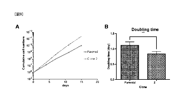
Figs. 9A and 9B respectively indicate growth curves of megakaryocytes (Clone
2) derived from a secondary iPS cell clone and megakaryocyte progenitor cells
(Parental) prior to cloning, and doubling times calculated from the growth
curves (**:
P<0.01).
Figs. 10A and 10B respectively indicate cell appearance and changes in cell
size
before and after maturation of megakaryocytes (Clone 2) derived from a
secondary
iPS cell clone and megakaryocyte progenitor cells (Parental) prior to cloning.
Fig. 11 is a graph comparing the numbers of proplatelets, which are
progenitors
of platelets, formed.
Description of Embodiments
[0014] The method for producing a stem cell clone according to the
present
invention comprises the steps of:
(i) introducing into stem cells an exogenous gene associated with induction of
differentiation into somatic cells;
(ii) inducing differentiation of the stem cells introduced with an exogenous
gene
8
CA 02982568 2017-10-12
into the somatic cells;
(iii) dedifferentiating the differentiation-induced somatic cells; and
(iv) isolating stem cells having the exogenous gene incorporated into a
chromosome thereof from a colony of the stem cells formed in step (iii).
[0015] Isolated stem cell clones are more suitable for being induced to
differentiate into somatic cells in comparison with stem cells prior to
cloning, and have
a high efficiency of being induced to differentiate into somatic cells per
cell.
[0016] In one embodiment thereof, the method for producing a stem
cell clone
according to the present invention may include the steps of:
(i) forming a stem cell colony by introducing a reprogramming factor into
somatic
cells having an exogenous gene functionally linked to a drug-responsive
promoter
incorporated into a chromosome thereof; and
(ii) isolating the stem cell colony obtained in step (i).
[0017] The resulting stem cell clone may be further induced to
differentiate to a
somatic cell. Induction of differentiation can be carried out by a person with
ordinary
skill in the art by suitably selecting a method suitable for inducing
differentiation into a
desired somatic cell, and may not be limited to a particular method, and may
further
include the step of:
(iii) inducing stem cells contained in the stem cell colony isolated in step
(ii) to
differentiate into somatic cells by contacting cells in any stage of
differentiation from
the stem cells to the somatic cells with a corresponding drug.
[0018] In the present invention, cloning refers to cloning of a cell
population in
the sense of isolating a cell population having uniform genetic information
from a cell
population having non-uniform genetic information.
[0019] Somatic Cells Submitted for Cloning
In the present invention, there are no particular limitations on the somatic
cells
submitted for cloning (to be referred to as primary somatic cells) provided
they are
cells produced by incorporating a gene functionally linked to a drug-
responsive
promoter in a chromosome thereof, and examples thereof include nerve cells (WO
2014/148646, Wapinski OL et al, Cell. 155:621-635, 2013), neural stem cells
(Han DW
et al, Cell Stem Cell. 10:465-472, 2012), neural crest cells (Kim YJ, et al,
Cell Stem
9
CA 02982568 2017-10-12
Cell. 15:497-506, 2014), myocardial cells (leda M et al, Cell. 142:375-386,
2010),
skeletal muscle cells (Tanaka A, et al, PLoS One. 8:e61540, 2013),
chondrocytes
(Outani H, et al, PLoS One. 8:e77365, 2013), hepatocytes (Huang P, et al, Cell
Stem
Cell. 14:370-384, 2014), melanocytes (Yang R, et al, Nat Commun. 5:5807,
2014),
hematopoietic progenitor cells (Batta K, Cell Rep. 9:1871-84, 2014),
erythroblasts
(Hirose S, et al, Stem Cell Reports. 1:499-508, 2013) and megakaryocyte
progenitor
cells (Nakamura S, et al, Cell Stem Cell. 14:535-548, 2014).
[0020] In the present invention, megakaryocyte progenitor cells are
suitable as
somatic cells cloned according to the method of the present invention since
they
cannot be cloned by the limiting dilution method. Erythroblasts are also
suitable as
somatic cells in the present invention. However, since somatic cells other
than these
cells also allow the obtaining of stem cell clones having an exogenous gene
associated with induction of differentiation into desired somatic cells
introduced into a
chromosome thereof, the somatic cells are not limited to megakaryocyte
progenitor
cells and erythroblasts.
[0021] The exogenous gene associated with induction of
differentiation into
somatic cells in the present invention refers in the broad sense to a gene
introduced
into a cell when inducing differentiation from a stem cell to a somatic cell.
In
explaining this gene using as an example the case of the somatic cells being
megakaryocyte progenitor cells, the gene associated with induction of
differentiation
may be at least one gene selected from the group consisting of oncogenes,
preferably
a member of the MYC gene family and more preferably c-Myc, genes suppressing
expression of p16 gene or p19 gene (polycomb genes) and preferably Bmi1, and
apoptosis suppressing genes and preferably BCL-XL gene. When using as an
example the case of the somatic cells being erythroblasts, the gene associated
with
induction of differentiation may be at least one gene selected from the group
consisting of oncogenes, preferably a member or the MYC gene family and more
preferably c-Myc, and apoptosis suppressing genes and preferably BCL-XL gene.
The exogenous gene associated with induction of differentiation into somatic
cells may
be operably linked to a drug-responsive promoter.
[0022] In the present invention, a drug-responsive promoter refers to
a promoter
CA 02982568 2017-10-12
that expresses a gene in the presence or absence of a corresponding drug. An
example of a promoter that expresses a gene in the presence of a corresponding
drug
is a TRE promoter (CMV minimal promoter having a Tet response sequence
including
seven repeats of a tet0 sequence). In the case of using a TRE promoter, a
system is
preferably used in which gene expression is induced in the presence of the
corresponding drug (such as tetracycline or doxycycline) by simultaneously
expressing a fusion protein (reverse tetR fusion protein) of reverse tetR
(rtetR) and
VP16AD within the same cells. In the case of using a reverse tetR fusion
protein, the
fusion protein is at least required to be expressed in a "step for inducing
differentiation
from stem cells to secondary somatic cells" to be subsequently described. For
example, by functionally linking a gene encoding a reverse tetR fusion protein
to a
drug-responsive promoter and introducing that gene when preparing primary
somatic
cells, the reverse tetR fusion protein can be expressed by addition or removal
of the
corresponding drug in the "step for inducing differentiation from stem cells
to
secondary somatic cells". In the case of functionally linking a drug-
responsive
promoter to two or more types of genes, the same type of drug-responsive
promoter
may be used for all of the genes or two or more types of drug-responsive
promoters
may be used.
[0023] Method for Deriving Primary Megakaryocyte Progenitor Cells
[0024] In the present invention, "megakaryocyte progenitor cells" refer to
cells
that become megakaryocytes as a result of maturing. These cells are not
multinucleated, and include cells characterized as
CD41a-positive/CD42a-positive/CD42b-weakly positive. The megakaryocyte
progenitor cells of the present invention are preferably cells that can be
grown by
expansion culturing, such as cells capable of undergoing expansion culturing
for at
least 60 days under suitable conditions. In the present invention,
megakaryocyte
progenitor cells may or may not be cloned, and although there are no
particular
limitations thereon, those that have been cloned are referred to as a
megakaryocyte
progenitor cell line. Megakaryocyte progenitor cells in the present invention
may be
derived from hematopoietic progenitor cells.
[0025] In the present invention, "megakaryocytes" are also referred
to as platelet
11
CA 02982568 2017-10-12
progenitor cells and megakaryocytic cells, are cells that produce platelets by
separation of their cytoplasm, may be multinucleated cells, and include cells
characterized as, for example, CD41a-positive/CD42a-positive/CD42b-positive.
In
addition, megakaryocytes may also be characterized as cells expressing GATA1,
FOG1, NF-E2 and 131-tubulin. Multinucleated megakaryocytes refer to a cell or
group
of cells in which the number of nuclei has undergone a relative increase in
comparison
with megakaryocyte progenitor cells. For example, in the case the nuclei of
megakaryocyte progenitor cells to which the method of the present invention is
applied
are 2N, cells in which the nuclei thereof are 4N or more are multinucleated
megakaryocytes. In addition, in the present invention, megakaryocytes may be
immortalized in the form of a megakaryocyte cell line or may be a cloned cell
group.
[0026] In the present invention, hematopoietic progenitor cells (H
PC) refer to
cells able to differentiate into blood cells such as lymphocytes, eosinophils,
neutrophils,
basophils, erythrocytes or megakaryocytes, and in the present invention, there
is no
distinction made between hematopoietic progenitor cells and hematopoietic stem
cells,
and refer to the same cells unless specifically indicated otherwise.
Hematopoietic
stem cells/progenitor cells can be recognized by, for example, being positive
for the
surface antigens, CD34 and/or CD43. In the present invention, hematopoietic
stem
cells can also be applied to hematopoietic progenitor cells that have been
induced to
differentiate from pluripotent stem cells or hematopoietic stem cells as well
as
progenitor cells derived from placental blood, bone marrow blood or peripheral
blood.
For example, in the case of using pluripotent stem cells, hematopoietic
progenitor cells
can be prepared from a net-like structure (ES-sac or iPS-sac) obtained by
culturing
pluripotent stem cells on C3H10T1/2 in the presence of VEGF in accordance with
the
method described in Takayama N., et al. J Exp Med. 2817-2830 (2010). Here,
"net-like structure" refers to a three-dimensional sac-like structure (having
a space
inside) derived from pluripotent stem cells that is formed by an endothelial
cell
population or the like and contains hematopoietic progenitor cells in the
interior thereof.
Other examples of methods used to induce differentiation from pluripotent stem
cells
to hematopoietic progenitor cells include a method that uses the formation of
an
embryoid body and the addition of a cytokine (Chadwick et at. Blood 2003, 102:
12
CA 02982568 2017-10-12
906-15, Vijayaragavan et al. Cell Stem Cell 2009, 4: 248-62, Saeki et al. Stem
Cells
2009, 27: 59-67) and a method including co-culturing with stromal cells
derived from
different species (Niwa A et al. J Cell Physiol. 2009 Nov;221(2):367-77.).
[0027] Examples of pluripotent stem cells include fertilized eggs and
cells such
as embryonic stem cells (ES cells), induced pluripotent stem cells (iPS cells)
or
embryonic germ cells (EG cells). Megakaryocyte progenitor cells serving as
somatic
cells of the present invention are preferably those that have been induced by
a step for
culturing the cells by overexpressing an oncogene, gene suppressing expression
of
p16 gene or p19 gene (polycomb gene) and/or gene suppressing apoptosis in
hematopoietic progenitor cells.
[0028] In the present invention, a "oncogene" refers to a gene that
causes a
malignant transformation of normal cells as a result of the expression,
structure or
function thereof being different from that of normal cells, and examples
thereof include
MYC family genes, Sic family genes, Ras family genes, Raf family genes, c-Kit
and
protein kinase family genes such as PDGFR or Abl. Examples of MYC family genes
include c-MYC, N-MYC and L-MYC. c-MYC genes refer to, for example, genes
composed of a nucleic acid sequence represented by NCBI accession no. NM
002467.
In addition, c-MYC genes include homologs thereof, and c-MYC gene homologs
refer
to genes for which, for example, the cDNA sequence thereof is composed of a
sequence that is substantially identical to the nucleic acid sequence
represented by
NCB! accession no. NM 002467. cDNA composed of a sequence substantially
identical to the nucleic acid sequence represented by NCBI accession no. NM
002467
refers to DNA composed of a sequence having identify of about 60% or more,
preferably about 70% or more, more preferably about 80%, 81%, 82%, 83%, 84%,
85%, 86%, 87%, 88%, 89%, 90%, 91%, 92%, 93%, 94%, 95%, 96%, 97% or 98%, and
most preferably about 99%, with DNA composed of a sequence represented by NCB!
accession no. NM 002467, or DNA able to hybridize under stringent conditions
with
DNA composed of a sequence complementary to the nucleic acid sequence
represented by NCBI accession no. NM 002467, with protein encoded by these DNA
contributing to expansion of cells such as hematopoietic progenitor cells that
are in a
stage of differentiation.
13
CA 02982568 2017-10-12
[0029] Here, stringent conditions refer to hybridization conditions
easily
determined by a person with ordinary skill in the art that are typically
empirical
experimental conditions dependent on probe length, washing temperature and
salt
concentration. In general, the temperature for proper annealing becomes higher
as
probe length increases, and the temperature becomes lower as probe length
decreases. Hybrid formation is typically dependent on the ability of the
complementary strand to undergo repeat annealing in an environment at a
temperature somewhat lower than the melting point thereof.
[0030] For example, an example of lowly stringent conditions includes
washing
at 0.1 x SSC in a 0.1% SDS solution under temperature conditions of 37 C to 42
C
during the stage of washing the filter following hybridization. In addition,
an example
of highly stringent conditions includes washing at 5 x SSC in a 0.1% SDS
solution at
65 C during the washing stage. Polynucleotides of higher homology can be
obtained
by using more highly stringent conditions.
[0031] In the present invention, since it is preferable to suppress the
expression
level of c-MYC, the c-MYC may be that which encodes a protein fused with a
destabilizing domain. A destabilizing domain acquired from ProteoTuner or
Clontech
Laboratories, Inc. can be used.
[0032] In the present invention, examples of "genes suppressing
expression of
p16 gene or p19 gene" include BMI1, Idl, Me118, Ring1a/b, Phc1/2/3,
Cbx2/4/6/7/8,
Ezh2, Eed, Suz12, HDAC and Dnmt1/3a/3b. BMI1 gene refers to, for example, a
gene composed of a nucleic acid sequence represented by NCB! accession no. NM
005180. In addition, BMI1 gene includes homologs thereof, and homologs of BMI1
gene refer to genes for which the cDNA sequence thereof is composed of a
sequence
substantially identical to the nucleic acid sequence represented by NCB!
accession no.
NM 005180. cDNA composed of a sequence substantially identical to the nucleic
acid sequence represented by NCB! accession no. NM 005180 refers to refers to
DNA
composed of a sequence having identify of about 60% or more, preferably about
70%
or more, more preferably about 80%, 81%, 82%, 83%, 84%, 85%, 86%, 87%, 88%,
89%, 90%, 91%, 92%, 93%, 94%, 95%, 96%, 97% or 98%, and most preferably about
99%, with DNA composed of the sequence represented by NCBI accession no. NM
14
CA 02982568 2017-10-12
005180, or DNA able to hybridize under stringent conditions with DNA composed
of a
sequence complementary to the nucleic acid sequence represented by NCBI
accession no. NM 005180, with protein encoded by that DNA promoting cell
expansion
by suppressing senescence of cells capable of inducing oncogenes occurring in
cells
that are expressed by oncogenes such as MYC family genes.
[0033] In the present invention, "apoptosis suppressing genes" refer
to genes
that suppress apoptosis, there are no particular limitations thereon, and
examples
thereof include BCL2 gene, BCL-XL gene, Survivin and MCL1. BCL-XL gene refers
to a gene composed of the nucleic acid sequence represented by NCB! accession
no.
NM 001191 or NM 138578. In addition, BCL-XL gene includes homologs thereof,
and
BCL-XL gene homologs refer to genes for which, for example, the cDNA sequence
thereof is composed of a sequence that is substantially identical to the
nucleic acid
sequence represented by NCB! accession no. NM 001191 or NM 138578. cDNA
composed of a sequence substantially identical to the nucleic acid sequence
represented by NCB! accession no. NM 001191 or NM 138578 refers to DNA
composed of a sequence having identify of about 60% or more, preferably about
70%
or more, more preferably about 80%, 81%, 82%, 83%, 84%, 85%, 86%, 87%, 88%,
89%, 90%, 91%, 92%, 93%, 94%, 95%, 96%, 97% or 98%, and most preferably about
99%, with DNA composed of a sequence represented by NCB! accession no. NM
001191 or NM 138578, or DNA able to hybridize under stringent conditions with
DNA
composed of a sequence complementary to the nucleic acid sequence represented
by
NCB! accession no. NM 001191 or NM 138578, with protein encoded by this DNA
having the effect of suppressing apoptosis.
[0034] In the present invention, the method for overexpressing the
above-mentioned genes in hematopoietic progenitor cells is preferably carried
out by
incorporating a gene functionally linked to a drug-responsive promoter in a
chromosome thereof, and can be achieved by, for example, introducing an
expression
vector containing a gene functionally linked to a drug-responsive promoter
into a
hematopoietic progenitor cell. Examples of vectors expressing these genes that
can
be used include retrovirus, lentivirus and other virus vectors as well as
animal cell
expression plasmids (such as pA1-11, pXT1, pRc/CMV, pRc/RSV or pcDNAI/Neo).
CA 02982568 2017-10-12
Retrovirus vectors or lentivirus vectors are used preferably from the
viewpoint of
incorporating in a chromosome.
[0035] In addition to a promoter, the expression vector may contain
an enhancer,
Poly(A) addition signal, selection marker gene or SV40 replication origin and
the like.
Examples of useful selection marker genes include dihydrofolate reductase
gene,
neomycin resistance gene and puromycin resistance gene.
[0036] In the present invention, a polycistronic vector, in which
genes are linked
longitudinally, may be obtained in order to introduce a plurality of genes
simultaneously.
In order to enable polycistronic expression, the 2A self-cleaving peptide of
foot and
mouth disease virus (refer to, for example, Science, 322, 949-953, 2008), or
an IRES
(Internal ribosome entry site) sequence, may be ligated between a plurality of
overexpressed genes.
[0037] In the present invention, in the case of a virus vector, the
method for
introducing an expression vector into hematopoietic progenitor cells can be
carried out
by introducing a plasmid containing the nucleic acid into suitable packaging
cells (such
as Plat-E cells) or a complementing cell line (such as 293 cells) followed by
recovering
virus produced in the culture supernatant and infecting hematopoietic
progenitor cells
by contacting with the virus. In the case of a non-virus vector, a plasmid
vector can
be introduced into cells by using a method such as lipofection, liposome
method,
electroporation, calcium phosphate co-precipitation, DEAE dextran method,
microinjection or a gene gun.
[0038] In one aspect of the method for inducing megakaryocyte
progenitor cells
according to the present invention, apoptosis suppressing gene may be
overexpressed after having overexpressed an oncogene or gene suppressing
expression of p16 gene or p19 gene in hematopoietic progenitor cells.
Overexpression of apoptosis suppressing gene can be carried out in the same
manner
as described above by introducing an expression vector, protein encoded by
these
genes, or RNA encoding these genes, into hematopoietic progenitor cells. In
the
case of subsequent expression of apoptosis suppressing gene, although there
are no
particular limitations thereon, overexpression of apoptosis suppressing gene
is
preferably carried out after overexpressing an oncogene or gene suppressing
16
CA 02982568 2017-10-12
expression of p16 gene or p19 gene for at least 14 days.
[0039] In the present invention, a caspase inhibitor may be contacted
with the
hematopoietic progenitor cells instead of overexpressing apoptosis suppressing
gene
in the cells. In the present invention, the caspase inhibitor may be any of a
peptidic
compound, non-peptidic compound or biological protein. Examples of peptidic
compounds include the artificial chemically synthesized peptidic compounds
indicated
in (1) to (10) below.
(1) Z-Asp-CH2-DCB (molecular weight: 454.26)
(2) Boc-Asp(OMe)-FMK (molecular weight: 263.3)
(3) Boc-Asp(OBzI)-CMK (molecular weight: 355.8)
(4) Ac-AAVALLPAVLLALLAP-YVAD-CHO (molecular weight: 1990.5) (SEQ ID NO:
1)
(5) Ac-AAVALLPAVLLALLAP-DEVD-CHO (molecular weight: 2000.4) (SEQ ID NO:
2)
(6) Ac-AAVALLPAVLLALLAP-LEVD-CHO (molecular weight: 1998.5) (SEQ ID NO:
3)
(7) Ac-AAVALLPAVLIALLAP-IETD-CHO (molecular weight: 2000.5) (SEQ ID NO:
4)
(8) Ac-AAVALLPAVLLALLAP-LEHD-CHO (molecular weight: 2036.5) (SEQ ID NO:
5)
(9) Z-DEVD-FMK (Z-Asp-Glu-Val-Asp-fluoromethylketone) (SEQ ID NO: 6)
(10) Z-VAD FMK
[0040] Examples of caspase inhibitors of peptidic compounds include:
(1)
VX-740 - Vertex Pharmaceuticals (Leung-Toung et al., Curr. Med. Chem. 9, 979-
1002
(2002)) and (2) HMR-3480 -Aventis Pharma AG (Randle et al., Expert Opin.
Investig.
Drugs 10, 1207-1209 (2001)).
[0041] Examples of caspase inhibitors of non-peptidic compounds
include: (1)
Anilinoquinazolines (AQZs), AstraZeneca Pharmaceuticals (Scott et al., J.
Pharmacol.
Exp. Ther. 304, 433-440 (2003)), (2) M826 - Merck Frosst (Han et al., J. Biol.
Chem.
277, 30128-30136 (2002)), (3) M867 - Merck Frosst (Methot et al., J. Exp. Med.
199,
199-207 (2004)), and (4) Nicotinyl aspartyl ketones - Merck Frosst (Isabel et
al., Bioorg.
17
CA 02982568 2017-10-12
Med. Chem. Lett. 13, 2137-2140 (2003)).
[0042] In addition, examples of caspase inhibitors of other non-
peptidic
compounds include: (1) IDN-6556 - !dun Pharmaceuticals (Hoglen et al., J.
Pharmacol.
Exp. Ther. 309, 634-640 (2004)), (2) MF-286 and MF-867 - Merck Frosst (Los et
al.,
Drug Discov. Today 8, 67-77 (2003)), (3) IDN-5370 - !dun Pharmaceuticals
(Deckwerth
et al., Drug Dev. Res. 52, 579-586 (2001)), (4) IDN-1965 - ldun
Pharmaceuticals
(Hoglen et al., J. Pharmacol. Exp. Ther. 297, 811-818 (2001)), and (5) VX-799 -
Vertex
Pharmaceuticals (Los et al., Drug Discov. Today 8, 67-77 (2003)). Other
examples of
caspase inhibitors include M-920 and M-791 - Merck Frosst (Hotchkiss et al.,
Nat.
Immunol. 1, 496-501 (2000)).
[0043] In the present invention, the caspase inhibitor is preferably
Z-VAD FMK.
In the case of using Z-VAD FMK for the caspase inhibitor, the Z-VAD FMK is
added to
the medium in which hematopoietic progenitor cells are cultured. The
preferable
concentration of Z-VAD FMK in the medium is, for example, 10 f.tM or more, 20
tiM or
more, 30 pM or more, 40 jAM or more or 50 tiM or more, and is preferably 30
jiM or
more.
[0044] Although there are no particular limitations thereon, the
medium used to
derive megakaryocyte progenitor cells from hematopoietic progenitor cells can
be
prepared by using medium used to culture animal cells as basal medium.
Examples
of basal media include IMDM medium, Medium 199 medium, Eagle's Minimum
Essential Medium (EMEM), aMEM medium, Dulbecco's modified Eagle's Medium
(DMEM), Ham's F12 medium, RPM! 1640 medium, Fischer's medium, Neurobasal
Medium (Life Technologies) and mixed media thereof. The medium may contain
serum or may be serum-free. The medium can also contain one or more substances
such as albumin, insulin, transferrin, selenium, fatty acids, trace elements,
2-mercaptoethanol, thiol glycerol, lipid, amino acids, L-glutamine, non-
essential amino
acids, vitamins, growth factors, low molecular weight compounds, antibiotics,
antioxidants, pyruvic acid, buffers, inorganic salts or cytokines as
necessary.
Cytokines refer to proteins that promote hematopoietic differentiation, and
examples
thereof include VEGF, TPO and SCF. Preferable medium in the present invention
is
IMDM medium containing serum, insulin, transferrin, serine, thiol glycerol,
ascorbic
18
CA 02982568 2017-10-12
acid and TPO. The medium more preferably further contains SCF. In addition, in
the case of using an expression vector containing a drug-responsive promoter,
a
corresponding drug such as tetracycline or doxycycline is preferably contained
in the
medium in the overexpression step.
[0045] In the present invention, although there are no particular
limitations
thereon, temperature conditions for deriving megakaryocyte progenitor cells
from
hematopoietic progenitor cells are such that promotion of differentiation into
megakaryocyte progenitor cells is confirmed by culturing hematopoietic
progenitor
cells at a temperature of 37 C or higher. Here, since a temperature that does
not
impart damage to cells is suitable, a temperature of 37 C or higher refers to,
for
example, a temperature of about 37 C to about 42 C and preferably a
temperature of
about 37 C to about 39 C. The duration of culturing at a temperature of 37 C
or
higher can be suitably determined by a person with ordinary skill in the art
while
monitoring such factors as the number of megakaryocyte progenitor cells.
Although
there are no particular limitations on this duration provided a desired number
of
megakaryocyte progenitor cells are obtained, examples thereof include a
duration of at
least 6 days or more, 12 days or more, 18 days or more, 24 days or more, 30
days or
more, 42 days or more, 48 days or more, 54 days or more or 60 days or more,
and
preferably 60 days or more. A long culturing period does not present a problem
with
respect to induction of megakaryocyte progenitor cells. In addition,
subculturing is
preferably suitably carried out during the culturing period.
[0046] Method for Reprogramming Somatic Cells
In the present invention, the introduction of a reprogramming factor into
somatic
cells can be carried out for the method used to reprogram somatic cells. Here,
examples of reprogramming factors include genes or gene products such as
Oct3/4,
Sox2, Sox1, Sox3, Sox15, Sox17, K1f4, K1f2, c-Myc, N-Myc, L-Myc, Nanog, Lin28,
Fbx15, ERas, ECAT15-2, Tell, beta-catenin, Lin28b, Sall1 , Sa114, Esrrb,
Nr5a2, Tbx3
or Glis1, and these reprogramming factors may be used alone or in combination.
Examples of combinations of reprogramming factors include the combinations
described in WO 2007/069666, WO 2008/118820, WO 2009/007852, WO
2009/032194, WO 2009/058413, WO 2009/057831, WO 2009/075119, WO
19
CA 02982568 2017-10-12
2009/079007, WO 2009/091659, WO 2009/101084, WO 2009/101407, WO
2009/102983, WO 2009/114949, WO 2009/117439, WO 2009/126250, WO
2009/126251, WO 2009/126655, WO 2009/157593, WO 2010/009015, WO
2010/033906, WO 2010/033920, WO 2010/042800, WO 2010/050626, WO
2010/056831, WO 2010/068955, WO 2010/098419, WO 2010/102267, WO
2010/111409, WO 2010/111422, WO 2010/115050, WO 2010/124290, WO
2010/147395, WO 2010/147612, Huangfu D, et al. (2008), Nat. Biotechnol., 26:
795-797, Shi Y, et al. (2008), Cell Stem Cell, 2: 525-528, Eminli S, et al.
(2008), Stem
Cells. 26:2467-2474, Huangfu D, et al. (2008), Nat. Biotechnol. 26:1269-1275,
Shi Y,
et al. (2008), Cell Stem Cell, 3, 568-574, Zhao Y, et al. (2008), Cell Stem
Cell,
3:475-479, Marson A, (2008), Cell Stem Cell, 3, 132-135, Feng B, et al.
(2009), Nat.
Cell Biol. 11:197-203, R. L. Judson et al., (2009), Nat. Biotechnol., 27:459-
461,
Lyssiotis CA, et al. (2009), Proc Natl Acad Sci USA. 106:8912-8917, Kim JB, et
al.
(2009), Nature. 461:649-643, lchida JK, et al. (2009), Cell Stem Cell. 5:491-
503, Heng
JC, et al. (2010), Cell Stem Cell. 6:167-74, Han J, et al. (2010), Nature.
463:1096-100,
Mali P, et al. (2010), Stem Cells. 28:713-720 and Maekawa M, et al. (2011),
Nature.
474:225-9. A more preferable combination of reprogramming factors includes
Oct3/4,
Sox2 and K1f4.
[0047] The above-mentioned reprogramming factors contain factors used
for the
purpose of enhancing establishment efficiency such as histone deacetylase
(HDAC)
inhibitors (such as small molecule inhibitors in the manner of valproic acid
(VPA),
trichostatin A, sodium butyrate, MC 1293 or M344,
nucleic acid expression inhibitors such as siRNA and shRNA against HDAC (for
example, HDAC1 siRNA Smartpool (Millipore), HuSH 29mer shRNA Constructs
against HDAC1 (On i Gene)), MEK inhibitors (such as PD184352, PD98059, U0126,
SL327 or PD0325901), glycogen synthase kinase-3 inhibitors (such as Bio or
CHIR99021), DNA methyl transferase inhibitors (such as 5-azacytidine), histone
methyl transferase inhibitors (such as small molecule inhibitors in the manner
of
BIX-01294 or nucleic acid expression inhibitors in the manner of siRNA and
shRNA
against Suv39h1, Suv39h2, SetDBI or G9a), L-channel calcium agonists (such as
Bayk8644), butyric acid, TGFP inhibitors or ALK5 inhibitors (such as LY364947,
CA 02982568 2017-10-12
SB431542, 616453 or A-83-01), p53 inhibitors (such as siRNA and shRNA against
p53), ARID3A inhibitors (such as siRNA and shRNA against ARID3A), miRNA such
as
miR-291-3p, miR-294, miR-295 or miR-302), Wnt signaling (such as soluble
Wnt3a),
neuropeptide Y, prostaglandins (such as prostaglandin E2 or prostaglandin J2),
hTERT,
SV4OLT, UTF1, IRX6, GLISI, PITX2 or DMRTBI, and in the present description,
there
are no particular distinctions made reprogramming factors and these factors
used for
the purpose of improving establishment efficiency.
[0048] In the case the reprogramming factor is in the form of a
protein, the
reprogramming factor may be introduced into somatic cells by a technique such
as
lipofection, fusion with a cell-permeating peptide (such as HIV-derived TAT or
polyarginine), or microinjection.
[0049] On the other hand, in the case the reprogramming factor is in
the form of
DNA, DNA can be introduced into somatic cells by a vector in the manner of a
virus,
plasmid or artificial chromosome, and by means such as ipofection, liposomes
or
microinjection. Examples of virus vectors include retrovirus vector,
lentivirus vector
(described in Cell, 126, pp. 663-676, 2006; Cell, 131, pp. 861-872, 2007;
Science, 318,
pp. 1917-1920, 2007), adenovirus vector (Science, 322, 945-949, 2008),
adeno-associated virus vector, and Sendai virus vector (WO 2010/008054). In
addition, examples of artificial chromosome vectors include human artificial
chromosomes (HAC), yeast artificial chromosomes (YAC) and bacterial artificial
chromosomes (BAC, PAC). Mammalian cell plasmids can be used as plasmids
(Science, 322:949-953, 2008). Vectors can contain a control sequence such as a
promoter, enhancer, ribosome binding sequence, terminator or polyadenylation
site to
enable expression of nuclear reprogramming substance, and can further contain
a
drug resistance gene (such as kanamycin resistance gene, ampicillin resistance
gene
or puromycin resistance gene), selection marker sequence such as thymidine
kinase
gene or diphtheria toxin gene, or a reporter sequence gene such as green
fluorescent
protein (GFP), p-glucuronidase (GUS) or FLAG as necessary. In addition, the
above-mentioned vectors may have an LoxP sequence before or after the vector
in
order to remove genes encoding the reprogramming factors or both a promoter
and
gene encoding a reprogramming factor bound thereto, following introduction
into
21
CA 02982568 2017-10-12
somatic cells.
[0050] In the case the reprogramming factor is in the form of RNA,
the
reprogramming factor can be introduced into somatic cells by a technique such
as
lipofection or microinjection, and RNA incorporating 5-methylcytidine and
pseudouridine (TriLink Biotechnologies) may be used to inhibit degradation
(Warren L,
(2010) Cell Stem Cell. 7:618-630).
[0051] Examples of culture broth for cells following reprogramming
include
DMEM containing 10% to 15% FBS, DMEM/F12 and DME culture broth (and these
culture broths can suitably further contain LIE, penicillin/streptomycin,
puromycin,
L-glutamine, non-essential amino acids or p-mercaptoethanol and the like), as
well as
commercially available culture broths (such as culture broth for culturing
mouse ES
cells (TX-WES culture broth, Thrombo-X), culture broth for culturing primate
ES cells
(Primate ES/iPS cell culture broth, ReproCELL Inc.), or serum-free medium
(mTeSR,
Stemcell Technology)).
[0052] An example of a method for culturing cells following reprogramming
comprises contacting somatic cells with reprogramming factor in DMEM
containing
10% FBS or DMEM/F12 culture broth at 37 C in the presence of 5% CO2 and
culturing
for about 4 days to 7 days, followed by reseeding the cells in feeder cells
(such as
mitomycin C-treated STO cells or SNL cells), and culturing in culture broth
for culturing
primate ES cells containing bFGF starting about 10 days after contacting the
somatic
cells with the reprograming factor to allow the formation of iPS-like colonies
after about
days to about 45 days or more from the time of contact.
[0053] Alternatively, somatic cells are cultured in DMEM medium
containing 10%
FBS (which may also suitably contain LIE, penicillin/streptomycin, puromycin,
25 L-glutamine, non-essential amino acids or p-mercaptoethanol and the
like) in feeder
cells (such as mitomycin C-treated STO cells or SNL cells) at 37 C in the
presence of
5% CO2 to allow the formation of ES-like colonies after about 25 days to about
30 days.
The reprogrammed somatic cells are preferably used as is instead of feeder
cells
(Takahashi K, et al. (2009), PLoS One. 4:e8067 or WO 2010/137746), or an
30 extracellular matrix is used (such as Laminin-5 (WO 2009/123349) or
Matrigel (Becton,
Dickinson and Company)).
22
CA 02982568 2017-10-12
[0054] In addition, examples of culturing methods include methods
using
medium that does not contain serum (Sun N, et al. Proc Natl Acad Sci USA.
106:15720-15725, 2009 or Nakagawa M, et al, Sci Rep. 4:3594, 2014). Moreover,
iPS cells may be established under hypoxic conditions (oxygen concentration of
0.1%
to 15%) in order to increase establishment efficiency (Yoshida Y, et al.
(2009), Cell
Stem Cell. 5:237-241 or WO 2010/013845).
[0055] The culture broth is replaced with fresh culture broth once a
day starting
on day 2 after the start of culturing during the above-mentioned culturing. In
addition,
although there are no particular limitations thereon, an example of the number
of
somatic cells used in reprogramming is within the range of about 5 x 10³ to
about 5 x
106 cells per 10 cm2 of culture dish area.
[0056] Step for Isolating Stem Cell Colonies Obtained by
Reprogramming
Somatic Cells
In the present invention, stem cell colonies can be obtained by introducing a
reprogramming factor into somatic cells and culturing the cells as described
above.
In the present invention, stem cells refer to cells having a self-replication
ability, which
enables the cells to produce cells identical to those cells by cell division,
and an ability
to differentiate into different types of cells, while also being able to
proliferate without
limitation. Although there are no particular limitations on the stem cells of
the present
invention provided they form colonies, these stem cells are pluripotent stem
cells
having the ability to differentiate to tissue cells excluding placental cells.
[0057] In the present invention, a colony refers to a cell mass
derived from a
single cell.
[0058] The cloning method of the present invention comprises a step
for isolating
the resulting stem cell colonies. This isolation can be carried out by
suitably
harvesting a single colony and then transferring to another culture dish.
[0059] iPS Cells for Inducing Megakaryocyte Progenitor Cells
In the present invention, in the case the somatic cells are megakaryocyte
progenitor cells, an exogenous oncogene and an exogenous gene suppressing
expression of p16 gene or p19 gene functionally linked to a drug-responsive
promoter
may be contained in the chromosomes of megakaryocyte progenitor cells as
23
CA 02982568 2017-10-12
previously described. In this case, secondary iPS cells, obtained by
reprogramming
the megakaryocyte progenitor cells according to the method described above,
similarly contain an exogenous oncogene and an exogenous gene suppressing
expression of p16 gene or p19 gene functionally linked to a drug-responsive
promoter
in the chromosomes thereof. At this time, the content ratio of exogenous gene
suppressing expression of p16 gene or p19 gene functionally linked to a
drug-responsive promoter to the exogenous oncogene functionally linked to a
drug-responsive promoter in the iPS cells for inducing megakaryocyte
progenitor cells
is preferably 2-fold to 7-fold and more preferably 3-fold to 5-fold.
Similarly, the
content ratio of exogenous gene suppressing expression of p16 gene or p19 gene
functionally linked to a drug-responsive promotor to exogenous oncogene
functionally
linked to a drug-responsive promoter in the megakaryocyte progenitor cells is
preferably 2-fold to 7-fold and more preferably 3-fold to 5-fold. Furthermore,
an
oncogene and a gene that suppresses expression of p16 gene or p19 gene are
suitably selected for the above-mentioned genes.
[0060] Step for Inducing Differentiation from Stem Cells to Secondary
Somatic
Cells
The cloning method of the present invention comprises a step for inducing
differentiation of stem cells obtained according to the method described above
to
secondary somatic cells. In the present invention, secondary somatic cells
refer to
somatic cells obtained by reprogramming primary somatic cells to stem cells
followed
by inducing to differentiate into secondary somatic cells, and the primary
somatic cells
and secondary somatic cells are preferably the same cells. The present
induction
step can be carried out by re-expressing an incorporated gene. Gene re-
expression
can be carried out by contacting cells at any stage of differentiation from
stem cells,
obtained by reprogramming primary somatic cells, to secondary somatic cells
with a
corresponding drug (in the case of a promoter that expresses a gene in the
presence
of a corresponding drug), or by interrupting contact between cells at any
stage of
differentiation from stem cells, obtained by reprogramming primary somatic
cells, into
secondary somatic cells and a corresponding drug (in the case of a promoter
that
expresses a gene when a corresponding drug is removed). For example, in the
case
24
CA 02982568 2017-10-12
of using a fusion gene (reverse tetR) of rtetR and VP16AD, the gene can be
re-expressed by administering a corresponding drug. "Cells at any stage of
differentiation from stem cells, obtained by reprogramming primary somatic
cells, to
secondary somatic cells" can be any cells in which the stem cells have gone
through
differentiation into secondary somatic cells. Thus, in the present invention,
the stem
cells may be induced to differentiate into other cells prior to gene re-
expression, and
examples of cells following this induction of differentiation include
fibroblasts and
hematopoietic progenitor cells. Induction of differentiation into "other
cells" can be
carried out in accordance with known methods. In the case of using
megakaryocyte
progenitor cells as somatic cells, the previously described method for
inducing
differentiation into hematopoietic progenitor cells is an example of a method
used to
induce differentiation. Namely, by administering a drug corresponding to a
medium
used to induce differentiation from stem cells to hematopoietic progenitor
cells and
induce differentiation from the previously described hematopoietic progenitor
cells to
megakaryocyte progenitor cells, an oncogene, gene suppressing expression of
p16
gene or p19 gene, and/or apoptosis suppressing gene can be overexpressed.
[0061] Step for Selecting Stem Cells or Hematopoietic Progenitor
Cells
In the present invention, since all stem cells are not necessarily able to be
induced to differentiate to megakaryocyte progenitor cells in the case of
using
megakaryocyte progenitor cells as somatic cells, stem cells capable of being
induced
to differentiate into megakaryocyte progenitor cells are preferably suitably
selected,
and an example of a method for carrying this out comprises selecting those
stem cells
that express MEG3. In the present invention, hematopoietic progenitor cells
derived
from stem cells expressing MEG3 may also be selected. In the present
invention, in
the case of humans, MEG3 refers to non-coding RNA composed of a nucleic acid
sequence represented by NCBI accession no. NR 002766, NR 003530, NR 003531,
NR 033358, NR 033359, NR 033360, NR 046464, NR 046465, NR 046466, NR
046467, NR 046468, NR 046469, NR 046470, NR 046471, NR 046472 or NR 046473.
Although there are no particular limitations thereon, stem cells to which the
method of
the present invention is applied may be primary pluripotent stem cells and are
more
preferably stem cell clones obtained according to the method described above.
A
CA 02982568 2017-10-12
high level of expression may refer to expression at level that is higher than
the average
value in a plurality of simultaneously measured stem cells or hematopoietic
progenitor
cells, or may refer to an expression level that is higher in comparison with
expression
of known stem cells or hematopoietic progenitor cells that cannot be induced
to
differentiate to megakaryocyte progenitor cells.
[0062] In the present invention, a method known among persons with
ordinary
skill in the art can be used as a method for confirming expression of MEG3,
and
examples thereof include reverse transcriptase PCR analysis, quantitative
reverse
transcriptase PCR analysis, Northern blotting analysis, immunohistochemical
analysis,
array analysis and combinations thereof.
[0063] Method for Causing Somatic Cells to be Deficient in HLA and
Method for
Producing HLA-Deficient Somatic Cells
In one embodiment thereof, the present invention provides a method for causing
somatic cells to be deficient in HLA that comprises the following steps, or a
method for
producing HLA-deficient somatic cells:
(i) forming pluripotent stem cells by introducing a reprogramming factor into
somatic cells;
(ii) causing the pluripotent stem cells obtained in step (i) to be deficient
in HLA;
and
(iii) inducing the HLA-deficient pluripotent stem cells obtained in step (ii)
to
differentiate into somatic cells.
[0064] The somatic cells submitted for use in the method for causing
a deficiency
of HLA of the present invention, the reprogramming factor used, the method for
forming pluripotent stem cells, and the method for inducing pluripotent stem
cells to
differentiate into somatic cells are the same as in the case of the somatic
cells
submitted for use in the above-mentioned cloning. Thus, the present invention
also
provides a method for cloning somatic cells and further causing those cells to
be
deficient in HLA.
[0065] In the present invention, HLA refers to human lymphocyte
antigen, and
refers to a class I antigen composed of an a chain and an L chain, a class II
antigen
composed of a13 chain encoding DRB1 gene and an a chain encoding DRA gene, and
26
CA 02982568 2017-10-12
a class III antigen. Since the expressed HLA differs according to the somatic
cell, the
HLA to be deleted can be suitably selected, and in the case of using
megakaryocyte
progenitor cells as somatic cells, a class I antigen is preferably selected as
HLA and
deleted. Deletion of HLA refers to deletion of the a chain, L chain or f3
chain, and in
the case of deleting a class I antigen, the L chain, namely 32-microglobulin,
is
preferably deleted.
[0066] The method for causing a chromosome to be deficient in HLA in
pluripotent stem cells of the present invention can be carried out by suitably
selecting
a known method such as homologous recombination.
[0067] Method for Producing Platelets
The method for producing platelets of the present invention comprises a step
for
cloning megakaryocyte progenitor cells using the cloning method of the present
invention, and a step for allowing the cloned megakaryocyte progenitor cells
to mature
into megakaryocytes and release platelets. The step for allowing the cloned
megakaryocyte progenitor cells to mature and release platelets can be carried
out in
accordance with a known method or method complying therewith. For example, in
the case the megakaryocyte progenitor cells contain at least one gene selected
from
the group consisting of MYC family genes, polycomb genes and apoptosis
suppressing genes, maturation of megakaryocytes can be carried out by
suppressing
expression of MY0 family genes, polycomb genes and/or apoptosis suppressing
genes by removing a corresponding drug from the medium following the
above-mentioned step (iii). The matured megakaryocytes become multinucleated
and release platelets.
[0068] The platelets may be in the form of a platelet preparation by
combining
with ACD-A solution, FFP, sodium citrate, citric acid or glucose and the like,
or may be
in the form of a blood preparation by combining with erythrocytes.
[0069] In the case of obtaining megakaryocyte clones deficient in HLA
according
to the method described above, platelets deficient in HLA can be obtained by
allowing
the megakaryocyte clones to mature and release platelets. HLA-deficient
platelets
are useful since they can be transfused irrespective of the HLA type of the
recipient.
[0070] Method for Improving Proliferative Capacity of Megakaryocyte
Progenitor
27
CA 02982568 2017-10-12
Cells
The present invention provides a method for improving the proliferative
capacity
of megakaryocyte progenitor cells by preparing stem cells by reprogramming
megakaryocyte progenitor cells and subsequently converting to megakaryocyte
progenitor cells. Thus, in one embodiment thereof, the method for improving
the
proliferative capacity of megakaryocyte progenitor cells of the present
invention
comprises the steps of:
(i) forming a stem cell colony by introducing a reprogramming factor into
megakaryocyte progenitor cells having an exogenous gene expressed in response
to
a drug;
(ii) isolating the stem cell colony obtained in step (i); and
(iii) inducing stem cells contained in the stem cell colony isolated in step
(ii) to
differentiate into megakaryocyte progenitor cells, wherein induction into the
somatic
cells comprises a step for contacting with a corresponding drug.
[0071] In the present invention, improvement of proliferative capacity
refers to
increasing the length of a telomere sequence in a chromosome. In the present
invention, a telomere sequence refers to a repetitive sequence including
TTAGGG,
and an increase in length of a telomere sequence means that the number of
repeats
has increased.
Examples
[0072] Although the following provides a more detailed explanation of
the
present invention based on examples and test examples, the present invention
is not
limited to the following examples.
[0073] Production of Megakaryocyte Progenitor Cells
Hematopoietic progenitor cells (HPC) were derived through iPS-sac from iPS
cells (SeV2: prepared by introducing c-MYC, OCT3/4, SOX2 and KLF4 into neonate
human fibroblasts using a Sendai virus vector in accordance with the method
described in WO 2010/134526) in a semi-confluent state and maintained in a 6
cm
dish in which MEF were disseminated at 3 x 10⁵ cells/dish. More specifically,
the iPS
cells were separated using human trypsin solution, and about 1/30 to 1/50 of
the cells
were disseminated on C3H10T1/2 (available from Riken, Japan.) treated with
28
CA 02982568 2017-10-12
mitomycin C (MMC) in the form of a colony mass. Furthermore, the MMC-treated
C3H10T1/2 was prepared by disseminating in a 10 cm dish at 8 x 10⁵ cells/dish
on the
day before disseminating the iPS cells. Following dissemination, culturing was
started in Eagle's Basal Medium (EBM) containing 20 ng/ml VEGF in an
atmosphere
of 5% 02 and 5% CO2 at 37 C (day 0). The medium was replaced with the same
medium on day 3 and day 6.
[0074] On day 7, culturing was continued in an atmosphere of 20% 02
and 5%
CO2 at 37 C. The medium was replaced with the same medium on day 9, day 11 and
day 13. On day 14, the cells were physically detached using a cell scraper or
the tip
of a pipette, and cells of uniform size were recovered by passing through a 40
micrometer cell strainer. The recovered cells were confirmed to be
hematopoietic
progenitor cells (HPC) based on cell size.
[0075] On day 14, the recovered HPC were disseminated in MMC-
processed
C3H10T1/2 at 3 x 10⁴ to 1 x 10⁵ cells/well. EBM containing SCF at 50 ng/ml,
TPO at
50 ng/ml and doxycycline at 0.5 ptg/mlwas used for the medium. Continuing, c-
MYC
and BMII were introduced into the HPC with a lentivirus vector. The lentivirus
vector
used was a tetracycline-controlled inducible vector, and was prepared by
recombining
an mOKS cassette of LV-TRE-mOKS-Ubc-tTA-12G to c-MYC or BMIl
(LV-TRE-c-MYC-xL-Ubc-tTA-12G or LV-TRE-BM11-Ubc-tTA-I2G, respectively)
(Nakamura S, et al, Cell Stem Cell. 14:535-548, 2014). The virus particles
used for
infection were prepared by infecting 293T cells with the lentivirus vector
(M01300).
Protamine was added only during infection. Subsequently, the medium was
replaced
every other day and the C3H10T1/2 and medium were replaced once or twice a
week.
[0076] BCL-xl was introduced at MOI 10 using a lentivirus vector two
weeks after
introducing c-MYC and BMII. The lentivirus vector used to introduce BCL-xl was
a
tetracycline-controlled inducible vector, and was prepared by recombining an
mOKS
cassette to contain BCL-xl in the same manner as described above
(LV-TRE-BCL-xL-Ubc-tTA-I2G) (Nakamura S, et al, Cell Stem Cell. 14:535-548,
2014).
Protamine was added only during infection. Subsequently, culturing was
maintained
in EBM containing SCF at 50 ng/ml, TPO at 50 ng/ml and doxycycline at 0.5
lig/m1 in
10T1/2 feeder cells in a 10 cm dish to prepare megakaryocyte progenitor cells
(to also
29
CA 02982568 2017-10-12
be referred to as imMKCL).
Reference Example 1
[0077] Limiting Dilution Method
Megakaryocyte progenitor cells (imMKCL) were disseminated in a 96-well plate
at a density of 1.5 cells/300 pt/well followed by culturing for 10 days to 14
days in
Iscove's modified Dulbecco's medium (IMDM) containing 15% fetal bovine serum
(FBS), human SCF (R&D Systems) at 50 ng/ml, TPO at 50 ng/ml, doxycycline
(Clontech) at 5 mg/nil and puromycin (Sigma-Aldrich) at 2 mg/ml in an
atmosphere of
5% CO2 at 37 C. Culturing was continued in the same manner after transferring
the
contents of each well to a 24-well plate and 6-well plate for the purpose of
scaling up
culturing. The cells in each well were designated as megakaryocyte progenitor
cell
clones.
[0078] Analysis of Megakaryocyte Progenitor Cell Clones (First Round)
Each of the megakaryocyte progenitor cell clones obtained according to the
method described above was washed twice using PBS, disseminated in a 6-well
plate
at 4 x 10⁵ cells/3 ml, and cultured in IMDM containing human SCF at 50 ng/ml,
human
TPO at 50 ng/ml, SR1 (Calbiochem) at 750 nM and 15% FBS. The supernatant was
recovered 7 days later followed by evaluation of the number of platelets
produced and
platelet function. Evaluation of the number of platelets produced was carried
out in
the manner described below. Namely, fluorescent dye-bound antibodies to CD41
(BioLegend), CD42a (eBioscience) and CD42b (BioLegend) and propidium iodide
(Sigma-Aldrich) were added to the culture supernatant and incubated for 30
minutes
followed by analyzing using FACSVerse0 (BD Biosciences). Analysis including
excluding megakaryocyte progenitor cells based on size, followed by counting
those
cells positive for CD41, CD42a and CD42b and calculating as the number of
platelets
per megakaryocyte progenitor cell. Evaluation of platelet function was carried
out in
the manner described below. Namely, fluorescent dye-bound antibodies to CD41,
CD42b and activated glycoprotein (GP) Ilb/Illa (PAC-1; BD Biosciences) and 0.4
mM
phorbol 12-myristate 13-acetate (PMA) (Sigma-Aldrich) were added to the
culture
supernatant and incubated for 30 minutes followed by analyzing using
FACSVerse0.
In the analysis, the expression level of activated GP Ilb/Illa was used for
evaluation by
CA 02982568 2017-10-12
measuring as fluorescence intensity (MFI).
[0079] As a result of evaluating platelet production volume and
platelet function
for 22 megakaryocyte progenitor cell clones according to the method described
above,
platelet production volume was confirmed to be 1.6 times to 1.9 times higher
than the
control (megakaryocyte progenitor cells prior to cloning) for clone 1, clone 4
and clone
13 (Fig. 1A). With respect to platelet function, the highest level of activity
was
demonstrated by clone 13 and was indicated to react well to PMA stimulation in
comparison with the control (Fig. 1B).
[0080] Analysis of Megakaryocyte Progenitor Cell Clones (Second
Round)
The five clones (1, 2, 4, 11 and 13) that demonstrated high platelet
production
volumes and platelet function in the results for the first round of analysis
were
re-analyzed in the same manner. Platelet production volume was confirmed to be
about 1.3 times higher than the control for clone 4 only (Fig. 2A). In
addition,
although platelet function was confirmed to be about 3.7 times higher than the
control
for clone 13, results for the platelet function of clones 1, 2, 4 and 11
differed from the
results of the first round of analysis in that there was no change from the
control (Fig.
2B).
[0081] Analysis of Megakaryocyte Progenitor Cell Clones (Third Round)
Analyses were conducted again in the same manner for the results of the second
round of analysis. There were no changes in platelet production volume with
respect
to the control (Fig. 3A). In addition, although platelet function was
confirmed to be 1.7
times and 1.6 times higher, respectively, than the control for clones 4 and
13, the
differences were small (Fig. 3B).
[0082] According to these results, megakaryocyte progenitor cell
clones obtained
by the limiting dilution method were confirmed to not demonstrate stable
function with
respect to platelet production capacity and the platelets produced. Thus, use
of the
limiting dilution method was suggested to be unsuitable for cloning of
megakaryocyte
progenitor cells.
Example 1
[0083] Cloning of Megakaryocyte Progenitor Cells by Reprogramming
iPS cells (secondary iPS cells) were prepared by reprogramming megakaryocyte
31
CA 02982568 2017-10-12
progenitor cells obtained with the previously described method followed by
carrying
out cloning with the secondary iPS cells and again inducing the
differentiation of the
cells into megakaryocyte progenitor cells to clone these cells (secondary
megakaryocyte progenitor cells) (Fig. 4A). The following provides a detailed
description thereof.
[0084] After introducing four types of episomal vector plasmids
(pCXLE-hOCT3/4-shp53-F, pCXLE-hSK, pCXLE-hUL and pCXWB-EBNAI; Okita K, et
al., Stem Cells. 31:458-66, 2013 and Okita K, et al., Nat Methods. 8:409-12,
2011) into
1 x 10⁶ cells of a primary cultured megakaryocyte progenitor cell line by
electroporation using Amaxa Nucleofector, the cells were disseminated at 3 x
105
cells/dish in feeder cells in the form of MEF at 1-2 x 10⁵ cells/6 cm dish.
The resulting
colonies were picked from the dish 14 days later and subjected to expansion
culturing,
after which 10 secondary iPS cell clones were used in analysis.
[0085] Analysis of Secondary iPS Cell Clones
Genomic DNA was extracted from the 10 secondary iPS cell clones obtained
according to the method described above, and quantitative PCR was carried out
on
c-MYC and BMII within exons using primers compatible with the PCR reaction. As
a
result, an examination of the number of insertions of exogenous c-MYC and BMII
other than the two intrinsic copies revealed that the number of insertions of
exogenous
BM I I of the 10 types of secondary iPS cell clones was about 7 copies to 26
copies,
while the number of insertions of exogenous c-MYC was about 1 copy to 6
copies, and
were all different (Fig. 4B).
[0086] Genomic DNA was extracted from the 10 secondary iPS cell
clones
(using some clones that were different from those previously described)
obtained
according to the method described above followed by analyzing all of the
genome
sequences, and when the number of insertions of exogenous c-MYC and BMII were
examined, they were confirmed to be inserted into chromosomes at the ratios
indicated in the following Table 1. The secondary iPS cell clones were
confirmed to
contain about 3 times to 5 times the number of insertions of BMIl in
comparison with
the number of insertions of c-MYC.
[Table I]
32
CA 02982568 2017-10-12
BMI/MYC
2nd-1PS_1 3.90
2nd-iPS_4 2.89
2nd-iPS_11 4.97
2nd-iPS_13 3.47
2nd-1PS_14 3.97
2nd-1PS_15 2.95
2nd-1PS_16 4.75
2nd-iPS_19 2.94
2nd-iPS_20 4.27
2nd-iPS_22 4.89
[0087] According to the above results, although the number of
incorporations of
exogenous genes introduced in the step for producing megakaryocyte progenitor
cells
differed for each of the megakaryocyte progenitor cells, in the case of having
been
incorporated at a constant ratio, megakaryocyte progenitor cells were
suggested to
have been produced.
[0088] Induction of Secondary Megakaryocyte Progenitor Cell Clones
from
Secondary iPS Cell Clones
HPC clones were respectively obtained through iPS-sac 14 days later from three
secondary iPS cell clones (#4, #11 and #12) according to the previously
described
method. The resulting HPC clones were disseminated into 6-wells at 1 x 10⁵
cells/well followed by culturing for 27 days in EBM containing SCF at 50
ng/ml, TPO at
50 ng/ml and doxycycline at 0.5 g/mIto obtain secondary megakaryocyte
progenitor
cell clones. Analysis of the resulting three megakaryocyte progenitor cell
clones
revealed that the clones were positive for CD41a and CD41b, negative for
CD235, and
were obtained in the form of a uniform cell group (Figs. 5A and 5B).
[0089] Preparation of HLA-Deficient (HLA-null) Secondary iPS Cell
Clones
The Target vector indicated in Fig. 6A was introduced into a secondary iPS
cell
clone (#11) obtained according to the method described above to delete Exon 1
of
32-Microglobulin (32m) by homologous recombination. Whether or not homologous
recombination was carried out properly was determined by confirming PCR
products
using primers designed at the sites indicated in Fig. 6A for the wild type and
mutant
33
CA 02982568 2017-10-12
(Fig. 6B). As a result, I32m-deficient secondary iPS cell clones were
established for
two clones (Clones 3 and 4).
[0090] Continuing, 132m-deficient secondary megakaryocyte progenitor
cell
clones were induced according to the previously described method. In addition,
platelets were obtained from the supernatant according to the previously
described
method. When flow cytometry was carried out on the p2m-deficient secondary
megakaryocyte progenitor cell clones and platelets using antibodies to I32m
(BD
Pharmingen) and HLA (BD Pharmingen), all were confirmed to be deficient in
132m and
HLA (Fig. 7A).
[0091] Confirmation of Platelet Function of 82m-deficient Secondary
Megakaryocyte Progenitor Cell Clones
After stimulating platelets present in the culture supernatant of wild-type
secondary iPS cell clone (#11) and I32m-deficient secondary megakaryocyte
progenitor cell clones with PMA in the same manner as previously described,
the
clones were contacted with CD42a antibody, CD42b antibody and PAC1, and when
the positive levels of CD42a, CD42b and PAC1 were measured and evaluated on
the
basis of fluorescence intensity (MFI), there were no differences observed
between the
wild type and p2m-deficient type (Fig. 7B). On the basis of the above,
platelets
deficient in 32m were indicated to have the same function as wild-type
platelets.
Example 2
[0092] Search for iPS Cell Marker Suitable for Induction of
Megakarvocvte
Progenitor Cells
mRNA was extracted from two types of secondary iPS cells (to be referred to as
Good-iPSC) able to establish imMKCL obtained according to the previously
described method and from two types of iPS cells (to be referred to as Bad-
iPSC)
unable to establish imMKCL obtained according to the previously described
method
followed by averaging and analyzing the expression levels of the two types of
Good-iPSC and Bad-iPSC with a microarray using the IIlumina HumanHT-12 v4.0 or
Affymetrix Gene Chip Human Gene 2.0 ST Array in accordance with the manuals
provided by the manufacturers (Figs. 8A and 8B).
[0093] Moreover, hematopoietic progenitor cells (HPC) were induced
from the
34
CA 02982568 2017-10-12
Good-iPSC and Bad-iPSC using the same method as previously described
(respectively referred to as Good-HPC and Bad-HPC) followed by analyzing with
a
microarray using the IIlumina HumanHT-12 v4.0 or Affymetrix Gene Chip Human
Gene
2.0 ST Array (Figs. 8A and 8B).
[0094] As a result of analyzing with the above-mentioned microarrays, MEG3
was confirmed to be highly expressed for both the Good-iPSC and Good-HPC.
Thus,
in secondary iPS cell clones and HPC induced therefrom, confirmation of
expression
of MEG3 was suggested to enable selection of secondary iPS cell clones
suitable for
induction of megakaryocyte progenitor cells.
Example 3
[0095] Proliferative Capacity of Megakaryocyte Progenitor Cells
Derived from
Secondary iPS Cell Clone
The cell proliferative capacity of megakaryocyte progenitor cells derived from
a
secondary iPS cell clone was compared with that of a megakaryocyte progenitor
cell
line prior to cloning in order to examine the effect of the cloning according
to the
present invention. In this experiment, a clone derived from 2nd-iPS_11 listed
in Table
1 was used for the secondary iPS cell clone. More specifically, an HPC clone
obtained from 2nd-iPS 11 through iPS-sac 14 days later according to the
previously
described method was disseminated in a 6-well dish at 1 x 10⁵ cells/well
followed by
culturing for 27 days in EBM containing SCF at 50 ng/ml, TPO at 50 ng/ml and
doxycycline at 0.5 lig/mIto obtain a secondary megakaryocyte progenitor cell
clone
(Clone 2). Megakaryocyte progenitor cells (Parental) prior to cloning prepared
in the
section entitled "Production of Megakaryocyte Progenitor Cells" were used as a
comparative example. These cells were disseminated in a 6-well dish at 1 x 10⁵
cells/well and cultured for 15 days in EBM containing SCF at 50 ng/ml, TPO at
50
ng/ml and doxycycline at 0.5 jig/ml.
[0096] As a result of periodically counting the number of cells
during the 15 day
culturing period, megakaryocyte progenitor cells derived from the secondary
iPS cell
clone clearly demonstrated more rapid growth than the megakaryocyte progenitor
cells prior to cloning. The results are shown in Figs. 9A and 9B.
[0097] Maturation Capacity of Megakaryocyte Progenitor Cells Derived
from
CA 02982568 2017-10-12
Secondary iPS Cell Clone
A secondary megakaryocyte progenitor cell clone (Clone 2) cultured for 15 days
was cultured under conditions facilitating megakaryocyte maturation (including
adding
SCF at 50 ng/ml and TPO at 50 ng/mIto EBM followed by further adding SR-1
(StemRegenin1) (Selleckchem) at 750 nM and Y27632 (Wako) at 10 M).
Megakaryocyte progenitor cells (Parental) were also allowed to mature to
megakaryocytes under the same culturing conditions as the secondary
megakaryocyte progenitor cell clone. Images of both cells photographed by
time-lapse imaging are shown in Fig. 10A, while the results of analyzing the
resulting
images with ImageJ are shown in Fig. 10B. On the basis of these results,
megakaryocytes cloned in accordance with the present invention were shown to
demonstrate superior maturation capacity (cellular hypertrophy) than
megakaryocytes
derived from the original clone cells.
[0098] Platelet Production Capacity of Meqakaryocyte Progenitor Cells
Derived
from Secondary iPS Cell Clone
The number of cells that formed platelets was measured visually in the images
shown in Fig. 10A in order to compare platelet production capacity of the
resulting
megakaryocytes. As a result, megakaryocytes derived from cells cloned in
accordance with the present invention clearly demonstrated superior platelet
production capacity (number of proplatelets formed) (**: p<0.01).
36