Note: Descriptions are shown in the official language in which they were submitted.
84071438
HIP JOINT DEVICE
This is a divisional of Canadian Patent Application Serial No. 2,805,013 filed
on July 12,
2010.
FIELD OF INVENTION
The present invention relates generally to a medical device for implantation
in a human
patient.
BACKGROUND
Hip joint Osteoarthritis is a syndrome in which low-grade inflammation results
in pain in the
hip joints, caused by abnormal wearing of the Cartilage that acts as a cushion
inside if the hip
joint. This abnormal wearing of the cartilage also results in a decrease of
the joints lubricating
fluid called Synovial fluid. Hip joint Osteoarthritis is estimated to affect
80% of all people
over 65 years of age, in more or less serious forms.
The present treatment of hip osteoarthritis comprises NSAID drugs, local
injections of
Hyaluronic acid or Glucocorticoid to help lubricating the hip joint, and
replacing parts of the
hip joint with a prosthesis through hip joint surgery.
The replacing of parts of the hip joint is one of the most common surgeries to
date performed
at hundreds of thousands of patients in the world every year. The most common
method
comprises placing a metal prosthesis in Femur and a plastic bowl in
Acetabulum. This
operation is done through a lateral incision in the hip and upper thigh and
through, Fascia Lata
and the lateral muscles of the thigh. To get access to the joint, the
supporting hip joint capsule
attached to Femur and Ilium needs to be penetrated, making it difficult to get
a fully
functional joint after the surgery. Femur is then cut at the neck with a bone
saw and the
prosthesis is placed in femur either with bone cement or without. Acetabulum
is slightly
enlarged using an Acetabular reamer, and the plastic bowl is positioned using
screws or bone
cement.
1
CA 2983754 2019-02-28
g4071438
The surgery typically requires one week of hospitalization due to the
increased risk of
infection. The recovery process is on average about 6 weeks, but even after
this period the
patient should not perform any physical activates that places large strain on
the joint.
The correct placement of the prosthesis or prosthetic parts is an important
part of the operation
since it affects the hip joint's ability to heal correctly, and also affects
the function of the hip
joint after the hip joint replacement surgery. Due to limited reach and
visibility inside the hip
joint, the placing of the prosthesis is a difficult and time consuming step of
the operation with
numerous possibilities for errors.
2
CA 2983754 2017-10-25
84071438
SUMMARY
An implantable medical device for implantation in a hip joint of a human
patient is provided.
The hip joint comprises the caput femur shaped like a ball, being connected to
the collum
femur and being the upper extremity of the femoral bone. The collum femur and
caput femur
having a longitudinal axial distribution with a longitudinal caput femur
centre axis reaching
from the collum femur, in the centre of the collum femur and caput femur and
towards the
acetabulum. The acetabulum is a bowl shaped section of the pelvic bone, with
an opening
towards the caput femur, the acetabulum have an acetabulum centre axis
reaching from the
centre of the bottom of the bowl towards the centre of the opening and the
caput femur. The
caput femur centre axis is identical with the acetabulum centre axis in a
special centred
position, when the caput femur being aligned, centred and symmetrical in the
acetabulum.
The caput femur and acetabulum each have a hip joint carrying surface, facing
each other and
contacting each other, the hip joint carrying surfaces, carrying weight in the
hip joint. The
medical device comprises: at least one artificial hip joint surface adapted to
replace at least the
surface of at least one of the caput femur and acetabulum. At least one
artificial hip joint
surface comprises: a positioning hole with at least one opening in said at
least one artificial
hip joint surface. The hole is adapted to be placed and dimensioned such that
the medical
device is adapted to be fitted using a positioning shaft and at least partly
surround the shaft,
for positioning the at least one artificial hip joint surface in a desired
position in the hip joint.
The hole is adapted to be fitted using the positioning shaft, when the shaft
is stabilized and
placed in at least one of the femoral bone and the pelvic bone for positioning
said medical
device inside the hip joint.
According to one embodiment the medical device comprises an artificial caput
femur or an
artificial caput femur surface, which could comprise at least two artificial
caput femur surface
parts adapted to be interconnected to form the artificial caput femur surface
during an
operation. Each of the at least two artificial caput femur surface parts could
comprise a
positioning hole adapted to at least partly surround a positioning shaft.
3
CA 2983754 2017-10-25
' 84071438
According to one embodiment the collum femur has an axial distribution leading
to the caput
femur, which has a substantially ball shaped configuration with a maximum
diameter
substantially perpendicular to the centre axis of the prolongation of the
axial distribution of
the collum femur. The caput femur is normally placed in an acetabulum creating
the hip joint.
According to another embodiment the artificial caput femur surface further
comprises at least
one first beyond part of the artificial caput femur surface adapted to cover
and/or go into the
bone of the caput femur on at least a part of the caput femur beyond the
maximum diameter of
the caput femur, away from the acetabulum cup towards the collurn femur, when
mounted on
the caput femur in its functional position in the joint. The at least one
first beyond part is
adapted to have a closest perpendicular distance to the centre axis, being
smaller than the
distance between the periphery of the maximum diameter of the caput femur and
the centre
axis. The medical device thus is adapted to create a more stable position of
said artificial
caput femur surface when mounted on the caput femur in the functional
position.
The hip joint has a ball shaped caput femur being the proximal part of the
femoral bone with a
convex hip joint surface and a bowl shaped acetabulum as part of the pelvic
bone with a
concave hip joint surface. According to one embodiment the artificial caput
femur surface
comprises an artificial concave hip joint surface adapted to be fixated to the
femoral bone.
The implantable medical device could have a largest diameter or a largest
cross-sectional
distance, and an opening. The largest diameter or cross sectional distance is
adapted to be
changed during an operation.
According to another embodiment of the implantable medical device, the
artificial hip joint
surface further comprises an artificial acetabulum or an artificial acetabulum
surface. The
artificial acetabulum surface could comprises a first positioning hole and a
second positioning
hole. The artificial acetabulum surface is adapted to be aligned with the
artificial caput femur
surface in a special position and adapted to use the positioning shaft placed
in at least one of
4
CA 2983754 2017-10-25
84071438
the femoral and pelvic bone, placed through at least one of the first
positioning hole in the
caput femur surface and the second positioning hole in the acetabulum surface
and further
placed at least partly through at least one of the first positioning hole in
the caput femur
surface and the second positioning hole in the acetabulum surface.
According to another embodiment the artificial acetabulum surface and the
artificial caput
femur surface are adapted to be in moveable connection with each other when
implanted in
the hip joint.
According to one embodiment the implantable medical device is adapted to be
fixated to at
least one of the caput femur, the collum femur and the femoral bone using a
fixation element,
which could be a fixation clement selected from a group consisting of, at
least one screw, at
least one pin, at least one portion of at least one of the parts adapted to be
introduced into the
other part, the parts being adapted to be sliding into the other part, form
fitting, welding,
adhesive, pin, wire, a ball mounted into a bowl being portions of said parts,
a male portion of
one part mounted into a female portion of the other part, a key introduced
into a lock being
portions of said parts, band, and other mechanical connecting members.
According to another embodiment the artificial hip joint surface comprises an
artificial
acctabulum or an artificial acetabulum surface, which could comprises at least
two artificial
acetabulum surface parts adapted to be interconnected to form the artificial
acetabulum
surface during an operation. Each of the at least two artificial acetabulum
surface parts could
comprise a positioning hole adapted to at least partly surround a positioning
shaft
The hip joint has a ball shaped caput femur, being the proximal part of the
femoral bone, with
a convex hip joint surface, and a bowl shaped acetabulum as part of the pelvic
bone with a
concave hip joint surface. The artificial acetabulum surface, according to one
embodiment,
could comprise an artificial convex hip joint surface adapted to be fixated to
the pelvic bone.
CA 2983754 2017-10-25
84071438
The artificial acetabulum surface has a largest diameter or a largest cross-
sectional distance,
and an opening. According to one embodiment the largest diameter or cross
sectional distance
could be adapted to be changed during an operation.
The positioning hole according to any of the embodiments herein could be
substantially
circular, non-circular or have a cut circumference.
The implantable medical device according to any of the embodiments could be
adapted to
inserted through the hip joint capsule or thc pelvic bone and could bc mounted
onto said
positioning shaft inside of the hip joint.
According to one embodiment the at least two artificial caput femur surface
parts could
comprise a positioning hole adapted to at least partly surround a positioning
shaft.
According to one embodiment each of the at least two artificial acetabulum
surface parts
could comprise a positioning hole adapted to at least partly surround a
positioning shaft.
The artificial acetabulum surface could be adapted to be fixated to the pelvic
bone using a
fixation element, such as a fixation element selected from a group consisting
of; at least one
screw, at least one pin, at least one portion of at least one of the parts
adapted to be introduced
into the other part, the parts being adapted to be sliding into the other
part, form fitting,
welding, adhesive, pin, wire, a ball mounted into a bowl being portions of
said parts, a male
portion of one part mounted into a female portion of the other part, a key
introduced into a
lock being portions of said parts, band, and other mechanical connecting
members.
A positioning shaft adapted to position a medical device inside of the hip
joint of a human
patient is further provided. The positioning shaft could be elongated and
further adapted to be
6
CA 2983754 2017-10-25
84071438
introduced into at least one of the femoral bone and the pelvic bone during
positioning of a
medical device inside the hip joint.
According to one embodiment the positioning shaft is adapted to be fixated to
the at least one
of the femoral bone and the pelvic bone during positioning of the medical
device inside the
hip joint.
The artificial hip joint surface comprises an artificial acetabulum or an
artificial acetabulum
surface. The hole, when fitted with the positioning shaft, could be adapted to
centre and hold
the artificial acetabulum or an artificial acetabulum surface during fixation
thereof in the hip
joint.
The artificial hip joint surface could comprise an artificial caput femur or
an artificial caput
femur surface. The hole, when fitted with the positioning shaft, could be
adapted to centre and
hold the artificial caput femur or an artificial caput femur surface during
fixation thereof in
the hip joint.
The artificial hip joint surface could further comprise an artificial
acetabulum or an artificial
acetabulum surface. The hole, whcn fitted with the positioning shaft, could be
adapted to
centre and hold both the artificial caput femur or an artificial caput femur
and the artificial
acetabulum or an artificial acetabulum surface during fixation thereof in the
hip joint.
The artificial caput femur surface could according to one embodiment comprise
a convex
shape towards the centre of the hip joint, and the artificial acetabulum
surface could comprise
a concave shape towards the centre of the hip joint. The artificial convex
caput femur surface
could be adapted to be fixated to the pelvic bone of the human patient, and
the artificial
concave acetabulum surface could be adapted to be fixated to the femoral bone
of the human
patient.
7
CA 2983754 2017-10-25
84071438
The caput femur has a convex hip joint surface towards the centre of the hip
joint and the
acetabulum has a concave hip joint surface towards the centre of the hip
joint. At least one
artificial hip joint surface comprises an artificial caput femur or an
artificial caput femur
surface, having a convex shape towards the centre of the hip joint, comprising
a first hole, and
an artificial acetabulum or an artificial acetabulum surface, having a concave
form towards
the centre of the hip joint, comprising a second hole. The artificial caput
femur or the artificial
caput femur surface and the artificial acetabulum or the artificial acetabulum
surface are
adapted to be centered in the hip joint by the positioning shaft placed in
said hole and at least
one of the femoral or pelvic bone. The artificial convex caput femur or
artificial convex caput
femur surface has the first hole placed in the convex part, for enabling the
positioning shaft to
be introduced into said hole and centering said artificial convex caput femur
or said artificial
convex caput femur surface, when placed in the hip joint. Furthermore the
artificial concave
acetabulum or the artificial concave acetabulum surface has the second hole
placed in the
concave part, for enabling the positioning shaft to be introduced into the
hole and centering
the artificial concave acetabulum or the artificial concave acetabulum
surface, when placed in
the hip joint.
The artificial concave acetabulum or the artificial concave acetabulum surface
could
according to one embodiment have the second hole placed in the centre of the
concave part.
The artificial convex caput femur or artificial convex caput femur surface
could have the first
hole placed in the centre of said convex part, adapted for enabling the
positioning shaft to be
introduced into said hole and centering said aitificial convex caput femur or
said at tificial
convex caput femur surface and said artificial concave acetabulum or said
artificial concave
acctabulum surface, when placed in the hip joint, and when said hip joint is
placed in said
special centered position.
The artificial convex caput femur surface could be adapted to be fixated to
the pelvic bone of
the human patient, and the artificial concave acetabulum surface could be
adapted to be
fixated to the femoral bone of the human patient.
8
CA 2983754 2017-10-25
84071438
According to another embodiment the positioning shaft further comprises a
screw thread
adapted to fixate the positioning shaft to at least one of: the femoral bone
and the pelvic bone,
during positioning of the medical device inside the hip joint. The positioning
shaft could be
adapted to be removed from the connection with at least one of the femoral
bone and the
pelvic bone, after the positioning of the medical device inside the hip joint.
According to one embodiment the positioning shaft is adapted to have a first
and second state.
The first state is a state in which the positioning shaft is flexible, and the
second state is a state
in which the positioning shaft is adapted to be less flexible than in the
first state.
The positioning shaft could be adapted to position the medical device in at
least one axis, in at
least two axis, in at least three axis and/or in at least one plane.
According to one embodiment the positioning shaft is adapted to be placed in a
positioning
hole of the medical device.
The positioning shaft could be adapted to position a medical device comprising
at least two
medical device parts, and the positioning shaft could be adapted to be placed
in a positioning
hole in at least one of thc at least two medical device parts.
According to one embodiment the positioning shaft has an area, substantially
perpendicular to
its elongated distribution, adapted to be positioned in said positioning hole,
being
substantially circular or non-circular.
The positioning shaft is adapted to be positioned in a positioning hole, which
could have a cut
circumference.
9
CA 2983754 2017-10-25
= 84071438
The positioning shaft could be adapted to receive a medical device inserted
into the hip joint
through the hip joint capsule or through the pelvic bone.
The positioning shaft, according to one embodiment could be adapted to receive
a medical
.. device to be mounted onto the positioning shaft inside of the hip joint.
A medical device system comprising a first medical device, according to any of
the
embodiments herein, and a second medical device, being a medical according to
any of the
embodiments herein, and a positioning shaft according to any of the
embodiments herein.
According to one embodiment of the medical device system, the first and second
medical
devices are adapted to be mounted onto the positioning shaft inside of the hip
joint.
According to yet another embodiment of the medical device system, the first
and said second
medical device are adapted to be mounted onto the positioning shaft outside of
the hip joint.
According to yet another embodiment the medical device system, further
comprises an
implantable lubrication system, the implantable lubrication system is adapted
to lubricate the
first and second medical devices after the first and second medical device has
been positioned
in the hip joint.
A medical device system for use in a hip joint of a patient is further
provided. The system
comprises a first positioning shaft according to any of the embodiments above,
and a second
positioning shaft according to any of the embodiments above. The first
positioning shaft is
adapted to position a first part of a medical device in the hip joint, and the
second positioning
shaft is adapted to position a second part of the medical device in the hip
joint.
CA 2983754 2017-10-25
= 84071438
According to another embodiment of the medical device system, the first part
of the medical
device and the second part of the medical device are adapted to be connected
to each other
after implantation in the hip joint of the patient.
According to yet another embodiment of the medical device system the first
positioning shaft
and the second positioning shaft are adapted to be connected to each other
after implantation
in the hip joint of the patient.
The medical device according to any of the embodiments herein could comprise
an artificial
acetabulum or an artificial acetabulum surface, and the hole, when fitted with
the positioning
shaft, could be adapted to centre and hold the artificial acetabulum or an
artificial acetabulum
surface during fixation thereof in the hip joint.
According to another embodiment of the medical device, the artificial hip
joint surface
comprises an artificial caput femur or an artificial caput femur surface. The
hole, when fitted
with the positioning shaft, could be adapted to centre and hold the artificial
caput femur or an
artificial caput femur surface during fixation thereof in the hip joint.
The artificial hip joint surface could further comprise an artificial
acetabulum or an artificial
acetabulum surface. The hole, when fitted with the positioning shaft, could be
adapted to
centre and hold both the artificial caput femur or an artificial caput femur
and the artificial
acetabulum or an artificial acetabulum surface, during fixation thereof in the
hip joint.
According to one embodiment the artificial caput femur surface comprises a
convex shape
towards the centre of the hip joint, and the artificial acetabulum surface
comprises a concave
shape towards the centre of the hip joint. The artificial convex caput femur
surface could be
adapted to be fixated to the pelvic bone of the human patient, and the
artificial concave
acetabulum surface could be adapted to be fixated to the femoral bone of the
human patient.
11
CA 2983754 2017-10-25
'84071438
According to one embodiment the medical device could comprise a positioning
shaft adapted
to be placed in the hole, for centering the artificial hip joint surface, when
placed in the hip
joint. The at least one artificial hip joint surface could comprise an
artificial acetabulum or an
artificial acetabulum surface, the positioning shaft, when fitted in said
hole, could be adapted
to center and hold the artificial acetabulum or the artificial acetabulum
surface during fixation
thereof in the hip joint.
The medical according to any of the embodiments above could comprise a
positioning shaft
adapted to be placed in the hole for centering the artificial hip joint
surface, when placed in
the hip joint. At least one artificial hip joint surface could comprise an
artificial caput femur
or an artificial caput femur surface, and the positioning shaft, when fitted
in the hole, is
adapted to centre and hold the artificial caput femur or an artificial caput
femur surface during
fixation thereof in the hip joint.
According to one embodiment the medical device, according to any of the
embodiments
above, comprises a positioning shaft adapted to be placed in the hole for
centering the at least
one artificial hip joint surface, when placed in the hip joint. The artificial
hip joint surface
further comprises an artificial acetabulum or an artificial acetabulum
surface, the positioning
shaft, when fitted in the hole, could be adapted to centre and hold both the
artificial caput
femur or the artificial caput femur surface, and the artificial acetabulurn,
or an artificial
acetabulum surface, during fixation thereof in the hip joint.
According to one embodiment the artificial caput femur surface comprises a
convex shape
towards the centre of the hip joint, and the artificial acetabulum surface
comprises a concave
shape towards the centre of the hip joint. The artificial convex caput femur
surface could be
adapted to be fixated to the pelvic bone of the human patient, and the
artificial concave
acetabulum surface could be adapted to be fixated to the femoral bone of the
human patient.
12
CA 2983754 2017-10-25
'84071438
According to another embodiment of the medical device, the caput femur have a
convex hip
joint surface towards the centre of the hip joint, and the acetabulum have a
concave hip joint
surface towards the centre of the hip joint, and at least one artificial hip
joint surface
comprises. an artificial caput femur or an artificial caput femur surface,
having a convex
shape towards the centre of the hip joint, comprising a first hole, and an
artificial acetabulum
or an artificial acetabulum surface, having a concave form towards the centre
of the hip joint,
comprising a second hole. The artificial caput femur or the artificial caput
femur surface and
the artificial acetabulum or the artificial acetabulum surface are adapted to
be centred in the
hip joint by the positioning shaft placed in the hole and at least one of the
femoral or pelvic
bone. The artificial convex caput femur or the artificial convex caput femur
surface have the
first hole placed in the centre of the convex part, for enabling the
positioning shaft to be
introduced into the hole and centering the artificial convex caput femur or
the artificial
convex caput femur surface, when placed in the hip joint, and the artificial
concave
acetabulum or the artificial concave acetabulum surface have the second hole
placed in the
centre of the concave part, for enabling the positioning shaft to be
introduced into the hole
and centering the artificial concave acetabulum or the artificial concave
acetabulum surface,
when placed in the hip joint.
According to one embodiment the artificial convex caput femur surface is
adapted to be
fixated to the pelvic bone of the human patient, and the artificial concave
acetabulum surface
is adapted to be fixated to the femoral bone of the human patient.
The caput femur has a convex hip joint surface towards the centre of the hip
joint, and the
acetabulum, has a concave hip joint surface towards the centre of the hip
joint. According to
one embodiment the medical device further comprises a positioning shaft
adapted to centre
the artificial hip joint surface when placed in the hole, wherein the at least
one artificial hip
joint surface comprises; an artificial caput femur or an artificial caput
femur surface, having a
convex form towards the centre of the hip joint, comprising a first hole. The
artificial hip joint
surface further comprises an artificial acetabulum or an artificial acetabulum
surface, having a
concave form towards the centre of the hip joint, comprising a second hole.
The artificial
caput femur or the artificial caput femur surface and the artificial
acetabulum or the artificial
13
CA 2983754 2017-10-25
84071438
acetabulum surface are adapted to be centred in the hip joint by the
positioning shaft placed in
the hole and in at least one of: the femoral bone and the pelvic bone. The
artificial convex
caput femur or the artificial convex caput femur surface having the first hole
placed in the
centre of the convex part, for enabling the positioning shaft to be introduced
into the hole and
centreing the artificial convex caput femur or the artificial convex caput
femur surface, when
placed in the hip joint, and the artificial concave acetabulum or the
artificial concave
acetabulum surface having the second hole placed in the centre of the concave
part, for
enabling the positioning shaft to be introduced into the hole and centreing
the artificial
concave acetabulum or the artificial concave acetabulum surface, when placed
in the hip joint.
In one embodiment the artificial convex caput femur surface is adapted to be
fixated to the
pelvic bone of the human patient, and the artificial concave acetabulum
surface is adapted to
be fixated to the femoral bone of the human patient.
According to another embodiment the positioning shaft is adapted to receive
the hip joint
surfaces inserted into the hip joint through the hip joint capsule and/or the
pelvic bone.
However it is equally conceivable that the positioning shaft is adapted to
receive the hip joint
surfaces to be mounted onto said positioning shaft, inside of the hip joint.
According to another embodiment, there is provided an implantable medical
device for
implantation in a hip joint of a human patient, the hip joint comprising the
caput femur shaped
like a ball, being connected to the collum femur and being the upper extremity
of the femoral
bone, the collum femur and caput femur having a longitudinal axial
distribution with a
longitudinal caput femur centre axis reaching from the collum femur, in the
centre of the
collum femur and caput femur and towards the acetabulum, the acetabulum is a
bowl shaped
section of the pelvic bone, with an opening towards the caput femur, the
acetabulum having
an acetabulum centre axis reaching from the centre of the bottom of the bowl
towards the
centre of the opening and the normal position of the caput femur, wherein the
caput femur
centre axis is aligned with the acetabulum centre axis in a special centered
position, when the
caput femur is aligned, centered and symmetrical in the acetabulum, the caput
femur and
acetabulum each having a hip joint carrying surface, facing each other and
contacting each
other, the hip joint carrying surfaces, carrying weight in the hip joint,
wherein said medical
14
CA 2983754 2019-02-28
84071438
device comprises: an artificial caput femur or an artificial caput femur
surface, adapted to
replace the surface of the caput femur, and an artificial acetabulum or an
artificial acetabulum
surface, adapted to replace the surface of the acetabulum, wherein the
artificial caput femur or
artificial caput femur surface comprises a first positioning hole, and said
artificial acetabulum
or artificial acetabulum surface comprises a second positioning hole, wherein
said first and
second positioning holes are adapted to receive a positioning shaft fixated to
at least one of
the femoral and pelvic bone for positioning said artificial caput femur or
artificial caput femur
surface in relation to said artificial acetabulum or artificial acetabulum
surface and in relation
to the femoral bone and/or the pelvic bone.
METHOD
A method of treating a hip joint of a patient is further provided. The method
comprising the
steps of: cutting the skin of the patient, dissecting an area of the hip
joint, placing a
positioning shaft in the hip joint, providing a medical device comprising a
positioning hole,
said hole being adapted to be placed and dimensioned such that the medical
device is adapted
to be fitted using said positioning shaft, placing said medical device at
least partly surrounding
said positioning shaft, fixating said medical device comprising an artificial
hip joint surface in
the right position in the hip joint using said positioning shaft.
14a
CA 2983754 2019-02-28
'84071438
The method could further comprise the step of removing said positioning shaft.
The step of fixating the medical device in the hip joint could according to
one embodiment
comprise the step of fixating the medical device in the hip joint using a
fixation element
selected from a group consisting of: at least one screw, at least one pin, at
least one portion of
at least one of the parts adapted to be introduced into the other part, the
parts being adapted to
be sliding into the other part, form fitting, welding, adhesive, pin, wire, a
ball mounted into a
bowl being portions of said parts, a male portion of one part mounted into a
female portion of
the other part, a key introduced into a lock being portions of said parts,
band, and other
mechanical connecting members.
The step of fixating the medical device in the hip joint could comprise the
step of fixating the
medical device to the caput femur or femoral bone.
The step of fixating the medical device in the hip joint could comprise the
step of fixating the
medical device to the acetabulum or pelvic bone.
The step of placing a positioning shaft in the hip joint could comprise the
step of placing a
positioning shaft in the caput femur or femoral bone.
The step of placing a positioning shaft in the hip joint could comprise the
step of placing a
positioning shaft in the acetabulum or pelvic bone.
The step of placing the medical device at least partly surrounding said
positioning shaft, could
comprise the step of placing an artificial acetabulum or artificial acetabulum
surface onto the
positioning shaft in the hip joint, wherein said positioning shaft is
introduced into said hole.
CA 2983754 2017-10-25
=
84071438
The method according to any of the embodiment herein could comprise the step
of placing
said medical device at least partly surrounding the positioning shaft, which
could comprise
placing an artificial caput femur or artificial caput femur surface onto the
positioning shaft in
the hip joint, the positioning shaft could be introduced into the hole.
The step of placing the medical device at least partly surrounding the
positioning shaft could
comprise placing an artificial acetabulum or artificial acetabulum surface
onto the positioning
shaft in the hip joint, wherein the positioning shaft is introduced into said
hole.
The at least one of the artificial caput femur or artificial caput femur
surface and the artificial
acetabulum or artificial acetabulum have a through going hole with an inlet
and an outlet. The
method could further comprise introducing the positioning shaft into said hole
inlet and
passing out from the hole outlet.
An arthroscopic method of treating a hip joint of a patient is further
provided. The method
comprises the steps of: inserting at least one needle or a tube like
instrument into the patient's
hip joint, using the needle or tube like instrument to fill the joint with a
fluid, placing at least
two arthroscopic trocars in the joint, inserting a camera through one of the
arthroscopic
trocars into the joint, dissecting an area of the hip joint, placing a
positioning shaft in the hip
joint, providing a medical device comprising a positioning hole, said hole
being adapted to be
placed and dimensioned such that the medical device is adapted to be fitted
using said
positioning shaft, placing said medical device at least partly surrounding
said positioning
shaft, fixating said medical device in the right position in the hip joint
using said positioning
shaft.
The method could further comprise the step of removing said positioning shaft.
The step of fixating the medical device in the hip joint could according to
one embodiment
comprise the step of fixating the medical device in the hip joint using a
fixation element
selected from a group consisting of: at least one screw, at least one pin, at
least one portion of
16
CA 2983754 2017-10-25
84071438
at least one of the parts adapted to be introduced into the other part, the
parts being adapted to
be sliding into the other part, form fitting, welding, adhesive, pin, wire, a
ball mounted into a
bowl being portions of said parts, a male portion of one part mounted into a
female portion of
the other part, a key introduced into a lock being portions of said parts,
band, and other
mechanical connecting members.
The step of fixating the medical device in the hip joint could comprise the
step of fixating the
medical device to the caput femur or femoral bone.
The step of fixating the medical device in the hip joint could comprise the
step of fixating the
medical device to the acetabulum or pelvic bone.
The step of placing a positioning shaft in the hip joint could comprise the
step of placing a
positioning shaft in the caput femur or femoral bone.
The step of placing a positioning shaft in the hip joint could comprise the
step of placing a
positioning shaft in the acctabulum or pelvic bone.
The step of placing the medical device at least partly surrounding said
positioning shaft, could
.. comprise the step of placing an artificial acetabulum or artificial
acetabulum surface onto the
positioning shaft in the hip joint, wherein said positioning shaft is
introduced into said hole.
The method according to any of the embodiment herein could comprise the step
of placing
said medical device at least partly surrounding the positioning shaft, which
could comprise
placing an artificial caput femur or artificial caput femur surface onto the
positioning shaft in
the hip joint, the positioning shaft could be introduced into the hole.
17
CA 2983754 2017-10-25
= .84071438
The step of placing the medical device at least partly surrounding the
positioning shaft could
comprise placing an artificial acetabulum or artificial acetabulum surface
onto the positioning
shaft in the hip joint, wherein the positioning shaft is introduced into said
hole.
The at least one of the artificial caput femur or artificial caput femur
surface and the artificial
acetabulum or artificial acetabulum have a through going hole with an inlet
and an outlet. The
method could further comprise introducing the positioning shaft into said hole
inlet and
passing out from the hole outlet.
Please note that any embodiment or part of embodiment, feature, method,
associated system,
part of system described herein may be combined in any way. Please note that
the description
in general should be seen as describing both of an apparatus and a method.
18
CA 2983754 2017-10-25
84071438 = =
BRIEF DESCRIPTION OF DRAWINGS
The invention is now described, by way of example, with reference to the
accompanying
drawings; in which:
Fig. la shows the hip joint in section,
Fig. lb shows the collum femur in section having cancellous bone and cortical
bone,
Fig. 2 shows the human patient in a frontal view when incisions are being made
in the
abdominal region,
Fig. 3 shows the human patient in a frontal view when incisions are being made
in the
abdominal region,
Fig. 4 shows a lateral view of a human patient,
Fig. 5 shows a lateral view of a human patient when the femoral bone
dissected,
Fig. 6 shows a lateral view of the human patient when a positioning shaft is
being placed in
the caput and collum femur,
Fig. 7 shows the step of creating a hole in the caput and collum femur,
Fig. 8a shows the placing of a positioning shaft in the caput femur and collum
femur,
Fig. 8b shows the placing of a medical device onto the caput femur using the
positioning
shaft,
Fig. 9 shows the process of placing an artificial caput femur surface onto the
caput femur,
through a hole in the pelvic bone,
Fig. 10 shows the process of placing an artificial caput femur surface onto
the caput femur,
through a hole in the pelvic bone,
Fig. 11 shows the removal of the positioning shaft,
Fig. 12 shows the placing of an artificial caput femur surface onto the caput
femur,
Fig. 13 shows the placing of an artificial caput femur surface onto the caput
femur,
Fig. 14 shows the removal of the positioning shaft,
Fig. 15 shows the placing of adhesive on a section of a surface of the collum
femur,
19
CA 2983754 2017-10-25
84071438
Fig. 16 shows the placing of a medical device inside the hip joint through a
hole in the pelvic
bone,
Fig. 17 shows the hip joint in section when a medical device is being fixated,
Fig. 18 shows the placing of a prosthetic part in the hole of the pelvic bone,
Fig. 19 shows a lateral view of the human patient when an instrument for
creating a hole in
the pelvic bone is provided,
Fig. 20 shows different instruments possible to place on a force transferring
member,
Fig. 21 shows the hip joint in section when a hole in the pelvic bone is being
created,
Fig. 22 shows the hip joint in section when a hole in the pelvic bone is being
created,
Fig. 23 shows the hip joint in section when the caput femur is being reamed,
Fig. 24a-c shows the an expandable reamer,
Fig. 25 shows the hip joint in section when an artificial caput femur surface
is being provided,
Fig. 26 shows the creation of a hole in the pelvic bone from the opposite side
from
acetabulum,
Fig. 27 shows the hip joint in section when a hole in the pelvic bone is being
created,
Fig. 28 shows the hip joint in section when the caput femur is being
surgically modified,
Fig. 29 shows the hip joint in section when a concave surface id created in
the collum femur
and caput femur,
Fig. 30 shows the injecting of an adhesive in the concave surface in the caput
femur and
collum femur,
Fig. 31 shows the positioning of a medical device in the collum femur and
caput femur
through a hole in the pelvic bone,
Fig. 32 shows the placing of a medical device through a hole in the pelvic
bone,
Fig. 33 shows the placing of a prosthetic part in the hole in the pelvic bone,
Fig. 34 shows the fixation of El prosthetic part in the hole in the pelvic
bone,
Fig. 35a shows a medical device in a schematic view,
CA 2983754 2017-10-25
84071438
Fig. 35b shows a medical device in section,
Fig. 36a shows the placing of a band onto a medical device,
Fig. 36b shows the femoral bone when a medical device has been placed on the
caput femur,
Fig. 37 shows an embodiment of a medical device,
Figs. 38a-e shows a medical device in an operable embodiment,
Fig. 39a shows a medical device in an expandable embodiment, in a first state,
Fig. 39b shows a medical device in an expandable embodiment, in a second
state,
Fig. 40a shows the medical device in an embodiment where the medical device
comprises
multiple parts,
Fig. 40b shows the medical device in an embodiment where the medical device
comprises
multiple parts, in greater detail,
Fig. 40e shows the medical device in an embodiment where the medical device
comprises
multiple parts, when assembled,
Fig. 41 shows an artificial acetabulum surface according to one embodiment,
Fig. 42a shows the femoral bone when a medical device is being placed on the
caput femur,
Fig. 42b shows the femoral bone when a medical device is being placed on the
caput femur,
in a top view,
Fig. 43a,b shows an instrument for placing positioning shafts in the caput
femur and collum
femur,
Fig. 44a shows the placing of a prosthetic part in the hole in the pelvic
bone,
Fig. 44b shows the operation of the prosthetic part in the hole in the pelvic
bone,
Fig. 45 shows hip joint in section when a prosthetic part is being placed,
Fig. 46 shows the hip joint in section when a bone plug or medical device is
being fixated,
Fig. 47 shows the hip joint in section when a bone plug is being provided,
Fig. 48 shows the removal of the positioning shaft from the hip joint,
Fig. 49 shows a prosthetic part, in further detail,
21
CA 2983754 2017-10-25
84071438
Fig. 50 shows injecting members injecting material into the holes in the
femoral bone and the
pelvic bone,
Fig. 51 shows an implantable lubricating system,
Fig. 52a shows a frontal view of the human patient when sutures or staplers
are being
provided,
Fig. 52b shows a frontal view of the human patient when sutures or staplers
are being
provided,
22
CA 2983754 2017-10-25
84071438
DETAILED DESCRIPTION
Biocompatible material is to be understood as being a material with low level
of immune
response. Biocompatible materials are sometimes also referred to as
biomaterials. Analogous
is biocompatible metals a biocompatible metal with low immune response such as
titanium or
tantalum. The biocompatible metal could also be a biocompatible alloy
comprising at least
one biocompatible metal.
A metal alloy is to be understood as a mixture of two or more elements in
solid solution in
which the major component is a metal. A steel alloy is hence an alloy wherein
one of the
components is steel which in turn is an alloy of iron and carbon. A titanium
alloy is hence an
alloy wherein one of the components is titanium.
Elasticity is to be understood as a materials ability to deform in an elastic
way.
Carrying surfacc and weight carrying surface is to bc understood as a surface
adapted to carry
weight inside of the hip joint.
Form fitting is to be understood as an element having a part or section which
is adapted to
enable a mechanical connection of said element to at least one other element
using said part or
section. Form fitted structure is a structure of an element which enables form
fitting.
Functional hip movements are to be understood as movements of the hip that at
least partly
correspond to the natural movements of the hip. On some occasions the natural
movements of
the hip joint might be somewhat limited or altered after hip joint surgery,
which makes the
functional hip movements of a hip joint with artificial surfaces somewhat
different than the
functional hip movements of a natural hip joint.
The functional position of an implantable medical device or prosthesis is the
position in which
the hip joint can perform functional hip movements.
Functional hip joint is a hip joint that can perform functional hip movements
either with or
without an implanted medical device or prosthesis.
Connection line is to be understood as a line of the connecting surface of at
least two medical
device parts connecting to each other.
23
CA 2983754 2017-10-25
84071438 =
In the following a detailed description of preferred embodiments of the
present invention will
be given. In the drawing figures, like reference numerals designate identical
or corresponding
elements throughout the several figures. It will be appreciated that these
figures are for
illustration only and are not in any way restricting the scope of the
invention. Thus, any
references to direction, such as "up" or "down", are only referring to the
directions shown in
the figures. Also, any dimensions etc. shown in the figures are for
illustration purposes.
The medical device according to any of the embodiments could comprise at least
one material
selected from a group consisting of: polytetrafluoroethylene (PTFE),
perfluoroalkoxy (PFA)
and fluorinated ethylene propylene (FEP). Tt is furthermore conceivable that
the material
comprises a metal alloy, such as cobalt-chromium-molybdenum or titanium or
stainless steel,
or polyethylene, such as cross-linked polyethylene or gas sterilized
polyethylene. The use of
ceramic material is also conceivable, in the contacting surfaces or the entire
medical device
such as zirconium or zirconium dioxide ceramics or alumina ceramics. The part
of the
medical device in contact with human bone for fixation of the medical device
to human bone
could comprise a poorhouse structure which could be a porous micro or nano-
structure
adapted to promote the growth-in of human bone in the medical device for
fixating the
medical device. The porous structure could be achieved by applying a hydroxy-
apatite (HA)
coating, or a rough open-pored titanium coating, which could be produced by
air plasma
spraying, a combination comprising a rough open-pored titanium coating and a
HA top layer
is also conceivable. The contacting parts could be made of a self lubricated
material such as a
waxy polymer, such as PTFE, PFA, FEP, PE and UHMWPE, or a powder metallurgy
material
which could be infused with a lubricant, which preferably is a biocompatible
lubricant such as
a Hyaluronic acid derivate. It is also conceivable that the material of
contacting parts or
surfaces of the medical device herein is adapted to be constantly or
intermittently lubricated.
According to some embodiments the parts or portions of the medical device
could comprise a
combination of metal materials and/or carbon fibers and/or boron, a
combination of metal and
plastic materials, a combination of metal and carbon based material, a
combination of carbon
and plastic based material, a combination of flexible and stiff materials, a
combination of
elastic and less elastic materials, Conan or acrylic polymers.
Fig. la shows the hip joint of a human patient in section. The hip joint
comprises a caput
femur 5 placed at the very top of collum femur 6 which is the top part of the
femur bone 7.
24
CA 2983754 2017-10-25
84071438
The caput femur 5 is in connection with the acetabulum 8 which is a bowl
shaped part of the
pelvic bone 9. Both the caput femur surface 10 and the acetabulum surface 11
is covered with
articular cartilage 13 which acts as a cushion in the hip joint. In patients
with hip joint
osteoarthritis, this articular cartilage 13 is abnormally worn down due to a
low grade
inflammation. The hip joint is surrounded by the hip joint capsule 12 which
provides support
for the joint and hinders luxation. After conventional hip joint surgery,
penetrating the hip
joint capsule 12, the capsule 12 is dramatically weakened due to the limited
healing
possibilities of its ligament tissue. By performing hip joint surgery without
damaging the hip
joint capsule 12, the patient can fully recover and place equal amount of
strain on an artificial
joint as is possible on a natural one.
Fig. lb shows the collum femur 6 in section. Both caput femur 5 and collum
femur 6 further
comprises cortical bone 601, the outer more sclerotic bone, and cancellous
bone 602, placed
in the bone marrow 603. The cortical bone is much more dense and beneficial to
anchor a
prosthesis two, or to place a positioning shaft in, whereas the cancellous
bone 602 provides
stability in the bone due to its sandwich construction, but is easy to remove
to make room for
a fixation member of a prosthesis.
Fig. 2 shows a frontal view of the body of a human patient. A surgical method
of operating
the hip joint from the opposite side from acetabulum, is according to a first
embodiment
performed starting with an incision 1 in the abdominal wall of the human
patient. The incision
1 passes through the abdominal muscles in to the abdomen of the human patent.
In a second
embodiment the incision 2 is conducted through the abdominal muscles and in to
the pelvic
area, below peritoneum. According to a third embodiment the incision 3 is
performed just
between Illium and the surrounding tissue, an incision 3 which could enable
the pelvic bone
to be dissected with very little penetration of fascia and muscular tissue.
According to a fourth
.. embodiment the incision 4 is made in the inguinal channel. In all of the
four embodiments the
tissue surrounding the pelvic bone 9 in the area opposite to acetabulum is
removed or
penetrated which enables the surgeon to reach the pelvic bone 9.
Fig. 3 shows a frontal view of the body of a human patient. A
laparoscopic/arthroscopic
method of operating the hip joint, from the opposite side from acetabulum, is
according to a
first embodiment performed starting with making small incisions 14 in the
abdominal wall of
the human patient. The small incisions enable the surgeon to insert
laparoscopic trocars into
CA 2983754 2017-10-25
84071438
the abdomen of the human patient. According to the first embodiment the
incisions 14 passes
through the abdominal wall, in to the abdomen of the human patent. According
to a second
embodiment the small incisions 15 is conducted through the abdominal wall, in
to the pelvic
area, below peritoneum. According to a third embodiment the small incisions 16
is performed
just between Illium and the surrounding tissue, an incision 16 which could
enable the pelvic
bone to be dissected with very little penetration of fascia and muscular
tissue. According to a
fourth embodiment the incision 17 is made in the inguinal channel. In all of
the four
embodiments the tissue surrounding the pelvic bone 9 in the area opposite to
acetabulum 8 is
removed or penetrated which enables the surgeon to reach the pelvic bone 9.
Fig. 4 shows a lateral view of a conventional hip joint surgery where an
incision 112 is made
in the thigh 113 enabling the surgeon to reach the femur bone 7 on which the
caput femur 5 is
located. In a conventional hip joint surgery the hip joint is accessed through
the hip joint
capsule, which forces the surgeon to, at least partly, savage the structure of
the capsule.
Fig. 5 to enable the surgeon to reach the caput femur 5 the femoral bone 7
comprising the
caput femur 5 is placed outside of the hip joint capsule 12. The surgeon can
thereby perform
surgical modifications on the caput femur including fixating holding members
or prosthetic
parts to the surface or the bone structure of any part of the femoral bone 7.
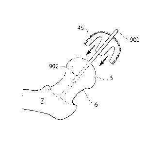
Fig. 6 shows the placing of a positioning shaft 900 in the caput femur 5 and
collum femur 6,
penetrating the surface and the cortical bone of the caput femur 5. For
example the
positioning shaft can be fixated to the bone structure of the femoral bone 7
the collum femur 6
or the caput femur 5 using mechanical fixating members, adhesive, a threaded
section of the
positioning shaft 900 or using an expanding part or section of the positioning
shaft 900.
Fig. 7 shows the femoral bone 7, comprising the collum femur 6 and the caput
femur 5, when
a hole 902 going through the surface and the cortical bone of the caput femur
5 and in to the
cancellous bone of the caput femur 5 and collum femur 6. The hole 902 is
created using a drill
901, which could be powered using an operation device or through manual force.
Fig. 8a shows the placing of a positioning shaft 900 in the hole 902 in the
surface of the caput
femur 5 the cortical bone of the caput femur 5 and the cancellous bone of the
collum femur 6.
For example the positioning shaft 900 can be fixated to the bone structure of
the femoral bone
7 the collum femur 6 or the caput femur 5 using mechanical fixating members,
adhesive, a
26
CA 2983754 2017-10-25
84071438
threaded section of the positioning shaft 900 or using an expanding part or
section of the
positioning shaft 900.
Fig. 8b shows the step of placing an artificial caput femur surface 45 on the
caput femur 5
using the positioning shaft 900. The artificial caput femur surface 45
comprises a hole
adapted to encircle the positioning shaft 900 such that the positioning shaft
900 positions and
centers the artificial caput femur surface 45 in a suitable position on the
caput femur 5.
Fig. 9 shows the hip joint in section, when an artificial caput femur surface
45 is positioned
on the caput femur using a positioning shaft 900 placed in a hole 902 in the
caput femur 5 and
collum femur 6. The caput femur 5 is according to this embodiment inserted
through a hole
18 in the pelvic bone 9 from the opposite side from acetabulum 8, which also
is the way the
positioning shaft 900 has been inserted in the caput femur 5 and collum femur
6. To enable
the artificial caput femur to be inserted through a hole 18 in the pelvic bone
9 having a
diameter dl smaller than the diameter d2 required for the artificial caput
femur surface 45 to
reach over the caput femur 5. The artificial caput femur surface 45 therefore
comprises elastic
material which enables the artificial caput femur surface 45 to in a first
state have a first
diameter dl, for passing through the hole 18 in the pelvic bone 9, and in a
second state have a
second diameter d2 for reaching over the caput femur 5, and in a third state
have a third
diameter d3 for clasping the caput femur 5, thereby creating a stable position
inside the hip
joint. The artificial caput femur surface comprises a hole adapted to encircle
the positioning
shaft 900 for positioning and centering the artificial caput femur surface 45
inside the hip
joint.
Fig. 10 shows the step of inserting an artificial acetabulum surface 65
through the hole 18 in
the pelvic bone 9 using a positioning shaft 900 placed in a hole 902 in the
caput femur 5 and
the collum femur 6. The positioning shaft 900 assists in the positioning and
centering of the
artificial caput femur surface 45 to the caput femur 5 in the hip joint. To
enable the insertion
of the artificial caput femur surface 45 through a hole 18 in the pelvic bone
9 the size of the
artificial caput femur surface 45 is adapted to be changed. in a first state
for inserting the
artificial caput femur surface 45 through a hole 18 in the pelvic bone 9 the
artificial caput
femur surface 45 has a largest diameter dl, smaller than the diameter of the
hole 18 in the
pelvic bone 9.
27
CA 2983754 2017-10-25
'84071438 =
Fig. 11 shows the removal of the positioning shaft 900 from the hole 902 in
the caput femur 5,
after the placing of the artificial acetabulum surface 65. After the
artificial acetabulum surface
65 has passed through the hole 18 in the pelvic bone 9 the surface is expanded
to represent the
entire contacting surface of the acetabulum 8, and the largest diameter is now
d2 which
confines the artificial acetabulum surface inside of the hip joint. The use of
the positioning
shaft 900 also ensures the correct centering of the caput femur surface 45 in
relation to the
artificial acetabulum surface 65 inside of the hip joint.
Figs. 12 ¨ 14 shows the process of placing an artificial caput femur surface
45 on the caput
femur 5 in the hip joint through a hole 18 in the pelvic bone 9. The
artificial caput femur
surface 45 is inserted through the hole 18 in the pelvic bone 9 in its folded
state, as shown in
fie. 12, positioned, guided and centered by the positioning shaft 900 fixated
to the caput
femur 5 and the collum femur 45. After the passing of the hole 18 in the
pelvic bone 9 the
artificial caput femur surface 45 is expanded as shown in fig. 13 and
eventually clasps the
caput femur 5 as shown in fig. 14, whereafter the positioning shaft 900 is
removed, as shown
in fig. 14.
An alternative embodiment of fixation of a medical device comprising an
artificial caput
femur will now be described with reference to figs. 15 ¨ 18.
Fig. 15 shows the hip joint in section in a step in which the caput femur 5
has been removed
and a surface of the section 610 in the collum femur 6 is being prcparcd for
the fixation of an
artificial caput femur 600. An injecting member 613 applies and adhesive 614
to the surface
of the section 610 of the collum femur 6. The injecting member 613 is adapted
to be
introduced through a hole 18 in the pelvic bone 9 and to apply the adhesive
614 which was
contained within the injecting member 613. Furthermore a positioning shaft 900
is placed in
the collum femur 6 from the hole 18 in the pelvic bone 9. The positioning
shaft is preferably
fixated to the cortical bone of the femoral bone 7 as well as the canccllous
bone of the collum
femur 6.
Fig. 16 shows the step of introducing and fixating the medical device 600 to
the collum femur
6, through a hole 18 in the pelvic bone 9. The stabilizing member 612 is
adapted to stabilize
the medical device 600 from the outside of the collum femur 6 substantially
perpendicular to
the longitudinal extension of the collum femur 6, and from the acetabulum
side, substantially
in line with the longitudinal extension of the collum femur 6 through the
stabilizing member
28
CA 2983754 2017-10-25
=
= 84071438
612 being placed in contact with the outside of the collum femur 6 and the
surface of the
section 610 in the collum femur 6. The stabilizing member 612 is fixated to
the outside of the
collum femur 6 and/or to the surface of the section 610 in the collum femur 6
by means of the
adhesive 614. However the adhesive 614 could be replaced or assisted by bone
cement or a
mechanical fixation element 615. The medical device 600 is positioned, guided
and centered a
positioning shaft 900 placed in the collum femur 6 from the hole 18 in the
pelvic bone 9. The
positioning shaft 900 is preferably fixated to the cortical bone of the
femoral bone 7 as well as
the cancellous bone of the collum femur 6.
Fig. 17 shows the hip joint in section when the medical device 600 is
positioned on the collum
femur 6. The stabilizing member 612 is here fixated to the collum femur 6 by
means of
adhesive 614 and a mechanical fixation element 615, such as a screw or pin.
Fig. 18 shows the hip joint in section when the medical device 600 is
positioned on the collum
femur 6. The stabilizing member 612 is here fixated to the collum femur by
means of
adhesive 614. A prosthetic part 98 comprising an artificial acetabulum surface
618 has been
positioned in the hole 18 in the pelvic bone 9. The artificial acetabulum
surface 618 is adapted
to be in direct of indirect connection with the artificial caput femur surface
607. In
embodiments where the artificial acetabulum surface 618 is adapted to be in
indirect
connection with the artificial caput femur surface 607 a lubricating fluid or
a lubricating
material (not shown) can be placed between said artificial acetabulum surface
618 and said
artificial caput femur surface 607. The prosthetic part 98 is adapted to carry
the load placed on
the artificial acetabulum surface 618 from weight of the human patient through
the contact
with the artificial caput femur surface 607 by means of the supporting members
99. The
prosthetic part 98 can further be fixated to the pelvic bone 9 by means of
bone cement,
adhesive, screws, form fitting, welding, sprints, band or some other
mechanical connecting
member. According to this embodiment the supporting members 99 are positioned
on the
acetabulum side of the pelvic bone 9, however it is also conceivable that the
supporting
members 99 are positioned on the abdominal side of the pelvic bone 9. The
supporting means
could be constructed in many different ways and this should be seen as
examples.
An alternative method of creating a hole in the pelvic bone, preparing the
surfaces of the
caput femur and the acetabulum, inserting the positioning shaft and inserting
and fixating
artificial hip joint surface parts will now be described with reference to
figs. 19 ¨ 23.
29
CA 2983754 2017-10-25
= = 84071438
Fig. 19 shows a human patient in section when an incision 1 is made in the
abdominal wall of
the human patient, and a second incision 200 in made in the lateral part of
the left thigh. A
drilling member 201 has been introduced through the incision 200 in the thigh,
penetrating the
fascia lata, and reaching the femoral bone 7. After the drilling member 201
has made contact
with the femoral bone 7, a drilling process is started which creates a hole
205 in the cortical
bone of the femoral bone 7 and into the cancellous bone of the femoral bone 7,
the hole 205
then propagates along a length axis of the collum femur 6 and eventually
reaches the caput
femur 5, from the inside thereof. The caput femur 5 is penetrated from the
inside and the
drilling member 201 continues to the acetabulum 8 which is a bowled shaped
part of the
pelvic bone 9. The drilling member 201 penetrates the pelvic bone 9 and
continues into the
abdominal area of the human patient. The drilling member 201 is then retracted
from the hole
205 which leaves a hole 201 reaching from the lateral side of the thigh, to
the area of the hip
joint. The drilling member 201 is powered by an operating device 202 which
could be an
electrically, hydraulically or pneumatically powered operating device 202.
After the hole 205 has been created along a length axis of the collum femur 6,
a force
transferring member 206 is inserted through the hole 205. The force
transferring member
could be a tubular or solid shaft, or a flexible member such as a wire.
Fig. 20 shows the hip joint in section when a force transferring member 206
has been inserted
through the hole 205. The force transferring member 206 comprises a tool
fixating member
218 positioned on the end of the force transferring member 206. The tool
fixating member
218 could comprise a screw-thread or a bayonet joint which could be activated
to fixate a tool
224,225,226 to the force transferring member 206, by the turning of said force
transferring
member by means of manual manipulation or an operating device 207. Fig. 5
further shows a
tool for creating a hole 224 in the pelvic bone 9, a tool 225 for manipulating
an implantable
device such as a prosthesis or a prosthetic part, and a tool 226 for reaming
the acetabulum 8
and/or the caput femur 5. The tools comprise a fixating member 219 which acts
together with
the tool fixating member 218 on the force transferring member 206 to fixate
the tool
224,225,226 to the force transferring member 206. The tools 224,225,226 is
inserted through
the incision in the abdominal region, as shown in fig. 4. where a tool 224 for
creating a hole in
the pelvic bone 9 is inserted through an incision 1 in the abdominal region of
the human
CA 2983754 2017-10-25
= '84071438
patient using a tool introducing member 203. The force transferring member 206
according to
any of the embodiments could be used as a positioning shaft, for positioning,
centering and
guiding a tool or a medical device, such as a prosthetic part.
Fig. 21 shows the hip joint in section when a tool 224 for creating a hole 18b
in the pelvic
bone is fixated to the tool fixating member 219 on the force transferring
member 206. When
the tool 224 for creating a hole in the pelvic bone 9 is applied to the force
transferring
member 206. the force transferring member 206 is preferably operated using an
operating
device 207, which could be an electrical, hydraulic or pneumatic operating
device. The tool
for creating a hole in the pelvic bone 9 comprises a bone contacting organ 22
which is
adapted to create the hole 18b in the pelvic bone 9 through a sawing, drilling
or milling
process powered by a rotating, vibrating or oscillating movement of the force
transferring
member 206.
Fig. 22 shows the hip joint in section when the hole 18b in the pelvic bone 9
has been created.
According to the embodiment shown the hole 18b is created through the creation
of a bone
plug 207 which can be adapted to be replaced after the steps of the operation
performed
through the hole 18b in the pelvic bone 9 has been concluded.
Fig. 23 shows the reaming of the acetabulum B and/or the caput femur 5 using a
reamer 226
comprising reaming blades 40. The reamer 226 is adapted to be introduced
through the pelvic
bone 9 through an incision as shown in fig. 2. The reamer 226 is operated
through manual
manipulation or an operating device 207.
Fig. 24a shows the reamer 226 according to an embodiment where the reamer 226
is adapted
to be expandable. The reaming blades 42 are folded, which facilitates the
introduction of the
reamer 226 through the hole 18b in the pelvic bone 9.
Fig. 24b shows the expandable reamer in its reaming state with thc reaming
blades 40
unfolded. The reaming blades 40 comprises an abrasive material which removes
material,
shapes and smoothens the surface of the acetabulum 8 and/or the caput femur 5.
41a denotes
the abrasive material on the outside of the reaming blade 40, adapted to ream
the acetabulum
8 surface.
Fig. 24c shows the expandable reamer from the inside thereof, with the reaming
blades 40 and
the abrasive material 41b adapted to ream the caput femur 5.
31
CA 2983754 2017-10-25
g 4071438 =
After the surfaces of the caput femur 5 and/or the acetabulum 8 has been
prepared the step of
providing the surfaces with an artificial acetabulum surface 65 and/or an
artificial caput femur
surface 65 is performed.
Fig. 25 shows the step of providing an artificial caput femur surface 45 which
is inserted
through the incision according to fig. 2 or fig. 3. The artificial caput femur
surface 45 is then
mounted on to the force transferring member 206 which acts a guide for the
surface 45,
facilitating the introduction and fixation of said surface 45. However it is
furthermore
conceivable that the force transferring member 206 is replaced by a
positioning shaft
according to any of the embodiments described herein, adapted to position,
center or guide the
artificial caput femur surface 45 on to the caput femur 5.
An alternative way of providing an artificial hip joint surface to a
surgically modified caput
femur will now be described, with reference to figs. 26 ¨ 34
Fig. 26 shows a lateral view of a human patient where a surgical instrument 35
adaptcd to
create a holc 18 in the pelvic bone 9 from the abdominal side of the pelvic
bone 9 is inserted
through an incision in the abdominal wall. The surgical instrument could
comprises a flexible
part or section 300, enabling the surgical instrument to be very precisely
adjusted to reach the
pelvic bone 9 or the hip joint from the abdominal side of the pelvic bone 9.
The stiffness of
said flexible part or section 300 could range from completely flexible to
completely stiff to fit
the surroundings of the particular operation. The surgical instrument 35 could
be powered
through an operating device which in turn could comprise an electrical,
hydraulic,
mechanical, pneumatic or magnetic engine and it could be adapted to create a
rotating,
oscillating, vibrating or repetitive movement.
Fig. 27 shows a hip joint in section wherein a surgical instrument 35 adapted
to create a hole
18 in the pelvic bone 9 is adapted to create a bone plug 31. The bone plug 31
could be
adapted to be replaced into said hole 18 after the surgical or laparoscopic
steps performed in
the hip joint has been concluded.
Fig. 28 shows a hip joint in section wherein a surgical instrument 604 for
removing the caput
femur 5 is provided through a hole 18 in the pelvic bone 9. The surgical
instrument comprises
a sawing member 605 adapted to separate the caput femur 5 from the collum
femur 6. The
32
CA 2983754 2017-10-25
'84071438 =
surgical instrument is powered through a force transferring member 21 which
transfers force
from an operation device or manual manipulation.
Fig. 29 shows the hip joint in section when the method of supplying a medical
device is
conducted according to another embodiment. The proximal part of the caput
femur has been
removed by the surgical instrument comprising a sawing member 605. A reaming
member 40
adapted to create a concave surface 103 in the caput femur 5 is here applied
to a force
transferring member 206 which is inserted through a hole 205 going from the
lateral side of
the thigh, penetrating the cortical bone of the femoral bone 7 propagating
along a length axis
of the collum femur 6 in the cancellous bone and entering the area of the hip
joint. The force
transferring member 206 is operated using an operating device 207 which could
be an
electrically powered operating device, a hydraulically powered operating
device or a
pneumatically powered operating device. The reamer 40 is inserted into the
body of the
patient through an incision and placed in the hip joint through a hole 18 in
the pelvic bone 9.
The reaming in the caput femur and part of the collum femur 6 is mainly
performed in the
cancellous bone, however that does not exclude the possibility the some of the
reaming needs
to be performed in the cortical bone of the caput femur 5 or the collum femur
6.
Fig. 30 shows the step of applying an adhesive 106 to the concave surface
created by the
reamer 40. The adhesive 106 is applied by an injecting member 104 comprising
an injecting
nozzle 105. The adhesive 106 is preferably a biocompatible adhesive such as
bone cement.
The injecting member 104 is in this embodiment adapted for introduction
through a hole 18 in
the pelvic bone 9, through the injecting member 104 being bent.
Fig. 16 shows the step of providing a medical device 109 comprising an
artificial concave hip
joint surface 110. The medical device is according to this embodiment provided
with a hole
905 positioned along the length axis of the collum femur 6. The medical device
109 is,
through the hole 905, adapted to be guided by the positioning shaft 900 placed
in the hole 902
along a length axis of the collum femur 6. The insertion of the medical device
109 into the hip
joint while the positioning shaft 900 runs through the hole 905 of the medical
device 109
facilitates the positioning of the medical device 109 and ensures that the
different parts of the
medical device 109 is centred for functioning as a unit. In the embodiment
shown in fig. 31
the medical device 109 is inserted into the hip joint as a single unit,
however it is equally
conceivable that the medical device 109 is inserted in parts (not shown) which
are then
33
CA 2983754 2017-10-25
84071438 =
connected to form the medical device 109 after implantation in the patient.
The artificial
concave hip joint surface 110 is fixated to the concave surface 103 created in
the caput femur
and collum femur 6. The medical device 109 comprises a fixation support 111
adapted to
anchor said artificial concave hip joint surface 110, to at least one of the
caput femur 5 and the
5 coll um femur 6. The medical device 109 is adapted to be introduced to
the hip joint through a
hole 18 in the pelvic bone 9 using a manipulation device 122 comprising a
gripping member
123. According to this embodiment the manipulation device 122 is bent and
thereby adapted
to operate through a hole 18 in the pelvic bone 9. According to one embodiment
the medical
device 109 comprises a self lubricating material such as PTFE, however it is
also conceivable
that said medical device comprises: titanium, stainless steel, corian, PE, or
other acrylic
polymers, in which case the medical device could be adapted to be lubricated
after insertion in
the hip joint.
Fig. 32 shows the hip joint in section when the artificial convex hip joint
surface is fixated in
the medical device 109 comprising a concave hip joint surface 110, the medical
device 109 is
positioned using the positioning shaft 900. The convex hip joint surface 112
is secured in
place by the locking element 116 which is fixated to the caput femur 5 using
screws 121, the
convex hip joint surface is guided using the positioning shaft 900. The
surface of the locking
element 116 and the concave hip joint surface 110 is placed in connection with
the convex hip
joint surface 112 and could be made of a friction reducing material such as
PTFE or a self
lubricating powder material. However it is also conceivable that the
connecting surfaces arc
lubricated using an implantable lubrication system adapted to lubricate the
medical device
after said medical device has been implanted in the human patient. The
positioning shaft 900
assists in the centering of at least one artificial hip joint surface inside o
f the hip joint.
According to the embodiment shown in fig. 32 the positioning shaft 900 is
inserted through
the femoral hone 7, however according to other embodiments, the positioning
shaft is
positioned inside of the hip joint from the acetabulum side.
Fig. 33 shows the placing of a prosthetic part 118 adapted to occupy the hole
18 created in the
pelvic bone 9. The prosthetic part 118 comprises supporting members 119
adapted to be in
contact with the pelvic bone 9 and assist in the carrying of the load placed
on the medical
device from the weight of the human patient. Furthermore fig. 33 shows the
fixation of a nut
120 to the medical device, which in turn is guided by the positioning shaft
900.
34
CA 2983754 2017-10-25
84071438
Fig. 34 shows the hip joint in section when all the elements of the medical
device has been
fixated in the area of the hip joint or its surroundings. The prosthetic part
118 adapted to
occupy the hole 18 in the pelvic bone 9 is here fixated with screws 121,
however these screws
121 could be assisted or replaced by an adhesive which could be applied to the
surface
between the prosthetic part and the pelvic bone 9. The positioning shaft 900
has been
retracted through the incision in the thigh.
Fig. 35a shows a schematic view of an artificial hip joint surface comprising
a beyond part 62,
being a part passing beyond the maximum diameter 61 of the artificial hip
joint surface,
thereby allowing an opening in the hip joint surface 52 to have a diameter
smaller than the
maximum diameter of the artificial hip joint surface.
35b shows an artificial hip joint surface according to an embodiment where the
artificial hip
joint surface comprises an inner surface 906, and an outer surface 907. The
inner surface has a
first point 908a, a second point 909a, a third point 908b, a fourth point
909b, a fifth point
908c, and a sixth point 909c, all points located on different places along a
length axis L of
.. said inner surface 906, wherein: a first straight line 910a, reaching from
said first point 908a
to said second point 909a is parallel to a second straight line 910b reaching
from said third
point 908b to said fourth point 909b, which in turn is parallel to a third
straight line 910c
reaching from said fifth point 908c to said sixth point 909c, wherein: said
first and said third
straight lines 910a, 910c are of equal length, and wherein said second
straight line 910b is
longer than said first 910a and said third 910c straight lines and positioned
between said first
910a and said third 910c straight lines. The artificial hip joint surface is
thereby passing
beyond the maximum diameter of the of the artificial hip joint surface, which
enables the
artificial hip joint surface to clasp an element such as the caput femur 5, an
artificial caput
femur surface or an artificial replacement for the caput femur.
Fig. 36a shows an artificial caput femur surface 45 according to an embodiment
where the
artificial caput femur surface is made of an expandable structure comprising
multiple slits 911
adapted to enable the mounting of the artificial caput femur surface 45 on a
caput femur
having a largest diameter larQer than the opening 52 of the artificial caput
femur 45. The
artificial caput femur 45 is adapted to be secured using a band 59 comprising
a self locking
element 60. The band 59 is adapted to encircle the artificial caput femur at
the base of the
artificial caput femur and to be confined by holding members 50 at the base of
the artificial
CA 2983754 2017-10-25
84071438
caput femur surface. The artificial caput femur surface 45 further comprises a
positioning hole
910 adapted to surround a positioning shaft 900 adapted to guide, position and
center the
artificial caput femur surface 45.
Fig. 36b shows the femoral bone 7 when the artificial caput femur surface 45
as shown in fig.
36a has been placed on the caput femur 5, and has been secured by means of the
band 60.
Encircling the base of the artificial caput femur surface 45. The positioning
shaft 900 is
placed through the positioning hole 910 of the artificial caput femur surface
45, and the
artificial caput femur surface is thereby guided, positioned and centered onto
the caput femur.
Fig. 37 shows an artificial caput femur surface 45 comprising a positioning
hole 910 placed
centrally in the artificial caput femur surface 45. The artificial caput femur
surface comprises
multiple elastic arms 50 adapted to clasp the caput femur 5. The multiple
elastic arms 50 have
a maximum diameter 51 which is larger than the opening 52 of the artificial
caput femur
surface 45, which enables the artificial caput femur surface 45 to clasp the
caput femur 5.
Figs. 38a.b,c,d,e shows the artificial caput femur surface 45 according to an
embodiment, in
which the artificial caput femur surface 45 comprises a first 53a and a second
53b section, as
shown in fig. 38b. The first and second sections 53a,b are displaceable in
relation to each
other. According to a first embodiment, the first section 53a can be rotated
in relation to the
second section 53b, such that the second section 53b travels underneath the
first section 53a to
create a displaced artificial caput femur surface 54, as shown in fig. 38c,
which is possible to
insert into a hip joint of a human patient through a hole 18 being oval, or at
least having an
area smaller than the cross sectional area of the artificial caput femur
surface 45 when in its
full functional size 45, as shown in fig. 38a. According to this embodiment
the two sections
are connected to each other when the artificial caput femur surface 45 is
returned to its full
functional size using a mechanical form fitting 55, as shown in fig 38e.
However it is also
conceivable that the connection is assisted or replaced with screws, welding,
sprints, band,
adhesive or some other mechanical connecting member. The artificial caput
femur surface
further comprises a positioning hole 910 centrally placed in the artificial
caput femur surface
45, and adapted to encircle a positioning shaft 900.
36
CA 2983754 2017-10-25
84071438 =
Fig. 39a.b shows the artificial caput femur surface 45 according to an
embodiment, in which
said artificial caput femur surface 45 comprises four slits. The artificial
caput femur surface
45 is flexible in its construction allowing the four artificial caput femur
arms 50 to be folded
towards the center axis of the artificial caput femur surface 45 thus allowing
the artificial
caput femur surface 45 to be inserted into a hip joint through a hole smaller
than the full
functional size of the artificial caput femur surface 45. The artificial caput
femur surface 45
according to this embodiment can be constructed to go beyond the maximum
diameter of the
caput femur 5, in which case the construction with the slits 49 allows the
artificial caput
femur surface 45 to change to both a smaller and a larger size than said full
functional size.
.. The artificial caput femur surface 45 further comprises a positioning hole
910 centrally placed
in the artificial caput femur surface 45, and adapted to encircle a
positioning shaft 900.
Fig. 39b shows the artificial caput femur surface 45 in section when said
artificial caput femur
surface arms 50 are folded for insertion through a hole 18 with an area
smaller than the largest
area of the artificial caput femur surface 45 when in its full functional
size.
Fig. 40a,b.c shows the artificial caput femur surface 45 according to an
embodiment, in which
the artificial caput femur surface 45 comprises multiple artificial caput
femur surface parts 46.
The multiple artificial caput femur surface parts 46 are adapted to be
connected to an
interconnecting artificial caput femur surface part 56 after insertion into a
hip joint. The
interconnecting artificial caput femur surface part 56 comprises self locking
connecting
.. members 57, shown in fig. 40b, that fits with corresponding self locking
members 58 of the
artificial caput femur surface parts 46. The artificial caput femur surface
parts 46 create an
artificial caput femur surface 45 when connected to each other, shown in fig.
40c. The self
locking members 57, 58 can be assisted or replaced with screws, welding,
sprints, band,
adhesive or some other mechanical connecting member. The artificial caput
femur surface 45
according to this embodiment can further be adapted to go beyond the maximum
diameter of
the caput femur 5. The artificial caput femur surface 45 further comprises a
positioning hole
910 centrally placed in the artificial caput femur surface 45, and adapted to
encircle a
positioning shaft 900.
The embodiments above have been discussed in relation to an artificial caput
femur surface,
however it is equally conceivable that the technical solutions presented in
the embodiments
37
CA 2983754 2017-10-25
84071438
are used for artificial acetabulum surfaces, which can be used on their own or
in connection
with an artificial caput femur surface.
Fig. 41 shows an artificial acetabulum surface 65 according to an embodiment,
in which the
artificial acetabulum surface 65 comprises at least one slit 66 enabling the
artificial
acetabulum surface 65 to vary in size for insertion through a hole smaller
than the full
functional size of the artificial acetabulum surface 65. The slits are placed
between one or
more artificial caput femur surface arms 67 which are flexible by means of the
material or by
means of a joint affecting the artificial acetabulum surface arms 67. The
artificial acetabulum
surface comprises a positioning hole 910 placed centrally in the artificial
acetabulum surface
65 and adapted to guide, center and position the artificial acetabulum surface
65 in the hip
joint in combination with an artificial caput femur surface 45 according to
any on the
embodiments above, or an artificial replacement for the entire caput femur.
Fig. 42a shows the femoral bone where multiple positioning shafts 900a,b,c are
placed in the
caput femur 5. The positioning shafts 900a,b,c arc adapted to guide, position
and center
artificial hip joint surface parts 913a,b on to the caput femur 5, or guide,
position and center
artificial hip joint surface parts 913a,b to be placed in the acetabulum. The
artificial hip joint
surface parts 913a,b each have a positioning hole 910a,b which are adapted to
encircle the
positioning shafts 900a,b,c placed in the caput femur 5. The artificial hip
joint surface parts
913a,b are adapted to be connected to each other after insertion the hip joint
using mechanical
connecting members 914a,b, wherein the mechanical connecting members comprises
a first
part 914a placed in a first artificial hip joint surface part 913b and adapted
to fit in a
corresponding second part 914b, placed in a second artificial hip joint
surface part 913a. The
multiple positioning shafts 900a,b thereby assists in the connection of
multiple artificial hip
joint surface parts 913a,b to each other. However the mechanical connecting
members 914a,b
could be assisted or replaced by an adhesive.
Fig. 42b shows the positioning of the artificial hip joint surface parts
913a,b from above with
the positioning holes 910a,b of the artificial hip joint surface parts 913a,b
encircling the
positioning shafts 900a,b,c and thereby the positioning shafts 900a,b,c
guiding, positioning
and centering the artificial hip joint surface parts 913a,b in the hip joint.
38
CA 2983754 2017-10-25
'84071438
Fig. 43a shows a tool for placing multiple positioning shafts 900a,b,c in the
caput femur 5.
The tool comprises the positioning shafts 900a,b,c which each comprise a
drilling member
916 placed in the distal ends of the positioning shafts 900a,b,c. The drilling
members 916 are
adapted to penetrate the surface and the cortical bone of the caput femur, and
enter the
cancello us bone of the collum femur 6, thereby creating holes in which the
positioning shafts
900a,b,c can be placed. The positioning shafts 900a,b,c of the tool are
connected to a base
part 917 which serves as transmission from an operation device 915, for
supplying all of the
drilling members 916 with force transferred over the positioning shafts
900a,b,c for creating
the holes in the caput femur 5.
Fig. 43b shows the tool from above where the positioning shafts 900a,b,c are
operably
connected to each other through driving elements 918, connected to the
operation device 915.
The driving elements 918 and. the positioning shafts 900a,b,c are operably
connected to the
base part 917 making up the foundation of the tool.
In embodiments where a hole 18 has been made in the pelvic bone 9 for
introduction of
medical devices according to any of the embodiments above, it is necessary to
close the hole
18 in the pelvic bone 9 using a bone plug or prosthetic part. The process and
embodiments of
the bone plug or prosthetic part will now be described.
Fig. 44a shows a prosthetic part 98 being inserted into a hole 18 in the
pelvic bone 9 from the
opposite side from acctabulum 8. According to one embodiment the prosthetic
part 98
comprises supporting members 99 adapted to correspond with sections 100 of the
hole 18 in
the pelvic bone 9. After the prosthetic part 98 has been inserted into said
hole 18 in the pelvic
bone 9 it is rotated so that the supporting members 99 comes in contact with
the pelvic bone 9
and can carry the load placed on the acetabulum 8 from weight of the human
patient through
the contact with the caput femur 5. Said prosthetic part 98 could also be
adapted to serve as
artificial acetabulum surface 65 according to any of the above mentioned
embodiments. The
prosthetic part comprises a positioning hole 910 placed centrally in the
prosthetic part 98 for
guiding, positioning and centering the prosthetic part in the hole 18 in the
pelvic bone 9
and/or in relation to an artificial caput femur surface and/or an artificial
acetabulum surface.
The positioning hole 910 encircles a positioning shaft 900 placed in the caput
femur 5.
39
CA 2983754 2017-10-25
g4071438 =
Fig. 44b shows the prosthetic part 98 when rotated to carry the load placed on
the acetabulum
8 from weight of the human patient through the contact with the caput femur 5.
Fig. 45 shows the hip joint of a human patient in section wherein the
prosthetic part 98 closes
the hole 18 in the pelvic bone 9 and carries the load placed on the acetabulum
8 from weight
of the human patient through the contact with the caput femur 5 by means of
the supporting
members 99. The prosthetic part 98 can further be fixated to the pelvic bone 9
by means of
bone cement, adhesive, screws, form fitting, welding, sprints, band or some
other mechanical
connecting member. According to the embodiment of fig. 45, a positioning shaft
is placed in
the (-Alum femur from the lateral side of the thigh, penetrating a hole 902 in
the femoral bone
7, the collum femur 6 and the caput femur 5 and entering through a hole 18 in
the pelvic bone
9 and into the abdomen, for guiding, positioning and centering medical
devices, such as a
prosthetic part as shows in fig. 44a,b, comprising a positioning hole 910, in
the hip joint.
Fig. 46 shows the hip joint of a human patient in section wherein bone plug 31
or prosthetic
part 98 is attached to the pelvic bone 9 by means of screws 101 placed from
the opposite side
from acetabulum 8. The screws 101 are possible to place in different angles
depending on
reach or need for support. The positioning shaft 900 has been retracted from
the hole 902 in
the femoral bone 7 the collum femur 6 and the caput femur 5, and a smaller
hole 18b in the
bone plug 207.
Fig. 47 shows the hip joint of a human patient in section wherein a bone plug
31 is placed in
the hole 18 in the pelvic bone 9 to close the hole 18. According to a first
embodiment the
artificial acetabulum surface 65 comprises supporting members 95 which carries
the load
placed on the acetabulum 8 from weight of the human patient through the
contact with the
caput femur 5. The supporting members 95 can be adapted to be displaceable 97
supporting
members. The bone plug 31 can be attached to the artificial acetabulum surface
11 and/or the
pelvic bone 9 by means of bone cement, adhesive, screws, form fitting,
welding, sprints, band
or some other mechanical connecting member. According to the embodiment shown
in fig. 47
an artificial caput femur surface 45 and an artificial acetabulum surface 65
has been provided,
guided, positioned and centered by a positioning shaft 900 placed in a hole
902 in the collum
femur 6 and the caput femur 5. The bone plug 31 also comprises a positioning
hole 18b,
CA 2983754 2017-10-25
84071438
centrally placed, for guiding, positioning and centering the bone plug 31 for
closing the hole
18 in the pelvic bone 9.
Fig 48 shows the hip joint of a human patient in section wherein the bone plug
31 placed in
the hole 18 in the pelvic bone 9 is further supported by supporting means 96
placed between
the bone plug 31 and the pelvic bone 9 on the opposite side from acetabulum 8
using at lest
one of: bone cement, adhesive, screws, form fitting, welding, sprints, band or
some other
mechanical connecting member. The positioning shaft 900 is retracted from the
hole 902 in
the caput femur 5 and the collum femur 6, and the hole in 18b in the bone plug
31 after the
guiding, positioning and centering of the medical devices in the hip joint is
concluded.
Fig. 49 shows a bone plug 31 or a prosthetic part 98 comprising several
displaceable
supporting members 97 adapted to carry the load placed on the acetabulum 8
from weight of
the human patient through the contact with the caput femur 5. The displaceable
parts 97 are
displaced into a corresponding part in or at the edge of the hole 18 in the
pelvic bone 9.
This supporting means could be constructed in many different ways and this
should be seen as
examples.
Fig. 50 shows the step of filling the hole 902 created in the femoral bone 7
and/or the collum
femur 6 and/or the caput femur 5 and/or the hole in the pelvic bone 18b. The
injecting
members 214 is adapted to inject a fluid 213 into the holes 902, 18b, the
fluid 213 could be
bone cement or another biocompatible fluid adapted to harden. The injecting
members 214
comprises a piston 216 which transfers force through a force transferring
member 215 which
could be flexible for facilitating the surgeon reaching the area where the
holes 902, 18b is
located. After the step of injecting a fluid 213 adapted to harden into the
hole 205 is
concluded the instruments used in the surgical or laparoscopic method is
retracted and the
tissue is closed in layers.
Fig. 51 shows an implantable lubrication system 920 which is adapted to
lubricate the
artificial hip joint surfaces after implantation in the patient. The
lubrication system 920 is
adapted to lubricate the artificial hip joint surfaces intermittently,
continuously or as a
response to a sensor signal. The lubrication system comprises a reservoir 924
adapted to hold
the lubricating fluid 926. The resetvoir is in fluid connection to an
injection port 922,
41
CA 2983754 2017-10-25
84071438
implanted subcutaneously in the patient. According to the embodiment shown in
fig. 51 the
reservoir is spring loaded using a spring 925 placed in the reservoir and
affecting a moveable
wall portion, thereby placing a pressure on the lubricating fluid 926 inside
of the reservoir
924. The lubricating system 920 further comprises control logic 923 for
controlling the
lubricating process by an input from the patient, e.g. through a wireless
remote control, or
through a sensor input, the sensor preferably sensing the state of lubrication
in the hip joint, or
through a timing device for intermittently lubricating the hip joint. The
lubricating fluid 926 is
transferred to the hip joint through a lubricating fluid transferring member
921 placing the
area of the hip joint in which the lubricating fluid 926 should be applied, in
fluid connection
with the implanted reservoir 924. The injection port 922 is placed
subcutaneously in the
patient and the entire implantable lubrication system is preferably fixated to
the muscular
tissue of the abdomen 927, through in outer part 930 and an inner part 931
clamping the
muscular tissue 927.
Fig. 52a shows the step of suturing 110 or stapling 111 the skin 106 of the
human patient in
the surgical method, whereas fig. 52b shows the step of suturing 110 or
stapling 111 the skin
106 of the human patient in the laparoscopic method.
Please note that any embodiment or part of embodiment as well as any method or
part of
method could be combined in any way. All examples herein should be seen as
part of the
general description and therefore possible to combine in any way in general
terms. Please
note that the description in general should be seen as describing both of an
apparatus and a
method.
42
CA 2983754 2017-10-25