Note: Descriptions are shown in the official language in which they were submitted.
CA 02984443 2017-10-27
WO 2016/187555 PCT/US2016/033558
COMPOSITIONS AND METHODS FOR PREVENTING THE PROLIFERATION AND
EPITHELIAL-MESENCHYMAL TRANSITION OF EPITHELIAL CELLS
CROSS REFERENCE
[0001] This application claims the benefit of and right of priority to U.S.
Provisional
Application No. 62/164,281 filed May 20, 2015, which is incorporated herein by
reference in its
entirety.
SUMMARY OF THE INVENTION
[0002] Disclosed herein, in certain embodiments, are methods for preventing or
reducing
proliferation, cell migration, or epithelial-mesenchymal transition (EMT) of
epithelial cells in an
individual in need thereof, comprising: administering to the individual a
therapeutically effective
amount of a composition, comprising: (a) a preparation of fetal support
tissue; and (b) a
pharmaceutically acceptable diluent, excipient, vehicle, or carrier, thereby
preventing or
reducing the proliferation, cell migration, or EMT of epithelial cells,
wherein the epithelial cells
are not retinal pigment epithelial cells. In some embodiments, the EMT is
associated with a
disease or disorder other than proliferative vitreoretinopathy (PVR). In some
embodiments, the
EMT is associated with a disease or disorder selected from cancer,
proliferative diabetic
retinopathy, fibrotic lesion, and Retro-corneal membrane. In some embodiments,
the fetal
support tissue is selected from the group consisting of: placenta, placental
amniotic membrane,
umbilical cord, umbilical cord amniotic membrane, chorion, amnion-chorion,
amniotic stroma,
amniotic jelly, amniotic fluid, and a combination thereof. In some
embodiments, the fetal
support tissue is frozen or previously frozen. In some embodiments, the
epithelial cells are
selected from conjunctival epithelial cells, corneal epithelial cells, limbal
epithelial cells, and
renal epithelial cells. In some embodiments, the epithelial cells are human
epithelial cells. In
some embodiments, the human epithelial cells are retinal pigment epithelial
cells (RPE). In some
embodiments, the human epithelial cells are conjunctival epithelial cells. In
some embodiments,
the human epithelial cells are corneal epithelial cells. In some embodiments,
the human
epithelial cells are limbal epithelial cells. In some embodiments, the human
epithelial cells are
renal epithelial cells. In some embodiments, the preparation of fetal support
tissue is an extract
of fetal support tissue, a homogenate, a powder, morselized fetal support
tissue, pulverized fetal
support tissue, ground fetal support tissue, purified HC-HA/PTX3, or a
combination thereof. In
some embodiments, the composition is a gel, a solution, or a suspension. In
some embodiments,
the composition is in an injectable form. In some embodiments, the preparation
of fetal support
-1-
CA 02984443 2017-10-27
WO 2016/187555 PCT/US2016/033558
tissue comprises substantially isolated HC-HA/PTX3. In some embodiments, the
preparation of
fetal support tissue consists of substantially isolated HC-HA/PTX3. In some
embodiments, the
preparation of fetal support tissue comprises reconstituted HC-HA/PTX3. In
some
embodiments, the preparation of fetal support tissue comprises high molecular
weight
hyaluronan (HA) that is cross-linked by a covalent bond to the heavy chain of
inter-a-trypsin
inhibitor (lad), the high molecular weight HA having a molecular weight
greater than 1000 kDa.
In some embodiments, the preparation of fetal support tissue comprises
pentraxin 3 (PTX-3). In
some embodiments, the preparation of fetal support tissue comprises tumor
necrosis factor-
stimulated gene 6 protein (TSG-6). In some embodiments, the preparation of
fetal support tissue
comprises thrombospondin-1 (TSP-1). In some embodiments, the ratio of total
protein to HA in
the composition is between 500 parts protein: 1 part HA and 500 parts HA: 1
parts protein. In
some embodiments, the composition prevents the proliferation and EMT of
epithelial cells by
counteracting the actions of growth factors and cytokines. In some
embodiments, the growth
factors and cytokines are selected from the group consisting of: EGF, FGF-2,
PDGF-A, PDGF-
AB, PDGF-B, PDGF-C, TGF-01, TGF-02, TGF-03, CTGF, HGF, IGF-1, G-CSF, IL-6, MCP-
1,
TNF-a, VEGF, and IFN-y. In some embodiments, the composition further comprises
an aqueous
adjuvant. In some embodiments, the composition is for local administration. In
some
embodiments the composition if formulated for injection. In some embodiments,
the
composition is formulated for intraocular injection, subretinal injection,
intravitreal injection,
periocular injection, subconjunctival injection, retrobulbar injection,
intracameral injection, or
sub-Tenon's injection.
[0003] Disclosed herein, in certain embodiments, are methods method for
treating or preventing
Proliferative Vitreoretinopathy (PVR) in an individual in need thereof,
comprising administering
to the individual a therapeutically effective amount of an injectable
composition, consisting
essentially of: (a) substantially isolated HC-HA/PTX3, reconstituted HC-
HA/PTX3, or a
combination thereof; and (b) a pharmaceutically acceptable diluent, excipient,
vehicle, or carrier,
thereby treating or preventing PVR. In some embodiments, the composition
consists of: (a)
substantially isolated HC-HA/PTX3, reconstituted HC-HA/PTX3, or a combination
thereof; and
(b) a pharmaceutically acceptable diluent, excipient, vehicle, or carrier. In
some embodiments,
the composition consists of reconstituted HC-HA/PTX3 and a pharmaceutically
acceptable
diluent, excipient, vehicle, or carrier. In some embodiments, the composition
consists of
substantially isolated HC-HA/PTX3 and a pharmaceutically acceptable diluent,
excipient,
vehicle, or carrier. In some embodiments, the substantially isolated HC-
HA/PTX3 is isolated
from fetal support tissue is selected from the group consisting of: placenta,
placental amniotic
-2-
CA 02984443 2017-10-27
WO 2016/187555 PCT/US2016/033558
membrane, umbilical cord, umbilical cord amniotic membrane, chorion, amnion-
chorion,
amniotic stroma, amniotic jelly, amniotic fluid, and a combination thereof. In
some
embodiments, the fetal support tissue is frozen or previously frozen. In some
embodiments, the
fetal support tissue is human, non-human primate, bovine, or porcine. In some
embodiments, the
fetal support tissue is human. In some embodiments, the substantially isolated
HC-HA/PTX3 is
isolated from fetal support tissue by ultracentrifugation. In some
embodiments, the
therapeutically effective amount is effective for preventing or reducing the
proliferation, cell
migration or EMT of epithelial cells. In some embodiments, the epithelial
cells are retinal
pigment epithelial cells (RPE). In some embodiments, the epithelial cells are
human epithelial
cells. In some embodiments, the human epithelial cells are retinal epithelial
cells. In some
embodiments, the injectable composition is a gel, a solution, or a suspension.
In some
embodiments, the composition comprises high molecular weight hyaluronan (HA)
that is cross-
linked by a covalent bond to the heavy chain of inter-a-trypsin inhibitor
(lad), the high
molecular weight HA having a molecular weight greater than 1000 kDa. In some
embodiments,
the composition comprises pentraxin 3 (PTX-3). In some embodiments, the
composition
comprises tumor necrosis factor-stimulated gene 6 protein (TSG-6). In some
embodiments, the
ratio of total protein to HA in the injectable composition is between 500
parts protein: 1 part
HA and 500 parts HA: 1 parts protein. In some embodiments, the injectable
composition
prevents the proliferation and EMT of epithelial cells by inhibiting or
suppressing the activity of
one or more growth factors or cytokines. In some embodiments, the growth
factors and
cytokines are selected from the group consisting of: EGF, FGF-2, PDGF-A, PDGF-
AB, PDGF-
B, PDGF-C, TGF(31, TGF-02, TGF-03, CTGF, HGF, IGF-1, G-CSF, IL-6, MCP-1, TNF-
a,
VEGF, and IFN-y. In some embodiments, the injectable composition further
comprises an
aqueous adjuvant. In some embodiments, the injectable composition is for local
administration.
In some embodiments, the injectable composition is formulated for intraocular
injection,
subretinal injection, intravitreal injection, periocular injection,
subconjunctival injection,
retrobulbar injection, intracameral injection or sub-Tenon's injection. In
some embodiments, the
composition is formulated for intravitreal injection.
[0004] Disclosed herein, in certain embodiments, are methods method for
treating or preventing
Proliferative Vitreoretinopathy (PVR) in an individual in need thereof,
comprising administering
to the individual a therapeutically effective amount of an injectable
composition, consisting
essentially of: (a) substantially isolated HC-HA/PTX3, reconstituted HC-
HA/PTX3, or a
combination thereof; (b) an additional therapeutic agent; and (c) a
pharmaceutically acceptable
diluent, excipient, vehicle, or carrier, thereby treating or preventing PVR.
In some embodiments,
-3-
CA 02984443 2017-10-27
WO 2016/187555 PCT/US2016/033558
the composition consists of: (a) substantially isolated HC-HA/PTX3,
reconstituted HC-
HA/PTX3, or a combination thereof; (b) an additional therapeutic agent; and
(c) a
pharmaceutically acceptable diluent, excipient, vehicle, or carrier. In some
embodiments, the
additional therapeutic agent is an additional agent for treating PVR. In some
embodiments, the
additional therapeutic agent is selected from the group consisting of: oral
Accutane, intravitreal
triamcinolone acetonide, ranibizumab, bevacizumab, dasatinib, pegaptanib
sodium, N-acetyl-
cysteine (NAC), pioglitazone, glucosamine, genistin, geldanamycin, fausdil,
resveratrol,
hepatocyte growth factor (HGF), BMP-7, LY-364947, diosgenin, emodin,
pentoxyfilline,
dipyridamole, a peroxisome proliferative-activated receptor-gamma (PPARy)
agonist, a female
sex hormone, and an antioxidant. In some embodiments, the female sex hormone
comprises
estradiol or progesterone. In some embodiments, the antioxidant comprises beta
carotene,
vitamin C, vitamin E, lutein, zeaxanthin, and omega-3 fatty acids. In some
embodiments the
additional therapeutic agent is an additional agent for treating inflammation.
In some
embodiments, the composition consists of: (a) substantially isolated HC-
HA/PTX3,
reconstituted HC-HA/PTX3, or a combination thereof; (b) an additional
therapeutic agent; and
(c) a pharmaceutically acceptable diluent, excipient, vehicle, or carrier. In
some embodiments,
the composition consists of reconstituted HC-HA/PTX3, an additional
therapeutic agent, and a
pharmaceutically acceptable diluent, excipient, vehicle, or carrier. In some
embodiments, the
composition consists of substantially isolated HC-HA/PTX3, an additional
therapeutic agent,
and a pharmaceutically acceptable diluent, excipient, vehicle, or carrier. In
some embodiments,
the substantially isolated HC-HA/PTX3 is isolated from fetal support tissue is
selected from the
group consisting of: placenta, placental amniotic membrane, umbilical cord,
umbilical cord
amniotic membrane, chorion, amnion-chorion, amniotic stroma, amniotic jelly,
amniotic fluid,
and a combination thereof. In some embodiments, the fetal support tissue is
frozen or previously
frozen. In some embodiments, the fetal support tissue is human, non-human
primate, bovine, or
porcine. In some embodiments, the fetal support tissue is human. In some
embodiments, the
substantially isolated HC-HA/PTX3 is isolated from fetal support tissue by
ultracentrifugation.
In some embodiments, the composition further comprises an additional
therapeutic agent. In
some embodiments, the therapeutically effective amount is effective for
preventing or reducing
the proliferation, cell migration or EMT of epithelial cells. In some
embodiments, the epithelial
cells are retinal pigment epithelial cells (RPE). In some embodiments, the
epithelial cells are
human epithelial cells. In some embodiments, the human epithelial cells are
retinal epithelial
cells. In some embodiments, the injectable composition is a gel, a solution,
or a suspension. In
some embodiments, the composition comprises high molecular weight hyaluronan
(HA) that is
-4-
CA 02984443 2017-10-27
WO 2016/187555 PCT/US2016/033558
cross-linked by a covalent bond to the heavy chain of inter-a-trypsin
inhibitor (lad), the high
molecular weight HA having a molecular weight greater than 1000 kDa. In some
embodiments,
the composition comprises pentraxin 3 (PTX-3). In some embodiments, the
composition
comprises tumor necrosis factor-stimulated gene 6 protein (TSG-6). In some
embodiments, the
preparation of fetal support tissue comprises thrombospondin-1 (TSP-1). In
some embodiments,
the ratio of total protein to HA in the injectable composition is between 500
parts protein: 1 part
HA and 500 parts HA: 1 parts protein. In some embodiments, the injectable
composition
prevents the proliferation and EMT of epithelial cells by inhibiting or
suppressing the activity of
one or more growth factors or cytokines. In some embodiments, the growth
factors and
cytokines are selected from the group consisting of: EGF, FGF-2, PDGF-A, PDGF-
AB, PDGF-
B, PDGF-C, TGF(31, TGF-02, TGF-03, CTGF, HGF, IGF-1, G-CSF, IL-6, MCP-1, TNF-
a,
VEGF, and IFN-y. In some embodiments, the injectable composition further
comprises an
aqueous adjuvant. In some embodiments, the injectable composition is for local
administration.
In some embodiments, the injectable composition is formulated for intraocular
injection,
subretinal injection, intravitreal injection, periocular injection,
subconjunctival injection,
retrobulbar injection, intracameral injection or sub-Tenon's injection. In
some embodiments, the
composition is formulated for intravitreal injection.
[0005] Disclosed herein, in certain embodiments, are methods for treating or
preventing
Proliferative Vitreoretinopathy (PVR) in an individual in need thereof,
comprising administering
to the individual a therapeutically effective amount of an injectable
composition, comprising: a
preparation of fetal support tissue comprising HC-HA/PTX3 and at least one
other component of
fetal support tissue; and a pharmaceutically acceptable diluent, excipient,
vehicle, or carrier,
thereby treating or preventing PVR. In some embodiments, the fetal support
tissue is placenta,
placental amniotic membrane, umbilical cord, umbilical cord amniotic membrane,
chorion,
amnion-chorion, amniotic stroma, amniotic jelly, amniotic fluid, or a
combination thereof. In
some embodiments, the fetal support tissue is frozen or previously frozen. In
some
embodiments, the fetal support tissue is human, non-human primate, bovine, or
porcine. In some
embodiments, the fetal support tissue is human. In some embodiments, the
therapeutically
effective amount is an amount effective for preventing or reducing the
proliferation, cell
migration or EMT of epithelial cells. In some embodiments, the epithelial
cells are retinal
pigment epithelial (RPE) cells. In some embodiments, the preparation of fetal
support tissue is
an extract of fetal support tissue, micronized fetal support tissue, a
homogenate, a powder,
morselized fetal support tissue, pulverized fetal support tissue, ground fetal
support tissue,
purified HC-HA/PTX3, or a combination thereof. In some embodiments, the
composition is a
-5-
CA 02984443 2017-10-27
WO 2016/187555 PCT/US2016/033558
gel, a solution, or a suspension. In some embodiments, the composition is
formulated for
intraocular injection, subretinal injection, intravitreal injection,
periocular injection,
subconjunctival injection, retrobulbar injection, intracameral injection or
sub-Tenon's injection.
[0006] Disclosed herein, in certain embodiments, are compositions for
preventing or reducing
proliferation, cell migration, and/or epithelial-mesenchymal transition (EMT)
of epithelial cells,
comprising: (a) a preparation of fetal support tissue; and (b) a
pharmaceutically acceptable
diluent, excipient, vehicle, or carrier, wherein the epithelial cells are not
retinal pigment
epithelial cells. In some embodiments, the EMT is associated with a disease or
disorder other
than proliferative vitreoretinopathy. In some embodiments, the EMT is
associated with a
disease or disorder selected from cancer, proliferative diabetic retinopathy,
fibrotic lesion, and
Retro-corneal membrane. In some embodiments, the fetal support tissue is
selected from the
group consisting of: placenta, placental amniotic membrane, umbilical cord,
umbilical cord
amniotic membrane, chorion, amnion-chorion, amniotic stroma, amniotic jelly,
amniotic fluid,
and a combination thereof. In some embodiments, the fetal support tissue is
frozen or previously
frozen. In some embodiments, the fetal support tissue is human, non-human
primate, bovine, or
porcine. In some embodiments, the fetal support tissue is human. In some
embodiments, the
composition is in a therapeutically effective amount for preventing or
reducing the proliferation,
cell migration or EMT of epithelial cells. In some embodiments, the epithelial
cells are selected
from conjunctival epithelial cells, corneal epithelial cells, limbal
epithelial cells, and renal
epithelial cells. In some embodiments, the epithelial cells are human
epithelial cells. In some
embodiments, the human epithelial cells are retinal pigment epithelial cells
(RPE). In some
embodiments, the human epithelial cells are conjunctival epithelial cells. In
some embodiments,
the human epithelial cells are corneal epithelial cells. In some embodiments,
the human
epithelial cells are limbal epithelial cells. In some embodiments, the human
epithelial cells are
renal epithelial cells. In some embodiments, the preparation of fetal support
tissue is an extract
of fetal support tissue, micronized fetal support tissue, a homogenate, a
powder, morselized fetal
support tissue, pulverized fetal support tissue, ground fetal support tissue,
or purified HC-
HA/PTX3. In some embodiments, the composition is a gel, a solution, or a
suspension. In some
embodiments, the preparation of fetal support tissue comprises HC-HA/PTX3. In
some
embodiments, the preparation of fetal support tissue comprises substantially
isolated HC-
HA/PTX3. In some embodiments, the preparation of fetal support tissue consists
of substantially
isolated HC-HA/PTX3. In some embodiments, the preparation of fetal support
tissue comprises
reconstituted HC-HA/PTX3. In some embodiments, the preparation of fetal
support tissue
comprises high molecular weight hyaluronan (HA) that is cross-linked by a
covalent bond to the
-6-
CA 02984443 2017-10-27
WO 2016/187555 PCT/US2016/033558
heavy chain of inter-a-trypsin inhibitor (lad), the high molecular weight HA
having a molecular
weight greater than 1000 kDa. In some embodiments, the preparation of fetal
support tissue
comprises pentraxin 3 (PTX-3). In some embodiments, the preparation of fetal
support tissue
comprises tumor necrosis factor-stimulated gene 6 protein (TSG-6). In some
embodiments, the
preparation of fetal support tissue comprises thrombospondin-1 (TSP-1). In
some embodiments,
the ratio of total protein to HA in the injectable composition is between 500
parts protein: 1 part
HA and 500 parts HA: 1 parts protein. In some embodiments, the injectable
composition
prevents the proliferation and EMT of epithelial cells by inhibiting the
actions of growth factors
and cytokines. In some embodiments, the growth factors and cytokines are
selected from the
group consisting of: EGF, FGF-2, PDGF-A, PDGF-AB, PDGF-B, PDGF-C, TGF-01, TGF-
02,
TGF-03, CTGF, HGF, IGF-1, G-CSF, IL-6, MCP-1, TNF-a, VEGF and IFN-y. In some
embodiments, the injectable composition further comprises an aqueous adjuvant.
In some
embodiments, the injectable composition is for local administration. In some
embodiments, the
composition is formulated for injection. In some embodiments, the injectable
composition is
formulated for intraocular injection, subretinal injection, intravitreal
injection, periocular
injection, subconjunctival injection, retrobulbar injection, intracameral
injection, or sub-Tenon's
injection.
[0007] Disclosed herein, in certain embodiments, are injectable compositions
for treating or
preventing Proliferative Vitreoretinopathy (PVR), comprising: (a)
substantially isolated HC-
HA/PTX3, reconstituted HC-HA/PTX3, or a combination thereof; and (b) a
pharmaceutically
acceptable diluent, excipient, vehicle, or carrier. In some embodiments, the
composition consists
of: (a) substantially isolated HC-HA/PTX3, reconstituted HC-HA/PTX3, or a
combination
thereof; and (b) a pharmaceutically acceptable diluent, excipient, vehicle, or
carrier. In some
embodiments, the composition consists of reconstituted HC-HA/PTX3 and a
pharmaceutically
acceptable diluent, excipient, vehicle, or carrier. In some embodiments, the
composition consists
of substantially isolated HC-HA/PTX3 and a pharmaceutically acceptable
diluent, excipient,
vehicle, or carrier. In some embodiments, the substantially isolated HC-
HA/PTX3 is isolated
from fetal support tissue is selected from the group consisting of: placenta,
placental amniotic
membrane, umbilical cord, umbilical cord amniotic membrane, chorion, amnion-
chorion,
amniotic stroma, amniotic jelly, amniotic fluid, and a combination thereof. In
some
embodiments, the fetal support tissue is frozen or previously frozen. In some
embodiments, the
fetal support tissue is human, non-human primate, bovine, or porcine. In some
embodiments, the
fetal support tissue is human. In some embodiments, the substantially isolated
HC-HA/PTX3 is
isolated from fetal support tissue by ultracentrifugation. In some
embodiments, the injectable
-7-
CA 02984443 2017-10-27
WO 2016/187555 PCT/US2016/033558
composition is in a therapeutically effective amount for preventing or
reducing the proliferation,
cell migration or EMT of epithelial cells. In some embodiments, the epithelial
cells are retinal
pigment epithelial cells (RPE). In some embodiments, the epithelial cells are
human epithelial
cells. In some embodiments, the human epithelial cells are retinal epithelial
cells. In some
embodiments, the injectable composition is a gel, a solution, or a suspension.
In some
embodiments, the preparation of fetal support tissue comprises high molecular
weight
hyaluronan (HA) that is cross-linked by a covalent bond to the heavy chain of
inter-a-trypsin
inhibitor (lad), the high molecular weight HA having a molecular weight
greater than 1000 kDa.
In some embodiments, the preparation of fetal support tissue comprises
pentraxin 3 (PTX-3). In
some embodiments, the preparation of fetal support tissue comprises tumor
necrosis factor-
stimulated gene 6 protein (TSG-6). In some embodiments, the ratio of total
protein to HA in the
injectable composition is between 500 parts protein: 1 part HA and 500 parts
HA: 1 parts
protein. In some embodiments, the injectable composition prevents the
proliferation and EMT of
epithelial cells by inhibiting or suppressing the activity of growth factors
and/or cytokines. In
some embodiments, the growth factors and cytokines are selected from the group
consisting of:
EGF, FGF-2, PDGF-A, PDGF-AB, PDGF-B, PDGF-C, TGF-01, TGF-02, TGF-03, CTGF,
HGF, IGF-1, G-CSF, IL-6, MCP-1, TNF-a, VEGF and IFN-y. In some embodiments,
the
injectable composition further comprises an aqueous adjuvant. In some
embodiments, the
injectable composition is for local administration. In some embodiments, the
injectable
composition is formulated for intraocular injection, subretinal injection,
intravitreal injection,
periocular injection, subconjunctival injection, retrobulbar injection,
intracameral injection, or
sub-Tenon's injection.
[0008] Disclosed herein, in certain embodiments, are injectable compositions
for treating or
preventing Proliferative Vitreoretinopathy (PVR), consisting essentially of:
(a) substantially
isolated HC-HA/PTX3, reconstituted HC-HA/PTX3, or a combination thereof; (b)
an additional
therapeutic agent; and (c) a pharmaceutically acceptable diluent, excipient,
vehicle, or carrier. In
some embodiments, the composition consists of: (a) substantially isolated HC-
HA/PTX3,
reconstituted HC-HA/PTX3, or a combination thereof; (b) an additional
therapeutic agent; and
(c) a pharmaceutically acceptable diluent, excipient, vehicle, or carrier. In
some embodiments,
the composition consists of reconstituted HC-HA/PTX3, an additional
therapeutic agent, and a
pharmaceutically acceptable diluent, excipient, vehicle, or carrier. In some
embodiments, the
composition consists of substantially isolated HC-HA/PTX3, an additional
therapeutic agent,
and a pharmaceutically acceptable diluent, excipient, vehicle, or carrier. In
some embodiments,
the substantially isolated HC-HA/PTX3 is isolated from fetal support tissue is
selected from the
-8-
CA 02984443 2017-10-27
WO 2016/187555 PCT/US2016/033558
group consisting of: placenta, placental amniotic membrane, umbilical cord,
umbilical cord
amniotic membrane, chorion, amnion-chorion, amniotic stroma, amniotic jelly,
amniotic fluid,
and a combination thereof. In some embodiments, the fetal support tissue is
frozen or previously
frozen. In some embodiments, the fetal support tissue is human, non-human
primate, bovine, or
porcine. In some embodiments, the fetal support tissue is human. In some
embodiments, the
substantially isolated HC-HA/PTX3 is isolated from fetal support tissue by
ultracentrifugation.
In some embodiments, the additional therapeutic agent is selected from the
group consisting of:
oral Accutane, intravitreal triamcinolone acetonide, ranibizumab, bevacizumab,
dasatinib,
pegaptanib sodium, N-acetyl-cysteine (NAC), pioglitazone, glucosamine,
genistin,
geldanamycin, fausdil, resveratrol, hepatocyte growth factor (HGF), BMP-7, LY-
364947,
diosgenin, emodin, pentoxyfilline, dipyridamole, a peroxisome proliferative-
activated receptor-
gamma (PPARy) agonist, a female sex hormone, and an antioxidant. In some
embodiments, the
female sex hormone comprises estradiol or progesterone. In some embodiments,
the antioxidant
comprises beta carotene, vitamin C, vitamin E, lutein, zeaxanthin, and omega-3
fatty acids. In
some embodiments, the injectable composition is in a therapeutically effective
amount for
preventing or reducing the proliferation, cell migration or EMT of epithelial
cells. In some
embodiments, the epithelial cells are retinal pigment epithelial cells (RPE).
In some
embodiments, the epithelial cells are human epithelial cells. In some
embodiments, the human
epithelial cells are retinal epithelial cells. In some embodiments, the
injectable composition is a
gel, a solution, or a suspension. In some embodiments, the preparation of
fetal support tissue
comprises high molecular weight hyaluronan (HA) that is cross-linked by a
covalent bond to the
heavy chain of inter-a-trypsin inhibitor (lad), the high molecular weight HA
having a molecular
weight greater than 1000 kDa. In some embodiments, the preparation of fetal
support tissue
comprises pentraxin 3 (PTX-3). In some embodiments, the preparation of fetal
support tissue
comprises tumor necrosis factor-stimulated gene 6 protein (TSG-6). In some
embodiments, the
ratio of total protein to HA in the injectable composition is between 500
parts protein: 1 part
HA and 500 parts HA: 1 parts protein. In some embodiments, the injectable
composition
prevents the proliferation and EMT of epithelial cells by inhibiting or
suppressing the activity of
growth factors and/or cytokines. In some embodiments, the growth factors and
cytokines are
selected from the group consisting of: EGF, FGF-2, PDGF-A, PDGF-AB, PDGF-B,
PDGF-C,
TGF-01, TGF-02, TGF-03, CTGF, HGF, IGF-1, G-CSF, IL-6, MCP-1, TNF-a, VEGF and
IFN-
y. In some embodiments, the injectable composition further comprises an
aqueous adjuvant. In
some embodiments, the injectable composition is for local administration. In
some
embodiments, the injectable composition is formulated for intraocular
injection, subretinal
-9-
CA 02984443 2017-10-27
WO 2016/187555 PCT/US2016/033558
injection, intravitreal injection, periocular injection, subconjunctival
injection, retrobulbar
injection, intracameral injection, or sub-Tenon's injection.
[0009] Disclosed herein, in certain embodiments, are injectable compositions
for treating or
preventing Proliferative Vitreoretinopathy (PVR) comprising: a preparation of
fetal support
tissue comprising HC-HA/PTX3 and at least one other component of fetal support
tissue; and a
pharmaceutically acceptable diluent, excipient, vehicle, or carrier; wherein
the composition is
suitable for injection. In some embodiments, the fetal support tissue is
placenta, placental
amniotic membrane, umbilical cord, umbilical cord amniotic membrane, chorion,
amnion-
chorion, amniotic stroma, amniotic jelly, amniotic fluid, or a combination
thereof. In some
embodiments, the fetal support tissue is frozen or previously frozen. In some
embodiments, the
fetal support tissue is human, non-human primate, bovine, or porcine. In some
embodiments, the
fetal support tissue is human. In some embodiments, the composition is in an
amount effective
for preventing or reducing the proliferation, cell migration or EMT of
epithelial cells. In some
embodiments, the epithelial cells are retinal pigment epithelial (RPE) cells.
In some
embodiments, the preparation of fetal support tissue is an extract of fetal
support tissue,
micronized fetal support tissue, a homogenate, a powder, morselized fetal
support tissue,
pulverized fetal support tissue, ground fetal support tissue, purified HC-
HA/PTX3, or a
combination thereof. In some embodiments, the composition is a gel, a
solution, or a suspension.
In some embodiments, the composition is formulated for intraocular injection,
subretinal
injection, intravitreal injection, periocular injection, subconjunctival
injection, retrobulbar
injection, intracameral injection or sub-Tenon's injection.
BRIEF DESCRIPTION OF THE FIGURES
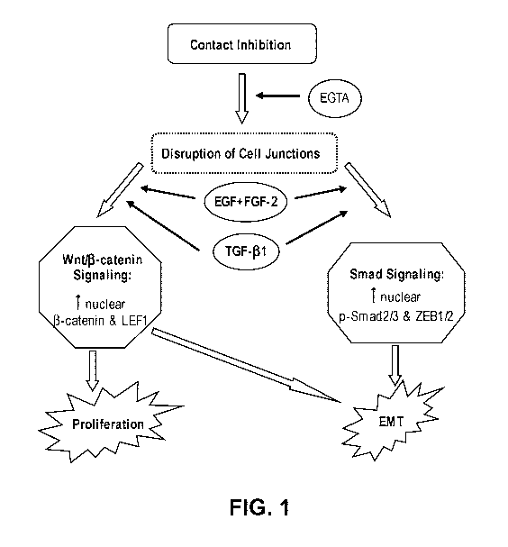
[0010] Figure 1 illustrates the signaling pathways in the regulating of EMT
with or without
proliferation by growth factors.
[0011] Figures 2A-2D illustrate HC-HA/PTX3 formation and characterization of
HC-HA/PTX3
purified from human AME. Figure 2A provides a schematic illustration of HC-
HA/PTX3
formation. Figure 2B illustrates HC-HA/PTX3 purified from human AME. Figure 2C
illustrates that the HC-HA/PTX3 purified from AME comprises HC1. Figure 2D
illustrates that
HC-HA/PTX3 purified from AME comprises PTX3.
[0012] Figures 3A- 3B illustrate canonical but not non-canonical Wnt signaling
is suppressed
by immobilized HC-HA/PTX3 in LEPCs/LNCs. Figure 3A illustrates that HC-HA/PTX3
downregulates canonical Wnt signaling in human limbal epithelial progenitor
cells (LEPCs) and
-10-
CA 02984443 2017-10-27
WO 2016/187555 PCT/US2016/033558
niche cells (LNCs). Figure 3B illustrates immunostaining of 13-catenin and C-
JUN seeded either
on Matrigel or on immobilized HC-HA/PTX3.
[0013] Figures 4A- 4D illustrate expression of TGF-13 and TGF-13 receptors in
human corneal
fibroblasts (HCF). Figure 4A illustrates TGF-131 expression in Human Corneal
Fibroblasts
(HCFs) seeded on plastic, HA, or HC-HA/PTX3, both with and without addition of
exogenous
TGF-131. Figure 4B illustrates TGF-132 expression in HCFs seeded on plastic,
HA, or HC-
HA/PTX3, both with and without addition of exogenous TGF-(31. Figure 4C
illustrates TGF-133
expression in HCFs seeded on plastic, HA, or HC-HA/PTX3, both with and without
addition of
exogenous TGF-(31. Figure 4D exemplifies a Northern blot showing expression of
TGF-13R1,
TGF-13R11, and TGF-13III in HCFs seeded on plastic, HA, or HC-HA/PTX3, both
with and
without addition of exogenous TGF-(31. Figure 4E exemplifies nuclear
translocation of
pSmad2/3 cause by addition of exogenous TGF- 131. Figure 4F exemplifies
positive cytoplasmic
expression of a-SMA caused by addition of exogenous TGF-1.
[0014] Figures 5A- 5C illustrate HC-HA/PTX3 inhibits proliferation in ARPE-19
cells when
stimulated with EGF+FGF-2. Figure 5A illustrates HC-HA/PTX3 does not affect
the viability
of normal ARPE-19 cells. Figures 5B illustrates proliferation of ARPE-19 cells
using
immunostaining. 5C illustrates proliferation of ARE-19 cells..
[0015] Figures 6A- 6B illustrate HC-HA/PTX3 inhibits nuclear translocation of
pSmad2/3 in
APRE-19 cells. Figure 6A illustrates nuclear localization of phosphorylated
Smad2/3 using
immunostaining. Figure 6B illustrates nuclear localization of phosphorylated
Smad2/3.
[0016] Figures 7A- 7D illustrate development of PVR in rabbits. Figure 7A
exemplifies fundus
photographs of a normal rabbit eye without PVR. Figure 7B exemplifies a rabbit
with tractional
PVR four weeks after gas vitrectomy and intravitreal injection of RPE cells.
Figure 7C
exemplifies a cross-section of the normal rabbit eye without PVR after
enucleation. Figure 7D
exemplifies a cross-section of the eye of the rabbit with tractional PVR with
retinal detachment
after enucleation.
[0017] Figure 8 illustrates the effect of the addition of collagen gel (Col),
AM extract AME, or
collagen gel mixed with AM extract (Col+AME) on the suppression of TGF-(3
promoter activity.
BSA was used as a control.
[0018] Figure 9 illustrates the effect of treatment with AME, HA, or HA+AME,
compared to a
control assay with BSA alone, on the suppression of TGF-(3 activity. The
promoter activity is
displayed as relative luciferase units (RLU).
-11-
CA 02984443 2017-10-27
WO 2016/187555 PCT/US2016/033558
[0019] Figure 10 illustrates the molecular weight ranges of hyaluronan in AM
extracts
separated by agarose gel electrophoresis. Amniotic membrane extracted by
buffer A, B, C were
treated with or without hyaluronidase and electrophoretically separated by a
0.5% agarose gel.
[0020] Figure 11 illustrates the molecular weight ranges of hyaluronan in AM
extracts
separated by agarose gel electrophoresis. Amniotic membrane extracted by
buffer PBS were
treated with or without hyaluronidase (10 units/ml in Tris-HC1, pH 7.5, 150 mM
NaC1) for 2 hr
at 37 C and run through 0.5% agarose gels. HA: positive hyaluronic acid
control; L: AM
extract after low speed centrifugation; H: AM extract after high speed
centrifugation.
[0021] Figure 12 illustrates a western blot demonstrating that the inter-a-
trypsin inhibitor (lad)
is present in AM extracts. IaI was present in AM extract A and C although the
signal of bikunin
was very weak (.about.39 kDa). Prior to transfer to the western blot, the
extract was separated on
a 4-15% denatured acrylamide gel.
[0022] Figure 13 illustrates an immunoblot demonstrating that the inter-a-
trypsin inhibitor (lad)
is present in the AM extracts even after low (LS) or high speed (HS)
centrifugation.
[0023] Figure 14 illustrates an immunoblot of TSG-6 (Tumor Necrosis Factor-
Stimulated Gene
6), either with (+) or without (-) hyaluronidase treatment. The samples
included total AM extract
without centrifugation (T), AM Extract after extraction in isotonic low salt
buffer (buffer A);
high salt buffer (B); or 4 M guanidine HC1(C); as detailed in Example 2. TSG-6
was present in
the total extract, buffer A extract, and buffer C extract. The addition of
hyaluronidase did not
appear to alter the TSG-6 level in the extracts.
[0024] Figure 15 illustrates an immunoblot analysis of the deglycosylation of
TSG-6 in AM.
AM extract A, B, and C were treated with (+) or without 20 units/ml PNGase F
at 37 C for 3
hours. Glycosylation of TSG-6 in AM was then analyzed by western blot. The
cell lysate of
human corneal fibroblast (HCF) was used as a positive control.
[0025] Figure 16 illustrates an immunoblot analysis of potential TSG-6
complexes in AM by
digestion with Chondroitin Sulfate ABC lyase. AM extract A, B, and C were
treated without (-)
or with (+) 1 unit/ml ABC lyase at 37 C for 2 hours. The possible disruption
of TSG-6
complexes was then analyzed by western blot using an anti-TSG-6 antibody RAH-
1:1:1000.
[0026] Figure 17 illustrates an immunoblot of potential TSG-6 complexes in AM
by digestion
with Chondroitin Sulfate ABC lyase. This is the same experiment as shown in
Figure 16 except
that a different TSG-6 antibody was used. Here, the anti-TSG-6 antibody was
obtained from
R & D Systems (cat# MAB2104).
[0027] Figure 18 illustrates an immunoblot demonstrating the presence of
Pentraxin (PTX3) in
AM, using a rat monoclonal anti-PTX3 antibody obtained from Alexis
Biochemicals. HCF:
-12-
CA 02984443 2017-10-27
WO 2016/187555 PCT/US2016/033558
human corneal fibroblast, T, A, B, C: AM extract Total, A, B, C, respectively;
HAse:
Hyaluronidase.
[0028] Figure 19 illustrates an immunoblot demonstrating the presence of TSP-1
in AM. The
monomeric TSP-1 (180 kDa) and the putative trimeric TSP-1 (540 kDa) are
indicated. The
positive control, TSP-1, was purified from human platelets (Calbiochem, Cat#
605225) and
loaded as 100 ng/lane.
[0029] Figure 20 illustrates an immunoblot demonstrating the presence of Smad
7 in AM. AM
was extracted with PBS or urea (2M urea in 50 mM Tris-HC1, pH 7.5). 20 1.tg of
total protein
was loaded for each extract. Smad 7 was detected with goat anti-human Smad 7
(AF2029,
1:1000, R & D Systems). Smad 7 migrated as a band of-5i kDa.
[0030] Figure 21 illustrates the number of migrated cells counted from six
random microscopic
fields (n=4, * indicates p<0.05 when compared with PBS+EGF+FGF-2+ TGF-01).
[0031] Figure 22 illustrates the percentage of gel contraction compared among
groups (n=4, *
indicates p<0.05 compared with PBS+TGF-131).
DETAILED DESCRIPTION OF THE INVENTION
[0032] The present application describes compositions and methods for
preventing or reducing
the proliferation, cell migration, and/or epithelial-mesenchymal transition
(EMT) of epithelial
cells, wherein the epithelial cells are human epithelial cells and the human
epithelial cells are
selected from: retinal pigment epithelial, conjunctival, retinal, corneal,
limbal, or renal epithelial
cells. Additionally, the present application describes compositions and
methods for the
prevention and treatment of proliferative vitreoretinopathy in an individual
in need thereof.
[0033] It is known that proliferation, cell migration and EMT occur when
epithelial cells such
as, for example, retinal pigment epithelial, human conjunctival, retinal,
corneal, limbal, or renal
epithelial cells are exposed to growth factors and cytokines such as, for
example, EGF, FGF-2,
PDGF-A, PDGF-AB, PDGF-B, PDGF-C, TGF-01, TGF-02, TGF-03, CTGF, HGF, IGF-1, G-
CSF, IL-6, MCP-1, TNF-a, VEGF or IFN-y and ethylene glycol tetraacetic acid
(EGTA) either
in vitro or in vivo.
[0034] Further, it is known that transplantation of cryopreserved amniotic
membrane (AM)
tissue onto the ocular surface provides anti-proliferative, anti-inflammatory,
anti-scarring and
anti-angiogenic actions in both corneal and limbal epithelial cells to promote
wound healing.
[0035] What is needed is a composition that prevents or reduces proliferation,
cell migration and
EMT of epithelial cells, can be administered without the need of surgical
transplantation and can
-13-
CA 02984443 2017-10-27
WO 2016/187555 PCT/US2016/033558
additionally be administered to non-surface epithelial cells such as, for
example, retinal and
renal epithelial cells.
[0036] A description of certain embodiments follows. It will be understood
that the particular
embodiments of the application are shown by way of illustration and not as
limitations of the
application.
Certain Terminology
[0037] Unless defined otherwise, all technical and scientific terms used
herein have the same
meaning as is commonly understood by one of skill in the art to which the
claimed subject
matter belongs. All patents, patent applications, published, applications and
publications,
GENBANK sequences, websites and other published materials referred to
throughout the entire
disclosure herein, unless noted otherwise, are incorporated by reference in
their entirety. In the
event that there is a plurality of definitions for terms herein, those in this
section prevail. Where
reference is made to a URL or other such identifier or address, it is
understood that such
identifiers can change and particular information on the internet can come and
go, but equivalent
information is known and can be readily accessed, such as by searching the
internet and/or
appropriate databases. Reference thereto evidences the availability and public
dissemination of
such information.
[0038] As used herein, ranges and amounts can be expressed as "about" a
particular value or
range. About also includes the exact amount. Hence, "about 5 g" means "about 5
lug" and also
"5 [lg." Generally, the term "about" includes an amount that would be expected
to be within
experimental error.
[0039] As used herein, the terms "subject", "individual", and "patient" are
used interchangeably.
None of the terms are to be interpreted as requiring the supervision of a
medical professional
(e.g., a doctor, nurse, physician's assistant, orderly, hospice worker). As
used herein, the subject
is any animal, including mammals (e.g., a human or non-human animal) and non-
mammals. In
one embodiment of the methods and compositions provided herein, the mammal is
a human.
[0040] As used herein, the terms "treat," "treating" or "treatment," and other
grammatical
equivalents, include: alleviating, abating or ameliorating one or more
symptoms of a disease or
condition. In some embodiments, treating is alleviating, abating or
ameliorating one or more
symptoms of epithelial-mesenchymal transition. In some embodiments, treating
is alleviating,
abating or ameliorating one or more symptoms of proliferative
vitreoretinopathy. In some
embodiments, treating is alleviating, abating or ameliorating one or more
symptoms of
inflammation. In some embodiments, treating is preventing or reducing the
appearance, severity
or frequency of one or more additional symptoms of a disease or condition. In
some
-14-
CA 02984443 2017-10-27
WO 2016/187555 PCT/US2016/033558
embodiments, the methods include preventing or reducing the appearance,
severity or frequency
of one or more additional symptoms of epithelial-mesenchymal transition. In
some
embodiments, the methods include preventing or reducing the appearance,
severity or frequency
of one or more additional symptoms of proliferative vitreoretinopathy. In some
embodiments,
the methods include preventing or reducing the appearance, severity or
frequency of one or more
additional symptoms of inflammation. In some embodiments, the methods include
ameliorating
or preventing the underlying metabolic causes of one or more symptoms of a
disease or
condition, inhibiting the disease or condition, such as, for example,
arresting the development of
the disease or condition, relieving the disease or condition, causing
regression of the disease or
condition, relieving a condition caused by the disease or condition, or
inhibiting the symptoms
of the disease or condition either prophylactically and/or therapeutically.
[0041] As used herein, "fetal support tissue" means tissue used to support the
development of a
fetus. Examples of fetal support tissue include, but are not limited to, (i)
placental amniotic
membrane (PAM), or substantially isolated PAM, (ii) umbilical cord amniotic
membrane
(UCAM) or substantially isolated UCAM, (iii) chorion or substantially isolated
chorion, (iv)
amnion-chorion or substantially isolated amnion-chorion, (v) amniotic stroma
or substantially
isolated amniotic stroma, (vi) placenta or substantially isolated placenta,
(vii) umbilical cord or
substantially isolated umbilical cord, (viii) amniotic fluid, or (ix) any
combinations thereof. Fetal
support tissue is also used interchangeably with "gestational tissue." In some
embodiments the
gestational tissue is "mammalian gestational tissue" or "human gestational
tissue ("HGT")." In
some embodiments, the fetal support tissue is obtained from a mammal. In some
embodiments,
the fetal support tissue is from human, non-human primate, cow, or pig. In
some embodiments,
the fetal support tissue is from human. In some embodiments, the fetal support
tissue is ground,
pulverized, morselized, a graft, a powder, a gel, a homogenate, or an extract.
In some
embodiments, the fetal support tissue is aseptically processed. In some
embodiment, the fetal
support tissue is terminally-sterilized.
[0042] As used herein, "placenta" means the organ that connects a developing
fetus to the
maternal uterine wall to allow nutrient uptake, waste elimination, and gas
exchange via the
maternal blood supply. The placenta is composed of three layers. The innermost
placental layer
surrounding the fetus is called amnion. The allantois is the middle layer of
the placenta (derived
from the embryonic hindgut); blood vessels originating from the umbilicus
traverse this
membrane. The outermost layer of the placenta, the chorion, comes into contact
with the
endometrium. The chorion and allantois fuse to form the chorioallantoic
membrane.
-15-
CA 02984443 2017-10-27
WO 2016/187555 PCT/US2016/033558
[0043] As used herein, "chorion" means the membrane formed by extraembryonic
mesoderm
and the two layers of trophoblasts. The chorionic villi emerge from the
chorion, invade the
endometrium, and allow transfer of nutrients from the maternal blood to fetal
blood. The chorion
consists of two layers: an outer layer formed by the trophoblast, and an inner
layer formed by
the somatic mesoderm; the amnion is contact with the latter. The trophoblast
is made up of an
internal layer of cubical or prismatic cells, the cytotrophoblast or layer of
Langhans, and an
external layer of richly nucleated protoplasm devoid of cell boundaries, the
syncytiotrophobast.
The avascular amnion is adherent to the inner layer of the chorion.
[0044] As used herein, "amnion-chorion" means a product comprising amnion and
chorion. In
some embodiments, the amnion and the chorion are not separated (i.e., the
amnion is naturally
adherent to the inner layer of the chorion). In some embodiments, the amnion
is initially
separated from the chorion and later combined with the chorion during
processing.
[0045] As used herein, "umbilical cord" means the organ that connects a
developing fetus to the
placenta. The umbilical cord is composed of Wharton's jelly, a gelatinous
substance made
largely form mucopolysaccharides. It contains one vein, which carries
oxygenated, nutrient-rich
blood to the fetus, and two arteries that carry deoxygenated, nutrient-
depleted blood away. In
some embodiments, the blood vessels have been substantially removed from the
umbilical cord
tissue. In some embodiments, a portion of the Wharton's Jelly has been
removed. In some
embodiments, the blood vessels and a portion of the Wharton's Jelly have been
removed.
[0046] As used herein, "placental amniotic membrane" (PAM) means amniotic
membrane
derived from the placenta. In some embodiments, the PAM is substantially
isolated.
[0047] As used herein, "umbilical cord amniotic membrane" (UCAM) means
amniotic
membrane derived from the umbilical cord. UCAM is a translucent membrane. The
UCAM has
multiple layers: an epithelial layer; a basement membrane; a compact layer; a
fibroblast layer;
and a spongy layer. It lacks blood vessels or a direct blood supply. In some
embodiments, the
UCAM is substantially isolated. In some embodiments, the UCAM comprises all of
the
Wharton's Jelly. In some embodiments, the UCAM comprises a portion of the
Wharton's Jelly.
In some embodiments, the UCAM comprises blood vessels and/or arteries. In some
embodiments, the UCAM comprises all of the Wharton's Jelly and blood vessels
and/or arteries.
In some embodiments, the UCAM comprises part of the Wharton's Jelly and blood
vessels
and/or arteries.
[0048] As used herein, "substantially isolated" or "isolated" means that the
fetal support tissue
product has been separate from undesired materials (e.g., red blood cells,
blood vessels, and
arteries) derived from the original source organism. Purity, or "isolation"
may be assayed by
-16-
CA 02984443 2017-10-27
WO 2016/187555 PCT/US2016/033558
standard methods, and will ordinarily be at least about 10% pure, more
ordinarily at least about
20% pure, generally at least about 30% pure, and more generally at least about
40% pure; in
further embodiments at least about 50% pure, or more often at least about 60%
pure; in still
other embodiments, at least about 95% pure.
[0049] As used herein, "biological activity" means the activity of
polypeptides and
polysaccharides. In some embodiments, the activity of polypeptides and
polysaccharides found
in umbilical cord (and substantially isolated umbilical cord), UCAM (and
substantially isolated
UCAM), placenta (and substantially isolated placenta), PAM (and substantially
isolated PAM),
chorion (and substantially isolated chorion), or amnion-chorion (and
substantially isolated
amnion-chorion). In some embodiments, the biological activity is anti-scarring
activity, anti-
inflammation activity, anti-angiogenic activity, and wound healing. In some
embodiments, the
biological activity is anti-inflammation activity. In some embodiments, the
biological activity
comprises the biological activity of a fetal support tissue preparation or
composition. In some
embodiments, the biological activity comprises the biological activity of HC-
HA/PTX3.
[0050] As used herein, the substantial preservation of biological activity or
structural integrity
means that when compared to the biological activity and structural integrity
of non-processed
tissue, the biological activity and structural integrity of the fetal support
tissue product has only
decreased by about 5%, about 10%, about 15%, about 20%, about 25%, about 30%,
about 35%,
about 40%, about 50%, or about 60%.
[0051] As used herein, "freezing" refers to exposing the fetal support tissue
product below about
or at 0 C, -5 C, -10 C, -20 C, -40 C, -50 C, -60 C, -70 C, -80 C, -90 C, or -
100 C for a period
of time of about 1 hour, 2 hours, 3 hours, 4 hours, 5 hours, 6 hours, 7 hours,
8 hours, 9 hours, 10
hours, 11 hours, 12 hours, 18 hours, 24 hours, or longer.
[0052] As used herein, "powder" means matter in the form of fine dry particles
or matrix. In
some embodiments, the particles are not uniform in size. In some embodiments,
the particles are
substantially uniform in size.
[0053] As used herein, "grinding" means any method of reducing fetal support
tissue to small
particles or a powder. The term grinding includes micronizing, pulverizing,
homogenizing,
filing, milling, grating, pounding, and crushing.
[0054] The terms "effective amount" or "therapeutically effective amount," as
used herein, refer
to a sufficient amount of an agent or a compound being administered which will
relieve to some
extent one or more of the symptoms of the disease or condition being treated.
The result can be
reduction and/or alleviation of the signs, symptoms, or causes of a disease,
or any other desired
alteration of a biological system. For example, an "effective amount" for
therapeutic uses is the
-17-
CA 02984443 2017-10-27
WO 2016/187555 PCT/US2016/033558
amount of the composition including a compound as disclosed herein required to
provide a
clinically significant decrease in disease symptoms without undue adverse side
effects. An
appropriate "effective amount" in any individual case may be determined using
techniques, such
as a dose escalation study. The term "therapeutically effective amount"
includes, for example, a
prophylactically effective amount. An "effective amount" of a compound
disclosed herein, is an
amount effective to achieve a desired effect or therapeutic improvement
without undue adverse
side effects. It is understood that "an effective amount" or "a
therapeutically effective amount"
can vary from individual to individual, due to variation in metabolism of the
injectable
composition, age, weight, general condition of the individual, the condition
being treated, the
severity of the condition being treated, and the judgment of the prescribing
physician. In some
embodiments, an effective amount is an amount that prevents or reduces the
symptoms of PVR.
In some embodiments, an effective amount is an amount that reduces, inhibits
or prevents cell
migration, cell proliferation and/or EMT of epithelial cells.
[0055] Epithelial-mesenchymal transition (EMT) is a process by which
epithelial cells lose their
cell polarity and cell-cell adhesion, and gain migratory and invasive
properties. EMT occurs in
processes such as mesoderm formation, neural tube formation, wound healing, as
well as the
initiation of metastasis for cancer progression. EMT can be induced through
several signal
signaling pathways, including TGF-13, FGF, EGF, HGF, Wnt/beta-catenin, and
Notch.
[0056] Proliferative vitreoretinopathy (PVR) is a disease that develops as a
complication of
rhegmatogenous retinal detachment. When fluid from the vitreous humor enters a
hole in the
retina and accumulates in the subretinal space, the tractional force of the
vitreous on the retina is
what results in rhegmatogenous retinal detachment. During this process the
retinal cell layers
come in contact with vitreous cytokines, which can trigger the retinal
pigmented epithelium
(RPE) to proliferate and migrate. The RPE cells undergo epithelial-mesenchymal
transition
(EMT) and develop the ability to migrate out into the vitreous. During
migration of the RPE,
these cells lay down fibrotic membranes which contract and pull at the retina,
and can lead to
secondary retinal detachment after primary retinal detachment surgery.
Compositions
[0057] Disclosed herein, in certain embodiments, are compositions for
preventing or reducing
proliferation, cell migration, and/or epithelial-mesenchymal transition (EMT)
of epithelial cells,
comprising: a preparation of fetal support tissue; and a pharmaceutically
acceptable diluent,
excipient, vehicle, or carrier. Further disclosed herein, in certain
embodiments, are injectable
compositions for preventing or reducing proliferative venous retinopathy (PVR)
in an individual
in need thereof, comprising: a preparation of fetal support tissue; and a
pharmaceutically
-18-
CA 02984443 2017-10-27
WO 2016/187555 PCT/US2016/033558
acceptable diluent, excipient, vehicle, or carrier. Further disclosed herein,
in certain
embodiments, are injectable compositions for preventing or reducing
proliferative venous
retinopathy (PVR) in an individual in need thereof, consisting essentially of:
substantially
isolated HC-HA/PTX3, reconstituted HC-HA/PTX3 (rcHC-HA/PTX3); and a
pharmaceutically
acceptable diluent, excipient, vehicle, or carrier. Further disclosed herein,
in certain
embodiments, are injectable compositions for preventing or reducing
proliferative venous
retinopathy (PVR) in an individual in need thereof, consisting essentially of:
substantially
isolated HC-HA/PTX3, reconstituted HC-HA/PTX3 (rcHC-HA/PTX3); an additional
therapeutic agent; and a pharmaceutically acceptable diluent, excipient,
vehicle, or carrier.
[0058] In some embodiments, the preparation of fetal support tissue comprises
HC-HA/PTX3.
In some embodiments, the preparation of fetal support tissue comprises: high
molecular weight
hyaluronan (HA) that is cross-linked by a covalent bond to the heavy chain of
inter-a-trypsin
inhibitor (lad), the high molecular weight HA having a molecular weight
greater than 1000 kDa.
In some embodiments, the preparation of fetal support tissue comprises:
pentraxin 3 (PTX-3,
PTX3). In some embodiments, the preparation of fetal support tissue comprises:
tumor necrosis
factor-stimulated gene 6 protein (TSG-6). In some embodiments, the preparation
of fetal support
tissue comprises: thrombospondin-1 (TSP-1). In some embodiments, the ratio of
total protein to
HA in the composition is less than 500 parts protein: 1 part HA. In some
embodiments, the ratio
of HA to total protein in the composition is less than 500 parts HA: 1 part
protein. In some
embodiments, the preparation of fetal support tissue comprises HC-HA/PTX3
complex. In some
embodiments, the preparation of fetal support tissue comprises substantially
purified HC-
HA/PTX3 complex.
[0059] In some embodiments, the epithelial cells are human epithelial cells.
In some
embodiments, the human epithelial cells are retinal pigment epithelial cells
(RPE). In some
embodiments, the human epithelial cells are corneal epithelial cells. In some
embodiments, the
human epithelial cells are limbal epithelial cells. In some embodiments, the
human epithelial
cells are conjunctival epithelial cells. In some embodiments, the human
epithelial cells are renal
epithelial cells.
[0060] In some embodiments, the composition prevents the proliferation and EMT
of epithelial
cells by suppressing the activity of growth factors and cytokines. In some
embodiments, the
growth factors and cytokines are selected from the group consisting of: EGF,
FGF-2, PDGF-A,
PDGF-AB, PDGF-B, PDGF-C, TGF-01, TGF-02, TGF-03, CTGF, HGF, IGF-1, G-CSF, IL-
6,
MCP-1, TNF-a, VEGF and IFN-y. In some embodiments, the composition inhibits
signaling
-19-
CA 02984443 2017-10-27
WO 2016/187555 PCT/US2016/033558
pathways in epithelial cells to inhibit proliferation and EMT. In some
embodiments, the
signaling pathways are canonical Wnt signaling and TGF-0-induced Smad/ZEB
signaling.
[0061] In some embodiments, the composition comprises the preparation of fetal
support tissue
and a pharmaceutically acceptable diluent, excipient, or carrier. In some
embodiments, the
composition further comprises an aqueous adjuvant. In some embodiments, the
composition is
for local administration. In some embodiments, the composition is formulated
for injection. In
some embodiments, the composition is formulated for intraocular injection,
subretinal injection,
intravitreal injection, periocular injection, subconjunctival injection,
retrobulbar injection,
intracameral injection or sub-Tenon's injection.
Preparations of Fetal Support Tissue
[0062] In some embodiments, the preparation of fetal support tissue comprises
placental tissue,
umbilical cord tissue, placental amniotic membrane tissue, chorion tissue,
amniotic stroma,
amnion-chorion tissue, UCAM tissue, amniotic fluid, or combinations thereof.
In some
embodiments, the preparation of fetal support tissue is an extract of fetal
support tissue,
micronized fetal support tissue, a homogenate of fetal support tissue, a
powder of fetal support
tissue, morselized fetal support tissue, pulverized fetal support tissue,
ground fetal support
tissue, purified HC-HA/PTX3, or a combination thereof. In some embodiments,
the preparation
of fetal support tissue is prepared from fresh, frozen or previously frozen
fetal support tissue. In
some embodiments, the preparation of fetal support tissue is prepared from
frozen or previously
frozen of fetal support tissue. In some embodiments, the preparation of fetal
support tissue
comprises HA, IaI, TSG-6, PTX-3, TSP-1, or a combination thereof. In some
embodiments, the
preparation of fetal support tissue comprises HC-HA/PTX3 complex. In some
embodiments,
the preparation of fetal support tissue comprises purified HC-HA/PTX3. In some
embodiments,
the preparation of fetal support tissue comprises ultracentrifuged HC-HA/PTX3.
In some
embodiments, the preparation of fetal support tissue consists of purified HC-
HA/PTX3. In some
embodiments, the preparation of fetal support tissue comprises reconstituted
HC-HA/PTX3.
[0063] In some embodiments, the preparation of fetal support tissue suppresses
TGF-13 promoter
activity; increases apoptosis in macrophages; decreases proliferation,
decreases migration, and
increases apoptosis of human vascular endothelial cells; decreases viability
of human
fibroblasts; decreases inflammation; and prevents apoptosis of epithelial
cells exposed to storage
and injury. In some embodiments, the preparations of fetal support tissue and
injectable
compositions described herein are used to treat diseases related to TGF-13
upregulation, such as
angiogenesis, wound healing, and tissue inflammation.
-20-
CA 02984443 2017-10-27
WO 2016/187555 PCT/US2016/033558
[0064] TGF-13 is the prototypic cytokine that is involved in tissue
inflammation, in addition to
wound healing and scar formation. Mammalian cells express three different TGF-
13s: TGF-01,
TGF-02, and TGF133. TGF-13 is the most potent cytokine promoting myofibroblast
differentiation by up-regulating expression of a-SMA, integrin a501, and EDA
domain-
containing fibronectin (Fn) in a number of cell types, including fibroblasts.
TGF-13 also up-
regulates the expression of such matrix components as collagens and
proteoglycans, down-
regulates proteinase and matrix metalloproteinases, and up-regulates their
inhibitors.
Collectively, these actions result in increased cell-matrix interactions and
adhesiveness, as well
as deposition and formation of scar tissue.
[0065] TGF-13s exert their actions via binding with TGF-13 receptors (TGF-
f3Rs) on the cell
membrane. In human cells, there are three TGF-f3Rs, namely TGF-13R type I (TGF-
13R1), type II
(TGF-13R11), and type III (TGF-13R111). TGF-13s, serving as ligands, bind with
a serine, threonine
kinase receptor complex made of TGF-13R1 and TGF-13R11; such a binding is
facilitated by TGF-
PRIII, which is not a serine, threonine kinase receptor. Binding with TGF-
13R11 activates TGF-
PRI, which is responsible for direct phosphorylation of a family of effector
proteins known as
Smads, which modulate transcription of a number of target genes, including
those described
herein, participating in scar formation.
[0066] Suppression of TGF-13 can be achieved by neutralizing antibodies to TGF-
13 and agents
that intercede the signaling mediated by TGF-13 such as decorin. Most of the
literature has shown
suppression of TGF-13 being achieved at the level of modulating the TGF-13
activation, binding
with its receptor, or its signal transduction. It has been shown that amniotic
membrane can
achieve such an inhibition at the level of transcription, i.e., to turn off
transcription of TGF-01
genes. In particular, amniotic membrane has been shown to suppress TGF-P
signaling in human
corneal and limbal fibroblasts, and human conjunctival and pterygium body
fibroblasts.
[0067] Hyaluronic acid (HA) is a natural sugar found in the synovial joint
fluid, the vitreous
humor of the eye, the cartilage, blood vessels, extra-cellular matrix, skin,
and umbilical cord. In
some embodiments, the cross-linking of HA is through a covalent bond to
another molecule,
such as a protein. In some embodiments, HA is covalently bound to the heavy
chain of inter-a-
trypsin inhibitor (lad). In some embodiments, the ratio of protein to HA in
the preparation of
fetal support tissue is less than about 500:1, less than about 200:1, less
than about 100:1, less
than about 50:1, or less than about 10:1 protein: HA. In some embodiments, the
ratio of HA to
protein in the preparation of fetal support tissue is less than about 500:1,
less than about 200:1,
less than about 100:1, less than about 50:1, or less than about 10:1 HA :
protein.
-21-
CA 02984443 2017-10-27
WO 2016/187555 PCT/US2016/033558
[0068] TSG-6 is a hyaluronan binding protein that plays a role in
extracellular matrix
remodeling, cell proliferation, and leucocyte migration. TSG-6 can form a
complex with the
serine protease inhibitor inter-a-inhibitor (lad) and catalyze the transfer of
a heavy chain from
IaI to HA. PTX-3 is Ca2+ dependent ligand binding protein that has a
pentameric discoid
structure and are present in plasma. TSP-1 (Thrombospondin 1) is a
homotrimeric glycoprotein
having a potent anti-angiogenic and other biological activities. TSP-1 is
secreted into the
extracellular matrix by a variety of cell types.
[0069] In some embodiments, the preparation of fetal support tissue comprises
a purified
component selected from HA, IaI, TSG-6, PTX-3, TSP-1, HC-HA/PTX3, or a
combination
thereof. In some embodiments the preparation of fetal support tissue comprises
reconstituted
HC-HA/PTX3. In some embodiments, the preparation of fetal support tissue
comprises purified
HC-HA/PTX3. In some embodiments, the preparation of fetal support tissue
consists of HC-
HA/PTX3. In some embodiments, the preparation of fetal support tissue
comprises purified HC-
HA/PTX3 at a high concentration. In some embodiments, the HC-HA/PTX3 is at a
concentration of 25 to 750 g/ml, 50 to 500 g/ml, 50 to 250 g/ml, or about
250 ug/ml, about
500 ug/ml, or about 750 ug/ml. In some embodiments, the purified component is
obtained from
any suitable source. In some embodiments, the purified component is obtained
from a fetal
support tissue. In some embodiments, the purified component of fetal support
tissue is obtained
from a commercial source. In some embodiments, the purified component of fetal
support tissue
is isolated from a transgenic organism. In some embodiments, a protein
sequence of the purified
component of fetal support tissue has a similarity of at least 90%, 93%, 95%,
97%, 99% or
99.5% to a human protein sequence. In some embodiments, the purified component
of fetal
support tissue is purified, substantially purified, partially purified, or are
present in crude
extracts. In some embodiments, the purified component of fetal support tissue
is HC-HA/PTX3.
In some embodiments, the purified component of fetal support tissue is
isolated from the
preparation of fetal support tissue at any time during the process.
[0070] In some embodiments, the preparation of fetal support tissue comprises
Smad7. In some
embodiments, Smad7 is obtained from any suitable source, such as from amniotic
membrane,
from a commercial source or isolated from a transgenic organism. In some
embodiments, Smad7
is purified, substantially purified, partially purified, or is present in a
crude extract.
[0071] In some embodiments, HA, IaI, TSG-6, PTX-3, TSP-1, and optionally Smad7
are
obtained from the preparation of fetal support tissue. In some embodiments,
the preparation of
fetal support tissue containing the combination of HA, IaI, TSG-6, PTX-3, TSP-
1 and optionally
Smad7 is prepared.
-22-
CA 02984443 2017-10-27
WO 2016/187555 PCT/US2016/033558
[0072] In some embodiments, after homogenization of the fetal support tissue,
is centrifuged to
remove the insoluble material. In some embodiments, after homogenization of
the fetal support
tissue, the insoluble material is left in the preparation of fetal support
tissue. In some
embodiments, the preparation of fetal support tissue is dried. In some
embodiments, a
preparation of fetal support tissue is prepared according to a method
described in Example 1.
[0073] In some embodiments, the fetal support tissue is obtained from sources
such as Bio-
Tissue, Inc. (Miami, FL) and Baptist Hospital (Miami, FL) (under IRB
approval). In some
embodiments, the fetal support tissue is obtained in either a fresh, frozen,
or previously frozen
state. In some embodiments, the fetal support tissue is washed to remove
excess storage buffer,
blood, or contaminants. In some embodiments, the excess liquid is removed
using a brief
centrifugation step, or by other means. In some embodiments, the fetal support
tissue is frozen
using liquid nitrogen or other cooling means to facilitate the subsequent
homogenization. In
some embodiments, the source of the fetal support tissue is a mammal. In some
embodiments,
the source of the fetal support tissue is a human. In some embodiments, other
sources of fetal
support tissue, such as non-human primate, bovine or porcine, are used.
[0074] In some embodiments, the preparation of fetal support tissue is
obtained from AM jelly.
In some embodiments, the AM jelly is obtained from fresh AM tissue. In some
embodiments,
AM jelly is obtained before freezing the fresh AM tissue. In some embodiments,
AM jelly is
obtained after freezing the fresh AM tissue. In some embodiments, AM jelly is
obtained from
frozen or previously frozen AM tissue. In some embodiments, the AM jelly is
frozen. In some
embodiments, the AM jelly is freeze-ground following the procedure for AM
preparations as
described herein. In some embodiments, the AM jelly is centrifuged. In some
embodiments, the
AM jelly is lyophilized.
[0075] In some embodiments, the preparation of fetal support tissue is made
from a stroma of
the AM. In some embodiments, the stroma is separated from a layer of fresh,
frozen, thawed, or
otherwise treated AM membrane. In some embodiments, the stroma removal occurs
by
enzymatic methods, mechanical methods, or by any other suitable means. In some
embodiments,
the stroma is fresh, frozen, or previously frozen. In some embodiments, the
stroma is ground or
freeze-ground following the procedure for generating the preparation of fetal
support tissue from
AM as described herein. In some embodiments, the stroma is centrifuged. In
some
embodiments, the stroma is lyophilized.
[0076] In some embodiment, the preparation is ground fetal support tissue. In
some
embodiments, the fetal support tissue is frozen prior to the grinding process.
In some
embodiments, the freezing step occurs by any suitable cooling process. In some
embodiments,
-23-
CA 02984443 2017-10-27
WO 2016/187555 PCT/US2016/033558
the fetal support tissue is flash-frozen using liquid nitrogen. In some
embodiments, the fetal
support tissue is placed in an isopropanol/dry ice bath or is flash-frozen in
other coolants. In
some embodiments, a commercially available quick freezing process is used. In
some
embodiments, the fetal support tissue is placed in a freezer and allowed to
equilibrate to the
storage temperature more slowly, rather than being flash-frozen. In some
embodiments, the fetal
support tissue is stored at any desired temperature. In some embodiments, the
fetal support
tissue is stored at -20 C or -80 C.
[0077] In some embodiment, the preparation is pulverized fetal support tissue.
In some
embodiments, the fetal support tissue is pulverized while frozen. In some
embodiments, fresh,
partially thawed, or thawed fetal support tissue is used in the grinding step.
In some
embodiments, the fetal support tissue (fresh, frozen, or thawed) is sliced
into pieces of a desired
size with a suitable device, such as a scalpel, then ground to fine particles
using a BioPulverizer
(Biospec Products, Inc., Bartlesville, OK) or other suitable devices, and
homogenized with a
homogenization device such as a Tissue Tearor (Biospec Products, Inc., Dremel,
WI), in a
suitable solution, forming a homogenate. Non-limiting examples of solutions
include, but are not
limited to, phosphate buffered saline (PBS), DMEM, NaC1 solution, and water.
In some
embodiments, the pH of the solution is adjusted as needed. In some
embodiments, the pH range
is from about 5.5 or 6.0 to about 8.5. In some embodiments, the frozen tissue
is ground in a
solution having a pH of between about 6.3 and about 7.8.
[0078] In some embodiment, the preparation is a homogenate of fetal support
tissue. In some
embodiments, the homogenate is mixed at any suitable speed, temperature, or
other parameters.
In some embodiments, the mixing occurs at a temperature range of from about 1
C, or 3 C, to
about 6 C, 10 C, 15 C, or 20 C. In some embodiments, the mixing occurs at
about 4 C. In some
embodiments, the homogenate is mixed, for example, from less than about 1
minute, 10 minutes,
or 20 minutes to about 1, 2, 3 or more hours.
[0079] In some embodiments, the homogenate is centrifuged to remove any
remaining insoluble
material and/or cellular debris. In some embodiments, the centrifugation is
performed using any
suitable range of time, temperature, protein concentration, buffers, and
speed. In some
embodiments, the centrifugation occurs at a range of about 1,000, 5,000, or
10,000xg to about
20,000xg. In some embodiments, the centrifugation occurs at about 15,000xg. In
some
embodiments, the centrifugation occurs for a duration of from less than 1
minute, 5 minutes, 10
minutes, 20 minutes, to about 40 minutes, 60 minutes, 1.5 hours, or more. In
some
embodiments, the supernatant is collected and stored in aliquots at -80 C. In
some embodiments,
the total protein is quantitated using any suitable commercial protein
analysis kit, such as a BCA
-24-
CA 02984443 2017-10-27
WO 2016/187555 PCT/US2016/033558
assay (Pierce, Rockford, IL). Example 2, Table 1 and FIG. 13 describe the
analysis of AM
preparations after low speed or high speed centrifugation.
[0080] In some embodiments, for biochemical characterization and purification,
the above
solutions are supplemented with protease inhibitors. An exemplary mixture of
protease
inhibitors is the following: 1 g/m1 aprotinin, 1 g/mlleupeptin, 1
g/m1pepstatin A, and 1 mM
PMSF. In some embodiments, a protease inhibitor is not added to the
preparation of fetal
support tissue if the preparation of fetal support tissue is to be added to
live cells or tissues.
[0081] In some embodiment, the preparation is an extract fetal support tissue.
In some
embodiments, any suitable buffer or liquid is used to prepare an extract of
fetal support tissue.
Example 2 examines the use of various extraction buffers (high salt, low salt,
PBS, etc.) on total
protein content and HA in the extract of fetal support tissue (Table 1).
Example 2 examined the
levels of the specific proteins TSG-6 (FIG. 14), PTX-3 (FIG. 18), TSP-1 (FIG.
19), and Smad7
(FIG. 20) using several extraction methods.
[0082] In some embodiments, the preparation of fetal support tissue is tested
to confirm the
presence of specific components or proteins. In some embodiments, the
preparation of fetal
support tissue is tested for the presence of molecules including, but not
limited to, HA, IaI,
TSG-6, PTX-3, TSP-1, and Smad7. In some embodiments, the preparation of fetal
support tissue
is tested to confirm the absence of pathogens at any point during the
preparation process.
[0083] In some embodiments, the preparation of fetal support tissue is a dry
powder. In some
embodiments, the dry powder does not require refrigeration or freezing during
storage to keep
the dry powder from degrading over time. In some embodiments, the dry powder
is stored and
reconstituted prior to use. In some embodiments, the dry powder is prepared by
preparing the
freeze-ground fetal support tissue as described herein, then removing at least
a portion of the
water in the preparation of fetal support tissue. In some embodiments, the
excess water is
removed from the preparation of fetal support tissue by any suitable means. In
some
embodiments, is the excess water is removed by use of lyophilization. In some
embodiments,
lyophilizing the preparation of fetal support tissue comprises using a
commercially available
lyophilizer or freeze-dryer. In some embodiments, suitable equipment is found,
for example,
through Virtis (Gardiner, NY); FTS Systems (Stone Ridge, NY); and SpeedVac
(Savant
Instruments Inc., Farmingdale, NY). In some embodiments, the amount of water
that is removed
is from about 5%, 10%, 20%, 30% to about 60, 70, 80, 90, 95 or 99% or more. In
some
embodiments, substantially all of the excess water is removed from the
preparation of fetal
support tissue. In some embodiments, the dry powder is stored. In some
embodiments, the
storage temperature varies from less than about -196 C, -80 C, -50 C, or -20 C
to more than
-25-
CA 02984443 2017-10-27
WO 2016/187555 PCT/US2016/033558
about 23 C. In some embodiments, the dry powder is characterized (weight,
protein content,
etc.) prior to storage.
[0084] In some embodiments, the dry powder is reconstituted in a suitable
solution or buffer
prior to use. Non-limiting examples of solutions include, but are not limited
to, PBS, DMEM,
and BSS. In some embodiments, the pH of the solution is adjusted as needed. In
some
embodiments, the dry powder is reconstituted with a sufficient volume of
solution to produce a
high concentration of the fetal support tissue reconstituted composition. In
some embodiments,
the dry powder is reconstituted with a sufficient volume of solution to
produce a low
concentration of the fetal support tissue reconstituted composition.
[0085] In some embodiments, the dry powder is reconstituted in a cream,
ointment, gel, foam or
lotion].
[0086] In some embodiments, the preparation of fetal support tissue is used to
produce a
phenotypic reversal of AMSCs from myofibroblasts to fibroblasts. In some
embodiments, the
preparation of fetal support tissue is used to prevent or slow differentiation
of various cell types.
In some embodiments, many types of cells are treated with the preparation of
fetal support
tissue.
Isolated nHC-HA/PTX3 Complexes
[0087] In some embodiments, the compositions include isolated native HC-
HA/PTX3
complexes (nHC-HA/PTX3).
[0088] In some embodiments, the nHC-HA/PTX3 complexes are isolated from an
isolated cell.
In some embodiments, the nHC-HA/PTX3 complexes are isolated from a cultured
cell. In some
embodiments, the nHC-HA/PTX3 complexes are isolated from a stem cell. In some
embodiments, the nHC-HA/PTX3 complexes are isolated from a water soluble
fraction of an
extract prepared from a tissue, such as umbilical cord or amniotic membrane.
In some
embodiments, the water soluble fraction is extracted with an isotonic salt
solution. In some
embodiments, the nHC-HA/PTX3 complexes are isolated from a water insoluble
fraction of an
extract prepared from a tissue, such as umbilical cord or amniotic membrane.
In some
embodiments, the insoluble fraction is extracted with GnHC1.
[0089] In some embodiments, the isolated nHC-HA/PTX3 complex is isolated from
an amniotic
tissue. In some embodiments, the isolated nHC-HA/PTX3 complex is isolated from
an amniotic
membrane or an umbilical cord. In some embodiments, the isolated nHC-HA/PTX3
complex is
isolated from fresh, frozen or previously frozen placental amniotic membrane
(PAM), fresh,
frozen or previously frozen umbilical cord amniotic membrane (UCAM), fresh,
frozen or
previously frozen placenta, fresh, frozen or previously frozen umbilical cord,
fresh, frozen or
-26-
CA 02984443 2017-10-27
WO 2016/187555 PCT/US2016/033558
previously frozen chorion, fresh, frozen or previously frozen amnion-chorion,
or any
combinations thereof. Such tissues can be obtained from any mammal, such as,
for example, but
not limited to a human, non-human primate, cow or pig.
[0090] In some embodiments, the nHC-HA/PTX3 is purified by any suitable
method. In some
embodiments, the nHC-HA/PTX3 complex is purified by centrifugation (e.g.,
ultracentrifugation, gradient centrifugation), chromatography (e.g., ion
exchange, affinity, size
exclusion, and hydroxyapatite chromatography), gel filtrationõ or differential
solubility, ethanol
precipitation or by any other available technique for the purification of
proteins (See, e.g.,
Scopes, Protein Purification Principles and Practice 2nd Edition, Springer-
Verlag, New York,
1987; Higgins, S. J. and Hames, B. D. (eds.), Protein Expression: A Practical
Approach, Oxford
Univ Press, 1999; and Deutscher, M. P., Simon, M. I., Abelson, J. N. (eds.),
Guide to Protein
Purification: Methods in Enzymology (Methods in Enzymology Series, Vol 182),
Academic
Press, 1997, all incorporated herein by reference).
[0091] In some embodiments, the nHC-HA/PTX3 is isolated from an extract. In
some
embodiments, the extract is prepared from an amniotic membrane extract. In
some
embodiments, the extract is prepared from an umbilical cord extract. In some
embodiments, the
umbilical cord extract comprises umbilical cord stroma and/or Wharton's jelly.
In some
embodiments, the nHC-HA/PTX3 complex is contained in an extract that is
prepared by
ultracentrifugation. In some embodiments, the nHC-HA/PTX3 complex is contained
in an
extract that is prepared by ultracentrifugation using a CsCl/4-6M guanidine
HC1 gradient. In
some embodiments, the extract is prepared by at least 2 rounds of
ultracentrifugation. In some
embodiments, the extract is prepared by more than 2 rounds of
ultracentrifugation (i.e. nHC-
HA/PTX3 2nd). In some embodiments, the extract is prepared by at least 4
rounds of
ultracentrifugation (i.e. nHC-HA/PTX3 4th). In some embodiments, the nHC-
HA/PTX3
complex comprises a small leucine-rich proteoglycan. In some embodiments, the
nHC-
HA/PTX3 complex comprises HC1, HA, PTX3 and/or a small leucine-rich
proteoglycan.
[0092] In some embodiments, ultracentrifugation is performed on an extract
prepared by
extraction in an isotonic solution. In some embodiments, the isotonic solution
is PBS. For
example, in some embodiments the tissue is homogenized in PBS to produce a
homogenized
sample. The homogenized sample is then separated into a soluble portion and
insoluble portion
by centrifugation. In some embodiments, ultracentrifugation is performed on
the soluble portion
of the PBS-extracted tissue. In such embodiments, the nHC-HA/PTX3 purified by
ultracentrifugation of the PBS-extracted tissue called an nHC-HA/PTX3 soluble
complex. In
some embodiments, the nHC-HA soluble complex comprises a small leucine-rich
proteoglycan.
-27-
CA 02984443 2017-10-27
WO 2016/187555 PCT/US2016/033558
In some embodiments, the nHC-HA/PTX3 soluble complex comprises HC1, HA, PTX3
and/or a
small leucine-rich proteoglycan.
[0093] In some embodiments, ultracentrifugation is performed on an extract
prepared by direct
guanidine HC1 extraction (e.g. 4-6 M GnHC1) of the amniotic membrane and/or
umbilical cord
tissue. In some embodiments, the GnHC1 extract tissues is then centrifuged to
produce GnHC1
soluble and GnHC1 insoluble portions. In some embodiments, ultracentrifugation
is performed
on the GnHC1 soluble portion. In such embodiments, the nHC-HA/PTX3 purified by
ultracentrifugation of the guanidine HC1-extracted tissue is called an nHC-
HA/PTX3 insoluble
complex. In some embodiments, the nHC-HA insoluble complex comprises a small
leucine-rich
proteoglycan. In some embodiments, the nHC-HA/PTX3 insoluble complex comprises
HC1,
HA, PTX3 and/or a small leucine-rich proteoglycan.
[0094] In some embodiments, ultracentrifugation is performed on an extract
prepared by further
guanidine HC1 extraction of the insoluble portion of the PBS-extracted tissue.
For example, in
some embodiments the tissue is homogenized in PBS to produce a homogenized
sample. The
homogenized sample is then separated into a soluble portion and insoluble
portion by
centrifugation. The insoluble portion is then further extracted in guanidine
HC1 (e.g. 4-6 M
GnHC1) and centrifuged to produce a guanidine HC1 soluble and insoluble
portions. In some
embodiments, ultracentrifugation is performed on the guanidine HC1 soluble
portion. In such
embodiments, the nHC-HA/PTX3 purified by ultracentrifugation of the guanidine
HC1-extracted
tissue is called an nHC-HA/PTX3 insoluble complex. In some embodiments, the
nHC-HA
insoluble complex comprises a small leucine-rich proteoglycan. In some
embodiments, the nHC-
HA/PTX3 insoluble complex comprises HC1, HA, PTX3 and/or a small leucine-rich
proteoglycan.
[0095] In some embodiments, the method of purifying the isolated nHC-HA/PTX3
extract
comprises: (a) dissolving the isolated extract (e.g. prepared by the soluble
or insoluble method
described herein) in CsCl/4-6M guanidine HC1 at the initial density of 1.35
g/ml, to generate a
CsC1 mixture, (b) centrifuging the CsC1 mixture at 125,000 x g for 48 h at 15
C, to generate a
first purified extract, (c) extracting the first purified extract and
dialyzing it against distilled
water to remove CsC1 and guanidine HC1, to generate a dialysate. In some
embodiments, the
method of purifying the isolated extract further comprises (d) mixing the
dialysate with 3
volumes of 95% (v/v) ethanol containing 1.3% (w/v) potassium acetate at 0 C
for 1 h, to
generate a first dialysate/ethanol mixture, (e) centrifuging the first
dialysate/ethanol mixture at
15,000 x g, to generate a second purified extract, and (f) extracting the
second purified extract.
In some embodiments, the method of purifying the isolated extract further
comprises: (g)
-28-
CA 02984443 2017-10-27
WO 2016/187555 PCT/US2016/033558
washing the second purified extract with ethanol (e.g., 70% ethanol), to
generate a second
purified extract/ethanol mixture; (h) centrifuging the second purified
extract/ethanol mixture, to
generate a third purified extract; and (i) extracting the third purified
extract. In some
embodiments, the method of purifying the isolated extract further comprises:
(j) washing the
third purified extract with ethanol (e.g., 70% ethanol), to generate a third
purified extract/ethanol
mixture; (k) centrifuging the third purified extract/ethanol mixture, to
generate a forth purified
extract; and (1) extracting the forth purified extract. In some embodiments,
the purified extract
comprises an nHC-HA/PTX3 complex.
[0096] In some embodiments, the nHC-HA/PTX3 complex is purified by
immunoaffinity
chromatography. In some embodiments, anti HC1 antibodies, anti-HC2 antibodies,
or both are
generated and affixed to a stationary support. In some embodiments, the
unpurified HC-HA
complex (i.e., the mobile phase) is passed over the support. In certain
instances, the HC-HA
complex binds to the antibodies (e.g., via interaction of (a) an anti-HC1
antibody and HC1, (b)
an anti-HC2 antibody and HC2, (c) an anti-PTX antibody and PTX3, (d) an anti-
SLRP antibody
and the SLRP, or (e) any combination thereof). In some embodiments the support
is washed
(e.g., with PBS) to remove any unbound or loosely bound molecules. In some
embodiments, the
support is then washed with a solution that enables elution of the nHC-HA/PTX3
complex from
the support (e.g., 1% SDS, 6M guanidine-HC1, or 8M urea).
[0097] In some embodiments, the nHC-HA/PTX3 complex is purified by affinity
chromatography. In some embodiments, HABP is generated and affixed to a
stationary support.
In some embodiments, the unpurified nHC-HA/PTX3 complex (i.e., the mobile
phase) is passed
over the support. In certain instances, the nHC-HA/PTX3 complex binds to the
HABP. In some
embodiments the support is washed (e.g., with PBS) to remove any unbound or
loosely bound
molecules. In some embodiments, the support is then washed with a solution
that enables elution
of the HC-HA complex from the support.
[0098] In some embodiments, the nHC-HA/PTX3 complex is purified by a
combination of
HABP affinity chromatography, and immunoaffinity chromatography using anti HC1
antibodies, anti-HC2 antibodies, anti-PTX3 antibodies, antibodies against a
SLRP or a
combination of SLRPs, or any combination of antibodies thereof.
[0099] In some embodiments, the nHC-HA/PTX3 complex is purified from the
insoluble
fraction as described herein using one or more antibodies. In some
embodiments, the nHC-
HA/PTX3 complex is purified from the insoluble fraction as described herein
using anti-SLRP
antibodies.
-29-
CA 02984443 2017-10-27
WO 2016/187555 PCT/US2016/033558
[0100] In some embodiments, the nHC-HA/PTX3 complex is purified from the
soluble fraction
as described herein. In some embodiments, the nHC-HA/PTX3 complex is purified
from the
soluble fraction as described herein using anti-PTX3 antibodies.
[0101] In some embodiments, the nHC-HA/PTX3 complex comprises a small leucine
rich
proteoglycan (SLRP). In some embodiments, the nHC-HA/PTX3 complex comprises a
class I,
class II or class II SLRP. In some embodiments, the small leucine-rich
proteoglycan is selected
from among class I SLRPs, such as decorin and biglycan. In some embodiments,
the small
leucine-rich proteoglycan is selected from among class II SLRPs, such as
fibromodulin,
lumican, PRELP (proline arginine rich end leucine-rich protein), keratocan,
and osteoadherin. In
some embodiments, the small leucine-rich proteoglycan is selected from among
class III SLRPs,
such as epipycan and osteoglycin. In some embodiments, the small leucine-rich
proteoglycan is
selected from among bikunin, decorin, biglycan, and osteoadherin. In some
embodiments, the
small leucine-rich protein comprises a glycosaminoglycan. In some embodiments,
the small
leucine-rich proteoglycan comprises keratan sulfate.
rcHC-HA/PTX3 Complexes
[0102] In some embodiments, the compositions comprise reconstituted HC-HA/PTX3
complexes (rcHC-HA/PTX3) with or without SLRPs.
[0103] In some embodiments, a method for generating reconstituted HC-HA/PTX3
complexes
comprises (a) contacting immobilized high molecular weight hyaluronan (HMW HA)
with
pentraxin 3 (PTX3) under suitable conditions to form a PTX3/HA complex, and
(b) contacting
the PTX3/HA complex with IaI and Tumor necrosis factor-Stimulated Gene-6 (TSG-
6).
Provided herein are rcHC-HA/PTX3 complexes produced by such method. In some
embodiments, TSG-6 catalyzes the transfer of heavy chain 1 (HC1) of inter-a-
inhibitor (lad) to
HA. In some embodiments, HC1 of IaI forms a covalent linkage with HA. In some
embodiments, the steps (a) and (b) of the method are performed sequentially in
order.
[0104] In some embodiments, a method for generating reconstituted HC-HA/PTX3
complexes
comprises contacting a PTX3/HA complex with IaI and TSG-6. In some
embodiments, TSG-6
catalyzes the transfer of heavy chain 1 (HC1) of inter-a-inhibitor (lad) to
HA. Provided herein
are rcHC-HA/PTX3 complexes produced by such method. In some embodiments, HC1
of IaI
forms a covalent linkage with HA.
[0105] In some embodiments, a method for generating a complex of HA bound to
PTX3
comprises contacting immobilized high molecular weight hyaluronan (HMW HA)
with
pentraxin 3 (PTX3) under suitable conditions to form a PTX3/HA complex.
Provided herein are
PTX3/HA complexes produced by such method.
-30-
CA 02984443 2017-10-27
WO 2016/187555 PCT/US2016/033558
[0106] In some embodiments, a method for generating reconstituted HC-HA/PTX3
complexes
comprises (a) contacting immobilized high molecular weight hyaluronan (HMW HA)
with IaI
and TSG-6 to HA to form an HC-HA complex pre-bound to TSG-6 and (b) contacting
the HC-
HA complex with pentraxin 3 (PTX3) under suitable conditions to form an rcHC-
HA/PTX3
complex. Provided herein are rcHC-HA/PTX3 complexes produced by such method.
In some
embodiments, HC lof IaI forms a covalent linkage with HA. In some embodiments,
the steps (a)
and (b) of the method are performed sequentially in order. In some
embodiments, the method
comprises contacting an HC-HA complex pre-bound to TSG-6 with PTX3.
[0107] In some embodiments, the method comprises first contacting high
molecular weight
hyaluronan (HMW HA) with pentraxin 3 (PTX3) under suitable conditions to form
a PTX3/HA
complex, then contacting the PTX3/HA complex with IaI and TSG-6.
[0108] In some embodiments, the IaI protein and TSG-6 protein are contacted to
the complex
at a molar ratio of about 1:1, 2:1, 3:1, 4:1, 5:1, 6:1, 7:1, 8:1, 9:1, 10:1,
15:1, or 20:1 (IaI:TSG-6).
In some embodiments the ratio of IaI:TSG-6 ranges from about 1:1 to about
20:1, such as about
1:1 to about 10:1, such as about 1:1 to 5 about:1, such as about 1:1 to about
3:1. In some
embodiments, the ratio of IaI:TSG-6 is 3:1 or higher. In some embodiments, the
ratio of
IaI:TSG-6 is 3:1.
[0109] In some embodiments, the steps (a) and (b) of the method are performed
sequentially
in order. In some embodiments, the method comprises contacting a PTX3/HA
complex with IaI
and TSG-6.
[0110] In certain instances, TSG-6 interacts with IaI and forms covalent
complexes with HC1
and HC2 of IaI (i.e. HC1=TSG-6 and HC2=TSG-6). In certain instances, in the
presence of HA,
the HCs are transferred to HA to form rcHC-HA. In some embodiments, a TSG-
6=HC1 complex
is added to pre-bound PTX3/HA complex to catalyze the transfer of HC1 to HA.
In some
embodiments, the method comprises first contacting immobilized high molecular
weight
hyaluronan (HMW HA) with pentraxin 3 (PTX3) under suitable conditions to form
a PTX3/HA
complex, then contacting the PTX3/HA complex with a HC1=TSG-6 complex. In some
embodiments, a combination of HC1=TSG-6 complex and HC2=TSG-6 complex is added
to a
PTX3/HA complex.
[0111] In some embodiments, the step of contacting PTX3 to immobilized HMW HA
occurs
for at least 10 minutes, at least 30 minutes, at least 1 hour, at least 2
hours, at least 3 hours, at
least 4 hours, at least 5 hours, at least 6 hours, at least 12 hours, or at
least 24 hours or longer. In
some embodiments, the step of contacting PTX3 to immobilized HMW HA occurs for
at least 2
hours or longer. In some embodiments, the step of contacting PTX3 to
immobilized HMW HA
-31-
CA 02984443 2017-10-27
WO 2016/187555 PCT/US2016/033558
occurs for at least 2 hours. In some embodiments, the step of contacting PTX3
to immobilized
HMW HA occurs at 37 C. In some embodiments, the step of contacting PTX3 to
immobilized
HMW HA occurs in 5 mM MgC12 in PBS.
[0112] In some embodiments, the step of contacting the PTX3/HA complex with
IaI and
TSG-6 to HA occurs for at least 10 minutes, at least 30 minutes, at least 1
hour, at least 2 hours,
at least 3 hours, at least 4 hours, at least 5 hours, at least 6 hours, at
least 12 hours, or at least 24
hours or longer. In some embodiments the step of contacting the PTX3/HA
complex with a
HC1=TSG-6 complex and/or a HC2=TSG-6 complex occurs for at least 10 minutes,
at least 30
minutes, at least 1 hour, at least 2 hours, at least 3 hours, at least 4
hours, at least 5 hours, at least
6 hours, at least 12 hours, or at least 24 hours or longer. In some
embodiments the step of
contacting the PTX3/HA complex with a HC1=TSG-6 complex and/or a HC2=TSG-6
complex
occurs for at least 2 hours or longer. In some embodiments the step of
contacting the PTX3/HA
complex with a HC1=TSG-6 complex and/or a HC2=TSG-6 complex occurs for at
least 2 hours.
In some embodiments the step of contacting the PTX3/HA complex with a HC1=TSG-
6
complex and/or a HC1=TSG-6 complex occurs at 37 C. In some embodiments the
step of
contacting the PTX3/HA complex with a HC1=TSG-6 complex and/or a HC1=TSG-6
complex
occurs in 5 mM MgC12 in PBS.
[0113] In some embodiments, the method comprises contacting high molecular
weight
hyaluronan (HMW HA) with a pentraxin 3 (PTX3) protein, inter-a-inhibitor (lad)
protein
comprising heavy chain 1 (HC1) and Tumor necrosis factor a-stimulated gene 6
(TSG-6)
simultaneously under suitable conditions to form a HC-HA/PTX3 complex. In some
embodiments, the contacting the HMW HA with PTX3, IaI and TSG-6 occurs for at
least 10
minutes, at least 30 minutes, at least 1 hour, at least 2 hours, at least 3
hours, at least 4 hours, at
least 5 hours, at least 6 hours, at least 12 hours, or at least 24 hours or
longer. In some
embodiments the step of contacting the HMW HA, PTX3, IaI, and TSG-6 occurs at
37 C. In
some embodiments the step of contacting the HMW HA, PTX3, IaI, and TSG-6
occurs in 5 mM
MgC12 in PBS.
[0114] In some embodiments, the method comprises contacting high molecular
weight
hyaluronan (HMW HA) with a pentraxin 3 (PTX3) protein, inter-a-inhibitor (lad)
protein
comprising heavy chain 1 (HC1) and Tumor necrosis factor a-stimulated gene 6
(TSG-6)
sequentially, in any order, under suitable conditions to form a HC-HA/PTX3
complex. In some
embodiments, the contacting the HMW HA with PTX3, IaI and TSG-6 occurs for at
least 10
minutes, at least 30 minutes, at least 1 hour, at least 2 hours, at least 3
hours, at least 4 hours, at
least 5 hours, at least 6 hours, at least 12 hours, or at least 24 hours or
longer. In some
-32-
CA 02984443 2017-10-27
WO 2016/187555 PCT/US2016/033558
embodiments the step of contacting the HMW HA, PTX3, IaI, and TSG-6 occurs at
37 C. In
some embodiments the step of contacting the HMW HA, PTX3, IaI, and TSG-6
occurs in 5 mM
MgC12 in PBS.
[0115] In some embodiments, the methods for production of an rcHC-HA/PTX3
complex
further comprises addition of one or more small leucine rich proteoglycans
(SLRPs). In some
embodiments, a method for generating reconstituted HC-HA/PTX3 complexes
comprises (a)
contacting immobilized high molecular weight hyaluronan (HMW HA) with
pentraxin 3 (PTX3)
under suitable conditions to form a PTX3/HA complex, (b) contacting the
PTX3/HA complex
with IaI and Tumor necrosis factor-Stimulated Gene-6 (TSG-6) and (c)
contacting the PTX3/HA
complex with one or more SLRPS. Provided herein are rcHC-HA/PTX3 complexes
produced by
such method. In some embodiments, TSG-6 catalyzes the transfer of heavy chain
1 (HC1) of
inter-a-inhibitor (lad) to HA. In some embodiments, HC1 of IaI forms a
covalent linkage with
HA. In some embodiments, the steps (a), (b), and (c) of the method are
performed sequentially
in order. In some embodiments, the steps (a), (b), and (c) of the method are
performed
simultaneously. In some embodiments, the step (a) of the method is performed
and then steps (b)
and (c) of the method are performed sequentially in order. In some
embodiments, the step (a) of
the method is performed and then steps (b) and (c) of the method are performed
simultaneously.
[0116] In some embodiments, a method for generating reconstituted HC-HA/PTX3
complexes
comprises (a) contacting immobilized high molecular weight hyaluronan (HMW HA)
with IaI
and TSG-6 to HA to form an HC-HA complex pre-bound to TSG-6, (b) contacting
the HC-HA
complex with pentraxin 3 (PTX3) and (c) contacting the HC-HA complex with one
or more
SLRPS under suitable conditions to form an rcHC-HA/PTX3 complex. Provided
herein are
rcHC-HA/PTX3 complexes produced by such method. In some embodiments, HC lof
IaI forms
a covalent linkage with HA. In some embodiments, the method comprises
contacting an HC-HA
complex pre-bound to TSG-6 with PTX3. In some embodiments, the steps (a), (b),
and (c) of the
method are performed sequentially in order. In some embodiments, the steps
(a), (b), and (c) of
the method are performed simultaneously. In some embodiments, the step (a) of
the method is
performed and then steps (b) and (c) of the method are performed sequentially
in order. In some
embodiments, the step (a) of the method is performed and then steps (b) and
(c) of the method
are performed simultaneously.
[0117] In some embodiments, the SLRP is selected from among a class I, class
II or class II
SLRP. In some embodiments, the SLRP is selected from among class I SLRPs, such
as decorin
and biglycan. In some embodiments, the small leucine-rich proteoglycan is
selected from among
class II SLRPs, such as fibromodulin, lumican, PRELP (proline arginine rich
end leucine-rich
-33-
CA 02984443 2017-10-27
WO 2016/187555 PCT/US2016/033558
protein), keratocan, and osteoadherin. In some embodiments, the small leucine-
rich
proteoglycan is selected from among class III SLRPs, such as epipycan and
osteoglycin. In some
embodiments, the small leucine-rich proteoglycan is selected from among
bikunin, decorin,
biglycan, and osteoadherin. In some embodiments, the small leucine-rich
protein comprises a
glycosaminoglycan. In some embodiments, the small leucine-rich proteoglycan
comprises
keratan sulfate.
[0118] PTX3
[0119] In some embodiments, PTX3 for use in the methods is isolated from a
cell or a
plurality of cells (e.g., a tissue extract). Exemplary cells suitable for the
expression of PTX3
include, but are not limited to, animal cells including, but not limited to,
mammalian cells,
primate cells, human cells, rodent cells, insect cells, bacteria, and yeast,
and plant cells,
including, but not limited to, algae, angiosperms, gymnosperms, pteridophytes
and bryophytes.
In some embodiments, PTX3 for use in the methods is isolated from a human
cell. In some
embodiments, PTX3 for use in the methods is isolated from a cell that is
stimulated with one or
more proinflammatory cytokines to upregulate PTX3 expression. In some
embodiments, the
proinflammatory cytokine is IL-1 or TNF-a.
[0120] In some embodiments, PTX3 for use in the methods is isolated from an
amniotic
membrane cell. In some embodiments, PTX3 for use in the methods is isolated
from an amniotic
membrane cell from an umbilical cord. In some embodiments, the amniotic
membrane cell is
stimulated with or more proinflammatory cytokines to upregulate PTX3
expression. In some
embodiments, the proinflammatory cytokine is IL-1 or TNF-a.
[0121] In some embodiments, PTX3 for use in the methods is isolated from an
umbilical cord
cell. In some embodiments, the umbilical cord cell is stimulated with or more
proinflammatory
cytokines to upregulate PTX3 expression. In some embodiments, the
proinflammatory cytokine
is IL-1 or TNF-a.
[0122] In some embodiments, PTX3 for use in the methods is isolated from an
amniotic
epithelial cell. In some embodiments, PTX3 for use in the methods is isolated
from an umbilical
cord epithelial cell. In some embodiments, the amniotic epithelial cell or
umbilical cord
epithelial cell is stimulated with or more proinflammatory cytokines to
upregulate PTX3
expression. In some embodiments, the proinflammatory cytokine is IL-1 or TNF-
a.
[0123] In some embodiments, PTX3 for use in the methods is isolated from an
amniotic
stromal cell. In some embodiments, PTX3 for use in the methods is isolated
from an umbilical
cord stromal cell. In some embodiments, the amniotic stromal cell or umbilical
cord stromal cell
-34-
CA 02984443 2017-10-27
WO 2016/187555 PCT/US2016/033558
is stimulated with or more proinflammatory cytokines to upregulate PTX3
expression. In some
embodiments, the proinflammatory cytokine is IL-1 or TNF-a.
[0124] In some embodiments, PTX3 for use in the methods is a native PTX3
protein isolated
from a cell. In some embodiments, the cell is stimulated with or more
proinflammatory
cytokines to upregulate PTX3 expression. In some embodiments, the
proinflammatory cytokine
is IL-1 or TNF-a.
[0125] In some embodiments, PTX3 is prepared by recombinant technology. In
some
embodiments, PTX3 is expressed from a recombinant expression vector. In some
embodiments,
nucleic acid encoding PTX3 is operably linked to a constitutive promoter. In
some
embodiments, nucleic acid encoding PTX3 is operably linked to an inducible
promoter. In some
embodiments, PTX3 is expressed in a transgenic animal. In some embodiments,
PTX3 is a
recombinant protein. In some embodiments, PTX3 is a recombinant protein
isolated from a cell.
In some embodiments, PTX3 is a recombinant protein produced in a cell-free
extract.
[0126] In some embodiments, PTX3 is purified from amniotic membrane, umbilical
cord,
umbilical cord amniotic membrane, chorionic membrane, amniotic fluid, or a
combination
thereof. In some embodiments, PTX3 is purified from amniotic membrane cells.
In some
embodiments, the amniotic membrane cell is an amniotic epithelial cell. In
some embodiments,
the amniotic membrane cell is an umbilical cord epithelial cell. In some
embodiments, the
amniotic membrane cell is an amniotic stromal cell. In some embodiments, the
amniotic
membrane cell is an umbilical cord stromal cell. In some embodiments, the
amniotic membrane
cell is stimulated with or more proinflammatory cytokines to upregulate PTX3
expression. In
some embodiments, the proinflammatory cytokine is IL-1 or TNF-a.
[0127] In some embodiments, PTX3 is not isolated from a cell or a plurality of
cells (e.g., a
tissue extract).
[0128] In some embodiments, PTX3 comprises a polypeptide having the sequence
set forth in
SEQ ID NO: 33 or a variant thereof having at least 65%, 70%, 75%, 80%, 85%,
90%, 95%,
96%, 97%, 98%, or 99% sequence amino acid identity to the polypeptide having
the sequence
set forth in SEQ ID NO: 33. Exemplary variants include, for example, species
variants, allelic
variants and variants that contain conservative and non-conservative amino
acid mutations. In
some embodiments, PTX3 comprises a fragment of PTX3 sufficient to bind to HA
and facilitate
the formation of rcHC-HA/PTX3 complex. In some embodiments, PTX3 comprises
Glul8 to
5er277 of human PTX3. Variants of PTX3for use in the provided methods include
variants with
an amino acid modification that is an amino acid replacement (substitution),
deletion or
insertion. In some embodiments, such modification improves one or more
properties of the
-35-
CA 02984443 2017-10-27
WO 2016/187555 PCT/US2016/033558
PTX3 polypeptides such as improving the one or more therapeutic properties of
the rcHC-
HA/PTX3 complex (e.g., anti-inflammatory, anti-immune, anti-angiogenic, anti-
scarring, anti-
adhesion, regeneration or other therapeutic activities as described herein).
[0129] In some embodiments PTX3 protein is obtained from a commercial source.
An
exemplary commercial source for PTX3 is, but is not limited to, PTX3 (Catalog
No. 1826-TS;
R&D Systems, Minneapolis, MN).
[0130] In some embodiments, the PTX3 protein used in the methods is a
multimeric protein.
In some embodiments, the PTX3 protein used in the methods is a homomultimer.
In some
embodiments, the homomultimer is a dimer, trimer, tetramer, hexamer, pentamer,
or octamer. In
some embodiments, the PTX3 homomultimer is a trimer, tetramer, or octamer. In
particular
embodiments, the PTX3 homomultimer is an octamer. In some embodiments, the
multimerization domain is modified to improve multimerization of the PTX3
protein. In some
embodiments, the multimerization domain is replaced with a heterogeneous
multimerization
domain (e.g., an Fc multimerization domain or leucine zipper) that when fused
to PTX3
improves the multimerization of PTX3.
[0131] TSG-6
[0132] In some embodiments, TSG-6 for use in the methods is isolated from a
cell or a
plurality of cells (e.g., a tissue extract). Exemplary cells suitable for the
expression of TSG-6
include, but are not limited to, animal cells including, but not limited to,
mammalian cells,
primate cells, human cells, rodent cells, insect cells, bacteria, and yeast,
and plant cells,
including, but not limited to, algae, angiosperms, gymnosperms, pteridophytes
and bryophytes.
In some embodiments, TSG-6 for use in the methods is isolated from a human
cell. In some
embodiments, TSG-6 for use in the methods is isolated from a cell that is
stimulated with one or
more proinflammatory cytokines to upregulate TSG-6 expression. In some
embodiments, the
proinflammatory cytokine is IL-1 or TNF-a.
[0133] In some embodiments, TSG-6 for use in the methods is isolated from an
amniotic
membrane cell. In some embodiments, TSG-6 for use in the methods is isolated
from an
amniotic membrane cell from an umbilical cord. In some embodiments, TSG-6 for
use in the
methods is isolated from an amniotic membrane cell that is stimulated with one
or more
proinflammatory cytokines to upregulate TSG-6 expression. In some embodiments,
the
proinflammatory cytokine is IL-1 or TNF-a.
[0134] In some embodiments, TSG-6 for use in the methods is isolated from an
umbilical cord
cell. In some embodiments, TSG-6 for use in the methods is isolated from an
umbilical cord cell
-36-
CA 02984443 2017-10-27
WO 2016/187555 PCT/US2016/033558
that is stimulated with one or more proinflammatory cytokines to upregulate
TSG-6 expression.
In some embodiments, the proinflammatory cytokine is IL-1 or TNF-a.
[0135] In some embodiments, TSG-6 for use in the methods is isolated from an
amniotic
epithelial cell. In some embodiments, TSG-6 for use in the methods is isolated
from an umbilical
cord epithelial cell. In some embodiments, TSG-6 for use in the methods is
isolated from an
amniotic epithelial cell or an umbilical cord epithelial cell that is
stimulated with one or more
proinflammatory cytokines to upregulate TSG-6 expression. In some embodiments,
the
proinflammatory cytokine is IL-1 or TNF-a.
[0136] In some embodiments, TSG-6 for use in the methods is isolated from an
amniotic
stromal cell. In some embodiments TSG-6 for use in the methods is isolated
from an umbilical
cord stromal cell. In some embodiments, TSG-6 for use in the methods is
isolated from an
amniotic stromal cell or an umbilical cord stromal cell that is stimulated
with one or more
proinflammatory cytokines to upregulate TSG-6 expression. In some embodiments,
the
proinflammatory cytokine is IL-1 or TNF-a.
[0137] In some embodiments, TSG-6 for use in the methods is a native TSG-6
protein isolated
from a cell. In some embodiments, the cell is stimulated with or more
proinflammatory
cytokines to upregulate TSG-6 expression. In some embodiments, the
proinflammatory cytokine
is IL-1 or TNF-a.
[0138] In some embodiments, TSG-6 is prepared by recombinant technology. In
some
embodiments, TSG-6 is expressed from a recombinant expression vector. In some
embodiments,
nucleic acid encoding TSG-6 is operably linked to a constitutive promoter. In
some
embodiments, nucleic acid encoding TSG-6 is operably linked to an inducible
promoter. In some
embodiments, TSG-6 is expressed in a transgenic animal. In some embodiments,
TSG-6 is a
recombinant protein. In some embodiments, TSG-6 is a recombinant protein
isolated from a cell.
In some embodiments, TSG-6 is a recombinant protein produced in a cell-free
extract.
[0139] In some embodiments, TSG-6 is purified from amniotic membrane, amniotic
membrane, chorionic membrane, amniotic fluid, or a combination thereof. In
some
embodiments, PTX3 is purified from amniotic membrane cells. In some
embodiments, the
amniotic membrane cell is an amniotic epithelial cell. In some embodiments,
the amniotic
epithelial cell is an umbilical cord epithelial cell. In some embodiments, the
amniotic membrane
cell is an amniotic stromal cell. In some embodiments, the amniotic membrane
cell is an
umbilical cord stromal cell. In some embodiments, the amniotic membrane cell
is stimulated
with or more proinflammatory cytokines to upregulate TSG-6 expression. In some
embodiments, the proinflammatory cytokine is IL-1 or TNF-a.
-37-
CA 02984443 2017-10-27
WO 2016/187555 PCT/US2016/033558
[0140] In some embodiments, TSG-6 is not isolated from a cell or a plurality
of cells (e.g., a
tissue extract).
[0141] In some embodiments, TSG-6 comprises a fragment of TSG-6 that is
sufficient to
facilitate or catalyze the transfer HC1 of IaI to HA. In some embodiments, TSG-
6 comprises the
link module of TSG-6. In some embodiments, TSG-6 comprises amino acids Trp18
through
Leu277 of TSG-6. In some embodiments, TSG-6 comprises a polypeptide having the
sequence
set forth in SEQ ID NO: 2 or a variant thereof having at least 65%, 70%, 75%,
80%, 85%, 90%,
95%, 96%, 97%, 98%, or 99% sequence amino acid identity to the polypeptide
having the
sequence set forth in SEQ ID NO: 2. Exemplary variants include, for example,
species variants,
allelic variants and variants that contain conservative and non-conservative
amino acid
mutations. Natural allelic variants of human TSG-6 include, for example, TSG-6
containing the
amino acid replacement Q144R. Variants of TSG-6 or HA binding fragments
thereof for use in
the provided methods include variants with an amino acid modification that is
an amino acid
replacement (substitution), deletion or insertion. In some embodiments, such
modification
improve one or more properties of the TSG-6 polypeptides such as improved
transfer of HC1 of
IaI to HA or improved release of the TSG-6 polypeptide from the rcHC-HA/PTX3
complex
following transfer of HC1 of IaI to HA.
[0142] In some embodiments, TSG-6 comprises an affinity tag. Exemplary
affinity tags
include but are not limited to a hemagglutinin tag, a poly-histidine tag, a
myc tag, a FLAG tag, a
glutathione-S-transferase (GST) tag. Such affinity tags are well known in the
art for use in
purification. In some embodiments, such an affinity tag incorporated into the
TSG-6 polypeptide
as a fusion protein or via a chemical linker. In some embodiments, TSG-6
comprises an affinity
tag and the unbound TSG-6 is removed from the rcHC-HA/PTX3 complex by affinity
purification.
[0143] In some embodiments TSG-6 protein is obtained from a commercial source.
An
exemplary commercial source for TSG-6 is, but is not limited to, TSG-6
(Catalog No. 2104-TS
R&D Systems, Minneapolis, MN).
[0144] la
[0145] In some embodiments, the IaI comprises an HC1 chain. In some
embodiments, the IaI
comprises an HC1 and an HC2 chain. In some embodiments, the IaI comprises an
HC1 and
bikunin. In some embodiments, the IaI comprises an HC1, and HC2 chain and
bikunin. In some
embodiments, the IaI comprises an HC1, and HC2 chain and bikunin linked by a
chondroitin
sulfate chain.
-38-
CA 02984443 2017-10-27
WO 2016/187555 PCT/US2016/033558
[0146] In some embodiments, IaI is isolated from a biological sample. In some
embodiments
the biological sample is a biological sample from a mammal. In some
embodiments, the
mammal is a human. In some embodiments, the biological sample is a blood,
serum, plasma,
liver, amniotic membrane, chorionic membrane or amniotic fluid sample. In some
embodiments,
the biological sample is a blood, serum, or plasma sample. In some
embodiments, the biological
sample is a blood sample. In some embodiments, the biological sample is a
serum sample. In
some embodiments, the biological sample is a plasma sample. In some
embodiments, the IaI is
purified from human blood, plasma or serum. In some embodiments, IaI is
isolated from human
serum. In some embodiments, IaI is not isolated from serum. In some
embodiments, IaI for use
in the methods is produced in an amniotic membrane cell. In some embodiments,
IaI for use in
the methods is produced in an umbilical cord cell. In some embodiments, IaI
for use in the
methods is produced in an amniotic membrane cell from an umbilical cord. In
some
embodiments, IaI for use in the methods is produced in an amniotic epithelial
cell. In some
embodiments, IaI for use in the methods is produced in an umbilical cord
epithelial cell. In some
embodiments, IaI for use in the methods is produced in an amniotic stromal
cell. In some
embodiments, IaI for use in the methods is produced in an umbilical cord
stromal cell. In some
embodiments, IaI for use in the methods is produced in a hepatic cell. In some
embodiments, IaI
is prepared by recombinant technology.
[0147] In some embodiments, HC1 of IaI is isolated from a biological sample.
In some
embodiments the biological sample is a biological sample from a mammal. In
some
embodiments, the mammal is a human. In some embodiments, the biological sample
is a blood,
serum, plasma, liver, amniotic membrane, chorionic membrane or amniotic fluid
sample. In
some embodiments, the biological sample is a blood, serum, or plasma sample.
In some
embodiments, the biological sample is a blood sample. In some embodiments, the
biological
sample is a serum sample. In some embodiments, the biological sample is a
plasma sample. In
some embodiments, the HC1 of IaI is purified from human blood, plasma or
serum. In some
embodiments, IaI is isolated from human serum. In some embodiments, HC1 of IaI
is not
purified from serum. In some embodiments, HC1 of IaI is prepared by
recombinant technology.
In some embodiments, HC1 of IaI is purified from hepatic cells. In some
embodiments, HC1 of
IaI is purified from amniotic membrane cells. In some embodiments, HC1 of IaI
is purified from
amniotic epithelial cells or umbilical cord epithelial cells. In some
embodiments, HC1 of IaI is
purified from amniotic stromal cells or umbilical cord stromal cells.
[0148] In some embodiments, HC1 comprises a polypeptide having the sequence
set forth in
SEQ ID NO: 47 or a polypeptide having at least 75%, 80%, 85%, 90%, 95%, 96%,
97%, 98%,
-39-
CA 02984443 2017-10-27
WO 2016/187555 PCT/US2016/033558
or 99% sequence amino acid identity to the polypeptide having the sequence set
forth in SEQ ID
NO: 47.
[0149] In some embodiments, HC2 of IaI is isolated from a biological sample.
In some
embodiments the biological sample is a biological sample from a mammal. In
some
embodiments, the mammal is a human. In some embodiments, the biological sample
is a blood,
serum, plasma, liver, amniotic membrane, chorionic membrane or amniotic fluid
sample. In
some embodiments, the biological sample is a blood, serum, or plasma sample.
In some
embodiments, the biological sample is a blood sample. In some embodiments, the
biological
sample is a serum sample. In some embodiments, the biological sample is a
plasma sample. In
some embodiments, the HC2 of IaI is purified from human blood, plasma or
serum. In some
embodiments, HC2 of IaI is isolated from human serum. In some embodiments, HC2
of IaI is
isolated from human serum. In some embodiments, HC2 of IaI is not isolated
from blood serum.
In some embodiments, HC2 of IaI is prepared by recombinant technology. In some
embodiments, HC2 of IaI is purified from hepatic cells. In some embodiments,
HC2 of IaI is
purified from amniotic membrane cells. In some embodiments, HC2 of IaI is
purified from
amniotic epithelial cells or umbilical cord epithelial cells. In some
embodiments, HC2 of IaI is
purified from amniotic stromal cells or umbilical cord stromal cells.
[0150] In some embodiments, HC2 comprises a polypeptide having the sequence
set forth in
SEQ ID NO: 49 or a polypeptide having at least 75%, 80%, 85%, 90%, 95%, 96%,
97%, 98%,
or 99% sequence amino acid identity to the polypeptide having the sequence set
forth in SEQ ID
NO: 49.
[0151] In some embodiments, IaI comprises bikunin. In some embodiments,
bikunin
comprises a polypeptide having the sequence set forth in SEQ ID NO: 53 or a
polypeptide
having at least 75%, 80%, 85%, 90%, 95%, 96%, 97%, 98%, or 99% sequence amino
acid
identity to the polypeptide having the sequence set forth in SEQ ID NO: 53. In
some
embodiments, IaI comprises a chondroitin sulfate chain.
[0152] HA
[0153] In some embodiments, HA is purified from a cell, tissue or a fluid
sample. In some
embodiments, HA is obtained from a commercial supplier (e.g., Sigma Aldrich or
Advanced
Medical Optics, Irvine, CA (e.g., Healon)). In some embodiments, HA is
obtained from a
commercial supplier as a powder. In some embodiments, HA is expressed in a
cell. Exemplary
cells suitable for the expression of HA include, but are not limited to,
animal cells including, but
not limited to, mammalian cells, primate cells, human cells, rodent cells,
insect cells, bacteria,
and yeast, and plant cells, including, but not limited to, algae, angiosperms,
gymnosperms,
-40-
CA 02984443 2017-10-27
WO 2016/187555 PCT/US2016/033558
pteridophytes and bryophytes. In some embodiments, HA is expressed in a human
cell. In some
embodiments, HA is expressed in a transgenic animal. In some embodiments, HA
is obtained
from a cell that expresses a hyaluronan synthase (e.g., HAS1, HAS2, and HAS3).
In some
embodiments, the cell contains a recombinant expression vector that expresses
an HA synthase.
In certain instances, an HA synthase lengthens hyaluronan by repeatedly adding
glucuronic acid
and N-acetylglucosamine to the nascent polysaccharide as it is extruded
through the cell
membrane into the extracellular space.
[0154] HA for use in the methods is typically high molecular weight (HMW) HA.
In some
embodiments, the weight average molecular weight of HMW HA is greater than
about 500
kilodaltons (kDa), such as, for example, between about 500 kDa and about
10,000 kDa, between
about 800 kDa and about 8,500 kDa, between about 1100 kDa and about 5,000 kDa,
or between
about 1400 kDa and about 3,500 kDa. In some embodiments, the weight average
molecular
weight of HMW HA is about 3000 kDa.
[0155] Additional Components
[0156] In some embodiments, one or more additional components are added to
generate an
rcHC-HA/PTX3 complex. In some embodiments, a small leucine rich proteoglycan
(SLRP) is
added to generate an rcHC-HA/PTX3 complex. In some embodiments, the SLRP is a
class I,
class II or class II SLRP. In some embodiments, the SLRP is selected from
among class I
SLRPs, such as decorin and biglycan. In some embodiments, the SLRP is selected
from among
class II SLRPs, such as fibromodulin, lumican, PRELP (proline arginine rich
end leucine-rich
protein), keratocan, and osteoadherin. In some embodiments, the SLRP is
selected from among
class III SLRPs, such as epipycan and osteoglycin. In some embodiments, the
SLRP is selected
from among bikunin, decorin, biglycan, and osteoadherin. In some embodiments,
the SLRP
comprises a glycosaminoglycan. In some embodiments, the SLRP comprises keratan
sulfate.
[0157] HA Immobilization
[0158] In some embodiments, HMW HA is immobilized by any suitable method. In
some
embodiments, HMW HA is immobilized to a solid support, such as culture dish,
bead, a column
or other suitable surfaces, such as, for example, a surface of an implantable
medical device or a
portion thereof or on a surface that is subsequently connected to or combined
with an
implantable medical device as described herein. In some embodiments, HMW HA is
immobilized directly to the solid support, such a by chemical linkage. In some
embodiments,
HMW HA is attached indirectly to the solid support via a linker or an
intermediary protein.
Numerous heterobifunctional cross-linking reagents that are used to form
covalent bonds
between amino groups and thiol groups and to introduce thiol groups into
proteins, are known to
-41-
CA 02984443 2017-10-27
WO 2016/187555 PCT/US2016/033558
those of skill in this art. In some embodiments, HMW HA is immobilized
directly to the solid
support via crosslinking to the solid support. In some embodiments, HMW HA is
immobilized
directly to the solid support without crosslinking to the solid support. In
some embodiments,
HMW HA is immobilized directly to the solid support as a coating. In some
embodiments,
HMW HA is immobilized to a CovalinkTm-NH surface. In some embodiments, HMW HA
is
immobilized directly to the solid support as a coating. In some embodiments,
HMW HA is
immobilized to a CovalinkTm-NH surface for about 16 h at 4 C.
[0159] In some embodiments, the method comprises immobilizing HMW HA to a
solid
surface via direct linkage to a solid support (i.e. without an intermediary
protein). In some
embodiments, the solid support is washed to remove unbound HMW HA prior to
contacting the
immobilized HA with PTX3. In some embodiments, the solid support is washed
with washes of
8M GnHC1 and PBS to remove unbound HMW HA prior to contacting the immobilized
HA
with PTX3.
[0160] In some embodiments, the method comprises immobilizing HA to a solid
surface via
an intermediary protein or a linker. In some embodiments, the linker is a
peptide linker. In some
embodiments, the intermediary protein is an HA binding protein (HABP). In some
embodiments, HABP is first attached to a solid support (e.g., by cross-
linking, chemical linkage
or via a chemical linker). In some embodiments, the solid support comprising
HABP is then
contacted with HA (e.g., HMW HA) to immobilize HA to the solid support via
binding of the
HABP to HA. In some embodiments, the solid support is washed to remove unbound
HMW HA
prior to contacting the immobilized HMW HA with PTX3. In some embodiments, the
solid
support is washed with washes of 8M GnHC1 and PBS to remove unbound HMW HA
prior to
contacting the immobilized HA with PTX3.
[0161] In some embodiments, the method comprises immobilizing HA to a solid
surface via
attachment of a peptide linker to the solid support and attachment HA to the
peptide linker. In
some embodiments, the peptide linker comprises a protease cleavage site.
[0162] In some embodiments, the method comprises immobilizing HA to a solid
surface via
attachment of a cleavable chemical linker, such as, but not limited to a
disulfide chemical linker.
[0163] In some embodiments, the HABP selected for use in the methods is an
HABP that is
dissociated from HA following formation of the rcHC-HA/PTX3 complex. In some
embodiments, the HABP non-covalently binds to HA. In some embodiments, the
method further
comprises dissociating the rcHC-HA/PTX3 complex from HABP using one or more
dissociating
agents. Dissociating agents for the disruption of non covalent interactions
(e.g., guanidine
hydrochloride, urea and various detergents, e.g., SDS) are known in the art.
In some
-42-
CA 02984443 2017-10-27
WO 2016/187555 PCT/US2016/033558
embodiments the dissociating agent is urea. In some embodiments the
dissociating agent is
guanidine hydrochloride. In some embodiments, the dissociation agent is about
4M to about 8M
guanidine-HC1. In some embodiments, the dissociation agent is about 4M, about
5M, about 6M,
about 7M, about 8M guanidine-HC1. In some embodiments, the dissociation agent
is about 4M
to about 8M guanidine-HC1 in PBS at pH 7.5.
[0164] In some embodiments, such dissociating agents are employed to
dissociate the rcHC-
HA/PTX3 complex from an intermediary HABP. An HABP for use in the methods
typically is
selected such that the binding affinity for HA is strong enough to permit
assembly of the rcHC-
HA/PTX3 complex but is dissociated from the rcHC-HA/PTX3 complex with a
suitable
dissociation agent. In some embodiments the dissociating agent is guanidine
hydrochloride.
[0165] Exemplary HABPs for use with the methods provided herein include, but
are not
limited to, HAPLN1, HAPLN2, HAPLN3, HAPLN4, aggrecan, versican, neurocan,
brevican,
phosphacan, TSG-6, CD44, stabilin-1, stabilin-2, or portions thereof (e.g.,
link modules thereof)
sufficient to bind HA. In some embodiments, the HABP comprises a polypeptide
having the
sequence set forth in any of SEQ ID NOS: 54-99 or a polypeptide having at
least 75%, 80%,
85%, 90%, 95%, 96%, 97%, 98%, or 99% sequence amino acid identity to the
polypeptide
having the sequence set forth in any of SEQ ID NOS: 54-99. In some
embodiments, the HABP
is versican. In some embodiments, the HABP is a recombinant protein. In some
embodiments,
the HABP is a recombinant mammalian protein. In some embodiments, the HABP is
a
recombinant human protein. In some embodiments, the HABP is a recombinant
versican protein
or a portion thereof sufficient to bind to HA. In some embodiments, the HABP
is a recombinant
aggrecan protein or a portion thereof sufficient to bind to HA. In some
embodiments, the HABP
is a native HABP or a portion thereof sufficient to bind to HA. In some
embodiments, the native
HABP is isolated from mammalian tissue or cells. In some embodiments, the HABP
is isolated
from bovine nasal cartilage (e.g. HABP from Seikagaku which contains the HA
binding
domains of aggrecan and link protein).
[0166] In some embodiments, the HABP comprises a link module of HAPLN1,
HAPLN2,
HAPLN3, HAPLN4, aggrecan, versican, neurocan, brevican, phosphacan, TSG-6,
CD44,
stabilin-1, or stabilin-2. In some embodiments, the HABP comprising a link
module comprises a
polypeptide having the sequence set forth in any of link domains of SEQ ID
NOS: 54-99 or a
polypeptide having at least 75%, 80%, 85%, 90%, 95%, 96%, 97%, 98%, or 99%
sequence
amino acid identity to the polypeptide having the sequence set forth in any of
link domains of
SEQ ID NOS: 54-99. In some embodiments, the HABP comprises a link module of
versican. In
-43-
CA 02984443 2017-10-27
WO 2016/187555 PCT/US2016/033558
some embodiments, the HABP comprising a link module is a recombinant protein.
In some
embodiments, the HABP comprising a link module of versican is a recombinant
protein.
[0167] In some embodiments, the or intermediary protein, such as an HABP,
contains a
proteolytic cleavage sequence that is recognized by and is hydrolyzed by a
site specific protease,
such as furin, 3C protease, caspase, matrix metalloproteinase or TEV protease.
In such
embodiments, assembled rcHC-HA/PTX3 complexes are released from the solid
support by
contacting the immobilized complexes with a protease that cleaves the specific
cleavage
sequence.
[0168] In some embodiments, the rcHC-HA/PTX3 complex is purified. In some
embodiments,
the rcHC-HA/PTX3 complex is purified by any suitable method or combination of
methods. The
embodiments described below are not intended to be exclusive, only exemplary.
[0169] In some embodiments, the rcHC-HA/PTX3 complex is purified by
chromatography
(e.g., ion exchange, affinity, size exclusion, and hydroxyapatite
chromatography), gel filtration,
centrifugation (e.g., gradient centrifugation), or differential solubility,
ethanol precipitation or by
any other available technique for the purification of proteins.
[0170] In some embodiments, the rcHC-HA/PTX3 complex is purified by
immunoaffinity
chromatography. In some embodiments antibodies are generated against a
component of the
rcHC-HA/PTX3 complex (e.g., anti-HC1, anti-PTX, an antibody against one or
more SLRPs of
the rcHC-HA/PTX3 complex, e.g., anti-bikunin, anti-decorin, anti-biglycan, or
anti-
osteoadherin) and affixed to a solid support. In some embodiments, the
unpurified rcHC-
HA/PTX3 complex (i.e., the mobile phase) is passed over the support. In
certain instances, the
rcHC-HA/PTX3 complex binds to the antibodies. In some embodiments, the support
is washed
(e.g., with PBS) to remove any unbound or loosely bound molecules. In some
embodiments, the
support is then washed with a solution that enables elution of the rcHC-
HA/PTX3 complex from
the support (e.g., 1% SDS, 6M guanidine-HC1, or 8M urea). In some embodiments,
the
dissociating agent is removed from the dissociated rcHC-HA/PTX3 complex. In
some
embodiments, the dissociating agent is removed from the dissociated rcHC-
HA/PTX3 complex
by a method including, but not limited to, ion-exchange chromatography,
dialysis, gel filtration
chromatography, ultrafiltration, or diafiltration.
[0171] In some embodiments, the rcHC-HA/PTX3 complex is purified by affinity
chromatography. In some embodiments, an HABP is employed to bind to the rcHC-
HA/PTX3
complex for purification of the complex and affixed to a stationary support.
In some
embodiments, the unpurified rcHC-HA/PTX3 complex (i.e., the mobile phase) is
passed over
the support. In certain instances, the rcHC-HA/PTX3 complex binds to the HABP.
In some
-44-
CA 02984443 2017-10-27
WO 2016/187555 PCT/US2016/033558
embodiments the support is washed (e.g., with PBS) to remove any unbound or
loosely bound
molecules. In some embodiments, the support is then washed with a solution
(e.g., a dissociating
agent) that enables elution of the rcHC-HA/PTX3 complex from the support. In
some
embodiments, the dissociating agent is removed from the dissociated rcHC-
HA/PTX3 complex
by a method including, but not limited to, ion-exchange chromatography,
dialysis, gel filtration
chromatography, ultrafiltration, or diafiltration.
[0172] In some embodiments, the rcHC-HA/PTX3 complex is purified by a
combination of
HABP affinity chromatography, and immunoaffinity chromatography using
antibodies against
one or more components of the rcHC-HA/PTX3 complex.
[0173] In some embodiments, one or more components of the rcHC-HA/PTX3 complex
disclosed herein comprise an affinity tag (e.g., a fusion protein of PTX3 or
HC1 with an affinity
tag). Exemplary affinity tags that are incorporated into one or more
components of the rcHC-
HA/PTX3 complex in some embodiments include, but are not limited to, a
hemagglutinin tag,
poly-histidine, a myc tag, a FLAG tag, or glutathione-S-transferase sequence.
In some
embodiments, the ligand for the affinity tag is affixed to the solid support.
In some
embodiments, the unpurified rcHC-HA/PTX3 complex is passed over the support.
In certain
instances, the rcHC-HA/PTX3 complex binds to the ligand. In some embodiments
the support is
washed (e.g., with PBS) to remove any unbound or loosely bound molecules. In
some
embodiments, the support is then washed with a solution that enables elution
of an rcHC-
HA/PTX3 complex disclosed herein from the support. In some embodiments, the
elution agent
is removed from the dissociated rcHC-HA/PTX3 complex by a method including,
but not
limited to, ion-exchange chromatography, dialysis, gel filtration
chromatography, ultrafiltration,
or diafiltration.
[0174] In some embodiments, the PTX3, TSG-6, and/or HC1 are conjugated to a
label. A
"label" refers to a detectable compound or composition which is conjugated
directly or
indirectly to a polypeptide so as to generate a labeled polypeptide. In some
embodiments, the
label is detectable by itself (e.g., radioisotope labels or fluorescent
labels) or, in the case of an
enzymatic label, catalyzes chemical alteration of a substrate compound
composition which is
detectable. Non-limiting examples of labels include fluorogenic moieties,
dyes, fluorescent tags,
green fluorescent protein, or luciferase.
Excipients
[0175] In some embodiments, the compositions comprise excipients. In some
embodiments, the
excipient is chosen from the group comprising pH modifiers, buffers, collagen,
HA, antibiotics,
surfactants, stabilizers, proteins, and combinations thereof. In some
embodiments, excipient
-45-
CA 02984443 2017-10-27
WO 2016/187555 PCT/US2016/033558
comprises an extracellular matrix (ECM) component. In some embodiments, the
ECM
component comprises collagen, fibrin, HA, or a combination thereof.
[0176] Collagen is a major structural protein found in the body. It provides
support for tissues,
connects tissue to bone, and provides the structure of the body. When the body
is in the healing
process, collagen plays a role in helping to build a cellular structure.
Hyaluronic acid is a natural
sugar found in the synovial joint fluid, the vitreous humor of the eye, the
cartilage, blood
vessels, extra-cellular matrix, skin, and umbilical cord. Fibrin is a protein
involved in the
clotting of blood.
[0177] In some embodiments, the preparation of fetal support tissue is mixed
with collagen,
fibrin or HA. Collagen, fibrin and HA can be suitable delivery vehicles, as AM
preparations
mixed with collagen or HA were shown to exert a suppressive effect upon TGF 13
promoter
activity. Although the preparations of fetal support tissue were mixed with
collagen gel and HA
gel in the experiments described herein, in some embodiments, any soluble form
(e.g., liquid) of
collagen and HA or other ECM components (e.g., fibrin) is used. In some
embodiments, the
collagen, fibrin or HA is derived from any suitable source. In some
embodiments, the ratio of
AM to collagen, fibrin or HA is varied. In some embodiments, the ratio of AM
to collagen,
fibrin, or HA is less than about 0.001:1, 0.01:1, 0.05:1, or 0.1:1, to about
1:1, 1.5:1, 2:1, 5:1,
10:1, 100:1 or 1000:1 or more is used.
[0178] In some embodiments, collagen gel is prepared by diluting the stock
solution (4 mg/ml)
with 0.1 N acetic acid and by mixing it with appropriate volume ratios of 20x
of DMEM or
suitable buffer, and 1 N NaOH, as described in Example 1. In some embodiments,
the collagen
in the composition is present at a range of from less than about 2 mg/ml to
more than about 4
mg/ml.
[0179] In some embodiments, the HA is a high molecular weight (MW) HA. In some
embodiments, various dilutions of high MW HA are prepared by diluting
commercially prepared
HA (HealonTM (10 mg HA/nil) (Pharmacia, LaJolla, CA) in DMEM or suitable
buffer. In some
embodiments, dry powder and water-soluble forms of the preparation of fetal
support tissue are
diluted in a solution such as PBS, DMEM, or other solutions into the desired
collagen
concentration. In some embodiments, the HA in the preparation of fetal support
tissue is present
at a range of from less than about 2 g/m1 to more than about 129 g/ml.
Illustrative preparations
[0180] Examples 8 through 15 represent illustrative methods for preparing the
preparations of
fetal support tissue described and used herein.
-46-
CA 02984443 2017-10-27
WO 2016/187555 PCT/US2016/033558
Compositions
[0181] In some embodiments, the composition comprising the preparation of
fetal support tissue
is formulated for administration purposes as a non-solid dosage form. In some
embodiments, the
non-solid dosage form comprises combining the preparation with a delivery
vehicle to create a
composition such as a solution, drop, suspension, paste, spray, ointment, oil,
emulsion, aerosol,
coated bandage, patch, cream, lotion, gel, and the like. The formulation used
will depend upon
the particular application. Gels are useful for administering the composition
because they allow
better retention of the active ingredient at the site of introduction,
allowing the active ingredient
to exert its effect for a longer period of time before clearance of the active
ingredient. In some
embodiments, the composition is formulated as extended-release solid dosage
forms (including
oral dosage forms).
[0182] In some embodiments, the composition is formulated in a conventional
manner using one
or more physiologically acceptable carriers including excipients and
auxiliaries which facilitate
processing of preparation of fetal support tissue into compositions which are
used
pharmaceutically. Proper formulation is dependent upon the route of
administration chosen. Any
of the well-known techniques, carriers, and excipients may be used as suitable
and as understood
in the art.
[0183] In some embodiments, the composition of fetal support tissue is in a
liquid, suspension, a
gel, or lyophilized powder, or other forms. In some embodiments, the
composition is injectable.
In some embodiments, the composition of fetal support tissue comprises an
antimicrobial agent.
In some embodiments, the antimicrobial agent is an antibiotic or anti-fungal
agent. In some
embodiments, the composition of fetal support tissue comprises an additional
substance to
stabilize and/or preserve the composition of fetal support tissue. In some
embodiments, the
composition of fetal support tissue is packaged and stored at room
temperature, -20 C or -80 C
prior to use.
[0184] In certain embodiments, the composition comprises a pharmaceutically
acceptable
diluent, excipient, or carrier. In some embodiments, the composition further
comprises other
active ingredients, as in combination therapy. In some embodiments, the
composition comprises
other medicinal or pharmaceutical agents, carriers, adjuvants, such as
preserving, stabilizing,
wetting or emulsifying agents, solution promoters, and salts for regulating
the osmotic pressure,
buffers, or a combination thereof. In some embodiments, the composition
comprises an
additional therapeutic agent.
[0185] In some embodiments, the composition further comprises a chemical
component, such as
a carrier, stabilizer, diluent, dispersing agent, suspending agent, thickening
agent, excipient, or a
-47-
CA 02984443 2017-10-27
WO 2016/187555 PCT/US2016/033558
combination thereof. In some embodiments, the composition facilitates
administration of the
preparation to the individual. In some embodiments, a therapeutically
effective amount of the
composition of fetal support tissue is administered as an injectable
composition to an individual
having a disease, disorder, or condition to be treated. In some embodiments,
the individual is a
mammal. In some embodiments, the mammal is a human. In some embodiments, the
therapeutically effective amount varies depending on the severity of the
disease, the age and
relative health of the individual, the potency of the composition used and
other factors. In some
embodiments, the composition is used singly or in combination with one or more
therapeutic
agents as components of mixtures.
Ophthalmic Compositions:
[0186] In some embodiments, the ophthalmic compositions comprise a preparation
of a fetal
support tissue; and a pharmaceutically acceptable diluent, excipient, vehicle,
or carrier. In some
embodiments, the ophthalmic compositions consist essentially of substantially
isolated HC-
HA/PTX3, reconstituted HC-HA/PTX3, or a combination thereof; and a
pharmaceutically
acceptable diluent, excipient, vehicle, or carrier. In some embodiments, the
composition is
prepared for local delivery to the eye. In some embodiments, the composition
is administered
systemically, such as intravenously. In some embodiments, the composition is
administered
topically to the eye. In some embodiments, the composition is formulated into
a variety of
topically administrable ophthalmic compositions. In some embodiments, the
topically
administrable ophthalmic composition comprises a solution, suspension, gel or
ointment. In
some embodiments, the composition is formulated for injection into the eye. In
some
embodiments, the composition is administered by intravitreal injection into
the eye. In some
embodiments, the composition is administered by intraocular injection,
subretinal injection,
intravitreal injection, periocular administration, subconjunctival injections,
retrobulbar
injections, intracameral injections (including into the anterior or vitreous
chamber), or sub-
Tenon's injections. In some embodiments, the composition is administered by
implants,
ophthalmic solutions, ophthalmic suspensions, ophthalmic ointments, ocular
implants and ocular
inserts, intraocular solutions, use of iontophoresis, incorporation in
surgical irrigating solutions,
and packs (by way of example only, a saturated cotton pledget inserted in the
fornix).
[0187] In some embodiments, the composition is a liquid composition where the
preparation of
fetal support tissue is present in solution, in suspension or both. In some
embodiments, the
composition includes a gel formulation. In other embodiments, the liquid
composition is
aqueous. In some embodiments, the composition is an ointment.
-48-
CA 02984443 2017-10-27
WO 2016/187555 PCT/US2016/033558
[0188] In some embodiments, the composition is an aqueous composition. In some
embodiments, the aqueous composition is an aqueous solution, suspension or
solution/suspension. In some embodiments, the aqueous composition is presented
in the form of
eye drops. In some embodiments, a desired dosage is administered via a known
number of drops
into the eye. For example, for a drop volume of 25 [11, administration of 1-6
drops will deliver
25-150 1 of the composition. In some embodiments, the aqueous composition
comprises from
about 0.01% to about 50% weight/volume of the preparation of fetal support
tissue or purified
component. In some embodiments, the aqueous composition comprises from about
0.1% to
about 20% weight/volume of the preparation of fetal support tissue or purified
component. In
some embodiments, the aqueous composition comprises from about 0.2% to about
10%
weight/volume of the preparation of fetal support tissue or purified
component. In some
embodiments, the aqueous composition comprises from about 0.5% to about 5%,
weight/volume
of the preparation of fetal support tissue or purified component.In some
embodiments, the
aqueous composition has an ophthalmically acceptable pH and osmolality.
"Ophthalmically
acceptable" with respect to a formulation, composition or ingredient typically
means having no
persistent detrimental effect on the treated eye or the functioning thereof,
or on the general
health of the subject being treated. Transient effects such as minor
irritation or a "stinging"
sensation are common with topical ophthalmic administration of agents and
consistent with the
formulation, composition or ingredient in question being "ophthalmically
acceptable."
[0189] In some embodiments, the composition is an aqueous composition and
comprises a
polymer as a suspending agent. In some embodiments, the aqueous composition
comprises more
than one polymer as the suspending agent. In some embodiments, the polymer
comprises a
water-soluble polymer, a water-insoluble polymer, or a combination thereof. In
some
embodiments, the water-soluble polymer comprises a cellulosic polymer. In some
embodiments,
the cellulosic polymer comprises hydroxypropyl methylcellulose. In some
embodiments, the
water-insoluble polymer comprises a cross-linked carboxyl-containing polymer.
In some
embodiments, the aqueous composition comprises an ophthalmically acceptable
mucoadhesive
polymer. In some embodiments, the mucoadhesive polymer comprises
carboxymethylcellulose,
carbomer (acrylic acid polymer), poly (methylmethacrylate), polyacrylamide,
polycarbophil,
acrylic acid/butyl acrylate copolymer, sodium alginate, dextran, or a
combination thereof.
[0190] In some embodiments, the composition comprises an ophthalmically
acceptable
solubilizing agent to aid in the solubility of the preparation of fetal
support tissue in the
composition. In some embodiments, the composition comprises an ophthalmically
acceptable
solubilizing agent to aid in the solubility of purified HC-HA/PTX3 in the
composition. The term
-49-
CA 02984443 2017-10-27
WO 2016/187555 PCT/US2016/033558
"solubilizing agent" generally includes agents that result in formation of a
micellar solution or a
true solution of the agent. In some embodiments, the ophthalmically acceptable
solubilizing
agent is a nonionic surfactants. In some embodiments, the nonionic surfactant
comprises
polysorbate 80, glycol, polyglycol, polyethylene glycol 400, glycol ethers,
derivatives thereof, or
any combination thereof.
[0191] In some embodiments, the composition comprises one or more
ophthalmically
acceptable pH adjusting agents or buffering agents. In some embodiments, the
pH adjusting
agent comprises an acid. In some embodiments, the acid is chosen from a list
comprising: acetic,
boric, citric, lactic, phosphoric acid, and hydrochloric acid. In some
embodiments, the pH
adjusting agent comprises a base. In some embodiments, the base is chosen from
a list
comprising: sodium hydroxide, sodium phosphate, sodium borate, sodium citrate,
sodium
acetate, sodium lactate and tris-hydroxymethylaminomethane. In some
embodiments, the
buffering agent is chosen from a list comprising: citrate/dextrose, sodium
bicarbonate, and
ammonium chloride. In some embodiments, the acid, the base or the buffers are
included in an
amount required to maintain pH of the composition in an ophthalmically
acceptable range.
[0192] In some embodiments, the composition comprises an ophthalmically
acceptable salt in
an amount required to bring osmolality of the composition into an
ophthalmically acceptable
range. In some embodiments, the salt comprises sodium, potassium or ammonium
cations and
chloride, citrate, ascorbate, borate, phosphate, bicarbonate, sulfate, thio
sulfate or bisulfite
anions. In some embodiments, the salt is chosen from a list comprising: sodium
chloride,
potassium chloride, sodium thiosulfate, sodium bisulfite, ammonium sulfate, or
a combination
thereof.
[0193] In some embodiments, the composition comprises an ophthalmically
acceptable
preservative to inhibit microbial activity. In some embodiments, the
preservative comprises a
mercury-containing substance, stabilized chlorine dioxide, a quaternary
ammonium compound,
or a combination thereof. In some embodiments, the mercury-containing
substance comprises
merfen, thiomersal, or a combination thereof. In some embodiments, the
quaternary ammonium
compound comprises benzalkonium chloride, cetyltrimethylammonium bromide,
cetylpyridinium chloride, or a combination thereof.
[0194] In some embodiments, the composition comprises one or more
ophthalmically
acceptable surfactants to enhance physical stability or for other purposes. In
some embodiments,
the surfactant comprises a nonionic surfactant. In some embodiments, the
nonionic surfactant is
chosen from a list comprising: polyoxyethylene fatty acid glycerides and
vegetable oils, e.g.,
-50-
CA 02984443 2017-10-27
WO 2016/187555 PCT/US2016/033558
polyoxyethylene (60) hydrogenated castor oil; and polyoxyethylene alkylethers
and alkylphenyl
ethers, e.g., octoxyno110, octoxyno140.
[0195] In some embodiments, the composition comprises one or more antioxidants
to enhance
chemical stability where required. In some embodiments, the antioxidant
comprises ascorbic
acid, sodium metabisulfite, or a combination thereof.
[0196] In some embodiments, the composition is packaged in single-dose non-
reclosable
containers. In some embodiments, the composition is packaged in a multiple-
dose reclosable
container. In some embodiments, the composition further comprises a
preservative when
packaged in the multiple-dose reclosable container.
[0197] In some embodiments, the composition is in the form of a solid article
that is inserted
between the eye and eyelid or in the conjunctival sac, where it releases the
preparation. In some
embodiments, the preparation is released to the lacrimal fluid that bathes the
surface of the
cornea, or directly to the cornea itself, with which the solid article is
generally in intimate
contact. In some embodiments, the solid article suitable for implantation in
the eye comprises
polymers. In some embodiments, the solid article suitable for implantation in
the eye is
biodegradable or non-biodegradable.
Injectable Compositions:
[0198] In some embodiments, the composition is an injectable composition. In
some
embodiments, the injectable compositions comprise a preparation of a fetal
support tissue; and a
pharmaceutically acceptable diluent, excipient, vehicle, or carrier. In some
embodiments, the
injectable compositions consist essentially of substantially isolated HC-
HA/PTX3, reconstituted
HC-HA/PTX3, or a combination thereof; and a pharmaceutically acceptable
diluent, excipient,
vehicle, or carrier. In some embodiments, the injectable composition is
suitable for intraocular,
intramuscular, subcutaneous, or intravenous injection. In some embodiments,
the injectable
composition comprises physiologically acceptable sterile aqueous or non-
aqueous solutions,
dispersions, suspensions or emulsions, and sterile powders for reconstitution
into sterile
injectable solutions or dispersions. Non-limiting examples of suitable aqueous
and non-aqueous
carriers, diluents, solvents, or vehicles including water, ethanol, polyols
(propyleneglycol,
polyethylene-glycol, glycerol, cremophor and the like), suitable mixtures
thereof, vegetable oils
(such as olive oil) and injectable organic esters such as ethyl oleate. In
some embodiments,
proper fluidity is maintained by the use of a coating, a surfactant, or a
combination thereof. In
some embodiments, the coating is lecithin. In some embodiments, the injectable
composition
comprises an additive. In some embodiments, the additive is chosen from the
list comprising: a
preserving agent, a wetting agent, an emulsifying agent, a dispensing agent,
or a combination
-51-
CA 02984443 2017-10-27
WO 2016/187555 PCT/US2016/033558
thereof. In some embodiments, the injectable composition comprises an
antibacterial or
antifungal agent. In some embodiments, the antibacterial or antifungal agent
is comprises a
paraben, chlorobutanol, phenol, sorbic acid, or a combination thereof. In some
embodiments, the
injectable composition comprises an isotonic agent. In some embodiments, the
isotonic agent
comprises sugar, sodium chloride, or a combination thereof. In some
embodiments, the
injectable composition comprises an absorption delaying agent. In some
embodiments, the
absorption delaying agent comprises aluminum monostearate gelatin, or a
combination thereof.
[0199] In some embodiments, the injectable composition is administered
intravenously. In some
embodiments, the injectable composition is formulated in an aqueous solution,
in a
physiologically compatible buffer such as Hank's solution, Ringer's solution,
a physiological
saline buffer, or another suitable solution. In some embodiments, for
transmucosal
administration, a penetrant appropriate to the barrier to be permeated is used
in the formulation.
Such penetrants are generally known in the art. In some embodiments, for a
parenteral injection,
an appropriate formulation includes aqueous or nonaqueous solutions,
preferably with
physiologically compatible buffers or excipients. Such excipients are
generally known in the art.
[0200] In some embodiments, parenteral injections involve bolus injection or
continuous
infusion. In some embodiments, the composition is presented in unit dosage
form, e.g., in
ampoules or in multi dose containers, with an added preservative. In some
embodiments, the
injectable composition is in a formulation suitable for parenteral injection
as a sterile
suspensions, solutions or emulsions in oily or aqueous vehicles. In some
embodiments, the
injectable composition comprises a formulary agent. In some embodiments, the
formulary agent
is a suspending agent, stabilizing agent, dispersing agent, or a combination
thereof. In some
embodiments, the injectable composition for parenteral administration
comprises the aqueous
solution of preparation of fetal support tissue in water soluble form. In some
embodiments, the
suspension of the active compounds is prepared as an oily injection
suspension. In some
embodiments, the injectable composition comprises a lipophilic solvent or
vehicle. Non-limiting
examples of lipophilic solvents or vehicles include, but are not limited to,
fatty oils such as
sesame oil, or synthetic fatty acid esters, such as ethyl oleate or
triglycerides, or liposomes. In
some embodiments, the injectable injection composition contains a substance
which increases
the viscosity of the suspension, such as sodium carboxymethyl cellulose,
sorbitol, or dextran. In
some embodiments, the injectable composition contains suitable stabilizers or
agents which
increase the solubility of the compounds to allow for the preparation of
highly concentrated
solutions. In some embodiments, the preparation of fetal support tissue is in
powder form for
constitution with a suitable vehicle, e.g., sterile pyrogen-free water, before
use.
-52-
CA 02984443 2017-10-27
WO 2016/187555 PCT/US2016/033558
Methods of Dosing and Treatments Regimens:
[0201] In some embodiments, the composition is administered by any suitable
technique. In
some embodiments, composition is administered directly to a target site (e.g.,
ocular surface,
vitreous, etc.). In some embodiments, the composition is administered
topically. In some
embodiments, the composition is administered parentally (e.g., subcutaneous).
In some
embodiments, composition is administered intraocularly.
[0202] In some embodiments, the composition is administered for prophylactic
and/or
therapeutic applications. In some embodiments, the composition is administered
to an individual
already suffering from a disease or condition, in an amount sufficient to cure
or at least partially
arrest the symptoms of the disease or condition. In some embodiments, amounts
effective for
this use depend on the severity and course of the disease or condition,
previous therapy, the
individual's health status, weight, and response to the drugs, and the
judgment of the treating
physician.
[0203] In some embodiments, the composition is administered to an individual
susceptible to or
otherwise at risk of a particular disease, disorder or condition. Such an
amount is defined to be a
"prophylactically effective amount or dose." In this use, the precise amounts
also depend on the
individual's state of health, weight, and the like. In some embodiments, a
dose escalation trial is
used to determine a prophylactically effective amount. In some embodiments,
any suitable
method is used to determine the prophylactically effective amount. In some
embodiments, the
prophylactically effective amount depends on the severity and course of the
disease, disorder or
condition, previous therapy, the individual's health status and response to
the drugs, and the
judgment of the treating physician.
[0204] In the case wherein the individual's condition does not improve, upon
the doctor's
discretion the composition is administered chronically, that is, for an
extended period of time,
including throughout the duration of the individual's life in order to
ameliorate or otherwise
control or limit the symptoms of the individual's disease or condition.
[0205] In the case wherein the individual's status does improve, upon the
doctor's discretion the
composition is given continuously or the dose of drug being administered is
temporarily reduced
or temporarily suspended for a certain length of time (i.e., a "drug
holiday"). In some
embodiments, the length of the drug holiday varies between 2 days and 1 year,
including by way
of example only, 2 days, 3 days, 4 days, 5 days, 6 days, 7 days, 10 days, 12
days, 15 days, 20
days, 28 days, 35 days, 50 days, 70 days, 100 days, 120 days, 150 days, 180
days, 200 days, 250
days, 280 days, 300 days, 320 days, 350 days, or 365 days. In some
embodiments, the dose
reduction during a drug holiday is from 10%-100%, including, by way of example
only, 10%,
-53-
CA 02984443 2017-10-27
WO 2016/187555 PCT/US2016/033558
15%, 20%, 25%, 30%, 35%, 40%, 45%, 50%, 55%, 60%, 65%, 70%, 75%, 80%, 85%,
90%,
95%, or 100%.
[0206] Once improvement of the individual's conditions has occurred, a
maintenance dose is
administered if necessary. Subsequently, the dosage or the frequency of
administration, or both,
is reduced, as a function of the symptoms, to a level at which the improved
disease, disorder or
condition is retained. In some embodiments, the individual requires
intermittent treatment on a
long-term basis upon any recurrence of symptoms.
[0207] In some embodiments, the amount of the composition administered to the
individual
varies depending upon factors such as the disease or condition and its
severity, the identity (e.g.,
weight, gender, age, overall health) of the individual in need of treatment.
In some
embodiments, the amount of composition administered is determined in a manner
known in the
art according to the particular circumstances surrounding the case, including,
e.g., the specific
preparation, composition, or formulation being administered, the route of
administration, the
condition being treated, and the individual being treated. In some
embodiments, the amounts or
doses employed for adult human treatment are in the range of 0.02-5000 mg per
day, preferably
1-1500 mg per day. In some embodiments, a desired dose is presented in a
single dose or as
divided doses administered simultaneously (or over a short period of time) or
at appropriate
intervals, for example as two, three, four or more sub-doses per day.
[0208] In some embodiments, the composition is in unit dosage forms suitable
for single
administration of precise amounts or dosages. In unit dosage form, the
composition is divided
into unit doses containing appropriate amounts or doses of the composition. In
some
embodiments, the unit dosage is in the form of a package containing discrete
quantities of the
composition. Non-limiting examples are powders packaged in vials or ampoules.
In some
embodiments, compositions are packaged in single-dose non-reclosable
containers. In some
embodiments, multiple-dose reclosable containers are used, in which case it is
typical to include
a preservative in the composition. In some embodiments, the composition for
parenteral
injection are presented in unit dosage form, which include, but are not
limited to ampoules, or in
multi-dose containers, with an added preservative.
[0209] In some embodiments, the daily dosage appropriate for the composition
is from about
0.01 to 2.5 mg/kg per body weight. An indicated daily dosage is in the range
from about 0.5 mg
to about 100 mg, conveniently administered in divided doses, including, but
not limited to, up to
four times a day or in extended release form. The foregoing ranges are merely
suggestive, as the
number of variables in regard to an individual treatment regime is large, and
considerable
excursions from these recommended values are not uncommon. In some
embodiments, the
-54-
CA 02984443 2017-10-27
WO 2016/187555 PCT/US2016/033558
dosage is altered depending on a number of variables, not limited to the
activity of the
composition, the disease or condition to be treated, the mode of
administration, the requirements
of the individual, the severity of the disease or condition being treated, and
the judgment of the
practitioner.
[0210] In some embodiments, the toxicity and therapeutic efficacy of such
therapeutic regimens
is determined by standard pharmaceutical procedures in cell cultures or
experimental animals,
including, but not limited to, the determination of the LD50 (the dose lethal
to 50% of the
population) and the ED50 (the dose therapeutically effective in 50% of the
population). In some
embodiments, the dose ratio between the toxic and therapeutic effects is the
therapeutic index
and is expressed as the ratio between LD50 and ED50. Compositions exhibiting
high therapeutic
indices are preferred. In some embodiments, data obtained from a cell culture
assay or animal
study is used in formulating a range of dosage for use in the individual. In
some embodiments,
the dosage of the composition is within a range of circulating concentrations
that include the
ED50 with minimal toxicity. In some embodiments, the dosage varies within this
range
depending upon the dosage form employed and the route of administration
utilized.
Combination Treatments:
[0211] In some embodiments, the composition is co-administered with an
additional therapeutic
compound. In some embodiments, the additional therapeutic agent is not
administered in the
same composition. In some embodiments, the additional therapeutic agent is
administered by a
different route than the composition. The determination of the mode of
administration and the
advisability of administration, where possible, in the same composition, is
well within the
knowledge of the skilled clinician. In some embodiments, the initial
administration is made
according to established protocols known in the art, and then modified by the
skilled clinician
based upon the observed effects, the dosage, modes of administration and times
of
administration.
[0212] In some embodiments, the particular choice of the additional
therapeutic compound used
depends upon the diagnosis of the attending physicians and their judgment of
the condition of
the individual and the appropriate treatment protocol. In some embodiments,
the additional
therapeutic compound is administered concurrently (e.g., simultaneously,
essentially
simultaneously or within the same treatment protocol) or sequentially,
depending upon the
nature of the disease, disorder, or condition, the condition of the
individual, and the actual
choice of compounds used. The determination of the order of administration,
and the number of
repetitions of administration of each therapeutic agent during a treatment
protocol, is well within
-55-
CA 02984443 2017-10-27
WO 2016/187555 PCT/US2016/033558
the knowledge of the skilled physician after evaluation of the disease being
treated and the
condition of the individual.
[0213] In some embodiments, a therapeutically-effective dosage varies when the
composition is
used in a combination treatment. In some embodiments, any suitable method is
used to
determine a therapeutically effective dosage of a drug and other agents for
use in the
combination treatment regimens. In some embodiments, metronomic dosing (i.e.,
providing
more frequent, lower doses in order to minimize toxic side effects) is used to
determine a
therapeutically effective dosage of a drug and other agents for use in the
combination treatment.
In some embodiments, the combination treatment comprises periodic treatments
that start and
stop at various times to assist with the clinical management of the
individual.
[0214] In some embodiments, dosage of the additional therapeutic agent varies
depending on the
type of co-drug employed, on the specific drug employed, on the disease or
condition being
treated and so forth. In some embodiments, when co-administered with one or
more additional
therapeutic agents, the composition is administered either simultaneously with
the additional
therapeutic agent, or sequentially. If administered sequentially, the
attending physician will
decide on the appropriate sequence of administering the composition in
combination with the
additional therapeutic agent.
[0215] In some embodiments, multiple additional therapeutic agents are
administered in
combination with the composition. In some embodiments, the multiple additional
therapeutic
agents are administered in any order or even simultaneously. If
simultaneously, the multiple
additional therapeutic agents are provided in a single, unified form, or in
multiple forms (by way
of example only, either as a single pill or as two separate pills). In some
embodiments, one of
the additional therapeutic agents is given in multiple doses, or both may be
given as multiple
doses. In some embodiments, if administration is not simultaneous, the timing
between the
multiple doses varies from more than zero weeks to less than four weeks.
[0216] In some embodiments, the dosage regimen to treat, prevent, or
ameliorate the
condition(s) for which relief is sought, is modified in accordance with a
variety of factors. In
some embodiments, the factors comprise: a disorder from which the individual
suffers, as well
as the age, weight, sex, diet, and medical condition of the individual, or a
combination thereof.
In some embodiments, the dosage regimen varies widely and deviates from the
dosage regimens
set forth herein.
[0217] In some embodiments, the composition and additional therapeutic agent
which make up
the combination therapy are a combined dosage form or in separate dosage forms
intended for
substantially simultaneous administration. In some embodiments, the
composition and additional
-56-
CA 02984443 2017-10-27
WO 2016/187555 PCT/US2016/033558
therapeutic agent that make up the combination therapy are administered
sequentially, with
either the composition or the additional therapeutic agent being administered
by a regimen
calling for two-step administration. In some embodiments, the two-step
administration regimen
calls for sequential administration of the composition and additional
therapeutic agent or spaced-
apart administration of the composition and additional therapeutic agent. In
some embodiments,
the time period between the multiple administration steps ranges from, a few
minutes to several
hours, depending upon the properties of each pharmaceutical agent, such as
potency, solubility,
bioavailability, plasma half-life and kinetic profile of the composition or
additional therapeutic
agent. In some embodiments, circadian variation of the composition or
therapeutic agent
concentration determines the optimal dose interval.
[0218] In some embodiments, the composition is used in combination with
procedures that may
provide additional or synergistic benefit to the individual. By way of example
only, individuals
are expected to find therapeutic and/or prophylactic benefit in the methods
described herein,
wherein the composition or the composition in combination with the additional
therapeutic agent
is combined with genetic testing to determine whether that individual is a
carrier of a mutant
gene that is known to be correlated with certain diseases or conditions.
[0219] In some embodiments, the composition and combination therapies are
administered
before, during or after the occurrence of a disease or condition, and the
timing of administering
the composition containing a compound varies. In some embodiments, the
composition is used
as a prophylactic and administered continuously to individuals with a
propensity to develop
conditions or diseases in order to prevent the occurrence of the disease or
condition. In some
embodiments, the composition is administered to the individual during or as
soon as possible
after the onset of the symptoms. In some embodiments, the administration of
the composition is
initiated within the first 48 hours of the onset of the symptoms, preferably
within the first 48
hours of the onset of the symptoms, more preferably within the first 6 hours
of the onset of the
symptoms, and most preferably within 3 hours of the onset of the symptoms. In
some
embodiments, the initial administration is via any route practical, such as,
for example, an
intravenous injection, a bolus injection, infusion over 5 minutes to about 5
hours, and the like, or
combination thereof. In some embodiments, the composition is administered as
soon as is
practicable after the onset of a disease or condition is detected or
suspected, and for a length of
time necessary for the treatment of the disease, such as, for example, from
about 1 month to
about 3 months. In some embodiments, the length of treatment varies for each
individual, and
the length is determined using the known criteria. In some embodiments, the
composition is
-57-
CA 02984443 2017-10-27
WO 2016/187555 PCT/US2016/033558
administered for at least 2 weeks, preferably about 1 month to about 5 years,
and more
preferably from about 1 month to about 3 years.
Methods of Treatment:
[0220] Disclosed herein, in certain embodiments, are methods for preventing or
reducing
proliferation, cell migration, and/or EMT of epithelial cells in an individual
in need thereof,
comprising administering to the individual a therapeutically effective amount
of an injectable
composition, comprising: (a) a preparation of a fetal support tissue; and (b)
a pharmaceutically
acceptable diluent, excipient, vehicle, or carrier, thereby preventing or
reducing the
proliferation, cell migration, and/or EMT of epithelial cells. In some
embodiments, the EMT is
associated with a disease other than PVR.
[0221] Disclosed herein, in certain embodiments, are methods for treating or
preventing of
Proliferative Vitreoretinopathy (PVR) in an individual in need thereof,
comprising administering
to the individual a therapeutically effective amount of an injectable
composition, comprising: (a)
a preparation of fetal support tissue; and (b) a pharmaceutically acceptable
diluent, excipient,
vehicle, or carrier, thereby treating or preventing PVR.
[0222] In some embodiments, the preparation of fetal support tissue comprises
HC-HA/PTX3.
In some embodiments, the preparation of fetal support tissue comprises
purified HC-HA/PTX3.
In some embodiments, the preparation of fetal support tissue comprises
ultracentrifuged HC-
HA/PTX3. In some embodiments, the preparation of fetal support tissue consists
of purified
HC-HA/PTX3. In some embodiments, the preparation of fetal support tissue
comprises
reconstituted HC-HA/PTX3. In some embodiments, the preparation of fetal
support tissue
comprises: high molecular weight hyaluronan (HA) that is cross-linked by a
covalent bond to
the heavy chain of inter-a-trypsin inhibitor (lad), the high molecular weight
HA having a
molecular weight greater than 1000 kDa. In some embodiments, the preparation
comprises:
pentraxin 3 (PTX-3). In some embodiments, the preparation of fetal support
tissue comprises:
tumor necrosis factor-stimulated gene 6 protein (TSG-6). In some embodiments,
the preparation
of fetal support tissue comprises: thrombospondin-1 (TSP-1). In some
embodiments, the ratio of
total protein to HA in the composition is less than 500 parts protein: 1 part
HA. In some
embodiments, the ratio of HA to total protein in the compositions is less than
500 parts HA: 1
part protein.
[0223] In some embodiments, the epithelial cells are human epithelial cells.
In some
embodiments, the human epithelial cells are retinal pigment epithelial cells
(RPE). In some
embodiments, the human epithelial cells are renal epithelial cells. In some
embodiments, the
-58-
CA 02984443 2017-10-27
WO 2016/187555 PCT/US2016/033558
human epithelial cells are corneal epithelial cells. In some embodiments, the
human epithelial
cells are limbal epithelial cells. In some embodiments, the human epithelial
cells are
conjunctival epithelial cells.
[0224] In some embodiments, the composition prevents the proliferation and EMT
of epithelial
cells by inhibiting or suppressing the activity of growth factors or
cytokines. In some
embodiments, the growth factors and cytokines are selected from the group
consisting of: EGF,
FGF-2, PDGF-A, PDGF-AB, PDGF-B, PDGF-C, TGF-01, TGF-02, TGF-03, CTGF, HGF,
IGF-1, G-CSF, IL-6, MCP-1, TNF-a, VEGF and IFN-y. In some embodiments, the
composition
inhibits signaling pathways in epithelial cells to inhibit proliferation and
EMT. In some
embodiments, the signaling pathways are canonical Wnt signaling and TGF-0-
induced
Smad/ZEB signaling.
[0225] In some embodiments, the compositions comprise a preparation of fetal
support tissue
prepared from placental tissue, umbilical cord tissue, umbilical cord amniotic
membrane tissue,
placental amniotic membrane tissue, amniotic stromal tissue, amnion-chorion
tissue, chorion
tissue, amniotic fluid, or combinations thereof. In some embodiments, the
placental tissue,
umbilical cord tissue, amniotic membrane tissue, chorion tissue or
combinations thereof is
homogenized, pulverized or ground. In some embodiments, the placental tissue,
umbilical cord
tissue, amniotic membrane tissue, chorion tissue or combinations thereof is
fresh, frozen or has
been previously frozen. In some embodiments, a composition comprises the
preparation of fetal
support tissue and a pharmaceutically acceptable diluent, excipient, or
carrier. In some
embodiments, the composition further comprises an aqueous adjuvant. In some
embodiments,
the composition is for local administration. In some embodiments, the
composition is for
injection. In some embodiments, the composition is formulated for intraocular
injection,
subretinal injection, intravitreal injection, periocular injection,
subconjunctival injection,
retrobulbar injection, intracameral injection or sub-Tenon's injection.
[0226] The methods disclosed herein have many uses including research and
clinical
applications. In some embodiments, the methods are applied to tissues or cells
to achieve a
desired modulation of physiology. In some embodiments, the methods are used on
cell cultures
or tissue cultures to achieve a desired effect.
[0227] In some embodiments, the methods are used to prevent, lessen, or treat
apoptosis in
tissues. In some embodiments, the methods are used to decrease or prevent
apoptosis in a tissue
that has been injured. In some embodiments, the methods are used to prolong
the life of organs
being stored prior to transplant. In some embodiments, the methods are used to
treat or prevent
damage during and after surgical procedures.
-59-
CA 02984443 2017-10-27
WO 2016/187555 PCT/US2016/033558
[0228] In some embodiments, methods are useful for preserving tissues (e.g.,
cornea) before
transplantation. In some embodiments, the methods lessen cellular damage due
to the storage
process. In some embodiments, the methods are used to decrease the amount of
degradation that
occurs in a tissue that is being stored prior to transplantation or surgical
procedures. In some
embodiments, the preparation or composition is added to the storage medium,
with or without
collagen and/or HA. Stored tissues such as eyes, organs, skin, and the like
can benefit from the
decreased cellular apoptosis that occurs when the composition is added.
[0229] In some embodiments, the methods further comprise storing a donor
tissue in a storage
medium until transplantation after the donor tissue is harvested. In some
embodiments, the
composition is added to the storage medium to prevent cellular apoptosis. In
some embodiments,
the composition is added to storage media for preserving limbal epithelial
stem cells. In some
embodiments, the composition is added to cell culture medium or digestion
medium to prevent
cellular (e.g., keratocyte) apoptosis. Because studies described herein show
that incubation of
composition during dispase digestion (a treatment which mimics surgical and
pathological
insults such as excimer ablation in PRK and recurrent corneal erosion,
respectively) significantly
reduced apoptosis of both epithelial cells and keratocytes. In some
embodiments, the
composition is administered to an eye receiving mechanical scraping or excimer
laser
photoablation to attempt to reduce keratocyte apoptosis, and hence reduce
corneal haze. In some
embodiments, the methods are used in a surgical condition or disease such as
recurrent corneal
erosion or keratoconus where the basement membrane is dissolved to reduce the
keratocyte
apoptosis.
[0230] In some embodiments, the method is used to produce a phenotypic
reversal of AMSCs
from myofibroblasts to fibroblasts. In some embodiments, the method is used to
prevent or slow
differentiation of various cell types. In some embodiments, many types of
cells are treated with
the method. This method is particularly useful for expanding cell cultures
without causing
differentiation of the culture to unwanted cell types.
Kits/Articles of Manufacture:
[0231] For use in the methods described herein, kits and articles of
manufacture are also
described herein. In some embodiments, the kits comprise a carrier, package,
or container that is
compartmentalized to receive one or more containers such as vials, tubes, and
the like, each of
the container(s) including one of the separate elements to be used in a method
described herein.
In some embodiments, the container is a bottle, vial, syringe, or test tube.
In some embodiments,
the container is formed from a variety of materials such as glass or plastic.
In some
-60-
CA 02984443 2017-10-27
WO 2016/187555 PCT/US2016/033558
embodiments, the kit comprising one or more prefilled syringes comprising a
composition
disclosed herein.
[0232] In some embodiments, the article of manufacture contains packaging
materials.
Packaging materials for use in packaging pharmaceutical products are well
known to those of
skill in the art. Non-limiting examples of pharmaceutical packaging materials
include, but are
not limited to, blister packs, bottles, tubes, inhalers, pumps, bags, vials,
containers, syringes,
bottles, and any packaging material suitable for a selected formulation and
intended mode of
administration and treatment. A wide array of formulations of the preparations
and compositions
provided herein are contemplated as are a variety of treatments for any
disease, disorder, or
condition.
[0233] In some embodiments, the container includes one or more preparations of
fetal support
tissue, optionally in a composition or in combination with another agent as
disclosed herein. In
some embodiments, the container comprises a sterile access port. In some
embodiments, the
container is an intravenous solution bag or a vial. In some embodiments, the
sterile access port is
a stopper pierceable by a hypodermic injection needle. In some embodiments,
the kit comprises
a composition with an identifying description or label or instructions
relating to its use in the
methods described herein.
[0234] In some embodiments, the kit comprises one or more additional
containers, each with
one or more of various materials (such as reagents, optionally in concentrated
form, and/or
devices) desirable from a commercial and user standpoint for use of the
composition comprising
fetal support tissue. Non-limiting examples of such materials include, but not
limited to, buffers,
diluents, filters, needles, syringes; carrier, package, container, vial and/or
tube labels listing
contents and/or instructions for use, and package inserts with instructions
for use. In some
embodiments, a set of instructions is included.
[0235] In some embodiments, a label is on or associated with the container. In
some
embodiments, the label is on a container when letters, numbers or other
characters forming the
label are attached, molded or etched into the container itself. In some
embodiments, the label is
associated with a container when it is present within a receptacle or carrier
that also holds the
container, e.g., as a package insert. In some embodiments, the label is used
to indicate that the
contents are to be used for a specific therapeutic application. In some
embodiments, the label
indicates directions for use of the contents, such as in the methods described
herein.
[0236] In certain embodiments, the injectable composition is presented in a
pack or dispenser
device which contains one or more unit dosage forms containing the injectable
composition
provided herein. In some embodiments, the pack contains metal or plastic foil,
such as a blister
-61-
CA 02984443 2017-10-27
WO 2016/187555 PCT/US2016/033558
pack. In some embodiments, the pack or dispenser device is accompanied by
instructions for
administration. In some embodiments, the pack or dispenser device is
accompanied with a notice
associated with the container in form prescribed by a governmental agency
regulating the
manufacture, use, or sale of pharmaceuticals, which notice is reflective of
approval by the
agency of the form of the drug for human or veterinary administration. In some
embodiments,
the notice is the labeling approved by the U.S. Food and Drug Administration
for prescription
drugs, or the approved product insert. In some embodiments, the injectable
composition is
prepared, placed in an appropriate container, and labeled for treatment of an
indicated condition.
[0237] The compositions and methods described herein are provided in further
detail in the
following examples. These examples are provided by way of illustration and are
not intended to
limit the invention in any way.
EXAMPLES
EXAMPLE 1: Example preparation
[0238] An injectable composition is prepared by mixing 10 mg each of: HA, TSG-
6, PTX-3,
and TSP-1, each of which is obtained from a commercial source, with 100 mg of
a preparation
comprising: placental tissue, umbilical cord tissue, amniotic membrane tissue,
chorion tissue or
combinations thereof; and then mixed with 10 mL of 0.9% sterile saline. The
mixture is
incorporated into a dosage unit form suitable for administration by injection.
EXAMPLE 2: Characterization of amniotic membrane components
Material and methods
[0239] The concentration of proteins in each extract was quantitated by the
BCA Protein Assay
Kit (Pierce, Rockford, IL.). The concentration of hyaluronic acid (HA) in each
extracts was
assayed with Hyaluronic Acid (HA) Quantitative Test Kit (Corgenix,
Westminster, CO.) based
on ELISA using a standard curve provided by the manufacturer prepared by
serial dilution of
HA.
HA molecular weight range analysis by hyaluronidase digestion
[0240] The HA molecular weight ranges of the extracts were analyzed by agarose
gel
electrophoresis according to the method described by Lee and Cowman (Lee H. G.
and
Cowman, M. K. An Agarose Gel Electrophoretic Method for Analysis of Hyaluronan
Molecular
Weight Distribution. Analytical Biochemistry, 1994, 219, 278-287). The samples
were subjected
to 0.5% agarose gel electrophoresis followed by staining using 0.005% Stains-
All (Sigma, cat#
23096-0) in 50% ethanol. The gel was stained overnight under a light-
protective cover at room
temperature (Shorter staining periods of 3-4 hr can also give acceptable
results). HA was
visualized as blue bands after destaining by transferring the gel to H20 and
exposed to the room
-62-
CA 02984443 2017-10-27
WO 2016/187555 PCT/US2016/033558
light for approximately 6 hr. The molecular weight standards included 2\., DNA-
BstE II digested
restriction fragments (cat# D9793, Sigma) ranging in MW from 0.9 to 5.7x10⁶.
The authenticity
of HA was further verified by incubation of the extract with or without 10
units/ml
hyaluronidase (Sigma #H1136) in the reaction buffer (50 mM Tris-HC1, pH7.5,
0.1 M NaC1, 1%
Triton X-100, 0.1% BSA supplemented with the above protease and phosphatase
inhibitors) for
2 hat 37 C using a positive control of high MW HA (cat# H1876, Sigma) purified
from human
umbilical cords.
Western blot analyses
[0241] The above extracts were electrophoresed on 4-15% denatured acrylamide
gels and
transferred to the nitrocellulose membrane, and then immunoblotted with a
rabbit antihuman
inter-a-trypsin inhibitor (rabbit polyclonal antibody (cat# A0301, DAKO at
1:1000), a rabbit
anti-human TSG-6 polyclonal antibody (provided by Dr. Tony Day at 1:1000
dilution), a rat
monoclonal anti-PTX3 antibody (Alexis Biochemicals, ALX-804-464, 1 gin* an
anti-
thrombospondin-1 antibody obtained from Calbiochem (Cat# BA24), and a goat
anti-human
Smad 7 antibody (AF2029, 1:1000, R & D Systems). Imunoreactive protein bands
were detected
by Western LightingTM Chemiluminesence Reagent (PerkinElmer).
Results
[0242] Experiments showed that the observed suppressive effect on the TGF 131
promoter
activity was abolished when water-soluble AM extracts were pre-heated at 90 C
for 10 minutes,
suggesting that the responsible component(s) most likely contained protein(s),
of which the
conformation is important.
Quantitation of HA and proteins in AM extracts
[0243] The results, summarized Table 1, showed that all AM and jelly extracts
contained both
HA and proteins. In general, the weight ratio between proteins and HA was high
in the Total
Extract than the supernatant (e.g., L and H for PBS, and A for Buffer A) after
centrifugation for
AM, suggesting that most protein-containing materials were eliminated by
centrifugation.
However, this trend was not noted in AM Jelly, suggesting that AM extracts
contained more
proteins than Jelly (see T under PBS and T under A/B/C). The ratio between
proteins and HA
was also increased from Extract A to Extracts B and C for both AM and AM
jelly, further
supporting that HA was mostly present in the soluble form, and vice versa
proteins were found
more in the water-insoluble components. Furthermore, HA was largely removed
from AM Jelly
after centrifugation in A/B/C.
TABLE 1
Tissue AM Jelly
-63-
CA 02984443 2017-10-27
WO 2016/187555 PCT/US2016/033558
Buffer PBS A/B/C PBS A/B/C
Fraction T L H T A B C T L H T A
Protein
864 137 146 8645 2731 930 2698 3836 3645 3589 3836 3893 527 1364
(Kg/m1) 5 0 7
HA
75 62 44 60 74 7 35 80 90 96 129 94 2 7
(Kg/m1)
Protein/H 115 22 33 144 37 133 77 48 41 37 30 41 264 195
A
[Note]: T: Total; L: the supernatant following the low speed centrifugation of
the total extract;
H: the supernatant following the low speed centrifugation of the total
extract; A, B, C: Extracts,
see text.
HA in different AM extracts had molecular weights greater than one million
daltons
[0244] High molecular weight (>106 daltons) of HA was present in the total
extracts and Extract
A (FIG. 10). However, even higher MW of HA was present in Extract B, while HA
was found
in a narrow band with even higher MW in Extract C (FIG. 10). All of the HA-
containing
components disappeared after hyaluronidase digestion, confirming that they
indeed contained
HA. Compared to the positive control of HA obtained from Sigma (cat# H1136), a
similar high
molecular weight (>106 daltons) of HA was also found in both supernatants
obtained after low
and high speeds of centrifugation (FIG. 11). Again these HA-containing bands
disappeared after
hyaluronidase digestion. A similar result was obtained for AM jelly.
Inter-a-Trypsin Inhibitor (IaI) was present in different AM extracts and its
heavy chains
(HCs) were covalently linked with HA
[0245] FIG. 12 showed that before digestion with hyaluronidase, free heavy
chains were present
in different complexes, and a small amount of light chain was also present
(UTI or bikunin).
However, in all extracts, i.e., total and Extracts A, B, and C, there was also
a covalently linked
complex between HA and heavy chains of IaI as the latter was released only
after hyaluronidase
digestion. The same result was obtained in Extracts H and L obtained by two
different speeds of
centrifugation (FIG. 13).
Tumor Necrosis Factor-Stimulated Gene 6 (TSG-6) was also present in AM
extracts
[0246] FIG. 14 showed that TSG-6 (about 38 kDa) was present in Total, Extract
A and Extract
C. In Extract A, there was a band of about 38 kDa migrated close to that of
the purified TSG-6
(35kD). The identity of other bands of about 45 and 55 kDa was unknown. Total
AM extract
(without centrifugation) "T" showed two bands (both above 35 kD), and the
higher one (55 kD)
that were found in Extract A (after centrifugation), while the lower one (45
kD) was found in
Extract C. All of these bands were not significantly altered when samples were
treated with
hyaluronidase (FIG. 14) or with F-glycosidase (FIG. 15). However, digestion
with chondroitin
-64-
CA 02984443 2017-10-27
WO 2016/187555 PCT/US2016/033558
sulfate ABC lyase resulted in more noticeable 38 kD band using antibody RAH-1
(FIG. 16) but
not using antibody MAB2104 (FIG. 17).
Pentraxin-3 (PTX-3) was exclusively present in water-soluble AM extracts and
formed a
complex with HA
[0247] FIG. 18 showed that PTX3 was also present in AM extracts and was
complexed with HA
in the water soluble extract A only.
Thrombospondin-1 (TSP-1) was present in different AM extracts
[0248] FIG. 19 showed that all AM extracts had a high molecular weight band of
TSP-1 while
the total extract (T) and Extract C also had some bands between 35-120 kDa.
Hyaluronidase
digestion did not change the reactive pattern except some bands became a
little stronger or
weaker.
Smad7 was present in mostly in water-insoluble AM extracts
[0249] Smad 7 was found in both PBS extracts and urea extracts of AM (FIG.
20).
EXAMPLE 3: Signaling pathways control proliferation and EMT of epithelial
cells
[0250] Proliferation and EMT by dysfunctional epithelial cells are two major
pathological
processes. During rhegmatogenous retinal detachments (RRDs), retinal pigment
epithelium
(RPE) cells are dispersed into the vitreous, which contains many growth
factors and cytokines
(e.g., EGF, FGF, PDGF, TGF-13, VEGF and IFN-y) necessary for the bioactivity
of proliferative
vitreoretinopathy (PVR) as identified recently. To understand how growth
factors might
contribute to proliferation and EMT of dispersed RPE cells, an in vitro
culturing model of
ARPE-19 cells, which these cells exhibit contact inhibition after seven days
of post-confluence
was used.
[0251] Following perturbation of contact inhibition by EGTA, cell
proliferation (BrdU
labeling) and EMT (loss of normal RPE phenotype markers of N-cadherin, ZO-1,
Na,K-ATPase,
and RPE65, and express of mesenchymal phenotype markers of vimentin, S100A4,
and a-SMA)
were only induced in the presence of EGF and/or FGF-2. This pathological
process required the
activation of canonical Wnt signaling, as evidenced by the increased nuclear
level and
interaction of 13-catenin and LEF, as well as upregulation of TCF/LEF
transcriptional activation.
The activation of canonical Wnt signaling was confirmed by using a Wnt
inhibitor XAV939 and
overexpression of constitutive active 13-catenin (533Y) in blocking or
rescuing experiments.
Addition of TGF-01 also lead to EMT by activating Smad/ZEB1/2 signaling, which
suppressed
proliferation and activation of canonical Wnt signaling. Furthermore,
canonical Wnt signaling
triggered by EGF+FGF-2 was sufficient and synergized with TGF-01 to lead to
EMT (FIG. 1).
These findings provided the mechanistic insight for us to target these two
signaling pathways so
-65-
CA 02984443 2017-10-27
WO 2016/187555 PCT/US2016/033558
as to prevent PVR. The in vitro model using ARPE-19 cell line was further
optimized based on a
low cell density instead of by adding EGTA to confluent cells to initiate
proliferation to better
mimic PVR. The results showed HC-HA/PTX3 did not harm the non-stimulated ARPE-
19 cells
(FIG. 5A), but significantly suppressed the proliferation (FIGS. 5B and 5C)
and the nuclear
localization of phosphorylated Smad2/3 (FIGS. 6A and 6B) after stimulation
with EGF+FGF-2
and EGF+FGF-2+TGF-(31, respectively. The establishment of such an in vitro
model allowed
for the determination of optimal dosing of HC-HA/PTX3 to be used for in vivo
testing, in the
rabbit PVR animal model provided herein (FIGS. 7A-D).
EXAMPLE 4: Development of an animal model of PVR
[0252] PVR was successfully reproduced in rabbits (see FIGS. 7A-7D) by
vitreous detachment
by gas compression vitrectomy followed by intravitreal injection of rabbit RPE
cells to mimic
human PVR. Rabbits were chosen because they can develop medullary wing
detachments that
simulate retinal detachments in humans and show PVR-like features.
[0253] NZW rabbits (Female, aged 3-7 months, weighing between 1.5 and 5.0 kg)
were
subjected to vitrectomy by intravitreal gas injection by injecting 0.3 ml of
100% C3F8 gas into
the vitreous cavity using a 32 gauge 1/2" needle 3 mm posterior to the
corneoscleral limbus under
direct visualization using indirect ophthalmoscopy following anterior chamber
paracentesis
performed to lower the intraocular pressure and reduce the possibility of
ocular damage caused
by an acute increase in pressure. Indirect ophthalmoscopy was performed to
ensure there is
normal vascular flow in the retina. The intraocular pressure was checked using
a Perkins
tonometer until the intraocular pressure is below 20 mmHg. PVR was created by
intravitreal
injection of 2.0 x i05 rabbitRPE cells that had been prepared from tissue
cultured homologous
primary rabbit RPE cells in a total of 0.1 ml volume via a 32 gauge
1/2"needle, with the bevel
facing upward, and injected into the vitreous cavity, just in front of the
optic nerve head (slowly,
to prevent retinal damage). If the treatment of HC-HA/PTX3 was simultaneous
with RPE cells,
then PBS or two different doses of HC-HA/PTX3 were injected similarly into the
vitreous cavity
of the control rabbits or treated rabbits, respectively. If the treatment of
HC-HA/PTX3 was
subsequent with RPE cells, PBS and HC-HA/PTX3 was injected into the vitreous
cavity one
week later. In each condition, the rabbits were immediately placed on their
backs for 1 h to
allow the cells and reagents to settle over the vascular wings of the retina.
[0254] Four weeks after injection of the intravitreal HC-HA/PTX3 or saline the
rabbits were
sacrificed by euthanasia by anesthetic overdose with Euthasol (390 mg/mL/kg,
intravenous).
The eyeball was enucleated with all conjunctival tissues, and fixed in 10%
formalin. The eyes
underwent external examination and then the superior cap is removed to allow
internal
-66-
CA 02984443 2017-10-27
WO 2016/187555 PCT/US2016/033558
examination. The gross anatomical examination of the enucleated eyes was
photographed using
a Nikon D600 camera (FIG. 7C and FIG. 7D).
EXAMPLE 5: HC-HA/PTX3 is a unique matrix component responsible for AM's
therapeutic
actions
[0255] HC-HA/PTX3 complex, first found in the cumulus-oocyte complex
surrounding the
ovulated oocyte, plays a critical role in fertilization. HC-HA/PTX3 is
abundantly present in
human AM and this discovery has led to several exciting findings: (1) AM
epithelial and stromal
cells express all components (HA, HC1, HC2, bikunin, TSG-6, and PTX3)
necessary for HC-
HA/PTX3 biosynthesis (Fig. 2A); (2) HC-HA/PTX3 purified from AM extract (AME)
consists
of HMW HA (>3000 kDa) with covalently linked HC1 of IaI and tightly bound PTX3
(Fig. 2B-
2D), but not HC2, bikunin, or TSG-6 and (3) HC-HA/PTX3 is responsible for AM's
therapeutic
actions which is briefly summarized below.
[0256] To make sure HC-HA/PTX3 prepared from each lot of AM donors was
consistent
biochemically and functionally, a manufacturing process using optimized SOPs
under GMP
facility was established. Although the yield of HC-HA/PTX3 from different AM
donors varied,
no significant differences in the potency assays were observed, which were
developed based on
inhibition of tartrate resistant acid phosphatase activity in osteoclasts and
on promotion of
macrophage M2 polarization in IFN-y/LPS-stimulated macrophages. Consequently,
the
reference material to validate the release of each lot of HC-HA/PTX3 to be
used in in vitro and
in vivo studies was established.
[0257] Inflammation involving neutrophils and macrophages plays an important
role in PVR
development. Injection of macrophages into the rabbit vitreous induced
epiretinal membranes,
posterior vitreous separation, and retinal detachment. Macrophages can
transdifferentiate into
fibroblast-like cells and secrete growth factors (e.g., PDGF), which
contribute to proliferation
and EMT of RPE cells, the two key events in PVR pathogenesis. Soluble HC-
HA/PTX3, but not
HA, significantly promoted apoptosis of activated (by fMLP or LPS) but not
resting neutrophils.
Similarly, soluble HC-HA/PTX3, but not HA, dose-dependently promoted apoptosis
of activated
(by IFN-y, LPS or IFN-y/LPS) but not resting macrophages. In addition, soluble
and
immobilized HC-HA/PTX3, but not HA, promoted phagocytosis of apoptotic
neutrophils by
macrophages. Immobilized HC-HA/PTX3 promoted anti-inflammatory M2 polarization
of LPS-
or IFN-y/LPS-activated macrophages. In addition, such M2 polarization was
coupled with
notable downregulation of IL-23, which was produced by activated macrophages
and dendritic
cells to activate Th17 cells. Consequently, subconjunctival injection of HC-
HA/PTX3 prolonged
survival of allogeneic corneal transplants in mice. These data support the
notion that HC-
-67-
CA 02984443 2017-10-27
WO 2016/187555 PCT/US2016/033558
HA/PTX3 is a novel complex which can suppress inflammation mediated by both
neutrophils
and macrophages.
EXAMPLE 6: HC-HA/PTX3 downregulated canonical Wnt signaling in human limbal
epithelial
progenitor and niche cells
[0258] AM inhibited squamous metaplasia of human conjunctival epithelium by
downregulating
the expression, phosphorylation, and nuclear translocation of 13-catenin.
Furthermore, HC-
HA/PTX3 downregulated canonical Wnt signaling in human limbal epithelial
progenitor cells
(LEPCs) and niche cells (LNCs). Specifically, immobilized HC-HA/PTX3
upregulated
transcript expression of non-canonical but not canonical Wnt ligands (e.g.,
Wnt 2B, Wnt 3A,
Wnt 5A, Wnt 5B, Wnt7A), Wnt negative regulators, and planer cell polarity
(PCP) factors in
LEPCs/LNCs as measured by Wnt Signaling Pathway RT2 Profiler PCR Array Plate
(Fig. 3A).
The immuno staining data further confirmed that immobilized HC-HA/PTX3
prevented the
nuclear translocation of 13-catenin as shown in the positive control cells
seeded in 3D Matrigel.
In contrast, transcript expression (Fig. 3A) and nuclear translocation (Fig.
3B) of C-JUN, a key
player of non-canonical Wnt (PCP) signaling, was noted in LNCs when seeded on
immobilized
HC-HA/PTX3 but not 3D Matrigel (Fig. 3A). Note that activation of non-
canonical Wnt (PCP)
signaling is known to suppress that of canonical Wnt signaling.
EXAMPLE 7: HC-HA/PTX3 downregulates canonical TGF- 131/Smad signaling in human
corneal fibroblasts (HCF)
[0259] It has been reported that expression of TGF-01,2,3 and TGF-13R11
transcripts (using
Northern blot) is downregulated in HCF and human limbal and conjunctival
fibroblasts cultured
on the AM stroma. AME induced cell aggregation and prevents expression of a-
SMA by
myofibroblasts. Human and mouse keratocytes seeded on AM stroma maintained
their normal
phenotype without eliciting nuclear translocation of pSmad2/3 even if they
were exposed to
serum or TGF-01. Soluble HC-HA/PTX3 suppressed the TGF-01 promoter activity of
HCF
(FIG. 4A). It is known that exogenous TGF-01 expectedly upregulates TGF-01,
but not TGF-02
(FIG. 4B), in HCF seeded on both plastic and immobilized HA. However, TGF-01
upregulation
was not observed on immobilized HC-HA/PTX3. Surprisingly, TGF-03, an anti-
scarring
isoform, was upregulated only by HC-HA/PTX3, with or without TGF-01 (Fig. 4C).
Expression
of TGF-13R11 was reduced to nearly nil on HC-HA/PTX3 after TGF-01 challenge
(Fig. 4D). As
expected, exogenous TGF-01 caused the nuclear translocation of pSmad2/3 (Fig.
4E) and
positive cytoplasmic expression of a-SMA (Fig. 4F) in HCF on plastic and HA.
However, HC-
HA/PTX3 effectively blocked these TGF-01-induced changes in HCF. Collectively,
HC-
-68-
CA 02984443 2017-10-27
WO 2016/187555 PCT/US2016/033558
HA/PTX3 downregulated canonical TGF-01 signaling and prevented myofibroblast
differentiation triggered by exogenous TGF-01 in HCF.
EXAMPLE 8: Preparation of preserved human fetal support tissue
[0260] Human placenta was collected at elective cesarean delivery. The
placenta was flattened
onto nitrocellulose paper (Hybond N+, Amersham, England), with the epithelium
surface up.
The fetal support tissue samples were stored at -80 C in DMEM/glycerol 1:2
(v/v) until use.
EXAMPLE 9: Amniotic membrane extract preparations
[0261] Fresh and frozen human placentas were obtained from Bio-tissue, Inc.
(Miami, FL). The
entire procedure for preparation of total human AM extracts (AME) was carried
out aseptically
so as to be used for subsequent cell culture-based experiments. The AM was
sliced into small
pieces to fit into the barrel of a BioPulverizer (Biospec Products, Inc.,
Bartlesville, OK), frozen
in the liquid nitrogen, pulverized into a fine powder, and weighed. Cold 1xPBS
buffer, pH 7.4,
containing protease inhibitors (protease inhibitor cocktail, P8340, Sigma, and
supplemented with
1 mM PMSF) and phosphatase inhibitors (50 mM sodium fluoride and 0.2 mM sodium
vanadate) was added to the powder at 1:1 (mug). The mixture was kept on ice
and homogenized
with a Tissue Tearor (Biospec Products, Inc., Dremel, WI) 5 times, 1 minute
each, with a 2
minute cooling interval. These water-soluble extracts were designated as
"Total" AM extracts
(AME).
[0262] Total AM extracts were divided into two 50 ml conical centrifuge tubes.
One was
centrifuged at high speed (HS, 48,000xg) and the other one was centrifuged at
a low speed (LS,
15,000xg) at 4C Aliquots of the HS and LS supernatant were transferred to
sterile 1.5 ml
Eppendorf tubes and were designated as AM/HS and AM/LS, respectively. Desired
AM/HS
samples were frozen at -20C for 1 h before lyophilization. The samples were
then placed in the
chamber of FreeZone (Labconco, Kansas City, MO) with holes on the cap. Samples
were
lyophilized at -50C at a vacuum of 0.85 mBar for 5 hours. Before use, the
samples were
reconstituted with the sterile distilled H20 to the same volume. The same
method was also used
to prepare extracts from AM jelly, which was easily scraped from the adherent
material on the
AM stroma.
EXAMPLE 10: Total soluble human amniotic membrane and amniotic membrane jelly
extract
preparations
[0263] Frozen human placenta material was obtained from Bio-Tissue, Bio-
tissue, Inc. (Miami,
FL). The entire procedure for preparation of total human AM extracts (AME) was
carried out
aseptically so as to be used for subsequent cell culture-based experiments.
The AM was sliced
into small pieces to fit into the barrel of a BioPulverizer (Biospec Products,
Inc., Bartlesville,
-69-
CA 02984443 2017-10-27
WO 2016/187555 PCT/US2016/033558
OK), frozen in the liquid nitrogen, pulverized into a fine powder, and
weighed. Cold 1xPBS
buffer, pH 7.4, containing protease inhibitors (protease inhibitor cocktail,
P8340, Sigma, and
supplemented with 1 mM PMSF) and phosphatase inhibitors (50 mM sodium fluoride
and 0.2
mM sodium vanadate) was added to the powder at 1:1 (mug). The mixture was kept
on ice and
homogenized with a Tissue Tearor (Biospec Products, Inc., Dremel, WI) 5 times,
1 minute each,
with a 2 minute cooling interval. These water-soluble extracts were designated
as "Total" AM
extracts (AME).
[0264] The total water-soluble extract was mixed for 1 hr at 4C , subsequently
fractionated by
two different speeds of centrifugation at 4 C for 30 min, i.e., 15000xg and
48000xg, and the
resultant supernatant was designated as L and H, respectively. Each
supernatant was divided into
aliquots and stored at -80 C. This method was also used to prepare extracts
from AM jelly,
which was easily scraped from the adherent material on the AM stroma.
EXAMPLE 11: Total soluble human amniotic membrane and amniotic membrane jelly
extracts
by different buffers and fractionation methods
[0265] In examining preparations in different extraction buffers, the powder
as prepared from
above was weighed and mixed with Buffer A (Isotonic Low salt): 100 mM Tris-
HC1, pH 7.6,
150 mM NaC1, 4 mM EDTA, 1% Triton X-100 at the wet weight (g) of AM to the
buffer (m1)
at 1:1 ratio by stirring at 4 C for 1 hr. After centrifugation at 48000xg, the
resultant pellet was
subsequently extracted by Buffer B (high salt): 100 mM Tris-HC1, pH 7.6, 1 M
NaC1, 4 mM
EDTA, 1% Triton X-100 by stirring at 4C for 1 hr. Again after centrifugation
at 48000xg, the
pellet was finally extracted by Buffer C (4 M guanidine hydrochloride): 100 mM
sodium
acetate, pH 5.8, 4 M guanidine hydrochloride, 4 mM EDTA, 1% Triton X-100 by
stirring at 4 C
for 24 hr. All the above three buffers were supplemented with the following
protease and
phosphatase inhibitors: 1 g/m1 aprotinin, 1 g/mlleupeptins, 1 g/m1pepstatin
A, 0.5 mM
PMSF, 50 ILIM sodium fluoride and 0.2 ILIM sodium vanadate. The resultant
supernatants,
designated as Extract A, B, and C, respectively, were dialyzed against the
dialysis buffer (50
mM Tris-HC1, pH 7.5, 0.15 M NaC1) supplemented with 0.5 mM PMSF at 4 C for 6
hr and
dialysis buffer was changed twice, each with 500x(the volume ratio of dialysis
buffer: samples).
After dialysis, the volume of each sample was measured and adjusted to the
same volume with
the dialysis buffer. The same method was also used to prepare extracts from AM
jelly, which
was the adherent material on the AM stroma that could be easily scraped off.
EXAMPLE 12: Preparation of total soluble human amniotic membrane extracts in
PBS
[0266] The entire procedure for preparation of total soluble human AM extracts
(T) was carried
out aseptically so as to be used for subsequent cell culture-based
experiments. Frozen human
-70-
CA 02984443 2017-10-27
WO 2016/187555 PCT/US2016/033558
placenta was obtained from Bio-Tissue, Inc. (Miami, FL), from which AM was
retrieved. AM
was sliced into small pieces to fit into the barrel of a BioPulverizer
(Biospec Products, Inc.,
Bartlesville, OK), frozen in the liquid nitrogen, and then pulverized into a
fine powder. The
powder was weighed and mixed with cold PBS buffer (prepared by adding
distilled H20 to
1xPBS, pH7.4, from 10xPBS, cat# 70011-044, Invitrogen, Carlsbad, CA) with
protease
inhibitors (protease inhibitor cocktail, P8340, Sigma, and supplemented with 1
mM PMSF) and
phosphatase inhibitors (50 mM sodium fluoride and 0.2 mM sodium vanadate) at
1:1 (mug).
The mixture was kept on ice and homogenized with a Tissue Tearor (Biospec
Products, Inc.,
Dremel, WI) for 5 times, 1 min each with a 2 min interval cooling. This water-
soluble extract
was designated as "Total" (T). The total water-soluble extract was mixed for 1
hr at 4 C,
centrifuged at 4 C for 30 min at 48000xg. The supernatant was divided into
aliquots and stored
at -80 C.
EXAMPLE 13: Preparation of water-soluble AM stromal extracts
[0267] Using aseptic techniques, frozen human AM obtained from Bio-Tissue,
Inc. (Miami, FL)
was briefly washed 2-3 times with HBSS to remove the original storage medium.
The AM
stroma was scraped by spatula, frozen in the air phase of liquid nitrogen and
grounded to fine
particles by BioPulverizer (Biospec Products, Inc., Bartlesville, OK) followed
by
homogenization on ice with Tissue Tearor (Biospec Products, Inc., Dremel, WI)
in PBS, pH 7.4,
for 1 min. The homogenate was mixed by rotation for 1 h and centrifuged at
14,000xg for 30
min at 4 C. The supernatant in PBS was then collected, and stored in aliquots
at -80 C. The
protein concentration was determined by BCA assay. This water-soluble protein
extract,
designated as amniotic stromal extract (ASE), was used for experiments
described herein.
EXAMPLE 14: AM stromal extract preparation
[0268] The complete procedure for preparation of protein extracts was carried
out aseptically.
Frozen human AM obtained from Bio-Tissue (Miami, FL) was briefly washed 2-3
times with
HBSS (Invitrogen, Carlsbad, CA) to remove the storage medium. AM stroma was
scraped from
the stromal side of the AM by spatula for AM stroma extract preparation. Human
placenta as
well as chorion obtained from Baptist Hospital (Miami, FL) was rinsed 3 times
with HBSS to
remove blood. To prepare the water-soluble protein extract, total AM, scraped
AM stroma,
stroma-removed AM, placenta, and chorion were each frozen in the air phase of
liquid nitrogen
and each ground to fine particles using a BioPulverizer (Biospec Products,
Inc., Bartlesville,
OK) followed by homogenization on ice with Tissue Tearor (Biospec Products,
Inc., Dremel,
WI) in PBS (pH 7.4) for 1 min. Each homogenate was mixed for 1 h and
centrifuged at 14,000 g
for 30 min at 4 C. Each supernatant (in PBS) was then collected and stored in
aliquots (0.5 ml)
-71-
CA 02984443 2017-10-27
WO 2016/187555 PCT/US2016/033558
at -80 C. The BCA assay (Pierce, Rockford, IL) was used to quantitate the
total protein in
different extracts.
EXAMPLE 15: Preparing water-soluble and lyophilized powder forms of human AM
extracts
[0269] To prepare human AM extracts, the entire procedure was carried out
aseptically. Unless
otherwise noted, the AM extracts were handled at room temperature during the
steps of the
procedure. First, fresh or frozen human AM was obtained, preferably from Bio-
Tissue, Inc.
(Miami, FL). Frozen AM was briefly washed 2-3 times with HBSS (Invitrogen,
Carlsbad, CA)
to remove the storage medium. Fresh human placenta or chorion was rinsed 3
times with HBSS
to remove blood.
[0270] To prepare the water-soluble form of AM extracts, the AM (e.g., AM
stroma, stroma-
removed AM, placenta, chorion) was transferred to a sterile 50 ml centrifuge
tube and
centrifuged at 4 C for 5 min at 5000xg to remove the excess fluid. The AM was
weighed,
transferred to a 100 mm or 150 mm sterile Petri dish, and frozen in the air
phase of a liquid
nitrogen container for 20 min to facilitate the subsequent homogenization. The
frozen AM was
then sliced into small pieces with a disposable scalpel or ground to fine
particles using a
BioPulverizer (Biospec Products, Inc., Bartlesville, OK) or other suitable
device, and
homogenized with Tissue Tearor (Biospec Products, Inc., Dremel, WI), or other
suitable device,
in phosphate buffered saline (PBS) or DMEM without phenol red (Invitrogen,
Carlsbad, CA) at
neutral pH. For biochemical characterization and purification, the above
solutions were
supplemented with the following proteinase inhibitors: 1 g/mlaprotinin, 1
g/mlleupeptin, 1
g/m1pepstatin A, and 1 mM PMSF. However, if the extract was to be directly
added to cell
culture, no protease inhibitors were added. The homogenate was mixed at 4 C
for 30 min and
centrifuged at 15,000x.g for 30 min. The supernatant (i.e., AM extract) was
collected and stored
in aliquots (0.5 ml) at -80 C. The BCA assay (Pierce, Rockford, IL) was used
to quantitate the
total protein in each AM extract.
[0271] To prepare the lyophilized powder form of AM extracts, frozen AM was
ground to fine
particles using a BioPulverizer (Biospec Products, Inc., Bartlesville, OK), or
other suitable
device, and further homogenized as described herein. Aliquots of approximately
0.5 ml were
lyophilized by SpeedVac (Savant Instruments Inc., Farmingdale, NY) at 4 C for
4 h to decrease
the weight from 280 mg to 32 mg (about 89% reduction). The lyophilized powder
was weighed
and stored at -80 C. Before use, the lyophilized powder was reconstituted in a
suitable buffer.
[0272] To prepare AM stromal extracts, the AM stroma was scraped from the
stromal surface of
intact total AM leaving the basement membrane and the amniotic epithelium
intact, and the
frozen AM stroma was ground using a BioPulverizer as described herein. The
stroma was
-72-
CA 02984443 2017-10-27
WO 2016/187555 PCT/US2016/033558
extracted with PBS at a neutral pH at 4 C for 30 min and centrifuged at
15,000xg for 30 min.
The supernatant was collected and stored in aliquots (0.5 ml) at -80 C. The
BCA assay (Pierce,
Rockford, IL) was used to quantitate the total protein in the AM stromal
extract.
EXAMPLE 16: Suppression of TGF-01 promoter activity
[0273] The fetal support preparations and compositions described herein
suppress of TGF-01
promoter activity as shown herein; thus the fetal support preparations and
compositions
described herein can be used for anti-scarring, anti-inflammatory, and anti-
angiogenic therapies.
The fetal portion of the frozen amniotic membrane has a significantly higher
anti-scarring effect
than that of fresh amniotic membrane; the placental portion of the frozen
amniotic membrane
also has a significantly higher anti-scarring effect than the fresh amniotic
membrane. Therefore,
the frozen fetal support tissue, either the placental or fetal portion, showed
more potent
suppressive effects in TGF-13 than the fresh fetal support tissue. This
suppressive effect mediated
by total fetal support tissue extract obtained from frozen fetal support
tissue was dose-dependent
over a range of 0.4 to 125 g/m1 (FIG. 8). Furthermore, such a suppressive
effect could not be
substituted by high MW HA alone (exceeding 100x of equivalent AM extract), and
was lost
after digestion with hyaluronidase (FIG. 9), suggesting that it was mediated
by a complex
between HA-lad. Centrifugation at low or high speed did not affect the
suppressive effect
significantly. However, subsequent lyophilization and reconstitution produced
a more potent
suppressive effect. Additionally, the overall suppressive effect of AM was
more potent than that
of AM jelly.
EXAMPLE 17: Fetal support tissue preparations and purified compositions used
to culture cells
[0274] To examine the effect of fetal support tissue on the cell
differentiation process,
myofibroblasts differentiated from AMSCs at passage 2 were subcultured onto
the stromal
matrix of AM, and compared to those subcultured on collagen I-coated dish as a
control. After 7
days of cultivation in DMEM with 10% FBS, AMSCs on collagen I still maintained
a
myofibroblastic shape. In contrast, cells seeded on fetal support tissue
stromal matrix exhibited a
mixture of round, spindle, elongated, and dendritic shapes. Thus, in some
embodiments, fetal
support tissue preparations have dedifferentiation abilities, and are used to
slow cell
differentiation.
EXAMPLE 18: Effect of HC-HA/PTX3 on cell migration and collagen gel
contraction
Cell culture and treatment
[0275] ARPE-19, a human diploid RPE cell line, was cultured in HEPES-buffered
DMEM and
Ham's F-12 (1:1) supplemented with 10% FBS, 50 units/ml penicillin, and 50
g/m1
streptomycin at 37 C in humidified air with 5% CO2. For post-confluence
experiments, cells
-73-
CA 02984443 2017-10-27
WO 2016/187555 PCT/US2016/033558
were continuously cultured for 7 days upon 100% confluence before being
tested. For low cell
density assays, cells were seeded at 1 x10⁴/cm2 or other densities overnight
(20 - 24 h) followed
by treatment with growth factors and cytokines for 24 ¨ 120 h or 48 h (after
optimization). In the
case of serum starvation, cells were incubated in serum-free (SF) medium for
24 h followed by
treatment with growth factors and cytokines for 24 ¨ 120 h. BrdU (10 M)
labeling was
performed for 4 h prior to the termination of the growth factors/cytokines
treatment.
Purification of HC-HA/PTX3 from human AM
[0276] HC-HA/PTX3 was prepared from cryopreserved human placentas provided by
Bio-
Tissue, Inc. (Miami, FL). AM from the same donor was extracted by PBS (pH 7.4)
to generate
the PBS extract as reported. The extract was then fractionated by
ultracentrifugation in a CsC1
gradient at an initial density of 1.35 g/m1 in 4 M GnHC1 at 35,000 rpm for 48
h at 15 C (LM8
model, 5W41 rotor, Beckman Coulter, Indianapolis, IN). A total of 12 fractions
(1 ml/fraction)
was collected from each ultracentrifuge tube. The weight of each fraction was
measured to
calculate the density. After the biochemical analysis (HA ELISA, BCA protein
assay, and
Western blot, see below), fractions containing HA but little or no proteins
were pooled and
subjected to the second run of ultracentrifugation in a CsC1 gradient at an
initial density of 1.40
g/ml. Selective fractions (containing HA but undetectable proteins measured by
BCA assay and
designated as HC-HA/PTX3) were pooled and dialyzed against distilled water,
lyophilized, and
stored at -80 C. Therefore, the amount of HC-HA/PTX3 was expressed based on
the HA
amount present in the complex.
Cell migration
[0277] The migration assay was performed in 24-well transwell plate (8 pm pore
size, Costar,
Kennebunk, ME) when 0.5 ml DMEM/F12 (1 :1) without or with EGF (10 ng/ml), FGF-
2 (20
ng/ml), and TGF-01 (10 ng/ml) was added in the lower compartment while 0.1 ml
of ARPE-19
cell re-suspended in DMEM/F12 (2 x 10⁶/m1) treated with PBS (vehicle control),
HA (25
gin* or HC-HA/PTX3 (25 g/m1) was added to the upper compartment. After
incubation at
37 C for 4 h, cells not migrating through the pores were removed by a cotton
swap, while cells
on the filter facing the lower compartment were fixed with 5% glutaraldehyde,
stained with 1%
crystal violet, and counted from six random microscopic fields for each
control or treatment
group.
Collagen gel contraction
[0278] 0.25 ml of collagen type I solution (Corning, Bedford, MA) in cold
DMEM/F12 (2.5
mg/ml) was added to each well of 24-well plates, followed by incubation at 37
C for 1 h before
adding 0.5 ml of ARPE-19 cells or primary human RPE cells (each at 5 x 10⁵/m1)
without or
-74-
CA 02984443 2017-10-27
WO 2016/187555 PCT/US2016/033558
with TGF-01 (10 ng/ml) and treatment of PBS (vehicle control), HA (25 gin* or
HC-
HA/PTX3 (25 g/m') on the top of collagen gel. After 24 h, the gels were freed
from the walls
of the culture wells with a small spatula. The photographic images of collagen
gels were
digitalized and the area was measured with NIH ImageJ 1.45 software. The
percentage of gel
contraction was determined by measuring the gel size at 72 h when compared to
the initial size
(at 0 h) and compared among groups.
Results
[0279] HC-HA/PTX3 (25 Wm') as well as HA (25 Wm') completely suppressed
migration of
ARPE-19 cells under the stimulation of EGF (10 ng/ml), FGF-2 (20 ng/ml), and
TGF-01 (10
ng/ml) (FIG. 21). In contrast, HC-HA/PTX3, but not HA, significantly reduced
the TGF-01-
induced collagen gel contraction in both ARPE-19 cells and primary human RPE
cells (FIG.
22).
***
[0280] While preferred embodiments have been shown and described herein, it
will be obvious
to those skilled in the art that such embodiments are provided by way of
example only.
Numerous variations, changes, and substitutions may now occur. It should be
understood that
various alternatives to the embodiments described herein can be employed in
practicing the
described methods. It is intended that the following claims define the scope
of the embodiments
and that methods and structures within the scope of these claims and their
equivalents be
covered thereby.
-75-