Note: Descriptions are shown in the official language in which they were submitted.
CA 02984505 2017-10-31
WO 2016/179027 PCT/US2016/030223
NUCLEIC ACID DETECTION AND QUANTIFICATION METHOD AND
COMPOSITIONS
CROSS REFERENCE TO RELATED U.S. PATENT APPLICATIONS
This application claims priority under 35 U.S.C. 119(e) to U.S. Provisional
Patent Application No. 62/155,778, filed May 1, 2015 and U.S. Provisional
Patent Application
=No. 62/165,127, filed May 21, 2015, the disclosures of which are incorporated
herein by
reference.
TECHNICAL FIELD
This invention relates to a method of detecting a gene. The invention also
relates
to a method of determining the expression level of a gene. The invention also
relates to
compositions for use in these methods.
BACKGROUND AND SUMMARY
Numerous microorganisms live among humans, domestic animals and wildlife.
The majority of these species exist in a beneficial or symbiotic manner,
however, some species
are capable of producing toxins or can be detrimental in other ways which can
consequently
result in disease, or other detrimental effects, with either subclinical or
clinic indicators for
humans, domestic animals, and wildlife. Accordingly, a process is needed that
allows detection
and/or quantification of microorganisms (e.g., pathogenic microorganisms) or
detrimental
substances produced by microorganisms (e.g., toxins or other virulence
factors) in a specific,
sensitive, and time and cost efficient manner where samples of the
microorganisms are
transported over long distances. Traditional methods require mailing a sample
to a lab (making
it susceptible to being compromised from temperature and other environmental
conditions),
plating the sample on selective media to isolate individual colonies, using
endpoint polymerase
chain reaction (PCR) to amplify any gene of interest, and running an
electrophoresis gel to
determine the size and purity of the nucleic acid. These methods allow
determination of the
presence or absence of nucleic acids, but do not allow for quantification.
More recently,
quantitative real-time PCR has been developed to quantify the amount of a
nucleic acid using
fluorescence technology. Although this process advances the detection system,
it does not
address the labor intensiveness and price associated with using selective
plating methods to
recover microorganisms.
1
CA 02984505 2017-10-31
WO 2016/179027
PCT/US2016/030223
Applicants have developed a method that 1) eliminates the need for labor
intensive and costly selective plating methods to recover microorganisms, and
2) is capable of
quantifying microorganisms and/or their specific genes (e.g., toxin or
virulence-associated
genes). The method also allows for determination of total microbial load and
for stabilization
of nucleic acids transported over long distances, =for example, at room
temperature. This
method, and the compositions therefor, allow for an advanced process that is
more rapid, more
sensitive, and provides more accurate results than with previous methods.
Several embodiments of the invention are also described by the following
enumerated clauses:
1. A method of quantifying the expression level of a gene from a
microorganism, the method comprising the steps of:
recovering the nucleic acid from a sample stabilized on a card,
amplifying the nucleic acid; and
quantifying the expression level of the gene, wherein a forward primer, and a
reverse primer are used for the amplification.
2. The method of clause 1 further comprising the step of hybridizing a
probe to the nucleic acid to specifically identify the gene.
3. The method of clause 1 or 2 wherein the reverse primer comprises a
sequence selected from the group consisting of SEQ ID =NO: 6 and SEQ ID NO: 8.
4. The method of any one of clauses 1 to 3 wherein the nucleic acid is
DNA.
5. The method of any one of clauses 1 to 3 wherein the nucleic acid is
RNA.
6. The method of any one of clauses 1 to 5 wherein the nucleic acid is
amplified using PCR.
7. The method of clause 6 wherein the PCR is reverse transcription PCR.
8. The method of clause 6 wherein the PCR is reverse transcription-
quantitative PCR.
9. The method of any one of clauses 2 to 8 wherein the probe is
fluorescently labeled.
10. The method of any one of clauses 1 to 9 wherein the
primer is
fluorescently labeled.
2
CA 02984505 2017-10-31
WO 2016/179027
PCT/US2016/030223
11. The method of any one of clauses 1 to 10 wherein the microorganism is
selected from the group consisting of Vibrio harveyi, Vibrio campbellii,
Vibrio fluvialis, and
Vibrio parahaemolyticus
12. The method of any one of clauses 1 to 10 wherein the microorganism is
selected from the group consisting of Clostridium perfringens, Campylobacter
jejuni, and
Campylobacter coll.
13. The method of any one of clauses 1 to 12 wherein the sample is a sample
from an animal.
14. The method of any one of clauses 1 to 13 wherein the sample is an
aquatic sample.
15. The method of clause 14 wherein the aquatic sample is from a fish
hatchery.
16. The method of clause 14 wherein the aquatic sample is from a shrimp
pond.
17. The method of any one of clauses 1 to 13 wherein the sample is an
agricultural sample.
18. The method of clause 17 wherein the agricultural sample is from animal
litter.
19. The method of clause 17 wherein the agricultural sample is a swab from
a swine or a poultry species.
20. The method of any one of clauses 1 to 19 wherein the gene is a gene
encoding a toxin.
21. The method of any one of clauses 1 to 20 wherein the gene is a gene of
a
bacterial species.
22. The method of any one of clauses 1 to 20 wherein the gene is a gene of
a
viral species.
23. The method of any one of clauses 1 to 11 or 13 to 21 wherein the gene
is
a hemolysin (hly) gene.
24. The method of any one of clauses 1 to 10 or 12 to 21 wherein the gene
is
a Clostridium perfringens enterotoxin (cpe) gene.
25. The method of any one of clauses 1 to 10 or 12 to 21 wherein the gene
is
a Clostridium perfringens beta toxin (cpb) gene.
26. The method of any one of clauses 1 to 25 wherein the nucleic acid is
stabilized on the card for a period of time to allow transportation overseas.
3
CA 02984505 2017-10-31
WO 2016/179027
PCT/US2016/030223
27. The method of any one of clauses 1 to 26 wherein the nucleic acid is
stabilized on the card for a period of time to allow transportation over
greater than 1000 miles.
28. The method of any one of clauses 1 to 26 wherein the nucleic acid is
stabilized on the card for a period of time to allow transportation over
greater than 2000 miles.
29. The method of any one of clauses 1 to 26 wherein the nucleic acid is
stabilized on the card for a period of time to allow transportation over
greater than 3000 miles.
30. The method of any one of clauses 1 to 26 wherein the nucleic acid is
stabilized on the card for a period of time to allow transportation over
greater than 5000 miles.
31. The method of any one of clauses 1 to 30 wherein the card is a
WHATMAN FTA Card.
32. The method of any one of clauses 1 to 31 wherein the reverse primer has
the sequence of SEQ ID NO: 6.
33. The method of any one of clauses 1 to 31 wherein the reverse primer has
the sequence of SEQ ID NO: 8.
34. A kit comprising at least one primer pair, wherein the at least one
primer
pair comprises a forward primer and a reverse primer, and wherein the reverse
primer has a
sequence selected from the group consisting of SEQ ID NO: 6 and SEQ ID NO: 8.
35. The kit of clause 34 wherein the at least one primer pair is
fluorogenic.
36. The kit of clause 35 wherein the at least one primer pair is
fluorescently
labeled.
37. The kit of any one of clauses 34 to 36 wherein the forward primer has a
sequence selected from the group consisting of SEQ ID NO: 1, SEQ ID NO: 3, SEQ
ID NO: 5,
and SEQ ID NO. 7.
38. The kit of any one of clauses 34 to 37 wherein the reverse primer has
the
sequence of SEQ ID NO. 6.
39. The kit of any one of clauses 34 to 37 wherein the reverse primer has
the
sequence of SEQ ID NO. 8.
40. The kit of any one of clauses 34 to 39 further comprising a reverse
transcriptase.
41. The kit of any one of clauses 34 to 40 further comprising a DNA
polymerase.
42. The kit of any one of clauses 34 to 41 further comprising dNTPs.
43. The kit of any one of clauses 34 to 42 further comprising a fluorogenic
probe.
4
CA 02984505 2017-10-31
WO 2016/179027 PCT/US2016/030223
44. The method of any one of clauses 1 to 10 or 13 to 21
wherein
microorganism is selected from the group consisting of swine enterotoxigenic
E. coli (ETEC),
avian pathogenic E. coli (APEC), attaching and effacing E. coli (EAEC),
enterohaemorrhagic E.
coli (EHEC), and shiga toxin-producing E. coli (STEC).
45. The method of clause 44 wherein the ETEC is an antigenic type selected
from the group consisting of K88, F18, F41, 987P, and K99.
46. The method of any one of clauses 1 to 33, 44, or 45 wherein reverse
transcription-PCR and endpoint PCR are performed.
47. The method of clause 6, wherein the PCR is quantitative PCR.
BRIEF DESCRIPTION OF THE DRAWINGS
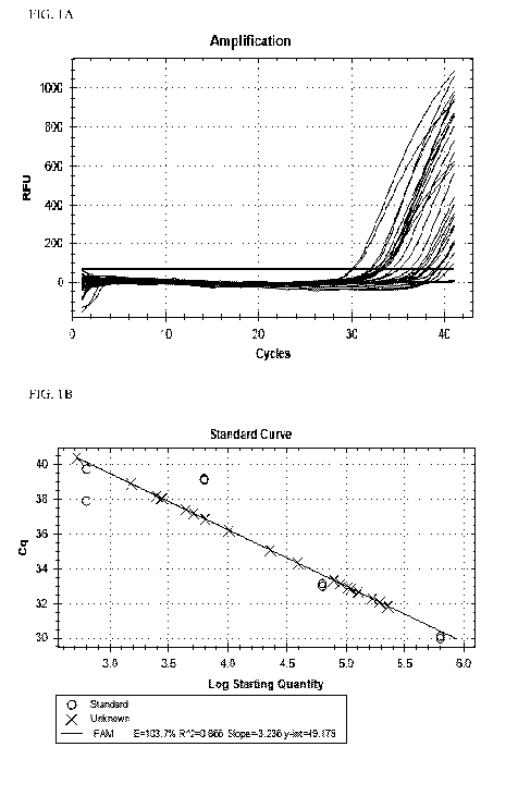
FIGURE lA shows the amplification analysis for a qPCR reaction.
FIGURE 1B shows the standard curve analysis for a qPCR reaction.
FIGURE 2A shows relative quantities of Closiridium perfringens from poultry
litter samples of pen 43.
FIGURE 2B shows relative quantities of Clostridium pofringens from poultry
litter samples of pen 41.
FIGURE 2C shows relative quantities of Clostridium pofringens from poultry
litter samples of pen 14.
FIGURE 2D shows relative quantities of Clostridium perfringens from poultiy
litter samples of pen 44.
FIGURE 3A shows the amplification analysis for a qPCR reaction using FTA
cards and genomic DNA controls.
FIGURE 3B shows the standard curve analysis for a qPCR reaction using FTA
cards and genomic DNA controls.
FIGURE 4A shows the relative quantity levels for the 16S reference gene, along
with the lcdA and icdB genes of interest.
5
CA 02984505 2017-10-31
WO 2016/179027
PCT/US2016/030223
FIGURE 4B shows the expression levels for tcd4 and tcdB genes of interest
normalized to the 16S reference gene.
FIGURE 5A shows the standard curve analysis for a qPCR reaction using FTA
cards and genomic DNA control samples.
FIGURE 5B shows the amplification analysis for a qPCR reaction using FTA
cards and genomic DNA control samples.
FIGURE 6A shows the amplification analysis for a qPCR reaction using FTA
card controls and samples.
FIGURE 6B shows the standard curve analysis for a qPCR reaction using FTA
card controls and samples.
FIGURE 7A shows quantification data based on a standard curve.
FIGURE 7B shows quantification data based on a standard curve.
FIGURE 8 shows the correlation between total bacterial load and total Vibrio
load in the samples tested.
FIGURE 9A shows an RNA extraction comparative analysis for Vibrio spp.
(hyl).
FIGURE 9B shows a storage time and temperature comparative analysis for
Vibrio spp. (hyl).
FIGURE 10A shows aquaculture pond analysis of bacterial counts for V
campbellii, V. harveyi, and total Vibrio.
FIGURE 10B shows aquaculture pond analysis of bacterial load for V.
campbellii, V. harveyi, and other Vibrio.
DETAILED DESCRIPTION OF ILLUSTRATIVE EMBODIMENTS
In one embodiment, a method is provided for quantifying the expression level
of
a gene from a microorganism. The method comprises the steps of recovering the
nucleic acid
6
CA 02984505 2017-10-31
WO 2016/179027 PCT/US2016/030223
from a sample stabilized on a card, amplifying the nucleic acid, and
quantifying the expression
level of the gene, wherein a forward primer, and a reverse primer are used for
the amplification.
In another embodiment, a kit is provided. The kit comprises at least one
primer pair, wherein
the at least one primer pair comprises a forward primer and a reverse primer,
and wherein the
reverse primer has a sequence consisting of SEQ TD NO: 6 or SEQ ID NO:8. In
one
embodiment, the kit further comprises a fluorogenic probe. In another
embodiment, the kit
further comprises a card (e.g., an FTA card).
As used herein, the term "nucleic acid" can mean, for example, DNA, RNA,
including mRNA, an siRNA, an iRNA, or a microRNA.
As used herein, the term "card" can means any tangible medium (e.g., paper)
that has been chemically modified or chemically treated to stabilize nucleic
acids. An example
of a "card" for use in the method described herein is a Whatman FTA Card.
Several embodiments of the invention are described in the Summary section of
this patent application and each of the embodiments described in this Detailed
Description
secfion of the application applies to the embodiments described in the
Summary, including the
embodiments described by the enumerated clauses below. In any of the various
embodiments
described herein, the following features in the enumerated clauses may be
present where
applicable, providing additional embodiments of the invention. For all of the
embodiments, any
applicable combination of embodiments is also contemplated.
1. A method of quantifying the expression level of a gene from a
microorganism, the method comprising the steps of:
recovering the nucleic acid =from a sample stabilized on a card,
amplifying the nucleic acid; and
quantifying the expression level of the gene, wherein a forward primer, and a
reverse primer are used for the amplification.
2. The method of clause 1 further comprising the step of hybridizing a
probe to the nucleic acid to specifically identify the gene.
3. The method of clause 1 or 2 wherein the reverse primer comprises a
sequence selected from the group consisting of SEQ ID NO: 6 and SEQ ID NO: 8.
4. The method of any one of clauses 1 to 3 wherein the nucleic acid is
DNA.
5. The method of any one of clauses 1 to 3 wherein the
nucleic acid is
RNA.
7
CA 02984505 2017-10-31
WO 2016/179027
PCT/US2016/030223
6. The method of any one of clauses 1 to 5 wherein the nucleic acid is
amplified using PCR.
7. The method of clause 6 wherein the PCR is reverse transcription PCR.
8. The method of clause 6 wherein the PCR is reverse transcription-
quantitative PCR.
9. The method of any one of clauses 2 to 8 wherein the probe is
fluorescently labeled.
10. The method of any one of clauses 1 to 9 wherein the primer is
fluorescently labeled.
11. The method of any one of clauses 1 to 10 wherein the microorganism is
selected from the group consisting of Vibrio harveyi, Vibrio campbellii,
Vibrio fluvialis, and
Vibrio parahaemolytieus.
12. The method of any one of clauses 1 to 10 wherein the microorganism is
selected from the group consisting of Clostridium petfringens, Campylobacter
fejuni, and
Campylobacter coll.
13. The method of any one of clauses 1 to 12 wherein the sample is a sample
from an animal.
14. The method of any one of clauses 1 to 13 wherein the sample is an
aquatic sample.
.70 15. The method of clause 14 wherein the aquatic sample is
from a fish
hatcheiy.
16. The method of clause 14 wherein the aquatic sample is from a shrimp
pond.
17. The method of any one of clauses 1 to 13 wherein the sample is an
agricultural sample.
18. The method of clause 17 wherein the agricultural sample is from animal
litter.
19. The method of clause 17 wherein the agricultural sample is a swab from
a swine or a poultry species.
20. The method of any one of clauses 1 to 19 wherein the gene is a gene
encoding a toxin.
21. The method of any one of clauses 1 to 20 wherein the gene is a gene of
a
bacterial species.
8
CA 02984505 2017-10-31
WO 2016/179027
PCT/US2016/030223
22. The method of any one of clauses 1 to 20 wherein the gene is a gene of
a
viral species.
23. The method of any one of clauses 1 to 11 or 13 to 21 wherein the gene
is
a hemolysin (hly) gene.
24. The method of any one of clauses 1 to 10 or 12 to 21 wherein the gene
is
a Clostridium perfringens enterotoxin (cpe) gene.
25. The method of any one of clauses 1 to 10 or 12 to 21 wherein the gene
is
a Clostridium perfringens beta toxin (cpb) gene.
26. The method of any one of clauses 1 to 25 wherein the nucleic acid is
stabilized on the card for a period of time to allow transportation overseas.
27. The method of any one of clauses 1 to 26 wherein the nucleic acid is
stabilized on the card for a period of time to allow transportation over
greater than 1000 miles.
28. The method of any one of clauses 1 to 26 wherein the nucleic acid is
stabilized on the card for a period of time to allow transportation over
greater than 2000 miles.
29. The method of any one of clauses 1 to 26 wherein the nucleic acid is
stabilized on the card for a period of time to allow transportation over
greater than 3000 miles.
30. The method of any one of clauses 1 to 26 wherein the nucleic acid is
stabilized on the card for a period of time to allow transportation over
greater than 5000 miles.
31. The method of any one of clauses 1 to 30 wherein the card is a
WHAT'MANO FTA Card.
32. The method of any one of clauses 1 to 31 wherein the reverse primer has
the sequence of SEQ ID NO: 6.
33. The method of any one of clauses 1 to 31 wherein the reverse primer has
the sequence of SEQ ID NO: 8.
34. A kit comprising at least one primer pair, wherein the at least one
primer
pair comprises a forward primer and a reverse primer, and wherein the reverse
primer has a
sequence selected from the group consisting of SEQ ID NO: 6 and SEQ ID NO: 8.
35. The kit of clause 34 wherein the at least one primer pair is
fluorogenic.
36. The kit of clause 35 wherein the at least one primer pair is
fluorescently
labeled.
37. The kit of any one of clauses 34 to 36 wherein the forward primer has a
sequence selected from the group consisting of SEQ ID NO: 1, SEQ ID NO: 3, SEQ
ID NO: 5,
and SEQ ID NO. 7.
9
CA 02984505 2017-10-31
WO 2016/179027 PCT/US2016/030223
38. The kit of any one of clauses 34 to 37 wherein the reverse primer has
the
sequence of SEQ ID NO. 6.
39. The kit of any one of clauses 34 to 37 wherein the reverse primer has
the
sequence of SEQ ID NO. 8.
40. The kit of any one of clauses 34 to 39 further comprising a reverse
transcriptase.
41. The kit of any one of clauses 34 to 40 further comprising a DNA
polymerase.
42. The kit of any one of clauses 32 to 41 further comprising dNTPs.
43. The kit of any one of clauses 34 to 42 further comprising a fluorogenic
probe.
44. The method of any one of clauses 1 to 10 or 13 to 21 wherein
microorganism is selected from the group consisting of swine enterotoxigenic E
coli (ETEC),
avian pathogenic E. coli (APEC), attaching and effacing E. coli (EAEC),
enterohaemorrhagic E.
coli (EHEC), and shiga toxin-producing E. coli (STEC).
45. The method of clause 44 wherein the ETEC is an antigenic type selected
from the group consisting of K88, F18, F41, 987P, and K99.
46. The method of any one of clauses 1 to 33, 44, or 45 wherein reverse
transcription-PCR and endpoint PCR are performed.
47. The method of clause 6, wherein the PCR is quantitative PCR.
The methods and compositions for detection and/or quantification of
microorganisms or their genes (e.g., toxin or virulence genes) are specific
and sensitive. In
various embodiments, the microorganism that is detected or for which the level
of expression of
a gene is quantified may be any microorganism that infects an animal. In
various embodiments,
the microorganism may include such pathogens as bacteria, including gram-
negative or gram-
positive cocci or bacilli, fungi, viruses, including DNA and RNA viruses,
mycoplasma, and
parasites.
In one embodiment, the microorganism is a bacterium. In one aspect of this
embodiment, the bacteria may include, but are not limited to, Acetobacter,
Actinomyces.
Agrobacterium, Anaplasma, Azorhizobium, Azotobacter, Bacillus. Bacteroides,
Bartonella,
Bordetella. Burkholderia, Campylobacter, Chlamydia, Chlamydophila,
Clostridium,
Corynebacterium, Coxiella, Ehrlichia, Enterobacter, Enterococcus, Escherichia,
Francisella,
Tiisobacterium, Gardnerella, Haemophilus, Helicobacter, Klebsiella,
Lactobacillus,
Legionella, Listeria. Methanobacterium, Microbacterium, Micrococcus,
Moraxella,
CA 02984505 2017-10-31
WO 2016/179027 PCT/US2016/030223
Mycobacterium, Mycoplasma, Neisseria, Pasteurella, Peptostreptococcus,
Porphyromonas,
Prevotella, Pseudomonas, Rhizobium, Rickettsia, Rochalimaea, Rochalimaea.
Rothia,
Salmonella. Serratia, Shigella, Staphylococcus, Stenotrophomoncrs,
Streptococcus,
Treponema,Vibrio. Wolbachia, or Yersinia species.
In another aspect, the microorganism is selected from the group consisting of
swine enterotoxigenic E. coli (ETEC), avian pathogenic E. coli (APEC),
attaching and effacing
E. coli (EAEC), enterohaemorrhagic E coli (EHEC), and shiga toxin-producing E.
coli (STEC).
In yet another illustrative aspect, the enterotoxigenic E. coli (ETEC) is an
antigenic type
selected from the group consisting of K88, F18, F41, 987P, and K99. The avian
pathogenic E.
coli (APEC) produces toxins such as, but not limited to, labile toxin (LT),
stable toxin A (StA),
stable toxin B (StB), and verotoxin (shiga-like toxin, SLT).
Enterohaemorrhagic E. coli (EHEC)
is a bacterium that can cause severe foodborne disease. Shiga toxin-producing
E coli (STEC)
is a bacterial pathotype that is most commonly described in the media as the
cause of foodborne
disease outbreaks.
In another embodiment, the microorganism is a virus. In one aspect, the
viruses
may include, but are not limited to, DNA viruses such as papilloma viruses,
parvoviruses,
adenoviruses, herpesviruses and vaccinia viruses, and RNA viruses, such as
arenavinises,
coronaviruses, rhinoviruses, respiratory syncytial viruses, influenza viruses,
picomaviruses,
paramyxoviruses, reoviruses, retroviruses, and rhabdoviruses.
Examples of fungi include fungi that grow as molds or are yeastlike,
including,
for example, fungi that cause diseases such as ringworm, histoplasmosis,
blastomycosis,
aspergillosis, cryptococcosis, sporotrichosis, coccidioidomycosis,
paracoccidio-idomycosis, and
candidiasis.
Exemplary parasites include, but are not limited to, somatic tapeworms, blood
flukes, tissue roundworms, ameba, and Plasmodium, Trypanosoma, Leishmania, and
Toxoplasma species. In various aspects of the microorganism embodiments
described in the
preceding paragraphs, the expression of any gene expressed by any of these
microorganisms
can be quantified using the method described herein. In various embodiments,
the gene can be
a gene encoding a toxin or a virulence factor.
In another embodiment, the sample that is tested can be any sample from any
animal. As used herein the word "animal" means a human, a domestic animal
(e.g., a canine or
a feline species), a laboratory animal, an agricultural animal, or wildlife,
or any other type of
animal. As used herein, an agricultural animal may include any animal that is
raised for
personal use (e.g., for providing food, fuel, etc.) or for profit. In yet
another embodiment, a
11
CA 02984505 2017-10-31
WO 2016/179027 PCT/US2016/030223
domestic animal may include any animal that is kept or raised for
companionship purposes
(e.g., a dog or a cat). Accordingly, in various embodiments, the invention can
be applied to
samples from animals including, but not limited to, humans (e.g, a human
patient), laboratory
animals such rodents (e.g, mice, rats, hamsters, etc.), rabbits, monkeys,
chimpanzees, domestic
animals such as dogs, cats, and rabbits, agricultural animals such as cows,
horses, ponies, pigs,
sheep, goats, fish, crustaceans, shrimp, chickens, turkeys, pheasants, quails,
ostriches, and
ducks, and wild animals, for example, wild animals in captivity, such as
bears, pandas, lions,
tigers, leopards, elephants, zebras, giraffes, gorillas, dolphins, and whales.
In one aspect, the agricultural animal from which a sample is taken may
include
a bovine species (e.g., cattle and bison), an equine species (e.g., horses,
ponies, and donkeys),
an ovine species (e.g., sheep), a caprine species (e.g., goats), rabbits, and
poultry (e.g., chickens,
turkeys, pheasant, ducks, ostriches, emu, quail, and geese).
In other embodiments, the sample may be from the environment, including the
environment of an animal. The sample may be an aquatic sample, such as a water
sample from
a fish hatchery, a sample from a shrimp pond, a sample from an animal's
drinking water, etc.
In another aspect, the sample may be an agricultural sample, such as a sample
from animal
litter, or any other agricultural environmental sample, a swab from the
intestinal tract of an
agricultural animal (e.g., a swine or poultry species), a swab from the nasal
tract of an
agricultural animal, a swab from the skin of an agricultural animal, a swab
from the ear of an
agricultural animal, a swab from the eye of an agricultural animal, a urine
sample from an
agricultural animal, a nasal secretion sample from an agricultural animal, a
bronchial lavage
from an agricultural animal, a spinal fluid sample of an agricultural animal,
a pleural fluid
sample from an agricultural animal, a synovial fluid sample from an
agricultural animal, a
gastric secretions sample from an agricultural animal, a sample from feces of
an agricultural
animal, or a serum or plasma sample from an agricultural animal.
In various illustrative embodiments, samples from humans that can be tested
for
the presence of microorganism or their genes or from which gene expression can
be quantified,
include, but are not limited to, urine, nasal secretions, nasal washes, inner
ear fluids. bronchial
lavages, bronchial washes, alveolar lavages, spinal fluid, bone marrow
aspirates, sputum,
pleural fluids, synovial fluids, pericardial fluids, peritoneal fluids,
saliva, tears, gastric
secretions, stool, reproductive tract secrefions, such as seminal fluid, lymph
fluid, and whole
blood, serum, or plasma. In another embodiment, the samples can be prepared
for testing as
described herein using the types of cards described herein. In various
embodiments, these
samples can include tissue biopsies. As used herein, the term "tissue"
includes, but is not
12
CA 02984505 2017-10-31
WO 2016/179027 PCT/US2016/030223
limited to, biopsies, autopsy specimens, cell extracts, tissue sections,
aspirates, tissue swabs,
and fine needle aspirates. In another embodiment, the sample can be any
environmental
sample.
The samples tested in accordance with the method described herein can be
stabilized (e.g., the nucleic acid can be stabilized) on a card (e.g., a
Whatman FTA Card) =for
a period of time to allow transportation overseas or over a long distance. In
various
embodiments, the nucleic acid is stabilized on the card for a period of time
to allow
transportation over greater than 1000 miles, greater than 2000 miles, greater
than 3000 miles,
greater than 4000 miles, greater than 5000 miles, greater than 6000 miles,
greater than 7000
miles, greater than 8000 miles, greater than 9000 miles, or greater than 10000
miles. In other
embodiments, the nucleic acid is stabilized on the card for a period of time
to allow
transportation over greater than l 0 miles, over greater than 20 miles, over
greater than 30 miles,
over greater than 40 miles, over greater than 50 miles, over greater than 60
miles, over greater
than 70 miles, over greater than 80 miles, over greater than 90 miles, over
greater than 100
miles, over greater than 200 miles, over greater than 300 miles, over greater
than 400 miles,
over greater than 500 miles, over greater than 600 miles, over greater than
700 miles, over
greater than 800 miles, or over greater than 900 miles. In various
embodiments, the nucleic
acid can be stabilized on the card for a period of time selected from the
group consisting of 3
days, 4 days, 5 days, 6 days, 7 days, 8 days, 9 days, 10 days, 11 days, 12
days, 13 days, 14 days,
15 days, 16 days, 17 days, 18 days, 19 days, 20 days, 21 days, 22 days, 23
days, 24 days, 25
days, 26 days, 27 days, 28 days, 29 days, 30 days, 1 week, 2 weeks, 3 weeks, 4
weeks, 5 weeks,
6 weeks, 7 weeks, 8 weeks, 9 weeks, 10 weeks, 1 month, 2 months, 3 months, 4
months, 5
months, 6 months, 7 months, 8 months, 9 months, 10 months, 11 months, and 12
months, or
greater than any of these time periods.
The methods and compositions described herein can be used to detect and/or
quantify microorganisms and/or their genes (e.g., the level of expression of a
gene). In one
illustrative embodiment, a method is provided of quantifying the expression
level of a gene
from a microorganism. The method comprises the steps of recovering a nucleic
acid from a
sample on a card, amplifying the nucleic acid, and quantifying the expression
level of the gene
A reverse primer and a fonvard primer are used in the amplification step. The
method can
further comprise hybridizing a probe to the nucleic acid to specifically
identify the gene.
In one aspect, the methods described herein can be more sensitive than
endpoint PCR, for example at least 50-fold, at least 60-fold, at least 70-
fold, at least 80-fold, at
least 90-fold, or at least 100-fold more sensitive. In another embodiment, the
methods
13
CA 02984505 2017-10-31
WO 2016/179027 PCT/US2016/030223
described herein can detect from 1-3, from 1-5, from 1-10, from 1-20, from 1-
30, from 1-40,
from 1-50, from 1-60, from 1-70, from 1-80, from 1-90, or from 1-100 cell
equivalents per PCR
tube. Thus, the methods described herein are surprisingly more sensitive than
other assays.
In some embodiments, real-time PCR-based methods can be used to amplify
the nucleic acid and to detect and/or quantify the microorganism and/or the
gene expressed by
the microorganism by hybridization of a probe to the nucleic acid. PCR is
described in U.S.
Patent Nos. 4,683,202 and 4,800,159, incorporated herein by reference, and
methods for PCR
are well-known in the art. Real-time PCR can combine amplification and
simultaneous probe
hybridization to achieve sensitive and specific detection andlor quantitation
of microorganisms
or the genes they express in real-time thereby providing instant detection
and/or quantification.
In this embodiment, the time to detect and/or quantify the microorganism or
the gene
expression is greatly reduced. Real-time PCR can be conducted according to
methods well-
known in the art. Reverse transcription PCR is a highly sensitive technique
for the detection
and quantification of mRNA that comprises the synthesis of cDNA from RNA by
reverse
transcription and the amplification of a specific cDNA by PCR. In one aspect,
reverse
transcription quantitative PCR quantitatively measures the amplification of
the cDNA by using
fluorescent probes. Real-time PCR and reverse transcription quantitative PCR
can also be
performed without probes.
Exemplary probes and primers and their target nucleic acids that can be used
in
accordance with the invention are shown below. Forward primers and reverse
primers are
shown and are well-known terms in the art.
Table 1. Primers
14
CA 02984505 2017-10-31
WO 2016/179027 PCT/US2016/030223
Sequence Description 1
----- ---.-.-h-.-.-.-.-. Sequence
...:::,:uu:::::,:m7u:u:u:,....:,
Forward Primer (SEQ ID NO: 1) ' CIATTGGTGGAACGCAC
Reverse Primer (SEQ ID NO: 2) -- GTATTCTGTCCATACAAAC
:3IiiiiiialliliabliariIiIiIiIiIiIiIiIiIiIiIiIiIiIiIiIiIiIiIiIilanBirriIiIiIiIiI
iIiIiIiIiIiIiIiIiIiIiIiIiIiIiIiIiIiIiIiIiIiIiIiIiIiIiIiIiIiIiIiIiIiIiIiIiIiIiIi
IiIiIiIiIiIiIiIiIiIiIiIiIiIiIiIiIiIiIiIiIiN lit
Forward Pritner (SEQ ID NO: 3) GAGTTCGCITTTCTTTCAACi
Reverse Primer (SEQ ID NO: 4) -- TGTAGTTTTTCGCTAATTTC --
iiiiii:iii::ii:i;õiiiiiim;::::11::::::;:;::::::;g::;::N:õ:m::::::::::-
::::N:N:N:N::N:
:N:iIIIIIIIIIIIIIIIIIIIIIIIIIIIIIIIIIIIIIIIIIIIIIIIIIIIIIIIIIIIIIIIIIIIIIIIIIII
IIIIIIIIIM:...,
LimillIteaitgii2IIPIIIP_I2I2II2I2II2II2I2IIIIPIIIPIIIPII2I2II2I
giti2IIIIIII2II2IIII2I2IIPII_IPIIIP_IliPliiPli2i2iiliPli_iPli_iP_EiP_iPiSiSiiR"
'µ
Forward Primer (SEQ ID NO: 5) I CIATIGGIGGAACGCAC
Reverse Primer (SEQ ID NO: 6) C AG C G AAGT AG CiTAAT(iTC
isaiNIWiliAiNSIIiItniIi(ONO
Iirkag00)IiIiIiIiIiiIiIiIiIiIiiIiIiIiIiIiIiIililililililililililililililililili
lililililililililililililililililililililililililililililililililililililililil
ilililililililililililililililililililililililiIiIii
Forward Primer (SEQ ID NO: 7) GAGI"rCGGITICTI"rc AAG
Reverse Primer (SEQ ID NO: 8) AAACGGTTATCGGCTG
Forward Primer (SEQ ID NO: 9) GGCCiTAAAGCGCATGCAGGT
Reverse Primer (SEQ ID NO: 10) GAAATTCTACCCCCCTCTACAG
______________
...............................................................................
.7:747777,.....................................................................
...............................................................................
.... .........
o
27F:
Forward Primer (SFQ ID NO: 11) I
AGAGTTTGATCMTGGCTCAG
1492R:
Reverse Primer (SEQ ID NO: 12)
GGTTACCTTGTTACCiACTT
CA 02984505 2017-10-31
WO 2016/179027
PCT/US2016/030223
In various embodiments described herein, the primers and probes used for
amplification of the nucleic acid and for detection and/or quantification of
microorganisms
and/or their genes are oligonucleotides from about ten to about one hundred,
more typically
from about ten to about thirty or about six to about twenty-five base pairs
long, but any suitable
sequence length can be used. In illustrative embodiments, the primers and
probes may be
double-stranded or single-stranded, but the primers and probes are typically
single-stranded. In
another embodiment, the primers and probes described herein are capable of
specific
hybridization, under appropriate hybridization conditions (e.g., appropriate
buffer, ionic
strength, temperature, formamide, and MgC12 concentrations), to a region of
the target nucleic
acid. In another aspect, the primers and probes described herein are designed
based on having a
melting temperature within a certain range, and substantial complementarily to
the target
nucleic acid. Methods for the design of primers and probes are described in
Sambrook et al.,
"Molecular Cloning: A Laboratory Manual", 3rd Edition, Cold Spring Harbor
Laboratory Press,
(2001), incorporated herein by reference.
In one illustrative embodiment, universal primers can be used to provide a
method for determining the presence of a nucleic acid before conducting target-
specific assays
or for determining the level of a specific nucleic acid relative to total
nucleic acid present.
Exemplary bacterial universal primers can have the sequences:
Example 16S universal bacterial primers:
forward primer, 5'-GCGGATCCGCGGCCGCTGCAGAGTTTGATCCTGGCTCA G-3' (SEQ
ID NO. 13)
forward primer 5'-GCGGATCCTCTAGACTGCAGTGCCAGCAGCCGCGGTAA-3' (SEQ ID
NO. 14)
reverse primer 5'-GGCTCGAGCGGCCGCCCGGGTTACCTTGTTACGAC'TT-3' (SEQ ID
NO. 15).
In various embodiments, the primers and probes described herein for use in PCR
can be modified by substitution, deletion, truncation, and/or can be fused
with other nucleic
acid molecules wherein the resulting primers and probes hybridize specifically
to the intended
target nucleic acids and are useful in the methods described herein for
amplification of the
target nucleic acids. In one embodiment, derivatives can also be made such as
phosphorothioate, phosphotriester, phosphoramidate, and methylphosphonate
derivatives, that
16
CA 02984505 2017-10-31
WO 2016/179027
PCT/US2016/030223
specifically bind to single-stranded DNA or RNA, for example (Goodchild, et
al., Proc. Natl.
Acad. Sci. 83:4143-4146 (1986)).
In one embodiment, the invention encompasses isolated or substantially
purified
nucleic acids. In another embodiment, an "isolated" or "purified" nucleic acid
molecule is
substantially free of other cellular material, or culture medium when produced
by recombinant
techniques. or substantially free of chemical precursors or other chemicals
when chemically
synthesized. Preferably, an "isolated" or "purified" nucleic acid is free of
sequences that
naturally flank the nucleic acid in the genomic nucleic acid from which it is
derived. For
example, in various embodiments, the isolated or purified nucleic acid can
contain less than
about 5 kb, 4 kb, 3 kb, 2 kb, 1 kb, 0.5 kb, or 0.1 kb of nucleotide sequences
that naturally flank
the nucleic acid in the genomic nucleic acids of the cell from which the
nucleic acid is derived.
In one embodiment, nucleic acids complementary to the probes and primers
described herein, and those that hybridize to the nucleic acids described
herein or those that
hybridize to their complements under highly stringent conditions are provided.
In one aspect,
"highly stringent conditions" means hybridization at 65 C in 5X SSPE and 5 0
% formamide,
and washing at 65 C in 0.5X SSPE. Conditions for high stringency, low
stringency, and
moderately stringent hybridization are described in Sambrook et al.,
"Molecular Cloning: A
Laboratory Manual", 3rd Edition, Cold Spring Harbor Laboratory Press, (2001),
incorporated
herein by reference. In some illustrative aspects, hybridization occurs along
the full-length of
the nucleic acid.
In one embodiment, nucleic acids having about 60%, about 70%, about 75%,
about 80%, about 85%, about 90%, about 95%, 96%, 97%, 98%, 99%, or 99.5%
homology to
the probes and primers described herein can be used. Determination of percent
identity or
similarity between sequences can be done, for example, by using the GAP
program (Genetics
Computer Group, software; now available via Accelrys on
http://www.accehys.com), and
alignments can be done using, for example, the ClustalW algorithm (VNTI
software, InforMax
Inc.). In one aspect, a sequence database can be searched using the nucleic
acid sequence of
interest. In another aspect, algorithms for database searching are typically
based on the BLAST
software (Altschul et al., 1990), and the percent identity can be determined
along the full-length
of the nucleic acid.
As used herein, the term "complementary" refers to the ability of purine and
pyrimidine nucleotide sequences to associate through hydrogen bonding to form
double-
stranded nucleic acids. Guanine and cytosine, adenine and thymine, and adenine
and uracil are
complementary and can associate through hydrogen bonding resulting in the
formation of
17
CA 02984505 2017-10-31
WO 2016/179027
PCT/US2016/030223
double-stranded nucleic acids when two nucleic acids have "complementary"
sequences. The
complementary sequences can be DNA or RNA sequences. The complementary DNA or
RNA
sequences are referred to as a "complement."
Techniques for synthesizing the probes and primers described herein are well-
known in the art and include, but are not limited to, chemical syntheses and
recombinant
methods. Such techniques are described in Sambrook et al., "Molecular Cloning:
A Laboratory
Manual", 3rd Edition, Cold Spring Harbor Laboratory Press, (2001),
incorporated herein by
reference. Primers and probes can also be made commercially (e.g., CytoMol,
Sunnyvale, CA
or Integrated DNA Technologies, Skokie, IL). Techniques for purifying or
isolating the probes
and primers described herein are well-known in the art. Exemplary techniques
are described in
Sambrook et al., "Molecular Cloning: A Laboratory Manual", 3rd Edition, Cold
Spring Harbor
Laboratory Press, (2001), incorporated herein by reference. The primers and
probes described
herein can be analyzed by techniques known in the art, such as, for example,
restriction enzyme
analysis or sequencing, to determine if the sequence of the primers and probes
is correct.
In various embodiments of the methods and compositions described herein, the
probes andlor primers can be labeled, such as fluorescently labeled,
radioactively labeled, or
labeled with antigens, compounds such as biotin-avidin, colorimetric
compounds, or other
labeling agents known to those of skill in the art, to allow detection and
quantification of
amplified nucleic acids, such as by real-time reverse transcription
quantitative PCR. In
illustrative embodiments, the labels may include 6-carboxyfluorescein (FAMTm),
TETTm
(tetrachloro-6-carboxyfluorescein), JOE Tm (2,7, -dimethoxy-4,5-dichloro-6-
carboxyfluorescein), VICTm, HEX (hexachloro-6-carboxyfluorescein), TAMRATm (6-
carboxy-
N,N,N',NI-tetramethylrhodamine), BHQ1m, SYBRO Green, Alexa 350, Alexa 430,
AlexaFluor
488, and AlexaFlour 647 (Molecular Probes, Eugene, Oregon), AMCA, BODIPY
630/650,
BODIPY 650/665, BODIPY-FL, BODIPY-R6G, BODIPY-TMR, BODIPY-TRX, Cascade
Blue, Cy3, Cy5, Cy7, 6-FAM, fluorescein, rhodamine, phycoer),,,thrin, biotin,
ruthenium,
DyLight fluorescent agents (DyLight 680, CW 800, trans-cydooctene, tetrazine,
methyltetraiine, and the like), Oregon Green, such as Oregon Green 488, Oregon
Green 500,
and Oregon Green 514, Pacific Blue, REG, Rhodamine Green, Rhodamine Red, ROX,
and/or
Texas Red. In one embodiment, the probes and/or primers can be fluorogenic
(i.e., generate or
enhance =fluorescence). For example, the probes and/or primers may comprise a
=fluorescent
label or a non-fluorescent molecule which is acted upon by a compound (e.g.,
an enzyme) to
produce or enhance fluorescence.
The method embodiments described herein can provide methods of diagnosing
18
CA 02984505 2017-10-31
WO 2016/179027 PCT/US2016/030223
infections. In one embodiment, humans in need of diagnosis of an infection can
include a
person exhibiting the symptoms of an infection, cancer patients, post-
operative patients,
transplant patients, wound-care patients, patients undergoing chemotherapy,
immunosuppressed
patients, and the like. In another embodiment, domestic animals, agricultural
animals,
laboratory animals, or wildlife in need of diagnosis of an infection can
include any animal
exhibiting the signs or symptoms of an infection.
In one embodiment, kits are provided. The kits are useful for detecting andlor
quantitating microorganisms and/or their gene expression (e.g., the expression
of a toxin or a
virulence gene). In one aspect, the kit can contain the probes and/or primers
described herein.
In one aspect, the primers or the probe can be fluorogenic (e.g.,
fluorescently labeled). In
another embodiment, the kit can also contain components for nucleic acid
amplification, such as
a heat stable DNA polymerase (e.g., Taq polymerase or Vent polymerase),
buffers, MgC12,
H20, dNTPs, a reverse transcriptase, and the like. In one embodiment, the
reagents can remain
in liquid form. In another embodiment, the reagents can be lyophilized. In
another illustrative
embodiment, the kits can also contain instructions =for use.
In another embodiment, a kit comprising a nucleic acid with a sequence
selected
from the group consisting of SEQ ID NO: 1 to SEQ ID NO: 15 or a complement of
a sequence
selected from the group consisting of SEQ ID NO: 1 to SEQ ID NO: 15 is
provided. In another
embodiment, a kit comprising a nucleic acid with a sequence selected from the
group consisting
of SEQ ID NO: 6 and SEQ ID NO: 8 or a complement of a sequence selected from
the group
consisting of SEQ ID NO: 6 and SEQ ID NO: 8 is provided. The kits are useful
for detecting
and/or quantitating microorganisms andlor their gene expression (e.g., the
expression of a toxin
or a virulence gene). In one aspect, the kit can contain the probes and/or
primers described in
this paragraph. In one aspect, the primers or the probe can be fluorogenic
(e.g., fluorescently
labeled). In another embodiment, the kit can also contain components =for
nucleic acid
amplification, such as a heat stable DNA polymerase (e.g., Taq polymerase or
Vent
polymerase), buffers, MgC12, H20, dNTPs, a reverse transcriptase, and the
like. In one
embodiment, the reagents can remain in liquid form. In another embodiment, the
reagents can
be lyophilized. In another illustrative embodiment, the kits can also contain
instructions for
use.
In one embodiment, a purified or isolated nucleic acid is provided comprising
or
consisting of a sequence of SEQ ID NO: 1 to SEQ ID NO: 15 or a sequence that
hybridizes
under highly stringent conditions to a sequence consisting of SEQ ID NO: 1 to
SEQ ID NO: 15.
In another embodiment, a purified or isolated nucleic acid is provided
comprising a
19
CA 02984505 2017-10-31
WO 2016/179027 PCT/US2016/030223
complement of a sequence of SEQ ID NO: 1 to SEQ ID NO: 15 or a sequence that
hybridizes
under highly stringent condi fions to the complement of a sequence consisting
of SEQ ID NO: 1
to SEQ ID NO: 15. In another embodiment, a kit comprising a purified or
isolated nucleic acid
with a sequence selected from the group of consisting of a sequence of SEQ ID
NO: 6 or SEQ
ID NO: 8 or a sequence that hybridizes under highly stringent conditions to a
sequence
consisting of SEQ ID NO: 6 or SEQ ID NO: 8. In another embodiment, a purified
or isolated
nucleic acid is also provided comprising a complement of a sequence of SEQ ID
=NO: 6 or SEQ
ID NO: 8 or a sequence that hybridizes under highly stringent conditions to
the complement of
a sequence consisting of SEQ ID NO: 6 or SEQ ID NO: 8. In one embodiment,
"highly
stringent conditions" means hybridization at 65 C in 5X SSPE and 50%
formamide, and
washing at 65 C in 0.5X SSPE.
In another embodiment, the primer or probe, or a combination thereof,
described
herein is provided in a sterile container (e.g, a vial) or package, for
example, an ampoule or a
sealed vial.
As described herein the "card" can be, for example, an FTA Card (Whatman
FTA Card; for example, Whatman catalogue numbers: WB12-0205, WB12-0206, WB12-
0055, WB12-0056, WB12-0210, WB12-0210, WB12-0211, and WB12-0208; GE Healthcare
Life Sciences, Pittsburgh, PA). Cards, such as a Whatman FTA Card, are
conventionally
used in the forensic sciences to collect, for example, blood or buccal cells.
Whatman FTA
Cards simplify the handling and processing of nucleic acids (e.g, DNA and RNA,
including
mRNA, an siRNA, an iRNA, a microRNA, etc.). Whatman FTA Cards contain
chemicals
that lyse cells, denature proteins, and protect nucleic acids =from nucleases,
oxidation and UV
damage. Moreover, they rapidly inactivate organisms and prevent the growth of
bacteria and
other microorganisms. When a sample is applied to a Whatman FTA Card, cell
membranes
and organelles are lysed and the released nucleic acids are entrapped in the
fibers of the matrix
and are preserved (e.g., reduced degradation) throughout transport at room
temperature. Upon
arrival at a distant location, for example, the nucleic acid can be readily
eluted from punches of
the card through purification steps and prepared =for downstream processing,
as is known in the
art. This technology' also eliminates the labor intensiveness of selective
plating and culture
growth.
Moreover, this technology provides a start to finish process that encompasses
all aspects of sample collection and analysis by utilizing cards, such as FTA
Cards. The cards
also enable nucleic acid preservation in a sample from farm collection to long
term lab storage
and analysis. Stabilized nucleic acids can then be extracted from samples and
tested for gene
CA 02984505 2017-10-31
WO 2016/179027
PCT/US2016/030223
detection, quantification, and expression of a multitude of pathogenic
microorganisms, such as
bacteria. Complete sample analysis by the technology described herein
constructs a broader
view of pathogen-pathogen interaction, rather than singularly considering
individual bacterial
species effects. Thus, the present technology provides a more rapid, a more
accurate, and a
more cost effective analytical tool of identifying and understanding the
greater pathogenic
effects leading to total microbial load of agricultural species than is
presently available.
The following examples provide illustrative methods for cariying out the
practice of the present invention. As such, these examples are provided for
illustrative purposes
only and are not intended to be limiting.
EXAMPLES
EXAMPLE 1
SAMPLE COLLECTION AND FTA CARD APPLICATION
Litter Samples
25mL of the most sterile water available was added to a 50mL conical tube. One
=full
spoonful of litter material was added into the 50mL conical tube and shaken
vigorously for 30
seconds. Wood chips and other thick materials were allowed to briefly settle
to the bottle.
Using the transfer pipette, 1254 of solution was added onto the FTA Card.
Cards were
allowed to diy in a cool diy area for 2-3 hours minimum. Card(s) were placed
into a supplied
zip bag with 2 desiccant packs.
Swab Samples
Samples were collected by swabbing swine rectal or poultry cloaca to collect
and absorb
material. The swab was firmly pressed and rolled over the FTA Card application
circle.
Card(s) were allowed to diy in a cool diy area for 2-3 hours minimum. Card(s)
were placed
into a supplied zip bag with 2 desiccant packs.
EXAMPLE 2
DNA EXTRACTION
Gram-negative Bacteria
21
CA 02984505 2017-10-31
WO 2016/179027
PCT/US2016/030223
Six discs were punched (Miltex Biopsy Punch) from the card and placed in a 96
well block. 25 L of Proteinase K and 180pL of Buffer T1 were mixed for each
sample. 2004
of solution was added into each well of the Round-well Block. The Block was
incubated at
56 C for at least 6 hours (or optionally overnight). The Block was centrifuged
to collect
condensation. 200pL of Buffer BQ1 and 2004. of 96-100% ethanol were added to
each
sample. The samples were mixed vigorously by shaking for 10-15 seconds and
briefly spun to
collect the sample. Lysates were transferred into wells of a Tissue Binding
Plate and spun at
5000g for 10 min. 500pL of Buffer BW was added and spun at 5000g for 2 min.
7004 of
Buffer B5 was added, and spun at 5000g for 4 min. The Binding Plate was placed
onto an
opened Rack of Tube Strips and incubated at 70 C for 10min to evaporate all
the ethanol. The
DNA was eluted by adding 1004 of 70 C preheated Buffer BE, and spun at 5000g
for 2 min.
Gram-positive Bacteria
Samples were pretreated with 1804 Lysis Buffer and Lysozyme for at least 45
min at 37 C. The sample protocol was followed as stated for gram-negative
bacteria.
Concentrations were quantified using Quantus Fluorometer. dsDNA dye was
prepared at a
1:200 concentration in lx TE Buffer. 104 DNA, 90gL lx TE Buffer, and 100pL of
prepared
dsDNA dye were added and vortexed. The tube was placed in a Fluorometer and
measured.
EXAMPLE 3
QUANTITATIVE REAL-TIME PCR
The amount of 2X MasterMix, each primer and dH20 was calculated based on
reaction ntunber, primer concentrations and reaction volume. Probe
concentration can also be
calculated if required for the reaction. All components were added, mixed, and
distributed into
PCR tubes. Samples were serially diluted at 10-1 and the appropriate reference
strain in 2 L
added to PCR tubes. 2 L of Sample DNA template was added to each tube and the
strips were
vortexed. Each qPCR reaction was set up for the appropriate cycle conditions
in accordance
with the primer set used. Once the reaction was complete, the Bio-Rad program
was used to
analyze the Cq values in comparison to those of the reference strain.
22
CA 02984505 2017-10-31
WO 2016/179027 PCT/US2016/030223
EXAMPLE 4
EXAMPLE OF DNA OUALITY FROM LITTER SAMPLE COLLECTION
Measurements were performed on a Quantus Fluorometer Machine and results were
as
follows (see FIGS. 2A-2D):
Pen 14 = 0.402 ng/LiL
Pen 41 = 0.418 ngli.it
Pen 43 = 0.502 ngt L
Pen 44 = 0.299 ng/ 1.,
DNA Normalization Calculations
Equation: V 1C1=V2C2
EXAMPLE 5
qPCR REACTIONS
Quantitative PCR amplifies purified DNA based on specifically designed
primers which target a particular region in the gene sequence. In addition,
qPCR goes one step
further by incorporating a fluorogenic probe to enable real-time measurements
of fluorescence
as the DNA is amplified to quantify the sample rather than determining this
based on band
intensity in end point PCR. The oligonucleotide probe also adds a heightened
specificity factor.
The probes are designed specifically to target a gene sequence and fluoresce
only when bound,
therefore the Thermocycler measures when the probe is bound specifically to
the target gene
dsDNA whereas end point PCR amplifies any dsDNA. Amplification was measured as
a Cq
Value (FIG. 1A) based on how many cycles it takes the DNA sample to begin
amplifying and
the strength of the fluorescence was measured in RFU values. The lower the Cq
value, the less
cycles it took to amplify, therefore the more target gene was present.
Quantification numbers
were derived from a Standard Curve (FIG. 1B) which was calculated from a
serially diluted
reference strain with a known starting initial count. Acceptable standard
curves achieved a
slope between -3.1 and -3.6 giving reaction efficiencies between 80 and 110%.
EXAMPLE 6
RELATIVE QUANTITY CALCULATIONS
Relative quantities were calculated as a percentage of the total microbial
load
from each of the Cq Values. qPCR reactions were run at the species level
(Clostridium
23
CA 02984505 2017-10-31
WO 2016/179027
PCT/US2016/030223
perfringens), the genus level (Clostridium spp.), and for total microbes. As
described herein,
the Clostridium counts were comprised from Clostridium Clusters I, IV, and XIV
which
encompass the majority of the intestinal Clostridium. FIGS. 2A, 2B, 2C, and 2D
show
examples of poultiy litter samples taken from a farm trial. The pie charts
represent the amount
of C. perfringens within the total Clostridium spp. within the total microbial
load.
EXAMPLE 7
ABSOLUTE QUANTITY CALCULATIONS
Absolute quantities were calculated from a standard curve created from a
serially
diluted reference strain with a known initial count (See FIG. 3A). This
technology is used to
quantify the absolute amount of each target gene in a sample. Starting
quantities of a sample
were calculated by determining where its Cq value falls on the linear curve
(See FIG. 3B).
Counts are reported as gene copies rather than exact counts due to target
genes having variable
gene copies per cell.
EXAMPLE 8
QUANTIFICATION OF CLOSTRIDIUM PERFRIIVGENS
As previously described, absolute quantities were derived from a standard
curve
created from a serially diluted reference strain with a known initial count.
This technology was
used to quantify the absolute amount of C. perfringens in a sample. SQ values
represent the
calculated count for each sample.
Table 2. Quantification of Clostridium perfringens.
24
CA 02984505 2017-10-31
WO 2016/179027 PCT/US2016/030223
Fluor Target Sample Cq SQ Fluor Target Sample Cq
='SC1
FAM C. perf P13-T1 33.33 7.91E+04
_
F-AIVFMMMMMMMNeg,Ctrr*K***K*K*KT=stilv**K*K,K:N/.A*-:, FAM C. perf P43-
T1 32.27 1.68E+05
FAM C. perf P43 T1 32.66 1.27E+05
FAM C. perf P2 T2 31.85 2.27E+05
FAM C. perf P2 T2 32.07 1.93E+05
FAM C. perf P14 T2 34.32 3.91E+04
FAM C. perf P14 T2 35.07 2.29E+04
FAM C. perf P44 T2 32.93 1.05E+05
FAM C. perf P44-T2 32.86 1.11E+05
FAM C. perf P6 T3 38.19 2.49E+03
EAM C. perf P6 T3 38.89 1.51E+03
giMY.f.:EM!:*:tEgMEgEMINVONtgOORM FAM C. perf P171-3 36.86 6.41E+03
FAM C. perf P1-11. 37.17 5.13E+03 FAM C.
perf P17-T3 37.39 4.41E+03
FAM C. perf P1-T1 36.2 1.02E+04 FAM C.
perf P41-T3 38.05 2.76E+03
FAM C. perf P13-T1 33.14 9.04E+04 FAM C.
perf P41-T3 40.38 r 5.24E-F02
EXAMPLE 9
TOXIN DETECTION
Methods described herein were used to detemiine the presence or absence along
with either absolute or relative quantities of specific toxin associated genes
for a pathogenic
bacterial species of interest. An important distinction in this technique is
detection of the
presence or absence of the gene from a DNA sarnple and not measuring the
amotuu= of toxin
gene expressed. I-Ia.ving knowledge of which toxin genes are in the samples
is important in
assessing the risk for diseases. RNA from the samples was accessed to
deterntine how much of
the toxin was actually being produced and expressed, as that directly
correlates to disease
occurrence. Toxin detection was also achieved through qPCR reactions that
measured
amplification of sample DNA in relation to a known reference strain containing
the target gene.
Another aspect of this technology is the ability to determine quantities of
specific toxin or virulence associated genes for pathogenic bacteria of
interest. Absolute
quantities of any plasmid-borrie toxin gene may not be able to be determined
since they are
capable of horizontal gene transfer resulting in an unknown number of copies
per DNA sample,
and thus reported as copy number. However, toxin genes that are
clirotnosomally -borne can be
quantified because there Will be one chromosome per DNA amplifi- ed, which
is reported as
colony-forming unit or CFU,
There were approximately 37 genes validated for toxin detection according to
the following: 1) BV4-5 region for universal bacteria, 2) 16s gene of
Clostridium Cluster I, 3)
16s gene of Clostridium Cluster IV, 4) 16s gene of Clostridium Cluster XIV, 5)
16s gene of
CA 02984505 2017-10-31
WO 2016/179027 PCT/US2016/030223
Clostridium perfringens, 6) cpn60 gene of Clostridium perfringens,7)cpa toxin
gene of
Clostridium perfringens, 8) cpb toxin gene of Clostridium perfringens, 9) cpb2
toxin gene of
Clostridium perfringens, 10) cpe toxin gene of Clostridium perfringens, 11)
etx toxin gene of
Clostridium perfringens, 12) 16s gene of Clostridium diflicile, 13) tcdA toxin
gene of
Clostridium difficile, 14) tcdB toxin gene of Clostridium difficile,, 15) 16s
gene of E. coil, 16)
Stxl toxin gene of E. coli, 17) Stx2 toxin gene of E. coli, 18) LT toxin gene
of E. coli, 19) STa
toxin gene of E. coli, 20) STb toxin gene of E. coli, 21) eaeA virulence
factor gene of E coli,
22) EAST1 toxin gene of E. coli, 23) hlyF virulence factor gene of E. coli,
24) ompT virulence
factor gene of E. coli, 25) iroN virulence factor gene of E. coli, 26) iutA
virulence factor gene of
E coli, 27) iss virulence factor gene of E coli, 28) 16s gene of Campylobacter
spp., 29) cpn60
gene of Campylobacter jejuni, 30) CDT toxin of Campylobacter jejuni, 31) cpn60
gene of
Campylobacter coli, 32) invA gene of Salmonella spp., 33) fliC virulence
factor gene of
Salmonella enterica enterica Typhimurium, 34) sefA virulence factor gene of
Salmonella
enterica enterica Entertidis, 35) cpsJ2 virulence gene of Streptococcus suis,
36) P46, P97, and
P107 virulence proteins of Mycoplasma hyopneumoniae, and 37) Omp virulence
gene of
ikzemophilus parasuis.
EXAMPLE 10
TOXIN EXPRESSION
In addition to toxin and virulence gene quantification, this technology also
determines the amount of a gene that is present that is actually expressed.
Gene presence
determines the potential of the gene. However, the expression level of a gene
more accurately
represents a risk of that gene for pathogenesis. Thus, gene expression is
accomplished by
extracting RNA rather than DNA, reverse transcribing the RNA product into
cDNA, and then
analyzing the resulting cDNA in a real-time PCR reaction. Similar to gene
detection analysis,
gene expression analysis requires gene-specific primers designed in a
particular gene region to
amplify a target sequence.
Gene expression analysis requires a validated and constitutively expressed
housekeeping gene to be used as a reference gene. Relative quantity levels of
the gene of
interest are then compared to the relative quantity of the corresponding
reference gene in order
=to determine relative normalized expression levels. Figure 4A illustrates the
relative quanfity
levels of the 16S reference gene and the tcdA and tedB toxin genes of
interest. Figure 4B
illustrates the normalized expression levels of the tcdA and tcdB toxin genes
after being
normalized by the 16S reference gene expression level. This technology enables
a better
26
CA 02984505 2017-10-31
WO 2016/179027 PCT/US2016/030223
understanding as to which toxin and/or virulence genes may be contributing to
symptoms of
pathogenic diseases.
There were approximately 18 validated genes for toxin expression according
to the following: 1) 16s (Clostridium perfringens reference gene), 2) rpoA
(Clostridium
perfringens single copy reference gene), 3) cpa toxin gene of Clostridium
perfringens, 4) cpb
toxin gene of Clostridium perfringens, 5) cpb2 toxin gene of Clostridium
perfringens, 6) cpe
toxin gene of Clostridium perfringens, 7) etx toxin gene of Clostridium
perfringens, 8) 16s
(Clostridium difficile reference gene), 9) tcdA toxin gene of Clostridium
difficile, 10) tcdB toxin
gene of Clostridium difficile,11) GAPDH (E. coli reference gene), 12) Stx1
toxin gene of E.
coli, 13) Stx2 toxin gene of E coli, 14) LT toxin gene of E. coli, 15) STa
toxin gene of E coli,
16) STb toxin gene of E coli, 17) eaeA virulence factor gene of E. coli, and
18) EAST1 toxin
gene of E. coll.
EXAMPLE 11
QPCR ANALYSIS OF FTA CARDS (16S rDNA TRIAL)
The performance of a universal bacterial 16S rDNA qPCR assay with DNA from
cells in pond water preserved on FTA cards was analyzed. Control DNA for assay
validation
consisted of serial dilutions of Vibrio campbellii genomic DNA in sterile
water, run in
triplicate. V. campbellii-spiked FTA cards from a previous Vibrio detection
study with
concentrations between 5.8x10⁸ CFU/ml and 5.8x10³ CFUlml were used as
quantification
standards for the card method. FTA cards from a shrimp farm were the unknowns.
All samples
were run in triplicate. Quantitative PCR was performed using a 20 I reaction
mixture of Bio-
iTaq SYBR Green Supermix (1x), universal bacterial 16S primers 1099F and 1510R
from
(Reysenbach et al., Appl Environ Microbiol. 1994 Jun; 60(6): 2113-2119)(400 nM
each), and 5
I of template DNA extracted from FTA cards. No-template controls were
included. Cycling
conditions were designed with the protocol auto-writer in Bio-Rad's CFX
Manager software
and were as follows: 3 minutes at 95 C, 40 cycles of (10 seconds at 95 C, 20
seconds at 55 C.
20 seconds at 72 C followed by a plate read), followed by melt curve analysis
from 65 C to
95 C in 0.5 C increments. Results using genomic DNA controls (E=96/6%,
R2=0.993) are
shown in FIG.3, Panels A and B. Amplification after 33 cycles occurred in no
template
controls, but this is common with universal primers due to E. coli gDNA
contamination in most
Taq polymerases and other PCR enzymes (See FIGS. 5A and 5B).
27
CA 02984505 2017-10-31
WO 2016/179027
PCT/US2016/030223
The standard curves for FTA card controls and samples are shown in FIGS. 6A
and 6B. Quantification data varied depending on the points used to construct a
standard curve.
The most conservative version excludes the 5.8x10³ CFU/m1 and 5.8x10⁴CFU/m1
data points,
as they overlap with no template controls, and one outlier point, keeping a 4-
log range on the
standard curve (See FIG. 7A). Eliminating the 5.8x10⁸ CFUlml dilution improved
the fit of the
standard curve but reduced the range (See FIG. 7B). Correlation between total
bacterial load
and total Vibrio load in the samples tested is shown in FIG. 8.
Table 3. Quantification data varied depending on the version of the curve used
3 log curve
4 log curve
Mean Mean
starting Standard starting Standard
Pond quantity deviation 95% Confidence Interval
Pond quantity , deviation , 95% Confidence Interval
Al _ 1.60E+07 2.19E+06 1.38E+07 to 1.82E+07 Al 1.23E+07 1.40E+06
1.09E+07 to 1.37E+07_
A2 3.47E+07 3.18E+06 3.15E+07 to 3.79E+07_ A2 2.32E+07 1.82E+06 _
2.14E+07 to 2.50E+07.1
A3 8.59E+06 9.34E+05 7.66E+06 to 9.52E+06 A3 7.39E+06 6.50E+05 6.74E+06 to
8.04E+06
A4 6.46E+06 4.72E+05 5.99E+06 to 6.93E+06 A4 5.83E+06 3.42E+05 5.49E+06 to
6.17E+06
AS 1.86E+07 4.44E+06 1.42E+07 to 2.30E+07 AS 1.39E+07 2.74E+06 1.12E+07 to
1.66E+07
A6 2.62E+07 1.74E+06 2.45E+07 to 2.79E+07 A6 1.83E+07 9.79E+05 1.73E+07 to
1.93E+07
Table 4. Correlation between total bacterial load and total Vibrio load in the
samples
Total quantity (Universal
Sample Vibrio Quantity (Vibrio 16S)
165)
A1 1.60E+07 1.95E+05
A2 3.47E+07 5.63E+04 .
A3 8.59E+06 1.77E+04
A4 6.46E+06 2.89E+05
A5 1.86E+07 4.33E+04
A6 2.62E+07 7.27E+05
EXAMPLE 12
DETECTION OF THE V. HARVEYI AND V. CAMPBELLII HEMOINSIN GENE IN FTA
CARD SAMPLES
Vibrio harveyi and Vibrio eampbellii were detected in water samples and FTA
card samples via PCR assays targeting their hemolysin (hly) gene sequences,
and the expression
of the hemolysin gene in FTA card samples was analyzed. An endpoint PCR assay
was used to
detect presence or absence of the hly gene in diluted pond water samples and
pond water
samples stored dry on FTA cards. The PCR assay was evaluated for performance
in
28
CA 02984505 2017-10-31
WO 2016/179027 PCT/US2016/030223
quantitative PCR with water samples and FTA card samples. A reverse primer was
developed
to amplify a smaller section of the hly gene than the original assay (for
better performance in
qPCR) and checked for specificity against published V. harveyi and V.
campbellii sequences.
Additional PCR assays for total Vibrio and total bacteria were used to
determine the
proportional abundance of V. campbellii and V. harveyi in pond water samples.
The stability of
V. harveyi RNA on FTA cards was evaluated to determine the possibility of gene
expression
analysis. The PCR assay was evaluated for performance in qRT-PCR gene
expression analysis,
detecting hly mRNA in liquid culture. The sensitivity of the qRT-PCR assay was
evaluated
using V harveyi RNA extracted from FTA cards.
FTA card sampling and storage:
Performed as directed by the manufacturer.
FTA card DNA extraction:
For endpoint PCR, the manufacturer's directions for amplification directly
from
an FTA card sample punch are used. For quantitative PCR, DNA was eluted from
the cards
with the Qiagen DNeasy Mini Kit. (Protocol: DNA Purification from Dried Blood
Spots. A
nearly identical procedure with the QiaAmp DNA investigator Kit is listed in
GE Life Science's
application note 28-9822-22 AA).
FTA card RNA extraction:
The manufacturer's protocol was used for extraction with an RNA processing
buffer (Preparation of RNA from Blood and Tissue Culture on FTA Cards for RT-
PCR or
Northern Blot Analysis) or the Qiagen RNeasy Mini kit.
V. campbelki and V. harvevi hlv PCR assays:
Primer sets are as follows:
Table 5. Vibrio PCR primers and assays
PCR primers and assays
29
CA 02984505 2017-10-31
WO 2016/179027
PCT/US2016/030223
Target Forward primer Reverse primer qPCR cycle
3 min @ 95')C, 40
Vibriocycles of (10 s
CTATTGGTGGAACGCAC GTAT'TCTGTCCATACAAAC
campbelhi 95 C, 20 s
(SEQ ID NO. 1) (SEQ ID NO. 2)
hly
72 C)
3 min @ 95 C, 40
Vibrio cycles of (10 s
@
GAGTTCGGITTCTTTCAA TGTAGTTTTTCGCTAATTTC
hanPeyi 95 C, 20 s @
(SEQ ID NO. 3) (SEQ ID NO. 4)
54 C, 20 s
72 C)
Vibrio 3 min Eip 95 C,
40
campbellii CTATTGGTGGAACGCAC CAGCGAAGTAGGTAATGTC cycles of (10s
rib
hly (short (SEQ ID NO. 5) (SEQ ID NO. 6) 95 C, 30 s ricp
amplicon) 55 C)
Vïbrio 3 min @ 95 C,
40
GAGTTCGGTTTCTTTCAA
hanPeyi G AAACGGTTATCGGCTG cycles of (10s
hly (short
(SEQ ID NO. '7) (SEQ ID NO. 8) 95 C, 30 s @
ampi icon) 55 C)
3 min @ 95 C, 40
Vibrio GGCGTAAAGCGCATGCA GAAATTCTACCCCCCTCTACA cycles of (10s (di
16S rRNA GUT (SEQ ID NO. 9) G (SEQ ID NO. 10) 95 C, 30 s @
55 C)
3 min @ 95 C, 40
2717: 1492R:
Bacterial cycles of (10s (a)
AGAGTTTGATCMTGGCTC GGTTACCTTGTTACGACTT
16S rRNA 95 C, 30 s ricp
AG (SEQ ID NO. 11) (SEQ ID NO. 12)
55 C)
EXAMPLE 13
RNA EXTRACTION AND QRT-PCR
RNA was extracted from 4 x 2.0 mm punches for determination of yield, or half
the sampling area of each FTA card for subsequent gene expression analysis,
with the RNeasy
Mini kit (Qiagen) with on-column DNase digestion and an additional DNase
digestion in
solution, followed by RNA cleanup with RNeasy mini. RNA was quantified with
the
fluorometric method and 80 ng of RNA from each treatment was reverse
transcribed with
iScript Reverse transcriptase (Bio-Rad) in duplicate, and SYBR Green
quantitative PCR (Bio-
Rad iTaq SYBR Green Supermix) was performed using 2 I of cDNA template per 20
gl
reaction, 3 technical replicates per RT reaction. Genes amplified were hly and
Vibrio-specific
16S rRNA. Relative normalized expression with PCR efficiency correction was
computed via
the AACq method in CFX Manager.
CA 02984505 2017-10-31
WO 2016/179027 PCT/US2016/030223
RNA yield from 2-day-old FTA cards ranged from < 5 ng (with the Direct-Zol
kit) to 610 ng (with the Whatman RNA processing buffer). The low-yielding
Direct-Zol
method was excluded from further analysis. The effect of storage temperature
was determined
with samples extracted with the high-yield method (VVhatman's RNA processing
buffer). RNA
loss was not observed after 20 days of storage at ¨20 or 20 C. A 25% decrease
was observed at
37 C, but yield remained above 400 ng per 5-punch extraction, an amount
sufficient for reverse
transcription with standard kits such as the iScript RT supermix used in this
study.
RNA was successfully recovered from FTA cards stored for at least 2 months,
with yields from pure culture stored on FTA cards as high as 500 ng per 5-
punch prep, or 100
ng per 4inm punch. No decline in RNA concentration was detected in the room-
temperature or
frozen samples over the course of the experiment. A 25% decline in RNA yield
was observed
at 37 C with sustained storage, but FTA cards will still be suitable =for
shipment from remote
locations where short periods of thermal stress during shipping are expected
(See FIGS. 9A
and 9B).
EXAMPLE 14
AQUACULTURE POND ANALYSIS
Pond water samples on FTA cards collected from six shrimp ponds in Vietnam
were assayed =for V. campbellii, V. harveyi, total Vibrio, and total bacteria
with the qPCR assays
described above, using 18 x 2.0min FTA card discs per DNA extraction and 100
I of template
DNA per qPCR. The standard curve for quantification consisted of serial
tenfold dilutions of
Vibrio harveyi and V campbellii cells applied to FTA cards and extracted with
the same
method.
V harveyi and V. campbelki concentrations ranged from 1.5 x 10⁴ to 1.5 x 10⁵
cells/ml in the pond samples tested, while total Vibrio concentration ranged
from 9.7 x 10⁴ to
2.3 x 10⁶ cells per ml and estimated total bacterial population ranged from
6.5 x 10⁶ to 3.5 x
107 cells/ml. In all ponds tested, V. campbellii and V. harveyi represented
less than 2% of
estimated bacterial count In ponds A4 and A6, other Vibrio were dominant,
representing 8-
11% of estimated bacterial abundance (See FIGS. 10A and 10B).
EXAMPLE 15
ALTERNATIVE RNA EXTRACTION AND QRT-PCR METHOD
31
CA 02984505 2017-10-31
WO 2016/179027
PCT/US2016/030223
RNA was extracted from 12 x 2.0 mm punches for determination of yield, and
placed in a 1.5 ml centrifuge tube. Two times the volume of RNAprotect
Bacterial Reagent
was placed in the centrifuge tube, and 1) incubated for 5 min at RT, 2)
centrifuged for 10 min at
8000 rom, and 3) the supernatant was decanted. 200 1 of Proteinase K was
added to 200 1 of
TE buffer containing lysozyme (at 20 mg/mL) and added to the tube. The mixture
was
incubated at RT for 45 mins with continuous shaking. 700 pl of Buffer RLT was
added and the
mixture was vortexed vigorously. 500 1 of 96%400% ethanol was added to the
tube and
mixed using a pipet. 700 1 of the lysate was transferred to a RNeas3õ' Mini
Spin column in a 2
mL collection tube, centrifuged at 8000 rpm for 15 sec, and repeated. 350 pl
of Buffer RW1
was added and centrifuged at 8000 rpm for 15 sec. Separately, 10 pl of DNAse I
was added to
70 1 Buffer RDD and mixed by inversion. 80 1 of that solution was directly
added to a
column membrane and incubated for 15 min at RT. 500 I of Buffer RPE was added
and
centrifuged at 8000 rpm for 15 sec. An additional 500 pl of Buffer RPE was
added and
centrifuged at 8000 rpm for 2 min. Optionally, the column may be centrifuged
for an additional
1 min to prevent ethanol carryover. After centrifuging, the column was placed
in a new 1.5 inL
tube, 600 I of RNase-free water was added and centrifuged for 1 min at 8000
rpm.
Alternatively, 30 pl of RNase-free water may be added and centrifuged to
increase RNA
concentration.
For each RNA sample, 16 I of eluted RNA was added to two different tubes
(i.e., Tube 1 and Tube 2). 4 1 of Reverse Transcriptase Supermix was added to
Tube 1. 4 pl
of No-RT Supermix Control was added to Tube 2. Each tube underwent a PCR
reaction with
the following conditions: 25 C for 5 mins, 42 C for 30 mins, and 85 C for 5
mins.
Real-Time qPCR reactions were prepared by calculating the amount of 2x
MasterMix, each primer, and dH20 based on the reaction number, primer
concentration, and
reaction volume. All components were added and mixed in a PCR tube. 2 pi of
RNA template
was added to each tube and vortex strips. The qPCR reactions, including
reference genes and
samples of interested, were set up for the appropriate cycle conditions in
accordance with the
primer set used. Once the qPCR reactions were complete, the Bio-Rad program
was used to
analyze the Cq values, and to determine the relative difference in quantity
and expression
between the reference gene (baseline control) and the sample of interest.
EXAMPLE 16
DETECTION OF CAMP YLOBACIER STP. AND CLOSTRIDIUM SP1'. GENES IN FTA
CARD SAMPLES
32
CA 02984505 2017-10-31
WO 2016/179027 PCT/US2016/030223
The previously used elution method (for vibrio) was not optimal for
Clostridium
because the gram+ structure of Clostridium is tougher to lyse. For Clostridium
spp., DNA
isolation protocols were simultaneously performed incorporating different
aspects from the
Dried Blood Spot Protocol (for vibrio) and the gram+ bacteria pretreatment
protocol.
Lysozyme pretreatment and the addition of proteinase K and an AL lysis buffer
were added to
the samples using varying combinations of temperatures and times to determine
which yielded
the greatest DNA concentration and lowest Cq values. The gram+ bacteria
pretreatment
protocol was followed through the additional of ethanol step and then the spin
column process
was finalized from the dried blood spot protocol yielding the best DNA
results.
Primer Sets:
1) 16S- Universal Bacteria BV4-5; Clostridium Clusters 1, IV, and X1V; Campy
spp.
2) Cpn60- C. perfringens; C. jejuni; C. coil
3) Toxin Gene- cpe and cpb
4) Universal Bacteria (cpn60)
5) Clostridium Family Clusters- I, IV, XIV
Results will show, for example, relative quantification of total clostridium
in
relation to universal bacteria, absolute quantification of C. perf cpe, and
cpb toxin genes, and
absolute quantification of Campy spp., C jejuni and C. coll.
Table 6. Primer Set Conditions
Rxn Type Target Primer Probe Anneal Conditions
¨Universal - SYBR 16s 200nM X 60C 95C 3m,(95C 10s-i 60C 30s-i
72C 1m)x39
CC I SYBR 16s 200nM X 57.5C 95C 3m,(95C 10s+57.5C 30s+72C
1m)x39
CC IV SYBR 16s 200nM X 57.5C 95C 3m,(95C 10s+57.5C 30s+72C
1m)x39
CC XIV SYBR 16s 200nM X 60C 95C 3m,(95C 10s+60C 30s+72C
1m)x39
C. perf TaqMan Cpn60 500nM 200nM 59C 94C 2m,(94C 30s+59C 30s)x40
cpe/cpb TaqMan Gene 250nM 100nM 54C 95C 30s,(95C 30s+54C 1m)x40
Campy SYBR 16s 1000nM X 61C 95C 5m,(95C 15s+61C 20s+72C
45039 +72C 5m
sPla=
33
CA 02984505 2017-10-31
WO 2016/179027 PCT/US2016/030223
1 C. jejuni SYBR
Cpn60 400nM X 62.5C
95C 3m,(95C 15s+62.5C 15s+72C 15s)x40 +95C lm
C. con SYBR Cpn60 400nM X 64.6C
95C 3m,(95C 15s4-64.6C 15s+72C 15s)x40 +95C lm
Table 7. Testing of Samples (*Pen 41- Positive for cpb toxin gene)
Universal CC I % CCIV / % CCXIV / % % of Uni C.
perf
SWABS
Pen 13-C 17.84 22.91/4.6% 35.76/.002% 28.45/0.16% -4 4.76%
3.28e6
Pen 43-C 16.63 23.56/1.5% 24.30/1.0% 18.99/23.9% 26.4% 1.50e6
Pen 2-N 18.58 35.26/.004% 27.02/0.60% 23.25/5.90% 6.51%
2.71e3
Pen 14-N 21.47 38.43/.003% 33.71/0.06% 28.69/1.30% 1.36%
2.18e2
Pen 44-N 17.96 36.16/.002% 30.22/0.06% 25.44/1.10% 1.16%
4.31e3
Pen 6-8 22.86 39.79/.004% 5.35e2
Pen 17-8 17.17 37.59/.0004% 30.64/0.03% 24.54/1.10% 1.13% X
Pen 41-EI 19.17 31.78/.05% 27.84/0.52% 22.49/13.4% 13.9%
2.00e4*
GS-ILEUM
Pen 1-C 22.21 X X X X
Pen 13-C 19.84 27.94/.55% X 32.02/0.04% 0.59% 3.33e5
Pen 2-N 21.58 34.74/.02% X 32.56/0.09% 0.11% 6.37e2
Pen 14-N 21.85 38.87/.002% X 31.50/0.20% 0.20% X
Pen 6-8 18.87 37.36/.0007% 36.81/.001% 20.82/28.6% 28.6% 6.83e2
Pen 41-EI 21.85 38.07/.002% X X 2.14e2
GS- CECUM
Pen 1-C 18.04 37.13/.0005% 23.28/4.18% 20.11/26.5% 30.7% 2.68e2
Pen 13-C 21.29 37.51/.003% 25.16/9.58% 23.38/26.1% 35.7%
2.53e3
Pen 2-N 17.42 39.57/.00007% 21.71/7.43% 19.64/24.1% 31.5% 4.60e2
Pen 14-N 1.8.29 38.59/.0002% 22.30/8.80% 21.16/15.8% 24.6% X
Pen 6-8 15.89 X 18.57/19.7% 17.77/30.0% 49.7% X
Pen 4143 16.80 30.15/.02% 22.47/3.22% 18.55/32.5% 35.74%
3.27e4
EXAMPLE 17
USE OF FTA CARDS AND DIAGNOSTIC REVERSE TRANSCRIPTASE PCR TO
DETERMINE VIBRIO HARVLYJ HEMOLYSIN TOXIN GENE EXPRESSION IN SHRIMP
POND SAMPLES
The efficacy, stability, and yield of V. harveyi RNA extracted from cells
preserved on Whatman FTA (fast technology- for analysis) cards was determined
using three
RNA extraction protocols. Downstream performance was assessed with reverse
transcription
(RT)-qPCR, and the stability of samples stored between -20 and 37 C was
assessed after 20
days. This method was also used to detect changes in hemolysin (hl,v) toxin
gene expression in
cells exposed to varying pH and salinity treatments prior to storage on FTA
cards. Two of the
three RNA extraction protocols successfully recovered RNA from the FTA cards,
and RNA
yield did not decrease substantially after 20 days at -20, 25, or 37 C. RT-OCR
analysis of
34
CA 02984505 2017-10-31
WO 2016/179027
PCT/US2016/030223
gene expression in the treatments at varying pH or salinity determined that
hly gene expression
increased up to five fold relative to control conditions. RT-qPCR protocols
applied to FTA card
samples collected in the field could be used to monitor for and reduce the
incidence of vibriosis
due to poor water quality concerns in aquaculture applications.
EXAMPLE 18
EXAMPLE OF DNA QUALITY FROM SWAB SAMPLE COLLECTION
Measurements were performed on a Quantus Fluorometer Machine and results are
as
follows:
Isolate 1 = 2.78 nglpt
Isolate 2 = 2.81 ng/pL
Isolate 3 = 2.56 ng/pt
DNA Normalization Calculations
Equation: VICI=V2C
EXAMPLE 19
PCR AMPLIFICATION CONDITIONS 0
k...)
o
,-,
o
Table 8 details the PCR conditions used to amplify the respective genes of
-4
,o
o
interest for various microorganisms as described throughout the present
disclosure. k...)
-4
Table 8. PCR Amplification Conditions
cn
C Name Species Target Primer Anneal Ext Step
Primers Primers and Probes
0J Conc Temp and
CD
¨I Probes
P
¨I diff 16s C C. difficile 16s
500-1000 (50-70) 58C 72C 45
sec Forward GCAAGTTGAGCGATTTACTTCGGT . =
.
Iv
,.o
.
. .... 20 sec. :=:.:
= ..
..
:
= co
Reverse GTACTGGCTCACCTTTGATATTYAAGAG
.
o,
o,
CD
1Iv
o
i Probe FAM-
TGCCTCTCAAATATATTATCCCGT-TAMRA:: ,
M
...1
...............................
M
I
tcd A C. difficile gene 400 200 56C 50 sec Forward
CAGTCGGATTGCAAGTAATTGACAAT ,
,
(HEX) set
,..
,
X
C 2
i¨
............................. .....................
M Reverse
AGTAGTATCTACTACCATTAACAdiader---1
n.)
a) Probe HEX-
TTGAGATGATAGCAGTGTCAGGAT-BHQ
perf 16s ..c:...krfringfrIc 1.61:: c. 4oix 60C 30Set
.72C 30seilz Forward TGAAAGATGGCATCATCATTCAAC
Reverse GGTACCGTCATTATCTTCCCCAAA
: cpa MP T.::IM:i'fring"AtVgeiieV'"1.00ir1 :16iiir1 ::57e::10:::Aitir
Forward GCTAATGTrACTGCCGTTGA
IV
(HEX) :: i:
.
.
r)
Reverse CCTCTGATACATCGTGTAAG
cr
)...)
Probe H
E X-TTG G AATCAAAACAAAG G ATGG AAAAACTCAAG TAM- RA o
o
Campy Campylobacter gene 500 60C 30
sec x Forward AGC AAA GGA TTT GGC
GAT GC o
c..)
cdt
o
k...)
Reverse T6C. GTG ATT GCT TGC ATC IC::
=::.:c.:.:: )...)
c..)
;;.
Campy Campylobacter 16s 500-1000 (50-70)61C 72C 45 sec
Forward GGATGACACTTTTCGGAG
16s 20 sec
Reverse AATrCCATCTGCCTCTCC
::.:
.:.:.:.:.:.:.:.:.:.:.:.:.:.:.:.:.:::::.:.:.:.:.:.:.:.:.:.:.:.:.:.:.:.:.:.:.:.:.
:.:.:.:.:.:.:.:.:.:.:.:.:.:.:.:.:.:.:::::.:.:.:.:.:.:.:.:.:.:.:.:.:.:.:.:.:.:::
.:.:.:.:.:.:.:.:.:.:.:.:.:.:.:.:.:.:.:.:.:.:.:.:::.:.:.:.:
.:.:.:.:.:.:.:.:.:.:.:.:.:.:.:.:.:.:.:.:::.:.:.:.:.:.:.:.:.:.:.:.:.:.:.:.:.:.:.
:.:.:.:.:.:.:.:.:.:.:.:.:.:.:.:.:.:=
0
pcv2 Circovirus type 2 gene 400 200
60C 40 sec Forward
CGGATATTGTAkTCCTGGTCGTA k....)
o
(FAM)
c7,
=
iii iii::Reverse
CCTGTCCTAGATTCCCCTATTGA1T.. -....1
..
v:
Probe
FAM-CTAGGCCTACGTGGTCTACATTTC-TAM RA o
k....)
-....1
i STV........................i ir.t. %eiiiiir-----::. gene¨ 2G0 60C 60C
20 sec 72C 30 see Forward TGCTAAACCAGTAGAGTCCTCAAAA:::
Reverse GCAGGATTACAACACAATTCACAGC
stxi.:.:.:.:.:.:.:.:.:.:.:.:.:i
i:t.:.::::th:Rti.:.:.:.:.:.:.:.:.:.:.:.:.:.:.:.:.:.:.:.:.:.:.:.:::::.:.:idw.g.:
.:.:.:.:i::.:.".=
0i.:.:.:.:.:.:.:.:.:.:.:.:ii:.:.:n1r.:.:.:.:.:.:.:.:.:.:.:.:.:.:.=6j.t.:1:tiiii
ti.:.:.:.:.:.:.:.:.: Forward GTGGCATTAATACTGAATTGTCATC:
(Texas .= ::: .
..
Red
..... .....
......
.. =
.::
CD
C Reverse
GCGTAATCCCACGGACTCTTC
0J
Cn Probe
TxRed-TGATGAGMCCTTCTATGTGTCCGGCAGAT-BH4linl
-.....1 ............
....................................
P
¨I Omp H. parasuis gene 100, 400 100 58C
60 sec Forward
TGATGGTCAATTGCGTCT .
C
Iv
¨I (FAM,
,..
co
M Cy5)
u,
CD
u,
2 :=
: Reverse =
CGAGTCTCATAACGACCAAA:: Iv
0
M
i-k
....1
M Probe 1
FAM-AATAATICICGTITCGGIATTICIATCAAACA-TAM RA ,
:-
-I
.
,
L.
X
.................................................................. ,
................ .................. Probe 2 CYS-
AATAGTIC:TCOTTICGGTAUTZEia.GMACi:V;Bii.00
C
r hlyA146 L. gene 1000 60C 15
sec 72C 1 min Forward AAATCTGTCTCAGG YGATGT
M manacytagenes
rs.,)
........
a ) Reverse
CGATGATTTGAACTTCATCTTTTGq
iap Listeria spp. gene 1000 60C 15 sec 72C 1
min Forward cay CCGC WAG CAC WG tag tag t
Reverse GCGTCRACAGTWGTSCCHTT
.......................................................................:
.......... ........................................
..........................
Mhyo 46, M. gene 500 300 60C 60
sec Forward
ATTCCGATTGTTGCCTATGATC IV
r)
(FAM) hyopneumoniae
......
........
:
.:
..
= :. :ii Reverse
AATTGAATCAAAAGCACCATCTre.
cr
t....)
Probe FAM-ATAGACCCGCCGCAAGTGAAAGAC-BHO1
o
1¨,
o
Forward CGCAAAGACTGAACCCACTAATTT
o
o
Reverse TTGCCTCTGTTGTTACTTGGAGAT
t....)
t....)
c...)
.. PEDVi
i.:150ii.:.:.:.:.:.:.:.:.:.:.:.:.:.:.:.:.:.:.:.:.:.:.:.:.:.:.:Iii:.:.gd VV
6'i.:.:.:.:.::ISLIC Or .6.TT
. :. ØSul .0 % 60C 60
*C.v.'''. te .= Cy5-TGGCCATTGCCACGACTCCTGC-BHOr-
---I
:: :: =.:.:::.= .:..:.::... ...:.:.:.:.::. .:..::
..:..:.:::.:.: ...:.:.:.:.:= = ::: :: Pro: :...:.:::.:.:::
=-(Cy5) ii
sefA S. Enteritidis gene 200 50 64C 60 sec
Forward GGTAAAGGGGCTTCGGTATC
0
(Cy5)
k...)
o
-.
Rev e'r= TATTGGCTCCCTGAATACGC=
o.,
1-,
Probe Cy5-TGGTGGTGTAGCCACTGTCCCGT-BHO1a
-a
,.0
0
' cps21. W.fifi.0i :::. ......................:: ::::
::,,,,,
:::: gene:: ::::: 4.1A4::: :::6iIt'llTiie 42tgiTiie
Forward GGTTACTTGCTACTTTTGATGGAAATT =.
k...)
.. -a
==
Reverse CGCACCTCTTTTATCTCTTCCAA
.= :: Probe FAM-
TCAAGAATCTGAGCTGCAAAAGTGTCAAATTGA-TAM Rik
.....:::..::
..... . . . . ................................µ
SfC (HEX) S. Typhimurium gene 200 50 64C 60 sec
Forward TGCAGAAAATTGATGCTGCT
........ ...........
Cl) c.,..) Reverse
TTGCCCAGGTTGGTAATAGC.
C
0J Probe JOE-
ACCTGGGTGCGGTACAGAACCGT-BHQ1a
CD
¨1
..inAi...........ii ii.:SaimoneJia sh41:;.-iiiii-gdifiirlii.4dif...........i
ii.65t 30 sec ii = 72t 30 ..#6 Forward CATTTCTATGTTCGTCATTCCATTACC ..
P
¨1 ...................................
.
C Reverse
AGGAAACGTTGAAAAACTGAGGATTCT Iv
,o
co
........................ %v. ..........................
................ al.
M :. ipaH Shigella sppZ--- gene 200 65C 30
sec 72C 30 see Forward
CGCGACGGACAACAGAATACACTCCATO u,
CD
u,
2 Reverse
ATGTTCAAAAGCATGCCATATCTGTG Iv
0
M
r
....1
M k...f.6 . i='&V...i i..Aspergillus
fumigati4r............,;4t6...............iti. c 30 56.
::i....4::i.............................. Forward GCCCGCCGT1TCGAC ,
,
¨I ...:. ::..
..
............................
:::::.:y::::::::::::::::.?.............,::::::::::::::::::.?:yff:: . .
............ .:::::..,.,.:..........::::..,.:¨......: .
,
A.fum-R Reverse
CCGTTGTTGAAAGTTTTAACTGATTAC L..
,
X
C pro.Pg:.
::: .:.:.cy5-AATCAACTCAGACTGCACGCTTTCAGACAG-TAM
...............................................................................
........................... :.:::::::::::.:.:.:.:.:.:.:.:.:.:.:.:
M
rs..)
ci)
IV
r)
cr
r...)
o
1-,
o
o
(....)
o
r...)
r...)
(....)