Note: Descriptions are shown in the official language in which they were submitted.
TITLE: METHODS AND SYSTEMS FOR IMAGE DATA PROCESSING
BACKGROUND OF THE INVENTION
1. Field of the Invention
This invention generally relates to methods and systems for image data
processing. Certain
embodiments relate to methods and systems for performing one or more steps for
processing images of
particles for multiplexed applications.
2. Description of the Related Art
The following descriptions and examples are not admitted to be prior art by
virtue of their
inclusion within this section.
Imaging using detectors such as charged coupled device (CCD) detectors is
employed in several
currently available instruments in biotechnology applications. Many of the
commercially available
systems are configured to image target human (or other animal) cells. Such
systems, however, are not
utilized to generate images using different wavelengths of light for
determining the identity of or subset
to which the cells belong. For multiplexed applications in which CCD detectors
are used to measure
fluorescent emission of cells, the subset or class of cells or other particles
is based on the absolute
position of the fluorescence emission within the image rather than the
characteristics of the fluorescence
emission such as wavelength composition.
Accordingly, it would be desirable to develop methods and systems for data
processing of images
of particles for multiplexed applications.
SUMMARY OF THE INVENTION
The problems outlined above may be in large part addressed by computer-
implemented methods,
storage mediums, and systems for performing one or more steps associated with
data image processing of
particles. The following are mere exemplary embodiments of the computer-
implemented methods,
storage mediums, and systems and are not to be construed in any way to limit
the subject matter of the
claims.
Embodiments of the computer-implemented methods, storage mediums, and systems
may be
configured to separate an image of particles having fluorescence-material
associated therewith into an
array of subsections, determine a statistical value of an optical parameter
measured for a plurality of
pixels within a subsection, and assign the determined statistical value as
background signal for the
corresponding subsection.
Other embodiments of the computer-implemented methods, storage mediums, and
systems may
additionally or alternatively be configured to analyze an image of particles
having fluorescence-material
CA 2984772 2017-11-06
2
associated therewith to identify one or more pixels within the image that
exhibit an optical parameter
value above a first predetermined threshold. In addition, the methods, storage
mediums, and systems
may be configured to determine locations within sets of the one or more
identified pixels that
respectively exhibit maximum values for the optical parameter within the sets
and compute a rate of
intensity change of the optical parameter for a plurality of pixels
surrounding at least one of the
locations.
Other embodiments of the computer-implemented methods, storage mediums, and
systems may
additionally or alternatively be configured to acquire data for multiple
images of the particles, wherein
each of the multiple images corresponds to a different wavelength band.
Moreover, the methods, storage
mediums, and systems may be configured to create a composite image of the
multiple images and
manipulate the coordinates of at least one of the multiple images such that
spots corresponding to the
particles within each of the multiple images converge within an ensuing
composite image.
Yet other embodiments of the computer-implemented methods, storage mediums,
and systems
may additionally or alternatively be configured to analyze a first image of
particles having a uniform
concentration of fluorescence-material associated therewith and a second image
of particles having an
unknown concentration of fluorescence-material associated therewith to
respectively identify one or
more pixels within the first and second images that exhibit an optical
parameter value above a first
predetermined threshold. In addition, the methods, storage mediums, and
systems may be configured to
categorize, within respective subsections of the first and second images,
collections of pixels respectively
identified during the step of analyzing the first and second images, wherein
dimensions of the
subsections in the first and second images are substantially equal. The
methods, storage mediums, and
systems may also be configured to develop for each respective subsection
within the first image a statistic
representative of the fluorescence emission level of the collections of pixels
categorized thereto.
Moreover, the methods, storage mediums, and systems may be configured to
divide the fluorescence
emission level of each collection of pixels identified during the step of
analyzing the second image by the
statistic developed for the corresponding first image subsection to obtain a
normalized value of
fluorescence.
BRIEF DESCRIPTION OF THE DRAWINGS
Other objects and advantages of the invention will become apparent upon
reading the following
detailed description and upon reference to the accompanying drawings in which:
Fig. 1 is a schematic diagram illustrating a cross-sectional view of one
embodiment of a system
configured to acquire and process images for multiplexed applications;
Fig. 21s a flowchart outlining a method for determining background signals
within an image;
Fig. 3 is a flowchart outlining a method of particle discovery and
determination of particle
acceptance or rejection for further imaging processing;
CA 2984772 2017-11-06
3
Fig. 4 is a flowchart outlining a method of inter-image alignment; and
Fig. 5 is a flowchart outlining a method for creating a normalization matrix
for a imaging system
and applying the normalization matrix for subsequent imaging.
While the invention is susceptible to various modifications and alternative
forms, specific
. embodiments thereof are shown by way of example in the drawings and will
herein be described in
detail. The scope of the claims should not be limited by the preferred
embodiments and examples, but
should be given the broadest interpretation consistent with the description as
a whole.
DETAILED DESCRIPTION OF THE PREFERRED EMBODIMENTS
Although embodiments are described herein with respect to particles, it is to
be understood that
the systems and methods described herein may also be used with microspheres,
polystyrene beads,
microparticles, gold nanoparticles, quantum dots, nanodots, nanoparticles,
nanoshells, beads, microbeads,
latex particles, latex beads, fluorescent beads, fluorescent particles,
colored particles, colored beads,
tissue, cells, micro-organisms, organic matter, non-organic matter, or any
other discrete substances
known in the art. The particles may serve as vehicles for molecular reactions.
Examples of appropriate
particles are illustrated and described in U.S. Patent Nos. 5,736,330 to
Fulton, 5,981,180 to Chandler et
al., 6,057,107 to Fulton, 6,268,222 to Chandler et al., 6,449,562 to Chandler
et al., 6,514,295 to Chandler
et al., 6,524,793 to Chandler et al., and 6,528,165 to Chandler.
The systems and methods described herein may be used with any of the particles
described in these patents. In addition, particles for use in method and
system embodiments described
herein may be obtained from manufacturers such as Luminex Corporation of
Austin, Texas. The terms
"particles" and "microspheres" are used interchangeably herein.
In addition, the types of particles that are compatible with the systems and
methods described
herein include particles with fluorescent materials attached to, or associated
with, the surface of the
particles. These types of particles, in which fluorescent dyes or fluorescent
particles are coupled directly
to the surface of the particles in order to provide the classification
fluorescence (i.e., fluorescence
emission measured and used for determining an identity of a particle or the
subset to which a particle
belongs), are illustrated and described in U.S. Patent Nos. 6,268,222 to
Chandler et al. and 6,649,414 to
Chandler et al. The
types of particles
that can be used in the methods and systems described herein also include
particles having one or more
fluorochromes or fluorescent dyes incorporated into the core of the particles.
Particles that can be used in the methods and systems described herein further
include particles
that in of themselves will exhibit one or more fluorescent signals upon
exposure to one or more
appropriate light sources. Furthermore, particles may be manufactured such
that upon excitation the
CA 2984772 2017-11-06
4
particles exhibit multiple fluorescent signals, each of which may be used
separately or in combination to
determine an identity of the particles. As described below, image data
processing may include
classification of the particles, particularly for a multi-analyte fluid, as
well as a determination of the
amount of analyte bound to the particles. Since a reporter signal, which
represents the amount of analyte
bound to the particle, is typically unknown during operations, specially dyed
particles, which not only
emit fluorescence in the classification wavelength(s) or wavelength band(s)
but also in the reporter
wavelength or wavelength band, may be used for the processes described herein.
The methods described herein generally include analyzing one or more images of
particles and
processing data measured from the images to determine one or more
characteristics of the particles, such
as but not limited to numerical values representing the magnitude of
fluorescence emission of the
particles at multiple detection wavelengths. Subsequent processing of the one
or more characteristics of
the particles, such as using one or more of the numerical values to determine
a token ID representing the
multiplex subset to which the particles belong and/or a reporter value
representing a presence and/or a
quantity of analyte bound to the surface of the particles, can be performed
according to the methods
described in U.S. Patent Nos. 5,736,330 to Fulton, 5,981,180 to Chandler et
al., 6,449,562 to Chandler et
al., 6,524,793 to Chandler et al., 6,592,822 to Chandler, and 6,939,720 to
Chandler et al.
In one example, techniques described in U.S. Patent
No. 5,981,180 to Chandler et al. may be used with the fluorescent measurements
described herein in a
multiplexing scheme in which the particles are classified into subsets for
analysis of multiple analytes in
a single sample.
Turning now to the drawings, it is noted that Fig. 1 is not drawn to scale. In
particular, the scale
of some of the elements of the figure is greatly exaggerated to emphasize
characteristics of the elements.
Some elements of the system have not been included in the figures for the sake
of clarity.
One embodiment of a system configured to generate, acquire, or supply images
of particles and to
process the images according to embodiments of methods described herein is
shown in Fig. 1. The
system shown in Fig. 1 may be used in applications such as multi-analyte
measurement of particles. The
system includes an imaging subsystem that includes light source 10. Light
source 10 may include one or
more light sources such as light emitting diodes (LED), lasers, arc lamps,
incandescent lamps, or any
other suitable light sources known in the art. In addition, or alternatively,
the light source may include
more than one light source (not shown), each of which is configured to
generate light a different
wavelength or a different wavelength band. One example of an appropriate
combination of light sources
for use in the system shown in Fig. 1 includes, but is not limited to, two or
more LEDs. Light from more
than one light source may be combined into a common illumination path by a
beam splitter (not shown)
or any other suitable optical element known in the art such that light from
the light sources may be
directed to the particles simultaneously. Alternatively, the imaging subsystem
may include an optical
element (not shown) such as a reflecting mirror and a device (not shown)
configured to move the optical
CA 2984772 2017-11-06
5
element into and out of the illumination path depending on which light source
is used to illuminate the
particles. In this manner, the light sources may be used to sequentially
illuminate the particles with
different wavelengths or wavelength bands of light. The light source(s) may
also illuminate the substrate
from above, rather than below the substrate (not shown).
The light source(s) may be selected to provide light at wavelength(s) or
wavelength band(s) that
will cause the particles or material coupled thereto to emit fluorescence. For
instance, the wavelength(s)
or wavelength band(s) may be selected to excite fluorescent dyes or other
fluorescent materials
incorporated into the particles and/or coupled to a surface of the particles.
In this manner, the
wavelength(s) or wavelength band(s) may be selected such that the particles
emit fluorescence that is
used for classification of the particles. In addition, the wavelength(s) or
wavelength band(s) may be
selected to excite fluorescent dyes or other fluorescent materials coupled to
the particles via a reagent on
the surface of the particles. As such, the wavelength(s) or wavelength band(s)
may be selected such that
the particles emit fluorescence that is used to detect and/or quantify
reaction(s) that have taken place on
the surface of the particles.
is As shown in Fig. 1, the imaging subsystem may include optical element 12
that is configured to
direct light from light source 10 to substrate 14 on which particles 16 are
immobilized. In one example,
optical element 12 may be a collimating lens. However, optical element 12 may
include any other
appropriate optical element that can be used to image light from light source
10 onto substrate 14. In
addition, although the optical element is shown in Fig. 1 as a single optical
element, it is to be understood
that optical element 12 may include more than one refractive element.
Furthermore, although optical
element 12 is shown in Fig. 1 as a refractive optical element, it is to be
understood that one or more
reflective optical elements may be used (possibly in combination with one or
more refractive optical
elements) to image light from light source 10 onto substrate 14.
Particles 16 may include any of the particles described above. Substrate 14
may include any
appropriate substrate known in the art. The particles immobilized on substrate
14 may be disposed in an
imaging chamber (not shown) or any other device for maintaining a position of
substrate 14 and particles
16 immobilized thereon with respect to the imaging subsystem. The device for
maintaining a position of
substrate 14 may also be configured to alter a position of the substrate
(e.g., to focus the imaging
subsystem onto the substrate) prior to imaging. Immobilization of the
particles on the substrate may be
performed using magnetic attraction, a vacuum filter plate, or any other
appropriate method known in the
art. Examples of methods and systems for positioning microspheres for imaging
are illustrated in U.S.
Publication No. US 2006/0105395 to Pempsell filed November 9, 2005.
The particle immobilization method itself is not particularly
important to the method and systems described herein. However, the particles
are preferably
immobilized such that the particles do no move perceptibly during the detector
integration period, which
may be multiple seconds long.
CA 2984772 2017-11-06
= 6
As shown in Fig. 1, the imaging subsystem may include optical element 18 and
beam splitter 20.
Optical element 18 is configured to focus light from substrate 14 and
particles 16 immobilized thereon to
beam splitter 20. Optical element 18 may be further configured as described
above with respect to
optical element 12. Beam splitter 20 may include any appropriate beam splitter
known in the art. Beam
splitter 20 may be configured to direct light from optical element 18 to
different detectors based on the
wavelength of the light. For example, light having a first wavelength or
wavelength band may be
transmitted by beam splitter 20, and light having a second wavelength or
wavelength band different than
the first may be reflected by beam splitter 20. The imaging subsystem may also
include optical element
22 and detector 24. Light transmitted by beam splitter 20 may be directed to
optical element 22. Optical
element 22 is configured to focus the light transmitted by the beam splitter
onto detector 24. The
imaging subsystem may further include optical element 26 and detector 28.
Light reflected by beam
spiffier 20 may be directed to optical element 26. Optical element 26 is
configured to focus the light
reflected by the beam splitter onto detector 28. Optical elements 22 and 26
may be configured as
described above with respect to optical element 12.
Detectors 24 and 28 may include, for example, charge coupled device (CCD)
detectors or any
other suitable imaging detectors known in the art such as CMOS detectors, two-
dimensional arrays of
photosensitive elements, time delay integration ('[Dl) detectors, etc. In some
embodiments, a detector
such as a two-dimensional CCD imaging array may be used to acquire an image of
substantially an entire
substrate or of all particles immobilized on a substrate simultaneously. In
this manner, all photons from
the illuminated area of the substrate may be collected simultaneously thereby
eliminating error due to a
sampling aperture used in other currently available systems that include a
photomultiplier tube (PMT)
and scanning device. In addition, the number of detectors included in the
system may be equal to the
number of wavelengths or wavelength bands of interest such that each detector
is used to generate images
at one of the wavelengths or wavelength bands.
Each of the images generated by the detectors may be spectrally filtered using
an optical
bandpass element (not shown) or any other suitable optical element known in
the art, which is disposed
in the light path from the beam splitter to the detectors. A different filter
"band" may be used for each
captured image. The detection wavelength center and width for each wavelength
or wavelength band at
which an image is acquired may be matched to the fluorescent emission of
interest, whether it is used for
particle classification or the reporter signal. In this manner, the imaging
subsystem of the system shown
in Fig. 1 is configured to generate multiple images at different wavelengths
or wavelength bands
simultaneously. Although the system shown in Fig. 1 includes two detectors, it
is to be understood that
the system may include more than two detectors (e.g., three detectors, four
detectors, etc.). As described
above, each of the detectors may be configured to generate images at different
wavelengths or
wavelength bands simultaneously by including one or more optical elements for
directing light at
different wavelengths or wavelength bands to the different detectors
simultaneously.
CA 2984772 2017-11-06
7
In addition, although the system is shown in Fig. 1 to include multiple
detectors, it is to be
understood that the system may include a single detector. The single detector
may be used to generate
multiple images at multiple wavelengths or wavelength bands sequentially. For
example, light of
different wavelengths or wavelength bands may be directed to the substrate
sequentially, and different
images may be generated during illumination of the substrate with each of the
different wavelengths or
wavelength bands. In another example, different filters for selecting the
wavelength or wavelength bands
of light directed to the single detector may be altered (e.g., by moving the
different filters into and out of
the imaging path) to generate images at different wavelengths or wavelength
bands sequentially.
The imaging subsystem shown in Fig. 1, therefore, is configured to generate a
plurality or series
of images representing the fluorescent emission of particles 16 at several
wavelengths of interest. In
addition, the system may be configured to supply a plurality or series of
digital images representing the
fluorescence emission of the particles to a processor (i.e., a processing
engine). In one such example, the
system may include processor 30. Processor 30 may be configured to acquire
(e.g., receive) image data
from detectors 24 and 28. For example, processor 30 may be coupled to
detectors 24 and 28 in any
suitable manner known in the art (e.g., via transmission media (not shown),
each coupling one of the
detectors to the processor, via one or more electronic components (not shown)
such as analog-to-digital
converters, each coupled between one of the detectors and the processor,
etc.). Preferably, processor 30
is configured to process and analyze these images to determine one or more
characteristics of particles 16
such as a classification of the particles and information about a reaction
taken place on the surface of the
particles. The one or more characteristics may be output by the processor in
any suitable format such as
a data array with an entry for fluorescent magnitude for each particle for
each wavelength. Specifically,
the processor may be configured to perform one or more steps of the method
embodiments described
herein to process and analyze the images.
Processor 30 may be a processor such as those commonly included in a typical
personal
computer, mainframe computer system, workstation, etc. In general, the term
"computer system" may be
broadly defined to encompass any device having one or more processors, which
executes instructions
from a memory medium. The processor may be implemented using any other
appropriate functional
hardware. For example, the processor may include a digital signal processor
(DSP) with a fixed program
in firmware, a field programmable gate array (FPGA), or other programmable
logic device (PLD)
employing sequential logic "written" in a high level programming language such
as very high speed
integrated circuits (VHSIC) hardware description language (VHDL). In another
example, program
instructions (not shown) executable on processor 30 to perform one or more
steps of the computer-
implemented methods described herein may be coded in a high level language
such as C#, with sections
in C-14 as appropriate, ActiveX controls, JavaBeans,'Microsoft Foundation
Classes ("MFC"), or other
technologies or methodologies, as desired. The program instructions may be
implemented in any of various
CA 2984772 2017-11-06
8
ways, including procedure-based techniques, component-based techniques, and/or
object-oriented
techniques, among others.
Program instructions implementing methods such as those described herein may
be transmitted
over or stored on a storage medium. The storage medium may include but is not
limited to a read-only
memory, a random access memory, a magnetic or optical disk, or a magnetic
tape. For each image, all
located particles and the values and/or statistics determined for each
identified particle may be stored in a
memory medium within the storage medium. The image processing methods
described herein may be
performed using one or more algorithms. As described in more detail below, the
algorithms may be
complex and, therefore, may be best implemented through a computer. As such,
the methods described
herein and particularly in reference to Figs. 2-5 may be referred to as
"computer-implemented methods"
and, thus, the terms "method" and "computer-implements method" may be used
interchangeably herein.
It is noted that the computer-implemented methods and program instructions of
the systems described
herein may, in some cases, be configured to perform processes other than those
associated with methods
described herein and, therefore, the computer-implemented methods and program
instructions of systems
described herein are not necessarily limited to the depiction of Figs. 2-5.
The imaging based systems described herein are viable candidates to replace
traditional flow
cytometry type measurement systems. The methods, storage mediums, and systems
described herein may
be more complex in a data processing sense than that which is necessary for
flow cytometry based
applications. However, the hardware of the systems described herein (e.g., the
light source, optical
elements, detectors, etc.) has the potential to be significantly less
expensive and more robust than that of
typical flow cytometers. It is expected that further evaluation and
improvement of the methods described
herein (e.g., further evaluation and improvement of algorithms that may be
used to implement the
methods) will lead to a reduced need for processing power and more accurate
determination of
fluorescent emission values and, therefore, more accurate determination of one
or more characteristics of
the particles.
According to one embodiment, a computer-implemented method for image data
processing
includes one or more of the following steps (i.e., high level operations):
background signal measurement,
particle identification (i.e., discovery) using classification dye emission
and cluster rejection, inter-image
alignment, inter-image particle correlation, fluorescence integration of
reporter emission, and image
plane normalization. These steps may be performed sequentially in the order
listed above.
In general, background signal measurement may be performed such that the
fluorescence emitted
from a particle may be accurately determined (i.e., the measurement of
fluorescence from a particle may
be determined irrespective of the level of reflective light in the background
of the image as well as noise
and dark current offset from the imaging system used to image the particle).
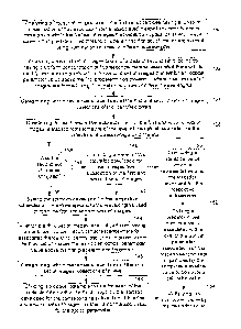
Fig. 2 illustrates a flowchart
illustrating an exemplary sequence of steps for measuring the background
signal of an image. As shown
in block 40 of Fig. 2, the method may include separating an image of particles
having fluorescence-
CA 2984772 2017-11-06
9
material associated therewith into an array of subsections. Such an array may
include any number of
rows and columns, depending on the clarity of desired background signal, the
processing capability of the
system, and/or the number of particles being analyzed. As further shown in
Fig. 2, the route the method
continues along after block 40 may depend on the occupancy of the particles
within the image. In
particular, after block 40, the flowchart continues to block 42 in which a
determination is made as to
whether the occupancy of the particles within the image has been quantified.
In embodiments in which the occupancy of particles has been quantified, the
method may
continue to block 44 in which a determination of whether the occupancy is less
than a predetermined
threshold. The flowchart in Fig. 2 specifically notes a threshold of 50%
occupancy within block 44, but
it is noted that the method is not necessarily so limited. In particular, the
method may be modified to
consider any predetermined quantity of occupancy by which to determine the
course of action for
measuring the background signal of an image. In embodiments in which particles
of interest occupy less
than a predetermined threshold (e.g., less than about 50%) of the imaging
area, background signal
measurement may include determining a statistical value of an optical
parameter among all pixels within
a subsection as noted in block 46. Consequently, fluorescence values of the
relatively bright pixels
corresponding to particles within the subsection may be merged with signals
from background pixels
(pixels which do not correspond to the presence of pixels) within the
subsection. Since the particles
occupy a smaller amount of the subsection, however, the statistical value may
be more representative of
the background pixels. In general, the statistical value may include any
number of statistical parameters,
including but not limited to median, mean, mode, and trimmed mean. In some
embodiments, determining
a median value may be particularly advantageous.
In other embodiments, the method may continue to blocks 50, 52, and 54 to
determine a
statistical value of an optical parameter of less than all of the pixels
within a subsection. In particular, in
(- embodiments in which the occupancy of particles of interest is greater
than or equal to a predetermined
threshold of the imaging area (e.g., greater than or equal to about 50% as
noted by the arrow connecting
block 44 to block 50) or when the occupancy of the imaging area by the
particles is unknown (e.g., as
noted by the arrow connecting block 42 to block 50), the method for background
signal measurement
may compensate for the larger or unknown ratio of particle area to background
area by determining a
statistical value of an optical parameter of less than all of the pixels
within a subsection. In particular,
pixels within an image exhibiting an optical parameter value above a
predetermined threshold may be
identified as noted in block 50. In some cases, the pixels identified in block
50 may be grouped with
pixels arranged immediately adjacent thereto as noted in block 52. Such a
process step, however, is
optional as denoted by the dotted line border of the block and, therefore, may
be omitted from the method
in some cases.
In any case, the method may continue to block 54 in which a statistical value
of an optical
parameter is determined solely among a set of pixels within the subsection
which are not identified to
CA 2984772 2017-11-06
= 10
exhibit an optical parameter above the predetermined threshold outlined in
block 50. In some
embodiments, pixels grouped with such identified pixels may also be excluded
from the determination of
the statistical value of the optical parameter. In this manner, pixels
identified in block 50 and, in some
cases, the pixels grouped with the identified pixels in block 52 may be
isolated from the measurement of
the background signal.
In any case, regardless of the sequence of process steps used, the optical
parameter of which a
statistical value is determined may be any fluorescence emission of the
particle measured at one or more
detection wavelengths, emissions of scattered light in the background of the
image as well as any noise
and dark current offset from the imaging system used to image the particle. In
addition, regardless of the
sequence of process steps used, the method may continue to block 56 to assign
the determined statistical
value as background signal for the corresponding subsection. More
specifically, the background signal
level for all pixels within a subsection may be assigned the statistical value
computed for the subsection.
In some cases, the process steps of blocks 46, 50, 52, 54, and 56 may be
repeated for other subsections in
the image and, in some cases, for all subsections in the image. In this
manner, a statistical value of an
optical parameter may be determined for each of a plurality of subsections
and, in some cases, for all of
the subsections. In some cases, a relatively sharp contrast in statistical
values may be present at the
boundary between two subsections. In order to smooth the discontinuous
difference in the statistical
values between adjacent subsections, a two-dimensional statistical filter
(e.g., a median filter or a mean
filter) may be performed on the array of subsections. As a result, the
subsections may be smoothed at
their edges. Regardless of whether such a statistical filter is used, a
resultant n x m matrix of subsections
of pixels containing the computed statistical values may be saved as a
"background image," which may
be utilized as described further herein.
It is noted that the method described in reference to Fig. 2 may include
additional steps of the
above-described method for background signal measurement and, therefore, the
method is not necessarily
limited by the depiction of Fig. 2. For example, the omission of a reiteration
of blocks 46, 50, 52, 54, and
56 in Fig. 2 does not necessarily limit the inclusion of such a possibility
for the method described herein.
As noted above, the method described herein for image data processing may
include a process of particle
discovery using fluorescence emission from the classification dye(s) and
cluster rejection (i.e., rejection
of particles that are located relatively close together). In some embodiments,
the process of particle
discovery described herein may be performed subsequent to the determination of
a level of background
signal within an image and, in some cases, may be specifically performed
subsequent to the method of
background signal measurement described in reference to Fig. 2. In other
embodiments, however, the
process of particle discovery described herein may be performed independent of
background signal
measurements.
Fig. 3 illustrates a flowchart illustrating an exemplary sequence of steps for
a process of particle
discovery. As shown in block 60 of Fig. 3, the method may include analyzing an
image of particles
CA 2984772 2017-11-06
11
having fluorescence-material associated therewith to identify one or more
pixels within the image that
exhibit an optical parameter value above a predetermined threshold. For
example, a classification image
(i.e., an image generated from light emitted at a wavelength or wavelength
band of a classification dye)
may be searched for pixels that exhibit fluorescence higher in intensity than
the background signal of the
image. In some embodiments, the image may be searched for pixels significantly
higher in intensity than
the background signal of the image, such as on the order of 2 to 1000 times
higher in intensity. Smaller
or larger intensity levels relative to the background signal of the image may
also be used. In other cases,
the image may be searched for pixels exhibiting a fixed value of fluorescence,
which may or may not be
dependent on the background signal of the image. In any case, a higher level
of fluorescence emission by
a pixel or a collection of pixels may indicate the presence of a fluorescence
emitting particle. In some
embodiments; the particle may be contained within a single pixel. In other
embodiments, however, the
particle may spread across a plurality of pixels.
In any case, the pixels identified in block 60 may be evaluated to determine
the location of
particles within the image. In particular, the method outlined in Fig. 3 may
continue to block 62 to
determine locations within sets of one or more identified pixels that
respectively exhibit a maximum
value for the optical parameter to detect the presence particles within the
image. Although the pixels
may be evaluated individually and, therefore, a location within a single pixel
may be determined by block
62, block 62 may also include determining a location among a collection of
identified pixels. As used
herein, a "collection of pixels" may generally refer to a grouping of pixels
which are arranged
immediately adjacent to each other (i.e., a cluster or conglomerate of
contiguously arranged pixels).
In some embodiments, it may be advantageous to evaluate a collection of pixels
for determining
locations of particles within an image. In particular, as noted above, a
particle may spread across a
plurality of pixels and, as such, determining locations within individual
pixels may falsely convey the
presence of more particles than are actually imaged. Furthermore, if a
particle is located relatively close
to one or more other particles in an image, the fluorescence of the particles
may affect the evaluation of
each other's characteristics. Consequently, data for the particles may not be
accurately determined. In
some cases, a collection of pixels may be rejected (e.g., eliminated from any
further image processing) if
it is determined the characteristics of an encompassed particle cannot be
accurately evaluated.
Exemplary manners in which to determine whether a collection of pixels may be
accepted or rejected for
further image processing are described in more detail below in reference to
blocks 70-78 of Fig. 3.
In general, the process outlined in block 62 for determining locations within
sets of one of more
identified pixels may be conducted in a number of different manners. Some
exemplary methods are
outlined in blocks 64, 66, and 68 in Fig. 3 (blocks 64, 66, and 68 extend from
block 62 by dotted lines
and are bordered by dotted lines, indicating the processes are exemplary). As
shown in Fig. 3, block 64
outlines a process for ascertaining peak pixels among the sets of one or more
identified pixels that
respectively exhibit maximum values for the optical parameter. In such a
process, each set of pixels may
CA 2984772 2017-11-06
12
be iterated through to determine if the fluorescence value measured for each
pixel has the maximum
value within the set of pixels. The pixel with the maximum value may be
ascertained as the "peak pixel".
In some cases, a central portion of the peak pixel may be designated as the
location. In such cases, the
process of determining the location as outlined in block 62 may be simply
conducted by the process
outlined in block 64.
In some embodiments, however, it may be advantageous to determine if an
alternative portion of
the peak pixel is more suitable for the location exhibiting the maximum value
for the optical parameter.
For instance, particles may not be perfectly aligned among the pixels of the
image and, consequently, the
energy from a particle may not be evenly distributed among a set of identified
pixels. In such cases, a
central portion of a peak pixel may not be representative of the maximum
fluorescence measurement for
the particle and, therefore, it may be advantageous to determine if an off-
center portion of the peak pixel
may better represent the maximum fluorescence measurement for the particle. In
such cases, the method
may continue to block 66 to compute a centroid location within at least one
the set of one or more
identified pixels that exhibits a maximum value for the optical parameter. hi
particular, the method may
include integrating fluorescence measurements of pixels adjacent to and within
a predetermined radius of
a peak pixel. An exemplary radius from the peak pixels may be selected from a
range of 1 to 10 pixels,
but other radii may be considered. It is noted that in embodiments in which
the predetermined radius
encompasses pixels which have not been identified to have an optical parameter
above a predetermined
threshold, the background signal all of such "background pixels" may be
subtracted from this integral.
In some cases, it may be advantageous to analyze whether to assign the
computed centroid
location as the location exhibiting a maximum value for the optical parameter.
As such, in some
embodiments, the method may, in some embodiments, continue to block 68
depending on the
characteristics of the computed centroid location. For example, if the
centroid location is greater than
one half of a pixel width in any direction, the computed location rounded up
to the next integer value
(e.g., in x and y coordinates) may be assigned as the location exhibiting a
maximum value for the optical
parameter. Although block 68 specifies a dimensional threshold for the
computed centroid location to be
greater than 50% of the dimensions of the pixels to assign the centroid
location, the contingency process
is not necessarily so limited, hi particular, any dimensional threshold for
the centroid location (including
those which are independent of the pixel dimensions) may be used to determine
whether to assign the
centroid location.
Subsequent to the process for determining the locations exhibiting a maximum
value for the
optical parameter, the method may continue to processes for accepting and
rejecting pixels for further
image processing. For example, the method may, in some embodiments, continue
to block 70 in which a
distance between two peak pixels is computed. The identification of the peak
pixels may generally be
performed by the process described above in reference to block 64 and,
therefore, the flowchart in Fig. 3
includes a dotted line connecting blocks 64 and 70 to indicate the
correlation. Based upon the distance
CA 2984772 2017-11-06
13
computed in block 70, a set of pixels corresponding to one of the two peak
pixels may be accepted or
rejected for further image processing as noted in block 72. For example, a set
of pixels corresponding to
one of the two peak pixels may be rejected for further image processing if the
distance between the peak
pixels is less than (and/or equal to) a predetermined threshold, such as but
not limited a threshold
equivalent to projected diameters of one or two imaged particles or any
distance therebetween. In this
manner, fluorescence emissions of particles which are arranged too close to a
particle of interest, which
may hinder the evaluation of the particle of interest, may be averted.
In general, the term "projected diameter of an imaged particle," as used
herein, may refer to an
estimated diameter for an imaged particle based on component configurations of
a system used to image
the particles. In general, the size of an imaged particle may differ from
dimensions of an actual particle
depending on the magnification of the imaging system used to image the
particle. In addition, other
component configurations of an imaging system may affect the diameter as well.
For example, an
imperfect lens, diffraction from optical apertures, optical filter distortion,
as well as several other
components of an imaging system may affect and, in some cases, distort
dimensions of an imaged pixel
(referred to herein as the smear of the imaged particles). In some cases, the
point spread function (PSF)
(alternately quantified as the modulation transfer function (MT'F)) of the
imaging lens may be the
primary contributor to distortion.
Although either set of pixels corresponding to the two peak pixels may be
rejected, it may be
advantageous to reject the set of pixels corresponding to the peak pixel
having a lower fluorescence
measurement since the characteristics of such a set of pixels may be less
distinguishable versus the other
set of pixels during further image processing. In some cases, the method may
continue to evaluate the
remaining set of pixels to determine if it is sufficient for further imaging
processing. For example, the
method may continue to block 74 to determine whether a fate of intensity
change of an optical parameter
among the set of pixels is indicative of a single particle or a clump of
particles. Generally, it is desirable
to reject clumps of particles due to the difficulty of obtaining accurate and
distinct information for each
of the particles. In yet other embodiments, the selection of the two sets of
pixels for rejection in block 72
may be determined by computing the rate of intensity change of an optical
parameter among the sets of
pixels. In particular, upon determining the distance between the peak pixels
is less than a predetermined
threshold, rates of intensity change of an optical parameter may be computed
for each set of pixels as an
indicator of which set should be rejected. Different manners for computing a
rate of intensity change
among a set of pixels are outlined in blocks 76-78 and 80-82, respectively,
and described in more detail
below.
Since the method of particle rejection may include a combination or sequential
processing of
blocks 72 and 74, the flowchart in Fig. 3 includes a dotted line between
blocks 72 and 74 to indicate the
possibility of such a connection between the respective processes. Such a
connection, however, is
optional. In particular, blocks 72 and 74 may not, in some embodiments, be
performed in conjunction
CA 2984772 2017-11-06
14
and, therefore, the arrow between blocks 72 and 74 may be omitted. In other
embodiments, the
processing of blocks 74 and 72 may be reversed and, as such, the method
described herein may include a
connection between blocks 78 and/or 82 and block 70. In other embodiments,
blocks 70 and 72 may be
omitted from the method. Alternatively, block 74 (and its exemplary procedures
for performing such a
process outlined in blocks 76-82) may be omitted from the method. In yet other
cases, the method may
be configured to select the route of image processing subsequent to block 62
and, therefore, may lead to
either of blocks 70 and 74 as illustrated in Fig. 3.
Referring to block 74, a rate of intensity of an optical parameter among a
plurality of pixels
surrounding at least one of the locations determined in block 62 may be
computed. As noted above, this
rate may be used to accept the *tick or to reject the particle for further
image data processing. More
specifically, the rate may be a measure of the spatial gradient of the
emission characteristics of the
particle (i.e., the distribution of the fluorescence emission level) and the
spatial gradient may be used to
determine how isolated the particle of interest is from neighboring particles.
In some embodiments, the
process of block 74 may follow the sequence of steps outlined in blocks 76 and
78. In particular, the
method may include computing a rate of intensity of an optical parameter for a
set of pixels arranged
within a predetermined radius surrounding a location determined in block 62.
In some embodiments, the
predetermined radius may be approximately equal to a projected diameter of the
particle represented by
the determined location. In other cases, the predetermined radius may be
greater or less than a projected
diameter of the imaged particle represented by the determined location.
After the rate of intensity change of the optical parameter is computed, the
method may continue
to block 78 in which the set of pixels may be accepted or rejected for further
image data processing by
comparing the rate of intensity to a predetermined threshold. In some
embodiments, block 78 may
include accepting the set of pixels for further processing upon computing the
rate of intensity change is
greater than or equal to a predetermined threshold. In particular, a
relatively high rate of intensity change
of an optical parameter may be indicative of a single particle within the set
of pixels, which may be
desirable for further image processing. In addition to such a process, block
78 may include rejecting the
set of pixels for further processing upon computing the rate of intensity
change is less than a
predetermined threshold. In particular, a relatively low rate of intensity
change of an optical parameter
may be indicative of a clump of particles within the set of pixels, which as
noted above may be
undesirable for further image processing.
An alternative manner in which to compute a rate of intensity change of an
optical parameter
within a set of pixels is outlined in blocks 80 and 82 in Fig. 3. In
particular, the method may additionally
or alternatively be routed to block 80 to sum values of the optical parameter
for two distinct sets of pixels
respectively arranged within different predetermined radii surrounding one of
the locations determined in
block 62. It is noted that the radii may be adjusted to best match the
particle's spread across the detector
pixel array, which usually varies depending upon the point spread function
(PSF) (alternately quantified
CA 2984772 2017-11-06
15
as the modulation transfer function (MTF)) of the imaging lens, the position
of the particle with respect
to the focal plane of the imaging subsystem, and the size of the particle
itself. For example, in some
embodiments, it may be advantageous for one predetermined radius to be
approximately equal to a
projected diameter of a single particle within the image and the other
predetermined radius to be
approximately 1.5 times greater than a projected diameter of a single particle
within the image. Other
radii, however, may be used as well as different ratios of the radii may be
used. It is further noted that if
values of a background signal is subtracted for pixels within one radius, the
background signal may also
be subtracted from the values for the pixels within other radius.
Subsequent to summing the values of the optical parameter, a ratio of the
summed values
corresponding to each of the radii may be computed. In particular, the summed
values obtained using the
smaller radius may be divided by the summed values obtained using the larger
radius or vice versa. In
either case, the ratio may be used to accept or reject the set of pixels for
further image data processing as
noted in block 82. In particular, block 82 may include accepting the set of
pixels for further evaluation
upon determining the ratio differs from a set value by an amount less than or
equal to a predetermined
threshold. In addition, block 83 may include rejecting the set of pixels for
further evaluation upon
determining the ratio differs from the set value by an amount greater than the
predetermined threshold.
The determination of the threshold may depend on a variety of factors,
including but not limited
to radii chosen for performing the process outlined in block 80, the size of
the particles to be imaged, the
smear of the particles within the image, as well as the settings of the
imaging system used.
Consequently, the predetermined threshold for accepting and rejecting set of
pixels in block 83 may vary
greatly among different applications. However, a general guideline is that a
ratio closer to unity may be
indicative of a set of pixels that may be desirable for further processing
since there is little contribution
from the pixels outside the smaller radius. In other words affects of optical
parameter values from
neighboring particles is likely to be minimal and, thus, the error in a value
for an optical parameter of
interest will be relatively small. Alternatively, if this ratio is
significantly less than unity, then it is likely
that a neighboring bright particle is affecting the optical parameter value of
the particle of interest. In
such instances, the particle of interest may be discarded, or the integration
radii may have been
improperly chosen. In this manner, before the image data for a particle of
interest is discarded, the
integrations described above may be performed with different radii.
An algorithm for performing such an additional integration may include
establishing an inner
diameter to outer diameter for each bead at some fixed ratio (such as the
1.5x) and storing the results. In
such cases, the inner diameter may be slightly larger than the expected bead
projection will be, such as
1.5 times large than the expected bead projection. Then the inner and outer
diameters may be reduced
slightly (keeping same ratio of as before) for each bead. Subsequent thereto,
the collection of ratios may
be compared to see if a majority of the ratios have changed. If most of the
ratios have not changed, the
inner diameter is still too big and no energy is (yet) getting outside the
inner circle to the outer circle, so
CA 2984772 2017-11-06
16
the inner and outer diameters need to be reduced again for each bead.
Conversely, if some of the ratios
have changed, it may be indicative that some energy may be moving to the outer
circle.
The process may be iterated any number of times based on the distribution of
the changes from
the last diameter's collection of ratios. For example, if the percentage of
particles that coincide is known
(and, consequently, should be discarded), the percentage may be equated to a
desired percentage of ratios
to end the iteration. An estimation of the percentage of particles that
coincide may be drawn from
knowledge of how the system typically behaves from past data off the
production line, or alternatively
a visual examination of the test image. If the percentage of coinciding
particles is unknown, the "history"
of the changes step by step for an emerging percentage that change and remain
constant with decreasing
inner diameter may be an indicator to terminate the iteration. As an example,
given 5% of the ratios
change with one reduction, then 10%, then 10% again, and 12% the fourth time.
In such an example,
10% may the number of particles that should be discarded. When the percentage
of 12% was reached,
the inner circle may have been too small, cutting off the smaller-single-good
beads. As such, the
previous diameter should be used as the stopping point. All of such process
steps may be repeated with
different inner/outer diameter ratios to see if a clearer trend of percentage
changes emerges. In such
cases, the process may include an "outer loop" in the algorithm where you
start first with a larger ratio,
then step through sweeping the ratio until you are actually smaller than the
original one (optionally
skipping the original ratio since it has already been computed.
As noted above, the method described herein for image data processing may
include a process of
inter-image alignment. In some embodiments, the process of inter-image
alignment described herein may
be performed subsequent to the determination of a level of background signal
within an image and/or
subsequent to discovery of particles within an image. hi some cases, the
process of inter-image
alignment may be specifically performed subsequent to the method of background
signal measurement
described in reference to Fig. 2 and/or subsequent to the method of particle
discovery described in
reference to Fig. 3. In other embodiments, however, the process of inter-image
alignment described
herein may be performed independent of background signal measurement and/or
particle discovery
processes. In any case, the inter-image alignment process may be performed at
the factory after the
instrument has been assembled. In addition or alternatively, the inter-image
alignment process may be
performed in the field, particularly if components of the system are changed
after shipment from the
factory.
In general, inter-image alignment may be performed if multiple images of
particles are acquired
using two or more detectors, each of which may be coupled to an optical filter
as described above, or if
interchangeable optical filters are substituted between images taken with a
single camera, since the filter
itself may affect the image. The multiple images are generally taken at
different wavelengths such that
different levels of fluorescence may be measured and used to classify the
particles. Due to the
mechanical tolerances of the imaging subsystem hardware, however, spots
corresponding to particles
CA 2984772 2017-11-06
17
within the each of the multiple images may not be in absolute alignment in a
composite of the multiple
images. Such mis-registration of the spots may undesirably inhibit the ability
to associate a particle's
location in all channels imaged. The image-to-image registration, however, may
be modified using the
inter-image alignment technique described herein to better align the spots. As
described below, the inter-
image alignment correction process may be a simple translation of image
coordinates in the x and/or y
directions. In addition or alternatively, the inter-image alignment process
may include rotation of one or
more of the multiple images.
Fig. 4 illustrates a flowchart illustrating an exemplary sequence of steps for
a process of inter-
image alignment. As shown in block 90 of Fig. 4, the process may include
acquiring data for multiple
images of particles having fluorescence-material associated therewith, wherein
each of the multiple
images corresponds to a different wavelength band. In some cases, the data may
be acquired directly
from an imaging system, but in other cases, the data may be acquired from a
storage medium. In either
case, the data may be representative of multiple images taken at different
wavelengths as noted above.
Exemplary wavelengths that may be used may correspond to different color
channels, such as but not
limited to red for classification channel 1, green for classification channel
2, blue for the reporter
channel. As further noted above, in order to accommodate each color channel,
the particles used for the
method described herein may be specially dyed to emit at all wavelengths or in
all wavelength bands of
interest. In particular, in order to measure both classification and reporter
signals within the multiple
images, the inter-image alignment process described herein may be performed
using specially dyed
particles, which not only emit fluorescence in the classification
wavelength(s) or wavelength band(s), but
also in the reporter wavelength or wavelength band.
After the data for the multiple images has been acquired, the method may
continue to block 92 in
which a composite image of the multiple images is created. In general, the
composite image is a single
image with the multiple images overlapped relative to each other. As noted
above, due to the mechanical
tolerances of the imaging subsystem hardware, spots corresponding to particles
within the each of the
multiple images may not be in absolute alignment in a composite of the
multiple images. As such, inter-
image alignment may be needed. In particular, the method may include
manipulating coordinates of at
least one of the multiple images such that spots corresponding to the
particles within each of the multiple
images converge within an ensuing composite image as noted in block 94. In
some embodiments, the
coordinate values of all of the multiple images but one (the one being
referred to herein as the "reference
image") may be manipulated. Alternatively, the coordinate values of fewer
multiple images may be
manipulated. In this manner, the coordinate values of images other than the
reference image may be
maintained for the inter-image alignment process. In some cases, the image
acquired at the wavelength
or wavelength band of light emitted by the reporter dye may be designated as
the reference image. In
other embodiments, the image acquired at a wavelength or wavelength band of
light emitted by a
classification dye may be designated as the reference image.
CA 2984772 2017-11-06
18
As noted above and illustrated in Fig. 4, the manipulation of the coordinates
may, in some cases,
include be an orthogonal offset of image coordinates in the x and/or y
directions as noted in block 96. In
addition or alternatively, the manipulation of the coordinates may include
rotation of one or more of the
multiple images as noted in block 98. Blocks 96 and 98 are outlined by dotted
lines, indicating either or
both of the processes may be used for the manipulation of the image
coordinates.
In the process of orthogonal translation, a positive or negative integer
translation offset in either
the x or y dimension may be determined for the manipulation of the coordinate
values. The respective
offsets may be added to the coordinates of one or more non-reference images,
and a new composite
image may be created with the multiple images, some of which having the new
coordinates. In general,
the orthogonal translation correction steps may be performed until no further
improvement in alignment
within a composite image is possible. Upon determining no further improvement
by orthogonal
translation may be obtained, the x translation and y translation values for
each non-reference image
having coordinates which were manipulated by the process may be saved for
subsequent imaging of
particles. Any appropriate data structure, such as a table, may be suitable
for such values.
As noted above, the manipulation of the coordinate values may additionally or
alternatively
include rotating coordinates of one or more non-reference images. In some
embodiments, the rotation
process may be employed if the images are not aligned sufficiently via
translation correction. In other
embodiments, the rotation process may be performed prior to, instead of, or
alternately with the
orthogonal translation process. In yet other cases, the rotation process may
be performed concurrently
with the orthogonal translation process. In particular, one or more non-
reference images may be rotated
and one or more other non-references may be translated with orthogonal offsets
for the manipulation of
image coordinates. In other embodiments, coordinates of individual non-
reference images may be both
rotated and orthogonally offset. In any case, the range of orthogonal offsets
which may be employed for
the inter-image alignment process may, in some embodiments, be +/- 10 pixels
and the range of rotational
offsets may be +/-2 degrees. Larger or smaller amounts of offsets, however,
may be employed for either
or both manners of manipulating the coordinates.
Regardless of the manner in which the rotation of images is incorporated
relative to orthogonal
offsets of image coordinates, the rotation process may generally include
selecting the origin (i.e., center
of rotation) to be near the midpoint of the x and y dimensions of the image
(denoted as xi, = = , y.). A
new blank image buffer may be created with the same dimensions as the source
image (i.e., the non-
reference image to be rotated). For each pixel in the source image, a current
vector from the center of
rotation may be determined. In particular, the distance from the pixel of
interest to center of rotation of
the image may be determined from the square root of [(x-x01) 2+ (Y-Yorig)21, x
and y being the
coordinates of the pixel. In addition, the current vector's angle may be
determined from the arctangent of
the Ydistance divided by the )(distance and adding or subtracting a quadrant-
dependent modifier from the value
of the arctangent to adjust the angle per quadrant. In such cases, ydistance
is the distance along the y-axis
CA 2984772 2017-11-06
19
between yofigh, and the pixel of interest and Xdistance is the distance along
the x-axis between )(origin and the
pixel of interest.
Subsequent to the aforementioned computations, a constant user defined
"adjustment" angle may
be added to the current pixel's vector to determine the angle by which to
rotate the pixel. The new
location for the pixels (e.g., in x and y coordinates) may be determined by
the following equations:
new x coordinate = square root of [(x-x.igin) 2+ (y¨yorigin)2] * cos(rotated
angle) + Xorigin
Xtransiation (if applicable) + 0.5 (1)
new y coordinate = square root of [(X¨Xorigin) 2+ (y..yori )2,
gir * sin(rotated angle) +
yofigin
Ytranslation (if applicable) + 0.5 (2)
The value of the pixel under consideration may be copied to the blank image
buffer's pixel at the new x
and y coordinates. After non-reference images intended for rotation have been
processed, a new
composite pseudo-color image may be recreated. In general, the steps outlined
above for the rotation
(- process may be repeated to minimize the color variance across each non-
reference image. The final
rotation values may be saved for each non-reference image in a suitable data
structure such as an
adjustment table for subsequent imaging.
In general, the iteration of coordinate manipulation described above in
reference to block 94 may
be conducted in reference to a number of different parameters. For instance,
the iteration of coordinate
manipulation may depend on the amount of color variance among spots of a
composite image, aggregate
error or mean square difference of intensities among pixels corresponding to
spots of a composite image,
and/or aggregate error or mean square difference of locations of spots within
a composite image. An
outline of each of such techniques is outlined in blocks 100-128 in Fig, 4 and
described in more detail
below.
In particular, block 100 includes a process of algorithmically determining
(i.e., by means of an
algorithm) an offset to modify coordinates of at least one of the multiple
images such that an amount of
color variance among the spots in an ensuing composite image is reduced
relative to a preceding
composite image. The color variance in the composite image is generally
induced by misalignment of at
least one of the multiple images. For example, in embodiments in which red,
green, and blue channels
are used for the respective multiple images, the converged color of a spot
corresponding to a particle in a
composite image is expected to be white. Alignment variations of the multiple
images, however, may
cause spots on the individual images corresponding to one or more of the red,
green, and blue channels to
be offset relative to each other. As a consequence, the individual colors in
the composite image may
extend beyond an edge of the white spot, inducing a variance of color at the
spot. It is noted that the
formation of a white spot in a composite image is a result of the combination
of the images produced by
the red, green, and blue channels, but the method described herein is not
necessarily limited to making
images with such channels. In particular, any number of multiple images may be
formed by several
CA 2984772 2017-11-06
20
different color channels and, consequently, the method described herein is not
restricted to the formation
of three images or the color channels of red, green, and blue.
As described above and outlined in block 102 of Fig. 4, the misalignment of
the images may be
reduced by adjusting the coordinates of one or more of the multiple images by
predetermined offsets.
Such predetermined offsets may include orthogonal offsets and/or rotational
offsets as described above in
reference to blocks 96 and 98. Subsequent to block 102, a different composite
image of the multiple
images including the predetermined offsets may be created as noted in block
104. The method may
continue to block 106 in which the color variance among the spots in the newly
created composite image
is determined. As noted in decision block 108, blocks 100-106 may be repeated
in embodiments in
which the color variance is greater than (and/or equal to) a predetermined
error allowance for particular
offset amount. Conversely, the method of inter-image alignment may terminate
at block 110 in
embodiments in which the color variance is less than (and/or equal to) the
predetermined error
allowance. In general, the predetermined error allowance set for block 108 may
depend on the accuracy
desired for the composite image as well as the offset amount and, therefore,
may vary among
applications.
Techniques for the iteration of coordinate manipulation based on aggregate
error or mean square
difference of pixel intensities and/or locations of spots within a composite
image are described in
reference to blocks 112-128 in Fig. 4. In particular, both techniques may
start at block 112 at which i is
set equal to 1. Such a designation is used to reference the 1.5t of several
predetermined offsets to adjust
the coordinates of at least one of the multiple images as noted in block 114.
In some embodiments, the
selection of predetermined offsets through which the processes are iterated
may be specific to the
parameter by which alignment in the composite image is measured (i.e., by
aggregate error or mean
square difference of pixel intensities or locations of spots within a
composite image). In other
embodiments, the selection of predetermined offsets may be independent of the
technique used. In either
case, the processes may continue to block 116 to create a different composite
image of the multiple
images including the predetermined offsets. Thereafter, processes specific to
the techniques may be
employed. For example, the method may continue to block 118 to determine an
aggregate error or mean
squared difference in intensities among the pixels of the composite image
created in block 116.
Alternatively, the method may continue to block 120 to determine an aggregate
error or mean squared
difference in locations of spots within the composite image created in block
116.
In either case, a determination may be subsequently made at block 122 as to
whether i equals n, n
being the number of predetermined offsets by which to adjust the coordinates
of the multiple images. In
cases in which i does not equal n, the method may continue to block 124 to
increase the value of i by one
and the processes outlined in blocks 114-120 may be repeated. Upon determining
i equals n, the method
may continue to block 126 in which the computed values of aggregate error or
mean square differences
for each of the different composite images are evaluated. In particular, the
computed values of aggregate
CA 2984772 2017-11-06
= 21
error or mean square differences for each of the different composite images
may be evaluated to identify
the offset (i.e., the translation and/or rotation) values that resulted in the
minimum error for a composite
image. The identified offset values may be saved for each non-reference image
for which coordinates
were adjusted as noted in block 128. Any appropriate data structure, such as a
table, may be suitable for
such values. In both of the above described embodiments, the identified offset
values may be applied to
the coordinate systems of classification images created during subsequent
images. In particular, the
classification images may be translated and/or rotated directly into new image
buffers using the equations
described above and the original classification image buffers may then be
discarded.
Inter-image particle correlation may be performed after the image coordinate
systems are aligned.
In particular, after the image coordinate systems are aligned, actual
particles may be discarded by
position, assuming that more than one classification image is acquired at more
than one wavelength or
wavelength band. Simply stated, if a particle is not present across all
classification images, it may be
eliminated from further processing.
In one example, using each classification image's particle collection
previously identified using
the particle discovery method described above and the translation/rotation
values for each classification
image, the best matching particle that lies within a given radius may be
identified. Identifying the best
matching particle may include creating a nested series of n loops, each level
of which represents a
classification image, for iterating through each collection of particles. At
the deepest nesting level, the
method may include determining if the particle's adjusted coordinates from all
outer loops lie within a
given radius. The coordinates at each nesting level may be translated
according to the alignment table
and equations described above for inter-image alignment before the distance is
determined. If the
distance is less than a given radius, the innermost loop's particle location
may be temporarily stored for
later comparison against other matches at the innermost level. If the distance
of the second particle at the
innermost level is less than that of a previously found particle, the
temporarily stored particle location
may be replaced with the present distance. If not, the method may be continued
for the next particle. At
the end of the iteration of the second from outermost loop, the temporary
location of the best match to the
outermost location may be stored to a collection. If there are no matches
within a given radius for the
outer loop particle, then the instance of the particle is automatically
eliminated from further consideration
as the output of the correlation algorithm is the collection created above.
To speed up the overall process, if there is a match identified as described
above, the particle
may be identified as "already used" in each subsequent loop such that
processing time is not expended to
consider it again. The images may also be separated into a number of
subsections, and each subsection
may be correlated separately to reduce processing time. In such an instance,
the number of subsections is
preferably selected such that the total savings in loop iterations is not lost
in the time it takes to
decompose the image into sections. In addition, to avoid loss of comparison
capability at the boundaries
of the subsections, the regions may have a slight overlap. Furthermore, if the
regions are overlapped, the
CA 2984772 2017-11-06
22
degree to which regions overlap may be selected to reduce the potential to
duplicate particles at the
overlap.
The method may also include fluorescence integration of reporter fluorescence
emission. Since
the reporter emission level is not constant and is an unknown, it is not
possible or necessary to use the
particle discovery technique employed for the classification images to
identify the pixels in the reporter
image that are used in the integration. Instead, the fluorescence at the same
x and y coordinates of the
particles found in the classification images may be used.
In one such example, using the translation and rotation values from the
adjustment table
determined by the inter-image correlation, each discovered particle may be
mapped to the appropriate
coordinates of the reporter image. For the starting location of each particle,
the coordinate system from
the non-adjusted reference classification image may be used. The translation x
and y values and rotation
angle that were determined for the reporter represent the direction an imaged
particle in the reporter
image may be moved to thereby coincide with the location of the particle in
the classification reference
image. However, the transformation that is performed here involves translating
the reference coordinate
system to the reporter coordinate system. The x and y translation values can
be "converted" by simply
inverting the sign of each adjustment parameter (negative values become
positive and vice versa).
Similarly, the sign of the rotation angle may also be inverted before the
reporter coordinate is found.
After the signs of all parameters are inverted, the equations described above
for the inter-image
alignment step may be used to identify the center of integration. The integral
of all reporter pixels that
lie within the given integration radius may be determined.
As noted above, the method described herein for image data processing may
include a process of
image plane normalization. Ideally, an imaging system is evenly illuminated to
prevent position
dependent emission variance among particles. In reality, however, each spot on
the imaging field has a
given illumination level. In addition, the fluorescence bandpass filter(s) and
the imaging lens or lenses
used within the system may not transmit the same amount of light for all
points in the image. In order to
compensate for such variations across the image, a normalization method or
algorithm may be applied to
the measured values of optical parameters. In some embodiments, the process of
image plane
normalization described herein may be performed subsequent to one or more of
the process described
above, particularly with regard to those described in reference to Figs. 2-4.
In other embodiments,
however, the image plane normalization described herein may be performed
independent one or more of
such processes.
Fig. 5 illustrates a flowchart illustrating an exemplary sequence of steps.
for a process of image
plane normalization. As shown in block 130 of Fig. 5, the process may include
analyzing a first set of
images taken of a first set of particles having a uniform concentration of
fluorescence-material associated
therewith to identify one or more pixels within the first set of images that
exhibit an optical parameter
value above a first predetermined threshold. The first set of images may
include any number of images,
CA 2984772 2017-11-06
23
including a single image or a plurality of images. In embodiments in which a
plurality of images are
taken, the first set of images are formed using illumination sources of
different wavelengths, such as but
limited to wavelengths corresponding to red, green, and blue channels.
In some cases, the method may optionally (as indicated by the dotted line
border) include block
132 in which a second set of images taken of a second distinct set of
particles having a uniform
concentration of fluorescence-material associated therewith is analyzed to
identify one or more pixels
within the second set of images that exhibit an optical parameter value above
a first predetermined
threshold. As with the first set of images, the second set of images may
include any number of images
and, in cases in which a plurality of images are taken, the plurality of
images may be formed using
illumination sources of different wavelengths. In some embodiments, analyzing
a second set of images
taken for a different set of particles with known concentrations may be
advantageous for reducing the
effects of noise and particle non-uniformity among the statistics subsequently
developed for respective
subsections of the first and second sets of images. In particular, the effects
of noise and particle non-
uniformity may be reduced by taking a mean of the optical parameter values
measured for each of the
respective subsections of the first and second sets of images as described
below in reference to block
140.
Regardless of whether the method includes analyzing the second set of images,
the method may
continue to block 134 to categorize, within respective subsections of the
first set of images and in some
cases the second set of images, collections of the pixels identified in the
processes described in reference
to blocks 130 and 132. In particular, the sets of images may be separated into
an array of subsections and
collections or conglomerates of contiguously arranged pixels may be arranged
within the subsections
based upon their location within the image. More specifically, for each
particle that is identified, the
subsection within the first and second sets of images to which it belongs may
be determined. The array
of subsections may include any number of rows and columns, depending on the
clarity of desired
background signal, the processing capability of the system, and/or the number
of particles being
analyzed. As further shown in Fig. 5, the method may continue to block 136 to
develop, for each
respective subsection of the first set of images and, in some cases, the
second set of images, a single
statistic representative of the level of the optical parameter for the
collections of pixels categorized
thereto. In general, the statistic may be selected from any number of
statistical parameters, including but
not limited to median, mean, mode, and trimmed mean. In some embodiments,
determining a median
value may be particularly advantageous.
As noted by decision block 138 in Fig. 5, the method may continue to block 140
in embodiments
in which two sets of images taken for two distinct sets of particles are
analyzed. Block 140 specifies that
a mean of the statistics developed for each respective subsection of the first
and second sets of images is
computed. Following block 140 or upon determining that only one set of images
are analyzed for the
categorization of pixel collections in block 138, the method may continue to
block 142 to save the
CA 2984772 2017-11-06
24
statistics developed for the respective subsections in matrices specific to
the wavelengths used to form
the first and second sets of images. Such matrices are used to compute
normalized values for optical
parameters measured for image particles having an unknown concentration of
fluorescence-material
associated therewith as further described below in reference to block 148.
In particular, the method may include block 144 for analyzing a third set of
images taken of
particles having an unknown concentration of fluorescence-material associated
therewith to identify one
or more pixels within the third set of images that exhibit an optical
parameter value above the first
predetermined threshold. As with the first set of images, the third set of
images may include any number
of images and, in cases in which a plurality of images are taken, the
plurality of images may be formed
using illumination sources of different wavelengths. The method may continue
to block 146 in which a
collections of pixels identified in block 144 are categorized into respective
subsections of the third set of
images. hi order to compensate for position dependent emission variances among
the particles having
= unknown concentrations of fluorescence material, normalized values for
the measured optical parameters
may be computed. In particular, block 148 outlines that the optical parameter
value for each of the pixels
identified within the image may be divided by the statistic developed for the
corresponding subsection of
the first and second sets of images to obtain a normalized value for the
optical parameter.
In some embodiments, the resultant normalization value for each identified
pixel may be
multiplied by a single "calibrator" value to adjust its final calibrated value
relative to an external
standard. The calibrator value may be determined from the normalization matrix
as described above for a
substantially uniform set of particles of known concentration. In particular,
the method may optionally
include (as noted by the dotted line borders) block 150 for computing a
statistical value which is
representative of all of the statistics developed for the respective
subsections of one or both of the first
and second sets of images. The statistical value may be selected from any
number of statistical
parameters, including but not limited to median, mean, mode, and trimmed mean.
In some embodiments,
determining a median value may be particularly advantageous. The determination
of the calibration
value may farther include dividing a predetermined numerical value associated
with a level of the optical
parameter associated with the different sets of particles having uniform
concentrations of fluorescence-
material associated therewith by the computed statistical value as noted in
block 152. As noted above
and in block 154 in Fig. 5 the calibrator value May be multiplied by a
normalized value obtained for an
optical parameter of a particle having an unknown concentration to adjust its
value to an external
standard.
It is noted that the normalizing and calibrating techniques described above
are not limited to
normalizing each pixel in all images. Rather, the normalizing and calibrating
techniques may be applied
particles identified within an image. Such a process may be particularly
advantageous for minimizing
calculations versus applications specific normalizing and calibrating pixels.
CA 2984772 2017-11-06
25
It will be appreciated to those skilled in the art having the benefit of this
disclosure that this
invention is believed to provide computer-implemented methods, storage
mediums, and systems for
image data processing. Further modifications and alternative embodiments of
various aspects of the
invention will be apparent to those skilled in the art in view of this
description. Accordingly, this
description is to be construed as illustrative only and is for the purpose of
teaching those skilled in the art
the general manner of carrying out the invention. It is to be understood that
the forms of the invention
shown and described herein are to be taken as the presently preferred
embodiments. Elements and
materials may be substituted for those illustrated and described herein, parts
and processes may be
reversed, and certain features of the invention may be utilized independently,
all as would be apparent to
one skilled in the art after,having the benefit of this description of the
invention.
The scope of the claims should not be limited by the preferred embodiments and
examples, but should be
given the broadest interpretation consistent with the description as a whole.
CA 2984772 2017-11-06