Note: Descriptions are shown in the official language in which they were submitted.
CA 02985656 2017-11-09
WO 2016/183296
PCT/US2016/032046
DRUG DELIVERY FROM HYDROGELS
CROSS REFERENCE TO RELATED APPLICATIONS
This application claims priority to U.S. Provisional Application No.
62/160,394, filed
May 12, 2015, which is hereby incorporated by reference herein.
TECHNICAL FIELD
The technical field, in general, relates to drug delivery involving hydrogels
as used for
various medical conditions, and includes hydrogels formed in an eye with
extended drug
release times.
BACKGROUND
Age-related macular degeneration (AMD), diabetic retinopathy, diabetic macular
edema (DME) posterior uveitis, choroidal neovascularization (CNV) and cystoid
macular
edema (CME) are sight-threatening back-of-the-eye diseases. Age related
macular
degeneration and diabetic retinopathy are significant causes of visual
impairment in the United
States and elsewhere; these conditions are generally caused by angiogenesis
(unwanted blood-
vessel growth in the eye) that damages the retina and ultimately can cause
blindness. Posterior
uveitis is a chronic inflammatory condition that causes about ten percent of
the blindness in the
United States.
SUMMARY
One invention disclosed herein is a crosslinked hydrogel formed in situ that
releases a
therapeutic agent that can be used, e.g., to treat back-of-the eye diseases.
In this embodiment,
aqueous polymeric precursor(s) are combined in flowable
concentrations/viscosities with an
agent and injected through a small gauge needle into the eye, where the
precursor(s) form a
crosslinked in situ hydrogel that releases the drug over time. The hydrogel
may be formulated
to adhere to itself or a tissue in or around the eye to enhance drug release
effects and stability,
to degrade to biocompatible components without causing inflammation, and to
crosslink in
place. A shape-stable hydrogel thus formed can effectively deliver the agent
and
advantageously have a well-controlled size, shape, and surface area. The
hydrogels can be
made to degrade after release of the drug.
1
CA 02985656 2017-11-09
WO 2016/183296
PCT/US2016/032046
BRIEF DESCRIPTION OF THE DRAWINGS
Figure 1 depicts anatomical features of an eye from a frontal view;
Figure 2 is a partially cut-away perspective view of an eye;
Figure 3 is a cross-sectional view of an eye;
Figure 4 depicts various delivery alternatives for ocular implants;
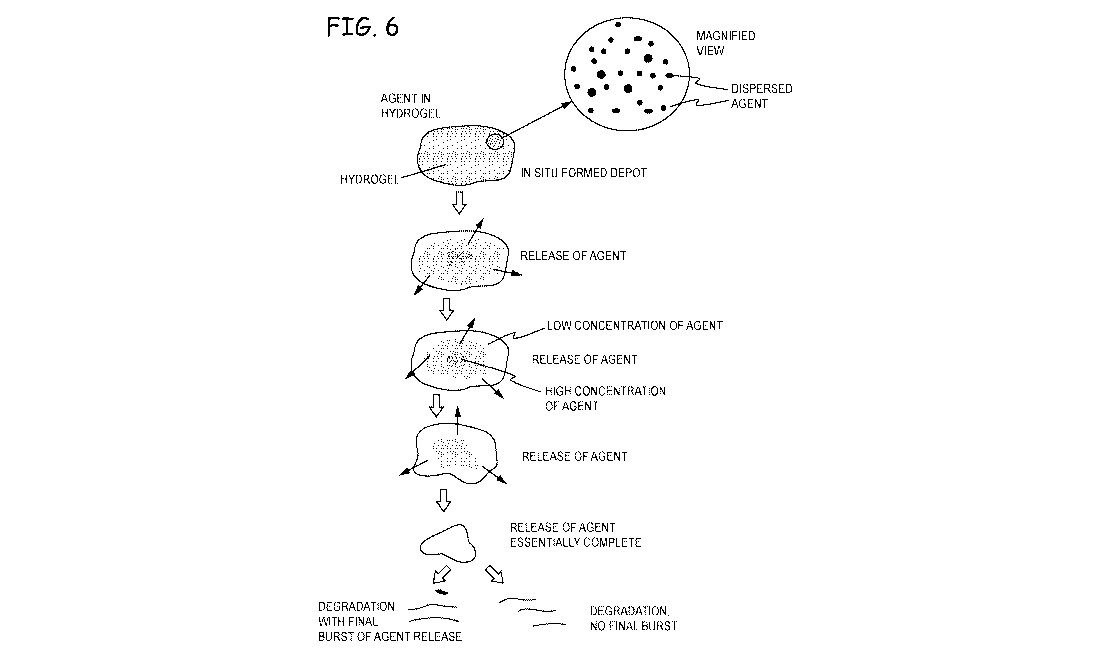
Figure 5 depicts suprachoroidal material placement;
Figure 6 depicts release of an agent from an in situ formed hydrogel in an
intracameral
or intravitreal space;
Figure 7 is a plot of swelling of a hydrogel volume as it degrades without
exterior
constraints;
Figure 8 is a plot of the dimensions of the hydrogel depot of Figure 7;
Figure 9 is a montage of photographs of an in situ formed hydrogel releasing
an agent,
shown immediately after placement in vivo (panel a), after 40 hours (panel b),
1 week (panel
c) or 12 days (panel d) in physiological buffered saline (PBS);
Figure 10 is a photograph of a hydrogel implant with an agent entrapped in the
implant
for release, in a PBS;
Figure 11A is a plot of a release profile in PBS of agents entrapped in a
hydrogel depot
or as placed directly into the PBS;
Figure 11B is the plot of Fig. 11A presented as a Higuchi plot;
Figure 11C is a representation of the data of FIG. 11A to show the Higuchi
factor, K,
as having a linear relationship with drug solubility for low or very low
solubility agents released
from the hydrogels;
Figure 12 is a photographic montage of a controlled release of an agent from a
hydrogel
depot showing clearance of the agent over time;
Figure 13 is an example of zone clearance of loteprednol etabonate from a
hydrogel
depot in PBS;
Figure 14 is an example of zone clearance of prednisolone from a hydrogel
depot in
PBS;
Figure 15 is a photographic montage of a controlled release of dexamethasone
from a
hydrogel depot showing clearance of the agent over time; and
Figure 16 is a photomontage of various agents released from hydrogels for the
indicated
times, and is an example showing that zone clearance of agents from hydrogel
depots in release
media such as PBS visually correlates with drug solubility in release media
over time.
2
CA 02985656 2017-11-09
WO 2016/183296
PCT/US2016/032046
DETAILED DESCRIPTION
An embodiment of the invention is a method of drug delivery to a tissue,
particularly
an eye, comprising forming a hydrogel implant in situ with a therapeutic agent
in the hydrogel
(e.g., dissolved, suspended, dispersed throughout), the agent having a low
solubility in water.
Drug delivery to the eye is an active field. Improvements in drugs for
treatment of eye
diseases have created new options for patients, including controlled release
devices. One
approach to ocular extended release was to put drugs into degradable particles
that were
injected into the eye. There were sometimes problems, however, with the
particles settling
onto the retina and causing contact toxicity. Others have created small drug
delivery devices
that are biodegradable rods of poly(lactic-co-glycolic acid) copolymers
(PLAJPGA) that are
impregnated with drugs and inserted into the eye. As they erode, the drug is
able to move out
of the PLA/PGA matrix, so that the degradation controls the rate of release.
These devices
provide extended release as they are eroded by the aqueous solution in the
eye. Another
approach has involved the use of certain hydrogels that are formed in situ, as
in US
2009/0252781. While these were useful in certain situations, there are further
techniques that
can be used to improve biocompatibility and increase the range of clinical
treatments that can
be made with controlled release devices.
In particular, there are opportunities to use the properties of the agents,
themselves, in
combination with certain properties of the hydrogels to make depots (also
referred to as
implants) that release the agents over long periods of time in a controlled
fashion to achieve an
effective concentration without reaching toxic levels. A low solubility agent
can go into
solution particularly slowly in a hydrogel. The hydrogel can be made to
readily allow diffusion
without requiring degradation of the hydrogel (bioerosion) for release of the
agent. The
hydrogel's properties can be tailored to take advantage of the solubility of
the agent to control
release. Such properties can include a matrix structure that provides for
diffusion of the agent
without depending on bioerosion, a process of making the hydrogel that allows
for dispersion
of the agent in the hydrogel, and providing for the agent to be suspended as,
e.g., micro and/or
nano particles or droplets. The agent does not have to be encapsulated in
particles, or otherwise
combined with materials that need bioerosion to release them. Further, the
hydrogels can be
made to last longer than the agents they deliver so that delivery is
controlled and the release of
a final burst of the agent is kept within limits that avoid potentially toxic
effects.
Some embodiments provide for encapsulation of agents in particles as an
alternative or
addition to non-encapsulated agents, particularly in areas outside the eye.
The particles can be
3
CA 02985656 2017-11-09
WO 2016/183296
PCT/US2016/032046
mixed with one or more precursors that form a hydrogel around them.
Encapsulating particles
are further discussed below.
In general, the eye presents an environment with competing design
requirements. On
the one hand, the volume of the eye is limited such that a large volume depot
is disfavored. On
the other hand, placing the depot in the eye, for instance by injection,
involves some discomfort
and trauma such that a large depot is helpful for minimizing the frequency of
placement.
Further, the eye is generally sensitive and placement of depots at locations
that interfere with
its requirements for effective vision points to making small depots. Moreover,
therapeutic
agents require a minimum concentration to be effective but may have toxic
effects at
concentrations that are too high. Therefore the agent must be released quickly
and consistently
enough to be effective without being released at too great a rate through the
entire life of the
implant. Use of a hydrogel around an agent presents the challenge of adding
volume to the
implant. In the case of a hydrogel that has internal space to allow for
diffusion of agents, there
are mechanical challenges to make an implant that resists mechanical forces
applied to the eye
such as rubbing the eyes or accidental application of force, or an elevated
intraocular pressure
present in some pathologies that are the target of the agent. An open, lightly
crosslinked
hydrogel structure tends to have more flexibility, but less mechanical
strength compared to a
relatively more closed hydrogel that has more closely spaced crosslinks.
But it is possible to use the small volume of the eye as an advantage instead
of a
disadvantage. A hydrogel that allows diffusion of an agent is affected not
only by the
concentration of the agent in the hydrogel but also by the concentration of
the agent in the
limited volume of the eye. A hydrogel depot with a relatively open matrix can
be thus use the
small volume of the eye as a parameter to control release because the amount
of released agent
can limit further release. The hydrogel structure, size, shape, loading, and
choice of materials
can thus be balanced, in combination with the properties of the agent, to
provide an effective
controlled release implant device. These various competing design features
can, in fact, be
reconciled to provide delivery of an effective concentration of an agent
during a period of time,
while avoiding toxic over-release of the agent.
In contrast to hydrogels that are permissive to agent diffusion, erodible
hydrogels
prevent diffusion until the matrix is eroded. Such designs have an advantage
of directly
controlling a rate of release of the agent. Since such designs have relatively
densely crosslinked
matrices, they can be made mechanically strong to resists mechanical forces
involved in their
implantation or after implantation, for example, by patients rubbing their
eyes or receiving an
4
CA 02985656 2017-11-09
WO 2016/183296
PCT/US2016/032046
accidental application of force, or stresses internal to the eye in some
pathologies that are the
target of the agent.
Locally formed hydrogels made in situ from precursors in aqueous solution can
serve
as depots of drugs or other therapeutic agents for ocular drug delivery, or
delivery of agents at
other sites. Described herein are hydrogels that can be formed in situ on a
tissue or organ to
deliver agents. The term on a tissue is broad, and includes contact with a
tissue, in the tissue,
around the tissue, in a tissue void or defect, in a potential space in the
body, and so forth. An
organ is a tissue. The term on an organ is broad and includes in the organ, on
it, around it, and
so forth. In situ refers to forming a material at the site where it is
intended to be located. Thus
a hydrogel may be formed in situ in a patient at the site wherein the hydrogel
is intended to be
used, e.g., as a drug depot for controlled release. Some drugs, such as some
tyrosine kinase
inhibitors (TKIs), have demonstrated corneal toxicity even in eye-drop form
because the drugs
are contacting the eye tissue directly. An advantage of the hydrogel is that
the hydrogel shields
the tissue from contact with the agent, e.g., as a solid particle or a
suspended form. The agent
is slowly released from the hydrogel in a diffuse form.
Alternative embodiments include hydrogels formed outside the body and
implanted
into the body, e.g., intravitreally. Example 1 describes the swelling and
persistence of two
hydrogels made with a polyethylene glycol (PEG) matrix at a solids
concentration of a 5% or
10% w/w PEG, see Table 3. The hydrogels were made from a first PEG precursor
having an
electrophilic end group (succinimidyl azelate, SAZ) and a second PEG precursor
having
nucleophilic end group (amine). The PEGs had 4 or 8 arms and a nominal
molecular weight
of 20k each. They were combined in buffered solution in presence of a
polysaccharide
(hyaluronic acid, HA, at 1% w/w). The combination was found to have a low
viscosity suited
for injection through small bore needles and the resultant hydrogel matrix
provided a structured
that maintained its shape and mechanical integrity within a space gelation,
e.g., intracameral,
in a vitreous body, or other location. The precursors had good syringeability
and good cohesion
characteristics. The HA is a high molecular weight non-newtonian linear
molecule; it enhanced
viscosity of the precursor solution and performed well under high shear
situations (passage
through a thin gauge needle). A variety of different dilutions of 850kDa HA
were tested, with
about 1% w/w providing a good result in this case. The buffers used to
dissolve each precursor
made a neutral pH when mixed, and the buffer with the SAZ precursor was of low
pH in order
to maintain stability of the polymer in solution (to avoid pre-hydrolysis).
Each of these
components, when mixed together, formed a hydrogel that maintained shape
stability and
volume stability, keeping its shape and position in a space until forming a
hydrogel in 2-3
5
CA 02985656 2017-11-09
WO 2016/183296
PCT/US2016/032046
minutes. Figs. 7 and 8 depict plots of swelling and dimensional change,
respectively, for these
hydrogel depots placed in vitro in physiological buffer solution (PBS). It was
further observed
that, as the hydrogels degraded, they continued to swell in a linear trend
upwards to 1000%
before liquefying. Most dimensional changes occur within the first hour. As
the hydrogels
degraded, they became mechanically more weak and swelled. These tests were
conducted in
an unrestrained area and, if formed in vivo, will swell minimally in vivo
under conditions where
surrounding mechanical forces limit swelling.
In Example 2, a hydrogel of the composition of Example 1 further comprising a
small
amount of fluorescein for visualization and the agent dexamethasone was formed
in situ in an
eye in a volume of about 25 4. The depot was explanted and placed in an excess
of PBS to
observe release of the agent and persistence of the hydrogel. The agent was
observed to be
cleared from the hydrogel in inward direction, with the edges of the implant
having the lowest
concentration of the agent and the interior of the hydrogel having the highest
concentration
(Fig. 9). The hydrogel was essentially persistent during the observed time of
12 days and the
visual observations were consistent with volumetric release and persistence
data shown in plots
herein. The hydrogel had a stable shape and consistent mechanical properties,
based on
manipulation of the hydrogel. Examples 3-8 provide detailed examples of making
and using
various hydrogels and agents. Example 9 is an example of how to make a kit for
making
hydrogels in vivo to release agents. Artisans can readily appreciate how to
apply these
Examples, and all the Examples more generally, to make and use hydrogels using
the full range
of precursors, agents, and sites of application set forth in other portions of
this same disclosure.
Example 10 shows release profiles for a variety of exemplary agents,
Flunisolide
(solubility 90 g/mL), Betamethasone Sodium Phosphate (freely soluble in
water), Budesonide
(30 mg/mL, and Triamcinolone Acetonide (20 jig/mL. These agents were placed
into PBS or
dispersed in a hydrogel (Fig. 10) placed into PBS, the hydrogel being made
from a hydrophilic
precursor (4-armed PEG) with electrophilic groups and a small hydrophilic
precursor with
nucleophilic groups (trilysine). The release rate from the agent- containing
hydrogel depots
was compared to the dissolution profile of the same amount of the agent in a
neat formulation
(Fig. 11A).
Figs. 11B and 11C are plots of the data of Example 10. Fig. 11B is a Higuchi
plot
showing that drug release versus square root of time is a linear relationship,
with slopes (the
slope is equal to the Higuchi factor, K) proportional to drug solubility, drug
diffusivity, initial
drug concentration, depot surface area, and other depot design factors. Fig.
11C shows the
linear function of solubility relative to K, for the same initial drug
concentrations and other
6
CA 02985656 2017-11-09
WO 2016/183296
PCT/US2016/032046
depot design factors. A visual representation of drug release from the depots
over time is
presented in Fig. 12 for an exemplary agent (flunisolide). The results
demonstrated that
entrapment of an equal mass of agent within the confines of the hydrogel
appreciably slowed
the agents' release rate compared to freely dispersed agents in an equal
volume of dissolution
media, and that the tapered drug release profile correlated with drug
solubility.
These data further establish the extended release rate from the in situ depots
and model
in vivo conditions. Injection of the pre-hydrogel material containing a
suspension of exemplary
agents (steroids) into the confines of the viscous vitreous was observed to
create a spheroidal
hydrogel depot at the injection site. These depot examples formed in vitro
created a similar
spheroidal hydrogel depot (e.g., Fig. 10). The drug release data from these in
vitro formed
depots allows prediction of the in vivo drug release rate. The Higuchi
equation for release from
this type of construct can be represented by the following (Siepmann, J.;
Peppas, N. A.
Modeling of Drug Release from Delivery Systems Based on Hydroxypropyl
Methylcellulose
(HPMC). Adv. Drug Deliv. Rev. 2012, 64, Supplement, 163-174):
Equation 1
Mt = AV2C0DCst
Where Mt is the mass of drug eluted at time t, A is the surface area, Co is
the initial drug
concentration, Cs is the drug solubility, D is the diffusion coefficient. This
equation assumes
Co>> Cs, edge effects are negligible, swelling or dissolution of the hydrogel
depot is negligible,
diffusivity is constant, temperature and pH are constant, and perfect sink
conditions are
maintained. More generally, the equation can be represented as
Equation 2
Mt/Mcc, = IcA5
Where Moo is the cumulative drug released at infinite time and k is a constant
(Higuchi
factor) reflecting the depot design variables. Thus, the drug or drug fraction
release profile is
tapered when plotted versus time, but linear when plotted versus the square
root of time.
The release of the low solubility agent is thus regulated by the limited
solubility of the
agent in the physiological environment within the hydrogel and by the
concentration gradient
at the hydrogel interface with the physiological environment, which equals the
drug solubility
under perfect sink conditions. A tapered drug release profile is created as
the front of the
concentration gradient recedes from the interface. This retreating front can
be observed as a
7
CA 02985656 2017-11-09
WO 2016/183296
PCT/US2016/032046
gradually increasing clear zone at the periphery of the depot. Regulating the
amount of low
solubility agent within the depot can therefore control the duration of the
drug release. The
drug release rate from the depot in the vitreous is expected to be extended
relative to an
injection of unconstrained neat steroid in the vitreous thereby prolonging the
duration of action
of the drug within the eye. An additional benefit is that particles of drug
are entrapped within
the hydrogel, whereas migrating insoluble drug particles within the eye may
result in an adverse
tissue reaction or vision impairment when particles enter the visual axis.
Factors expected to
influence the rate of drug release from the in situ formed hydrogel depot
include: drug
solubility, drug particle (liquid or solid) size, common solubility factors
(pH, temperature, salts,
and so forth), drug amount within the depot creating differing concentrations
and gradients,
uniformity of drug within the depot, depot surface area, fluid turnover or
exchange rate at the
depot interface, hydrogel degradation and dissolution, depot additive agents
(such as
surfactants), and possibly other factors known to alter the solubility of an
agent.
In a similar construct described in Example 11, various agents were suspended
in
hydrophilic hydrogel precursor solutions crosslinked and formed as cylindrical
depots. The
agent-suspended gels were removed from the tubing and ex vivo release was
initiated in
dissolution media. Zone clearance (steroid released) from the depot interface
inward was
observed and visually recorded. See Figs. 13-16. A similar observation is
expected to occur
over time during in vivo drug release.
Examples 12-14 detail results of experiments testing potential toxicity of
agents
released in bursts. The hydrogel depots consistently release effective
concentrations of the
agents over a period of time. After that period of time, the hydrogel loses
mechanical integrity
and the matrix structure becomes loosely organized. If there is any remaining
agent during this
phase of degradation, the agent might be released more rapidly, or in a burst,
such that the agent
is at a concentration that is higher than what is needed for effectiveness or
is, potentially, in a
toxic amount with respect to local tissues. In vivo tests in eyes were
conducted to measure the
potential effects of a burst release to understand how much persistence would
be necessary
relative to the total volume and remaining volume of the agents. Considering
the many design
variables involved in the delivery process, some experimentation was needed to
establish that
the delivery processes described herein are suitable for the ocular space. The
results show that
the depots can be designed with a suitable persistence, loading, and other
factors to effectively
deliver drugs over a sustained period of time without falling short of the
various design
parameters.
8
CA 02985656 2017-11-09
WO 2016/183296
PCT/US2016/032046
The hydrogel depot is designed to provide an effective concentration of the
agent at its
site of intended use. The term effective amount or effective concentration or
therapeutically
effective/concentration refers to the amount of an agent that is sufficient to
effect beneficial or
desired results. The effective amount may vary depending upon one or more of:
the subject
and disease condition being treated, the weight and age of the subject, the
severity of the disease
condition, the manner of administration and the like, which can readily be
determined by one
of ordinary skill in the art. An effective concentration can be shown by
pharmacodynamic
effect. As an alternative, a calculated effective amount may be provided,
meaning that 50-100
times the IC50 for the agent against the substrate; Artisans will immediately
appreciate that all
ranges and values between the explicitly stated bounds are contemplated, with,
e.g., any of the
following being available as an upper or lower limit: 50, 60, 70, 80, 90, or
100. IC50 refers to
the Median Inhibition Concentration (concentration that reduces the effect by
50%), e.g.,
inhibition of the unwanted pathological effect.
Anatomy of the eye
The structure of the mammalian eye can be divided into three main layers or
tunics: the
fibrous tunic, the vascular tunic, and the nervous tunic. The fibrous tunic,
also known as the
tunica fibrosa oculi, is the outer layer of the eyeball consisting of the
cornea and sclera. The
sclera is the supporting wall of the eye and gives the eye most of its white
color. It is extends
from the cornea (the clear front section of the eye) to the optic nerve at the
back of the eye.
The sclera is a fibrous, elastic and protective tissue, composed of tightly
packed collagen fibrils,
containing about 70% water.
Overlaying the fibrous tunic is the conjunctiva. The conjunctiva is a membrane
that
covers the sclera (white part of the eye) and lines the inside of the eyelids.
The conjunctiva
effectively surrounds, covers, and adheres to the sclera. It is has cellular
and connective tissue,
is somewhat elastic, and can be removed, teased away, or otherwise taken down
to expose a
surface area of the sclera. The vascular tunic, also known as the tunica
vasculosa oculi, is the
middle vascularized layer which includes the iris, ciliary body, and choroid.
The choroid
contains blood vessels that supply the retinal cells with oxygen and remove
the waste products
of respiration.
The nervous tunic, also known as the tunica nervosa oculi, is the inner
sensory which
includes the retina. The retina contains the photosensitive rod and cone cells
and associated
neurons. The retina is a relatively smooth (but curved) layer. It does have
two points at which
it is different; the fovea and optic disc. The fovea is a dip in the retina
directly opposite the
9
CA 02985656 2017-11-09
WO 2016/183296
PCT/US2016/032046
lens, which is densely packed with cone cells. The fovea is part of the
macula. The fovea is
largely responsible for color vision in humans, and enables high acuity, which
is necessary in
reading. The optic disc is a point on the retina where the optic nerve pierces
the retina to
connect to the nerve cells on its inside.
The mammalian eye can also be divided into two main segments: the anterior
segment
and the posterior segment. The anterior segment consists of an anterior and
posterior chamber.
The anterior chamber is located in front of the iris and posterior to the
corneal endothelium and
includes the pupil, iris, ciliary body and aqueous fluid. The posterior
chamber is located
posterior to the iris and anterior to the vitreous face where the crystalline
lens and zonules fibers
are positioned between an anterior and posterior capsule in an aqueous
environment.
Light enters the eye, passes through the cornea, and into the first of two
humors, the
aqueous humour. Approximately two-thirds of the total eyes refractive power
comes from the
cornea which has a fixed curvature. The aqueous humor is a clear mass which
connects the
cornea with the lens of the eye, helps maintain the convex shape of the cornea
(necessary to
the convergence of light at the lens) and provides the corneal endothelium
with nutrients.
The posterior segment is located posterior to the crystalline lens and in
front of the
retina. It represents approximately two-thirds of the eye that includes the
anterior hyaloid
membrane and all structures behind it: the vitreous humor, retina, and optic
nerve. On the other
side of the lens is the second humour, the vitreous humour, which is bounded
on all sides: by
the lens, ciliary body, suspensory ligaments and by the retina. It lets light
through without
refraction, helps maintain the shape of the eye and suspends the delicate
lens.
Figure 1 depicts eye 10 having sclera 12, iris 14, pupil 16, and eyelid 18.
Figure 2
depicts a perspective view of eye 10 with a partial cross-section that depicts
lens 20, inferior
oblique muscle 21, inferior rectus muscle 23, and optic nerve 25. Figure 3 is
a cross-section of
eye 10 and depicts cornea 22 that is optically clear and allows light to pass
iris 14 and penetrate
lens 20. Anterior chamber 24 underlies cornea 22 and posterior chamber 26 lies
between iris
14 and lens 20. Ciliary body 28 is connected to lens 20. Figure 3 depicts a
portion of the
conjunctiva 30, which overlies the sclera 12. The vitreous body 32 comprises
the jelly-like
vitreous humor, with hyaloid canal 34 being in the same. Fovea 36 is in the
macula and retina
38 overlies choroid 37. Zonular spaces 42 are depicted.
Figure 4 depicts various intravitreal deposition schemes. A plurality of
depots may be
formed, or one. The depots may have various shapes, e.g., elongate,
spheroidal, spherical,
essentially spherical, ellipsoidal, cylindroid, essentially cylindroid,
discoidal, or essentially
discoidal. The term essentially spherical means that the hydrogel occupies at
least 70% of the
CA 02985656 2017-11-09
WO 2016/183296
PCT/US2016/032046
volume of a sphere drawn around the hydrogel. The term spherical means that
the hydrogel
occupies at least 85% of the volume of a sphere drawn around the hydrogel. The
term
essentially discoidal means that the hydrogel occupies at least 70% of the
volume of a cylinder
drawn around the hydrogel, with a height of the cylinder being less or equal
to the diameter of
the cylinder. The term essentially discoidal means that the hydrogel occupies
at least 85% of
the volume of a cylinder drawn around the hydrogel, with a height of the
cylinder being less
than the diameter of the cylinder. The term essentially cylindroid means that
the hydrogel
occupies at least 70% of the volume of a cylinder drawn around the hydrogel,
with a height of
the cylinder being greater than the diameter of the cylinder. The term
essentially cylindroid
means that the hydrogel occupies at least 85% of the volume of a cylinder
drawn around the
hydrogel, with a height of the cylinder being greater than the diameter of the
cylinder. Other
shapes and sizes may be chosen as suited for the site and application, and
irregular shapes are
also contemplated. Volumes set forth elsewhere herein may be applicable, e.g.,
less than 1 ml,
from 0.005 to 5 ml; Artisans will immediately appreciate that all ranges and
values between
the explicitly stated bounds are contemplated, with any of the following being
available as an
upper or lower limit: 10, 20, 25, 50, 100, 150, 200, 250 L; 0.3, 0.4, 0.5,
0.6, 0.7, 0.8, 0.9, 1.0,
2, 3, 4, 5 mls. One or more such depots may be formed. Fig. 5 illustrates
suprachoroidal
placement. Other organs may be a site for placement of a hydrogel, as
described in more detail
below. For instance, the hydrogels may be formed in natural or surgical voids
or potential
spaces, including other sites where cancer has been removed or is located.
Sites include
placement of the hydrogel material at a site of a cancer, for example, at a
prostate for therapy
of prostate cancer or breast cancer.
Application of Precursors to form hydrogels in situ
Back of the eye diseases can be treated with drugs utilizing, e.g., topical,
systemic,
intraocular and subconjunctival delivery routes. Systemic and topical drug
delivery modalities
can fall short in delivering therapeutic drug levels to treat posterior
segment diseases. These
methods of drug delivery encounter diffusion and drug dilution issues due to
the inherent
anatomical barriers of the intraocular and systemic systems, causing
significant patient side
effects (due to multiple daily dosing), poor bioavailability and compliance
issues. The delivery
site for placement of an intraocular drug delivery implant is generally
dependent upon the
disease that needs to be treated and the type of drug therapy.
The delivery of therapeutic amounts of a drug to the retina in posterior
segment eye
diseases remains a challenge. Although intravitreal injections into the
vitreous cavity of anti-
11
CA 02985656 2017-11-09
WO 2016/183296
PCT/US2016/032046
VEGF agents have shown promise to arrest and in some cases reverse chronic age-
related
diseases like macular degeneration, these techniques and procedures are not
without risks and
side effects. Intravitreal administration of therapeutic agents into the
vitreous cavity can cause
cataracts, endophthalmitis and retinal detachments. This form of therapy
requires many
patients to receive monthly intraocular injections of an anti-VEGF drug over a
12 month time
period thus increasing the risk of infection, vitreous wicks and retinal
detachments.
Embodiments directed to an in situ hydrogel biodegradable drug implant provide
an effective
alternative treatment for eye diseases, and are expected to reduce the common
side-effects
associated with repeated intravitreal injections. Embodiments of an
intravitreal, intracameral
or other ocular biodegradable drug delivery implant system are summarized
below.
Figures 4 and 5 show certain points of delivery at or near eye 10. Locations
include
intracamerally, intravitreally or at or near the retina. Hydrogels can be put
on the retina
although some separation from the retina is typically useful. Separation may
be, e.g., 0.1 to 10
mm; Artisans will immediately appreciate that all ranges and values between
the explicitly
stated bounds are contemplated, with any of the following being available as
an upper or lower
limit: 0.2, 0.5, 1, 2, 3, 4, 5, 6, 7, 8, 9, 10.
As described in more detail in other sections, a drug depot of the in situ
hydrogel drug
delivery implant may be designed for controlled, long term drug release
ranging from, e.g.,
about one to about twelve or thirty six months; and may optionally be directed
to treatment of
diseases of the posterior segment including, for example, age-related macular
degeneration,
diabetic retinopathy, diabetic macular edema, retinal vein occlusion, and
cystoid macular
edema. The device can carry a drug payload of various types of therapeutic
agents for various
conditions.
One mode of application is to apply a mixture of precursors and other
materials (e.g.,
therapeutic agent, viscosifying agent, accelerator, initiator) through a
needle, cannula, catheter,
or hollow wire to a site in or near an eye. The mixture may be delivered, for
instance, using a
manually controlled syringe or mechanically controlled syringe, e.g., a
syringe pump.
Alternatively, a dual syringe or multiple-barreled syringe or multi-lumen
system may be used
to mix the precursors at or near the site. Syringe-to-syringe mixing may be
used when
appropriate.
Sites where drug delivery depots may be formed include an eye, the anterior
chamber, the
vitreous, episcleral, in the posterior subtenon's space (Inferior fornix),
subconjunctival, sub-
tenon, retinal, subretinal, intracanalicular, intracameral, intravitreal,
intrasceleral, choroidal,
suprachoroidal, a retina, subretinal, or a lens, a surface of the cornea or
the conjunctiva, among
12
CA 02985656 2017-11-09
WO 2016/183296
PCT/US2016/032046
others. Accordingly, embodiments include providing an effective amount or a
calculated
effective amount at such a site, e.g., an effective amount at an eye, the
anterior chamber, the
vitreous, episcleral, in the posterior subtenon's space (Inferior fornix),
subconjunctival, sub-
tenon, retinal, subretinal, intracanalicular, intracameral, intravitreal,
intrasceleral, choroidal,
suprachoroidal, a retina, subretinal, or a lens, a surface of the cornea or
the conjunctiva.
Sites for formation of a hydrogel depot further include a tissue, lumen, void,
potential
space, inside an animal (human or otherwise), or on a surface of an animal.
The term tissue is
broad. Sites include iatrogenic sites, sites where tissue is removed, and
surgical sites. Sites
include cancer tissue, at or near cancer tissue, dental tissue, gums,
periodontal, sinus, brain,
intravascular, aneurysm, and site of a pathology.
Viscosijfing Agents
Viscosifying agents can be useful for hydrogels formed in or on an eye, with
the agent
helping the solution cling to its site of deposition, or maintain a cohesive
mass, while the
hydrogel forms. The choice of the agent must be made in light of the kind of
crosslinking that
is taking place. Viscosity enhancers may be used in conjunction with
precursors. In general,
the viscosity enhancers do not react with the precursors to form covalent
bonds. While it is
appreciated that precursors that are generally free of such bonding may
sometimes participate
in unwanted side reactions, these have little effect on the hydrogel so that
the precursors are
"free" of such reactions. For instance, if the precursors react by
electrophile-nucleophile
reactions, the viscosity enhancers may be free of electrophiles or
nucleophiles that can form
covalent bonds with functional groups of the precursors, even if there is some
low level of
unwanted side reactions. Viscosity enhancers are, in general, hydrophilic
polymers with a
molecular weight of at least 20,000, 100,000 or from about 100,000 to about
2,000,000 Daltons;
artisans will immediately appreciate that all values and ranges between these
explicitly stated
values are described, e.g., at least about 100,000, 200,000, more than
500,000, more than
550,000, 600,000. A concentration of about 5% to about 25% w/w may be used,
for instance.
PEG (e.g., M.W. 100,000 to 250,000) is useful, for example. Viscosity
enhancers may be free
of electrophiles and/or nucleophiles. Viscosity enhancers may be fee of one or
more functional
groups such as hydroxyl, carboxyl, amine, or thiol. Viscosity enhancers may
include one or
more biodegradable links as described herein for precursors. Viscosity
enhancers can be useful
to prevent precursors from running-off a tissue site before the precursor's
crosslink to form a
gel.
13
CA 02985656 2017-11-09
WO 2016/183296
PCT/US2016/032046
Another consideration is whether the agent has to pass through a small
diameter syringe
or catheter, a property referred to as syringeability. A thixotropic
viscosifying agent may be
used so that, in motion, it provides little resistance but, when static, forms
a thick gel.
Hyaluronic Acid (HA) has been found to be a useful thixotropic viscosifying
agent. Molecular
weights (average w/w) from 100,000 to 2,500,000 have been tested. These
results show that a
higher MW (e.g., 5000k) may be also be used. Artisans will immediately
appreciate that all
ranges and values between the explicitly stated bounds are contemplated, e.g.,
with any of the
following being available as an upper or lower limit: 100k, 200k, 300k, 400k,
500k, 600k,
700k, 800k, 900k, 1000k, 1500k, 1800k, 2000k, 2250k, 2500k, 3000k, 4000k,
5000k. Other
thixotropic viscosifying agents include high molecular weight polysaccharides,
or hydrophilic
polymers, or PEGs. A percentage of 0.3 to 2.5 % w/w has been tested, with the
optimal
percentage depending on the MW tested. In general, a polysaccharide in a range
of 0.2 to 5%
may be added to the hydrogel/hydrogel precursors, Artisans will immediately
appreciate that
all ranges and values between the explicitly stated bounds are contemplated,
with any of the
following being available as an upper or lower limit: 0.2, 0.3, 0.4, 0.5, 0.6,
0.7, 1, 2, 2.5, 3, 3.5,
4, 4.5, 5 w/w percent.
Hydrogel features and properties
The hydrogel is, in one embodiment, formed from precursors having functional
groups
that form crosslinks to crosslink the hydrogels and thereby form the hydrogel.
The crosslinks
may be covalent and/or physical in nature. The hydrogel delivers drugs to the
eye or elsewhere.
Some embodiments use highly flowable precursors that gel slowly enough to be
forced through
a very small bore cannula or needle to essentially cross-link only after
injection, but nonetheless
gel quickly enough so that they do not migrate back through the track of the
incision. The gel
degrades in the physiological fluid in or around the eye without causing
inflammation by
degrading into components that are biocompatible and not acidic. In some
embodiments the
gel adheres to the tissue.
The hydrogel can be made to persist, or essentially persist, until after it
has released its
therapeutic agent contents, or until it has essentially released the contents.
The hydrogel is
preferably made so that the agent can diffuse through the hydrogel. One the
one hand, allowing
the agent to diffuse out of the gel removes an option for controlling a rate
of drug delivery. For
that reason, conventional practice with drug delivery from degradable
materials is to require
the material to degrade so that the drug can be released. In the case of a
hydrogel, the distance
between crosslinks can be made small enough so that a drug cannot move through
the hydrogel
14
CA 02985656 2017-11-09
WO 2016/183296
PCT/US2016/032046
until it erodes; it is the bioerosion rate that controls release. Nonetheless,
abandoning the
bioerosion-based approach can be useful. Accordingly, embodiments of the
invention may be
made with a hydrogel that allows diffusion of a therapeutic agent through the
hydrogel. The
matrix may be made with a spacing between crosslinks that allows diffusion.
The term essentially released means, unless otherwise indicated, about 97%
w/wi of the
drug is released, meaning the drug in the hydrogel had an initial weight Iv;
and a weight, w, at
the time of measurement. Other endpoints may be chosen, for instance, from 50
to 100 percent;
Artisans will immediately appreciate that all ranges and values between the
explicitly stated
bounds are contemplated, with any of the following being available as an upper
or lower limit:
50, 60, 70, 80, 85, 90, 95, 96, 97, 98, 99, 99.5, 99.9, 99.99 percent. The
term range, unless,
otherwise indicated, means that the numerical value can fall anywhere in the
range. The term
essentially persist means, unless otherwise indicated, about 97% w/wi of the
dry weight of the
hydrogel is retained, meaning hydrogel had an initial dry weight wi and a dry
weight, w, at the
time of measurement. Other endpoints may be chosen, for instance, from 50 to
100 percent
persistence; Artisans will immediately appreciate that all ranges and values
between the
explicitly stated bounds are contemplated, with any of the following being
available as an upper
or lower limit: 50, 60, 70, 80, 85, 90, 95, 96, 97, 98, 99, 99.5, 99.9, 99.99
percent. Persistence
is the dry weight of the hydrogel relative to an initial dry weight of the
hydrogel; this can be
measured directly after washing an explanted hydrogel and accounting for the
weight of tissue
infiltrates, for instance, by digesting the depot and measuring the content of
the hydrogel matrix
after removing tissue infiltrates. Further, the ranges/values of
persistence/release may be
mixed and matched. For example, a hydrogel persistence of 95% when the drug is
99%
released. As is evident, all of these percentage values are w/w unless
otherwise indicated.
It is also useful to speak of the hydrogel/thug combinations in terms of
persistence and
release at various points. For instance, it may be desirable to have a certain
persistence when
the release of the agent is at 50%. Accordingly, besides the
persistence/release combinations
already indicated, there can be a range of persistence from 0% to 100% and a
range of release
from 0% to 100%; Artisans will immediately appreciate that all ranges and
values between the
explicitly stated bounds are contemplated, with any of the following being
available as an upper
or lower limit: 0, 1, 2, 3, 4, 5, 10, 15, 20, 25, 30, 35, 40, 45, 49.9, 50,
50.1, 55, 65, 70, 75, 80,
85, 90, 95, 100, all being w/w percentages. Drug is a broad term that is used
interchangeably
herein with the term therapeutic agent.
Stability and mechanical integrity are two further factors involved in
controlling
hydrogels. Stability, in this context, is stability of shape. At a time of
formation, a hydrogel
CA 02985656 2017-11-09
WO 2016/183296
PCT/US2016/032046
might be stable but then lose stability as it loses mechanical integrity,
changing its shape, and
becoming deformed, expanding or contracting. One measure of stability is
change in volume
(volume stability). The hydrogel, once hydrated in situ, will have an initial
volume. The
volume at 24 hours after placement is a usually good measure of initial volume
since the
hydrogel has fully equilibrated with local fluids and, for gels that degrade
in a time span of two
or more weeks, little degradation has taken place. Accordingly, hydrogels can
be made with
an initial volume of 100% and, if they are fully biodegraded, will eventually
achieve a volume
of 0%. Another metric for stability is the percentage change in position of
the initial shape: an
overlay of the shape at a point in time is compared to the initial shape
(shape stability). The
amount of volume of the initial shape that has not been moved and has not
disappeared is
calculated, with complete stability being 100% and complete ending of
stability being 0%.
Stability can be described relative to time, as in days, weeks, or months.
And/or stability can
be described relative to a release of an agent. When a hydrogel designed to be
deployed as a
cohesive mass is being used in vivo, the forces acting on the hydrogel will
typically not deform
it from its initial shape so long as the hydrogel retains its initial
mechanical integrity. Therefore
stability can be used as a proxy for mechanical integrity in many cases.
Essentially stable
means more than about 97% by shape or volume measure. Shape or volume
stability can be
set in light of persistence or release, and thus may be chosen to be a value,
for example, from
80 to 100 percent when release is from 0 to 100 percent; Artisans will
immediately appreciate
that all ranges and values between the explicitly stated bounds are
contemplated, with any of
the following being available as an upper or lower limit: 0, 1, 2, 3, 4, 5,
10, 15, 20, 25, 30, 35,
40, 45, 49.9, 50, 50.1, 55, 65, 70, 75, 80, 85, 90, 95, 100, all being
percentages of shape or
volume or w/w release percentages.
Stability and mechanical integrity can also be used to reference an injectable
solution
that comprises precursor(s) and maintains shape and mechanical integrity
within a space from
injection until it gels, whether that space be a vitreous body, or other
location. Examples of
another space are puncta (canaliculus, upper/lower canaliculus), ocular
fornix, upper/lower
ocular fornix, subtenon space, choroid, suprachoroid, tenon, cornea, cancer
tissue, organ,
prostate, breast, surgically created space or injury, void space, and
potential space.
Embodiments include in situ formation of a punctal plug, with precursor(s)
being introduced
into the canaliculus and forming a punctal plug there. Accordingly, the shape
and volume
stability, described above, is contemplated for the solution.
In general, precursors may be combined as described herein at a site in or
near an eye
or other tissue to make a crosslinked hydrogel that comprises a therapeutic
agent that is released
16
CA 02985656 2017-11-09
WO 2016/183296
PCT/US2016/032046
into the eye to treat a disease over a suitable period of time. The hydrogel
may be low-swelling,
as measurable by the hydrogel having a weight increasing no more than about
10% or about
50% upon exposure to a physiological solution for twenty-four hours relative
to a weight of the
hydrogel at the time of formation; artisans will immediately appreciate that
all the ranges and
values within the explicitly stated ranges are contemplated. The hydrogel also
may be water-
degradable, as measurable by the hydrogel being dissolvable in vitro in an
excess of water by
degradation of water-degradable groups in the hydrogel. A composition with the
precursors
mixed therein can be introduced through a small-gauge needle provided that the
composition
has a suitable viscosity, which in turn depends on precursor properties,
concentrations, and
chemistry. Further, the hydrogels' mechanical strengths and reaction time are
adjusted though
control of the precursors and functional groups. The precursors and hydrogels
may have
various features that can be mixed-and-matched as guided by the considerations
for making an
effective device; the following sections describe some of these features.
Precursor materials
The hydrogels are made from precursors. Precursors are chosen in consideration
of the
properties that are desired for the resultant hydrogel. There are various
suitable precursors for
use in making the hydrogels. The term precursor refers to those molecules
crosslinked to form
the hydrogel matrix. While other materials might be present in the hydrogel,
such as
therapeutic agents or fillers, they are not precursors. The term matrix is
applicable for
hydrogels. Such matrices include matrices with a water content of more than
about 20% w/w;
Artisans will immediately appreciate that all ranges and values between the
explicitly stated
bounds are contemplated, with any of the following being available as an upper
or lower limit:
20%, 99%, 80%, 95%, at least 50%, and so forth, with the percentages being w/w
and the
solvent being water for hydrogels. Hydrogels may be formed by crosslinking
water soluble
molecules to form networks of essentially infinite molecular weight. Hydrogels
with high
water contents are typically soft, pliable materials. Hydrogels and drug
delivery systems as
described in U.S. Publication Nos. 2009/0017097, 2011/0142936 and 2012/0071865
may be
adapted for use with the materials and methods herein by following the
guidance provided
herein; these references are hereby incorporated herein by reference for all
purposes, and in
case of conflict, the instant specification is controlling.
Hydrogels may be formed from natural, synthetic, or biosynthetic polymers.
Natural
polymers may include glycosminoglycans, polysaccharides, and proteins. Some
examples of
glycosaminoglycans include dermatan sulfate, hyaluronic acid, the chondroitin
sulfates, chitin,
17
CA 02985656 2017-11-09
WO 2016/183296
PCT/US2016/032046
heparin, keratan sulfate, keratosulfate, and derivatives thereof.
In general, the
glycosaminoglycans are extracted from a natural source and purified and
derivatized.
However, they also may be synthetically produced or synthesized by modified
microorganisms
such as bacteria. These materials may be modified synthetically from a
naturally soluble state
to a partially soluble or water swellable or hydrogel state. This modification
may be
accomplished by various well-known techniques, such as by conjugation or
replacement of
ionizable or hydrogen bondable functional groups such as carboxyl and/or
hydroxyl or amine
groups with other more hydrophobic groups.
For example, carboxyl groups on hyaluronic acid may be esterified by alcohols
to
decrease the solubility of the hyaluronic acid. Such processes are used by
various
manufacturers of hyaluronic acid products (such as Genzyme Corp., Cambridge,
MA) to create
hyaluronic acid based sheets, fibers, and fabrics that form hydrogels. Other
natural
polysaccharides, such as carboxymethyl cellulose or oxidized regenerated
cellulose, natural
gum, agar, agrose, sodium alginate, carrageenan, fucoidan, furcellaran,
laminaran, hypnea,
eucheuma, gum arabic, gum ghatti, gum karaya, gum tragacanth, locust beam gum,
arbinoglactan, pectin, amylopectin, gelatin, hydrophilic colloids such as
carboxymethyl
cellulose gum or alginate gum crosslinked with a polyol such as propylene
glycol, and the like,
also form hydrogels upon contact with aqueous surroundings.
Hydrogels may be biostable or biodegradable. Examples of biostable hydrophilic
polymeric materials are poly(hydroxyalkyl methacrylate), poly(electrolyte
complexes),
poly(vinylacetate) cross-linked with hydrolysable or otherwise degradable
bonds, and water-
swellable N-vinyl lactams. Other hydrogels include hydrophilic hydrogels known
as
CARBOPOL , an acidic carboxy polymer (Carbomer resins are high molecular
weight,
allylpentaerythritol-crosslinked, acrylic acid-based polymers, modified with
C10-C30 alkyl
acrylates), polyacrylamides, polyacrylic acid, starch graft copolymers,
acrylate polymer, ester
cross-linked polyglucan. Such hydrogels are described, for example, in U.S.
Patent No.
3,640,741 to Etes, U.S. Patent No. 3,865,108 to Hartop, U.S. Patent No.
3,992,562 to
Denzinger et al., U.S. Patent No. 4,002,173 to Manning et al., U.S. Patent No.
4,014,335 to
Arnold and U.S. Patent No. 4,207,893 to Michaels, all of which are
incorporated herein by
reference, with the present specification controlling in case of conflict.
Hydrogels may be made from precursors. The precursors are crosslinked with
each
other. Crosslinks can be formed by covalent bonds or physical bonds. Examples
of physical
bonds are ionic bonds, hydrophobic association of precursor molecule segments,
and
crystallization of precursor molecule segments. The precursors can be
triggered to react to
18
CA 02985656 2017-11-09
WO 2016/183296
PCT/US2016/032046
form a crosslinked hydrogel. The precursors can be polymerizable and include
crosslinkers
that are often, but not always, polymerizable precursors. Polymerizable
precursors are thus
precursors that have functional groups that react with each other to form
matrices and/or
polymers made of repeating units. Precursors may be polymers.
Some precursors thus react by chain-growth polymerization, also referred to as
addition
polymerization, and involve the linking together of monomers incorporating
double or triple
chemical bonds. These unsaturated monomers have extra internal bonds which are
able to
break and link up with other monomers to form the repeating chain. Monomers
are
polymerizable molecules with at least one group that reacts with other groups
to form a
polymer. A macromonomer (or macromer) is a polymer or oligomer that has at
least one
reactive group, often at the end, which enables it to act as a monomer; each
macrornonomer
molecule is attached to the polymer by reaction the reactive group. Thus
macromonomers with
two or more monomers or other functional groups tend to form covalent
crosslinks. Addition
polymerization is involved in the manufacture of, e.g., polypropylene or
polyvinyl chloride.
One type of addition polymerization is living polymerization.
Some precursors thus react by condensation polymerization that occurs when
monomers bond together through condensation reactions. Typically these
reactions can be
achieved through reacting molecules incorporating alcohol, amine or carboxylic
acid (or other
carboxyl derivative) functional groups. When an amine reacts with a carboxylic
acid an amide
or peptide bond is formed, with the release of water. Some condensation
reactions follow a
nucleophilic acyl substitution, e.g., as in U.S. Patent No. 6,958,212, which
is hereby
incorporated by reference herein in its entirety to the extent it does not
contradict what is
explicitly disclosed herein. Some precursors react by a chain growth
mechanism. Chain
growth polymers are defined as polymers formed by the reaction of monomers or
macromonomers with a reactive center. A reactive center is a particular
location within a
chemical compound that is the initiator of a reaction in which the chemical is
involved. In
chain-growth polymer chemistry, this is also the point of propagation for a
growing chain. The
reactive center is commonly radical, anionic, or cationic in nature, but can
also take other
forms. Chain growth systems include free radical polymerization, which
involves a process of
initiation, propagation and termination. Initiation is the creation of free
radicals necessary for
propagation, as created from radical initiators, e.g., organic peroxide
molecules. Termination
occurs when a radical reacts in a way that prevents further propagation. The
most common
method of termination is by coupling where two radical species react with each
other forming
a single molecule. Some precursors react by a step growth mechanism, and are
polymers
19
CA 02985656 2017-11-09
WO 2016/183296
PCT/US2016/032046
formed by the stepwise reaction between functional groups of monomers. Most
step growth
polymers are also classified as condensation polymers, but not all step growth
polymers release
condensates. Monomers may be polymers or small molecules. A polymer is a high
molecular
weight molecule formed by combining many smaller molecules (monomers) in a
regular
pattern. Oligomers are polymers having less than about 20 monomeric repeat
units. A small
molecule generally refers to a molecule that is less than about 2000 Daltons.
The precursors
may thus be small molecules, such as acrylic acid or vinyl caprolactam, larger
molecules
containing polymerizable groups, such as acrylate-capped polyethylene glycol
(PEG-
diacrylate), or other polymers containing ethylenically-unsaturated groups,
such as those of
U.S. Patent No. 4,938,763 to Dunn et al., U.S. Patent Nos. 5,100,992 and
4,826,945 to Cohn et
al., or U.S. Patent Nos. 4,741,872 and 5,160,745 to DeLuca et al., each of
which is hereby
incorporated by reference herein in its entirety to the extent it does not
contradict what is
explicitly disclosed herein.
To form covalently crosslinked hydrogels, the precursors must be covalently
crosslinked together. In general, polymeric precursors are polymers that will
be joined to other
polymeric precursors at two or more points, with each point being a linkage to
the same or
different polymers. Precursors with at least two reactive centers (for
example, in free radical
polymerization) can serve as crosslinkers since each reactive group can
participate in the
formation of a different growing polymer chain. In the case of functional
groups without a
reactive center, among others, crosslinking requires three or more such
functional groups on at
least one of the precursor types. For instance, many electrophilic-
nucleophilic reactions
consume the electrophilic and nucleophilic functional groups so that a third
functional group
is needed for the precursor to form a crosslink. Such precursors thus may have
three or more
functional groups and may be crosslinked by precursors with two or more
functional groups.
A crosslinked molecule may be crosslinked via an ionic or covalent bond, a
physical force, or
other attraction. A covalent crosslink, however, will typically offer
stability and predictability
in reactant product architecture.
In some embodiments, each precursor is multifunctional, meaning that it
comprises two
or more electrophilic or nucleophilic functional groups, such that a
nucleophilic functional
group on one precursor may react with an electrophilic functional group on
another precursor
to form a covalent bond. At least one of the precursors comprises more than
two functional
groups, so that, as a result of electrophilic-nucleophilic reactions, the
precursors combine to
form crosslinked polymeric products.
CA 02985656 2017-11-09
WO 2016/183296
PCT/US2016/032046
The precursors may have biologically inert and hydrophilic portions, e.g., a
core. In
the case of a branched polymer, a core refers to a contiguous portion of a
molecule joined to
arms that extend from the core, with the arms having a functional group, which
is often at the
terminus of the branch. A hydrophilic molecule, e.g., a precursor or precursor
portion, has a
solubility of at least 1 g/100 mL in an aqueous solution. A hydrophilic
portion may be, for
instance, a polyether, for example, polyalkylene oxides such as polyethylene
glycol (PEG),
polyethylene oxide (PEO), polyethylene oxide-co-polypropylene oxide (PPO), co-
polyethylene oxide block or random copolymers, and polyvinyl alcohol (PVA),
poly (vinyl
pyrrolidinone) (PVP), poly (amino acids, dextran, or a protein. The precursors
may have a
polyalkylene glycol portion and may be polyethylene glycol based, with at
least about 80% or
90% by weight of the polymer comprising polyethylene oxide repeats. The
polyethers and
more particularly poly (oxyalkylenes) or poly (ethylene glycol) or
polyethylene glycol are
generally hydrophilic. As is customary in these arts, the term PEG is used to
refer to PEO with
or without hydroxyl end groups.
A precursor may also be a macromolecule (or macromer), which is a molecule
having
a molecular weight in the range of a thousand to many millions. The hydrogel
may be made
with at least one of the precursors as a small molecule of about 1000 Da or
less (alternatively:
2000 Da or less). The macromolecule, when reacted in combination with a small
molecule (of
about 1000 Da or less / 200 Da or less), is preferably at least five to fifty
times greater in
molecular weight than the small molecule and is preferably less than about
60,000 Da; artisans
will immediately appreciate that all the ranges and values within the
explicitly stated ranges
are contemplated. A more preferred range is a macromolecule that is about
seven to about
thirty times greater in molecular weight than the crosslinker and a most
preferred range is about
ten to twenty times difference in weight. Further, a macromolecular molecular
weight of 5,000
to 50,000 is useful, as is a molecular weight of 7,000 to 40,000 or a
molecular weight of 10,000
to 20,000. There are certain advantage to having a small molecule, such as
diffusivity for
completion of reactions.
Certain macromeric precursors are the crosslinkable, biodegradable, water-
soluble
macromers described in U.S. Patent No. 5,410,016 to Hubbell et al., which is
hereby
incorporated herein by reference in its entirety to the extent it does not
contradict what is
explicitly disclosed. These macromers are characterized by having at least two
polymerizable
groups, separated by at least one degradable region.
Synthetic precursors may be used. Synthetic refers to a molecule not found in
nature
or not normally found in a human. Some synthetic precursors are free of amino
acids or free
21
CA 02985656 2017-11-09
WO 2016/183296
PCT/US2016/032046
of amino acid sequences that occur in nature. Some synthetic precursors are
polypeptides that
are not found in nature or are not normally found in a human body, e.g., di-,
tri-, or tetra-lysine.
Some synthetic molecules have amino acid residues but only have one, two, or
three that are
contiguous, with the amino acids or clusters thereof being separated by non-
natural polymers
or groups. Polysaccharides or their derivatives are thus not synthetic.
Alternatively, natural proteins or polysaccharides may be adapted for use with
these
methods, e.g., collagens, fibrin(ogen)s, albumins, alginates, hyaluronic acid,
and heparins.
These natural molecules may further include chemical derivitization, e.g.,
synthetic polymer
decorations. The natural molecule may be crosslinked via its native
nucleophiles or after it is
derivatized with functional groups, e.g., as in U.S. Patent Nos. 5,304,595,
5,324,775,
6,371,975, and 7,129,210, each of which is hereby incorporated by reference to
the extent it
does not contradict what is explicitly disclosed herein. Natural refers to a
molecule found in
nature. Natural polymers, for example proteins or glycosaminoglycans, e.g.,
collagen,
fibrinogen, albumin, and fibrin, may be crosslinked using reactive precursor
species with
electrophilic functional groups. Natural polymers normally found in the body
are
proteolytically degraded by proteases present in the body. Such polymers may
be reacted via
functional groups such as amines, thiols, or carboxyls on their amino acids or
derivatized to
have activatable functional groups. While natural polymers may be used in
hydrogels, their
time to gelation and ultimate mechanical properties must be controlled by
appropriate
introduction of additional functional groups and selection of suitable
reaction conditions, e.g.,
pH.
Precursors may be made with a hydrophobic portion provided that the resultant
hydrogel retains the requisite amount of water, e.g., at least about 20%. In
some cases, the
precursor is nonetheless soluble in water because it also has a hydrophilic
portion. In other
cases, the precursor makes dispersion in the water (a suspension) but is
nonetheless reactable
to from a crosslinked material. Some hydrophobic portions may include a
plurality of alkyls,
polypropylenes, alkyl chains, or other groups. Some precursors with
hydrophobic portions are
sold under the trade names PLURONIC F68, JEFFAMINE, or TECTRONIC. A
hydrophobic
molecule or a hydrophobic portion of a copolymer or the like is one that is
sufficiently
hydrophobic to cause the molecule (e.g., polymer or copolymer) to aggregate to
form micelles
or microphases involving the hydrophobic domains in an aqueous continuous
phase or one that,
when tested by itself, is sufficiently hydrophobic to precipitate from, or
otherwise change phase
while within, an aqueous solution of water at pH from about 7 to about 7.5 at
temperatures
from about 30 to about 50 degrees Centigrade.
22
CA 02985656 2017-11-09
WO 2016/183296
PCT/US2016/032046
Embodiments of the invention include choosing a low-solubility agent or agent
with
other solubility as set forth herein and choosing a precursor that comprises
hydrophobic and
hydrophilic portions. The hydrophobic/hydrophilic precursor may comprise one
or more
functional groups: nucleophiles or electrophiles. The hydrophilic portion, the
hydrophobic
portion, or both, may be chosen to receive such functional groups.
Precursors may have, e.g., 2-100 arms, with each arm having a terminus,
bearing in
mind that some precursors may be dendrimers or other highly branched
materials. An arm on
a hydrogel precursor refers to a linear chain of chemical groups that connect
a crosslinkable
functional group to a polymer core. Some embodiments are precursors with
between 3 and
300 arms; artisans will immediately appreciate that all the ranges and values
within the
explicitly stated ranges are contemplated, e.g., 4 to 16, 8 to 100, or at
least 6 arms.
Thus hydrogels can be made, e.g., from a multi-armed precursor with a first
set of
functional groups and a low molecular-weight precursor having a second set of
functional
groups. For example, a six-an-fled or eight-armed precursor may have
hydrophilic arms, e.g.,
polyethylene glycol, terminated with primary amines, with the molecular weight
of the arms
being about 1,000 to about 40,000; artisans will immediately appreciate that
all ranges and
values within the explicitly stated bounds are contemplated. Such precursors
may be mixed
with relatively smaller precursors, for example, molecules with a molecular
weight of between
about 100 and about 5000, or no more than about 800, 1000, 2000, or 5000
having at least
about three functional groups, or between about 3 to about 16 functional
groups; ordinary
artisans will appreciate that all ranges and values between these explicitly
articulated values
are contemplated. Such small molecules may be polymers or non-polymers and
natural or
synthetic.
Precursors that are not dendrimers may be used. Dendritic molecules are highly
branched radially symmetrical polymers in which the atoms are arranged in many
arms and
subarms radiating out from a central core. Dendrimers are characterized by
their degree of
structural perfection as based on the evaluation of both symmetry and
polydispersity and
require particular chemical processes to synthesize. Accordingly, an artisan
can readily
distinguish dendrimer precursors from non-dendrimer precursors. Dendrimers
have a shape
that is typically dependent on the solubility of its component polymers in a
given environment,
and can change substantially according to the solvent or solutes around it,
e.g., changes in
temperature, pH, or ion content.
Precursors may be dendrimers, e.g., as in U.S. Publication Nos. 2004/0086479
and
2004/0131582 and PCT Publication Nos. W02007005249, W02007001926 and
23
CA 02985656 2017-11-09
WO 2016/183296
PCT/US2016/032046
W02006031358, or the U.S. counterparts thereof; dendrimers may also be useful
as
multifunctional precursors, e.g., as in U.S. Publication Nos. 2004/0131582 and
2004/0086479
and PCT Publication No. W006031388; each of which US and PCT applications are
hereby
incorporated by reference herein in its entirety to the extent they do not
contradict what is
explicitly disclosed herein. Dendrimers are highly ordered possess high
surface area to volume
ratios, and exhibit numerous end groups for potential functionalization.
Embodiments include
multifunctional precursors that are not dendrimers.
Some embodiments include a precursor that consists essentially of an
oligopeptide
sequence of no more than five residues, e.g., amino acids comprising at least
one amine, thiol,
carboxyl, or hydroxyl side chain. A residue is an amino acid, either as
occurring in nature or
derivatized thereof. The backbone of such an oligopeptide may be natural or
synthetic. In
some embodiments, peptides of two or more amino acids are combined with a
synthetic
backbone to make a precursor; certain embodiments of such precursors have a
molecular
weight in the range of about 100 to about 10,000 or about 300 to about 500
Artisans will
immediately appreciate that all ranges and values between these explicitly
articulated bounds
are contemplated.
Precursors may be prepared to be free of amino acid sequences cleavable by
enzymes
present at the site of introduction, including free of sequences susceptible
to attach by
metalloproteinases and/or collagenases. Further, precursors may be made to be
free of all
amino acids, or free of amino acid sequences of more than about 50, 30, 20,
10, 9, 8, 7, 6, 5, 4,
3, 2, or 1 amino acids. Precursors may be non-proteins, meaning that they are
not a naturally
occurring protein and cannot be made by cleaving a naturally occurring protein
and cannot be
made by adding synthetic materials to a protein. Precursors may be non-
collagen, non-fibrin,
non-fibrinogen, and non-albumin, meaning that they are not one of these
proteins and are not
chemical derivatives of one of these proteins. The use of non-protein
precursors and limited
use of amino acid sequences can be helpful for avoiding immune reactions,
avoiding unwanted
cell recognition, and avoiding the hazards associated with using proteins
derived from natural
sources. Precursors can also be non-saccharides (free of saccharides) or
essentially non-
saccharides (free of more than about 5% saccharides by w/w of the precursor
molecular weight.
Thus a precursor may, for example, exclude hyaluronic acid, heparin, or
gellan. Precursors can
also be both non-proteins and non-saccharides.
Peptides may be used as precursors. In general, peptides with less than about
10
residues are preferred, although larger sequences (e.g., proteins) may be
used. Artisans will
immediately appreciate that every range and value within these explicit bounds
is included,
24
CA 02985656 2017-11-09
WO 2016/183296
PCT/US2016/032046
e.g., 1-10, 2-9, 3-10, 1, 2, 3, 4, 5, 6, or 7. Some amino acids have
nucleophilic groups (e.g.,
primary amines or thiols) or groups that can be derivatized as needed to
incorporate
nucleophilic groups or electrophilic groups (e.g., carboxyls or hydroxyls).
Polyamino acid
polymers generated synthetically are normally considered to be synthetic if
they are not found
in nature and are engineered not to be identical to naturally occurring
biomolecules.
Some hydrogels are made with a polyethylene glycol-containing precursor.
Polyethylene glycol (PEG, also referred to as polyethylene oxide when
occurring in a high
molecular weight) refers to a polymer with a repeat group (CH2CH20)n, with n
being at least
3. A polymeric precursor having a polyethylene glycol thus has at least three
of these repeat
groups connected to each other in a linear series. The polyethylene glycol
content of a polymer
or arm is calculated by adding up all of the polyethylene glycol groups on the
polymer or arm,
even if they are interrupted by other groups. Thus, an arm having at least
1000 MW
polyethylene glycol has enough CH2CH20 groups to total at least 1000 MW. As is
customary
terminology in these arts, a polyethylene glycol polymer does not necessarily
refer to a
molecule that terminates in a hydroxyl group. Molecular weights are
abbreviated in thousands
using the symbol k, e.g., with 15K meaning 15,000 molecular weight, i.e.,
15,000 Daltons.
Molecular weights expressed herein are number average molecular weights unless
otherwise
specified. NH2 refers to an amine termination. SG refers to succinimidyl
glutarate. SS refers
to succinimidyl succinate. SAP refers to succinimidyl adipate. SAZ refers to
succinimidyl
azelate. SS, SG, SAP and SAZ are succinimidyl esters that have an ester group
that degrades
by hydrolysis in water. Hydrolytically degradable or water-degradable thus
refers to a material
that would spontaneously degrade in vitro in an excess of water without any
enzymes or cells
present to mediate the degradation. A time for degradation refers to effective
disappearance of
the material as judged by the naked eye. Trilysine (also abbreviated LLL) is a
synthetic
tripeptide. PEG and/or hydrogels, as well as compositions that comprise the
same, may be
provided in a form that is pharmaceutically acceptable, meaning that it is
highly purified and
free of contaminants, e.g., pyrogens.
Hydro gel Structures
The hydrogel's structure and the material composition of the hydrogel's
precursors
determine its properties. Precursor factors include properties such as
biocompatibility, water
solubility, hydrophilicity, molecular weight, arm length, number of arms,
functional groups,
distance between crosslinks, degradability, and the like. The choice of
reaction conditions also
effects the hydrogel's structure and properties, including choices of
solvents, reaction schemes,
CA 02985656 2017-11-09
WO 2016/183296
PCT/US2016/032046
reactant concentrations, solids content, and the like. There can be a variety
of ways to achieve
certain properties, or combination of properties. On the other hand some
properties are in
tension with each other, for instance brittleness may increase as a distance
between crosslinks
or solids content increases. Strength may be increased by increasing the
number of crosslinks
but swelling may thereby be reduced. Artisans will appreciate that the same
materials may be
used to make matrices with a great range of structures that will have highly
distinct mechanical
properties and performance, such that the achievement of a particular property
should not be
merely assumed based on the general types of precursors that are involved.
The spacing between molecular strands of the hydrogel (the matrix) affects
several
hydrogel properties, including a rate of diffusion of molecules. The
crosslinking density can
be controlled by the choice of the overall molecular weight of the
precursor(s) used as
crosslinker(s) and other precursor(s) and the number of functional groups
available per
precursor molecule. A lower molecular weight between crosslinks such as 200
will give much
higher crosslinking density as compared to a higher molecular weight between
crosslinks such
as 500,000; artisans will immediately appreciate that all ranges and values
within this range are
contemplated and supported, e.g., 200 to 250,000, 500 to 400,000, 5,000,
10,000, 20,000,
30,000, 40,000, 50,000, 60,000, 70,000, 80,000, 90,000, 100,000, and so forth.
The
crosslinking density also may be controlled by the overall percent solids of
the crosslinker and
functional polymer solutions. Yet another method to control crosslink density
is by adjusting
the stoichiometry of nucleophilic functional groups to electrophilic
functional groups. A one
to one ratio leads to the highest crosslink density. Precursors with longer
distances between
crosslinkable sites form gels that are generally softer, more compliant, and
more elastic. Thus
an increased length of a water-soluble segment, such as a polyethylene glycol,
tends to enhance
elasticity to produce desirable physical properties. Thus certain embodiments
are directed to
precursors with water soluble segments having molecular weights in the range
of 2,000 to
100,000; artisans will immediately appreciate that all the ranges and values
within the explicitly
stated ranges are contemplated, e.g., 5,000 to 35,000. The solids content of
the hydrogel can
affect its mechanical properties and biocompatibility and reflects a balance
between competing
requirements. A relatively low solids content is useful, e.g., between about
2.5% to about 20%,
including all ranges and values there between, e.g., about 2.5% to about 10%,
about 5% to
about 15%, or less than about 15%.
One way to construct the materials so that the delay is controlled or
minimized is to
design the hydrogels with different rates of diffusion for the agent. Often
the molecular weight
(MW) of the agent is the controlling variable. There are a number of
approaches for relating
26
CA 02985656 2017-11-09
WO 2016/183296
PCT/US2016/032046
hydrogel properties to diffusion. These include the free volume theory, the
hydrodynamic
theory, the obstruction theory, combination theories, and parameters such as
mesh size, sieving
terms, distributions of openings between chains, and so forth (Amsden,
Macromolecules
(1998) 31:8382-8395). In practice, however, hydrogels can be made with various
distances
between their crosslinks and tested for a particular molecule to create a
hydrogel that provides
a desired diffusion rate. In general, a distance between crosslinks that is
large compared to the
molecule's size provides for a high rate of diffusion, a distance between
crosslinks that is small
compared to the molecule's size provides for a slow diffusion, and a distance
between
crosslinks that is smaller than the molecule provides for essentially no
diffusion. A molecule's
molecular weight is generally a useful measure of it size. There are other
factors that can be
important and these can be accounted for when creating the hydrogel: for
instance, interactions
between the molecule and the hydrogel, such as affinity or charge-charge, and
solvent effects
such as hydrophobicity of the molecule.
Reaction kinetics are generally controlled in light of the particular
functional groups
unless an external initiator or chain transfer agent is required, in which
case triggering the
initiator or manipulating the transfer agent can be a controlling step. In
some embodiments,
the molecular weights of the precursors are used to affect reaction times.
Precursors with lower
molecular weights tend to speed the reaction, so that some embodiments have at
least one
precursor with a molecular weight of at least 5,000 to 50,000 or 150,000
Daltons. Preferably
the crosslinking reaction leading to gelation occurs within about 0.1 to about
10 or to about 30
minutes; artisans will immediately appreciate that all the ranges and values
within the explicitly
stated ranges are contemplated, e.g., at least 120 seconds, or between 5 to
600 seconds, e.g., 5,
10, 30, 60, 100, 200, 300 seconds. Gelation time is measured by applying the
precursors to a
flat surface and determining the time at which there is substantially no flow
down the surface
when it is titled at an angle of about 60 degrees (i.e., a steep angle, close
to perpendicular).
The hydrogel is generally low-swelling, as measurable by the hydrogel having a
weight
increasing no more than about 0% to about 10% or to about 50% upon exposure to
a
physiological solution for twenty-four hours relative to a weight of the
hydrogel at the time of
formation. One embodiment for reducing swelling is to increase the number of
crosslinks,
bearing in mind, however, that crosslinks can increase rigidity or
brittleness. Another
embodiment is to reduce the average chain distance between crosslinks. Another
embodiment
is to use precursors with many arms, as explained below.
Another embodiment to reduce swelling is to control the degree of
hydrophilicity, with
less hydrophilic materials tending to swell less; for instance, highly
hydrophilic materials such
27
CA 02985656 2017-11-09
WO 2016/183296
PCT/US2016/032046
as PEOs can be combined with less hydrophilic materials such as PPO or even
hydrophobic
groups such as alkyls.
Another embodiment to reduce swelling is to choose precursors that have a high
degree
of solvation at the time of crosslinking but subsequently become less solvated
and having a
radius of solvation that effectively shrinks; in other words, the precursor is
spread-out in
solution when crosslinked but later contracts. Changes to pH, temperature,
solids
concentration, and solvent environment can cause such changes; moreover, an
increase in the
number of branches (with other factors being held effectively constant) will
tend to also have
this effect. The number of arms are believed to stericly hinder each other so
that they spread-
out before crosslinking, but these steric effects are offset by other factors
after polymerization.
In some embodiments, precursors have a plurality of similar charges so as to
achieve these
effects, e.g., a plurality of functional groups having a negative charge, or a
plurality of arms
each having a positive charge, or each arm having a functional group of
similar charges before
crosslinking or other reaction.
Hydrogels described herein can include hydrogels that swell minimally after
deposition.
Such medical low-swellable hydrogels may have a weight upon polymerization
that increases
no more than, e.g., about 50%, about 10%, about 5%, about 0% by weight upon
reaching an
equilibrium water content upon exposure to a physiological solution, or that
shrink (decrease
in weight and volume), e.g., by at least about 5%, at least about 10%, or
more. Artisans will
immediately appreciate that all ranges and values within or otherwise relating
to these explicitly
articulated limits are disclosed herein. Unless otherwise indicated, swelling
of a hydrogel
relates to its change in volume (or weight) between the time of its formation
when crosslinking
is effectively complete and the time after being placed in vitro a
physiological solution in an
unconstrained state for twenty-four hours, at which point it may be reasonably
assumed to have
achieved its equilibrium swelling state. For most embodiments, crosslinking is
effectively
complete within no more than about fifteen minutes such that the initial
weight can generally
be noted at about 15 minutes after formation as Weight at initial formation.
Accordingly, this
formula is used: % swelling = [(Weight at 24 hours - Weight at initial
formation)/ Weight at
initial formation] * 100= n the case of hydrogels that have substantial
degradation over twenty-
four hours, the maximum weight may be used instead of a 24-hour weight, e.g.,
as measured
by taking successive measurements. The weight of the hydrogel includes the
weight of the
solution in the hydrogel. A hydrogel formed in a location wherein it is
constrained is not
necessarily a low-swelling hydrogel. For instance, a swellable hydrogel
created in a body may
be constrained from swelling by its surroundings but nonetheless may be a
highly swellable
28
CA 02985656 2017-11-09
WO 2016/183296
PCT/US2016/032046
hydrogel as evidenced by measurements of its swelling when unconstrained
and/or the forces
against a constraint.
Functional Groups
The precursors for covalent crosslinking have functional groups that react
with each
other to form the material via covalent bonds, either outside a patient, or in
situ. The functional
groups generally are polymerizable, a broad category that encompasses free
radical, addition,
and condensation polymerization and also groups for electrophile-nucleophile
reactions.
Various aspects of polymerization reactions are discussed in the precursors
section herein.
Thus in some embodiments, precursors have a polymerizable group that is
activated by
photoinitiation or redox systems as used in the polymerization arts, or
electrophilic functional
groups, for instance: carbodiimidazole, sulfonyl chloride, chlorocarbonates, N-
hydroxysuccinimidyl ester, succinimidyl ester or sulfasuccinimidyl esters, or
as in U.S. Patent
Nos. 5,410,016 or 6,149,931, each of which are hereby incorporated by
reference herein in its
entirety to the extent they do not contradict what is explicitly disclosed
herein. The
nucleophilic functional groups may be, for example, amine, hydroxyl, carboxyl,
and thiol.
Another class of electrophiles are acyls, e.g., as in U.S. Patent No.
6,958,212, which describes,
among other things, Michael addition schemes for reacting polymers.
Certain functional groups, such as alcohols or carboxylic acids, do not
normally react
with other functional groups, such as amines, under physiological conditions
(e.g., pH 7.2-11.0,
37 C). However, such functional groups can be made more reactive by using an
activating
group such as N-hydroxysuccinimide. Certain activating groups include
carbonyldiimidazole,
sulfonyl chloride, aryl halides, sulfosuccinimidyl esters, N-
hydroxysuccinimidyl ester,
succinimidyl ester, epoxide, aldehyde, maleimides, imidoesters and the like.
The N-
hydroxysuccinimide esters or N-hydroxysulfosuccinimide (NHS) groups are useful
groups for
crosslinking of proteins or amine-containing polymers, e.g., amino terminated
polyethylene
glycol. An advantage of an NHS-amine reaction is that the reaction kinetics
are favorable, but
the gelation rate may be adjusted through pH or concentration. The NITS-amine
crosslinking
reaction leads to formation of N-hydroxysuccinimide as a side product.
Sulfonated or
ethoxylated forms of N-hydroxysuccinimide have a relatively increased
solubility in water and
hence their rapid clearance from the body. An NHS-amine crosslinking reaction
may be carried
out in aqueous solutions and in the presence of buffers, e.g., phosphate
buffer (pH 5.0-7.5),
triethanolamine buffer (pH 7.5-9.0), or borate buffer (pH 9.0-12), or sodium
bicarbonate buffer
(pH 9.0-10.0). Aqueous solutions of NHS based crosslinkers and functional
polymers
29
CA 02985656 2017-11-09
WO 2016/183296
PCT/US2016/032046
preferably are made just before the crosslinking reaction due to reaction of
NHS groups with
water. The reaction rate of these groups may be delayed by keeping these
solutions at lower
pH (pH 4-7). Buffers may also be included in the hydrogels introduced into a
body.
In some embodiments, each precursor comprises only nucleophilic or only
electrophilic
functional groups, so long as both nucleophilic and electrophilic precursors
are used in the
crosslinking reaction. Thus, for example, if a crosslinker has nucleophilic
functional groups
such as amines, the functional polymer may have electrophilic functional
groups such as N-
hydroxysuccinimides. On the other hand, if a crosslinker has electrophilic
functional groups
such as sulfosuccinimides, then the functional polymer may have nucleophilic
functional
groups such as amines or thiols. Thus, functional polymers such as proteins,
poly(ally1 amine),
or amine-terminated di-or multifunctional poly(ethylene glycol) can be used.
One embodiment has reactive precursor species with 2 to 16 nucleophilic
functional
groups each and reactive precursor species with 2 to 16 electrophilic
functional groups each;
artisans will immediately appreciate that all the ranges and values within the
explicitly stated
ranges are contemplated, for instance 2, 3, 4, 5, 6, 7, 8, 9, 10, 11, 12, 13,
14, 15, or 16 groups.
The functional groups may be, e.g., electrophiles reactable with nucleophiles,
groups
reactable with specific nucleophiles, e.g., primary amines, groups that form
amide bonds with
materials in the biological fluids, groups that form amide bonds with
carboxyls, activated-acid
functional groups, or a combination of the same. The functional groups may be,
e.g., a strong
electrophilic functional group, meaning an electrophilic functional group that
effectively forms
a covalent bond with a primary amine in aqueous solution at pH 9.0 at room
temperature and
pressure and/or an electrophilic group that reacts by a of Michael-type
reaction. The strong
electrophile may be of a type that does not participate in a Michaels-type
reaction or of a type
that participates in a Michaels-type reaction.
A Michael-type reaction refers to the 1, 4 addition reaction of a nucleophile
on a
conjugate unsaturated system. The addition mechanism could be purely polar, or
proceed
through a radical-like intermediate state(s); Lewis acids or appropriately
designed hydrogen
bonding species can act as catalysts. The term conjugation can refer both to
alternation of
carbon-carbon, carbon-heteroatom or heteroatom-heteroatom multiple bonds with
single
bonds, or to the linking of a functional group to a macromolecule, such as a
synthetic polymer
or a protein. Michael-type reactions are discussed in detail in U.S. Patent
No. 6,958,212, which
is hereby incorporated by reference herein in its entirety for all purposes to
the extent it does
not contradict what is explicitly disclosed herein.
CA 02985656 2017-11-09
WO 2016/183296
PCT/US2016/032046
Examples of strong electrophiles that do not participate in a Michaels-type
reaction are:
succinimides, succinimidyl esters, or NHS-esters. Examples of Michael-type
electrophiles are
acrylates, methacrylates, methylmethacrylates, and other unsaturated
polymerizable groups.
Initiating Systems
Some precursors react using initiators. An initiator group is a chemical group
capable
of initiating a free radical polymerization reaction. For instance, it may be
present as a separate
component, or as a pendent group on a precursor. Initiator groups include
thermal initiators,
photoactivatable initiators, and oxidation-reduction (redox) systems. Long
wave UV and
visible light photoactivatable initiators include, for example, ethyl eosin
groups, 2, 2-
dimethoxy-2-phenyl acetophenone groups, other acetophenone derivatives,
thioxanthone
groups, benzophenone groups, and camphorquinone groups. Examples of thermally
reactive
initiators include 4, 4' azobis (4-cyanopentanoic acid) groups, and analogs of
benzoyl peroxide
groups. Several commercially available low temperature free radical
initiators, such as V-044,
available from Wako Chemicals USA, Inc., Richmond, Va., may be used to
initiate free radical
crosslinking reactions at body temperatures to form hydrogel coatings with the
aforementioned
monomers.
Metal ions may be used either as an oxidizer or a reductant in redox
initiating systems.
For example, ferrous ions may be used in combination with a peroxide or
hydroperoxide to
initiate polymerization, or as parts of a polymerization system. In this case,
the ferrous ions
would serve as a reductant2 Alternatively, metal ions may serve as an oxidant.
For example,
the ceric ion (4+ valence state of cerium) interacts with various organic
groups, including
carboxylic acids and urethanes, to remove an electron to the metal ion, and
leave an initiating
radical behind on the organic group. In such a system, the metal ion acts as
an oxidizer.
Potentially suitable metal ions for either role are any of the transition
metal ions, lanthanides
and actinides, which have at least two readily accessible oxidation states.
Particularly useful
metal ions have at least two states separated by only one difference in
charge. Of these, the
most commonly used are ferric/ferrous; cupric/cuprous; ceric/cerous;
cobaltic/cobaltous;
vanadate V vs. IV; permanganate; and manganic/manganous. Peroxygen containing
compounds, such as peroxides and hydroperoxides, including hydrogen peroxide,
t-butyl
hydroperoxide, t-butyl peroxide, benzoyl peroxide, cumyl peroxide may be used.
An example of an initiating system is the combination of a peroxygen compound
in one
solution, and a reactive ion, such as a transition metal, in another. In this
case, no external
initiators of polymerization are needed and polymerization proceeds
spontaneously and
31
CA 02985656 2017-11-09
WO 2016/183296
PCT/US2016/032046
without application of external energy or use of an external energy source
when two
complementary reactive functional groups containing moieties interact at the
application site.
Precursors for making hydrogels and/or hydrogels made from precursors may be
free
of one or more of: initiators, photoactivable groups, and visualization
agents, imaging agents.
Visualization agents
A visualization agent may be present in the hydrogel; it reflects or emits
light at a
wavelength detectable to a human eye so that a user applying the hydrogel
could observe the
object when it contains an effective amount of the agent. Chemicals that
require a machine aid
for imaging are referred to as imaging agents herein, and examples include:
radioopaque
contrast agents and ultrasound contrast agents. Some biocompatible
visualization agents are
FD&C BLUE #1, FD&C BLUE #2, and methylene blue. These agents, if used, are
preferably
present in the final electrophilic-nucleophilic reactive precursor species mix
at a concentration
of more than 0.05 mg/ml and preferably in a concentration range of at least
0.1 to about 12
mg/ml, and more preferably in the range of 0.1 to 4.0 mg/ml, although greater
concentrations
may potentially be used, up to the limit of solubility of the visualization
agent. Visualization
agents may be covalently linked to the molecular network of the
xerogel/hydrogel, thus
preserving visualization after application to a patient until the hydrogel
hydrolyzes to
dissolution. Visualization agents may be selected from among any of the
various non-toxic
colored substances suitable for use in medical implantable medical devices,
such as FD&C
BLUE dyes 3 and 6, eosin, methylene blue, indocyanine green, or colored dyes
normally found
in synthetic surgical sutures. Reactive imaging agents such as NHS-fluorescein
can be
incorporated into the molecular network of the xerogel/hydrogel. Fluorescein
is typically an
imaging agent unless indicated as being in sufficient concentrations to be
visualized without
machine aid. The visualization agent may be present with either reactive
precursor species,
e.g., a crosslinker or functional polymer solution. The preferred colored
substance may or may
not become chemically bound to the hydrogel.
Biodegradation
An hydrogel may be formed so that, upon hydration in physiological solution, a
hydrogel is formed that is water-degradable, as measurable by the hydrogel
losing its
mechanical strength and eventually dissipating in vitro in an excess of water
by hydrolytic
degradation of water-degradable groups. This test is predictive of
hydrolytically-driven
dissolution in vivo, a process that is in contrast to cell or protease-driven
degradation.
32
CA 02985656 2017-11-09
WO 2016/183296
PCT/US2016/032046
Significantly, however, polyanhydrides or other conventionally-used degradable
materials that
degrade to acidic components tend to cause inflammation in tissues. The
hydrogels, however,
may exclude such materials, and may be free of polyanhydrides, anhydride
bonds, and/or free
of precursors that degrade into acid or diacids, and/or free of PLA, PLGA,
PLA/PLGA.
For example, electrophilic groups such as SG (N-hydroxysuccinimidyl
glutarate), SS
(N-hydroxysuccinimidyl succinate), SC (N-hydroxysuccinimidyl carbonate), SAP
(N-
hydroxysuccinimidyl adipate) or SAZ (N-hydroxysuccinimidyl azelate) may be
used and have
esteric linkages that are hydrolytically labile. More linear hydrophobic
linkages such as
pimelate, suberate, azelate or sebacate linkages may also be used, with these
linkages being
less degradable than succinate, glutarate or adipate linkages. Branched,
cyclic or other
hydrophobic linkages may also be used. Polyethylene glycols and other
precursors may be
prepared with these groups. The crosslinked hydrogel degradation may proceed
by the water-
driven hydrolysis of the biodegradable segment when water-degradable materials
are used.
Polymers that include ester linkages may also be included to provide a desired
degradation rate,
with groups being added or subtracted near the esters to increase or decrease
the rate of
degradation. Thus it is possible to construct a hydrogel with a desired
degradation profile, from
a few days to many months, using a degradable segment. If polyglycolate is
used as the
biodegradable segment, for instance, a crosslinked polymer could be made to
degrade in about
1 to about 30 days depending on the crosslinking density of the network.
Similarly, a
polycaprolactone based crosslinked network can be made to degrade in about 1
to about 8
months. The degradation time generally varies according to the type of
degradable segment
used, in the following order: polyglycolate < polylactate < polytrimethylene
carbonate <
polycaprolactone. Thus it is possible to construct a hydrogel with a desired
degradation profile,
from a few days to many months, using a degradable segment. Some embodiments
include
precursors that are free of adjacent ester groups and/or have no more than one
ester group per
arm on one or more of the precursors: control of the number and position of
the esters can assist
in uniform degradation of the hydrogel.
A biodegradable linkage in the organogel and/or xerogel and/or hydrogel and/or
precursor may be water-degradable or enzymatically degradable. Illustrative
water-degradable
biodegradable linkages include polymers, copolymers and oligomers of
glycolide, dl-lactide,
1-lactide, dioxanone, esters, carbonates, and trimethylene carbonate.
Illustrative enzymatically
biodegradable linkages include peptidic linkages cleavable by
metalloproteinases and
collagenases. Examples of biodegradable linkages include polymers and
copolymers of
33
CA 02985656 2017-11-09
WO 2016/183296
PCT/US2016/032046
poly(hydroxy acid)s, poly(orthocarbonate)s, poly(anhydride)s, poly(lactone)s,
poly(aminoacid)s, poly(carbonate)s, and poly(phosphonate)s.
If it is desired that a biocompatible crosslinked matrix be biodegradable or
absorbable,
one or more precursors having biodegradable linkages (or just one
biodegradable linkage, for
example an ester) present in between the functional groups may be used. The
biodegradable
linkage optionally also may serve as the water soluble core of one or more of
the precursors
used to make the matrix. For each approach, biodegradable linkages may be
chosen such that
the resulting biodegradable biocompatible crosslinked polymer will degrade or
be absorbed in
a desired period of time.
The eye is a very sensitive organ. The inventors observed that fragmentation
of
biodegradable implants in the eye was a particulr factor that was
contradicting
biocompatibility. Besides its sensitivity, the interior of the eye is a small
space such that the
agent has to be present at a high concentrations if an extended time of
release is a goal. It is
believed, without being bound to a single theory, that, in this environment in
particular, the
presence of the drug when implants reached a stage of becoming fragmented is
exacerbating
bio-responses to the materials. The macrophages can begin to recognize the
drug as a foreign
substance when the fragments are cell-sized or bacterial-sized. Biologic drugs
tend to provoke
this response, but even small molecule drugs are believed to have an unwanted
enhancing
effect. Therefore, instead of minimizing persistence of the implant,
embodiments include
hydrogels with an extended persistence time that enhances biocompatibility.
The persistence
time may be extended until the agent, or agents, in the hydrogel are fully
released. This
approach enhances biocompatibility.
Drugs or other therapeutic agents for delivery
Therapeutic agents include, for example, agents for treating conditions that
may result
from inflammatory or abnormal vascular conditions, retinal vein occlusion,
geographic
atrophy, retinitis pigmentosa, retinoblastoma, etc. For cancer, agents may be,
e.g., anti-cancer
drugs, anti-VEGFs, or drugs known for use in cancer treatment.
Therapeutic agents may be those that are, e.g., anti-VEGF, blocks VEGFR1,
blocks
VEGFR2, blocks VEGFR3, anti-PDGF, anti-angiogenesis, Sunitinib, E7080, Takeda-
6d,
Tivozanib, Regorafenib, Sorafenib, Pazopanib, Axitinib, Nintedanib, Cediranib,
Vatalanib,
Motesanib, macrolides, sirolimus, everolimus, tyrosine kinase inhibitors
(TKIs), Imatinib
(GLEE VAC) gefinitib (IRESSA), toceranib (PALLADIA), Erlotinib (TARCEVA),
Lapatinib
34
CA 02985656 2017-11-09
WO 2016/183296
PCT/US2016/032046
(TYKERB) Nilotinib, Bosutinib Neratinib, lapatinib, Vatalanib, dasatinib,
erlotinib, gefitinib,
imatinib, lapatinib, lestaurtinib, nilotinib, semaxanib, toceranib,
vandetanib.
The therapeutic agent may comprise a macromolecule, for example an antibody or
antibody fragment. The therapeutic macromolecule may comprise a VEGF
inhibitor, for
example ranibizumab, the active ingredient in the commercially available
LucentisTM. The
VEGF (Vascular Endothelial Growth Factor) inhibitor can cause regression of
the abnormal
blood vessels and improvement of vision when released into the vitreous humor
of the eye.
Examples of VEGF inhibitors include LucentisTM (ranibizumab), EyleaTM
(aflibercept or
VEGF Trap), AvastinTM (bevacizumab), MacugenTM (pegaptanib). Platelet derived
growth
factor (PDGF) inhibitors may also be delivered, e.g., FovistaTM, an anti-PGDF
aptamer.
The therapeutic agent may comprise small molecules such as of a steroid or
corticosteroid and analogues thereof. For example, the therapeutic
corticosteroid may
comprise one or more of trimacinalone, trimacinalone acetonide, dexamethasone,
dexamethasone acetate, fluocinolone, fluocinolone acetate, loteprednol
etabonate, or analogues
thereof Alternatively or in combination, the small molecules of therapeutic
agent may
comprise a tyrosine kinase inhibitor.
The therapeutic agent may comprise an anti-VEGF therapeutic agent. Anti-VEGF
therapies and agents can be used in the treatment of certain cancers and in
age-related macular
degeneration. Examples of anti-VEGF therapeutic agents suitable for use in
accordance with
the embodiments described herein include one or more of monoclonal antibodies
such as
bevacizumab (AvastinTM) or antibody derivatives such as ranibizumab
(LucentisTm), or small
molecules that inhibit the tyrosine lcinases stimulated by VEGF such as
lapatinib (TykerbTm),
sunitinib (SutentTm), sorafenib (NexavarTm), axitinib, or pazopanib.
The therapeutic agent may comprise a therapeutic agent suitable for treatment
of dry
AMD such as one or more of SirolimusTM (Rapamycin), CopaxoneTM (Glatiramer
Acetate),
OtheraTM, Complement C5aR blocker, Ciliary Neurotrophic Factor, Fenretinide or
Rheopheresis.
The therapeutic agent may comprise a therapeutic agent suitable for treatment
of wet
AMD such as one or more of REDD14NP (Quark), SirolimusTM (Rapamycin), ATG003;
RegeneronTM (VEGF Trap) or complement inhibitor (POT-4).
The therapeutic agent may comprise a kinase inhibitor such as one or more of
BIBW
2992 (small molecule targeting EGFR/Erb2), imatinib (small molecule),
trastuzumab
(monoclonal antibody), gefitinib (small molecule), ranibizumab (monoclonal
antibody),
pegaptanib (small molecule), sorafenib (small molecule), dasatinib (small
molecule), sunitinib
CA 02985656 2017-11-09
WO 2016/183296
PCT/US2016/032046
(small molecule), erlotinib (small molecule), nilotinib (small molecule),
lapatinib (small
molecule), panitumumab (monoclonal antibody), vandetanib (small molecule) or
E7080 (small
molecule commercially available from Esai, Co.)
Therapeutic agents may include various classes of drugs. Drugs include, for
instance,
steroids, non-steroidal anti-inflammatory drugs (NSAIDS), anti-cancer drugs,
antibiotics, an
anti-inflammatory (e.g., Diclofenac), a pain reliever (e.g., Bupivacaine), a
Calcium channel
blocker (e.g., Nifedipine), an Antibiotic (e.g., Ciprofloxacin), a Cell cycle
inhibitor (e.g.,
Simvastatin), a protein (e.g., Insulin). Therapeutic agents include classes of
drugs including
steroids, NSAIDS, antibiotics, pain relievers, inhibitors of vascular
endothelial growth factor
(VEGF), chemotherapeutics, anti-viral drugs, for instance. Examples of NSAIDS
are
Ibuprofen, Meclofenamate sodium, mefanamic acid, salsalate, sulindac, tolmetin
sodium,
ketoprofen, diflunisal, piroxicam, naproxen, etodolac, flurbiprofen,
fenoprofen calcium,
Indomethacin, celoxib, ketrolac, and nepafenac. Examples of steroids are
flunisolide
(solubility 90 lig/mL), betamethasone sodium phosphate (freely soluble in
water), budesonide
(30 [tg/mL, and triamcinolone acetonide (20 [tg/mL). The drugs themselves may
be small
molecules, proteins, RNA fragments, proteins, glycosaminoglycans,
carbohydrates, nucleic
acid, inorganic and organic biologically active compounds where specific
biologically active
agents include but are not limited to: enzymes, antibiotics, antineoplastic
agents, local
anesthetics, hormones, angiogenic agents, anti-angiogenic agents, growth
factors, antibodies,
neurotransmitters, psychoactive drugs, anticancer drugs, chemotherapeutic
drugs, drugs
affecting reproductive organs, genes, and oligonucleotides, or other
configurations.
Therapeutic agents may include a protein or other water soluble biologics.
These
include peptides and proteins. The term protein, as used herein, refers to
peptides of at least
about 5000 Dalions. The term peptide, as used herein, refers to peptides of
any size. The term
oligopeptide refers to peptides having a mass of up to about 5000 Daltons.
Peptides include
therapeutic proteins and peptides, antibodies, antibody fragments, short chain
variable
fragments (scFv), growth factors, angiogenic factors, and insulin. Other water
soluble
biologics are carbohydrates, polysaccharides, nucleic acids, antisense nucleic
acids, RNA,
DNA, small interfering RNA (siRNA), and aptamers.
The therapeutic agents may be used as part of a method of treating the
indicated
condition or making a composition for treating the indicated condition. For
example, AZOPT
(a brinzolamide opthalmic suspension) may be used for treatment of elevated
intraocular
pressure in patients with ocular hypertension or open-angle glaucoma. BETADINE
in a
Povidone-iodine ophthalmic solution may be used for prepping of the periocular
region and
36
CA 02985656 2017-11-09
WO 2016/183296
PCT/US2016/032046
irrigation of the ocular surface. BETOPTIC (betaxolol HC1) may be used to
lower intraocular
pressure, or for chronic open-angle glaucoma and/or ocular hypertension.
CILOXAN
(Ciprofloxacin HC1 opthalmic solution) may be used to treat infections caused
by susceptible
strains of microorganisms. NATACYN (Natamycin opthalmic suspension) may be
used for
treatment of fungal blepharitis, conjunctivitis, and keratitis. NEVANAC
(Nepanfenac
opthalmic suspension) may be used for treatment of pain and inflammation
associated with
cataract surgery. TRAVATAN (Travoprost ophthalmic solution) may be used for
reduction of
elevated intraocular pressure - open-angle glaucoma or ocular hypertension.
FML FORTE
(Fluorometholone ophthalmic suspension) may be used for treatment of
corticosteroid-
responsive inflammation of the palperbral and bulbar conjunctiva, cornea and
anterior segment
of the globe. LUMIGAN (Bimatoprost ophthalmic solution) may be used for
reduction of
elevated intraocular pressure - open-angle glaucoma or ocular hypertension.
PRED FORTE
(Prednisolone acetate) may be used for treatment of steroid-responsive
inflammation of the
palpebral and bulbar conjunctiva, cornea and anterior segment of the globe.
PROPINE
(Dipivefrin hydrochloride) may be used for control of intraocular pressure in
chronic open-
angle glaucoma. RESTASIS (Cyclosporine ophthalmic emulsion) may be used to
increases
tear production in patients, e.g., those with ocular inflammation associated
with
keratoconjunctivitis sicca. ALREX (Loteprednol etabonate ophthalmic
suspension) may be
used for temporary relief of seasonal allergic conjunctivitis. LOTEMAX
(Lotepreelnol
etabonate ophthalmic suspension) may be used for treatment of steroid-
responsive
inflammation of the palpebral and bulbar conjunctiva, cornea and anterior
segment of the globe.
MACUGEN (Pegaptanib sodium injection) may be used for Treatment of neovascular
(wet)
age-related macular degeneration. OPTIVAR (Azelastine hydrochloride) may be
used for
treatment of itching of the eye associated with allergic conjunctivitis.
XALATAN (Latanoprost
ophthalmic solution) may be used to reduce elevated intraocular pressure in
patients, e.g., with
open-angle glaucoma or ocular hypertension. BETIMOL (Timolol opthalmic
solution) may
be used for treatment of elevated intraocular pressure in patients with ocular
hypertension or
open-angle glaucoma. Latanoprost is the pro-drug of the free acid form, which
is a prostanoid
selective FP receptor agonist. Latanoprost reduces intraocular pressure in
glaucoma patients
with few side effects. Latanoprost has a relatively low solubility in aqueous
solutions, but is
readily soluble in organic solvents typically employed for fabrication of
microspheres using
solvent evaporation.
Further embodiments of therapeutic agents for delivery include those that
specifically
bind a target peptide in vivo to prevent the interaction of the target peptide
with its natural
37
CA 02985656 2017-11-09
WO 2016/183296
PCT/US2016/032046
receptor or other ligands. AVASTIN, for instance, contains bevacizumab, which
is an antibody
that binds VEGF. And AFLIBERCEPT is a fusion protein that includes portions of
a VEGF
receptor to trap VEGF. An IL-1 trap that makes use of the extracellular
domains of IL-1
receptors is also known; the trap blocks IL-1 from binding and activating
receptors on the
surface of cells. Embodiments of agents for delivery include nucleic acids,
e.g., aptamers.
Pegaptanib (MACUGEN), for example, is a pegylated anti-VEGF aptamer. Fovista
is a
pegylated anti-PDGF aptamer. An advantage of the particle-and-hydrogel
delivery process is
that the aptamers are protected from the in vivo environment until they are
released. Further
embodiments of agents for delivery include macromolecular drugs, a term that
refers to drugs
that are significantly larger than classical small molecule drugs, i.e., drugs
such as
oligonucleotides (aptamers, antisense, RNAi), ribozymes, gene therapy nucleic
acids,
recombinant peptides, and antibodies.
One embodiment comprises extended release of a medication for allergic
conjunctivitis.
For instance, ketotifen, an antihistamine and mast cell stabilizer, may be
provided in particles
and released to the eye as described herein in effective amounts to treat
allergic conjunctivitis.
Seasonal Allergic Conjunctivitis (SAC) and Perennial Allergic Conjunctivitis
(PAC) are
allergic conjunctival disorders. Symptoms include itching and pink to reddish
eyes. These two
eye conditions are mediated by mast cells. Non-specific measures to ameliorate
symptoms
conventionally include: cold compresses, eyewashes with tear substitutes, and
avoidance of
allergens. Treatment conventionally consists of antihistamine mast cell
stabilizers, dual
mechanism anti-allergen agents, or topical antihistamines. Corticosteroids
might be effective
but, because of side effects, are reserved for more severe forms of allergic
conjunctivitis such
as vernal keratoconjunctivitis (VKC) and atopic keratoconjunctivitis (AKC).
Oxifloxacin is the active ingredient in VIGAMOX, which is a fluoroquinolone
approved for use to treat or prevent ophthalmic bacterial infections. Dosage
is typically one-
drop of a 0.5% solution that is administered 3 times a day for a period of one-
week or more.
VKC and AKC are chronic allergic diseases where eosinophils, conjunctival
fibroblasts,
epithelial cells, mast cells, and/or TH2 lymphocytes aggravate the
biochemistry and histology
of the conjunctiva. VKC and AKC can be treated by medications used to combat
allergic
conjunctivitis. Permeation agents are agents and may also be included in a
gel, hydrogel,
organogel, xerogel, and biomaterials as described herein. These are agents
that assist in
permeation of a drug into an intended tissue. Permeation agents may be chosen
as needed for
the tissue, e.g., permeation agents for skin, permeation agents for an
eardrum, permeation
agents for an eye.
38
CA 02985656 2017-11-09
WO 2016/183296
PCT/US2016/032046
Solubilities of Therapeutic agents
Embodiments of the invention include a method of drug delivery to a tissue,
eye,
intracameral space, or other sites set forth herein comprising forming a
hydrogel in situ with a
drug in the hydrogel (e.g., dissolved, suspended, liquid, solid, or dispersed
throughout), the
drug having a low solubility in water or other solubility as set forth herein.
Examples of such
agents are, in general, TKIs. Low-solubility is a broad term that means no
more than 200 g/m1
soluble in water at 25 C, the water being pure water, and the drug being
essentially pure or a
salt. Similarly, very low solubility is a broad term that means no more than
50 g/m1 soluble
in water at 25 C. Other descriptive terms are set forth in Table 1 and have
the definition
provided therein, and are directed to bands of solubility, except for
practically insoluble or
insoluble, which is a defined as having an upper limit. Artisans will
immediately appreciate
that all ranges and values between the explicitly stated bounds are
contemplated, with any of
the following solubilities being available as an upper or lower limit:
200,000, 100,000, 33,000,
10,000, 1,000, 225, 200, 150, 100, 50, 25, 20, 1, e.g., less than 100 or less
than 50, or less than
20 g/m1 soluble in water, or from 0.001 to 225, 1 to 200, 2 to 125 g/ml
soluble in water.
The United States Pharmacopeia defines relative solubility in descriptive
terms of
various compendial substances, and these descriptive terms can be translated
into quantitative
solubility using the units of micrograms per milliliter, as shown in Table 1.
Various forms of
pharmacological agents are suitable for sustained release from in situ formed
hydrogel depots.
Suitable drugs would include various agents, e.g., as set forth herein,
antibacterials, antifungals,
antivirals, anti-angiogenesis, anti-allergy, steroids, immunosuppressants,
glaucoma drugs,
NSAIDs, and so forth, of both small and large (where applicable) molecular
size.
The sustained release of small molecules from the depot can be controlled by
their
limited solubility and agents classified as very slightly soluble, practically
insoluble, or
insoluble would be generally preferable candidates as exemplified in the Table
2. Experimental
water solubility (when available in the scientific literature) is added into
the table to support
the descriptive term. This experimental aqueous and/or water solubility is
often dependent
upon test conditions (pH, temperature) and it is understood that variation in
these conditions
may alter the experimental solubility value. It should be understood that
solubility is controlled
by many factors, of particular interest is the difference in solubility of
various salts forms of
the same parent drug molecule. For instance a dexamethasone sodium phosphate
salt form is
considered soluble whereas either a dexamethasone alcohol or acetate is
considered practically
insoluble, or insoluble, as shown in Table 2.
39
CA 02985656 2017-11-09
WO 2016/183296
PCT/US2016/032046
It should be understood that all drugs can also be released from the hydrogel
depot using
a secondary forms of encapsulation to potentially provide a more tailored drug
release profile
which is not regulated by drug solubility, but rather by degradation of the
microparticles. For
examples, drugs classified as slightly soluble to very soluble most likely
require a secondary
form of encapsulation (e.g., microparticles) to provide sustained release.
Because the hydrogel is formed during crosslinking of PEG arms it forms a
network
structure with a defined porosity of limited molecular size. The entrapment of
large
macromolecules, such as anti-angiogenesis biologics shown in Table 2, which
exceed that
limited molecular size are therefore physically entrapped within the hydrogel
network.
Therefore degradation of the gel network is necessary to release the entrapped
large
macromolecules even though these macromolecules would be considered to be
soluble to very
soluble.
Table 1: USP Descriptive Solubility
Descriptive Term Parts of Solvent Required Solubility Range
for 1 Part of Solute ( g/mL)
Very soluble Less than 1 >1,000,000
Freely soluble From 1 to 10 100,000-1,000,000
Soluble From 10 to 30 33,000-100,000
Sparingly soluble From 30 to 100 10,000-33,000
Slightly soluble From 100 to 1000 1,000-10,000
Very slightly soluble From 1000 to 10,000 100-1,000
Practically insoluble, or
10,000 and over <100
Insoluble
Table 2: Descriptive Solubility of Agents for Potential Sustained Release from
In Situ Formed
Hydrogel Depots
Descriptive
Experimental
Class Agent
Solubility
Solubility
Practically
Axitinib 1 lig/mL
Insoluble
Cabozantinib (XL184, BMS-907351) Practically
Insoluble
Cediranib (AZD2171) Sparingly
Anti-angiogenesis - Soluble
small molecules Dovitinib (TKI-258) Dilactic Acid Soluble
Imatinib Mesylate (STI571) Very Soluble -
Practically
Lenvatinib (E7080)
Insoluble
Practically
Linifanib (ABT-869)
Insoluble
CA 02985656 2017-11-09
WO 2016/183296 PCT/US2016/032046
Masitinib (AB1010) Practically
Insoluble
Motesanib Diphosphate (AMG-706) Sparingly
Soluble
Nilotinib Hydrochloride Practically
Insoluble
_
Nintedanib (BIBF 1120) Practically
Insoluble
OSI-930 Practically
Insoluble
Pazopanib Practically
Insoluble
Pazopanib HC1 (GW786034 HC1) Practically
Insoluble
-
Regorafenib (BAY 73-4506) Practically
Insoluble
Semaxanib (SU5416) Practically
Insoluble
Sorafenib Tosylate Practically
Insoluble
Sunitinib Malate Soluble 25,000 1.tg/mL
Telatinib Practically
Insoluble
Tivozanib (AV-951) Practically
Insoluble
TSU-68 (SU6668, Orantinib) Practically
Insoluble
Vandetanib (ZD6474) Practically 8 i.ig/mL
Insoluble
Vatalanib (PTK787) 2HC1 Sparingly
Soluble
33,000
Bevacizumab Soluble
Anti-Angiogenesis - _________________________________________________________
Biologic33,000
Ranibizumab Soluble
Pginil-
Aflibercept Freely Soluble
> 100,000
1.1g/mL
Cyclosporine A Practically 28 i_ig/mL
Insoluble
Practically
Everolimus 10 p,g/mL
Insoluble
Immuno-
Practically
suppressants Tacrolimus 8 pg/mL
Insoluble
Practically
Sirolimus <100 j.ig/mL
Insoluble
Pimecrolimus Practically
Insoluble
Steroids
Beclomethasone Dipropionate Practically 49 mg/L
Insoluble
41
CA 02985656 2017-11-09
WO 2016/183296 PCT/US2016/032046
Betamethasone Sodium Phosphate Practically 67 ptg/mL
Insoluble
Budesonide, Micronized Practically 20 tig/rnL
Insoluble
Flunisolide, Anhydrous, USP Practically 90 p.g/mL
Insoluble
Practically
Fluticasone Propionate 1 mg/mL
Insoluble
Triamcinolone Acetonide Practically 80 mg/L
Insoluble
Triamcinolone Hexacetonide Practically 4 tig/mL
Insoluble
Triamcinolone Diacetate Practically 35 pg/mL
Insoluble
Dexamethasone alcohol Practically 89 g/mL
Insoluble
Dexamethasone acetate Practically 6 tig/mL
Insoluble
50,0000
Dexamethasone sodium phosphate Soluble
iag/mL
Very slightly
Prednisolone 223 pg/mL
soluble
sligihtly
Methylprednisolone Very 120 tig/mL
soluble
Prednisolone acetate Practically 17 g/mL
Insoluble
Loteprednol etabonate Practically 5 g/mL
Insoluble
Practically
Difluprednate
Insoluble
Fluorometholone Practically 30 pg/mL
Insoluble
Practically
Flurbiprofen Sodium 61 1.1g/mL
Insoluble
Fluocinolone Acetonide Practically
Insoluble
Triamcinolone Acetonide Practically 18 tig/mL
Insoluble
Triamcinolone Hexacetonide Practically 4 g/mL
Insoluble
Mometasone furoate Practically 20 pg/mL
Insoluble
Practically
Budesonide 24 pg/mL
Insoluble
Practically
Ibuprofen 21 tig/mL
Insoluble
NSAIDs Meclofenamate sodium Freely Soluble -
Mefanamic Acid Practically
Insoluble
42
CA 02985656 2017-11-09
WO 2016/183296 PCT/US2016/032046
Naproxen Sodium Soluble
Practically
Flurbiprofen
Insoluble
Fenoprofen Calcium Slightly Soluble -
Celecoxib Slightly Soluble 3,300
ug/mL
Practically
Nepafenac 14 ug/mL
Insoluble
Bromfenac Soluble 53,000 ug/mL
Ketorolac Tromethamine Freely Soluble -
Very Slightly
Diclofenac 600 iag/mL
Soluble
Moxifloxacin HC1 Sparingly
Soluble
Practically
Besifloxacin Base 90 g/mL
Insoluble
Besifloxacin HC1 Sparingly
Soluble
Antibiotics
Ciprofloxacin HC1 Sparingly
Soluble
Sparingly
Ofloxacin 28,000 ug/mL
Soluble
Gatifloxacin Soluble 60,000
p,g/mL
Practically
Azithromycin 69 ug/mL
Insoluble
Trifluridine Slightly Soluble 1,500
g/mL
Anti-Virals
Ganciclovir Slightly Soluble 4,300
ug/mL
Practically
Travoprost 44 In
ug/mL
Insoluble
Practically
Latanoprost 40 ug/mL
Glaucoma Drugs Insoluble
Bimatoprost Slightly Soluble -
Timolol Maleate Soluble 2,800 ug/mL
Practically
Tafluprost
Insoluble
Practically
Ketotifen base 15 pg/mL
Insoluble
Ketotifen fumarate Slightly Soluble 10,000
tig/mL
Antihistamine - Practically
Azelastine Base 50 pg/mL
Mast Cell Stabilizer Insoluble
Practically
Azelastine Embonate 15 ug/mL
Insoluble
Olopatadine HC1 Slightly Soluble 2,0000
p,g/mL
Eye Disease States
The materials described herein may be used to deliver drugs or other
therapeutic agents
(e.g., imaging agents or markers) to eyes or tissues nearby. Some of the
disease states are back-
43
CA 02985656 2017-11-09
WO 2016/183296
PCT/US2016/032046
of-the-eye diseases. The term back-of-the eye disease is recognized by
artisans in these fields
of endeavor and generally refers to any ocular disease of the posterior
segment that affects the
vasculature and integrity of the retina, macula or choroid leading to visual
acuity disturbances,
loss of sight or blindness. Disease states of the posterior segment may result
from age, trauma,
surgical interventions, and hereditary factors. Disease is a broad term that
generally includes
pathologies. Some back-of-the-eye disease are; age-related macular
degeneration (AMID)
cystoid macular edema (CME), diabetic macular edema (DME), posterior uveitis,
and diabetic
retinopathy. Some back-of-the-eye diseases result from unwanted angiogenesis
or vascular
proliferation, such as macular degeneration or diabetic retinopathy. Drug
treatment options for
these and other conditions are further discussed elsewhere herein.
Hydrogel Loading with Agents; Preparation as Particles
The hydrogels may be loaded with an agent or agents that are disposed directly
and/or
indirectly in the hydrogel. While encapsulation in particles is not preferred
in some
circumstances, it can be useful to place agents into particles, especially
outside the eye.
Encapsulation may involve mixing an agent with a biodegradable material.
Directly refers to
placing the agent in direct contact with the matrix, e.g., by forming a matrix
in a presence of
the agent in solid or soluble form. An indirect loading process is, e.g.,
placing the agent in
particles and forming the hydrogel around them, so that the agent is inside
the particle and, at
the time of formation, is not in direct contact with the matrix. Biodegradable
vehicles in which
the active agent may be present include: encapsulation vehicles, such as
microparticles,
microspheres, microbeads, micropellets, where the active agent is encapsulated
in a
bioerodable or biodegradable polymers such as polymers and copolymers of:
poly(anhydride),
poly(hydroxy acid)s, poly(lactone)s, poly(trimethylene carbonate),
poly(glycolic acid),
poly(lactic acid), poly(glycolic acid)-co-poly(glycolic acid),
poly(orthocarbonate),
poly(caprolactone), crosslinked biodegradable hydrogel networks like fibrin
glue or fibrin
sealant, caging and entrapping molecules, like cyclodextrin, molecular sieves
and the like.
Microspheres made from polymers and copolymers of poly (lactone)s and poly
(hydroxy acid)
are useful as biodegradable encapsulation vehicles. The therapeutic agent or
encapsulated
therapeutic agent may be present in solution or suspended form. Further, a
particle may be
made that is free of one or more of: binders, non-peptidic polymers,
surfactants, oils, fats,
waxes, hydrophobic polymers, polymers comprising alkyl chains longer than 4
CH2 groups,
phospholipids, micelle-forming polymers, micelle-forming compositions,
amphiphiles,
polysaccharides, polysaccharides of three or more sugars, fatty acids, and
lipids. Lyophilized,
44
CA 02985656 2017-11-09
WO 2016/183296
PCT/US2016/032046
spray dried or otherwise processed proteins are often formulated with sugars
such as trehalose
to stabilize the protein through the lyophilization or other processes used to
prepare the
proteins. These sugars may be allowed to persist in the particle throughout
the
organogel/xerogel process. The particles may be made to comprise between about
10% and
about 100% (dry w/w) protein; artisans will immediately appreciate that all
the ranges and
values within the explicitly stated ranges are contemplated, e.g., about 40%
to about 80% or at
least 50% or at least 80% or at least 90% or at least 99%.
A gel or organogel or hydrogel may be formed around an agent and then reduced
to
encapsulating particles that are subsequently treated to remove the organic or
aqueous solvent
or solvents to form a xerogel particle. For an injectable form, the organogel
or hydrogel can
be macerated, homogenized, extruded, screened, chopped, diced, or otherwise
reduced to a
particulate form. Alternatively, the organogel or hydrogel can be formed as a
droplet or a
molded article containing the suspended protein particles. One process for
making such
particles involves creation of a material that is broken up to make the
particles.
The particles may be separated into collections with a desired size range and
distribution of sizes by a variety of methods. Very fine control of sizing is
available, with sizes
ranging from 1 micron to several mm, and with a mean and range of particles
sizes being
controllable with a narrow distribution. Artisans will immediately appreciate
that all the ranges
and values within the explicitly stated ranges are contemplated, e.g., from
about 1 to about 10
inn or from about 1 to about 30 m. About 1 to about 500 microns is another
such range that
is useful, with sizes falling throughout the range and having a mean sizing at
one value within
the range, and a standard deviation centered around the mean value, e.g., from
about 1% to
about 100%. A simple method for sizing particles involves using custom-made or
standardized
sieve mesh sizes. The term particle is broad and includes spherical,
cylindrical, discoidal, and
irregularly shaped particles. Embodiments include making a plurality of
collections of
particles, with the collections having different rates of degradation in vivo,
and mixing
collections for a degradation performance as desired.
Kits or Systems
Kits or systems for making hydrogels may be prepared. The kits are
manufactured
using medically acceptable conditions and contain precursors that have
sterility, purity and
preparation that is pharmaceutically acceptable. The kit may contain an
applicator as
appropriate, as well as instructions. A therapeutic agent may be included pre-
mixed or
available for mixing. Solvents/solutions may be provided in the kit or
separately, or the
CA 02985656 2017-11-09
WO 2016/183296
PCT/US2016/032046
components may be pre-mixed with the solvent. The kit may include syringes
and/or needles
for mixing and/or delivery. In some embodiments, the kit has at least one
precursor and an
applicator. Visualization or imaging agents may be incorporated into the
material. The kit
may include a viscosifying agent, e.g., a hyaluronic acid, pre-mixed or
separate from other
components. Embodiments include kits comprising one or more precursors set
forth herein
and one or more agents set forth herein, optionally with an applicator and
solvent (e.g., water)
for making solutions of precursors and/or agents.
One or more of the precursors may be provided in dry form (e.g., cake, powder,
immobilized pellet). A diluent for the same may be included, e.g., aqueous. A
buffer may be
in the dry material, the diluent, or both. A process for making a dry
precursor is, for instance,
making the precursor in, or dissolving it in, an organic solution. If the
functional groups are
electrophiles, the solvent and/or pH may be chosen so that the electrophiles
are unreactive. If
the precursor comprises hydrolytically labile groups, such as many forms of
esters, the solvent
may be chosen to be free of water, e.g., a dry organic solvent, dimethyl
carbonate (DMC),
dimethylformamide (DMF), polar aprotic solvents. The precursor solution may be
frozen and
lyophilized. The lyophilate can be ground or otherwise reduced to a powder,
compressed to a
cake, made into a pellet, or lyophilized in its end-use container.
In some embodiments, kits having precursors and other materials as needed to
form a
hydro gel in situ with a therapeutic agent may be provided, with the component
parts including
those described herein. In some embodiments, features of the hydrogels can
thus be chosen to
make hydrogels that are minimally swelling, delivered through a small needle,
can be put into
an aqueous low viscosity preparation to gel after placement. The use of fluent
aqueous
precursors to form a biodegradable drug depot allows for administration
through small (e.g.,
gauge) needles. Also, since the hydrogel can be made to not break down into
acidic
25 byproducts, the drug depots are well tolerated by sensitive tissues,
such as the eye.
Due to this, the implants can be made rather large in size (e.g., 1 ml
capacity, referring
to the eye) relative to implants that are made from conventional biodegradable
polymers, which
are conventionally much smaller. On the other hand, small depots can also be
useful.
Accordingly, some embodiments are hydrogels with volumes between about 0.005
to about 5
30 ml; artisans will immediately appreciate that all the ranges and values
within the explicitly
stated ranges are contemplated, e.g., 0.005, 0.01, 0.05, 0.1, 0.5, 1, 2, 3, 4,
5 ml.
46
CA 02985656 2017-11-09
WO 2016/183296
PCT/US2016/032046
EXAMPLES
Some precursors are referred to by a nomenclature of naxxKpppff, where n is
the
' number of arms, xx is the molecular weight (MW), ppp is the polymer, and fff
is the functional
end group. Thus 8a15KPEGSAP refers to an 8-armed Polyethylene glycol (PEG)
with a MW
of 15,000 g/mol = 15K PEG. Succinimidyl adipate is: SAP. Succinimidyl
glutarate is SG.
Succinimidyl azelate is: SAZ.
Example 1
This example relates to assess the swelling and persistence of a 5% and 10%
PEG
formulation. It provides an injectable solution that would maintain shape and
mechanical
integrity within a space until it gels, whether that space be a vitreous body,
or other location.
The precursors have good syringeability and good cohesion characteristics.
Hyaluronic acid
(HA) is a high molecular weight non-newtonian linear molecule; it enhanced
viscosity of the
precursor solution and performed well under high shear situations (passage
through a thin
gauge needle). A variety of different dilutions of 850kDa HA was tested, with
about 1% being
suitable in this case. The buffers used to dissolve each precursor made a
neutral pH when
mixed, and the buffer with the SAZ precursor must be of low pH in order to
maintain stability
of the polymer in solution (avoid prehydrolisis). Each of these components,
when mixed
together, maintained its shape stability and volume stability, keeping its
shape and position in
a space until forming a hydrogel in 2-3 minutes (Table 3).
Procedure:
4.8% PEG formulation
Two stock buffers were made up to be used to dissolve each PEG:
-Preparation of 10mL of the PEG amine buffer (to be used with the 8a20k NH3).
-105mg of Sodium tetraborate decahydrate into 10.0mL of WFI, vortexed until in
solution.
-200mg of 850kDa HA into 9.8mL of Borate/WFI solution in a TD20 stirring
apparatus.
Stirred at 6000 RPM for 15 minutes (until totally in solution).
-Preparation of 10mL of the PEG ester buffer (to be used with the 4a20k SAZ).
-70mg of Sodium phosphate monobasic into 10.0mL of WFI, vortexed until in
solution.
47
CA 02985656 2017-11-09
WO 2016/183296
PCT/US2016/032046
-32mg of 8a20k NH3+ into a 3mL Beckton Dickinson (BD) syringe then connected
luer
to luer with 968j..tL of PEG amine buffer. Added buffer into powder and mixed
between
syringes until in solution.
-64mg of 4a20k SAZ into a 3mL BD syringe then connected luer to luer with 9464
of
PEG ester buffer. Added buffer into powder and mixed between syringes until in
solution.
-500mg of 8a20k NH3 + solution into a 3mL BD syringe connected luer to luer
with
500mg of 4a20k SAZ solution, mixed back and forth for 30 seconds and dispensed
onto
aluminum weight boat for gel time testing. Depots are called gels when they
begin to adapt a
solid state and are no longer a liquid.
-Gel time was broad and anywhere between 2:30min and 3:00min.
9.6% PEG formulation
Two stock buffers were made up to be used to dissolve each PEG:
-Preparation of 10mL of the PEG amine buffer (to be used with the 8a20k NH3).
-105mg of Sodium tetraborate decahydrate into 10.0mL of WFI, vortexed until in
solution.
-200mg of 850kDa HA into 9.8mL of Borate/WFI solution in a TD20 stirring
apparatus.
Stirred at 6000 RPM for 15 minutes (until totally in solution).
-Preparation of 10mL of the PEG ester buffer (to be used with the 4a20k SAZ):
-70mg of Sodium phosphate monobasic into 10.0mL of WFI, vortexed until in
solution.
-64mg of 8a20k NH3+ into a 3mL BD syringe then connected luer to luer with
946i_tL
of PEG amine buffer. Added buffer into powder and mixed between syringes until
in solution.
-128mg of 4a20k SAZ into a 3mL BD syringe then connected luer to luer with
8724
of PEG ester buffer. Added buffer into powder and mixed between syringes until
in solution.
-500mg of 8a20k NH3 + solution into a 3mL BD syringe connected luer to luer
with
500mg of 4a20k SAZ solution, mixed back and forth for 30 seconds and dispensed
onto
aluminum weight boat for gel time testing. Depots are called gels when they
begin to adapt a
solid state and are no longer a liquid.
48
CA 02985656 2017-11-09
WO 2016/183296 PCT/US2016/032046
TABLE 3 - Showing components and concentrations of formulations. Gel time was
2:30- 3:00
min
Formulation 1 2
PEG 4a20k SAZ, 8a20k NH3+ 4a20k SAZ, 8a20k NH3+
% PEG 4.80% 9.60%
% HA 1% (850kDa) 1% (850kDa)
Buffer (Ester) 7mg/mL Monobasic (pH 4.0) 7mg/nriL Monobasic
(pH 4.0)
Buffer (Amine) 10.5mg/mL Borate (w/ 2% HA) 10.5mg/mL Borate (w/ 2% HA)
Gel Time 2:30-3:00 min 2:30-3:00 min
Observation:
At 4.8% PEG, there is a linear relationship between borate concentration and
gel time,
which allows for a good target range for that syringe:
y = -0.2223x + 2.6816 where x = [borate] and y = gel time
10.7mg/mL (2min GT) > x > 9.35mg/mL (4min GT)
Swelling and Dimensional Analysis:
Figs. 7 and 8 depict plots of swelling and dimensional change, respectively,
for
hydrogel depots placed in vitro in physiological buffer solution (PBS). It was
further observed
that, as the hydro gels degraded, they continued to swell in a linear trend
upwards to 1000%
before liquefying For swelling preparation, formulations were cast in 2mmID
silicon tubing
and left to cure at a 100% RH environment at 37C for 24 hours before being
placed in 1X PBS
pH 7.2. Depots were massed at t= llu= and 24hr to catch the burst, then less
frequently after
that.
For dimensional analysis, formulation 2 (9.8% PEG) was cast in a 2mmID silicon
tubing and left to cure at a 100% RH environment at 37C for 24hrs before being
placed in 1X
PBS pH 7.2. Most dimensional changes occur within the first hour.
Example 2
(1 dry syringe, 1 wet syringe)
Procedure:
20% Dexamethazone loaded, 10% PEG formulation (all-in-one formulation)
49
CA 02985656 2017-11-09
WO 2016/183296
PCT/US2016/032046
Preparation of 4a20K SAZ / 8a2OK NH3+ suspension in Dimethyl carbonate (DMC):
Performed the following preparation under dry conditions:
-1.00g of 4a20k SAZ into a preweighed 10mL serum vial.
-Addition of 0.50g of 8a20K NH3 + into the same serum vial.
-Serum vial is then sealed with rubber stopper. Using a volumetric syringe,
added
8.50mL of DMC.
-Vortexed the vial until the suspension appeared homogenous.
Preparation of Syringes with Dexamethasone powder:
-40mg of Dexamethasone powder was weighed into a lmL Soft-ject syringe.
-1334 of previously prepared 4a20K SAZ / 8a20K NH3 + suspension in Dimethyl
carbonate was added using a volumetric syringe and needle through the luer of
the Soft-ject
syringe.
-Syringe was then capped and immediately frozen on a customized tote (-50C).
-Once frozen, syringe cap was removed and tote was placed in lyophilizer.
-Syringe was then freeze dried overnight to remove any residual solvents.
Preparation of Diluent:
-2.56g of Monobasic sodium phosphate, 2.56g of Dibasic sodium phosphate, 8.50g
of
Sodium Chloride, and 0.50g of Sodium tetraborate decahydrate were added into a
1L
volumetric flask and brought to volume using WFI.
-pH of the solution was then adjusted to 6.8 using 6N hydrochloric acid.
-To make diluent, 400 L of the prepped buffer solution was mixed syringe to
syringe
with 16004, of Provisc (1% HA 2000kDa) which resulted in a 2mL stock diluent
solution.
Creation of gel:
-Dry syringe with 40mg of Dexamethasone and 20mg of dry PEG powders (4a20K
SAZ / 8a20K NH3) was mixed with 140 L of prepared diluent solution for about
30seconds.
-Resulting suspension was dispensed onto an aluminum weight boat for gel time
testing.
-Depots are called gels when they begin to adapt a solid state and are no
longer a liquid.
Gel time for these depots are between 3-6 minutes.
CA 02985656 2017-11-09
WO 2016/183296
PCT/US2016/032046
In vivo injections:
-20% Dex formulation (with fluoresceinated Amine) used for in vivo injections
at
PARF. 25[11, were injected using a 504 Hamilton syringe with the 27G 1/2" RN.
Formulation
is extremely syringeable. Some depots were left in for in vivo release. One
depot was explanted
immediately after injection for in vitro release (tracking dex clearance
visually); results are
shown in Fig. 9. The depot was placed in an excess of PBS for the indicated
time,
photographed, and observed for release of the agent. The matrix was
fluoresceinated and had
a yellow appearance. The presence of the agent made the matrix appear opaque.
As the agent
was released, the depot became more translucent. The depot is thicker in its
central portions
and, in the images, the coloration of the matrix gives it an appearance of
being more opaque.
The release of the agent is most easily observed in the edges.
Example 3: Dexamethasone intravitreal depot
This example details a process that could be used for making and testing or
using a
hydrogel for release of an agent.
A. The following components are mixed:
= 20 mg 4arm 20 K PEG SAZ
= 10 mg 8 arm 20 K PEG amine HC1 salt
= 30 mg of micronized dexamethasone
= 240 mg dimethyl carbonate
The mixture is lyophilized to form a dried lyophilizate.
B. An aqueous buffer solution is prepared as follows:
= 8 mg/mL sodium hyaluronate (850 KDa)
= 0.1 mg/mL sodium tetraborate decahydrate
= 0.51 mg/mL sodium phosphate monobasic
= 0.51 mg/mL sodium phosphate dibasic
= 1.7 mg/mL sodium chloride
= adjust pH to 6.8 using 6N hydrochloric acid solution
Combine 10 mg of A with 40 mg of B in a syringe and inject into the posterior
chamber
of an eye. A roughly spheroidal shape is formed in the eye, which solidifies
into a hydrogel in
approximately 2 to 5 minutes. The dexamethasone is slowly released into the
vitreous fluid
51
CA 02985656 2017-11-09
WO 2016/183296
PCT/US2016/032046
and gradually transfers into the adjoining tissues, e.g., retina, ciliary
body, anterior chamber
and choroid for therapeutic benefit.
Example 4: Loteprednol etabonate intravitreal depot
This example details a process that could be used for making and testing or
using a
hydrogel for release of an agent.
A. The following components are mixed:
= 20 mg 4arm 20 K PEG SAZ
= 10 mg 8 arm 20 K PEG amine HC1 salt
= 30 mg of micronized dexamethasone
= 240 mg dimethyl carbonate
The mixture is lyophilized to form a dried lyophilizate.
B. An aqueous buffer solution is prepared as follows:
= 8 mg/mL sodium hyaluronate (850 KDa)
= 0.1 mg/mL sodium tetraborate decahydrate
= 0.51 mg/mL sodium phosphate monobasic
= 0.51 mg/mL sodium phosphate dibasic
= 1.7 mg/mL sodium chloride
= adjust pH to 6.8 using 6N hydrochloric acid solution
Combine 10 mg of A with 40 mg of B in a syringe and inject into the posterior
chamber
of an eye. A roughly spheroidal shape is formed in the eye, which solidifies
into a hydrogel in
approximately 2 to 5 minutes. The loteprednol etabonate is slowly released
into the vitreous
fluid and gradually transfers into the adjoining tissues, e.g., retina,
ciliary body, anterior
chamber and choroid for therapeutic benefit. -
Example 5: Axitinib intravitreal depot
This example details a process that could be used for making and testing or
using a
hydrogel for release of an agent.
A. The following components are mixed:
= 20 mg 4arm 20 K PEG SAZ
52
CA 02985656 2017-11-09
WO 2016/183296
PCT/US2016/032046
= 10 mg 8 arm 20 K PEG amine HC1 salt
= 30 mg of micronized loteprednol etabonate
= 240 mg dimethyl carbonate
The mixture is lyophilized to form a dried lyophilizate.
B. An aqueous buffer solution is prepared as follows:
= 8 mg/mL sodium hyaluronate (850 KDa)
= 0.1 mg/mL sodium tetraborate decahydrate
= 0.51 mg/mL sodium phosphate monobasic
= 0.51 mg/mL sodium phosphate dibasic
= 1.7 mg/mL sodium chloride
= adjust pH to 6.8 using 6N hydrochloric acid solution
Combine 10 mg of A with 40 mg of B in a syringe and inject into the posterior
chamber
of an eye. A roughly spheroidal shape is formed in the eye, which solidifies
into a hydrogel in
approximately 2 to 5 minutes. The axitinib is slowly released into the
vitreous fluid and
gradually transfers into the adjoining tissues, e.g., retina, ciliary body,
anterior chamber and
choroid for therapeutic benefit.
Example 6: Axitinib intravitreal depot with triggered gelation
This example details a process that could be used for making and testing or
using a
hydrogel for release of an agent.
A. The following components are mixed:
= 20 mg 4arm 20 K PEG SAZ
= 10 mg 8 arm 20 K PEG amine HC1 salt
= 30 mg of micronized loteprednol etabonate
= 240 mg dimethyl carbonate
The mixture is lyophilized to form a dried lyophilizate.
B. An aqueous solution is prepared as follows:
= 8 mg/mL sodium hyaluronate (850 KDa)
= 1.7 mg/mL sodium chloride
C. An aqueous solution is prepared as follows:
= 0.1 mg/mL sodium tetraborate decahydrate
53
CA 02985656 2017-11-09
WO 2016/183296
PCT/US2016/032046
= 0.51 mg/mL sodium phosphate monobasic
= 0.51 mg/mL sodium phosphate dibasic
= 50 mg/mL polyethylene glycol (8KDa)
Coat a polyethylene tube, length 3.25 mm, outer diameter 3.02 mm, inner
diameter 1.26
mm with C and allow to dry, forming a film coating on all surfaces. Place the
coated tubing in
the hub of a Nipro 30 gauge thin wall Luer Lok needle.
Combine 10 mg of A with 40 mg of B in a syringe and attach the needle
containing the
coated tube. In a smooth motion over approximately 2 seconds, inject the
syringe contents into
the posterior chamber of an eye. A roughly spheroidal shape is formed in the
eye, which
solidifies into a hydrogel in approximately 2 to 5 minutes. The axitinib is
slowly released into
the vitreous fluid and gradually transfers into the adjoining tissues, e.g.,
retina, ciliary body,
anterior chamber and choroid for therapeutic benefit.
Example 7: Axitinib intravitreal depot with triggered gelation
This example details a process that could be used for making and testing or
using a
hydrogel for release of an agent.
A. The following components are mixed:
= 20 mg 4arm 20 K PEG SAZ
= 10 mg 8 arm 20 K PEG amine HC1 salt
= 30 mg of micronized loteprednol etabonate
= 240 mg dimethyl carbonate
The mixture is lyophilized to form a dried lyophilizate.
B. An aqueous solution is prepared as follows:
= 11 mg/mL sodium hyaluronate (850 KDa)
= 2.3 mg/mL sodium chloride
C. An aqueous solution is prepared as follows:
= 0.4 mg/mL sodium tetraborate decahydrate
= 2.0 mg/mL sodium phosphate monobasic
= 2.0 mg/mL sodium phosphate dibasic
54
CA 02985656 2017-11-09
WO 2016/183296
PCT/US2016/032046
Combine 10 mg of A with 30 mg of B in the larger lumen of a two-barrel syringe
with
a 2:1 barrel radius ratio and a needle containing a static mixing element. In
a smooth motion
over approximately 2 seconds, inject the syringe contents into the posterior
chamber of an eye.
A roughly spheroidal shape is formed in the eye, which solidifies into a
hydrogel in
approximately 2 to 5 minutes. The axitinib is slowly released into the
vitreous fluid and
gradually transfers into the adjoining tissues, e.g., retina, ciliary body,
anterior chamber and
choroid for therapeutic benefit.
Example 8: Axitinib intravitreal depot
This example details a process that could be used for making and testing or
using a
hydrogel for release of an agent.
A. The following components are mixed:
= 20 mg 4arm 20 K PEG SAZ
= 10 mg 8 arm 20 K PEG amine HC1 salt
= 30 mg of micronized axitinib
= 240 mg dimethyl carbonate
The mixture is lyophilized to form a dried lyophilizate.
B. An aqueous solution is prepared by adjusting the pH of water for injection
4.0 using 0.1N
hydrochloric acid solution
Combine 10 mg of A with 40 mg of B in a syringe and inject into the posterior
chamber
of an eye. A roughly spheroidal shape is formed in the eye, which gradually
solidifies into a
hydrogel as the local pH increases to equal the vitreous fluid pH, about 7.2.
The axitinib is
slowly released into the vitreous fluid and gradually transfers into the
adjoining tissues, e.g.,
retina, ciliary body, anterior chamber and choroid for therapeutic benefit.
Example 9: Preparation and testing of kits
Preparation of envelope PEG and anhydrous dimethyl carbonate suspension
-500mg of 4arm 20k SAZ was massed into a pre-weighed vial.
-250mg of 8arm 20k NH3 + was massed into the same vial.
CA 02985656 2017-11-09
WO 2016/183296
PCT/US2016/032046
-9.25mL of anhydrous dimethyl carbonate was added to the powders under
Nitrogen
until suspension was homogenous (this created a 5%: 2.5% 4arm 20k SAZ: 8arm
20k NH3+
preparation).
-2.5mL of PEG/DMC suspension was poured into a small aluminum weigh boat.
-Weight boat was then placed on a cold aluminum surface (recently removed from
a -
40 C freezer) placed under the glove bag so that the PEG/DMC could freeze
without exposure
to atmosphere.
-Still frozen, weigh boat was transferred to lyophilizer shelf and cycle was
run to
remove all solvent.
Preparation of PEG in needle hub:
-27G 1/2" needles were pre-weighed on balance in dry conditions.
-Previously prepared PEG powder was transferred into the needle hub by using
the
needle as a biopsy punch (target was between 3-6 mg of PEG).
-4.96mg of envelope PEG was weighed out into a needle. This was placed aside
for
future use.
Preparation of xerogel:
-The following components were dissolved in dimethyl carbonate:
-11.4% 8arm 20k NH2 in DMC.
-8.6% 8arm 15k SG in DMC.
-1600mg of anhydrous rnicrofine lactose from DFE Pharma was suspended in 42004
of 11.4% 8arm 20k NH2 (above).
-This syringe was mixed syringe to syringe with 42004 of 8.6% 8arm 15k SG
until
bulk gel was formed.
-Particle size of the bulk gel was then reduced by running through
homogenizer.
-Particles were then dried using a filter drier to remove DMC and fines.
-Final particle size was a d50 of 430um.
Preparation of Diluent:
-Monobasic sodium phosphate, Dibasic sodium phosphate, Sodium tetraborate
decahydrate were added into a 1L volumetric flask and brought to volume using
water.
-pH of the solution was then adjusted to 7.2 using 6N hydrochloric acid.
56
CA 02985656 2017-11-09
WO 2016/183296
PCT/US2016/032046
-To make diluent, 1120 L of the prepped buffer solution was mixed syringe to
syringe
with 8804 of Provisc (1% HA 2000kDa), which resulted in a 2mL stock diluent
solution.
Preparation of Hydrogel slurry:
-212mg of a previously prepared xerogel (above) was massed into a syringe.
-1.829g of previously prepared diluent (above) was then added syringe to
syringe and
hydrogel suspension was mixed until fully homogenous.
Injection and gelation:
Table 4 showing each preparation including kit components and resulting gel
times.
Gelation is achieved by injecting the slurry through the needle onto an
aluminum weigh boat.
PEG
Kit: Slurry Gel Time
(needle)
1 4.44mg 70 L 3:00min
2 4.85mg 804 2:40min
3 5.49mg 87 L 2:40min
4 5.65mg 90 L 2:50min
5 6.14mg 98 L 2:30min
6 4.96mg 80 L 2:45min
7 3.76mg 604 2:30min
Example 10
Steroid Candidates
1. Flunisolide, anhydrous, USP
2. Micronized budesonide
3. Betamethasone sodium phosphate, USP
4. Triamcinolone acetonide, powder, USP
57
CA 02985656 2017-11-09
WO 2016/183296
PCT/US2016/032046
Steroid Solubility Studies
Solubility of the steroid candidates was assessed in dissolution media (PBS,
pH 6.3) at
ambient temperatures for 68 hours. UV detection of soluble steroid was
determined relative to
a standard curve. The maximal determined solubility is shown below:
= Flunisolide Max Solubility in PBS, pH 6.3 =90 ug/mL (245 nm).
= Betamethasone Sodium Phosphate Max Solubility in PBS, pH 6.3 => 100,000
ug/mL
(freely soluble in water) (241 nm).
= Budesonide Max Solubility in PBS, pH 6.3 =30 p,g/mL (247 nm).
= Triamcinolone Acetonide Max Solubility in PBS, pH 6.3 =20 ug/mL (241 nm).
Depot Formulation
Syringe 1: 10 mg of steroid was and 42 mg of 4-arm 20,000 molecular weight PEG
succinimidyl glutarate (4a2OKSG) was weighed into the syringe and then
dissolved in 233 1.1I
of 1 mg/mL sodium phosphate monobasic containing 1 mg of trilysine (LLL). The
low pH
(-4.5) prevents reactivity between the PEG and LLL components.
Syringe 2: 6 mg of sodium tetraborate was weighed into a syringe and dissolved
in 233
uL of water for injection (WFI).
The two syringes were mixed between the syringes using a luer connector and
injected
into a 10 nun borosilicate tube containing a small stir bar over a stir plate.
The mixing
prevented settling of the steroid suspension until sufficient viscosity was
achieved during
hydrogel formation. This resulted in an approximate 0.45 mL volume of 10 mg of
steroid
entrapped with a 9% hydrogel (w/v). A visual representation of the depot shape
is shown in
Fig. 10.
Release Rate from in vitro Depots
The release rate from the 10 mg steroid containing hydrogel depots in 1 L of
PBS, pH
6.3 at ambient temperature with gentle stirring was performed for each steroid
candidate and
compared to the dissolution profile of 10 mg of neat drug dispersed in an
equal volume of
dissolution media (Fig. 11). A visual representation of drug release from the
depots over time
is observed in Fig. 12 for the flunisolide steroid candidate.
58
CA 02985656 2017-11-09
WO 2016/183296
PCT/US2016/032046
Example 11
In a similar construct, lotepredenol etabonate, dexamethasone, micronized
dexamethasone, prednisolone and prednisolone acetate were suspended in PEG
hydrogel
precursor solutions and injected into tubing and allowed to gel as covalently
crosslinked
hydrogels and then cut into barrel shaped depots. The steroid suspended gels
were removed
from the tubing and ex vivo release was initiated in dissolution media. Zone
clearance (steroid
released) from the depot interface inward was observed and visually recorded.
See Figs. 13-
16.
Example 12: 100 g Micronized Axitinib Suspension Injection
Buffer Preparation
10x PBS (VWR International) was diluted 1:10 with water and the pH was brought
to
7.2 using 0.01N NaOH and 0.01N HC1. This solution was then filtered at a rate
of 2mL/min
through a 0.2 m filter to remove any endotoxin or bioburden.
Creating the suspension
200iug of micronized axitinib was weighed into a 50mL amber vial. It was
dried,
stoppered and crimped, and gamma irradiation sterilized. After irradiation,
9.80mL of buffer
was added to the vial. The suspension was then placed in a sonication bath for
20 minutes to
homogenously disperse the micronized particles.
Injection of material
50 L of the 2% axitinib suspension was then drawn into a 1004 luer-lok
Hamilton
syringe using a 21G 1.5" needle. The needle was swapped for a fresh 27G TW
needle
(Nipro). The 50 1, suspension was then injected at the 6 o'clock position
within the vitreous
of a Male New Zealand white rabbit. After 1 month, eyes were explanted and
prepared for
histology.
59
CA 02985656 2017-11-09
WO 2016/183296
PCT/US2016/032046
Histology Method
Two eyes were fixed (Davidson's), blocked, sectioned, mounted and stained for
microscopical examination by a board certified veterinary pathologist. Eyes
were sectioned
according to the following scheme: A suture had been placed at the 12 o'clock
position for
orientation at harvest. Typically eyes were trimmed in half in the plane from
12 o'clock to 6
o'clock through the lens and optic nerve along the midline. This captures as
many optic
structures in one plane as is possible. The trimmed eyes were examined grossly
and
abnormalities noted. Each half of the globe trimmed was embedded in its own
cassette.
Cassette A is always the nasal half of the eye, and cassette B is always the
temporal half of the
eye. For each block 6 hematoxylin and eosin (H&E)-stained slides were prepared
that were
separated by 1000 microns (1mm). Each slide contained 2 serial sections of eye
on it. All
slides were evaluated by a board-certified veterinary pathologist at Charter
Preclinical
Services. Tissues were scored on a semi-quantitative scale from 0-5 for any
abnormalities.
Eyes were scored in several categories for signs of inflammation or other
adverse
findings. Inflammation scores were as follows:
0 ¨ No change; normal
1 ¨ Rare foci of change; minimal
2 ¨ Mild diffuse change or more pronounced focal change
3 ¨ Moderate diffuse change
4¨ Marked diffuse change
5 ¨ Severe diffuse change
Histology result:
Inflammation within the vitreous Chamber: 0.0 0.0
Inflammation around the injected material: 0.04 0.29
Other adverse findings (retina, sclera, lens, etc.): none
Example 13: 2001.ig Micronized Axitinib Suspension Injection
Axitinib dissolution
195mg of Axitinib (manufactured by LGM Pharma, GMP grade) was dissolved into
110mL of Ethanol (Sigma Aldrich) in a glass serum vial, capped and crimped
(1.77mg
Axitinib/mL ethanol). This vial was then wrapped in aluminum foil to protect
the solution
CA 02985656 2017-11-09
WO 2016/183296
PCT/US2016/032046
from light, and sonicated until completely dissolved. Solution was then
aspirated into two
60mL polyethylene (PE) luer-lok syringes (BD) wrapped in aluminum foil.
Axitinib Precipitation
1800mL of sterile Water For Injection (WFI) was measured into a 2L beaker and
placed
on a stir plate stirring at 600RPM with a stir bar, creating a large WFI
vortex in the center of
the beaker. One 60mL BD syringe containing axitinib in ethanol was placed on a
syringe pump
which had been clamped above the WFI beaker. A hypodermic needle (21G, BD) was
connected to the syringe and aimed directly into the center of the vortex for
dispensation of the
axitinib solution. The axitinib solution was added dropwise to the WFI to
precipitate
micronized axitinib.
Axitinib Suspension Filtration and Collection
After micronization, the Axitinib suspended in 5.7% ethanol/ 94.3% WFI was
filtered
through a 0.2um vacuum filter (Thermo Scientific) and rinsed 3x with 100mL of
WFI. After
filtration, Axitinib powder was collected from the filter using a spatula, and
vacuum dried
overnight in a 10mL serum vial to remove all excess solvent.
Particle Size Analysis
Particle size was analyzed using a Beckman Coulter LS 120 Particle Size
Analyzer.
Samples were sonicated for 15 minutes in Deionized water before analysis. On
average the
particle size distribution is such: dl 0 = 0.773um, d50 = 2.605um, d90 =
6.535um.
Creating the suspension
40gg of micronized Axitinib was weighed into a sterile 3mL BD luer-lok
syringe.
960pL of Provisc (Alcon, Inc., 1% 2000kDa Hyaluronic acid solution) was added
to a fresh
3mL BD luer-lok syringe. The two syringes were mixed using a luer connector.
Injection of material
504 of the 4% Axitinib suspension was then drawn into a 100pL luer-lok
Hamilton
syringe using a 21G 1.5" needle. The needle was swapped for a fresh 27G 1/2"
TW needle
(Nipro). The 501AL suspension was then injected at the 6 o'clock position
within the vitreous
of a Male New Zealand white rabbit. After 1 month, eyes were explanted and
prepared for
histology.
61
CA 02985656 2017-11-09
WO 2016/183296
PCT/US2016/032046
Histology Method
Two eyes were fixed (Davidson's fixative solution), blocked, sectioned,
mounted and
stained for microscopical examination by a board certified veterinary
pathologist. Eyes were
sectioned according to the following scheme: A suture had been placed at the
12 o'clock
position for orientation at harvest. Typically eyes were trimmed in half in
the plane from 12
o'clock to 6 o'clock through the lens and optic nerve along the midline. This
captures as many
optic structures in one plane as is possible. The trimmed eyes were examined
grossly and
abnormalities noted. Each half of the globe trimmed was embedded in its own
cassette.
Cassette A is always the nasal half of the eye, and cassette B is always the
temporal half of the
eye. For each block 6 hematoxylin and eosin (H&E)-stained slides were prepared
that were
separated by 1000 microns (1mm). Each slide contained 2 serial sections of eye
on it. All
slides were evaluated by a board-certified veterinary pathologist at Charter
Preclinical
Services. Tissues were scored on a semi-quantitative scale from 0-5 for any
abnormalities.
Eyes were scored in several categories for signs of inflammation or other
adverse
findings as described above.
Histology result
Inflammation within the vitreous Chamber: 0.14 0.35
Inflammation around the injected material: 0.08 0.37
Other adverse findings (retina, sclera, lens, etc.): none
Example 14: 400ljg Micronized Axitinib Suspension Injection
Axitinib dissolution
195mg of Axitinib (manufactured by LGM Pharma, GMP grade) was dissolved into
110mL of Ethanol (Sigma Aldrich) in a glass serum vial, capped and crimped
(1.77mg
Axitinib/mL ethanol). This vial was then wrapped in aluminum foil to protect
the solution
from light, and sonicated for 20 minutes until completely dissolved. Solution
was then drawn
into two 60mL polyethylene (PE) luer-lok syringes (BD) wrapped in aluminum
foil.
Axitinib Precipitation
1800mL of sterile Water For Injection (WFI) was measured into a 2L beaker and
placed
on a stir plate stirring at 600RPM with a stir bar, creating a large WFI
vortex in the center of
62
CA 02985656 2017-11-09
WO 2016/183296
PCT/US2016/032046
the beaker. One 60mL BD syringe containing axitinib in ethanol was placed on a
syringe pump
which had been clamped above the WFI beaker. A hypodermic needle (21G, BD) was
connected to the syringe and aimed directly into the center of the vortex for
dispensation of the
axitinib solution. The syringe pump was then run at 7.5mL/min in order to add
the axitinib
solution dropwise to the WFI to precipitate micronized Axitinib.
Axitinib Suspension Filtration and Collection
After micronization, the Axitinib suspended in 5.7% ethanol/ 94.3% WFI was
filtered
through a 0.2tun vacuum filter (Thermo Scientific) and rinsed 3x with 100mL of
WFI. After
filtration, Axitinib powder was collected from the filter using a spatula, and
vacuum dried
overnight in a 10mL serum vial to remove all excess solvent.
Particle Size Analysis
Particle size was analyzed using a Beckman Coulter LS 120 Particle Size
Analyzer.
Samples were sonicated for 15 minutes in Deionized water before analysis. On
average the
particle size distribution is such: dl 0 = 0.773um, d50 = 2.605um, d90 =
6.535um.
Creating the suspension
80 g of micronized Axitinib was weighed into a sterile 3mL BD luer-lok
syringe.
9204 of Provisc (Alcon, Inc., 1% 2000kDa Hyaluronic acid solution) was added
to a fresh
3mL BD luer-lok syringe. The two syringes were mixed using a luer connector.
Injection of material
50gL of the 8% axitinib suspension was then drawn into a 1004 luer-lok
Hamilton
syringe using a 21G 1.5" needle. The needle was swapped for a fresh 27G 1/2"
TW Nipro
needle. The 50pL suspension was then injected at the 6 o'clock position within
the vitreous of
both eyes in Male New Zealand white rabbit. After 1 month, eyes were explanted
and prepared
for histology.
Histology Method
Two eyes were fixed (Davidson's fixative solution), blocked, sectioned,
mounted and
stained for microscopical examination by a board certified veterinary
pathologist. Eyes were
sectioned according to the following scheme: A suture had been placed at the
12 o'clock
position for orientation at harvest. Typically eyes were trimmed in half in
the plane from 12
63
CA 02985656 2017-11-09
WO 2016/183296
PCT/US2016/032046
o'clock to 6 o'clock through the lens and optic nerve along the midline. This
captures as many
optic structures in one plane as is possible. The trimmed eyes were examined
grossly and
abnormalities noted. Each half of the globe trimmed was embedded in its own
cassette.
Cassette A is always the nasal half of the eye, and cassette B is always the
temporal half of the
eye. For each block 6 hematoxylin and eosin (H&E)-stained slides were prepared
that were
separated by 1000 microns (1mm). Each slide contained 2 serial sections of eye
on it. All
slides were evaluated by a board-certified veterinary pathologist at Charter
Preclinical
Services. Tissues were scored on a semi-quantitative scale from 0-5 for any
abnormalities.
Eyes were scored in several categories for signs of inflammation or other
adverse
findings as described above.
Histology result
Inflammation within the vitreous Chamber: 0.14 0.35
Inflammation around the injected material: 0.14 0.35
Other adverse findings (retina, sclera, lens, etc.): none
Further disclosure
1. A method of agent delivery to a tissue comprising forming a hydrogel
implant
in situ with a therapeutic agent in the hydrogel (e.g., dissolved, suspended,
dispersed
throughout), the agent having a low solubility or a very low solubility in
water. The site may
be, e.g., in an eye, in an eye tissue, intracameral, or intravitreal.
2. The method of 1 wherein the hydrogel is water-degradable, as measurable
by
the hydrogel being dissolvable in vitro in an excess of water by degradation
of water-
degradable groups.
3. The method of 1 or 2 wherein the hydrogel essentially persists until the
agent is
essentially released.
4. The method of 1 or 2 with 50% to 100% w/w (Artisans will immediately
appreciate that all ranges and values between the explicitly stated bounds are
contemplated,
with, e.g., any of the following being available as an upper or lower limit:
55%, 60%, 65%,
70%, 75% 80%, 85%, 90%, 95%, or 99%, e.g., 90% to 99%, or 55% to 99%) of the
agent being
released when the hydrogel is from 100% to 90% persistent. Alternatively, when
the hydrogel
is from 100% to 80% persistent.
5. The method of any of 1-4 wherein the hydrogel delivers the agent at a
therapeutically effective concentration for a period of time that is in a
range of 1-36 months
64
CA 02985656 2017-11-09
WO 2016/183296
PCT/US2016/032046
after formation of the hydrogel in situ. Artisans will immediately appreciate
that all ranges and
values between the explicitly stated bounds are contemplated, with, e.g., any
of the following
being available as an upper or lower limit: 2, 3, 4, 5, 6, 7, 8, 9, 10, 11,
12, 13, 14, 15, 16, 17,
18, 19, 20, 21, 22, 23, 24,25, 26, 27, 28, 29, 30, 31, 32, 33, 34, 35, 36
months. Shorter periods
of time could also be used, e.g., 1-31 days; Artisans will immediately
appreciate that all ranges
and values between the explicitly stated bounds are contemplated, with, e.g.,
any of the
following being available as an upper or lower limit: 2, 3õ4 5, 6, 7, 8, 9,
10, 11, 12, 13, 14, 20,
25, 30, or 31 days.
6. The method of 5 wherein, after the period of time, the hydrogel has
released all
of the agent and is at least 80% persistent.
7. The method of 5 wherein, after the period of time, the hydrogel releases
an
amount of the agent that is non-toxic.
8. The method of 5 or 7 wherein the hydrogel delivers a final 1%-20% w/w of
the
agent after the period of time. Artisans will immediately appreciate that all
ranges and values
between the explicitly stated bounds are contemplated, with, e.g., any of the
following being
available as an upper or lower limit: 1, 2, 3, 4, 5, 10, 15, 16, 17, 18, 19 %
w/w.
9. The method of any of 1-8 wherein the hydrogel delivers a final 1%-20%
w/w of
the agent by degradation of the hydrogel. Artisans will immediately appreciate
that all ranges
and values between the explicitly stated bounds are contemplated, with, e.g.,
any of the
following being available as an upper or lower limit: 1, 2, 3,4, 5, 10, 15,
16, 17, 18, 19 % w/w.
10. The method of any of 1-9 wherein the hydrogel is no more than 10%
degraded
(alternatively no more than 15%, 20%, or 25% degraded) for a time that is in a
range of 1-36
months after formation of the hydrogel in situ. Artisans will immediately
appreciate that all
ranges and values between the explicitly stated bounds are contemplated, with,
e.g., any of the
following being available as an upper or lower limit: 2, 3, 4, 5, 6, 7, 8, 9,
10, 11, 12, 13, 14, 15,
16, 17, 18, 19, 20, 21, 22, 23, 24, 25, 26, 27, 28, 29, 30, 31, 32, 33, 34,
35, 36 months.
11. The method of any of 1-10 wherein 50% w/wi of the agent delivered is at
time
that is in a range of 1-20 months after formation of the hydrogel in situ.
Artisans will
immediately appreciate that all ranges and values between the explicitly
stated bounds are
contemplated, with, e.g., any of the following being available as an upper or
lower limit: 2, 3,
4, 5, 6, 7, 8, 9, 10, 11, 12, 13, 14, 15, 16, 17, 18, 19, or 20 months.
12. The method of any of 1-11 wherein 50% w/w i of the hydrogel is degraded
at a
time that is 1-20 months after formation of the hydrogel in situ. Artisans
will immediately
appreciate that all ranges and values between the explicitly stated bounds are
contemplated,
CA 02985656 2017-11-09
WO 2016/183296
PCT/US2016/032046
with, e.g., any of the following being available as an upper or lower limit:
2, 3, 4, 5, 6, 7, 8, 9,
10, 11, 12, 13, 14, 15, 16, 17, 18, 19, or 20 months.
13. The method of any of 1-12 wherein the tissue and/or the site of
formation of the
hydrogel is an eye, intracanalicular, sub-tenons, intracameral, intravitreal,
intrasceleral,
choroidal, suprachoroidal, a retina, subretinal, a lens, a tissue, lumen,
void, potential space,
inside an animal (human or otherwise), or on a surface of an animal,
iatrogenic site, site where
tissue is removed, surgical site, cancer tissue, at or near cancer tissue,
dental tissue, gums,
periodontal, sinus, brain, intravascular, aneurysm, or site of a pathology.
14. The method of any of 1-13 wherein the agent is for treatment of a back
of the
eye disease.
15. The method of 14 wherein the back of the eye disease is age-related
macular
degeneration (AMD) cystoid macular edema (CME), diabetic macular edema (DME),
posterior
uveitis, diabetic retinopathy, retinal vein occlusion, or glaucoma.
16. The method of any of 1-15 wherein the tissue is a retina, lens, cornea,
or sclera.
17. The method of any of 1-16 wherein the agent comprises anti-VEGF, blocks
VEGFR1, blocks VEGFR2, blocks VEGFR3, anti-PDGF, anti-PDGF-R blocks PDGFRI3,
anti-
angiogenesis, Sunitinib, E7080, Takeda-6d, Tivozanib, Regorafenib, Sorafenib,
Pazopanib,
Axitinib, Nintedanib, Cediranib, Vatalanib, Motesanib, macrolides, sirolimus,
everolimus,
tyrosine kinase inhibitors (TKIs), Imatinib (GLEEVAC) gefinitib (IRESSA),
toceranib
(PALLADIA), Erlotinib (TARCEVA), Lapatinib (TYKERB) Nilotinib Bosutinib
Neratinib,
lapatinib, or Vatalanib. Also wherein the agent is a steroid, nonsteroidal
antiflarnmatory drug,
antibiotic, or pain killer.
18. The method of any of 1-17 wherein the agent comprises low-soluble
prostaglandin analogues for glaucoma, Nepafenac for uveitis, Macrolides, such
as rapamycin,
sirolimus, tacrolimus, to block mTOR receptors for AMD/CNV
19. The method of any of 1-18 wherein the agent is a suspension (liquid or
solid) in
the hydrogel. For example: particles of the agent or drops of the agent, the
particles or drops
being microscopic (1-500 microns diameter) and/or nanoscopic (less than 1
micron diameter).
20. The method of any of 1-19 wherein the agent is dispersed throughout the
hydrogel.
21. The method of any of 1-20 wherein a volume of the hydrogel is from 1 to
1000
4. Artisans will immediately appreciate that all ranges and values between the
explicitly
stated bounds are contemplated, with, e.g., any of the following being
available as an upper or
lower limit: 1, 10, 20, 50, 100, 200, 300, 400, 500, 900, 1000 L.
66
CA 02985656 2017-11-09
WO 2016/183296
PCT/US2016/032046
22. The method of any of 1-21 wherein the hydrogel is a first hydrogel, the
method
further comprising forming a second hydrogel in situ with a second agent in
the second
hydrogel, the second agent optionally having a low solubility in water.
23. The method of 22 wherein the first agent and the second agent have the
same
active ingredient.
24. The method of 22 or 23 wherein the first agent and the second agent
comprise
different enantiomers, salts, or free bases.
25. The method of 22 or 23 wherein the first agent and the second agent
have
identical chemical structures.
26. The method of any of 22-25, with the first hydrogel releasing the agent
more
quickly than the second hydrogel.
27. The method of any of 22-26 with the first hydrogel having a larger
surface area
than the second hydrogel.
28. The method of any of 22-27 with the first hydrogel providing a faster
rate of
diffusion for the agent relative to the second hydrogel.
29. The method of any of 22-28 wherein the first agent and the second agent
are
different chemical moieties.
30. The method of any of 22-29 wherein the hydrogel is formed by combining
a
first precursor and a second precursor that react with each other to form the
hydrogel.
31. The method of 30 wherein the hydrogel is formed without covalent
crosslinks
between the first precursor and the second precursor.
32. The method of any of 1-31 wherein the hydrogel is formed by combining a
first
precursor comprising nucleophilic groups with a second precursor comprising
electrophilic
groups to form covalent crosslinks by reaction of the nucleophilic groups with
the electrophilic
groups to form the hydrogel.
33. The method of any of 30-32 comprising injecting an aqueous mixture of
the
precursors to the site.
34. The method of any of 30-33 wherein the first precursor and the second
precursor
are hydrophilic.
35. The method of any of 30-34 wherein the first precursor and/or the
second
precursor comprise poly(ethylene) glycol repeats.
36. The method of any of 1-35 further comprising hyaluronic acid or
hydrophilic
polymers that do not form part of a matrix of the hydrogel.
67
CA 02985656 2017-11-09
WO 2016/183296
PCT/US2016/032046
37. The method of any of 1-36 comprising activating a precursor to form the
hydrogel.
38. The method of any of 1-37 comprising mixing a plurality of precursors
to start
a chemical reaction between the plurality of precursors, with the plurality of
precursors reacting
with each other to form the hydrogel.
39. The method of any of 32-38 wherein the precursors are activated and/or
mixed
before, after, or during placement at the site.
40. The method of any of 1-39 further comprising a buffering agent.
41. The method of 40 wherein the buffering agent is a solid.
42. The method of 41 wherein the solid is disposed in an applicator for
placing a
hydrogel precursor at the site, with the precursor contacting the solid as the
precursor is passed
through the applicator.
43. The method of 41 or 42 wherein the solid is disposed in a lumen of the
applicator
that receives the precursor, is disposed in a hub of a needle, is disposed in
a syringe, or is a
pellet for placement in an/the applicator.
44. The method of any of 40-43 wherein the buffering agent comprises a
phosphate,
bicarbonate, or carbonate.
45. The method of any of 1-44 wherein the site and/or the tissue is a
tumor, a
damaged tissue, a diseased tissue, an infected tissue, an organ, a
vasculature, an adventitia, an
artery, a vein, or a nerve.
46. The method of any of 1-45 wherein the hydrogel is elongate, spheroidal,
spherical, essentially spherical, ellipsoidal, cylindroid, essentially
cylindroid, discoidal, or
essentially discoidal.
47. The method of any of 1-46 wherein the agent is delivered at an
effective amount
or a calculated effective amount.
51. A hydrogel implant with a therapeutic agent in the hydrogel (e.g.,
dissolved,
suspended, dispersed throughout), the agent having a low solubility or a very
low solubility in
water. The site may be, e.g., in an eye, in an eye tissue, intracameral, or
intravitreal.
52. The hydrogel of 51 wherein the hydrogel is water-degradable, as
measurable by
the hydrogel being dissolvable in vitro in an excess of water by degradation
of water-
degradable groups.
53. The hydrogel of 51 or 52 wherein the hydrogel essentially persists
until the
agent is essentially released.
68
CA 02985656 2017-11-09
WO 2016/183296
PCT/US2016/032046
54. The hydrogel of 51 or 52 with 50% to 100% w/w (Artisans will
immediately
appreciate that all ranges and values between the explicitly stated bounds are
contemplated,
with, e.g., any of the following being available as an upper or lower limit:
55%, 60%, 65%,
70%, 75% 80%, 85%, 90%, 95%, or 99%, e.g., 90% to 99%, or 55% to 99%) of the
agent being
released when the hydrogel is from 100% to 90% persistent. Alternatively, when
the hydrogel
is from 100% to 80% persistent.
55. The hydrogel of any of 51-54 wherein the hydrogel delivers the agent at
a
therapeutically effective concentration for a period of time that is in a
range of 1-36 months
after formation of the hydrogel in situ. Artisans will immediately appreciate
that all ranges and
values between the explicitly stated bounds are contemplated, with, e.g., any
of the following
being available as an upper or lower limit: 2, 3,4, 5, 6, 7, 8, 9, 10, 11, 12,
13, 14, 15, 16, 17,
18, 19, 20, 21, 22, 23, 24, 25, 26, 27, 28, 29, 30, 31, 32, 33, 34, 35, 36
months. Shorter periods
of time could also be used, e.g., 1-31 days; Artisans will immediately
appreciate that all ranges
and values between the explicitly stated bounds are contemplated, with, e.g.,
any of the
following being available as an upper or lower limit: 2, 3, 4, 5, 6, 7, 8, 9,
10, 11, 12, 13, 14, 20,
25, 30, or 31 days.
56. The hydrogel of 55 wherein, after the period of time, the hydrogel has
released
all of the agent and is at least 80% persistent.
57. The hydrogel of 55 wherein, after the period of time, the hydrogel
releases an
amount of the agent that is non-toxic.
58. The hydrogel of 55 or 57 wherein the hydrogel delivers a final 1%-20%
w/w of
the agent after the period of time. Artisans will immediately appreciate that
all ranges and
values between the explicitly stated bounds are contemplated, with, e.g., any
of the following
being available as an upper or lower limit: 1, 2, 3, 4, 5, 10, 15, 16, 17, 18,
19 % w/w.
59. The
hydrogel of any of 1-8 wherein the hydrogel delivers a final 1%-20% w/w
of the agent by degradation of the hydrogel. Artisans will immediately
appreciate that all
ranges and values between the explicitly stated bounds are contemplated, with,
e.g., any of the
following being available as an upper or lower limit: 1, 2, 3, 4, 5, 10, 15,
16, 17, 18, 19 % w/w.
60. The
hydrogel of any of 51-59 wherein the hydrogel is no more than 10%
degraded (alternatively no more than 15%, 20%, or 25% degraded) for a time
that is in a range
of 1-36 months after formation of the hydrogel in situ. Artisans will
immediately appreciate
that all ranges and values between the explicitly stated bounds are
contemplated, with, e.g., any
of the following being available as an upper or lower limit: 2, 3, 4, 5, 6, 7,
8, 9, 10, 11, 12, 13,
14, 15, 16, 17, 18, 19, 20, 21, 22, 23, 24, 25, 26, 27, 28, 29, 30, 31, 32,
33, 34, 35, 36 months.
69
CA 02985656 2017-11-09
WO 2016/183296
PCT/US2016/032046
61. The hydrogel of any of 51-60 wherein 50% w/wi of the agent delivered is
at
time that is in a range of 1-20 months after formation of the hydrogel in
situ. Artisans will
immediately appreciate that all ranges and values between the explicitly
stated bounds are
contemplated, with, e.g., any of the following being available as an upper or
lower limit: 2, 3,
4, 5, 6, 7, 8, 9, 10, 11, 12, 13, 14, 15, 16, 17,18, 19, or 20 months.
62. The hydrogel of any of 51-61 wherein 50% w/wi of the hydrogel is
degraded at
a time that is 1-20 months after formation of the hydrogel in situ. Artisans
will immediately
appreciate that all ranges and values between the explicitly stated bounds are
contemplated,
with, e.g., any of the following being available as an upper or lower limit:
2, 3, 4, 5, 6, 7, 8, 9,
10, 11, 12, 13, 14, 15, 16, 17, 18, 19, or 20 months.
63. The hydrogel of any of 51-62 wherein the tissue and/or the site of
formation of
the hydrogel is an eye, intracanalicular, sub-tenons, intracameral,
intravitreal, intrasceleral,
choroidal, suprachoroidal, a retina, subretinal, a lens, a tissue, lumen,
void, potential space,
inside an animal (human or otherwise), or on a surface of an animal,
iatrogenic site, site where
tissue is removed, surgical site, cancer tissue, at or near cancer tissue,
dental tissue, gums,
periodontal, sinus, brain, intravascular, aneurysm, or site of a pathology.
64. The hydrogel of any of 51-63 wherein the agent is for treatment of a
back of the
eye disease.
65. The hydrogel of 64 wherein the back of the eye disease is age-related
macular
degeneration (AMD) cystoid macular edema (CME), diabetic macular edema (DME),
posterior
uveitis, diabetic retinopathy, retinal vein occlusion, or glaucoma.
66. The hydrogel of any of 51-65 wherein the tissue is a retina, lens,
cornea, or
sclera.
67. The hydrogel of any of 51-66 wherein the agent comprises anti-VEGF,
blocks
VEGFR1, blocks VEGFR2, blocks VEGFR3, anti-PDGF, anti-PDGF-R blocks PDGFRI3,
anti-
angiogenesis, Sunitinib, E7080, Takeda-6d, Tivozanib, Regorafenib, Sorafenib,
Pazopanib,
Axitinib, Nintedanib, Cediranib, Vatalanib, Motesanib, macrolides, sirolimus,
everolimus,
tyrosine kinase inhibitors (TKIs), Imatinib (GLEEVAC) gefinitib (IRESSA),
toceranib
(PALLADIA), Erlotinib (TARCEVA), Lapatinib (TYKERB) Nilotinib Bosutinib
Neratinib,
lapatinib, or Vatalanib. Also wherein the agent is a steroid, nonsteroidal
antiflammatory drug,
antibiotic, or pain killer.
68. The hydrogel of any of 51-67 wherein the agent comprises low-soluble
prostaglandin analogues for glaucoma, Nepafenac for uveitis, Macrolides, such
as rapamycin,
sirolimus, tacrolimus, to block mTOR receptors for AMD/CNV
CA 02985656 2017-11-09
WO 2016/183296
PCT/US2016/032046
69. The hydrogel of any of 51-68 wherein the agent is a suspension (liquid
or solid)
in the hydrogel. For example: particles of the agent or drops of the agent,
the particles or drops
being microscopic (1-500 microns diameter) and/or nanoscopic (less than 1
micron diameter).
70. The hydrogel of any of 1-19 wherein the agent is dispersed throughout
the
hydrogel.
71. The hydrogel of any of 1-70 wherein a volume of the hydrogel is from 1
to 1000
L. Artisans will immediately appreciate that all ranges and values between the
explicitly
stated bounds are contemplated, with, e.g., any of the following being
available as an upper or
lower limit: 1, 10, 20, 50, 100, 200, 300, 400, 500, 900, 1000 L.
72. The hydrogel of any of 1-71 wherein the hydrogel is a first hydrogel,
the
hydrogel further comprising forming a second hydrogel in situ with a second
agent in the
second hydrogel, the second agent optionally having a low solubility in water.
73. The hydrogel of 72 wherein the first agent and the second agent have
the same
active ingredient.
74. The hydrogel of 72 or 73 wherein the first agent and the second agent
comprise
different enantiomers, salts, or free bases.
75. The hydrogel of 72 or 73 wherein the first agent and the second agent
have
identical chemical structures.
76. The hydrogel of any of 72-75, with the first hydrogel releasing the
agent more
quickly than the second hydrogel.
77. The hydrogel of any of 72-76 with the first hydrogel having a larger
surface area
than the second hydrogel.
78. The hydrogel of any of 72-77 with the first hydrogel providing a faster
rate of
diffusion for the agent relative to the second hydrogel.
79. The hydrogel of any of 72-78 wherein the first agent and the second
agent are
different chemical moieties.
80. The hydrogel of any of 72-79 wherein the hydrogel is formed by
combining a
first precursor and a second precursor that react with each other to form the
hydrogel.
81. The hydrogel of 80 wherein the hydrogel is formed without covalent
crosslinks
between the first precursor and the second precursor.
82. The hydrogel of any of 1-81 wherein the hydrogel is formed by combining
a
first precursor comprising nucleophilic groups with a second precursor
comprising
electrophilic groups to form covalent crosslinks by reaction of the
nucleophilic groups with the
electrophilic groups to form the hydrogel.
71
CA 02985656 2017-11-09
WO 2016/183296
PCT/US2016/032046
83. The hydrogel of any of 80-82 comprising injecting an aqueous mixture of
the
precursors to the site.
84. The hydrogel of any of 80-83 wherein the first precursor and the second
precursor are hydrophilic.
85. The hydrogel of any of 80-84 wherein the first precursor and/or the
second
precursor comprise poly(ethylene) glycol repeats.
86. The hydrogel of any of 51-85 further comprising hyaluronic acid or
hydrophilic
polymers that do not form part of a matrix of the hydrogel.
87. The hydrogel of any of 51-86 comprising activating a precursor to form
the
hydrogel.
88. The hydrogel of any of 51-87 comprising mixing a plurality of
precursors to
start a chemical reaction between the plurality of precursors, with the
plurality of precursors
reacting with each other to form the hydrogel.
89. The hydrogel of any of 82-88 wherein the precursors are activated
and/or mixed
before, after, or during placement at the site.
90. The hydrogel of any of 81-88 further comprising a buffering agent.
91. The hydrogel of 90 wherein the buffering agent is a solid.
92. The hydrogel of 91 wherein the solid is disposed in an applicator for
placing a
hydrogel precursor at the site, with the precursor contacting the solid as the
precursor is passed
through the applicator.
93. The hydrogel of 91 or 92 wherein the solid is disposed in a lumen of
the
applicator that receives the precursor, is disposed in a hub of a needle, is
disposed in a syringe,
or is a pellet for placement in an/the applicator.
94. The hydrogel of any of 90-93 wherein the buffering agent comprises a
phosphate, bicarbonate, or carbonate.
95. The hydrogel of any of 1-94 wherein the site and/or the tissue is a
tumor, a
damaged tissue, a diseased tissue, an infected tissue, an organ, a
vasculature, an adventitia, an
artery, a vein, or a nerve.
96. The hydrogel of any of 1-95 wherein the hydrogel is elongate,
spheroidal,
spherical, essentially spherical, ellipsoidal, cylindroid, essentially
cylindroid, discoidal, or
essentially discoidal.
97. The hydrogel of any of 1-96 wherein the agent is delivered at an
effective
amount or a calculated effective amount.
98. A use of the method or the hydrogel of any of 1-97.
72
CA 02985656 2017-11-09
WO 2016/183296
PCT/US2016/032046
99. A use of the method or the hydrogel of any of 1-97 for delivery of an
effective
amount of an agent. For instance, to treat a disease. For instance, to treat a
disease of an eye
as in any of 1-97.
100. A use of the method or the hydrogel of any of 1-97 for delivery of an
effective
amount of an agent to a tissue. For instance, to treat a disease.
101. An agent as set forth in any of 1-97 for treatment of a condition as set
forth in
any of 1-97. A use of an agent set forth herein or in any of 1-97 for
treatment of a condition as
set forth herein or in any of 1-97.
102. A kit combining a precursor and an agent from any of 1-101 or as set
forth
herein.
103. A kit for any method, use, or agent as set forth in any of 1-101, the kit
combining
a precursor and an agent.
104. A process of making a kit of any of 102-103.
105. A process of making a hydrogel of any of 51-97.
106. The process of 105 comprising a method of any of 1-49.
107. The process of 105 comprising preparing a precursor as set forth herein
or in
any of 1-97.
108. The process of 107 further comprising adding an agent to the precursor.
109. A process of making a medicament comprising making a hydrogel of any of
51-
97 or comprising a method of any of 1-50.
110. The process of 109 for treating a condition, e.g., a condition as set
forth herein
or in any of 1-97.
Many embodiments have been set forth herein. In general, components of the
embodiments may be mixed-and-matched with each other as guided for the need to
make
functional embodiments. Patent application, patents, journal articles, and
publications set forth
herein are hereby incorporated by reference herein; in case of conflict, the
instant specification
controls.
73