Note: Descriptions are shown in the official language in which they were submitted.
CA 02985847 2017-11-14
WO 2016/179712 PCT/CA2016/050556
TITLE OF THE INVENTION
Intratumoral Modulation Therapy
FIELD OF THE INVENTION
The present invention relates to intratumoral modulation therapy. More
specifically, the present invention relates to the treatment of tumors and
cancer by
delivering in situ electrical stimulation.
BACKGROUND OF THE INVENTION
Glioblastoma (GBM) is the most common primary brain tumor in adults, with
highly invasive cells that infiltrate multiple cerebral lobes, deep nuclei and
across
midline commissures. Standard of care entails maximal safe resection followed
by
chennoradiation and affords a median survival of 12-18 months (1). Surgery may
be
limited or not safely feasible when eloquent brain regions are involved, thus
further
reducing the duration of tumor control and patient survival. Various forms of
electrotherapy have been tested for efficacy against systemic cancers, with
less
progress made in effective application for brain tumors, such as GBM.
Electroporation, for example, delivers short trains of high voltage pulses
that
produce nanoscale holes in the cell membrane. This effect facilitates uptake
of
chemotherapeutic agents or leads to metabolic instability and cell death. Four
to
eight pulses at a frequency of 1 Hz, intensity of 1000-1750 V/cm and pulse
width of
0.1 msec produce marked cytotoxicity and enhance sensitivity to chemotherapy
in
GBM cells in vitro (2-4). In vivo studies showed that pulse trains of 400V
delivered
within the glioma mass, and combined with bleomycin, significantly prolonged
animal
survival (5). Unfortunately, the extreme stimulation intensities of
electroporation
pose significant limitations on its use in human GBM patients, particularly
when the
CA 02985847 2017-11-14
WO 2016t179712 PCT/CA2016/050556
- 2 -
tumor is diffuse or in eloquent brain areas (6).
The use of alternating electric fields (AEFs) is another
electrotherapeutic strategy that can decrease cell proliferation and viability
in various
cancers, including GBM. The biological action is frequency-dependent and anti-
cancer effects may be achieved at rates between 10-1000 kHz, above which risks
tissue heating and thermal injury (7, 8). AEFs interfere with charged
intracellular
molecules and thereby disrupt spindle microtubule organization, leading to
ineffective cytokinesis and membrane rupture. Post-mitotic (i.e., non-
neoplastic)
neural cells in the brain are less impacted and AEFs appear to confer a degree
of
tumor selectivity. A portable, battery-powered device to deliver low intensity
(1-2
V/cm) AEFs of 200 kHz across the patients' cranium is now a U.S. Food and Drug
Administration (FDA)-approved treatment for individuals with recurrent GBM who
have exhausted surgical and radiation treatments. A phase Ill clinical trial
was
conducted to compare AEFs (n=116) with physician-choice chemotherapy (n=113)
in recurrent GBM (9). The AEFs are delivered using arrays of insulated
electrodes
that are adhered to the patients' shaved scalp and connected via multiple
cables to
a portable generator that is carried around with the individual.
Dermatological
complications, including allergic and irritant dermatitis, mechanical lesions,
ulcers,
and skin infection are common (28). Treatment cycles were 4 weeks in duration
and
uninterrupted therapy was recommended, with a 1 hour break twice per day.
While
there was no difference in the overall or progression-free survival between
the
groups, subgroup analysis indicated that AEFs may produce better outcomes,
when
groups are controlled for compliance and completion of the therapeutic regime
(10).
More recent data indicates that AEFs may also prolong the progression-free and
CA 02985847 2017-11-14
WO 2016/179712 PCT/CA2016/050556
- 3 -
overall survival when combined with TMZ (29). Patient compliance may also
present
a significant challenge in successful AEF application as adherence to therapy
was
the main predictor of improved overall survival, with patients who used the
device for
more than 18 hours a day living significantly longer than those who used it
less.
.. AEFs have no half-life and continuous application is required to maintain
therapeutic
effect. The reasons for compliance difficulties have not been defined but
could relate
to the operational aspects (e.g., requiring a shaved scalp, dermatological
complications, perpetual application) and stigma of using an external
treatment
system. Treatment efficacy of these externally-applied AEFs may also be
limited by
an inability to conform field dimensions to maximize stimulation intensity and
avoid
off-target injury
Deep brain stimulation (DBS; 100-300 Hz) is commonly used to treat
movement disorders (e.g., Parkinson's disease) and delivered through an
implantable lead and generator system. The technology entails implantation of
multi-
contact leads into target brain regions, with control via a remote-accessed
pulse
generator housed in the subcutaneous tissues of the chest. The impact of DBS
in
the brain is complex and has been widely studied. It is generally accepted
that this
intervention serves to modulate electrochemical communication within disease-
affected circuitry and thereby disrupts pathogenic neural activity (11).
Little is known
about the therapeutic potential of DBS-type therapy in intracerebral tumors.
Garcia et al. (2011) describe initiation of electroporation in cells by
exposing
cells or tissues to electric fields. Garcia et al. deliver in vivo 500 V-625 V
to a canine
malignant glioma (30).
US Pat. No. 6528315 describes methods for transferring in vivo nucleic acids
CA 02985847 2017-11-14
WO 2916/179712 PCT/CA21116/4150556
- 4 -
into cells using electric fields of 1-600 V/cm, frequency 1 Hz. This patent
document,
however, does not teach that these electric fields may be used to kill tumor
cells.
US Pat. Appl. Publ. No. 20050222646 describes the use of electrical therapy
in the treatment of cancer. This patent application only describes the use of
direct
current (see examples 1, 2, 4 and 5). That is, US Pat. Appl. Publ. No.
20050222646
does not provide any parameters with regard to alternating current or electric
fields.
According to this patent application the method involves delivering between 3
to 25
volts of direct current to kill cancer cells. The effect on cell death, as
defined only by
voltage, will be highly variable depending upon the other parameters
(frequency,
pulse width, current etc). The effects will also be highly contextual as
'voltage' will be
mitigated by hardware and biological factors that influence tissue/electrode
resistance/impedance. This patent application does not teach what will
universally
work, particularly for neoplasms affecting the nervous system.
US Pat. No. 6366808 ("808") describes an implantable electrical method and
apparatus for the treatment of solid tumors based on the usage of various
electrical
voltages to assist in specific ways to reduce tumor size. It describes the use
of 20
mV- 500 mV and 100 mV to 10 V of direct current to kill tumor cells. '808 does
not
disclose the use of alternating electrical stimulation and is silent about
frequencies.
Furthermore, '808 teaches duration between 1 minute and 1 month of treatment
(i.e., not a perpetual or chronically sustained therapy), and the device
description
indicates that the mechanical stringency of the requirements for the hardware
is
therefore less than for e.g. pacemaker devices. '808 also teaches that the
electrical
therapy delivered by the source of electrical power also involves the
application of
between 1 and 10,000 voltage pulses. GBM, in particular, is highly
recalcitrant to
CA 02985847 2017-11-14
WO 2016/179712 PCT/CA2016/ii50556
- 5 -
current therapies and so one would need to be prepared to deliver a more
perpetual
treatment to optimize remission time or obtain a cure. '808 is silent with
respect to,
and does not teach about, frequencies. In view of the foregoing, '808 does not
teach or suggest the chronic treatment of cancer.
There is significant potential for the use of electrotherapy in brain/nervous
system tumor management, including GBM management, however the technologies
described above have significant disadvantages that limit clinical
applicability and/or
efficacy.
SUMMARY OF THE INVENTION
There is a critical need for effective strategies to treat neoplasms of the
nervous system, particularly high grade gliomas, such as glioblastoma (GBM).
Tumor and cancer cells have known vulnerability to changes in the
electrochemical
environment, but direct stimulation techniques have not been developed for
tumors
of the nervous system. In one embodiment, the present invention provides
strong
evidence to support a new treatment called intratumoral modulation therapy
(IMT),
which uses implanted electrodes and offers distinct advantages over existing
therapies, including direct lesion targeting for continuous, focused
treatment,
adjustable stimulation settings to maximize benefit and lessen side-effects
and low
maintenance, concealed hardware for improved self-perception and quality of
life.
IMT may provide direct anti-cancer benefits, enable development of
personalized
gene therapies and enhance the effect of existing treatments to improve
outcomes
for patients with GBM and other systemic and nervous system tumors. The IMT of
the present invention is for a chronic, i.e. permanent, implant to provide
chronically
active therapy in patients in need. The IMT paradigm of the present invention
is
CA 02985847 2017-11-14
WO 2016/179712 PCT/CA2016/050556
- 6 -
designed for sustained therapeutic delivery of greater than 10,000 voltage
pulses or
cycles, including greater than 400,000 voltage pulses or cycles per hour, or
even
greater than 700,000 voltage pulses or cycles per hour.
In a first embodiment, the present invention provides for an intratumoral
modulation therapy (IMT) method for chronically treating nervous system and
systemic tumors in a patient including: (a) chronically implanting an
electrode(s)
adjacent to or within a site of the patient suspected of having tumor cells,
such as
adjacent to or within the tumor of the patient or adjacent or within a
residual tumor
bed, the electrode(s) having electrical leads connected thereto; and (b)
generating
continuous, alternating or pulsed electric stimulation and applying the
electric
stimulation through the electrical leads to the electrode(s) adjacent to or
within the
site, the continuous or pulsed electric stimulation being applied at a
frequency or
anatomical location that avoids neural entrainment or significant adverse
neurological effects or significant adverse effects.
In one embodiment of the IMT method of the first embodiment, the
continuous, alternating or pulsed electric stimulation is applied at about 0.1
milli-
amps (mA) to about 4 amps (A).
In another embodiment of the IMT method of the first embodiment, the
continuous, alternating or pulsed electric stimulation is applied at about 2
nnA.
In another embodiment of the IMT method of the first embodiment, the
method involves the application of voltages of about +1- 1-10 Vat a frequency
of 500
Hz to 500 kHz or 1-10 V at a frequency of 500 Hz to 500 kHz. If the electric
stimulation is applied at a location that avoids neural entrainment or
significant
adverse neurological effects, then the method involves the application of
voltages of
CA 02985847 2017-11-14
WO 2016/179712 PCT/C A 2016/050556
- 7 -
about +1- 1-10 V at a frequency of 50 Hz to 500 kHz or 1-10 V at a frequency
of 50
Hz to 500 kHz.
In another embodiment of the IMT method of the first embodiment, the
method involves the application of voltages of about +1- 1-10 V at a frequency
of 500
Hz to 500 kHz or 1-10 V at a frequency of over 10 kHz to 500 kHz.
In another embodiment of the IMT method of the first embodiment, the
method involves the application of 1-10 V at 500 Hz or more.
In another embodiment of the IMT method of the first embodiment, the
method involves the application of voltages of about +1- 1-2 V at a frequency
of 200
kHz.
In another embodiment of the IMT method of the first embodiment, the
method involves the application of voltages of about 4 V at a frequency of 130
Hz
square wave.
In another embodiment of the IMT method of the first embodiment, the
continuous or pulsed electric stimulation is applied at a frequency of more
than 10
kHz.
In another embodiment of the IMT method of the first embodiment, the
electric stimulation is pulsed electric current and the method involves the
application
of voltages pulses with a pulse width of less than 100s.
In another embodiment of the IMT method of the first embodiment, the
electric stimulation is pulsed electric current and the method involves the
application
of more than 10,000 voltage pulses.
In another embodiment of the IMT method of the first embodiment, step (a)
comprises chronically implanting a single electrode in the tumor or the site
and
CA 02985847 2017-11-14
WO 2016/179712 PCT/CA2016/4150556
- 8 -
implanting an extratumoral electrode.
In another embodiment of the IMT method of the first embodiment, the
extratumoral electrode is implanted in a subgaleal or subdural spaces of the
patient.
In another embodiment of the IMT method of the first embodiment, step (a)
comprises chronically implanting multiple electrodes (i.e. more than one
electrodes)
within the tumor or the site or around the tumor or the site.
In another embodiment of the IMT method of the first embodiment, the
method further comprises delivering genetic material to the tumor. In one
aspect,
the genetic material is associated with the expression of one or more genes.
In
another aspect, the genetic material is associated with the inhibition one or
more of
the following: gene expression and/or function, cell proliferation, cell
migration, anti-
apoptotic mechanisms, radiation resistance and drug resistance. In another
aspect,
the genetic material is siRNA or miRNA.
In one embodiment according to any of the previous IMT method
embodiments, the method further comprises treating the patient with a
therapeutic
agent such as a chemotherapeutic and/or radiation. In one aspect of this
embodiment, the therapeutic agent is temozolomide.
In another embodiment of the IMT method according to any of the previous
embodiments, the tumor is a glial or non-glial tumor of the nervous or somatic
system tissues.
The present invention, in a second embodiment, provides for a method of
transferring or facilitating the transfer of genetic material to a tumor or
cancer cell,
the method including: (a) positioning at least one electrode adjacent to the
tumor or
cancer cell, the at least one electrode having electrical leads connected
thereto; (b)
CA 02985847 2017-11-14
WO 2016/179712 PCT/CA2016/1150556
- 9 -
generating an electric stimulus and applying the electric stimulus through the
electrical leads to the electrode adjacent the tumor or cancer cell; and (c)
delivering
the genetic material to the tumor or cancer cell treated with the electric
stimulus,
thereby facilitating the transfer of the delivered genetic material to the
tumor or
cancer cell. In one aspect of this embodiment, the electrode may be
chronically
positioned adjacent to the tumor cell or cancer cell. In one aspect of this
method, the
method is in vitro or in vivo. In one aspect of this embodiment, the electric
stimulus
being applied at a frequency or anatomical location that avoids neural
entrainment or
significant adverse neurological effects.
The present invention, in a third embodiment, provides for a method for the
treatment of a tumor or cancer in a patient including: (a) implanting at least
one
electrode adjacent to or within tumor or a site of the patient suspected of
having
tumor or cancer cells, the at least one electrode having electrical leads
connected
thereto; (b) generating an electric stimulus and applying the electric
stimulus through
the electrical leads to the electrode adjacent to the tumor or the site; and
(c) during
the electric stimulation, delivering to the tumor or the site genetic material
associated
with the inhibition of one or more of the following: gene expression, gene
function,
cell proliferation, cell migration, anti-apoptotic mechanisms radiation
resistance and
drug resistance, wherein a synergistic effect on the tumor treatment of the
combination of the electric stimulation and the genetic material is
substantially
greater than the effect of each the electric stimulation and the genetic
material taken
alone. In one aspect of this embodiment, the electric stimulus being applied
at a
frequency or anatomical location that avoids neural entrainment or significant
adverse neurological effects.
CA 02985847 2017-11-14
WO 2016/179712 PCT/CA2016/4150556
- 10 -
In one embodiment of the second and third embodiments, the electric
stimulation is continuous alternating current, continuous alternating field,
pulsed
current or pulsed field.
In another embodiment of the second and third embodiments, the electric
stimulus is applied at about 0.1 milli-amps (mA) to about 4 amps (A).
In another embodiment of the second and third embodiments, the electric
stimulus is applied at about 2 mA.
In another embodiment of the second and third embodiments, the method
involves the application of voltage of about 1-10 V at a frequency of 50 Hz to
500
kHz or the application of voltage of about +/- 1-10 V at a frequency of 50 Hz
to 500
kHz.
In another embodiment of the IMT method of the second and third
embodiments, the method involves the application of voltages of about +/- 1-10
V at
a frequency of 50 Hz to 500 kHz or 1-10 V at a frequency of over 10 kHz to 500
kHz
In another embodiment of the IMT method of the second and third
embodiments, the method involves the application of 1-10 Vat 50-200 Hz.
In another embodiment of the second and third embodiments, the method
involves the application of voltage of about +/- 1-2 V at a frequency of 200
kHz.
In another embodiment of the second and third embodiments, the method
involves the application of voltage of about 4 V at a frequency of 130 Hz.
In another embodiment of the second and third embodiments, the electric
stimulation is applied at a frequency of more than 10 kHz.
In another embodiment of the second and third embodiments, the stimulus is
pulsed electromagnetic stimulation and the method involves the application of
CA 02985847 2017-11-14
WO 2016/179712 PCT/CA2016/050556
- 11 -
voltages pulses with a pulse width of less than 100ps.
In another embodiment of the second and third embodiments, the stimulation
is pulsed stimulation and the method involves the application of more than
10,000
voltage pulses or cycles.
In another embodiment of the second embodiment, step (a) comprises
implanting a single electrode in a tumor, around a tumor or tumor bed or
anticipated
tumor involved area having the tumor cell and implanting an extratumoral
electrode.
In another embodiment of the second embodiment, step (a) comprises
implanting multiple electrodes implanting multiple electrodes in a tumor or
around a
tumor, tumor bed or anticipated tumor-involved area.
In another embodiment of the second and third embodiments, the genetic
material is associated with the expression of one or more genes. In one
aspect, the
genetic material is associated with the inhibition one or more of the
following: gene
expression and/or function, cell proliferation, cell migration, anti-apoptotic
mechanisms, radiation resistance and drug resistance.
In another embodiment of the second and third embodiments, the genetic
material is siRNA or miRNA.
In another embodiment of the second and third embodiments, the method
further comprises treating the patient with a therapeutic agent (including
chemotherapeutics) and/or radiation. In one aspect of this embodiment, the
therapeutic agent is temozolomide.
In another embodiment of the second and third embodiments, the tumor or
cancer cell is a glial or non-glial tumor cell of the nervous or a somatic
tumor or
cancer cell.
CA 02985847 2017-11-14
WO 2016/179712 PCT/CA2016/050556
- 12 -
In another embodiment of the second and third embodiments, prior to step (a)
the method comprises providing a device, the device including the at least one
electrode to deliver the electric stimulus and one or more reference
electrodes that
are implanted in proximity to the at least one electrode that delivers the
electric
stimulus. In one aspect of this embodiment, the device further includes a
cannula
through which the genetic material or therapeutic agent is delivered,
In another embodiment of the present invention, the site suspected of having
tumor or cancer cells includes a residual tumor bed.
In another embodiment of the present invention the tumor or tumor cell is a
glial or non-glial tumor cell affecting the nervous or a somatic tumor cell.
In another embodiment the present invention is a method for the treatment of
a tumor or cancer in a patient including: (a) implanting at least one
electrode
adjacent to or within a tumor or a site of the patient suspected of having
tumor or
cancer cells, the at least one electrode having electrical leads connected
thereto; (b)
generating continuous alternating or pulsed electric stimulation and applying
the
electric stimulation through the electrical leads to the at least one
electrode adjacent
to or within the tumor or the site; and (c) during electric stimulation,
treating the
patient with a therapeutic agent, radiation, or both the therapeutic agent and
radiation, wherein a synergistic effect on the tumor treatment of the
combination of
the electric stimulation and the therapeutic agent, radiation or both the
therapeutic
agent and radiation is substantially greater than the effect of each the
electric
stimulation, therapeutic agent, radiation, or both the therapeutic agent and
radiation
taken alone.
In another embodiment, the present invention is an implantable device
CA 02985847 2017-11-14
WO 2016/179712 PCT/CA21116/0511556
- 13 -
comprising: (a) a hollow tube housing a cannula to deliver a biological
material, (b)
an electrode(s) to deliver an electric stimulus and (c) a reference
electrode(s).
In another embodiment, the present invention is an implantable device
comprising: (a) an electrode(s) to deliver an electric stimulus and (b) a
reference
electrode(s).
In another embodiment, the present invention is an implantable device
comprising: (a) a hollow tube housing a cannula to deliver a biological
material, (b)
an electrode(s) to deliver an electric stimulus, (c) a reference electrode(s),
(d)
stimulus generator and (e) necessary connective wiring and hardware.
In another embodiment, the present invention is an implantable device
comprising: (a) an electrode(s) to deliver an electric stimulus, (b) a
reference
electrode(s), (c) stimulus generator and (d) necessary connective wiring and
hardware.
In another embodiments, the present invention relates to a synergistic use of
.. electric stimulus in combination with a genetic material, a therapeutic
agent, or the
genetic material and therapeutic agent, in the treatment of a tumor. In aspect
of the
invention, the synergistic use includes the parameters described in the first,
second
and third embodiments, including: (a) wherein the electric stimulus is
continuous
alternating current or pulsed current, (b) wherein the electric stimulus is
applied at
about 0.1 milli-amps (mA) to about 4 amps (A), (c) wherein the electric
stimulus is
applied at about 2 mA, (d) wherein the method involves the application of
voltage of
about 1-10 V at a frequency of 50 Hz to 500 kHz or the application of voltage
of
about +1- 1-10 V at a frequency of 50 Hz to 500 kHz, or 1-10 V at 50-200 Hz,
(e)
wherein the method involves the application of voltage of about +1- 1-2 V at a
CA 02985847 2017-11-14
WO 2016/179712 PCT/CA2016/050.556
- 14 -
frequency of 200 kHz, (f) wherein the method involves the application of
voltage of
about 4 V at a frequency of 130 Hz, (g) wherein the electric stimulation is
applied at
a frequency of more than 10 kHz, (h) wherein the electric stimulus is pulsed
electric
current and the method involves the application of voltages pulses with a
pulse width
of less than 100p5, (i) wherein the electric stimulus is pulsed electric
current and the
method involves the application of more than 10,000 voltage pulses, (j)
wherein the
genetic material is associated with the inhibition of one or more of the
following:
gene expression, gene function, cell proliferation, cell migration, anti-
apoptotic
mechanisms, radiation resistance and drug resistance, (k) wherein the genetic
material is siRNA or miRNA, (I) wherein the use further comprises using a
therapeutic agent, (m) wherein the therapeutic agent is temozolomide, (n)
wherein
the tumor or tumor cell is a glial or non-glial tumor cell affecting the
nervous or a
somatic tumor cell.
In another embodiment of any of the previous embodiments, the electrode is
insulated.
BRIEF DESCRIPTION OF THE DRAWINGS
The following figures illustrate various aspects and preferred and alternative
embodiments of the invention.
FIG 1. In vitro IMT model. Panel A: Schematic representation of the in
vitro
IMT model. Panels B and C: brightfield microscopy (x20) photographs of primary
patient GBM cells treated with 96 hours of sham conditions (panel B) or IMT
(panel
C) and stained with the membrane-impermeant dye, trypan blue. Cell viability
was
also evaluated using the spectral MTT (3-(4,5-dimethylthiazol-2-y1)-2,5-
CA 02985847 2017-11-14
WO 2016/179712 PCT/CA2016/050556
- IS -
diphenyltetrazolium bromide (blue) stain) assay shown in the cell culture
photographs of panels D (sham treated cells) and E (IMT treated cells). Panel
F:
histogram showing the mean cell viability in 3 primary patient GBM cell
preparations treated with sham conditions or IMT for 24 or 96 hours (mean +
standard deviation). (Asterisks; P< 0.05.)
FIG 2. Shown are photographs
of embryonic rat neuronal cultures sham treated
(panel A) or treated for 3 days with IMT (panel B) and imaged with bright
field
microscopy (x20) after exposure to trypan blue viability dye. Panel C is a
histogram
showing the relative viability in each group as measured with the MIT
spectrophotometric assay.
FIG 3. IMT-mediated apoptosis
correlates with caspase-3 activation (cleaved
caspase-3) in GBM cells. Panels A and B: Confocal imaging (x63 magnification)
of sham (A) and IMT-treated (B) primary patient GBM cells immunolabeled for
activated caspase-3 (stains red) and counterstained with the nuclear dye, DAPI
(stains blue). Panel C is a representative western blots are shown that
confirm the
imm unocytochemistry data.
FIG 4. A-D: Representative
flow cytometry scatterplots showing annexin and
propidium iodide (PI) labeling of apoptotic and dead patient GBM cells,
respectively, after 72 hours of the indicated treatment. There were
approximately
30,000 cells analyzed for each treatment condition. Note the markedly elevated
fractions of apoptotic (annexin-positive) and dead (annexin and PI ¨positive)
cells
with the combination of IMT and TMZ, relative to those observed with sham or
either stand-alone treatments. These studies were performed in triplicate
using
primary GBM cells from 3 different patients. Potent anti-tumor effects of
CA 02985847 2017-11-14
WO 2016/179712 PCT/CA2016/050556
- 16 -
combined IMT and TMZ in GBM Flow cytometry with PI and annexin labeling
showed a marked increase in dead or apoptotic GBM cells (cells in right upper
and lower quadrants, respectively) with combination IMT + TMZ therapy (lower
right panel D), compared to sham conditions (upper left panel, A), or
treatment
.. with either IMT (lower left panel, C) or TMZ (upper right panel, B) alone.
FIG 5. Panel A: Histogram of
the flow cytometry data showing the percentage
of live and apoptotic/dead GBM cells following the indicated treatments.
Single
asterisk indicate significant difference between the percentage of live and
apoptotic/dead cells within all groups (P<0.05, ANOVA). Double asterisks
indicate a significant difference between the live or apoptotic/dead fractions
and
the respective value obtained from untreated cells (P< 0.05, ANOVA). Panel B:
Histogram of the MTT (temozolomide) assay measured GBM cell viability
following control, single agent TMZ or IMT, and concomitant IMT and TMZ
treatments. Relative values were normalized to those of untreated cells.
Single
asterisks indicate significant difference from sham treatment values. Double
asterisks indicate a significant difference between the indicated treatment
pairs
(P<0.05, ANOVA). For both flow cytometry and MTT studies, the duration of
treatment was 72 hours and each measure shown represents the mean +
standard deviation for primary GBM cells from 3 patients.
FIG 6. Panel A: Representative western blot analysis using primary GBM
cells
derived from an operative tumor specimen. Panels B and C represent mean
densitometry values of HSP27 (panel B) and HSP90 (panel C) levels in GBM cells
from 3 patients. Values represent mean + standard deviation. Single asterisk
indicate a significant difference from the protein expression measured under
sham
CA 02985847 2017-11-14
WO 2016/179712 PCT/CA2016/050556
- 17 -
conditions; double asterisk indicates a significant difference in protein
expression
between the indicated treatment pair (P< 0.05, ANOVA). OD, optical density.
FIG 7. Histogram illustrating
intratumoral modulation therapy (IMT)-enhanced
tunnoricidal effect of heat-shock protein 27 (HSP27) gene silencing in
glioblastonna
(GBM). Adjuvant IMT enhanced the tumoricidal effect of targeted HSP27
knockdown in patient GBM cells. Individual measurements show the normalized
MIT viability after 48 h of the indicated treatment. IMT alone produced marked
loss of GBM viability that was robustly potentiated with HSP27-specific siRNA,
but
not control siRNA. Significant difference: *from the sham-treated group,
**between
the indicated treatment pair (p<0.05, ANOVA). Samples were assessed in
triplicate
using primary GBM cells from three different patients and shown as
mean standard deviation. TR, Transfection reagent.
FIGS. Panels A-D are
photographs of GBM cultures A: sham, B: IMT, C: TMZ
and D: IMT + TMZ. IMT with temozolomide (TMZ) potentiates GBM cell death.
FIG 9. A: photograph of GBM cultures under sham conditions; B: photograph
of
GBM cultures under IMT treatment; C: histogram illustrating quantified TUNEL
cells. TUNEL-positive GBM cells were rarely seen with sham conditions (A), but
abundant following IMT (B).
FIG 10. A: Photograph of an animal receiving IMT in its home cage. The therapy
is delivered using a waveform generator (top of picture) connected to an
indwelling
brain electrode via a commutator that allows free movement of the animal at
all
times BI Photograph of a cannula electrode construct. CI Closer view of the
animal of panel A.
FIG 11. A-B: Photographs of an extracted rat brain that housed bilateral GBM
CA 02985847 2017-11-14
WO 2016/179712 PCT/CA 2016/050556
- 18 -
tumors in the striatum. IMT implants had been placed bilaterally (now removed)
but
only activated on the right side. Note the IMT-mediated reduction in
hemispheric
volume on the right compared to the left. The image of panel B shows the same
brain of panel B with an overlaid grid for size calibration.
FIG 12. A: In vivo bioluminescence imaging (BLI) of F98 GBM cells transduced
to stably express Firefly luciferase implanted into the striatum of a Fischer
rat. B:
rostral (top) to caudal hematoxylin-stained brain sections through the tumor
(arrows) of the rat of panel A. These data were obtained 4 days after
implanting a
striatal deposit of 2p1 DMEM containing 40,000 F98 GBM cells and demonstrate
the aggressive tumorigenesis produced in this model.
FIG 13. A: photograph of a hematoxylin-stained brain section through a
striatal
F98 GBM tumor (arrow) in a Fischer rat and the corresponding coronal (B),
axial
(C) and sagittal (D) T2-weighted MR images of the tumor (arrows) taken prior
to
sacrifice and brain retrieval.
FIG 14. Images through the brains of adult Fischer rats treated with sham
conditions (i.e., no stimulation) or IMT for 7 days (200 kHz, +/-2V). Panels A
and B
show distinct 11-day old tumors after the indicated treatment. The tumor in
Panel A
was treated using an insulated electrode that did not emit current but rather
established a localized electric field. The tumor in Panel B was treated with
an
uninsulated electrode to deliver electrical current to the GBM. Panel C: Image
through the brain of a control animal with implanted bilateral IMT constructs,
but no
tumor cells, Asterisks indicate the hardware defect noted in all sham and
treated
tissues. No overt injury was produced in normal brain tissue by IMT. IMT
appears
to selectively target dividing neoplastic cells. The scale bar in panel B
applies to
CA 02985847 2017-11-14
WO 2016/179712 PCT/CA2016/050556
- 19 -
panels A and C as well.
FIG 15. Panels A-D: images of brain sections through bilateral GBM tumors in
four additional Fischer rats. The IMT hardware was implanted on both sides but
activated only on the side indicated by the arrow. The IMT-treated tumors in
these
animals were markedly smaller than in the sham-treated controls.
FIG 16. In vivo F98 GEM model
Electrodes were implanted and GBM tumors
grown bilaterally in the Fischer rat sthata. A: The left side of rat striate
was sham
(i.e., no stimulation). B: IMT treatment on the right side of rat striate. C:
caspase-
3 activation (stains red) on the sham side (no red stain is seen). D: capsase-
3
activation (stains red) on the IMT-treated tumor side. E: The CT scout view
shows another rat with a unilateral electrode in a F98 GBM tumor being
prepared
for radiotherapy. F: photograph of a radiation dosing plan that can be used in
combination with IMT to treat the GBM tumor.
FIG 17. IMT enhances siRNA
uptake in patient GBM cells. A-D: Confocal
microscopy photographs showing 48-hour, fluorescent-labeled siRNA
transfection using conventional lipid-based methods in the absence (A, C) and
presence (B, D) of IMT. Note the dramatic increase in siRNA signal in GBM
cells
concurrently receiving IMT. The lower panels show the respective images above,
with DAPI nuclear stain overlay.
FIG IS. IMT enhances targeted gene silencing in patient GBM cells. Western
blot analysis from primary GBM cells derived from 3 patient tumors (GBM1,
GBM2 and GBM3). HSP27 siRNA 48-hour transfection produced a modest target
knockdown in primary GBM cells that was markedly potentiated with concurrent
IMT. No reduction in HSP27 was observed with control siRNA without or with
Cl. 02985847 2017-11-14
WO 2016/179712 PCT/CA2016/050556
- 20 -
IMT, and non-target HSP90 expression was unchanged with any of the
treatments (not shown), indicating target specificity of IMT-siRNA treatment.
FIG 19. IMT improves uptake of
cell impermeable substances in GBM cells.
Time lapse video fluoroscopic images of live patient GBM cells in culture
medium
containing the membrane impermeant dye, propidium iodide (P1; red
fluorescence).
FIG 20. Photographs of patient-
derived GBM cell cultures with various control
treatments (panels A-E and G), HSP27 siRNA treatment (panel H), IMT only
treatment (panel F) or combination HSP27 siRNA/IMT therapy (panel I). The
cells are stained with the blue viability dye, MTT.
FIG 21. Panel A: Representative western blot analysis using primary GBM cells
derived from 3 operative tumor specimens. HSP27 siRNA transfection produced a
modest target knockdown that was markedly potentiated with concurrent IMT.
Sham conditions, IMT and control siRNA were ineffective at reducing HSP27
levels. The levels of another tumor-promoting HSP, HSP90, was not affected by
the targeted HSP27 and therapies. Panels B and C: Mean densitometry values of
HSP27 (B) and HSP90 (C) levels in GBM cells from the 3 patients confirmed the
robust and specific knockdown of HSP27 that was significantly enhanced with
the
co-administration of IMT. HSP90 levels were not notably affected by any of the
treatment conditions. Values represent mean + standard deviation. Single
asterisk
indicate a significant difference from the protein expression measured under
sham
conditions; double asterisk indicates a significant difference in protein
expression
between the indicated treatment pair (P< 0.05, ANOVA).
FIG 22. Flow cytometry data
showing the percentage of live and apoptotiddead
CA 02985847 2017-11-14
WO 2016/179712 PCT/CA2016/050556
- 21 -
GBM cells following the indicated treatments. There was a significant
difference
between the percentage of live and apoptotic/dead cells within all groups
(single
asterisk, ANOVA P<0.05). Note, however, that the TMZ+IMT group had reversed
major proportions of live and apoptotic/dead cells compared to the other
groups.
Double asterisks indicate a significant difference between the live or
apoptotic/dead fractions and the respective value obtained from untreated
cells
(P< 0.05, ANOVA). Each treatment condition was analyzed in quadruplicate using
approximately 30,000 GBM cells per run. The duration of treatment was 72 hours
and each measure shown represents the mean + standard deviation for primary
GBM cells from 3 patients. TMZ, temozolomide.
FIG 23. High frequency (200 kHz) IMT activates caspase-3 in GBM cells. Shown
are Western blot studies from 3 patient-derived GBM cell preparations treated
with
72 hours of sham or IMT (+/- 2V, 200 kHz) conditions. The levels of intact
caspase-3 are markedly reduced and correspond to an increase in the activated
(cleaved) form, indicative of apoptosis induction, during IMT.
DESCRIPTION OF THE INVENTION
Definitions
Unless defined otherwise, all technical and scientific terms used herein have
the same meaning as commonly understood by one of ordinary skill in the art to
which this invention belongs. Also, unless indicated otherwise, except within
the
claims, the use of "or" includes "and" and vice versa. Non-limiting terms are
not to
be construed as limiting unless expressly stated or the context clearly
indicates
otherwise (for example "including", "having" and "comprising" typically
indicate
"including without limitation"). Singular forms including in the claims such
as "a", "an"
CA 02985847 2017-11-14
WO 7016/179712 PCT/CA2016/050556
- 22 -
and "the" include the plural reference unless expressly stated otherwise. In
order to
aid in the understanding and preparation of the within invention, the
following
illustrative, non-limiting, examples are provided.
"Effective amount" refers to an amount of the composition that is capable of
producing a medically desirable result in a treated subject. The methods of
the
present invention may be performed alone or in combination with other drugs or
therapies.
"Subject" refers to a human or non-human mammal having or likely to develop a
tumor.
By the term "treating" or "treatment", is meant reversing, minimizing,
alleviating,
substantially inhibiting the progress of a tumor, or preventing the formation
or
recurrence of a tumor.
Overview
An implantable device to deliver electrical stimulation, including alternating
.. current, within tumor-affected brain regions may exploit the known
electrosensitivity
of GBM cells while providing targeted, sustained and titratable therapy for
the
subject patient. Electric stimulation delivered within the brain mandates
pulse
settings in line with clinical neuromodulation strategies (see below) rather
than high
voltage, cytoablative or electroporation currents. Chronic electric
stimulation
delivered within the brain mandates the avoidance of disrupting normal
neurological
function or producing disabling neurological symptoms (e.g., pain, motor
contractions, sensory changes etc.). Adverse neurological effects may be
avoided
by focusing the treatment on tumor and tumor-affected regions of the nervous
system that are inherently pathological (central nervous system or peripheral
CA 02985847 2017-11-14
WO 2016/179712 PCT/CA2016/050556
- 23 -
nervous system). In addition, the use of stimulation frequencies outside the
range of
neuronal entrainment (eg, >500 Hz) will also limit treatment-induced side
effects.
The approach of the present invention is referred to as intratumoral
modulation
therapy or IMT when applied to the treatment of neoplastic disease. IMT is
novel in
the management of tumors, including tumors of the nervous and somatic system
tissues. The present invention may also be used to prevent tumors from forming
or
recurring.
The IMT methods of the present invention may comprise the use of insulated
or non-insulated stimulating or reference electrodes of various composition,
number,
size and configuration, to generate voltage-based, current-based or field-
based IMT
parameters.
Methods
The present invention, in one embodiment, provides for a method of treating
a tumor in a subject. The method may include positioning an electrode adjacent
to
or within the tumor, and using the electrode to deliver an electrical
stimulation to the
tumor. The stimulation, in one embodiment, may be continuous current or pulsed
current. The electrode may also be positioned adjacent to or within a residual
tumor
bed, i.e, a site from which a tumor was surgically removed so as to prevent
the
tumor from recurring.
IMT may entail surgical placement of electrodes adjacent to, in the vicinity
of,
or into target tumors or residual tumor beds, including tumors of the nervous
system,
or somatic system tissue tumors such as lung, breast, prostate, melanoma,
liver,
colon, pancreas and so forth, with control via a remote-accessed pulse
generator,
which may be housed in the subcutaneous tissues of the chest or it may be an
CA 02985847 2017-11-14
WO 2016/179712 PCT/CA2016/050556
- 24 -
external pulse generator (i.e. non-implanted). The pulse generator may
generate
continuous current (including altemative current or direct current) or pulsed
current.
The current may be characterized by amplitude (volts), current (amps),
frequency
(Hz), and pulse width (microseconds). Preferably, the pulse generator may
generate
frequencies that avoid neuronal entrainment.
A typical IMT lead may be an insulated lead comprising insulated or non-
insulated electrodes, which may be composed of platinum/iridium and spaced
millimetres apart along the length of the lead. One or multiple leads may be
implanted in a target tumor or regions to provide in situ low dose of
continuous
stimulation; and/or implanted in the extra-cranial tissue planes. The lead
is
connected to a pulse generator (PG), which serves as a controller and power
source. The PG typically includes a battery and circuitry for telemetered
communication with an external programming device used to adjust, or "tune,"
the
IMT lead stimulation parameters, which may include stimulation frequency,
amplitude, pulse width (or wavelength), and contact configuration (that is,
the
selection of which electrodes are utilized from among the electrodes available
on a
lead, and, if two or more electrodes are active, the relative polarity of
each). These
parameters may be initially set during implantation surgery and may then
further
fined-tuned in the outpatient clinic or in a doctor's office following surgery
to
maximize therapeutic benefit and minimize undesirable stimulation-induced side
effects.
In one embodiment, the IMT system for chronic treatment of a tumor may
include a pulse generator, a treatment electrode, a reference electrode and
electrical
leads connecting the treatment and reference electrodes to the pulse
generator.
CA 02985847 2017-11-14
WO 2016/179712 PCT/CA2016/050556
- 25 -
The pulse generator may be an implantable device that generates frequencies
that
avoid neuronal entrainment, i.e. frequencies of about 500 Hz or more. If the
implantable device is placed in a location of the nervous system (peripheral
and
central) that would not be predispose to neuronal entrainment or pose adverse
symptoms from the treatment, then frequencies lower than 500 Hz may be used,
such as 50 Hz or above, including 130 Hz and 200 Hz.
In one embodiment of the present invention, the continuous or pulsed
stimulation may be applied at about 0.1 milli-amps (mA) to about 4 amps (A),
including any mA or A there in between, such as 0.2 mA, 0.3 mA, 0.4 mA, 0.5
mA,
0.6 mA, 0.7 mA, 0.8 mA, 0.9 mA, 1mA, 1.5 mA, 2 mA, 2.5 mA, 3 mA, 3.5 mA, 4 mA,
4.5 mA, 5 mA and so forth, and 1 A, 1.5 A, 2 A, 2.5 A, 3 A, 3.5 A. As such, in
another embodiment, the pulsed or continuous stimulation is applied at about 2
mA.
If the PG generates direct current, then the PG may include an inverter or
device
that will convert direct current to altemating current.
Continuous alternating or pulsed current may be applied at about +/- 1-10 V
at a frequency of 50 Hz to 500 kHz or any combination thereof. For example,
continuous alternating current may be +/-1-2 V at a frequency of 200 kHz
sinusoidal
wave or it may be +/-4 V at a frequency of 130 Hz. The frequency may also
range
over 10 kHz to 500 kHz.
In one embodiment of the present invention, the PG generates pulsed
current, which may be applied at about 0.1 milli-amps (mA) to about 4 amps
(A),
including any mA or A there in between, such as 2 mA.
Preferably, the frequencies used in the methods of the present invention
would not produce neuronal entrainment. 500 Hz or more may be used to avoid
CA 02985847 2017-11-14
WO 2016/179712 PCT/CA2016/050556
-26 -
neuronal entrainment.
The pulsed current may be applied at about 1-10 Vat a frequency of 50 Hz to
200 kHz or any combination thereof. For example, the pulsed current may be 1-2
V
at a frequency of 200 kHz or it may be 4 V at a frequency of 130 Hz square
wave.
The IMT method may involve the application of voltages pulses with a pulse
width of less than 100ps. The period (interval between pulses or pulse
spacing) may
be less than 1 second. In another embodiment, the period may be less than 500
msec. In another embodiment, the period may be less than 20 msec. In yet
another
embodiment, the period may be 5 psec for the high freq, and less than 20 msec
for
low freq IMT. At the low frequency the period may be less than 10 msec or less
than
8 msec or less than 7 msec. It should be understood that when the period is
less
than, let say, 10 msec, this period includes any range in between the
integers, for
example, 9.9, 9.8, 9.7, 9.6, 9.5, 9.4, 9.3, 9.2, 9.1, 9, 8.9, 8.8 and so forth
msec.
The IMT method may involve the application of more than 10,000 voltage
pulses. The IMT of the present invention may be for a permanent implant to
provide
chronically active therapy in patients in need.
IMT may induce caspase activation and apoptotic death of GBM cell lines,
patient-derived GBM cells and in F98 rat GBM tumors. Post-mitotic neurons
showed
no significant loss of viability with IMT, consistent with a selective action
on
proliferative, neoplastic cells. IMT also produces a dramatic sensitization of
GBM
cells to TMZ chemotherapy (Figs. 1-8). There have been no major discrepancies
in
the efficacy of treatment achieved in genetically unscreened specimens,
suggesting
the mechanism of IMT is independent of the tumor molecular profile.
Electro-gene therapy using IMT
CA 02985847 2017-11-14
WO 2016/179712 PCT/CA2016/050.556
- 27 -
The present invention, in another embodiment, provides for a method of
transferring genetic material to a tumor/cancer cell, the method may include:
(a)
positioning an electrode adjacent to the tumor/cancer cell, the electrode
having
electrical leads connected thereto; (b) generating an electric stimulus and
applying
the electric stimulus through the electrical leads to the electrode adjacent
the cancer
cell; and (c) delivering the genetic material to the tumor cell treated with
the
continuous alternating or pulsed electrical stimulation.
The electrical stimulation may be continuous current or pulsed current.
In one embodiment of the present invention, the current may be applied at
about 0.1 milli-amps (mA) to about 4 amps (A), including any mA or A there in
between. As such, in another embodiment, the current is applied at
substantially 2
mA.
Continuous alternating stimulation may be applied at about +/- 1-10 V at a
frequency of 50 Hz to 200 kHz or any combination thereof. For example,
continuous
alternating current may be +/-1-2 V at a frequency of 200 kHz sinusoidal wave
or it
may be +/-4 Vat a frequency of 130 Hz.
Direct or pulsed current may be applied at about 1-10 Vat a frequency of 50
Hz to 200 kHz or any combination thereof. For example, the current may be 1-2
Vat
a frequency of 200 kHz or it may be 4 V at a frequency of 130 Hz square wave.
Another example may be the application of 1-10 V at 50-200 Hz. Another example
may be the application of frequency of over 10 kHz to 200 kHz.
The method may involve the application of voltages pulses with a pulse width
of less than 100ps. The method may involve the application of more than 10,000
voltage pulses.
CA 02985847 2017-11-14
WO 2016/179712 PCT/CA2016/050556
- 28 -
Specific inhibitors are unavailable for most newly identified molecular
targets
for GBM, however small interfering RNA (siRNA) are highly effective for
reducing
expression of specific genes and offer significant clinical promise.
Unfortunately,
poor cellular uptake remains a barrier to practical application, as these
molecules do
not readily cross cell membranes (12). Lipid-based carriers can be
problematic, with
variable efficacy and uptake by endosomes vulnerable to immune stimulation.
Electric fields have been used for decades to enhance uptake of large or
charged
molecules into tumor cells. Long duration/low intensity pulses drive migration
of
charged molecules across cell membranes (i.e., electrophoresis) whereas short
.. duration/high intensity stimuli produce hydrophilic pores through which
charged
substances may pass (i.e., electroporation) (13, 14). Neither electrophoresis
nor
electroporation have been described with IMT-type stimulation in GBM. The heat
shock protein, HSP27, was chosen as a prototypic target for IMT-related
studies due
to its roles in cancer cell proliferation, migration, anti-apoptotic
mechanisms and
Is drug resistance (15-17). Other heat shock proteins are also involved in
tumor-
promoting activities, including therapeutic resistance mechanisms (22, 23). As
with
many of these proteins, there are no known selective natural or synthetic
protein
inhibitors and targeted interruption of their expression or function requires
gene
silencing strategies. siRNA-mediated HSP27 inhibition reduces viability and
produces robust chemoradiation sensitivity in treatment-resistant GBM cell
lines (18-
20). The same robust effects are difficult to achieve in patient-derived
specimens.
With concurrent IMT, however, a dramatic increase in cytoplasmic siRNA in
nearly
every cell exposed was achieved (Fig. 9) and this was associated with a robust
knockdown of HSP27 protein levels and potentiation of IMT-mediated GBM cell
- 29 -
death (Figs. 10, 11). These results indicate that IMT potently facilitates the
uptake of
therapeutic genetic material to produce a specific and robust response in
patient
GBM cells.
Table 1 provides exemplary (i.e. non-limiting) specific parameters and ranges
of parameters that may be used to carry out the present invention, either for
the IMT
method or the method of transferring genetic material into a cell of the
present
invention.
Table 1
Frequency Range: 50 Hz - 500 kHz
Voltage Range: 1-10 V
Duty Cycle Range: 0.45%-50% or higher
Pulse width Range: 2.5 psec - 90 psec or more
Period Range: 5 psec -20 msec
# pulses Range >10,000
The specific examples below are to be construed as merely illustrative, and
not !imitative of the remainder of the disclosure in any way whatsoever.
Without
further elaboration, it is believed that one skilled in the art can, based on
the
description presented herein, utilize the present invention to the full
extent. Any
mechanism proposed below does not in any way restrict the scope of the claimed
invention.
Date Recue/Date Received 2021-09-07
- 30 -
Example 1 ¨ In vitro IMT model
1. Materials and Methods
GBM tissue preparation and cell cultures
This study was approved by the Research Ethics Board at the University of
Western Ontario (Approval #17290). GBM specimens were obtained at the time of
operative resection and placed immediately into phosphate-buffered saline
(PBS)
with 0.5% fetal bovine serum (FBS; Life Technologies, Burlington, ON, Canada).
The
tissue was washed, digested and filtered through a 100-pm cell strainer.
Samples
were then centrifuged and resuspended in Dulbecco's modified Eagle's medium
(DMEM; Wisent Bioproducts, St. Bruno, PQ, Canada) supplemented with 10% FBS,
1% non-essential amino acids and 1% penicillin/streptomycin (Life
Technologies)
before plating to a 35-mm dish for 30 min to allow blood cells to separate.
The upper
cell suspension was then transferred to two wells of a 24-well plate, freshly
pre-
coated with 10 pg/ml poly-L-lysine (Trevigen Inc., Gaithersburg, MD, USA) and
incubated at 37 C with 5% 002. Cultures were passaged at approximately 80%
confluence and split 1:2 using 0.25% trypsin with 0.53 mM
ethylenediaminetetraacetic acid (EDTA; Wisent). The medium was changed twice
per week. All assays were conducted using GBM cells from cultures at passages
4
through 12.
Human LN229 GBM cells (ATCC, Manassas, VA, USA) were maintained in
DMEM supplemented with 10% FBS, 1% nonessential amino acids and 1%
penicillin/streptomycin (Life Technologies) at 37 C in a humidified atmosphere
of 5%
CO2. The cells were passaged every 2-3 days using 0.25% trypsin- EDTA
(Wisent).
Date Recue/Date Received 2021-09-07
CA 02985847 2017-11-14
WO 21116/179712 PCT/CA2016/0511556
- 31 -
At the exponential phase of growth, cells were seeded in 35 mm wells of a 6-
well
plate in maintenance medium for 24 h prior to treatments.
Embryonic rat neuronal cultures
This protocol met the standards of the Canadian Council on Animal Care and
was approved by the University of Western Ontario Animal Use Subcommittee
(Approval #2014-016). IMT was performed in primary neuronal cultures (N=3) to
determine its effects on post-mitotic neural cells. As primary human neurons
are not
readily available, these studies were conducted in preparations isolated from
embryonic rat brain. Pregnant female Wistar rats (Charles River, Montreal, PQ,
Canada) were sacrificed by cervical dislocation for surgical removal of E18
embryos.
Cortices from each embryo were extracted and placed in a 14 ml conical tube
containing 1.8 ml of Hank's balanced salt solution (HBSS; Wisent) and
centrifuged
at 4000 x g for 1 min at room temperature. HBSS was aspirated and 1.8 ml of
solution A containing 5 ml HBSS, 6 pl MgSO4 (1 M) and 2 ml trypsin (Sigma
Aldrich,
St. Louis, MO, USA) were added. The tube was mixed well, ensuring the neurons
were free floating, and placed in an automated rotator at 37'C for 25 minutes.
After
rotation, 3.6 ml of solution B containing 7 ml HBSS, 8 pl MgSO4 (1 M), 175 pl
DNase1 (10 mg/ml) and 112 pl trypsin inhibitor (100 pg/ml; Roche Life
Sciences,
Indianapolis, IN, USA) was added to the conical tube and mixed for 2 minutes,
centrifuged at 4000 x g for 5 min at room temperature, after which the HBSS
was
aspirated. Finally, 6 ml of a solution C containing 20 ml of HBSS, 48 pl MgSO4
(1 M),
1.3 ml DNase1 (10 mg/m1), and 1 ml trypsin inhibitor (100 pg/m1) was added to
the
resulting cell pellet (Roche). These cells were transferred to a 50 ml falcon
tube and
another 6 ml of solution C was added. The cells were titrated, centrifuged at
4000 x
CA 02985847 2017-11-14
WO 2016/179712 PCT/CA2016/059.556
-32 -
g for 5 minutes and the supernatant aspirated. The cell pellet was resuspended
in
36 ml of neurobasal plating media containing 96% neural basal media (Wisent),
2%
B27 supplement, 0.8% N2 Supplement, 0.5% penicillin/streptomycin, 0.25%
Glutamax (Life Technologies), and 0.1% Amphotericin B solution (Sigma
Aldrich).
Cells were counted with a hemocytometer, plated in 35 mm wells coated with 7%
poly-L-Ornithine (Sigma Aldrich) at density of 0.5x10⁶ cells/well and kept in
an
incubator at 37'C with 5% CO2. The medium was changed on the third day of
culture, then wells were fitted with the I MT apparatus (see below) for
delivery of 72 h
of sham or IMT conditions.
to In vitro IMT Model
The in vitro IMT model was developed by the applicant's laboratory and
consists of calibrated 35 mm wells fitted with a central stimulating electrode
and
peripheral strip electrode to deliver chronic stimulation using parameters
typically
with low voltage (<10y) and a broad range of frequencies and waveforms. The
parameters used in this study are 4 V of 130 Hz and 2 V of 200 kHz. In one
model,
IMT is delivered using a 1.3 mm cathodic electrode placed in the centre of the
cell
field, with an anodic electrode at the periphery (FIG. 1A). Electrodes are
composed
of clinical grade platinum or platinum/iridium alloy, with square or
sinusoidal waves
produced by a waveform generator and delivered continuously. Various durations
of
IMT treatment have being tested and found that 72 hours is practical and
efficacious
for cultured GBM cells and will be applied to the in vitro component of this
study. IMT
experiments are run in parallel with a battery of control conditions. This
work
demonstrated robust tumoricidal effects of IMT with either low or high
frequency
parameters, and marked potentiation of therapeutic effect when combined with
TMZ
CA 02985847 2017-11-14
WO 2016/179712 PCT/CA2016/050556
- 33 -
treatment (FIGs.1-7).
GBM cells (2x10⁵ cells in 2 ml maintenance DMEM) were transferred to the
35 mm wells in standard 6-well plates and allowed to grow to ¨70% confluence
before treatment. A clinical grade, platinum-based reference strip electrode
(AD-
S Tech, Racine, WI, USA) around the periphery and a stimulating electrode
(Medtronic
Ltd., Brampton, ON, Canada) in the centre of the well. The electrodes were
connected to a waveform generator set to produce monophasic, square-wave
pulses
of 4 volts, with pulse width of 90 psec and frequency of 130 Hz. This setting
is in the
range of that commonly used in clinical neuromodulation treatment for symptoms
of
movement disorders, such as Parkinson's disease (11). Control wells (i.e. sham-
treated) were fitted with electrodes but no current was delivered. Treatment
durations between 24-96 h were used to allow adequate time for antitumor
effect
while avoiding the need for medium change once IMT was initiated. Thus, all
intact
GBM cells, adherent and floating, contributed to the viability measures
described
below. GBM cells treated with chemotherapy were plated with DMEM containing
temozolomide (50 pM; Sigma Aldrich) in 35 mm wells fitted with the IMT
apparatus
and received 72 h of concomitant IMT or sham conditions. The 50 pM
temozolomide
concentration reflects clinically relevant levels corresponding to the in vivo
plasma
concentration of 150 mg/m2 in the adjuvant phase of GBM treatment (24).
Concomitant /MT and HSP27 knockdown
Primary human patient GBM cells (1x10⁵ cells in 2 ml maintenance DMEM)
were seeded into one 35 mm well equipped with the IMT system and allowed to
grow to ¨70% confluence. Cells were transfected with siRNA targeting human
HSP27 mRNA (50 nM) or an equivalent concentration of non-specific control
siRNA
CA 02985847 2017-11-14
WO 2016/179712 PCT/CA2016/050556
- 34 -
(siRNA Universal Negative Control, Sigma Aldrich) using jetPRIMEP,
transfection
reagent (Polyplus Transfection, New York, NY, USA) (18). The culture medium
was
replaced with 210 pl of jetPRIME-siRNA complex in 2 ml DMEM with 10% FBS. The
transfected cells were incubated for 48 h at 37 C with 5% CO2. In the IMT-
siRNA
conditions, IMT was initiated at the time of transfection and maintained for
the entire
48 h, after which the extent of target knockdown and GBM cell viability were
assessed.
Cell viability assays
Cell viability was evaluated using the 3-(4,5-dimethylthiazol-2-y1)-2,5-
diphenyltetrazolium (MTT) spectral analysis (Sigma Aldrich). This colorimetric
assay
measures the reduction of yellow MTT by mitochondrial succinate dehydrogenase
to
an insoluble, dark purple Formosan product. Immediately following the GBM cell
treatments described above, MTT (80 pl at 5 mg/ml) was added to the 35 mm
wells
and incubated for 3 hours at 37 C in a humidified 5% CO2 atmosphere. The cells
were then lysed to release the purple Formosan product by the addition of 600
pl
dimethyl sulfoxide for 15 min at room temperature. Absorbance was measured
using an enzyme-linked immunosorbent assay plate reader (Fisher Scientific,
Nepean, ON, Canada). Cell viability was estimated using optical density values
at
570 nm with references at 655 nm detected in each well.
Trypan blue exclusion was used as a confirmatory, qualitative measure of cell
viability. Briefly, 0.1 ml of a 0.4% trypan blue solution (Lonza,
Walkersville, MD,
USA) was added for every 1 ml culture media and the cells then incubated for 2
min
at room temperature. Brighffield images of cells were obtained using a Motic
AE31
inverted microscope fitted with an Infinity1-3 scientific complementary metal-
oxide
CA 02985847 2017-11-14
WO 2016/179712 PCT/CA2016/050556
- 35 -
semiconductor camera (Lumenera Corp., Ottawa, ON, Canada).
Flow cytometry
An Annexin V Apoptosis Detection Kit with propidium iodide (PI; BioLegend,
San Diego, CA, USA) was used for identification of apoptotic and dead cells,
as per
the manufacturer's instructions. Cell fractions were analyzed using a Becton
Dickinson LSR II SORP flow cytometer running FACSDiva software (BD
Biosciences, Mississauga, ON, Canada). Cells were first gated on forward
scatter
(FSC-) versus side scatter (SSC-) characteristics before excluding doublets
using
consecutive gating FSC-Area versus FSC-Width and SSC-Area versus SSC-Width
plots. The populations of annexin V+/P1-, annexin V+/P1+, annexin V-/P1+ and
annexin V-/P1- were then calculated with quadrant gates. Approximately 30,000
single cells were acquired per sample at a maximum event rate of 5,000 events
per
second. Data were analyzed using FlowJo v 9.6.3 (TreeStar, Inc., Ashland, OR,
USA).
Western blot analysis
Cells were collected in lysis buffer (50 mM Tris HCI, 150 mM NaCl, 1%
Nonidet P40, pH 7.4) supplemented with S1GMAFASTTm Protease Inhibitor cocktail
(1:10), incubated on ice for 15 min then sonicated (Sigma Aldrich). The cell
lysates
were centrifuged and the protein supernatant collected. Twenty micrograms of
each
protein extract were separated on a 10% sodium dodecyl sulphate polyacrylamide
gel and transferred electrophoretically to Immun-Blot membranes (Bio-Rad
Laboratories Ltd., Mississauga, ON, Canada). The membranes were blocked, then
incubated overnight at 4 C with primary antibodies to HSP27 (1:1000), HSP90
(1:800), or activated caspase-3 (1:500; EMD Millipore Corp., Billerica, MA.,
USA).
CA 02985847 2017-11-14
WO 2016/179712 PCT/CA2016/050556
- 36 -
Membranes were washed then incubated with a horseradish peroxidase-conjugated
secondary antibody (1:3,000; Bio-Rad) for 1 hour at room temperature.
Peroxidase
activity was visualized using an enhanced chemiluminescence and detection
system
imager (GE Healthcare Biosciences, Piscataway, NJ, USA). Membranes were then
stripped, blocked and re-probed with an anti-13-actin antibody (1:5,000; Abcam
Inc,
Toronto, ON, Canada) to assess protein loading.
lmmunofluorescence labeling of activated cspase-3 and con focal microscopy
GBM cells were plated on 12 mm round cover slips (VWR International,
Mississauga, ON, Canada) and collected 24 h after treatment. Cells were
washed,
fixed in 4% paraformaldehyde and permeabilized prior to blocking with 1%
bovine
serum albumin (EMD Millipore Corp.) and incubation with a primary rabbit
antibody
to activated caspase-3 (1:100, EMD Millipore Corp.) overnight at 4 C. Cells
were
then washed and incubated with Alexa Fluor 546 goat anti-rabbit IgG secondary
antibody (1:200; Life Technologies) for 1 h at room temperature and counter-
stained
with 4'-6-diamidino-2-phenylindole (DAPI; Life Technologies) for nuclear
visualization. Control cover slips were processed in parallel without primary
antibody.
Cells were imaged using a Zeiss LSM-510 META laser-scanning microscope with a
Zeiss 63x NA 1.4 oil immersion lens, appropriate filters and AIM software (Cad
Zeiss
GmbH, Jena, Germany, EU).
Statistical analysis
Paired and multiple comparisons were made with Student's t-test or one-way
analysis of variance (ANOVA) followed by Newman-Keuls post-hoc analysis,
respectively (SigmaStat, Systat Software Inc., San Jose, CA, USA). All data
are
presented as the mean standard deviation and comparisons were considered
CA 02985847 2017-11-14
WO 2016/179712 PCT/CA2016/050556
- 37 -
significant at p<0.05.
2. Results
IMT induces GBM cell death
LN229 GBM cells and GBM cells derived from three patient primary tumors
.. were treated with 96 hours of sham conditions (FIG. 1B) or IMT (FIG. 10)
and
stained with the membrane-impermeable dye, tyrpan blue. Note the reduced cell
density, extensive pyknosis and trypan blue uptake in the IMT-treated
preparations
(FIG. 1C) compared to the sham condition (FIG. 1B). Cell viability was also
evaluated using the spectral MTT assay in sham (FIG. 1D) and IMT-treated (FIG.
1E) cells. The sham cultures (FIG. 1D) stained purple with MTT and extended
across most of the culture well. In contrast, the I MT-treated preparations
(FIG. 1E)
exhibited markedly diminished, patchy staining, consistent with extensive GBM
cell
death. The histogram of FIG. 1F shows the mean cell viability in 3 primary
patient
GBM cell preparations treated with sham conditions or IMT for 24 or 96 hours
(mean
+ standard deviation). Note the significant loss of viability with IMT at both
time
points (asterisks; P< 0.05).
In contrast to the impact on GBM cells, IMT did not produce overt alterations
in morphology or viability of rat post-mitotic neurons. Embryonic rat neuronal
cultures were treated for 3 days with sham conditions (FIG. 2A) or IMT (FIG.
2B) and
imaged with brighffield microscopy (x20) after exposure to trypan blue
viability dye.
No significant labeling or morphological changes were identified after IMT in
these
cells. The histogram of FIG. 20 shows the relative viability in each group as
measured with the spectral MTT assay (mean + standard deviation). No loss of
neuronal viability was found with IMT (relative MTT values: sham=0.63 0.00;
CA 02985847 2017-11-14
WO 2016/179712 PCT/CA2016/050556
-38 -
1MT=0.64 0.02; p=0.36; FIG. 2C).
Apoptosis and enhanced chemotherapeutic effect in GBM cells treated with
IMT
The mechanism of IMT-mediated GBM cell death was evaluated by
immunolabeling of activated caspase-3, a marker of apoptosis, and flow
cytometric
detection of the apoptosis and cell death markers, annexin and PI,
respectively.
IMT reliably and robustly increased the cellular level of activated caspase-3
in
immortalized and primary patient GBM cells, consistent with the pyknotic
morphology of IMT-treated GBM cells and indicative of an apoptotic death (FIG.
3).
Flow cytometry was performed in triplicate on primary GBM cells from three
patient specimens (-30,000 cells per treatment condition for each patient
specimen)
to detect the apoptotic marker, annexin, and uptake of the membrane impermeant
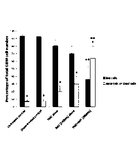
dye, PI (Figure 4 and 5A). Note in FIG. 4 the markedly elevated fractions of
apoptotic (annexin-positive) and dead (annexin and PI ¨positive) cells with
the
combination of IMT and TMZ, relative to those observed with sham or either
stand-
alone treatments. The flow cytometry scatterplots of FIG. 4 illustrate dead
(upper
right quadrant), apoptotic (lower right quadrant) and live (lower left
quadrant). The
flow cytometry scatterplots of FIG. 4 show that under sham IMT treatment (A)
5.4%
of the cells are dead, 2% apoptotic and 92.1% are live; under TMZ (B) 9.4% of
the
cells are dead, 14. 1% are apoptotic and 76.1% live; under IMT (C), 21.2% of
the
cells are dead, 23.3% apoptotic and 54.5% live; while under both TMZ+IMT
treatment (D), 16% of the cells are dead, 58%.3% apoptotic and 25.2% live. The
combined fraction of apoptotic (annexin+) and dead (annexin+ and PH) GBM cells
rose dramatically from untreated (5.712.5%) and sham conditions (5.9 2.8%) to
CA 02985847 2017-11-14
WO 2016/179712 KT/CA2016/050556
- 39 -
single-modality temozolomide (16.9 7.4%) or IMT (28.5 14.9%), and finally to
combination treatment with temozolomide and IMT (52.4 21.8%). The results of
the
quantitative metabolic MTT assay further confirmed the detrimental impact of
each
treatment modality and the heightened benefits of combined IMT and
temozolomide
on reducing primary GBM cell viability (Figure 5B). As standalone treatments,
IMT
(52.2 4.8% viability relative to untreated cells) was significantly more
effective than
temozolomide (69.7 11.8 /0 viability), as measured by MTT metabolism. The
combination of IMT with temozolomide produced further significant GBM cell
death
compared to either treatment alone (29.1 3.2% viability; FIG. 5B). Comparable
effects were produced in immortalized LN229 GBM cells (data not shown).
IMT enhances the efficacy of siRNA-mediated gene knockdown in GBM
Gene silencing methods in primary, patient-derived GBM cells are hindered by
poor
uptake of hydrophilic genetic material across lipid membranes. This study
tested
whether IMT may act in concert with HSP27 siRNA, to enhance uptake and
.. bioavailability of siRNA in the cells or through a secondary means of
impairing
cytokinesis and anti-apoptotic mechanisms. In this example, the pro-tumor
chaperone, HSP27, was chosen as the therapeutic target. HSP27 siRNA
transfection produced a modest target knockdown that was markedly potentiated
with concurrent IMT (FIG. 6 panel A). Sham conditions, IMT and control siRNA
were
ineffective at reducing HSP27 levels (FIG. 6 panels A-C). Mean densitometry
values
of FIG. 6 panel B HSP27 and panel C HSP90 levels in GBM cells from 3 patients
confirmed the robust and specific knockdown of HSP27 that was significantly
enhanced with the co-administration of IMT. HSP90 levels were not notably
affected
by any of the treatment conditions. Values represent mean plus (+ sign)
standard
CA 02985847 2017-11-14
WO 2016/179712 PCT/CA2016/050556
-40 -
deviation. Single asterisk indicate a significant difference from the protein
expression
measured under sham conditions: double asterisk indicates a significant
difference
in protein expression between the indicated treatment pair (P< 0.05, ANOVA).
OD,
optical density.
There was avid expression of HSP27 in patient GBM cells that was not
notably affected by control or IMT conditions. In contrast, non-viral
transfection of
HSP27-specific siRNA (50 nM) using a cationic polymer resulted in a moderate
reduction in HSP27 levels that was significantly and consistently enhanced
with
concomitant IMT (Figure 6A). Quantitative densitometry of western blot
analyses
was performed in triplicate using GBM cells obtained from three patients and
confirmed no significant change in the level of HSP27 expression among sham-
treated cells [0.40 0.08 normalized optical density (00)], control siRNA-
treated
(0.46 0.05 OD), IMT-treated (0.45 0.08 OD), or the combination of IMT with
control
siRNA-treated cells (0.43 0.09 OD). In contrast, GBM cells transfected with
HSP27
siRNA alone (0.27 0.04 OD) or the combination of IMT and HSP27 siRNA
(0.07 0.02 OD) exhibited significant reductions in HSP27 levels of 32.5% and
82.5%, respectively (FIG. 6B). With either HSP27 siRNA or IMT plus HSP27 siRNA
treatment, there was no reduction in the expression of HSP90, a related stress-
response chaperones, further supporting the specificity of the gene-targeting
method
and antitumor impact (FIG. 6C). GBM cell viability in patient specimens was
quantified with MTT and, as in the previous series, demonstrated significantly
reduced values following IMT alone (60.3 7.7% viability relative to untreated
cells).
HSP27 siRNA alone also produced significant cytotoxic effects (70.3 5.4%
viability).
The combination of IMT and control siRNA did not further reduce cell viability
CA 02985847 2017-11-14
WO 2016/179712 PCT/CA 2016/050556
-41 -
compared to IMT alone (57.1 8.8% viability); however, IMT with concomitant
HSP27
siRNA produced a robust and significant increase in GBM cell death (35.9 12.8%
viability; see also FIG. 7).
IMT-enhanced tumoricidal effect of HSP27 gene silencing in GBM Adjuvant
With reference to FIG. 7 IMT enhanced the tumoricidal effect of targeted
HSP27 knockdown in patient GBM cells. Individual measures show the normalized
MTT viability after 48 hours of the indicated treatment. IMT alone produced
marked
loss of GBM viability that was robustly potentiated with the HSP27-specific
siRNA,
but not control siRNA. Single asterisks indicate a significant difference from
the
sham group. The double asterisks of FIG. 7 indicate a significant difference
between
the indicated treatment pair (P<0.05, ANOVA). Samples were assessed in
triplicate
using primary GBM cells from 3 different GBM patients and shown as mean +
standard deviation. TR, transfection reagent.
With reference to FIG. 20, note the similar density and appearance of the
GBM cells under control conditions (panels A-E) but marked loss with either
HSP27
siRNA (panel H) or IMT (panel F) alone. This anti-tumor benefit was
dramatically
potentiated by combining HSP27 siRNA and IMT (panel l).
The example shows the synergistic effect combining IMT and siRNA
treatment. The combination of IMT and siRNA is substantially more effective
than
each treatment taken alone.
Example 2
This example complements the results shown in FIGs. 4-8, 17, 18 and 20, to
demonstrate the synergistic enhancement of gene-targeted therapy using high
frequency IMT (200 kHz) and synergistic enhancement of high frequency IMT (200
CA 02985847 2017-11-14
WO 2016/179712 PCT/CA2016/050556
- 42 -
kHz) when combined with TMZ.
High frequency (200 kHz) IMT enhances gene therapy in GBM
The pro-tumor chaperone, HSP27, was chosen as the therapeutic target.
Panel A of FIG. 21 is a representative western blot analysis using primary GBM
cells
derived from 3 operative tumor specimens. HSP27 siRNA transfection produced a
modest target knockdown that was markedly potentiated with concurrent IMT (200
kHz). Sham conditions, IMT and control siRNA were ineffective at reducing
HSP27
levels. The levels of another tumor-promoting HSP, HSP90, was not affected by
the
targeted HSP27 and therapies. Mean densitometry values of FIG. 21B HSP27 and
FIG. 210 HSP90 levels in GBM cells from the 3 patients confirmed the robust
and
specific knockdown of HSP27 that was significantly enhanced with the co-
administration of IMT at 200 kHz. HSP90 levels were not notably affected by
any of
the treatment conditions. Values represent mean + standard deviation. Single
asterisk indicate a significant difference from the protein expression
measured under
sham conditions; double asterisk indicates a significant difference in protein
expression between the indicated treatment pair (P< 0.05, ANOVA).
Quantitative effect of high frequency IMT (200 kHz) combined with TMZ on
patient GBM cells
FIG. 22 illustrates flow cytometry data showing the percentage of live and
apoptotic/dead GBM cells following the indicated treatments. There was a
significant
difference between the percentage of live and apoptoticidead cells within all
groups =
(single asterisk, ANOVA P<0.05). Note, however, that the TMZ+IMT group had
reversed major proportions of live and apoptotic/dead cells compared to the
other
groups. Double asterisks indicate ia significant difference between the live
or
CA 02985847 2017-11-14
WO 2016/179712 PCT/CA2016/050556
-43 -
apoptoticidead fractions and the respective value obtained from untreated
cells (P<
0.05, ANOVA). Each treatment condition was analyzed in quadruplicate using
approximately 30,000 GBM cells per run. The duration of treatment was 72 hours
and each measure shown represents the mean + standard deviation for primary
GBM cells from 3 patients. TMZ, temozolomide
Example 3 ¨ In vivo IMT model
The F98 rat GBM model is used in this study. Briefly, F98 cells are derived
from an anaplastic glioma in a Fischer rat and produce treatment-resistant
brain
tumors with GBM properties when implanted into syngeneic host brains (21).
Adult
male rats undergo stereotactic implantation of a commercial cannula/electrode
combination bilaterally into the striatum. This MRI-compatible device permits
infusion
of the F98 cells and siRNA, with concurrent IMT, at the epicenter of the
growing
tumor. A reference electrode is tunneled through the nuchal skin for easy
access.
The IMT cables are suspended via a commutator, so that the animal can move
freely within its home cage during treatment (see FIG. 10).
Adult Fischer rats underwent stereotactic implantation of F98 GBM tumor
cells into bilateral striate. After 4 days of tumor growth in the brain, one
side was
treated for 7 days using IMT with a frequency of 200 kHz and amplitude of +/-
2V.
The contralateral tumor was fitted with electrode hardware but did not receive
treatment (i.e., sham). Shown in FIGs. 10A and 100 is a representative animal
receiving IMT in its home cage. With reference to FIG. 10, the therapy is
delivered
using a waveform generator 801 connected to an indwelling brain electrode via
a
commutator 802 that allows free movement of the animal at all times. Cannula
CA 02985847 2017-11-14
WO 2016/179712 PCT/CA2016/050556
- 44 -
electrode constructs 803 were implanted bilaterally into the striatum. Only
the
treated tumor received IMT: the other side had the same hardware implanted but
was not treated. The construct 803 consists of a brain cannula 804 through
which
the F98 cells were implanted (dashed arrow) and adjacent brain electrode 805
to
deliver IMT within the epicenter of the GBM field (short solid arrow). There
is a
reference electrode 806 (long solid arrow) that is implanted in the nucchal
soft
tissues. The reference electrode is not restricted to the nuchal soft tissues,
and it
may also be implanted in other places, such as the subgaleal or subdural
spaces, or
other areas appropriate for tumor treatment. FIG. 100 is a closer view of the
subject
undergoing IMT. Animals showed no evidence of ongoing discomfort, medical
complications, neurological deficits or seizures during the therapy.
IMT reduces overall brain tumor mass
FIG. 11 are photographs of an extracted rat brain that housed bilateral GBM
tumors
in the striatum. IMT implants had been placed bilaterally (now removed) but
only
activated on the right side. Note the IMT-mediated reduction in hemispheric
volume
on the right compared to the left. The image shown in panel B of FIG. 11 shows
the
same brain of panel A of FIG. 11 with an overlaid grid for size calibration.
In vivo bioluminescence imaging (BLI) in the F98 GBM model
F98 GBM cells transduced to stably express Firefly luciferase were implanted
into
the striatum of a Fischer rat. FIG. 12 A shows BLI tumor signal and 12 B
rostral (top)
to caudal hematoxylin-stained brain sections through the tumor (arrows), These
data
were obtained 4 days after implanting a striatal deposit of 2p1 DMEM
containing
40,000 F98 GBM cells and demonstrate the aggressive tumorigenesis produced in
this model.
CA 02985847 2017-11-14
WO 201(p/179712 PCT/CA2016/050556
- 45 -
The T2-weighted MRI shown in FIGs. 13 B-D provides accurate 3-dimensional
delineation of the tumor (arrows), suitable for volumetric analysis, and
associated
cerebral edema evident as brighter signal around the tumor. The MRI studies
complement the BLI and immunohistology to evaluate tumor response to IMT in
this
project.
Anti-tumor effects of IMT in vivo
FIG. 14 includes representative images through the brains of adult Fischer
rats
treated with sham conditions (i.e., no stimulation) or IMT for 7 days (200
kHz, +/-2V).
The IMT treatment was initiated 4 days after injecting 2p1 DMEM containing
40,000
F98 GBM cells into bilateral striata. Panels A and B of FIG. 14 therefore show
an 11-
day old tumor after the indicated treatment. Panels A and B of FIG. 14 are two
example of brains housing bilateral GBM tumors with IMT or sham conditions
delivered for 1 week. Electrodes used in panel A were insulated with
EntellanO.
Electrodes used in panel B were uninsulated. Note the dramatic attenuation of
the
.. treated tumor relative to the sham control tumor, with IMT effectively
reducing the
growth and spread of the GBM cells through the brain. Panel C of FIG. 14,
which
represents control animals with implanted bilateral IMT constructs, but no
tumor
cells, revealed that IMT does not produce notable injury to normal brain
tissue
relative to the sham control conditions. Asterisks in FIG. 14C indicate the
hardware
defect noted in all sham and treated tissues. The scale bar in B applies to
panels A-
C of FIG. 14.
Therapeutic benefit of IMT in vivo
FIG. 15 panels A-D shown brain sections through bilateral GBM tumors in four
additional Fischer rats. The IMT hardware was implanted on both sides but
activated
CA 02985847 2017-11-14
WO 2016/179712 PCl/CA2016/050556
- 46 -
only on the side indicated by the arrow. The IMT-treated tumors in these four
additional animals were markedly smaller than in the sham-treated controls.
With reference to FIG. 16 in vivo F98 GBM model Electrodes were implanted and
GBM tumors grown bilaterally in the Fischer rat striata. The left side was
sham (i.e.,
.. no stimulation) and showed robust tumor growth (marked by arrows in FIG,
16A).
Conversely, IMT on the right side produced a marked reduction in tumor volume
(arrows, FIG. 16B). Scant caspase-3 activation (stains red) occurred on the
sham
side (FIG. 16C), whereas the IMT-treated tumor was robustly red labeled (FIG.
16D).
The CT scout view shows another rat with a unilateral electrode in a F98 GBM
tumor
being prepared for radiotherapy (FIG. 16E). Radiation dosing plan that can be
used
in combination with IMT to treat the GBM tumor is illustrated in FIG. 16F.
Example 4¨ Mechanism of IMT-enhanced transfection
The in vitro and in vivo studies show that GBM cells treated with IMT undergo
caspase-activated apoptosis, however membrane disruption was also evident by
the
cellular uptake of impermeable dyes (FIGs. 3 and 16). These findings may
reflect
necrotic death, membrane degeneration after apoptosis, or facilitated dye
uptake
through endocytosis, electrophoresis or electroporation. To address this
question,
primary patient GBM cells were treated with IMT and subjected to the
investigations
below. Live cell imaging: These studies evaluate acute changes in membrane
integrity in live GBM cells exposed to IMT. Cells are imaged for 1-3 hours
under
sham or IMT conditions, using an Olympus FluoViewTM FV1000 confocal
microscope for evidence of IMT-mediated uptake of propidium iodide, a membrane-
impermeable fluorescent molecule (Fig. 19). Panel A of FIG. 19: no IMT, Panels
B-I
of FIG. 19: IMT after 13, 26, 39, 52, 65, 78, 91 and 104 seconds respectively.
CA 02985847 2017-11-14
WO 2016/179712 PCT/CA2016/050556
- 47 -
With reference to FIG. 19, at baseline (panel A, i.e., no IMT), there is no
substantial fluorescent signal from the cells; with uptake seen only in the
occasional
degenerating cell, as is normal under culture conditions. As IMT (+/- 2V AC,
200
kHz) is initiated, there is slow, progressive enhancement of the signal
emanating
from nuclear (small narrow arrows in panel I of FIG. 19) and cytoplasmic
(large
bolded arrow in panel I of FIG. 19) compartments of the GBM cells, The images
of
FIG. 19 illustrate that IMT increases cellular uptake of membrane impermeable
agents through nuclear and cytoplasmic envelopes.
Example 5 ¨ In vivo IMT-enhanced transfection using the F98 GBM
model
These experiments evaluate the in vivo efficacy of IMT-mediated transfection,
with and without standard chemoradiation. Continuous IMT is initiated 1 week
following surgery as described before. Seven animal groups (10 animals/group)
are
used for both low and high frequency IMT stimulation parameters, with
bilateral
striatal GBM; one side used for sham control. The group size is chosen to
adequately temper inter-animal variability, with potential loss due to
unexpected
problems/deaths, and be completed within the 3 year study window. IMT is
performed alone (group 1), with single agent siRNA targeting HSP27 or HSP70
(groups 2, 3), with dual siRNA therapy (group 4), or with the prior siRNA
options and
chemoradiation treatment (groups 5-7). siRNA (50 nM in 2p1 PBS) is delivered
through the cannula 803 shown in FIG. 10B on day 1 and day 4 of IMT treatment.
Dual siRNA therapy use a total volume of 2 pi, with each siRNA concentration
adjusted to 50 nM. Temozolomide (TMZ) dosing was 18 mg/kg i.p., the clinical
equivalent of 200 mg/m2/day (24-25); the radiation dose is 30 Gy in 2
fractions. F98
CA 02985847 2017-11-14
WO 2016/179712 PCT/CA2016/050556
- 48 -
cells respond to in vitro radiation doses between 12-18 Gy (26) and radiation
necrosis occurs in F98 models using 60 Gy (27). The in vivo RT dose in this
study
falls between these values to induce anti-tumor effect without radiation
necrosis.
Cranial imaging is performed using a 9.4T MRI system immediately prior to, and
following, the treatment course. Animals are euthanized at the end of the
treatment
period or earlier if there are signs of severe neurological compromise. Brains
are
sectioned for histological staining and immunocytochemistry. Volumetric tumor
dimensions are quantified, and indices of proliferation (MIB1), apoptosis
(activated
caspase-3 and TUNEL) and HSP27/70 expression are assessed. A small number of
animals (N=5) is used to evaluate the in vivo distribution of a fluorescent-
labeled
control siRNA delivered to the tumor in the absence and presence of IMT.
Determining the mechanisms of IMT-induced cell death and IMT-enhanced
transfection in GBM allows to maximally exploit these effects before
translating to
the clinical settings.
High frequency (200 kHz) IMT activates caspase-3 in GBM cells. Shown in
FIG. 23 are Western blot studies from 3 patient-derived GBM cell preparations
treated with 72 hours of sham or IMT (+/- 2V, 200 kHz) conditions. The levels
of
intact caspase-3 are markedly reduced and correspond to an increase in the
activated (cleaved) form, indicative of apoptosis induction, during IMT.
The above disclosure generally describes the present invention. Changes in
form
and substitution of equivalents are contemplated as circumstances may suggest
or
render expedient. Although specific terms have been employed herein, such
terms
are intended in a descriptive sense and not for purposes of limitation. Other
variations and modifications of the invention are possible. As such
modifications or
CA 02985847 2017-11-14
WO 2916/179712 PCIICA2016/1150556
- 49 -
variations are believed to be within the sphere and scope of the invention as
defined
by the claims appended hereto.
References
1. Cavenee WK, Louis DN, Ohgaki H. WHO Classification of Tumours of
the Central Nervous System. WHO Pulblications Fourth edition, 2007.
2. Orlowski, S.; Belehradek, J.; Paoletti, C.; Mir, L. M. Transient
electropermeabilizationof cells in culture. Increase of the cytotoxicity of
anticancer drugs. Biochem. Pharmacol. 37:4727¨ 4733; 1988.
3. Zimmermann U. Electric field-mediated fusion and related electrical
phenomena.Biochim Biophys Acta. 1982 Nov 30;694(3):227-77.
4. Horikoshi T, Naganuma H, Ohashi Y, Ueno T, Nukui H.Enhancing
effect of electric stimulation on cytotoxicity of anticancer agents against
rat and human glioma cells.Brain Res Bull, 2000 Mar 15;51(5):371-8.
5. Salford LG, Persson BR, Brun A, Ceberg CP, Kongstad PC, Mir LM. A
new brain tumour therapy combining bleomycin with in vivo
electropermeabilization. Biochem Biophys Res Commun. 1993 Jul
30;194(2):938-43
6. Pudenz, R. Adverse effects of electrical energy applied to the nervous
system. Neurosurgery 1:190-191; 1977.
7. Kirson ED, Dbaly V, Tovarys F, Vymazal J, Soustiel JF, Itzhaki A,
Mordechovich D, Steinberg-Shapira S, Gurvich Z, Schneiderman R,
Wasserman Y, Salzberg M, Ryffel B, Goldsher D, Dekel E, Palti
Y.Altemating electric fields arrest cell proliferation in animal tumor
models and human brain tumors.Proc Natl Acad Sci U S A. 2007 Jun
CA 02985847 2017-11-14
WO 2016/179712 PCT/CA2016/050556
- 50 -12;104(24):10152-7. Epub 2007 Jun 5.
8. Pless M, Weinberg U.Tumor treating fields: concept, evidence and
future. Expert Opin Investig Drugs. 2011 Aug;20(8):1099-106. doi:
10.1517/13543784.2011.583236. Epub 2011 May 9.
9. Stupp R, Wong ET, Kanner AA, Steinberg D, Engelhard H, Heidecke
V, Kirson ED, Taillibert S, Liebermann F, Dbaly V, Ram Z. Villano JL,
Rainov N, Weinberg U, Schiff D, Kunschner L, Raizer J, Honnorat J,
Sloan A, Malkin M, Landolfi JC, Payer F, Mehdorn M, Weil RJ,
Pannullo SC, Westphal M, Smrcka M, Chin L, Kostron H, Hofer S,
Bruce J, Cosgrove R, Paleologous N, Palti Y, Gutin PH.NovoTTF-100A
versus physician's choice chemotherapy in recurrent glioblastoma: A
randomised phase III trial of a novel treatment modality.Eur J Cancer.
2012 Sep;48(14):2192-202. Epub 2012 May 18.
10.Kanner AA, Wong ET, Villano JL, Ram Z; EF-11 Investigators. Post
Hoc analyses of intention-to-treat population in phase III comparison of
NovoTTF-100ATm system versus best physician's choice
chemotherapy. Semin Oncol, 2014 Oct;41 Suppl 6:S25-34.
11.Deniau JM, Degos B, Bosch C, Maurice N. Deep brain stimulation
mechanisms: beyond the concept of local functional inhibition.Eur J
Neurosci. 2010 Oct;32(7):1080-91. doi: 10.1111/1.1 460-
9568.2010.07413.x.
12. Wang J, Lu Z, Wientjes MG, Au JL. Delivery of siRNA therapeutics:
barriers and carriers. AAPS J. 2010 Dec;12(4):492-503.
13.Mossop BJ, Barr RC, Henshaw JW, Zaharoff DA, Yuan F. Electric
CA 02985847 2017-11-14
WO 2016/179712 PCT/CA2016/050556
-51 -
fields in tumors exposed to external voltage sources: implication for
electric field-mediated drug and gene delivery. Ann Biomed Eng, 2006
Oct;34(10):1564-72.
14.Tang L, Yao C, Sun C. Apoptosis induction with electric pulses - a new
approach to cancer therapy with drug free. Biochem Biophys Res
Commun. 2009 Dec 25;390(4):1098-101.
15.Acunzo J, Andrieu C, Baylot V, So A, Rocchi P. Hsp27 as a
therapeutic target in cancers. Curr Drug Targets. 2014 Apr;15(4):423-
31.
16.Lianos GD, Alexiou GA, Mangano A, Mangano A, Rausei S, Boni L,
Dionigi G, Roukos DH. The role of heat shock proteins in cancer.
Cancer Lett. 2015 May 1360(2):114-8.
17.Yang I, Fang S, Parsa AT. Heat shock proteins in glioblastomas.
Neurosurg Clin N Am. 2010 Jan;21(1):111-23.
18.Belkacemi L, Hebb MO. HSP27 knockdown produces synergistic
induction of apoptosis by HSP90 and kinase inhibitors in glioblastoma
multiforme. Anticancer Res. 2014 Sep;34(9):4915-27,
19.Aloy MT, Hadchity E, Bionda C, Diaz-Latoud C, Claude L, Rousson R,
Arrigo AP, Rodriguez-Lafrasse C. Protective role of Hsp27 protein
against gamma radiation-induced apoptosis and radiosensitization
effects of Hsp27 gene silencing in different human tumor cells. Int J
Radiat Oncol Biol Phys. 2008 Feb 1;70(2):543-53.
20.Jakubowicz-Gil J, Langner E, Bdziul D, Wertel I, Rzeski W. Silencing of
Hsp27 and Hsp72 in glioma cells as a tool for programmed cell death
CA 02985847 2017-11-14
WO 2016/179712 PC1'/CA20116/050556
-52 -
induction upon temozolomide and quercetin treatment. Toxicol App!
Pharmacol. 2013 Dec 15;273(3):580-9.
21.Mathieu D, Lecomte R, Tsanaclis AM, Larouche A, Fortin D.
Standardization and detailed characterization of the syngeneic
Fischer/F98 glioma model. Can J Neurol Sci. 2007 Aug;34(3):296-306.
22.Beaman GM, Dennison SR, Chatfield LK, Phoenix DA. Reliability of
HSP70 (HSPA) expression as a prognostic marker in glioma. Mol Cell
Biochem. 2014 Aug;393(1-2):301-7.
23.Wang X, Chen M, Zhou J, Zhang X. HSP27, 70 and 90, anti-apoptotic
proteins, in clinical cancer therapy (Review). Int J Oncol. 2014
Jul;45(1):18-30.
24.0stermann S, Csajka C, Buclin T, Leyvraz S, Lejeune F, Decosterd
LA, Stu pp R. Plasma and
cerebrospinal fluid population
pharrnacokinetics of temozolomide in malignant glioma patients. Clin
Cancer Res. 2004 Jun 1;10(11).3728-36.
25.Zhou Q, Guo P, Wang X, Nuthalapati S, Gallo JM. Preclinical
pharmacokinetic and pharmacodynamic evaluation of metronomic and
conventional temozolomide dosing regimens. J Pharmacol Exp Thar.
2007 Apr;321(1):265-75.
26.Gil S, Sarun S, Biete A, Prezado Y, Sabes M. Survival analysis of F98
glioma rat cells following minibeam or broad-beam synchrotron
radiation therapy. Radiat Oncol. 2011 Apr 13;6:37.
27.Bolcaen J, Descamps B, Deblaere K, Boterberg T, De Vos Pharm F,
Kalala JP, Van den Broecke C, Decrock E, Leybaert L, Vanhove C,
CA 02985847 2017-11-14
WO 2016/179712 PCT/CA 2016/050556
- 53 -
Goethals I. (18)F-fluoromethylcholine (FCho), (18)Ffluoroethyltyrosine
(FET), and (18)F-fluorodeoxyglucose (FOG) for the discrimination
between highgrade glioma and radiation necrosis in rats: a PET study.
Nucl Med Biol. 2015 Jan;42(1):38-45.
28.Lacouture ME, Davis ME, Elzinga G, Butowski N, Tran D, Villano JL,
DiMeglio L, Davies AM, Wong ET. Characterization and management
of dermatologic adverse events with the NovoTTF-100A System, a
novel anti-mitotic electric field device for the treatment of recurrent
glioblastoma. Semin Oncol. 2014 Jun;41 Suppl 4:S1-14. doi: 10.1053..
29.Stupp R, Taillibert S, Kanner AA, Kesaii S, Steinberg DM, Toms SA,
Taylor LP, Lieberman F, Silvani A, Fink KL, Barnett GH, Zhu JJ,
Henson JW, Engelhard HH, Chen TC, Tran DD, Sroubek J, Tran ND,
Hottinger AF, Landolfi J, Desai R, Caroli M, Kew Y, Honnorat J, ldbaih
A, Kirson ED, Weinberg U, Palti Y, Hegi ME, Ram Z. Maintenance
Therapy With Tumor-Treating Fields Plus Temozolomide vs
Temozolomide Alone for Glioblastoma: A Randomized Clinical Trial.
JAMA. 2015 Dec 15:314(23):2535-43. doi: 10.1001
30.Garcia PA, Pancotto T, Rossmeisl JH Jr, Henao-Guerrero N,
Gustafson NR, Daniel GB, Robertson JL, Ellis TL, Davelos RV. Non-
thermal irreversible electroporafion (N-TIRE) and adjuvant fractionated
radiotherapeutic multimodal therapy for intracranial malignant glioma in
a canine patient. Technol Cancer Res Treat. 2011 Feb;10(1):73-83.