Note: Descriptions are shown in the official language in which they were submitted.
CA 02986575 2017-11-20
WO 2016/187594 PCT/US2016/033644
TRISPECIFIC BINDING PROTEINS AND METHODS OF USE
CROSS-REFERENCE
[0001] This application claims the benefit of U.S. Provisional Application No.
62/305,088,
filed March 8, 2016; U.S. Provisional Application No. 62/165,833, filed May
22, 2015; and U.S.
Provisional Application No. 62/165,153, filed May 21, 2015, all of which
applications are
incorporated herein by reference in their entirety.
SEQUENCE LISTING
[0001.1] The instant application contains a Sequence Listing which has been
submitted
electronically in ASCII format and is hereby incorporated by reference in its
entirety. Said
ASCII copy, created on May 17, 2016, is named 47517 701 601 SL.txt and is
128,516 bytes in
size.
BACKGROUND OF THE INVENTION
[0002] The selective destruction of an individual cell or a specific cell type
is often desirable in
a variety of clinical settings. For example, it is a primary goal of cancer
therapy to specifically
destroy tumor cells, while leaving healthy cells and tissues intact and
undamaged. One such
method is by inducing an immune response against the tumor, to make immune
effector cells
such as natural killer (NK) cells or cytotoxic T lymphocytes (CTLs) attack and
destroy tumor
cells.
SUMMARY OF THE INVENTION
[0003] Provided herein are trispecific antigen-binding protein, pharmaceutical
compositions
thereof, as nucleic acids, recombinant expression vectors and host cells for
making such
trispecific antigen-binding proteins, and methods of use for the treatment of
diseases, disorders,
or conditions. In one aspect, described herein are trispecific antigen-binding
proteins wherein
said proteins comprise (a) a first domain (A) which specifically binds to
human CD3; (b) a
second domain (B) which is a half-life extension domain; and (c) a third
domain (C) which
specifically binds to a target antigen, wherein the domains are linked in the
order H2N-(A)-(B)-
(C)-COOH, H2N-(A)-(C)-(B)-COOH, H2N-(B)-(A)-(C)-COOH, H2N-(B)-(C)-(A)-COOH,
H2N-
(C)-(B)-(A)-COOH, or H2N-(C)-(A)-(B)-COOH by linkers Li and L2.
[0004] Also provided herein in certain aspects are trispecific antigen-binding
proteins, wherein
said proteins comprise (a) a first domain (A) which specifically binds to
human CD3; (b) a
second domain (B) which is a half-life extension domain; and (c) a third
domain (C) which
specifically binds to a target antigen, wherein the domains are linked in the
order H2N-(A)-(C)-
(B)-COOH, H2N-(B)-(A)-(C)-COOH, H2N-(C)-(B)-(A)-COOH, or by linkers Li and L2.
-1-
CA 02986575 2017-11-20
WO 2016/187594 PCT/US2016/033644
[0005] Also provided herein in certain aspects are trispecific antigen-binding
proteins, wherein
said proteins comprise (a) a first domain (A) which specifically binds to
human CD3; (b) a
second domain (B) which is a half-life extension domain; and (c) a third
domain (C) which
specifically binds to a target antigen, wherein the domains are linked in the
order H2N-(A)-(B)-
(C)-COOH, H2N-(A)-(C)-(B)-COOH, H2N-(B)-(A)-(C)-COOH, H2N-(B)-(C)-(A)-COOH,
H2N-
(C)-(B)-(A)-COOH, or H2N-(C)-(A)-(B)-COOH by linkers Li and L2, and wherein
the first
domain binds to human CD3 with a KD of greater than 100 nM.
[0006] Also provided herein in certain aspects are trispecific antigen-binding
proteins, wherein
said proteins comprise (a) a first domain (A) which specifically binds to
human CD3; (b) a
second domain (B) which is a half-life extension domain; and (c) a third
domain (C) which
specifically binds to a target antigen, wherein the domains are linked in the
order H2N-(A)-(B)-
(C)-COOH, H2N-(A)-(C)-(B)-COOH, H2N-(B)-(A)-(C)-COOH, H2N-(B)-(C)-(A)-COOH,
H2N-
(C)-(B)-(A)-COOH, or H2N-(C)-(A)-(B)-COOH by linkers Li and L2, and wherein
the protein
has a molecular weight of less than 55 kDa.
[0007] Also provided herein in certain aspects are trispecific antigen-binding
proteins, wherein
said proteins comprise (a) a first domain (A) which specifically binds to
human CD3; (b) a
second domain (B) which is a half-life extension domain; and (c) a third
domain (c) which
specifically binds to a target antigen, wherein the domains are linked in the
order H2N-(A)-(B)-
(C)-COOH, H2N-(A)-(C)-(B)-COOH, H2N-(B)-(A)-(C)-COOH, H2N-(B)-(C)-(A)-COOH,
H2N-
(C)-(B)-(A)-COOH, or H2N-(C)-(A)-(B)-COOH by linkers Li and L2, and wherein B
comprises
a single domain antibody that binds to serum albumin.
[0008] Various embodiments of trispecific antigen-binding proteins are also
provided herein,
contemplated for any aspect herein, alone or in combination. In some
embodiments, first
domain comprises a variable light chain and variable heavy chain each of which
is capable of
specifically binding to human CD3. In some embodiments, the variable light
chain is a X,
(lamda) light chain. In some embodiments, the variable light chain is a lc
(kappa) light chain. In
some embodiments, the first domain comprises a single-chain variable fragment
(scFv) specific
to human CD3. In some embodiments, the first domain is specific for CD3E
(epsilon). In some
embodiments, the first domain is specific for CD3 6 (delta). In some
embodiments, the first
domain is specific for CD3y (gamma). In some embodiments, the first domain
comprises
complementary determining regions (CDRs) selected from the group consisting of
muromonab-
CD3 (OKT3), otelixizumab (TRX4), teplizumab (MGA031), visilizumab (Nuvion),
SP34, X35,
VIT3, BMA030 (BW264/56), CLB-T3/3, CRIS7, YTH12.5, F111-409, CLB-T3.4.2, TR-
66,
WT32, SPv-T3b, 11D8, XIII-141, XIII-46, XIII-87, 12F6, T3/RW2-8C8, T3/RW2-4B6,
OKT3D, M-T301, SMC2, F101.01, UCHT-1 and WT-31. In some embodiments, the first
-2-
CA 02986575 2017-11-20
WO 2016/187594 PCT/US2016/033644
domain is humanized or human. In some embodiments, the first domain has a KD
binding of
1000 nM or less to CD3 on CD3 expressing cells. In some embodiments, the first
domain has a
KD binding of 100 nM or less to CD3 on CD3 expressing cells. In some
embodiments, the first
domain has a KD binding of 10 nM or less to CD3 on CD3 expressing cells. In
some
embodiments, the first domain has crossreactivity with cynomolgus CD3. In some
embodiments, the first domain comprises an amino acid sequence provided
herein.
[0009] In some embodiments, the second domain binds human serum albumin. In
some
embodiments, the second domain comprises a scFv, a variable heavy domain (VH),
a variable
light domain (VL), a single domain antibody, a peptide, a ligand, or a small
molecule. In some
embodiments, the second domain comprises a scFv. In some embodiments, the
second domain
comprises a VH domain. In some embodiments, the second domain comprises a VL
domain. In
some embodiments, the second domain comprises a single domain antibody. In
some
embodiments, the second domain comprises a peptide. In some embodiments, the
second
domain comprises a ligand. In some embodiments, the second domain comprises a
small
molecule entity.
[0010] In some embodiments, the third domain comprises a scFv, a VH domain, a
VL domain, a
non-Ig domain, a ligand, a knottin, or a small molecule entity that
specifically binds to a target
antigen. In some embodiments, the third domain is specific to a cell surface
molecule. In some
embodiments, the third domain is specific to a tumor antigen.
[0011] In some embodiments, linkers Li and L2 are peptide linkers. In some
embodiments,
linkers Li and L2 independently consist of about 20 or less amino acid
residues. In some
embodiments, linkers Li and L2 are each independently selected from (GS)n (SEQ
ID NO: 49),
(GGS)n (SEQ ID NO: 50), (GGGS)n (SEQ ID NO: 51), (GGSG)n (SEQ ID NO: 52),
(GGSGG)n (SEQ ID NO: 53), or (GGGGS)n (SEQ ID NO: 54), wherein n is 1, 2, 3,
4, 5, 6, 7,
8, 9, or 10. In some embodiments, linkers Li and L2 are each independently
(GGGGS)4 (SEQ
ID NO: 55) or (GGGGS)3 (SEQ ID NO: 56). In some embodiments, linkers Li and L2
are
chemical linkers.
[0012] In some embodiments, the first domain is at the N-terminus of the
protein. In some
embodiments, the second domain is at the N-terminus of the protein. In some
embodiments, the
third domain is at the N-terminus of the protein. In some embodiments, the
first domain is at the
C-terminus of the protein. In some embodiments, the second domain is at the C-
terminus of the
protein. In some embodiments, the third domain is at the C-terminus of the
protein.
[0013] In some embodiments, the protein is less than about 80 kDa. In some
embodiments, the
protein is about 50 to about 75 kDa. In some embodiments, the protein is less
than about 50
kDa. In some embodiments, the protein is less than about 40 kDa. In some
embodiments, the
-3-
CA 02986575 2017-11-20
WO 2016/187594 PCT/US2016/033644
protein is about 20 to about 40 kDa. In some embodiments, the protein has an
elimination half-
time of at least about 50 hours. In some embodiments, the protein has an
elimination half-time
of at least about 100 hours. In some embodiments, the protein has increased
tissue penetration
as compared to an IgG to the same target antigen.
[0014] Also provided herein, in another aspect are polynucleotides encoding
trispecific antigen-
binding proteins according to any one of the above embodiments. In another
aspect provided
herein are vectors comprising the described polynucleotides. In another
aspect, provided herein
are host cells transformed with the described vectors
[0015] In yet another aspect, provided herein are pharmaceutical compositions
comprising a
trispecific antigen-binding protein of any of the above embodiments, a
polynucleotide encoding
a trispecific antigen-binding protein of any of the above embodiments, a
vector comprising the
described polynucleotides, or a host cell transformed with a vector of any of
the above
embodiments and a pharmaceutically acceptable carrier.
[0016] Also provided herein, are processes for the production of trispecific
antigen-binding
proteins according to any of the aspects and embodiments herein, said process
comprising
culturing a host transformed or transfected with a vector comprising a nucleic
acid sequence
encoding any trispecific antigen-binding protein herein under conditions
allowing the expression
of the protein and recovering and purifying the produced protein from the
culture.
[0017] Also provided herein are methods for the treatment amelioration of a
proliferative
disease, a tumorous disease, an inflammatory disease, an immunological
disorder, an
autoimmune disease, an infectious disease, viral disease, allergic reactions,
parasitic reactions,
graft-versus-host diseases or host-versus-graft diseases comprising the
administration of a
trispecific antigen-binding protein of any of the above embodiments to a
subject in need of such
a treatment or amelioration. In some embodiments, the subject is a human. In
some
embodiments, the method further comprises administration of an agent in
combination with the
trispecific antigen-binding protein described herein.
INCORPORATION BY REFERENCE
[0018] All publications, patents, and patent applications mentioned in this
specification are
herein incorporated by reference to the same extent as if each individual
publication, patent, or
patent application was specifically and individually indicated to be
incorporated by reference.
BRIEF DESCRIPTION OF THE DRAWINGS
[0019] The novel features of the invention are set forth with particularity in
the appended
claims. A better understanding of the features and advantages of the present
invention will be
obtained by reference to the following detailed description that sets forth
illustrative
-4-
CA 02986575 2017-11-20
WO 2016/187594 PCT/US2016/033644
embodiments, in which the principles of the invention are utilized, and the
accompanying
drawings of which:
[0020] Figure 1 is schematic representation of an exemplary trispecific
antigen-binding protein
where the protein has an constant core element comprising an anti-CD3E single
chain variable
fragment (scFv) and an anti-HSA variable heavy chain region; and a variable
target binding
domain that can be a VH, scFv, a non-Ig binder, or ligand.
[0021] Figure 2 is schematic representation of additional exemplary
trispecific antigen-binding
proteins constructed for optimal tissue penetration. Figure 2 left, an
exemplary trispecific
antigen-binding protein comprising single domain antibody fragments for all
its domains.
Figure 2 middle, an exemplary trispecific antigen-binding protein comprising a
knottin that
binds to a target antigen. Figure 2 right, an exemplary trispecific antigen-
binding protein
comprising a natural ligand that binds to a target antigen.
[0022] Figure 3 is a schematic representation of attaching a small molecule
entity binder to a
trispecific antigen-binding protein. The trispecific antigen-binding protein
comprises a sortase
recognition sequence as its target antigen binding domain. Upon incubating the
protein with a
sortase and a glycine-attached small molecule binder, the sortase ligates or
conjugates the small
molecule binder onto the recognition site. Figure discloses "LPETGG" as SEQ ID
NO: 60 and
"LPETG" as SEQ ID NO: 57.
[0023] Figure 4 is schematic representation of the six different ways in which
the three domains
of these trispecific antigen binding molecules can be arranged.
[0024] Figure 5 compares the ability of BiTE molecules (EGFR targeting BiTE
from
Lutterbuese et al. 2007. PNAS 107: 12605-12610 and PSMA targeting BiTE
pasotuxizumab)
with the ability of EGFR and PSMA targeting VH domain containing trispecific
molecules to
induce primary human T cells to kill tumor cells.
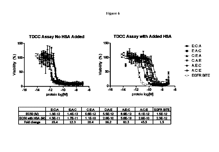
[0025] Figure 6 shows that all six possible configurations of a trispecific
molecule containing
an EGFR targeting VH domain can induce T cells to kill the human tumor cell
line NCI-1563.
The experiment was performed in the absence (left side) and presence (right
side) of human
serum albumin with an EGFR targeting BiTE as positive control.
[0026] Figure 7 assesses the ability of five possible configurations of a
trispecific molecule
containing a PSMA targeting VH domain to induce T cells to kill the human
tumor cell line
22Rv1. The experiment was performed in the absence (left side) and presence
(right side) of
human serum albumin with a PSMA targeting BiTE as positive control. Also shown
is the
activity of a PSMA targeting trispecific molecule with a PSMA targeting scFv.
-5-
CA 02986575 2017-11-20
WO 2016/187594 PCT/US2016/033644
[0027] Figure 8 shows that that the trispecific molecules can consist of a
constant core element
comprising an anti-CD3E single chain variable fragment (scFv) and an anti-HSA
variable heavy
chain region; and a variable target binding domain that can be a scFv.
[0028] Figure 9 demonstrates that trispecific molecules that use a fynomer as
opposed to an
antibody derived domain for tumor targeting can induce T cells to kill tumor
cells.
[0029] Figure 10 shows that when EGFR targeting trispecific molecules redirect
T cells to kill
human CaPan2 tumor cells (panel A), the T cells get activated and produce the
cytokines TNF-
a (panel B) and IFNy (panel C) in a manner dependent on the dose of the
trispecific.
[0030] Figure 11 shows that when PSMA targeting trispecific molecules redirect
T cells to kill
human 22Ry1 tumor cells (panel A), the T cells get activated and produce the
cytokines TNF-a
(panel B) and IFNy (panel C) in a manner dependent on the dose of the
trispecific.
[0031] Figure 12 shows that MSLN targeting trispecific molecules can migrate
through
matrigel faster than conventional antibodies.
[0032] Figure 13 shows phage titration on biotin-CD3E and biotin-HSA.
DETAILED DESCRIPTION OF THE INVENTION
[0033] Described herein are trispecific antigen-binding proteins,
pharmaceutical compositions
thereof, as well as nucleic acids, recombinant expression vectors and host
cells for making such
trispecific antigen-binding proteins. Also provided are methods of using the
disclosed trispecific
antigen-binding proteins in the prevention, and/or treatment of diseases,
conditions and
disorders. The trispecific antigen-binding proteins are capable of
specifically binding to a target
antigen as well as CD3 and a half-life extension domain, such as a domain
binding human serum
albumin (HSA). Figure 1 depicts one non-limiting example of a trispecific
antigen-binding
protein.
[0034] In one aspect, the trispecific antigen-binding proteins comprise a
domain (A) which
specifically binds to CD3, a domain (B) which specifically binds to human
serum albumin
(HSA), and a domain (C) which specifically binds to a target antigen. The
three domains in
trispecific antigen-binding proteins are arranged in any order. Thus, it is
contemplated that the
domain order of the trispecific antigen-binding proteins are:
H2N-(A)-(B)-(C)-COOH,
H2N-(A)-(C)-(B)-COOH,
H2N-(B)-(A)-(C)-COOH,
H2N-(B)-(C)-(A)-COOH,
H2N-(C)-(B)-(A)-COOH, or
H2N-(C)-(A)-(B)-COOH.
-6-
CA 02986575 2017-11-20
WO 2016/187594 PCT/US2016/033644
[0035] In some embodiments, the trispecific antigen-binding proteins have a
domain order of
H2N-(A)-(B)-(C)-COOH. In some embodiments, the trispecific antigen-binding
proteins have a
domain order of H2N-(A)-(C)-(B)-COOH. In some embodiments, the trispecific
antigen-
binding proteins have a domain order of H2N-(B)-(A)-(C)-COOH. In some
embodiments, the
trispecific antigen-binding proteins have a domain order of H2N-(B)-(C)-(A)-
COOH. In some
embodiments, the trispecific antigen-binding proteins have a domain order of
H2N-(C)-(B)-(A)-
COOH. In some embodiments, the trispecific antigen-binding proteins have a
domain order of
H2N-(C)-(A)-(B)-COOH.
[0036] Trispecific antigen-binding proteins described herein optionally
comprise a polypeptide
having a sequence described in Table 6 or Table 7 (SEQ ID NOS: 1-48) and
subsequences
thereof In some embodiments, the trispecific antigen binding protein comprises
a polypeptide
having at least 70%-95% or more homology to a sequence described in Table 6 or
Table 7 (SEQ
ID NOS: 1-48). In some embodiments, the trispecific antigen binding protein
comprises a
polypeptide having at least 70%, 75%, 80%, 85%, 90%, 95%, or more homology to
a sequence
described in Table 6 or Table 7 (SEQ ID NO: 1-48). In some embodiments, the
trispecific
antigen binding protein has a sequence comprising at least a portion of a
sequence described in
Table 6 or Table 7 (SEQ ID NOS: 1-48). In some embodiments, the trispecific
antigen-binding
protein comprises a polypeptide comprising one or more of the sequences
described in Table 6
or Table 7 (SEQ ID NOS: 1-48).
[0037] The trispecific antigen-binding proteins described herein are designed
to allow specific
targeting of cells expressing a target antigen by recruiting cytotoxic T
cells. This improves
efficacy compared to ADCC (antibody dependent cell-mediated cytotoxicity) ,
which is using
full length antibodies directed to a sole antigen and is not capable of
directly recruiting cytotoxic
T cells. In contrast, by engaging CD3 molecules expressed specifically on
these cells, the
trispecific antigen-binding proteins can crosslink cytotoxic T cells with
cells expressing a target
antigen in a highly specific fashion, thereby directing the cytotoxic
potential of the T cell
towards the target cell. The trispecific antigen-binding proteins described
herein engage
cytotoxic T cells via binding to the surface-expressed CD3 proteins, which
form part of the
TCR. Simultaneous binding of several trispecific antigen-binding protein to
CD3 and to a target
antigen expressed on the surface of particular cells causes T cell activation
and mediates the
subsequent lysis of the particular target antigen expressing cell. Thus,
trispecific antigen-
binding proteins are contemplated to display strong, specific and efficient
target cell killing. In
some embodiments, the trispecific antigen-binding proteins described herein
stimulate target cell
killing by cytotoxic T cells to eliminate pathogenic cells (e.g., tumor cells,
virally or bacterially
infected cells, autoreactive T cells, etc). In some of such embodiments, cells
are eliminated
-7-
CA 02986575 2017-11-20
WO 2016/187594 PCT/US2016/033644
selectively, thereby reducing the potential for toxic side effects. In other
embodiments, the same
polypeptides could be used to enhance the elimination of endogenous cells for
therapeutic effect,
such as B or T lymphocytes in autoimmune disease, or hematopoietic stem cells
(HSCs) for
stem cell transplantation.
[0038] The trispecific antigen-binding proteins described herein confer
further therapeutic
advantages over traditional monoclonal antibodies and other smaller bispecific
molecules.
Generally, the effectiveness of recombinant protein pharmaceuticals depends
heavily on the
intrinsic pharmacokinetics of the protein itself One such benefit here is that
the trispecific
antigen-binding proteins described herein have extended pharmacokinetic
elimination half-time
due to having a half-life extension domain such as a domain specific to HSA.
In this respect, the
trispecific antigen-binding proteins described herein have an extended serum
elimination half-
time of about two, three, about five, about seven, about 10, about 12, or
about 14 days in some
embodiments. This contrasts to other binding proteins such as BiTE or DART
molecules which
have relatively much shorter elimination half-times. For example, the BiTE
CD19xCD3
bispecific scFv-scFv fusion molecule requires continuous intravenous infusion
(i.v.) drug
delivery due to its short elimination half-time. The longer intrinsic half-
times of the trispecific
antigen-binding proteins solve this issue thereby allowing for increased
therapeutic potential
such as low-dose pharmaceutical formulations, decreased periodic
administration and/or novel
pharmaceutical compositions.
[0039] The trispecific antigen-binding proteins described herein also have an
optimal size for
enhanced tissue penetration and tissue distribution. Larger sizes limit or
prevent penetration or
distribution of the protein in the target tissues. The trispecific antigen-
binding proteins
described herein avoid this by having a small size that allows enhanced tissue
penetration and
distribution. Accordingly, the trispecific antigen-binding proteins described
herein, in some
embodiments have a size of about 50 kD to about 80 kD, about 50 kD to about 75
kD, about 50
kD to about 70 kD, or about 50 kD to about 65 kD. Thus, the size of the
trispecific antigen-
binding proteins is advantageous over IgG antibodies which are about 150 kD
and the BiTE and
DART diabody molecules which are about 55 kD but are not half-life extended
and therefore
cleared quickly through the kidney.
[0040] In further embodiments, the trispecific antigen-binding proteins
described herein have an
optimal size for enhanced tissue penetration and distribution. In these
embodiments, the
trispecific antigen-binding proteins are constructed to be as small as
possible, while retaining
specificity toward its targets. Accordingly, in these embodiments, the
trispecific antigen-
binding proteins described herein have a size of about 20 kD to about 40 kD or
about 25 kD to
about 35 kD to about 40 kD, to about 45 kD, to about 50 kD, to about 55 kD, to
about 60 kD, to
-8-
CA 02986575 2017-11-20
WO 2016/187594 PCT/US2016/033644
about 65 kD. In some embodiments, the trispecific antigen-binding proteins
described herein
have a size of about 50kD, 49, kD, 48 kD, 47 kD, 46 kD, 45 kD, 44 kD, 43 kD,
42 kD, 41 kD,
40 kD, about 39 kD, about 38 kD, about 37 kD, about 36 kD, about 35 kD, about
34 kD, about
33 kD, about 32 kD, about 31 kD, about 30 kD, about 29 kD, about 28 kD, about
27 kD, about
26 kD, about 25 kD, about 24 kD, about 23 kD, about 22 kD, about 21 kD, or
about 20 kD. An
exemplary approach to the small size is through the use of single domain
antibody (sdAb)
fragments for each of the domains. For example, a particular trispecific
antigen-binding protein
has an anti-CD3 sdAb, anti-HSA sdAb and an sdAb for a target antigen. This
reduces the size
of the exemplary trispecific antigen-binding protein to under 40 kD. Thus in
some
embodiments, the domains of the trispecific antigen-binding proteins are all
single domain
antibody (sdAb) fragments. In other embodiments, the trispecific antigen-
binding proteins
described herein comprise small molecule entity (SME) binders for HSA and/or
the target
antigen. SME binders are small molecules averaging about 500 to 2000 Da in
size and are
attached to the trispecific antigen-binding proteins by known methods, such as
sortase ligation
or conjugation. In these instances, one of the domains of a trispecific
antigen-binding protein is
a sortase recognition sequence, e.g., LPETG (SEQ ID NO: 57). To attach a SME
binder to a
trispecific antigen-binding protein with a sortase recognition sequence, the
protein is incubated
with a sortase and a SME binder whereby the sortase attaches the SME binder to
the recognition
sequence. Known SME binders include MIP-1072 and MIP-1095 which bind to
prostate-
specific membrane antigen (PSMA). In yet other embodiments, the domain which
binds to a
target antigen of a trispecific antigen-binding proteins described herein
comprise a knottin
peptide for binding a target antigen. Knottins are disufide-stabilized
peptides with a cysteine
knot scaffold and have average sizes about 3.5 kD. Knottins have been
contemplated for
binding to certain tumor molecules such as fibronectin and VEGF-receptor. In
further
embodiments, domain which binds to a target antigen of a trispecific antigen-
binding proteins
described herein comprise a natural receptor ligand such as B-cell activating
factor
(BAFF/BLyS).
[0041] Another feature of the trispecific antigen-binding proteins described
herein is that they
are of a single-polypeptide design with flexible linkage of their domains.
This allows for facile
production and manufacturing of the trispecific antigen-binding proteins as
they can be encoded
by single cDNA molecule to be easily incorporated into a vector. Further,
because the
trispecific antigen-binding proteins described herein are a monomeric single
polypeptide chain,
there are no chain pairing issues or a requirement for dimerization. It is
contemplated that the
trispecific antigen-binding proteins described herein have a reduced tendency
to aggregate
-9-
CA 02986575 2017-11-20
WO 2016/187594 PCT/US2016/033644
unlike other reported molecules such as bispecific proteins with Fc-gamma
immunoglobulin
domains.
[0042] In the trispecific antigen-binding proteins described herein, the
domains are linked by
internal linkers Li and L2, where Li links the first and second domain of the
trispecific antigen-
binding proteins and L2 links the second and third domains of the trispecific
antigen-binding
proteins. Linkers Li and L2 have an optimized length and/or amino acid
composition. In some
embodiments, linkers Li and L2 are the same length and amino acid composition.
In other
embodiments, Li and L2 are different. In certain embodiments, internal linkers
Li and/or L2
are "short", i.e., consist of 0, 1, 2, 3, 4, 5, 6, 7, 8, 9, 10, 11 or 12 amino
acid residues. Thus, in
certain instances, the internal linkers consist of about 12 or less amino acid
residues. In the case
of 0 amino acid residues, the internal linker is a peptide bond. In certain
embodiments, internal
linkers Li and/or L2 are "long", i.e., consist of 15, 20 or 25 amino acid
residues. In some
embodiments, these internal linkers consist of about 3 to about 15, for
example 8, 9 or 10
contiguous amino acid residues. Regarding the amino acid composition of the
internal linkers
Li and L2, peptides are selected with properties that confer flexibility to
the trispecific antigen-
binding proteins, do not interfere with the binding domains as well as resist
cleavage from
proteases. For example, glycine and serine residues generally provide protease
resistance.
Examples of internal linkers suitable for linking the domains in the
trispecific antigen-binding
proteins include but are not limited to (GS)õ (SEQ ID NO: 49), (GGS)õ (SEQ ID
NO: 50),
(GGGS)õ (SEQ ID NO: Si), (GGSG)õ (SEQ ID NO: 52), (GGSGG)õ (SEQ ID NO: 53), or
(GGGGS)õ (SEQ ID NO: 54), wherein n is 1, 2, 3, 4, 5, 6, 7, 8, 9, or 10. In
one embodiment,
internal linker Li and/or L2 is (GGGGS)4 (SEQ ID NO: 55) or (GGGGS)3 (SEQ ID
NO: 56).
CD3 Binding Domain
[0043] The specificity of the response of T cells is mediated by the
recognition of antigen
(displayed in context of a major histocompatibility complex, MHC) by the TCR.
As part of the
TCR, CD3 is a protein complex that includes a CD3y (gamma) chain, a CD3 6
(delta) chain, and
two CD3E (epsilon) chains which are present on the cell surface. CD3
associates with the a
(alpha) and 0 (beta) chains of the TCR as well as CD3 (zeta) altogether to
comprise the
complete TCR. Clustering of CD3 on T cells, such as by immobilized anti-CD3
antibodies leads
to T cell activation similar to the engagement of the T cell receptor but
independent of its clone-
typical specificity.
[0044] In one aspect, the trispecific antigen-binding proteins described
herein comprise a
domain which specifically binds to CD3. In one aspect, the trispecific antigen-
binding proteins
described herein comprise a domain which specifically binds to human CD3. In
some
-10-
CA 02986575 2017-11-20
WO 2016/187594 PCT/US2016/033644
embodiments, the trispecific antigen-binding proteins described herein
comprise a domain which
specifically binds to CD3y. In some embodiments, the trispecific antigen-
binding proteins
described herein comprise a domain which specifically binds to CD3. In some
embodiments,
the trispecific antigen-binding proteins described herein comprise a domain
which specifically
binds to CD3E.
[0045] In further embodiments, the trispecific antigen-binding proteins
described herein
comprise a domain which specifically binds to the TCR. In certain instances,
the trispecific
antigen-binding proteins described herein comprise a domain which specifically
binds the a
chain of the TCR. In certain instances, the trispecific antigen-binding
proteins described herein
comprise a domain which specifically binds the 0 chain of the TCR.
[0046] In certain embodiments, the CD3 binding domain of the trispecific
antigen-binding
proteins described herein exhibit not only potent CD3 binding affinities with
human CD3, but
show also excellent crossreactivity with the respective cynomolgus monkey CD3
proteins. In
some instances, the CD3 binding domain of the tri specific antigen-binding
proteins are cross-
reactive with CD3 from cynomolgus monkey. In certain instances,
human:cynomolgous KD
ratios for CD3 are between 5 and 0.2.
[0047] In some embodiments, the CD3 binding domain of the trispecific antigen-
binding protein
can be any domain that binds to CD3 including but not limited to domains from
a monoclonal
antibody, a polyclonal antibody, a recombinant antibody, a human antibody, a
humanized
antibody. In some instances, it is beneficial for the CD3 binding domain to be
derived from the
same species in which the trispecific antigen-binding protein will ultimately
be used in. For
example, for use in humans, it may be beneficial for the CD3 binding domain of
the trispecific
antigen-binding protein to comprise human or humanized residues from the
antigen binding
domain of an antibody or antibody fragment.
[0048] Thus, in one aspect, the antigen-binding domain comprises a humanized
or human
antibody or an antibody fragment, or a murine antibody or antibody fragment.
In one
embodiment, the humanized or human anti-CD3 binding domain comprises one or
more (e.g.,
all three) light chain complementary determining region 1 (LC CDR1), light
chain
complementary determining region 2 (LC CDR2), and light chain complementary
determining
region 3 (LC CDR3) of a humanized or human anti- CD3 binding domain described
herein,
and/or one or more (e.g., all three) heavy chain complementary determining
region 1 (HC
CDR1), heavy chain complementary determining region 2 (HC CDR2), and heavy
chain
complementary determining region 3 (HC CDR3) of a humanized or human anti-CD3
binding
domain described herein, e.g., a humanized or human anti-CD3 binding domain
comprising one
or more, e.g., all three, LC CDRs and one or more, e.g., all three, HC CDRs.
-11-
CA 02986575 2017-11-20
WO 2016/187594 PCT/US2016/033644
[0049] In some embodiments, the humanized or human anti-CD3 binding domain
comprises a
humanized or human light chain variable region specific to CD3 where the light
chain variable
region specific to CD3 comprises human or non-human light chain CDRs in a
human light chain
framework region. In certain instances, the light chain framework region is a
X, (lamda) light
chain framework. In other instances, the light chain framework region is a lc
(kappa) light chain
framework.
[0050] In some embodiments, the humanized or human anti-CD3 binding domain
comprises a
humanized or human heavy chain variable region specific to CD3 where the heavy
chain
variable region specific to CD3 comprises human or non-human heavy chain CDRs
in a human
heavy chain framework region.
[0051] In certain instances, the complementary determining regions of the
heavy chain and/or
the light chain are derived from known anti-CD3 antibodies, such as, for
example, muromonab-
CD3 (OKT3), otelixizumab (TRX4), teplizumab (MGA031), visilizumab (Nuvion),
SP34, TR-
66 or X35-3, VIT3, BMA030 (BW264/56), CLB-T3/3, CRIS7, YTH12.5, F111-409, CLB-
T3.4.2, TR-66, WT32, SPv-T3b, 11D8, XIII-141, XIII-46, XIII-87, 12F6, T3/RW2-
8C8,
T3/RW2-4B6, OKT3D, M-T301, SMC2, F101.01, UCHT-1 and WT-31.
[0052] In one embodiment, the anti-CD3 binding domain is a single chain
variable fragment
(scFv) comprising a light chain and a heavy chain of an amino acid sequence
provided herein.
As used herein, "single chain variable fragment" or "scFv" refers to an
antibody fragment
comprising a variable region of a light chain and at least one antibody
fragment comprising a
variable region of a heavy chain, wherein the light and heavy chain variable
regions are
contiguously linked via a short flexible polypeptide linker, and capable of
being expressed as a
single polypeptide chain, and wherein the scFv retains the specificity of the
intact antibody from
which it is derived. In an embodiment, the anti-CD3 binding domain comprises:
a light chain
variable region comprising an amino acid sequence having at least one, two or
three
modifications (e.g., substitutions) but not more than 30, 20 or 10
modifications (e.g.,
substitutions) of an amino acid sequence of a light chain variable region
provided herein, or a
sequence with 95-99% identity with an amino acid sequence provided herein;
and/or a heavy
chain variable region comprising an amino acid sequence having at least one,
two or three
modifications (e.g., substitutions) but not more than 30, 20 or 10
modifications (e.g.,
substitutions) of an amino acid sequence of a heavy chain variable region
provided herein, or a
sequence with 95-99% identity to an amino acid sequence provided herein. In
one embodiment,
the humanized or human anti-CD3 binding domain is a scFv, and a light chain
variable region
comprising an amino acid sequence described herein, is attached to a heavy
chain variable
region comprising an amino acid sequence described herein, via a scFv linker.
The light chain
-12-
CA 02986575 2017-11-20
WO 2016/187594 PCT/US2016/033644
variable region and heavy chain variable region of a scFv can be, e.g., in any
of the following
orientations: light chain variable region- scFv linker-heavy chain variable
region or heavy chain
variable region- scFv linker-light chain variable region.
[0053] In some instances, scFvs which bind to CD3 are prepared according to
known methods.
For example, scFv molecules can be produced by linking VH and VL regions
together using
flexible polypeptide linkers. The scFv molecules comprise a scFv linker (e.g.,
a Ser-Gly linker)
with an optimized length and/or amino acid composition. Accordingly, in some
embodiments,
the length of the scFv linker is such that the VH or VL domain can associate
intermolecularly
with the other variable domain to form the CD3 binding site. In certain
embodiments, such scFv
linkers are "short", i.e. consist of 0, 1, 2, 3, 4, 5, 6, 7, 8, 9, 10, 11 or
12 amino acid residues.
Thus, in certain instances, the scFv linkers consist of about 12 or less amino
acid residues. In
the case of 0 amino acid residues, the scFv linker is a peptide bond. In some
embodiments,
these scFv linkers consist of about 3 to about 15, for example 8, 9 or 10
contiguous amino acid
residues. Regarding the amino acid composition of the scFv linkers, peptides
are selected that
confer flexibility, do not interfere with the variable domains as well as
allow inter-chain folding
to bring the two variable domains together to form a functional CD3 binding
site. For example,
scFv linkers comprising glycine and serine residues generally provide protease
resistance. In
some embodiments, linkers in a scFv comprise glycine and serine residues. The
amino acid
sequence of the scFv linkers can be optimized, for example, by phage-display
methods to
improve the CD3 binding and production yield of the scFv. Examples of peptide
scFv linkers
suitable for linking a variable light chain domain and a variable heavy chain
domain in a scFv
include but are not limited to (GS) n (SEQ ID NO: 49), (GGS)n (SEQ ID NO: 50),
(GGGS)n
(SEQ ID NO: 51), (GGSG)n (SEQ ID NO: 52), (GGSGG)n (SEQ ID NO: 53), or
(GGGGS)n
(SEQ ID NO: 54), wherein n is 1, 2, 3, 4, 5, 6, 7, 8, 9, or 10. In one
embodiment, the scFv linker
can be (GGGGS)4 (SEQ ID NO: 55) or (GGGGS)3 (SEQ ID NO: 56). Variation in the
linker
length may retain or enhance activity, giving rise to superior efficacy in
activity studies.
[0054] In some embodiments, CD3 binding domain of a trispecific antigen-
binding protein has
an affinity to CD3 on CD3 expressing cells with a KD of 1000 nM or less, 500
nM or less, 200
nM or less, 100 nM or less, 80 nM or less, 50 nM or less, 20 nM or less, 10 nM
or less, 5 nM or
less, 1 nM or less, or 0.5 nM or less. In some embodiments, the CD3 binding
domain of a
trispecific antigen-binding protein has an affinity to CD3c, y, or 6 with a
KID of 1000 nM or less,
500 nM or less, 200 nM or less, 100 nM or less, 80 nM or less, 50 nM or less,
20 nM or less, 10
nM or less, 5 nM or less, 1 nM or less, or 0.5 nM or less. In further
embodiments, CD3 binding
domain of a trispecific antigen-binding protein has low affinity to CD3, i.e.,
about 100 nM or
greater.
-13-
CA 02986575 2017-11-20
WO 2016/187594 PCT/US2016/033644
[0055] The affinity to bind to CD3 can be determined, for example, by the
ability of the
trispecific antigen-binding protein itself or its CD3 binding domain to bind
to CD3 coated on an
assay plate; displayed on a microbial cell surface; in solution; etc. The
binding activity of the
trispecific antigen-binding protein itself or its CD3 binding domain of the
present disclosure to
CD3 can be assayed by immobilizing the ligand (e.g., CD3) or the trispecific
antigen-binding
protein itself or its CD3 binding domain, to a bead, substrate, cell, etc.
Agents can be added in
an appropriate buffer and the binding partners incubated for a period of time
at a given
temperature. After washes to remove unbound material, the bound protein can be
released with,
for example, SDS, buffers with a high pH, and the like and analyzed, for
example, by Surface
Plasmon Resonance (SPR).
Half-Life Extension Domain
[0056] Contemplated herein are domains which extend the half-life of an
antigen-binding
domain. Such domains are contemplated to include but are not limited to HSA
binding domains,
Fc domains, small molecules, and other half-life extension domains known in
the art.
[0057] Human serum albumin (HSA) (molecular mass ¨67 kDa) is the most abundant
protein in
plasma, present at about 50 mg/ml (60011M), and has a half-life of around 20
days in humans.
HSA serves to maintain plasma pH, contributes to colloidal blood pressure,
functions as carrier
of many metabolites and fatty acids, and serves as a major drug transport
protein in plasma.
[0058] Noncovalent association with albumin extends the elimination half-time
of short lived
proteins. For example, a recombinant fusion of an albumin binding domain to a
Fab fragment
resulted in an in vivo clearance of 25- and 58-fold and a half-life extension
of 26- and 37-fold
when administered intravenously to mice and rabbits respectively as compared
to the
administration of the Fab fragment alone. In another example, when insulin is
acylated with
fatty acids to promote association with albumin, a protracted effect was
observed when injected
subcutaneously in rabbits or pigs. Together, these studies demonstrate a
linkage between
albumin binding and prolonged action.
[0059] In one aspect, the trispecific antigen-binding proteins described
herein comprise a half-
life extension domain, for example a domain which specifically binds to HSA.
In some
embodiments, the HSA binding domain of a trispecific antigen-binding protein
can be any
domain that binds to HSA including but not limited to domains from a
monoclonal antibody, a
polyclonal antibody, a recombinant antibody, a human antibody, a humanized
antibody. In
some embodiments, the HSA binding domain is a single chain variable fragments
(scFv), single-
domain antibody such as a heavy chain variable domain (VH), a light chain
variable domain
(VL) and a variable domain (VHH) of camelid derived single domain antibody,
peptide, ligand
-14-
CA 02986575 2017-11-20
WO 2016/187594 PCT/US2016/033644
or small molecule entity specific for HSA. In certain embodiments, the HSA
binding domain is
a single-domain antibody. In other embodiments, the HSA binding domain is a
peptide. In
further embodiments, the HSA binding domain is a small molecule. It is
contemplated that the
HSA binding domain of a trispecific antigen-binding protein is fairly small
and no more than 25
kD, no more than 20 kD, no more than 15 kD, or no more than 10 kD in some
embodiments. In
certain instances, the HSA binding is 5 kD or less if it is a peptide or small
molecule entity.
[0060] The half-life extension domain of a trispecific antigen-binding protein
provides for
altered pharmacodynamics and pharmacokinetics of the trispecific antigen-
binding protein itself.
As above, the half-life extension domain extends the elimination half-time.
The half-life
extension domain also alters pharmacodynamic properties including alteration
of tissue
distribution, penetration, and diffusion of the trispecific antigen-binding
protein. In some
embodiments, the half-life extension domain provides for improved tissue
(including tumor)
targeting, tissue distribution, tissue penetration, diffusion within the
tissue, and enhanced
efficacy as compared with a protein without an half-life extension domain. In
one embodiment,
therapeutic methods effectively and efficiently utilize a reduced amount of
the trispecific
antigen-binding protein, resulting in reduced side effects, such as reduced
non-tumor cell
cytotoxicity.
[0061] Further, the binding affinity of the half-life extension domain can be
selected so as to
target a specific elimination half-time in a particular trispecific antigen-
binding protein. Thus, in
some embodiments, the half-life extension domain has a high binding affinity.
In other
embodiments, the half-life extension domain has a medium binding affinity. In
yet other
embodiments, the half-life extension domain has a low or marginal binding
affinity. Exemplary
binding affinities include KD concentrations at 10 nM or less (high), between
10 nM and 100 nM
(medium), and greater than 100 nM (low). As above, binding affinities to HSA
are determined
by known methods such as Surface Plasmon Resonance (SPR).
Target Antigen Binding Domain
[0062] In addition to the described CD3 and half-life extension domains, the
trispecific antigen-
binding proteins described herein also comprise a domain that binds to a
target antigen. A target
antigen is involved in and/or associated with a disease, disorder or
condition. In particular, a
target antigen associated with a proliferative disease, a tumorous disease, an
inflammatory
disease, an immunological disorder, an autoimmune disease, an infectious
disease, a viral
disease, an allergic reaction, a parasitic reaction, a graft-versus-host
disease or a host-versus-
graft disease. In some embodiments, a target antigen is a tumor antigen
expressed on a tumor
-15-
CA 02986575 2017-11-20
WO 2016/187594 PCT/US2016/033644
cell. Alternatively in some embodiments, a target antigen is associated with a
pathogen such as
a virus or bacterium.
[0063] In some embodiments, a target antigen is a cell surface molecule such
as a protein, lipid
or polysaccharide. In some embodiments, a target antigen is a on a tumor cell,
virally infected
cell, bacterially infected cell, damaged red blood cell, arterial plaque cell,
or fibrotic tissue cell.
[0064] The design of the trispecific antigen-binding proteins described herein
allows the binding
domain to a target antigen to be flexible in that the binding domain to a
target antigen can be any
type of binding domain, including but not limited to, domains from a
monoclonal antibody, a
polyclonal antibody, a recombinant antibody, a human antibody, a humanized
antibody. In
some embodiments, the binding domain to a target antigen is a single chain
variable fragments
(scFv), single-domain antibody such as a heavy chain variable domain (VH), a
light chain
variable domain (VL) and a variable domain (VHH) of camelid derived single
domain antibody.
In other embodiments, the binding domain to a target antigen is a non-Ig
binding domain, i.e.,
antibody mimetic, such as anticalins, affilins, affibody molecules, affimers,
affitins, alphabodies,
avimers, DARPins, fynomers, kunitz domain peptides, and monobodies. In further
embodiments, the binding domain to a target antigen is a ligand or peptide
that binds to or
associates with a target antigen. In yet further embodiments, the binding
domain to a target
antigen is a knottin. In yet further embodiments, the binding domain to a
target antigen is a
small molecular entity.
Trispecific Protein Modifications
[0065] The trispecific antigen-binding proteins described herein encompass
derivatives or
analogs in which (i) an amino acid is substituted with an amino acid residue
that is not one
encoded by the genetic code, (ii) the mature polypeptide is fused with another
compound such as
polyethylene glycol, or (iii) additional amino acids are fused to the protein,
such as a leader or
secretory sequence or a sequence for purification of the protein.
[0066] Typical modifications include, but are not limited to, acetylation,
acylation, ADP-
ribosylation, amidation, covalent attachment of flavin, covalent attachment of
a heme moiety,
covalent attachment of a nucleotide or nucleotide derivative, covalent
attachment of a lipid or
lipid derivative, covalent attachment of phosphatidylinositol, cross-linking,
cyclization, disulfide
bond formation, demethylation, formation of covalent crosslinks, formation of
cystine,
formation of pyroglutamate, formylation, gamma carboxylation, glycosylation,
GPI anchor
formation, hydroxylation, iodination, methylation, myristoylation, oxidation,
proteolytic
processing, phosphorylation, prenylation, racemizati on, selenoylation,
sulfati on, transfer-RNA
mediated addition of amino acids to proteins such as arginylation, and
ubiquitination.
-16-
CA 02986575 2017-11-20
WO 2016/187594 PCT/US2016/033644
[0067] Modifications are made anywhere in trispecific antigen-binding proteins
described
herein, including the peptide backbone, the amino acid side-chains, and the
amino or carboxyl
termini. Certain common peptide modifications that are useful for modification
of trispecific
antigen-binding proteins include glycosylation, lipid attachment, sulfation,
gamma-
carboxylation of glutamic acid residues, hydroxylation, blockage of the amino
or carboxyl group
in a polypeptide, or both, by a covalent modification, and ADP-ribosylation.
Polynucleotides Encoding Trispecific Antigen-Binding Proteins
[0068] Also provided, in some embodiments, are polynucleotide molecules
encoding a
trispecific antigen-binding protein described herein. In some embodiments, the
polynucleotide
molecules are provided as a DNA construct. In other embodiments, the
polynucleotide
molecules are provided as a messenger RNA transcript.
[0069] The polynucleotide molecules are constructed by known methods such as
by combining
the genes encoding the three binding domains either separated by peptide
linkers or, in other
embodiments, directly linked by a peptide bond, into a single genetic
construct operably linked
to a suitable promoter, and optionally a suitable transcription terminator,
and expressing it in
bacteria or other appropriate expression system such as, for example CHO
cells. In the
embodiments where the target antigen binding domain is a small molecule, the
polynucleotides
contain genes encoding the CD3 binding domain and the half-life extension
domain. In the
embodiments where the half-life extension domain is a small molecule, the
polynucleotides
contain genes encoding the domains that bind to CD3 and the target antigen.
Depending on the
vector system and host utilized, any number of suitable transcription and
translation elements,
including constitutive and inducible promoters, may be used. The promoter is
selected such that
it drives the expression of the polynucleotide in the respective host cell.
[0070] In some embodiments, the polynucleotide is inserted into a vector,
preferably an
expression vector, which represents a further embodiment. This recombinant
vector can be
constructed according to known methods. Vectors of particular interest include
plasmids,
phagemids, phage derivatives, virii (e.g., retroviruses, adenoviruses, adeno-
associated viruses,
herpes viruses, lentiviruses, and the like), and cosmids.
[0071] A variety of expression vector/host systems may be utilized to contain
and express the
polynucleotide encoding the polypeptide of the described trispecific antigen-
binding protein.
Examples of expression vectors for expression in E. coli are pSKK (Le Gall et
al., J Immunol
Methods. (2004) 285(1):111-27) or pcDNA5 (Invitrogen) for expression in
mammalian cells.
[0072] Thus, the trispecific antigen-binding proteins as described herein, in
some embodiments,
are produced by introducing a vector encoding the protein as described above
into a host cell
-17-
CA 02986575 2017-11-20
WO 2016/187594 PCT/US2016/033644
and culturing said host cell under conditions whereby the protein domains are
expressed, may be
isolated and, optionally, further purified.
Pharmaceutical Compositions
[0073] Also provided, in some embodiments, are pharmaceutical compositions
comprising a
trispecific antigen-binding protein described herein, a vector comprising the
polynucleotide
encoding the polypeptide of the trispecific antigen-binding proteins or a host
cell transformed by
this vector and at least one pharmaceutically acceptable carrier. The term
"pharmaceutically
acceptable carrier" includes, but is not limited to, any carrier that does not
interfere with the
effectiveness of the biological activity of the ingredients and that is not
toxic to the patient to
whom it is administered. Examples of suitable pharmaceutical carriers are well
known in the art
and include phosphate buffered saline solutions, water, emulsions, such as
oil/water emulsions,
various types of wetting agents, sterile solutions etc. Such carriers can be
formulated by
conventional methods and can be administered to the subject at a suitable
dose. Preferably, the
compositions are sterile. These compositions may also contain adjuvants such
as preservative,
emulsifying agents and dispersing agents. Prevention of the action of
microorganisms may be
ensured by the inclusion of various antibacterial and antifungal agents.
[0074] In some embodiments of the pharmaceutical compositions, the trispecific
antigen-
binding protein described herein is encapsulated in nanoparticles. In some
embodiments, the
nanoparticles are fullerenes, liquid crystals, liposome, quantum dots,
superparamagnetic
nanoparticles, dendrimers, or nanorods. In other embodiments of the
pharmaceutical
compositions, the trispecific antigen-binding protein is attached to
liposomes. In some instances,
the trispecific antigen-binding protein are conjugated to the surface of
liposomes. In some
instances, the trispecific antigen-binding protein are encapsulated within the
shell of a liposome.
In some instances, the liposome is a cationic liposome.
[0075] The trispecific antigen-binding proteins described herein are
contemplated for use as a
medicament. Administration is effected by different ways, e.g. by intravenous,
intraperitoneal,
subcutaneous, intramuscular, topical or intradermal administration. In some
embodiments, the
route of administration depends on the kind of therapy and the kind of
compound contained in
the pharmaceutical composition. The dosage regimen will be determined by the
attending
physician and other clinical factors. Dosages for any one patient depends on
many factors,
including the patient's size, body surface area, age, sex, the particular
compound to be
administered, time and route of administration, the kind of therapy, general
health and other
drugs being administered concurrently. An "effective dose" refers to amounts
of the active
-18-
CA 02986575 2017-11-20
WO 2016/187594 PCT/US2016/033644
ingredient that are sufficient to affect the course and the severity of the
disease, leading to the
reduction or remission of such pathology and may be determined using known
methods.
Methods of treatment
[0076] Also provided herein, in some embodiments, are methods and uses for
stimulating the
immune system of an individual in need thereof comprising administration of a
trispecific
antigen-binding protein described herein. In some instances, the
administration of a trispecific
antigen-binding protein described herein induces and/or sustains cytotoxicity
towards a cell
expressing a target antigen. In some instances, the cell expressing a target
antigen is a cancer or
tumor cell, a virally infected cell, a bacterially infected cell, an
autoreactive T or B cell,
damaged red blood cells, arterial plaques, or fibrotic tissue.
[0077] Also provided herein are methods and uses for a treatment of a disease,
disorder or
condition associated with a target antigen comprising administering to an
individual in need
thereof a trispecific antigen-binding protein described herein. Diseases,
disorders or conditions
associated with a target antigen include, but are not limited to, viral
infection, bacterial infection,
auto-immune disease, transplant rejection, atherosclerosis, or fibrosis. In
other embodiments,
the disease, disorder or condition associated with a target antigen is a
proliferative disease, a
tumorous disease, an inflammatory disease, an immunological disorder, an
autoimmune disease,
an infectious disease, a viral disease, an allergic reaction, a parasitic
reaction, a graft-versus-host
disease or a host-versus-graft disease. In one embodiment, the disease,
disorder or condition
associated with a target antigen is cancer. In one instance, the cancer is a
hematological cancer.
In another instance, the cancer is a solid tumor cancer.
[0078] As used herein, in some embodiments, "treatment" or "treating" or
"treated" refers to
therapeutic treatment wherein the object is to slow (lessen) an undesired
physiological condition,
disorder or disease, or to obtain beneficial or desired clinical results. For
the purposes described
herein, beneficial or desired clinical results include, but are not limited
to, alleviation of
symptoms; diminishment of the extent of the condition, disorder or disease;
stabilization (i.e.,
not worsening) of the state of the condition, disorder or disease; delay in
onset or slowing of the
progression of the condition, disorder or disease; amelioration of the
condition, disorder or
disease state; and remission (whether partial or total), whether detectable or
undetectable, or
enhancement or improvement of the condition, disorder or disease. Treatment
includes eliciting
a clinically significant response without excessive levels of side effects.
Treatment also includes
prolonging survival as compared to expected survival if not receiving
treatment. In other
embodiments, "treatment" or "treating" or "treated" refers to prophylactic
measures, wherein the
object is to delay onset of or reduce severity of an undesired physiological
condition, disorder or
-19-
CA 02986575 2017-11-20
WO 2016/187594 PCT/US2016/033644
disease, such as, for example is a person who is predisposed to a disease
(e.g., an individual who
carries a genetic marker for a disease such as breast cancer).
[0079] In some embodiments of the methods described herein, the trispecific
antigen-binding
proteins are administered in combination with an agent for treatment of the
particular disease,
disorder or condition. Agents include but are not limited to, therapies
involving antibodies,
small molecules (e.g., chemotherapeutics), hormones (steroidal, peptide, and
the like),
radiotherapies (y-rays, X-rays, and/or the directed delivery of radioisotopes,
microwaves, UV
radiation and the like), gene therapies (e.g., antisense, retroviral therapy
and the like) and other
immunotherapies. In some embodiments, the trispecific antigen-binding proteins
are
administered in combination with anti-diarrheal agents, anti-emetic agents,
analgesics, opioids
and/or non-steroidal anti-inflamatory agents. In some embodiments, the
trispecific antigen-
binding proteins are administered before, during, or after surgery.
Certain Definitions
[0080] As used herein, "elimination half-time" is used in its ordinary sense,
as is described in
Goodman and Gillman 's The Pharmaceutical Basis of Therapeutics 21-25 (Alfred
Goodman
Gilman, Louis S. Goodman, and Alfred Gilman, eds., 6th ed. 1980). Briefly, the
term is meant to
encompass a quantitative measure of the time course of drug elimination. The
elimination of
most drugs is exponential (i.e., follows first-order kinetics), since drug
concentrations usually do
not approach those required for saturation of the elimination process. The
rate of an exponential
process may be expressed by its rate constant, k, which expresses the
fractional change per unit
of time, or by its half-time, t112 the time required for 50% completion of the
process. The units of
these two constants are time' andtime, respectively. A first-order rate
constant and the half-
time of the reaction are simply related (kxtu2=0.693) and may be interchanged
accordingly.
Since first-order elimination kinetics dictates that a constant fraction of
drug is lost per unit time,
a plot of the log of drug concentration versus time is linear at all times
following the initial
distribution phase (i.e. after drug absorption and distribution are complete).
The half-time for
drug elimination can be accurately determined from such a graph.
EXAMPLES
Example 1: Construction of an Exemplary Trispecific Antigen-binding Protein to
CD20
Generation of a scFv CD3 binding domain
[0081] The human CD3E chain canonical sequence is Uniprot Accession No.
P07766. The
human CD3y chain canonical sequence is Uniprot Accession No. P09693. The human
CD36
chain canonical sequence is Uniprot Accession No. P043234. Antibodies against
CD3E, CD3y
or CD36 are generated via known technologies such as affinity maturation.
Where murine anti-
-20-
CA 02986575 2017-11-20
WO 2016/187594 PCT/US2016/033644
CD3 antibodies are used as a starting material, humanization of murine anti-
CD3 antibodies is
desired for the clinical setting, where the mouse-specific residues may induce
a human-anti-
mouse antigen (HAMA) response in subjects who receive treatment of a
trispecific antigen-
binding protein described herein. Humanization is accomplished by grafting CDR
regions from
murine anti-CD3 antibody onto appropriate human germline acceptor frameworks,
optionally
including other modifications to CDR and/or framework regions. As provided
herein, antibody
and antibody fragment residue numbering follows Kabat (Kabat E. A. et al,
1991; Chothia et al,
1987).
[0082] Human or humanized anti-CD3 antibodies are therefore used to generate
scFv sequences
for CD3 binding domains of a trispecific antigen-binding protein. DNA
sequences coding for
human or humanized VL and VH domains are obtained, and the codons for the
constructs are,
optionally, optimized for expression in cells from Homo sapiens. The order in
which the VL
and VH domains appear in the scFv is varied (i.e., VL-VH, or VH-VL
orientation), and three
copies of the "G4S" (SEQ ID NO: 58) or "G45" (SEQ ID NO: 58) subunit (G45)3
(SEQ ID NO:
56) connect the variable domains to create the scFv domain. Anti-CD3 scFv
plasmid constructs
can have optional Flag, His or other affinity tags, and are electroporated
into HEK293 or other
suitable human or mammalian cell lines and purified. Validation assays include
binding
analysis by FACS, kinetic analysis using Proteon, and staining of CD3-
expressing cells.
Generation of a scFv CD20 binding domain
[0083] CD20 is one of the cell surface proteins present on B-lymphocytes. CD20
antigen is
found in normal and malignant pre-B and mature B lymphocytes, including those
in over 90% of
B-cell non-Hodgkin's lymphomas (NHL). The antigen is absent in hematopoetic
stem cells,
activated B lymphocytes (plasma cells) and normal tissue. As such, several
antibodies mostly of
murine origin have been described: 1F5, 2B8/C2B8, 2H7, and 1H4.
[0084] A scFv binding domain to CD20 is generated similarly to the above
method for
generation of a scFv binding domain to CD3.
Cloning of DNA expression constructs encoding the trispecific antigen-binding
protein
[0085] The anti-CD3 scFv domains are used to construct a trispecific antigen-
binding protein in
combination with an anti-CD20 scFv domain and a HSA binding domain (e.g, a
peptide or VH
domain), with the domains organized as shown Figure!. For expression of a
trispecific
antigen-binding protein in CHO cells, coding sequences of all protein domains
are cloned into a
mammalian expression vector system. In brief, gene sequences encoding the CD3
binding
domain, HSA binding domain, and CD20 binding domain along with peptide linkers
Li and L2
are separately synthesized and subcloned. The resulting constructs are then
ligated together in
-21-
CA 02986575 2017-11-20
WO 2016/187594 PCT/US2016/033644
the order of `CD20 binding domain ¨ Li ¨ CD3 binding domain ¨ L2 ¨ HSA binding
domain' to
yield a final construct. All expression constructs are designed to contain
coding sequences for
an N-terminal signal peptide and a C-terminal hexahistidine (6xHis)-tag (SEQ
ID NO: 59) to
facilitate protein secretion and purification, respectively.
Expression of trispecific antigen-binding proteins in stably transfected CHO
cells
[0086] A CHO cell expression system (Flp-In , Life Technologies), a derivative
of CHO-Kl
Chinese Hamster ovary cells (ATCC, CCL-61) (Kao and Puck, Proc. Natl. Acad Sci
USA
1968;60(4):1275-81), is used. Adherent cells are subcultured according to
standard cell culture
protocols provided by Life Technologies.
[0087] For adaption to growth in suspension, cells are detached from tissue
culture flasks and
placed in serum-free medium. Suspension-adapted cells are cryopreserved in
medium with 10%
DMSO.
[0088] Recombinant CHO cell lines stably expressing secreted trispecific
antigen-binding
proteins are generated by transfection of suspension-adapted cells. During
selection with the
antibiotic Hygromycin B viable cell densities are measured twice a week, and
cells are
centrifuged and resuspended in fresh selection medium at a maximal density of
0.1x10⁶ viable
cells/mL. Cell pools stably expressing trispecific antigen-binding proteins
are recovered after 2-
3 weeks of selection at which point cells are transferred to standard culture
medium in shake
flasks. Expression of recombinant secreted proteins is confirmed by performing
protein gel
electrophoresis or flow cytometry. Stable cell pools are cryopreserved in DMSO
containing
medium.
[0089] Trispecific antigen-binding proteins are produced in 10-day fed-batch
cultures of stably
transfected CHO cell lines by secretion into the cell culture supernatant.
Cell culture
supernatants are harvested after 10 days at culture viabilities of typically
>75%. Samples are
collected from the production cultures every other day and cell density and
viability are
assessed. On day of harvest, cell culture supernatants are cleared by
centrifugation and vacuum
filtration before further use.
[0090] Protein expression titers and product integrity in cell culture
supernatants are analyzed
by SDS-PAGE.
Purification of trispecific antigen-binding proteins
[0091] Trispecific antigen-binding proteins are purified from CHO cell culture
supernatants in a
two-step procedure. The constructs are subjected to affinity chromatography in
a first step
followed by preparative size exclusion chromatography (SEC) on Superdex 200 in
a second
step. Samples are buffer-exchanged and concentrated by ultrafiltration to a
typical concentration
-22-
CA 02986575 2017-11-20
WO 2016/187594 PCT/US2016/033644
of >1 mg/mL. Purity and homogeneity (typically >90%) of final samples are
assessed by SDS
PAGE under reducing and non-reducing conditions, followed by immunoblotting
using an anti-
HSA or anti idiotype antibody as well as by analytical SEC, respectively.
Purified proteins are
stored at aliquots at -80 C until use.
Example 2: Determination of antigen affinity by flow cytometry
[0092] The trispecific antigen-binding proteins of Example 1 are tested for
their binding
affinities to human CD3+ and CD20+ cells and cynomolgus CD3+ and CD20+ cells.
[0093] CD3+ and CD20+ cells are incubated with 100 tL of serial dilutions of
the trispecific
antigen-binding proteins of Example 1. After washing three times with FACS
buffer the cells
are incubated with 0.1 mL of 10 tg/mL mouse monoclonal anti-idiotype antibody
in the same
buffer for 45 min on ice. After a second washing cycle, the cells are
incubated with 0.1 mL of
15 tg/mL FITC-conjugated goat anti-mouse IgG antibodies under the same
conditions as
before. As a control, cells are incubated with the anti-His IgG followed by
the FITC-conjugated
goat anti-mouse IgG antibodies without the trispecific antigen-binding
proteins. The cells were
then washed again and resuspended in 0.2 mL of FACS buffer containing 2 tg/mL
propidium
iodide (PI) in order to exclude dead cells. The fluorescence of lx104 living
cells is measured
using a Beckman-Coulter FC500 MPL flow cytometer using the MXP software
(Beckman-
Coulter, Krefeld, Germany) or a Millipore Guava EasyCyte flow cytometer using
the Incyte
software (Merck Millipore, Schwalbach, Germany). Mean fluorescence intensities
of the cell
samples are calculated using CXP software (Beckman-Coulter, Krefeld, Germany)
or Incyte
software (Merck Millipore, Schwalbach, Germany). After subtracting the
fluorescence intensity
values of the cells stained with the secondary and tertiary reagents alone the
values are them
used for calculation of the KD values with the equation for one-site binding
(hyperbola) of the
GraphPad Prism (version 6.00 for Windows, GraphPad Software, La Jolla
California USA).
[0094] CD3 binding affinity and crossreactivity are evaluated in titration and
flow cytometric
experiments on CD3+ Jurkat cells and the cynomolgus CD3+ HSC-F cell line
(JCRB,
cat.ICRB1164). CD20 binding and crossreactivity are assessed on the human
CD20+ tumor cell
lines. The KD ratio of crossreactivity is calculated using the KD values
determined on the CHO
cell lines expressing either recombinant human or recombinant cynomolgus
antigens.
Example 3: Cytotoxicity Assay
[0095] The trispecific antigen-binding protein of Example 1 is evaluated in
vitro on its
mediation of T cell dependent cytotoxicity to CD20+ target cells.
[0096] Fluorescence labeled CD20+ REC-1 cells (a Mantle cell lymphoma cell
line, ATCC
CRL-3004) are incubated with isolated PBMC of random donors or CB15 T-cells
(standardized
-23-
CA 02986575 2017-11-20
WO 2016/187594 PCT/US2016/033644
T-cell line) as effector cells in the presence of the trispecific antigen-
binding protein of Example
1. After incubation for 4 h at 37 C. in a humidified incubator, the release of
the fluorescent dye
from the target cells into the supernatant is determined in a
spectrofluorimeter. Target cells
incubated without the trispecific antigen-binding protein of Example land
target cells totally
lysed by the addition of saponin at the end of the incubation serve as
negative and positive
controls, respectively.
[0097] Based on the measured remaining living target cells, the percentage of
specific cell lysis
is calculated according to the following formula: [1-(number of living
targets(sample)/number of
living targets(spontaneous)] X 100%. Sigmoidal dose response curves and EC50
values are
calculated by non-linear regression/4-parameter logistic fit using the
GraphPad Software. The
lysis values obtained for a given antibody concentration are used to calculate
sigmoidal dose-
response curves by 4 parameter logistic fit analysis using the Prism software.
Example 4: Pharmacokinetics of Trispecific Antigen-binding Proteins
[0098] The trispecific antigen-binding protein of Example 1 is evaluated for
half-time
elimination in animal studies.
[0099] The trispecific antigen-binding protein is administered to cynomolgus
monkeys as a 0.5
mg/kg bolus injection intramuscularly. Another cynomolgus monkey group
receives a
comparable protein in size with binding domains to CD3 and CD20, but lacking
HSA binding.
A third and fourth group receive a protein with CD3 and HSA binding domains
and a protein
with CD20 and HSA binding domains respectively, and both comparable in size to
the
trispecific antigen-binding protein. Each test group consists of 5 monkeys.
Serum samples are
taken at indicated time points, serially diluted, and the concentration of the
proteins is
determined using a binding ELISA to CD3 and/or CD20.
[00100] Pharmacokinetic analysis is performed using the test article plasma
concentrations.
Group mean plasma data for each test article conforms to a multi-exponential
profile when
plotted against the time post-dosing. The data are fit by a standard two-
compartment model with
bolus input and first-order rate constants for distribution and elimination
phases. The general
equation for the best fit of the data for i.v. administration is: c(t)=Ae-
at+Be-14, where c(t) is the
plasma concentration at time t, A and B are intercepts on the Y-axis, and a
and 0 are the
apparent first-order rate constants for the distribution and elimination
phases, respectively. The
a-phase is the initial phase of the clearance and reflects distribution of the
protein into all
extracellular fluid of the animal, whereas the second or fl-phase portion of
the decay curve
represents true plasma clearance. Methods for fitting such equations are well
known in the art.
For example, A=D/V(a¨k21)/(a-0), B=D/V(0¨k21)/(a-0), and a and 0 (for a>0) are
roots of
-24-
CA 02986575 2017-11-20
WO 2016/187594 PCT/US2016/033644
the quadratic equation: r2+(k12+k21+k10)r+k21k10=0 using estimated parameters
of V=volume
of distribution, kl0=elimination rate, k12=transfer rate from compartment 1 to
compartment 2
and k21=transfer rate from compartment 2 to compartment 1, and D=the
administered dose.
[00101] Data analysis: Graphs of concentration versus time profiles are made
using
KaleidaGraph (KaleidaGraphTM V. 3.09 Copyright 1986-1997. Synergy Software.
Reading, Pa.).
Values reported as less than reportable (LTR) are not included in the PK
analysis and are not
represented graphically. Pharmacokinetic parameters are determined by
compartmental analysis
using WinNonlin software (WinNonling Professional V. 3.1 WinNonlinTM Copyright
1998-
1999. Pharsight Corporation. Mountain View, Calif.). Pharmacokinetic
parameters are computed
as described in Ritschel W A and Kearns G L, 1999, IN: Handbook Of Basic
Pharmacokinetics
Including Clinical Applications, 5th edition, American Pharmaceutical Assoc.,
Washington,
D.C.
[00102] It is expected that the trispecific antigen-binding protein of Example
1 has improved
pharmacokinetic parameters such as an increase in elimination half-time as
compared to proteins
lacking an HSA binding domain.
Example 5: Xenograft Tumor Model
[00103] The trispecific antigen-binding protein of Example 1 is evaluated in a
xenograft model.
[00104] Female immune-deficient NOD/scid mice are sub-lethally irradiated (2
Gy) and
subcutaneously inoculated with 4x10⁶ Ramos RA1 cells into their the right
dorsal flank. When
tumors reach 100 to 200 mm3, animals are allocated into 3 treatment groups.
Groups 2 and 3 (8
animals each) are intraperitoneally injected with 1.5x10⁷ activated human T-
cells. Three days
later, animals from Group 3 are subsequently treated with a total of 9
intravenous doses of 50 i.tg
trispecific antigen-binding protein of Example 1 (qdx9d). Groups 1 and 2 are
only treated with
vehicle. Body weight and tumor volume are determined for 30 days.
[00105] It is expected that animals treated with the trispecific antigen-
binding protein of
Example 1 have a statistically significant delay in tumor growth in comparison
to the respective
vehicle-treated control group.
Example 6: Proof-of-Concept Clinical Trial Protocol for Administration of the
Trispecific
Antigen-binding Protein of Example 1 to B-cell Lymphoma Patients
[00106] This is a Phase I/II clinical trial for studying the trispecific
antigen-binding protein of
Example 1 as a treatment for with B-cell Lymphoma.
[00107] Study Outcomes:
[00108] Primary: Maximum tolerated dose of trispecific antigen-binding protein
of Example 1
-25-
CA 02986575 2017-11-20
WO 2016/187594 PCT/US2016/033644
[00109] Secondary: To determine whether in vitro response of trispecific
antigen-binding
protein of Example 1 is associated with clinical response
[00110] Phase I
[00111] The maximum tolerated dose (MTD) will be determined in the phase I
section of the
trial.
1.1 The maximum tolerated dose (MTD) will be determined in the phase I
section of
the trial.
1.2 Patients who fulfill eligibility criteria will be entered into the
trial to trispecific
antigen-binding protein of Example 1.
1.3 The goal is to identify the highest dose of trispecific antigen-binding
protein of
Example 1 that can be administered safely without severe or unmanageable side
effects
in participants. The dose given will depend on the number of participants who
have been
enrolled in the study prior and how well the dose was tolerated. Not all
participants will
receive the same dose.
[00112] Phase II
2.1 A subsequent phase II section will be treated at the MTD with a goal of
determining if therapy with therapy of trispecific antigen-binding protein of
Example 1
results in at least a 20% response rate.
Primary Outcome for the Phase II ---To determine if therapy of trispecific
antigen-
binding protein of Example 1 results in at least 20% of patients achieving a
clinical
response (blast response, minor response, partial response, or complete
response)
[00113] Eligibility:
Histologically confirmed newly diagnosed aggressive B-cell lymphoma according
to the
current World Health Organisation Classification, from 2001 to 2007
Any stage of disease.
Treatment with R-CHOP or R-CHOP like regimens (+/- transplant).
Age > 18 years
Karnofsky performance status > 50% or ECOG performance status 0-2
Life expectancy > 6 weeks
Example 7: Methods to assess binding and cytotoxic activities of trispecific
antigen binding
molecules
[00114] Protein Production
[00115] Sequences of trispecific molecules were cloned into mammalian
expression vector
pCDNA 3.4 (Invitrogen) preceded by a leader sequence and followed by a 6x
Histidine Tag
-26-
CA 02986575 2017-11-20
WO 2016/187594 PCT/US2016/033644
(SEQ ID NO: 59). Expi293F cells (Life Technologies A14527) were maintained in
suspension
in Optimum Growth Flasks (Thomson) between 0.2 to 8 x 1e6 cells/ml in Expi293
media.
Purified plasmid DNA was transfected into Expi293 cells in accordance with
Expi293
Expression System Kit (Life Technologies, A14635) protocols, and maintained
for 4-6 days post
transfection. Conditioned media was partially purified by affinity and
desalting
chromatography. Trispecific proteins were subsequently polished by ion
exchange or,
alternatively, concentrated with Amicon Ultra centrifugal filtration units
(EMD Millipore),
applied to Superdex 200 size exclusion media (GE Healthcare) and resolved in a
neutral buffer
containing excipients. Fraction pooling and final purity were assessed by SDS-
PAGE and
analytical SEC.
[00116] Affinity Measurements
[00117] The affinities of the all binding domains molecules were measured by
biolayer
inferometry using an Octet instrument.
[00118] PSMA affinities were measured by loading human PSMA-Fc protein (100
nM) onto
anti-human IgG Fc biosensors for 120 seconds, followed by a 60 second
baseline, after which
associations were measured by incubating the sensor tip in a dilution series
of the trispecific
molecules for 180 seconds, followed by dissociation for 50 seconds. EGFR and
CD3 affinities
were measured by loading human EGFR-Fc protein or human CD3-Flag-Fc protein,
respectively, (100 nM) onto anti-human IgG Fc biosensors for 120 seconds,
followed by a 60
second baseline, after which associations were measured by incubating the
sensor tip in a
dilution series of the trispecific molecules for 180 seconds, followed by
dissociation for 300
seconds. Affinities to human serum albumin (HSA) were measured by loading
biotinylated
albumin onto streptavidin biosensors, then following the same kinetic
parameters as for CD3
affinity measurements. All steps were performed at 30 C in 0.25% casein in
phosphate-buffered
saline.
[00119] Cytotoxicity assays
[00120] A human T-cell dependent cellular cytotoxicity (TDCC) assay is used to
measure the
ability of T cell engagers, including trispecific molecules, to direct T cells
to kill tumor cells
(Nazarian et al. 2015. J Biomol Screen. 20:519-27). In this assay, T cells and
target cancer cell
line cells are mixed together at a 10:1 ratio in a 384 wells plate, and
varying amounts of T cell
engager are added. After 48 hours, the T cells are washed away leaving
attached to the plate
target cells that were not killed by the T cells. To quantitate the remaining
viable cells,
CellTiter-Glog Luminescent Cell Viability Assay (Promega) is used.
-27-
CA 02986575 2017-11-20
WO 2016/187594 PCT/US2016/033644
[00121] Cytokine assays
[00122] AlphaLISA assays (Perkin Elmer) for TNFalpha and Interferon gamma are
used to
obtain evidence that T cells are activated by trispecific molecules in the
presence of target cells.
For this assay, primary human T cells and human tumor cells are incubated in
the presence of
test molecules as described under cytotoxicity assays. After 48 h of
incubation, 2 microliter
aliquots of the assay supernatants are analyzed according to the
manufacturer's instructions.
[00123] Diffusion assays
[00124] A layer of Matrigel (75 [iL) was added to 24 well Transwell inserts
(0.4 [im), after
which PBS was added to the upper and lower chambers (100 and 1025 [iL,
respectively) and
equilibrated overnight at 4 C. 100 pmol of IgG or Fab (goat anti-human Fc,
Jackson
ImmunoResearch) or trispecific molecules was added to the upper chamber, and
diffusion of
each molecule into the lower chamber was quantified over time by an ELISA
specific to each
molecule. IgG and Fab were captured by donkey anti-goat IgG (Jackson
ImmunoResearch) that
had been immobilized on ELISA plates, and were detected with a horseradish
peroxidase
conjugated donkey anti-goat IgG (Jackson ImmunoResearch) and TMB development.
Trispecific molecules were captured by human serum albumin (Athens Research &
Technology)
that had been immobilized on ELISA plates, and were detected with a
horseradish peroxidase
conjugated anti-His antibody (Genscript) and TMB development.
[00125] Relative diffusion at each timepoint was calculated as: (concentration
in the lower
chamber at time = t)/(concentration in the upper chamber at time = t).
[00126] Statistically significant differences in diffusion between the IgG
molecule and the Fab
or trispecific molecules were identified using an unpaired t-test.
Example 8: Affinity measurements for EGFR targeting trispecific molecules
[00127] The affinities of the three binding domains in the EGFR targeting
molecule were
measured by biolayer inferometry using an Octet instrument and are summarized
in Table 1.
[00128] Trispecific molecules in which the EGFR binding domain is located at
the N-terminus
of the molecule showed significantly higher affinities to EGFR, compared to
trispecific
molecules that contained the EGFR binding domain in the center or in the C-
terminal position.
Similarly, the trispecific molecules containing the albumin binding domain at
the N-terminus
also exhibited higher affinities to HSA than those containing albumin in the
middle or C-
terminal positions. In contrast, all trispecific molecules exhibited very
similar affinities to
human CD3, independent of the position of the binding domain within the
trispecific molecule.
-28-
CA 02986575 2017-11-20
WO 2016/187594 PCT/US2016/033644
Example 9: Affinity measurements for PSMA targeting trispecific molecules
[00129] The affinities of the three binding domains in the PSMA targeting
molecules were
measured by biolayer inferometry using an Octet instrument and are summarized
in Table 2.
[00130] Trispecific molecules containing the albumin binding domain at the N-
terminus had
higher affinities to HSA than those containing the albumin binding domain in
the middle or C-
terminal positions. In contrast, the position of the CD3 binding domain did
not affect the affinity
for its target. Likewise, the position of the PSMA binding domain had little
impact on affinity,
with all trispecific molecules having affinities for human PSMA within 3-fold
of each other.
Example 10: Cytotoxicity assays with trispecific molecules
[00131] Trispecific molecules were tested in T cell dependent cytotoxicity
(TDCC) assays for
their ability to induce primary human T cells to kill human tumor cells in a
tumor target
dependent manner.
[00132] Trispecific molecules containing single domain antibody derived tumor
targeting
domains against EGFR or PSMA can induce potent cell killing in a manner
comparable to
bispecific T cell engagers (BiTE), see Figure 5.
[00133] Six EGFR targeting trispecific molecules with a single domain anti-
EGFR antibody
(see Figure 4) and a trispecific molecule containing an anti-EGFR scFv were
tested in TDCC
assays using NCI-1563 human lung adenocarcinoma cell line. For comparison, an
EGFR BiTE
was included in each assay (Lutterbuese et al. 2007. PNAS 107: 12605-12610).
All 7 EFGR
targeting trispecific molecule configurations were demonstrated to effectively
kill target cells
(see representative data in Tables 3 and 4 and Figures 6 and 8) with a similar
potency to the
EGFR BiTE. The TDCC assay was also performed with the addition of 15 mg/ml
human serum
albumin to assess the impact of albumin binding on the TDCC activity of the
trispecific
molecules. As expected, the potency of the EGFR BiTE, which lacks an albumin
binding
domain, was similar in the absence or presence of albumin. The potencies of
the trispecific
molecules decreased in the presence of albumin, but the amount of the decrease
was dependent
on the configuration of the molecule. The configurations whose potencies
decreased the least in
the presence of albumin were the EGFR-scFv:C:A and E:A:C (anti-EGFR-scFv:anti-
CD3E-
scFv:anti-ALB-sdAb and anti-EGFR-sdAb:anti-ALB-sdAb:anti-CD3E-scFv).
[00134] To demonstrate that the results of the EGFR targeting trispecific
molecules may apply
to all trispecific molecules, five PSMA targeting trispecific molecules with a
single domain anti-
PSMA antibody and a trispecific molecule containing an anti-PSMA scFv were
tested in a
TDCC assay using 22Rv1 human prostate carcinoma epithelial cell line. For
comparison, a
PSMA BiTE (pasotuxizumab) was included in the assay. Representative results
are found in
-29-
CA 02986575 2017-11-20
WO 2016/187594 PCT/US2016/033644
Table 5 and Figure 7. Most of the PSMA targeting trispecific molecules had
similar activity to
the PSMA BiTE in the TDCC assay except for a trispecific molecule with a A:C:P
configuration
(anti-PSMA-sdAb:anti-CD3E-scFv:anti-ALB-sdAb). These trispecific molecules
were also
tested in a TDCC assay containing 15 mg/ml human serum albumin to assess the
impact of
albumin binding on the TDCC activity of the trispecific molecules. As
expected, the potency of
the PSMA BiTE, which lacks an albumin binding domain, was similar in the
absence or
presence of albumin. The potencies of the trispecific molecules decreased in
the presence of
albumin, but the amount of the decrease was dependent on the configuration of
the molecule.
The configurations whose potency decreased the least in the presence of
albumin was the P:A:C
(anti-PSMA-sdAb:anti-ALB-sdAb:anti-CD3E-scFv).
[00135] The trispecific molecules described here can utilize various
modalities to target tumor
cells. Figures 5, 6 and 7 show trispecific molecules with sdAb derived tumor
targeting domains,
and Figures 7 and 8 show that trispecific molecules with a scFv derived tumor
binding domain
can work equally well. Figure 9 demonstrates that the tumor targeting domain
is not limited to
constructs derived from antibodies like sdAbs and scFvs, but that non-
immunoglobulin domains
can also work. In this example, a 7 kDa fynomer specific to Her2 is used to
redirect resting
human T cells to kill the human ovarian cancer cells.
Example 11: Cytokine production assays with trispecific molecules
[00136] In order to show that the trispecific molecules tested here did
activate T cells and
redirected these T cells to kill tumor cells, the production of the cytokines
TNFa and IFNy was
determined in parallel to the cell killing activity of the T cells, since T
cells produce these
cytokines as they get activated.
[00137] As shown in Figures 10 and 11, the four tested EGFR and PSMA targeting
trispecific
molecules stimulated TNFa and Interferon y production with potency similar to
their cell killing
activity. These data are consistent with the statement that the trispecific
molecules activate the
T Cells when engaging target cells.
Example 12: Diffusion Assays
[00138] The trispecific molecules analyzed here are smaller than conventional
IgG molecules,
and hence are expected to diffuse faster and penetrate tissues better than
monoclonal antibodies.
A diffusion/migration assay through matrigel was developed to assess this
property. For this
purpose, transwell assay plates were coated with matrigel, a gelatinous
protein mixture
resembling the complex extracellular environment found in many tissues.
Trispecific molecules,
full length IgG or Fab fragments were added to the upper chamber. After eight
and 12 hours, the
lower chamber was assessed for the amount of macromolecule able to migrate
through the
-30-
CA 02986575 2017-11-20
WO 2016/187594 PCT/US2016/033644
matrix. As shown in Figure 12, the trispecific molecules migrated at both time
points at a rater
much faster than full length IgG molecules.
Example 13: Identification of anti-CD3 scFv variants with varying affinities
for human CD3E
Characterization of Parental anti-CD3E Phage
[00139] The parental anti-CD3E showed good binding to biotin-CD3E and low
binding to
biotin-HSA (Figure 13).
Anti-CD3E scFv Phage Libraries
[00140] A single substitution library was provided for the heavy chain CDR1,
heavy chain
CDR2, heavy chain CDR3, light chain CDR1, light chain CDR2, and light chain
CDR3
domains. Residues were varied one at a time via mutagensis.
Selection of clones and determination of binding affinity
[00141] Single substitution libraries were bound to biotinylated hu-CD3E,
washed, eluted, and
counted. Biotinylated cynoCD3 was used as the roundl selection target, and
washed for 4 hours
after combinatorial phage binding from the two independent libraries (-2x
selection).
Biotinylated hu-CD3 was used as the round 2 selection target, and washed for 3
hours after
binding of both libraries (<2x selection). PCRed inserts from the second round
of selection were
subcloned into the pcDNA3.4 His6 expression vector. 180 clones were picked and
DNA was
purified, sequenced, and transfected into Expi293. A panel of sixteen clones
with a range of
affinities for human CD3E were selected for more precise Kd determination
(Table 6).
[00142] Table 1 summarizes the affinities of trispecific molecules containing
an EGFR
targeting single domain antibody for the three target antigens. Key to table
abbreviations: E =
anti-EGFR single domain antibody, C = anti-CD3E scFv, A = anti-albumin single
domain
antibody.
Affinity
huEGFR huCD3 HSA
Trispecific KD KD KD
Configuration (nisi) (1-11) (nil)
E:C:A 0.4 4.7 22.2
E:A:C 0.8 4.7 17.7
C:E:A 44.8 4.0 17.9
C:A:E 54.5 4.2 17.2
A:E:C 48.3 4.5 4.1
A:C:E 49.1 3.7 3.8
[00143] Table 2 summarizes the affinities of trispecific molecules containing
a PSMA
targeting single domain antibody for the three target antigens. Key to table
abbreviations: P =
-31-
CA 02986575 2017-11-20
WO 2016/187594 PCT/US2016/033644
anti-PSMA single domain antibody, C = anti-CD3E scFv, A = anti-albumin single
domain
antibody.
Affinity
huPSMA huCD3 HSA
Trispecific KD KD KD
Configuration 0-11fl (1311) (nM)
P:C:A 16.7 3.6 24.0
P:A:C 31.6 4.1 21.0
C:A:P 51.0 4.2 21.7
A:P:C 25.0 2.1 3.5
A:C:P 39.7 2.7 3.5
[00144] Table 3 summarizes the potencies of trispecific molecules containing
an EGFR
targeting single domain antibody in cell killing assays. EC50 values are
presented as molar
concentrations. Key to table abbreviations: E = anti-EGFR single domain
antibody, C = anti-
CD3E scFv, A = anti-albumin single domain antibody.
with
Protein EC50 (M) EC50 Fold change
HSA (M)
E:C:A 1.30E-12 4.50E-11 35.4
E:A:C 1.40E-12 1.70E-11 12.3
C:E:A 5.60E-12 1.10E-10 20.4
CuscE 5.50E-12 2.00E-10 36.2
A:E:C 6.90E-12 5.60E-10 81.5
A:C:E 6.10E-12 2.80E-10 45.5
EGFR BIEL 1.50E-12 2.30E-12 1.5
[00145] Table 4 summarizes the potencies of trispecific molecules containing
an EGFR
targeting scFv antibody and a BiTE molecule in cell killing assays. EC50
values are presented
as molar concentrations. Key to table abbreviations: E = anti-EGFR single
domain antibody, C =
anti-CD3E scFv, A = anti-albumin single domain antibody.
Protein EC50 (M) EC50 with HSA (M) Fold change
EGFR-scFv:C:A 1.60E-12 1.30E-11 7.8
EGFR BIM 1.30E-12 1.70E-12 1.3
[00146] Table 5 summarizes the potencies of trispecific molecules containing a
PSMA
targeting single domain antibody in cell killing assays. EC50 values are
presented as molar
concentrations. Key to table abbreviations: P = anti-PSMA single domain
antibody, C = anti-
CD3E scFv,A = anti-albumin single domain antibody.
-32-
CA 02986575 2017-11-20
WO 2016/187594 PCT/US2016/033644
e.n EC5)=osth
P:C: A '2 36E ,(39 14 a
PA:CI a 2X-10
____ :CAP ___________________________ 496
492O3 44
p
A 2 00E09.
PSMA aTE ii 3
[00147] Table 6 summarizes binding affinities of CD3e scFv phage libraries.
KD tntvl)I 1 KD (nÃV1)
cynolhum
100ti-CD34, say ko0(1./N4 I: kcii$(1,4)
kon(lifil$1 kdig1,10
hum CD3e wrio CD3.e
ratio
................................ --t ...
wt 4.4
4.71E+06 2,07E-03 3,9 4,63E+05 1,83E-03 0.9
282 3.8 6.08E+05 2.32E-03 3,5
5.57E+05 1.93E-03 0.9
9F2 4.1
3.61E:+05 133E-03 34 3,38E+05 1,05E-03 0.9
.3 ¨
542 4
S66+06 2,36E-03 4.2 435E+05 1,93E43 1.0
6A2 4.7
5.22E+05 2,43E-03 4,9 436E+05 2,22E-03 1,0
202 6,4
5.27E+05 3.39E-03 6,6 4.71E+03 3.09E-03 1,0
3F2 7,04E+05 5,02E-03 6,8 ________________ 7.12E+05
4,38E-03 O<8
2E4 14.4
4.16E+05 5,99E-03 13,2 4,04E+05 5,32E-03 0,9
.. 22 16,0
5.87E+05 9.0:6E-03 16.0 5,25E4-05 8,37E-03 1,0
1082 17,9
4.90E+05 3,74E-03 16,5 4,93E+05 3,15E-03 0,9
142 19.9 5.99E+05 1,19E-02 17
5,31E+05 9,03E-03 0.9
1C2 36,8 6.63E+05 2,44E-02 30
6.69E+06 1,97E-02 0,8
244 46,3
4E+05 1,56E-02 43.4 3,53E+05 1.53E-02 0,9
tt __
" 10E4 49,3 5 22E+05, 2,60E-02 46,8 5,08E+05
2,38E-02 0,9
945 109 7.46E+05 3,10E-02 103 _____________________________
713E+05 7,44E-02 0,9
2GS 117
9.94E+05 1,15E-01 115 9,54E+05 1,11E-01 1.0
1G4 132.9
1.67E+05 2,20E-02 133,7 1,64E+05 2,19E-02 1,0
[00148] Table 7: Sequences
SEQ ID
NO: Construct Abbreviation Sequence
EVQLVESGGGLVQAGGSLRLSCAASGRTFSSYAMGWFRQAPGKE
REFVVAINWSSGSTYYADSVKGRFTISRDNAKNTMYLQMNSLKPE
DTAVYYCAAGYQINSGNYNFKDYEYDYWGQGTQVTVSSGGGGS
GGGSEVQLVESGGGLVQPGGSLKLSCAASGFTFNKYAMNWVRQ
APGKGLEWVARIRSKYNNYATYYADSVKDRFTISRDDSKNTAYLQ
MNNLKTEDTAVYYCVRHGNFGNSYISYWAYWGQGTLVTVSSGG
GGSGGGGSGGGGSQTVVTQEPSLTVSPGGTVTLTCGSSTGAVTS
GNYPNWVQQKPGQAPRGLIGGTKFLAPGTPARFSGSLLGGKAAL
TLSGVQPEDEAEYYCVLWYSNRWVFGGGTKLTVLGGGGSGGGS
aEGFR:aCD3:
EVQLVESGGGLVQPGNSLRLSCAASGFTFSSFGMSWVRQAPGKG
aAlbumin
LEWVSSISGSGSDTLYADSVKGRFTISRDNAKTTLYLQMNSLRPED
1 Trispecific E:C:A TAVYYCTIGGSLSRSSQGTLVTVSSHHHHHH
-33-
CA 02986575 2017-11-20
WO 2016/187594 PCT/US2016/033644
EVQLVESGGGLVQAGGSLRLSCAASGRTFSSYAMGWFRQAPGKE
REFVVAINWSSGSTYYADSVKGRFTISRDNAKNTMYLQMNSLKPE
DTAVYYCAAGYQINSGNYNFKDYEYDYWGQGTQVTVSSGGGGS
GGGSEVQLVESGGGLVQPGNSLRLSCAASGFTFSSFGMSWVRQA
PGKGLEWVSSISGSGSDTLYADSVKGRFTISRDNAKTTLYLQMNSL
RPEDTAVYYCTIGGSLSRSSQGTLVTVSSGGGGSGGGSEVQLVES
GGGLVQPGGSLKLSCAASGFTFNKYAMNWVRQAPGKGLEWVA
RIRSKYNNYATYYADSVKDRFTISRDDSKNTAYLQMNNLKTEDTA
aEGFR: VYYCVRHGNFGNSYISYWAYWGQGTLVTVSSGGGGSGGGGSGG
aAlbumin: GGSQTVVTQEPSLTVSPGGTVTLTCGSSTGAVTSGNYPNWVQQK
aCD3
PGQAPRGLIGGTKFLAPGTPARFSGSLLGGKAALTLSGVQPEDEAE
2 Trispecific E:A:C YYCVLWYSNRWVFGGGTKLTVLHHHHHH
EVQLVESGGGLVQPGGSLKLSCAASGFTFNKYAMNWVRQAPGK
GLEWVARIRSKYNNYATYYADSVKDRFTISRDDSKNTAYLQMNNL
KTEDTAVYYCVRHGNFGNSYISYWAYWGQGTLVTVSSGGGGSG
GGGSGGGGSQTVVTQEPSLTVSPGGTVTLTCGSSTGAVTSGNYP
NWVQQKPGQAPRGLIGGTKFLAPGTPARFSGSLLGGKAALTLSG
VQPEDEAEYYCVLWYSNRWVFGGGTKLTVLGGGGSGGGSEVQL
VESGGGLVQAGGSLRLSCAASGRTFSSYAMGWFRQAPGKEREFV
VAINWSSGSTYYADSVKGRFTISRDNAKNTMYLQMNSLKPEDTA
VYYCAAGYQINSGNYNFKDYEYDYWGQGTQVTVSSGGGGSGGG
aCD3:aEGFR: SEVQLVESGGGLVQPGNSLRLSCAASGFTFSSFGMSWVRQAPGK
aAlbumin
GLEWVSSISGSGSDTLYADSVKGRFTISRDNAKTTLYLQMNSLRPE
3 Trispecific C:E:A DTAVYYCTIGGSLSRSSQGTLVTVSSHHHHHH*
EVQLVESGGGLVQPGGSLKLSCAASGFTFNKYAMNWVRQAPGK
GLEWVARIRSKYNNYATYYADSVKDRFTISRDDSKNTAYLQMNNL
KTEDTAVYYCVRHGNFGNSYISYWAYWGQGTLVTVSSGGGGSG
GGGSGGGGSQTVVTQEPSLTVSPGGTVTLTCGSSTGAVTSGNYP
NWVQQKPGQAPRGLIGGTKFLAPGTPARFSGSLLGGKAALTLSG
VQPEDEAEYYCVLWYSNRWVFGGGTKLTVLGGGGSGGGSEVQL
VESGGGLVQPGNSLRLSCAASGFTFSSFGMSWVRQAPGKGLEW
VSSISGSGSDTLYADSVKGRFTISRDNAKTTLYLQMNSLRPEDTAV
aCD3: YYCTIGGSLSRSSQGTLVTVSSGGGGSGGGSEVQLVESGGGLVQA
aAlbumin: GGSLRLSCAASGRTFSSYAMGWFRQAPGKEREFVVAINWSSGST
aEGFR YYADSVKGRFTISRDNAKNTMYLQMNSLKPEDTAVYYCAAGYQI
4 Trispecific C:A:E NSGNYNFKDYEYDYWGQGTQVTVSSHHHHHH
EVQLVESGGGLVQPGNSLRLSCAASGFTFSSFGMSWVRQAPGKG
LEWVSSISGSGSDTLYADSVKGRFTISRDNAKTTLYLQMNSLRPED
TAVYYCTIGGSLSRSSQGTLVTVSSGGGGSGGGSEVQLVESGGGL
VQAGGSLRLSCAASGRTFSSYAMGWFRQAPGKEREFVVAINWSS
GSTYYADSVKGRFTISRDNAKNTMYLQMNSLKPEDTAVYYCAAG
YQINSGNYNFKDYEYDYWGQGTQVTVSSGGGGSGGGSEVQLVE
SGGGLVQPGGSLKLSCAASGFTFNKYAMNWVRQAPGKGLEWV
ARIRSKYNNYATYYADSVKDRFTISRDDSKNTAYLQMNNLKTEDT
AVYYCVRHGNFGNSYISYWAYWGQGTLVTVSSGGGGSGGGGSG
aAlbumin: GGGSQTVVTQEPSLTVSPGGTVTLTCGSSTGAVTSGNYPNWVQQ
aEGFR:aCD3
KPGQAPRGLIGGTKFLAPGTPARFSGSLLGGKAALTLSGVQPEDEA
Trispecific A:E:C EYYCVLWYSNRWVFGGGTKLTVLHHHHHH*
-34-
CA 02986575 2017-11-20
WO 2016/187594 PCT/US2016/033644
EVQLVESGGGLVQPGNSLRLSCAASGFTFSSFGMSWVRQAPGKG
LEWVSSISGSGSDTLYADSVKGRFTISRDNAKTTLYLQMNSLRPED
TAVYYCTIGGSLSRSSQGTLVTVSSGGGGSGGGSEVQLVESGGGL
VQPGGSLKLSCAASGFTFNKYAMNWVRQAPGKGLEWVARIRSK
YNNYATYYADSVKDRFTISRDDSKNTAYLQMNNLKTEDTAVYYCV
RHGNFGNSYISYWAYWGQGTLVTVSSGGGGSGGGGSGGGGSQ
TVVTQEPSLTVSPGGTVTLTCGSSTGAVTSGNYPNWVQQKPGQA
PRGLIGGTKFLAPGTPARFSGSLLGGKAALTLSGVQPEDEAEYYCV
LWYSNRWVFGGGTKLTVLGGGGSGGGSEVQLVESGGGLVQAG
aAlbumin: GSLRLSCAASGRTFSSYAMGWFRQAPGKEREFVVAINWSSGSTYY
aCD3:aEGFR ADSVKGRFTISRDNAKNTMYLQMNSLKPEDTAVYYCAAGYQINS
6 Trispecific A:C:E GNYNFKDYEYDYWGQGTQVTVSSHHHHHH*
DI LLTQSPVI LSVSPG ERVSFSCRASQSIGTN I HWYQQRTNGSPRLL
IKYASESISGIPSRFSGSGSGTDFTLSINSVESEDIADYYCQQNNNW
PTTFGAGTKLELKGGGGSGGGGSGGGGSQVQLKQSGPGLVQPS
QSLSITCTVSGFSLTNYGVHWVRQSPGKGLEWLGVIWSGGNTDY
NTPFTSRLSINKDNSKSQVFFKMNSLQSNDTAIYYCARALTYYDYE
FAYWGQGTLVTVSASGGGGSEVQLVESGGGLVQPGGSLKLSCAA
SGFTFNKYAMNWVRQAPGKGLEWVARIRSKYNNYATYYADSVK
DRFTISRDDSKNTAYLQMNNLKTEDTAVYYCVRHGNFGNSYISYW
AYWGQGTLVTVSSGGGGSGGGGSGGGGSQTVVTQEPSLTVSPG
GTVTLTCGSSTGAVTSGNYPNWVQQKPGQAPRGLIGGTKFLAPG
TPARFSGSLLGGKAALTLSGVQPEDEAEYYCVLWYSNRWVFGGG
7 EGFR BITE TKLTVLHHHHHH
DI LLTQSPVI LSVSPG ERVSFSCRASQSIGTN I HWYQQRTNGSPRLL
IKYASESISGIPSRFSGSGSGTDFTLSINSVESEDIADYYCQQNNNW
PTTFGAGTKLELKGGGGSGGGGSGGGGSQVQLKQSGPGLVQPS
QSLSITCTVSGFSLTNYGVHWVRQSPGKGLEWLGVIWSGGNTDY
NTPFTSRLSINKDNSKSQVFFKMNSLQSNDTAIYYCARALTYYDYE
FAYWGQGTLVTVSASGGGGSEVQLVESGGGLVQPGGSLKLSCAA
SGFTFNKYAMNWVRQAPGKGLEWVARIRSKYNNYATYYADSVK
DRFTISRDDSKNTAYLQMNNLKTEDTAVYYCVRHGNFGNSYISYW
AYWGQGTLVTVSSGGGGSGGGGSGGGGSQTVVTQEPSLTVSPG
GTVTLTCGSSTGAVTSGNYPNWVQQKPGQAPRGLIGGTKFLAPG
TPARFSGSLLGGKAALTLSGVQPEDEAEYYCVLWYSNRWVFGGG
TKLTVLGGGGSGGGSEVQLVESGGGLVQPGNSLRLSCAASGFTFS
SFGMSWVRQAPGKGLEWVSSISGSGSDTLYADSVKGRFTISRDN
EGFR-
AKTTLYLQMNSLRPEDTAVYYCTIGGSLSRSSQGTLVTVSSHHHHH
8 EGFR-scFv:C:A scFv:C:A H
EVQLVESGGGLVQPGGSLTLSCAASRFMISEYSMHWVRQAPGKG
LEWVSTINPAGTTDYAESVKGRFTISRDNAKNTLYLQMNSLKPED
TAVYYCDGYGYRGQGTQVTVSSGGGGSGGGSEVQLVESGGGLV
QPGGSLKLSCAASGFTFNKYAMNWVRQAPGKGLEWVARIRSKY
NNYATYYADSVKDRFTISRDDSKNTAYLQMNNLKTEDTAVYYCVR
HGNFGNSYISYWAYWGQGTLVTVSSGGGGSGGGGSGGGGSQT
VVTQEPSLTVSPGGTVTLTCGSSTGAVTSGNYPNWVQQKPGQAP
RGLIGGTKFLAPGTPARFSGSLLGGKAALTLSGVQPEDEAEYYCVL
WYSNRWVFGGGTKLTVLGGGGSGGGSEVQLVESGGGLVQPGN
aPSMA:aCD3: SLRLSCAASGFTFSSFGMSWVRQAPGKGLEWVSSISGSGSDTLYA
aAlbumin
DSVKGRFTISRDNAKTTLYLQMNSLRPEDTAVYYCTIGGSLSRSSQ
9 Trispecific P:C:A GTLVTVSSHHHHHH
-35-
CA 02986575 2017-11-20
WO 2016/187594 PCT/US2016/033644
EVQLVESGGG LVQPGGSLTLSCAASRFM ISEYSM HWVRQAPG KG
LEWVSTINPAGTTDYAESVKGRFTISRDNAKNTLYLQMNSLKPED
TAVYYCDGYGYRGQGTQVTVSSGGGGSGGGSEVQLVESGGGLV
QPGNSLRLSCAASGFTFSSFGMSWVRQAPGKGLEWVSSISGSGS
DTLYADSVKGRFTISRDNAKTTLYLQM NSLRPEDTAVYYCTIGGSL
SRSSQGTLVTVSSGGGGSGGGSEVQLVESGGGLVQPGGSLKLSCA
ASGFTFNKYAM NWVRQAPG KG LEWVARI RSKYN NYATYYADSV
KDRFTISRDDSKNTAYLQMNNLKTEDTAVYYCVRHGNFGNSYISY
WAYWGQGTLVTVSSGGGGSGGGGSGGGGSQTVVTQEPSLTVS
aPSMA: PGGTVTLTCGSSTGAVTSGNYPNWVQQKPGQAPRGLIGGTKFLA
aAlbum in :aCD PGTPARFSGSLLGGKAALTLSGVQPEDEAEYYCVLWYSNRWVFG
3 Trispecific P:A:C GGTKLTVLHHHHHH
EVQLVESGGGLVQPGGSLKLSCAASGFTFNKYAMNWVRQAPGK
GLEWVARIRSKYNNYATYYADSVKDRFTISRDDSKNTAYLQM NNL
KTEDTAVYYCVRHGNFGNSYISYWAYWGQGTLVTVSSGGGGSG
GGGSGGGGSQTVVTQEPSLTVSPGGTVTLTCGSSTGAVTSGNYP
NWVQQKPGQAPRGLIGGTKFLAPGTPARFSGSLLGGKAALTLSG
VQPEDEAEYYCVLWYSNRWVFGGGTKLTVLGGGGSGGGSEVQL
VESGGG LVQPG NSLRLSCAASG FTFSSFG MSWVRQAPG KG LEW
VSSISGSGSDTLYADSVKGRFTISRDNAKTTLYLQM NS LRPEDTAV
aCD3: YYCTIGGSLSRSSQGTLVTVSSGGGGSGGGSEVQLVESGGGLVQP
aAlbumin: GGSLTLSCAASRFM ISEYSM HWVRQAPG KG LEWVSTI N
PAGTTD
aPSMA YAESVKGRFTISRDNAKNTLYLQMNSLKPEDTAVYYCDGYGYRGQ
11 Trispecific C:A:P GTQVTVSSHHHHHH
EVQLVESGGG LVQPG NSLRLSCAASG FTFSSFG M SWVRQAPG KG
LEWVSSISGSGSDTLYADSVKGRFTISRDNAKTTLYLQMNSLRPED
TAVYYCTIGGSLSRSSQGTLVTVSSGGGGSGGGSEVQLVESGGGL
VQPGGSLTLSCAASRFM ISEYSM HWVRQAPG KG LEWVSTI N PAG
TTDYAESVKGRFTISRDNAKNTLYLQM NSLKPEDTAVYYCDGYGY
RGQGTQVTVSSGGGGSEVQLVESGGGLVQPGGSLKLSCAASG FT
FNKYAM NWVRQAPGKGLEWVARIRSKYNNYATYYADSVKDRFTI
SRDDSKNTAYLQM NNLKTEDTAVYYCVRHGNFGNSYISYWAYW
GQGTLVTVSSGGGGSGGGGSGGGGSQTVVTQEPSLTVSPGGTV
aAlbum in: TLTCGSSTGAVTSGNYPNWVQQKPGQAPRGLIGGTKFLAPGTPA
aPSMA:aCD3 RFSGSLLGGKAALTLSGVQPEDEAEYYCVLWYSNRWVFGGGTKLT
12 Trispecific A: P:C VLHHHHHH*
EVQLVESGGG LVQPG NSLRLSCAASG FTFSSFG M SWVRQAPG KG
LEWVSSISGSGSDTLYADSVKGRFTISRDNAKTTLYLQMNSLRPED
TAVYYCTIGGSLSRSSQGTLVTVSSGGGGSGGGSEVQLVESGGGL
VQPGGSLKLSCAASGFTFNKYAMNWVRQAPGKGLEWVARIRSK
YN NYATYYADSVKDRFTISRDDSKNTAYLQM N NLKTEDTAVYYCV
RHGNFGNSYISYWAYWGQGTLVTVSSGGGGSGGGGSGGGGSQ
TVVTQEPSLTVSPGGTVTLTCGSSTGAVTSGNYPNWVQQKPGQA
PRGLIGGTKFLAPGTPARFSGSLLGGKAALTLSGVQPEDEAEYYCV
LWYSNRWVFGGGTKLTVLGGGGSGGGSEVQLVESGGGLVQPG
aAlbumin: GSLTLSCAASRFM ISEYSM HWVRQAPG KG LEWVSTI N
PAGTTDY
aCD3:aPSMA AESVKGRFTISRDNAKNTLYLQM NSLKPEDTAVYYCDGYGYRGQ
13 Trispecific A:C:P GTQVTVSSHHHHHH*
-36-
CA 02986575 2017-11-20
WO 2016/187594 PCT/US2016/033644
QVQLVESGGGLVKPGESLRLSCAASGFTFSDYYMYWVRQAPGKG
LEWVAIISDGGYYTYYSDIIKGRFTISRDNAKNSLYLQMNSLKAEDT
AVYYCARGFPLLRHGAMDYWGQGTLVTVSSGGGGSGGGGSGG
GGSDIQMTQSPSSLSASVGDRVTITCKASQNVDTNVAWYQQKP
GQAPKSLIYSASYRYSDVPSRFSGSASGTDFTLTISSVQSEDFATYYC
QQYDSYPYTFGGGTKLEIKSGGGGSEVQLVESGGGLVQPGGSLKL
SCAASGFTFNKYAMNWVRQAPGKGLEWVARIRSKYNNYATYYA
DSVKDRFTISRDDSKNTAYLQMNNLKTEDTAVYYCVRHGNFGNS
YISYWAYWGQGTLVTVSSGGGGSGGGGSGGGGSQTVVTQEPSL
TVSPGGTVTLTCGSSTGAVTSGNYPNWVQQKPGQAPRGLIGGTK
FLAPGTPARFSGSLLGGKAALTLSGVQPEDEAEYYCVLWYSNRWV
aPSMA- FGGGTKLTVLGGGGSGGGSEVQLVESGGGLVQPGNSLRLSCAAS
scFv:aCD3: GFTFSSFGMSWVRQAPGKGLEWVSSISGSGSDTLYADSVKGRFTI
aAlbumin PSMA-
SRDNAKTTLYLQMNSLRPEDTAVYYCTIGGSLSRSSQGTLVTVSSH
14 Trispecific scFv:C:A HHHHH
QVQLVESGGGLVKPGESLRLSCAASGFTFSDYYMYWVRQAPGKG
LEWVAIISDGGYYTYYSDIIKGRFTISRDNAKNSLYLQMNSLKAEDT
AVYYCARGFPLLRHGAMDYWGQGTLVTVSSGGGGSGGGGSGG
GGSDIQMTQSPSSLSASVGDRVTITCKASQNVDTNVAWYQQKP
GQAPKSLIYSASYRYSDVPSRFSGSASGTDFTLTISSVQSEDFATYYC
QQYDSYPYTFGGGTKLEIKSGGGGSEVQLVESGGGLVQPGGSLKL
SCAASGFTFNKYAMNWVRQAPGKGLEWVARIRSKYNNYATYYA
DSVKDRFTISRDDSKNTAYLQMNNLKTEDTAVYYCVRHGNFGNS
YISYWAYWGQGTLVTVSSGGGGSGGGGSGGGGSQTVVTQEPSL
TVSPGGTVTLTCGSSTGAVTSGNYPNWVQQKPGQAPRGLIGGTK
FLAPGTPARFSGSLLGGKAALTLSGVQPEDEAEYYCVLWYSNRWV
15 PSMA BITE FGGGTKLTVLHHHHHH
GVTLFVALYDYTSYNTRDLSFHKGEKFQILRMEDGVWWEARSLTT
GETGYIPSNYVAPVDSIQGGGGSGGGSEVQLVESGGGLVQPGGS
LKLSCAASGFTFNKYAMNWVRQAPGKGLEWVARIRSKYNNYATY
YADSVKDRFTISRDDSKNTAYLQMNNLKTEDTAVYYCVRHGNFG
NSYISYWAYWGQGTLVTVSSGGGGSGGGGSGGGGSQTVVTQEP
SLTVSPGGTVTLTCGSSTGAVTSGNYPNWVQQKPGQAPRGLIGG
TKFLAPGTPARFSGSLLGGKAALTLSGVQPEDEAEYYCVLWYSNR
Her2- WVFGGGTKLTVLGGGGSGGGSEVQLVESGGGLVQPGNSLRLSC
Fynomer:aCD3: AASGFTFSSFGMSWVRQAPGKGLEWVSSISGSGSDTLYADSVKG
aAlbumin
RFTISRDNAKTTLYLQMNSLRPEDTAVYYCTIGGSLSRSSQGTLVTV
16 Trispecific SSHHHHHH*
EVQLVESGGGLVQPGGSLKLSCAASGFTFNKYAMNWVRQAPGK
GLEWVARIRSKYNNYATYYADSVKDRFTISRDDSKNTAYLQMNNL
KTEDTAVYYCVRHGNFGNSYISYWAYWGQGTLVTVSSGGGGSG
GGGSGGGGSQTVVTQEPSLTVSPGGTVTLTCGSSTGAVTSGNYP
NWVQQKPGQAPRGLIGGTKFLAPGTPARFSGSLLGGKAALTLSG
VQPEDEAEYYCVLWYSNRWVFGGGTKLTVLGGGGSGGGSEVQL
VESGGGLVQPGNSLRLSCAASGFTFSSFGMSWVRQAPGKGLEW
VSSISGSGSDTLYADSVKGRFTISRDNAKTTLYLQMNSLRPEDTAV
aCD3: YYCTIGGSLSRSSQGTLVTVSSGGGGSGGGSQVQLVQSGGGLVQ
aAlbumin: PGGSLRLSCAASDFDFAAYDMSWVRQAPGQGLEWVAIISHDGID
aMSLN KYYDDSVKGRFTISRDNSKNTLYLQMNTLRAEDTATYQCLRLGAV
17 Trispecific C:A:M GQGTLVTVSSHHHHHH
-37-
CA 02986575 2017-11-20
WO 2016/187594 PCT/US2016/033644
EVQLVESGGG LVQPG NSLRLSCAASG FTFSSFG M SWVRQAPG KG
LEWVSSISGSGSDTLYADSVKGRFTISRDNAKTTLYLQMNSLRPED
TAVYYCTIGGSLSRSSQGTLVTVSSGGGGSGGGSQVQLVQSGGGL
VQPGGSLRLSCAASD FDFAAYDM SWVRQAPGQG LEWVAI IS H D
GIDKYYDDSVKGRFTISRDNSKNTLYLQM NTLRAEDTATYQCLRLG
AVGQGTLVTVSSGGGGSGGGSEVQLVESGGGLVQPGGSLKLSCA
ASGFTFNKYAM NWVRQAPG KG LEWVARI RSKYN NYATYYADSV
KDRFTISRDDSKNTAYLQMNNLKTEDTAVYYCVRHGNFGNSYISY
WAYWGQGTLVTVSSGGGGSGGGGSGGGGSQTVVTQEPSLTVS
aAlbum in:
PGGTVTLTCGSSTGAVTSGNYPNWVQQKPGQAPRGLIGGTKFLA
aMSLN:aCD3
PGTPARFSGSLLGGKAALTLSGVQPEDEAEYYCVLWYSNRWVFG
18 Trispecific A: M :C GGTKLTVLHHHHHH
EVQLVESGGG LVQPG NSLRLSCAASG FTFSSFG M SWVRQAPG KG
LEWVSSISGSGSDTLYADSVKGRFTISRDNAKTTLYLQMNSLRPED
TAVYYCTIGGSLSRSSQGTLVTVSSGGGGSGGGSEVQLVESGGGL
VQPGGSLKLSCAASGFTFNKYAMNWVRQAPGKGLEWVARIRSK
YN NYATYYADSVKDRFTISRDDSKNTAYLQM N NLKTEDTAVYYCV
RHGNFGNSYISYWAYWGQGTLVTVSSGGGGSGGGGSGGGGSQ
TVVTQEPSLTVSPGGTVTLTCGSSTGAVTSGNYPNWVQQKPGQA
PRGLIGGTKFLAPGTPARFSGSLLGGKAALTLSGVQPEDEAEYYCV
LWYSNRWVFGGGTKLTVLGGGGSGGGSQVQLVQSGGGLVQPG
aAlbumin: GSLRLSCAASDFDFAAYDMSWVRQAPGQGLEWVAIISH DG
I DKY
aCD3:aMSLN
YDDSVKGRFTISRDNSKNTLYLQMNTLRAEDTATYQCLRLGAVGQ
19 Trispecific A:C:M GTLVTVSSHHHHHH*
[00149] Table 8: Sequences
SEQ ID
NO: Binder Name Chain Sequence
DI KLQQSGAELARPGASVKMSCKTSGYTFTRYTM HWVKQRPGQ
G LEWIGYI N PSRGYTNYNQKFKD KATLTTDKSSSTAYMQLSS LTS E
DSAVYYCARYYDDHYCLDYWGQGTTLTVSSVEGGSGGSGGSGGS
Anti-
GGVDDIQLTQSPAIMSASPGEKVTMTCRASSSVSYMNWYQQKS
huCD3E- GTSPKRWIYDTSKVASGVPYRFSGSGSGTSYSLTISSM
EAEDAATY
20 CD3 scFy YCQQWSSNPLTFGAGTKLELK
QVQLVESGGGVVQPGRSLRLSCAASGFKFSGYGMHWVRQAPGK
Anti- Heavy
GLEWVAVIWYDGSKKYYVDSVKGRFTISRDNSKNTLYLQMNSLR
21 CD3 huCD3E variable AEDTAVYYCARQMGYWHFDLWGRGTLVTVSS
EIVLTQSPATLSLSPG E RATLSC RASQSVSSYLAWYQQKPGQAP RL
Anti- Light LIYDASN RATG I PARFSGSGSGTD FTLTISSLEPED
FAVYYCQQRSN
22 CD3 huCD3E variable WPPLTFGGGTKVEIK
EVQLLESGGG LVQPGGSLRLSCAASG FTFSSFPMAWVRQAPG KG
Anti- Heavy LEWVSTISTSGG RTYYRDSVKG RFTISRD NS
KNTLYLQM NSLRAED
23 CD3 huCD3E variable TAVYYCAKFRQYSGGFDYWGQGTLVTVSS
DIQLTQPNSVSTSLGSTVKLSCTLSSG NI EN NYVHWYQLYEGRSPT
Anti- Light TM
IYDDDKRPDGVPDRFSGSIDRSSNSAFLTIHNVAIEDEAIYFCHS
24 CD3 huCD3E variable YVSSFNVFGGGTKLTVLR
DVQLVQSGAEVKKPGASVKVSCKASGYTFTRYTM HWVRQAPGQ
GLEWIGYINPSRGYTNYADSVKGRFTITTDKSTSTAYM ELSSLRSED
TATYYCARYYDDHYCLDYWGQGTTVTVSSGEGTSTGSGGSGGSG
Anti- GAD DIVLTQSPATLSLSPG ERATLSCRASQSVSYM
NWYQQKPGK
huCD3E-
APKRWIYDTSKVASGVPARFSGSGSGTDYSLTINSLEAEDAATYYC
25 CD3 scFy QQWSSNPLTFGGGTKVEIK
-38-
CA 02986575 2017-11-20
WO 2016/187594 PCT/US2016/033644
Anti-
huCD3E QVQLVQSGGGVVQPGRSLRLSCKASGYTFTRYTM HWVRQAPGK
(humanized Heavy GLEWIGYI NPSRGYTNYNQKVKDRFTISRDNSKNTAFLQM
DSLRP
26 CD3 OKT3) variable EDTGVYFCARYYDDHYCLDYWGQGTPVTVSS
Anti-
huCD3E D I QMTQS PSS LSASVG D RVTITCSASSSVSYM
NWYQQTPG KAP K
(humanized Light
RWIYDTSKLASGVPSRFSGSGSGTDYTFTISSLQPEDIATYYCQQW
27 CD3 OKT3) variable SSNPFTFGQGTKLQITR
EVQLLESGGGLVQPGGSLRLSCAASGFTFSTYAM NWVRQAPG KG
Heavy LEWVSRIRSKYN NYATYYADSVKGRFTISRDDSKNTLYLQM
NSLRA
28 CD3 CD3 binder variable EDTAVYYCVRHGNFGNSYVSWFAYWGQGTLVTVSS
QAVVTQEPSLTVSPGGTVTLTCGSSTGAVTTSNYANWVQEKPGQ
Light
AFRGLIGGTNKRAPGTPARFSGSLLGGKAALTLSGAQPEDEAEYYC
29 CD3 CD3 binder variable ALWYSNLWVFGGGTKLTVL
EVQLVESGGGLVQPGRSLRLSCAASGFTF DDYTM HWVRQAPGK
Heavy GLEWVSGISWNSGSIGYADSVKGRFTISRDNAKKSLYLQM
NSLRA
30 CD3 CD3 binder variable EDTALYYCAKDNSGYGHYYYGMDVWGQGTTVTVAS
AE IVMTQS PATLSVS PG E RATLSCRASQSVSS N LAWYQQKPGQA
Light
PRLLIYGASTRATGIPARFSGSGSGTEFTLTISSLQSEDFAVYYCQHYI
31 CD3 CD3 binder variable NWPLTFGGGTKVEIK
QVQLQQSGAELARPGASVKMSCKASGYTFTRSTM HWVKQRPG
Heavy QGLEWIGYI
NPSSAYTNYNQKFKDKATLTADKSSSTAYMQLSSLTS
32 CD3 CD3 binder variable EDSAVYYCASRQVHYDYNGFPYWGQGTLVTVSS
QVVLTQSPAI M SAF PG E KVTMTCSASSSVSYM NWYQQKSGTSPK
Light RWIYDSSKLASGVPARFSGSGSGTSYSLTISSM
ETEDAATYYCQQ
33 CD3 CD3 binder variable WSRNPPTFGGGTKLQITR
EVKLLESGGGLVQPKGSLKLSCAASGFTFNTYAM NWVRQAPG KG
Heavy LEWVARIRSKYNNYATYYADSVKDRFTISRDDSQSILYLQM
NNLKT
34 CD3 CD3 binder variable EDTAMYYCVRHGNFGNSYVSWFAYWGQGTLVTVSA
QAVVTQESALTTSPGETVTLTCRSSTGAVTTSNYANWVQEKPDHL
Light
FTGLIGGTNKRAPGVPARFSGSLIGDKAALTITGAQTEDEAIYFCAL
35 CD3 CD3 binder variable WYSNLWVFGGGTKLTVLG
EVQLVESGGGLVQPGGSLRLSCAASGFTFNTYAM NWVRQAPGK
GLEWVGRIRSKYN NYATYYADSVKGRFTISRDDSKNTLYLQM NS L
RAEDTAVYYCVRHGNFGNSYVSWFAYWGQGTLVTVSSGGGGSG
GGGSGGGGSQAVVTQEPSLTVSPGGTVTLTCGSSTGAVTTSNYA
humaninzed NWVQQKPGQAPRGLIGGTNKRAPGVPARFSGSLLGGKAALTLSG
36 CD3 scFy AQPEDEAEYYCALWYSN LWVFGGGTKLTVL
QVQLQQSGAELARPGASVKMSCKASGYTFTRYTM HWVKQRPG
QGLEWIGYI NPSRGYTNYNQKFKDKATLTTDKSSSTAYMQLSSLTS
EDSAVYYCARYYDDHYSLDYWGQGTTLTVSSAKTTPDIVLTQSPAI
M SAS PG E KVTMTCSASSSVSYM NWYQQKSGTSPKRWIYDTSKLA
SGVPAH FRGSGSGTSYSLTISGM EAEDAATYYCQQWSSN PFTFGS
37 CD3 CD3 binder GTKLEINRADTAAAGSHHHHHH
EVQLLESGGGLVQPGGSLRLSCAASGFTFSKYWMSWVRQAPG K
VH only GLEWVSSIDFMGPHTYYADSVKGRFTISRDNSKNTLYLQMNSLRA
38 HSA domain EDTAVYYCAKGRTSM LP M KG KF DYWGQGTLVTVSS
EVQLLESGGGLVQPGGSLRLSCTASGFTF DEYN MSWVRQAPG KG
VH only LEWVSTI LP HG DRTYYADSVKG RFTISRDNSKNTLYLQM
NS LRAE
39 HSA domain DTAVYYCAKQDPLYRFDYWGQGTLVTVSS
DIQMTQSPSSLSASVGDRVTITCRASQKIATYLNWYQQKPGKAPK
VL only
LLIYRSSSLQSAVPSRFSGSGSGTVFTLTISSLQPEDFATYYCQQTYA
40 HSA domain VPPTFGQGTKVEI KR
-39-
CA 02986575 2017-11-20
WO 2016/187594 PCT/US2016/033644
DIQMTQSPSSLSASVGDRVTITCRASQSISSYLNWYQQKPGKAPKL
VL only
LIYRNSPLQSGVPSRFSGSGSGTDFTLTISSLQPEDFATYYCQQTYR
41 HSA domain VPPTFGQGTKVEIKR
QVQLQESGGGLVQPGGSLRLSCEASGFTFSRFGMTWVRQAPGK
GVEWVSGISSLGDSTLYADSVKGRFTISRDNAKNTLYLQMNSLKPE
42 HSA MSA21 DTAVYYCTIGGSLNPGGQGTQVTVSS
NON-
NATURAL
CONSENSUS
ALBUMIN
BINDING
43 HSA DOMAINS
LKEAKEKAIEELKKAGITSDYYFDLINKAKTVEGVNALKDEILKA
EVQLLESGGGLVQPGGSLRLSCAVSGIDLSNYAINWVRQAPGKCL
anti-ALB Heavy
EWIGIIWASGTTFYATWAKGRFTISRDNSKNTVYLQMNSLRAEDT
44 HSA FAB variable AVYYCARTVPGYSTAPYFDLWGQGTLVTVSS
DIQMTQSPSSVSASVGDRVTITCQSSPSVWSNFLSWYQQKPGKA
anti-ALB Light
PKLLIYEASKLTSGVPSRFSGSGSGTDFTLTISSLQPEDFATYYCGGG
45 HSA FAB variable YSSISDTTFGCGTKVEIKRT
AVQLVESGGGLVQPGNSLRLSCAASGFTFRSFGMSWVRQAPGKE
PEWVSSISGSGSDTLYADSVKGRFTISRDNAKTTLYLQMNSLKPED
46 HSA HSA VH only TAVYYCTIGGSLSRSSQGTQVTVSS
EVQLVESGGGLVQPGNSLRLSCAASGFTFSSFGMSWVRQAPGKG
LEWVSSISGSGSDTLYADSVKGRFTISRDNAKTTLYLQMNSLRPED
47 HSA HSA VH only TAVYYCTIGGSLSRSSQGTLVTVSS
AVQLVESGGGLVQGGGSLRLACAASERIFDLNLMGWYRQGPGN
ERELVATCITVGDSTNYADSVKGRFTISMDYTKQTVYLHMNSLRPE
48 HSA HSA VH only DTGLYYCKIRRTWHSELWGQGTQVTVSS
[00150] While preferred embodiments of the present invention have been shown
and described
herein, it will be obvious to those skilled in the art that such embodiments
are provided by way
of example only. Numerous variations, changes, and substitutions will now
occur to those
skilled in the art without departing from the invention. It should be
understood that various
alternatives to the embodiments of the invention described herein may be
employed in practicing
the invention. It is intended that the following claims define the scope of
the invention and that
methods and structures within the scope of these claims and their equivalents
be covered
thereby.
-40-