Note: Descriptions are shown in the official language in which they were submitted.
CA 02987207 2017-11-23
WO 2016/187715
PCT/CA2016/050593
TUNABLE OPTICAL DEVICE,
TUNABLE LIQUID CRYSTAL LENS ASSEMBLY AND
IMAGING SYSTEM USING SAME
FIELD
[0001] The improvements generally relate to imaging systems and optical
devices and
more particularly to tunable imaging systems and optical devices.
BACKGROUND
[0002] Imaging systems that can image tissue at a given depth within
biological tissue are
useful in a multitude of medical applications. In these medical applications,
the imaging
system commonly used in the field has an imaging assembly optically coupled to
a probe
having an elongated imaging lens generally provided in the form of a gradient-
index (GRIN)
lens. The elongated imaging lens has a longitudinal axis defined between two
opposite ends
thereof. One end is coupled to the imaging assembly while the other end,
referred to as the
tip, is inserted within the biological tissue for imaging thereof. The typical
imaging system
has a fixed focal point spaced from the tip along the longitudinal axis such
that imaging of
the biological tissue is limited to the fixed focal point.
[0003] Another imaging system commonly used for such application incorporates
a
mechanical actuator to move optical components of the imaging assembly in
order to vary
the focal point of the imaging system in the biological tissue. While allowing
to image the
biological tissue at a varying focal, such imaging systems suffer from
drawbacks inherent to
the presence of the mechanical actuator. Such drawbacks can be particularly
significant in
vivo applications, and more particularly for free-behaving animal experiments.
There thus
remains room for improvement.
SUMMARY
[0004] There is disclosed an imaging system including a tunable optical device
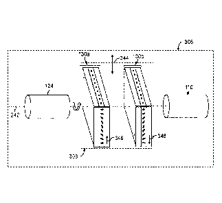
which has
a tunable liquid crystal lens (TLCL) assembly, a gradient-index (GRIN) lens
assembly having
a base optically connected to the TLCL assembly, and a tip opposite the base.
The tunable
optical device defines a focal point at a working distance from the tip, and
the working
distance is adjustable by tuning the TLCL assembly.
CA 02987207 2017-11-23
WO 2016/187715
PCT/CA2016/050593
- 2 -
[0005] In accordance with an aspect, there is provided an imaging system for
use in
imaging a sample comprising: an imaging assembly comprising a light source and
a light
detector; and a probe optically coupled to the imaging assembly, the probe
being configured
to, during use, direct light from the light source to a focal point to
illuminate the sample, and
from the focal point to the light detector, the probe comprising: a TLCL
assembly comprising
at least one pair of TLCLs, the TLCLs of the pair being superposed to one
another, a GRIN
lens assembly having a base being optically connected to the TLCL assembly,
and a tip
opposite to the base, the focal point being at a working distance from the
tip, the working
distance being adjustable relative to the tip by tuning each TLCL of the TLCL
assembly
during use.
[0006] In accordance with another aspect, there is provided a TLCL assembly
comprising
an optical axis and one pair of TLCLs each superposed one to the other,
wherein one TLCL
of the pair is rotated by 180 degrees about the optical axis with respect to
the other TLCL of
the pair.
[0007] In accordance with another aspect, there is provided a tunable
optical device for
interrogating a sample, the tunable optical device comprising: a GRIN lens
assembly; and at
least one segmented TLCL optically connected to the GRIN lens assembly, the at
least one
segmented TLCL having an annularly segmented electrode, the annularly
segmented
electrode having a number of electrode segments being independently drivable
to
compensate for aberrations of at least one of the GRIN lens assembly and the
sample.
[0008] It is understood that while being useful in an imaging system,
the tunable optical
device and the TLCL assembly can be used in optical systems (non-imaging
systems for
interrogating a sample) other than the exemplary imaging system described
herein. Such
optical systems and many further features and combinations thereof concerning
the present
improvements will appear to those skilled in the art following a reading of
the instant
disclosure.
DESCRIPTION OF THE FIGURES
[0009] In the figures,
CA 02987207 2017-11-23
WO 2016/187715
PCT/CA2016/050593
- 3 -
[0010] Fig. 1A is a schematic view of an example of an imaging system;
[0011] Fig. 1B is an enlarged view of a portion of the imaging system of
Fig. 1A;
[0012] Fig. 2 is a schematic view of an example of a probe, with two TLCLs in
a
polarization independent configuration;
[0013] Fig. 3 is an oblique and exploded view of another example of a probe,
with two
TLCLs in an aberration compensation configuration;
[0014] Fig. 4A is a schematic view of another example of a probe, with four
TLCLs in a
first aberration compensation configuration;
[0015] Fig. 4B is a schematic view of another example of a probe, with four
TLCLs in a
second aberration compensation configuration;
[0016] Fig. 4C is a schematic view of another example of a probe, with four
TLCLs in a
third aberration compensation configuration;
[0017] Fig. 5 is a schematic view of another example of an imaging system;
[0018] Figs. 6A-D are images taken, using the imaging system of Fig. 5,
at four different
working distances inside a specimen;
[0019] Fig. 7A is a top plan view of a TLCL as seen through a
polarimetric system,
showing a coma aberration, in accordance with an embodiment;
[0020] Fig. 7B is a top plan view of a TLCL as seen through a polarimetric
system,
showing a corrected coma aberration, in accordance with an embodiment;
[0021] Fig. 8A is a partial side view of an example of a tip of a probe,
showing
aberrations;
[0022] Fig. 8B is a schematic, and partial side view of another example
of a probe,
focusing at a focal point with corrected aberrations;
CA 02987207 2017-11-23
WO 2016/187715
PCT/CA2016/050593
- 4 -
[0023] Fig. 9 is an oblique, exploded view of another example of a
probe, with one
segmented tunable liquid crystal lens;
[0024] Fig. 10A is a top plan view of an example of a segmented TLCL tuned to
autofocus;
[0025] Fig. 10B is a top plan view of an example of a segmented TLCL tuned to
correct an
astigmatism aberration;
[0026] Fig. 100 is a top plan view of an example of a segmented TLCL tuned to
correct a
prism aberration;
[0027] Fig. 10D is a top plan view of an example of a segmented TLCL tuned to
correct a
coma aberration;
[0028] Fig. 11 is a schematic view of an example of a standalone imaging
system;
[0029] Fig. 12 is a schematic view of an example of a probe with a
reflector; and
[0030] Fig. 13 is a schematic view of an example of a probe with an
external illumination
fiber, in accordance with an embodiment.
[0031] These drawings depict example embodiments for illustrative purposes,
and
variations, alternative configurations, alternative components and
modifications may be
made to these example embodiments.
DETAILED DESCRIPTION
[0032] Fig. 1A shows an example of an imaging system 100 that can be
used for imaging
a specimen 102, in accordance with an embodiment.
[0033] As shown, the imaging system 100 has an imaging assembly 104 optically
coupled
to a tunable optical device or probe 106. The probe 106 has a tunable liquid
crystal lens
(TLCL) assembly 108, a gradient-index (GRIN) lens assembly 110 (also referred
to as
"imaging GRIN lens assembly 110") having a base 125 optically connected to the
TLCL
assembly 108, and a tip 126 opposite the base 125.
CA 02987207 2017-11-23
WO 2016/187715
PCT/CA2016/050593
- 5 -
[0034] As depicted, the imaging assembly 104 has a light source 112 and a
light
detector 116 coupled to the probe 106. The probe 106 is configured to, during
use, direct
light generated by the light source 112 to a focal point F to illuminate the
specimen 102
along direction 114 and from the focal point F to the light detector 116 along
direction 118.
[0035] As best seen in Fig. 1B, the focal point F is at a working distance WD
from the
tip 126. During use, the working distance WD is adjustable relative to the tip
126 by tuning
each TLCL of the TLCL assembly 108. As shown, the working distance associated
with the
focal point F can be adjusted from WDmin to WDmõ (yielding an adjustability
range of AWD)
by suitable tuning of the TLCL assembly 108.
[0036] Such tuning of the TLCL assembly 108 can avoid moving mechanical parts
such
as mechanical actuators and the like and may not cause any image deformation
since the
focus of the imaging GRIN lens assembly 110 is adjusted by the TLCL assembly
108.
[0037] Referring back to the embodiment of Fig. 1A, the light generated
by the light
source 112 is coupled into the probe 106 using a dichroic mirror 120 that
reflects the light
into an objective lens 122 which, in turn, injects the light into a waveguide
124 (also referred
to as "coupling waveguide 124") optically coupled to the probe 106. Once
illuminated,
imaging beams associated with the specimen 102 are propagated along an imaging
path
which follows the probe 106, the coupling waveguide 124, the objective lens
122 and the
dichroic mirror 120 towards the light detector 116. The imaging assembly 104
shown in
Fig. 1A is only exemplary, other suitably imaging assemblies can be deemed fit
by one
skilled in the art.
[0038] As will be understood, the TLCL assembly 108 is typically tuned by
modifying a
driving signal provided thereto via a power source 128. To do so, the power
source 128 is
electrically connected to electrodes (not shown) of the TLCL assembly 108.
Modifying the
driving signal can include modifying its frequency, its amplitude, its
voltage, and/or any
combination thereof. In some embodiments, the TLCL assembly 108 can be tuned
to react
with a temporal response which ranges from 10 ms (e.g., using a rapid
frequency modulation
protocol) to 1000 ms.
CA 02987207 2017-11-23
WO 2016/187715
PCT/CA2016/050593
- 6 -
[0039] The optical connection between the TLCL assembly 108 and the GRIN lens
assembly 110 can be obtained by fixedly connecting the TLCL assembly 108 to
the GRIN
lens assembly 110 by using a suitable connector such as a mount or glue, for
instance.
[0040] As illustrated, the TLCL assembly 108 shown in Fig. 1A has one TLCL
130. The
liquid crystal (LC) is a birefringent material such that incident light
passing through a LC
layer 132 of the TLCL can be analyzed as two orthogonal light polarizations.
The single
TLCL 130 shown in Fig. 1A thus focuses a single polarization of light and
leaves the other
orthogonal polarization essentially unaffected, such a TLCL assembly 108 is
said to be
polarization dependent. In this embodiment, the light source 112 can be
polarized or the
imaging assembly 104 can have an optional polarizer 134 in order to limit
either the incident
light or the emitted light (i.e. light emitted from the specimen 102) to only
one polarization,
which corresponds to the polarization of the single TLCL 130.
[0041] As will be detailed herein, the TLCL assembly 108 can have one TLCL or
more
than one TLCL (e.g., two, four or more TLCLs). When the TLCL assembly 108
comprises
more than one TLCL, the TLCLs are superposed to one another such that incident
light can
propagate through each of the TLCLs of the assembly 108 in a serial manner. In
other
words, the TLCLs are stacked to one another so that the apertures of the TLCLs
face each
other and are aligned along an optical axis of the probe. Further, the GRIN
lens
assembly 110 can have a single GRIN lens, or any combination of GRIN elements
(e.g.,
guiding GRIN rod, collimating GRIN lens, focussing GRIN lens).
[0042] Fig. 2 shows another example of a probe 206. As illustrated, the probe
206 has a
TLCL assembly 208 having a pair of TLCLs (first and second TLCLs 130a and
130b) and is
said to be "polarization independent", as will be explained in the following
paragraph. The
pair of TLCLs 130a and 130b is positioned between the coupling waveguide 124
such as a
GRIN waveguide or an optical-fiber and the GRIN lens assembly 110. In the
illustrated
embodiment, the light from the light source 112 propagates along the direction
114 while
light from a specimen, during use, propagates along the direction 118. As it
may be readily
understood, each TLCL 130 has the LC layer 132 comprised between a first face
236 and a
second face 238. When the TLCL assembly 208 has more than one TLCL, the TLCLs
130a
and 130b are superposed, or stacked, on one another by abutting the first face
236 of the
CA 02987207 2017-11-23
WO 2016/187715
PCT/CA2016/050593
- 7 -
second TLCL 130b on the second face 238 of the adjacent, first TLCL 130a. Such
abutting
may include fixing two adjacent TLCLs with an optical glue. Such an optical
glue can also be
used to optically couple the TLCL assembly to the imaging GRIN lens assembly
and/or the
coupling GRIN waveguide, for instance.
[0043] The probe 206 shown in Fig. 2 is in a polarization independent
configuration.
Indeed, since LC is a birefringent material, addressing the two orthogonal
polarization of the
incident light propagating along an optical axis 242 is of importance. This is
achieved by
positioning each TLCL 130a and 130b of the pair such that each TLCL acts on a
different
orthogonal polarization of the light in order to render the probe 206
independent of the
polarization of the incident light. In other words, still referring to Fig. 2,
the LCs of the
leftmost TLCL 130a have an orientation (through the page) perpendicular to an
orientation of
the LCs of the rightmost TLCL 130b (along the page). Providing the TLCL
assembly 208 in
the polarization independent configuration is preferred in situations where
the light
source 112 is polarized in more than one polarization. Indeed, in
circumstances where the
light source 112 is a white electroluminescent diode (LED), the white light
contains a chaotic
mixture of polarizations (which can be represented as a sum of the two
orthogonal
polarizations), and a polarization independent TLCL assembly 208 is required
to focus the
light along the two orthogonal polarizations.
[0044] Fig. 3 shows an exploded view of a probe 306 having the coupling
waveguide 124
(e.g., GRIN rod), a TLCL assembly 308 and the imaging GRIN lens assembly 110.
As
depicted, the imaging GRIN lens assembly 110 generally has an elongated,
cylindrical
shape but can have any suitable shape. Such a TLCL assembly 308 is said to be
systematic
aberrations independent or compensated. As depicted, the TLCL assembly 308 has
a pair of
TLCLs (first and second TLCLs 130a and 130b) wherein the first TLCL 130a is
rotated by a
half-rotation (i.e. 180 degrees) about the optical axis 242. As it may be
readily understood,
the probe 306 shown in Fig. 3 has an increased optical power compared to the
probe 106,
shown in Fig. 1A, having only one TLCL 130, for instance. As opposed to the
probe 206
shown in Fig. 2, the probe 306 is not polarization independent so polarizing
the emitted light,
polarizing the incident light or using a polarized light source is required.
Indeed, even if two
different TLCL assemblies have a corresponding number of TLCLs, the way the
TLCLs are
CA 02987207 2017-11-23
WO 2016/187715
PCT/CA2016/050593
- 8 -
superposed one to the other dictates the function that the resulting TLCL
assembly will
perform (i.e. polarization independent and/or systematic aberrations
independent).
[0045] The configuration shown in Fig. 3 is referred to as an aberration
compensation
configuration (or systematic aberrations independent) since it helps reducing
the effect of
systematic aberrations (e.g., coma) that are generated during the manufacture
of the TLCLs.
In context, the manufacture process of the TLCLs typically begins with the
production of an
optical wafer upon a series of manufacturing steps. Once the optical wafer is
diced into a
multitude of separate TLCLs, TLCLs associated with the same optical wafer are
typically
characterized by similar systematic aberrations. Accordingly, providing the
TLCL
assembly 308 with two TLCLs 130a and 130b having similar systematic
aberrations adjacent
but rotated by 180 degrees from one another helps compensating for these
systematic
aberrations. For example, knowing that the two TLCLs 130a and 130b have
similar
systematic aberrations causing a coma aberration along a transverse
orientation 344, if the
two TLCLs 130a and 130b are positioned in the aberration compensation
configuration
described above (the first TLCL 130a is rotated of 180 degrees about the
optical axis 242
with respect to the second TLCL 130b), each TLCL will cause the incident light
to be
modified by the (same) coma aberration but in two opposing directions 346 and
348 such
that the coma aberration is substantially canceled out. It is noted that the
aberration
compensation provided by the aberration compensation configuration is not
limited to the
coma aberration but can extend to other types of aberrations.
[0046] It was found that such systematic aberrations become more significant
as the
nominal diameter of the TLCL decreases. More specifically, TLCLs having a
nominal
diameter of less than 1 mm, preferably 0.5 mm, are especially useful in vivo
since the GRIN
lens assembly is less damageable due to its reduced footprint. With the TLCLs
and the
GRIN lens assembly having such nominal diameters, positioning the TLCLs in the
aberration
compensation configuration is thus preferred. Accordingly, the imaging GRIN
lens
assembly 110 can have a nominal diameter of 1 mm or less, preferably 0.5 mm.
Providing
such a small nominal diameter may help reduce damage to biological tissue
occurring when
inserting the imaging GRIN lens assembly 110 in the biological tissue. In some
CA 02987207 2017-11-23
WO 2016/187715
PCT/CA2016/050593
- 9 -
embodiments, the length of the GRIN lens assembly 110 is more than 2 mm,
preferably
7 mm.
[0047] Figs. 4A-C show examples of probes 406a, 406b and 406c wherein the TLCL
assemblies 408a, 408b and 408c each have two pairs of TLCLs configured to
render the
probe both polarization independent and free from the systematic aberrations
mentioned
above as well as to enhance the optical power of the probes. For ease of
reading, first and
second TLCLs of the first pair are shown at 130a and 130b and third and fourth
TLCLs of the
second pair are shown at 130c and 130d throughout Figs. 4A-C. Some other
configurations
of the probe having two pairs of TLCLs are possible, the embodiments shown in
Figs. 4A-C
are only examples. Before specifically describing Figs. 4A-C, it should be
understood that
the configuration of the TLCLs of the TLCL assembly can influence the
performances of the
TLCL assembly as a whole. Indeed, a focal point mismatch between a first focal
point of one
orthogonal polarization and a second focal point of the other orthogonal
polarization of the
incident light can be reduced or increased depending on how the TLCLs are
distanced to
one another. Further, the configuration of the TLCLs can influence the way the
systematic
aberrations are compensated also. More specifically, as will be understood by
the reading of
the following paragraphs and referring to Figs. 4A-C, the focal point mismatch
(F2-F1)
between the two orthogonal polarizations of the light is proportional to the
spacing distance
Ax. Additionally, the systematic aberrations compensation are inversely
proportional to the
TLCL interval p, i.e. the distance between two TLCLs associated with one of
two orthogonal
polarizations of the light which are rotated by 180 degrees with one another
about the probe
axis.
[0048] Fig. 4A shows the probe 406a wherein the two TLCLs of each pair are
adjacent
from one another. The first pair of TLCLs 130a and 130b acts on a first
polarization and is
configured in a systematic aberrations compensation configuration. The second
pair of
TLCLs 130c and 130d acts on a second polarization, orthogonal to the first
polarization and
is configured also in a systematic aberrations compensation configuration. In
the illustrated
embodiment, the spacing distance dx between a first median position x1 of the
TLCLs acting
on the first polarization and a second median position x2 of the TLCLs acting
on the second
polarization is maximized. Such a spacing distance, dx = x2 ¨ x1, causes a
focal point F1
CA 02987207 2017-11-23
WO 2016/187715
PCT/CA2016/050593
- 10 -
associated with the first polarization and a focal point F2 associated with
the second
polarization to be increased. However, in this configuration, the systematic
aberration
compensation (e.g., coma compensation) is optimized since the TLCL interval p,
which
separates the two TLCLs of the first and the second pair, is minimal, i.e. the
two TLCLs of
each pair are immediately adjacent from one another. Following a ZemaxTM
simulation, F1
can differ from F2 by about 5 1 pm, for instance, when the spacing distance
Ax is 480 pm
and the TLCL interval p is 240 pm. In this exemplary simulation, the TLCLs are
set to a
thickness of 40 pm, a nominal diameter of 0.5 mm and a nominal optical power
of 350
diopters; a GRIN rod 1210 (see Fig. 12) that is set to a nominal diameter of
0.5 mm, a
.. numerical aperture of 0.2, a pitch of 0.5; and the imaging GRIN lens
assembly 110 is set to
have a nominal diameter of 0.5 mm, a numerical aperture of 0.5 and a pitch of
0.23. As will
be understood by the skilled reader, a ray incident on a GRIN lens follows a
sinusoidal path
therealong. The pitch of the GRIN lens is the fraction of a full sinusoidal
period that the ray
traverses in the lens. For instance, a GRIN lens with a pitch of 0.25 has a
length equal to 1/4
.. of a sine wave, which would collimate a point source at the surface of the
GRIN lens.
[0049] Fig. 4B shows the probe 406b wherein the first and the second
TLCLs 130a and
130b of the first pair are interspersed with the third and the fourth TLCLs
130c and 130d of
the second pair. In the illustrated embodiment, the spacing distance Ax
between the first and
the second median positions is reduced and the TLCL interval p is increased
compared to
the embodiment shown in Fig. 4A. The reduced spacing distance Ax yields a
smaller
difference between the first and the second focal points F1 and F2, but this
is at the expense
of the systematic aberration compensation which is less optimized. Indeed, the
systematic
aberrations that are caused by the third TLCL 130c are propagated along a
longer TLCL
interval p so that the fourth TLCL 130d is less capable of compensating the so-
called
"propagated systematic aberrations". This applies for the first pair of TLCLs
130a and 130b
also. In another ZemaxTM simulation having similar simulation parameters than
for the
simulation shown in Fig. 4A, the difference between the first and the second
focal points F1
and F2 is simulated to be 2.5 1 pm when the spacing distance Ax is 240 pm
and the TLCL
interval p is 480 pm.
CA 02987207 2017-11-23
WO 2016/187715
PCT/CA2016/050593
- 11 -
[0050] Fig. 4C shows the probe 406c wherein the first pair of TLCLs 130a and
130b is
sandwiched between the third and the fourth TLCLs 130c and 130d of the second
pair. In
this embodiment, the difference between the first and the second focal points
F1 and F2 is
optimized since the spacing distance Ax vanishes while the systematic
aberrations are even
less compensated than for the embodiment shown in Fig. 4B. Indeed, in still
another
ZemaxTM simulation having similar simulation parameters than for the
simulation shown in
Fig. 4A, the difference between the first and the second focal points F1 and
F2 is simulated to
be 0 1 pm when the spacing distance Ax is null and the TLCL interval p is
720 pm.
[0051] Still referring to Figs. 4A-C, it is understood that the focal
point mismatch between
F1 and F2 caused by the spacing distance Ax can be overcome by modulating each
pair of
TLCLs separately. With such a modulation, the focal point mismatch (e.g., F1¨
F2 # 0) can
be minimized while optimizing the systematic aberration compensation. Further,
chromatic
aberration can be corrected in a similar manner. Indeed, since each wavelength
focuses at a
different focal point (e.g., FA1 # FA2), the chromatic aberration can be
corrected by modulating
each pair of TLCLs separately.
[0052] Fig. 5 is a schematic view of an example of an imaging system 500 for
imaging
biological cells in the brain tissue having fluorescent molecules therein, in
accordance with
an embodiment. As shown, the imaging system 500 has an imaging assembly 504
which is
optically coupled to the "endoscope" probe 406a. As described above, the TLCL
assembly 408a has two pairs of TLCLs to render the TLCL assembly 408a
polarization
independent and free from the systematic aberrations (as described above). The
probe 406a
has the coupling waveguide 124 provided opposite the imaging GRIN lens
assembly 110
with respect to the TLCL assembly 408a. The illumination path of the imaging
system 500
starts at the light source 112 which is a white LED in the illustrated
embodiment. The white
light is diffused and condensed by a diffuser 552 and a condenser 554 disposed
along the
illumination path, proximate the light source 112. The illumination light is
filtered by an
excitation filter 556 which lets pass wavelengths substantially corresponding
to an excitation
wavelength of the fluorescent molecules and filters out other wavelengths. The
remaining
excitation wavelength is reflected through the dichroic mirror 120 towards the
objective lens
122 which is used to inject the excitation wavelength into the coupling
waveguide 124
CA 02987207 2017-11-23
WO 2016/187715
PCT/CA2016/050593
- 12 -
towards the specimen 102 (e.g., biological brain tissue). Once excited at the
excitation
wavelength, the fluorescent molecules of the biological brain tissue emit a
fluorescence
signal which is collected by the probe 406a. The imaging path of the imaging
system 500
passes through the probe 406a, back to the objective lens 122, through the
dichroic mirror
120, through an emission filter 558 towards the light detector 116. The
emission filter
typically lets pass the fluorescence signal while filtering out light
associated with the
excitation wavelength. In the illustrated embodiment, a tube lens 560 is
optionally provided
between the emission filter 558 and the light detector 116. In this example,
the light detector
116 is a charge-coupled device (CCD) camera which images the biological brain
tissue. In
another embodiment, the imaging system 500 can have optional polarization
control
elements 555 positioned between the probe 406a and the dichroic mirror 120.
The
polarization control elements 555 (e.g., linear and/or circular polarizers or
wave plates) are
used in order to enable the use of the imaging system 500 for polarization
discrimination
imaging.
[0053] Still referring to Fig. 5, the light source 112 is powered by a
current generator 562
and is triggered by a triggering device 564. In this embodiment, the
triggering device 564 is
connected to a computer 566 which sends triggering instructions A to the
triggering
device 564 in order to operate the light source 112 for only given period of
time (e.g., five
minutes). Such triggering instructions A prevent from overheating or damaging
the biological
tissue. The computer 566 sends tuning instructions B in order to tune a
function generator
568 at a given electrical function C having a given tension, frequency and
phase modulation.
The given electrical function C acts on the electrodes of each of the TLCLs of
the TLCL
assembly 408a in order to adjust more or less the working distance of the
probe 406a. In this
embodiment, the frequency of the given electrical function C determines the
working
distance at which the probe 406a focuses (e.g., the tension is constant).
During use, the light
detector 116 communicates images D to the computer 566 which can display the
images
suitably to a user for instance. In this embodiment, it is understood that the
triggering
instructions A, the tuning instructions B and the given electrical function C
are controlled by
the computer 566 using a suitable program such as LabVlEWTM, for instance.
CA 02987207 2017-11-23
WO 2016/187715
PCT/CA2016/050593
- 13 -
[0054] Figs. 6A-D show exemplary images of olfactory bulb granule cells
670 in the brain
tissue of a mouse, in accordance with an embodiment. In this example, the
frequency of the
given electrical function C tuning the TLCL assembly is : 1 kHz which yields a
reference
focal point as seen in Fig. 6A, 10 kHz which yields a focal point 22 pm
shallower than the
reference focal point as seen in Fig. 6B, 11 kHz which yields a focal point 26
pm shallower
than the reference focal point as seen in Fig. 6C, and 15 kHz which yields a
focal point 41
pm shallower than the reference focal point as seen in Fig. 6D. As it can be
seen, working
distance adjustability in neuroimaging applications is useful due to the
complex three
dimensional nature of the neurons.
[0055] Fig. 7A shows an example of an image, seen through a polarimetric
system such
as two crossed polarizers and an interference filter, of a TLCL 130 with
systematic
aberrations such as a coma aberration 772 for an uncompensated TLCL assembly.
Fig. 7B
shows an example of an image of a TLCL 130 with a compensated coma aberration
774,
also seen through a polarimetric system, when using the TLCL assembly in the
aberration
compensation configuration as indicated in the TLCL assembly 408a.
[0056] In some embodiments, a TLCL can be driven with a driving voltage of 2.4
V. This
may be achieved by providing its electrodes on each side of the crystal layer
or, in other
words, by providing one of the two electrodes sandwiched between the first
face and the
crystal layer, and by providing the other one of the two electrodes sandwiched
between the
second face and the crystal layer. In some other embodiments, the TLCL can be
driven by a
driving voltage of 24 V. In some embodiments, each TLCL has an optical power
of
320 diopters.
[0057] Fig. 8A shows an example of the GRIN lens assembly 110 and its
associated
inherent aberrations 876 which can reduce the resolution of the image taken by
the imaging
system, for instance. Fig. 8B shows an example of a probe 806 where the
inherent
aberrations caused by the GRIN lens assembly 110 are corrected (see corrected
aberrations 878) using the TLCL assembly in accordance with an embodiment. To
do so, the
TLCLs is provided in the form of segmented TLCLs 830a and 830b. Such a
segmented
TLCL has one of its electrode being embodied as an annularly segmented
electrode 880,
which is best shown in Fig. 9. It is understood that the probe 806 can have
one or more than
CA 02987207 2017-11-23
WO 2016/187715
PCT/CA2016/050593
- 14 -
one TLCL each superposed one to the other and that each of the one or more
than one
TLCL of the embodiments described herein can be segmented TLCLs. Referring
back to Fig.
8B, the segmented TLCLs 830a and 830b are used to correct aberrations of the
optical
system dynamically (e.g., in real-time) in order to enhance the resolution of
the imaging
system.
[0058] Fig. 9 shows an exploded view of a probe 906 having a segmented TLCL
830. The
annularly segmented electrode 880 has a plurality of electrode segments 882
which are
operable independently. Each of the electrode segments 882 is operable by the
power
source in function of a given tension, a given frequency and/or a given phase
modulation. In
an embodiment, the electrode segments 882 are independently drivable by the
power
source which is controlled by the computer 566. The computer 566 can have a
memory on
which is stored an aberration compensation program which is configured to
compensate for
aberrations of the probe 906 (e.g., TLCL assembly 908, the coupling waveguide
124, the
imaging GRIN lens assembly 110) as well as to compensate for aberrations
caused by the
specimen. In cases where the specimen is a living organism, the aberration
compensation
program can compensate for aberrations due to movement of the living organism
such as
breathing or walking, for instance, in free-behaving animals applications. In
other words, the
imaging system is adapted to drive the segmented TLCLs 830 in order to
compensate,
dynamically, for aberrations caused by the optical components of the imaging
system (non-
living matter) and by the specimen (living matter) which are involved.
[0059] It was found that while increasing the number of electrode segments 882
generally
allows a greater control on the aberration correction, it also generates an
undesirable
fringing field of a greater importance. Suitably selecting the number of
electrode
segments 882 of the annularly segmented electrode 880 is preferred. In an
embodiment, the
annularly segmented electrode 880 has four or eight electrode segments 882.
Other number
of electrode segments can also be used.
[0060] In another embodiment, the segmented TLCLs 830 are incorporated in an
imaging
system similar to the one shown in Fig. 5. In this specific embodiment, the
specimen is
excited with different excitation wavelengths such that more than one
fluorescent molecule
types are excited simultaneously. This can yield a fluorescence signal
incorporating different
- 15 -
emission wavelengths which can be imaged using the light detector. In such an
embodiment,
the segmented TLCLs 830 can be used to correct chromatic aberrations due to
the different
excitation wavelengths as well as aberrations due to the different emission
wavelengths
(non-segmented TLCLs 130 can also be used to correct chromatic aberrations, as
mentioned above). In other words, by using the segmented TLCLs 830, the
different
excitation wavelengths can be focused at the same focal point in the specimen
(and/or the
different emission wavelengths can be focused on the light detector 116) such
that the
chromatic aberrations are corrected in the imaging system. In another
embodiment, optional
segmented TLCLs can be provided near the light source in order to correct
aberration of the
light source 112 and to control the emission wavelengths. In still another
embodiment,
optional electrically-controllable polarizers are provided near the light
source 112 and the
light detector 116 in order to actively control the polarization of the light
which is emitted by
the light source 112 and the light which is detected by the light detector
116. It is understood
that the imaging system described herein is not limited to fluorescence and
can also be used
in Raman spectroscopy and other types of spectroscopy.
[0061] Figs. 10A-D show the segmented TLCL 830 having the annularly segmented
electrode 880, in accordance with some embodiments. Respectively, the
segmented
TLCL 830 can be used for autofocusing (e.g., 20 diopters) and also for
aberration
compensation such as astigmatism, prism, coma as shown throughout Figs. 10A-D.
Spherical and defocus aberrations can also be compensated. Such a segmented
TLCL 830
is described in US Patent Application Publication Number 2013/0250197.
[0062] Fig. 11 is a schematic, sectional view of an imaging system 1100, in
accordance
with another embodiment. In this illustrated embodiment, the imaging system
1100 is meant
to be a wireless, self-powered, "miniature" and standalone device. An
exemplary application
of such an imaging system 1100 can be to image biological tissue of a free-
behaving subject
while avoiding wires such as electrical wires to control the light source, the
light detector and
the TLCL assembly 1108 or optical-fibers to carry the measured signal to a
remote light
detector, for instance. Indeed, the imaging assembly 1104 is enclosed in a
probe module
1184 which is directly mounted to the probe 1106. As depicted, the probe
module 1184 is
Date recue/Date received 2023-04-28
CA 02987207 2017-11-23
WO 2016/187715
PCT/CA2016/050593
- 16 -
mounted adjacent to the TLCL assembly 1108. In the illustrated embodiment, the
probe
module 1184 has a power source embodied by a battery 1186 which is enclosed in
the
probe module 1184. The battery 1186 is used to power the TLCL assembly 1108
during use
and/or any of the optical components of the imaging assembly 1104. In an
embodiment, the
battery 1186 is rechargeable and removable via an access 1188. Moreover, the
probe
module 1184 has a communication module 1190 having an antenna 1192 for
receiving
instructions wirelessly from the computer 566, for instance. With such a probe
module 1184,
remote control of the electrodes (conventional or annularly segmented) is
allowed. Further,
the images which are detected by the imaging assembly 1104 can be wirelessly
communicated to the computer 566 using the antenna 1192 and/or stored on a
memory
1198 enclosed in the probe module 1184.
[0063] In this embodiment, the TLCL assembly 1108 can be any type of TLCL
assembly
described herein. For instance, the TLCL assembly 1108 can have a pair of
TLCLs that can
be configured in the polarization independent configuration or in the
aberration
compensation configuration. Alternatively, the TLCL assembly 1108 can have
four TLCLs
configured in the polarization independent configuration and in the aberration
compensation
configuration (e.g., the TLCL assemblies shown in Figs. 4A-C).
[0064] Fig. 12 shows an example of a probe 1206, in accordance with another
embodiment. It is understood that the imaging GRIN lens assembly can be a
single GRIN
lens or a combination of suitable GRIN elements such as waveguide(s) and/or
lens(es). In
the illustrated embodiment, the imaging GRIN lens assembly 110 comprises a
GRIN rod
1294 which propagates the light from the TLCL assembly 1208 to a GRIN
focussing lens
1210. The GRIN rod 1294 is optional but useful in situations where the tip of
the probe 1206
is to be inserted at a given depth into the specimen.
[0065] Still referring to Fig. 12, the probe 1206 has a reflector 1296
positioned at the end
of the imaging GRIN lens assembly 110. In this case, the tip 126 is defined by
the reflector
1296. The reflector 1296 forms an angle a with respect to an optical axis 242
of the probe
1206 such that the focal point is not only positioned along the optical axis
242 but also along
an axis depending on the angle a. As it may be readily understood, rotating
the probe 1206
about the optical axis 242, while the probe 1206 is into the specimen, during
use, allows
CA 02987207 2017-11-23
WO 2016/187715
PCT/CA2016/050593
- 17 -
imaging a three-dimensional disk of the specimen for each axial position of
the probe 1206
into the specimen, for instance. In an embodiment, the reflector 1296 is
provided in the form
of a piercing member having a sharp edge. With such a piercing member, the
probe 1206
can be used to pierce the biological tissue in order to insert the probe 1206
within the
biological tissue while minimizing damage caused to the tissue.
[0066] Fig. 13 is a schematic view of a probe 1306, in accordance with an
embodiment.
As it will be understood by one skilled in the art, the probe 1306 can be
configured so that
the light from the light source 112 is directed to the specimen without
passing through the
TLCL assembly 1308 and the imaging GRIN lens 110 by using an external
illumination
fiber 1398. The illumination light is thus directed along the direction 114,
as shown in Fig. 13,
while the light received from the specimen 102 is directed along the direction
118, towards
the light detector 116. Indeed, the external illumination fiber 1398 can have
an end coupled
to a light source and another end 1399 provided near the tip 126 of the probe
1306, towards
the specimen. The form of the end 1399 is not limited to the angle-polished
end shown in
Fig. 13, but encompasses other forms deemed suitable by a person skilled in
the art.
[0067] As it can be understood, the examples described above and illustrated
are
intended to be exemplary only. The tunable optical device can be used in
telecommunication
in the form of a tunable collimating lens or a tunable focusing lens or in
medical endoscope
applications (in vitro or in vivo). Medical applications include, but is not
limited to,
laparoscopy, arthroscopy, cystoscopy, obstretrics, gynecology, bronchoscopy,
laryngoscopy,
mediatinoscopy, otoscopy, gastrointestinal, boroscopy and the like. It will be
understood that
while the embodiments described herein are suitable for imaging specimens,
these
embodiments can be used, in the same or a modified form, to image other forms
of samples,
which can consist of inert materials for instance. It will also be understood
that in various
embodiments, the TLCUGRIN combination while optically coupled to one another
can be
separated from one another by distance and/or one or more other optical
components. The
scope is indicated by the appended claims.