Note: Descriptions are shown in the official language in which they were submitted.
CA 02988609 2017-12-07
WO 2016/197206
PCT/AU2016/050480
MICROWAVE ABLATION DEVICE
Field of the invention
This invention relates to a microwave ablation device and a method of using
such device. The invention may find application in the field of endovascular
sympathectomy or denervation such as renal artery denervation. The invention
may
also find application in other fields of medical ablation including the
treatment of atrial
and ventricular arrhythmias.
Background of the invention
Hypertension is a significant medical condition that leads to morbidity and
mortality from end organ injury, such as strokes, heart attack and kidney
failure. Many
patients require multiple medications for blood pressure control and for some
patients,
medications are poorly tolerated or ineffective altogether. Renal artery
denervation has
emerged as a possible treatment option to control hypertension in these
patients who
are refractory or intolerant of medical therapy. The procedure aims to
eliminate the
efferent and afferent nerves that relay neural messages between the kidneys
and the
central nervous system as these form essential components of neuro-hormonal
reflexes
that elevate blood pressure. The efferent and afferent nerves travel in the
outer layer
(i.e. adventitia) of the renal artery and the perinephric fat, mostly between
1 and 6 mm
from the inner (i.e. luminal) surface of the renal arteries and can
potentially be
destroyed by endovascular catheter ablation.
Early clinical trials with radiofrequency catheter ablation for renal artery
denervation showed promising results in blood pressure reduction. These
results have
fuelled interest and the development by various medical companies and research
institutes of radiofrequency ablation catheters for this application.
More recently, a clinical trial of renal artery denervation compared a
procedure
performed by a renal denervation system developed by Medtronic with an
operation
sham control, and this failed to show significant benefit in blood pressure
reduction. One
hypothesis offered by experts in view of the disappointing results is that
ineffective renal
artery denervation occurred during this trial.
Prior art radiofrequency catheters used for renal artery denervation may have
a
disadvantage of injuring the full thickness of the renal artery before the
renal nerves are
1
Subs titue Sheets
(Rule 26)
RO/AU
affected. For this reason, conservative ablation of the artery is typically
performed,
thereby to avoid renal artery stenosis. This type of conservative ablation is
however
done at the cost of reducing the potential efficacy in denervating renal
nerves with this
energy source. For example, typically the catheters produce focal endovascular
ablation
lesions in a spiral configuration along a renal artery so as not to cause
circumferential
injury to the muscle layer, or media, of the artery, as this is what may lead
to renal
artery stenosis.
In light of the above, there is a need for an alternative type of ablation
device.
Reference to any prior art in the specification is not an acknowledgment or
suggestion that this prior art forms part of the common general knowledge in
any
jurisdiction or that this prior art could reasonably be expected to be
understood,
regarded as relevant, and/or combined with other pieces of prior art by a
skilled person
in the art.
Summary of the invention
In one embodiment of the invention there is provided a microwave ablation
device comprising a feed line, a microwave radiator and a device outer sheath
in which
at least part of the feed line is contained, the device outer sheath in use
allowing an
irrigation liquid to flow therethrough, wherein the feed line has a junction
with the
microwave radiator and has an outer conducting shield terminating and
insulated at the
junction, the feed line having a conductive core that extends to the microwave
radiator,
a part of the conductive core forming the microwave radiator at a distal end
thereof,
electrically insulated from its surrounding environment, wherein the microwave
radiator
is not matched to the impedance of the feed line and is unbalanced at the
distal end.
In a further embodiment, there is provided a method of use of the microwave
ablation device, the method comprising:
positioning the microwave radiator of the device adjacent an area to be
ablated;
and
transferring microwave energy to the microwave radiator.
According to another embodiment there is provided an microwave ablation
device comprising a feed line, a microwave radiator and a device outer sheath
in which
at least part of the feed line is contained, the sheath in use allowing an
irrigation liquid to
flow therethrough, wherein the feed line has a junction with the radiator and
has an
2
Date Recue/Date Received 2022-07-14
outer conducting shield terminating and insulated at the junction, the feed
line having a
conductive core that extends to the radiator, the conductive core forming a
radiating
element electrically insulated from its surrounding environment, wherein the
radiator is
unbalanced.
Preferably, the feed line is also unbalanced.
According to another embodiment there is provided a microwave ablation
device comprising an electrically insulated feed line, a microwave radiator
and a device
outer sheath in which at least part of the feed line is contained, the sheath
in use
allowing an irrigation liquid to flow therethrough, wherein the outer
conducting shield of
the feed line terminates and is insulated from the conductive core and the
surrounding
environment, the feed line having a conductive core extending beyond the
shield and
becoming the radiator, the conductive core forming a radiating element
electrically
insulated from its surrounding environment, wherein the radiator is not
matched to the
impedance of the feed line and is unbalanced at the distal end.
2a
Date Recue/Date Received 2022-07-14
CA 02988609 2017-12-07
WO 2016/197206
PCT/AU2016/050480
According to another embodiment there is provided a microwave ablation
device comprising a feed line, a microwave radiator, and an outer device
sheath in
which at least part of the feed line is contained, the sheath in use allowing
an irrigation
liquid to flow therethrough, wherein the feed line has an outer conducting
shield
terminating and insulated at its junction with the radiator, the feed line
having a
conductive core that extends without electro-magnetic interruption to the
radiator, the
conductive core forming a radiating element electrically insulated from its
surrounding
environment.
Each of the above embodiments may include the features of any one or both of
the other embodiments.
For any of the above embodiments, the outer conducting shield may be
electrically insulated at the junction by an insulating adhesive or sleeve
that covers the
distal end of the outer conducting shield. The outer conducting shield may
also be
insulated from an external surface of the device by the outer device sheath.
Thus the
outer conducting shield is insulated from to any adjacent conductive
components, such
as the radiator, the patient's blood pool and the outside environment.
Preferably, a distal end of the outer conducting shield is not connected to a
choke.
The sheath may contain the microwave radiator and at least part of the feed
line.
The sheath may further include one or more locating formations configured to
centre and locate the device in use in a vessel.
The radiator may include an insulating layer extending over, or an insulating
cover encasing, the radiating element.
The device may further be configured for the outer device sheath to be
connected to the feed line and/or the insulated radiating element thereby to
allow
relative movement of the sheath to the feed line in use, wherein the one or
more
connecting formations comprise sections of slits in the sheath to form splines
that
deploy to form convex protrusions that interact with vessel walls.
Preferably the device has a distal end that includes an opening for the
irrigation
fluid to flow out of the device and over the radiator to cool the vessel.
Preferably the
3
CA 02988609 2017-12-07
WO 2016/197206
PCT/AU2016/050480
opening is at a distal end of the feed line, so that the irrigation fluid can
cool the feed
line.
The microwave ablation device may be driven by a microwave energy source.
The microwave energy source may operate at 2.45 GHz, with a power output
sufficient to produce circumferential thermal ablation of targeted
neurological structures
while enabling sparing of the tissue closer to the renal artery lumen, such as
the renal
artery wall, by cooling of said tissue closer to the renal artery by arterial
blood flow and
said irrigation fluid.
According to a further aspect there is provided a method of microwave ablation
comprising:
introducing a distal end of a device, according to any embodiment defined
above, into a human body;
locating the radiator of the device adjacent an area within the human body to
be
ablated; and
transferring microwave energy to the radiator.
Preferably the microwave energy is transferred for a predetermined period of
time. In one embodiment, the period of time is approximately or exactly 3
minutes.
Preferably, the microwave energy is driven by a microwave energy source that
operates
at a said power output.
The area of the human body may be a renal artery.
The method may further comprise feeding the irrigation liquid to flow between
the outer device sheath and the feed line, to cool the feed line while in use.
Preferably said irrigation liquid flows out of the distal end of the feedline
to
further cool said tissue closer to the renal artery lumen.
As used herein, except where the context requires otherwise, the term
"comprise" and variations of the term, such as "comprising", "comprises" and
''comprised", are not intended to exclude further additives, components,
integers or
steps.
Further aspects of the present invention and further embodiments of the
aspects described in the preceding paragraphs will become apparent from the
following
description, given by way of example and with reference to the accompanying
drawings.
4
CA 02988609 2017-12-07
WO 2016/197206
PCT/AU2016/050480
Brief description of the drawings
Illustrative embodiments of the various aspects of the present invention will
now
be described by way of non-limiting example only, with reference to the
accompanying
drawings. In the drawings:
Figure 1 shows a partial cross-sectional view of a microwave ablation device
in
accordance with an example embodiment;
Figure 2 shows a cross-sectional view along line A-A' of Figure 1;
Figure 3 shows a pictorial view of a microwave ablation device in accordance
with an example embodiment and similar to that of Figure 1 in a deployed
state;
Figure 4 shows a cross-sectional view of a microwave ablation device in
accordance with another example embodiment;
Figures 5A to 5C show pictorial views of a microwave ablation device in
accordance with an example embodiment and similar to that of Figure 3 with the
outer
device sheath in various states of deployment in an artery;
Figure 6A shows a partial cross-sections of the distal end of a microwave
ablation device, but not showing an outer sheath of the microwave ablation
device, the
figure illustrating a tapered structural support component in accordance with
another
embodiment, showing the device's radiator and its junction with the feed line,
with the
structural support component encasing the radiator;
Figure 6B shows a partial cross-section of only the device's radiator and its
junction with the feed line of Figure 6A, without the structural support
component;
Figure 6C shows a cross-section of only the structural support component
which encases the radiator of Figure 6B, thereby to form the end of the device
as shown
in Figure 6A;
Figure 7 shows the microwave ablation device of Figures 6A to 6C, without the
outer sheath of the device so as to illustrate the effect of the support
component when
the device is being positioned in a renal artery prior to ablation;
Figure 8 shows an example prototype of a microwave ablation device in
accordance with an example embodiment and in a model of a renal artery with
heating
patterns indicated; and
5
CA 02988609 2017-12-07
WO 2016/197206
PCT/AU2016/050480
Figure 9 shows a cross-sectional view of a microwave ablation device in
accordance with a further example embodiment.
Where the Figures represent the same or similar features the same reference
numerals will be used.
Detailed description of the embodiments
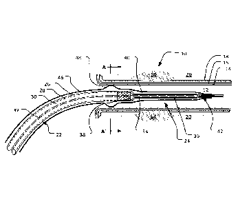
Turning to Figures 1 and 3, a microwave ablation device 10 is shown. In this
embodiment the ablation device 10 is a microwave emitting catheter used for
microwave denervation of renal arteries. In Figure 1, the microwave ablation
device 10
is shown in a vessel, e.g., a renal artery 12, formed by various artery walls:
the inner
layer (or vessel intima) 14, the middle layer (or vessel media) 16 and the
outer layer (or
vessel adventitia) 18. Adjacent to the vessel adventitia 18 lies the renal
nerves 20,
which in this embodiment is to be ablated.
Although the device 10 is described with reference to denervation of renal
arteries, a person skilled in the art will appreciate that the device may be
used in other
-- medical ablation applications.
The microwave ablation device 10 comprises a feed line 22 connected, in use,
to an energy source (not shown), in particular a microwave energy source. The
feed line
22 terminates in a radiator 24 (or antenna) having a single radiating element,
which
radiates microwave energy to the surrounding environment. As will become more
-- apparent from the description below, the microwave energy is transmitted to
the
surrounding area and absorption produces heat. Blood flow dissipates this heat
quickly,
protecting the intima and media layers 14 and 16 of the artery walls,
resulting in
preferential heating of the renal artery adventitia layer 18 and deeper
regions thereby to
ablate the renal nerves 20 located in the deeper regions to the renal artery
12.
The feed line 22 may be a cable, for example, a co-axial cable which is well
known to comprise, from the outer layers to the inner layers, an insulating
outer sheath
26, an outer conducting shield 28, a tubular insulating layer 30 and a
conductive core
(also called an inner conductor) 32.
The radiator 24 has a radiating element 34 that has a diameter that is always
less than the diameter of the feed line 22 and is concentric with the feed
line 22. The
radiating element is an extension of the conductive core 32 of the feed line
22, so has a
constant diameter, being the same diameter as conductive core 32. The
"junction"
6
CA 02988609 2017-12-07
WO 2016/197206
PCT/AU2016/050480
between the radiator 24 and feed line 22, indicated by reference numeral 38,
is where
the outer conducting shield 28 terminates. The radiating element 34 is
electrically
insulated from the surrounding environment. For example, the radiating element
34 may
be encased in an insulating material shown in Figure 1 by reference numeral
36. The
.. radiating element 34 may alternatively be covered by or encased in a layer
of insulating
material. The insulating material may be PTFE (i.e. teflon), although any
other suitable
electrically insulating material which will tolerate the particular
temperatures may be
used e.g., FEP (fluorinated ethylene polymer).
At the junction 38 between the feed line 22 and the radiator 24, the outer
conducting shield 28 is terminated and sealed by an insulative structural
support
component 40. As will be described in more detail below with reference to
Figures 6A to
6C in which the structural support component 40 is best seen, this component
40
provides the device 10 at the junction with structural support and flexibility
and acts as a
cover of the radiator 24.
The device 10 does not have a choke attached to outer conducting shield at the
distal end of the feed line 22. Thus, the conductive core extends without
electro-
magnetic interruption to the radiator. Further, there is no end-cap at the
distal end of the
radiating element 24, nor a 5/8 A coil or any other structure for impedance
matching
attached to the radiator 24. As a result of not having a choke, the radiator
radiates
.. relatively more energy at the radial distance from the radiator at which
denervation is to
be performed. This is further aided by not having the end-cap or coil attached
to the
radiating element. By contrast the inclusion of such a choke would concentrate
the
radiation pattern closer to the radiating element, even the more so if an end
cap or coil
is attached to the radiating element. Such end-caps may take a variety of
forms, but in
.. effect add capacitance to the radiator element. For example, the end-cap
may be
electrically connected to the distal tip of the radiating element and from
there feed
proximally over some distal section of the radiating element, but radially
insulated from
the radiating element. Such coils, on the other hand typically are connected
at one end
to the outer conducting shield and at the other end to location along the
length of the
.. radiating element, eg about a 5/8 A from the junction 38.
There is also no electrical shield (like a ground plane) or radials extending
laterally from
the outer conducting shield By omitting such shields and radials the maximum
diameter
7
CA 02988609 2017-12-07
WO 2016/197206
PCT/AU2016/050480
of the device is kept to a minimum so as not to add unnecessary bulk or
interfere with
the vessel in which the ablation device is deployed.
Without a choke, coil, ground plane, radials, end cap or any other such
structures, the device is significantly 'unbalanced' in that the load on the
outer
.. conducting shield 28 and the conductive core 32 (including the radiating
element 34
portion of the conductive core 32) is not matched. This contrasts with
conventional
antenna design practice in which these structures are used to produce an
antenna with
minimal power loss and efficient transmission in the far field. .
Also, with the radiating element 34 being insulated, energy cannot be
dissipated
through alternating current flow (ohmic heating) to the surrounding
environment, i.e.
blood flowing in the renal artery 12 or other irrigation fluid described
later. The only
energy dissipation from the radiating element 24 is accordingly by radiation.
As will
become apparent below, these factors result in a more favourable heating
pattern
across the area to be ablated and greater deployability, at the cost of
comparatively
higher loss of energy along the feed line 22 due to circulating currents (eddy
currents) in
the conducting shield 28, and consequent greater feed line 22 heating. Part of
the
favourability of the heating pattern is that it is generally spread, in the
near field, across
a greater length of the radiator, as opposed to being concentrated as a hot
spot at one
end of the radiator (as may be the case where a ground plane, choke, coil
and/or end
cap is employed). This results in providing a greater length over which
perivascular
nerves may be ablated. This may improve the durability of the denervation
procedure by
widening the gap that neuroregeneration would need to bridge to re-establish
functional
connections.
The cover 40 may carry a component 42 that may be an attachment or a
continuation of the cover 40 or outer sheath 46. The component 42 carries a
monorail
segment 44 for tracking an angioplasty wire (e.g. a 0.014 inch angioplasty
wire). The
structural support cover may extend over the terminating end of the outer
layers of the
feed line 22 to the tip (and beyond) of the radiator 24. In one example
embodiment the
cover 40 is manufactured from a polyolefin material due to its features of
being heat-
shrinkable, thereby creating a tight fit. However, it will be appreciated that
other suitable
materials may be used such as PTFE (Teflon) or other high temperature plastics
like
FEP.
8
CA 02988609 2017-12-07
WO 2016/197206
PCT/AU2016/050480
In the embodiment shown in Figure 1, part of the device 10 is contained in an
outer device sheath 46. The sheath 46 is typically manufactured from a
suitable material
that is soft and thin, e.g., a polymer such as polyolefin, which can generally
be safely
used in the human body.
The sheath 46, in this embodiment, comprises a locating formation 48 which
acts as a centering mechanism and which is formed by linear slits 50 (best
shown in
Figures 2 and 3) along the length of the sheath 46 which form a section of
splines 52
(see Figures 2 and 3) along part of the sheath 46. As the splines 52 are soft,
when the
feed line 22 is moved relative to the sheath 46, the splines 52 are deployed
by
expansion to form a convex protrusion against the inner walls 14 of the artery
12. The
locating formation then secures and centrally locates the sheath 46, and with
that, the
radiator 24 and feed line 22 in place. Thus, the locating formation 48 adjusts
to maintain
contact pressure and concentricity with the local arterial wall 14. The
collapsing of the
mechanism is guaranteed by simply pulling the outer device sheath 46 back.
The outer device sheath 46 is sufficiently sized, i.e. it has a sufficient
diameter,
in comparison to the feed line 22, to allow for an irrigation or cooling
liquid in use to
pass between the insulating outer sheath 26 of the feed line 22 and the outer
device
sheath 46. Typically, a saline solution is pumped into and through the sheath
46, for it to
exit into the artery 12 at the locating formation 48. As the saline solution
flows along the
length of the feed line 22, heat is removed from the device 10 to ensure that
any
clinically important temperature rises are addressed, and maintains the
catheter lumens
clear of blood to prevent thrombosis.
Turning to Figures 4 and 5A to 5C, another example embodiment of a
microwave ablation device 60 is shown. The device 60 has the same or similar
features
as the device 10, and these features are accordingly indicated by the same
reference
numerals used in Figures 1 to 3. Also, like device 10, device 60 does not have
a ground
plane, choke, coil or end-cap. The outer device sheath 46 of the device 60 is
however
adapted to provide two locating formations 48.1 and 48.2, a distal locating
formation
48.2 located towards the free tip (and connector) of the radiating element 24
and a
proximal formation 48.1 closer to, or adjacent, a part of the feed line 22.
Each of these
locating formations 48.1 and 48.2 acts as part of a centering mechanism and is
formed
by linear slits 50 along the length of the outer device sheath 46 which form
respective
sections of splines 52 along parts of the sheath 46. Again, relative movement
of the
9
CA 02988609 2017-12-07
WO 2016/197206
PCT/AU2016/050480
feed line 22 in a proximal direction with respect to the outer device sheath
46 (i.e.
movement towards the aorta) is used to deploy these sections of splines into
convex
protrusions of the locating formations 48.1 and 48.2, allowing each to expand
to the
particular vessel (artery) size according to the amount that the outer device
sheath 46 is
moved relative to the feed line 22. As best shown in Figures 5A to 5C, the two
locating
formations 48.1 and 48.2 self-adjust to maintain equal contact pressure and
concentricity with the local arterial wall by their design. During expansion,
it is likely that
one locating formation will expand first before the other. However, as soon as
the first
locating formation contacts the vessel wall, it is restrained by the wall and
the other
locating formation then expands till it too is providing the same pressure on
the wall.
This minimises the risk of trauma to the vessel at a place of natural or
pathologic
narrowing or dilatation.
Collapsing of the locating formations 48.1 and 48.2 are managed by simply
moving the outer device sheath 46 relative to the feed line 22 in the proximal
direction.
This method of collapsing the formations 48.1 and 48.2 provides what is
considered a
safe way to reduce its diameter before removing the device 60.
Similar to the description relevant to Figures 1 to 3, an irrigation liquid
such as a
saline solution is pumped into the sheath 46, with the saline solution in this
embodiment
passing, not only over the feed line 22, but also along most of the length of
the radiating
element 34 as contained by the insulating material 36. This assists with the
removal of
localised heat caused by radiation of the microwave energy, as well as the
unbalanced
nature of the device. It will further be appreciated that blood flow between
the outer
device sheath 46 and the inner walls 14 of the artery 12 (i.e. the luminal
surface of the
renal artery), which allow for further (and secondary) localised cooling
during the
ablation process. This flow of blood protects the intima and media (inner and
middle)
layers 16 and 18 of the artery 12 while deeper regions (e.g., including the
outer or
adventitia layer) containing the renal nerves are ablated.
The soft outer device sheath 46 is attached (secured) to the distal end of the
feed line 22 or the distal end of the radiator 24. However the outer sheath 46
is
.. otherwise free to move with respect to the feed line 22 in order to allow
for the relative
movement of the outer device sheath 46 in relation to at least the feed line
22, as well
as to allow irrigation of the feed line 22 and the radiator 24 when the
centering
mechanism of the locating formation(s) is appropriately expanded. In the case
of
CA 02988609 2017-12-07
WO 2016/197206
PCT/AU2016/050480
providing two formations 48.1 and 48.2 (e.g. Figure 4, 5, 8, or 9) the soft
outer sheath
46 is attached to the distal end of the radiator 24 rather than the feed line
22.
As mentioned above, the device may terminate in a monorail segment 44 which
permits the delivery of the device over a conventional angioplasty wire 62.
This
angioplasty wire 62 is shown in Figures 3, 5A and 5B. Prior to deployment and
ablation,
the angioplasty wire is withdrawn so that it does not interfere with the
microwave
radiation.
Feed line and radiator manufacturing
In one example embodiment, the feed line 22 of the device is formed from
RG178 coaxial cable. As is well known, this consists of an outer FEP sheath of
approximately 1.83 mm diameter +1- 0.03 mm (i.e. the insulating outer sheath
26), a
silver-plated copper braid (i.e. the outer conducting shield 28), a PTFE
dielectric layer
(i.e. the tubular insulating layer 30) of 0.86 mm outer diameter and a central
core (the
conductive core 32) of 0.3 mm diameter made of seven strands of silver-coated
copper
clad steel wire.
As mentioned, other materials may be used for the feed line 22, although it
will
be appreciated that they may have a larger diameter or smaller diameter. It is
possible
that smaller diameter feed lines, in particular where the diameter of the
conductive core
32 (which also forms the radiating element 34 of the radiator 24) is too
small, may not
be able to deliver the required power output for denervation. In contrast, if
the diameter
is larger, the microwave ablation device may be less flexible and may occupy
more
space in the blood vessels which would result in more difficult usage and
increased heat
generation. It is expected that upscaling from a 1.8 mm cable to a 2.2 mm
cable could
reduce flexibility to a point where medical professionals such as cardiologist
may opt not
to use it. The type of conductive core has also been found to influence the
ease of use
of the device. For example, if a cable is used as the feed line 22 and the
radiating
element 34 with a single steel wire core rather than the seven strands of the
RG178
cable, the relative stiffness of the microwave ablation device is increased to
the point
where it may be too difficult to conform the device to blood vessel changes.
In this example, the radiator 24 is formed by removing the FEP sheath (i.e.
the
insulating outer sheath 26), and the copper braid (i.e. the outer conducting
shield 28),
from the terminating end of the feed line 22 for a distance of about 23 mm.
This
exposes the PTFE dielectric (i.e. the tubular insulating layer 30), which, as
mentioned,
11
CA 02988609 2017-12-07
WO 2016/197206
PCT/AU2016/050480
is about 0.86 mm in diameter. The PTFE dielectric is soft and flexible and
forms the
insulating layer 36 of the radiator. As the transition at this junction 38
from the full
coaxial cable (feed line 22) to the PTFE dielectric is abrupt, this results in
a potential
structural weakness in the device that may cause difficulties with locating
the device in
an artery. For example, the abruptness may cause a potential bending point
where the
device 10, 60 will not follow the tip of the radiator 24 around corners, but
instead will
bend abruptly at that point and refuse to be advanced further into the site of
interest.
The junction is strengthened by adding the structural support component 40
discussed above. For example, a small piece of tubing, which may be heat-
shrinkable,
is wrapped around a portion of the feed line 22. Typically, the FEP sheath
(i.e. the
insulating outer sheath 26) is removed for about 3 mm, exposing the copper
braid (i.e.
the outer conducting shield 28). The structural support component, in the form
of a
polyolefin (or other suitable material) tube is then placed over the exposed
copper braid
(i.e. the outer conducting shield 28) and overlaid on the PTFE dielectric
(i.e. tubular
insulating layer 30), and extends from the point of termination of the outer
sheath 26 to
at least beyond the junction. The tube may be an approximate length of 17 mm.
The
structural support component may provide a stepped and/or gradually tapering
formation between the feed line 22 and its outer layer 26 and the insulated
radiating
element 34. The component provides the junction 38 with more support and makes
the
transition in stiffness more gradual to reduce the risks of kinking at this
point during
deployment into the renal artery.
In one example embodiment of the device, as shown in Figures 6A to 6C
without the outer sheath 46, the structural support component 40 is
manufactured as a
cover that extends from the point of termination of the tubular insulating
layer 30 at the
distal end of the ablation device to the tip of the radiator 24. As is best
shown in Figure
6A, the component 40 extends over part of the outer conducting shield 28 and
gradually
tapers from its terminating end at the junction 38 to the radiator element 34
encased by
the insulating layer 36. As mentioned, this component as a cover of the
radiator 24
ensures that the junction does not hinder the process of locating the device
in the renal
artery, and ensures flexibility over the length of the device to reduce the
risk of arterial
and device damage. To assist in the understanding of the operation of
component 40,
the device of Figures 6A to 6C is illustrated in Figure 7 without its outer
sheath 46, as
the device enters a renal artery. It should be borne in mind, however, that as
the outer
12
CA 02988609 2017-12-07
WO 2016/197206
PCT/AU2016/050480
sheath 46 is omitted, Figure 7 does not show the ideal disposition of the
radiating
element. Were the outer sheath included, the radiating element would be better
centred
in the artery due to the action of the splines 52 (as shown in Figures 5B and
5B), rather
than being pressed against the arterial wall.
Figure 9 shows a device 80 in accordance with a further embodiment of the
invention. Device 80 is the same as device 60 of Figures 3 and 5A to 5C,
except that
device 80 also includes a support component 54. The support component 54 is
the
same as support component 40, except that rather than extending to the end of
the
radiator 24, the distal end 41 of the support component 54 ends about midway
along the
radiating element 34. This provides a stepped thickness along the length of
the radiator
24 that results the radiator 24 being more flexible at its distal end than at
the distal end
41 of the support component 54. In other embodiments, there may be multiple
steps in
thickness along the length of the radiator 24 and/or the cover 54 may have a
tapering
profile. The tapering of the support component 54 at the junction 38 and the
stepped
thickness along the radiator 24 each contribute to providing the radiator 24
with a
greater flexibility at its distal end 41 than at its proximal end.
By having a more flexible distal end, the radiator 24 is better able to track
the
angioplasty wire and there may be improved centering of the radiating element
34. By
comparison, a stiffer radiator may bias the radiating element 34 into one side
of the
vessel wall and overpower the soft centering splines 52.
Figure 9 also illustrates further details in relation to the monorail segment
44.
the monorail segment is in this embodiment attached to or is a continuation of
the distal
end 39 of the outer sheath 46, and is attached to the distal end 43 of the
radiator 24.
Having the radiator 24 stiffer towards its proximal end also provides the
radiator 24 with
enough structural integrity to push along the monorail without buckling.
In all embodiments described herein (but only illustrated in Figure 9), the
outer
sheath 46 is more flexible towards the distal end 45 of the catheter 10, 60,
80 where it
will sit inside the renal artery, than more proximally. This is because from a
location 49
about 50-100 mm from the radiator 24, the outer sheath 46 is thicker. This
increased
thickness is achieved by having a second layer 51 or a transition or join to
another
material with stiffer properties which allow for greater transmission of push
to advance
the system over the monorail.
13
CA 02988609 2017-12-07
WO 2016/197206
PCT/AU2016/050480
The thicker portion of outer sheath 46 extends back from the location 49 to
fix to
a haemostatic valve (not shown) at the proximal end (not shown) of catheter.
Beyond
the valve, the feed line 22 may be pulled with respect to the valve and outer
sheath 46
to cause the splines 52 to protrude, or may be pushed to cause the splines 52
to retract.
The valve includes an input which is used for introducing the saline solution
to the
catheter. The valve may include a Y connector, with one of the arms of the Y
acting as
the input. An example of such a part is part number 80303 manufactured by
Qosina
(Ronkonkoma, NY, USA).
The saline solution will flow from the input site, along the space between the
coaxial cable (feed line 22) and the outer sheath 46 and emerge from the slits
in the
outer sheath in the formation that produces the splines 52. Irrigation of the
catheter
during ablation prevents excessive temperature rise in the catheter shaft and
prevents
ingress of blood and thrombosis in the catheter.
Having the catheter relatively more flexible towards its distal end (by having
a
relatively thinner outer sheath 46) enables the distal end 45 to follow the
contour of the
artery into which it is pushed, while the rest of the catheter is stiffer to
enable the distal
end 45 to be pushed into the artery.
The optimal length of the radiating element depends on the near field
environment of the radiating element and the frequency of operation of the
microwave
generator. The structural support component may necessitate appreciable
changes in
the resonant length of the radiator 24 and the radiating element 34 at which
maximal
radiation occurs at the proposed operating frequency. This is due to the
structural
support component changing the nearby environment around the radiator to which
the
microwave field couples.
In this embodiment, the microwave ablation device 10, 60 and 80 is designed to
work at a frequency of 2.45GHz, and at this frequency, the length of a quarter
wave
radiating element would typically be about 4 mm. This is on the assumption
that the
radiator 24 is located in the blood pool. Because of the Teflon dielectric,
which is on the
radiating element to achieve electrical insulation, and because of the support
component 54, the quarter wave length of the radiating element is increased to
about 5
mm or more.
It will be appreciated that a half wave radiating element may also be
selected,
i.e. a length of about 11 mm, and that full wave radiating element may
alternatively be
14
CA 02988609 2017-12-07
WO 2016/197206
PCT/AU2016/050480
selected, with a length of about 22 mm. However, radiating element lengths
beyond a
full wavelength may cause unwanted results such as bilobal radiation from the
tip and
root of the element.
A person skilled in the art will know that radiation patterns from a quarter
wave,
half wave or full wave radiator are not equal. Experiments by the present
inventors have
shown that a quarter wave radiating element radiates less energy into the near
field
than a half wave radiating element. In the case of half wave radiating
element,
approximately 11mm in length, the energy is bunched in an approximately 5mm
zone,
while the full wave radiating element radiates energy in a more spread pattern
along the
length of the radiating element. This pattern may have a length of about 15-
19mm for a
full wave radiator measuring 22mm in a matched environment, concentrated
around the
half-way point of the radiator.
Power level
There is a large range in the power required to drive the catheter to perform
optimal ablation, as the required power depends on the embodiment of this
system.
This is predominantly a result of feedline energy losses being dependent on
the length
of the feedline, and other factors. The proportion of the supplied power
emitted by the
radiator depends on the feedline energy losses. Therefore by keeping the
catheter
length to minimum, lower applied power is required. This may be as low 40-60W
for a
short length (eg using an approximately 80 cm long catheter feedline) system
and as
high as 120-160W for a longer system (eg using an approximately 140 cm long
catheter
feedline). The appropriate power required depends on the end radiator
microwave
output, the size of the renal artery, the rate of renal artery flow, and other
patient factors.
The power output is chosen to provide a high enough dose of microwave energy
in
order to ablate the perivascular tissues of the renal artery containing the
renal nerves
while being low enough to avoid injury to the arterial wall. Experimentally, a
microwave
energy dose delivery over about 3 minutes generally enables renal artery flow
(along
with saline irrigation) to keep the vessel luminal surface sufficiently cool
to provide some
sparing of injury to the renal artery wall under normal physiological
conditions.
Irrigation
As mentioned, an irrigation liquid in the form of a saline irrigant/solution
is used
as a flow between the outer device sheath 46 and the feed line 22 and in some
instances the insulated radiating element 24. The saline solution is fed, in
one example,
CA 02988609 2017-12-07
WO 2016/197206
PCT/AU2016/050480
at a rate of about 20 mUminute along part of the device inserted into the
body. The
aims of this feed are to prevent a clot forming in the device bore, and also
to provide
cooling for the feed line 22. In one embodiment, the power rating of the feed
line 22 is
78W continuous, in air. For such a feedline, the microwave ablation device 10,
60, 80
.. may be operated up to about 160W if liquid saline cooling is used. This
provides a
sufficient level of cooling to permit the device to operate effectively
without disturbance.
Also, during operation of the device, the renal artery may benefit from the
flow as it is
flushed with the saline solution. Although the vessel (renal artery) may spasm
during the
procedure, the guaranteed flow caused by the saline solution through the
device keeps
the artery walls cooler than due to reliance on blood flow alone.
Use of the microwave ablation device
In the example denervation use of the microwave ablation device 10, 60, 80 in
a
renal artery 12, the device is introduced via a peripheral artery, such as the
femoral
artery, within a guiding sheath used to engage the ostium of the renal artery.
Following
fluoroscopic confirmation of sheath engagement and definition of renal artery
anatomy
with radiopaque contrast injection, the device is introduced either directly
or in an over
the wire fashion into a segment of the renal artery. As mentioned above, the
device may
be delivered to the renal artery 12 through the use of a conventional
angioplasty wire.
Once in position, the locating formations 48.1 and 48.2 are deployed by moving
the feed
line relative to the outer device sheath until the locating formations 48.1
and 48.2 rest
against the inner layers of the renal artery. The centering splines are
capable of
expanding to abut the walls of renal arteries of varying calibre, depending on
how much
relative movement occurs between the feed line and the outer device sheath.
Angiographic estimation of renal artery calibre is made prior to spline
deployment and
graduated deployment of the splines is undertaken to centre device without
causing
arterial injury.
The microwave generator is then activated for a period of approximately 3
minutes during which microwaves radiate from the radiating element. Due to the
radiating element being insulated, and as mentioned above, alternating current
cannot
flow from the element into the surrounding biological environment and ohmic
energy
losses through current flow are thereby curtailed. Due to the flow of the
saline solution
within the device, and the continued flow of blood in the artery, the area
immediately
adjacent the radiator, including the inner and middle layers of the renal
artery is
16
CA 02988609 2017-12-07
WO 2016/197206
PCT/AU2016/050480
sufficiently cooled for ablation thereof not to occur. However, due to there
being no
cooling of the deeper regions, substantial heating will occur in these
regions, resulting in
ablation. For example, in both Figures 1 and 4, ablated areas are shown by
reference
numeral 64. In vitro testing of prototypes of the device on microwave phantom
gel
models of renal artery ablation, has shown to produce substantial heating with
the
potential to form lesions, while sparing the tissue adjacent to the renal
artery lumen to a
depth of about lmm depth. The depth of sparing is influenced by renal artery
flow and
other patient factors and is controllable by changing the dose and power of
microwave
energy delivery. Accordingly, this microwave ablation device, unlike
radiofrequency
energy probes/catheters, appears to be capable of denervating renal nerves
without
significant injury to the muscle layer and endothelial surface of the renal
artery which
are within approximately 0.5 mm deep from vessel lumen. Furthermore, because
heating from microwave energy does not require catheter contact, it is
possible to
deliver a circumferential lesion to the outer layer 18 of the renal artery 12,
and to deeper
regions in the perinephric fat containing the renal nerves, with an
appropriately centred
microwave device of the invention, and to perform renal artery denervation
with one
energy application, potentially shortening and simplifying the procedure.
Prototype example
A prototype of the microwave ablation device 70 is shown in Figure 8
positioned
in our longitudinal model 72 of a renal artery. This consisted of a tunnel
(i.e. lumen) 74
in a microwave gel phantom material filled with 0.9% saline solution at 37 C
flowing at a
rate of 0.5L/min, which is the usual flow within the human renal artery.
Within the
phantom material is embedded a thermo-chromic liquid crystal sheet 76 which
changes
colour with temperatures between 50 C and 78 C, permitting assessment of the
thermal
lesion by photography and in-house built software for colour-temperature
conversion.
The feedline consisted of a 137cm long 50Q coaxial cable. The microwave
ablation
device 70 was introduced into the lumen 74 of the model 72 of the renal artery
and an
ablation at 2.45GHz, with 140W power for 180 seconds was performed to yield
the final
lesion shown by reference numeral 78. As would be understood by a person
skilled in
the art, the elongate shape of the lesion, as shown in Figure 8, is a visual
indication of
the elongate shape of the radiating pattern. 53 C is the commonly accepted
approximate temperature beyond which cell death occurs and the thermo-chromic
liquid
crystal sheet displays this temperature band as the transition between red and
green
17
CA 02988609 2017-12-07
WO 2016/197206
PCT/AU2016/050480
colours. It can be observed that the microwave ablation spares the first 1-2mm
and
extends to about 5-6mm deep to the surface of the modelled renal artery lumen.
This is
sufficient to yield thermal injury to the majority of renal nerves, the bulk
of which exist 1-
6mm from the vessel lumen while sparing the vessel intima and media which is
within
the first approximately 0.5mm.
Method of use in vivo
An exemplary method by which the catheter 10, 60, 80 of the present invention
may be used for renal artery denervation involves the following steps:
1. A vascular guide sheath (not shown) is inserted into a peripheral artery
of a
patient, usually the femoral artery. Any existing deflectable or non-
deflectable guide
sheath shaped to engage the renal artery may be used.
2. Systemic anticoagulation is administered to the patient to prevent
intravascular
thrombosis.
3. A 0.014" angioplasty wire is loaded onto the short monorail segment 44
of the
catheter tip.
4. The catheter is flushed and de-aired under saline with irrigation at
high flow
(-60mUmin) before introduction into the guide sheath. Irrigation at 30-
60mLimin is
maintained after flushing.
5. The microwave ablation catheter is introduced via the vascular guide
sheath
after it is engaged in the renal artery such that its tip reaches the distal
end of the
vascular sheath.
6. The angioplasty wire is advanced down the renal artery or its branches
and
guided angiographically.
7. The ablation catheter is mono-railed over the angioplasty wire down to
the site
targeted for ablation.
8. The angioplasty wire is withdrawn.
9. The centering splines are deployed by pulling on the inner coaxial cable
portion
(feed line 22) of the catheter. The degree of displacement of the feed line 22
determines
the extent to which the centering splines will protrude. This may be adapted
to the size
of the vessel as assessed with angiography.
10. Centering of the radiating element is checked in orthogonal
fluoroscopic views.
18
CA 02988609 2017-12-07
WO 2016/197206
PCT/AU2016/050480
11. Ablation is performed (eg 120-160W for 3min).
12. The splines are collapsed by pushing the feed line 22 relative to the
sheath 46.
13. The catheter is withdrawn. If desired, further ablations more
proximally in the
renal artery can be performed by redeploying the splines when the catheter is
at a more
proximal location in the artery.
The microwave ablation device of the present invention is configured, in use,
to
allow for effective heating patterns that allow a single energy application,
the heating
pattern being spread across much of the length of the radiating element.
Further, the
heating pattern is more spread out (less circular / more elongate) than were
the
radiating element balanced and/or electromagnetically interrupted from the
feedline by a
choke and/or ground plane. The device is also configured to allow sufficient
cooling of
the feedline to enable high power to be used while renal artery flow and
irrigant flow
ensure protection of the inner layers of the artery from thermal injury, while
denervation
still occurs. By the use of soft splines as part of the locating formations,
which can be
deployed and collapsed manually, there is more control and graduation in the
force
exerted on the vessel wall in centering the catheter within the renal artery
thus reducing
the likelihood of vessel trauma.
It will be understood that the invention disclosed and defined in this
specification
extends to all alternative combinations of two or more of the individual
features
mentioned or evident from the text or drawings. All of these different
combinations
constitute various alternative aspects of the invention.
19