Note: Descriptions are shown in the official language in which they were submitted.
CA 02988626 2017-12-07
WO 2016/197249
PCT/CA2016/050661
STEREORADIOGRAPHY MEASUREMENT
OF ARTHROPLASTY IMPLANT LOOSENING
FIELD OF THE INVENTION
The present invention relates to stereo radiographic assessments of implant
loosening
and to diagnostic methods for diagnosing implant loosening.
BACKGROUND OF THE INVENTION
During their lifetime, one in two people will develop symptomatic knee
osteoarthritis
and one in four will develop symptomatic hip osteoarthritis. When symptoms
become too
severe and the osteoarthritic process reaches its end stages, total joint
replacement
(arthroplasty) is a well-established and generally successful treatment
option. The number of
hip and knee joint replacements is expected to increase significantly over the
next decades to
approximately 1.0 million hip replacements and 4.3 million knee replacements
annually by
2030. However, 5-10% of patients who received a joint replacement will require
a revision
surgery within 10 years of the index surgery. A major cause of hip and knee
replacement
failure and subsequent revision is aseptic loosening.
Gross loosening of implants is visible on conventional x-rays and may take
several
years to develop. However, the earlier stages of implant loosening involve
very subtle sub-
millimeter movements or migration of the implant relative to the host bone.
Such small initial
movements cannot be detected with conventional methods. Stereo orthopaedic
radiography
(also known as Roentgen Stereophotogrammetric Analysis, Radio Stereometric
Analysis, or
RSA) is a measurement methodology designed to measure early implant loosening.
This
methodology requires the implantation of at least three radiopaque markers
(typically 1.0-mm
diameter tantalum balls) into the host bone during the arthroplasty procedure
to serve as an
accurate reference frame for measurement of the implant's migration. Following
the index
surgery and marker implantation, a series of stereo orthopaedic radiography
(SOR) images
are taken over time consisting of two x-ray images taken at the same time from
different
angles and with overlapping beams such that a triangulation method for
measurement
reconstruction is possible. Software is used to analyze these image pairs to
assess the
implant's position relative to the host bone. Assessing these positions at
multiple time points
1
CA 02988626 2017-12-07
WO 2016/197249
PCT/CA2016/050661
enables generation of implant migration curves in multiple dimensions. Such
migration
curves have been demonstrated to predict implant loosening.
At least three radiopaque markers are required for implantation into the bone
for
precise three-dimensional imaging of bone position and for detection and
assessments of
implant loosening. There is often an insufficient number of markers present,
or no markers
present, to perform the measurements required to detect a loose implant.
SUMMARY OF THE INVENTION
The embodiments of the present disclosure relate to devices and methods for
use in
assessing implant loosening. Specifically, the exemplary embodiments of the
present
disclosure pertain to patients who did not have markers implanted in the host
bone of their
joint replacement or other implant of interest at the time of installation
surgery.
Some embodiments of the present disclosure may relate to patients who did not
have a
sufficient number of markers implanted in their host bone or alternatively, an
insufficient
number of markers visible in the x-ray images to allow precision measurements.
Rather than surgical implantation of markers into the bone post index-surgery,
the
exemplary embodiments of the present disclosure comprise a device that may be
securely
attached around a patient's limb and secured to a patient's bone in a
minimally invasive
manner for the duration of an assessment episode, and which can subsequently
be removed
once the assessment has been completed. The device is securely attached to the
host bone by
applying sterile sharp geometry components exemplified by pins or needles,
connected to a
frame and through the skin to make direct contact with the bone using suitable
sterile
procedures and under local anaesthetic when necessary. Suitable sharp geometry
components
are exemplified by cannulated or solid sharp objects that can be inserted
through the skin to
contact the underlying bone and which will not slide on the bone surface once
in contact with
the bone. Such suitable geometry components are exemplified by needles such as
injection
needles and biopsy needles, wires such as Kirschner wires, and pins such as
Steinmann pins,
and the like.
According to one embodiment of the present device, the frame component of the
device contains radiopaque markers. According to another exemplary embodiment,
the sharp
2
CA 02988626 2017-12-07
WO 2016/197249
PCT/CA2016/050661
geometry components of the device contain radiopaque markers. According to
another
exemplary embodiment, the assessment method uses the tips of the sharp
geometry
components, or alternatively, the shapes of the sharp geometry components, or
alternatively,
other unique marker features associated with the sharp geometry components to
establish
suitable marker reference points relative to the host bone to measurements of
implant motion
or migration relative to host bone. According to another exemplary embodiment
of the
invention, the needle components may contact the host bone and/or the implant
surfaces, or
alternatively, may penetrate the host bone.
Some embodiments of the present invention comprise methods for measuring
implant
loosening with the devices disclosed herein. The methods generally comprise
the steps of: (i)
obtaining at least two sets of stereo orthopaedic radiographs of a selected
host bone and
implant during engagement with the external marker device attached to a bone
within the
subject's appendage during at least two different loading conditions designed
to displace a
loose implant relative to its host bone, (ii) assessing the implant position
relative to the
temporary reference provided by the external marker device in each loading
condition, and
(iii) calculating the amount of implant motion between the two or more loading
conditions.
Displacements above a certain threshold exemplified by being a translation, a
rotation, and/or
a maximum total point motion, are considered indicative of a loose implant.
Maximum total
point motion is the amount of motion of the point on an implant which moved
the most.
Persons of skill in the art will recognize that there are a variety of more
advanced benchmarks
that can be developed to be indicative of implant loosening without limiting
the foregoing.
Persons of skill in the art will recognize that the same method can be
followed using single
plane x-ray imaging (e.g., single plane radiology or fluoroscopy) instead of
stereo
orthopaedic radiographs at the expense of possibly losing out-of-plane
precision and accuracy
without limiting the foregoing.
BRIEF DESCRIPTION OF THE DRAWINGS
These and other features of the invention will become apparent in the
following
detailed description in which reference is made to the appended drawings.
Fig. 1 is a conceptual illustration of a knee joint area of a tibia bone and
fibula bone
adjacent to an implant prior to installation of the implant;
3
CA 02988626 2017-12-07
WO 2016/197249
PCT/CA2016/050661
Fig. 2 is a conceptual illustration of the device attached to the tibia bone,
according to
embodiments of the present disclosure;
Fig. 3 is a conceptual illustration of the mechanism used to maintain contact
between
the bone and a sharp geometry component according to embodiments of the
present
disclosure;
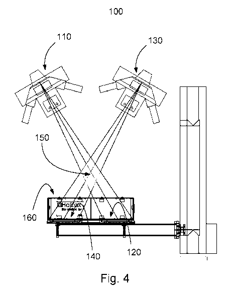
Fig. 4 is a schematic illustration of a stereo orthopaedic radiography imaging
system;
and
Fig. 5 is a schematic illustration of several loading conditions for the knee.
DETAILED DESCRIPTION OF THE INVENTION
Aseptic loosening is a common cause for revision in joint replacement surgery
and is
difficult to diagnose. Not until there is substantial loosening are
radiolucent lines visible
around the implant on standard radiographs. Radio stereometric analysis of
stereo
orthopaedic radiography images is a very accurate measurement technique able
to measure
precise 3D location of implants and host bones. Assessing these positions at
multiple time
points enables production of implant migration curves in multiple dimensions.
Such early
migration measurements have been shown to be able to predict aseptic
loosening. However,
the standard technique is based on the implantation of tantalum markers into a
patient's host
bone at the time of the joint replacement surgery for the purpose of providing
a reference
frame for 3D positioning and migration measurements. If these markers are not
implanted at
the time of surgery, the migration measurements cannot be made due to the lack
of an
accurate reference frame. Post-operative implantation of markers is possible
but carries
significant additional clinical risk if not done in an OR setting and
therefore, is not clinically
practical.
The embodiments of the present disclosure describe devices and methods that
allow
for the assessment of implant loosening without the requirement to have
markers permanently
implanted into the host bone. Specifically, the embodiments of the present
disclosure provide
a set of temporary reference points for accurate implant loosening measurement
using at least
three or more spaced-apart sharp geometry components that temporarily contact
the bone.
The sharp geometry components are housed within holders that are disposed
about a frame
4
CA 02988626 2017-12-07
WO 2016/197249
PCT/CA2016/050661
that is temporarily de-attachably mountable around a subject's joint area of a
host bone that
houses an installed implant.
Some exemplary embodiments of the present disclosure pertain to methods of
imaging the frame, the sharp geometry components and implant using a stereo
orthopaedic
radiography system under two or more loading conditions aimed at loading the
implant of
interest such that a loose implant moves (migrates) relative to the host bone,
and thus, relative
to the temporary reference frame when it is temporarily secured in place
against the host
bone.
Definitions
Unless defined otherwise, all technical and scientific terms used herein have
the same
meaning as commonly understood by one of ordinary skill in the art to which
the invention
belongs.
As used herein, the terms "x-ray" and "radiographic imaging" are used
interchangeably through the application to mean the use of electromagnetic
radiation to view
the internal skeletal structures within a mammalian subject's body.
As used herein, the term "about" refers to an approximately +/-10% variation
from a
given value. It is to be understood that such a variation is always included
in any given value
provided, whether or not it is specifically referred to.
As used herein, the term "sharp geometry component" refers to a cannulated or
solid
sharp contact geometry that can be inserted into and through the skin to
contact the
underlying bone, and which will not move relative to the bone once in contact
with the bone
and secured to a patient's appendage.
For purposes of illustration, the devices and methods of the invention are
described
below with reference to the knee of the human body. However, as will be
appreciated by
those skilled in the art, the devices and methods can be employed with any
mammal and for
any joint wherein an implant has been securely installed. Exemplary
embodiments of the
present disclosure will now be described by reference to Figs. 1 to 5.
5
CA 02988626 2017-12-07
WO 2016/197249
PCT/CA2016/050661
Temporary reference device for providing a temporary reference frame
Persons of skill in the art will recognize that there are a variety of devices
that may be
used to place at least three or more sharp geometry components in contact with
the bone
around a joint replacement, or other type of, implant for the purpose of
measuring implant
loosening. Some exemplary embodiments of the present disclosure relate to a
device
comprising of a frame for encircling and engaging a portion of a subject's
appendage with the
host bone and installed implant (fore example, a knee joint), at least three
attachments
engaged and cooperating with the frame wherein each attachment is configured
to retain a
sharp geometry component and to apply a small load to a sharp geometry
component, and at
least three sterile sharp geometry components able to penetrate the skin and
underlying soft
tissues and to touch the bone without significantly penetrating the bone. It
is within the scope
of the present disclosure for the frame and/or the attachments to comprise a
rigid material, a
semi-rigid material, or a soft material. According to some aspects, more than
three pins
and/or needles may be used for contacting the subject's target bone. According
to some
aspects, the frame may be secured in place about the appendage with the target
joint with one
or more straps, belts, bands, or other type of securing mechanism.
Alternatively, the frame
may be placed into a harness for securing to the subject's appendage.
Fig. 1 illustrates the knee joint area with an implant 30 to be installed into
the tibia 30
(the fibula 25 is shown for reference). As illustrated in Fig. 2, an exemplary
device of the
present disclosure comprises a frame 10 positioned over the tibia 20 into
which a tibial
component 30 of a knee joint replacement has been installed. In this exemplary
embodiment,
three attachment mechanisms 40 are attached to frame 10 wherein each of the
attachment
mechanisms 40 is provided with a retractably extendible sharp geometry
component 50 for
contacting the tibia bone 20. The sharp geometry components are sterile so as
to avoid
infection and are selected for easy entry into and through the skin and
underlying soft tissue,
but which will not: (i) significantly penetrate the cortical bone, and (ii)
easily slide over the
bone surface when a sheer load is applied. These sharp geometry components are
engaged
with a patient's appendage and host bone using proper sterile methods. It is
critical for the
invention that the tips of the sharp geometry components do not change
location for the
duration of the measurement. According to further embodiments of the current
disclosure,
the sharp geometry components may also be inserted through a small stab
incision. One or
more straps 60 may optionally be provided to secure the frame to the subject's
appendage, in
6
CA 02988626 2017-12-07
WO 2016/197249
PCT/CA2016/050661
this case a knee joint area, to keep the frame in place during initial set-up
and/or during the
imaging procedure during which the patient may be required to move.
An exemplary attachment mechanism 40 is illustrated in Fig. 3. The attachment
mechanism 40 is attached to the frame 10 with a two-piece adjustable housing
70a, 70b
which may or may not extend from the frame down to the skin. The sharp
geometry
component 50 is securely mounted in a holder 55 provided in the housing 70b
and held in
place relative to frame 10. The housing 70a is provided with a loading
mechanism 80
exemplified by a spring, which is used to apply a slight force to the sharp
geometry
component 50 such that the sharp geometry component 50 stays in firm contact
with the
underlying bone 90.
Measurement of implant loosening
Migration of an implant relative to its host bone can be measured accurately
using
stereo orthopaedic radiography. Persons of skill in the art will recognize
that there are a
variety of devices that may be used to obtain simultaneous x-ray images of an
implant taken
using two x-ray systems and from two different vantage points (i.e., stereo
orthopaedic
images). In addition, persons of skill in the art will recognize that two
sequential images
using one or two x-ray systems may be used to obtain x-ray images of an
implant taken from
two different vantage points which under appropriate conditions may also
constitute stereo
orthopaedic images. Persons of skill in the art will recognize that the same
method can be
followed using single plane x-ray imaging (e.g., single plane radiology or
fluoroscopy)
instead of stereo orthopaedic radiographs at the expense of possibly losing
out-of-plane
precision and accuracy without limiting the foregoing. Some exemplary
embodiments of the
present disclosure relate to a method for detecting and assessing migration of
an installed
implant wherein the method comprises the steps of securing the device around a
subject's
joint of interest so that each of the sharp geometry component holders is
positioned about a
target location on the host bone, inserting each of the sharp geometry
components through the
subject's skin surface and soft tissue until the tip of the sharp geometry
component touches
the host bone surface, obtaining a first pair of stereo radiographs of the
implant and host bone
area under a first loading condition, placing a second load on the joint,
obtaining a second
pair of stereo radiographs of the implant and host bone area under the loaded
condition,
comparing the first pair of stereo radiographs and the second pair of stereo
radiographs,
detecting if the implant was displaced in the second loaded condition, if a
displacement was
7
CA 02988626 2017-12-07
WO 2016/197249
PCT/CA2016/050661
detected, determining the distance the implant was displaced in the second
loaded condition,
and determine if the displacement distance is indicative of a loosened implant
or not. It is to
be noted that a displacement may be translational or rotational and in may
occur in one or
more dimensions.
Persons of skill in the art will recognize that there are a variety of methods
that may
be used to apply a load to an implant directly or indirectly in an attempt to
induce motion of
the implant relative to the host bone when the implant is loose. Without
limiting the
foregoing, certain embodiments of the present disclosure may load or unload
the joint which
contains the implant of interest by laying down on a table, by bearing weight
or partial weight
on the limb containing the implant or both limbs, by applying a rotatory
moment to the joint
or limb, by applying weights or force directly to the joint, etc.
Referring to Fig. 4, an exemplary stereo radiography system 100 is
illustrated. An x-
ray source 110 is aimed at an x-ray detector 120 at an angle from vertical. In
addition, a
second x-ray source 130 is aimed at an x-ray detector 140 such that the x-ray
beams overlap
in the 3D viewing area 150. As long as the removable reference frame and
implant are placed
in the 3D viewing area under the various loading conditions the accurate
measurement of
implant displacement relative to the reference frame can be made. The stereo
radiography
system 100 may or may not include a reference box 160 containing fiducial and
control
markers to aid in accurately determining the 3D x-ray configuration.
Figs. 5(A)-5(F) illustrates suitable loading conditions for a knee joint for
assessment
with an exemplary method disclosed herein, exemplified by (A) unloading by
lying down
(supine), (B) full weight bearing during standing, (C) partial weight bearing
during standing,
(D) full lunge position, (E) partial lunge position, (F) stair stepping
positions.
8