Note: Descriptions are shown in the official language in which they were submitted.
CA 02988988 2017-12-08
WO 2016/201448 PCT/US2016/037278
PEGYLATED GRANULOCYTE COLONY STIMULATING FACTOR (GCSF)
RELATED APPLICATION
[0001] This invention claims priority to provisional patent applications
U.S. Ser. No.
62/174,373 filed on June 11, 2015, and U.S. Ser. No. 62/184,042 filed June 24,
2015, the
entire contents of which are both incorporated herein by reference.
FIELD OF THE INVENTION
[0002] This invention relates to novel PEG-GCSF conjugates having
unexpected
therapeutic efficacy, while avoiding or substantially reducing the likelihood
of adverse side
effects.
BACKGROUND
[0003] In recent years, non-antigenic water-soluble polymers, such as
polyethylene
glycol ("PEG"), have been used for the covalent modification of polypeptides
of therapeutic
and diagnostic importance. PEG is a polymer that is nontoxic, nonimmunogenic,
highly
water soluble, and readily cleared from the body. PEG has many applications
and is
commonly used in foods, cosmetics, beverages, and prescription medicines.
Pharmaceutical
grade PEGs are approved for use in the United States by the FDA and are widely
used as
biopharmaceutical carriers, given their high degree of biocompatibility.
PEGylation can
modify certain characteristics of biopharmaceuticals without altering their
function, thereby
enhancing the therapeutic effect.
[0004] Neutrophil granulocytes are the most abundant type of white blood
cells in
mammals, and comprise an essential part of the innate immune system. Their
production is
regulated via granulocyte colony stimulating factor (GCSF) engagement to its
cognate
receptor located on the surface of CD34+ myeloid precursor cells. Receptor
engagement
results in receptor chain oligomerization, rearrangement and signal
transduction mediated
through intracellular kinases, resulting in gene expression patterns promoting
differentiation
and cell division, thereby increasing neutrophil counts. The importance of
GCSF receptor
signaling is exemplified in individuals with inborn genetic errors in the
cytokine:receptor
signaling pathway. Collectively, limited signaling will result in a reduced
ability to maintain
appropriate levels of neutrophils. Signaling via the GCSF receptor is
important for the
production and maintenance of neutrophils, and individuals with inborn genetic
errors in
CA 02988988 2017-12-08
WO 2016/201448 PCT/US2016/037278
GCSF signaling have reduced neutrophil counts and therefore are predisposed to
serious and
recurrent microbial infections.
[0005] Similarly, many cancer therapies exhibit potent inhibition of
neutrophil levels
due to their anti-proliferative activity. One of the most serious potential
side effects of many
types of chemotherapy drugs is a low white blood cell count that includes
decreased
neutrophil levels (neutropenia). Neutropenia can put some patients at risk for
severe
infections and therefore may force cessation of chemotherapy treatment cycle.
In fact,
complications associated with a low white blood cell count are the most common
causes of
dose reductions or delays in chemotherapy (see Link, et al. (2001) Cancer
92:1354-1367;
Lyman, et al. (2003) 1 Clin. Oncol. 21:4524-4531; and Lyman, et al. (2002) Am.
i Med.
112:406-411, the entirety of each of which are incorporated herein by
reference). This dose-
dependent phenomenon has dramatically limited the therapeutic dosages of many
oncology
drugs.
[0006] The development of recombinant GCSF (filgrastim) for clinical use
has led to
dramatic improvement of both individuals born with severe chronic neutropenia
(SCN) as
well as those undergoing cancer therapies with potent anti-neutrophil
activity. GCSF is a
small protein that is readily removed through the renal system. The mouse
version of GCSF
was purified from explanted tissues in 1983, and the human equivalent purified
from a cancer
cell line grown in culture inadvertently expressing GCSF in high
concentrations in 1985 (see
e.g. Welte, et al. (1985) PNAS USA 82:1526-30, which is incorporated herein by
reference in
its entirety). The human GCSF was found to be a glycoprotein around 19kD which
was
variably acidic depending on the carbohydrate component. It was later found
that the
carbohydrate component was optional for biologic activity. The cloning and
characterization
of human recombinant GCSF took place between 1984 and 1986, and led to its
expression in
E. coli cells and eventually to human clinical trials testing the compound in
patients suffering
from chemotherapy-induced neutropenia. In 1991, recombinant human GCSF made in
E.
coli was approved by the U.S. FDA for this use (named Filgrastim, trade-named
Neupogeng), and in 1993 a related Chinese hamster ovary cell expressed form
was approved
in Europe (under the name lenograstim). It was found that the core protein
included 174
amino acids, although multiple variants are known to exist (see e.g. Ngata, et
al. (1986)
Nature 319:415-18; Souza, et al. (1986) Science 232:61-5; U.S. Patent No.
4,999,291, each of
which are incorporated herein by reference).
2
CA 02988988 2017-12-08
WO 2016/201448 PCT/US2016/037278
[0007] U.S. Patent Nos. 4,810,643, 4,999,291, 5,582,823 and 5,580,755,
assigned to
Amgen, Inc. and claiming priority back to U.S. Patent Application 07/768,959,
filed August
23, 1985, provide certain human pluripotent GCSF molecules and methods of
their
production, each of which are incorporated herein by reference in their
entirety. These
molecules form the basis for the approved Neupogen product. There is no
discussion of
potential PEGylation of the molecule in these cases.
[0008] Because Filgrastim is readily degraded in vivo, Neupogen requires
daily
administration during an incidence of febrile neutropenia brought on by cancer
treatments.
However, PEGylation represents a plausible approach to increasing the
hydrodynamic radius
of the GCSF protein, reducing serum clearance and promoting drug half-life in
vivo. Using
site-specific PEGylation at the N-terminus of GCSF with aldehyde-activated, 20
kDa linear
PEG (see PCT Publication No. WO 96/11953 as well as U.S. Patent Nos. 5,824,784
and
7,090,835), PEG-Filgrastim was developed, and was approved by the U.S. FDA in
2002
under the tradename NEULASTA (ID (NEULASTA (ID [package insert]. Thousand
Oaks, CA,
Amgen, Inc., revised 02/2010; NEULASTA (ID [package insert]. Thousand Oaks,
CA,
Amgen, Inc., revised 4/2016 vl, both revisions incorporated herein by
reference). This
mono-PEGylated version of GCSF, with the PEG moiety covalently attached to the
amino
terminus of the protein, increases the molecular weight of the GCSF protein,
greatly reducing
renal clearance. The location of the PEG group at the amino terminus is not
particularly
disruptive to the GCSF protein ¨ GCSF receptor interaction, since the protein
residues in the
binding region involved in receptor interaction are not directly PEGylated or
sterically
hindered by the amino terminal 20 kDa PEG
[0009] Several alternate strategies for providing a stabilized GCSF
molecule also
have been proposed. Linking PEG to a cysteine residue has provided certain
improvements
in targeting. Thiol reactive PEGs (including PEG-maleimide) have been linked
to GCSF at its
free cysteine residue. Veronese, et al. (2007) Bioconjugate Chem. 18:1824-1830
described the
PEGylation of GCSF at Cys18, which was shown to increase aggregation although
the
aggregates were not covalently aggregated. Similarly, Hao, et al. (2006)
Biodrugs 20:357-
363 described the conjugation of PEG-maleimide to Cys18, which was shown to
increase the
half life of the molecule.
[0010] Site-specific mutagenesis is a further approach which has been
used to prepare
polypeptides for site-specific polymer attachment. For example, U.S. Patent
No. 6,646,110
describes polypeptide conjugates that exhibit GCSF activity and have an amino
acid residue
3
CA 02988988 2017-12-08
WO 2016/201448 PCT/US2016/037278
that comprise an attachment group for a PEG or oligosaccharide moiety
inserted. These can
include lysine, glutamic acid, cysteine or aspartic acid.
[0011] WO 2011/041376 reflects yet another approach to site-specific
PEGylation, by
one of the inventors of the instant application. The contents of WO
2011/041376 are
incorporated by reference in its entirety. In this earlier work, methoxy-PEG
acetaldehyde
was reacted with GCSF in a DMSO-containing reaction buffer to yield a
population of
monoPEGylated GCSF conjugates, wherein the conjugation is at a lysine group
near the N-
terminus, and wherein at least 30% of the composition is not N-terminally
PEGylated. In an
alternate embodiment, the composition comprised at least 80% monoPEGylated
GCSF
conjugate, wherein at least 30% of the composition is not N-terminally
PEGylated.
[0012] As an alternative to site-specific PEGylation, random PEGylation
using N-
hydroxy-succinimide esters forms stable protein-PEG conjugates via amide
bonds. These
ester reagents are relatively specific for the reaction with amino groups of
the lysine residues
and the N-terminus, but react to minor degrees also with other protein
nucleophiles like
histidine, serine and tyrosine residues. Reaction conditions like temperature,
pH, amount of
PEG reagent, and time define the heterogeneity of the product (i.e., mono-, di-
, tri- and
higher-PEGylated conjugates can be formed). Due to reactions with different
nucleophilic
groups on the protein, multi-PEGylated (and even mono-PEGylated) conjugates
yield
positional isomers that can differ substantially in their biological and
biomedical properties.
The high degree of PEGylation variability, as well as the capability to
manufacture in a
reproducible manner, has limited the use of SC-PEG in clinical drug
development. However
there are examples (e.g., Oncaspar, Adagen) that demonstrate such conjugates
can be
clinically relevant in some situations.
[0013] Prior attempts to employ amine-reactive PEGs to form PEG-GCSF by
attaching at exposed amine groups on lysine residues and N-terminal amino
acids have been
reported with limited success. It was observed that such an approach is not
optimal for GC SF
because the protein contains four lysine residues and an N-terminal amino acid
with the
lysine residues located in receptor binding regions. Modification of GCSF with
amine-
reactive PEG reagents therefore reduces in vitro biological activity of the
protein by 3- to 50-
fold, depending on the number and sizes of attached PEG molecules. Loss of in
vitro
bioactivity is greatest when GCSF is modified with large PEGs, e.g., 20 kDa
PEGs, which are
most useful in extending the protein's half-life. Amine-PEGylated GCSF is
heterogeneous,
4
CA 02988988 2017-12-08
WO 2016/201448 PCT/US2016/037278
occurring as a complex mixture of at least four isoforms and multiple
molecular weight
species, all of which may have different specific activities.
[0014] A particular example of this approach is described in two journal
articles from
the early 1990s, by a group of pharmaceutical investigators at the Kirin
Brewery Company:
Tanaka et al. (1991) Cancer Research 51:3710-3714 and Satake-Ishikawa et al.
(1992) Cell
Structure and Function 17:157-160. These investigators prepared mixtures of
conjugates
wherein each molecule of the GCSF protein apparently was modified by one, two,
or three
PEGs, with an average of two. The activated PEG reagent utilized by these
investigators was
SS-PEG (4.5 kDa or 10 kDa). Although the resulting amide bond between the
protein and
PEG is stable, the linker contains an ester group which is hydrolytically
labile. Such
hydrolysis will occur as long as the compound is in an aqueous medium and,
therefore, the
PEG number continuously decreases as long as it is in solution. Hydrolysis of
the ester
linkage leaves behind a succinate group which can cyclize to a succinimidyl
group. Such
non-natural residues can potentially result in antibody responses, including
immunogenicity.
[0015] Side effects associated with known versions of GCSF, including
PEGylated
versions, include dose-related glomerulonephritis and adverse and serious
adverse events of
bone pain. This has resulted in many cancer patients suffering through painful
treatment
periods or, in some cases, reduction or cessation of all treatments due to
kidney damage
and/or bone pain serious adverse effects. The side effects with filgrastim or
PEG-filgrastim
are associated with dosage levels. Therefore, newer versions of GCSF
(preferably PEGx-
GCSF with improved PK profiles) are warranted to provide a clinically
beneficial increase of
neutrophils with reduced side effects. A drug formulation that can yield
neutrophil increases
similar to current treatments, but at lower dosages, therefore would be a
desirable approach to
improving conditions associated with neutropenia.
[0016] An additional, potential consequence of long-term GCSF therapy is
the
increased chance of developing a malignancy. Patients with severe chronic
neutropenia
(SCN), who require life-long GCSF therapy, are at an increased risk for
myelodysplastic
syndrome that is directly proportional to the time they have been treated with
GCSF. It also
is known that GCSF may exacerbate myelogenous cancers. Therefore, Neupogen is
not
recommended in patients with, e.g., myelodysplastic syndrome, chronic
myelogenous
leukemia, and secondary Acute Myeloid Leukemia (AML). Accordingly, it also
would be
advantageous to provide a GCSF therapy having proliferative activity that is
more selective
CA 02988988 2017-12-08
WO 2016/201448 PCT/US2016/037278
for normal cells, and therefore avoids or reduces the proliferation of cancer
cells, as
compared with currently existing treatments.
SUMMARY OF THE INVENTION
[0017] Embodiments of the invention are directed to PEGx-GCSF, wherein x
represents the number of PEG per GCSF and is an integer ranging from 4 to 8.
[0018] In embodiments of PEGx-GCSF, the PEG moiety has an average
molecular
weight from about 3 to about 15 kDa, or preferably from about 5 to about 6
kDa.
[0019] In certain embodiments of the inventive PEGx-GCSF, PEG is attached
to
GCSF through an amine originating from GCSF. In alternative embodiments, the
PEGx-
GCSF comprises a non-hydrolyzable linkage, for example, a urethane linkage.
[0020] In additional embodiments of the inventive PEGx-GCSF, GCSF is a
protein
having an amino acid sequence selected from the group consisting of SEQ ID NO:
1, SEQ ID
NO: 3, SEQ ID NO: 4, and functional derivatives and homologs thereof In
further
embodiments, the GCSF amino acid sequence is SEQ ID NO: 1 and each PEG is
attached to
a GCSF position selected from the group consisting of: the N-terminus, a
lysine residue at
position 17, a lysine residue at position 35, a lysine residue at position 41,
a histidine residue
at position 44, a histidine residue at position 53, a histidine residue at
position 80, a histidine
residue at position 157 and a histidine residue at position 171.
[0021] Embodiments of the invention are also directed to PEG[x]-GCSF, a
composition that comprises a population of PEGx-GCSF, wherein [x] is the
average value of
x for the population, and wherein [x] is greater than or equal to about 4;
wherein [x] is from
about 4 to about 8; wherein [x] is from about 4 to about 6; or wherein [x] is
from about 5 to
about 6.
[0022] In certain embodiments, PEG[x]-GCSF is characterized by one or
more of the
following: PEG[x]-GCSF comprises less than 10% PEGx-GCSF wherein x is from 1
to 3;
PEG[x]-GCSF comprises at least about 15% PEGx-GCSF wherein x is 4; PEG[x]-GCSF
comprises at least about 30% PEGx-GCSF wherein x is 5; PEG[x]-GCSF comprises
at least
about 10% PEGx-GCSF wherein x is 6; and PEG[x]-GCSF comprises less than 15%
PEGx-
GCSF wherein x is 7.
6
CA 02988988 2017-12-08
WO 2016/201448 PCT/US2016/037278
[0023] In
additional embodiments, PEG[x]-GCSF comprises at least about 15%
PEGx-GCSF wherein x is in the range from 6 to 7; or comprises at least about
35% PEGx-
GCSF wherein x is in the range from 5 to 7.
[0024]
Additional embodiments are directed to a pharmaceutical formulation
comprising a pharmaceutically active amount of PEGx-GCSF or PEG[x]-GCSF and a
protein-free carrier.
[0025]
Additional embodiments of the invention are directed to a method for
preparing inventive PEGx-GCSF, wherein x is from 4 to 8, or PEG[x]-GCSF,
wherein
[x] is 4 or greater, the method comprising the steps of: (a) obtaining a GCSF
solution
having a concentration of at least about 5.0 mg/ml; (b) combining the GCSF
solution with
PEG; wherein the molar amount of PEG is about 65 to about 75 times the molar
amount of
the GCSF; (c) allowing sufficient time for the GCSF and PEG to react to
produce PEGx-
GCSF; (d) adding hydroxylamine in an amount sufficient to react with residual
PEG; and (e)
isolating PEG[x]-GCSF from unreacted PEG; N-hydroxysuccinimide and
hydroxylamine.
Individual PEGx-GCSF are further isolated from the population by methods known
in the art
for isolating purified protein conjugates, including methods of separating
according to
molecular weight.
[0026] The
compositions of the invention provide unexpected utility in the treatment
of various medical conditions where existing, commercially available GCSF
and/or PEG-
GCSF treatments may be contraindicated due to the occurrence of bone pain or
the risk of
cancer cell proliferation.
Such medical conditions include severe congenital/chronic
neutropenia, autoimmune/idiopathic neutropenias, as well as neutropenias
associated with the
treatment of cancers.
[0027]
Additional advantages of the present invention will be readily apparent to
those skilled in this art from the following detailed description, wherein
only certain
embodiments of the invention are shown and described. As will be realized, the
invention is
capable of other and different embodiments, and its several details are
capable of routine
modifications in various respects, all without departing from the invention.
The present
invention may be practiced without some or all of these specific details.
Accordingly, the
description is to be regarded as illustrative in nature, and not as
restrictive.
7
CA 02988988 2017-12-08
WO 2016/201448 PCT/US2016/037278
BRIEF DESCRIPTION OF THE FIGURES
[0028] Example embodiments of the disclosure may be understood by
referring, in
part, to the present disclosure and the accompanying drawings, which are
briefly described
below.
[0029] FIG 1 represents the amino acid sequence of the predominant, fully
processed
human granulocyte colony stimulating factor ("GCSF") (SEQ ID NO: 1). The
corresponding
DNA sequence is provided as SEQ ID NO: 2.
[0030] FIG 2 describes the sequencing data for GCSF proteins used in
certain
embodiments of the invention described herein. FIG 2 describes SEQ ID NO: 3
and SEQ ID
NO: 4, respectively.
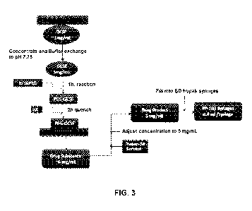
[0031] FIG 3 provides a flow chart of a process for preparing the
inventive PEGx-
GCSF and PEG[x]-GCSF.
[0032] FIG 4 is a representative bioanalyzer electropherogram obtained
from analysis
of inventive PEG[x]-GCSF samples.
[0033] FIG 5 is a representative of SDS-PAGE analysis results for
inventive PEG[x]-
GCSF samples.
[0034] FIG 6 is a potency graph illustrating results from a bioassay of
inventive
PEG[x]-GCSF (ANF-Rho) compared to a commercial PEG-GCSF (NEULASTA ,
NLSTA). M-NFS-60 cells were treated with indicated GCSF compounds for 48 hours
prior
to viability staining. Data were normalized to untreated controls and fit to a
three parameter
logistic curve-fit model. Data represents mean and standard error of duplicate
wells.
[0035] FIG 7 is a quadrant gating of bivariate plots of fluorescent
intensity of CD66
and CD14 cells, showing the effect of inventive PEG[x]-GCSF (ANF-Rho) vs.
NEULASTA
on human CD34(+) cells in vitro. Cells were treated with 50 ng per ml of
either the
inventive PEG[x]-GCSF (ANF-Rho) or NEULASTA for 14 days prior to surface
staining
for CD66 and CD14. Antigen expression was quantified by flow cytometry. Cell
events
appearing in the E4 gate are indicative of the granulocyte population.
[0036] FIG 8 are graphs of single-dose pharmacokinetics of various
commercial
PEG-GCSF (NEULASTA (ID) and inventive PEG[x]-GCSF samples in neutropenic rats.
Rats
were made neutropenic by injection of cyclophosphamide (CPA) on Day -1. Four
different
concentrations of inventive PEG[x]-GCSF (ANF-Rho) and NEULASTA were
administered
8
CA 02988988 2017-12-08
WO 2016/201448 PCT/US2016/037278
subcutaneously at the indicated dosages on Day 1. Blood samples were obtained
from the
rats on the days indicated, and GCSF plasma concentrations were determined by
ELISA.
Data are mean and standard error for 8 rats per group. FIG 8A is a linear
plot, and FIG 8B is
a log plot of GCSF concentration.
[0037] FIG 9 are graphs showing the plasma exposure effects of commercial
PEG-
GCSF (NEULASTA (ID) and inventive PEG[x]-GCSF samples (lots 1-3) in
neutropenic rats.
FIG 9A shows area under the curve (AUC) for three individual lots of inventive
PEG[x]-
GCSF at three different concentrations (100, 50 and 25 microgram per kilogram,
i.e., [tg/kg)
and a single concentration of NEULASTA (ID (100 [tg/kg). Asterisk indicates
significant
difference in inventive PEG[x]-GCSF AUC at indicated dosages compared to
NEULASTA (ID
administered at 100 microgram per kilogram (m/kg). FIG 9B shows linearity of
PEG-GCSF
plasma exposure and dosage. AUC values for Lot 1, Lot 2, and Lot 3 of
inventive PEG[x]-
GCSF were pooled and subjected to linear regression analysis. Pooled mean and
95%
confidence intervals are shown above each data set. Dotted line and shaded
area indicates
mean AUC and upper and lower 95% confidence intervals of 100 [tg/kg NEULASTA
(ID
treatment group. Asterisks indicate significant differences (p(0.05) by ANOVA
and Dunnet's
multicomparison tests post-hoc analysis as compared to NEULASTA (ID treated
group.
[0038] FIG 10 is a graph showing representative changes in neutrophil
cell counts in
neutropenic rats treated daily with inventive PEG[x]-GCSF lot 1, NEULASTA (ID
or
formulation buffer (FB). Rats were made neutropenic by injection of
cyclophosphamide
(CPA) on Day -1. On Day 1 and after, rats received daily injections of PEG[x]-
GCSF (100
[tg/kg), NEULASTA (ID (100 [tg/kg) or vehicle solution (FB). Blood samples
were obtained
from the rats on the days indicated to determine absolute neutrophil counts
(ANC). Data are
means and standard error for 8 rats per group. Shaded area indicates ANC
values associated
with initial neutrophil release, which were not included in area under the
curve calculations.
[0039] FIG 11 illustrates the absolute neutrophil counts (ANC) from
PEG[x]-GCSF
and NEULASTA (ID dosed neutropenic rats. FIG 11A shows ANC values plotted as a
function of hours post administration to determine AUC, using the second rise
ANC peak
shown in FIG 10. Values for each PEG[x]-GCSF lot at each dose were pooled, and
data prior
to 96 hours (representing release of pre-formed neutrophils) was excluded from
analysis.
Asterisks above and below data set represent significant difference (p(0.05)
by ANOVA of
pooled dosages of PEGx-GCSF compared to formulation buffer and NEULASTA (ID
treatment groups, respectively. FIG 11B shows data grouped of the three
concentrations
9
CA 02988988 2017-12-08
WO 2016/201448 PCT/US2016/037278
from three separate lots of the PEG[x]-GCSF at 25 [tg/kg (squares), 50 [tg/kg
(inverted
triangles) and 100 [tg/kg (circles). The shaded area represents the values for
100 [tg/kg
NEULASTA (ID and 95% CI (confidence interval). Correlation analysis between
ANC-AUC
and Plasma AUC of all lots with an r2 value of 0.64 indicates significant
correlation between
drug levels and ANC pharmacodynamics.
DETAILED DESCRIPTION
DEFINITIONS
[0040] "Substantially homologous," in reference to an amino acid
sequence, is
defined herein as a sequence with at least 70%, typically at least about 80%,
and more
typically at least about 90% identity to another amino acid sequence, as
determined by the
FASTA search method in accordance with Pearson and Lipman, Proc. Natl. Acad.
Sci. USA
85, 2444-2448 (1988).
[0041] As used herein, the term "N-terminus," "amino-terminus," or
analogous terms
when used in the context of a covalent linkage of a protein to another
molecule refer to a
covalent linkage via the amino-terminal a-amino group of the protein.
[0042] As used herein, the term "wild type" or "native" refers to a
protein or
polypeptide in its operative or functional form, typically as it is found
naturally functioning in
the body. These terms also refer to the protein in a form in which it has not
been artificially
modified or altered. The terms can thus relate to recombinant proteins.
Accordingly, the terms
can refer to a protein with an altered glycosylation pattern, including lack
of glycosylation,
relative to that as produced in the animal from which the nucleic acid and/or
amino acid
sequence of the protein was originally derived.
[0043] The term "ANF-Rho" is used herein to refer to an exemplary sample
of
PEG[x]-GCSF of the present invention used in the Examples. See, e.g., Example
3 and Table
2.
[0044] NEULASTA (ID is the brand name of PEGfilgrastim, a PEGylated form
of the
recombinant human granulocyte colony-stimulating factor (GCSF) analog
filgrastim. The
drug is prepared by coupling a 20 kDa polyethylene glycol (PEG) molecule to
the N-terminus
of the filgrastim protein.
CA 02988988 2017-12-08
WO 2016/201448 PCT/US2016/037278
GCSF
[0045] In general, a GCSF protein useful in the practice of this
invention may be of
any form isolated from mammalian organisms, a product of prokaryotic or
eukaryotic host
expression of exogenous DNA sequences obtained by genomic or cDNA cloning or
by DNA
synthesis or alternatively a product of chemical synthetic procedures or by
endogenous gene
activation. Thus, the protein can be of a natural or recombinant source
obtained from tissue,
mammalian/microbial cell cultures, plant cell cultures, transgenic animals,
yeasts, fungi and/or
transgenic plants. Suitable prokaryotic hosts include various bacteria such as
E. colt; suitable
eukaryotic hosts include yeasts such as S. cerevisiae or Pichia pastoris,
mammalian cells
such as Chinese hamster ovary cells or monkey cells, transgenic animals such
as mice,
rabbit, goat, sheep, insect or plant cell culture and transgenic plants such
as
Physcomitrellapatens (a moss). Depending upon the host employed, the protein
expression
product may be glycosylated with mammalian, plant or other eukaryotic
carbohydrates, or it
may be non-glycosylated.
[0046] As used herein, the term "GCSF" or granulocyte colony stimulating
factor
includes a protein having the amino acid sequence set out in SEQ ID NO: 1 (FIG
1) or an
amino acid sequence substantially homologous thereto, whose biological
properties relate to
the stimulation of white blood cell production. As used herein, the term GCSF
includes such
proteins modified deliberately, as for example, by site directed mutagenesis,
or accidentally
through mutations; such that they have additions, deletions, or substitutions
of amino acid
residues with respect to native GCSF. These terms include both natural and
recombinantly
produced human GCSF. GCSF refers to both the naturally occurring or
recombinant protein,
typically human, as obtained from any conventional source such as tissues,
protein synthesis,
cell culture with natural or recombinant cells.
[0047] A GCSF expression product useful in the practice of the invention
may also
include an initial methionine amino acid residue at position 1. The present
invention
contemplates the use of any and all such forms of GCSF, although recombinant
GCSF,
especially E. co/i-derived, is typical. Certain GCSF analogues have been
reported to be
biologically functional, and these may also be conjugated according to the
present invention.
These GCSF analogues may include those having amino acid additions, deletions
and/or
substitutions as compared to the GCSF amino acid sequence according to SEQ ID
NO: 1. In
certain embodiments, the sequence includes an insertion of amino acids as
compared to SEQ
11
CA 02988988 2017-12-08
WO 2016/201448 PCT/US2016/037278
ID NO: 1, such as, for example, an insertion of VSE at positions 36, 37 and 38
of SEQ ID
NO: 1. In certain embodiments, the sequence is as in SEQ ID NO: 3 or SEQ ID
NO: 4.
[0048] The term "GCSF" as used herein encompasses proteins having the
activity of
GCSF described above, including the natural human glycoprotein GCSF, mutants
of GCSF,
glycosylated GCSF, non-glycosylated GCSF and/or otherwise modified structural
and/or
functional variants of GCSF. In a further embodiment, GCSF has the amino acid
sequence
identified in SEQ ID NO: 1 that corresponds to recombinant GCSF produced in
bacteria,
having 174 amino acids and an extra N-terminal methionyl residue. Amino acid
sequences of
biologically active GCSF, which differ from SEQ ID NO: 1 in that they do not
contain a
methionyl residue at position 1, are also included.
PEG
[0049] The term "PEG" generally refers to a polyalkylene glycol compound
or
derivative thereof, with or without linkers or activating moieties. The term
PEG as used
herein includes, but is not limited to, polyethylene glycol homopolymers,
copolymers of
ethylene glycol with propylene glycol and derivatives and equivalents thereof,
wherein said
homopolymers and copolymers are unsubstituted or substituted, for example, at
one end with
an alkyl group. The PEG polymers for use with the present invention can be
linear, branched,
comb or star-shaped with a wide range of molecular weights. The average
molecular weight
of the PEG for use with embodiments of the present invention can range from 5
to about 100
kDa.
[0050] Numerous derivatives of PEG and methods for making them and
conjugating
them to a protein are known in the art and are suitable for use in the present
invention. One
particularly preferred PEG for use in the invention is a PEG having one end of
the polymer
terminating with a relatively inert group, such as a lower C1.6 alkoxy group.
Preferably, the
PEG is a monomethoxy-PEG (commonly referred to as mPEG), which is a linear
form of
PEG wherein one terminus of the polymer is a methoxy (--OCH3) group.
[0051] Even more preferably, the PEG used in the invention is an
"activated mPEG"
in which one end of the linear PEG terminates with a methoxy group and the
other end
terminates with a linker appropriate for coupling to the preferred sites on
GCSF in order to
facilitate PEGylation with a desired activated mPEG
12
CA 02988988 2017-12-08
WO 2016/201448 PCT/US2016/037278
[0052] Preferred linkers include amine reactive linkers, i.e., synthetic
chemical
groups that will form chemical bonds with primary amines. These include
isothiocyanates,
isocyanates, acyl azides, NHS esters, sulfonyl chlorides, aldehydes, glyoxals,
epoxides,
oxiranes, carbonates, aryl halides, imidoesters, carbodiimides, anhydrides,
and fluorophenyl
esters. Most of these amine reactive linkers conjugate to amines by either
acylation or
alkylation.
[0053] Exemplary linkages are hydrolytically stable, and water soluble.
Representative suitable linkers can comprise any combination of amide, a
urethane (also
known as carbamate), amine, thioether (also known as sulfide), or urea (also
known as
carbamide) groups.
[0054] In a particular embodiment, methoxylated PEG ("mPEG") can be
activated for
subsequent covalent attachment to amino groups by methods well known in the
art, i.e.,
mPEG can be modified to contain varying reactive moieties suitable for
subsequent
attachment to proteins via amino acid residues containing available amino
residues, e.g.,
lysinyl residues. Such activated PEGs include mPEG-succinimidyl succinate ("SS-
PEG"),
mPEG-succinimidyl carbonate ("SC-PEG"), mPEGimidate, and mPEG-cyanuric
chloride. In
a preferred embodiment, the linkers are selected to provide PEG-GCSF linkages
that are
stable to hydrolysis.
[0055] In certain embodiments, the average molecular weight of the PEG
for use with
the present invention is in the range from about 2 to about 50 kDa, about 3 to
about 25 kDa,
about 4 to about 10 kDa or any subrange defined by two of the endpoints
provided herein,
including any single number integer (whole number) or non-integer (fraction)
found within
these ranges such as 4.5, 5, 5.6 and 6 kDa.
[0056] In a particular embodiment, the PEG for use in the various
embodiments of the
present invention has an average molecular weight of about 5 to 6 kDa SC-PEG;
more
particularly about 5.6 kDa SC-PEG The PEGx-GCSF resulting from reaction of
GCSF
primary amino groups with SC-PEG comprises urethane linkages that are stable
to
hydrolysis, unlike the hydrolytically labile linkages in the multi-PEGylated
conjugates of the
Tanaka et al. and Satake-Ishikawa et al., discussed above.
PEGx-GCSF
13
CA 02988988 2017-12-08
WO 2016/201448 PCT/US2016/037278
[0057] One embodiment of the present invention is directed to "PEGx-GCSF"
which,
as defined herein, is a GCSF conjugate comprising x number of PEG moieties
covalently
attached thereto, wherein x is an integer from 4 to 7. Particular embodiments
of PEGx-GCSF
include wherein x is 4, 5, 6, 7 and 8.
[0058] In certain embodiments, each PEG is attached to GCSF through an
amine
moiety originating from GCSF, for example, the N terminus, or any lysine or
histidine
residue. In these particular embodiments, covalent attachment is formed by
reaction between
PEG activated with an amino-reactive linker and a GCSF amine moiety. In
particular
embodiments, upon reaction with an amine, the amino-reactive linker forms a
non-
hydrolysable linkage to GCSF. In further embodiments, PEGx-GCSF comprises a
non-
hydrolysable linkage, for example, a urethane linkage.
[0059] Embodiments of PEGx-GCSF include wherein GCSF is a protein having
an
amino acid sequence selected from the group consisting of SEQ ID NO: 1, SEQ ID
NO: 3,
SEQ ID NO: 4, and functional derivatives and homologs of any of these
sequences. In
particular embodiments, the amino acid sequence is SEQ ID NO: 1, a functional
derivative of
SEQ ID NO: 1, or a homolog of SEQ ID NO: 1, where the GCSF has a lysine
residue at
position 17, a lysine residue at position 35, a lysine residue at position 41,
and optionally, a
histidine residue at position 44, a histidine residue at position 53, a
histidine residue at
position 80, a histidine residue at position 157 and a histidine residue at
position 171. In
related embodiments of PEGx-GCSF, each PEG is attached to GCSF at a position
selected
from the group consisting of: the N-terminus, the lysine residue at position
17, the lysine
residue at position 35, the lysine residue at position 41, and optionally at
the histidine residue
at position 44, the histidine residue at position 53, the histidine residue at
position 80, the
histidine residue at position 157 and the histidine residue at position 171.
[0060] Specific embodiments are directed to PEGx-GCSF, where GCSF is a
protein
having the amino acid sequence of SEQ ID NO: 1, and wherein each PEG is
attached to a
GCSF originating amine, such as the N-terminus, a lysine, or a histidine
residue. For
example, embodiments where PEG is attached to a GCSF position selected from
the group
consisting of: the N-terminus, a lysine residue at position 17, a lysine
residue at position 35,
and a lysine residue at position 41. In a further embodiment, PEG is attached
to a GCSF
position selected from the group consisting of: a histidine residue at
position 44, a histidine
residue at position 53, a histidine residue at position 80, a histidine
residue at position 157
and a histidine residue at position 171 .
14
CA 02988988 2017-12-08
WO 2016/201448 PCT/US2016/037278
[0061] In certain embodiments of PEGx-GCSF, the average molecular weight
of PEG
is in the range from about 2 to about 50 kDa, about 3 to about 25 kDa, about 4
to about 10
kDa or any sub-range defined by two of the endpoints provided herein,
including any single
number integer (whole number) or non-integer (fraction) found within these
ranges such as
4.5, 5, 5.6 and 6 kDa. Particular embodiments of PEGx-GCSF include those
wherein PEG
has an average molecular weight from about 5 to about 6 kDa, preferably 5.6
kDa.
PEG[x]-GCSF
[0062] An embodiment of the invention is directed to "PEG[x]-GCSF" which,
as
defined herein, is a composition that comprises a population, including
various proportions of
any individual PEGx-GCSF described above, wherein [x] is the average value of
x for the
population, and wherein [x] is a positive number (including fractional values)
greater than or
equal to about 4, for example from about 4 to about 8; from about 4 to about
6; or from about
to about 6. PEG[x]-GCSF encompasses embodiments comprising a "heterogeneous
population" wherein the PEGx-GCSF conjugates have different values of x,
wherein PEG is
attached at different sites on GCSF molecules, and/or wherein the PEG has
different
molecular weights.
[0063] Embodiments of PEG[x]-GCSF include populations of any of the
various
individual PEGx-GCSFs described herein, including PEGx-GCSF wherein the
average
molecular weight of PEG is in the range from about 2 to about 50 kDa, about 3
to about 25
kDa, about 4 to about 10 kDa. Particular embodiments of PEGx-GCSF include
those wherein
PEG has an average molecular weight from about 5 to about 6 kDa, preferably
5.6 kDa.
[0064] Certain embodiments of PEG[x]-GCSF are characterized by one or
more of
the following: comprising less than 10%, less than 8% or less than 5% PEGx-
GCSF wherein
x is from 1 to 3; comprising at least about 15%, at least about 18%, at least
about 20%, at
least about 25%, or at least about 30% PEGx-GCSF wherein x is 4; comprising at
least about
30%, at least about 35%, or at least about 40% PEGx-GCSF wherein x is 5;
comprising at
least about 10%, at least about 12%, or at least about 15% PEGx-GCSF wherein x
is 6;
comprising at least about 3%, at least about 5%, and/or less than about 15%
PEGx-GCSF
wherein x is 7; comprising at least about 15%, at least about 20%, at least
about 25%, or at
least about 35% PEGx-GCSF wherein x is in the range from 6 to 7; and
comprising at least
about 35%, at least about 40%, at least about 45%, at least about 50%, at
least about 60%, at
CA 02988988 2017-12-08
WO 2016/201448 PCT/US2016/037278
least about 75% or at least about 80% PEGx-GCSF wherein x is in the range from
5 to 7.
Additional embodiments of PEG[x]-GCSF may include those comprising less than
about
15%, less than about 12%, or less than about 10% PEGx-GCSF wherein x is 7.
[0065] Embodiments of PEG[x]-GCSF include populations of any of the
various
individual PEGx-GCSFs described herein, wherein [x], the average value of the
x for the
population, is greater than 4. For example, embodiments of PEG[x]-GCSF include
compositions of PEGx-GCSF where PEG is attached to GCSF through an amine
originating
from GCSF, such as the N-terminus, a lysine, or a histidine; wherein PEGx-GCSF
comprises
a non-hydrolyzable linkage, for example a urethane linkage; wherein GCSF is a
protein
having an amino acid sequence of SEQ ID NO: 1, SEQ ID NO: 3, SEQ ID NO: 4, and
functional derivatives and homologs of any one of these sequences; wherein
each PEG is
attached to GCSF according to SEQ ID NO: 1 or a derivative or homologue
thereof, at a
position selected from the group consisting of: the N-terminus, the lysine
residue at position
17, the lysine residue at position 35, the lysine residue at position 41, the
histidine residue at
position 44, the histidine residue at position 53, the histidine residue at
position 80, the
histidine residue at position 157 and the histidine residue at position 171.
[0066] In some embodiments, PEG[x]-GCSF comprises a population of PEGx-
GCSF
characterized by one or more of the following:
from about 0% to about 5% of PEGx-GCSF wherein x is 3;
from about 22% to about 32% of PEGx-GCSF wherein x is 4;
from about 38% to about 42% of PEGx-GCSF wherein x is 5;
from about 18% to about 28% of PEGx-GCSF wherein x is 6; and
from about 0% to about 9% of PEGx-GCSF wherein x is 7.
[0067] Another embodiment of PEG[x]-GCSF comprises a population of PEGx-
GCSF wherein the PEG is attached to the GCSF through a urethane linkage and,
optionally,
wherein the PEG has an average molecular weight molecular weight from about 3
to about 15
kDa, more preferably, from about 5 to about 6 kDa. In a particular embodiment,
the PEG[x]-
GCSF consists of a population of PEGx-GCSF wherein the PEG is attached to the
GCSF
through a urethane linkage and, optionally, wherein the PEG has an average
molecular weight
molecular weight from about 3 to about 15 kDa, more preferably, from about 5
to about 6
kDa.
16
CA 02988988 2017-12-08
WO 2016/201448 PCT/US2016/037278
PROCESS OF CONJUGATION
[0068] The
following process is followed in order to produce the PEGx-GCSF
conjugates where x, i.e., the number of PEG per GCSF, is from 4 to 8, and the
PEG[x]-GCSF
conjugate populations where [x], i.e., the average number of PEG per GCSF
present in the
population of PEGx-GCSF, is 4 or greater in accordance with the present
invention.
[0069] As
shown in FIG 3, GCSF protein is concentrated to about 5.0 mg/ml and
subject to buffer exchange using a 10 kDa diafiltration membrane. The reaction
vessel,
equipped with a stirring mechanism, is filled with the protein solution. A
dual-blade impeller
system set to the desired blade depth, is submerged into the vessel and turned
on. SC-PEG
powder (5 kDa), at a molar excess of from about 65 to about 75 times the
amount of protein,
is slowly added to the reaction vessel over a period of about 15 minutes. The
pH is monitored
and maintained at about 7.75 while the reaction continues about an additional
45 minutes.
Following this, hydroxylamine (HA) is added to the reaction vessel and mixed
for about
another 2 hours to quench residual reactive PEG and to strip weakly associated
PEGs from
the product.
Throughout the reaction process, the contents of the reaction vessel are
maintained at ambient temperature (i.e., "room temperature"). The reaction
mixture is
diafiltered two times using a 50 kDa membrane to remove the residual (i.e.,
unreacted) PEG;
N-hydroxysuccinimide and hydroxylamine. The obtained "drug substance" is
concentrated to
between 5.0 and 6.0 mg/ml. The "drug product" is then formulated through the
addition of
TWEEN 20 (polyethylene glycol sorbitan monolaurate, Sigma-Aldrich, St. Louis,
MO) and
sorbitol, adjusting volume to a final drug product concentration from about 2
to about
10mg/ml, preferably about 5.0 mg/ml. The drug product is dispensed into
sterile container
closures such as vials or syringes.
PHARMACEUTICAL FORMULATIONS
[0070] In
certain embodiments, the invention relates to a pharmaceutical formulation
comprising a PEGx-GCSF conjugate, wherein x is from 4 to 8, or a PEG[x]-GCSF
population of individual conjugates, wherein [x] is 4 or greater, as described
herein,
optionally in a pharmaceutically acceptable carrier. In certain embodiments,
the carrier is
substantially protein free.
[0071] The
formulations of the invention may be further rendered suitable for
injection by mixture or combination with an additional pharmaceutically
acceptable carrier or
17
CA 02988988 2017-12-08
WO 2016/201448 PCT/US2016/037278
vehicle by methods known in the art. Among the pharmaceutically acceptable
carriers for
formulating the products of the invention are saline, human serum album, human
plasma
proteins, etc. The invention also relates to pharmaceutical compositions
comprising a
conjugate as described above and a pharmaceutically acceptable excipient
and/or carrier.
Such pharmaceutically acceptable carriers may be aqueous or non-aqueous
solutions,
suspensions, and emulsions. Examples of non-aqueous solvents are propylene
glycol,
polyethylene glycol, vegetable oils such as olive oil, and injectable organic
esters such as
ethyl oleate. Aqueous carriers include water, alcoholic/aqueous solutions,
emulsions or
suspensions, including saline and buffered media. Parenteral vehicles include
sodium
chloride solution, Ringer's dextrose, dextrose and sodium chloride, lactated
Ringer's or fixed
oils. Intravenous vehicles include fluid and nutrient replenishers,
electrolyte replenishers such
as those based on Ringer's dextrose, and the like. Preservatives and other
additives may also
be present, such as, for example, antimicrobials, antioxidants, chelating
agents, inert gases
and the like.
[0072] Pharmaceutical compositions of the invention comprise effective
amounts of
PEGx-GCSF conjugate, wherein x is from 4 to 8, or a PEG[x]-GCSF population of
individual conjugates, wherein [x] is 4 or greater, of the present invention
together with
pharmaceutically acceptable diluents, preservatives, solubilizers,
emulsifiers, adjuvants
and/or carriers. Such compositions includes diluents of various buffer
content, such as Tris-
HC1, acetate, phosphate, pH and ionic strength; additives such as detergents
and solubilizing
agents such as TWEEN 80 (non-ionic oleic acid, >58.0% (balance primarily
linoleic,
palmitic, and stearic acids) average mol wt 1310, available from Sigma Aldrich
¨also referred
to as Polysorbate 80), antioxidants such as ascorbic acid and sodium
metabisulfite,
preservatives such as benzyl alcohol and bulking substances such as lactose or
mannitol;
incorporation of the material into particulate preparations of polymeric
compounds such as
polylactic acid, polyglycolic acid, etc. or into liposomes. Such compositions
may influence
the physical state, stability, rate of in vivo release and rate of in vivo
clearance of the PEGx-
GCSF conjugates according to the present invention.
[0073] PEGx-GCSF conjugates, wherein x is from 4 to 8, or PEG[x]-GCSF
populations of individual conjugates, wherein [x] is 4 or greater, prepared in
accordance with this invention may be formulated in pharmaceutical
compositions suitable
for injection with a pharmaceutically acceptable carrier or vehicle by methods
known in the
art. See, e.g., W097/09996, W097/40850, W098/58660, and W099/07401, the entire
contents
18
CA 02988988 2017-12-08
WO 2016/201448 PCT/US2016/037278
of which are incorporated herein by reference. The compounds of the present
invention may
be formulated, for example, in 10 mM sodium/potassium phosphate buffer at pH 7
containing
a tonicity agent, e.g. 132 mM sodium chloride. Optionally, the pharmaceutical
composition
may contain a preservative.
[0074] The pharmaceutical compositions generally comprise PEGx- GC SF
conjugates, wherein x is from 4 to 8, or PEG[x]-GCSF populations of individual
conjugates, wherein [x] is 4 or greater, prepared in accordance with this
invention, a
multiply charged inorganic anion in a pharmaceutically acceptable buffer
suitable to keep the
solution pH in the range of from about 4.0 to about 7.0 (but most preferably
at the lower end
of this range; i.e., about 4.0), and optionally one or more pharmaceutically
acceptable carriers
and/or excipients.
METHODS OF USE
[0075] In another aspect of the invention, a method is provided for
increasing white
blood cell count in a patient in need thereof, comprising administering to
said patient a
pharmaceutical formulation of the invention. In certain embodiments, the
patient is at risk
of, or suffering from, neutropenia. In certain other embodiments, the patient
is being treated
with an agent that decreases his/her white blood cell count. In certain
embodiments, the
patient has decreased endogenous levels of GCSF. In certain other embodiments,
the patient
is undergoing radiation treatment. The patient may be suffering from lung
cancer,
lymphoma, breast cancer, bone marrow transplantation, testicular cancer, AIDS-
related
malignancies, myelodysplastic disorders, acute leukemia, congenital and cyclic
neutropenias
or aplastic anemia (see Mortsyn, et al.(1998)Filgrastim (r-metHuGCSF). In
Clinical Practice,
2'd Ed., Marcel Dekker, Inc., New York, NY). In certain embodiments, the
formulation is
administered to a patient at risk of infection.
[0076] The following are examples of primary neutropenias, due to
intrinsic defects
in myeloid cells or their precursors, for which the inventive method of
treatment is expected
to be useful: Aplastic anemia; Chronic idiopathic neutropenia, including
benign neutropenia;
Cyclic neutropenia; Myelodysplasia; Neutropenia associated with
dysgammaglobulinemia;
Paroxysmal nocturnal hemoglobinuria; Severe congenital neutropenia (Kostmann
syndrome);
and Syndrome-associated neutropenias (e.g., cartilage-hair hypoplasia
syndrome, Chediak-
Higashi syndrome, dyskeratosis congenita, glycogen storage disease type IB,
Shwachman-
19
CA 02988988 2017-12-08
WO 2016/201448 PCT/US2016/037278
Diamond syndrome, Myelokathexis syndrome, congenital immunologic deficiency
syndromes).
[0077] The following are exemplary causes of secondary or acquired
neutropenias for
which the inventive method of treatment is expected to be useful: Alcoholism;
Autoimmune
neutropenia, including chronic secondary neutropenia in AIDS; Autoimmune
diseases (e.g.,
Felty's syndrome/Rheumatoid arthritis, Sjogren's syndrome, Systemic lupus
erythematosus);
Bone marrow replacement or stem cell transplantation; Cancer (e.g., bone
marrow infiltration
by leukemia, myeloma, lymphoma, or metastatic solid tumors ¨ e.g., breast,
prostate
cancers); Ty lymphoproliferative disease; Febrile neutropenias caused by
cytotoxic
chemotherapy or radiation therapy; Drug-induced neutropenia; Folate or vitamin
B12
deficiency (megaloblastic anemias); Hemodialysis; Hypersplenism; Infection
(e.g.,
parvovirus, hepatitis viruses, malaria, Lyme disease, salmonella, sepsis);
Myelofibrosis (i.e.
granulomatous infections); Gaucher's disease; Poisoning (e.g., Arsenic); and
Primary
immunodeficiencies ¨ e.g., X-linked, Common Variable Immune Deficiency (CVID),
X-
linked Agammaglobulinemia (XLA), WHIM syndrome, Wiskott-Aldrich Syndrome and
GATA2 deficiency.
[0078] The pharmaceutical compositions of the invention may be especially
useful in
the treatment of certain myeloid cancers (e.g., Acute myeloid leukemia,
Chronic
myelogenous leukemia, Acute promyelocytic leukemia) for which the
administration of
currently commercially available GCSF products is contraindicated. This is due
to the
unexpected selectivity of the inventive PEGx-GCSF, and inventive PEG[x]-GCSF
populations thereof, in causing the proliferation of normal white blood cells,
while avoiding
or reducing the proliferation of cancer cells, as demonstrated in Example 3,
below.
[0079] Moreover, in addition to the especially advantageous treatment of
cancer
patients provided by the present invention, there are patients with conditions
that can result in
severe chronic neutropenia (SCN) that require life-long GCSF therapy. These
patients are at
an increased risk for myelodysplastic syndrome that is directly proportional
to their
cumulative exposure to GCSF protein. Thus, the inventive compositions, which
avoid or
reduce the proliferation of cancer cells, also could provide a unique benefit
to these patients.
[0080] Further, in addition to treating neutropenia, GCSF has been used
in peripheral
blood stem cell mobilization in autologous transplant patients and in
allogeneic donors. Prior
to a transplant, the donor or patient is treated with GCSF to increase the
number of progenitor
CA 02988988 2017-12-08
WO 2016/201448 PCT/US2016/037278
stem cells, so there is a better harvest of stem cells and therefore better
likelihood of success
of the transplant procedure. The pharmaceutical compositions of the invention
are expected
to be particularly useful for this purpose, in view of their enhanced
bioactivity, as
demonstrated in Example 4, below.
[0081] The PEGx-GCSF conjugates, wherein x is from 4 to 8, or PEG[x]-
GCSF populations of individual conjugates, wherein [x] is 4 or greater,
prepared in
accordance with this invention also have utility in the treatment of severe
sepsis and septic
shock due to down-regulation of the GCSF receptor by, e.g., endotoxin.
[0082] In certain embodiments, an inventive formulation is provided in a
single dose
during a course of chemotherapy. In some embodiments, the formulation is
provided as
multiple doses over the course of chemotherapy. In certain embodiments, the
formulation is
administered once daily, once weekly, once every two weeks or once a month.
The
formulation can be administered within twenty-four hours of a dose of
chemotherapy. In
certain embodiments, the formulation is administered at least 14 days before a
dose of
chemotherapy. However, as explained in greater detail below, the inventive
formulations
provide much greater dosing flexibility than is the case with the commercially
available
NEULASTA product. The inventive formulations advantageously may be
administered to
a patient at any time during chemotherapy.
[0083] In certain embodiments, the formulation is administered as an
injection. In
some embodiments, the formulation is suitable for multiple administration
routes including
subcutaneous, intramuscular and intraperitoneal. In other embodiments, the
formulation is
suitable for intravenous administration. The formulation can also be provided
as an orally
available form. A patient may receive a dose at least about once a week. In
other
embodiments, the patient receives a dose at least about once every two weeks,
at least about
once every three weeks, or at least about once every month.
[0084] The therapeutically effective amount is that amount of PEGx-GCSF
conjugates, wherein x is from 4 to 8, or PEG[x]-GCSF populations of individual
conjugates,
wherein [x] is 4 or greater, prepared in accordance with this invention
necessary for the in
vivo biological activity of causing bone marrow cells to increase production
of white blood
cells. The exact amount of PEGx-GCSF or PEG[x]-GCSF is a matter of preference,
subject to
such factors as the exact type of condition being treated, the condition of
the patient being
treated, as well as other ingredients in the composition. The pharmaceutical
formulations
21
CA 02988988 2017-12-08
WO 2016/201448 PCT/US2016/037278
containing the PEGx-GCSF or PEG[x]-GCSF may be formulated at a strength
effective for
administration by various means to a human patient experiencing disorders
characterized by
low or defective white blood cell production. Average therapeutically
effective amounts of
the PEGx-GCSF or PEG[x]-GCSF may vary and in particular should be based upon
the
recommendations and prescription of a qualified physician. For example, 0.01
to 10 1.tg per
kg body weight, typically 0.1 to 3 1.tg per kg body weight, may be
administered, e.g., once a
chemotherapy cycle. Alternatively, the pharmaceutical compositions of the
invention may
contain a fixed dose of the PEGx-GCSF or PEG[x]-GCSF , e.g., from 1 to 10 mg,
or from 2-9
or about 6 mg in a fixed dose formulation useful for a host over 45 kg.
However, as
demonstrated in Example 4, below, these amounts may be decreased in view of
the inventive
compositions' dramatically increased level of in vivo activity as compared
with, e.g.,
NEULAS TA (11).
EXAMPLES
Example 1: Synthesis of the Inventive PEG[xl-GCSF
[0085] Example PEGx-GCSF conjugates, wherein x is from 4 to 8, or PEG[x]-
GCSF populations of individual conjugates, wherein [x] is 4 or greater, were
produced in accordance with the general procedure set forth in the "PROCESS OF
CONJUGATION" section above, utilizing four primary steps: (1) Diafiltration pH
7.75 -10kDa, (2) PEGylation reaction, (3) Diafiltration pH 4.0 - 30kDa/50kDa
and filtration, and
(4) Fill finish into sterile borosilicate stoppered glass vials or BD Hypak
syringes (Becton
Dickinson, Franklin Lakes, New Jersey), as presented in greater detail below.
Diafiltration pH 7. 75-10kDa
[0086] GCSF protein was buffer exchanged into PEGylation buffer (100 mM
phosphate buffer at pH 7.75) using 10kDa membrane with 20 volumes of
PEGylation buffer.
After buffer exchange, the solution was concentrated to 5 mg/ml solution as
determined by
UV spectrophotometry.
PEGylation reaction and addition of hydroxylamine
[0087] The concentrated GCSF (5 mg/ml) in phosphate buffer in a 600 mL
glass
beaker was slowly stirred as PEG was added under the following conditions: pH
of 7.75;
22
CA 02988988 2017-12-08
WO 2016/201448 PCT/US2016/037278
ambient temperature; and 1-hour reaction time. SC-PEG powder (5 kDa), at a
molar ratio
(PEG:GCSF) of 65x to 75x, was added the slowly, and was under constant
stirring at 150 rpm
throughout the addition, which was completed in about 15 minutes. Reaction pH
was
monitored and maintained at 7.75 by addition of lON NaOH during PEG addition
as
necessary; subsequently the pH remained constant. After stirring for 1 hour,
the reaction was
terminated by adding 1.2 M hydroxylamine hydrochloride (HA). Addition of
hydroxylamine
purges the labile PEG additions at histidines and improves product homogeneity
and stability.
A mixture of PEGylated GCSF protein was formed from these reactions, i.e.,
PEG[x]-GCSF,
with PEG attached at various sites on the GCSF molecules.
Diafiltration at pH 4.0-30kDa and filtration
[0088] The reaction mixture then was buffer exchanged to sodium acetate
pH 4 buffer
using a 30/50KDa diafiltration system with 20 volumes of acetate buffer (10 mM
sodium
acetate, pH 4.0). PEG[x]-GCSF was collected in the retentate; N-
hydroxysuccinimide
(NHS), hydroxylamine and 5kDa free PEG were removed in permeate. A second
identical
diafiltration then was performed to promote further removal of process related
impurities.
After buffer exchange, the solution was concentrated to ¨5.5 mg/ml solution.
The resulting
PEG[x]-GCSF solution was filtered through a 0.2 p.m filter aseptically and
stored at 2-8 C in
sodium acetate buffer, as the final purified PEG[x]-GCSF (also referred to as
the "Drug
Substance"). The yield of the product obtained from the batches listed was
estimated by
measuring 0D280 using a spectrophotometer.
Fill/Finish into BD/Hypak glass syringes
[0089] Concentration of the obtained PEG[x]-GCSF was adjusted to 5 mg/ml
using
sodium acetate buffer at pH 4 after adding the formulation excipients at
estimated volume at
mg/ml. The final "Drug Product" was then formulated through the addition of
TWEEN
20 (polyethylene glycol sorbitan monolaurate, Sigma-Aldrich, St. Louis, MO)
and sorbitol,
adjusting volume to a final drug product concentration from about 2 to about
10 mg/ml,
preferably about 5.0 mg/ml and filled into BD Hypak glass syringes at 0.6 ml/
syringe using a
hand filling tool and repeater pipettor. Final drug product also has been
formulated from to
2.0 and 5.0 mg/ml and aseptically dispensed into sterile glass vials with
stoppers.
Example 2: Characterization of PEG[xl-GCSF
23
CA 02988988 2017-12-08
WO 2016/201448 PCT/US2016/037278
[0090] This Example describes analytical work that was performed in order
to
characterize the inventive embodiments of individual PEGx-GCSF and PEG[x]-GCSF
populations exemplified herein, particularly with respect to the number x and
location of PEG
molecules attached to a GCSF protein. In addition, the attributes of inventive
PEG[x]-GCSF
were compared with those of the multi-PEGylated conjugates described in Tanaka
et al.
(1991) Cancer Research 51:3710-3714 and Satake-Ishikawa et al. (1992) Cell
Structure and
Function 17:157-160.
Bioanalyzer Procedure:
[0091] The Agilent 2100 Bioanalyzer is a microchip-based capillary
electrophoresis
system which can rapidly separate proteins based on size, and provides
automated dye-based
visualization and quantification capabilities. 4 IA of a PEG-GCSF sample --
either drug
substance (i.e., the inventive PEG[x]-GCSF formulated in 10 mM sodium acetate,
pH 4) or
drug product (i.e., drug substance to which Sorbitol and TWEENg-20 have been
added ) ¨
was diluted to 1 mg/ml and combined with 2 IA of Agilent Denaturing Solution
with
Dithiothreitol (DTT) (7 IA of 1M solution added to new vial of Agilent
Denaturing solution)
and heated to 95-100 C for 5 minutes. The denatured sample was diluted
further by adding
84 IA of water prior to loading upon the chip. The Agilent Protein 230 kit
ladder and an in-
house-produced 5K PEG ladder were prepared identically to the PEG-GCSF sample.
The 5K
PEG ladder is a mixture of PEG-GCSF conjugates with extents of PEGylation
ranging from 1
to 4 and is used as a check for system performance and as a reference for
evaluation of the
composition of the PEG-GCSF samples being analyzed. For each sample analyzed,
an
electropherogram is captured which represents the peak area for each separated
protein
species. For PEG-GCSF, typically 4-5 peaks are seen which correspond variously
to GCSF
containing 3-7 PEG's. A weighted average of the peak area of each PEGylated
species will
produce the average PEG number for the sample tested.
[0092] In the Bioanalyzer gel image shown in FIG 4, the lane at the far
left contains
molecular weights for the proteins that are analyzed in the next lane labeled
"Ladder." The
"5K Ladder" in the next column contains a PEG-GCSF sample comprised of various
amounts
of PEG1-GCSF, PEG2-GCSF, and PEG3-GCSF, with a small amount of PEG4-GCSF. PEG-
GCSF lot PG-051412-2 is applied to the next two lanes, followed immediately by
PEG-
GCSF lot PG-042413, first as a "spacer" and then applied in duplicate for
analytical
purposes. The numbers at the border of lanes 2 and 3 represent the number x of
PEG bound to
24
CA 02988988 2017-12-08
WO 2016/201448 PCT/US2016/037278
GCSF for each PEGx-GCSF species displayed. The molecular weight of PEG used
for all
batches was 5.6 kDa.
[0093] It should be noted that the PEGx-GCSF species labeled as having 4
PEG's per
GCSF molecule (i.e., where x = 4) runs at an apparent molecular weight greater
than 63 kDa,
based on the migration of the 63 kDa protein marker, despite the fact that the
true average
molecular weight is approximately 41 kDa. As discussed below for the data
reported in the
Tanaka paper, the molecular weight of the PEGylated compounds is overestimated
when
using globular proteins as molecular weight markers. This is true whether
analysis is by the
Bioanalyzer, as described here, or SDS-PAGE, as described in the Tanaka paper.
[0094] The bands in FIG 4 are quantifiable and are used for area %
determination of
each PEGx-GCSF species. Table 1, immediately below, contains a summary of the
ranges of
results obtained for a number of different batches of inventive PEG[x]-GCSF:
Table 1
PEGx-GCSF Percentage Composition
x = 3 0-5%
x = 4 22-32%
x = 5 38-42%
x = 6 18-28%
x = 7 0-9%
SDS-PAGE:
[0095] Since SDS-PAGE is the analytical method described in the Tanaka
paper, a
brief discussion of results for the presently inventive PEGx-GCSF conjugates,
wherein x
is from 4 to 8, or PEG[x]-GCSF populations of individual conjugates, wherein
[x] is
4 or greater, is presented here for comparative purposes. PEG[x]-GCSF samples
were
prepared by taking a sample volume containing approximately 3 tg of protein in
an
Eppendorf tube and diluting 6X with sample buffer with DTT. Samples were
loaded into the
wells of a Nupage 4-12% Bis-tris gel. The gel was run at 200V for 10 min and
150V for 40
min in SDS-MOPS running buffer using Invitrogen's Xcell Surelock
electrophoresis system.
Fixing was accomplished with acetic acid/methanol, followed by staining with
0.01%
Coomassie in 10% acetic acid, 10% Me0H.
[0096] The results of SDS-PAGE analysis are shown in FIG 5. Results for
inventive
PEG[x]-GCSF samples appear in Lanes 1-5. Lane 6 contains results for a "5K
Ladder"
comprised of PEG-GCSF with known PEG/GCSF ratios ranging from 1 to 4. Lane 7
CA 02988988 2017-12-08
WO 2016/201448 PCT/US2016/037278
contains protein standard markers. Numbers between lanes 5 and 6 correspond to
the
expected PEG/GCSF ratios for the various bands present.
[0097] It should be noted that the PEG4-GCSF species labeled as having 4
PEGs per
GCSF molecule runs at an apparent molecular weight of approximately 55.4 kDa
despite the
fact that the true average molecular weight is approximately 41 kDa. As
discussed for the
data reported in the Tanaka paper, the molecular weight of the PEGylated
compounds is
overestimated when using globular proteins as molecular weight markers.
Quantification of
bands can be done by densitometry, similar to the procedure used by Tanaka.
However, the
rapid, automated quantification feature offered by the Bioanalyzer is used in
preference to the
more laborious, tedious quantification procedure needed for SDS-PAGE, and the
results
obtained by the two techniques are similar.
[0098] Based on a comparison of these results with the information in the
Tanaka and
Satake-Ishikawa papers, the extent of PEGylation is substantially lower in the
previously
described compositions, as compared with the extent of PEGylation in the
inventive PEGx-
GCSF conjugates and PEG[x]-GCSF conjugate mixtures. Comparison with the
densitometric
scans in Figure 1 of the Satake-Ishikawa paper shows that Satake-Ishikawa's
reaction of SS-
PEG with GCSF at PEG/protein ratios of 1, 5, 10, and 50 results in profiles
that consist of
mono-PEG; mono-PEG + di-PEG mono-PEG + di-PEG + tri-PEG; to di/tri/tetra-PEG
mixtures; but predominantly a mixture of mono- and di-PEGylated GCSF.
[0099] The work in the Tanaka paper utilizes GCSF modified with "PEG2"
which is
understood by those of skill in the art to be 2,4-bis (0-methoxypolyethylene
glycol)-6-chloro-
s-triazine (activated PEG 10,000; also called PEG2) as described in the Satake-
Ishikawa
paper. This is a substantially different chemistry than the SC-PEG that is
preferably used in
the inventive process. The Tanaka paper states that PEG2 (average molecular
weight 10,000)
was used for PEGylation of GCSF, and the resulting molecular weight was about
45 kDa
distributed among 30 kDa, 40 kDa, and 66 kDa. This is consistent with
modification by one,
two, and three PEG's, respectively, with an average of 2.
[0100] Thus, the conjugates prepared by the methods disclosed by either
of the
Tanaka and Satake-Ishikawa papers result in a mixture of conjugates
characterized by having
a much lower ratio of PEGgylation than is present in the inventive PEGx-GCSF
conjugates, wherein x is from 4 to 8, or PEG[x]-GCSF populations of individual
conjugates, wherein [x] is 4, 5, 6, 7 or 8 or greater.
26
CA 02988988 2017-12-08
WO 2016/201448 PCT/US2016/037278
Sites of PEGylation in the PEGx-GCSF Conjugates
[0101] All numerical references to sites on a GCSF molecule in the
following
discussion correspond to the amino acid sequence shown in FIG 1, i.e. SEQ ID
NO: 1.
[0102] A sample of a conjugate mixture of PEG[x]-GC SF having [x] of 4 or
greater in
accordance with an embodiment of the present invention was digested with
endoproteinase
Glu-C, which is specific for the carboxy side of acidic residues glutamate and
aspartate. Glu-
C fragment 1-20, which contains the N-terminus and lysine-17, and Glu-C
fragment 35-47,
which contains lysine-35 and lysine-41, were not found in the non-PEGylated
region of the
Glu-C peptide map, consistent with their being PEGylated. In contrast, Glu-C
fragment 21-
34, which contains lysine-24, revealed no PEGylation at that residue,
suggesting complete
absence or trace amounts below the level of detection. This data, taken
together with the
extent of PEGylation data indicating that an average of 4 to 6 PEGs are
present on each
molecule of GCSF, is consistent with extensive PEGylation of the inventive
conjugates at the
N-terminus and the lysines at positions 17, 35, and 41 of GCSF. This data
meets with
expectations in that the N-terminus and lysines at positions 17, 35, and 41
are highly exposed
and, consequently, reactive with electrophilic reagents such as SC-PEG; while
the lysine at
position 24 is relatively buried in the 3-dimensional conformation of the
protein and,
consequently exhibits highly limited reactivity with SC-PEG Since the evidence
from the
Bioanalyzer and SDS-PAGE experiments, described above, is that as many as 7
PEGs may be
attached to GCSF according to the invention and the average number of PEGs
attached to
GCSF is closer to 5 than 4, the position of attachment of the remaining 3 PEGs
is believed to
be on the imidazole groups of Histidine residues, which exist at positions 44,
53, 80, 157, and
171, with preferential modification governed by relative degrees of exposure
and local
electronic circumstances of the individual Histidine residues.
Example 3: In vitro study results
[0103] This Example compares the cell proliferation activity of the
inventive PEG[x]-
GCSF vs. that of NEULASTA (ID with respect to (i) certain cancer cells and
(ii) normal bone
marrow progenitor cells.
[0104] The standard biological screening tests for evaluating Growth
Factors include
the use of cell lines developed specifically for the analysis and
pharmaceutical lot release of
27
CA 02988988 2017-12-08
WO 2016/201448 PCT/US2016/037278
GCSF products. The murine M-NSF-60 cell line was developed and is currently in
widespread use for the pharmaceutical release testing of GCSF proteins (Mire-
Sluiset. Al.,
Pharm. Pharmacol. Commun. 5, 45-49; Shirafuji N1, Exp Hematol. 1989
Feb;17(2):116-9).
[0105] A sample of PEG[x]-GCSF prepared in accordance with Example 1,
above,
was analyzed in a bioassay in parallel with the International Standard of PEG-
GCSF from the
World Health Organization (WHO STD) and commercial formulation of Amgen
(NEULASTA (ID) using M-NFS-60 cells that were commercially obtained from ATCC
and
serially passaged twice weekly for a minimum of 20 passages in the presence of
62 ng per
mL of recombinant GCSF (r-GCSF). Cell viability was monitored via trypan blue
dye
exclusion to ensure culture viability remained >95%. On Day 1, cells were
counted and cell
density was adjusted to 5x10⁵ cells per mL in growth media (RPMI 1640
supplemented with
10% FBS, 0.05 mM BME, lx Penicillin/Streptomycin , and 62 ng per mL human r-
GCSF).
Twenty four hours later, cell number was determined by trypan blue exclusion,
and cells were
collected by centrifugation and suspended at 5x10⁵ cells per mL in assay
medium (RPMI
1640 supplemented with 10% Fetal Bovine Serum (FBS), 0.05mM BME, lx
Penicillin/Streptomycin), and returned to 37 C for 24 hours. On day 3, cells
were counted
and density was adjusted to 2x10⁵ cells per mL in assay, and 0.1 mL per well
of cell
suspension was distributed to 96-well plates. Serial 5-fold dilutions at 2x
assay concentration
of test material in assay medium were prepared and 0.1 ml added. After sample
addition,
plates were returned to 37 C incubator for 48 hours. On day 5, 0.04 mL of
Promega Cell
Titer Aqueous One (Promega, Madison, WI) was added to each well and incubated
at 37 C.
After 4 hours of incubation, optical density (OD) at 490 nM was measured and
data were
exported to a Microsoft Excel (MSXL) Workbook File. The OD of treated cells
were
normalized to the OD of untreated control wells and expressed as a percentage
of control.
Potency estimations were made by fitting data to a three parameter logistic
curve fit model
(FIG 6). Representative values of EC50, 95% confidence intervals, along with
goodness of fit
from FIG 6 are shown in Table 2 immediately below. The "Relative Potency"
column in
Table 2 represents the fold difference between the inventive PEG[x]-GCSF
(labeled "ANF-
Rho" in the Table) and WHO PEG-GCSF or NEULASTA g.
28
CA 02988988 2017-12-08
WO 2016/201448 PCT/US2016/037278
Table 2
EC50 95%Confidence2 Relative
ng per mL r
Intervals Potency
ANF-RHO 38.3 18-79 0.93 1
WHO PEG-GCSF 0.02 0.01-0.03 0.98 1915
Neulasta 0.15 0.11-0.19 0.99 255
[0106] As shown in FIG 6 and Table 2, WHO STD PEG-GCSF and NEULASTA (ID
demonstrated sub-nanogram potency values of 0.15 and 0.02 ng, respectively. In
contrast, the
EC50 value of the inventive PEG[x]-GCSF (labeled "ANF-Rho" in FIG 6 and Table
2) was
estimated to be 255 to 1955-fold less potent, with an EC50 value of about 38
ng per mL. All
three preparations of PEG-GCSF were able to cause cell proliferation equally
at their
respective maximum treatment concentrations.
[0107] For purposes of comparison with the aforementioned study conducted
using
murine cancer cell lines, the effect of the inventive PEG[x]-GCSF was also
measured on
normal human cells. Specifically, hematopoietic stem cells were treated with
either
NEULASTA (ID or the inventive PEG[x]-GCSF, and the differentiation and
proliferation of
CD34" stem cells into mature neutrophils was quantified by flow cytometry,
according to
the following procedure.
[0108] Cryopreserved, bone marrow CD34+ cells and CFC kit were purchased
from
Stem Cell Technologies (Vancouver, BC, Canada), and the cells were stored at -
135 C until
time of assay. Iscove's Modification of Dulbecco's Medium (IMDM) (Life
Technologies,
Grand Island, New York) supplemented with 10% FBS was warmed to 37 C. Cells
were
thawed by placing the vial in a 37 C bath, and viability was immediately
determined on a
0.01 mL sample. Cells were transferred to a sterile 50m1 tube containing 300pg
of Dnase I
(Life Technologies). Cells were continued to be slowly warmed by gently
swirling the tube
and adding warm medium until the final volume was 20m1. The suspension was
mixed by
gentle inversion. Tube was then centrifuged in a swinging bucket rotor at 200g
(840rpm,
R&D lab, Beckman J54.2) at room temperature for 15 minutes. Supernatant was
carefully
removed with a pipette, leaving a small amount of medium. Cell pellet was then
suspended
in remaining medium and then 20 mL of medium was added and cells were mixed by
gentle
inversion. After this step was then repeated once again, cell pellet was then
suspended in
lmL of medium so that cell density was 1.5x10⁵ cells per ml.
29
CA 02988988 2017-12-08
WO 2016/201448 PCT/US2016/037278
[0109] MethoCult medium (Stemcell Technologies, Vancouver, BC, Canada)
was
thawed overnight at 4 C and then brought to 25 C just before assay start.
NEULASTA (ID
and inventive PEG[x]-GCSF were then added to a final assay concentration of 50
ng per mL
and gently mixed by vortex. Two 35 mm culture dishes with lids inside were
placed in a 100
mm petri dish with lid. A third 35 mm culture dish without a lid, containing
water, was added
to maintain proper humidity level. Cells were diluted in IMDM supplemented
with 2% FBS
to final concentration of 5x10⁵ cells per mL. 0.3 ml of diluted cells was
added to 3 ml
MethoCult tube and gently mixed by vortex. Mixture was allowed to stand for at
least 5
minutes to allow the bubbles to rise to the top. A sterile 16-gauge blunt-end
needle was
attached to a sterile 3 ml syringe. Air was expelled from the syringe, and
then the needle was
placed below the surface of the solution and approximately 1 ml was drawn into
syringe. The
plunger was gently depressed to expel the expel medium completely and repeated
until no air
space was visible. MethoCult mixture containing cells was drawn into the
syringe and
dispensed at a volume of 1.1 ml into each 35 mm dish without touching the
syringe to the
dish. Medium was evenly distributed across the surface of each 35 mm dish by
gently tilting
and rotating the dish to allow the medium to attach to the wall of the dish on
all sides, while
ensuring no medium touched the lid. The contents of the culture dishes were
placed into the
100 mm dish, adding about 3 ml of sterile water to the uncovered 35 mm dish
and incubated
at 37 C in 5% CO2 with >95% humidity for 14 days. Cells were recovered from
the
methylcellulose matrix by adding 1 ml of IMDM supplemented with 2% FBS medium
to
each well and pipetted up and down to thoroughly mix. The entire mixture was
transferred to
a 15 mL tube, and 11 ml of medium was added. Cells were pelleted at 600xg for
10 minutes,
and media supernatant was aspirated. Cell pellet was suspended in remaining
medium, and
cell number was determined by trypan blue exclusion.
[0110] Cell pellets were washed twice in 320 IA of cold Stain Buffer (BD
Pharmingen, San Jose, CA) by centrifuging at 300xg for 5 minutes. Cells were
fixed by the
addition of 50 IA of Cytofix (BD Pharmingen) to each pellet and resuspended by
gently
vortexing. Cells were incubated for 20 minutes at 4 C and then centrifuged at
300 x g for 5
minutes. Cells were then washed with 1 ml of stain buffer for a total of three
times. Cells
were stained with antibodies anti-CD66b and CD-14 directly conjugated with
Fluorescein
isothiocyanate (FITC) or phycoerythrin (PE). Staining specificity was ensured
by using
directly conjugated isotype control antibodies. Flow cytometry and data
acquisition then
were performed.
CA 02988988 2017-12-08
WO 2016/201448 PCT/US2016/037278
1 1 1] FIG 7 shows quadrant gating of bivariate plots of fluorescent
intensity of
CD66 and CD14. CD66(+), CD14(-) cells are indicative of granulocytes (Gate
E4), while
CD66(-) cells, CD14(+) cells (Gate El) are characteristic of macrophages.
Cells which stain
positive for both these antigens appear in Gate E2. NEULASTA (ID treatment
(left panel of
FIG 7) resulted in 11% of the cells staining positive for CD66, while
treatment with the
inventive PEG-GCSF conjugates (right panel of FIG 7) resulted in 51% of the
cells staining
positive for CD66. The fraction of granulocytes was therefore 4.6 times higher
in the
inventive-conjugate-treated sample as compared to the sample treated with the
same
concentration of NEULASTA
[0112] These results demonstrate that, in contrast to the data in the M-
NFS-60
bioassay discussed above (which uses a cell line derived from a myelogenous
leukemia, and
wherein the inventive PEG[x]-GCSF was much less potent than currently
available products),
the inventive PEG[x]-GCSF has approximately three orders of magnitude greater
potency
than NEULASTA (ID in causing differentiation and proliferation of normal bone
marrow cells
into granulocytes. When the respective potency data for cancer cell lines and
normal cells is
taken together, the inventive PEG[x]-GCSF conjugates would be expected to have
a larger
clinical therapeutic window compared to filgrastim and PEG-filgrastim in
causing
proliferation of normal bone marrow cells without exacerbating tumor growth of
certain
cancer types.
[0113] Thus, when compared with NEULASTA (ID, the inventive PEG[x]-GCSF
surprisingly has much more of the desired activity of stimulating
proliferation of bone
marrow cells, while much less of the undesirable activity of causing
proliferation of cancer
cells.
Example 4: Animal study results
[0114] This Example presents the results of in vivo pharmacokinetic and
pharmacodynamic studies in rats, demonstrating the superior ability of the
inventive PEG[x]-
GCSF to stimulate neutrophil production, when compared to NEULASTA
[0115] The pharmacokinetics and pharmacodynamics of three lots of the
inventive
PEG[x]-GCSF, prepared in accordance with Example 1, above, were compared with
NEULASTA (ID after a single subcutaneous (SC) dose to rats. The NEULASTA (ID
was
obtained commercially. In addition, formulation buffer used to prepare the
proper dosages
was included as a negative control in the study.
31
CA 02988988 2017-12-08
WO 2016/201448 PCT/US2016/037278
[0116] Ninety-six male 10-and-a-half week old Sprague Dawley (SD) rats
were
administered 90 milligrams per kilogram of the cancer chemotherapeutic
cyclophosphamide
on Day -1 by intraperitoneal (IP) injection in order to induce neutropenia.
The respective test
articles were administered on Day 1 by subcutaneous (SC) injection. Individual
doses were
calculated based on body weights taken on Day -1 and Day 1 prior to
administration of
cyclophosphamide and the test articles, respectively. All test article
solutions were allowed to
reach ambient temperature and were gently inverted and swirled prior to
administration. All
formulations were clear solutions. The cyclophosphamide was reconstituted in
Sterile Water
for Injection and was sonicated to provide a clear solution. Three separate
lots of the
inventive PEG[x]-GCSF were administered at 25, 50 and 100 [tg/kg. NEULASTA (ID
was
administered at 100 [tg/kg. The IP and SC doses were administered by syringe
and needle in
the abdominal and shaved mid-scapular regions, respectively.
[0117] Blood (approximately 0.8 mL) was collected from a jugular vein via
syringe
and needle and transferred into tubes containing K2 EDTA anticoagulant pre-
dose and at 12,
24, 48, 72, 96, 120, 144, 168, 192, 216, 240, 264, and 288 hours post-dose.
Blood was
collected from 2 animals/group pre-dose and from 4 animals/group/time point
for all post-
dose collections. The total blood sample volume for each animal was within the
approved
limits of the Institutional Animal Care and Use Committee (IACUC).
[0118] Each blood sample was divided, with approximately 400 tL of blood
being
transferred to a new tube and centrifuged for plasma. Blood samples were
maintained on wet
ice prior to centrifugation. Centrifugation began within 1 hour of collection.
Plasma was
transferred into screw-top cryovials and placed on dry ice prior to storage at
approximately -
70 C. Absolute Neutrophil Counts (ANC) were determined.
[0119] Human GCSF plasma levels then were determined using a commercially
purchased ELISA kit according to manufacturer's instructions. (Catalog No.
DCS50, RnD
Systems, Minneapolis, MN). The plasma concentration of inventive PEG[x]-GCSF
batches
and NEULASTA (ID as a function of time post administration are shown in FIG 8.
(Data sets
corresponding to the inventive PEG[x]-GCSF are labeled as "Lot 1," "Lot 2,"
and "Lot 3,"
respectively.)
[0120] Statistical analysis: ANC (expressed as 103 cells per mL) and GCSF
plasma
levels (expressed as ng per mL) were collated using Excel (Microsoft, Redmond
WA). Data
were visualized as ANC or GCSF concentration as a function of time post-
administration
32
CA 02988988 2017-12-08
WO 2016/201448 PCT/US2016/037278
using Prism Graphpad (Graphpad Software, La Jolla, CA). Area under the curve,
one-phase
decay, and linear regression analysis for both GCSF and ANC were performed
according to
software instructions. Analysis of variance (ANOVA) with a Dunnet's post hoc
test was
performed to demonstrate significant differences (p<0.05) between treatment
groups.
[0121] FIG 8A shows that the levels of both PEGylated GCSF molecules peak
by <
24 hours post administration and then drop off relatively quickly. However,
when
concentrations are visualized on a log scale in FIG 8B, the differences
between the inventive
conjugates and NEULASTA (ID become apparent. The concentration of NEULASTA (ID
at 100
[tg/kg drops approximately 3 orders of magnitude after 72 hours. This is in
contrast to the
inventive conjugates, which show a slow, somewhat steady, decrease in plasma
concentration
during the course of the study and remain up to 20-100 times the level of
NEULASTA (ID
after 5 days after administration.
[0122] In an effort to further evaluate the differences in plasma levels
of treatment
groups, pharmacokinetics using "area under the curve" analysis (AUC) was used
to quantify
the plasma exposure of the inventive conjugates and NEULASTA g. FIG 9A
summarizes
the AUC values for each inventive conjugate lot and dosage, as well as
NEULASTA g. Each
lot of the inventive conjugate dosed at 100 [tg/kg resulted in significantly
greater plasma
concentration as compared to the equal dose of NEULASTA g. Inventive PEG[x]-
GCSF
plasma levels from animals dosed with 25 or 50 [tg/kg were significantly less
than the GCSF
levels in plasma from NEULASTA (ID dosed rats (since the NEULASTA (ID dose was
100
[tg/kg). To further characterize the relationship between dose and plasma
exposure, the AUC
values of each inventive conjugate lot were pooled according to dose and
subjected to linear
regression analysis (FIG 9B). The pooled AUC of 7322 ng per ml *hour for 100
[tg/kg
dosage of inventive PEG[x]-GCSF was significantly higher than 5543 ng per ml
*hour
calculated for NEULASTA g. Inventive conjugate dosages of 50 and 25 [tg/kg
yielded
significantly lower AUC values of 3127 and 1280, than NEULASTA (ID,
respectively (which,
again, is not unexpected, since the NEULASTA (ID dose was 100 [tg/kg). There
is a linear
relationship between dosage and plasma AUC in this study (r2=0.91). Further,
when the
individual 100 [tg/kg inventive conjugate AUC values are compared to the mean
and 95%
confidence interval from the rats administered NEULASTA (ID, 22 of the 24 rats
achieved
greater plasma exposure at the same dose.
[0123] FIG 10 shows a representative graph of absolute neutrophil count
(ANC) as a
function of time from rats dosed with inventive PEG[x]-GCSF, NEULASTA (ID
(NLST), or
33
CA 02988988 2017-12-08
WO 2016/201448 PCT/US2016/037278
formulation buffer (FB). Two points have to be considered: first, the gradual
rise of
neutrophil levels from the rats dosed with FB as the innate proliferation
response recovers
after cyclophosphamide treatment; and second, the sharp increase in ANC levels
that
immediately follows administration of either PEGylated test article. This
second point is
attributed to the GCSF-mediated release of pre-formed immature neutrophils
from bone
marrow and other compartments (shaded area); i.e., "stem cell reservoir
mobilization."
Therefore, to accurately compare de novo proliferation that is mediated by
inventive
conjugate or NEULASTA (ID, the AUC analysis considered only the ANC levels
after 96
hours; data falling within the shaded area of FIG 10 were not included in ANC
analysis.
Comparison of ANC counts between ¨125 and 250 hours showed higher ANC counts
with
inventive PEG[x]-GCSF as compared with NEULASTA (ID at equivalent dosage
levels.
[0124] To better analyze potency differences, the AUC of the absolute
neutrophil
counts (AUC-ANC) over the course of the de novo proliferation period was
evaluated with
three unique lots of inventive PEG[x]-GCSF at three separate concentrations
(25, 50 and
100[tg/kg). In this way, the level and duration of neutrophil production can
be quantified.
FIG 11A summarizes the AUC of the absolute neutrophil counts from inventive
conjugate
and NEULASTA (ID treated neutropenic rats. When compared to the formulation
buffer, both
NEULASTA (ID and the inventive PEG[x]-GCSF at each dosage were able to
significantly
increase neutrophil proliferation. Furthermore, when compared to NEULASTA (ID
dosed at
100 [tg/kg, inventive PEG[x]-GCSF at the 50 and 25 [tg/kg level achieved
similar neutrophil
proliferation and, in the case of the 50 [tg/kg dose, slightly increased
neutrophil proliferation.
Additionally, inventive PEG[x]-GCSF at the same 100 [tg/kg dose as NEULASTA
achieved significantly higher neutrophil levels. A linear regression plot was
also produced
using the same data sets to further illustrate the pharmacodynamic properties
of inventive
PEG[x]-GCSF as compared to NLST (shaded area of FIG 11B).
[0125] If the pharmacokinetics (FIG 9) and pharmacodynamics (FIG 10) data
are
taken together, the inventive PEG[x]-GCSF presents a profile different than
NEULASTA g.
Dosages of 25 and 50 [tg/kg of the inventive PEG[x]-GCSF gave significantly
lower plasma
levels as compared to 100 [tg/kg NEULASTA g. The plasma GCSF AUC of 25 and 50
[tg/kg was 4.3 and 1.8 fold lower compared to 100 [tg/kg NEULASTA (ID, with
peak levels
7.2 and 2.9 times respectively lower. However, the lower doses (25 and 50
[tg/kg) of
inventive conjugate resulted in equivalent neutrophil production in vivo as
compared with
100 [tg/kg of NEULASTA g. Coupled with the in vitro CD34 (+) progenitor
results
34
CA 02988988 2017-12-08
WO 2016/201448 PCT/US2016/037278
presented in Example 3, which show that an equal dose of inventive conjugate
could produce
greater than 4-fold more granulocytes than NEULASTA (ID, these data suggest
that the
PEG[x]-GCSF is more potent than NEULASTA (ID in neutrophil production, and has
a
significantly longer circulating half-life. This suggests the following
advantages of the
inventive formulations:
= Unlike currently commercially available formulations such as NEULASTA ,
the
inventive PEG[x]-GCSF may be administered and, if necessary, re-administration
at
current or increased dosage may be considered, at any time during
chemotherapy. The
rise in ANC after administration of the inventive PEG[x]-GCSF is slower than
that
attributable to NEULASTA over the 7-day period following chemotherapy ¨ a
critical
time when the risk of severe neutropenia is highest. This is because NEULASTA
apparently mobilizes the majority of the stem cell pool (with the result of
rapidly
producing neutrophils), and therefore the dosing instructions for NEULASTA
call for a
delay of 14 days before administering NEULASTA again ¨ to allow for the stem
cells
to regenerate themselves. In contrast, the inventive PEG[x]-GCSF seems to
mobilize
only a smaller part of the stem cell pool (resulting in a more gradual and
more prolonged
production of neutrophils). For this reason, the 14-day precaution is not
necessary in the
case of the inventive formulations, based on the slower and more persistent
increase of
ANC as demonstrated above. Moreover, even without such "repeat dosing," the
action of
the inventive formulations keeps ANCs from dropping to neutropenic levels
(<2.0 x
10e5/L). Clinicians typically monitor the response to chemotherapy during the
21-day
cycle period. Thus, the inventive PEG[x]-GCSF can be used to support chemo
dose-
intensification at any time throughout such 21-day cycle if the desired
reduction in tumor
growth is not observed.
= The inventive PEG[x]-GCSF offers an improved quality of life for
patients, in that they
may be treated with a lower, but equally or more effective, dosage of
inventive PEG[x]-
GCSF, and/or with less frequent dosing, as compared to NEULASTA , resulting
in, e.g.,
reduced bone pain.
Example 5: Clinical study (bone pain)
[0126] In a number of trials of non-PEGylated and PEGylated GCSF, bone
pain is the
most significant and injurious adverse event (Renwick et. al., 2009). There
have been several
types of therapeutic interventions which have attempted to prevent and treat
GCSF treatment
CA 02988988 2017-12-08
WO 2016/201448 PCT/US2016/037278
mediated bone pain. Acetaminophen, non steroidal anti-inflammatory drugs,
antihistamines
and opioids have been employed in attempts to alleviate the pain, with varying
degrees of
success (Kirsher, 2007, Oagata 2005). One observational, retrospective study
showed that no
bone pain was observed in 25 patients when the dose of PEG-filgrastim was
reduced from 6
mg to 4 mg (Paba et al 2008). Based on the aforementioned preclinical
pharmacokinetics and
pharmacodynamics, the inventive PEG[x]-GCSF has the potential to be as potent
as
NEULASTA (ID at lower doses. It would then follow that, at such lower doses,
the inventive
conjugates would not cause the bone pain associated with NEULASTA (ID
administration.
[0127] A clinical study was conducted, that included ascending single
subcutaneous
doses of inventive conjugate from 5 to 10 to 20 to 40 to 80 1.tg/kg weight
based dosing
regimens. A double-blind, randomized, placebo controlled study also has been
conducted to
study the safety, tolerability, pharmacokinetics and pharmacodynamics of
PEG[x]-GCSF in
humans in comparison with NEULASTA (ID. Volunteers are subcutaneously
administered
increasing dosages of inventive conjugate with a primary safety endpoint. In
addition, among
other parameters, bone pain scores are compared to baseline value for both
inventive
conjugate and NEULASTA (ID. A visual analog scale (VAS) is used to determine
overall pain,
along with a specific bone pain questionnaire. Both VAS and bone pain are
quantified using
a 100 mm horizontal line anchored with pain descriptors and pain indicated by
measuring
distance from the left hand (no pain) side of the line. Patients who are
administered inventive
conjugate report less bone pain than patients who are administered NEULASTA g.
36