Note: Descriptions are shown in the official language in which they were submitted.
CA 02989555 2017-12-14
WO 2016/209312 PCT/US2015/065989
COMPACT ULTRA-SHORT Pll LSE D LASER EYE SURGERY WORKSTATION
SPECIFICATION
Cross-Reference to Related Applications
100011 This application is a non-provisional application and claims the
benefit under 35
U.S.C. 119(e) of U.S. Provisional Application Serial No. 62/183,637, filed
June 23, 2015,
which is incorporated herein in its entirety by reference. Full Paris
Convention priority is
hereby expressly reserved.
Technical Field
100021 Embodiments of this invention generally relate to the field of eye
surgery, and more
specifically to ophthalmic laser surgery using a mobile, compact surgical
system having a
mode-locked fiber oscillator-based ultra-short pulsed laser, a resonant
optical scanner, a
scan-line rotator, a movable XY-scanning stage, a z-scan mechanism, a
controller, and
compact visualization optics. The system can be configured such that eye
surgery can be
performed while the patient is in supine position. Alternatively, the system
may be
configured so that surgery is performed while the patient is sitting up in an
upright position.
Backeround
100031 Vision impairments such as myopia (near sightedness), hyperopia (far
sightedness), and astigmatism can be corrected using eyeglasses or contact
lenses.
Alternatively, they can be corrected with eye surgery.
100041 Traditionally, surgeons performed eye surgery using manual surgical
tools, such
as microkeratomes and forceps. More recently, however, laser ophthalmic
surgery has
gained popularity. Surgical laser systems are now used in a variety of ways to
treat visual
disorders such as myopia, hyperopia, astigmatism, cataracts, and glaucoma.
Physicians
prefer a surgical laser beam over manual tools because it can be focused
accurately on
extremely small amounts of ocular tissue, thereby enhancing precision and
reliability of the
procedure, as well as improving healing time. Studies show that more patients
achieve an
1
CA 02989555 2017-12-14
WO 2016/209312 PCT/US2015/065989
improved level of post-operative visual acuity in the months after surgery
with a laser
system than with manual tools.
100051 Depending on the procedure, and/or the required visual correction or
indication,
laser eye surgery may involve one or more types of surgical lasers, including
for example,
ultraviolet excimer lasers, and near-infrared, ultra-short pulsed lasers that
emit radiation in
the picosecond or femtosecond range. Non-ultraviolet, ultra-short pulsed
lasers emit
radiation with pulse durations as short as 10 femtoseconds and as long as 3
nanoseconds,
and with a wavelength between 300 nm and 3000 nm. Both ultraviolet and non-
ultraviolet
ultra-short pulsed lasers are used in the commonly-known LAS1K (laser in-situ
keratomileusi s) procedure.
100061 With LAS1K, a surgeon typically uses a non-ultraviolet, ultra-short
pulsed laser to
cut a superficial flap in the cornea, which is still attached to epithelial
tissue in a hinged
area. The surgeon lifts the flap to expose the corneal stroma, which he or she
then
photoablates with an ultraviolet excimer laser to reshape the cornea.
Reshaping the cornea
helps correct refractive vision problems such as myopia, hyperopia, and
astigmatism.
100071 Besides cutting corneal flaps, non-ultraviolet, ultra-short pulsed
lasers are used for
other types of eye surgery, including for example, performing incisions for
corneal
implants, performing intrastromal incisions for refractive correction
including astigmatism,
as well as performing incisions for cataract surgery, such as clear corneal
incisions that
allow access to the lens capsule, capsulotomy that incises the capsular bag
for access to the
cataractous lens, and incisions in the lens for softening and segmenting the
lens so it can be
removed from the eye, and replaced with an artificial intraocular lens.
10081 Conventional ultra-short pulsed laser systems are typically large,
bulky, and
complex, requiring significant storage space and cumbersome maintenance. For
example,
Abbott Medical Optics Inc.'s iFS Advanced Femtosecond Laser System is a fixed
system
of approximately 47"Wx41"Lx60"H with a weight of 865 lbs. Alcon's Wavelight
FS200
System weighs about 970 lbs with a standard bed, and approximately 1050 lbs
with a
swiveling bed. Its dimensions are approximately 98"Wx59"Lx51"H for a laser
with a
standard bed, and 98"Wx86"Lx51"H for a laser with a swiveling bed. Carl Zeiss
Meditec
AG's VisuMax Laser System is about 150"x173" big and weighs about 1916 lbs.
Indeed,
Ziemer's LDV Z4, Z6, and Z8 systems, which are the smallest available systems
on the
2
CA 02989555 2017-12-14
WO 2016/209312 PCT/US2015/065989
market are about 22"Wx40"Lx30"H, and weigh about 473 lbs. As would be
expected,
these systems require large room for storage. For instance, the IFS Advanced
Femtosecond
Laser System requires approximately 3.5 x 4.2 m2 storage space.
100091 Moreover, because these conventional laser machines are large and
contain complex
optics, they often require a mechanical arm such as an articulating arm or a
gantry to
support the optical head. The systems also require cooling mechanisms for the
laser
generator. The complexity of the opto-mechanical design is further exacerbated
due to
safety and accuracy requirements for the mechanical arm configuration. And,
their large
footprint and complexity in turn makes these conventional ultra-short pulsed
laser systems
costly to manufacture as well as to maintain.
100101 Since a corneal flap is prepared before treatment with an excimer laser
during
LASIK, surgeons find it convenient to place the non-ultraviolet ultra-short
pulsed laser near
an excimer system so as to improve the workflow as well as to enhance
sterility and reduce
the potential for infection. But, sometimes, the mere size of the systems
requires that the
flap-cutting laser be located outside the operating room in a different area
from the excimer
laser system. Most of these laser systems are fixed systems, however, so
moving them
from room-to-room is not a feasible option. Further, moving the system from
room-to-
room may not be preferred because the systems have complex and sensitive
optical
components. Having the systems located in different rooms impacts workflow.
100111 Hence, there is a need for improved utra-short pulsed laser surgery
systems that can
perform robustly while serving larger patient populations and providing better
workflow to
physicians.
Summary of the Invention
100121 Accordingly, this disclosure provides systems and methods for use in
suitable
ophthalmic laser surgery systems so as to obviate one or more problems due to
limitations
and disadvantages of the related art. Embodiments as described herein provide
improved
methods and apparatus to facilitate ophthalmic surgical procedures for the
eye.
100131 In a first aspect, an ophthalmic surgical laser system includes a laser
delivery
system configured to deliver a pulsed laser beam at a focal point of a target
in a patient's
eye, the pulsed laser beam having a pulse repetition rate in the range between
5 MHz and
3
CA 02989555 2017-12-14
WO 2016/209312 PCT/US2015/065989
25 MHz. A resonant optical scanner is provided with the scanner oscillating at
a frequency
between 200 Hz and 21000 Hz. An xy-scan device is configured to move the
pulsed laser
beam in a lateral direction. A z-scan device is configured to modify a depth
of focus of the
pulsed laser beam. A controller is operably coupled with the laser delivery
system, the xy-
scan device and the z-scan device. The controller is configured to direct the
laser delivery
system to output the pulsed laser beam in a desired pattern at the focal point
of the target in
the eye so as to modify the target.
100141 In some embodiments, the laser delivery system may include a diode-
pumped fiber
laser. The diode-pumped fiber laser may include a mode-locked fiber oscillator-
based
laser. The mode-locked fiber oscillator-based laser may be a single-mode,
double-clad
fiber oscillator. The laser delivery system may further be a fiber laser
amplifier. The
mode-locked fiber oscillator-based laser may further include all positive
dispersion
elements.
100151 In some embodiments, the laser delivery system may deliver the pulsed
laser beam
at the focal point of the target in a patient's eye in a raster pattern. The
focal point of the
target in the patient's eye may include one or more of a cornea, stroma,
capsular bag,
crystalline lens, and zonule.
100161 In some embodiments, the laser delivery system may produce the pulsed
laser beam
having a pulse duration between the range of 10 femtoseconds and 10
picoseconds. The
laser delivery system may be configured to produce the pulsed laser beam
having a pulse
energy between the range of 1 nJ and 5 J. The laser delivery system may be
configured to
produce the pulsed laser beam having a wavelength between the range of 1020nm
and 1060
nm. The laser delivery system may further include a closed-loop control
mechanism.
100171 In some embodiments, the resonant optical scanner may be configured to
scan the
pulsed laser beam from the laser delivery system in a line. The laser system
may further
include a scan-line rotator, the scan-line rotator may be configured to rotate
the scanned
line in a desired orientation.
100181 In some embodiments, the xy scan device may be a movable xy scanning
stage
having a final focusing objective mounted thereon. The movable xy-scanning
stage may be
a recoilless stage configured to reduce or eliminate mechanical vibration. The
xy-scanning
4
CA 02989555 2017-12-14
WO 2016/209312 PCT/US2015/065989
stage may be configured to move the pulsed laser beam in a lateral direction
such that the
laser beam covers the entire surgical field of the patient's eye.
100191 In some embodiments, the pulsed laser beam modifies the target in the
patient's eye
to produce corneal tissue modification. The corneal tissue modification may
include
corneal cross-linking.
100201 In some embodiments, the pulsed laser beam modifies the target in the
patient's eye
to produce a desired incision. The desired incision includes one or more of an
xy lamellar
dissection, a spiral lamellar dissection, a vertical side-cut, a piano-
vertical side cut, an
intrastromal incision, a lenticular incision, and any three-dimensional
dissection.
100211 In some embodiments, the ophthalmic surgical laser system may include
an imaging
video camera. The z-scan device may be a fast-z scan device. The ophthalmic
surgical
laser system may include a beam expander. An interface may be provided for
coupling the
patient's eye to the ophthalmic surgical laser system. An auto-z module may be
configured
to measure a distal end of a lens cone of the patient interface coupled to the
patient's eye
and to provide a depth reference for the z-scan device of the ophthalmic laser
system.
100221 In some embodiments, the laser delivery system delivers the pulsed
laser beam to
the focal point of the target in the patient's eye while the patient is seated
in an upright
position or in a reclining position.
100231 In another embodiment, an interface is provided for coupling a
patient's eye to an
ophthalmic surgical laser system. The interface includes a lens cone defining
a first plane
surface coupled with a delivery tip of the ophthalmic laser system. The lens
cone further
includes an apex ring coupled to the first plane surface, the apex ring
comprising a distal
end. A first receptacle is configured to receive an attachment ring, the
attachment ring
configured to overlay an anterior surface of the patient's eye. A central
cavity is configured
to receive the lens cone.
100241 In some embodiments, the first receptacle and the attachment ring are
disposable.
The interface includes a contact lens configured to applanate the anterior
surface of the
patient's eye.
100251 In some embodiments, one or more beam-splitter optics are configured to
allow a
pulsed laser beam to pass through the interface to a focal point of the target
in the patient's
eye. The beam-splitter optics may include one or more multi-facet beam-
splitter optics.
CA 02989555 2017-12-14
WO 2016/209312 PCT/US2015/065989
The beam-splitter optics may include a side-imaging optical channel that is
configured to
rotate to a temporal side of the patient's eye. The beam-splitter optics may
include dual
imaging channels. The beam-splitter optics may be configured to manipulate non-
telecentric imaging rays at a full optical cone angle equal to or greater than
fifteen degrees.
100261 This summary and the following detailed description are merely
exemplary,
illustrative, and explanatory, and are not intended to limit, but to provide
further
explanation of the invention as claimed. Additional features and advantages of
the
invention will be set forth in the descriptions that follow, and in part will
be apparent from
the description, or may be learned by practice of the invention. The
objectives and other
advantages of the invention will be realized and attained by the structure
particularly
pointed out in the written description, claims and the appended drawings.
Brief Description of the Drawings
100271 The novel features of the invention are set forth with particularity in
the appended
claims. A better understanding of the features and advantages will be
facilitated by
referring to the following detailed description that sets forth illustrative
embodiments using
principles of the invention, as well as to the accompanying drawings, in which
like
numerals refer to like parts throughout the different views. Like parts,
however, do not
always have like reference numerals. Further, the drawings are not drawn to
scale, and
emphasis has instead been placed on illustrating the principles of the
invention. All
illustrations are intended to convey concepts, where relative sizes, shapes,
and other
detailed attributes may be illustrated schematically rather than depicted
literally or
precisely.
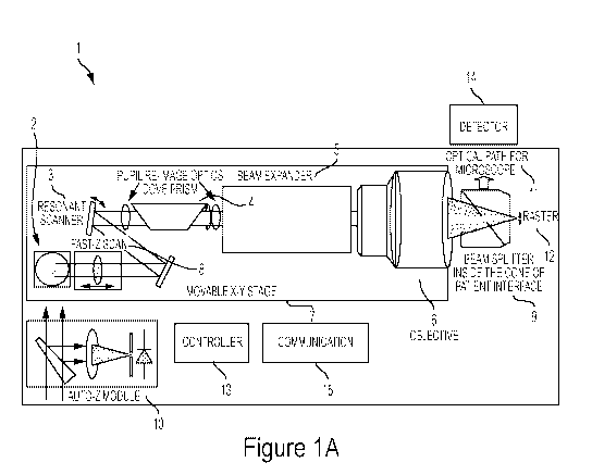
100281 FIGs. IA and 1B are simplified diagram views of a surgical ophthalmic
laser
system according to an embodiment of the present invention.
100291 FIGs. 2A and 2B are simplified views of an optical field according to
an
embodiment of the present invention.
100301 FIG. 3 is a diagram of a pulsed laser beam according to an embodiment
of the
present invention.
6
CA 02989555 2017-12-14
WO 2016/209312 PCT/US2015/065989
[0031] FIG. 4 is a graph related to laser beam optimization according to an
embodiment of
the present invention.
[0032] FIG. 5 illustrates resonant scanners according to embodiments of the
present
invention.
[0033] FIG. 6 is a graph related to resonant scanning operation according to
an
embodiment of the present invention.
[0034] FIG. 7 illustrates a schematic view of a beam delivery system according
to an
embodiment of the present invention.
[0035] FIG. 8 illustrates a schematic view of a scanner according to an
embodiment of the
present invention.
[0036] FIG. 9 is a table of scanner parameters according to an embodiment of
the present
invention.
[0037] FIG. 10 illustrates a perspective view of a scanner system according to
an
embodiment of the present invention.
[0038] FIG. 11 illustrates a perspective and graphical view of a scan line
rotator according
to an embodiment of the present invention.
[0039] FIGs. 12A-12C illustrate bed cut scanning patterns according to an
embodiment of
the present invention.
[0040] FIG. 13 illustrates bed cut scanning patterns according to an
embodiment of the
present invention.
[0041] FIGs. 14A-14B illustrate side cut scanning patterns according to an
embodiment of
the present invention.
[0042] FIG. 15 illustrates synchronization according to an embodiment of the
present
invention.
[0043] FIGs. 16A-16C illustrate prior art patient interfaces.
[0044] FIG. 17 illustrates a patient interface according to an embodiment of
the present
invention.
[0045] FIG. 18A-18B illustrate beam splitter optics according to an embodiment
of the
present invention.
100461 FIG. 19 illustrates a table of visualization parameters according to an
embodiment
of the present invention.
7
CA 02989555 2017-12-14
WO 2016/209312 PCT/US2015/065989
100471 FIG. 20 illustrates beam splitter optics according to another
embodiment of the
present invention.
Detailed Description of the Embodiments
100481 Embodiments of this invention are generally directed to systems and
methods for
laser-assisted ophthalmic procedures.
100491 Referring to the drawings, FIG. lA shows an ophthalmic surgical laser
system 1 for
making an incision in a target material such as a cornea of an eye. A laser 2
may comprise
a femtosecond laser capable of providing pulsed laser beams, which may be used
in optical
procedures, such as localized photodisruption (e.g., laser induced optical
breakdown).
Localized photodisruptions can be placed at or below the surface of the
material to produce
high-precision material processing. The laser may be a micro-chip picosecond
laser. For
example, a laser beam delivery system may be used to scan the pulsed laser
beam to
produce an incision in the material, create a flap of material, create a
pocket within the
material, form removable structures of the material, and the like. The term
"scan" or
"scanning" refers to the movement of the focal point of the pulsed laser beam
along a
desired path or in a desired pattern.
100501 Although the laser system 1 may be used to photoalter a variety of
materials (e.g.,
organic, inorganic, or a combination thereof), the laser system 1 is suitable
for ophthalmic
applications. For example, the focusing optics direct the pulsed laser beam
toward an eye
(for example, onto or into a cornea) for plasma mediated (for example, non-UV)
photoablation of superficial tissue, or into the stroma of the cornea for
intrastromal
photodi srupti on of tissue.
100511 The system 1 includes, but is not limited to, a laser source 2 capable
of generating a
pulsed laser beam, a resonant scanner 3 for producing a fast scan line or
raster 12 of the
pulsed laser beam, an XY scan device 4 or scan line rotator (e.g., a Dove
prism, Pechan
prism, or the like) for rotating the scan line 12, a beam expander 5, an
objective 6, a
moveable XY stage 7 for deflecting or directing the pulsed laser beam from the
laser 1 on
or within the target, a fast-Z scan device 8, a patient interface 9 that may
include a
visualization beam splitter inside a cone, an auto-Z device 10 for modifying
the depth of the
8
CA 02989555 2017-12-14
WO 2016/209312 PCT/US2015/065989
pulse laser beam and providing a depth reference, an optical path 11, a
controller 13, and a
communication module 15. An imaging video camera may further be included.
100521 The laser beam delivery system of the system 1 delivers a pulsed laser
beam at a
focal point of a target in a patient's eye in a raster pattern and may include
the resonant
scanner 3, beam expander 5, objective 6 and patient interface 9.
100531 The focal point of the target in the patient's eye may include one or
more of a
cornea, stroma, capsular bag, crystalline lens, and zonule. The pulsed laser
beam may
modify the target in the patient's eye to produce corneal tissue modification
such as corneal
cross-linking. As a result of the pulsed laser beam, a desired incision may be
produced in
the patient's eye.
100541 The resonant scanner 3 generates a fast scan line at a fixed resonant
frequency. The
resonant scanner 3 may produce a raster between 1 mm and 2 mm where a width of
the
scan line may be adjusted. A resonant scanner scans very fast and produces a
one-
dimensional scan that is, for example, a horizontal line.
100551 The XY scan device 4 or scan line rotator moves the pulsed laser beam
raster 12 in
a lateral direction so as to rotate the scan line to any desired orientation
on an XY plane.
For example, a Dove prism or Pechan prism rotates the raster to any direction
on an XY
plane such as a scan line perpendicular to the XY device 7 trajectory to
provide scan
coverage over a larger area.
100561 The XY scan device 7 is a movable XY scanning stage having a final
focusing
objective 6 mounted thereon. The XY scan device 7 carries the final objective
6 to move
the fast scan line to cover an entire treatment area. The movable XY scanning
stage 7 may
include a recoilless stage configured to reduce or eliminate mechanical
vibration. The XY
scanning stage 7 is configured to move the pulsed laser beam in a lateral
direction such that
the laser beam may cover an entire surgical field of the patient's eye.
Accordingly, the scan
line rotator 4 modifies an orientation of the scan line while the moveable XY
scanning
stage moves the optical field of the scan line across an XY plane.
100571 The fast Z scan device 8 modifies a depth of focus of the pulsed laser
beam and may
provide fine depth control. The fast Z scan device 8 may either be set at a
fixed position or
run dynamically to correct the system's inherent depth variations at different
(X,Y)
locations. In the latter case, a fast Z position is determined by the XY
trajectory and does
9
CA 02989555 2017-12-14
WO 2016/209312 PCT/US2015/065989
not affect the XY trajectory. A fast Z scan sets a cut depth and moves the
focus in the
depth direction to produce, for example, a side-cut in a target material.
100581 A shutter (not shown) can be kept open during a bed cut or may be
controlled to
open/close to block the unwanted pulses during a bed cut.
100591 The patient interface 9 couples the patient's eye to the ophthalmic
surgical laser
system 1. The patient interface design has a fixed cone nose on the system.
The disposable
part of the patient interface is single-piece device that allows the use of
flat applanation, or
the use of liquid interface, for patient sitting upright, respectively. Any
design with a
separated suction ring does not apply for a patient sitting upright. The
patient interface 9
may include a visualization beam splitter in the cone of the patient
interface. A beam
splitter is placed inside this cone to allow the full eye to be imaged via
visualization optics.
This allows the system to be made smaller. The patient interface may be
removed when an
eye-tracking system is used. Visualization may be provided through, for
example, a video
microscope or ocular microscope.
100601 The auto Z module 10 measures a distal end surface of a lens cone of
the patient
interface coupled to the patient's eye and provides a depth reference for the
Z scan device 8
of the ophthalmic laser system. The auto Z module 10 uses the focus of a
surgical beam as
the measurement probe, so there is no need to calibrate the measurement
reference and the
laser focus, which is otherwise required for other depth measurement methods,
such as
optical coherence tomography (OCT).
100611 The controller 13 is operably coupled with the laser delivery system,
the XY scan
device 4, the Z scan device 8, detector 14 and the communication module 15.
The
controller 13 is configured to direct the laser delivery system to output the
pulsed laser
beam in a desired pattern at the focal point of the target in the eye so as to
modify the
target.
100621 The controller 13, such as a processor operating suitable control
software, is
operatively coupled with the components of the system 1 to direct a fast scan
line 12 of the
pulsed laser beam along a scan pattern on or in the target material.
100631 In some embodiments, the system 1 includes a beam splitter within the
patient
interface 9 and a detector 14 coupled to the controller 13 for closed-loop
feedback control
CA 02989555 2017-12-14
WO 2016/209312 PCT/US2015/065989
mechanism (not shown) of the pulsed laser beam. Other feedback methods may
also be
used, including but not necessarily limited to position encoder on the scanner
3 or the like.
100641 In one embodiment, the pattern of pulses may be summarized in machine-
readable
data of tangible storage media in the form of a treatment table. The treatment
table may be
adjusted according to feedback input into the controller 13 from an automated
image
analysis system in response to feedback data provided from an ablation
monitoring system
feedback system (not shown). Optionally, the feedback may be manually entered
into the
controller 13 by a system operator.
100651 The feedback may also be provided by integrating a wavefront
measurement system
(not shown) with the laser surgery system 1. The controller 13 may continue
and/or
terminate at least one incision in response to the feedback, and may also
modify the
planned sculpting based at least in part on the feedback. Measurement systems
are further
described in U.S. Patent No. 6,315,413, the entire disclosure of which is
incorporated
herein by reference.
100661 The communication module 15 provides information to the operator of the
laser
system 1 at the system and/or remotely via wired or wireless data connection.
The
communication module 15 may include a display device and input/output devices
as known
in the art to display information to an operator. An operator may control the
system 1 via
any known input control system including but not limited to a keyboard, a
mouse, voice
control, a motion sensing system, a joystick, and an eye-tracking system. The
system 1
may be operated remotely and may also be monitored and serviced remotely.
100671 In another embodiment, FIG. 1B shows the beam delivery optics of a
system 20.
The system 20 includes, but is not limited to, an input pulsed laser beam 21
from laser
source (not shown), fast-Z scan 22, a resonant scanner 23 for producing a fast
scan line 30
of the pulsed laser beam 21, a scan line rotator 24 (e.g., a Dove or Pechan
prism, or the
like) for rotating the scan line 30, a beam expander 25, an objective 26 with
an adjustable
Z-baseline (slow-Z scan) 26, a moveable X-Y stage 27 for deflecting or
directing the pulsed
laser beam 21 on or within the target, a patient interface 28 that may include
a beam
splitter, an optical path 29, a controller 31, a detector 32, and a
communication module 33.
The slow-Z scan 26 sets the focus at a fixed depth and may set the Z-baseline.
For
example, the slow-Z scan 26 is stationary during a bed cut.
11
CA 02989555 2017-12-14
WO 2016/209312 PCT/US2015/065989
100681 Some embodiments of the system are compact desktop systems that are
placed on a
table or the like. Other embodiments may include a motorized stage. The
compact system
allows a patient and patient interface to be oriented downwards, upwards, or
in any
direction, and not necessarily upright.
100691 Next, FIG. 2A provides a simplified view of a surgical field 40.
Typically, laser-
assisted ophthalmic procedures are performed within a surgical field 40 of an
eye that has a
diameter of about 10 mm. Some of these systems utilize solid state femtosecond
lasers
including an oscillator, stretcher, amplifier and compressor. Conventional
laser systems
include a laser with optics large enough to generate a laser beam with an
optical field that
matches the surgical field. Scanning mirrors or other optics (not shown) may
be provided
to angularly deflect and scan the pulsed laser beam over the entire surgical
field. These
scanning mirrors may be driven by a set of galvanometers that further add to
the bulk and
complexity of conventional laser systems.
100701 However, providing a sufficient numerical aperture (NA) to perform
laser surgery
requires large, expensive optics and a corresponding cumbersome, heavy and
expensive
beam delivery system. For example, an objective of the iFS Advanced
Femtosecond Laser
System alone weighs over 30 lbs. in order to allow a pulsed laser beam to scan
freely within
the 10 mm surgical field. These systems provide a practical maximum NA of
about 0.4 due
to the increasing cost, size and complexity of system components when NA is
increased.
100711 FIG. 2B illustrates an optical field 42 according an embodiment of the
invention
that is significantly smaller in diameter than the surgical field 41. The
diameter of the
optical field 42 depends on the length of the fast scan line 12 generated by
the resonant
scanner 3. For example, the diameter of the optical field 42 may be between 1
mm and 2
mm, and may preferably be 1.2 mm. This allows the laser to be made much
smaller with
laser beam tissue interaction in a low-density plasma mode.
100721 For a given NA, the size and cost of the laser optics is reduced as the
optical field is
reduced in size. Consequently, increasing an NA value is significantly more
cost effective
for a smaller optical field. Since the optical field 42 may be about five to
ten times smaller
than the surgical field 41, a higher NA is achievable at a reduced cost
compared to an
optical field matching the surgical field 40. Accordingly, the invention
provides higher NA
at lower cost.
12
CA 02989555 2017-12-14
WO 2016/209312 PCT/US2015/065989
100731 As shown in FIG. 2B, an optical field 42 does not by itself cover an
entire surgical
field 41. However, the optical field 42 is moved mechanically by the moveable
XY device
7 across the entire surgical field 41. As will be described later, a resonant
scanner 3
generates a very fast scan line within the optical field 42 that is oriented
(rotated) within the
optical field 42 by an XY scan device 4 and moved within the entire surgical
field 41 by the
moveable XY scan device 7. Reducing the size of the optical field
significantly reduces the
complexity, size, and weight of the laser source. Furthermore, an opto-
mechanic arm
mechanism is unnecessary in the laser system 1. In this manner, the laser
optics are
provided at a much lower cost with improved focus to achieve better surgical
outcomes.
100741 Embodiments of the invention may utilize a femtosecond oscillator or
oscillator low
energy laser. The laser source 2 may include an active medium fiber laser
amplifier,
oscillator and compressor, but need not include a stretcher. The laser source
2 may be fiber
oscillator based, such as a diode-pumped fiber laser. The diode-pumped fiber
laser may be
a mode-locked fiber oscillator based laser having a single-mode, double-clad
fiber
oscillator and all positive dispersion elements.
100751 The laser may generate a pulsed laser beam having a pulse repetition
rate in the
range between 5 MHz and 25 MHz, pulse energy in the range between 1 nJ and 5
tJ, a
wavelength between the range of 1020 nm and 1065 nm, a pulse duration between
the
range of 10 femtoseconds and 10 picoseconds, a spot size between 0.2 gm and
2.0 pm
(FWHM), and a numerical aperture NA between 0.25 and 1.3. An NA of 0.6
produces a
1.1 pm FWHM spot. The NA value is preferably provided between 0.25 and 1.0,
more
preferably between 0.4 and 1.0, and may be 0.6 in the illustrated examples.
100761 Moreover, the reduction in size and complexity of the system 1 allows
the laser
delivery system to be configured to deliver the pulsed laser beam to the focal
point of the
target in the patient's eye while the patient is seated either in an upright
position or in a
reclining position.
100771 FIG. 3 is a diagram of a pulsed laser beam 50 including the
relationship between
the beam diameter, pulse energies, focus spot diameters and effective focal
length. The
focus spot 51 generated by a laser 2 may be provided at a focus point of the
cornea to
generate a bubble that separates and dissects tissue.
13
CA 02989555 2017-12-14
WO 2016/209312 PCT/US2015/065989
100781 A pulsed laser beam directed at corneal tissue will first generate
plasma Additional
pulses then generates a bubble in tissue. Finally, the bubble expands to
generate tissue
separation/dissection .
100791 A pulsed laser beam applied to tissue first generates plasma, that then
generates a
bubble, and finally leads to tissue separation/dissection. A typical threshold
value for tissue
dissection is 1013W/cm2. To perform tissue dissection, a pulsed laser beam
needs to reach
or exceed this threshold value determined by the equation cha, where e is the
energy of the
beam, T is the pulse width, and a is the area of the beam.
100801 Based on this relationship, for a given amount of energy, decreasing
the spot size
will increase the optical density of the beam since the same amount of beam
energy is
concentrated in a smaller area. Likewise, as the spot size of the beam
decreases, the
amount of energy of the beam may be reduced while still exceeding the tissue
dissection
threshold value. A smaller amount of beam energy applied in a smaller area
results in a
finer tissue cut.
100811 An inverse relationship exists between spot size and numerical aperture
such that as
NA becomes larger, a spot size 51 becomes smaller. Numerical aperture
represents the sine
of the half angle of the cone of a laser beam. Accordingly, a higher NA value
is desirable
in providing a finer cut.
100821 For example, the laser system 1 outputs an energy level of 0.14 [LT
that is 20% of the
energy level output of 0.7 J from the IFS Laser System. Similarly, the system
1 provides a
pulse width of 120 fs and area of ir0.52 ilm2 while the iFS Laser System
provides a pulse
width of 600 fs and area of RØ821=2.
100831 FIG. 4 is a graph 60 related to laser beam optimization. As illustrated
in FIG. 3, a
beam diameter 52 may be different from the diameter of a lens 53 that focuses
the light
pulse into a focus spot 51. Selection of a beam diameter 52 smaller than the
lens diameter
53 ensures that all of the light energy passes through the lens. However, an
inverse
relationship exists between a beam diameter and a focus spot size such that
the focus spot
size will increase as the beam diameter decreases. FpEAK represents energy
area density
and T represents energy transmission.
10084J Similarly, laser overfield is a configuration where the beam diameter
52 is greater
than the lens diameter 53 such that a portion of the light energy is not
transmitted through
14
CA 02989555 2017-12-14
WO 2016/209312 PCT/US2015/065989
the lens and lost. However, the loss in energy efficiency by laser overfield
does provides
the benefit of a smaller focus spot size 51.
100851 In balancing the factors of energy efficiency and spot size, FIG. 4
illustrates the
optimal conditions to attain maximum energy density. In particular, a maximum
peak
fluence is achieved with about a 10% loss of transmission. In other words, the
optimum
ratio of energy transmission to spot size occurs when the pulsed laser beam
diameter is
about 10% larger than the lens diameter.
100861 A laser as described above may operate at very high frequencies such as
on the
order of 10 MHz (or 10,000,000 pulses/sec). Laser pulses that are not scanned
will be
directed at a single point which is unsuitable for ophthalmic procedures.
Therefore, a
scanner is needed to operate at a sufficient frequency to scan these pulses
across a surgical
area.
100871 The scanner 3 of the system 1 may be a high frequency resonant optical
scanner
having a fixed frequency in a range between 3500 Hz and 21,000 Hz. In an
preferred
embodiment, a 7910 Hz resonant scanner is implemented. Use of a resonant
scanner is
particularly effective as they have no wearing parts, are reliable, cost-
effective and compact
(e.g., 1.0"W x 0.7"D x 2.5"H). The resonant scanner 3 produces a line raster
pattern with a
length of the raster pattern between 0.5 mm and 2 mm. In some embodiments, the
resonant
optical scanner is configured to scan the pulsed laser beam from the laser
delivery system
in a line.
100881 FIG. 5 illustrates exemplary resonant scanners 70 and 71 that include a
mirror
attached to a metal rod that vibrates at an inherent resonant frequency. The
shape and
composition of the rod are selected to operate at a desired frequency to scan
laser pulses.
The resonant scanner 3 does not require a plurality of mirrors or a set of
cumbersome
galvos to scan across a surgical field as other systems do. Instead, the scan
line may be
rotated by a scan line rotator within an optical field and the scanner 3 may
be scanned
across a surgical field by a moveable XY stage. In some embodiments, the
resonant
scanner 3 provides an order of magnitude in weight and cost savings over the
scanner
system provided in the iFS Laser System. The resonant scanner 3 may scan at a
rate of
about 20 m/s while the iFS scanner scans at a rate of about 3 in/s.
CA 02989555 2017-12-14
WO 2016/209312 PCT/US2015/065989
100891 As illustrated in the graph 80 of FIG. 6, the scanning provided by a
resonant optical
scanner 3 is characterized by a sinusoidal curve. Thea resonant optical
scanner may
oscillate at a frequency between 200 Hz and 21000 Hz. The curve 81 represents
the
scanning angle of a resonant scanner 3 and curve 82 represents the scanning
speed. As
shown by the curve 82, the scanning speed continually varies such that the
density of laser
spots along the scan line will vary. Accordingly, that the distribution of
laser pulses is
uneven.
100901 For instance, scan line 86 illustrates the sinusoidal distribution of
laser spots
provided by a resonant scanner 3. Whether a scanning speed reaches zero or a
maximum
speed, laser pulses will continue to be emitted at the same rate. Undesirable
spot
overlapping 83 occurs when the scan speed is at and near zero. This may lead
to areas of
tissue that are overcut from an excess number of laser pulses.
100911 Some embodiments of the invention overcome this by preventing
overlapping spots
83. In one embodiment, the overlapping spots 83 are emitted but physically
blocked 84
from scanning a target material to provide a higher quality tissue cut.
100921 FIG. 7 illustrates a schematic view of a beam delivery optics system. A
pulsed
laser beam 91 emitted by a laser source (not shown) reaches a resonant optical
scanner 92
and is delivered into a beam expander 93. The beam expander includes a lens 94
that
focuses the beam through a scan line rotator 95 and another lens 94. A
predetermined
portion 97 of the beam 91 is blocked by a field stop 96 to limit the scan
length of the raster.
100931 The pulses 97 may, for example, correspond to the blocked portion 84
overlapping
spots 83 in FIG. 6. In this manner, undesirable light pulses are physically
blocked within a
beam expander 93 as the light focuses, ensuring that laser spots are not
concentrated too
densely within a spot or scan line area. The blocker or field stop 96 may be
provided near
but not precisely at the focal plane so as to prevent the blocker from
burning. It is noted
that conventional scanners do not exhibit sinusoidal wave characteristics such
that those
systems have no need to provide blocking.
100941 In an alternative embodiment, FIG. 8 illustrates a schematic view of a
scanning
system 100. A resonant optical scanner 101 is illustrated as vibrating so as
to produce a
scan line 104. A laser (not shown) producing laser pulses is synchronized with
the
frequency of the scanner 101 such that the laser is turned on 102 and off 103
when the
16
CA 02989555 2017-12-14
WO 2016/209312 PCT/US2015/065989
scanner 101 approaches a predetermined maximum scan angle with a corresponding
zero
velocity in order to prevent overlapping focus spots in successive pulses.
100951 Equation 1 is an algorithm for determining a duty cycle that is a
percentage time
that a beam passes an aperture, scanner frequency, optical peak-to-peak angle,
a pupil
diameter for given laser pulse repetition rate, and desired numerical aperture
of the optical
system. An example for NA=0.6 is provided below:
rõ
2400 cos(¨ aprp !gaol. Dpupzik f LAsgit
2
100961 (Eq. 1)
100971 Equation 1 guides the selection of resonant scanner parameters for a
spot edge to
edge conidtion, as shown in Table 110 in FIG. 9. Table 110 highlights the
values that
satisfy a requirement of spot size (FWHM=1.1um) and avoiding laser spot
overlap.
100981 In some embodiments, a fast raster scanning pattern can be generated by
synchronizing a plurality of resonant scanners in the laser system 1. For
example, FIG. 10
illustrates a pair of perpendicular scanning resonant mirrors 120 with the
same frequency,
the same amplitude, and a phase difference of 90 between them that generate a
fast circular
scan line 121, for example.
100991 A circular scan line exhibits a number of advantages including equal
spot
distribution so as to render blocking techniques redundant. In this case, the
linear speed of
the scanning is a constant, and is equal to the maximum speed that can be
achieved with a
single scanner. Therefore, there is no need to block the "zero speed" points
as in the case
of using a single scanner, and the duty cycle is 100%, i.e., 100% of laser
pulses will be used
for tissue dissection. Furthermore, a circular scan line ensures that targeted
tissue receives
two pulses with each pass, thereby ensuring a cut. Also, a circular scan line
is also well
matched against another curve, such as the edge of a circular surgical field.
1001001 The first scanner may be provided for the x axis while the second
scanner may
be provided for the y axis in different phase relation to generate a plurality
of two-
dimensional scan patterns that may obviate the need for a scan line rotator.
The use of at
least two scanners may generate a line oriented at any desired angle, circle,
curve, etc.
1001011 Another arrangement of synchronization is to synchronize two parallel
scanners
so that the optical peak-to-peak angle is doubled in comparison with a system
using one
17
CA 02989555 2017-12-14
WO 2016/209312 PCT/US2015/065989
resonant scanner. In yet another embodiment, a plurality of resonant scanners
may be
synchronized to extend the scanning range of a single scanner.
1001021 Next, embodiments of a scan line rotator will be discussed. A resonant
scanner
produces a one dimensional scan line in a single direction. However, this
output is not
ideal for cutting near an edge or curve of a surgical field. For example, when
an optical
field is provided along an edge of surgical field, the line must be rotated to
fit the curve.
Therefore, a scan line rotator is configured to rotate the scanned line in a
desired
orientation.
1001031 FIG. 11 illustrates a perspective view of an exemplary scan line
rotator 130 and
graphical views of a scan line rotated by a scan line rotator. The scan line
rotator 130 is a
Dove prism, but may also be a Pechan prism or a set of mirrors.
Implementations of a scan
line rotator using a Dove prism or Pechan prism are cost-effective, compact
and
lightweight, and contribute to a compact laser system. The input scan line 131
is a non-
rotated scan line. As the scan line rotator 130 rotates by an angle 0, the
input scan line 131
will follow the rotation and the output scan line will be rotated by 20.
1001041 The output raster 133 is thus oriented in any desired direction to
scan an entire
optical field. In combination with an XY stage, the system 1 may scan an
entire treatment
area. Tissue fibers may sometimes be aligned in certain directions that favor
a rotated
raster. Furthermore, a scan line rotator allows for flap creation, cornea
incisions, IEK,
inlays, rings, etc. and procedures such as SmILE or ReLEx.
1001051 FIGs. 12A-12C illustrates scanning patterns provided by an XY stage 7.
The
XY stage 7 moves the optical field and raster line across a surgical field.
Raster line
scanning patterns 140 and 141 may be provided in a number of configurations,
as
illustrated in FIG. 12A. For example, in order to cover an intended lamellar
dissection area
142, the XY stage 7 may move the raster line 143 up and down systematically
across the
surgical field along path 144 to cover the full flap bed and provide a bed
cut. The width of
each pass may be selected so as to provide overlapping cuts where tissue is
cut a plurality
of times by the raster.
1001061 FIG. 12B illustrates other scanning patterns of the XY stage 7 that
may move
the raster line horizontally along a path 145 across the surgical field, as
well as along a path
146 at a predetermined angle. The raster may be aligned perpendicularly with
the
18
CA 02989555 2017-12-14
WO 2016/209312 PCT/US2015/065989
movement of the XY stage, for example. FIG. 12C illustrates a circular path
147 and a
spiral path 148 that each cover an entire predetermined treatment area.
[00107] Turning to FIG. 13, combinations of the above scanning patterns may be
provided within a surgical area. A first bed cut pattern 150 includes an
annulus scan 151
and a set of linear (rectangular) scans 152 that ensure that every portion of
the treatment
area has been scanned by the optical field. Similarly, a second bed cut
pattern 155 includes
an annulus scan 156, a spiral scan 157 and a rectangular scan 158.
[00108] Next, FIGs. 14A-14B are directed to side cuts. A fast Z scan device 8
modifies
a depth of focus of the pulsed laser beam. Some embodiments of the fast Z scan
device 8
are realized through a voice coil that drives a lens. When the lens moves, the
curvature of
the beam is changed, leading to focus a depth change. The z-scan frequency may
be
between 50 Hz and 15,000 Hz. With a fast Z scan device and X-Y stage, a 90
side-cut can
be generated where 0 is defined as the radial direction in the lamellar cut
bed. A 90 side-
cut can be applied for flap creation, for example.
[00109] In FIG. 14A, a scan line is moved along a path 160 sinusoidally in a
depth
direction by the fast Z scan device and circularly by the X-Y stage to produce
a vertical
slice. FIG. 14B illustrates another path 161 generated by a low Z scan
frequency. A side
cut 162 is generated by a linear raster 163 generated by a resonant scanner.
The raster line
is moved 164 up and down by the fast Z scan device along a circumference by an
X-Y
stage. Rotation of the scan line by a scan line rotator ensures that the
raster 165 is kept
tangential to the circumference during a side cut.
[00110] For example, for a 9.5mm diameter flap, 20MHz laser repetition rate,
10kHz
raster scan with 1 mm scan length, an 85Hz Z-scan frequency and +1-60 gm Z-
scan
amplitude may be provided. The side-cut may be completed within one second,
during
which the raster scan passes any given location five times to ensure tissue
separation. The
side cut need not be vertical and may also be angled to better match the
tissue.
[00111] Turning to FIG. 15, synchronization between a resonant scanner and
fast-z
voice coil scan is illustrated. Let a raster scan be described by Equation 2:
[00112] X(t) = Ax = sin(27c=fx =t) (Eq. 2)
[00113] Ax is the adjustable amplitude of the raster on the focal plane, and
fx is the fixed
resonant scanner frequency. The fast-z scan may be described by Equation 3:
19
CA 02989555 2017-12-14
WO 2016/209312 PCT/US2015/065989
1901141 Z(t) = Az= sin(27r. ft + Ozx) (Eq. 3)
1001151 Az is the adjustable amplitude of the z-scan at the focus, fz is the
adjustable fast-
z scan frequency, and (Dzx is the adjustable relative phase between the fast-z
scan and the
resonant scanner.
1001161 A variety of side-cut patterns may be produced by adjustment of the
parameters
Ax, Az, fz, and (Dzx. For example, FIG. 15 illustrates a 70 side-cut 170 and
an "M" shape
side-cut 171 which increases a contact surface. Synchronization provided in
this manner
allows for a plurality of three-dimensional laser patterns for tissue
dissection and for other
light-assisted effect such as refractive index modification.
1001171 A plurality of incision patterns combining the aforementioned bed cut
and side
cuts may thus be generated, including an xy lamellar dissection, a spiral
lamellar dissection,
a vertical side-cut, a plano-vertical side cut, an intrastromal incision, a
lenticular incision,
as well as any three-dimensional dissection. Other cuts include a flap cut for
LASIK, lens
cut for myopia correction, ring resection for inlay, arcuate incision for
astigmatism, clear
cornea incision for a cataract entry cut, penetrating cut for cornea
transplant, anterior and
posterior deep lamellar cut for cornea transplant, corneal ring cut for
insertion of stiffening
material, pocket cut to treat presbyopia, intralase enabled keratoplasty (IEK)
for corneal
transplants, and so forth.
1001181 Next, an optimal sequence of cutting is described. A byproduct of
tissue
dissection is the release of gas. If gas from laser tissue dissection has
nowhere to vent, the
gas will penetrate back into the tissue and create an opaque bubble layer that
will hinder the
dissection of tissue beneath that layer. Embodiments of the invention herein
eliminate the
opaque bubble layer by providing a channel for gas to escape during tissue
dissection.
1001191 First, a side cut is performed in the Z plane that will also serve as
a venting
channel for gas to escape from the cornea. Next, a ring cut is performed in
the XY plane.
Gas emitted from the ring cut will pass through the side cut and out of the
cornea. Finally,
a band cut (e.g., rectangular cut, spiral cut) in the XY plane is performed
with the gas
generated therein escaping via the side cut via the ring cut channels created.
A bed cut
refers to the combination of the ring cut and rectangular cut or spiral cut.
This sequence of
cuts ensures that the generated gas has a channel to escape from the cornea so
as to reduce
or eliminate an opaque ring bubble layer.
CA 02989555 2017-12-14
WO 2016/209312 PCT/US2015/065989
1001201 Since a band cut is provided on the same plane as a ring cut, gas from
the band
cut will pass through ring cut, which will then pass through the side cut,
thus providing a
gas vent throughout the entire procedure. Furthermore, this procedure
effectively
eliminates the need for the creation of a "pocket" in a flap cut. Prior art
approaches
perform cutting in the opposite sequence and require another cut (pocket)
whose sole
purpose is to provide the air channel.
[00121] FIGs. 16A-16C illustrate a prior art patient interfaces according to
an
embodiment of the present invention. For many prior art femtosecond laser
workstations,
the field of view for visualization optics 184 such as a CCD and video
microscope is
similar to the field of view of surgical beam scanning such that a
visualization beam splitter
183 is positioned above the focusing objective 181, patient interface 180, and
cornea 185.
In this configuration, the size of the optics system, including both beam
delivery 182 and
visualization 184, is generally large and unwieldy.
[00122] FIG. 16B is a schematic cross-sectional view of the patient interface
180 that
includes a cone 186 that is fixed to the system, a visualization beam splitter
187, and a
disposable patient interface lens 188. A beam splitter 187 is coated for
reflecting the visual
spectrum but passes light for the femtosecond laser wavelength and is placed
inside the
cone frame 186 of the patient interface 180 fixed to the system. The ocular
video
microscope optical path goes through this beam splitter. Accordingly, a
cutting process may
be viewed and/or displayed in-situ.
[00123] FIG. 16C is a cross-sectional view of another patient interface 189
where a
visualization beam splitter is placed inside the cone of the patient interface
189. This design
is sufficient for a limited range of numerical aperture of surgical beam, for
example, NA <
0.4. For yet greater NA, such as NA = 0.6, some oblique rays of the surgical
beam will
experience high loss at the beam splitting surface (the 450 surface as shown
in the diagram).
As the NA increases, the size of the beam splitter will need to increase as
well.
[00124] The visualization optical path may be provided outside the cone of the
patient
interface in a side channel. However, for deep set eyes, the side channel has
to be placed
much higher, increasing the size and bulk of the beam splitter. Consequently,
the outer
dimension of the patient interface cone will not fit the normal anatomy of a
patient eye and
is thus inadequate based on human factors. Simply put, a user's facial
features will occupy
21
CA 02989555 2017-12-14
WO 2016/209312 PCT/US2015/065989
the same space as the enlarged patient interface necessary to accommodate a
visualization
beam splitter for high NA laser systems.
[00125] FIG. 17 illustrates a patient interface according to an embodiment of
the present
invention. To overcome the issues described above, a rotatable protruding
portion 192 of
the patient interface 190 is rotatable about an axis 191 and provided on the
temporal side of
the patient head. To fit both left and right eyes of a patient, the
visualization optics
(including the beam splitting optics, the patient interface 190, the imaging
optics, and the
CCD) are rotated 180 degrees in accordance with treating left and right eyes,
respectively.
In this manner, the larger visualization beam splitter elements are better
positioned to avoid
conflict with a user's face.
1001261 FIGs. 18A-18B illustrate beam splitting optics according to an
embodiment of
the present invention. A patient interface 200 is provided including two beam-
splitting
surfaces, BG and GP. These two surfaces divide the full field of view into
left half and right
half, and form two separate visualization channels. As a result, this
reduction in the size of
the channels allows the channels to fit into the cone of the patient interface
200 such that no
rotation of the visualization beam splitter is needed when treating left and
right eyes.
Furthermore, the channels support high NA (NA = 0.6) surgical beams. FIG. 18B
illustrates a cross-sectional perspective view of the visualization beam
splitter in the cone.
[00127] An interface for coupling a patient's eye to an ophthalmic surgical
laser system
includes a lens cone defining a first plane surface coupled with a delivery
tip of the
ophthalmic laser system. The lens cone includes an apex ring coupled to the
first plane
surface. The apex ring includes a distal end including a first receptacle
configured to
receive an attachment ring, the attachment ring configured to overlay an
anterior surface of
the patient's eye. The first receptacle and the attachment ring may be
disposable. A central
cavity is provided to receive the lens cone. A contact lens may applanate the
anterior
surface of the patient's eye.
[00128] One or more beam-splitter optics are provided to allow a pulsed laser
beam to
pass through the interface to a focal point of the target in the patient's
eye. The beam-
splitter optics may include one or more multi-facet beam-splitter optics and a
side-imaging
optical channel that is configured to rotate to a temporal side of the
patient's eye.
Alternatively, the beam-splitter optics may include dual imaging channels. The
beam-
22
CA 02989555 2017-12-14
WO 2016/209312 PCT/US2015/065989
splitter optics may be provided to manipulate non-telecentric imaging rays at
a full optical
cone angle equal to or greater than fifteen degrees.
[00129] FIG. 19 illustrates a table of visualization parameters according to
an
embodiment of the present invention. The specific numerical values for the
half cone angle
(a), the beam splitting surface angle (13), and the edge ray incident angle
(y), and the
geometry dimensions of the visualization beam splitter are given in table 220.
[00130] FIG. 20 illustrates beam splitting optics according to another
embodiment of the
present invention. The patient interface 230 in FIG. 20 divides the full field
of view into
two halves, images the two halves into two different optical channels, and
processes to
combine the two half-images together to reconstruct the full field of view. In
this manner,
the entire visualization beam splitting optics can be placed inside the cone
of a compact
patient interface 230.
[00131] This approach of dividing the full field of view into several smaller
fields, and
then combining the images of the smaller results to reconstruct the original
large field of
view may also be applied to measurement such as an optical channel for Optical
Coherence
Tomography, for ophthalmology surgical lasers including but not limited to
femtosecond
laser workstations.
[00132] All patents and patent applications cited herein are hereby
incorporated by
reference in their entirety.
[00133] The use of the terms "a" and "an" and "the" and similar referents in
the context
of describing the invention (especially in the context of the following
claims) are to be
construed to cover both the singular and the plural, unless otherwise
indicated herein or
clearly contradicted by context. The terms "comprising," "having,"
"including," and
"containing" are to be construed as open-ended terms (i.e., meaning
"including, but not
limited to,") unless otherwise noted. The term "connected" is to be construed
as partly or
wholly contained within, attached to, or joined together, even if there is
something
intervening. Recitation of ranges of values herein are merely intended to
serve as a
shorthand method of referring individually to each separate value falling
within the range,
unless otherwise indicated herein, and each separate value is incorporated
into the
specification as if it were individually recited herein. All methods described
herein can be
performed in any suitable order unless otherwise indicated herein or otherwise
clearly
23
CA 02989555 2017-12-14
WO 2016/209312 PCT/US2015/065989
contradicted by context. The use of any and all examples, or exemplary
language (e.g.,
"such as") provided herein, is intended merely to better illuminate
embodiments of the
invention and does not pose a limitation on the scope of the invention unless
otherwise
claimed. No language in the specification should be construed as indicating
any non-
claimed element as essential to the practice of the invention.
1001341 While certain illustrated embodiments of this disclosure have been
shown and
described in an exemplary form with a certain degree of particularity, those
skilled in the
art will understand that the embodiments are provided by way of example only,
and that
various variations can be made without departing from the spirit or scope of
the invention.
Thus, it is intended that this disclosure cover all modifications, alternative
constructions,
changes, substitutions, variations, as well as the combinations and
arrangements of parts,
structures, and steps that come within the spirit and scope of the invention
as generally
expressed by the following claims and their equivalents.
24