Note: Descriptions are shown in the official language in which they were submitted.
CA 02990133 2017-12-19
WO 2017/037523 PCT/1B2016/000993
1
COMPOSITIONS AND METHODS FOR VIRAL EMBOLIZATION
FIELD
[0001] The present disclosure relates to compositions and methods related to
transarterial
embolization with oncolytic viruses.
BACKGROUND
[0002] Therapeutic vascular occlusion (embolization) is a technique used to
treat pathological
conditions in situ by injection of an occlusion agent (embolic material) into
a vessel.
Embolization is carried out by means of catheters, making it possible to
position particulate
occlusion agents or gels (emboli) in the circulatory system. In active
embolization therapy,
embolic agents are formulated with therapeutic agents, such as a drug or
chemotherapeutic,
resulting in both mechanical blockage and in situ delivery of the therapeutic
agent. The use
of embolization in cancer therapy has also been established. For example,
blood vessels
which nourish cancerous tumors are deliberately blocked by injection of an
embolic material
into the vessel. Vascular occlusion can limit blood loss during the surgical
interventions, and
contribute to tumoral necrosis and recession. Combining the occlusion agent
with a
chemotherapeutic can allow delivery of the chemotherapeutic directly to the
tumor without
significant systemic deliverly, which allows higher doses of chemotherapeutic
to be used.
[0003] Transarterial embolization (TAE) or transarterial chemoembolization
(TACE) have
been used extensively to treat patients with hypervascular tumors confined to
the liver or
tumors where the intrahepatic component is the main source of mobidity and
mortality.
TAE/TACE are considered effective palliative care for unresectable tumors or
as an adjuvant
to manage postoperative recurrent tumors (Camma et al, (2002) Radiology,
224:47-54; Llovet
et al., (2002) Lancet 359:1734-1739; Jelic et al., (2010) Ann Oncol. 21 Suppl
5:v59-v64).
Various embolization agents and chemotherapeutics have been used, although
clear
superiority for any particular regimen or chemotherapeutic has not been
demonstrated
(Nakamura et al., (1994) Cancer Chemother Pharmacol 33 Suppl:S89-S92; Bruix et
al.,
(2004) Gastroenterology 127:S179-S188).
[0004] In parallel, oncolytic viruses are in development for treatment of
cancer. For example,
replication-selective oncolytic viruses hold promise for the treatment of
cancer (Kim et al.,
CA 02990133 2017-12-19
WO 2017/037523 PCT/1B2016/000993
2
(2001) Nat. Med., 7(7):781-787). These viruses can cause tumor cell death
through direct
replication-dependent and/or viral gene expression-dependent oncolytic effects
(Kim et al.,
(2001) Nat. Med., 7(7):781-787). In addition, viruses are able to enhance the
induction of
cell-mediated antitumor immunity within the host (Todo et al., (2001) Cancer
Res., 61:153-
161; Sinkovics et al., (2000) J. Clin. Viro., 16:1-15). These viruses also can
be engineered to
expressed therapeutic transgenes within the tumor to enhance antitumor
efficacy (Hermiston,
(2000) J. Clin. Invest., 105:1169-1172).
[0005] However, major limitations exist to this therapeutic approach. Although
a degree of
natural tumor-selectivity can be demonstrated for some virus species, new
approaches are still
needed to engineer and/or enhance tumor-selectivity for oncolytic viruses in
order to
maximize safety and efficacy. This selectivity is particularly important when
intravenous
administration is used, and when potentially toxic therapeutic genes are added
to these viruses
to enhance antitumor potency; gene expression will need to be tightly limited
in normal
tissues. In addition, increased antitumor potency through additional
mechanisms such as
induction of antitumor immunity or targeting of the tumor-associated
vasculature is highly
desirable. Therefore, more effective and less toxic therapies for the
treatment of cancer are
needed.
[0006] Initial attempts to combine oncolytic viral therapy with embolization
have been made.
An oncolytic form of vesicular stomatitis virus (VSV) has been tested in tumor
models
(Altomonte et al. (2008) Hepatology 48:1864-1873). VSV was an ideal candidate
to test with
embolization. VSV, a member of the rhabdoviridae family, is a negative-sense
RNA virus
180nm long and 75nm wide. VSV enters and is released from the basolateral
surfaces of
polarized cells. The basolateral release of VSV allows it to readily infect
underlying tissues,
including tumor tissues (Basak et al., (1989) J. Virology, 63(7):3164-3167).
In addition, the
small size of VSV, particularly the 75nm diameter of its smallest axis, allows
its passage
through the leaky junctions between blood vessel cells, allowing the infection
of underlying
tissues and through the basolateral surface of the blood vessel cells. Due to
its small size and
basal surface budding, one of skill in the art could have expected successful
infection of
tumor tissue during viral embolization of VSV.
[0007] Given the ideal characteristics of VSV, it is difficult to extrapolate
other oncolytic
viruses. For example, viruses that that have a diamerter along their smallest
axis that is larger
than the junctions between cells of blood vessels may not be able to pass from
the blood
CA 02990133 2017-12-19
WO 2017/037523 PCT/1B2016/000993
3
stream to the surrounding issue. Similarly, viruses that release from the
apical side of polar
cells are typically limited to infection along epithelial cell linings (Basak
et al. (1989) J.
Virology, 63(7):3164-3167). When such a virus infects a polar endothelial
blood vessel cell,
the replicated viruses could simply be released back into the blood stream
rather than into the
underlying tissue if released apically. Vaccinia virus is an example of a
virus that has a
number of undesirable characteristics that could have been expected to prevent
effective
embolization. Vaccinia virus (VV), a member of the poxvirus family, is a large
virus roughly
360nm by 250nm in size. Vaccinia virus preferentially infects through the
basolateral surface
of polar cells, but its viral progeny are released from the apical surface
(Vermeer et al., (2007)
J. Virology, 81(18):9891-9899). A virus that is apically released from the
polar endothelial
cells that create the blood vessel waslls is thus at risk of being washed away
by the blood
stream. In addition, due to its large size, vaccinia virus would have
difficulty passing through
cellular junctions between cells of the blood vessel walls to reach to the
basolateral surface of
the endothelial cells and subsequently infect underlying tissues in any
substantial amount.
Based upon its lifecycle and size, one could not extrapolate from the VSV
results to oncolytic
vaccinia virus, or other large viruses or viruses that release from the apical
surface for that
matter, being able to achieve significant penetration into a tumor during
vascular
embolization.
[0008] All references cited herein, including patent applications and
publications, are hereby
incorporated by reference in their entirety.
SUMMARY
[0009] Some aspects of this invention are based upon the discovery that
oncolytic vaccina
viruses, despite the size and the apical release from polarized cells, as an
exemplary oncolytic
virus, can be used in a surprisingly effective manner in combination with
embolization
therapy.
[0010] An aspect of the invention includes compositions comprising an
oncolytic Poxviridae,
Herpesviridae, or Measles virus and a biocompatible microparticle or
hydrophilic polymer gel
agent suitable for active embolization. In some embodiments, the oncolytic
virus is a
Poxviridae virus selected from the group consisting of: vaccinia virus,
myxomavirus, and
parapoxvirus. In some embodiments, the oncolytic virus is an oncolytic
vaccinia virus. In
some embodiments, the oncolytic vaccinia virus does not comprise an active
thymidine
CA 02990133 2017-12-19
WO 2017/037523 PCT/1B2016/000993
4
kinase gene. In some embodiments, which may be combined with any of the
preceding
embodiments that include an oncolytic vaccinia virus, the oncolytic vaccinia
virus does not
comprise an active vaccinia growth factor (VGF) gene. In certain embodiments,
which can
be combined with any of the preceding embodiments that include an oncolytic
vaccinia virus,
the oncolytic vaccinia virus comprises transgenes encoding Renilla luciferase,
green
fluorescent protein, P-galactosidase, and P-glucuronidase. In certain
embodiments, which can
be combined with any of the preceding embodiments that include an oncolytic
vaccinia virus,
the oncolytic vaccinia virus is a Copenhagen strain, a Western Reserve strain,
a Wyeth strain,
or a Lister strain. In certain embodiments, which can be combined with any of
the preceding
embodiments that include an oncolytic vaccinia virus, the oncolytic vaccinia
virus further
comprises one of more of a granulocyte-macrophage colony stimulating factor
protein, a
cytosine deaminase protein, and somatostatin receptor type 2 protein. In some
embodiments,
the oncolytic virus is a Herpesviridae virus selected from the group
consisting of: herpes
simplex virus-1, herpes simplex virus-2, and cytomegalovirus. In some
embodiments, the
oncolytic virus is a herpes simplex virus 1. In certain embodiments, the
herpes simplex virus-
1 is derived from strain JS-1. In certain embodiments, which can be combined
with any of
the preceding embodiments that include a herpes simplex virus-1, the herpes
simplex virus-1
has one or more of: an inactivated ICP34.5 gene, an inactivated ICP45 gene, an
earlier
insertion of the US ii gene, an inactivated ICP6 gene, a human granulocyte-
macrophage
colony stimulating factor gene, and a nitroreductase gene. In certain
embodiments, which can
be combined with any of the preceding embodiments that include a herpes
simplex virus-1,
the herpes simplex virus-1 has an inactivated ICP34.5 gene, an inactivated
ICP45 gene, and a
human granulocyte-macrophage colony stimulating factor gene. In some
embodiments,
oncolytic virus is a myxomavirus. In some embodiments, the myxomavirus is
derived from
strain Lausanne. In certain embodiments, which can be combined with any of the
preceding
embodiments that include a myxomavirus, the myxomavirus has one or more
inactivated
genes selected from: M010L, M011L, M-T5, M151R, MOO1R, M152R, M153R, M154L,
M156R, M008.1R, MOO8R, MOO7R, MOO6R, MOO5R, M004.1R, MOO4R, M003.2R,
M003. 1R, and MOO2R. In some embodiments, the oncolytic virus is a
parapoxvirus. In some
embodiments, the parapoxvirus is derived from an orf virus strain. In some
embodiments, the
orf strain is selected from OV NZ-2, OV NZ-7, and OV-SA00. In certain
embodiments,
which can be combined with any of the preceding embodiments that include a
parapoxvirus,
CA 02990133 2017-12-19
WO 2017/037523 PCT/1B2016/000993
the parapoxvirus has an insertion of one or more heterologous host range
genes. In some
embodiments, the heterologous host range genes are selected from SPI-1, SPI-2,
KIL, C7L,
p28/N1R, B5R, E3L, K3L, M-T2, M-T4, M-T5, Ml1L, M13L, M063, and Fl1L. In some
embodiments, the oncolytic virus is Measles virus. In some embodiments, the
Measles virus
is derived from an Edmonston, Moraten, Leningrad, Moscow, or Schwarz strain.
In certain
embodiments, which can be combined with any of the preceding embodiments that
include a
Measles virus, the Measles virus has an insertion of a gene encoding human
thyroidal sodium
iodide symporter (NIS). In certain embodiments, which can be combined with any
of the
preceding embodiments, the oncolytic virus is at least 0Am in diameter along
its shortest
axis. In certain embodiments, which can be combined with any of the preceding
embodiments, the oncolytic virus is at least 0.2i.tm in diameter along its
shortest axis. In
certain embodiments, which can be combined with any of the preceding
embodiments, the
biocompatible microparticle or hydrophilic polymer gel agent is selected from
the list
consisting of: degradable starch, polyvinyl alcohol, gelatin foam, and
sulfonated polyvinyl
alcohol hydrogel. In certain embodiments, which can be combined with any of
the preceding
embodiments, the microparticles of the biocompatible microparticle agent are
between
100i.tm and 2000m, between 150 p.m and 350m, between 150i.tm and 200m, between
200i.tm and 250iim in size, between 250iim and 300m, or between 300 p.m and
350i.tm in
size. In certain embodiments, which can be combined with any of the preceding
embodiments, individual particles of the biocompatible microparticle agent
vary in size from
about 01.tm to about 10011m, from about 01.tm to about 501.tm, or from about
01.tm to about
251.tm. In certain embodiments, which can be combined with any of the
preceding
embodiments, individual particles of the biocompatible microparticle agent
have an average
difference in diameter of 1001.tm or less, about 501.tm or less, about 25i.tm
or less, about 10i.tm
or less or about 51.tm or less. In certain embodiments, which can be combined
with any of the
preceding embodiments, individual particles of the biocompatible microparticle
agent are
aggregates of particulates that are between 10 and 2001.tm or between 10 and
10011m. In
certain embodiments, which can be combined with any of the preceding
embodiment that
include a hydrophilic polymer gel agent, the hydrophilic polymer gel agent
comprises
particulates that are between 10 and 2001.tm or between 10 and 10011m. In
certain
embodiments, which can be combined with any of the preceding embodiments, the
CA 02990133 2017-12-19
WO 2017/037523 PCT/1B2016/000993
6
biocompatible microparticle or hydrophilic polymer gel agent is a temporary
embolic agent or
a permanent embolic agent.
[0011] Another aspect of the invention includes compositions comprising an
oncolytic virus
at least 0Am in diameter along the shortest axis of the virus and a
biocompatible
microparticle or hydrophilic polymer gel suitable for active embolization. In
some
embodiments, the oncolytic virus is at least 0.15m, or at least 0.2i.tm in
diameter along its
shortest axis. In some embodiments, the oncolytic virus is from 0.1-0.2i.tm,
from 0.2-0.3i.tm,
from 0.3-0.4m, from 0.4-0.5m, from 0.5-0.6m, from 0.6-0.7m, from 0.1-0.7m,
from
0.15-0.7m, or from 0.2-0.7m in diameter along the shortest axis of the virus.
In certain
embodiments, which can be combined with any of the preceding embodiments, the
biocompatible microparticle or hydrophilic polymer gel agent is selected from
the list
consisting of: degradable starch, polyvinyl alcohol, gelatin foam, and
sulfonated polyvinyl
alcohol hydrogel. In certain embodiments, which can be combined with any of
the preceding
embodiments, the microparticles of the biocompatible microparticle agent are
between
100i.tm and 2000m, between 150 p.m and 350m, between 150i.tm and 200m, between
200i.tm and 250iim in size, between 250iim and 300m, or between 300 p.m and
350i.tm in
size. In certain embodiments, which can be combined with any of the preceding
embodiments, individual particles of the biocompatible microparticle agent
vary in size from
about 01.tm to about 10011m, from about 01.tm to about 501.tm, or from about
01.tm to about
251.tm. In certain embodiments, which can be combined with any of the
preceding
embodiments, individual particles of the biocompatible microparticle agent
have an average
difference in diameter of 1001.tm or less, about 501.tm or less, about 25i.tm
or less, about 10i.tm
or less or about 51.tm or less. In certain embodiments, which can be combined
with any of the
preceding embodiments, individual particles of the biocompatible microparticle
agent are
aggregates of particulates that are between 10 and 2001.tm or between 10 and
10011m. In
certain embodiments, which can be combined with any of the preceding
embodiment that
include a hydrophilic polymer gel agent, the hydrophilic polymer gel agent
comprises
particulates that are between 10 and 2001.tm or between 10 and 10011m. In
certain
embodiments, which can be combined with any of the preceding embodiments, the
biocompatible microparticle or hydrophilic polymer gel agent is a temporary
embolic agent or
a permanent embolic agent.
CA 02990133 2017-12-19
WO 2017/037523 PCT/1B2016/000993
7
[0012] Yet another aspect of the invention includes compositions comprising an
oncolytic
virus that buds from an apical surface of an infected polarized cell and a
biocompatible
microparticle or hydrophilic polymer gel agent suitable for active
embolization. In certain
embodiments, the oncolytic virus is at least 0Am in diameter along its
shortest axis. In
certain embodiments, which can be combined with any of the preceding
embodiments, the
oncolytic virus is at least 0.2i.tm in diameter along its shortest axis. In
certain embodiments,
which can be combined with any of the preceding embodiments, the biocompatible
microparticle or hydrophilic polymer gel agent is selected from the list
consisting of:
degradable starch, polyvinyl alcohol, gelatin foam, and sulfonated polyvinyl
alcohol hydrogel.
In certain embodiments, which can be combined with any of the preceding
embodiments, the
microparticles of the biocompatible microparticle agent are between 100i.tm
and 2000m,
between 150 p.m and 350m, between 150i.tm and 200m, between 200i.tm and
250i.tm in
size, between 250iim and 300m, or between 300 p.m and 350i.tm in size. In
certain
embodiments, which can be combined with any of the preceding embodiments,
individual
particles of the biocompatible microparticle agent vary in size from about
01.tm to about
10011m, from about 01.tm to about 501.tm, or from about 01.tm to about 251.tm.
In certain
embodiments, which can be combined with any of the preceding embodiments,
individual
particles of the biocompatible microparticle agent have an average difference
in diameter of
1001.tm or less, about 501.tm or less, about 25i.tm or less, about 10i.tm or
less or about 51.tm or
less. In certain embodiments, which can be combined with any of the preceding
embodiments, individual particles of the biocompatible microparticle agent are
aggregates of
particulates that are between 10 and 2001.tm or between 10 and 10011m. In
certain
embodiments, which can be combined with any of the preceding embodiment that
include a
hydrophilic polymer gel agent, the hydrophilic polymer gel agent comprises
particulates that
are between 10 and 2001.tm or between 10 and 10011m. In certain embodiments,
which can be
combined with any of the preceding embodiments, the biocompatible
microparticle or
hydrophilic polymer gel agent is a temporary embolic agent or a permanent
embolic agent.
[0013] Still another aspect of the invention includes compositions comprising
an oncolytic
virus and a biocompatible microparticle or hydrophilic polymer gel agent
suitable for active
embolization, wherein the biocompatible microparticle or hydrophilic polymer
gel agent
increases the viral output from tumor cells cultured in vitro by at least 50%.
In some
embodiments, the biocompatible microparticle or hydrophilic polymer gel agent
increases the
CA 02990133 2017-12-19
WO 2017/037523 PCT/1B2016/000993
8
viral output from tumor cells cultured in vitro by at least 75%, at least
100%, at least 150%, at
least 200% or at least 300%. In some embodiments, the biocompatible
microparticle or
hydrophilic polymer gel agent increases the viral output from tumor cells
cultured in vitro by
between 50% and 400%, between 75% and 400%, between 100% and 400%, between
150%
and 400%, between 200% and 400%, or between 300% and 400%. In certain
embodiments,
which can be combined with any of the preceding embodiments, the composition
comprises
an oncolytic Poxviridae, Herpesviridae, or Measles virus and a biocompatible
microparticle
or hydrophilic polymer gel agent suitable for active embolization. In some
embodiments, the
oncolytic virus is a Poxviridae virus selected from the group consisting of:
vaccinia virus,
myxomavirus, and parapoxvirus. In some embodiments, the oncolytic virus is an
oncolytic
vaccinia virus. In some embodiments, the oncolytic vaccinia virus does not
comprise an
active thymidine kinase gene. In some embodiments, which may be combined with
any of the
preceding embodiments that include an oncolytic vaccinia virus, the oncolytic
vaccinia virus
does not comprise an active vaccinia growth factor (VGF) gene. In certain
embodiments,
which can be combined with any of the preceding embodiments that include an
oncolytic
vaccinia virus, the oncolytic vaccinia virus comprises transgenes encoding
Renilla luciferase,
green fluorescent protein, P-galactosidase, and P-glucuronidase. In certain
embodiments,
which can be combined with any of the preceding embodiments that include an
oncolytic
vaccinia virus, the oncolytic vaccinia virus is a Copenhagen strain, a Western
Reserve strain,
a Wyeth strain, or a Lister strain. In certain embodiments, which can be
combined with any
of the preceding embodiments that include an oncolytic vaccinia virus, the
oncolytic vaccinia
virus further comprises one of more of a granulocyte-macrophage colony
stimulating factor
protein, a cytosine deaminase protein, and somatostatin receptor type 2
protein. In some
embodiments, the oncolytic virus is a Herpesviridae virus selected from the
group consisting
of: herpes simplex virus-1, herpes simplex virus-2, and cytomegalovirus. In
some
embodiments, the oncolytic virus is a herpes simplex virus 1. In certain
embodiments, the
herpes simplex virus-1 is derived from strain JS-1. In certain embodiments,
which can be
combined with any of the preceding embodiments that include a herpes simplex
virus-1, the
herpes simplex virus-1 has one or more of: an inactivated ICP34.5 gene, an
inactivated ICP45
gene, an earlier insertion of the US ii gene, an inactivated ICP6 gene, a
human granulocyte-
macrophage colony stimulating factor gene, and a nitroreductase gene. In
certain
embodiments, which can be combined with any of the preceding embodiments that
include a
CA 02990133 2017-12-19
WO 2017/037523 PCT/1B2016/000993
9
herpes simplex virus-1, the herpes simplex virus-1 has an inactivated ICP34.5
gene, an
inactivated ICP45 gene, and a human granulocyte-macrophage colony stimulating
factor
gene. In some embodiments, oncolytic virus is a myxomavirus. In some
embodiments, the
myxomavirus is derived from strain Lausanne. In certain embodiments, which can
be
combined with any of the preceding embodiments that include a myxomavirus, the
myxomavirus has one or more inactivated genes selected from: M010L, M011L, M-
T5,
M151R, MOO1R, M152R, M153R, M154L, M156R, M008.1R, MOO8R, MOO7R, MOO6R,
MOO5R, M004. 1R, MOO4R, M003.2R, M003. 1R, and MOO2R. In some embodiments, the
oncolytic virus is a parapoxvirus. In some embodiments, the parapoxvirus is
derived from an
orf virus strain. In some embodiments, the orf strain is selected from OV NZ-
2, OV NZ-7,
and OV-SA00. In certain embodiments, which can be combined with any of the
preceding
embodiments that include a parapoxvirus, the parapoxvirus has an insertion of
one or more
heterologous host range genes. In some embodiments, the heterologous host
range genes are
selected from SPI-1, SPI-2, KIL, C7L, p28/N1R, B5R, E3L, K3L, M-T2, M-T4, M-
T5, Ml1L,
Ml3L, M063, and Fl1L. In some embodiments, the oncolytic virus is Measles
virus. In
some embodiments, the Measles virus is derived from an Edmonston, Moraten,
Leningrad,
Moscow, or Schwarz strain. In certain embodiments, which can be combined with
any of the
preceding embodiments that include a Measles virus, the Measles virus has an
insertion of a
gene encoding human thyroidal sodium iodide symporter (NIS). In certain
embodiments,
which can be combined with any of the preceding embodiments, the oncolytic
virus is at least
0.1i.tm in diameter along its shortest axis. In certain embodiments, which can
be combined
with any of the preceding embodiments, the oncolytic virus is at least 0.2i.tm
in diameter
along its shortest axis. In certain embodiments, which can be combined with
any of the
preceding embodiments, the biocompatible microparticle or hydrophilic polymer
gel agent is
selected from the list consisting of: degradable starch, polyvinyl alcohol,
gelatin foam, and
sulfonated polyvinyl alcohol hydrogel. In certain embodiments, which can be
combined with
any of the preceding embodiments, the microparticles of the biocompatible
microparticle
agent are between 100i.tm and 2000m, between 150 p.m and 350m, between 150i.tm
and
200m, between 200i.tm and 250i.tm in size, between 250i.tm and 300m, or
between 300 p.m
and 350i.tm in size. In certain embodiments, which can be combined with any of
the
preceding embodiments, individual particles of the biocompatible microparticle
agent vary in
size from about 01.tm to about 10011m, from about 01.tm to about 501.tm, or
from about 01.tm to
CA 02990133 2017-12-19
WO 2017/037523 PCT/1B2016/000993
about 251.tm. In certain embodiments, which can be combined with any of the
preceding
embodiments, individual particles of the biocompatible microparticle agent
have an average
difference in diameter of 1001.tm or less, about 501.tm or less, about 25iim
or less, about 10iim
or less or about 51.tm or less. In certain embodiments, which can be combined
with any of the
preceding embodiments, individual particles of the biocompatible microparticle
agent are
aggregates of particulates that are between 10 and 2001.tm or between 10 and
1001.tm. In
certain embodiments, which can be combined with any of the preceding
embodiment that
include a hydrophilic polymer gel agent, the hydrophilic polymer gel agent
comprises
particulates that are between 10 and 2001.tm or between 10 and 1001.tm. In
certain
embodiments, which can be combined with any of the preceding embodiments, the
biocompatible microparticle or hydrophilic polymer gel agent is a temporary
embolic agent or
a permanent embolic agent.
[0014] Another aspect of the invention includes methods for active
embolization of a
vascular site in a mammal, comprising introducing into the vascular site of
the mammal the
compositions of any of the preceding four aspects and any of their embodiments
and
combinations of embodiments. In certain embodiments, the vascular site is in a
tumor,
supplies blood to the tumor, or is proximal to the tumor. In some embodiments,
the tumor is
in the liver. In certain embodiments, which can be combined with any of the
proceding
embodiments, the tumor is a primary tumor or a secondary tumor. In certain
embodiments,
the secondary tumor is a metastasized malignant melanoma. In some embodiments,
the tumor
is in the liver. In certain embodiments, which can be combined with any of the
proceding
embodiments, the mammal is a human. In certain embodiments, which can be
combined with
any of the proceding embodiments, a contrast agent is introduced into the
vasculature. In
certain embodiments, the contrast agent is selected from: metrizamide,
iopamidol, iodixanol,
iohexol, iopromide, iobtiridol, iomeprol, iopentol, iopamiron, ioxilan,
iotrolan, gadodiamide,
gadoteridol, iotrol, ioversol, or combinations thereof.
[0015] Yet another aspect of the invention includes methods for treating
cancer by debulking
a tumor mass, comprising introducing into a vascular site of a mammal the
compositions of
any preceding four composition related aspects and any of their embodiments
and
combinations of embodiments, wherein the method induces necrosis in at least
75% of the
embolized tumor mass. In certain embodiments, the method induces necrosis in
at least 85%
of the embolized tumor mass, at least 90% of the embolized tumor mass, or even
at least 95%
CA 02990133 2017-12-19
WO 2017/037523 PCT/1B2016/000993
11
of the embolized tumor mass. In certain embodiments, which can be combined
with any of
the preceding embodiments, the vascular site is in a tumor, supplies blood to
the tumor, or is
proximal to the tumor. In some embodiments, the tumor is in the liver. In
certain
embodiments, which can be combined with any of the proceding embodiments, the
tumor is a
primary tumor or a secondary tumor. In certain embodiments, the secondary
tumor is a
metastasized malignant melanoma. In some embodiments, the tumor is in the
liver. In certain
embodiments, which can be combined with any of the proceding embodiments, the
mammal
is a human. In certain embodiments, which can be combined with any of the
proceding
embodiments, a contrast agent is introduced into the vasculature. In certain
embodiments, the
contrast agent is selected from: metrizamide, iopamidol, iodixanol, iohexol,
iopromide,
iobtiridol, iomeprol, iopentol, iopamiron, ioxilan, iotrolan, gadodiamide,
gadoteridol, iotrol,
ioversol, or combinations thereof.
[0016] Still another aspect of the invention includes methods for active
embolization of a
vascular site in a mammal, comprising introducing into the vascular site of
the mammal a
composition comprising an oncolytic virus and a biocompatible microparticle or
hydrophilic
polymer gel agent suitable for active embolization, wherein the mammal one day
after the
introducing step has less than 10 pfu of the oncolytic virus per ml of blood.
In certain
embodiments, the compositions may be any of the preceding four composition
related aspects
and any of their embodiments and combinations of embodiments. In certain
embodiments,
the mammal one day after the introducing step has less than 5 pfu of the
oncolytic virus per
ml of bloodor even less than 2 pfu of the oncolytic virus per ml of blood. In
certain
embodiments, which can be combined with any of the preceding embodiments, the
vascular
site is in a tumor, supplies blood to the tumor, or is proximal to the tumor.
In some
embodiments, the tumor is in the liver. In certain embodiments, which can be
combined with
any of the proceding embodiments, the tumor is a primary tumor or a secondary
tumor. In
certain embodiments, the secondary tumor is a metastasized malignant melanoma.
In some
embodiments, the tumor is in the liver. In certain embodiments, which can be
combined with
any of the proceding embodiments, the mammal is a human. In certain
embodiments, which
can be combined with any of the proceding embodiments, a contrast agent is
introduced into
the vasculature. In certain embodiments, the contrast agent is selected from:
metrizamide,
iopamidol, iodixanol, iohexol, iopromide, iobtiridol, iomeprol, iopentol,
iopamiron, ioxilan,
iotrolan, gadodiamide, gadoteridol, iotrol, ioversol, or combinations thereof.
CA 02990133 2017-12-19
WO 2017/037523 PCT/1B2016/000993
12
[0017] It is to be understood that one, some, or all of the properties of the
various
embodiments described herein may be combined to form other embodiments of the
present
disclosures. These and other aspects of the disclosure will become apparent to
one of skill in
the art.
BRIEF DESCRIPTION OF THE FIGURES
[0018] FIG. 1 shows viral output of HuH-7 cells infected with JX-594 virus pre-
incubated
with Lipiodol, Adriamicyn, and/or Gelfoam. Viral output was measured in cell
culture
supernatants collected at 24 (FIGS. 1A&B) and 48 (FIGS. 1C&D) hours post
infection at an
MOI of 100 (FIGS. 1A&C) or 1 (FIGS. 1B&D).
[0019] FIG. 2 shows an experimental timeline (FIG. 2A) and tissue collection
and analysis
plan (FIG. 2B) for transcatheter embolotherapy with Gelfoam formulated Pexa-
Vec in a
rabbit liver tumor model.
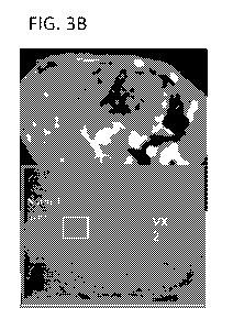
[0020] FIG. 3 shows representative CT and histological staining images of
liver tissue from a
control animal (FIG. 3A) and an animal treated with Gelfoam formulated Pexa-
Vec (FIG.
3B). FIG. 3C shows a representative angiography image of bile ducts from an
animal treated
with Gelfoam formulated Pexa-Vec. FIGS. 3D&E show representative H&E stained
images
of the junction between normal liver and tumor tissues (FIG. 3D) and normal
liver
parenchyma (FIG. 3E) in an animal treated with Gelfoam formulated Pexa-Vec.
[0021] FIG. 4 shows an experimental timeline for efficacy and pharmacokinetic
(PK) studies
of TAVE versus oncolytic virotherapy and embolization in a rabbit tumor model.
DETAILED DESCRIPTION OF PREFERRED EMBODIMENTS
[0022] Certain aspects of the inventions disclosed herein are based upon the
surprising
discovery that oncolytic vaccinia virus can effectively be administered by
transarterial
embolization techniques despite its large size and suboptimal basolateral
infection and apical
release from epithelial cells such as the cells forming blood vessel walls.
Based upon this
surprising discovery one can readily extrapolate to other oncolytic viruses of
the same size
and smaller and to other oncolytic viruses of similarly suboptimal life
cycles. Exemplary
oncolytic viruses include double stranded DNA viruses such as Poxviridae
viruses and herpes
viruses, viruses larger than 100nm in along their smalles axis, and viruses
the bud from the
apical membrane of polar cells such as blood vessel endothelial cells.
CA 02990133 2017-12-19
WO 2017/037523 PCT/1B2016/000993
13
[0023] Certain aspects of the inventions disclosed herein are based upon other
surprising
improvements over prior art direct tumoral injection and trans-arterial
oncolytic VSV
embolization including, without limitation, the lack of oncolytic virus from
the claimed
compositions and methods seeping into the blood stream. Preferably, after
introducing or
administering the oncolytic virus as disclosed herein, the subject will have
one day after
administration less than 10 pfu of the oncolytic virus per ml of blood, less
than 5 pfu of the
oncolytic virus per ml of blood, or less than 2 pfu of the oncolytic virus per
ml of blood.
[0024] An aspect of embolization with the oncolytic viruses as disclosed in
this specification
is to debulk the tumor mass using virus-mediated killing of tumor cells much
more effectively
than through either transarterial embolization or direct tumoral injection of
oncolytic virus
alone. In preferred embodiments, the embolization with the oncolytic viruses
as disclosed in
this specification debulk the tumor mass using virus-mediated killing of tumor
cells much
more effectively than through any of transarterial embolization, transarterial
chemoembolization, transarterial radioembolization, or direct tumoral
injection of oncolytic
virus alone. The oncolytic viruses of the disclosure, when delivered with an
embolizing
agent, are retained in the tumor microenvironment, thereby allowing more viral
infection of
cancer cells and preventing oncolytic virus from entering the blood stream.
Transient
vascular shut down and viral replication subsequently result in tumor necrosis
throughout the
tumor microenvironment, not just the tumor environment local to the
vaculature, thereby
`debulking' the tumor mass without observable damage to the surrounding
healthy tissue. In
preferred embodiments, the method of debulking results in necrosis of at least
75%, at least
80%, at least 85%, at least 90%, or at least 95% of the embolized tumor mass.
In one aspect,
the disclosure provides compositions containing an oncolytic virus and a
biocompatible
microparticle or hydrophilic polymer gel suitable for active embolization. In
another aspect,
the disclosure provides a method for active embolization of a vascular site in
a mammal by
introducing into the vasculature of a mammal an oncolytic virus and a
biocompatible
microparticle or hydrophilic polymer gel suitable for active embolization.
Oncolytic Viruses
[0025] The compositions and methods disclosed in this specification will
involve oncolytic
viruses other than VSV. Exemplary oncolytic viruses include double stranded
DNA viruses
such as pox viruses and herpes viruses. A preferred oncolytic virus is
vaccinia virus. In one
aspect, the oncolytic virus buds from an apical surface of an infected
polarized cell. In a
CA 02990133 2017-12-19
WO 2017/037523 PCT/1B2016/000993
14
preferred embodiment, the oncolytic virus that buds from an apical surface of
an infected
polarized cell is oncolytic vaccinia virus. In another aspect, which may be
combined with any
of the preceding aspects, the oncolytic virus is at least 0.1[1m in diameter
along the shortest
axis of the virus. In one embodiment, the oncolytic virus is at least 0.1m, at
least 0.15m, or
at least 0.23.tm in diameter along its shortest axis. The oncolytic virus may
be from 0.1-
0.2m, from 0.2-0.3m, from 0.3-0.4m, from 0.4-0.5m, from 0.5-0.6m, from 0.6-
0.7m,
from 0.1-0.7m, from 0.15-0.7m, or from 0.2-0.7 m in diameter along the
shortest axis of
the virus.
A. Oncolytic Vaccinia Virus
[0026] In a preferred embodiment, the oncolytic virus is a Poxviridae virus,
such as oncolytic
vaccinia virus. Vaccinia virus (VV) is a complex enveloped virus having a
linear double-
stranded DNA genome of about 190K bp and encoding for approximately 250 genes.
Vaccinia virus is a large virus roughly 360nm by 250nm in size. Vaccinia is
well-known for
its role as a vaccine that eradicated smallpox. Post-eradication of smallpox,
scientists have
been exploring the use of vaccinia as a tool for delivering genes into
biological tissues in gene
therapy and genetic engineering applications.
[0027] Vaccinia virus preferentially infects through the basolateral surface
of cells, but its
viral progeny are released from the apical surface. Polarized cells include,
without limitation,
epithelial cells, endothelial cells, immune cells, osteoclasts, neurons, and
fibroblasts.
[0028] Vaccinia virus and other poxviridae are unique among DNA viruses as
they replicate
only in the cytoplasm of the host cell. Therefore, the large genome is
required to code for
various enzymes and proteins needed for viral DNA replication. During
replication, vaccinia
produces several infectious forms which differ in their outer membranes: the
intracellular
mature virion (IMV), the intracellular enveloped virion (IEV), the cell-
associated enveloped
virion (CEV) and the extracellular enveloped virion (EEV). IMV is the most
abundant
infectious form and is thought to be responsible for spread between hosts. On
the other hand,
the CEV is believed to play a role in cell-to-cell spread and the EEV is
thought to be
important for long range dissemination within the host organism. The above
forms are
merely illustrative of the forms for the oncolytic vaccinia virus for use in
the compositions
and methods in this disclosure.
CA 02990133 2017-12-19
WO 2017/037523 PCT/1B2016/000993
[0029] Any oncolytic strain of vaccinia virus may be used as the vaccinia
virus component of
the combination of the present disclosure. In preferred embodiments, the
oncolytic vaccinia
virus of the compositions and methods of the present disclosure is a
Copenhagen, Western
Reserve or Wyeth strain. Other strains can readily be used including, for
example, strains
circulating in Korea.
[0030] The oncolytic vaccinia virus of the present disclosure can be
engineered to express a
foreign protein such as granulocyte-macrophage colony stimulating factor, or
GM-CSF. GM-
CSF is a protein secreted by macrophages that stimulates stem cells to produce
granulocytes
(neutrophils, eosinophils, and basophils) and macrophages. Human GM-CSF is
glycosylated
at amino acid residues 23 (leucine), 27 (asparagine), and 39 (glutamic acid)
(see U.S. Patent
5,073,627, incorporated herein by reference).
[0031] The oncolytic vaccinia virus may be engineered to lack one or more
functional genes
in order to increase the cancer selectivity of the virus. In one aspect, the
oncolytic vaccinia
virus may be engineered to lack Thymidine kinase (TK) activity. A TK-deficient
vaccinia
virus requires thymidine triphosphate for DNA synthesis, which leads to
preferential
replication in dividing cells (particularly cancer cells). In another aspect,
the oncolytic
vaccinia virus may be engineered to lack vaccinia virus growth factor (VGF).
This secreted
protein is produced early in the infection process, acting as a mitogen to
prime surrounding
cells for infection. In another aspect, the oncolytic vaccinia virus may be
engineered to lack
both VFG and TK activity. In other aspects, the oncolytic vaccinia virus may
be engineered to
lack one or more genes involved in evading host interferon (TN) response such
as E3L, K3L,
Bl8R, or B8R. In some embodiments, the oncolytic vaccinia virus is a Western
Reserve or
Wyeth strain and lacks a functional TK gene. In other embodiments, the
oncolytic vaccinia
virus is a Western Reserve strain lacking a functional Bl8R and/or B8R gene.
[0032] In some embodiments, the oncolytic vaccinia virus lacks a functional TK
gene and
expresses human GM-CSF. In a preferred embodiment, the oncolytic vaccinia
virus is a
Wyeth strain oncolytic vaccinia virus that lacks a functional TK gene and
expresses human
GM-CSF.
[0033] In a particularly preferred embodiment, the oncolytic vaccinia virus is
JX-594. JX-
594 is a replication-competent, recombinant vaccinia virus derived from the
New York Board
of Health vaccinia strain that was sold commercially as Dryvax (Wyeth
Laboratories) which
is now commonly referred to as Wyeth strain vaccinia virus. JX-594 was derived
by inserting
CA 02990133 2017-12-19
WO 2017/037523 PCT/1B2016/000993
16
the genes for human GM-CSF and E. coli B-galactosidase into the thymidine
kinase (TK)
gene of the virus (under the control of the synthetic early-late and p7.5
promoters,
respectively), thereby rendering the TK gene inactive. Inactivation of the TK
gene has been
shown to decrease the virulence of vaccinia virus and to increase tumor
specific replication.
JX-594 has demonstrated replication and GM-CSF expression, associated with
tumor
responses in patients on clinical trials via both intratumoral and intravenous
administration at
doses up to 1 x 10⁹ pfu/dose.
[0034] In some embodiments, the oncolytic vaccinia virus is SJ103r3 (also
known as vvDD-
CDSR). The vvDD-CDSR virus is a replication-selective oncolytic vaccinia virus
with
double deletions in the TK and Vaccinia Growth Factor (VGF) genes. vvDD-CDSR
is
derived by inserting Cytosine Deaminase (CD), Human Somatostatin Receptor Type
2
(SSTR2), and gpt into the TK gene of the Western Reserve (WR) strain of
Vaccinia Virus
(under the control of the synthetic early-late, synthetic late, and p7.5
promoters, repectively).
E.coli P-galactosidase is inserted with homologous recombination into the VGF
gene.
Inactivation of both the TK and VGF gene has been shown to decrease the
virulence of
vaccinia virus for safety as well as to enhance tumour specific replication
for selectivity.
Inactivation of either or both may be achieved by such insertions, by
inactivating mutations
and/or by partial or complete deletion of the gene. In some embodiments, the
oncolytic
vaccinia virus is vvDD. vvDD is a replication-selective oncolytic vaccinia
virus with double
disruptions of the TK and VGF genes of the parental WR strain. Inactivation of
both genes
increases tumour specificity for viral replication and attenuates the virus
for safety. In some
embodiments, the oncolytic vaccinia virus is SJ-102. SJ-102 is a replication-
competent,
recombinant vaccinia virus derived from the Wyeth-calf adapted New York City
Department
of Health Laboratories strain. The parental vaccinia virus Wyeth strain was
engineered by
inserting gpt and green fluorescent protein (GFP) at the TK locus to produce
the SJ-102 virus.
gpt is a selection marker, controlled under the p7.5 early-late viral promoter
and confers
resistance to an inhibitor of the enzyme inosine monophosphate dehydrogenase.
GFP is
another visual selection marker and is controlled under a synthetic early-late
promoter pSE/L.
In some embodiments, the oncolytic vaccinia virus is SJ-103. The parental
vaccinia virus for
the recombinant virus SJ-103 is Western Reserve (WR) strain. Western Reserve
strain is
derived from Wyeth strain by passaging in mice in order to enhance tumour
selectivity in a
mouse cell line and increase the oncolytic effect in vitro. The thymidine
Kinase (TK) gene of
CA 02990133 2017-12-19
WO 2017/037523 PCT/1B2016/000993
17
WR strain is disrupted by inserting gpt and green fluorescent protein (GFP) to
produce the SJ-
103 virus. gpt is controlled under the p7.5 early-late viral promoter and GFP
is controlled
under a synthetic early-late promoter pSE/L. In some embodiments, the
oncolytic vaccinia
virus is WR TK(-). The parental vaccinia virus for the recombinant virus WR
TK(-) is
Western Reserve (WR) strain. The thymidine kinase gene of WR strain has been
disrupted in
WR TK(-) by inserting a selection marker. In some embodiments, the oncolytic
vaccinia
virus is a Lister strain variant from the Institute of Viral Preparations
(LIVP). In some
embodiments, the oncolytic vaccinia virus is GL-ONC1 (Genelux), also known as
GLV-1h68
or RVGL21. GL-ONC1 is a genetically-engineered attenuated LIVP strain vaccinia
virus
carrying transgenes encoding Renilla luciferase, green fluorescent protein
(both inserted at the
F14.5L locus), P-galactosidase (inserted at the J2R locus, which encodes
thymidine kinase),
and B-glucuronidase (inserted at the A56R locus, which encodes hemagglutinin).
In some
embodiments, the oncolytic vaccinia virus is WR AB18R luc+. WR AB18R luc+ is
the WR
vaccinia virus with the Bl8R gene deleted and a luciferase gene inserted to
the TK gene.
[0035] Vaccinia virus may be propagated using the methods described by Earl
and Moss
(Ausubel et al. (1994) Current Protocols in Molecular Biology, pages 16.15.1
to 16.18.10) or
the methods described in WIPO Publication No.W02013/022764, both of which are
incorporated herein by reference.
B. Other Poxviruses
[0036] The genus Orthopoxvirus is relatively more homogeneous than other
members of the
Chordopoxvirinae subfamily and includes 11 distinct but closely related
species, which
includes vaccinia virus, variola virus (causative agent of smallpox), cowpox
virus, buffalopox
virus, monkeypox virus, mousepox virus and horsepox virus species as well as
others (see
Moss, (1996) Fields Virology, 3:3637-2672). Certain embodiments of the present
disclosure,
as described herein, may be extended to other members of Orthopoxvirus genus
as well as the
Parapoxvirus, Avipoxvirus, Capripoxvirus, Leporipoxvirus, Suipoxvirus,
Molluscipoxvirus,
and Yatapoxvirus genus. A genus of poxvirus family is generally defined by
serological
means including neutralization and cross-reactivity in laboratory animals.
Various members
of the Orthopoxvirus genus, as well as other members of the Chordovirinae
subfamily utilize
immunomodulatory molecules, examples of which are provided herein, to
counteract the
immune responses of a host organism. Thus, the present disclosure described
herein is not
limited to vaccinia virus, but may be applicable to a number of viruses.
CA 02990133 2017-12-19
WO 2017/037523 PCT/1B2016/000993
18
Myxomavirus
[0037] In one embodiment, the oncolytic virus for use in the compositions and
methods of
this disclosure is Myxoma virus. Myxoma Virus ("MV") is the causative agent of
myxomatosis in rabbits. MV belongs to the Leporipoxvirus genus of the
Poxviridae family,
the largest of the DNA viruses. MV induces a benign disease in its natural
host, the
Sylvilagus rabbit in the Americas. However, it is a virulent and host-specific
poxvirus that
causes a fatal disease in European rabbits, characterized by lesions found
systemically and
especially around the mucosal areas. (Cameron C, Hota-Mitchell S, Chen L,
Barrett J, Cao J
X, Macaulay C, Wilier D, Evans D, McFadden G. Virology 1999, 264(2): 298-318;
Kerr P &
McFadden G. Viral Immunology 2002, 15(2): 229-246).
[0038] MV is a large virus with a double-stranded DNA genome of 163 kb which
replicates
in the cytoplasm of infected cells (B. N. Fields, D. M. Knipe, P. M. Howley,
Eds., Virology
Lippincott Raven Press, New York, 2nd ed., 1996). MV is known to encode a
variety of cell-
associated and secreted proteins that have been implicated in down-regulation
of the host's
immune and inflammatory responses and inhibition of apoptosis of virus-
infected cells. MV
can be taken up by all human somatic cells. MV can infect and kill cancer
cells, including
human tumour cells.
[0039] The Myxoma virus may be any virus that belongs to the Leporipoxvirus
species of
pox viruses that is replication-competent. The Myxoma virus may be a wild-type
strain of
Myxoma virus or it may be a genetically modified strain of Myxoma virus.
[0040] The Myxoma virus genome may be readily modified to express one or more
therapeutic transgenes using standard molecular biology techniques known to a
skilled
person, and described for example in Sambrook et al. ((2001) Molecular
Cloning: a
Laboratory Manual, 3rd ed., Cold Spring Harbour Laboratory Press). A skilled
person will be
able to readily determine which portions of the Myxoma viral genome can be
deleted such
that the virus is still capable of productive infection. For example, non-
essential regions of the
viral genome that can be deleted can be deduced from comparing the published
viral genome
sequence with the genomes of other well-characterized viruses (see for example
C. Cameron,
S. Hota-Mitchell, L. Chen, J. Barrett, J.-X. Cao, C. Macaulay, D. Willer, D.
Evans, and G.
McFadden, Virology (1999) 264: 298-318)).
[00411 In some embodiments, the oncolytic Myxoma virus is vMyxlac: a
recombinant
Lausanne strain containing the E. coli lacZ gene inserted at an innocuous site
between open
CA 02990133 2017-12-19
WO 2017/037523 PCT/1B2016/000993
19
reading frames MO1OL and M011L. In some embodiments, the oncolytic Myxoma
virus is
vMyxT5KO, a recombinant virus with copies of the M-T5 gene replaced by lacZ.
In some
embodiments, the oncolytic Myxoma virus is SG33, also known as CNCM 1-1594.
SG33
virus contains a deletion of about 15 kb in the right-hand portion of its
genome. Compared to
a reference Lausanne strain, the genes M15 1R and MOO1R are only partially
deleted,
producing inactive truncated proteins. The genes M152R, M153R, M1544 M156R, as
well
as the genes for the right-hand ITR M008. 1R, MOO8R, 1\4007R, MOO6R, MOO5R,
M004.1R,
MOO4R, M003 .2R, M003. 1R, and 1\4002R are completely deleted. Another
alteration between
the genome of the SG33 strain and that of the reference Lausanne strain is at
the level of the
1\4011L gene (positions 14125-13628 in the genome of the Lausanne strain),
encoding an
inhibitor of apoptosis (M1 1L, GenBank NP____051725). It is possible to use a
modified
attenuated Myxoma virus expressing a desired gene (for example a therapeutic
gene of the
heipesvirus Thymidine kinase type or FCUl, produced from the fusion between
the genes
encoding Cytosine deamina.se and Uracil phospholibosyltransferase) (ERBS et
al, Cancer
Gene Therapy, 15, 18-28, 2008). Attenuated Myxoma viruses modified to express
a gene of
interest are described in FR2736358.
Parapoxvirus
[0042] In one embodiment, the oncolytic virus for use in the compositions and
methods of
this disclosure is Parapoxvirus. Parapoxvirus orf virus is a poxvirus that
induces acute
cutaneous lesions in different mammalian species, including humans.
Parapoxvirus orf virus
naturally infects sheep, goats and humans through broken or damaged skin,
replicates in
regenerating epidermal cells and induces pustular leasions that turn to scabs.
The
parapoxvirus orf virus encodes the gene 0V20.0L that is involved in blocking
PKR activity.
The parapoxvirus orf virus is unable to replicate in cells that do not have an
activated Ras-
pathway. A more preferred oncolytic virus is an "attenuated parapoxvirus orf
virus" or
"modified parapoxvirus orf virus," in which the gene product or products which
prevent the
activation of PKR are lacking, inhibited or mutated such that PKR activation
is not blocked.
Preferably, the gene 0V20.0L is not transcribed. Such attenuated or modified
parapoxvirus
orf virus would not be able to replicate in normal cells that do not have an
activated Ras-
pathway, but it is able to infect and replicate in cells having an activated
Ras-pathway.
[0043] In some embodiments, the oncolytic Parapoxvirus is an orf virus strain
selected from
OV NZ-2 (New Zealand-2), OV NZ-7 (New Zealand-7), and OV-SA00. In some
CA 02990133 2017-12-19
WO 2017/037523 PCT/1B2016/000993
embodiments, the oncolytic Parapoxvirus is a recombinant orf virus (ORFV)
containing one
or more heterologous host range genes, wherein said genes allow for
replication of the virus
in human cells. The heterologous host range genes can include, without
limitation, SPI-1,
SPI-2, KIL, C7L, p28/N1R, B5R, E3L, K3L, M-T2, M-T4, M-T5, Ml1L, M13L, M063,
and
Fl1L.
C. Herpesviruses
Herpes Simplex Virus
[0044] In one aspect, the disclosure provides a composition containing an
oncolytic virus and
a biocompatible microparticle or hydrophilic polymer gel suitable for active
embolization. In
one embodiment, the oncolytic virus is a Herpesviridae virus, such as herpes
simplex virus-1
(HSV-1) or herpes simplex virus-2 (HSV-2). Human herpesviridae viruses include
herpes
simplex virus-1 ("HSV-1"), herpes simplex virus-2 ("HSV-2"), human
cytomegalovirus
("HCMV"), Epstein-Barr virus ("EBV"), Kaposi's sarcoma ("HHV-8"), roseolovirus-
6A
("HHV-6A"), and roseolovirus-6B ("HHV-6B").
[0045] The HSV virion is a large (120 to 300nm in diameter), enveloped virus
with an
icosahedral capsid. It has double stranded DNA with a genome that encodes at
least 70
polypeptides. This large amount of regulatory information permits the virus to
control its own
gene expression and to modify multiple complex events within the infected
cell. The herpes
simplex virus enters the host by direct contact, is spread to a target tissue
only, spreads within
the host via neuronal axonal flow, targets the dorsal root ganglia and after
recovery of the
host from an acute infection, remains latent in the targeted tissue. The
limited spread makes
HSV a good candidate for an oncolytic virus.
[0046] In HSV, mutations allowing selective oncolytic activity include
mutation to the genes
encoding ICP34.5, ICP6 and/or thymidine kinase (TK), preferably ICP34.5. Such
mutations
to the ICP34.5-encoding gene in laboratory strains of HSV are described in
Chou et al 1990,
Maclean et al 1991, although any mutation in which ICP34.5 is non-functional
may be used.
Accordingly, in an HSV strain, the viruses preferably modified such that it
lacks one or more
of a functional ICP34.5-encoding gene, a functional ICP6-encoding gene, a
functional
glycoprotein H-encoding gene, a functional thymidine kinase-encoding gene; or
in a non-
HSV strain, the virus lacks a functional gene equivalent to one of said HSV
genes. More
preferably, the virus lacks a functional ICP34.5-encoding gene. Other
modifications may also
CA 02990133 2017-12-19
WO 2017/037523 PCT/1B2016/000993
21
be made. In particular, the HSV virus may be modified such that it lacks a
functional ICP47
gene. This is because ICP47 usually functions to block antigen presentation in
HSV-infected
cells so its disruption leads to a virus that does not confer on infected
tumor cells particular
properties that might protect such HSV infected cells from the host's immune
system.
Further, the HSV virus may be modified to express the human GM-CSF gene.
Secreted or
otherwise released GM-CSF can attract dendritic cells to the tumor enhancing
the immune
response against the tumor cells.
[0047] When the virus of the invention is a herpes simplex virus, the virus
may be derived
from, for example HSV1 or HSV2 strains, or derivatives thereof, preferably
HSV1. A
preferred HSV-1 strain is JS-1, which can be modified by inactivation of the
ICP34.5 and
ICP47 genes and addition of the human GM-CSF (e.g., Senzer et al. JCO (2009)
27(34):
5763-5771). In some embodiments, wild-type HSV-1 is obtained from ATCC (VR-
735) and
no engineering is performed. In some embodiments, the HSV-1 strain is MP
(mutant strain of
Herpes Simplex Virus type 1). In some embodiments, the HSV-1 virus is
Talimogene
laherparepvec, also known as OncoVEX GMCSF or T-VEC (AMGEN). T-VEC was
produced by modification of the HSV-1 JS-1 parent strain to attenuate the
virus and increase
selectively for cancer cells. The JS-1 strain was modified via deletion of the
ICP34.5 and
ICP47 genes (to prevent infection of non-tumor cells and enables antigen
presentation,
respectively), earlier insertion of the US11 gene (to increase replication and
oncolytic ability),
and insertion of the human GM-CSF gene (to increase the anti-tumor immune
response). In
some embodiments, the oncolytic HSV-1 virus is H5V1716, also known as
SEPREHVIR.
The H5V1716 strain contains a deletion of the ICP34.5 gene, allowing for
selective
replication in tumor cells. In some embodiments, the HSV-1 virus is
HSV1716NTR, an
oncolytic virus generated by inserting the enzyme nitroreductase (NTR) into
the virus
H5V1716 as a gene-directed enzyme prodrug therapy (GDEPT) strategy. In some
embodiments, the HSV-1 virus is G207, an oncolytic virus derived by deletion
of the ICP34.5
gene and inactivation of the ICP6 gene by insertion of the E. coli LacZ gene
into a parent
HSV-1 laboratory strain F. In some embodiments, the HSV-1 virus is NV1020, an
oncolytic
virus derived by deletion of one copy of the ICP34.5 gene.
[0048] Derivatives include inter-type recombinants containing DNA from HSV1
and HSV2
strains. Such inter-type recombinants are described in the art, for example in
Thompson et al
(1998) and Meignier et al (1988). A derivative may have the sequence of a HSV1
or HSV2
CA 02990133 2017-12-19
WO 2017/037523 PCT/1B2016/000993
22
genome modified by nucleotide substitutions, for example from 1, 2 or 3 to 10,
25, 50 or 100
substitutions. The HSV1 or HSV2 genome may alternatively or additionally be
modified by
one or more insertions and/or deletions and/or by an extension at either or
both ends.
Cytomegalovirus
[0049] In one embodiment, the oncolytic virus for use in the compositions and
methods of
the disclosure is Cytomegalovirus. Cytomegalovirus (CMV), also known as human
herpesvirus 5 (HHV-5), is a herpes virus classified as being a member of the
beta subfamily
of herpesviridae. According to the Centers for Disease Control and Prevention,
CMV
infection is found fairly ubiquitously in the human population, with an
estimated 40-80% of
the United States adult population having been infected. The virus is spread
primarily through
bodily fluids and is frequently passed from pregnant mothers to the fetus or
newborn. In most
individuals, CMV infection is latent, although virus activation can result in
high fever, chills,
fatigue, headaches, nausea, and splenomegaly.
[0050] Although most human CMV infections are asymptomatic, CMV infections in
immunocompromised individuals, (such as HIV-positive patients, allogeneic
transplant
patients and cancer patients) or persons whose immune system has yet fully
developed (such
as newborns) can be particularly problematic (Mocarski et al.,
Cytomegalovirus, in Field
Virology, 2701-2772, Editor: Knipes and Howley, 2007). CMV infection in such
individuals
can cause severe morbidity, including pneumonia, hepatitis, encephalitis,
colitis, uveitis,
retinitis, blindness, and neuropathy, among other deleterious conditions. In
addition, CMV
infection during pregnancy is a leading cause of birth defects (Adler, 2008 J.
Clin Virol,
41:231; Arvin et al, 2004 Clin Infect Dis, 39:233; Revello et al, 2008 J Med
Virol, 80:1415).
CMV infects various cells in vivo, including monocytes, macrophages, dendritic
cells,
neutrophils, endothelial cells, epithelial cells, fibroblasts, neurons, smooth
muscle cells,
hepatocytes, and stromal cells (Plachter et al. 1996, Adv. Virus Res. 46:195).
Although
clinical CMV isolates replicate in a variety of cell types, laboratory strains
AD169 (Elek &
Stem, 1974, Lancet 1:1) and Towne (Plotkin et al., 1975, Infect. Immun.
12:521) replicate
almost exclusively in fibroblasts (Hahn et al., 2004, J. Virol. 78:10023). The
restriction in
tropism, which results from serial passages and eventual adaptation of the
virus in fibroblasts,
is stipulated a marker of attenuation (Gerna et al., 2005, J. Gen. Virol.
86:275; Gerna et al,
2002, J. Gen Virol. 83:1993; Gerna et al, 2003, J. Gen Virol. 84:1431; Dargan
et al, 2010, J.
Gen Virol. 91:1535). Mutations causing the loss of epithelial cell,
endothelial cell, leukocyte,
CA 02990133 2017-12-19
WO 2017/037523 PCT/1B2016/000993
23
and dendritic cell tropism in human CMV laboratory strains have been mapped to
three open
reading frames (ORFs): UL128, UL130, and UL131 (Hahn et al., 2004, J. Virol.
78:10023;
Wang and Shenk, 2005 J. Virol. 79:10330; Wang and Shenk, 2005 Proc Natl Acad
Sci USA.
102:18153). Biochemical and reconstitution studies show that UL128, UL130 and
UL131
assemble onto a gH/gL scaffold to form a pentameric gH complex (Wang and
Shenk, 2005
Proc Natl Acad Sci USA. 102:1815; Ryckman et al, 2008 J. Virol. 82:60).
Restoration of this
complex in virions restores the viral epithelial tropism in the laboratory
strains (Wang and
Shenk, 2005 J. Virol. 79:10330). Loss of endothelial and epithelial tropism
has been
suspected as a deficiency in the previously evaluated as vaccines such as
Towne (Gerna et al,
2002, J. Gen Virol. 83:1993; Gerna et al, 2003, J. Gen Virol. 84:1431).
Neutralizing
antibodies in sera from human subjects of natural CMV infection have more than
15-fold
higher activity against viral epithelial entry than against fibroblast entry
(Cui et al, 2008
Vaccine 26:5760). Humans with primary infection rapidly develop neutralizing
antibodies to
viral endothelial and epithelial entry but only slowly develop neutralizing
antibodies to viral
fibroblast entry (Gerna et al, 2008 J. Gen. Virol. 89:853). Furthermore,
neutralizing activity
against viral epithelial and endothelial entry is absent in the immune sera
from human
subjects who received Towne vaccine (Cui et al, 2008 Vaccine 26:5760). More
recently, a
panel of human monoclonal antibodies from four donors with HCMV infection was
described, and the more potent neutralizing clones from the panel recognized
the antigens of
the pentameric gH complex (Macagno et al, 2010 J. Virol. 84:1005).
D. Measles Virus
[0051] In one embodiment, the oncolytic virus for use in the compositions and
methods of
this disclosure is Measles virus. Measles virions are large and pleitrophic
with diameters of
up to ¨550nm. Measles virus is a negative strand RNA virus whose genome
encodes six
protein products, the N (nucleocapsid), P (polymerase cofactor
phosphoprotein), M (matrix),
F (fusion), H (hemaglutinin) and L (large RNA polymerase) proteins. The H
protein is a
surface glycoprotein which mediates measles virus attachment to its receptor,
CD46 (Dorig,
et al., Cell 75: 295-305, 1993). The F protein is responsible for cell¨cell
fusion after viral
attachment has taken place. Measles virus has a natural tropism for lymphoid
cells and, in
particular, cancerous lymphoid cells.
[0052] The tumor selectivity of the virus is due to intracellular restrictions
to the life cycle of
the virus that is strongly inhibitory to virus propagation in nontransformed
cells, but which
CA 02990133 2017-12-19
WO 2017/037523 PCT/1B2016/000993
24
are overriden by cellular factors present in neoplastic cells (Robbins, et
al., Virology 106:
317-326,1980; Robbins, Intervirology 32: 204-208,1991). Measles infectivity of
lymphoid
cells causes a very characteristic cytopathic effect. Multinucleated giant
cells develop during
measles virus replication in lymph nodes as a result of gross cell¨cell fusion
(Warthin, Arch.
Pathol. 11: 864-874,1931). In tissue culture, infection with measles virus can
cause fusion of
a whole monolayer of cells. The F and H antigens are found on the surface of
infected cells.
Thus, cells which are infected by measles virus and whose membranes express F
and H
proteins become highly fusogenic and can cause fusion not only of other
infected cells but
also of neighboring cells which are not infected (Norrby and Oxman, "Measles
Virus." In
Virology, 1990, B. N. Fields, et al., eds. New York, Raven Press, Ltd., pp
1013-1044). The
expression of viral antigens on the surface of a tumor cell can also mediate a
tumor specific
immune response.
[0053] An attenuated strain of virus can be obtained by serial passage of the
virus in cell
culture (e.g., in non-human cells), until a virus is identified which
immunogenic but not
pathogenic. While wild type virus will cause fatal infection in marmosets,
vaccine strains do
not. In humans, infection with wild type viral strains is not generally fatal
but is associated
with classic measles disease. Classic measles disease includes a latent period
of 10-14 days,
followed by a syndrome of fever, coryza, cough, and conjunctivitis, followed
by the
appearance of a maculopapular rash and Koplik's spots (small, red, irregularly
shaped spots
with blue-white centers found inside the mouth). The onset of the rash
coincides with the
appearance of an immune response and the initiation of virus clearance. In
contrast,
individuals receiving an attenuated measles virus vaccine do not display
classical measles
symptoms. Attenuation is associated with decreased viral replication (as
measured in vivo by
inability to cause measles in monkeys), diminished viremia, and failure to
induce
cytopathological effects in tissues (e.g., cell¨cell fusion, multinucleated
cells). However,
these biological changes have not been mapped to any single genetic change in
the virus
genome.
[0054] An attenuated strain of measles virus which has been clinically tested
as a vaccine for
measles infection is used to provide an effective dose which will limit and/or
cause regression
of a group of cancer cells, such as a tumor. The Moraten attenuated form of
the virus has been
used world-wide as a vaccine and has an excellent safety record (Hilleman, et
al., J. Am.
Med. Assoc. 206: 587-590,1968). Accordingly, in one embodiment of the
invention, the
CA 02990133 2017-12-19
WO 2017/037523 PCT/1B2016/000993
Moraten strain is used to provide an effective dose. The Moraten vaccine is
commercially
available from Merck and is provided lyophilized in a vial which when
reconstituted to 0.5
ml comprises 103 pfu/ml. A vaccine against the Moraten Berna strain is
available from the
Swiss Serum Vaccine Institute Berne.
[0055] In a further embodiment of the invention, the Edmonston-B vaccine
strain of measles
virus is used (MV-Edm) (Enders and Peebles, Proc. Soc. Exp. Biol. Med. 86: 277-
286,
1954). MV-Edm grows efficiently in tumor cells but its growth is severely
restricted in
primary cultures of human peripheral blood mononuclear cells, normal dermal
fibroblasts,
and vascular smooth muscle cells. A form of the Enders attenuated Edmonston
strain is
available commercially from Merck (Attenuvax ). In some embodiments, the
measles virus
is MV-NIS. MV-NIS is a measles virus encoding the human thyroidal sodium
iodide
symporter (MV-NIS). The measles virus for MV-NIS is an attenuated oncolytic
Edmonston
(ED) strain. MV-NIS is selectively destructive to myeloma plasma cells and MV-
NIS
infected cells can be imaged via uptake of iodine 123 (1-123).
[0056] Other attenuated measles virus strains are also encompassed within the
scope of the
invention, such as Leningrad-16, and Moscow-5 strains (Sinitsyna, et al., Res.
Virol. 141(5):
517-31, 1990), Schwarz strain (Fourrier, et al., Pediatrie 24(1): 97-8, 1969),
9301B strain
(Takeda, et al. J. VIROL. 72/11: 8690-8696), the AIK-C strain (Takehara, et
al., Virus Res
26 (2): 167-75, 1992 November), and those described in Schneider-Shaulies, et
al., PNAS
92(2): 3943-7, 1995, the entireties of which are incorporated by reference
herein.
[0057] In a further embodiment of the invention, the measles virus is provided
in a
composition comprising a mixture of attenuated oncolytic viruses. In one
embodiment, the
mumps measles and rubella vaccine (MMR) is used. The MMR vaccine was
introduced into
the United States in 1972 and into the United Kingdom in 1998. Commercially
available
preparations of the MMR vaccine is obtainable from Merck, Pasterur Merieux
Connaught, or
SmithKline Beecham, and also contain the Moraten strain of attenuated measles
virus at a
minimum titer of 1 PFU/ml. In still a further embodiment of the invention, the
measles virus
is provided in a composition comprising Edmonston Zagreb measles strain (an
attenuated
strain obtained from the Edmonston-enders stain) and the Wistar RA 27/3 strain
of rubella
(Swiss Serum Vaccine Institute Berne). It should be apparent to those of skill
in the art that
any clinically tested measles vaccine is acceptable for use in the invention,
and is
encompassed within the scope of the invention.
CA 02990133 2017-12-19
WO 2017/037523 PCT/1B2016/000993
26
[0058] In still a further embodiment of the invention, recombinant measles
viruses
comprising genetic modifications are derived from wild type measles virus to
generate
attenuated viruses, e.g., viruses having high immunogenicity (as measured by
70-100%
seroconversion) and no pathogenicity (e.g., not producing classical measles
symptoms, as
discussed above). In one embodiment of the invention, genetic modifications
are introduced
through random mutagenesis of a plasmid comprising the sequence of a wild type
measles
virus. Sequences of wild type isolates are disclosed in U.S. Pat. No.
5,578,448, the entirety of
which is enclosed herein by reference.
[0059] In another embodiment of the invention, particular cistrons in the
measles virus
genome are targeted to modify genes whose expression is associated with
attenuation
(Schneider-Shaulies, et al. PNAS 92(2): 3943-7, 1995; Takeda, et al. J. Virol.
1998 72/11
(8690-8696)). Thus, in one embodiment of the invention, a recombinant measles
virus strain
is generated comprising a single point mutation or multiple non-contiguous
point mutations
in any of an H protein, a V protein, a C protein, and combinations thereof. In
still a further
embodiment of the invention, natural variants of the wild type or attenuated
measles viruses
are identified (e.g., such as from cultures of virus from infected patients)
which have at least
one point mutation in their genome.
Engineering of the Oncolytic Virus
[0060] In certain embodiments, the oncolytic viruses for use in the
compositions and methods
of this disclosure may be engineered to improve the efficacy, safety or other
characteristic of
the virus. Viruses are frequently inactivated, inhibited or cleared by
immunomodulatory
molecules such as interferons (-a, -(3, -y) and tumor necrosis factor-a (TNF)
(Moss, 1996).
Inactivation may be achieved by inactivation of a viral gene, which may be
achieved by
insertion(s) into the gene, by inactivating mutations in the gene and/or by
partial or complete
deletion of the gene. Host tissues and inflammatory/immune cells frequently
secrete these
molecules in response to viral infection. These molecules can have direct
antiviral effects
and/or indirect effects through recruitment and/ or activation of inflammatory
cells and
lymphocytes. Given the importance of these immunologic clearance mechanisms,
viruses
have evolved to express gene products that inhibit the induction and/or
function of these
cytokines/chemokines and interferons. For example, vaccinia virus (VV; and
some other
poxviruses) encodes the secreted protein vCKBP (B29R) that binds and inhibits
the CC (two
adjacent cysteines) chemokines (e.g., RANTES, eotaxin, MIP-1-alpha) (Alcami et
al., 1998).
CA 02990133 2017-12-19
WO 2017/037523 PCT/1B2016/000993
27
Some VV strains also express a secreted viral protein that binds and
inactivates TNF (e.g.,
Lister A53R) (Alcami et al., 1999). Most poxvirus strains have genes encoding
secreted
proteins that bind and inhibit the function of interferons-a/13 (e.g., Bl8R)
or interferon¨y
(B8R). vC12L is an IL-18-binding protein that prevents IL-18 from inducing IFN-
y and NK
cell/ cytotoxic T-cell activation.
[0061] Most poxvirus virulence research has been performed in mice. Many, but
not all, of
these proteins are active in mice (B18R, for example, is not). In situations
in which these
proteins are active against the mouse versions of the target cytokine,
deletion of these genes
leads to reduced virulence and increased safety with VV mutants with deletions
of or
functional mutations in these genes. In addition, the inflammatory/immune
response to and
viral clearance of these mutants is often increased compared to the parental
virus strain that
expresses the inhibitory protein. For example, deletion of the T1/35kDa family
of poxvirus-
secreted proteins (chemokine-binding/-inhibitory proteins) can lead to a
marked increase in
leukocyte infiltration into virus-infected tissues (Graham et al., 1997).
Deletion of the vC12L
gene in VV leads to reduced viral titers/ toxicity following intranasal
administration in mice;
in addition, NK cell and cytotoxic T-lymphocyte activity is increased together
with IFN-y
induction (Smith et al., 2000). Deletion of the Myxoma virus T7 gene (able to
bind IFN-y
and a broad range of chemokines) results in reduced virulence and
significantly increased
tissue inflammation/infiltration in a toxicity model (Upton et al., 1992;
Mossman et al.,
1996). Deletion of the M-T2 gene from myxoma virus also resulted in reduced
virulence in a
rabbit model (Upton et al. 1991). Deletion of the B18R anti-interferon-a/43
gene product also
leads to enhanced viral sensitivity to IFN-mediated clearance, reduced titers
in normal tissues
and reduced virulence (Symons et al., 1995; Colamonici et al., 1995; Alcami et
al., 2000). In
summary, these viral gene products function to decrease the antiviral immune
response and
inflammatory cell infiltration into virus-infected tissues. Loss of protein
function through
deletion/mutation leads to decreased virulence and/or increased
proinflammatory properties
of the virus within host tissues. See PCT/US2003/025141, which is hereby
incorporated by
reference.
[0062] Cytokines and chemokines can have potent antitumoral effects (Vicari et
al., 2002;
Homey et al., 2002). These effects can be on tumor cells themselves directly
(e.g., TNF) or
they can be indirect through effects on non-cancerous cells. An example of the
latter is TNF,
which can have antitumoral effects by causing toxicity to tumor-associated
blood vessels; this
CA 02990133 2017-12-19
WO 2017/037523 PCT/1B2016/000993
28
leads to a loss of blood flow to the tumor followed by tumor necrosis. In
addition,
chemokines can act to recruit (and in some cases activate) immune effector
cells such as
neutrophils, eosinophils, macrophages and/or lymphocytes. These immune
effector cells can
cause tumor destruction by a number of mechanisms. These mechanisms include
the
expression of antitumoral cytokines (e.g., TNF), expression of fas-ligand,
expression of
perforin and granzyme, recruitment of natural killer cells, etc. The
inflammatory response
can eventually lead to the induction of systemic tumor-specific immunity.
Finally, many of
these cytokines (e.g., TNF) or chemokines can act synergistically with
chemotherapy or
radiation therapy to destroy tumors.
[0063] Clinically effective systemic administration of recombinant versions of
these
immunostimulatory proteins is not feasible due to (1) induction of severe
toxicity with
systemic administration and (2) local expression within tumor tissue is needed
to stimulate
local infiltration and antitumoral effects. Approaches are needed to achieve
high local
concentrations of these molecules within tumor masses while minimizing levels
in the
systemic circulation. Viruses can be engineered to express cytokine or
chemokine genes in an
attempt to enhance their efficacy. Expression of these genes from replication-
selective vectors
has potential advantages over expression from non-replicating vectors.
Expression from
replicating viruses can result in higher local concentrations within tumor
masses; in addition,
replicating viruses can help to induce antitumoral immunity through tumor cell
destruction/oncolysis and release of tumor antigens in a proinflammatory
environment.
However, there are several limitations to this approach. Serious safety
concerns arise from
the potential for release into the environment of a replication-competent
virus (albeit tumor-
selective) with a gene that can be toxic if expressed in high local
concentrations. Viruses that
express potent pro-inflammatory genes from their genome may therefore pose
safety risks to
the treated patient and to the general public. Even with tumor-targeting,
replication-selective
viruses expressing these genes, gene expression can occur in normal tissues
resulting in
toxicity. In addition, size limitations prevent expression of multiple and/or
large genes from
viruses such as adenovirus; these molecules will definitely act more
efficaciously in
combination. Finally, many of the oncolytic viruses in use express anti-
inflammatory
proteins and therefore these viruses will counteract the induction of a
proinflammatory milieu
within the infected tumor mass. The result will be to inhibit induction of
antitumoral
immunity, antivascular effects and chemotherapy-/radiotherapy-sensitization.
CA 02990133 2017-12-19
WO 2017/037523 PCT/1B2016/000993
29
Embolic Agents
[0064] Numerous biocompatible microparticle or hydrophilic polymer gel agents
can be used
the compositions and methods of this disclosure. In a preferred embodiment,
the
biocompatible microparticle or hydrophilic polymer gel agents are selected
from: degradable
starch microparticles, polyvinyl alcohol microparticles, gelatin foam
microparticles, and
sulfonated polyvinyl alcohol hydrogel microparticles. In one aspect the
biocompatible
microparticle or hydrophilic polymer gel agent increases the viral output from
tumor cells
cultured in vitro by at least 50%, at least 75%, at least 100%, at least 150%,
at least 200% or
at least 300%. In a related aspect the biocompatible microparticle or
hydrophilic polymer gel
agent increases the viral output from tumor cells cultured in vitro by between
50% and 400%,
between 75% and 400%, between 100% and 400%, between 150% and 400%, between
200%
and 400%, or between 300% and 400%.
[0065] Biocompatible microparticle or hydrophilic polymer gel agents ("embolic
agents") can
be either temporary or permanent. Exemplary temporary embolic agents include
gelfoam,
collagen, and thrombin. Exemplary permanent embolic agents include particles,
such as
polyvinyl alcohol particles (PVA) and embospheres, coils, such as pushable,
injectable,
detachable, mechanical, electrolytic, and hydrolytic coils, liquid agents,
such as glue, onyx,
alcohol, and ALGELTM (a hydrogel, sugar-based polymer derived from alginate),
and other
agents, including amplatzer plugs, Gianturco-Grifka vascular occlusive device
(GGVODs),
and detachable balloons. Different embolic agents can be used depending on the
size of the
vessel to be embolized, the desired length of vessel occlusion following
embolization, and
whether embolized tissue should remain viable after occlusion. Given the
extensive use of
embolization, a skilled interventional radiologist would have no difficulty in
selecting the
appropriate type of agent, size range of agent, etc. to achieve the desired
embolization.
Vessel occlusion is useful in clinical scenarios such as traumatic injury and
hemorrhage, or
when repeated embolization procedures are desired, such may be desirable as in
tumor
embolization with oncolytic viruses as disclosed in this specification.
[0066] In one embodiment, the biocompatible microparticle or hydrophilic
polymer gel
agents are gelatin foam microparticles. Exemplary gelatin foam includes
Gelfoam, produced
by Alicon/Scion Medical Technologies. Gelfoam is a biological substance made
from
purified skin gelatin, and is formulated in sterile sheets or as a powder.
Gelfoam has been
used in embolization applications for over 30 years, and is a low cost,
versatile embolic agent.
CA 02990133 2017-12-19
WO 2017/037523 PCT/1B2016/000993
Gelfoam slows blood flow by causing mechanical obstruction. Gelfoam powder
consists of
particulates that range in size from 150-1000m and can aggregate to form
larger
conglomerate particles upon water absorption. Gelfoam sheets can be cut into
numerous
different sizes and shapes and formulated with other aqueous agents upon
injection depending
upon the desired application. Gelfoam slurry containing both a contrast agent
and Gelfoam
sponge can be used to form a "cast" of proximal embolized vessels, while
Gelfoam torpedoes
or cubes can be used for larger vessels. Gelfoam temporarily occludes vessels
by slowing
blood flow, increasing thrombus formation, and functioning as a scaffold for
clots.
[0067] In one embodiment, the biocompatible microparticle or hydrophilic
polymer gel
agents are degradable starch microparticles. Exemplary degradable starch
microparticles
(DSM) are EMBOCEPTS particles produced by Pharmacept and SPHEREX particles
produced by Mangle Life Sciences. EMBOCEPTS particles (Amilomer as the active
substance) are cross-linked particles composed of hydrolyzed potato starch.
These particles
are suitable for temporary embolization, as they have a half-life of
approximately 35 minutes
and are degradable. SPHEREX particles are composed of DSM-S microparticles,
sterilized
and suspended in saline solution. Starch microparticles may be prepared from
an aqueous
solution of purified amylopectin-based starch of reduced molecular weight by
forming an
emulsion of starch droplets in an outer phase of polymer solution, converting
the starch
droplets to a gel, and drying the starch particles. A release-controlling
shell is optionally also
applied to the particles. Biodegradable microparticles, after parenteral
administration, are
dissolved in the body to form endogenic substances, ultimately, for example,
glucose. The
biodegradability can be determined or examined through incubation with a
suitable enzyme,
for example alpha-amylase, in vitro. The biodegradability can also be examined
through
parenteral injection of the microparticles, for example subcutaneously or
intramuscularly, and
histological examination of the tissue as a function of time. Biodegradable
starch
microparticles disappear normally from the tissue within a few weeks and
generally within
one week. In those cases in which the starch microparticles are coated with a
release-
controlling shell, for example coated, it is generally this shell which
determines the
biodegradability rate, which then, in turn, determines when alpha-amylase
becomes available
to the starch matrix.
[0068] In one embodiment, the biocompatible microparticle or hydrophilic
polymer gel
agents are polyvinyl alcohol (PVA) microparticles. Exemplary polyvinyl alcohol
CA 02990133 2017-12-19
WO 2017/037523 PCT/1B2016/000993
31
microparticles are produced by Boston Scientific Corporation (Natick, MA). PVA
particles
are made from a PVA foam sheet that is vacuum dried and scraped into
particles. The
particles are filtered with sieves and are available in sizes ranging from
100i.tm to 1100m.
Polyvinyl alcohol particles are irregular in size and shape, which promotes
aggregation. After
suspension, PVA particles can be oblong, oval, irregular, sharp, and angulated
with small
fragments after suspension. Polyvinyl alcohol particles deliver permanent
occlusion by
adhering to vessel walls and by blocking the smallest vessel into which they
pass. PVA
occlusion results in inflammatory reactions, local vessel necrosis, and
subsequent vessel
fibrosis.
[0069] In one embodiment, the biocompatible microparticle or hydrophilic
polymer gel
agents are sulfonated polyvinyl alcohol hydrogel microparticles. Exemplary
sulfonated
polyvinyl alcohol hydrogel microparticles are DC-Beads produced by
Biocompatibles (UK,
Surrey, UK). DC Beads are embolic microparticle products based on a polyvinyl
alcohol
hydrogel that has been modified with sulfonate groups. DC Beads have the
ability to actively
sequester anthracycline compounds in their salt form, such as doxorubicin HC1,
from solution
and release it in a controlled and sustained manner. A drug can be added
immediately prior to
embolization, allowing for a one-step procedure in which the drug and device
are delivered at
the same time, resulting in a sustained local delivery of the drug.
[0070] As mentioned above, one of skill in the art can readily select the
appropriate size of
the biocompatible microparticle or hydrophilic polymer gel agents based upon,
amontg other
factors, the size of the tumor vasculature and the nature of the desired
embolization. In a
preferred embodiment, the biocompatible microparticle or hydrophilic polymer
gel agents are
between 100i.tm and 2000i.tm in size. In a preferred embodiment, the
biocompatible
microparticle or hydrophilic polymer gel agents are between 150 and 350iim in
size. In one
embodiment, the biocompatible microparticle or hydrophilic polymer gel agents
are between
150 and 200i.tm in size. In one embodiment, the biocompatible microparticle or
hydrophilic
polymer gel agents are between 200 and 250iim in size. In one embodiment, the
biocompatible microparticle or hydrophilic polymer gel agents are between 250
and 300i.tm in
size. In one embodiment, the biocompatible microparticle or hydrophilic
polymer gel agents
are between 300 and 350i.tm in size.
[0071] In certain embodiments, the biocompatible microparticle or hydrophilic
polymer gel
agents are uniform in size. This means that the difference in diameter between
individual
CA 02990133 2017-12-19
WO 2017/037523 PCT/1B2016/000993
32
particles is from about 01.tm to about 1001.tm, from about 01.tm to about
501.tm, or from
about 01.tm to about 25 1.tm. In some embodiments, the microparticles have
differences in
diameter of 1001.tm or less, about 501.tm or less, about 25 p.m or less, about
10 p.m or less or
about 5 1.tm or less.
Methods of Embolization
[0072] In one aspect, the disclosure provides a method for active embolization
of a vascular
site in a mammal by introducing into the vasculature of a mammal an oncolytic
virus of the
disclosure and a biocompatible microparticle or hydrophilic polymer gel
suitable for active
embolization. In one aspect, the disclosure provides a method for active
embolization of a
vascular site in a mammal by introducing into the vasculature of a mammal an
oncolytic virus
at least 0.1um in diameter along the shortest axis and a biocompatible
microparticle or
hydrophilic polymer gel suitable for active embolization. In one aspect, the
disclosure
provides a method for active embolization of a vascular site in a mammal by
introducing into
the vasculature of a mammal an oncolytic virus that buds from an apical
surface of an
infected polarized cell and a biocompatible microparticle or hydrophilic
polymer gel suitable
for active embolization. In another aspect, the disclosure provides for a
method for treating
cancer by debulking a tumor mass, comprising introducing into the vasculature
of a mammal
an oncolytic virus and a biocompatible microsphere or hydrophilic polymer gel
agent suitable
for active embolization. Again, viral replication and transient vascular shut
down
subsequently result in tumor necrosis, thereby `debulking' the tumor mass
without observable
damage to the surrounding healthy tissue. In preferred embodiments, the method
of
debulking results in necrosis of at least 75%, at least 80%, at least 85%, at
least 90%, or at
least 85% of the embolized tumor mass.
[0073] Introduction of the biocompatible microparticle or hydrophilic polymer
gel agents, the
oncolytic viruses and the compositions of the present disclosure typically
carried out by
injection into blood vessels near and around tumors. In certain embodiments,
the
biocompatible microparticle or hydrophilic polymer gel agents, the oncolytic
viruses and the
compositions of the present disclosure are introduced by a catheter. In other
embodiments, the
biocompatible microparticle or hydrophilic polymer gel agents, the oncolytic
viruses and the
compositions of the present disclosure are introduced through injection by a
catheter attached
to a syringe. In some embodiments, introduction is into a blood vessel that
directly feeds a
tumor or portion of a tumor. In other embodiments, introduction is directly to
the site of
CA 02990133 2017-12-19
WO 2017/037523 PCT/1B2016/000993
33
action, for example into a blood vessel at the proximal end of the tumor. The
biocompatible
microparticle or hydrophilic polymer gel agent according to the present
disclosure can be
introduced already loaded with the oncolytic virus (i.e., the compositions of
the present
disclosure). In other embodiments, the biocompatible microparticle or
hydrophilic polymer
gel agents are introduced in combination with the oncolytic virus, wherein the
virus is
introduced prior, simultaneously or after the introduction of the the
biocompatible
microparticle or hydrophilic polymer gel agents. When introduced, the
biocompatible
microparticle or hydrophilic polymer gel agents, the oncolytic viruses and the
compositions of
the present disclosure are suitable for injection. In specific embodiments,
the biocompatible
microparticle or hydrophilic polymer gel agents, the oncolytic viruses and the
compositions of
the present disclosure are sterile.
[0074] The biocompatible microparticle or hydrophilic polymer gel agents, the
oncolytic
viruses and the compositions of the present disclosure may be delivered using
a catheter or
microcatheter. The catheter delivering the biocompatible microparticle or
hydrophilic
polymer gel agents, the oncolytic viruses and the compositions of the present
disclosure may
be a small diameter medical catheter. Catheter materials compatible with the
biocompatible
microparticle or hydrophilic polymer gel agents, the oncolytic viruses and the
compositions of
the present disclosure may include polyethylene, fluoropolymers and silicone.
Once a catheter
is in place, the biocompatible microparticle or hydrophilic polymer gel
agents, the oncolytic
viruses and/or the compositions of the present disclosure are introduced
through the catheters
slowly, typically with the assistance of fluoroscopic guidance. The
biocompatible
microparticle or hydrophilic polymer gel agents, the oncolytic viruses and the
compositions of
the present disclosure may be introduced directly into critical blood vessels
or they may be
introduced upstream of target vessels. The amount of the biocompatible
microparticle or
hydrophilic polymer gel agents or the compositions of the present disclosure
introduced
during an embolization procedure will be an amount sufficient to cause
embolization, e.g., to
reduce or stop blood flow through the target vessels. The amount of the
biocompatible
microparticle or hydrophilic polymer gel agents, the oncolytic viruses and the
compositions of
the present disclosure delivered can vary depending on, e.g., the total size
or area of the
vasculature to be embolized and the size and nature of the tumor. After
embolization, another
arteriogram may be performed to confirm the completion of the procedure.
Arterial flow will
still be present to some extent to healthy body tissue proximal to the
embolization, while flow
CA 02990133 2017-12-19
WO 2017/037523 PCT/1B2016/000993
34
to the diseased or targeted tissue is blocked. Further, a vasodilator (e.g.,
adenosine) may be
administered to the patient beforehand, simultaneously, or subsequently, to
facilitate the
procedure.
[0075] One of skill in the medical or embolizing art will understand and
appreciate how the
biocompatible microparticle or hydrophilic polymer gel agents, the oncolytic
viruses and the
compositions of the present disclosure as described herein can be used in
various
embolization processes by guiding a delivery mechanism to a desired vascular
body site, and
delivering an amount of the biocompatible microparticle or hydrophilic polymer
gel agents,
the oncolytic viruses or the compositions of the present disclosure to the
site, to cause
restriction, occlusion, filling, or plugging of one or more desired vessels
and reduction or
stoppage of blood flow through the vessels. Factors that might be considered,
controlled, or
adjusted for, in applying the process to any particular embolization process
might include the
chosen biocompatible microparticle or hydrophilic polymer gel agent, oncolytic
viruse and/or
composition of the present disclosure (e.g., to account for imaging, tracking,
and detection of
a radiopaque particle substrate); the biocompatible microparticle or
hydrophilic polymer gel
agents, the oncolytic viruses and the compositions of the present disclosure
delivered to the
body site; the method of delivery, including the particular equipment (e.g.,
catheter) used and
the method and route used to place the dispensing end of the catheter at the
desired body site,
etc. Each of these factors will be appreciated by one of ordinary skill, and
can be readily dealt
with to apply the described methods to innumerable embolization processes.
[0076] In one embodiment, primary and metastatic liver tumors may be treated
utilizing
embolization therapy of the present disclosure. The liver tumor may be a
primary or a
secondary tumor. The secondary tumor may be, for example, a metastasized
malignant
melanoma tumor. Briefly, a catheter is preferably inserted via the femoral
artery and advanced
into the hepatic artery by steering it through the arterial system under
fluoroscopic guidance.
The catheter is advanced into the hepatic arterial tree as far as necessary to
allow complete
blockage of the blood vessels supplying the tumor(s), while sparing as many of
the arterial
branches supplying normal structures as possible. Ideally this will be a
segmental branch of
the hepatic artery, but it could be that the entire hepatic artery distal to
the origin of the
gastroduodenal artery, or even multiple separate arteries, will need to be
blocked depending
on the extent of tumor and its individual blood supply. Once the desired
catheter position is
achieved, the artery is embolized by introducing the biocompatible
microparticle or
CA 02990133 2017-12-19
WO 2017/037523 PCT/1B2016/000993
hydrophilic polymer gel agents, the oncolytic viruses and the compositions of
the present
disclosure through the arterial catheter until flow in the artery to be
blocked ceases. Occlusion
of the artery may be confirmed by injecting radiopaque contrast through the
catheter and
demonstrating, preferably by fluoroscopy, that the vessel which previously
filled with contrast
no longer does so. The same procedure may be repeated with each feeding artery
to be
occluded.
Combination Therapy
[0077] In some embodiments, an additional therapeutic agent is used in
combination with
Transcatheter Arterial Viroembolization ("TAVE") methods of the present
disclosure.
Additional therapeutic agents include, without limitation, tyrosine kinsase
inhibitors
(sunitinib, sorafenib), radiation therapy, and traditional chemotherapeutics.
Additional
therapeutic agents can be administered following TAVE, for example 2-3 weeks
after TAVE.
In some embodiments, TAVE is preceeded by administration of an additional
therapeutic
agent. In some embodiments, TAVE is followed by administration of an
additional
therapeutic agent. In some embodiments, TAVE occurs simultaneously with
administration of
an additional therapeutic agent. In some embodiments sequential administration
of TAVE
and an additional therapeutic agent is repeated in multiple cycles.
Visualization Methods
[0078] In some embodiments, a contrast agent is used to visualize the
vasculature prior to
performing active embolization. Contrast agent visualization enables guidance
and
monitoring of catheter pacement within the vasculature, allowing active
embolization within
the desired blood vessel(s). The contrast agent can be tracked and monitored
by known
methods, including radiography and fluoroscopy. The contrast agent can be any
material
capable of enhancing contrast in a desired imaging modality (e.g., magnetic
resonance, X-ray
(e.g., CT), ultrasound, magnetotomography, electrical impedance imaging, light
imaging (e.g.
confocal microscopy and fluorescence imaging) and nuclear imaging (e.g.
scintigraphy,
SPECT and PET)). Contrast agents are well known in the arts of embolization
and similar
medical practices, with any of a variety of such contrast agents being
suitable for use in the
formulation and methods of the present disclosure.
[0079] In some embodiments, the constrast agent is radiopaque; in particular,
a radiopaque
material which exhibits permanent radiopacity, e.g., a metal or metal oxide.
Permanent
CA 02990133 2017-12-19
WO 2017/037523 PCT/1B2016/000993
36
radiopacity is unlike some other contrast-enhancing agents or radiopaque
materials used in
embolization or similar medical applications which biodegrade or otherwise
lose their
effectiveness (radiopacity) over a certain period, e.g., days or weeks, such
as 7 to 14 days.
(See, e.g., PCT/GB98/02621). Permanent radiopaque materials are often
preferable because
they can be monitored or tracked for as long as they remain in the body,
whereas other non-
permanent contrast-enhancing agents or radiopaque materials have a limited
time during
which they may be detected and tracked.
[0080] Radiopaque materials include paramagnetic materials (e.g., persistent
free radicals or
more preferably compounds, salts, and complexes of paramagnetic metal species,
for example
transition metal or lanthanide ions); heavy atom (i.e., atomic number of 37 or
more)
compounds, salts, or complexes (e.g., heavy metal compounds, iodinated
compounds, etc.);
radionuclide-containing compounds, salts, or complexes (e.g., salts, compounds
or complexes
of radioactive metal isotopes or radiodinated organic compounds); and
superparamagentic
particles (e.g., metal oxide or mixed oxide particles, particularly iron
oxides). Preferred
paramagnetic metals include Gd (III), Dy (III), Fe (II), Fe (III), Mn (III)
and Ho (III), and
paramagnetic Ni, Co and Eu species. Preferred heavy metals include Pb, Ba, Ag,
Au, W, Cu,
Bi and lanthanides, such as Gd.
[0081] The amount of contrast-enhancing agent used should be sufficient to
allow detection
of the embolus as desired. Preferably, the embolizing agent composition can
comprise from
about 1 to about 50 weight percent of contrast agent. The difference in
concentration for
radiopaque material is as follows: For example, in preferred embodiments, the
inverse
thermosensitive polymer mixture contains about 50 vol% radiopaque contrast
agent solution,
wherein preferred contrast agents, e.g., Omnipaque or Visipaque, are non-
ionic. For MRI
detection, the concentration of the MR detection agent is preferably about 1
weight%.
[0082] Examples of suitable contrast agents for use in the present disclosure
include, without
limitation, metrizamide, iopamidol (IsovueTM or IopamironTm), iodixanol
(VisipaqueTm),
iohexol (OmnipaqueTm), iopromide (UltravistTm), iobtiridol, iomeprol,
iopentol, iopamiron,
ioxilan, iotrolan, gadodiamide, gadoteridol, iotrol, ioversol (OptirayTM) or
combinations
thereof.
CA 02990133 2017-12-19
WO 2017/037523 PCT/1B2016/000993
37
EXAMPLES
[0083] The following are examples of methods and compositions of the present
disclosure. It
is understood that various other embodiments may be practiced, given the
general description
provided above.
Example 1: Evaluation of stability and viral replication of JX-594 vaccinia
virus
[0084] The stability of JX-594 oncolytic vaccinia virus in combination with
other agents used
for transarterial chemoembolization (TACE) was evaluated in vitro. JX-594 is a
replication-
competent, recombinant vaccinia virus derived from Wyeth strain vaccinia
virus. JX-594 was
derived by inserting the genes for human GM-CSF and E. coli Agalactosidase
into the
thymidine kinase (TK) gene of the virus (under the control of the synthetic
early-late and p7.5
promoters, respectively), thereby rendering the TK gene inactive. Inactivation
of the TK gene
has been shown to decrease the virulence of vaccinia virus and to increase
tumor specific
replication.
Methods
[0085] JX-594 vaccinia virus was mixed with Lipiodol, Adriamycin, and/or
Gelfoam
according to the treatment groups summarized in Table 1.
Table 1: JX-594 treatment groups and incubation conditions.
Group Incubation Conditions
1. JX-594 10 min at
RT before adding them to the cells
2. JX-594 + Lipiodol 10 min at
RT before adding them to the cells
3. JX-594 + Lipiodol + Adriamycin 10 min at RT before adding them to the
cells
4. Lipiodol + Adriamycin
Pretreatment of the cells 1 h, add JX-594
5. JX-594 + Gelfoam 10 min at RT before adding them to the
cells
Pretreatment of the cells 1 h, mix JX-594 +
6. Lipiodol + Adriamycin Gelfoam and incubate for 10 min at RT before
adding them to the cells
[0086] The mixtures described in the "Group" column of Table 1 were incubated
for the
indicated time periods before addition to cells (HuH-7 cell line). To prepare
viral inoculums,
JX-594 purified by sucrose cushion was vortexed vigorously for 30 seconds at
full power.
Designated amount of JX-594 suspension was diluted in DMEM containing 2.5 %
fetal
bovine serum (infection media). Adriamycin powder (Doxorubicin hydrochloride,
SIGMA-
ALDRICHC)), Lipiodol (Guerbet), and Gelfoam (Alicon , Hangzhou Alicon Pharm
CA 02990133 2017-12-19
WO 2017/037523 PCT/1B2016/000993
38
SCI&TEC Co., Ltd) were dissolved in the same infection media to prepare stock
Adriamycin,
Lipiodol, and Gelfoam solution. For groups 2, 3, 5, and 6, JX-594 was mixed
with
Adriamycin, Lipiodol, and/or Gelfoam and incubated for 10 minutes at room
temperature
before infection. For group numbers 4 and 6, cells were pre-treated with
Lipiodol and
Adriamycin for 1 hour at room temperature. 200 [IL of mixed inoculum was then
added into
each well of a 24-well tissue culture plate and incubated for 2 hours for
viral absorption at 37
C in humidified CO2 incubator.
[0087] Cells were infected at a multiplicity of infection (MOI) of 1 or 100.
After 2 hours of
viral absorption, cells were washed twice with 500 [IL of DMEM containing 2.5
% fetal
bovine serum. The plates were incubated for 24 or 48 hours at 37 C in a
humidified CO2
incubator. Reagents used for the viral cultures, along with their respective
concentrations and
sources, are summarized in Table 2. The HuH-7 cells were purchased from JCRB
cell bank
(Japanese Collection of Research Bioresources, Osaka, Japan) and were cultured
in complete
growth media (DMEM containing 10 % fetal bovine serum). The cells were
incubated at 37
C in a humidified CO2 incubator.
[0088] Cell culture supernatant was harvested at 24 and 48 hours post
infection, and viral
output (pfu/ml) was measured. Infected cells were harvested by scraping using
a rubber
plunger of a 1 mL syringe. The harvested cell suspension was lysed by three
cycles of
freezing and thawing. To measure the amount of infectious viral particles, a
plaque assay was
performed with lysed cell harvest. For the plaque assay, U-2 OS cells were
expanded and
seeded in 6-well tissue culture plates. The plates were incubated at 37 C in
a humidified CO2
incubator for 16 to 20 hours prior to viral titration. Before infection, lysed
HuH-7 cells were
serially diluted in serum free DMEM media. 900 [IL of DMEM serum free media
was added
to each well after aspirating complete growth media and 100 [IL of each
serially diluted
inoculum was added. For viral absorption, the plates were incubated at 37 C
in a humidified
CO2 incubator for 2 hours. After 2 hours, the inoculums were changed to 3 mL
of DMEM
containing 1.5 % carboxymethyl cellulose, supplemented with 2 % fetal bovine
serum and 1
% penicillin-streptomycin. The plates were incubated again at 37 C in a
humidified CO2
incubator for 3 days (72 6 hours) until visible plaques formed. At the end
of the 3 day
incubation period, the overlay was aspirated and U-2 OS cells were stained
with 1 mL of 0.1
% crystal violet solution for an hour. The number of plaques was counted after
removing the
CA 02990133 2017-12-19
WO 2017/037523 PCT/1B2016/000993
39
crystal violet solution and the titer of each lysate was calculated. The
reagents used for the
plaque assay are summarized in Table 3.
Table 2: Reagents used during JX-594 viral cultures.
Reagent Concentration Source
JX-594 (PexaVec) 2.75x10⁹pfu/m1 Sillajen
Lot #: 20140420
Adriamicyn
lOug/m1 Sigma
(Doxirubicin hydrochloride)
Lipiodol Ultra Fluid
(Ethyl esters of iodized fatty acids 2% and 100% Guerbet, France
of poppy seed oil)
Gelfoam 150mm ¨ 350 mm (Gelatin Alicon Pharm SCI & TEC
lmg/m1
Sponge Particle Embolic Agent) CO.,LTD
DMEM NA Hyclone
Cat#5H30243.01
Fetal bovine serum 2.5 % Hyclone
Cat#5H30919.03
Table 3: Reagents used for viral plaque assays.
Final
Reagent (for plaque assay) Source
Concentration
DMEM (liquid) NA Hyclone
Cat#5H30243.01
DMEM (powder) 1 X gibco
Cat#12800-017
Fetal bovine serum 2 % Hyclone
Cat#5H30919.03
Carboxymethyl cellulose 1.5 % Sigma
Cat#C-5678
penicillin-streptomycin 1 % Hyclone
Cat#5V30010
Crystal Violet 0.1 % Sigma
Cat#C-6158
Results
[0089] Cellular viral output was increased at 24 and 48 hours when JX-594
virus was
incubated with Gelfoam prior to infection (FIGS. 1A-D). Conversely, incubation
with
Lipiodol and/or Adriamicyn, with and without Gelfoam, decreased viral output
at 24 hours
(FIGS. 1A&B). At 48 hours, incubation with 2% Lipiodol alone or in combination
with
CA 02990133 2017-12-19
WO 2017/037523 PCT/1B2016/000993
Adriamicyn and Gelfoam decreased viral output at a MOI of 1, while incubation
with
Adriamicyn alone decreased viral output at a MOI of 1 and 100 (FIGS. 1C&D).
These
results indicate that Gelfoam surprisingly increases JX-594 viral replication
by nearly three
fold in vitro. Conversely, Adriamicyn and Lipiodol decrease JX-594 viral
replication in vitro.
Previous studies have shown that addition of embolic agents has no impact on
the viral output
of smaller viruses with basolateral release such as VSV, as indicated by
similar viral growth
curves when cultured with and without an embolic agent (Altomonte et al.
(2008) Hepatology
48:1864-1873). Therefore, the basic embolic agents are compatible with larger
oncolytic
viruses and/or oncolytic viruses with apical release such as vaccinia viruses
and surprisingly
even enhance viral output. Without being limited by theory, the increase in
viral output may
explain why the embolization methods of the disclosure are efficacious even
with large
oncolytic viruses and viruses that bud from the apical surface such as
vaccinia virus.
Example 2: Serum JX-594 virus detection
[0090] The presence of virus in peripheral blood post embolization of rabbits
with Gelfoam
formulated JX-594 was evaluated.
Methods
[0091] Three normal New Zealand White rabbits (Biogenomics, Seoul, Korea;
Samtako, Oh
San, South Korea), without VX2 tumor implantation, were embolized with Gelfoam
formulated JX-594. Embolotherapy was performed with fluoroscopic guidance.
Angiography
was usually performed with a transauricular approach, and detailed methods
were followed
(Chang et al., (2011) J Vasc Interv Radiol 22:1181-1187). Right and left
central auricular
arteries were cannulated to determine which side was advantageous for
performing hepatic
artery angiography.
[0092] For anesthesia, 1.5 mL of a 2:3 mixture of xylazine and
tiletamine/zolazepam was
injected intramuscularly at the posterior thigh. After anesthesia, the rabbit
was placed in the
supine position on a fluoroscopic table. Shaving of the hair was unnecessary
for transauricular
arterial access. The short hair at the puncture site was shaved with an
electric clipper. The
rabbits' ears were scrubbed with alcohol for sterilization. The central
auricular artery was
punctured percutaneously in one of the rabbit's ears with an 18-gauge
Angiocath needle
inserted in the retrograde direction. After advancing the plastic sheath of
the Angiocath
needle, the inner stylet needle was removed and the hub of the plastic sheath
was plugged
CA 02990133 2017-12-19
WO 2017/037523 PCT/1B2016/000993
41
with the cap of a three-way stopcock. The plastic sheath was fixed by applying
sticking
plaster.
[0093] After applying a modified drilled cap to the hub of plastic sheath, a
2.0-F
microcatheter (Progreat, Terumo, Tokyo, Japan) and a 0.016-inch guide wire
(Meister, Asahi
intec, Aichi, Co, Ltd, Japan) were introduced into the central auricular
artery by the
interventional radiologists. Approximately 1 mL of contrast agent was infused
to obtain a
roadmap from the extracranial carotid artery to the thoracic aorta. The guide
wire was
advanced carefully into the descending thoracic aorta, and the proper hepatic
artery was then
selected by manipulating the guide wire. After placing the tip of the
microcatheter in the
proper hepatic artery, hepatic artery angiography was performed by hand
injection of contrast
agent.
[0094] A mixture of 1X10⁸PFU Pexa-Vec (SillaJen, Busan, Korea) and 150 m ¨ 350
[tm
Gelfoam particles (Alicon, China) was prepared. Half of a vial of Gelfoam was
dissolved in
5mL of contrast media and 5mL of normal saline, and this mixture was mixed
with lmL of
Pexa-Vec. The end point of embolization was when an occlusion of tumor feeder
was
achieved. After selection of vessel with microcatheter, embolization was
performed using
1.5mL of prepared mixture of Pexa-Vec and Gelfoam particle. After removing the
microcatheter and plastic sheath from the central auricular artery, the
puncture site was
compressed manually.
[0095] A peripheral blood sample was collected at the indicated time points.
The blood
sample was collected from an ear blood vessel and centrifuged at 3,000 rpm for
5 minutes to
separate the serum. DNA in 200pL of serum was extracted using a QIAamp DNA
Blood Mini
Kit following the manufacturer's instructions (Blood and Body Fluid Spin
Protocol). In short,
20 [IL of protease was added in a 1.5 mL microcentrifuge tube and 200 [IL of
serum was
added. 200 [IL of lysis buffer AL (QIAGEN, Cat#19075) was added and the tube
was
incubated at 56 C for 10 minutes. 200 [IL of absolute ethanol was added to
the sample and
the mixture was transferred to the QIAamp spin column. Once the sample was
centrifuged at
6,000 x g for 1 minute, the column was washed twice with the provided washing
buffers
AW1 (QIAGEN, Cat#19081) and AW2 (QIAGEN, Cat#19072). The DNA was eluted in 200
[IL of elution buffer AE (QIAGEN, Cat#10977). qPCR analysis was performed on
the
vaccinia DNA polymerase gene, E9L, using a 7300 Real Time PCR System (Applied
CA 02990133 2017-12-19
WO 2017/037523 PCT/1B2016/000993
42
Biosystems, model: PRI SM7300). qPCR conditions are shown in Table 4. Mean
viral
quantity was measured. The limit of detection was 5.
Table 4: qPCR conditions
Volume for single
gogo44
ummoggagmogagg gmoggagmoggagmoggagmommgmognmAtae11011Egggggggggn
5'-GAA CAT TTT TGG CAG AGA GAG
Primer (F) 1.01AL
CC-3'
5'-CAA CTC TTA GCC GAA GCG TAT
Primer (R) 1.01AL
GAG-3'
6'FAM-CAG GCT ACC AGT TCA A-
Probe 1.01AL
MGBNFQ-3'
2X TaqMan
Universal PCR Roche, Part#4304437 10.01AL
Master Mix
ddH20 Invitrogen, Cat#10977-015 2.01AL
Template extracted DNA 5.01AL
Step Temperature Time
Denaturation 95 C 15 seconds
Annealing/
60 C 1 minute
Elongation
50 cycles, 2 hours and 10 minutes in total
Results
[0096] All serum viral detection values (expressed as copy number) were below
the limit of
detection (Table 5). In table 5, ND referes to a value that was not detected
(value=0), while
<LOD indicates a value that was below the limit of detection. These results
indicate that
virus delivered via embolotherapy localizes only to the tumor and does not
bleed out of the
tumor into other tissues or the bloodstream. Conversely, previous studies have
demonstrated
that JX-594 virus can be detected within the bloodstream after injection
directly into the
tumor. In addition, previous studies using VSV virus have demonstrated that
VSV is
detectable in the bloodstream 1 day after viral embolization, indicating that
smaller viruses
CA 02990133 2017-12-19
WO 2017/037523 PCT/1B2016/000993
43
are not retained well by embolic agents and can therefore reach tissues
outside of the tumor
(Shinozaki et al. (2004) Mol Therapy 9:368-376).
[0097] The lack of detectable JX-594 virus delivered via embolotherapy within
the systemic
circulation is quite surprising. These results indicate that JX-594
embolization represents a
safety improvement over direct injection of JX-594 into the tumor or
embolization with a
smaller virus such as VSV. JX-594 is generally regarded as safe, but other
oncolytic viruses
could damage or kill healthy tissues elsewhere in the body. JX-594 virus
delivered via viral
embolotherapy may not be transported outside of the tumor, and therefore is
not capable of
damaging or killing healthy tissues. Thus, JX-594 viral embolotherapy is a
safer alternative
to both direct injection into the tumor and viral embolotherapy with a smaller
virus such as
VSV. Again, without being limited by theory, small viruses such as VSV may not
be
adequately retained by embolic agents.
Table 5: Serum virus levels
Before inj. after inj. 10 min 30 min lhr 4hr D1 D2
Rabbit 1 <LOD <LOD 0 0 0 0 0 0
Rabbit 2 0 0 0 0 0 0 0 0
Rabbit 3 0 <LOD <LOD 0 <LOD 0 0 0
Example 3: Transcatheter Arterial Viroembolization with Pexa-Vec and Gelfoam
[0098] The impact of Gelfoam on transcatheter arterial viroembolization with
JX-594
oncolytic vaccinia virus (PexaVec) was evaluated in a rabbit VX2 liver tumor
model.
Methods
Animal Preparation
[0099] Four healthy New Zealand White rabbits (Biogenomics, Seoul, Korea;
Samtako, Oh
San, South Korea), weighing 2.5-3 kg each, were used in this study.
[0100] The VX2 carcinoma strain was maintained by means of successive
transplantation
into the hindlimb of a carrier rabbit. For anesthesia, 2.5-3 mL of a 2:3
mixture of xylazine
(Rompun; Bayer Korea, Seoul, Korea) and tiletamine/zolazepam (Zoletil; Virbac,
Carros,
France) were injected intramuscularly at the posterior thigh. Through a
midline abdominal
incision, 0.1 mL of minced VX2 carcinoma (2-3 mm3) was implanted into the
subcapsular
CA 02990133 2017-12-19
WO 2017/037523 PCT/1B2016/000993
44
parenchyma of the left medial lobe of the liver. Fourteen days after tumor
implantation, when
the tumors were 15-30 mm in diameter, the animals were used for experiments.
[0101] One day before PexaVec embolotherapy, computed tomography (CT) was
performed
(Somatom definition AS; Siemens Medical Systems, Erlangen,Germany) with the
animals in
prone or decubitus position. Nonenhanced CT was performed to cover the entire
liver (1.5-
mm collimation, 1.5 pitch, and 1-mm reconstruction interval). For contrast
material¨enhanced
CT, 13 mL of contrast material was injected at a rate of 0.5 mL/sec through
the auricular vein.
With bolus tracking technique, a hepatic arterial and portal venous phase scan
was obtained
in 5-second and 16-second intervals (Yoon et al., (2003) Radiology 229:126-
31).
[0102] On the CT scan, the location and size of the tumor were measured. The
volume (V) of
the tumor was calculated according to the equation V = L X S2/2, where L is
the longest and
S is the shortest diameter of the tumor (Okada et al., (1995) Br J Cancer
71:518-524;
Watanabe et al., (1994) Oncology 52:76-81.31).
PexaVec Transcatheter Arterial Viroembolization
[0103] Two weeks after implantation of VX2 carcinoma in the liver,
Embolotherapy was
performed with fluoroscopic guidance. Angiography was usually performed with a
transauricular approach, and detailed methods were followed (Chang et al.,
(2011) J Vasc
Interv Radiol 22:1181-1187). Right and left central auricular arteries were
cannulated to
determine which side was advantageous for performing hepatic artery
angiography.
[0104] For anesthesia, 1.5 mL of a 2:3 mixture of xylazine and
tiletamine/zolazepam was
injected intramuscularly at the posterior thigh. After anesthesia, the rabbit
was placed in the
supine position on a fluoroscopic table. Shaving of the hair was unnecessary
for transauricular
arterial access. The short hair at the puncture site was shaved with an
electric clipper. The
rabbits' ears were scrubbed with alcohol for sterilization. The central
auricular artery was
punctured percutaneously in one of the rabbit's ears with an 18-gauge
Angiocath needle
inserted in the retrograde direction. After advancing the plastic sheath of
the Angiocath
needle, the inner stylet needle was removed and the hub of the plastic sheath
was plugged
with the cap of a three-way stopcock. The plastic sheath was fixed by applying
sticking
plaster.
[0105] After applying a modified drilled cap to the hub of plastic sheath, a
2.0-F
microcatheter (Progreat, Terumo, Tokyo, Japan) and a 0.016-inch guide wire
(Meister, Asahi
intec, Aichi, Co, Ltd, Japan) were introduced into the central auricular
artery by the
CA 02990133 2017-12-19
WO 2017/037523 PCT/1B2016/000993
interventional radiologists. Approximately 1 mL of contrast agent was infused
to obtain a
roadmap from the extracranial carotid artery to the thoracic aorta. The guide
wire was
advanced carefully into the descending thoracic aorta, and the proper hepatic
artery was then
selected by manipulating the guide wire. After placing the tip of the
microcatheter in the
proper hepatic artery, hepatic artery angiography was performed by hand
injection of contrast
agent.
[0106] A mixture of 1x10⁸ PFU Pexa-Vec (SillaJen, Busan, Korea) and 150 m ¨
350 [tm
gelfoam particle (Caligel, Alicon, China) was prepared. Half of a vial of
gelfoam was
dissolved in 5cc of contrast media and 5cc of normal saline, and this mixture
was mixed with
lcc of Pexa-Vec. The end point of embolization was when an occlusion of tumor
feeder was
achieved. After selection of VX2 tumor with microcatheter, embolization was
performed
using 1.5cc of prepared mixture of Pexa-Vec and gelfoam particle. Control
animals received
embolization with Tris buffer, Pexa-Vec, or Gelfoam alone. After removing the
microcatheter
and plastic sheath from the central auricular artery, the puncture site was
compressed
manually. Composition and dosing regimens for the four treatment groups are
summarized in
Table 6.
Table 6: Transcatheter Arterial Viroembolization treatment groups.
Time of material Volume Injection # of
Group # Treatment Dose
mixture (ul) route Animals
1 10mM Tris pH 9 NA NA 100 ul HAT 1
2x10⁸
2 JX-594 NA 100 ul HAT 1
pfu
Gelfoam + 10mM
3 1 h 2-3 mm3 100 ul HAT 1
Tris pH 9
2x10⁸
JX-594 +
4 10 min pfu/ 100 ul HAT 1
Gelfoam
2-3 mm3
Animal Monitoring
[0107] Animals were observed for survival, tumor size, body weight, and
appearance
according to Table 7. A schematic diagram of the experimental timeline is
shown in FIG.
CA 02990133 2017-12-19
WO 2017/037523 PCT/1B2016/000993
46
2A. CT scans were performed immediately prior to embolization (day 0) and at
day 7. Blood
was collected at days -1, 3, and 9. Animals were sacrificed on day 9 (32 days
post-tumor
implant) and tissues were harvested for analysis (FIG. 2B).
Table 7: Animal monitoring schedule
Survival Daily
Tumor size Every other day and just before sacrifice
Body weight Every other day
Appearance Every other day: roughened fur, dehydration, difficulty
breathing,
lethargy.
If found unusual, take photos and report
Sacrifice 32 days post-tumor implant
Digital records ST Scan on procedure day and on day 7 after procedure
Tissue Imaging
[0108] Liver tissue harvested from treated and control animals was stained
with hematoxylin
and eosin (H&E) to visualize tumors. CT scans were performed with a 128-
section CT unit
(Somatom Definition AS Plus; Siemens Healthcare) with the following
parameters: tube
voltages of 120 kVp, effective tube current of 90 mA, field of view of 146 mm,
and
reconstruction thickness of 2 mm at 2-mm intervals. CT scans at baseline and
23 days after
serum treatment initiation were performed on a subset of animals. The CT
protocol included
the acquisition of non-enhanced images and subsequent acquisition of arterial,
venous, and
delayed-phase image series after the intravenous bolus injection of 8 to 9 ml
of nonionic
iodinated contrast material (300 mg of iodine per milliliter of iohexol
(Omnipaque; GE
Healthcare AS), 2 ml/kg, 2.4 to 2.7 g of iodine) at a rate of 2 ml/s via an
ear vein. Arterial
phase imaging was obtained 10 s after achieving enhancement of the descending
aorta to 100
Hounsfield units, as measured with the bolus tracking technique. Venous phase
imaging was
obtained 10 s after completion of the arterial phase, and delayed-phase
imaging was obtained
70 s after venous phase was complete. To obtain histology samples, VX2 bearing
rabbits
were euthanized by CO2 inhalation. Subsequently, the abdomen was surgically
incised to
isolate whole liver tissues which were treated in 10 % formalin solution for 2
days. After
careful incisions were made to the whole liver crossing the VX2 masses,
tissues were
CA 02990133 2017-12-19
WO 2017/037523 PCT/1B2016/000993
47
embedded in paraffin. After routine H&E staining, histological observation was
performed
under a x100-200 light microscope.
Results
[0109] An enhanced VX2 tumor mass, with a viable lesion, was observed in CT
scans prior
to JX-594 embolotherapy (FIG. 3A). Viable VX2 tissue (bright area in Fig 3A)
is surrounded
by normal liver parenchyma in histologically stained tissue. VX2 tissue showed
40-50 %
necrosis without any treatment, indicating spontaneous necrosis of the VX2
mass. The tumor
mass was observed in angiography images (black circle) before injection of the
JX-594
Gelfoam mixture via the left hepatic artery (Fig 3C).
[0110] However, after arterial injection of the JX-594 Gelfoam mixture,
complete tumor
necrosis was observed (FIG 3B and FIG. 3D) both in CT and histology images.
The highly
necrotic tumor tissue observed after JX-594 embolotherapy is identified in the
histological
images by the lack of pink tissue staining and absence of visibly stained
nuclei (FIG. 3D).
These results indicate that a single dose of JX-594 embolotherapy results in
complete death of
the tumor. Notably, the histology images demonstrate healthy, intact liver
tissue outside of
the tumor post JX-594 embolotherapy, as indicated by robust pink staining and
visibly stained
nuclei on the left side of the panel(FIG. 3D) and all of FIG. 3E. This
indicates that no
damage to normal liver tissue occurs as a result of JX-594 embolotherapy even
at the junction
between the tumor and healthy tissue as minimum inflammation was observed in
the
junctional area of the tumor and normal parenchyma (FIG. 3D). These results
highlight the
exquisite specificity of JX-594 embolotherapy, as the virus exclusively
targets the tumor,
leading to effective tumor necrosis, without damaging healthy liver tissue.
Therefore, the
embolization methods of the present disclosure represent a safe, yet
efficacious method to
decrease tumoral load.
[0111] Without wishing to be bound by theory, it is believed that the Gelfoam
embolic agent
is effective at holding larger viruses or viruses that are released apically
from polarized cells,
such as vaccinia viruses, in the proper location during embolotherapy. This
sustained
localization at the tumor interface in turn contributes to increased delivery
and targeted
infection of the tumor tissue. Conversely, smaller viruses, such as VSV, are
capable of
diffusing through embolic agents and dispersing away from the tumor, thereby
leading to less
targeted delivery and lower infection of tumor tissues. Again, without being
limited by
CA 02990133 2017-12-19
WO 2017/037523 PCT/1B2016/000993
48
theory, increased localization of virus at the target tissue when formulated
with Gelfoam may
explain why the embolization methods of the disclosure are not just
efficacious, but
surprisingly effective, even with large oncolytic viruses and viruses that bud
from the apical
surface such as vaccinia virus.
[0112] In addition, these results emphasize the robust efficacy of the
embolization methods of
the present disclosure, as a single treatment leads to complete tumor
necrosis, whereas
previously utilized transarterial chemoembolozation methods require repeated
treatments to
effectively eradicate tumors.
Example 4: Efficacy and PK Modulation of Oncolytic Vaccinia Virus after
Transcatheter Arterial Viroembolization in Rabbits
[0113] The efficacy of transcatheter artertial viroembolization (TAVE) versus
oncolytic
virotherapy and embolization is evaluated in a rabbit tumor model.
Pharmacokinetics (PK) of
injected virus is examined by measuring viral particle number in peripheral
blood samples.
Methods
[0114] Female New Zealand White rabbits (Biogenomics, Seoul, Korea; Samtako,
Oh San,
South Korea), weighing 2.5-3 kg each, are used in this study. Anesthesia is
performed by
injecting 2.5-3.0mL of a 2:3 mixture of Rompun and Zoletil intramuscularly at
the posterior
thigh. Minced VX2 carcinoma tumor tissue (0.1mL) is implanted into the
subcapsular
parenchyma of the left medial lobe of the liver through a midline abdominal
incision, and
incubated for 14-23 days until the tumor reaches 15-30mm in diameter. Vaccinia
virus strain
vvDD-CDSR, purified by sucrose cushion, is utilized in this study.
Embolotherapy is
performed according to the methods described in Example 3 using Gelfoam
particles
(SCION, Alicon, Hangzhou, China), sized 150-350um. 100mL of 320mg/mlIodixanol
(VISIPAQUE, GE Healthcare, Cork, Ireland) is used as a constrast agent during
embolization. The treatment groups, dosages, and study design are shown in
Table 8 and
FIG. 4. The three treatment groups include oncolytic virus (OV) only,
transarterial
embolization (TAE) only, and transarterial viroembolization (TAVE). Blood is
collected 1
day and immediately prior to treatment, and 30 minutes, 4 hours, 1, 7, 14, 28,
and 56 days
post treatment.
CA 02990133 2017-12-19
WO 2017/037523 PCT/1B2016/000993
49
Table 8: Study design
===============ggSg::::::::::: :::::::MO-fgegge-fgeggeg-f:,..-
:::::ggnORMROMERMWEE:::Egg.::-MON:X::::::g.:44%1WOVA*MON*
:4. Fem. a :a::
. OV:011114, AOMCORC :;':40r:3371a1:$093m:
[0115] Complete blood counts (CBCs) and biochemistry assays are performed on
collected
blood samples (Table 9). Viral particles in the blood samples are quantified
by quantitative-
PCR (Q-PCR). Antibody measurements are performed before embolization and at 28
days
post treatment using 300uL of plasma. CT scans are used to measure tumor size
before and
after embolotherapy. Animal monitoring is performed during the study according
to Table
10.
Table 9: Volume of blood required for analysis
Assessment Item Sample volume required Whole blood required
CB C lmL, whole blood lmL
Biochemistry lmL, plasma 2mL
qPCR 500uL, serum lmL
Table 10: Animal monitory schedule
Pre-study Clinical observations Visual abnormalities
Clinical observations Morbidity, Mortality, Clinical signs (fur,
dehydration, breathing and etc)
In-Life Duration Body weight
cytlier day)
Survival
Prior to Sacrifice Clinical observations Clinical signs, Body
weight
CA 02990133 2017-12-19
WO 2017/037523 PCT/1B2016/000993
Example 5: Transcatheter Arterial Viroembolization with vvDD-CDSR virus and
Gelfoam
[0116] The impact of Gelfoam on transcatheter embolotherapy with vvDD-CDSR
oncolytic
vaccinia virus is evaluated in a rabbit VX2 liver tumor model.
Methods
Animal Preparation
[0117] Four healthy New Zealand White rabbits (Biogenomics, Seoul, Korea;
Samtako, Oh
San, South Korea), weighing 2.5-3 kg each, are used in this study.
[0118] The VX2 carcinoma strain is maintained by means of successive
transplantation into
the hindlimb of a carrier rabbit. For anesthesia, 2.5-3 mL of a 2:3 mixture of
xylazine
(Rompun; Bayer Korea, Seoul, Korea) and tiletamine/zolazepam (Zoletil; Virbac,
Carros,
France) are injected intramuscularly at the posterior thigh. Through a midline
abdominal
incision, 0.1 mL of minced VX2 carcinoma (2-3 mm3) is implanted into the
subcapsular
parenchyma of the left medial lobe of the liver. Fourteen days after tumor
implantation, when
the tumors are 15-30 mm in diameter, the animals are used for experiments.
[0119] One day before vvDD-CDSR embolotherapy, computed tomography (CT) is
performed (Somatom definition AS ; Siemens Medical Systems, Erlangen,Germany)
with the
animals in prone or decubitus position. Nonenhanced CT is performed to cover
the entire
liver (1.5-mm collimation, 1.5 pitch, and 1-mm reconstruction interval). For
contrast
material¨enhanced CT, 13 mL of contrast material is injected at a rate of 0.5
mL/sec through
the auricular vein. With bolus tracking technique, a hepatic arterial and
portal venous phase
scan is obtained in 5-second and 16-second intervals (Yoon et al., (2003)
Radiology 229:126-
31).
[0120] On the CT scan, the location and size of the tumor is measured. The
volume (V) of the
tumor is calculated according to the equation V = L X S2/2, where L is the
longest and S is
the shortest diameter of the tumor (Okada et al., (1995) Br J Cancer 71:518-
524; Watanabe
et al., (1994) Oncology 52:76-81.31).
vvDD-CDSR Transcatheter Arterial Viroembolization
[0121] Two weeks after implantation of VX2 carcinoma in the liver,
embolotherapy is
performed with fluoroscopic guidance. Angiography is usually performed with a
transauricular approach, and detailed methods are followed (Chang et al.,
(2011) J Vasc
CA 02990133 2017-12-19
WO 2017/037523 PCT/1B2016/000993
51
Interv Radiol 22:1181-1187). Right and left central auricular arteries are
cannulated to
determine which side is advantageous for performing hepatic artery
angiography.
[0122] For anesthesia, 1.5 mL of a 2:3 mixture of xylazine and
tiletamine/zolazepam is
injected intramuscularly at the posterior thigh. After anesthesia, the rabbit
is placed in the
supine position on a fluoroscopic table. Shaving of the hair is unnecessary
for transauricular
arterial access. The short hair at the puncture site is shaved with an
electric clipper. The
rabbits' ears are scrubbed with alcohol for sterilization. The central
auricular artery is
punctured percutaneously in one of the rabbit's ears with an 18-gauge
Angiocath needle
inserted in the retrograde direction. After advancing the plastic sheath of
the Angiocath
needle, the inner stylet needle is removed and the hub of the plastic sheath
is plugged with the
cap of a three-way stopcock. The plastic sheath is fixed by applying sticking
plaster.
[0123] After applying a modified drilled cap to the hub of plastic sheath, a
2.0-F
microcatheter (Progreat, Terumo, Tokyo, Japan) and a 0.016-inch guide wire
(Meister, Asahi
intec, Aichi, Co, Ltd, Japan) are introduced into the central auricular artery
by the
interventional radiologists. Approximately 1 mL of contrast agent is infused
to obtain a
roadmap from the extracranial carotid artery to the thoracic aorta. The guide
wire is advanced
carefully into the descending thoracic aorta, and the proper hepatic artery is
then selected by
manipulating the guide wire. After placing the tip of the microcatheter in the
proper hepatic
artery, hepatic artery angiography is performed by hand injection of contrast
agent.
[0124] A mixture of 1X10⁸ PFU vvDD-CDSR (Ottawa Hospital Research Institute
(OHRI))
and 150 m ¨ 350 [tm gelfoam particle (Caligel, Alicon, China) is prepared. One
whole vial of
gelfoam is dissolved in 10 ml of normal saline with 20 mL syringe and the end
of syringe is
inserted into one side of the 3-way stopcock. 10 ml of contrast media is
prepared in separate
mL syringe and inserted in another side of 3-way stopcock. Gel-foam and saline
mixture
and constrast media are mixed smoothly by pumping plungers. 0.3 ml of gelfoam-
saline-
contrast media mixture is taken with 1 mL syringe and lcc of virus is mixed.
The end point of
embolization is when an occlusion of tumor feeder is achieved. After selection
of VX2 tumor
with microcatheter, embolization is performed using 0.4m1 of prepared mixture
of virus and
gelfoam particle. Control animals receive embolization with Tris buffer,
virus, or Gelfoam
alone. After removing the microcatheter and plastic sheath from the central
auricular artery,
the puncture site is compressed manually. Composition and dosing regimens for
the four
treatment groups are summarized in Table 11.
CA 02990133 2017-12-19
WO 2017/037523 PCT/1B2016/000993
52
Table 11: Transcatheter Arterial Viroembolization treatment groups.
Time of material Volume Injection # of
Group # Treatment Dose
mixture (ul) route Animals
1 10mM Tris pH 9 NA NA 100 ul HAT 1
5x10⁷
2 vvDD-CDSR NA 100 ul HAT 1
pfu
Gelfoam + 10mM
3 1 h 2-3 mm3 100 ul HAT 1
Tris pH 9
5x10⁷
vvDD-CDSR +
4 10 min pfu/ 100 ul HAT 1
Gelfoam
2-3 mm3
Animal Monitoring
[0125] Animals are observed for survival, tumor size, body weight, and
appearance according
to Table 12. CT scans are performed immediately prior to embolization (day 0)
and at day 7.
Blood is collected at days -1, 3, and 9. Animals are sacrificed on day 9 (32
days post-tumor
implant) and tissues are harvested for analysis.
Table 12: Animal monitoring schedule
Survival Daily
Tumor size Every other day and just before sacrifice
Body weight Every other day
Appearance Every other day: roughened fur, dehydration, difficulty
breathing,
lethargy.
If found unusual, take photos and report
Sacrifice 32 days post-tumor implant
Digital records ST Scan on procedure day and on day 7 after procedure
Tissue Imaging
[0126] Liver tissue harvested from treated and control animals is stained with
hematoxylin
and eosin (H&E) to visualize tumors. CT scans are performed with a 128-section
CT unit
(Somatom Definition AS Plus; Siemens Healthcare) with the following
parameters: tube
CA 02990133 2017-12-19
WO 2017/037523 PCT/1B2016/000993
53
voltages of 120 kVp, effective tube current of 90 mA, field of view of 146 mm,
and
reconstruction thickness of 2 mm at 2-mm intervals. CT scans at baseline and
23 days after
serum treatment initiation are performed. The CT protocol includes the
acquisition of non-
enhanced images and subsequent acquisition of arterial, venous, and delayed-
phase image
series after the intravenous bolus injection of 8 to 9 ml of nonionic
iodinated contrast material
(300 mg of iodine per milliliter of iohexol (Omnipaque; GE Healthcare AS), 2
ml/kg, 2.4 to
2.7 g of iodine) at a rate of 2 ml/s via an ear vein. Arterial phase imaging
is obtained 10 s
after achieving enhancement of the descending aorta to 100 Hounsfield units,
as measured
with the bolus tracking technique. Venous phase imaging is obtained 10 s after
completion of
the arterial phase, and delayed-phase imaging is obtained 70 s after venous
phase is complete.
To obtain histology samples, VX2 bearing rabbits are euthanized by CO2
inhalation.
Subsequently, the abdomen is surgically incised to isolate whole liver tissues
which are
treated in 10 % formalin solution for 2 days. After careful incisions are made
to the whole
liver crossing the VX2 masses, tissues are embedded in paraffin. After routine
H&E staining,
histological observation is performed under a x100-200 light microscope.
Example 6: Transcatheter Arterial Viroembolization with SJ-102 virus and
Gelfoam
[0127] The impact of Gelfoam on transcatheter embolotherapy with SJ-102
oncolytic
vaccinia virus is evaluated in a rabbit VX2 liver tumor model.
Methods
Animal Preparation
[0128] Four healthy New Zealand White rabbits (Biogenomics, Seoul, Korea;
Samtako, Oh
San, South Korea), weighing 2.5-3 kg each, are used in this study.
[0129] The VX2 carcinoma strain is maintained by means of successive
transplantation into
the hindlimb of a carrier rabbit. For anesthesia, 2.5-3 mL of a 2:3 mixture of
xylazine
(Rompun; Bayer Korea, Seoul, Korea) and tiletamine/zolazepam (Zoletil; Virbac,
Carros,
France) are injected intramuscularly at the posterior thigh. Through a midline
abdominal
incision, 0.1 mL of minced VX2 carcinoma (2-3 mm3) is implanted into the
subcapsular
parenchyma of the left medial lobe of the liver. Fourteen days after tumor
implantation, when
the tumors are 15-30 mm in diameter, the animals are used for experiments.
[0130] One day before SJ-102 embolotherapy, computed tomography (CT) is
performed
(Somatom definition AS ; Siemens Medical Systems, Erlangen,Germany) with the
animals in
CA 02990133 2017-12-19
WO 2017/037523 PCT/1B2016/000993
54
prone or decubitus position. Nonenhanced CT is performed to cover the entire
liver (1.5-mm
collimation, 1.5 pitch, and 1-mm reconstruction interval). For contrast
material¨enhanced CT,
13 mL of contrast material is injected at a rate of 0.5 mL/sec through the
auricular vein. With
bolus tracking technique, a hepatic arterial and portal venous phase scan is
obtained in 5-
second and 16-second intervals (Yoon et al., (2003) Radiology 229:126-31).
[0131] On the CT scan, the location and size of the tumor is measured. The
volume (V) of the
tumor is calculated according to the equation V = L X S2/2, where L is the
longest and S is
the shortest diameter of the tumor (Okada et al., (1995) Br J Cancer 71:518-
524; Watanabe
et al., (1994) Oncology 52:76-81.31).
SJ-102 Transcatheter Arterial Viroembolization
[0132] Two weeks after implantation of VX2 carcinoma in the liver,
embolotherapy is
performed with fluoroscopic guidance. Angiography is usually performed with a
transauricular approach, and detailed methods are followed (Chang et al.,
(2011) J Vasc
Interv Radiol 22:1181-1187). Right and left central auricular arteries are
cannulated to
determine which side is advantageous for performing hepatic artery
angiography.
[0133] For anesthesia, 1.5 mL of a 2:3 mixture of xylazine and
tiletamine/zolazepam is
injected intramuscularly at the posterior thigh. After anesthesia, the rabbit
is placed in the
supine position on a fluoroscopic table. Shaving of the hair is unnecessary
for transauricular
arterial access. The short hair at the puncture site is shaved with an
electric clipper. The
rabbits' ears are scrubbed with alcohol for sterilization. The central
auricular artery is
punctured percutaneously in one of the rabbit's ears with an 18-gauge
Angiocath needle
inserted in the retrograde direction. After advancing the plastic sheath of
the Angiocath
needle, the inner stylet needle is removed and the hub of the plastic sheath
is plugged with the
cap of a three-way stopcock. The plastic sheath is fixed by applying sticking
plaster.
[0134] After applying a modified drilled cap to the hub of plastic sheath, a
2.0-F
microcatheter (Progreat, Terumo, Tokyo, Japan) and a 0.016-inch guide wire
(Meister, Asahi
intec, Aichi, Co, Ltd, Japan) are introduced into the central auricular artery
by the
interventional radiologists. Approximately 1 mL of contrast agent is infused
to obtain a
roadmap from the extracranial carotid artery to the thoracic aorta. The guide
wire is advanced
carefully into the descending thoracic aorta, and the proper hepatic artery is
then selected by
manipulating the guide wire. After placing the tip of the microcatheter in the
proper hepatic
artery, hepatic artery angiography is performed by hand injection of contrast
agent.
CA 02990133 2017-12-19
WO 2017/037523 PCT/1B2016/000993
[0135] A mixture of 1X10⁸ PFU SJ-102 (generated from Wyeth strain (ATCC)) and
150 m ¨
350 [tm gelfoam particle (Caligel, Alicon, China) is prepared. Half of a vial
of gelfoam is
dissolved in 5cc of contrast media and 5cc of normal saline, and this mixture
is mixed with
lcc of virus. The end point of embolization is when an occlusion of tumor
feeder is achieved.
After selection of VX2 tumor with microcatheter, embolization is performed
using 1.5cc of
prepared mixture of virus and gelfoam particle. Control animals receive
embolization with
Tris buffer, virus, or Gelfoam alone. After removing the microcatheter and
plastic sheath
from the central auricular artery, the puncture site is compressed manually.
Composition and
dosing regimens for the four treatment groups are summarized in Table 13.
Table 13: Transcatheter Arterial Viroembolization treatment groups.
Time of material Volume Injection # of
Group # Treatment Dose
mixture (ul) route Animals
1 10mM Tris pH 9 NA NA 100 ul HAT 1
5x10⁷
2 SJ-102 NA 100 ul HAT 1
pfu
Gelfoam + 10mM
3 1 h 2-3 mm3 100 ul HAT 1
Tris pH 9
5x10⁷
SJ-102 +
4 10 min pfu/ 100 ul HAT 1
Gelfoam
2-3 mm3
Animal Monitoring
[0136] Animals are observed for survival, tumor size, body weight, and
appearance according
to Table 14. CT scans are performed immediately prior to embolization (day 0)
and at day 7.
Blood is collected at days -1, 3, and 9. Animals are sacrificed on day 9 (32
days post-tumor
implant) and tissues are harvested for analysis.
CA 02990133 2017-12-19
WO 2017/037523 PCT/1B2016/000993
56
Table 14: Animal monitoring schedule
Survival Daily
Tumor size Every other day and just before sacrifice
Body weight Every other day
Appearance Every other day: roughened fur, dehydration, difficulty
breathing,
lethargy.
If found unusual, take photos and report
Sacrifice 32 days post-tumor implant
Digital records ST Scan on procedure day and on day 7 after procedure
Tissue Imaging
[0137] Liver tissue harvested from treated and control animals is stained with
hematoxylin
and eosin (H&E) to visualize tumors. CT scans are performed with a 128-section
CT unit
(Somatom Definition AS Plus; Siemens Healthcare) with the following
parameters: tube
voltages of 120 kVp, effective tube current of 90 mA, field of view of 146 mm,
and
reconstruction thickness of 2 mm at 2-mm intervals. CT scans at baseline and
23 days after
serum treatment initiation are performed. The CT protocol includes the
acquisition of non-
enhanced images and subsequent acquisition of arterial, venous, and delayed-
phase image
series after the intravenous bolus injection of 8 to 9 ml of nonionic
iodinated contrast material
(300 mg of iodine per milliliter of iohexol (Omnipaque; GE Healthcare AS), 2
ml/kg, 2.4 to
2.7 g of iodine) at a rate of 2 ml/s via an ear vein. Arterial phase imaging
is obtained 10 s
after achieving enhancement of the descending aorta to 100 Hounsfield units,
as measured
with the bolus tracking technique. Venous phase imaging is obtained 10 s after
completion of
the arterial phase, and delayed-phase imaging is obtained 70 s after venous
phase is complete.
To obtain histology samples, VX2 bearing rabbits are euthanized by CO2
inhalation.
Subsequently, the abdomen is surgically incised to isolate whole liver tissues
which are
treated in 10 % formalin solution for 2 days. After careful incisions are made
to the whole
liver crossing the VX2 masses, tissues are embedded in paraffin. After routine
H&E staining,
histological observation is performed under a x100-200 light microscope.
CA 02990133 2017-12-19
WO 2017/037523 PCT/1B2016/000993
57
Example 7: Transcatheter Arterial Viroembolization with SJ-103 virus and
Gelfoam
[0138] The impact of Gelfoam on transcatheter embolotherapy with SJ-103
oncolytic
vaccinia virus is evaluated in a rabbit VX2 liver tumor model.
Methods
SJ-103 virus
[0139] SJ-103 attenuated vaccinia virus was engineered from Western Reserve
Vaccinia
(ATCC VR-1354, TC adapted) by insertion of two selection marker genes into the
thymidine
kinase (TK) gene of the virus, thereby rendering the TK gene inactive. The gpt
selection
gene, which confers resistance to an inhibitor of the enzyme inosine
monophosphate
dehydrogenase, was placed under the control of a p7.5 early-late viral
promoter. A GFP
fluorescent marker gene was placed under the control of a synthetic early-late
promoter. As a
result of the TK disruption, the SJ-103 virus selectively targets cancer
cells.
Animal Preparation
[0140] Four healthy New Zealand White rabbits (Biogenomics, Seoul, Korea;
Samtako, Oh
San, South Korea), weighing 2.5-3 kg each, are used in this study.
[0141] The VX2 carcinoma strain is maintained by means of successive
transplantation into
the hindlimb of a carrier rabbit. For anesthesia, 2.5-3 mL of a 2:3 mixture of
xylazine
(Rompun; Bayer Korea, Seoul, Korea) and tiletamine/zolazepam (Zoletil; Virbac,
Carros,
France) are injected intramuscularly at the posterior thigh. Through a midline
abdominal
incision, 0.1 mL of minced VX2 carcinoma (2-3 mm3) is implanted into the
subcapsular
parenchyma of the left medial lobe of the liver. Fourteen days after tumor
implantation, when
the tumors are 15-30 mm in diameter, the animals are used for experiments.
[0142] One day before SJ-103 embolotherapy, computed tomography (CT) is
performed
(Somatom definition AS ; Siemens Medical Systems, Erlangen,Germany) with the
animals in
prone or decubitus position. Nonenhanced CT is performed to cover the entire
liver (1.5-mm
collimation, 1.5 pitch, and 1-mm reconstruction interval). For contrast
material¨enhanced CT,
13 mL of contrast material is injected at a rate of 0.5 mL/sec through the
auricular vein. With
bolus tracking technique, a hepatic arterial and portal venous phase scan is
obtained in 5-
second and 16-second intervals (Yoon et al., (2003) Radiology 229:126-31).
[0143] On the CT scan, the location and size of the tumor is measured. The
volume (V) of the
tumor is calculated according to the equation V = L X S2/2, where L is the
longest and S is
CA 02990133 2017-12-19
WO 2017/037523 PCT/1B2016/000993
58
the shortest diameter of the tumor (Okada et al., (1995) Br J Cancer 71:518-
524; Watanabe
et al., (1994) Oncology 52:76-81.31).
SJ-103 Transcatheter Arterial Viroembolization
[0144] Two weeks after implantation of VX2 carcinoma in the liver,
embolotherapy is
performed with fluoroscopic guidance. Angiography is usually performed with a
transauricular approach, and detailed methods are followed (Chang et al.,
(2011) J Vasc
Interv Radiol 22:1181-1187). Right and left central auricular arteries are
cannulated to
determine which side is advantageous for performing hepatic artery
angiography.
[0145] For anesthesia, 1.5 mL of a 2:3 mixture of xylazine and
tiletamine/zolazepam is
injected intramuscularly at the posterior thigh. After anesthesia, the rabbit
is placed in the
supine position on a fluoroscopic table. Shaving of the hair is unnecessary
for transauricular
arterial access. The short hair at the puncture site is shaved with an
electric clipper. The
rabbits' ears are scrubbed with alcohol for sterilization. The central
auricular artery is
punctured percutaneously in one of the rabbit's ears with an 18-gauge
Angiocath needle
inserted in the retrograde direction. After advancing the plastic sheath of
the Angiocath
needle, the inner stylet needle is removed and the hub of the plastic sheath
is plugged with the
cap of a three-way stopcock. The plastic sheath is fixed by applying sticking
plaster.
[0146] After applying a modified drilled cap to the hub of plastic sheath, a
2.0-F
microcatheter (Progreat, Terumo, Tokyo, Japan) and a 0.016-inch guide wire
(Meister, Asahi
intec, Aichi, Co, Ltd, Japan) are introduced into the central auricular artery
by the
interventional radiologists. Approximately 1 mL of contrast agent is infused
to obtain a
roadmap from the extracranial carotid artery to the thoracic aorta. The guide
wire is advanced
carefully into the descending thoracic aorta, and the proper hepatic artery is
then selected by
manipulating the guide wire. After placing the tip of the microcatheter in the
proper hepatic
artery, hepatic artery angiography is performed by hand injection of contrast
agent.
[0147] A mixture of 1X10⁸ PFU SJ-103 (generated from WR strain (ATCC)) and
150[tm ¨
350 [tm gelfoam particle (Caligel, Alicon, China) is prepared. Half of a vial
of gelfoam is
dissolved in 5cc of contrast media and 5cc of normal saline, and this mixture
is mixed with
lcc of virus. The end point of embolization is when an occlusion of tumor
feeder is achieved.
After selection of VX2 tumor with microcatheter, embolization is performed
using 1.5cc of
prepared mixture of virus and gelfoam particle. Control animals receive
embolization with
Tris buffer, virus, or Gelfoam alone. After removing the microcatheter and
plastic sheath
CA 02990133 2017-12-19
WO 2017/037523 PCT/1B2016/000993
59
from the central auricular artery, the puncture site is compressed manually.
Composition and
dosing regimens for the four treatment groups are summarized in Table 15.
Table 15: Transcatheter Arterial Viroembolization treatment groups.
Time of material Volume Injection # of
Group # Treatment Dose
mixture (ul) route Animals
1 10mM Tris pH 9 NA NA 100 ul HAT 1
5x10⁷
2 SJ-103 NA 100 ul HAT 1
pfu
Gelfoam + 10mM
3 1 h 2-3 mm3 100 ul HAT 1
Tris pH 9
5x10⁷
SJ-103 +
4 10 min pfu/ 100 ul HAT 1
Gelfoam
2-3 mm3
Animal Monitoring
[0148] Animals are observed for survival, tumor size, body weight, and
appearance according
to Table 16. CT scans are performed immediately prior to embolization (day 0)
and at day 7.
Blood is collected at days -1, 3, and 9. Animals are sacrificed on day 9 (32
days post-tumor
implant) and tissues are harvested for analysis.
Table 16: Animal monitoring schedule
Survival Daily
Tumor size Every other day and just before sacrifice
Body weight Every other day
Appearance Every other day: roughened fur, dehydration, difficulty
breathing,
lethargy.
If found unusual, take photos and report
Sacrifice 32 days post-tumor implant
Digital records ST Scan on procedure day and on day 7 after procedure
CA 02990133 2017-12-19
WO 2017/037523 PCT/1B2016/000993
Tissue Imaging
[0149] Liver tissue harvested from treated and control animals is stained with
hematoxylin
and eosin (H&E) to visualize tumors. CT scans are performed with a 128-section
CT unit
(Somatom Definition AS Plus; Siemens Healthcare) with the following
parameters: tube
voltages of 120 kVp, effective tube current of 90 mA, field of view of 146 mm,
and
reconstruction thickness of 2 mm at 2-mm intervals. CT scans at baseline and
23 days after
serum treatment initiation are performed. The CT protocol includes the
acquisition of non-
enhanced images and subsequent acquisition of arterial, venous, and delayed-
phase image
series after the intravenous bolus injection of 8 to 9 ml of nonionic
iodinated con- trast
material (300 mg of iodine per milliliter of iohexol (Omnipaque; GE Healthcare
AS), 2
ml/kg, 2.4 to 2.7 g of iodine) at a rate of 2 ml/s via an ear vein. Arterial
phase imaging is
obtained 10 s after achieving enhancement of the descending aorta to 100
Hounsfield units, as
measured with the bolus tracking technique. Venous phase imaging is obtained
10 s after
completion of the arterial phase, and delayed-phase imaging is obtained 70 s
after venous
phase is complete. To obtain histology samples, VX2 bearing rabbits are
euthanized by CO2
inhalation. Subsequently, the abdomen is surgically incised to isolate whole
liver tissues
which are treated in 10 % formalin solution for 2 days. After careful
incisions are made to the
whole liver crossing the VX2 masses, tissues are embedded in paraffin. After
routine H&E
staining, histological observation is performed under a x100-200 light
microscope.
Example 8: Transcatheter Arterial Viroembolization with WR-TK(-) virus and
Gelfoam
[0150] The impact of Gelfoam on transcatheter embolotherapy with WR-TK(-)
oncolytic
vaccinia virus is evaluated in a rabbit VX2 liver tumor model.
Methods
Animal Preparation
[0151] Four healthy New Zealand White rabbits (Biogenomics, Seoul, Korea;
Samtako, Oh
San, South Korea), weighing 2.5-3 kg each, are used in this study.
[0152] The VX2 carcinoma strain is maintained by means of successive
transplantation into
the hindlimb of a carrier rabbit. For anesthesia, 2.5-3 mL of a 2:3 mixture of
xylazine
(Rompun; Bayer Korea, Seoul, Korea) and tiletamine/zolazepam (Zoletil; Virbac,
Carros,
France) are injected intramuscularly at the posterior thigh. Through a midline
abdominal
incision, 0.1 mL of minced VX2 carcinoma (2-3 mm3) is implanted into the
subcapsular
CA 02990133 2017-12-19
WO 2017/037523 PCT/1B2016/000993
61
parenchyma of the left medial lobe of the liver. Fourteen days after tumor
implantation, when
the tumors are 15-30 mm in diameter, the animals are used for experiments.
[0153] One day before WR-TK(-) embolotherapy, computed tomography (CT) is
performed
(Somatom definition AS ; Siemens Medical Systems, Erlangen,Germany) with the
animals in
prone or decubitus position. Nonenhanced CT is performed to cover the entire
liver (1.5-mm
collimation, 1.5 pitch, and 1-mm reconstruction interval). For contrast
material¨enhanced CT,
13 mL of contrast material is injected at a rate of 0.5 mL/sec through the
auricular vein. With
bolus tracking technique, a hepatic arterial and portal venous phase scan is
obtained in 5-
second and 16-second intervals (Yoon et al., (2003) Radiology 229:126-31).
[0154] On the CT scan, the location and size of the tumor is measured. The
volume (V) of the
tumor is calculated according to the equation V = L X S2/2, where L is the
longest and S is
the shortest diameter of the tumor (Okada et al., (1995) Br J Cancer 71:518-
524; Watanabe
et al., (1994) Oncology 52:76-81.31).
WR-TK(-)Transcatheter Arterial Viroembolization
[0155] Two weeks after implantation of VX2 carcinoma in the liver,
embolotherapy is
performed with fluoroscopic guidance. Angiography is usually performed with a
transauricular approach, and detailed methods are followed (Chang et al.,
(2011) J Vasc
Interv Radiol 22:1181-1187). Right and left central auricular arteries are
cannulated to
determine which side is advantageous for performing hepatic artery
angiography.
[0156] For anesthesia, 1.5 mL of a 2:3 mixture of xylazine and
tiletamine/zolazepam is
injected intramuscularly at the posterior thigh. After anesthesia, the rabbit
is placed in the
supine position on a fluoroscopic table. Shaving of the hair is unnecessary
for transauricular
arterial access. The short hair at the puncture site is shaved with an
electric clipper. The
rabbits' ears are scrubbed with alcohol for sterilization. The central
auricular artery is
punctured percutaneously in one of the rabbit's ears with an 18-gauge
Angiocath needle
inserted in the retrograde direction. After advancing the plastic sheath of
the Angiocath
needle, the inner stylet needle is removed and the hub of the plastic sheath
is plugged with the
cap of a three-way stopcock. The plastic sheath is fixed by applying sticking
plaster.
[0157] After applying a modified drilled cap to the hub of plastic sheath, a
2.0-F
microcatheter (Progreat, Terumo, Tokyo, Japan) and a 0.016-inch guide wire
(Meister, Asahi
intec, Aichi, Co, Ltd, Japan) are introduced into the central auricular artery
by the
interventional radiologists. Approximately 1 mL of contrast agent is infused
to obtain a
CA 02990133 2017-12-19
WO 2017/037523 PCT/1B2016/000993
62
roadmap from the extracranial carotid artery to the thoracic aorta. The guide
wire is advanced
carefully into the descending thoracic aorta, and the proper hepatic artery is
then selected by
manipulating the guide wire. After placing the tip of the microcatheter in the
proper hepatic
artery, hepatic artery angiography is performed by hand injection of contrast
agent.
[0158] A mixture of 1X10⁸ PFU WR-TK(-)and 150 m ¨ 350 [tm gelfoam particle
(Caligel,
Alicon, China) is prepared. Half of a vial of gelfoam is dissolved in 5cc of
contrast media and
5cc of normal saline, and this mixture is mixed with lcc of virus. The end
point of
embolization is when an occlusion of tumor feeder is achieved. After selection
of VX2 tumor
with microcatheter, embolization is performed using 1.5cc of prepared mixture
of virus and
gelfoam particle. Control animals receive embolization with Tris buffer,
virus, or Gelfoam
alone. After removing the microcatheter and plastic sheath from the central
auricular artery,
the puncture site is compressed manually. Composition and dosing regimens for
the four
treatment groups are summarized in Table 17.
Table 17: Transcatheter Arterial Viroembolization treatment groups.
Time of material Volume Injection # of
Group # Treatment Dose
mixture (ul) route Animals
1 10mM Tris pH 9 NA NA 100 ul HAT 1
5x10⁷
2 WR-TK(-) NA 100 ul HAT 1
pfu
Gelfoam + 10mM
3 1 h 2-3 mm3 100 ul HAT 1
Tris pH 9
5x10⁷
WR-TK(-) +
4 10 min pfu/ 100 ul HAT 1
Gelfoam
2-3 mm3
Animal Monitoring
[0159] Animals are observed for survival, tumor size, body weight, and
appearance according
to Table 18. CT scans are performed immediately prior to embolization (day 0)
and at day 7.
Blood is collected at days -1, 3, and 9. Animals are sacrificed on day 9 (32
days post-tumor
implant) and tissues are harvested for analysis.
CA 02990133 2017-12-19
WO 2017/037523 PCT/1B2016/000993
63
Table 18: Animal monitoring schedule
Survival Daily
Tumor size Every other day and just before sacrifice
Body weight Every other day
Appearance Every other day: roughened fur, dehydration, difficulty
breathing,
lethargy.
If found unusual, take photos and report
Sacrifice 32 days post-tumor implant
Digital records ST Scan on procedure day and on day 7 after procedure
Tissue Imaging
[0160] Liver tissue harvested from treated and control animals is stained with
hematoxylin
and eosin (H&E) to visualize tumors. CT scans are performed with a 128-section
CT unit
(Somatom Definition AS Plus; Siemens Healthcare) with the following
parameters: tube
voltages of 120 kVp, effective tube current of 90 mA, field of view of 146 mm,
and
reconstruction thickness of 2 mm at 2-mm intervals. CT scans at baseline and
23 days after
serum treatment initiation are performed. The CT protocol includes the
acquisition of non-
enhanced images and subsequent acquisition of arterial, venous, and delayed-
phase image
series after the intravenous bolus injection of 8 to 9 ml of nonionic
iodinated con- trast
material (300 mg of iodine per milliliter of iohexol (Omnipaque; GE Healthcare
AS), 2
ml/kg, 2.4 to 2.7 g of iodine) at a rate of 2 ml/s via an ear vein. Arterial
phase imaging is
obtained 10 s after achieving enhancement of the descending aorta to 100
Hounsfield units, as
measured with the bolus tracking technique. Venous phase imaging is obtained
10 s after
completion of the arterial phase, and delayed-phase imaging is obtained 70 s
after venous
phase is complete. To obtain histology samples, VX2 bearing rabbits are
euthanized by CO2
inhalation. Subsequently, the abdomen is surgically incised to isolate whole
liver tissues
which are treated in 10 % formalin solution for 2 days. After careful
incisions are made to the
whole liver crossing the VX2 masses, tissues are embedded in paraffin. After
routine H&E
staining, histological observation is performed under a x100-200 light
microscope.
CA 02990133 2017-12-19
WO 2017/037523 PCT/1B2016/000993
64
Example 9: Transcatheter Arterial Viroembolization with vvDD virus and Gelfoam
[0161] The impact of Gelfoam on transcatheter embolotherapy with vvDD
oncolytic vaccinia
virus is evaluated in a rabbit VX2 liver tumor model.
Methods
Animal Preparation
[0162] Four healthy New Zealand White rabbits (Biogenomics, Seoul, Korea;
Samtako, Oh
San, South Korea), weighing 2.5-3 kg each, are used in this study.
[0163] The VX2 carcinoma strain is maintained by means of successive
transplantation into
the hindlimb of a carrier rabbit. For anesthesia, 2.5-3 mL of a 2:3 mixture of
xylazine
(Rompun; Bayer Korea, Seoul, Korea) and tiletamine/zolazepam (Zoletil; Virbac,
Carros,
France) are injected intramuscularly at the posterior thigh. Through a midline
abdominal
incision, 0.1 mL of minced VX2 carcinoma (2-3 mm3) is implanted into the
subcapsular
parenchyma of the left medial lobe of the liver. Fourteen days after tumor
implantation, when
the tumors are 15-30 mm in diameter, the animals are used for experiments.
[0164] One day before vvDD embolotherapy, computed tomography (CT) is
performed
(Somatom definition AS ; Siemens Medical Systems, Erlangen,Germany) with the
animals in
prone or decubitus position. Nonenhanced CT is performed to cover the entire
liver (1.5-mm
collimation, 1.5 pitch, and 1-mm reconstruction interval). For contrast
material¨enhanced CT,
13 mL of contrast material is injected at a rate of 0.5 mL/sec through the
auricular vein. With
bolus tracking technique, a hepatic arterial and portal venous phase scan is
obtained in 5-
second and 16-second intervals (Yoon et al., (2003) Radiology 229:126-31).
[0165] On the CT scan, the location and size of the tumor is measured. The
volume (V) of the
tumor is calculated according to the equation V = L X S2/2, where L is the
longest and S is
the shortest diameter of the tumor (Okada et al., (1995) Br J Cancer 71:518-
524; Watanabe
et al., (1994) Oncology 52:76-81.31).
vvDD Transcatheter Arterial Viroembolization
[0166] Two weeks after implantation of VX2 carcinoma in the liver,
embolotherapy is
performed with fluoroscopic guidance. Angiography is usually performed with a
transauricular approach, and detailed methods are followed (Chang et al.,
(2011) J Vasc
Interv Radiol 22:1181-1187). Right and left central auricular arteries are
cannulated to
determine which side is advantageous for performing hepatic artery
angiography.
CA 02990133 2017-12-19
WO 2017/037523 PCT/1B2016/000993
[0167] For anesthesia, 1.5 mL of a 2:3 mixture of xylazine and
tiletamine/zolazepam is
injected intramuscularly at the posterior thigh. After anesthesia, the rabbit
is placed in the
supine position on a fluoroscopic table. Shaving of the hair is unnecessary
for transauricular
arterial access. The short hair at the puncture site is shaved with an
electric clipper. The
rabbits' ears are scrubbed with alcohol for sterilization. The central
auricular artery is
punctured percutaneously in one of the rabbit's ears with an 18-gauge
Angiocath needle
inserted in the retrograde direction. After advancing the plastic sheath of
the Angiocath
needle, the inner stylet needle is removed and the hub of the plastic sheath
is plugged with the
cap of a three-way stopcock. The plastic sheath is fixed by applying sticking
plaster.
[0168] After applying a modified drilled cap to the hub of plastic sheath, a
2.0-F
microcatheter (Progreat, Terumo, Tokyo, Japan) and a 0.016-inch guide wire
(Meister, Asahi
intec, Aichi, Co, Ltd, Japan) are introduced into the central auricular artery
by the
interventional radiologists. Approximately 1 mL of contrast agent is infused
to obtain a
roadmap from the extracranial carotid artery to the thoracic aorta. The guide
wire is advanced
carefully into the descending thoracic aorta, and the proper hepatic artery is
then selected by
manipulating the guide wire. After placing the tip of the microcatheter in the
proper hepatic
artery, hepatic artery angiography is performed by hand injection of contrast
agent.
[0169] A mixture of 1X10⁸ PFU vvDD (OHRI) and 150 m ¨ 350 pm gelfoam particle
(Caligel, Alicon, China) is prepared. Half of a vial of gelfoam is dissolved
in 5cc of contrast
media and 5cc of normal saline, and this mixture is mixed with lcc of virus.
The end point of
embolization is when an occlusion of tumor feeder is achieved. After selection
of VX2 tumor
with microcatheter, embolization is performed using 1.5cc of prepared mixture
of virus and
gelfoam particle. Control animals receive embolization with Tris buffer,
virus, or Gelfoam
alone. After removing the microcatheter and plastic sheath from the central
auricular artery,
the puncture site is compressed manually. Composition and dosing regimens for
the four
treatment groups are summarized in Table 19.
CA 02990133 2017-12-19
WO 2017/037523 PCT/1B2016/000993
66
Table 19: Transcatheter Arterial Viroembolization treatment groups.
Time of material Volume Injection # of
Group # Treatment Dose
mixture (ul) route Animals
1 10mM Tris pH 9 NA NA 100 ul HAT 1
5x10⁷
2 vvDD NA 100 ul HAT 1
pfu
Gelfoam + 10mM
3 1 h 2-3 mm3 100 ul HAT 1
Tris pH 9
5x10⁷
4 vvDD + Gelfoam 10 min pfu/ 100 ul HAT 1
2-3 mm3
Animal Monitoring
[0170] Animals are observed for survival, tumor size, body weight, and
appearance according
to Table 20. CT scans are performed immediately prior to embolization (day 0)
and at day 7.
Blood is collected at days -1, 3, and 9. Animals are sacrificed on day 9 (32
days post-tumor
implant) and tissues are harvested for analysis.
Table 20: Animal monitoring schedule
Survival Daily
Tumor size Every other day and just before sacrifice
Body weight Every other day
Appearance Every other day: roughened fur, dehydration, difficulty
breathing,
lethargy.
If found unusual, take photos and report
Sacrifice 32 days post-tumor implant
Digital records ST Scan on procedure day and on day 7 after procedure
Tissue Imaging
[0171] Liver tissue harvested from treated and control animals is stained with
hematoxylin
and eosin (H&E) to visualize tumors. CT scans are performed with a 128-section
CT unit
(Somatom Definition AS Plus; Siemens Healthcare) with the following
parameters: tube
CA 02990133 2017-12-19
WO 2017/037523 PCT/1B2016/000993
67
voltages of 120 kVp, effective tube current of 90 mA, field of view of 146 mm,
and
reconstruction thickness of 2 mm at 2-mm intervals. CT scans at baseline and
23 days after
serum treatment initiation are performed. The CT protocol includes the
acquisition of non-
enhanced images and subsequent acquisition of arterial, venous, and delayed-
phase image
series after the intravenous bolus injection of 8 to 9 ml of nonionic
iodinated contrast material
(300 mg of iodine per milliliter of iohexol (Omnipaque; GE Healthcare AS), 2
ml/kg, 2.4 to
2.7 g of iodine) at a rate of 2 ml/s via an ear vein. Arterial phase imaging
is obtained 10 s
after achieving enhancement of the descending aorta to 100 Hounsfield units,
as measured
with the bolus tracking technique. Venous phase imaging is obtained 10 s after
completion of
the arterial phase, and delayed-phase imaging is obtained 70 s after venous
phase is complete.
To obtain histology samples, VX2 bearing rabbits are euthanized by CO2
inhalation.
Subsequently, the abdomen is surgically incised to isolate whole liver tissues
which are
treated in 10 % formalin solution for 2 days. After careful incisions are made
to the whole
liver crossing the VX2 masses, tissues are embedded in paraffin. After routine
H&E staining,
histological observation is performed under a x100-200 light microscope.
Example 10: Transcatheter Arterial Viroembolization with HSV-1 and Gelfoam
[0172] The impact of Gelfoam on transcatheter embolotherapy with HSV-1 is
evaluated in a
rabbit VX2 liver tumor model.
Methods
Animal Preparation
[0173] Four healthy New Zealand White rabbits (Biogenomics, Seoul, Korea;
Samtako, Oh
San, South Korea), weighing 2.5-3 kg each, are used in this study.
[0174] The VX2 carcinoma strain is maintained by means of successive
transplantation into
the hindlimb of a carrier rabbit. For anesthesia, 2.5-3 mL of a 2:3 mixture of
xylazine
(Rompun; Bayer Korea, Seoul, Korea) and tiletamine/zolazepam (Zoletil; Virbac,
Carros,
France) are injected intramuscularly at the posterior thigh. Through a midline
abdominal
incision, 0.1 mL of minced VX2 carcinoma (2-3 mm3) is implanted into the
subcapsular
parenchyma of the left medial lobe of the liver. Fourteen days after tumor
implantation, when
the tumors are 15-30 mm in diameter, the animals are used for experiments.
[0175] One day before HSV-1 embolotherapy, computed tomography (CT) is
performed
(Somatom definition AS ; Siemens Medical Systems, Erlangen,Germany) with the
animals in
CA 02990133 2017-12-19
WO 2017/037523 PCT/1B2016/000993
68
prone or decubitus position. Nonenhanced CT is performed to cover the entire
liver (1.5-mm
collimation, 1.5 pitch, and 1-mm reconstruction interval). For contrast
material¨enhanced CT,
13 mL of contrast material is injected at a rate of 0.5 mL/sec through the
auricular vein. With
bolus tracking technique, a hepatic arterial and portal venous phase scan is
obtained in 5-
second and 16-second intervals (Yoon et al., (2003) Radiology 229:126-31).
[0176] On the CT scan, the location and size of the tumor is measured. The
volume (V) of the
tumor is calculated according to the equation V = L X S2/2, where L is the
longest and S is
the shortest diameter of the tumor (Okada et al., (1995) Br J Cancer 71:518-
524; Watanabe
et al., (1994) Oncology 52:76-81.31).
HSV-1 Transcatheter Arterial Viroembolization
[0177] Two weeks after implantation of VX2 carcinoma in the liver,
embolotherapy is
performed with fluoroscopic guidance. Angiography is usually performed with a
transauricular approach, and detailed methods are followed (Chang et al.,
(2011) J Vasc
Interv Radiol 22:1181-1187). Right and left central auricular arteries are
cannulated to
determine which side is advantageous for performing hepatic artery
angiography.
[0178] For anesthesia, 1.5 mL of a 2:3 mixture of xylazine and
tiletamine/zolazepam is
injected intramuscularly at the posterior thigh. After anesthesia, the rabbit
is placed in the
supine position on a fluoroscopic table. Shaving of the hair is unnecessary
for transauricular
arterial access. The short hair at the puncture site is shaved with an
electric clipper. The
rabbits' ears are scrubbed with alcohol for sterilization. The central
auricular artery is
punctured percutaneously in one of the rabbit's ears with an 18-gauge
Angiocath needle
inserted in the retrograde direction. After advancing the plastic sheath of
the Angiocath
needle, the inner stylet needle is removed and the hub of the plastic sheath
is plugged with the
cap of a three-way stopcock. The plastic sheath is fixed by applying sticking
plaster.
[0179] After applying a modified drilled cap to the hub of plastic sheath, a
2.0-F
microcatheter (Progreat, Terumo, Tokyo, Japan) and a 0.016-inch guide wire
(Meister, Asahi
intec, Aichi, Co, Ltd, Japan) are introduced into the central auricular artery
by the
interventional radiologists. Approximately 1 mL of contrast agent is infused
to obtain a
roadmap from the extracranial carotid artery to the thoracic aorta. The guide
wire is advanced
carefully into the descending thoracic aorta, and the proper hepatic artery is
then selected by
manipulating the guide wire. After placing the tip of the microcatheter in the
proper hepatic
artery, hepatic artery angiography is performed by hand injection of contrast
agent.
CA 02990133 2017-12-19
WO 2017/037523 PCT/1B2016/000993
69
[0180] A mixture of 1X10⁸ PFU HSV-1 and 150 m ¨ 350 [tm gelfoam particle
(Caligel,
Alicon, China) is prepared. Half of a vial of gelfoam is dissolved in 5cc of
contrast media and
5cc of normal saline, and this mixture is mixed with lcc of virus. The end
point of
embolization is when an occlusion of tumor feeder is achieved. After selection
of VX2 tumor
with microcatheter, embolization is performed using 1.5cc of prepared mixture
of virus and
gelfoam particle. Control animals receive embolization with Tris buffer,
virus, or Gelfoam
alone. After removing the microcatheter and plastic sheath from the central
auricular artery,
the puncture site is compressed manually. Composition and dosing regimens for
the four
treatment groups are summarized in Table 21.
Table 21: Transcatheter Arterial Viroembolization treatment groups.
Time of material Volume Injection # of
Group # Treatment Dose
mixture (ul) route Animals
1 10mM Tris pH 9 NA NA 100 ul HAT 1
5x10⁷
2 HSV-1 NA 100 ul HAT 1
pfu
Gelfoam + 10mM
3 1 h 2-3 mm3 100 ul HAT 1
Tris pH 9
5x10⁷
HSV-1 +
4 10 min pfu/ 100 ul HAT 1
Gelfoam
2-3 mm3
Animal Monitoring
[0181] Animals are observed for survival, tumor size, body weight, and
appearance according
to Table 26. CT scans are performed immediately prior to embolization (day 0)
and at day 7.
Blood is collected at days -1, 3, and 9. Animals are sacrificed on day 9 (32
days post-tumor
implant) and tissues are harvested for analysis.
CA 02990133 2017-12-19
WO 2017/037523 PCT/1B2016/000993
Table 22: Animal monitoring schedule
Survival Daily
Tumor size Every other day and just before sacrifice
Body weight Every other day
Appearance Every other day: roughened fur, dehydration, difficulty
breathing,
lethargy.
If found unusual, take photos and report
Sacrifice 32 days post-tumor implant
Digital records ST Scan on procedure day and on day 7 after procedure
Tissue Imaging
[0182] Liver tissue harvested from treated and control animals is stained with
hematoxylin
and eosin (H&E) to visualize tumors. CT scans are performed with a 128-section
CT unit
(Somatom Definition AS Plus; Siemens Healthcare) with the following
parameters: tube
voltages of 120 kVp, effective tube current of 90 mA, field of view of 146 mm,
and
reconstruction thickness of 2 mm at 2-mm intervals. CT scans at baseline and
23 days after
serum treatment initiation are performed. The CT protocol includes the
acquisition of non-
enhanced images and subsequent acquisition of arterial, venous, and delayed-
phase image
series after the intravenous bolus injection of 8 to 9 ml of nonionic
iodinated contrast material
(300 mg of iodine per milliliter of iohexol (Omnipaque; GE Healthcare AS), 2
ml/kg, 2.4 to
2.7 g of iodine) at a rate of 2 ml/s via an ear vein. Arterial phase imaging
is obtained 10 s
after achieving enhancement of the descending aorta to 100 Hounsfield units,
as measured
with the bolus tracking technique. Venous phase imaging is obtained 10 s after
completion of
the arterial phase, and delayed-phase imaging is obtained 70 s after venous
phase is complete.
To obtain histology samples, VX2 bearing rabbits are euthanized by CO2
inhalation.
Subsequently, the abdomen is surgically incised to isolate whole liver tissues
which are
treated in 10 % formalin solution for 2 days. After careful incisions are made
to the whole
liver crossing the VX2 masses, tissues are embedded in paraffin. After routine
H&E staining,
histological observation is performed under a x100-200 light microscope. This
example
demonstrates that oncolytic HSV-1 virus (including, without limitation, a JS-1
strains of HIV-
1 modified by inactivation of the ICP34.5 and ICP47 genes and addition of the
human GM-
CSF gene) may be used to treat liver cancers via the emolization methods for
the present
application (including, for example, melanoma cancers that have metastatasized
to the liver).