Note: Descriptions are shown in the official language in which they were submitted.
CA 02990971 2017-12-27
WO 2017/035050 PCT/US2016/047963
IN THE UNITED STATES PATENT AND TRADEMARK OFFICE
APPLICATION LETTERS OF PATENT
BE IT KNOWN THAT I, Peter Sayet, resident of the State of Florida and citizen
of the
United States of America have invented a certain new and useful:
BODY CANAL CONTACTING MEANS FOR BODY FLUID FLOW CONTROL
METHODS AND DEVICES
of which the following is a Specification.
CROSS-REFERENCE TO RELATED APPLICATIONS
[0001] This is a continuation in part application of co-pending U.S.
patent application
Ser. No. 13/776,746, filed February 26, 2013, which is a divisional
application of U.S. patent
application Ser. No. 10/379,431, filed Mar. 4,2003, now U.S. Pat. No.
7,011,621; which is a
continuation-in-part application of U.S. patent application Ser. No.
09/965,762, filed Sep. 28,
2001, now U.S. Pat. No. 6,689,046; which is a continuation-in-part application
of U.S. patent
application Ser. No. 09/676,336, filed Sep. 29, 2000, now U.S. Pat. No.
6,527,701.
FIELD OF THE INVENTION
[0002] The invention relates to an implantable medical device and a
method for the
control of fluid flow through a body host canal or vessel, such as a urethra.
BACKGROUND OF THE INVENTION
[0003] Incontinence is a condition wherein persons lose control over
their voluntary
urinary function. The condition can arise from various causes, which include a
variety of related
and unrelated diseases, aging, and deterioration of the voluntary urethra
sphincter muscle. The
cost and inconvenience to persons suffering from this condition are great.
Several remedies exist
that are known in the prior art. Among these, the most common are surgical
corrections both
1
CA 02990971 2017-12-27
WO 2017/035050 PCT/US2016/047963
minor and major, drugs, devices and diaper capture systems which serve to
capture discharges.
Another solution is to place a patch over the urinary orifice to prevent
unwanted discharge.
Possibly, the most effective solution to date is the use of an artificial
sphincter. This device is
surgically installed and is hydraulically or pneumatically driven, operating
by inflation of
ballasts to suppress fluid flow. However, control of this device is sometimes
difficult and is often
inconvenient. Throughout the full range of the available treatment
alternatives, the levels of
efficacy, useful life, and complications vary greatly, with none of the
current treatment
alternatives being particularly effective in especially severe cases.
Accordingly, there is a need
for an improved apparatus to control the loss of voluntary urinary function.
SUMMARY OF THE INVENTION
[0004] The present invention overcomes and alleviates the above-mentioned
drawbacks
and disadvantages in the art through novel implantable body fluid flow control
devices for the
control of fluid flow through a host body canal or vessel, such as a urethra.
[0005] Generally speaking, and in accordance with a first aspect of the
invention, an
implantable apparatus for controlling fluid flow within a host body comprises
a constricting
member for allowing fluid flow within a body canal when in an open position
and for reducing
fluid flow within a body canal when in a closed position, an actuating member
for operating the
constricting member between said open and closed positions, and control means
for operating
said actuating member.
[0006] Preferably, the constricting member comprises a first engaging
element and a
second engaging element for coupling to the first engaging element to encircle
a body canal. At
least one of the first engaging element and the second engaging element
preferably has apertures
to allow tissue growth therethrough from and to the surface of the body canal.
A locking member
2
CA 02990971 2017-12-27
WO 2017/035050 PCT/US2016/047963
is preferably provided for locking the first engaging element and second
engaging element into
the locked position.
[0007] The constricting member preferably comprises a plunging member
moveable such
that the plunging member may apply pressure against said body canal to
compress said body
canal into said closed position. The actuating member preferably comprises a
connector having
first and second ends. The first end of the connector is preferably attached
to said plunging
member and is axially moveable by said control means to move said plunging
member.
[0008] The actuating member may comprise a housing whereby the second end
of the
connector extends slidably through an aperture in the housing and is coupled
to an actuator
provided in the housing, for example physically or by way of magnetic fields,
such that
movement of the actuator results in movement of said plunging member away from
the body
canal to allow at least some fluid flow therethrough. The actuating member
preferably comprises
a motor operatively coupled to the second end of the connector so that
activation of the motor
causes the second end of the connector to be axially pulled towards the motor
resulting in
movement of said plunging member away from the body canal to allow at least
some fluid flow
therethrough.
[0009] A trigger mechanism is preferably provided for activating the
motor. The trigger
mechanism may be a magnetically operated switch, a radio-controlled circuit, a
manually
operated button implanted under the patient's skin, or any other suitable
trigger mechanism. A
manual override system may also be included. The manual override system may
include a
magnet that can be used outside the patient's body.
[0010] A second aspect of the invention provides an implantable apparatus
for
controlling fluid flow within a host body comprising a constricting member for
restricting fluid
3
CA 02990971 2017-12-27
WO 2017/035050 PCT/US2016/047963
flow within a body canal when in a closed position, and for allowing fluid
flow within the body
canal when in an open position; a control mechanism for controlling movement
of the
constricting member between said open and closed positions; and a link member
linking the
constricting member and the control mechanism such that the constricting
member and the
control mechanism are implantable in different parts of the host body.
[0011] The control mechanism can be separable from said link member so
that said
control mechanism may be replaced without removal of the constricting member
or the link
member from the host body.
[0012] Preferably, the link member is adapted for moving said
constricting member
between said open and closed positions so as to alter fluid flow within the
body canal, and an
actuating member is preferably provided for actuating said link member. The
link member may
be a cable provided in a protective sleeve, or may be any other suitable link
between the
constricting member and the control member such as a wire carrying electronic
control signals, a
wireless radio communication system, etc.
[0013] The actuating member and the control mechanism are preferably
provided in a
housing separate from the constricting member. The actuating member is
preferably a motor,
most preferably with a remotely operated trigger mechanism, for example, a
magnetically
operated trigger mechanism, for activating the motor or magnetic unit from a
position outside the
patient's body.
[0014] The motor or magnetic unit preferably acts through a worm gear.
Preferably, the
worm gear defines an axis, and the link member is attached to a casing, the
worm gear co-
operating with a threaded aperture provided in said casing in order to move
said casing in a
direction parallel to the axis of the worm gear.
4
CA 02990971 2017-12-27
WO 2017/035050 PCT/US2016/047963
[0015] According to another aspect of the present invention, there is
provided a seal for
an elongated link member, the link member extending between an implantable
apparatus for
implantation in a host body and a control mechanism. The link member extends
through an
opening in a housing. The seal includes a tubular membrane having two
openings, one opening
being sealed to the housing, the other opening being sealed to the link member
such that fluid
entering the housing around the link member is trapped by the membrane. The
membrane flexes
to allow movement of the shaft.
[0016] The membrane is preferably sealed to said link member by gripping
means
extending around the membrane and the shaft. The gripping means may comprise a
coil. The
membrane preferably comprises a bellows that folds inwardly when the link
member is moved
axially away from an interior of the housing, and expands when the link member
is moved
axially into the housing. The bellows may include a reinforcing ring so that
folding of the
bellows may be controlled.
[0017] According to yet another aspect of the invention, there is
provided an operating
mechanism for a constricting member for controlling fluid flow in a body
canal. The constricting
member is actuable between open and closed positions. The operating mechanism
includes an
axially moveable link member operatively connected to the constricting member
for actuating
the constricting member. Operating means are provided for axially moving the
link member. A
coupling for selectively transmitting the axial movement is connected between
the link member
and the operating means.
[0018] The coupling acts so that in one direction there is positive
engagement between
the operating means and the link member, whereas in an other direction, some
play is allowed
between the operating means and the link member. The coupling may be used so
that opening of
CA 02990971 2017-12-27
WO 2017/035050 PCT/US2016/047963
the body canal may be achieved by direct actuation of the operating means
acting on the link
member, but on closing of the body canal, the coupling prevents pressure being
directly applied
to the body canal by the operating means, thus reducing the likelihood of
damage to the body
canal.
[0019] The coupling may include magnets or a compressible member. A
magnet may be
attached to the link member, and at least one other magnet may be attached to
the operating
means. The magnets may be physically moveable towards and away from each
other, or they
may be electromagnets such that they may be operated when required. The
compressible member
may be provided in a moveable casing. The link member may be operatively
connected to the
compressible member, the motor acting to move the casing, and the compressible
member acting
to move the link member. Alternatively, the coupling may include chain links
or a jointed
extensible framework, or other means of preventing direct application of
pressure to the body
canal.
[0020] In the case of a coupling comprising magnets, a manual override
system may be
included, which manual override system comprises a further magnet operable
from outside the
patient's body. The manual override magnet should be of sufficient strength to
move the magnet
attached to the link member against the magnetic force of the magnet attached
to the operating
means.
[0021] Another aspect of the invention provides a method of controlling
fluid flow within
a host body. The method includes implanting a constricting member around a
body canal, the
constricting member reducing fluid flow in the body vessel when in a closed
position. The
method further includes implanting a control mechanism in the host body; and
providing and
implanting a link member between the constricting member and the control
mechanism to allow
6
CA 02990971 2017-12-27
WO 2017/035050 PCT/US2016/047963
the control mechanism to control the constricting member. The control
mechanism may be
removed from the host body and replaced without removal of the constricting
member and the
linking member.
[0022] The constricting member may include engaging elements defining an
opening
therebetween, the method including surrounding the body canal with the
engaging elements so
that the body canal extends through the opening.
[0023] The method may further include suturing the engaging elements to
the vessel. In
addition, the control mechanism may be implanted remote from the body canal.
[0024] Yet a further aspect of the invention includes a remote telemetry
system for an
implantable apparatus, the telemetry system including a signaling mechanism
capable of sending
and receiving signals to and from a control unit implanted in a host body in
order to monitor the
operation of the implantable apparatus, the telemetry system being capable of
altering operating
settings of the implantable apparatus.
[0025] The signals are preferably electromagnetic radiation, most
preferably radio
signals. The implantable apparatus may include sensors to monitor actions of
the implantable
apparatus on the host body, and the telemetry system would include a mechanism
to interrogate
the sensors to provide feedback on the sensed data. Preferably, the sensors
are capable of
monitoring pressure exerted by a moveable part of the implantable apparatus on
a part of the host
body, the feedback on the sensed data including commands to alter the range of
movement of the
moveable part of the implantable apparatus.
[0026] Another aspect of the invention includes an implantable apparatus
for controlling
fluid flow in a host body. The implantable apparatus includes a constricting
mechanism
including a reciprocable member for selectively applying pressure to a canal
of the host body in
7
CA 02990971 2017-12-27
WO 2017/035050 PCT/US2016/047963
order to selectively constrict the canal. A pressure sensor is included for
detecting the pressure
applied by the reciprocable member to the canal. A feedback system is also
included for altering
movement of said reciprocable member in response to the pressure sensed by
said pressure
sensor in order to prevent damage to said canal.
[0027] The object and advantages of the implantable fluid flow control
devices of the
present invention permit implantation and use without severing the canal or
vessel to be
constricted. Moreover, because trauma is minimized with respect to the canal
or vessel, and the
devices of the present invention are relatively small, lightweight and made of
corrosion-resistant
material, such as durable plastics, titanium or stainless steel, the devices
are suitable for use for
extended periods of time to control fluid flow through numerous types of
vessels to control, for
example, urination, defecation, ejaculation, nutrition absorption for control
of obesity, etc.
Splitting the fluid flow control device and its control box also provides
significant advantages.
The surgery to implant the fluid flow control device is delicate and involved,
whereas the surgery
to implant the control box is much less involved as the control box may be
implanted in an easily
accessible place, just under the skin of the patient. Thus, when any part of
the control box fails,
the control box may be removed and replaced with a new control box without
needing to adjust
the fluid flow control device. The replacement of the control box does not
therefore need to be
done by a specialist surgeon, and may be performed in a large number of
hospitals or even
physicians offices under local anaesthetic. The surgery is thus much less
traumatic for the patient
and may be performed in a location that is convenient for the patient rather
than in a hospital that
is able to perform specialized urological surgeries.
[0028] An implantable apparatus for controlling fluid flow within a host
body includes a
constricting member for allowing fluid flow within a body canal when in an
open position, and
8
CA 02990971 2017-12-27
WO 2017/035050 PCT/US2016/047963
for reducing fluid flow within a body canal when in a closed position. The
constricting member
comprises a bladder. The bladder receives fluid to reduce fluid flow within
the body canal and
expels fluid to allow fluid flow within the body canal. An actuating member
operates the
constricting member between the open and closed positions. The actuating
member comprises
structure for flowing fluid into and out of the bladder. Control means is
provided for operating
the actuating member.
[0029] The constricting member preferably comprises an engaging element
for
substantially encircling a portion of the canal. The bladder is positioned in
the constricting
member such that expansion of the bladder upon receiving the fluid will cause
the bladder to
compress the canal against the engaging element to reduce fluid flow within
the body canal. The
actuating member can comprise a pump, a fluid reservoir, and a fluid conduit
connecting the
fluid reservoir with the bladder. The pump moves fluid between the reservoir
and the bladder.
[0030] The pump can comprise a flexible transfer conduit and an impeller
for
compressing the fluid transfer conduit and thereby pumping fluid from the
fluid transfer conduit.
At least a portion of the fluid transfer conduit is preferably arcuately
disposed. A portion of the
pump impeller moves arcuately along the fluid transfer conduit to compress the
fluid transfer
conduit and pump fluid from the fluid transfer conduit. The impeller can
comprise a plurality of
radially disposed rollers. The rollers can be mounted on a drive disk rotated
by a motor to move
the rollers arcuately along the fluid transfer conduit for compressing the
fluid transfer conduit.
The motor direction is reversible such that in a first direction the pump will
move fluid into the
bladder, and in a second direction the pump will withdraw fluid from the
bladder. The fluid can
be any suitable fluid, including liquids such as water or gases such as air.
[0031] A telemetry system according to the invention is provided for
controlling the
9
CA 02990971 2017-12-27
WO 2017/035050 PCT/US2016/047963
operation of the constricting member. The telemetry system preferably
comprises structure for
sending and receiving electromagnetic signals which code for operating
commands for the
actuator. The signals can be a coded series of pulses such as short and long
pulses. The pulses are
received by suitable receivers and interpreted by suitable logic structure to
translate the pulses
into commands or information that is useful for maintaining or operating the
device. The
commands or information is then used to operate the motor or other features of
the invention.
[0032] These and other objects, features and advantages of the present
invention may be
better understood and appreciated from the following detailed description of
the embodiments
thereof, selected for purposes of illustration and shown in the accompany
drawings. It should
therefore be understood that the particular embodiments illustrating the
present invention are
exemplary only and not to be regarded as limitations of the present invention.
In particular, the
illustrated embodiment relates to an artificial sphincter for a urethra, but
it should be understood
that the device can be used with any body fluid flow canal or vessel.
BRIEF DESCRIPTION OF THE DRAWINGS
[0033] The foregoing and other objects, advantages and features of the
present invention,
and the manner in which the same are accomplished, will become more readily
apparent upon
consideration of the following detailed description of the present invention
taken in conjunction
with the accompany drawings which illustrate a preferred and exemplary
embodiment, and
wherein:
[0034] FIG. 1 is a front exploded view of a body fluid flow control
device according to
the invention;
[0035] FIG. 2 is a side exploded view of the body fluid flow control
device of FIG. 1;
[0036] FIG. 3 is a partial side view of the device of FIG. 1 in the
closed position;
CA 02990971 2017-12-27
WO 2017/035050 PCT/US2016/047963
[0037] FIG. 4 is a partial front view of the device of FIG. 1 in the
closed position;
[0038] FIG. 5 is a side exploded view of a control box and device for use
with a body
fluid flow control device;
[0039] FIG. 6 is a partial top view of the control box and device of FIG.
5;
[0040] FIG. 7 is a partial cross-sectional view of a motorized activating
member for use
with the device of FIG. 1 in the open position;
[0041] FIG. 8 is a partial cross-sectional view of the motorized
activating member of
FIG. 7 in an intermediate position;
[0042] FIG. 9 is a partial cross-sectional view of the motorized
activating member of
FIG. 7 in the closed position;
[0043] FIG. 10 is a top partial cross-sectional view of an alternative
embodiment of
control box and device;
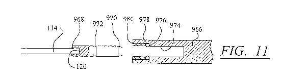
[0044] FIG. 11 is an enlarged cross-sectional view of the joint between
the cable and link
member of FIG. 10;
[0045] FIG. 12 is a partial cross-sectional view of an alternative
embodiment of
motorized actuating member;
[0046] FIG. 13 is a top partial cross-sectional view of yet a further
alternative
embodiment of control box and device;
[0047] FIG. 14 is a partial cross-sectional view of the control device of
FIG. 13;
[0048] FIG. 15 is a partial cross-sectional view of an alternative means
of connecting a
link member to a body fluid flow control device; and
[0049] FIG. 16 is a partial cross-sectional view of a further alternative
means of
connecting a link member to a body fluid flow control device.
11
CA 02990971 2017-12-27
WO 2017/035050 PCT/US2016/047963
[0050] FIG. 17 is a schematic diagram of a fluid operated implantable
apparatus for
controlling fluid flow within a host body.
[0051] FIG. 18 is a schematic diagram, partially broken away, of a pump
impeller
assembly.
[0052] FIG. 19 is a perspective view of a telemetry control device
according to the
invention.
[0053] FIG. 20A is a side view of an alternative embodiment of the control
box with
antenna for communicating with the telemetry control device of FIG. 19.
[0054] FIG. 20B is a front view of the alternative embodiment of the
control box with
antenna of FIG. 20A.
[0055] FIG. 20C is a close up view of a portion of the control box shown
in FIG. 20B.
DETAILED DESCRIPTION OF THE INVENTION
[0056] By way of illustrating and providing a more complete appreciation
of the present
invention and many of the attendant advantages thereof, the following detailed
description is
given concerning the novel implantable body fluid control device and uses
thereof.
[0057] Referring now in more detail to the drawings, in which like
numerals refer to like
parts throughout several views, FIGS. 1-4 show a body fluid flow control
device according to the
present invention. The body fluid flow control device comprises a first
engaging element 102
and a second engaging element 104. When the first engaging element 102 is
coupled with the
second engaging element 104, an inner diameter is formed which is suited for
fitting around a
host body canal, i.e., any tube or vessel V within the human or animal body,
such as the urethra.
[0058] The body fluid flow control device also comprises a locking
mechanism 106 for
locking the first and second engaging elements 102 and 104 together. The
locking mechanism
12
CA 02990971 2017-12-27
WO 2017/035050 PCT/US2016/047963
106 may be of any suitable form. In the illustrated embodiment, locking
mechanism 106 is in the
form of locking pins 108 located on the first engaging element 102 and locking
holes 110 located
on the second engaging element 104. In the illustrated embodiment, two locking
holes 110 are
provided on each side of engaging element 104. Each locking pin 108 is capable
of being
attached to either of the locking holes 110. The inner diameter formed between
parts 102 and
104 may thus be adjusted for use with different sized vessels. It should be
understood that any
other equivalent locking mechanism can be used for this purpose. Alternative
locking
mechanisms contemplated by the present invention include, but are not limited
to, the use of a
strap and snap pins or interconnecting molding on the first and second
engaging elements 102
and 104.
[0059] The body fluid flow control device of the present invention
preferably further
includes a piston-like or plunging member 112 located within the inner
diameter formed by the
coupling of the first and second engaging elements 102 and 104 such that the
plunging member
112 may apply pressure against a body canal or vessel, such as a urethra. As
can be seen most
clearly from FIGS. 2 and 15, plunging member 112 may have a curved profile
such that only
outer edge protrusions of the plunging member contact the vessel surface in
use. This
substantially reduces the likelihood of necrosis of the tissue of the vessel
because it allows
pressure to be placed on the vessel over a smaller area than would be possible
with a flat
plunging member. The curved profile of plunging member 112 may be provided on
a removable
plunger head, so that a surgeon may select an appropriately sized plunger head
for the size of the
vessel.
[0060] It should be appreciated that the fluid flow control device may
take other forms
than that illustrated. For example, instead of a plunging member provided in
two engagement
13
CA 02990971 2017-12-27
WO 2017/035050 PCT/US2016/047963
members, one of the engagement members could be moveable with respect to the
other to
compress the vessel in order to restrict fluid flow therein. Alternatively, a
fluid flow control
device in the form of an artificial external annular sphincter or other means
for compressing the
vessel may be applied to the vessel.
[0061] Apertures 113 may be provided in first engaging element 102. The
apertures 113
permit tissue growth therethrough from and to the surface of the vessel in
order to anchor the
body fluid control device onto the vessel. Further apertures (not shown) may
be provided to
allow dissolvable sutures to be used to secure the engaging element to the
vessel on a temporary
basis, until the engaging element is completely anchored in place by the
tissue growth.
Alternatively, the material of the engaging element may be such as to allow
suturing
therethrough, or the engaging element may be otherwise attached to the vessel.
It has been found
that tissue growth is achieved within a few weeks of implantation of the
device into a host body
and so it may also be possible to implant the device without any form of
attachment to the vessel,
and to simply let the tissue growth firmly attach the device to the vessel
over time.
[0062] All components of the device are made from biologically inert and
compatible
materials. For example, the fluid flow control device may be made of
polypropylene, silicone,
titanium, stainless steel and/or Teflon.
[0063] An actuating member is utilized by the body fluid flow control
device of the
present invention to bias the plunging member 112 to apply pressure against
the body vessel
when the body fluid flow control device is in the closed position, and to pull
the plunging
member 112 away from the vessel to open the device. The actuating member may
comprise a
cable 114 covered by a protective sleeve or sheath 116, the cable 114 having a
first end 118 and
a second end 120. Cable 114 is preferably a braided stainless steel cable,
although any suitable
14
CA 02990971 2017-12-27
WO 2017/035050 PCT/US2016/047963
material may be used. Protective sleeve 116 is preferably made from a bio-
compatible material
having non-stick properties to discourage tissue growth thereon. A suitable
material is Teflon.
The cable 114 may be slidably moveable within sleeve 116, or cable 114 and
sleeve 116 may be
slidably moveable together.
[0064] The first end 118 of the cable 114 runs slidably through an
aperture (not shown)
in the second engaging element 104 and is attached to the plunging member 112.
A collar 122 is
provided around the sleeve 116 where it passes through the aperture in the
second engaging
element 104, in order that any tissue growth on and around second engaging
element 104 does
not interfere with the movement of sleeve 116 through the aperture, if the
sleeve 116 is designed
to move with cable 114. If cable 114 is slidably moveable within sleeve 116,
collar 122 prevents
tissue ingress into the end of sleeve 116.
[0065] FIGS. 5-9 illustrate a control box for the fluid flow control
device that is
connected to end 120 of cable 114. The control box comprises a housing 202, a
motor 204
having a worm gear 206, a spring 208 and bellows 210 to provide a seal around
sleeve 116. The
housing 202 may be made of polypropylene or any other suitable biologically
inert material.
Batteries 212 are also provided, which should preferably be suitable for
implantation in the body,
such as batteries manufactured by Wilson Greatbatch Ltd, of Clarence, N.Y.,
USA. An operating
mechanism (not shown) may be provided in the control box, or may be implanted
separately in
the host body in an easily accessible place.
[0066] The arrangement of the control box and cable 114 allows the
control box to be
implanted in the body separately from the fluid flow control device. For
example, the control box
may be implanted close to the patient's skin in their abdomen, with the cable
114 and sleeve 116
extending from the control box 202 to the fluid flow control device that is
implanted around the
CA 02990971 2017-12-27
WO 2017/035050 PCT/US2016/047963
urethra or other body vessel.
[0067] Cable 114 is attached at end 120 to a nut 216 which is located in
the interior of a
slidably moveable casing 214 in housing 202. Spring 208 is also located within
casing 214,
which has a threaded aperture 218 to allow worm gear 206 to pass into the
interior of casing 214.
[0068] Spring 208 is interposed between the motor 204 and cable 114 in
order to provide
a coupling for selectively transmitting axial movement from the motor 204 to
the cable 114 and
hence to the body vessel V, the operation of which is described with reference
to FIGS. 7 to 9
below. In the illustrated embodiment, the motor 204 acts on casing 214 to move
spring 208 and
cable 114 by means of the nut 216. However, any suitable compressible member
may be used in
the casing 214 to cushion the vessel from the action of the motor, for
example, a resiliently
deformable material may be used, or a compressible fluid such as a gas could
be used if casing
214 was suitably sealed. Alternatively, a spring or other compressible member
may be connected
directly to or inserted in cable 114. Such an arrangement would preferably use
a compressible
member that was stiff enough so that pushing and pulling motions were still
imparted to the
cable 114 on operation of the motor.
[0069] The slidable casing 214 and worm gear 206 allow axial movement to
be imparted
to cable 114 by motor 204, but it should be appreciated that any suitable
axial actuation of cable
114 may be used. For example, the motor 204 may have an axially moveable
actuator, or suitable
gearing could be provided to act on a toothed rack or other axially moveable
element.
Alternatively, the cable could have a flexible end that may be wound around an
axle in housing
202.
[0070] The sleeve 116 containing cable 114 should be sealed to housing
202 to prevent
ingress of body fluids from damaging the motor and other components of the
control box. Any
16
CA 02990971 2017-12-27
WO 2017/035050 PCT/US2016/047963
suitable seal may be used, but it should be noted that where sleeve 116 is
designed to be slidably
moveable, it is not possible to seal tightly around sleeve 116, as the sleeve
needs to be axially
moveable in order to impart movement to plunging member 112. One method of
sealing sleeve
116 to housing 202 is to use a bellows mechanism. A suitable bellows mechanism
210 is
illustrated in FIGS. 7-9. Bellows 210 is designed so that as sleeve 116 moves
axially, bellows
210 expands or collapses in on itself so that fluid that seeps into housing
202 around sleeve 116
is captured by bellows 210, and can be forced back out of the housing 202 when
the device is
moved to a closed position.
[0071] The sleeve 116 may be sealed to bellows 210 and housing 202 by
means of a
threaded bolt 220, and a nut 222. Bolt 220 is passed through an aperture in
housing 202 with its
head 224 in the interior of the housing. Sleeve 116 passes through and is a
close fit with a central
bore 226 in bolt 220. Bellows mechanism 210 is generally tubular and is sealed
to the underside
of head 224 of bolt 220 by an 0-ring seal 228. As the nut 222 is tightened on
bolt 220,
compression of the 0-ring seal 228 causes a tight seal to prevent ingress of
fluid into housing
202 around the exterior of bolt 220. Bellows 210 extends around the head 224
of bolt 220 and is
sealed to sleeve 116 in the interior of housing 202 by a tightly wound spring
230. The spring 230
may be placed onto the bellows 210 before the sleeve 116 is forced through the
bellows 210 and
spring 230 in order to obtain the tightest seal possible. Other methods of
sealing bellows 210 to
sleeve 116 include cable clamps, C-clips, adhesive, etc. A reinforcing ring
234 is provided on
one surface of bellows 210, to ensure that the bellows 210 collapses correctly
as the sleeve 116 is
moved axially. The reinforcing ring 234 may be a thickened area in the wall of
the bellows 210,
or may be a separate ring that is attached to the bellows, by gluing or any
other suitable means.
Instead, or in addition to, the reinforcing ring 234, the bellows may be
pleated or folded in order
17
CA 02990971 2017-12-27
WO 2017/035050 PCT/US2016/047963
to ensure correct folding when the fluid flow control device is moved to the
closed position.
[0072] It should be noted that bellows 210 can be of any suitable shape,
provided that a
seal is made at the housing and around the sleeve, and that bellows allows
movement of the
sleeve into and out of the housing. For example, bellows 210 may be a simple
tubular shape,
with ends of the tube being sealed to the housing and sleeve. Alternatively,
bellows 210 may be
of a frusto-conical shape, or a more complicated shape such as a bell-shape or
could be folded or
pleated. The seal to the housing could be close to the aperture in the housing
through which the
seal extends, as illustrated, either inside the housing or outside the
housing. Alternatively, the
seal could be made to the wall of the housing, around or behind the bolt 220.
[0073] It is possible to seal the sleeve 116 and the housing 202 without
using a bellows
mechanism, but it has been found that energy losses are created as movement of
the sleeve 116
creates friction against the seal. This can cut the battery life of the motor
by up to 1/3. For
example, a flexible annular ring may be sealed between the sleeve 116 and the
housing 202, the
annular ring stretching as the sleeve is axially moved. Alternatively, a
series of seals may be
provided along sleeve 116, each seal preventing some fluid ingress to housing
202.
[0074] Control circuitry (not shown in FIGS. 7-9) is provided, which
operates the motor
on receipt of a signal from an operating mechanism. Any of the several well-
known control
devices can be used to control the operation of the body fluid flow control
devices of the present
invention by a user so long as the objectives of the present invention are not
defeated. Suitable
operating mechanisms include radio-control devices, or a magnetic devices that
can be sensed by
the control circuitry. With a magnetic device, the user may be provided with a
separate magnet
that they carry with them, and which they position adjacent the skin over the
implanted switch
when they wish to operate the device. The magnet may be of any suitable shape,
and may be
18
CA 02990971 2017-12-27
WO 2017/035050 PCT/US2016/047963
shaped for example like a pen or credit card so that its purpose is not
immediately apparent to
other people. The magnet should have a weak magnetic field so that it must be
placed close to
the switch in order to operate the device, in order to prevent accidental
operation of the device if
the magnet is carried in a pocket. Alternatively, a touch sensor, infrared,
voice or sound
activation may be used, or a manually operated switch may be implanted under
the skin of the
patient.
[0075] A remotely operated operating mechanism is preferred because the
device can be
operated without irritation to the skin, as would happen with a manually
operated trigger. In the
preferred embodiment, a manual override switch may be provided in addition to
the remotely
operated triggering mechanism. The manual override switch is designed to be
used temporarily if
the control box fails and the user is not close to a physician's office or
hospital to have the
control box changed. The manual override switch may be provided in the control
box, and may
be sealed from the interior of the control box until the first activation of
the switch, for example
by a membrane seal. Such a use of the manual override switch may eventually
allow fluid
ingress into the control box, which may then need to be replaced.
Alternatively, no manual
override switch may be provided, which would mean that the user would have to
use
incontinence pads until the control box could be replaced.
[0076] The control circuitry controls operation of the motor, and may
detect the position
of the plunging member, for example, via the position of the casing or via the
drag exerted on the
motor. Preferably, the control circuitry also monitors the level of charge in
the battery. The
control circuitry can be used to initiate opening or prevent closing of the
fluid flow control
device if a problem such as low battery or a defective motor is detected, so
that the device can be
caused to remain in the open position. For example, once the device has been
opened, an
19
CA 02990971 2017-12-27
WO 2017/035050 PCT/US2016/047963
abutment (not shown) may be caused to contact the casing 214 to prevent any
further movement
thereof. The motor may also be shut off. The device may still be operable by a
manual override,
as the spring 208 can be compressed and allowed to expand within casing 214 to
allow
movement of the cable 114 to open and close the device.
[0077] The control box 202 may also contain components that allow a
physician to
interrogate the control circuitry by a remote telemetry system without
accessing the box itself
Such components may be interrogated and/or controlled by radio waves or other
interactive
signals transmitted and received by the telemetry system, or any other
suitable mechanism. This
allows the physician to check the charge in the batteries, any internal
sensors, to alter the tension
in the cable 114, and to make other suitable adjustments. A pressure sensor
may be provided on
the plunger 112 to monitor the pressure between the plunger 112 and the vessel
V when the
plunger is in the closed position. The pressure sensor may also be
interrogated by the telemetry
system, which can then be used to alter the settings for the control device.
For example, the
number of turns that the motor 204 causes worm gear 206 to make on each
operation of the
device may be altered in order to set the correct distance of travel of the
cable 114, and hence
plunger 112 for any particular patient so as to alleviate any excess pressure
exerted on the vessel
V. In addition, the telemetry system may include control commands to cause the
motor to open
and close the body fluid flow control device, either as an override system to
the normal operating
means, or in addition to the normal operating means in order to test the
device in situ.
[0078] If the control box causes the device to fail or remain in the open
position if a
problem is detected, this will simply mean that the patient will return to the
condition that they
were in before implantation of the device, in other words, in a condition of
incontinence. If the
device failed in the closed position, the patient would need to be
catheterized. However, a
CA 02990971 2017-12-27
WO 2017/035050
PCT/US2016/047963
manual override system would allow the patient to operate the system manually
for a
considerable period of time or until medical aid was obtainable.
[0079]
Actuation of the device is described with reference to FIGS. 7 to 9. In the
open
position shown in FIG. 7, the motor 204 has operated the worm gear 206 to draw
casing 214
towards the motor 204. This pulls nut 216 along with the casing 214, and thus
acts on cable 114
to pull the plunging member 112 away from the vessel V. Bellows 210 is also at
its fully
extended position. In order to close the fluid control device, the motor 204
is activated to turn
worm gear 206 in the opposite direction to that used to open the device. As
worm gear 206 is
operated, casing 214 is moved away from the motor 204, spring 208 pushing on
nut 216 to bias
plunging member 112 against the vessel V, as shown in FIG. 8. As the motor 204
is operated
further, the vessel V prevents plunger 112 moving, and prevents movement of
cable 114 and
hence nut 216, due to the increased force needed to move cable 114 against the
vessel V when
the vessel V is already closed. Nut 216 presses against spring 208, causing
compression of the
spring 208, as shown in FIG. 9. It can thus be seen that any further movement
of worm gear 206
by motor 204 does not result in compression and injury of the vessel V, but
the further
compression of spring 208. In this way, axial movement of casing 214 may be
selectively
transmitted to cable 114. This protects the vessel V against failure of the
device by continuous
running of the motor 204, as the vessel cannot be further compressed due to
the interplay
between the vessel V and the spring 208.
[0080] An
alternative embodiment of the control box is illustrated in FIGS. 10 and 11.
The control box comprises a housing 902, a motor 904 having a worm gear 906, a
spring 908 and
bellows 910. Batteries 912 are also provided, along with control circuitry
(not shown). The
spring 908 is located in a slidable spring casing 914. An operating mechanism
(not shown) may
21
CA 02990971 2017-12-27
WO 2017/035050 PCT/US2016/047963
be provided in the control box, or may be implanted separately in the host
body in an easily
accessible place. The spring, worm gear and motor arrangement are as described
for FIGS. 5-9,
and will not be further described.
[0081] Housing 902 is preferably formed in two pieces, a main body 916
and an end lid
918. End lid 918 includes a lip 920 that fits inside an end 922 of main body
916. A groove 924 is
provided around lip 920, in order to receive an 0-ring 926. End lid 918 is
also sonically welded
to main body 916 in order to provide a good seal. A groove 928 is provided
around the exterior
of end 922 of main body 916, in order to allow for ease of removal of lid 918
with a suitable tool
when necessary. An interior housing 930 extends along the length of housing
902, to one side
thereof, in order to separate the motor 904, worm gear 906, slidable casing
914, bellows 910 and
other moveable parts from the batteries 912. Interior housing 930 has a flange
932 at an end 934
remote from end 922 of main body 916, with an 0-ring groove 936 provided in
flange 932. A set
screw 938 is also provided in interior housing 930, in order to lock motor
904. Electrical contacts
940 extend to motor 904 from end lid 918. An internally directed collar 942
having an internal
thread extends around flange 932 within housing 902, and interior housing 930
is secured into
housing 902 by means of an externally threaded nut 944 which is screwed into
place to hold
flange 932 in position. Nut 944 may have pin holes 946 to allow for tightening
thereof An
externally directed collar 948 having an internal thread is also provided in
housing 902, in order
to allow the cable 114 to pass into interior housing 930.
[0082] Sleeve 116 has an end 950 which is attached to a hollow connector
952 having a
first end 954 and a second end 956. At end 954, connector 952 has backwardly-
directed teeth
958 around the circumference thereof which attach to the inside of sleeve 116
adjacent to end
950, and act to prevent sleeve 116 from being pulled loose. The second end 956
of connector 952
22
CA 02990971 2017-12-27
WO 2017/035050 PCT/US2016/047963
has an external thread 960, as well as a groove 962 suitable for receiving an
0-ring 964. Thread
960 is screwed into the internal thread provided within collar 948 on housing
902. Cable 114
extends into housing 902 through connector 952, and is attached at its end 120
to a link member
966 which extends into casing 914 and terminates in nut 216. The connection
between cable 114
and link member 966 is shown enlarged in FIG. 11. The cable end 120 is fitted
into a connector
piece 968 that has a tapered end 970 and a groove 972 for receiving a sealing
ring. Link member
966 has an opening 974 for receiving connector piece 968, opening 974 having
an internal
shoulder 976. A metal 0-ring 978 is received by shoulder 976 and is held in
place by a ring
retainer 980. Connector piece 968 is pushed into opening 974 until the metal 0-
ring 978 seats in
groove 972 to form a seal between connector piece 968 and link member 966.
[0083] Bellows 910 are attached to housing 902 by means of nut 944
screwed into
inwardly directed collar 942. Bellows 910 has an end flange 982, which extends
adjacent to
flange 932 of interior housing 930, and has an integral 0-ring 984 to seal in
0-ring groove 936
of flange 932 so that bellows 910 is tightly sealed to housing 902 by interior
housing 930.
Bellows 910 is also attached to cable link member 966 by means of a cable link
986, and has a
pleated conical shape above flange 982 so that it may fold easily when
compressed. It should be
noted that in the embodiment of FIG. 10, the bellows 910 is not attached to
the sleeve 116, as the
sleeve 116 is not axially moveable. Instead, cable 114 is axially moveable
within sleeve 116. In
this embodiment, bellows 910 may not be necessary, as a good seal may be
provided between
connector 952 and control box 902. However, it is advantageous to provide an
additional seal,
for example using bellows 910, to prevent fluid ingress into control box 902.
[0084] The operation of the control box of FIG. 10 is the same as for the
control box of
FIGS. 5 to 9, and will not be further described.
23
CA 02990971 2017-12-27
WO 2017/035050 PCT/US2016/047963
[0085] A further alternative embodiment of a seal for the sleeve and an
actuator for the
cable is illustrated in FIG. 12. In the illustrated embodiment, control box
1200 is completely
sealed so that no fluid ingress into the box can take place. A hollow
cylindrical bore 1202 that is
sealed at one end 1204 is formed in control box 1200. Bore 1202 has internal
threads 1206
provided adjacent an outer surface of control box 1200.
[0086] An end of sleeve 116 is attached to a hollow connector 1208,
connector 1208
having an end 1210 and an end 1212. End 1210 of connector 1208 is dimensioned
to pass into
the end of sleeve 116, connector 1208 having outwardly and rearwardly directed
teeth 1214 at
end 1210 to engage the interior of sleeve 116, thereby securing connector 1208
to sleeve 116.
End 1212 of connector 1208 is dimensioned to be slightly larger in diameter
than sleeve 116, and
has external threads 1216. Connector 1208 may be screwed into bore 1202 of
control box 1200
by means of threads 1216 and 1206.
[0087] End 120 of cable 114 is located in bore 1202, and is provided with
a collar 1218.
An annular magnet 1220 is supported by collar 1218 around end 120 of cable
114. Cable 114 is
axially moveable within sleeve 116, and therefore a bellows seal is not
necessary around sleeve
116. In addition, as sleeve 116 is not moveable, tissue growth around the
sleeve cannot affect the
operation of the device.
[0088] A motor 1222 has a threaded worm gear 1224 engaged with a casing
1226
through a screw-threaded aperture 1228 located in the bottom of the casing.
Casing 1226 extends
around bore 1202, and an annular magnet 1230 is supported around the interior
of an upper edge
of casing 1226. Magnet 1230 is aligned with magnet 1220 located on end 120 of
cable 114.
[0089] In order to actuate cable 114 to open and close the fluid flow
control device, the
motor 1222 operates the worm gear 1224, which moves casing 1226 along the
exterior of bore
24
CA 02990971 2017-12-27
WO 2017/035050 PCT/US2016/047963
1202. Magnet 1230 acts through the plastic material comprising bore 1202, and
causes magnet
1220 to track its movement. This in turn causes cable 114 to be axially moved,
operating the
fluid flow control device. If the motor 1222 continues operating the worm gear
1224 towards the
cable 114 when the body vessel has already been closed, the attraction of
magnet 1220 for
magnet 1230 is not enough to cause the cable 114 to be moved further, due to
resistance from the
vessel walls, thus preventing potential damage to the vessel. Thus, axial
movement of casing
1226 is selectively transmitted to cable 114. In addition, the casing 1226
will come to rest against
bore 1202 or an interior surface of control box 1200, preventing the magnets
from getting too far
out of alignment.
[0090] It should be appreciated that a magnetic link between the motor
and cable may be
achieved in many ways other than that illustrated in FIG. 12. For example, the
magnets need not
be annular, but could be placed to one side of the cable. In addition, the
magnets need not
operate by mutual attractions, but could work by repelling each other to close
the vessel, with a
spring action or other means operating to open the vessel once the motor-
driven magnet was
pulled back towards the motor. Also, the magnetic coupling does not require
that the motor and
the cable or other structure driven by the motor each have a magnet, so long
as one is magnetic
and the other is capable of being moved by magnetic attraction or repulsion.
Electromagnets are
also possible. With a repelling action, magnets could be placed directly on
the ends of the cable
and an axially movable actuator driven by the motor. It will be appreciated
that the magnetic
drive mechanism of the invention can be utilized to operate many other types
of implanted
medical devices other than constricting devices.
[0091] An alternative embodiment of a magnetic coupling for selectively
transmitting
axial movement to the cable is illustrated in FIGS. 13 and 14. These figures
illustrate a control
CA 02990971 2017-12-27
WO 2017/035050 PCT/US2016/047963
box 1300 that is completely sealed. A bore 1302 having a blind end 1304 is
provided in the
control box 1300 for receiving the end 120 of cable 114. A connector 1306 is
used to connect
sleeve 116 to bore 1302. The connector 1306 has a first end 1308 with
rearwardly directed teeth
1310, a central shoulder 1312 and a second end 1314 having external screw
threads 1316. End
1308 of connector 1306 is pushed into the end of sleeve 116, the teeth 1310
acting on the inner
surface of the sleeve. End 1314 of connector 1306 is connected to control box
1300 by means of
an 0-ring seal 1318 and an internally threaded nut 1320 which is threaded onto
threads 1316.
Nut 1320 is welded at 1322 to the control box 1300 to form a tight seal.
[0092] The cable 114 extends into bore 1302. A cylindrical magnet 1324 is
attached to
end 120 of cable 114 by a collar 1326 which is deformed onto the magnet 1324
and cable end
120 for a tight fit. The control box 1300 includes a motor 1328, a worm gear
1330 and batteries
1332 as described for the FIG. 10 embodiment. A casing 1334 having an annular
magnet
arrangement 1336 is threaded onto worm gear 1330, and operates in the same
manner as in the
FIG. 10 embodiment so will not be further described. Control circuitry
including IC's 1338 and
other standard components 1340 including resistors and capacitors are also
shown.
[0093] FIG. 15 illustrates an embodiment of a connector joining first end
118 of cable
114 to the body fluid control device. Connector 1500 has a first end 1502
having outwardly
directed teeth 1504 which grip into the inner surface of sleeve 116. A second
end 1506 of
connector 1500 has a collar with inwardly directed threads 1508 which are
threaded onto
outwardly directed threads 1510 on a collar 1512 attached to the body fluid
flow control device.
An 0-ring 1514 forms a tight seal to the collar 1512.
[0094] FIG. 15 also illustrates plunger 112 in detail. Plunger 112
includes a perforated
metal bracket 1516 attached to a metal collar 1518. The main body of plunger
112 is formed of
26
CA 02990971 2017-12-27
WO 2017/035050 PCT/US2016/047963
silicon that is molded onto the perforated bracket 1516, the silicon extending
through the
perforations in the bracket to form a tight fit between plunger 112, bracket
1516 and collar 1518.
Metal collar 1518 may be simply crimped onto end 118 of cable 118.
[0095] FIG. 16 illustrates a further alternative method of connecting
cable 114 and sleeve
116 to the body fluid flow control device. In the embodiment of FIG. 16, the
fluid flow control
device has a collar 1600 with internal threads 1602. A connector 1604 is used
to connect sleeve
116 to collar 1600. Connector 1604 has external threads 1606, a central collar
1608 and
outwardly directed teeth 1610. It should be noted that connector 1604 may be
the same as
connector 1306 illustrated in FIG. 13. This allows for economies in
manufacture, as only one
type of connector need be provided for both ends of the sleeve 116. A metal
collar 1612 is used
to connect the plunger (not shown in FIG. 16) to end 118 of cable 114. An 0-
ring 1614 may seal
between collar 1612 and connector 1604.
[0096] There is shown in FIGS. 17-18 an implantable apparatus for
controlling fluid flow
within a host body. The apparatus includes a constricting member 1710 for
allowing body fluid
to flow within a body canal when in an open position and for reducing fluid
flow within a body
canal when in a closed position. The constricting member 1710 includes a
bladder 1714. The
bladder 1714 receives working fluid to compress the body canal and thereby
reduce body fluid
flow through the body canal, and expels working fluid to allow body fluid to
flow again through
the body canal. An actuating member 1720 is provided for operating the
constricting member
between the open and closed positions. The actuating member 1720 includes
structure for
moving working fluid into and out of the bladder 1714. This structure can
include a pump 1724,
as shown, having an impeller assembly 1728 and a motor 1730. A fluid transfer
conduit 1734
connects the pump 1724 with the bladder 1714. A reservoir 1740 can be provided
to store
27
CA 02990971 2017-12-27
WO 2017/035050 PCT/US2016/047963
working fluid for operating the bladder 1714.
[0097] An engaging element 1744 can be provided for substantially
encircling the body
canal. Expansion of the bladder 1714 causes the bladder to compress the body
canal against the
engaging element 1744 to reduce body fluid flow through the body canal.
Expelling working
fluid from the bladder permits the body canal to expand and body fluid to flow
within the body
canal. The engaging element 1744 can be of any suitable design. In the design
shown in FIG. 17,
the engaging element 1744 includes a first piece 1750 and a second piece 1754.
The first piece
1750 is joined to the second piece 1754 by suitable connection structure (not
shown). The
bladder 1714 can be seated in an appropriate seat in second piece 1750.
[0098] The bladder 1714 can be of different sizes and shapes, as well as
materials. It is
necessary that the bladder 1714 expand upon receiving working fluid so as to
constrict the body
canal. Polymeric materials that are biocompatible can be utilized. The bladder
material can be a
material which stretches upon being filled with the working fluid, or can be a
flexible material.
Alternatively, in place of a bladder other fluid-operated structure can be
provided, such as a rigid
piston in a chamber which is acted upon by the working fluid to move the
piston and press
against the canal.
[0099] The pump for controlling working fluid flow through the fluid
transfer conduit
1734 to the bladder 1714 can be of many different designs. The pump 1724 is
preferably
positioned within a suitable water-tight housing such as control box 1760. A
pump conduit 1770
transfers fluid between the reservoir 1740 and the fluid transfer conduit
1734. The pump 1724
has structure for compressing the pump conduit 1770 so as to force the working
fluid through the
pump conduit. In one embodiment, the pump 1724 has an impeller 1728 which has
a plurality of
rollers 1764. The rollers 1764 are provided adjacent to the pump conduit 1770
(FIG. 18). The
28
CA 02990971 2017-12-27
WO 2017/035050 PCT/US2016/047963
pump conduit 1770 is preferably provided along an arcuate housing 1774. The
rollers 1764
extend outward from the surface of impeller 1728. In this manner, rotation of
the impeller 1728
causes the rollers 1764 to compress the pump conduit 1770. Working fluid will
thereby be drawn
from a reservoir 1740, through a fluid inlet 1780, and into the pump conduit
1770. The working
fluid will be propelled by the compressing action of the impeller 1728 and
rollers 1764 on the
pump conduit 1770 through a fluid outlet 1784 and into the fluid transfer
conduit 1734. The
working fluid will travel through the fluid transfer conduit 1734 into the
bladder 1714 so as to
cause the bladder 1714 to expand and compress the body canal. Compression of
the body canal
will restrict the flow of body fluid through the body canal. Other pump
constructions are within
the scope of the invention.
[0100] The motor 1730 is reversible such that in one flow direction
working fluid is
caused to flow from the reservoir 1740 through the pump 1724 to the bladder
1714. In the
reverse direction, the pump 1724 will cause working fluid to be withdrawn from
the bladder
1714 and pumped into the reservoir 1740. The compression of the body canal
will thereby be
released and body fluid will be permitted to flow through the body canal. It
is also possible that,
in some constructions, reversal of the pump is not necessary to remove working
fluid from the
bladder 1714 and that turning off the pump 1724 will cause the working fluid
to drain from the
bladder 1714. This is possible if the bladder 1714 is elastic or if there is a
biasing on the bladder
1714 acting to return the bladder 1714 to the initial, non-expanded state.
Appropriate valves or
check valves can be positioned in the flow line to restrict or permit the flow
of working fluid as
desired.
[0101] A telemetry system according to the invention provides appropriate
information
and commands to control operation of the actuator and constricting member.
There is shown in
29
CA 02990971 2017-12-27
WO 2017/035050 PCT/US2016/047963
FIG. 19 a telemetry device 1910 according to the invention. The telemetry
device 1910 can
include a suitable housing 1920. Within the housing 1920 is suitable circuitry
for producing
telemetry signals which are transmitted to a control unit which controls
operation of the actuator
and the constricting device. The telemetry device 1910 can have an on/off
power switch 1924.
Suitable connection ports or jacks can be provided such as power-in jack 1930
and earphones
jack 1938. A display 1950 provides a visual indication of options and
telemetry information. A
select button 1954 is provided to select a function. A next button 1958 is
provided to indicate
different functions. An electromagnet 1962 can provide a link to the implanted
control unit, and
can be connected to jack 1966.
[0102] The telemetry device can be used to communicate with the control
box and
constricting device to transmit a variety of information. The telemetry
signals can be utilized to
initiate the device, to control its operation, and to recalibrate the device
upon use. This
information can relate to the status of the control box and constricting
device. The information
could also relate to the status of the patient, for example, body temperature.
Telemetry
commands can be particularly useful to calibrate and set the position of the
constricting member.
For example, telemetry commands can be used to adjust the tightness of the
constricting
member, to recalibrate to the starting point, to place in sleep mode, to awake
from sleep mode, or
for special options, such as unit diagnosis, when the battery is low, or when
there is no usage for
a selected time. Other functions are also possible.
[0103] Referring now to FIGS. 20A ¨ 20C there is shown an alternative
embodiment of
control structure 300. In this preferred embodiment the housing 302 of control
structure 300
includes a first part 304 and a second part 306. The second part of the
housing 306 may include
one or more segments 306' and 306". The control structure 300 operates the
actuator, the control
CA 02990971 2017-12-27
WO 2017/035050 PCT/US2016/047963
structure 300 being operated by the telemetry device 1910. The control
structure 300 includes
one or more antenna 308 for transmitting and receiving signals to and from the
telemetry device
1910. At least a portion of the one or more antenna 308 sufficient to receive
and transmit signals
to the telemetry device 1910 project within a first part of the housing 304 of
the control structure
300. The first part of the housing 304 comprises a material that is permeable
to telemetry signals.
The second part of the housing 306 comprises a material that is generally
impermeable to
telemetry signals. In a preferred embodiment, the first part of the housing
304 comprises a
flexible plastic material. The second part of the housing 306 comprises a
metal compound. In
one embodiment, the second part of the housing 306 comprises titanium. In a
preferred
embodiment, the first part of the housing 304 is hermetically sealed to the
second part of the
housing 306.
[0104] The first part of the housing 304 defines a void space 310 for
receiving the one or
more antenna 308. The void space 310 is configured to provide the one or more
antenna 308
projected therein with sufficient space such that the one or more antenna 308
positioned within
the void space 310 may move freely within the void space 310 and the one or
more antenna 308
do not contact the first part of the housing 304 even if the one or more
antenna 308 are subject to
vibrational or other movement. A gap is defined within the void space 310, the
gap being
positioned between the antenna 308 and the first part of the housing 304, the
gap further being of
sufficient size relative to the antenna 308, to allow for freedom of movement
of the antenna 308
within the first part of the housing 304, such that the antenna 308 can move
freely within the
housing 304 as a result of movement of the antenna without contacting the
housing 304.
The antenna 308 extend up into the void space 310 beyond the second part of
the housing 306
which generally comprises a metallic casing thereby permitting the
transmission and receiving of
31
CA 02990971 2017-12-27
WO 2017/035050 PCT/US2016/047963
telemetry command signals that do not interfere with the metallic casing of
the second part of the
housing 306. It is appreciated that only a portion of the one or more antenna
308 sufficient to
transmit and receive signals to and from the telemetry device 1910 need to
project within the first
part of the housing 304. The size of the void space 310 within the first part
of the housing 304 is
dimensioned relative to the size of the antenna 308, such that the void space
308 allows for the
free flow of telemetry signals to and from the antenna 308 within the control
unit 300 to the
telemetry device 1910. The antenna 308 may include an on-board PC board 312.
[0105] In a preferred embodiment, the antenna 308 comprises a solid
projection affixed
to an on-board PC-board. In yet another preferred embodiment, a module having
an internal
lattice placement structure is used to slide the antenna and on board PC-board
312 into the
housing 302.
[0106] The telemetry device communicates with the control box using
suitable
communication protocols and coding. This coding can be in the form of
different burst lengths of
electro-magnetic or magnetic radiation, similar to Morse code. An example of
suitable control
signals is indicated in Table 1. Other coding systems are possible, and can be
used to generate
output in several different formats, such as text, bar code or audio. Suitable
logic circuitry or a
microprocessor in the control box permits the translation of these control
signals into operating
commands for the motor, valves, or other structure in the device.
TABLE-US-00001 TABLE 1 PMD Telemetry Responses .cndot. Error, PMD received
unknown
command or could not perform the requested function * .cndot. .cndot. .cndot. -
- 6 turns; loosest
* .cndot. .cndot. .cndot. .cndot. - 7 turns; loose * .cndot. - - - - 8 turns;
normal default * .cndot.
.cndot. .cndot. .cndot. .cndot. 9 turns; tight * - .cndot. .cndot. .cndot.
.cndot. 10 turns; tightest *
.cndot. - .cndot. Recalibrate started but not finished; must set 6, 7, 8, 9,
or 10 turns * - .cndot.
32
CA 02990971 2017-12-27
WO 2017/035050 PCT/US2016/047963
.cndot. PMD disabled. PMD will respond only to check status command and enable
normal
operation command. PMD will respond with error to all others. - .cndot. Enable
normal
operation. PMD will restore number of turns in effect when it was disabled.
.cndot. = short signal
- = long signal * These codes can be signaled by the PMD when it receives a
Check Status
command.
[0107] These telemetry signals can be any suitable signal such as
magnetic, electro-
magnetic, acoustic, and any other suitable signals.
[0108] According to another aspect of the invention, an implantable
apparatus for
controlling fluid flow within a host body comprises a constricting member for
allowing fluid
flow within a body canal when in an open position and for reducing fluid flow
within a body
canal when in a closed position. An electrically-operated actuator operates
the constricting
member between the opened and closed positions. Control structure is provided
for operating the
actuator. The control structure includes voltage measuring structure for
controlling said
constricting member. The voltage measuring structure can measures battery
voltage. The voltage
measuring structure can additionally or alternatively measure voltage drawn by
the actuator. If
battery voltage is low it is an indication of a drained or defective battery.
If actuator voltage is
high it is an indication that the actuator is drawing too much current, as
would occur if the
constricting member has met an obstruction or mechanical resistance. If a
voltage irregularity
occurs, the actuator is caused to move the constricting member to the open
position, so as to
permit fluid flow through the body canal so as to avoid an accumulation of
fluid in the body.
[0109] It will be understood that various embodiments of the present
invention have been
disclosed by way of example and that other modifications and alterations may
occur to those
skilled in the art without departing from the scope and spirit of the appended
claims, such as, for
33
CA 02990971 2017-12-27
WO 2017/035050 PCT/US2016/047963
example, those embodiments described in U.S. Pat. No. 6,319,191, issued Nov.
20, 2001, which
is incorporated hereinto in its entirety by reference.
[0110] Thus, the invention described herein extends to all such
modifications and
variations as will be apparent to the reader skilled in the art, and also
extends to combinations
and subcombinations of the features of this description and the accompanying
figures. Although
preferred embodiments of the present invention have been illustrated in the
accompanying
figures. and described in the foregoing detailed description, it will be
understood that the present
invention is not limited the embodiments disclosed, but is capable of numerous
rearrangements,
modifications and substitutions without departing from the spirit of the
present invention as set
forth and defined by the following claims.
34