Note: Descriptions are shown in the official language in which they were submitted.
CA 02991300 2018-01-03
WO 2017/011507 PCT/US2016/042002
APPARATUSES FOR EVACUATION OF A ROOT CANAL AND METHODS OF USING SAME
CROSS REFERENCE
[0001] This application claims priority to U.S. Provisional Patent
Application Serial No.
62/191,845 filed July 13, 2015 and to U.S. Provisional Patent Application
Serial No. 62/220,534
filed September 18, 2015, the disclosures of which are expressly incorporated
by reference
herein in their entireties.
TECHNICAL FIELD
[0002] The invention relates generally to methods and apparatuses used
during
endodontic therapy or root canal therapy and, more particularly, to
apparatuses and methods for
dispensing fluids and evacuating those fluids from a root canal.
BACKGROUND
[0003] To preserve a tooth that has developed an infected pulp or abscess,
it is
necessary to remove the diseased tissue from the tooth. Removing the diseased
tissue
prevents further bacterial proliferation within the tooth and can save the
tooth. To that end,
endodontic therapy or root canal therapy of the identified tooth may be
necessary.
[0004] To begin a root canal, the clinician cuts an opening through the
crown of the
tooth to gain access to the pulp. The clinician then removes the pulp from the
pulp chamber and
from the root canal through the opening. Endodontic files, bores, reamers or
other
instrumentation are used to clean tissue from the root canal. The clinician
may also shape the
root canal to receive a filling material.
[0005] Following mechanical removal of tissue, the clinician flushes the
pulp chamber
and the enlarged root canal with one or more irrigants to disinfect them. This
minimizes the
presence of bacteria and cleans the surfaces of calcific debris created during
mechanical
debridement. Irrigants are preferably capable of dissolving tissue remnants to
permit their
removal. These include hydrogen peroxide and sodium hypochlorite but may be
any suitable
liquid, such as, water or various alcohols that simply carry debris out of the
root canal.
[0006] It is desirable to remove as much of the debris and necrotic tissue
as possible.
To do so, the irrigant may be typically applied under pressure using a syringe
and a needle
inserted into the canal. To clean the root canal near the apex of the tooth,
the syringe must be
placed very close to the apical foramen and must fit loosely enough in the
root canal to allow the
irrigant to flow from near the apical foramen toward the crown. The used
irrigant and debris is
then siphoned off through the opening in the crown. This technique may,
however, be
problematic, particularly for certain types of irrigants.
-1-
CA 02991300 2018-01-03
WO 2017/011507 PCT/US2016/042002
[0007] Even the tip of the smallest needles that deliver irrigants under
pressure must be
kept a safe distance (typically 4-6 mm) away from the apex to avoid
accidentally forcing the
irrigant through the apical foramen and into the periapical tissue. Irrigant
that escapes into the
periapical tissue can be a source of significant post treatment endodontic
pain or morbid clinical
complication, including excruciating pain, immediate swelling of the tissue,
and profuse
bleeding.
[0008] To avoid forcing irrigant into the periapical tissue, the clinician
may not insert the
syringe deeply enough into the root canal and so an area or zone adjacent the
apical foramen
may not be properly disinfected. Occasionally, even proper placement of the
syringe does not
guarantee that the irrigant has flushed the canal all the way to the apex.
Furthermore, irrigating
the regions near the apical foramen is very time consuming.
[0009] As a result, other techniques have been developed. One includes
evacuating the
irrigant from a region near the apex instead of at the crown. In this
technique, irrigant is
introduced at the crown and flows from the crown toward the apex where it is
suctioned away
through a cannula. While very successful, this technique has unique problems.
Because the
cannula must be very small to fit to within a few millimeters of the apical
foramen, any residual
debris tends to clog the cannula. Once clogged, the evacuation of the irrigant
ceases and the
technique becomes ineffective until the debris is cleared. As a consequence,
extreme care
must be taken to clean out the root canal before final cleaning of the region
near the apical
foramen.
[0010] Furthermore, due to the numerous components and the extremely small
size of
the cannulas necessary to effectuate proper evacuation of a root canal,
current techniques may
be difficult to physically manipulate within the patient's mouth. By virtue of
this difficulty, there is
a significant amount of wasted time to effectively complete treatment.
[0011] Following flushing with the irrigants, the clinician fills or
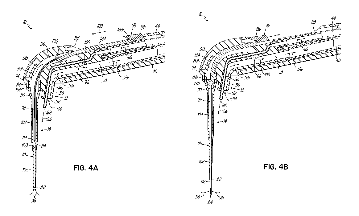
obturates the root canal
with a material such as gutta-percha and a sealer to seal the root canal. Once
sealed, the
clinician may place a crown or other restoration on the tooth to protect it
and restore it to its full
function.
[0012] A need therefore exists for apparatuses and methods which enable a
clinician to
disinfect and remove debris near the apex of a tooth without concern that
irrigant is forced
through the apical foramen and into the periapical tissue.
SUMMARY
[0013] An endodontic device for use in endodontic procedures and other
procedures
addresses these and other shortcomings and in one embodiment may include a
canal
evacuation system for evacuating a root canal of a tooth. The canal evacuation
system includes
a first cannula and a second cannula. The first cannula and the second cannula
are movable
-2-
CA 02991300 2018-01-03
WO 2017/011507 PCT/US2016/042002
relative to one another to an extended position in which the second cannula
extends from the
first cannula.
[0014] In one embodiment, the first cannula and the second cannula are
movable from a
retracted position in which the first cannula evacuates the root canal to the
extended position in
which the second cannula evacuates the root canal.
[0015] In one embodiment, when the second cannula is in the retracted
position, the
second cannula does not evacuate the root canal. In one embodiment, when in
the extended
position, the second cannula is concentric with the first cannula.
[0016] In one embodiment, the canal evacuation system includes an
extension control
system that is operatively coupled to at least one of the first cannula and
the second cannula
and by which a clinician can move at least one of the first cannula and the
second cannula
relative to one another between the retracted position and the extended
position.
[0017] In one embodiment, the endodontic device includes a locking system
that is
operatively coupled to the extension control system and that secures the
extension control
system at one of a plurality of predetermined locations selected by the
clinician.
[0018] In one embodiment, the endodontic device further includes an end
effector that
has a body defining a through bore in fluid communication with the first
cannula and with the
second cannula when the second cannula is in the extended position. The end
effector defines
a first axis and the first cannula defines a second axis that intersects the
first axis. In one
embodiment, an angle formed between the first axis and the second axis is
greater than 90 up
to about 145 .
[0019] In one embodiment, the endodontic device further includes a
handpiece to which
the end effector is releasably coupled at a joint. In one embodiment, the
endodontic device
further comprises an irrigant system including a fluid delivery tube that is
configured to dispense
fluid into the tooth. The handpiece may house at least one button mechanism
that is operable to
select the rate at which one fluid flows through the fluid delivery tube.
[0020] In one embodiment, an endodontic treatment system comprises the
endodontic
device and a fluid delivery system that is fluidly coupled to the endodontic
device by a plurality
of tubes.
[0021] In another aspect, an end effector for use with a handpiece through
which fluid
and vacuum are supplied during endodontic therapy addresses these and other
shortcomings
and in one embodiment, the end effector comprises at least one body that
defines a through
bore and through which vacuum is supplied. The end effector forms a joint with
the handpiece
at one end. A first cannula extends from the body proximate another end and is
capable of
evacuating at least a portion of a root canal. A fluid delivery tube is
supported by the body for
dispensing a fluid proximate the other end of the body into a tooth at a crown
of the tooth. The
end effector may be a disposable component.
-3-
CA 02991300 2018-01-03
WO 2017/011507 PCT/US2016/042002
[0022] In one embodiment, the end effector further includes a vacuum port
proximate
the fluid delivery tube for evacuating fluid at or near the crown of the
tooth.
[0023] In one embodiment, the end effector includes a second cannula that
has a
smaller diameter than the first cannula. The first cannula and the second
cannula are movable
relative to one another to an extended position in which the second cannula
extends from the
first cannula similar to that described above with respect to the endodontic
device.
[0024] In one embodiment, an endodontic device for use during endodontic
therapy may
be coupled to a fluid delivery system that includes a reservoir of fluid and a
source of vacuum.
The endodontic device includes a handpiece that includes a housing, at least
one tube that is
fluidly coupled to the reservoir, and a vacuum tube that is coupled to the
source of vacuum.
Each tube extends at least part way through the housing.
[0025] In one embodiment, the handpiece includes at least one control
mechanism and
a cable that extends at least part way through the housing and electrically
couples the at least
one control mechanism on the handpiece with the fluid delivery system.
[0026] In one embodiment, the endodontic device includes a canal
evacuation system
extending from the handpiece for evacuating a root canal of a tooth. The canal
evacuation
system includes a first cannula and a second cannula. The first cannula and
the second
cannula are fluidly coupled to the vacuum tube and are movable relative to one
another to an
extended position in which the second cannula extends from the first cannula.
[0027] In one aspect, a cannula for use during endodontic therapy
addresses these and
other shortcomings and in one embodiment comprises a sidewall that defines a
bore and is
closed at one end. The sidewall is sized to fit within a root canal with the
closed end at or near
an apical foramen and includes a plurality of openings proximate the closed
end and a mid-exit
hole remote from the closed end. The cannula includes a seal at an end
opposite the closed
end. In one embodiment, the sidewall is at least one of stainless steel,
plastic, or a combination
thereof. In one embodiment, the mid-exit hole has a greater open area than any
single one of
the openings.
[0028] In one aspect, a method for endodontic treatment of a root canal of
a tooth
addresses these and other shortcomings and includes moving a first cannula and
a second
cannula relative to one another from a first position to a second position in
which the second
cannula extends from the first cannula into the root canal and the method
includes evacuating
the irrigant from the root canal with the second cannula.
[0029] In one embodiment, prior to evacuating the irrigant with the second
cannula, the
method includes heating or cooling the irrigant.
[0030] In one embodiment, prior to evacuating the irrigant with the second
cannula, the
method includes supplying the tooth with the irrigant and evacuating the
irrigant from the root
canal with the first cannula.
-4-
CA 02991300 2018-01-03
WO 2017/011507 PCT/US2016/042002
[0031] In one embodiment, evacuating the irrigant with the first cannula
includes cutting
an end portion from the first cannula to restore evacuation through the first
cannula.
[0032] In one embodiment, while evacuating the irrigant with the first
cannula, the
method includes flowing the irrigant into an opening in a crown of the tooth.
In one embodiment,
following evacuating with the first cannula, the method includes reducing a
flow rate of the
irrigant into the tooth.
[0033] In one embodiment, the method includes moving the second cannula to
the
second position to seal the first cannula so that the first cannula does not
evacuate the root
canal.
[0034] In one embodiment, during evacuating the irrigant with the second
cannula, the
method includes retracting the second cannula to a position within the first
cannula to remove
debris from openings in the second cannula and restore evacuation of the
irrigant and then
extending the second cannula back into the root canal.
[0035] In one embodiment, prior to evacuating with the second cannula, the
method
includes measuring a location of an apical foramen with the second cannula.
[0036] In one embodiment, while evacuating with the second cannula, the
method
includes flowing irrigant into an opening in a crown of the tooth.
[0037] In one aspect, a method for endodontic treatment of a root canal of
a tooth
addresses these and other shortcomings and includes irrigating the tooth with
an irrigant, and
following irrigating, drying the root canal with the cannula, including
evacuating residual
moisture through the cannula.
BRIEF DESCRIPTION OF THE DRAWINGS
[0038] The accompanying drawings, which are incorporated in and constitute
a part of
this specification, illustrate embodiments of the invention and, together with
the detailed
description given below, serve to explain the invention.
[0039] Fig. 1 is a perspective view of an endodontic device according to
one
embodiment of the invention;
[0040] Fig. 2 is an enlarged perspective view of the endodontic device
shown in Fig. 1;
[0041] Fig. 3 is a cross-sectional view of the endodontic device shown in
Fig. 1 taken
along section line 3-3;
[0042] Figs. 4A and 4B are cross-sectional views of a portion of the
endodontic device
of Fig. 1 with a cannula shown in a retracted position and an extended
position, respectively;
[0043] Figs. 4C-4E are perspective views of a cannula according to one
embodiment of
the invention;
-5-
CA 02991300 2018-01-03
WO 2017/011507 PCT/US2016/042002
[0044] Fig. 5A is a cross-sectional schematic representation of the
endodontic device of
Fig. 1 during endodontic therapy;
[0045] Fig. 5B is an enlarged view of the encircled area 5A of Fig. 5A;
[0046] Fig. 6A is a cross-sectional schematic representation of the
endodontic device of
Fig. 1 during endodontic therapy;
[0047] Figs. 6B, 6C, and 6D are enlarged views of the encircled area 6A of
Fig. 6A;
[0048] Figs. 7A and 7B are schematic representations of one embodiment of
the
invention;
[0049] Fig. 8A is a perspective view of an endodontic device according to
one
embodiment of the invention;
[0050] Fig. 8B is an enlarged perspective view of the endodontic device
shown in Fig.
8A;
[0051] Fig. 9 is a cross-sectional view of the endodontic device shown in
Fig. 8A taken
along section line 9-9;
[0052] Fig. 9A is an enlarged cross-sectional view of Fig. 9 with a cannula
in a retracted
position;
[0053] Fig. 9B is an enlarged cross-sectional view of Fig. 9 with a cannula
in an
extended position;
[0054] Fig. 10A is a perspective view of an endodontic device according to
one
embodiment of the invention;
[0055] Fig. 10B is a cross-sectional view of the endodontic device of Fig.
10A;
[0056] Fig. 11A is a bottom plan view of one embodiment of the endodontic
device;
[0057] Fig. 11B is a disassembled cross-sectional view of one embodiment of
the
invention;
[0058] Fig. 11C is one embodiment of a multi-lumen cannula according to one
embodiment of the invention;
[0059] Fig. 11D illustrates the multi-lumen cannula relative to a tooth;
[0060] Fig. 12A is a perspective view of an endodontic device according to
one
embodiment of the invention;
[0061] Fig. 12B is a cross-sectional view of a vacuum hood and a cannula of
the
endodontic device of Fig. 12A;
[0062] Fig. 12C is a cross-sectional view of a handpiece of the endodontic
device shown
in Fig. 12A;
[0063] Fig. 13 is a perspective view of the endodontic device according to
one
embodiment of the invention;
[0064] Fig. 14A is a cross-sectional view of the endodontic device of Fig.
13;
-6-
CA 02991300 2018-01-03
WO 2017/011507
PCT/US2016/042002
[0065] Fig. 14B is a cross-sectional view of the endodontic device of Fig.
13 with a
portion of the endodontic device shown separated from a handpiece;
[0066] Fig. 15 is a perspective view of an endodontic device according to
one
embodiment of the invention;
[0067] Fig. 16 is a cross-sectional view of the endodontic device of Fig.
15;
[0068] Figs. 17A-17D illustrate one embodiment of a multi-lumen delivery
tube
according to an embodiment of the invention;
[0069] Fig. 18 is a perspective view of a fluid delivery system and an
endodontic device
according to embodiments of the invention;
[0070] Fig. 19 is a cross-sectional view of the fluid delivery system and
endodontic
device of Fig. 18;
[0071] Fig. 20 is a cross-sectional perspective view of the fluid delivery
system of Fig.
18;
[0072] Fig. 21 is a perspective view of a fluid delivery system according
to one
embodiment of the invention;
[0073] Fig. 22 is a cross-sectional view of the fluid delivery system of
Fig. 21 taken
along section line 22-22;
[0074] Fig. 23 is a cross-sectional view of the fluid delivery system of
Fig. 21 taken
along section line 23-23;
[0075] Fig. 24 is a perspective view of an endodontic device according to
one
embodiment of the invention;
[0076] Fig. 25 is a disassembled perspective view of the endodontic device
shown in
Fig. 24;
[0077] Fig. 26A is a cross-sectional view of the endodontic device shown
in Fig. 25
taken along section line 26A-26A;
[0078] Fig. 26B is a cross-sectional view of the endodontic device shown
in Fig. 24
according to one embodiment of the invention;
[0079] Fig. 26C is a cross-sectional view of the endodontic device shown
in Fig. 24
according to one embodiment of the invention;
[0080] Fig. 27 is an enlarged view of the cross-section shown in Fig. 26B;
[0081] Fig. 28 is a perspective view of a fluid delivery system according
to one
embodiment of the invention;
[0082] Fig. 29 is another perspective view of the fluid delivery system of
Fig. 28;
[0083] Fig. 30 is a cross-sectional view of the fluid delivery system of
Fig. 29 taken
along section line 30-30; and
[0084] Fig. 31 is a perspective view of one embodiment of a control
system;
-7-
CA 02991300 2018-01-03
WO 2017/011507 PCT/US2016/042002
[0085] Fig. 32 is a schematic view of an endodontic treatment system
according to one
embodiment of the invention;
[0086] Fig. 33 is a perspective view of an endodontic treatment system
according to one
embodiment of the invention;
[0087] Fig. 34 is a perspective view of an endodontic device according to
one
embodiment of the invention;
[0088] Fig. 35A is a disassembled perspective view of the endodontic device
shown in
Fig. 33, according to one embodiment of the invention;
[0089] Fig. 35B is a rear perspective view of a portion of the endodontic
device shown in
Fig. 33, according to one embodiment of the invention;
[0090] Fig. 36 is a cross-sectional view of the disassembled view of the
endodontic
device shown in Fig. 35A;
[0091] Fig. 37A is a cross-sectional view of the endodontic device shown in
Fig. 34
illustrating evacuation through a macrocannula;
[0092] Fig. 37B is an enlarged cross-sectional view of the endodontic
device shown in
Fig. 37A in relation to a tooth according to one embodiment of the invention;
[0093] Fig. 37C is a cross-sectional view of the endodontic device shown in
Fig. 34
illustrating evacuation through a microcannula;
[0094] Fig. 37D is an enlarged cross-sectional view of the endodontic
device shown in
Fig. 37C in relation to a tooth according to one embodiment of the invention;
[0095] Fig. 38A is a disassembled perspective view of a portion of the
endodontic device
shown in Fig. 34;
[0096] Fig. 38B is a rear disassembled perspective view of the portion of
the endodontic
device shown in Fig. 38A;
[0097] Fig. 39 is a disassembled perspective view of a fluid delivery
system according to
one embodiment of the invention;
[0098] Fig. 40 is a cross-sectional view of the fluid delivery system shown
in Fig. 39;
[0099] Fig. 41 is a rear view of the fluid system shown in Fig. 39
according to one
embodiment of the invention;
[00100] Fig. 42 is a perspective view of a cannula according to one
embodiment of the
invention in an expanded state; and
[00101] Fig. 43 is a perspective view of a cannula according to one
embodiment of the
invention in a contracted state.
-8-
CA 02991300 2018-01-03
WO 2017/011507 PCT/US2016/042002
DETAILED DESCRIPTION
[00102] With reference generally to the figures, embodiments of the present
invention
include an endodontic treatment system for irrigating and disinfecting a root
canal. Further in
this regard, embodiments of the present invention are intended to assist
clinicians with
improving the effectiveness of endodontic therapy while reducing the cost of
that therapy.
Embodiments of the invention also improve ergonomics for the clinician.
[00103] To these and other ends, in one embodiment and with reference to
Figs. 1-3, an
endodontic device 10 may include an irrigant system 12 and a canal evacuation
system 14 each
of which may be housed within a handpiece 16 and a portion of which may extend
from the
handpiece 16. The handpiece 16 is an elongated member configured to be held by
hand and at
least a portion of which is positioned within the patient's mouth. As is
described in more detail
below, the endodontic device 10 may be utilized during endodontic therapy in
which diseased
tissue is removed from a tooth 20 and the tooth 20 is ultimately restored with
a crown (not
shown) for protection. A tooth prepared for irrigation is shown in Fig. 3. As
shown, the tooth 20
includes an opening 22 in a crown 24 of the tooth 20. After creating an
opening, the clinician
removes pulp from a pulp chamber 26 in the crown 24 and from the root canals
28 in each root
30. Tissue may be removed to each apex 32 adjacent the corresponding apical
foramen 34.
[00104] A clinician may then manipulate the endodontic device 10 to a
position in which
each of the irrigant system 12 and the canal evacuation system 14 are
proximate the opening
22. The clinician may then control irrigant flow from or through the
endodontic device 10 into
the opening 22 of the tooth 20 while evacuating irrigant from the tooth 20 at
possibly two
locations within or proximate the tooth 20 to efficiently remove debris and
thoroughly disinfect
the pulp chamber 26 and root canals 28. Although not shown in the embodiment
shown in Fig.
1, the device 10 may include a button or other user selectable switch by which
the clinician may
control the flow of the irrigant from a fluid delivery system described below
through the irrigant
system 12 (see, e.g., Figs. 33-41). The push button may be push-on-release-off
control in
which irrigant flows from the system 12 while the clinician depresses the
button or push-on-
push-off control in which irrigant flows when the button is depressed and then
released and
does not stop flowing until the button is depressed and released a second
time.
[00105] With continued reference to Figs. 1 and 2, the endodontic device 10
may be
coupled to a vacuum system (not shown) within the clinician's office via a
tube 40 coupled to or
entering the handpiece 16 at one end 42. A fluid delivery line 44 (shown in
Fig. 3) for delivering
irrigant from an external source (not shown) may also be coupled to the end 42
of the handpiece
16. Vacuum is supplied to each of the irrigant system 12 and the canal
evacuation system 14
so that in the embodiment shown the endodontic device 10 includes two
locations at which
vacuum is provided. The clinician can then control each of the vacuum and
irrigant flow through
-9-
CA 02991300 2018-01-03
WO 2017/011507 PCT/US2016/042002
the irrigant system 12 and the canal evacuation system 14 to clean and
disinfect the pulp
chamber 26 and each of the root canals 28 prior to filling them.
[00106] In that regard, and with reference to Figs. 1, 2, 3, and 4A, the
irrigant system 12
includes a vacuum tube 50 that may project from the handpiece 16. The vacuum
tube 50
defines an opening 52 and may be coupled to the tube 40 within the handpiece
16 such that a
vacuum is formed at the opening 52 during endodontic therapy. Vacuum is
indicated by arrow
54 in Fig 2. As is shown in Figs. 2, 3, and 4A, vacuum at the opening 52 pulls
irrigant and
debris through the handpiece 16 as is indicated by arrow 56 and out of the
handpiece through
the tube 40 as indicated by arrow 58 (Fig. 3). In this way, the irrigant
system 12 may then be
used to evacuate irrigant and other materials, such as, debris, from proximate
the opening 22 of
the tooth 20.
[00107] With continued reference to Figs. 1, 2, 3, and 4A, in one
embodiment, the irrigant
system 12 includes a fluid delivery tube 60 that extends from the handpiece
16. The fluid
delivery tube 60 defines an opening 62 from which irrigant is dispensed from
the endodontic
device 10 into the opening 22 of the tooth 20 during endodontic therapy. The
fluid delivery tube
60 may be coupled to the fluid delivery line 44 within the handpiece 16. As is
shown in Figs. 2,
3, and 4A, irrigant flows (as indicated by arrows 66) through the fluid
delivery line 44, through
the fluid delivery tube 60, and is dispensed from the opening 62 into the pulp
chamber 26 of the
tooth 20 (Fig. 3). In the exemplary embodiment shown, the fluid delivery tube
60 passes
through the opening 52 of the vacuum tube 50 and may extend a few millimeters
beyond the
opening 52. The vacuum at the opening 52 may surround the fluid delivery tube
60. The
irrigant system 12 may be capable of delivering fluid with variation in
velocity and pressure.
[00108] In addition or alternatively, the irrigant system 12 may include a
valve or other
controllable restriction by which the vacuum and/or irrigant flow may be
pulsed. This may be
referred to as flow modulation. The oscillation in the vacuum and/or the
irrigant flow may
enhance the efficacy of cleaning and debris removal.
[00109] With continued reference to Figs. 1, 2, 3, and 4A, in one
embodiment, the canal
evacuation system 14 extends from the handpiece 16 and so may be inserted into
the root canal
28 (shown in Fig. 5B) during endodontic therapy. The canal evacuation system
14 may include
a cannula 70 and a cannula 72 that generally extend from another end 74
opposite end 42 of
the handpiece 16. As shown, the cannula 72 is smaller in one or more
dimensions so as to fit at
least partially within the cannula 70.
[00110] As is described below, the cannulas 70, 72 are movable with respect
to one
another. In one embodiment, the cannula 70 is mounted in a fixed relation to
the handpiece 16,
and the cannula 72 may be movable relative to the cannula 70. While each of
the cannula 70
and cannula 72 are described in more detail below, the cannula 70 has an end
or rim 82 that is
insertable into the root canal 28 (shown in Fig. 5A). The cannula 72 is
smaller than the cannula
-10-
CA 02991300 2018-01-03
WO 2017/011507 PCT/US2016/042002
70 and so may fit within the cannula 70. The cannula 72 has an end 84 and
because of the
relatively small size of the cannula 72, it is capable of being extended
further into the root canal
28 than the cannula 70. The cannula 72 may be moved so as to extend the end 84
from the
cannula 70 beyond the rim 82. The clinician may control the relative position
of the cannula 72
with an extension control system 76 and so may extend (according to arrow 78
in Fig. 2) the
cannula 72 to a predetermined distance during endodontic therapy. The
clinician may then use
the extension control system 76 to retract (according to arrow 80 in Fig. 2)
the cannula 72
relative to the cannula 70. The cannula 72 is shown in a retracted position
relative to cannula
70 in Fig. 4A and in an extended position in Fig. 4B. Each of these positions
may be utilized in
endodontic therapy, as is described below with reference to Figs. 5A-6D.
[00111] In one embodiment, each of the cannulas 70,72 is fluidly coupled to
a vacuum
source, such as, the same vacuum source in the clinician's office as is
coupled to the irrigant
system 12. The canal evacuation system 14 may also be coupled to the vacuum
source via the
tube 40. In that regard, the end 86 of the cannula 70 is secured to the
handpiece 16 at an
opening 88 of passageway 90. As shown best in Fig. 4A, the passageway 90
intersects the
tube 50 at junction 92. The vacuum source is thus divided between the irrigant
system 12 and
the canal evacuation system 14 at the junction 92. More particularly, the
passageway 90 and
tube 50 may have a roughly Y-shaped configuration and divide vacuum from the
vacuum source
between the two systems 12, 14.
[00112] A source of vacuum may be provided in the canal evacuation system
14 at rim 82
of the cannula 70 when the cannula 72 is in its retracted position (shown in
Fig. 4A). This is
shown schematically by arrows 96 at the rim 82. While vacuum may be supplied
via tube 40
(Fig. 1), in one embodiment, the device 10 is not coupled to a vacuum system
in the clinician's
office (e.g., shown in Fig. 32). Instead, the device 10 may generate a vacuum
internally, for
example, within the handpiece 16. That vacuum may then be coupled to each of
the irrigant
system 12 and the canal evacuation system 14, as described herein. By way of
example,
vacuum generation may be by way of a venturi device (not shown) fluidly
coupled to the irrigant
system 12. The venturi device may be contained within the handpiece 16. The
flow of irrigant
through the irrigant system 12 and the venturi may generate vacuum at the
opening 52 and thus
eliminate the need for a separate vacuum line extending from the handpiece 16
and eliminate
the need for a vacuum system accessible to the clinician.
[00113] With either source of vacuum, fluid and debris are removed from the
tooth. In the
embodiment shown in Figs. 4A, 5A, and 5B, fluid and debris are evacuated
through the rim 82
passes through passageway 90 according to arrow 98 and through the junction 92
and may
merge according to arrow 100 with debris and fluid, if any, evacuated through
the opening 52 of
the tube 50 of the irrigant system 12. By way of example, and with regard to
improving the
-11-
CA 02991300 2018-01-03
WO 2017/011507 PCT/US2016/042002
efficacy, one or both of the cannulas 70, 72 may include protrusions and/or
recesses that create
turbulence in the vacuum or irrigant flow.
[00114] Other features may be used alone or in conjunction with those
described herein
to vibrate the irrigant within the root canal, wherein vibration may include
sonic and ultrasonic
vibration. By way of example, one or both of the cannulas 70, 72 may include
an orifice (not
shown) in a respective side wall. The orifice may be at a location exposed to
atmospheric
pressure. When vacuum is pulled on the cannula 70, 72, air at atmospheric
pressure adjacent
the cannula 70, 72 may be sucked into the interior of the cannula 70, 72. The
rush of air
through the orifice may produce a "whistle" accompanied by vibration of the
cannula 70, 72.
This may be similar to a dog whistle though operating on vacuum. That is,
instead of blowing
pressurized air through an orifice, acoustic vibration is created by vacuum
which pulls air
through an orifice. When the cannula 70, 72 is submerged in fluid, the
vibration of the cannula
70, 72 may vibrate the fluid with a similar frequency. This vibration may be
in the sonic or
ultrasonic ranges and may enhance the efficacy of the cleaning process. As an
added benefit,
the sonic or ultrasonic vibration may mitigate clogging of the cannulas
described herein by
dislodging or breaking up any debris that may be lodged in the cannula
openings during fluid
evacuation.
[00115] When the cannula 72 is in its extended position, as is shown in
Fig. 4B, vacuum
is provided at the end 84 of the cannula 72. This is shown schematically by
arrows 96 adjacent
the end 84 of the cannula 72 in Fig. 4B. Fluid and debris evacuated by the
cannula 72 passes
through the passageway 90 and merges with debris and fluid, if any, evacuated
through the
opening 52 of the tube 50 according to arrow 100.
[00116] In the exemplary embodiment shown with reference to Figs. 4A and
5A, the
cannula 70 has a multi-tiered funnel-like configuration in which one or more
dimensions of the
cannula 70 change from the rim 82 to the end 86. By way of example only, the
cannula 70
includes a first portion 102, a second portion 104, and a third portion 106.
The first, second, and
third portions 102, 104, 106 are separated by tapered regions 108 and 110,
respectively. Each
of the portions 102, 104, 106 defines a different outside dimension of the
cannula 70. By way of
example only, the first portion 102 defines the rim 82 and defines the
smallest outside
dimension of the cannula 70. In this regard, the rim 82 is sized to fit within
the root canal 28 but
may be too large to fit to all the way to the apex 32 of the root 30. Each of
the second and third
portions 104 and 106 may be larger in dimension than the first portion 102,
particularly the
outside dimension of the rim 82. Because the cannula 70 is larger in diameter
than the cannula
72, it may be referred to herein as a macrocannula and the cannula 72 as a
microcannula.
[00117] In an exemplary embodiment shown and with reference to Figs. 4B and
5B, the
cannula 72 may be sized to fit within the cannula 70. That is, the cannula 72
may reside inside
the cannula 70, as shown. Accordingly, the outside diameter of the cannula 72
may be slightly
-12-
CA 02991300 2018-01-03
WO 2017/011507 PCT/US2016/042002
smaller than the inside diameter of the cannula 70 at the rim 82. In one
embodiment, the
relative size difference allows the cannula 72 to slide relative to the
cannula 72 but a vacuum
seal may be formed between the cannula 70 and the cannula 72 when a vacuum is
applied to
the canal evacuation system 14. In this regard, when the microcannula 72 is at
its extended
position (Fig. 4B), a vacuum seal may be formed between the macrocannula 70
and the
microcannula 72 in the region of overlap proximate the rim 82. This may be
referred to as
"analog switching." In an exemplary embodiment, the cannula 72 may function
similarly to a
needle valve with respect to the cannula 70. As the cannula 72 is extended
through the cannula
70, flow through the cannula 70 is decreased and is eventually shut off.
[00118] As shown, the outside diameter of the cannula 72 may be
substantially smaller
than the inside diameter of each of the second portion 104 and the third
portion 106 of the
cannula 70. When the cannula 72 is in its retracted position (Fig. 4A), the
cannula 72 is spaced
apart from the inside surface of the cannula 70 of the second portion 104. As
is described
below, the space between the cannula 72 and the second portion 104 of the
cannula 70 permits
the passage of irrigant and debris between the cannula 70 in the cannula 72
during evacuation
of a root canal with the cannula 70.
[00119] As is shown in Figs. 4C-4E, 6B and 6C, rather than having an
opening at the end
84, the cannula 72 includes a sidewall 112 that forms a tubular member and one
or more
openings 114 in the sidewall 112. The openings 114 evacuate irrigant and
debris from the root
canal 28 in a lateral direction. The end 84 is thus closed and may be rounded
or have a
spherical configuration. The rounded end 84 may be formed by swaging, laser
welding, or
placing a weld ball on an open ended tube to form the end 84. The openings 114
may be cut or
otherwise formed in the sidewall 112. The outside diameter of the cannula 72
may be sized to
fit within the root canal 28 to a location at or near the apical foramen 34
and, by way of example,
may be from about 0.25 millimeters to about 0.5 millimeters in dimension. With
the
microcannula, as described herein, at this location, evacuation through the
microcannula may
produce a negative apical pressure sufficient to clean debris and fluid from
the root canal to the
apical foramen 34.
[00120] As is shown in Figs. 6A, 6B, and 6C, the outside diameter of the
cannula 72 is
sufficiently small to fit within the root canal 28 to the apex 32 of the root
30 and still allow irrigant
and debris to flow between the outside diameter of the cannula 72 and the root
canal 28. The
end 84 may be extended all the way to the apical foramen 34. As shown, the end
84 may block
the apical foramen 34.
[00121] In Figs. 4C-4E, the openings 114 are not limited to any particular
configuration or
number. By way of example only, the openings 114 may be staggered quad slots
of about 0.10
mm (about 0.004 inch) in width by about 0.41 mm (about 0.016 inch) in length
(Fig. 4C),
staggered circular holes of about 0.10 mm (about 0.004 inch) in diameter (Fig.
4D), or dual slots
-13-
CA 02991300 2018-01-03
WO 2017/011507 PCT/US2016/042002
of about 0.20 mm (about 0.008 inch) in width by about 0.41 mm (about 0.016
inch) in length
(Fig. 4E). The end 84 including the openings 114 may be treated to remove any
burs that may
be formed during formation of the openings 114. Treatment may include a
pickling process,
double shooting, and/or electro polishing to remove any burs from the cannula
72. The cannula
72 may be clamped and rotated to ensure alignment.
[00122] As described above, the cannula 72 is movable relative to the
cannula 70 from a
retracted position within the cannula 70 (shown in Fig. 4A) to an extended
position (shown in
Fig. 4B). In the exemplary embodiment shown, the cannula 72 is concentric with
the cannula
70. In that regard, the cannulas 70, 72 may share a common center, and the
cannula 72 may
translate relative to the cannula 70 along an axis defining the common center
shared by the
cannulas 70, 72. Embodiments of the present invention are not limited to
concentric cannulas
70, 72 as the relative movement between the cannula 70 and the cannula 72 may
occur along
an axis that is not aligned with a longitudinal axis of either of the cannula
70 or the cannula 72,
depending upon which cannula translates. It may be sufficient that the outside
dimension of the
cannula 72 may be sized to slidably fit within the inside dimension of the
cannula 70. This
arrangement of a cannula-within-a-cannula allows a telescope type of relative
movement
between the cannula 72 and the cannula 70.
[00123] The telescoping movement of the cannula 72 relative to the cannula
70 may be
controlled by the clinician. In that regard and with reference to Fig. 1, the
clinician may
selectively operate the extension control system 76 to position the cannula 72
relative to the
cannula 70. In one embodiment, the extension control system 76 includes a
thumb slide 116.
As shown, the handpiece 16 includes a channel 118 in which the thumb slide 116
is exposed so
as to be selectively movable. The thumb slide 116 is movable relative to the
handpiece 16
along a longitudinal axis of the handpiece 16 as indicated by arrow 120.
Advantageously, the
clinician may selectively operate the thumb slide 116 to extend the cannula 72
to a
predetermined distance within a range of distances within the range of
movement of the cannula
72. For example, the handpiece 16 may be marked with measurement indicia (not
shown),
which may be in the form of a ruler, along the housing of the handpiece 16
adjacent the thumb
slide 116. The clinician may then position the thumb slide 116 adjacent the
indicia and be
assured that the cannula 72 is at a predetermined extended position relative
to the cannula 70.
[00124] With reference now to Figs. 4A and 4B, the thumb slide 116 is
coupled to a push
rod 124 at one end 126. In one embodiment, the push rod 124 is coupled to the
cannula 72 at
an opposite end 130. The clinician may therefore selectively move the thumb
slide 116 with a
thumb or forefinger and thereby move the cannula 72 relative to the cannula
70. The stroke or
range of movement of the cannula 72 may be approximately the same as the
distance the
thumb slide 116 is movable within the channel 118. In this regard, the stroke
distance of the
thumb slide 116 may be at least the same length as or slightly longer than the
first portion 102 of
-14-
CA 02991300 2018-01-03
WO 2017/011507 PCT/US2016/042002
the cannula 70. By way of example only, the thumb slide 116 may be moved by a
distance of
about 20% more than the length of the first portion 102 of the cannula 70. In
this way, the
clinician may move the cannula 72 from within the second portion 104 through
the first portion
102 of the cannula 70 so that the end 84 of the cannula 72 is positioned
beyond the rim 82. It
will be appreciated that the stroke of the thumb slide 116 may position the
end 84 of the cannula
72 proximate the apical foramen 34 of the root canal 28 (shown in Fig. 6A).
[00125] With continued reference to Figs. 4A and 4B, the end 130 of push
rod 124 may
be tapered so as to have a stopper-like configuration with the cannula 72
being centrally located
on the end 130. The end 130 may cooperate with the tapered region 110 between
the second
portion 104 and the third portion 106 of the cannula 70. As shown in Fig. 4B,
extension of the
cannula 72 from the cannula 70 by pushing the thumb slide 116 toward the end
74 of the
handpiece 16 pushes the end 130 of the push rod 124 into the tapered region
110. The
interference fit between the end 130 and the tapered region 110 or another
portion of the
cannula 70 seals the passageway 90 from the cannula 70 at that location. As a
consequence,
vacuum within the passageway 90 is routed through the microcannula 72. This
produces
vacuum at the openings 114 proximate the end 84 of the microcannula 72.
[00126] With reference now to Figs. 5A and 5B, the endodontic device 10 is
described in
conjunction with endodontic therapy. As shown, once the opening 22 is formed
in the tooth 20,
the tissue within the pulp chamber 26 and root canal 28 is removed, and the
root canal 28 is
shaped, the clinician may insert the irrigant system 12 and the canal
evacuation system 14
proximate or through the opening 22.
[00127] As an initial stage of cleaning and disinfecting the pulp chamber
26 and the root
canal 28, the clinician may fill the pulp chamber 26 and root canal 28 with
irrigant 136. One or
more irrigants may be utilized during endodontic therapy. Irrigants may
include sodium
hypochlorite (Na0C1) and ethylenediaminetetraacetic acid (EDTA), though other
fluids may
alternatively or additionally be utilized. The irrigant 136 is dispensed from
the irrigant system
12, particularly from the fluid delivery tube 60. It will be appreciated that
overfilling the pulp
chamber 26 may be prevented by evacuation of excess irrigant through the
vacuum tube 50 as
is indicated by arrows 54 and 56. In this manner, it is then possible to
provide a continuous
stream of irrigant 136 from the fluid delivery tube 60 into the pulp chamber
26 without concern
that the irrigant 136 overflows the opening 22. Advantageously, a continuous
stream of irrigant
136 provides a more thorough cleaning and disinfecting of the pulp chamber 26.
[00128] At the same time or subsequent to filling the pulp chamber 26 with
irrigant 136,
the clinician may evacuate the upper portion of the root canal 28 with the
cannula 70. Although
not shown, the clinician may cycle the endodontic device 10 in an occlusal-
gingival direction (as
is generally indicated by arrow 140) to pull the macrocannula 70 in and out of
the root canal 28.
The irrigant 136 and debris 138 residing in the upper portion of the root
canal 28 may be
-15-
CA 02991300 2018-01-03
WO 2017/011507 PCT/US2016/042002
evacuated through the cannula 70. This cyclic motion, when combined with
evacuation, may
remove the irrigant 136 in the root canal 28 through the passageway 90 as
indicated by arrow
98 and may also remove a substantial portion of any debris 138 in the root
canal 28. In this
manner, a region of negative pressure is produced in the upper portion of the
root canal 28
which may draw irrigant from the pulp chamber 26 into the root canal 28. It
will be appreciated
that the endodontic device 10 produces two sources of vacuum simultaneously in
the tooth.
One source of vacuum is at the crown of the tooth (i.e., at vacuum tube 50)
and the other source
of vacuum is in the root canal (i.e., at the rim 82). The apical third of the
root canal 28 however
may still require cleaning and disinfecting.
[00129] With reference now to Figs. 6A and 6B, in one embodiment, once the
upper
portion of the root canal 28 is sufficiently cleaned of debris and irrigant,
the clinician may extend
the cannula 72 to clean the remaining apical third of the root canal 28. As
described above this
may include operating the extension control system 76 by pushing the thumb
slide 116 toward
the end 74 of the endodontic device 10. Although not shown in Fig. 1, the
handpiece 16 may
include numerical indicia positioned proximate the thumb slide 116. The
clinician may then
move the thumb slide 116 to a predetermined location as indicated by the
indicia. Doing so
extends the cannula 72 a known distance beyond the rim 82 of the cannula 70.
Advantageously, the endodontic device 10 may eliminate the need to measure the
depth of the
root canal and mark that measured depth on a microcannula for insertion into
the root canal. By
way of example, in conjunction with an impedance measurement as may be found
in an apex
locator, it may be possible for the clinician to extend the cannula 72 into
the root canal and
measure the location of cannula relative to the apex 32.
[00130] Moving the thumb slide 116 also slides the push rod 124 so that the
end 130
engages the third portion 106 and/or the tapered region 110 of the cannula 70.
Once the end
130 seals against the macrocannula 70, the macrocannula 70 is isolated from
vacuum. Vacuum
is instead now routed through the microcannula 72 to the openings 114 at end
84.
Advantageously, there is no need to exchange a large cannula for a smaller
cannula as is
shown and described in U.S. Patent No. 8,827,705, which is incorporated by
reference herein in
its entirety.
[00131] As is shown in Figs. 6B and 6C, the cannula 72 may be extended to
the apex 32
and into contact with the apical foramen 34. Although not shown, the clinician
may force the
end 84 through the apical foramen 34 with little or no consequence. Because a
vacuum is
present at the openings 114, if the end 84 penetrates the apical foramen 34 it
is unlikely that any
irrigant 136 will escape into the surrounding tissue. While the cannula 72 may
clog do to other
issues, one possible reason that the cannula 72 may stop evacuating fluid from
the root canal is
that the cannula 72 is proximate the apical foramen 34 within the root canal
28. In one
-16-
CA 02991300 2018-01-03
WO 2017/011507 PCT/US2016/042002
embodiment, the end 84 may seal the apical foramen 34 and prevent irrigant
from passing
through the apical foramen 34 during irrigant flow.
[00132] Once the clinician is satisfied with the position of the cannula
72, evacuation of
the apical third of the root canal 28 proceeds. Similar to evacuation with the
cannula 70, the
endodontic device 10 may produce two sources of vacuum simultaneously in the
tooth. One
source of vacuum is at the crown of the tooth (i.e., at vacuum tube 50) and
the other source of
vacuum is in the root canal (i.e., at the openings 114). Because the cannula
72 provides apical
negative pressure, irrigant travels from the pulp chamber 26 toward the apex
32 and so cleans
and disinfects the apical third of the root canal 28. Irrigant flow, as
indicated by arrows 142, is
toward the openings 114 and then into the cannula 72. By way of example only,
the irrigant
may initially be Na0C1. Once irrigation with Na0C1 is complete, the clinician
may switch to
EDTA.
[00133] As described below, the clinician may select the desired irrigant
by simply
selecting one irrigant source from a multitude of irrigant sources. See, e.g.,
irrigant reservoirs in
Figs. 24-41. Advantageously, there is no need to replace one irrigant with
another by swapping
syringes with the microcannula. Other irrigants may include enzymes, such as,
pepsin and
serine protease. These irrigants may produce a "non-instrumental debridement."
According to
embodiments of the present invention, the clinician may iterate between two or
more irrigants
without swapping syringes of different irrigants. The efficacy of any irrigant
may be improved by
increasing the temperature of the irrigant or by perturbation of the irrigant
while in contact with
the tissue. In this regard, the device 10 may be capable of heating the
irrigant to increase the
temperature of the irrigant by up to 40 F from standard temperature or above
room
temperature, for example, to about 110 F or so. Likewise, or as an
alternative, the device 10
may be capable of cooling the irrigant to a temperature below room
temperature, for example, to
temperatures of about 5 F or 10 F before dispensing the irrigant into the
root canal. The
device 10 may be capable of sonic or ultrasonic vibration of the irrigant to
improve perturbation
within the root canal. Further, a combination of irrigants and mechanical
debridement and
heating may thus produce a chemical-mechanical endodontic process.
[00134] With the irrigant flow shown in Fig. 6C, for example, debris 138 is
also drawn
toward the openings 114. Debris 138 smaller than the dimension of the opening
114 may pass
into the cannula 72 with the irrigant 136 and be removed from the tooth 20. In
this regard, the
dimension of the opening 114 may be smaller than the inside dimension of the
cannula 72. This
relative size difference may prevent debris 138 from being lodged within the
cannula 72 and
blocking irrigant flow.
[00135] As is shown in Fig. 6C, it is anticipated that debris 138 larger
than the openings
114 may become lodged against the opening 114 due to the presence of vacuum at
this
location. If a sufficient amount of debris 138 becomes lodged on the opening
114, the clinician
-17-
CA 02991300 2018-01-03
WO 2017/011507 PCT/US2016/042002
may notice a drop in evacuation efficiency. The endodontic device 10 may
provide quantitative
information about clogging of the opening 114, as is described in more detail
below. If the
clinician notices a drop in cleaning efficiency of the cannula 72, the
clinician may withdraw the
cannula 72 by selectively moving the thumb slide 116 in a direction that
retracts the cannula 72.
Any debris 138 adhered to the openings 114 may be wiped off the exterior
surface of the
cannula 72 by virtue of the close fit between the outside diameter of the
cannula 72 and the rim
82 of the cannula 70. By this movement, the clinician may restore the
evacuation efficiency of
the cannula 72 and extend the cannula 72 to a position similar to that shown
in Fig. 6C to
resume cleaning. Thus, the entire length of the root canal, including the
apical third, is treated.
It will be appreciated that treatment is achieved without injecting the
irrigant anywhere in the root
canal near the apical foramen.
[00136] Once the pulp chamber 26 and root canals 28 are sufficiently clean,
the clinician
may dry the root canals 28 and the pulp chamber 26 with the cannula 72 in
preparation for filling
and sealing the tooth 20. In that regard, air may be blown through the
microcannula and/or
macrocannula. Alternatively, the microcannula may be used to evacuate residual
moisture
while air is blown through the opening 22 of the tooth (Fig. 5A). Evacuation
through the
microcannula simultaneously with blowing air through the opening 22 may
circulate air in the
apical third of the root canal 28 to more rapidly and thoroughly dry the root
canal 28. The
moisture within the root canal may be monitored via a capacitance or microwave
sensor or
similar device to provide real-time feedback on the moisture level within the
root canal. In one
embodiment, a moisture absorbent material, for example, a synthetic cotton
fiber, may be added
to the microcannula to absorb any moisture that evades evacuation.
[00137] Once the root canals 28 are clean and sufficiently dry, the
clinician may dispense
a sealant and then an obturation material into the prepared canals. Any of the
orthodontic
devices described herein may be used to fill the root canal 28 with the
obturation material. In
that regard, the obturation material may be injected directly into the root
canal 28 through the
macrocannula 70. The microcannula 72 may be inserted to near the apical
foramen 34.
Evacuation through the microcannula 72 may draw the obturation material to or
near the apical
foramen 34 without injecting the obturation material through the apical
foramen. The clinician
may then be assured that the material fills the apical third of the root canal
28. In one
embodiment, the microcannula 72 is left in the root canal 28 once filling is
complete. The
microcannula 72 may be plastic that is compatible with the obturation
material. By way of
example only, the microcannula 72 may be a polysulfone.
[00138] With reference now to Figs. 7A and 7B, in one embodiment an
alternative
method for cleaning the debris 138 from the openings 114 is shown. In that
regard, the cannula
70 includes a fin 144 that may extend from an interior surface 146. In Fig.
7A, a cannula 150,
similar to the cannula 72 described above, may have different outside
dimensions and, by way
-18-
CA 02991300 2018-01-03
WO 2017/011507 PCT/US2016/042002
of example, include a first portion 152 and a second portion 154 having
different outside
dimensions. A tapered region may produce a shoulder 156 that transitions from
the smaller
outside dimension of the first portion 152 to the larger outside dimension of
the second portion
154.
[00139] In an extended position shown in Fig. 7A (which may correspond to
the extended
position of the cannula 72 shown in Figs. 6A-6C, described above), the
shoulder 156 may
engage the fin 144. This may produce vacuum at the openings 114 to pull
irrigant and debris
138 toward the openings 114 as is generally indicated by arrows 160. As shown,
debris 138
larger than the openings 114 may become lodged against the outside surface of
the cannula
150 and thus reduce the cleaning efficiency of the cannula 150.
[00140] With reference to Fig. 7B, the clinician may move the cannula 150
relative to the
cannula 70 and fins 144. The fins 144 may wipe the debris 138 from the
openings 114. A
combination of the wiping and turbulence created by the fins 144 and the
change from
evacuating through the cannula 150 to evacuating through the cannula 70 may
free the cannula
150 of debris 138.
[00141] It will be appreciated that the fins 144 may be silicone or another
material.
Alternatively, rather than fins 144, the cannula 70 may have brushes, velvet-
like material, or
sponge in a similar configuration. Embodiments of the present invention are
not limited to the
fins 144 shown and described in Figs. 7A and 7B. The fins 144 may be used
alone to
mechanically unclog the cannulas 70, 72 or in combination with the
configuration of the cannula
70 and cannula 72 shown and described in Figs. 1-6D above.
[00142] Other methods of clearing debris from the openings 114 are
contemplated. By
way of example only and not limitation, ultrasonic or sonic energy may be used
to dislodge
debris from the cannula 72, 150. An ultrasonic transducer (not shown) or other
element may be
coupled to the cannula 72, 150 to vibrate the debris from the cannula 72. In
addition or
alternatively, a sonic element may be utilized to generate turbulent flow or
pulsed negative
pressure flow within the stream of fluid evacuated from the root canal. The
pulsing stream may
disrupt the force holding the debris to the cannula and may facilitate
dislodging debris and
restoring evacuation efficiency.
[00143] In one embodiment, the endodontic device 10 is capable of providing
the clinician
with quantitative or qualitative information regarding the flow status of the
cannulas 70 and/or
72. That is, the device 10 may provide information that indicates whether the
cannula 70 and/or
the cannula 72 is clogged or is not evacuating the root canal 28 as intended.
The clinician may
then clean any debris from the openings in the respective cannula 70, 72. In
one embodiment,
the endodontic device 10 may be capable of measuring capacitance or impedance
levels, such
as impedance spectroscopy. The electrical properties of the cannula 70, 72 may
change. This
change may be detected and that information may then be displayed and
considered by the
-19-
CA 02991300 2018-01-03
WO 2017/011507 PCT/US2016/042002
clinician in making a determination of the effectiveness of the irrigation of
the root canal 28 with
the corresponding cannula 70, 72. Other means for sensing a change in the flow
through the
cannula 70, 72 may include a flow meter or a pressure sensor sensitive to a
pressure drop.
[00144] In another embodiment and with reference now to Figs. 8A-9B, an
endodontic
device 200 performs substantially the same as the endodontic device 10,
described above. The
endodontic device 200 may be utilized in endodontic therapy for cleaning and
disinfecting a
diseased tooth. To that end, the endodontic device 200 includes an irrigant
system 202, a canal
evacuation system 204, and a handpiece 206. Each of the irrigant system 202
and the canal
evacuation system 204 may be at least partially contained within the handpiece
206 and extend
therefrom for cooperative placement relative to a tooth (not shown). Each of
the irrigant system
202 and the canal evacuation system 204 may perform substantially the same as
the irrigant
system 12 and the canal evacuation system 14 described above with reference to
Figs. 1-7B.
As shown, the endodontic device 200 further includes an extension control
system 208 housed
within the handpiece 206. The extension control system 208 may be coupled to
the canal
evacuation system 204 so that the clinician may selectively move a portion of
the canal
evacuation system 204, as described below.
[00145] With reference now to Figs. 8A and 8B, in one embodiment the
irrigant system
202 includes a vacuum ring 210 that may at least partially surround a portion
of the canal
evacuation system 204. In the exemplary embodiment shown, the vacuum ring 210
may have a
roughly horseshoe shaped configuration in which a plurality of vacuum ports
212 are formed
therein and surround the canal evacuation system 204. By way of example only,
the ports 212
may be oriented to face radially inwardly towards a longitudinal axis of the
irrigant system 202.
The vacuum ring 210 may be hollow so as to define a passage 218 (Fig. 9) that
may be fluidly
coupled by way of tubing (not shown) in the handpiece 206 to a source of
vacuum in the
clinician's office. In this way, a region of vacuum is located at each of the
ports 212. During
use, the clinician may position the vacuum ring 210 around the crown of the
tooth and may even
rest the vacuum ring 210 on the tooth to evacuate any fluids that would
otherwise escape into
the patient's mouth during endodontic therapy.
[00146] The irrigant system 202 may further include an on/off button 216
and a fluid
delivery tube 220 having an opening 222 generally directed in the same
direction as the
longitudinal axis of the canal evacuation system 204. Similar to the fluid
delivery tube 60
described above with respect to Figs. 1-4B, the fluid delivery tube 220 may be
fluidly coupled to
an irrigant source (not shown) external to the endodontic device 200. The
clinician may then
selectively flow an irrigant from the irrigant source by activating a pump,
described below, via
the on/off button 216. Activating the pump, flows irrigant through the fluid
delivery tube 220 and
out of the opening 222 into a cavity within a tooth according to arrow 226 in
Fig. 9. It will be
-20-
CA 02991300 2018-01-03
WO 2017/011507 PCT/US2016/042002
appreciated that any overflow of the irrigant from the cavity may be captured
by the vacuum ring
210 and removed from proximate the tooth according to arrows 228 in Fig. 9.
[00147] In one embodiment, and with reference to Figs. 9A and 9B, the canal
evacuation
system 204 includes a cannula 230 coupled at one end 232 to the handpiece 206.
The cannula
230 extends through the opening defined by the horseshoe-shaped vacuum ring
210 and has a
rim 234 defining an opening. The canal evacuation system 204 may be coupled to
an external
source of vacuum in the clinician's office. As such, the source of vacuum may
be routed to the
opening at the rim 234 of the cannula 230. Evacuation of debris and irrigant
through the rim 234
and into the cannula 230 may be similar to that described above with respect
the cannula 70 in
Fig. 4A.
[00148] With reference to Figs. 8A and 8B, the canal evacuation system 204
includes a
cannula 238 that may at least partially reside within the cannula 230. The
cannula 238 may
extend from within the cannula 230 and terminate at an end 240. The end 240
may be closed
and rounded similar to that described above with regard to the end 84 of the
cannula 72. A
plurality of openings 114 may be proximate the end 240. The vacuum from the
vacuum source
may be coupled to the openings 114 of the cannula 238. The end 240 of the
cannula 230 may
have any of the configurations disclosed, for example, in Figs. 4C-4D.
[00149] The relative size and arrangement of the cannula 230 and the
cannula 238 may
be similar to the arrangement between the cannula 70 and the cannula 72 shown
above in Fig.
2. In this regard, the cannula 238 may be sized to fit within the cannula 230
and the cannulas
230 and 238 may be arranged concentrically as is shown best in Fig. 9A. A
telescope-like
relationship may exist between cannula 230 and the cannula 238 in which the
cannulas 230,
238 move relative to one another between extended and retracted positions.
[00150] With reference to Figs. 9A and 9B, the clinician may operate the
extension
control system 208 to move the cannulas 230 and 238 relative to one another.
In the exemplary
embodiment shown, the cannula 238 is movable relative to the cannula 230. The
cannula 238
has a retracted position as shown in Fig. 9A and has an extended position
shown in Fig. 9B. In
these positions, the canal evacuation system 204 operates in a similar manner
as the canal
evacuation system 14 described above with respect to Figs. 1-4B. In the
retracted position
shown in Fig. 9A, vacuum is produced at the rim 234 as indicated by arrow 244.
Thus, irrigant
and debris adjacent the rim 234 is evacuated through the cannula 230 according
to arrow 244
and into the handpiece 206 as indicated by arrows 248. In the extended
position shown in Fig.
9B, the cannula 238 extends from the rim 234 and seals the cannula 230 at the
rim 234 so that
vacuum is produced at the openings 114 as is indicated by arrows 250 proximate
the end 240.
[00151] The cannula 238 is coupled to the extension control system 208. The
clinician
may therefore operate the extension control system 208 to move the cannula 238
relative to the
cannula 230 position shown in Fig. 9B.
-21-
CA 02991300 2018-01-03
WO 2017/011507 PCT/US2016/042002
[00152] The endodontic device 200 may be used in a similar manner as that
described
above with respect to the endodontic device 10. In that regard, the endodontic
device 200 may
be positioned proximate a tooth that has been prepared for cleaning and
disinfecting. The
clinician may introduce an irrigant into the tooth cavity via the fluid
delivery tube 220. Any
excess irrigant from the fluid delivery tube 220 may be evacuated away by the
vacuum ring 210
to prevent overflowing to the patient's mouth. The clinician may then begin
cleaning and
disinfecting the root canal of the tooth with the cannula 230. Similar to that
described above
with regard to the cannula 70, the cannula 230 may be used to disinfect and
clean
approximately 2/3 of the root canal. That leaves cleaning of the apical third
of the root canal.
[00153] Once the upper portion of the root canal is sufficiently clean, the
clinician may
operate the extension control system 208 to extend the cannula 238 toward the
apex of the
tooth. Thus, the cannula 238 may be extended into a position similar to that
shown in Fig. 6A.
At this location, the cannula 238 may produce a negative apical pressure. As a
result, irrigant is
drawn from the pump chamber through the root canal to near the apical foramen
where it is
pulled into the cannula 238 via the openings 114. Once the root canal is
thoroughly clean and
dry, the clinician may then fill the root canal and restore the tooth as is
known in the art.
[00154] In another embodiment and with reference now to Figs. 10A and 10B,
an
endodontic device 300 performs substantially the same as the endodontic
devices 10 and 200,
described above. In that regard, the endodontic device 300 may be utilized in
endodontic
therapy for cleaning and disinfecting a diseased tooth. To that end, the
endodontic device 300
includes an irrigant system 302, a canal evacuation system 304, and a
handpiece 306. Each of
the irrigant system 302 and the canal evacuation system 304 may be at least
partially contained
within the handpiece 306 and extend therefrom for cooperative placement
relative to a tooth (not
shown). Each of the irrigant system 302 and the canal evacuation system 304
may perform
substantially the same as the irrigant system 12, 202 and the canal evacuation
system 14, 204
described above with reference to Figs. 1-7B and 8A-9B, respectively.
[00155] As shown, the endodontic device 300 further includes an extension
control
system 308 housed within the handpiece 306. The extension control system 308
may be
coupled to the canal evacuation system 304 so that the clinician may
selectively move a portion
of the canal evacuation system 304, as described below.
[00156] With reference now to Figs. 10A and 10B, in one embodiment the
irrigant system
302 includes a vacuum hood 310 that encircles a portion of the canal
evacuation system 304.
In the exemplary embodiment shown, the vacuum hood 310 may be a ring around
the canal
evacuation system 304 at the junction between the canal evacuation system 304
and the
handpiece 306. By way of example only, at least a portion of the canal
evacuation system 304
may be concentric with the vacuum hood 310. The vacuum hood 310 may couple to
a passage
318 by way of tubing (not shown) and the handpiece 306 may be coupled to a
source of vacuum
-22-
CA 02991300 2018-01-03
WO 2017/011507 PCT/US2016/042002
in the clinician's office. In this way, a ring of vacuum is formed adjacent
the vacuum hood 310.
During use, the clinician may position the vacuum hood 310 over the crown of
the tooth to
evacuate any fluids that would otherwise escape into the patient's mouth
during endodontic
therapy.
[00157] With reference to Figs. 10A and 10B, the irrigant system 302 may
further include
a fluid delivery tube 320 within the vacuum hood 310. Similar to the fluid
delivery tube 60
described above with respect to Figs. 1-4B, the fluid delivery tube 320 may be
fluidly coupled to
an irrigant source (not shown) that is external to the endodontic device 300.
The clinician may
then selectively flow an irrigant from the irrigant source through the fluid
delivery tube 320 into a
cavity within a tooth. It will be appreciated that any overflow of the
irrigant from the cavity may
be captured by the vacuum hood 310 and be removed from proximate the tooth.
[00158] In one embodiment, and with continued reference to Figs. 10A and
10B, the
canal evacuation system 304 includes a cannula 330 coupled to the handpiece
306. As
described above, the cannula 330 extends through the opening defined by the
vacuum hood
310 and has a rim 334 defining an opening. The canal evacuation system 304 may
be coupled
to an external source of vacuum in the clinician's office. As such, the source
of vacuum may be
routed to the opening at the rim 334 of the cannula 330. Evacuation of debris
and irrigant
through the opening at the rim 334 and into the cannula 330 may be similar to
that described
above with respect to the cannulas 70, 230 in Figs. 4A and 8A, respectively.
[00159] The canal evacuation system 304 includes a cannula 338 that may at
least
partially reside within the cannula 330. During use, the cannula 338 may
extend from within the
cannula 330 and terminate at an end 340. Although not shown, the end 340 may
be closed and
rounded similar to that described above with regard to the end 84 of the
cannula 72. A plurality
of openings 114 may be proximate the end 340. The vacuum from the vacuum
source may be
coupled to the openings 114 of the cannula 338. The end 340 of the cannula 338
may have any
of the configurations disclosed, for example, in Figs. 4C-4D.
[00160] The relative size and arrangement of the cannula 330 and the
cannula 338 may
be similar to the arrangement between the cannula 70 and the cannula 72 shown
above in Fig.
2. In this regard, the cannula 338 may be sized to fit within the cannula 330,
and the cannula
330 and the cannula 338 may be arranged concentrically with respect to one
another. The
cannula 330 and the cannula 338 may slide relative to one another in a
telescope-like
relationship.
[00161] With continued reference to Figs. 10A and 10B, the clinician may
operate the
extension control system 308 to move the cannulas 330 and 338 relative to one
another. In the
exemplary embodiment shown, the cannula 338 is movable with the cannula 330
remaining in a
fixed location. The cannula 338 has a retracted position (not shown) and has
an extended
position shown in Fig. 10A. In these positions, the canal evacuation system
304 operates in a
-23-
CA 02991300 2018-01-03
WO 2017/011507 PCT/US2016/042002
similar manner as the canal evacuation system 14 described above with respect
to Figs. 1-4B.
Specifically, in the retracted position, vacuum is produced at the rim 334.
Thus, irrigant and
debris adjacent the rim 334 is evacuated through the cannula 330 and into the
handpiece 306.
In the extended position shown in Fig.10A, the cannula 338 extends from the
rim 334 and may
seal at or in an overlapping region near the rim 334 so that vacuum is
produced at the openings
(not shown).
[00162] The cannula 338 is coupled to the extension control system 308. The
clinician
may therefore operate the extension control system 308 to move the cannula 338
relative to the
cannula 330.
[00163] The endodontic device 300 may be used in a similar manner as that
described
above with respect to the endodontic device 10. In that regard, the endodontic
device 300 may
be positioned proximate a tooth that has been prepared for cleaning and
disinfecting. The
clinician may introduce an irrigant into the tooth cavity via the fluid
delivery tube 320. Any
excess irrigant from the fluid delivery tube 320 may be evacuated away by the
vacuum hood
310 to prevent overflowing to the patient's mouth. The clinician may then
begin cleaning and
disinfecting the root canal of the tooth with the cannula 330. Similar to that
described above
with regard to the cannula 70, the cannula 330 may be used to disinfect and
clean
approximately 2/3 of the root canal. That leaves cleaning of the apical third
of the root canal.
[00164] Once the upper portion of the root canal is sufficiently clean, the
clinician may
operate the extension control system 308 to extend the cannula 338 toward the
apex of the
tooth. Thus, the cannula 338 may be extended into a position similar to that
shown in Fig. 10A.
At this location, the cannula 338 may produce a negative apical pressure. As a
result, irrigant is
drawn from the pulp chamber through the root canal to near the apical foramen
where it is
pulled into the cannula 338 via the openings. Once the root canal is
thoroughly clean and dry,
the clinician may then fill the root canal and restore the tooth as is known
in the art.
[00165] In one embodiment and with reference to Fig. 11A, the irrigant
system 302
includes a plurality of ports 312 that are positioned at a location that is
recessed from the
opening of the vacuum hood 310. The ports 312 fluidly couple the vacuum hood
310 to the
source of vacuum. The vacuum hood 310 may have a castellated configuration. In
particular,
the ports 312 inside the hood 310 as is shown in Fig. 11A have a castellated
configuration.
[00166] In one embodiment and with reference to Fig. 11B, a portion of the
canal
evacuation system 304 may be disconnected from the handpiece 306. By way of
example, the
cannula 330 may be separated from the handpiece 306. A plug 342 may be used to
cap a port
344 in the handpiece 306 formed when the cannula 330 is removed. In this
configuration, the
handpiece 306 may be used in an irrigation only mode in which fluid may be
evacuated only
through the vacuum hood 310. While the cannula 330 may have a single lumen
through which
-24-
CA 02991300 2018-01-03
WO 2017/011507 PCT/US2016/042002
fluid and debris may be evacuated, embodiments of the invention are not
limited to single lumen
cannulas.
[00167] By way of example, and with reference to Fig. 11C, in one
embodiment, a multi-
lumen cannula 350 may include two or more cannulas 352 that extend from a
manifold-like main
body 356. The multi-lumen cannula 350 may be used with any single one of the
endodontic
devices disclosed herein. As shown, there may be one cannula 352 for each root
canal. As can
be appreciated, the clinician may then insert one cannula 352 into each root
canal with the main
body 356 coupled to a handpiece. The clinician may irrigate each root canal
simultaneously.
[00168] In another embodiment of the invention, with reference now to Figs.
12A-12C, an
endodontic device 400 performs substantially the same function as the
endodontic device 10,
described above. The endodontic device 400 may be utilized in endodontic
therapy for cleaning
and disinfecting a diseased tooth. To that end, the endodontic device 400
includes an irrigant
system 402, a canal evacuation system 404, and a handpiece 406. The irrigant
system 402
may be at least partially contained within the handpiece 406. The canal
evacuation system 404
may extend from the handpiece 406 and may be placed separately from the
handpiece 406
relative to a tooth (not shown). Each of the irrigant system 402 and the canal
evacuation
system 404 may perform substantially the same as any of the irrigant system
and the canal
evacuation system, respectively, described herein.
[00169] With reference now to Figs. 12A and 12B, in one embodiment, the
irrigant system
402 includes a vacuum hood 410 that may at least partially surround a portion
of the canal
evacuation system 404. In the exemplary embodiment shown, the vacuum hood 410
may have
a roughly bell-shaped housing 412 and may be concentric with a portion of the
canal evacuation
system 404. In the exemplary embodiment shown, a portion of the canal
evacuation system
404 extends coaxially from within the bell-shaped vacuum hood 410.
[00170] The irrigant system 402 further includes a flexible tube 414 that
defines at least
one vacuum passage 416. The flexible tube 414 may fluidly couple the bell-
shaped housing
412 with the handpiece 406. The clinician may then move the vacuum hood 410 to
within the
patient's mouth with one hand while retaining the handpiece 406 externally
with the other hand.
Vacuum and fluids may pass in each direction between the bell-shaped housing
412 and the
handpiece 406. The vacuum passage 416 may couple a vacuum source at 420 to the
vacuum
hood 410. During use, the clinician may position the vacuum hood 410 around
the crown of the
tooth and may even rest the vacuum hood 410 on the tooth to evacuate any
fluids that would
otherwise escape into the patient's mouth during endodontic therapy.
[00171] The irrigant system 402 may further include a fluid delivery tube
422 generally
directed in the same direction as the longitudinal axis of the canal
evacuation system 404. With
reference to Figs. 12A and 12C, the fluid delivery tube 422 may be fluidly
coupled via at least
one tube (not shown), which may be internal to the flexible tube 414, to an
irrigant source 430 in
-25-
CA 02991300 2018-01-03
WO 2017/011507 PCT/US2016/042002
the handpiece 406. The handpiece 406 may further include one or more buttons
432, 434
operably coupled to a corresponding chamber 436, 438. Chambers 436, 438 may be
prefilled
with a selected irrigant for use during endodontic therapy and be
pneumatically coupled to
compressed air or other energy source, as is indicated by arrows 442 in Fig.
12C. Buttons 432,
434 may activate the compressed air or other energy source to eject the
selected irrigant from
the corresponding chamber 436, 438. The clinician may then selectively flow an
irrigant from a
corresponding chamber 436, 438 through the fluid delivery tube 422 into a
cavity within a tooth
similar to that described above with regard to Fig. 9. It will be appreciated
that any overflow of
the irrigant from the cavity may be captured by the vacuum hood 410 and
removed from
proximate the tooth according to arrow 428 in Fig. 12B.
[00172] In one embodiment, and with reference to Figs. 12A and 12B, the
canal
evacuation system 404 includes a cannula 440 selectively coupled within the
vacuum hood 410
at a tubular joint 446. The cannula 440 extends through the opening defined by
the vacuum
hood 410 and has a rim 444 defining an opening. The canal evacuation system
404 may be
coupled to the external vacuum source 420. As such, vacuum may be routed to
the opening at
the rim 444 of the cannula 440. Evacuation of debris and irrigant through the
rim 444 (as
indicated by arrow 452) and into the cannula 440 may be similar to that
described above with
respect to the cannula 70 in Fig. 4A.
[00173] With reference to Fig.12A, the canal evacuation system 404 includes
a cannula
448 that is interchangeable with the cannula 440 at the tubular joint 446.
That is, the clinician
may disconnect the cannula 440 and then insert the cannula 448 at the tubular
joint 446. The
cannula 448 may terminate at a closed end 450. The end 450 may be closed and
rounded
similar to that described above with regard to the end 84 of the cannula 72. A
plurality of
openings 114 may be proximate the end 450. Vacuum from the vacuum source 420
may be
routed to the openings 114 of the cannula 448. The end 450 of the cannula 448
may have any
of the configurations disclosed, for example, and Figs. 4C-4D.
[00174] The relative size between the cannula 440 and the cannula 448 may
be similar to
that between the cannula 70 and the cannula 72 shown above in Fig. 2. In this
regard, the
cannula 448 may be sized to fit within the cannula 440, though the cannulas
440 and 448 are
not arranged concentrically and thus differ from the endodontic devices 10,
200, and 300
described above in that regard. The cannula 448 is sized to fit within the
apical third of a root
canal and may be inserted in the root canal all of the way to the apical
foramen.
[00175] The endodontic device 400 may be used in a similar manner as that
described
above with respect to the endodontic device 10. In that regard, the endodontic
device 400 may
be positioned proximate a tooth that has been prepared for cleaning and
disinfecting. The
clinician may selectively introduce an irrigant into the tooth cavity via the
fluid delivery tube 422.
Any excess irrigant from the fluid delivery tube 422 may be evacuated away by
the vacuum
-26-
CA 02991300 2018-01-03
WO 2017/011507 PCT/US2016/042002
hood 410 to prevent overflowing to the patient's mouth. The clinician may then
begin cleaning
and disinfecting the root canal of the tooth with the cannula 440. Similar to
that described above
with regard to the cannula 70, the cannula 440 may be used to disinfect and
clean
approximately 2/3 of the root canal.
[00176] In that regard, and with reference to Fig. 12A, the clinician may
insert the cannula
440 into a cavity within the patient's mouth. By virtue of the flexible tube
414, the clinician may
retain the handpiece 406 at a location externally of the patient's mouth. The
clinician may then
activate an irrigant contained within chamber 436 and/or 438 by pressing a
corresponding
button 432 and/or 434. The selected irrigant(s) may then flow from the fluid
delivery tube 422
into the pulp chamber of the tooth. Any overflow of the selected irrigant may
be evacuated by
the vacuum hood 410. Furthermore, the irrigant may be drawn toward the apex of
the tooth by
the negative apical pressure generated at the rim 444 of the cannula 440.
Although not shown,
the clinician may move the cannula 440 in a motion to move the rim 444 up and
down in the
canal. By this motion and arrangement, the irrigant and debris adjacent the
rim 444 is
evacuated through the cannula 440 and into the handpiece 406. The clinician
may activate
either or both of the irrigant chambers 436, 438 during cleaning and
disinfecting. In this manner,
multiple irrigants may be alternated during endodontic therapy.
[00177] Once the clinician is satisfied that the pulp chamber and root
canal are
sufficiently clean, the clinician may then remove the macrocannula 440 from
the joint 446 and
couple the microcannula 448 with the joint 446. The cannula 448 may then be
inserted into the
root canal to a position at which the end 450 is located proximate the apical
foramen of the root
canal, as is generally shown in Fig. 6A. The clinician may then selectively
activate either or both
of the chambers 436, 438 to introduce a selected irrigant via the irrigant
system 402 to the tooth.
Negative apical pressure at or near the apical foramen draws the selected
irrigant introduced in
the pulp chamber inferiorly along the root canal to the openings at or near
the apical foramen.
Once the root canal is thoroughly clean and dry, the clinician may then fill
the root canal and
restore the tooth as is known in the art.
[00178] In another embodiment and with reference now to Figs. 13, 14A, and
14B, an
endodontic device 500 performs substantially the same function as the
endodontic device 10,
described above. Specifically, the endodontic device 500 may be utilized in
endodontic therapy
for cleaning and disinfecting a diseased tooth. To that end, the endodontic
device 500 includes
an irrigant system 502, a canal evacuation system 504, and a handpiece 506.
Each of the
irrigant system 502 and the canal evacuation system 504 may be at least
partially contained
within the handpiece 506 and extend therefrom for cooperative placement
relative to a tooth (not
shown). Each of the irrigant system 502 and the canal evacuation system 504
may perform
substantially the same as the irrigant system 12 and the canal evacuation
system 14 described
above with reference to Figs. 1-7B, respectively. As shown, the endodontic
device 500 further
-27-
CA 02991300 2018-01-03
WO 2017/011507 PCT/US2016/042002
includes an extension control system 508 housed within the handpiece 506. The
extension
control system 508 may be coupled to the canal evacuation system 504 so that
the clinician
may selectively move a portion of the canal evacuation system 504, as
described below.
[00179] With reference now to Figs. 13 and 14A, in one embodiment, the
irrigant system
502 includes a vacuum tube 510 that extends from the handpiece 506 adjacent
the canal
evacuation system 504. This arrangement may be similar to the arrangement
between the
irrigant system 12 and the canal evacuation system 14 of the endodontic device
10 shown in
Fig. 1 and described above. In the exemplary embodiment shown, the vacuum tube
510 may
project from the handpiece 506. The vacuum tube 510 may be a flexible piece of
tubing
extending from the handpiece 506 and be coupled to a source of vacuum in the
clinician's office.
In this way, vacuum exists at the opening of the vacuum tube 510. During use,
the clinician may
position the vacuum tube 510 adjacent the crown of the tooth to evacuate any
fluids that would
otherwise escape into the patient's mouth during endodontic therapy.
[00180] The irrigant system 502 may further include a fluid delivery tube
520 extending
from within the vacuum tube 510. As shown, the fluid delivery tube may
penetrate the vacuum
tube 510 within the handpiece 506 and so project from the interior of the
vacuum tube 510 at a
position external to the handpiece 506. As shown, the fluid delivery tube 520
may extend or
project slightly beyond the opening of the vacuum tube 510 and so may be
closer to the tooth
when the endodontic device 500 is positioned proximate the tooth. Similar to
the fluid delivery
tube 60 described above with respect to Figs. 1-4B, the fluid delivery tube
520 may be fluidly
coupled to an irrigant source (not shown) external to the endodontic device
500. The clinician
may then selectively flow an irrigant from the irrigant source through the
fluid delivery tube 520
into a cavity within a tooth according to arrow 522 in Fig. 14A. It will be
appreciated that any
overflow of the irrigant from the cavity may be captured by the vacuum tube
510 and be
removed from proximate the tooth.
[00181] In one embodiment, and with continued reference to Figs. 13 and
14A, the canal
evacuation system 504 includes a cannula 530 coupled to the handpiece 506. As
described
above, the cannula 530 extends generally perpendicularly from the handpiece
506 and has a
rim 534 defining an opening. The canal evacuation system 504 may be coupled to
an external
source of vacuum in the clinician's office. As such, the source of vacuum may
be routed to the
opening at the rim 534 of the cannula 530. Evacuation of debris and irrigant
through the rim 534
and into the cannula 530 according to arrow 536 may be similar to that
described above with
respect to cannula 70 in Fig. 4A.
[00182] The canal evacuation system 504 further includes a cannula 538 that
may at
least partially reside within the cannula 530. That is, all or a portion of
the cannula 538 may be
inside the cannula 530. The cannula 538 may terminate at an end 540 and be
movable from
within the cannula 530 with the extension control system 508. Although not
shown, the end 540
-28-
CA 02991300 2018-01-03
WO 2017/011507 PCT/US2016/042002
may be closed and rounded similar to that described above with regard to the
end 84 of the
cannula 72. A plurality of openings 114 may be proximate the end 540. The
vacuum from the
vacuum source may be coupled to the openings 114 of the cannula 538. The end
540 of the
cannula 538 may have any of the configurations disclosed, for example, in
Figs. 4C-4D.
[00183] The relative size and arrangement of the cannula 530 and the
cannula 538 may
be similar to the arrangement between the cannula 70 and the cannula 72 shown
above in Fig.
2. In this regard, the cannula 538 is sized to fit within the cannula 530, and
the cannulas 530
and 538 may be arranged concentrically with respect to one another. A
telescope-like
relationship may exist between cannula 530 and the cannula 538 in which the
cannulas 530,
538 move relative to one another between retracted and extended positions.
[00184] With continued reference to Figs. 13 and 14A, the clinician may
operate the
extension control system 508 according to arrow 536 in Fig. 14A to move the
cannulas 530 and
538 relative to one another. In the exemplary embodiment shown, the cannula
538 is movable
relative to the cannula 530 which is held in a fixed relation to the handpiece
506. The cannula
538 has a retracted position as shown in Fig. 14A and, although not shown, has
an extended
position similar to that shown in Fig. 4B. In these positions, the canal
evacuation system 504
operates in a similar manner as the canal evacuation system 14 described above
with respect to
Figs. 1-4B. In the retracted position shown in Fig. 14A, vacuum is produced at
the rim 534
(indicated by arrow 536). Thus, irrigant and debris adjacent the rim 534 is
evacuated through
the cannula 530 and into the handpiece 506. In the extended position (not
shown), the cannula
538 extends from the rim 534 and may seal at or in an overlapping region near
the rim 534 so
that vacuum is produced at the openings 114 (shown in Fig. 14A).
[00185] The cannula 538 is coupled to the extension control system 508. The
clinician
may therefore operate the extension control system 508 to move the cannula 538
through the
cannula 530.
[00186] The endodontic device 500 may be used in a similar manner as that
described
above with respect to the endodontic device 10. The endodontic device 500 may
be positioned
proximate a tooth that has been prepared for cleaning and disinfecting. The
clinician may
introduce an irrigant into the tooth cavity via the fluid delivery tube 520.
Any excess irrigant from
the fluid delivery tube 520 may be evacuated away by the vacuum tube 510 to
prevent the
irrigant from overflowing into the patient's mouth. The clinician may then
begin cleaning and
disinfecting the root canal of the tooth with the cannula 530. Similar to that
described above
with regard to the cannula 70, the cannula 530 may be used to disinfect and
clean
approximately 2/3 of the root canal. It will be appreciated that the
endodontic device 500
produces two sources of vacuum simultaneously in the tooth. One source of
vacuum is at the
crown of the tooth (i.e., at vacuum tube 510) and the other source of vacuum
is in the root canal
(i.e., at the rim 534). That leaves cleaning of the apical third of the root
canal.
-29-
CA 02991300 2018-01-03
WO 2017/011507 PCT/US2016/042002
[00187] Once the upper two thirds portion of the root canal is sufficiently
clean, the
clinician may operate the extension control system 508 to extend the cannula
538 toward the
apex of the tooth. Thus, the cannula 538 may be extended into a position
similar to that shown
for the cannula 72 shown in Fig. 6A. At this location, the cannula 538 may
produce a negative
apical pressure proximate the apical foramen. As a result, irrigant is drawn
from the pulp
chamber through the root canal to near the apical foramen where it is pulled
into the cannula
538 via the openings 114. It will be appreciated that the endodontic device
500 produces two
sources of vacuum when the cannula 538 is in the extended position. One source
of vacuum is
at the crown of the tooth (i.e., at vacuum tube 510) and the other source of
vacuum is in the
root canal near the apical foramen (i.e., at the openings 114). Once the
entire root canal is
thoroughly clean and dry, the clinician may then fill the root canal and
restore the tooth as is
known in the art.
[00188] With reference now to Fig. 14B, in one embodiment, the irrigant
system 502 may
be movable relative to the canal evacuation system 504. In the exemplary
embodiment shown,
the vacuum tube 510 and the fluid delivery tube 520 may be removably coupled
via a snap fit or
other connection to the handpiece 506. When disconnected, the tubes 510 and
520 remained
tethered to the handpiece 506 by the tubes 510 and 520, which may each be
flexible.
Advantageously, this may improve placement of the fluid delivery tube 520 and
the vacuum tube
510 relative to the tooth and the canal evacuation system 504 during
endodontic therapy.
[00189] In another embodiment and with reference now to Figs. 15 and 16, an
endodontic
device 600 performs substantially the same as the endodontic device 10,
described above.
Specifically, the endodontic device 600 may be utilized in endodontic therapy
for cleaning and
disinfecting a diseased tooth. To that end, the endodontic device 600 includes
an irrigant
system 602, a canal evacuation system 604, and a handpiece 606. Each of the
irrigant system
602 and the canal evacuation system 604 may be at least partially contained
within the
handpiece 606 and extend therefrom for cooperative placement relative to a
tooth (not shown).
Each of the irrigant system 602 and the canal evacuation system 604 may
perform substantially
the same as any of the irrigant system and the canal evacuation system
described herein.
[00190] With reference now to Figs. 15 and 16, in one embodiment, the
irrigant system
602 includes a fluid delivery tube 620 extending from the handpiece 606.
Similar to the fluid
delivery tube 60 described above with respect to Figs. 1-4B, the fluid
delivery tube 620 may be
fluidly coupled to an irrigant source (see, e.g., Fig. 33) external to the
endodontic device 600.
The clinician may then selectively flow an irrigant from the irrigant source
through the fluid
delivery tube 620 into a cavity within a tooth. In the embodiment shown, the
irrigant system 602
is not equipped with a vacuum tube. Thus, the endodontic device 600 produces a
single source
of vacuum at the tooth, that is, in the root canal.
-30-
CA 02991300 2018-01-03
WO 2017/011507 PCT/US2016/042002
[00191] In one embodiment, and with continued reference to Figs. 15 and 16,
the canal
evacuation system 604 includes a cannula 630 coupled to the handpiece 606. As
described
above, the cannula 630 extends generally perpendicularly from the handpiece
606 and has a
rim 634 defining an opening. The canal evacuation system 604 may be coupled to
an external
source of vacuum in the clinician's office. As such, the source of vacuum may
be routed to the
opening at the rim 634 of the cannula 630. Evacuation of debris and irrigant
through the rim 634
and into the cannula 630 may be similar to that described above with respect
to cannula 70 in
Fig. 4A.
[00192] The canal evacuation system 604 further includes a cannula 638 that
may be
interchangeably coupled to the handpiece 606 in place of the cannula 630. The
cannula 638
may terminate at an end 640. Although not shown, the end 640 may be closed and
rounded
similar to that described above with regard to the end 84 of the cannula 72. A
plurality of
openings 114 may be proximate the end 640. The vacuum from the vacuum source
may be
coupled to the openings 114 of the cannula 638. The end 640 of the cannula 638
may have any
of the configurations disclosed, for example, in Figs. 4C-4D.
[00193] The dimensions of the cannula 630 and the cannula 638 may be
similar to the
dimensions of the cannula 70 and the cannula 72, respectively, shown above in
Fig. 2. In this
regard, although the cannula 638 may be sized to fit within the cannula 630,
the cannulas 630,
638 are used interchangeably with the handpiece 606. The cannulas 630 and 638
may be
disposable components of the endodontic device 600.
[00194] With continued reference to Figs. 15 and 16, the handpiece 606 may
be
generally of two-part construction of an elongate member 650 coupled to an end
effector 652.
With reference to Fig. 16, in one embodiment, the elongate member 650 includes
a housing 654
having a plurality of tubes 656 and 658 contained therein. The tube 656 may be
coupled to a
source of vacuum (not shown) in the clinician's office at one end thereof and
route vacuum to
the cannula 630 or the cannula 638. The tube 658 may be coupled to a source of
irrigant (not
shown) and supply a selected irrigant to the tooth via the fluid delivery tube
620.
[00195] The end effector 652 may include a main body portion 660 having a
vacuum
passage 662 and a fluid delivery passage 664 therein. The vacuum passage 662
is at least
partially defined by a fitting 666 to which the vacuum tube 656 is coupled.
Similarly, fluid
delivery passage 664 is at least partially defined by a fitting 668 to which
the fluid delivery tube
658 is coupled. The fluid delivery tube 620 may be housed within the main body
portion 660 in
fluid communication with the fluid delivery passage 664.
[00196] The end effector 652 may further include an end piece 672 that
defines a
passage 674 ending in a port 676. The end piece 672 is coupled to the main
body portion 660
and routes vacuum therefrom to the cannula 630 or the cannula 638. In
particular, either
cannula 630, 638 is received within the port 676 and is in fluid communication
with the passage
-31-
CA 02991300 2018-01-03
WO 2017/011507 PCT/US2016/042002
674 for routing vacuum to the cannula 630, 638 from the vacuum source. In the
exemplary
embodiment shown, the main body portion 660 further includes a sight tube 680
that couples
the end piece 672 to the main body portion 660. It will be appreciated that
the clinician may
visually ascertain the functioning of the cannula 630 or the cannula 638 by
observing fluid
extracted from the tooth as it passes through the sight tube 680.
[00197] With reference to Figs. 1 and 17A-17D, in one embodiment, the
device 10 may
include the fluid delivery line 44 for transporting irrigants to the handpiece
16, as is described
above. The device 10 is not limited to the fluid delivery line 44 but may
include multiple tubes
for delivering multiple irrigants to the handpiece 16. With reference to Figs.
17A-17D, a multi-
lumen tube 800 may fluidly couple the handpiece 16 to an equal number of
different fluids. In
particular, the clinician may then select one fluid from multiple available
fluids for dispensing
from the fluid delivery tube 60. As is described below, a fluid delivery
system may provide a
source of multiple fluids for use by the clinician. As is shown in Figs. 17A-
17D, the multi-lumen
tube 800 may include three separate lumens 802, 804, 806. The lumens 802, 804,
and 806
may be the same or different dimensions. By selecting the size of the lumen
802, 804, 806, the
clinician may more effectively regulate fluid flow and velocity. When coupled
with a user
selectable push button system, the clinician may select a fluid for delivery
through the multi-
lumen tube 800. A tube clamping system (not shown) may clamp the multi-lumen
tube 800 to
prevent flow of fluid through the tube 800. The clamping system may be
selectively engaged
with one or more of the lumens 802, 804, 806. For example, the tube clamping
system may
selectively engage one or more of the lumens to block flow through the lumen
while allowing a
selected fluid to flow through one lumen.
[00198] In addition or alternatively, the multi-lumen tube 800 may flow a
selected fluid
through one of the lumens 802, 804, 806 while the used fluid may be evacuated
through another
of the lumens 802, 804, 806. That is, the tube 800 may provide for bi-
directional use of the fluid
being delivered to the handpiece 16 and the fluid being evacuated from the
root canal.
[00199] In one embodiment, any single one of the endodontic devices
described herein
may be utilized to irrigate while simultaneously applying vacuum along the
depth of the root
canal in combination with shaping of the root canal. Irrigant may be delivered
during use of an
abrasive tip (not shown). The abrasive tip may be hollow and capable of
distributing vacuum
along the depth of the root canal during suctioning. For example, such a tip
may essentially be
a macrocannula or a microcannula as is described herein with an abrasive outer
layer or other
cutting points along the outside surface. By way of example, a hollow NiTi or
stainless steel file
may be used that is capable of rotating (e.g., small reciprocating motion or
slow low-torque
rotation) vibrating, and/or vertical movement while in the root canal. The
file may be configured
as a interconnected network of metal beams manufactured by cutting out
sections of a metal
sheet, as is described with reference to Figs. 42 and 43. This configuration
may be similar to a
-32-
CA 02991300 2018-01-03
WO 2017/011507 PCT/US2016/042002
Self Adjusting File (SAF), which has a cage-like structure capable of
localized expansion and
contraction to self-adjust to the variable shape of the root canal. A SAF or
similar tool may have
an adaptive diameter. SAFs may be commercially available from ReDent Nova. A
similar tool
may include a net of NiTi to which abrasive is attached. The net may expand
and contract to
conform to the shape of the root canal in a similar manner as a SAF. With
either tool, irrigant
may be dispensed and evacuated as described herein.
[00200] By way of example and with reference to Figs. 42 and 43, in one
embodiment, a
cannula 812 having an adaptive diameter similar to that described above is
shown. The
cannula 812 or a portion thereof may include a series of interconnected curved
beams 814 with
adjacent beams 814 meeting at merge sections 816 and so are formed into a
pyramid or a
cylinder 810 (as shown). The beams 814 include a curved segment 824 and are
formed with an
inflection point 826 between merge sections 816. The beams 814 may be formed
by removing
intermediate portions of a sheet of material. This interconnected beam
structure 808 is capable
of expanding (Fig. 42) and contracting (Fig. 43) in diameter and may be
coupled at a forward
end 820 or rear end 822 to an end of a cannula for use during endodontic
therapy.
[00201] To facilitate both expansion and contraction, the beams 814 may
have a cross-
section which is greater in the radial direction (i.e. thickness) than in the
circumferential direction
(i.e. width). The beams 814 are generally continuously curved to reduce or
minimize stress
concentration in the structure 808. The beams 814 straighten (i.e., the curved
sections 824
flatten) during compression until they are nearly straight, as is shown in
Fig. 43, in which
adjacent beams 814 may contact one another.
[00202] While compressed, the thickness of the beams 814 prevents overlap.
In a tightly
packed or contracted configuration, the curved sections 824 straightened out,
come together,
and generally lie flat in close proximity to each other. In one embodiment,
the sections 824
touch. The beams 814 resist overlap because the thickness of each beam 814
require
substantial radial displacement to move over or under adjacent the beam 814.
While expanded
and during expansion, the thickness of the beams 814 and the configuration of
the beams
increase the strength of the cannula 812 and reduce or minimize stress
concentrations in the
cannula 812. This structure may be connected to and form the tip any of the
cannulas
described herein. Embodiments of the invention are not limited to the
exemplary embodiment
shown in Figs. 42 and 43 as other expandable and collapsible structure may be
incorporated
into or form a cannula. Therefore, any one of those structures may incorporate
or be coated
with abrasive particles such that during endodontic therapy the structure may
expand and
conform to the root canal to remove a portion thereof while simultaneously
evacuating debris
and fluid from the canal.
[00203] With these tools, rotating, vibrating, and/or reciprocating motions
may be
mechanically generated within or external to a handpiece, described above. By
way of
-33-
CA 02991300 2018-01-03
WO 2017/011507 PCT/US2016/042002
example, reciprocating motion of either one or both of the cannulas in the
root canal may be
generated by pulsing the vacuum within the cannula and may produce turbulence
and shear
waves within or vibration of the fluid adjacent the cannula. The slight
perturbations of vacuum
may produce movement of the cannula and thus relative movement between the
tooth and the
cannula.
[00204] The clinician may then shape the root canal while simultaneously
flushing irrigant
through the root canal and evacuating the irrigant and debris. The debris
being evacuated from
the root canal may be monitored in real time or close to real time (i.e.,
monitoring with a slight
delay of a few seconds). In one embodiment, the evacuated debris and fluid may
be analyzed
in situ via the Root Canal Debridement Effectiveness Device and Method
described in U.S.
Patent Application No. 62/341,822, filed on May 26, 2016 and incorporated by
reference herein
in its entirety. Root canal cleanliness may be related to the quantity and/or
type of debris
evacuated. Thus, by monitoring the debris in real time, one of the endodontic
devices or
systems disclosed herein may be capable of notifying the clinician that the
root canal is
sufficiently clean. This may be when the debris is reduced to a predetermined
level or when
debris of a specific type is no longer present in a detectable quantity.
[00205] In another aspect of the present invention and with reference now
to Figs. 18-23,
in one embodiment, the endodontic device 200 may be operably coupled to a
fluid delivery
system 700. While the endodontic device 200 is depicted, it will be
appreciated that any of the
endodontic devices described herein may be coupled to a fluid delivery system
700. The fluid
delivery system 700 supplies irrigants used during endodontic therapy. That
is, the clinician
may selectively supply an irrigant from the fluid delivery system 700 to the
corresponding
endodontic device. The fluid delivery system 700 while being operably coupled
to the
endodontic device is separate from the device but may be in the vicinity of
the patient. As
described above, one or more irrigants may be utilized during endodontic
therapy. Typical
irrigants may include sodium hypochlorite (Na0C1) and
Ethylenediaminetetraacetic acid (EDTA),
though other fluids may alternatively or additionally be utilized.
[00206] The fluid delivery system 700 may include a frame 702, which may
have a
generally J-shaped configuration and may define a plurality of fluid chambers
704. In the
exemplary embodiment, the fluid delivery system 700 includes two chambers 704.
One
chamber 704 may be prefilled with Na0Cland the other chamber 704 may be filled
with EDTA.
The frame 702 further defines an inlet port 710 and an outlet port 712. The
inlet port 710 may
be coupled via a tube 718 to pressurized air available in the clinician's
office. The outlet port
712 is downstream of each of the chambers 704. Although not shown being
coupled together, a
tube 720 may fluidly couple the outlet port 712 to the endodontic device 200
and so fluidly
couple the chambers 704 to the endodontic device 200.
-34-
CA 02991300 2018-01-03
WO 2017/011507 PCT/US2016/042002
[00207] With reference to Figs. 24-27, in which like reference numerals
refer to like
features throughout the figures, in one embodiment, the endodontic device 900
is separable into
two components. One component may be reusable and the other component that
directly
contacts the patient during use of the device 900 may be disposable. In this
way, the clinician
need not be concerned with disinfecting the portion of the device 900 most
likely to be
contaminated with biological fluids. Rather, that portion is thrown away
following an endodontic
procedure. In the exemplary embodiment shown, the endodontic device 900
includes a
handpiece 902 and an end effector 904 shown assembled in Fig. 24 and
disassembled in Fig.
25. Each of the handpiece 902 and the end effector 904 is described in detail
below.
Consistent with the above, the end effector 904 may be a consumable that is
thrown away
following a single endodontic procedure.
[00208] In general, similar to the endodontic devices above, the endodontic
device 900
includes an irrigant system 906 and a canal evacuation system 908 each of
which may be at
least partially housed within each of the handpiece 902 and the end effector
904. The irrigant
system 906 and the canal evacuation system 908 extend beyond the end effector
904 and so
are insertable into a prepared tooth as is described above.
[00209] While being similar in some ways, the endodontic device 900 differs
from the
endodontic devices above in other ways. However, each of the irrigant system
906 and the
canal evacuation system 908 may perform substantially the same function as any
of the irrigant
system and the canal evacuation system described herein.
[00210] By way of comparison, the irrigant system 906 is oriented
differently than the
irrigant system 602 of the endodontic device 600 shown in Fig. 16. In
particular, the irrigant
system 906 includes a fluid delivery tube 912 on the upper side of the end
effector 904 rather
than being positioned underneath the handpiece 606 as is shown, for example,
in Fig. 15. The
fluid delivery tube 912 terminates at an end 914 at a location furthest from
the clinician or forms
the furthest-most projection from the end effector 904. Advantageously, when
activated, fluid
may be more easily observed to be dispensed from the end 914 and aimed toward
the opening
22 of the tooth 20 (Fig. 5A). Similar to the irrigant systems described
herein, the irrigant system
906 includes a source of vacuum (described below) in or projecting from the
handpiece 902 and
end effector 904 so that it is positioned proximate the opening 22 of the
tooth 20 during an
endodontic procedure.
[00211] The canal evacuation system 908 includes two cannulas 920, 922 of
different
sizes. In that regard, the cannulas 920, 922 may be positioned and movable
relative to one
another similar to cannulas 70, 72 described above with reference, for
example, to Figs. 1-4B.
As is shown by comparison of Fig. 24 with Fig. 25, the cannula 922 is movable
from within the
cannula 920 between an extended position (Fig. 24) and a retracted position
(Fig. 25). In the
extended position, the cannula 922 is insertable into a tooth in a manner
similar to the cannula
-35-
CA 02991300 2018-01-03
WO 2017/011507 PCT/US2016/042002
72 to disinfect and clean the apical third of a root canal. And, when the
cannula 922 is retracted
to a position within the cannula 920 (Figs. 25 and 26A), the cannula 920 is
insertable into a
tooth in a manner similar to the cannula 70 to disinfect and clean the upper
2/3 of the root canal.
[00212] With reference now to Fig. 25, the end effector 904 may be
selectively attached
to, and releasable from, the handpiece 902, for example, according to arrow
924. Each of the
end effector 904 and the handpiece 902 may be secured together at a joint such
that both
vacuum and irrigant may travel between them without leakage. That is, the
joint is fluid tight. In
that regard, and with reference to Figs. 25 and 26A, the end effector 904
includes a main body
926 coupled to a cap portion 928. A longitudinal bore 930 extends through the
main body 926
and is fluidly coupled to one or more ports 932 in the cap portion 928. The
ports 932 may
extend generally perpendicularly to the longitudinal bore 930 and be exposed
through a vacuum
hood 936 surrounding each of the cannulas 920, 922. The vacuum hood 936 and
ports 932
may be positioned adjacent a crown of a tooth during an endodontic procedure
so as to
evacuate fluid from the tooth and thereby prevent overflow of the fluid into
the patient's mouth.
The cannula 920 may be secured to the cap portion 928 and be open to the
longitudinal bore
930. While the end effector 904 is described with reference to two components,
i.e., a main
body 926 and a cap portion 928, embodiments of the invention are not limited
to any specific
number of components.
[00213] As is described above, the cannula 922 is movable within the end
effector 904
and, to that end, is coupled to a slider 938. With reference to Fig. 26A, the
cap portion 928
includes a secondary bore 942 that is open to the longitudinal bore 930. A
seal 940 caps the
secondary bore 942 at one end generally opposite the cannula 920. The cannula
922 is
capable of sliding through the seal 940 during use of the device 900. In that
regard, the slider
938 is operable by the clinician to move the cannula 922 relative to the main
body 926 through
the seal 940. In one embodiment, the slider 938 slides on the fluid delivery
tube 912.
[00214] The main body 926 may include indicia, such as a series of
graduation marks
954, by which the clinician may adjust the position of the slider 938 to
position the cannula 922
at a specific location within the tooth 20. That is, any particular extended
length of the cannula
922, for example, beyond the cannula 920, may be determined by observing the
location of the
slider 938 relative to the graduation marks 954. As shown best in Fig. 27, the
cannula 922
includes openings 950 at one end, similar to the openings 114 described above
in conjunction
with Figs. 4C-4E, and also includes one or more mid-exit holes 952 at a
location in which the
mid-exit holes 952 are open to the longitudinal bore 930 when the cannula 922
is extended for
use, as is shown in Fig. 27. The mid-exit hole 952 may be at least about 10 mm
from the closed
end of the cannula 922. In one embodiment, a single mid-exit hole 952 may be
have a greater
open area than any single one of the openings 114.
-36-
CA 02991300 2018-01-03
WO 2017/011507 PCT/US2016/042002
[00215] As described above, the end effector 904 is removably attachable to
the
handpiece 902. In that regard, the main body 926 includes one or more
projections 944 each of
which cooperate with recesses, described below, on the handpiece 902. As
shown, each of the
projections 944 may be a conical projection through which one of the
longitudinal bore 930 or
the fluid delivery tube 912 extends for fluid engagement with the handpiece
902. Each projection
944 may include an o-ring 946 to seal against the handpiece 902 to prevent
leakage of irrigant
and vacuum from the device 900.
[00216] In one embodiment, the handpiece 902 includes a housing 960, in
which one or
more tubes run longitudinally nearly the length of the handpiece 902, and may
extend from one
end thereof (as is shown in Fig. 24) to be connected to a fluid delivery
system, described herein,
and a vacuum source (shown, for example, in Fig. 32). As shown in Fig. 26A,
the housing 960
includes three tubes, a first tube 962 for vacuum, a second tube 964 for a
first fluid, and a third
tube 966 for a second fluid. Although not shown in Fig. 26A, the housing 960
may include yet
another tube for a third fluid. The handpiece 902 may include a manifold 970
at one end that
includes one or more recesses 972. The recesses 972 receive the projections
944 on the end
effector 904 during assembly of the device 900. By the manifold 970, the
clinician may direct
delivery of at least one of the first fluid, the second fluid, and the third
fluid from the end effector
904.
[00217] To that end, in one embodiment and with reference to Figs. 26A and
26B, the
manifold 970 includes a first bore 974 that extends longitudinally through a
first hose barb 976.
The first tube 962 is coupled to the first hose barb 976 so that vacuum may be
transmitted
through the tube 962 and through the manifold 970 to the end effector 904.
Similarly, a second
bore 978 in the manifold 970 extends longitudinally through a second hose barb
980. The
second tube 964 is coupled to the second hose barb 980 so that a fluid may be
delivered
through the tube 964 and through the manifold 970. A third bore 982 (shown in
phantom line)
intersects the second bore 978 within the manifold 970 and extends through a
third hose barb
984. The third tube 966 is coupled to the third hose barb 984 so that a fluid
may be delivered
through the tube 966 and through the manifold 970 via the second bore 978. By
this
arrangement, the first and second fluids are delivered from the manifold 970
to the end effector
904 via the second bore 978. Although not shown, the manifold 970 may include
other bores
that intersect the second bore 978. Each of these other bores may be fluidly
coupled to a tube
which is in turn coupled to a source of fluid. That is, multiple fluids may at
least partially share
the same pathway in the manifold 970, i.e., through the second bore 978, to
the end effector
904 to be dispensed from the fluid delivery tube 912. The clinician may select
which of the
multiple fluids is to flow through the manifold 970 to be dispensed to a
tooth.
[00218] In that regard, and with reference to Figs. 24-26A and 32, the
manifold 970
includes button mechanisms 990, 992 and valves 994 that separate each of the
tubes 964, 966
-37-
CA 02991300 2018-01-03
WO 2017/011507 PCT/US2016/042002
from their corresponding bores 978, 982. The clinician may press one of the
button mechanisms
990, 992 to turn on or off the fluid flow. This may include pressing one
button mechanism to
turn a supply pump on to supply fluid to the handpiece 902 and pressing the
other button
mechanism to turn the supply pump off.
[00219] The endodontic device 900 operates in a similar manner as the
other
endodontic devices described herein. In particular, and with reference to Fig.
26B, once the
handpiece 902 is assembled with the end effector 904, the cannula 920 may be
inserted into an
opening in a tooth (for example, in a manner similar to that shown in Fig.
5A). The cannula 922
is in its retracted position in which the vacuum is pulled through the cannula
920. In essence,
the cannula 922 does not interfere or participate in the initial evacuation
through the cannula
920.
[00220] Once the cannula 920 is inserted into a tooth, the clinician may
press one of the
button mechanisms 990, 992 to turn fluid flow on for delivery through the
fluid delivery tube 912.
As shown, fluid exits the end 914 of the fluid delivery tube 912 according to
arrow 996 in Fig.
26B. When active, vacuum at the tip of the cannula 920 may evacuate fluid
through the cannula
920 according to arrow 998 and through the longitudinal bore 930 of the end
effector 904. That
fluid may then be drawn through each of the bore 974 and tube 962 of the
handpiece 902
towards a source of vacuum. At the same time, fluid may be withdrawn from near
the crown of
the tooth through the vacuum hood 936 at the ports 932 according to arrow 999.
In accordance
with other embodiments of the endodontic device, the device 900 provides two
locations at
which to evacuate fluid from the tooth, one at the tip of the cannula 920 or
cannula 922 and the
other at the vacuum hood 936. At any point, the clinician may alternate
between two or more
fluids as described below.
[00221] Once initial evacuation through the cannula 920 is complete, the
clinician may
extend the cannula 922 from within the cannula 920. The clinician may then
selectively extend
the cannula 922 from within the cannula 920 to a position shown in Fig. 26C in
which the
openings 950 are outside of the cannula 920 and are exposed for evacuating
fluid and debris
from within a root canal. The clinician may slide the slider 938 in the
direction indicated by arrow
997 in Fig. 26B to a predetermined location according to the graduation marks
954. In this way,
and with reference to Fig. 26C in which the slider 938 is shown in its forward-
most position with
the cannula 922 being fully extended, the clinician may position the openings
950 of the cannula
922 at a predetermined depth within a root canal.
[00222] When active, and with reference to Figs. 26C and 27, vacuum at the
openings
950 pulls the fluid and debris proximate the openings 950 into the cannula
922, which may be
referred to as a microcannula herein and is described above. The evacuated
fluid and debris
pass through the longitudinal bore 930 and out of the handpiece 902 in a
manner similar to that
described above with regard debris and fluid evacuated through the cannula
920. Further in
-38-
CA 02991300 2018-01-03
WO 2017/011507 PCT/US2016/042002
that regard, vacuum passes within the cannula 922 and out of the mid-exit
holes 952 shown
best in Fig. 27. When extended, the cannula 920 and the cannula 922 may form a
vacuum seal
in a region of their overlap. Another seal is formed between the cannula 922
and the seal 940,
which seals a secondary bore 942 from the external environment. Thus, vacuum
within the
longitudinal bore 930 is transmitted via the mid-exit holes 952 to the
openings 950.
[00223] According to another aspect of the present invention, in one
embodiment and
with reference to Figs. 28, 29, and 32, an endodontic treatment system 1008
(Fig. 32) includes
an endodontic device, such as one of the endodontic devices disclosed herein,
coupled to a
fluid delivery system 1000. In the exemplary embodiment, the device 900 is
shown. However,
other endodontic devices disclosed herein may be coupled to the delivery
system 1000. The
fluid delivery system 1000 may include a fluid pumping unit 1002 for storing
and delivering one
or more irrigants to an endodontic device, such as endodontic device 900, via
tubes 964, 966.
As shown, the endodontic device 900 may be coupled to a remote vacuum source
1012 in the
clinician's office. The endodontic treatment system 1008 may include a control
system 1020
removably attached to the pumping unit 1002 and by which the clinician may
remotely control
fluid delivery to the patient through the endodontic device 900.
[00224] The fluid pumping unit 1002 includes one or more fluid reservoirs
1004. In the
exemplary embodiment shown, the pumping unit 1002 includes fluid reservoirs
1004 each of
which are fluidly coupled to an irrigant system, such as irrigant system 906
shown in Fig. 24, by
separate tubes. Embodiments of the invention are not limited to two fluid
reservoirs 1004. It will
be appreciated that there may be only a single reservoir or more than two
reservoirs. Each fluid
reservoir 1004 may be filled with a different irrigant (e.g., "Fluid A" and
"Fluid B") by removing a
respective lid 1006 and pouring the fluid into the fluid reservoirs 1004.
[00225] With reference to Figs. 28-30 and 32, the pumping unit 1002
includes a frame
1010 which supports various components including, for example, the fluid
reservoirs 1004. A
mainboard 1022 may house electronics for monitoring and powering the pumping
unit 1002. A
pump 1026 may be coupled to each fluid reservoir 1004 and may be controlled by
electronics on
the mainboard 1022. The pump 1026 may be pneumatically, electrically,
mechanically, or
chemically powered as is known in the art. By way of example only, the pump
1026 may be a
centrifugal pump. During endodontic therapy, the clinician may individually
activate each pump
1026 to deliver a selected irrigant from the corresponding fluid reservoir
1004 to an endodontic
device for dispensing into the opening in a tooth, as is described above.
[00226] Although not shown, the mainboard 1022 may house or be coupled to
sensors
that monitor the level of the fluid in the corresponding fluid reservoir 1004.
This may include a
low-medium-full type sensor or a sensor that merely indicates that one of the
fluid reservoirs
1004 is empty. The level sensors may be optical, a mechanical float, or
another type of sensor
known in the art.
-39-
CA 02991300 2018-01-03
WO 2017/011507 PCT/US2016/042002
[00227] The pumping unit 1002 may further include a pair of solenoids 1028
with each
solenoid 1028 being operatively coupled to a valve 1030. As shown, the valves
1030 may be
three-way valves. The fluid reservoir 1004 may be coupled to the pump 1026 by
a tube 1032.
When active, the pump 1026 may pump the fluid through tube 1034 to the valve
1030, which
may then direct the fluid through the tube 1036 to an endodontic device
operated by the
clinician. As shown, the valve 1030 may be coupled to a recirculation tube
1042 within the fluid
reservoir 1004 by a tube 1038. In one embodiment, the pump 1026 is a
centrifugal pump and
may be primed prior to delivering fluid to the endodontic device. Fluid flow
through the valve
1030 and recirculation tube 1042 may provide a pathway by which the pump 1026
may be
primed prior to use. The solenoids 1028 and pumps 1026 for each of the fluid
reservoirs 1004
may be controlled remotely by the clinician with the control system 1020 to
select a fluid
available within the fluid reservoir 1004 for delivery to an endodontic
device.
[00228] With reference to Figs. 28 and 31, in one embodiment, the clinician
may remotely
control the pumping unit 1002. In that regard, the control system 1020 may be
moved to a
location remote from the pumping unit 1002. For example, the control system
1020 may be
removably secured to an endodontic device described above. Alternatively, a
control system
may be integrated in to the endodontic device as is described in detail below.
The control
system 1020 may be operatively coupled to the on/off button mechanisms 990,
992, for
example, to control the delivery of the irrigant from the fluid delivery
system 1000 to the
endodontic device 900.
[00229] As shown, in one embodiment, the control system 1020 includes a
control pad
1050 that houses electronics necessary to control the pumps 1026 in the
pumping unit 1002. In
that regard, the control pad 1050 may include push buttons 1052, 1054 and 1056
for controlling
a respective one of the pumps 1026. In particular, for example, one push
button 1052 may
control one pump 1026 so that the clinician may dispense one irrigant (e.g.,
EDTA) from one of
the fluid reservoirs 1004. Activation of other push button 1054 may dispense a
different irrigant
into the tooth from the other reservoir 1004. The push button 1056 may
facilitate priming of the
pumps and all of the tubing between the pumping unit 1002 and an endodontic
device.
[00230] Specifically, prior to a procedure, the tubing between the pumping
unit 1002 and
the handpiece may be empty. Activation of the push button 1056 may fill all of
the tubing 964,
966, 1036 coupled to each of the fluid reservoirs 1004 thus priming the
pumping unit 1002 and
tubing to an endodontic device before use. Advantageously, the clinician may
selectively and
iteratively dispense different fluids into the tooth during an endodontic
procedure. Also shown in
Fig. 30 is a flow control knob 1060 by which the clinician may control the
flow rate of the
selected irrigant.
-40-
CA 02991300 2018-01-03
WO 2017/011507 PCT/US2016/042002
[00231] The control pad 1050 may include a strap 1062 so that the clinician
may attach
the control system 1020 to an endodontic device 1064 (Fig. 31), which may be
any single one of
the endodontic devices described herein.
[00232] With reference now to Figs. 33-41, in which like reference numerals
refer to like
features throughout the figures, in one embodiment, an endodontic treatment
system 1100
includes an endodontic device 1102 coupled to a docking station, referred to
herein as a fluid
delivery system 1104 (similar to fluid delivery system 1000), each described
in detail below. In
general, a clinician may utilize the endodontic device 1102 during an
endodontic procedure to
control the flow of irrigant into and evacuate that irrigant from a prepared
tooth, described
above. The fluid delivery system 1104 may contain at least one source of that
irrigant. The
various features described in relation to the embodiments above even though
those features
may not be described specifically with regard to the exemplary embodiments
shown in Figs. 33-
14 may be used alone or in any combination with the endodontic treatment
system 1100.
[00233] In general, the endodontic device 1102 and the fluid delivery
system 1104 may
be fluidly and electrically coupled together via a plurality of tubes 1106 and
an electrical cable
1108, respectively. One or more of the tubes 1106 permit one or more irrigants
contained within
the fluid delivery system 1104 to flow to the endodontic device 1102. Another
of the tubes 1106
may couple a source of vacuum to the endodontic device 1102 so that used
irrigant and debris
created during the endodontic procedure may be evacuated from the endodontic
device 1102 to
a source vacuum (not shown).
[00234] The electrical cable 1108 may provide electrical communication
between the
endodontic device 1102 and the fluid delivery system 1104 and so may allow the
clinician to
operate the fluid delivery system 1104 with controls located on the endodontic
device 1102.
Advantageously, the clinician need not release the endodontic device 1102 to
operate the fluid
delivery system 1104. It will be appreciated that other communication means,
such as wireless
communication devices, may allow the clinician to control the fluid delivery
system 1104 from
controls built in or on the endodontic device 1102 and so the cable 1108 may
not be used. The
endodontic device 1102 and the fluid delivery system 1104 may be similar to
the devices and
delivery systems described above such that any single one of the endodontic
devices described
above may be coupled to the fluid delivery system 1104, and other fluid
delivery systems (e.g.,
the fluid delivery system 1000) may be coupled to the endodontic device 1102.
Embodiments of
the invention are therefore not limited to the exemplary combination of the
endodontic device
1102 and the fluid delivery system 1104 shown in Figs. 33-41.
[00235] In the exemplary embodiment shown in Figs. 33, 34, 35A, and 35B,
the
endodontic device 1102 includes a handpiece 1110 and an end effector 1112
coupled together
at a joint 1114. With reference specifically to Fig. 34, the endodontic device
1102 includes an
irrigant system 1116 and a canal evacuation system 1118, each of which may be
at least
-41-
CA 02991300 2018-01-03
WO 2017/011507 PCT/US2016/042002
partially housed in or form a portion of the handpiece 1110 and housed in or
form a portion of
the end effector 1112. Each of the irrigant system 1116 and the canal
evacuation system 1118
may perform substantially the same as any of the irrigant system and the canal
evacuation
system described herein. In one embodiment, vacuum is supplied to each of the
irrigant system
1116 and the canal evacuation system 1118, similar to other endodontic devices
described
herein.
[00236] The irrigant system 1116 and the canal evacuation system 1118 may
terminate
at the end effector 1112 and so are at least partially insertable into a
prepared tooth as is
described above with reference to Figs. 5A-6D. A clinician may manipulate the
end effector
1112 to a position in which each of the irrigant system 1116 and the canal
evacuation system
1118 are proximate the opening 22 in the tooth 20 (Fig. 37B). The clinician
may then control
irrigant flow from or through the endodontic device 1102 into the opening 22
of the tooth 20
while evacuating irrigant from the tooth 20 at possibly two locations within
or proximate the tooth
20 to efficiently remove debris and thoroughly disinfect the pulp chamber 26
and root canals 28.
[00237] In one embodiment, at least the irrigant system 1116 is fluidly
coupled to the fluid
delivery system 1104 via one of the tubes 1106 so that at least one irrigant
may flow from the
fluid delivery system 1104 through the irrigant system 1116. More
specifically, and with
reference to Figs. 34, 35A, and 35B, the irrigant system 1116 includes the
fluid delivery tube
912 that terminates on the end effector 1112 at end 914. In this way, one or
more irrigants may
be dispensed from the fluid delivery system 1104, through one or more of the
tubes 1106,
through the handpiece 1110, and out of end 914. In one embodiment, the end
effector 1112
includes a main body portion 1126 coupled to a cap portion 1128 that are
separately molded
and then assembled with an adhesive or other securing means. With reference to
Fig. 35A, the
main body portion 1126 and the cap portion 1128 define a bore 1144 that
extends longitudinally
through the main body portion 1126 and generally follows any curvature of the
cap portion 1128.
The bore 1144 may open to a funnel-like receptacle 1146 at one end that
receives a portion of
the handpiece 1110 to form a portion of the joint 1114. As shown, the fluid
delivery tube 912
may be generally external to each of the main body portion 1126 and the cap
portion 1128.
While the end effector 1112 is described with reference to two components,
i.e., the main body
portion 1126 and the cap portion 1128, embodiments of the invention are not
limited to any
specific number of components.
[00238] With reference to Figs. 34, 37A, and 37B, the canal evacuation
system 1118
includes two cannulas 1120, 1122 of different sizes. In the exemplary
embodiment, the outside
diameter of the cannula 1122 fits within the inside diameter of the cannula
1120. In this regard,
the cannulas 1120, 1122 may be concentrically positioned relative to one
another similar to
other pairs of cannulas described above. The two cannulas 1120, 1122 may be
movable
relative to one another between a retracted position (Figs. 37A and 37B) and
an extended
-42-
CA 02991300 2018-01-03
WO 2017/011507 PCT/US2016/042002
position (Figs. 37C and 37D) during an endodontic procedure similar to that
shown in Figs. 5A-
6B, described above.
[00239] In the exemplary embodiment, the outside diameter of the cannula
1122 is
slightly less than to about equal to the inside diameter of the cannula 1120.
In this way, the
cannula 1122 is slidable relative to the cannula 1120 while also forming a
vacuum seal between
the cannula 1122 and the cannula 1120 when the cannula 1122 is in the extended
position and
a vacuum is routed through the canal evacuation system 1118. While the
difference in size
between the cannulas 1120 and 1122 enables a vacuum seal to be formed between
them,
embodiments of the invention are not limited to this configuration, as other
structural features on
the cannula 1120 or on the cannula 1122 may provide a seal when the cannulas
1120, 1122 are
extended relative to one another.
[00240] The cannula 1120 may be referred to herein as a macrocannula, and
the cannula
1122 may be referred to herein as a microcannula. The cannulas 1120, 1122 may
be fluidly
coupled to one of the tubes 1106 through which vacuum is provided in the end
effector 1112 so
that fluid proximate one of the cannulas 1120, 1122 may be evacuated from the
tooth through
the end effector 1112 and through the handpiece 1110 as is described below.
[00241] As shown best in Figs. 35A and 36, the macrocannula 1120 may
include a
uniform tubular member 1136 that is coupled to a separate hood portion 1138.
It will be
appreciated that embodiments of the invention are not limited to this two-part
construction of the
tubular member 1136 and hood portion 1138. For example, the tubular member
1136 and hood
portion 1138 may be formed of a single piece of plastic, for example. The hood
portion 1138
may include a through bore 1140, an enlarged umbrella-like rim 1142, and an
opening 1156
offset from but generally parallel to the through bore 1140. The through bore
1140 and opening
1156 provide different pathways through which vacuum is routed from the bore
1144. In the
exemplary embodiment shown, the hood portion 1138 distributes vacuum between
the irrigant
system 1116 and the canal evacuation system 1118 via the opening 1156 and the
through bore
1140, respectively.
[00242] The uniform tubular member 1136 has at least a uniform inside
dimension along
its length and may be plastic that is glued or secured by other means within
the hood portion
1138. By way of example only and not limitation, the tubular member 1136 may
be about 20
mm in length though embodiments of the present invention are not limited to
any specific length
macrocannula 1120.
[00243] The hood portion 1138 may be an elastic material, such as a
polyurethane or
similar material, a portion of which may have a cone-like configuration
terminating in the
umbrella-like rim 1142. An opening or a slot 1157 may extend through the hood
portion 1138 to
receive the fluid delivery tube 912. In view of the elastic nature of the hood
portion 1138, the
umbrella-like rim 1142 is compliant and so may seal against the crown of the
tooth during use of
-43-
CA 02991300 2018-01-03
WO 2017/011507 PCT/US2016/042002
the endodontic device 1102. It will be appreciated that the seal may not be a
complete seal in
view of at least the slot 1157 but that enclosing the opening 22 (Fig. 37B)
with the hood portion
1138 helps prevent irrigant from escaping into the patient's mouth as well as
facilitates
evacuation of the irrigant from the tooth 20.
[00244] The microcannula 1122 may be similar to other microcannulas
described herein.
By way of example only, the microcannula 1122 may be stainless steel (e.g.,
SAE 316 SS) or
NiTi and have the end 84 in one of the configurations shown in Figs. 4C-4E and
described
above. By way of further example, the microcannula 1122 may be constructed of
multiple
different materials, such as stainless steel and plastic. That is, a multi-
part construction with a
stainless steel tip coupled to a hollow plastic shaft. This two-part
construction may improve
bendability. The microcannula 1122 may be 30 mm in length though embodiments
of the
present invention are not limited to any particular length of microcannula
1122.
[00245] At the other end of the bore 1144, there is a port 1148 that is
sized to receive the
hood portion 1138. At least a portion of the port 1148 remains open when the
hood portion
1138 is assembled into the cap portion 1128. As shown, the port 1148 is
aligned with the
opening 1156 after insertion. The bore 1144 fluidly couples each of the
cannula 1120 and,
through the aligned opening 1156 and port 1148, a region generally bounded by
the enlarged
umbrella-like rim 1142 to a vacuum source (e.g., vacuum source 1012 in Fig.
32).
[00246] Further, the port 1148 may be smaller in cross-sectional dimension
than the
cross-sectional dimension of the opening 1156. As a result, the relative
dimensions between
the port 1148 and the openings 114 in the microcannula 1122 simultaneously
produce sufficient
evacuation proximate the hood portion 1138 in a region bounded by the rim 1142
and proximate
the end 84 of the cannula 1122, when the microcannula 1122 is in the extended
position (a fully
extended position is shown in Fig. 37B). By way of example only, the port 1148
need not be
circular and may be equivalent in area to a circular opening of about 0.1 mm
in diameter when
the configuration of the end 84 of the microcannula 1122 is that shown in Fig.
4C with a swaged
end and an outside diameter of about 0.13 mm and having openings 114 that are
about 0.102
mm in width and about 0.4 mm in length. Although not shown, in embodiments in
which the
opening 1156 is smaller in cross-sectional dimension than the port 1148, the
ratio of the cross-
sectional dimensions of the opening 1156 relative to the dimensions of the
openings 114 in the
microcannula 1122 determine the relative suction between the irrigant system
1116 and the
canal evacuation system 1118.
[00247] With reference to Figs. 37A and 37B, in one embodiment, the canal
evacuation
system 1118 further includes a seal portion 1130 which caps a guide channel
1134. The
microcannula 1122 passes through the main body portion 1126 at the seal
portion 1130, which
prevents vacuum leakage between the microcannula 1122 and each of the main
body portion
1126 and the cap portion 1128. The guide channel 1134 orients the microcannula
1122 for
-44-
CA 02991300 2018-01-03
WO 2017/011507 PCT/US2016/042002
extension and retraction through the macrocannula 1120. As shown in Fig. 37A,
in one
embodiment, the end 84 of the microcannula 1122 is positioned in the through
bore 1140 of the
hood portion 1138. During extension of the microcannula 1122, as is described
below, the
guide channel 1134 ensures that the end 84 of the microcannula 1122 enters the
macrocannula
1120.
[00248] As is generally shown in Fig. 37C, in one embodiment, the cannulas
1120, 1122
are not oriented perpendicularly to the main body portion 1126 and so may
differ from
orientations of the cannulas described above. Instead of 90 , the cannulas
1120, 1122 are
oriented at a non-orthogonal angle relative to a longitudinal axis 1158
defined by the main body
portion 1126. As shown, the longitudinal axis 1158 may be generally parallel
to the bore 1144
through the main body portion 1126. In the exemplary embodiment shown, the
handpiece 1110
may also lie on or be parallel to the axis 1158.
[00249] A longitudinal axis of the microcannula 1122 (when in an extended,
relaxed
position) may define an axis 1160, as shown. The axis 1158 and the axis 1160
may intersect
and define an angle 0, as shown in Fig. 37C. In one embodiment of the
invention, the angle 0
may be greater than 90 . By way of example only, the angle 0 may be greater
than 90 up to
about 145 , and by way of further example, the angle 0 may be greater than 90
and less than
about 110 . In the exemplary embodiment shown, the angle 0 is about 100 .
Advantageously,
Applicants identified that utilizing an angle greater than 90 permits the use
of a microcannula
made of stainless steel. In that regard, Applicants found that stainless steel
microcannulas work
harden when used at an angle of about 90 and may be more susceptible to
brittle failure during
use. The same stainless steel microcannula may be usable at angles greater
than 90 , such as
about 100 . Furthermore, stainless steel is corrosion resistant to many of the
irrigants used in
endodontic procedures, including EDTA. By contrast, NiTi cannulas, which are
capable of being
used at an angle of about 90 , because of their superelastic nature, were
found to corrode very
quickly when exposed to EDTA and so are not usable in contact with this fluid
in endodontic
procedures.
[00250] In one embodiment, and with reference to Figs. 34-36, the
endodontic device
1102 includes an extension control system 1150, which may be operatively
coupled to the canal
evacuation system 1118. In particular, by manipulating the extension control
system 1150, the
clinician may selectively extend and retract the cannula 1122. The extension
control system
1150 may serve other functions. For example, it may also permit the clinician
to measure the
position of the apical foramen of the root canal relative to the crown of the
tooth, as is described
below.
[00251] To these and other ends, the extension control system 1150 includes
a slider
1152 that is movable relative to the main body portion 1126. In the exemplary
embodiment, the
slider 1152 is slidably coupled to the fluid delivery tube 912. The clinician
may selectively move
-45-
CA 02991300 2018-01-03
WO 2017/011507 PCT/US2016/042002
the slider 1152 to different positions generally along the length of the main
body portion 1126.
By way of example only, the full travel of the slider 1152 along the end
effector 1112 may be
from about 25 mm to about 90 mm. As shown best in Fig. 36, the slider 1152
includes a bore
1154 that receives the fluid delivery tube 912. In this configuration, the
fluid delivery tube 912
may function like a rail and guide the slider 1152 as it is moved along the
main body portion
1126.
[00252] With reference to Figs. 36, 37A, and 37B, the slider 1152 receives
one end of the
cannula 1122. In the embodiment shown, the cannula 1122 is secured to the
slider 1152 in a
manner that plugs that end of the cannula 1122. As the clinician moves the
slider 1152 relative
to the main body portion 1126 according to arrow 1170 in Fig. 37A, the cannula
1122 also
moves relative to the main body portion 1126. By way of example only, and with
reference to
Fig. 37C, when the cannula 1122 is in the fully extended position, the slider
1152 may abut an
end portion of the main body portion 1126. Full extension is not required,
that is, slider 1152
may be positioned anywhere between the fully retracted position shown Fig. 37A
and the fully
extended position shown in Fig. 37C. In this way, the clinician may operate
the slider 1152 to
selectively extend and retract the cannula 1122 from the end effector 1112. As
is described
below, embodiments of the present invention contemplate predetermined relative
positions
between the microcannula 1122 and the macrocannula 1120.
[00253] Further in that regard, the extension control system 1150 may
provide an
indication of a relative position between the cannula 1122 and the hood
portion 1138. The
clinician may position the cannula 1122 at desired, predetermined positions
within the apical
third of the root canal without having to measure the extension of the cannula
1122 prior to
insertion into a tooth.
[00254] In one embodiment, the extension control system 1150 may further
include a
locking system 1162 that secures the extension control system 1150 at one or
more
predetermined positions. As shown in Fig. 35A, in the exemplary embodiment,
the locking
system 1162 may include a pair of clips 1164 that slide along the exterior
surface of the main
body portion 1126. The clips 1164 may extend from the slider 1152 and be
resiliently disposed
against the exterior surface of the main body portion 1126. A projection 1166
on the clips 1164
is pressed against the exterior surface of the main body portion 1126. The
main body portion
1126 may include a plurality of grooves or tick marks 1168 in which the
projections 1166 of the
clips 1164 releasably engage.
[00255] As can be appreciated by at least Fig. 35A, as the slider 1152 is
pushed along
the main body portion 1126, the projections 1166 on the clips 1164 encounter
the tick marks
1168. The configuration of the projections 1166 and the tick marks 1168
produce a plurality of
fixed positions between the slider 1152 and the main body portion 1126 that
resist invert
movement of the slider 1152. The resiliency of the clips 1164 against the main
body portion
-46-
CA 02991300 2018-01-03
WO 2017/011507 PCT/US2016/042002
1126 may emit a tactile and/or audible click as the projections 1166 engage
selective ones of
the tick marks 1168. By this response, a clinician may know the position of
the slider 1152 (and
the microcannula 1122) relative to the main body portion 1126 without a visual
check.
[00256] The tick marks 1168 may be equally spaced apart and a predetermined
distance
so as to present a ruler by which the clinician may measure extension of the
microcannula 1122.
The distance between the tick marks 1168 may correspond to a predetermined
distance of
movement of the end 84 of the microcannula 1122. By way of example only, and
not limitation,
the tick marks 1168 may be spaced apart at a distance sufficient to change the
depth of the
microcannula 1122 by about 1 mm per each tick mark 1168, though other
predetermined
distances may be utilized, such as about 0.5 mm or about 0.25 mm. In this way,
the tick marks
1168 may correlate to the length of the microcannula 1122 that extends from
the macrocannula
1120 or another fixed location on the end effector 1112. It will be
appreciated that the
arrangement of the projections and the grooves may be reversed from that shown
and
described herein. For example, the tick marks 1168 may be formed in the clip
1164 and the
projection 1166 may extend outwardly from the exterior surface of the main
body portion 1126.
Furthermore, the locking system 1162 may prevent inadvertent movement of the
extension
control system 1150, as is described below.
[00257] In one embodiment and with reference to Fig. 35A, the locking
system 1162 may
include an unlocking feature, such as tabs 1174, that are operable to unlock
the extension
control system 1150. In a locked position, the extension control system 1150
may remain in one
position and resist inadvertent movement, such as push back, of the cannula
1122. As shown,
the tabs 1174 may extend from the clips 1164 of the slider 1152. Once the
locking system 1162
is engaged, the clinician may desire to unlock the slider 1152 to reposition
the microcannula
1122 relative to the cannula 1120. To do so, the projection 1166 may be
disengaged from the
respective tick mark 1168. The clinician may squeeze the tabs 1174 which, via
a lever action,
overcomes the bias of the clips 1164 against the main body portion 1126. Thus,
squeezing the
tabs 1174 may disengage the projections 1166 from the tick marks 1168. The
slider 1152 may
then be more easily movable relative to the main body portion 1126 and so the
clinician may
selectively, intentionally reposition the slider 1152.
[00258] Similar to the end effectors described above, the end effector 1112
may be a
consumable that is thrown away after a single endodontic procedure while the
handpiece 1110
is a durable component that is reusable during additional procedures. With
reference to Figs.
35A, 35B, and 36, in view of the disposable, consumable nature of the end
effector 1112, it may
be selectively attached to, and releasable from, the handpiece 1110 according
to arrow 1124 at
the joint 1114. Further, the end effector 1112 and the handpiece 1110 may be
secured together
such that both vacuum and irrigant may travel between them without fluid
leakage during use.
In other words, the joint 1114 is fluid tight.
-47-
CA 02991300 2018-01-03
WO 2017/011507 PCT/US2016/042002
[00259] To these and other ends, in one embodiment, at that end of the end
effector 1112
at the joint 1114, as shown best in Figs. 35A and 35B, the fluid delivery tube
912 may pass into
or through a male fitting 1176 that projects from the main body portion 1126
to cooperate with
the handpiece 1110. In the exemplary embodiment, the male fitting 1176
includes an 0-ring
1178, which seals against the handpiece 1110 to prevent leakage of irrigant at
the joint 1114
during use of the endodontic device 1102. At the joint 1114, the funnel-like
receptacle 1146
receives a portion of the handpiece 1110.
[00260] In particular, in one exemplary embodiment, a manifold 1180 may
form one end
of the handpiece 1110. As shown, the manifold 1180 may extend beyond a housing
1200,
which forms an external case of the handpiece 1110, to form one half of the
joint 1114. At its
other end, the housing 1200 may also enclose an inner body 1226 and an
intermediate ring
1228 that compresses the inner body 1226 onto the tubes 1106 and electrical
cable 1108. The
housing 1200 may facilitate easy cleaning of the handpiece 1110.
[00261] The manifold 1180 may form the joint 1114 with the end effector
1112 in one
orientation. This one-way connection may prevent improper assembly of the end
effector 1112
onto the manifold 1180. To that end, the manifold 1180 may include a conical
projection 1182
that cooperates with the funnel-like receptacle 1146 in the main body portion
1126. The
manifold 1180 also includes a receptacle 1184 that receives the male fitting
1176 and forms a
fluid-tight seal with the handpiece 1110 utilizing the 0-ring 1178.
Embodiments of the invention
are not limited to the arrangement shown. Other configurations of the manifold
1180 and the
end effector 1112 may prevent improper assembly. For example, the arrangement
of the
projections and receptacles on the manifold 1180 and end effector 1112 may be
reversed from
that shown. The manifold 1180 may also include a rim 1190 that defines a
cavity 1192. As
shown, the conical projection 1182 may extend from within the cavity 1192
beyond the rim 1190.
[00262] In further regard to the joint 1114, and with reference to Figs. 36
and 37A, when
the end effector 1112 is assembled with the handpiece 1110, the funnel-like
receptacle 1146
cooperates with the conical projection 1182 to form a vacuum tight seal in the
joint 1114. This
may be true even though the conical projection 1182 may not fully seat to the
bottom of the
funnel-like receptacle 1146, as shown by the gap 1186 between the conical
projection 1182 and
the funnel-like receptacle 1146 in Fig. 37A. Even if the conical projection
1182 does not extend
fully into the funnel-like receptacle 1146, a vacuum-tight seal may be formed
between the
surfaces of the conical projection 1182 and the receptacle 1146. When the male
fitting 1176
enters the receptacle 1184 to a depth at which the 0-ring 1178 contacts the
manifold 1180, a
fluid-tight seal is formed between the handpiece 1110 and the fluid delivery
tube 912.
Advantageously, this configuration of the funnel-like receptacle 1146 and the
receptacle 1184
may allow for variation in manufacturing tolerances in the dimensions of the
funnel-like
receptacle 1146 while still forming a fluid-tight seal in the joint 1114. This
configuration ensures
-48-
CA 02991300 2018-01-03
WO 2017/011507 PCT/US2016/042002
that a single one of the handpieces 1110 may accept a wider range in the
manufacturing
tolerances of disposable end effectors 1112. The arrangement of the conical
projection 1182
and receptacle 1184, which is generally a male and female arrangement, may
also prevent
improper assembly of the end effector 1112 to the handpiece 1110, described
above.
[00263] Further, as shown in Figs. 37A and 38A, the main body portion 1126
may be
inserted into the cavity 1192 such that the rim 1190 surrounds at least a
portion of the main
body portion 1126 when the end effector 1112 is assembled onto the handpiece
1110. This
arrangement may improve the mechanical stability of the joint 1114 and so
further ensure the
fluid-tight seal of the joint 1114.
[00264] In one embodiment, and with reference to Figs. 34, 37A, 38A, and
38B, the
housing 1200 may enclose a portion of the manifold 1180 at one end thereof and
enclose the
tubes 1106 which may extend longitudinally nearly the length of the housing
1200 to couple to
the manifold 1180. The tubes 1106 fluidly couple the handpiece 1110 to the
fluid delivery
system 1104 (Fig. 33) and supply irrigant to the end effector 1112, described
above.
[00265] In particular, with reference to Fig. 38A, the manifold 1180 may be
an assembly
of at least three components including a valve portion 1202, a valve housing
1204, and a
coupling portion 1206, which may be glued together prior to insertion into the
housing 1200. As
shown, the coupling portion 1206 forms an end of the manifold 1180 and
includes the conical
projection 1182 and the receptacle 1184 that form a portion of the joint 1114
with the end
effector 1112. The coupling portion 1206 further includes a hose connection
1208, such as a
hose barb, to which the tube 1106 is coupled. Collectively, the hose
connection 1208 and the
conical projection 1182 define a through bore 1212 which is intermediate
between the tube 1106
and the bore 1144 of the end effector 1112 and so transmits vacuum from the
tube 1106 to the
end effector 1112. The coupling portion 1206 may further include a V-shaped
channel 1214 that
fluidly communicates with the receptacle 1184.
[00266] The valve housing 1204 is positioned intermediate the coupling
portion 1206 and
includes a V-shaped channel 1216 that matches the V-shaped channel 1214 of the
coupling
portion 1206. Collectively, V-shaped channels 1214, 1216 form an irrigant flow
channel 1220
(labeled in Fig. 37A) that communicates with the receptacle 1184 and with the
fluid delivery tube
912 when the end effector 1112 is assembled with the handpiece 1110.
[00267] The valve portion 1202 includes a plurality of valves 1222. In the
exemplary
embodiment, the valve portion 1202 includes two valves 1222 though it will be
appreciated that
the number of valves 1222 may correspond to the number of irrigants utilized
in the endodontic
procedure. Valves 1222 may substantially prevent unintended backflow past the
valve portion
1202 and so prevent cross contamination of the irrigants from different tubes
1106. By way of
example only, the valves 1222 may be duckbill valves. Each valve 1222 includes
a hose
connector 1224 to which one of the tubes 1106 is coupled. In the exemplary
embodiment, fluid
-49-
CA 02991300 2018-01-03
WO 2017/011507 PCT/US2016/042002
flow through either of the valves 1222 passes through the irrigant flow
channel 1220 and into the
end effector 1112 at the receptacle 1184. As shown, the relative volume of the
irrigant flow
channel 1220 is small and may measure only about 1 mL or less. Thus, the
volume of irrigant in
the channel 1220 is also small. In this regard, there is very limited cross-
contamination between
different irrigants, when present, in the irrigant flow channel 1220.
Furthermore, when the end
effector 1112 is disconnected from the handpiece 1110 at the joint 1114 only a
small amount of
fluid (e.g., a drop), if any, in the irrigant flow channel 1220 may drain from
the manifold 1180.
[00268] In one embodiment, and with reference now to Figs. 34, 35A, and
37A, the
clinician may control the flow of irrigant prior to, during, and/or following
an endodontic
procedure by manipulating controls contained within the handpiece 1110 and/or
within the fluid
delivery system 1104. In regard to the handpiece 1110, the housing 1200 may
enclose a
portion of the irrigant system 1116. For example, the irrigant system 1116 may
include an
irrigant control system 1210 enclosed in the handpiece 1110 by which the
clinician may select
one or more irrigants and adjust the flow rate of the selected irrigant that
is dispensed from the
fluid delivery tube 912. In the exemplary embodiment, the irrigant control
system 1210 may be
coupled to the fluid delivery system 1104 via the electrical cable 1108. The
clinician may then
select and dispense one of the irrigants available in the fluid delivery
system 1104 by operating
the irrigant control system 1210. The electrical cable 1108 then transmits a
plurality of control
signals from the handpiece 1110 to the fluid delivery system 1104.
[00269] To that end and with continued reference to Figs. 34, 35A, and 37A,
in one
embodiment, the irrigant control system 1210 includes a plurality of button
mechanisms 1230,
1232, 1234, and 1236 accessible to the clinician on the handpiece 1110. The
button
mechanisms 1230, 1232, 1234, and 1236 may be combinations of microswitches
and/or
membrane switches as are known in the art. The button mechanisms 1230, 1232,
1234, and
1236 may be operatively coupled to one or more printed circuit boards 1240
(labeled in Fig.
37A) also contained within the handpiece 1110. The printed circuit board 1240
may then be
operatively coupled to the fluid delivery system 1104 with the electrical
cable 1108. The
clinician may then control the fluid delivery system 1104 by manipulating the
mechanisms1230,
1232, 1234, and 1236.
[00270] By way of example only, the button mechanisms 1230 and 1236 may
control the
electrical power of the endodontic treatment system 1100. In one embodiment,
for example,
depression of one of the button mechanisms 1230 and 1236 may turn the
treatment system
1100 "on" in which case a selected irrigant may flow from the fluid delivery
tube 912.
Depressing one of the button mechanisms 1230 and 1236 again may turn the
treatment system
1100 "off" in which case the selected irrigant may stop flowing from the fluid
delivery tube 912.
This may be referred to as push-on-push-off control. Alternatively, the
clinician may press and
hold one of the button mechanisms 1230 and 1236 to turn the treatment system
1100 "on" and
-50-
CA 02991300 2018-01-03
WO 2017/011507 PCT/US2016/042002
keep the treatment system 1100 "on." Releasing the button mechanism 1230 or
1236 turns the
treatment system 1100 "off." This may be referred to as push-on-release-off
control.
[00271] In one embodiment of the invention, when the treatment system 1100
is turned
off, the fluid delivery system 1104 may withdraw all of the irrigant or a
portion of the irrigant from
the fluid delivery tube 912. Basically the fluid delivery system 1104 may
suction the irrigant from
the end effector 1112. In this way, the treatment system 1100 avoids residual
dripping of
irrigant from the fluid delivery tube 912 at a time when the clinician does
not wish to have fluid
exiting the fluid delivery tube 912, such as when drips would land in the
patient's mouth.
[00272] By way of further example, button mechanism 1232 may control the
flow rate of
the selected irrigant from the fluid delivery system 1104. In the exemplary
embodiment, and
with reference to Fig. 34, the clinician may select between one of two
different flow rates, for
example, a first flow rate and a second, lower flow rate by depressing the
button mechanism
1232. By way of example only, the first flow rate may be about 8 mL per minute
and the
second, lower flow rate may be about 4 mL per minute.
[00273] The button mechanism 1232 may be operatively coupled to indicator
lights 1242,
1244 positioned adjacent the button mechanism 1232. As can be appreciated from
Fig. 34, the
indicator light 1242 is a larger droplet shaped light than the smaller droplet
shaped light 1244.
When lit, the light 1242 indicates a higher flow rate has been selected. When
the clinician
depresses the button mechanism 1232, the indicator light 1242 may energize and
so visually
indicate a high flow rate. When the clinician depresses the button mechanism
1232 again, the
indicator light 1244 may energize while the indicator light 1242 may turn off
to confirm a switch
to a low flow rate. The reverse operation from a low flow rate to a high flow
rate is also
contemplated.
[00274] By way of further example and with reference to Fig. 34, button
mechanism 1234
may allow the clinician to select one of the irrigants from multiple irrigants
available from the
fluid delivery system 1104. In the exemplary embodiment, a clinician may
select one irrigant
from a choice of two irrigants, for example, EDTA and Na0Clas is noted by the
indicia 1238
(Fig. 34) on the housing 1200. Depression of the button mechanism 1234 once
may select
EDTA in which case an indicator light 1246 may turn one color for that
particular liquid and the
fluid delivery system 1104 may deliver EDTA through the fluid delivery tube
912.
[00275] If the clinician depresses the button mechanism 1234 again, the
indicator light
1246 may turn a second, different color and the fluid delivery system 1104 may
deliver Na0C1
through the fluid delivery tube 912. Accordingly, with the button mechanisms
1230, 1232, 1234,
1236, the clinician may control the type of irrigant delivered, when the
endodontic device 1102
delivers irrigant, and the flow rate of that irrigant through a corresponding
one of the tubes 1106
coupled to the fluid delivery system 1104.
-51-
CA 02991300 2018-01-03
WO 2017/011507 PCT/US2016/042002
[00276] Referring now to Figs. 39-41, one exemplary embodiment of the fluid
delivery
system 1104 and one exemplary embodiment of a mounting system 1248 are shown.
The fluid
delivery system 1104 includes a housing 1250a, 1250b, and 1250c, which is
referred to
collectively as housing 1250 when assembled, that encloses a plurality of
irrigant supply
systems 1252a, 1252b each sufficient to store and pump a respective irrigant
through a
corresponding one of the tubes 1106. The irrigant supply systems 1252a, 1252b
thus supply
the irrigant system 1116 with irrigant for use in an endodontic procedure. The
housing 1250 and
the plurality of irrigant supply systems 1252a, 1252b enclosed by the housing
1250 may be
secured to a piece of office furniture, such as a chair or a table, by the
mounting system 1248.
[00277] With reference to Figs. 39 and 40, in one embodiment, each of the
irrigant supply
systems 1252a, 1252b include a reservoir 1260a, 1260b, which may include an
irrigant 1262.
Without being restricted to any particular size, each reservoir 1260a, 1260b
may be sized to
contain up to about 100 mL of irrigant though between about 20 mL and about 25
mL may be
sufficient for any particular endodontic procedure. Each reservoir 1260a,
1260b may be
selectively removable from and reattachable to the fluid delivery system 1104.
In that regard, a
clinician may remove the reservoir 1260a, 1260b for filling at a location
remote from the fluid
delivery system 1104. Once the reservoir 1260a, 1260b is full, it may be
inserted back into the
fluid delivery system 1104. Advantageously, refilling the reservoirs may be
completed at a
location in which spills of NaCOlmay not cause problems and may be easily
cleaned up. A fluid
level sensing mechanism 1264 detects the level of the irrigant 1262 in the
reservoir 1260a.
Although not shown, a separate fluid level sensing mechanism is operably
coupled to the
reservoir 1260b. The fluid level sensing mechanism 1264 may then transmit a
signal to a
printed circuit board 1270 indicative of the amount of irrigant remaining in
the reservoir 1260.
The fluid level sensing mechanism 1264 includes a float 1272 movably mounted
in a guide
1274. The float 1272 may support a magnet 1278 that may be magnetically
coupled to one or
more of a plurality of sensors 1284. For clarity, only one reservoir and one
fluid level sensing
mechanism of two is shown in Fig. 40.
[00278] Accordingly, as the irrigant 1262 in the reservoir 1260 is reduced,
the float 1272
(and magnet 1278) moves downward toward the bottom of the reservoir 1260a. A
signal
generated by the sensing mechanism 1264 via the magnetic coupling of the
magnet 1278 to
one or more of the sensors 1284 may be received at the printed circuit board
1270. Ultimately,
the printed circuit board 1270 sends signals to energize or de-energize one or
more indicator
lights 1276, which are visible through the housing 1250. Thus, in one
exemplary embodiment,
as the irrigant 1262 is utilized in an endodontic procedure, one or more of
the indicator lights
1276 may be de-energized so as to give the clinician a visual indication of
the usable fluid
remaining in the reservoir 1260. In one embodiment, the indicator lights 1276
provide a one
-52-
CA 02991300 2018-01-03
WO 2017/011507 PCT/US2016/042002
minute guarantee of irrigant availability at the selected flow rate. The
clinician may then be
aware of an imminent loss of irrigant and so may plan accordingly.
[00279] Each of the irrigant supply systems 1252a, 1252b may include a pump
1280a,
1280b that is fluidly coupled to the corresponding reservoir 1260a, 1260b and
by which an
irrigant may be pumped through a corresponding tube 1106 to the endodontic
device 1102. By
way of example, the pumps 1280a, 1280b may each be a peristaltic pump or
another pump type
described herein capable of pumping at least about 8 mL per minute according
to a first flow
rate. Embodiments of the invention are not limited to a pump having any
particular flow rate as
different capacity pumps may be utilized in accordance with embodiments of the
present
invention.
[00280] Referring to Fig. 33, the fluid delivery system 1104 may include
controls, such as
an on/off button mechanism 1310 that is accessible through the housing 1250.
The button
mechanism 1310 may be operatively coupled to the printed circuit board 1270
for controlling the
electrical power to each of the pumps 1280a, 1280b. The clinician may simply
press the on/off
button 1310 to turn the power to the fluid delivery system 1104 on.
[00281] In one embodiment, the fluid delivery system 1104 includes another
button
mechanism that may form one component of the irrigant control system 1210. For
example, in
the exemplary embodiment, a system prime button mechanism 1312 is accessible
through the
housing 1250 and may be operatively coupled to the printed circuit board 1270.
Depression of
the system prime button mechanism 1312 may cause the fluid delivery system
1104, particularly
the irrigant supply systems 1252a, 1252b to automatically flow a predetermined
amount of
irrigant through the tubes 1106 and into the endodontic device 1102.
[00282] In particular, activation of the system prime button mechanism 1312
may cause
each of the pumps 1280a, 1280b to pump irrigant from their respective
reservoirs 1260a, 1260b
through the tube 1106 and into or through the endodontic device 1102. By way
of example only,
each of the pumps 1280a, 1280b may pump at a rate of about 25 mL per minute
through the
endodontic treatment system 1100 to prime all the tubing that exists between
the pump 1280a,
1280b and the end 914 of the fluid delivery tube 912. It is estimated that
this may take less than
one minute to complete. In this way, the clinician may conveniently prepare
the treatment
system 1100 for use by filling at least two of the tubes 1106 with fluid so
that the fluid is
immediately accessible for dispensing from the handpiece 1110.
[00283] During an endodontic procedure, the irrigant 1262 may be depleted.
Referring to
Figs. 34 and 40, the irrigant in the reservoirs 1260a, 1260b, such as the
irrigant 1262 in the
reservoir 1260a, may be replenished by injecting the irrigant through a
fitting 1282a, 1282b
mounted in an opening in the reservoir 1260a, 1260b. By way of example only,
the fitting
1282a, 1282b may be a Luer lock type fitting capable of receiving a Luer lock
syringe. Each of
the fittings 1282a, 1282b is covered by a movable door 1290a, 1290b by which
the clinician may
-53-
CA 02991300 2018-01-03
WO 2017/011507 PCT/US2016/042002
gain access to the fitting 1282a, 1282b through the housing 1250b. In the
exemplary
embodiment shown, each irrigant supply system 1252a, 1252b further includes a
vent 1288a,
1288b on each reservoir 1260a, 1260b. As irrigant is drawn from the reservoir
1260a, 1260b,
the vent 1288a, 1288b allows a back flow of air into the reservoir 1260a,
1260b and
consequently prevents vacuum build up in the headspace above the irrigant in
the reservoir
1260a, 1260b.
[00284] With reference to Figs. 39 and 41, the mounting system 1248
includes a
generally U-shaped frame 1254 that cooperates with a structural component in
the clinician's
office, such as a support on a tool station, a leg of a cart, or an arm or leg
of a chair, among
other furniture. By way of example only, the structural component may be a
tubular bar that is
vertically or horizontally oriented. A strap 1256 may then be wrapped around
the structural
component and pass through an opening 1292 in a buckle 1294. The buckle 1294
may then be
used to tension or stretch (i.e., the strap 1256 may be elastic material) the
strap 1256 around
the structural component to securely fasten the mounting system 1248 to the
structural
component. Once the mounting system 1248 is secured onto a chair or other
structure, the
housing 1250 may be slidably secured within a recess 1300 on the backside of
the housing
1250 as is indicated by the arrows 1304 in Fig. 41. With reference to Figs. 33
and 39, the
housing 1250 may include a receptacle 1308 in which the endodontic device 1102
may be
stored between endodontic procedures.
[00285] As an alternative to the mounting system 1248, the housing 1250 may
include a
plurality of feet 1306, shown in Fig. 41, so that the fluid delivery system
1104 may be placed on
a table or similar horizontal surface. The feet 1306 may be of an antiskid
material so that the
fluid delivery system 1104 may resist inadvertent sliding movement on the
table.
[00286] With reference to Figs. 33 and 40, in one embodiment, a shroud 1302
may
surround various ports for coupling each of the tubes 1106 and electrical
cable 1108 to a
respective one of the pumps 1280a, 1280b and the printed circuit board 1270.
The tubes 1106
and the electrical cable 1108 may be secured within the shroud 1302.
[00287] In one embodiment, a clinician may operate the treatment system
1100 during an
endodontic procedure, such as during a root canal described above. With
reference to Figs. 33
and 39, prior to operating the treatment system 1100, the clinician may
energize the fluid
delivery system 1104 by pressing button 1310. The clinician may fill the
reservoirs 1260a,
1260b with different irrigants, such as Na0Cland EDTA, through the fittings
1282a, 1282b. As
the clinician fills the reservoirs 1260a, 1260b, the float 1272 in each of the
reservoirs 1260a,
1260b floats toward the top of the reservoir 1260a, 1260b and the indicator
lights 1276 may then
visually indicate the level of irrigant in the reservoirs 1260a, 1260b.
[00288] To fill the tubes 1106 with irrigant, the clinician may then press
the button 1312.
Each of the irrigant supply systems 1252a, 1252b may alternatively or
simultaneously activate.
-54-
CA 02991300 2018-01-03
WO 2017/011507 PCT/US2016/042002
In either case, the corresponding pump 1280a, 1280b may pump irrigant from the
reservoir
1260a, 1260b through the attached tube 1106 to the corresponding valve 1222.
In this way, the
fluid delivery system 1104 primes the tube 1106 and the handpiece 1110 with
irrigant. Irrigant
may therefore fill the tube 1106 up to the valve 1222.
[00289] Prior to dispensing irrigant from the fluid delivery tube 912, and
with reference to
Figs. 33, 34, and 37A, the clinician may press one more of the button
mechanisms 1230, 1232,
1234, 1236. By way of example, the clinician may press the button mechanism
1234 to select
Na0C1 in which case the indicator light 1246 may turn blue, the color blue
being associated with
Na0C1. Then, the clinician may press the button 1232 to select a high flow
rate in which case
the printed circuit board 1240 energizes the light 1242. Accordingly, the
handpiece 1110 may
visually confirm the clinician's selections of Na0C1 at a high flow rate for
dispensing from the
fluid delivery tube 912. The clinician may then easily identify an improper
selection and make
the necessary changes to the selected irrigant and/or flow rate prior to
dispensing irrigant from
the fluid delivery tube 912.
[00290] During an endodontic procedure, with a high flow rate of NaOCI
selected, the
clinician may dispense Na0C1from the fluid delivery tube 912 into the
patient's root canal. The
clinician may dispense irrigant in a similar manner as with other endodontic
devices described
herein. In the exemplary embodiment, and with reference to Figs. 37A and 37B,
once the end
effector 1112 is assembled with the handpiece 1110 in accordance with the
arrow 1124 in Fig.
35A, the cannula 1120 may be inserted into an opening of a tooth.
[00291] If necessary, and with the cannula 1122 in the retracted position
shown in Figs.
37A and 37B, the clinician may trim the tubular member 1136 with a knife or a
pair scissors to a
desired length in order to fit the tubular member 1136 into the upper two
thirds of the patient's
root canal. That is, the clinician may custom fit the cannula 1120 to a
particular patient's tooth.
[00292] Once any trimming is complete, the clinician may insert the cannula
1120 into the
patient's root canal. The clinician may insert the macrocannula 1120 into the
patient's tooth to a
depth sufficient to place the hood portion 1138, particularly the umbrella-
like rim 1142, into
contact with the crown of the tooth, as is generally shown in Fig. 37B. The
rim 1142 may seal
the opening 22 in the tooth and may also provide a fixed reference point from
which the depth of
the root canal 28 may be measured, as is described below.
[00293] With the microcannula 1122 in the retracted position, the clinician
may operate
the irrigant control system 1210 to dispense the selected irrigant (e.g.,
Na0C1) into the canal via
the delivery fluid to 912. In particular, the clinician may start the flow of
Na0C1 into the root
canal 28 by pressing one of the buttons 1230 and 1236 that causes the
corresponding pump
1280a or 1280b to force fluid through the corresponding valve 1222, through
the fluid delivery
tube 912 according to arrows 1196 in Fig. 37A, and from the end 914 according
to arrow 1196.
Advantageously, the location of buttons 1230 and 1236 allow the clinician to
use one or more of
-55-
CA 02991300 2018-01-03
WO 2017/011507 PCT/US2016/042002
a finger or a thumb to control the flow of Na0C1 (i.e., irrigant 136 in Fig.
37B) from the fluid
delivery tube 912. For procedures on the maxillary jaw, the clinician may opt
for pressing button
1236 to dispense irrigant, and for procedures on the mandibular jaw, the
clinician may opt for
using button 1230 to dispense irrigant. Thus, the handpiece 1110 accounts for
an
ergonomically proper grip for ease of access to the buttons 1230 or 1236 and
so addresses the
needs of the clinician to efficiently complete procedures on either jaw.
[00294] As is best shown in Figs. 37C and 37D, the fluid delivery tube 912
may be
pointed toward the macrocannula 1120 so that discharge of the irrigant from
the end 914
according to arrow 1196 causes irrigant to impinge upon the macrocannula 1120.
In other
words, discharge of the irrigant from the end 914 may not be parallel to the
axis of the
macrocannula 1120. By way of example only, and not limitation, an angle (1)
(labeled in Fig.
37D) between an axis perpendicular to the axis of the macrocannula 1120 and
the axis of the
fluid delivery tube 912 proximate the end 914 may be greater than about 450
but less than about
90 . In the exemplary embodiment shown in Fig. 37A, the angle (1) is about 810
.
Advantageously, the angled relationship between the stream of irrigant from
the fluid delivery
tube 912 and the macrocannula 1120 reduces splash of the irrigant by taking
advantage of the
surface tension between the irrigant and the macrocannula 1120. The surface
tension acts to
draw the irrigant toward the macrocannula 1120 and help retain the irrigant on
the
macrocannula 1120. Overall, this configuration reduces splashing as the
irrigant enters the pulp
chamber 26 and root canal 28.
[00295] As the clinician dispenses Na0C1from the fluid delivery tube 912,
the canal
evacuation system 1118 withdraws used NaOCI along with debris and other fluids
from the
tooth. In particular, and with reference to Fig. 37B, vacuum is pulled through
the macrocannula
1120, as is indicated by the arrow 1198, proximate its rim 82. The irrigant
system 1116 also
provides vacuum at the hood portion 1138 particularly proximate the openings
1156 in a region
under the umbrella-like rim 1142. In this configuration, two sources of vacuum
are therefore
provided at the tooth, that is, one source in the root canal and one at or
near the opening
sufficient to prevent inadvertent splashing and overflow of the irrigant from
the tooth 20.
Furthermore, when the endodontic device 1102 is utilized in an inverted
orientation, such as
during an endodontic procedure on the maxillary jaw, the umbrella-like rim
1142 captures
irrigant that is not drawn into the root canal 28.
[00296] With reference to Figs. 37A and 37B, debris and fluid evacuated
from the tooth
may be evacuated from either location (i.e., at rim 82 or proximate hood
portion 1138) and pass
through the bore 1144 of the end effector 1112 according to arrows 1198,
through the manifold
1180 and out of the handpiece 1110 via the tube 1106. As is described above,
in one
embodiment, the evacuated debris and fluid may be analyzed in situ via the
Root Canal
Debridement Effectiveness Device and Method described in U.S. Patent
Application No.
-56-
CA 02991300 2018-01-03
WO 2017/011507 PCT/US2016/042002
62/341,822 and incorporated by reference herein in its entirety. In accordance
with the
endodontic devices described herein, it is possible to provide a continuous
stream of irrigant 136
(e.g., Na0C1) from the fluid delivery tube 912 into the pulp chamber 26
without concern that the
irrigant overflows the opening 22. Advantageously, a continuous stream of
irrigant 136 provides
a more thorough cleaning and disinfecting of the tooth 20.
[00297] At the same time or subsequent to filling the pulp chamber with
irrigant, the
clinician may clean the upper two thirds of the root canal 28 with the
macrocannula 1120.
Similar to the other devices described herein, although not shown, the
clinician may cycle the
endodontic device 1102 in an occlusal-gingival direction (as is generally
indicated by arrow 140)
to pull the macrocannula 1120 in and out of the root canal 28. The Na0C1136
and debris
residing in the upper portion of the root canal 28 may be evacuated through
the macrocannula
1120. This cyclic motion, when combined with evacuation, may remove Na0C1136
in the root
canal 28 and may also remove a substantial portion of any debris in the root
canal 28. In this
manner, a region of negative pressure proximate the rim 82 is produced in the
upper portion of
the root canal 28 that may draw Na0C1136 from the pulp chamber 26 into the
root canal 28. If
the cannula 1120 becomes clogged, according to one embodiment of the
invention, the clinician
may cut off an end portion of the cannula 1120 to remove the clog and restore
evacuation at the
newly established rim. The apical third of the root canal 28, however, may
still require cleaning
and disinfecting.
[00298] In that regard, with reference now to Figs. 33, 34, 37C and 37D, in
one
embodiment, once the upper portion of the root canal 28 is sufficiently
cleaned of debris and
used irrigant, the clinician may extend the microcannula 1122 to clean the
remaining apical third
of the root canal 28. Prior to or at about the same time, the clinician may
also select a reduced
flow rate of irrigant from the fluid delivery system 1104 by pressing the
button 1232. The
handpiece 1110 may confirm the clinician's selection of the lower flow rate by
energizing the
light 1244 while extinguishing the light 1242. A signal may be sent from the
handpiece 1110 to
the fluid delivery system 1104 by which the corresponding pump 1280a, 1280b
pumps Na0Clat
a lower rate through the end effector 1112 from the end 914 and into the pulp
chamber 26.
[00299] As described above, the clinician may operate the extension control
system 1150
by pushing the slider 1152 toward the cap portion 1128 of the end effector
1112. Further in this
regard, in one embodiment, the clinician may extend the microcannula 1122 from
within the
macrocannula 1120 until it reaches the apical foramen 34. The clinician may
feel resistance to
further extension when the end 84 reaches that location. In this way, the
clinician may take a
meaningful measurement of the depth of the root canal 28 in relation to the
hood portion 1138,
particularly relative to the umbrella-like rim 1142 if it is seated against
the crown 24 of the tooth
20. The clinician may then retract the slider 1152 to withdraw the
microcannula 1122 to a
predetermined location in the root canal 28 relative to the apical foramen 34.
This may ensure
-57-
CA 02991300 2018-01-03
WO 2017/011507 PCT/US2016/042002
that the end 84 is a known distance from the apical foramen 34. By way of
example, the
clinician may then retract the slider 1152 by about 1 mm or approximately the
distance from one
tick mark 1168 to an adjacent tick mark 1168. This may position the end 84 at
about 1 mm from
the apical foramen 34. Moving the slider 1152 may also engage the locking
system 1162
(shown in Fig. 34) at the desired extension of the microcannula 1122.
[00300] Once locked, the locking system 1162 may resist inadvertent
movement of the
microcannula 1122 during irrigation. For example, the microcannula 1122 may
not be
inadvertently pushed back or pushed further away from the apical foramen 34
during irrigation
or during insertion of the microcannula 1122 if the microcannula 1122 is
bumped against
another object. Only by intentionally engaging the tabs 1174 may the locking
system 1162 be
released so that the slider 1152 and cannula 1122 may be moved.
[00301] As shown in Fig. 37D, moving the slider 1152 moves the mid-exit
holes 952 into
communication with the bore 1140 of the hood portion 1138. As shown the bore
1140 fluidly
communicates with the bore 1144, and the microcannula 1122. In view of the
seal 1130,
vacuum is routed from the bore 1144, through the mid-exit holes 952, and
through the
microcannula 1122 to the openings 114 at end 84. Debris and used irrigant
therefore flows in
the opposite direction according to arrows 1198 through the end effector 1112.
Advantageously, there is no need to remove a large cannula and insert a
smaller cannula during
the endodontic procedure as both are immediately available in the end effector
1112.
[00302] As is shown in Fig. 37D, the microcannula 1122 may be extended to
the apex 32
and into contact with the apical foramen 34. The cannula 1122 may flex to
follow the canal 28
(as is shown in phantom line in Fig. 37D). Although not shown, the clinician
may force the end
84 through the apical foramen 34 with little or no consequence in a manner
similar to that
described above with regard to Fig. 6C. Because a vacuum is present at the
openings 114, if
the end 84 penetrates the apical foramen 34, it is unlikely that any irrigant
136 will escape into
the surrounding tissue. Furthermore, the clinician may note the loss of fluid
being vacuumed
from the root canal 28 and so may understand that the cannula 1122 is
proximate the apical
foramen 34 within the root canal 28. In one embodiment, the end 84 may seal
the apical
foramen 34 and prevent irrigant from passing through the apical foramen 34
during irrigant flow
and during evacuation through the microcannula 1122.
[00303] Once the clinician is satisfied with the position of the cannula
1122, evacuation of
the apical third of the root canal 28 proceeds. Similar to evacuation with the
cannula 1120, the
endodontic device 1102 may produce two sources of vacuum simultaneously in or
near the
tooth. One source of vacuum is at the crown of the tooth (i.e., at 1156) with
the umbrella-like
rim 1142 enhancing evacuation by limiting air ingress into the pulp chamber
26. The other
source of vacuum is in the root canal 28 (i.e., at the openings 114). Because
the cannula 1122
provides apical negative pressure, irrigant 136 travels from the pulp chamber
26 toward the
-58-
CA 02991300 2018-01-03
WO 2017/011507 PCT/US2016/042002
apex 32 and so cleans and disinfects the apical third of the root canal 28.
Irrigant flow, as
indicated by arrows 142, is toward the openings 114 and then into the cannula
1122.
[00304] Once irrigation with Na0C1 is complete, the clinician may switch to
EDTA. To do
so and with reference to Fig. 34, the clinician may first release button
mechanism 1230 or 1236
or otherwise turn off the flow of Na0C1from the end effector 1112. In one
embodiment, when
irrigant flow from the fluid delivery system 1104 is stopped either by
releasing button mechanism
1230 1236 or by pressing the button mechanism 1230 or 1236 again (i.e., when
the fluid
delivery system 1104 is on and therefore actively pumping irrigant), the
corresponding pump
1280a, 1280b may automatically reverse flow of irrigant to withdraw any
residual irrigant from
the end effector 1112. This may be achieved, for example, by reversing the
pumping direction
of the corresponding pump 1280a, 1280b by 180 to drawback irrigant from
between the joint
1114 and the end 914 of the fluid delivery tube 912. Withdrawing residual
irrigant from the end
effector 1112, for example, to at least the corresponding valve 1222 in the
manifold 1180, may
prevent irrigant from dripping from the end effector 1112 when it cannot be
captured by the
canal evacuation system 1118. Furthermore, when the end effector 1112 is
removed from the
handpiece 1110, such as when a new end effector 1112 is to be assembled with
the handpiece
1110, withdrawal of irrigant into the handpiece 1110 may prevent residual
irrigant from escaping
the handpiece 1110 or from the used end effector 1112. Advantageously, this
may improve the
safety and cleanliness of the endodontic device 1102. It will be appreciated
that a reverse
pumping feature may be applicable to any of the endodontic devices described
herein.
[00305] Once Na0C1flow is stopped and any residual Na0C1 is withdrawn from
the end
effector 1112 according to embodiments of the invention described above, the
clinician may
then select EDTA by simply pressing the button 1234. Pressing button 1234 may
send one or
more signals from the handpiece 1110 to the fluid delivery system 1104 (Figs.
33 and 39)
shutting down the corresponding pump 1280a, 1280b (if not already idled) for
the reservoir
1260a, 1260b containing Na0Cland activating the corresponding pump 1280a,
1280b for the
reservoir 1260a, 1260b containing EDTA.
[00306] When EDTA is selected via the button 1234, the light 1246 may
change colors
from, for example, blue to purple, providing a visual confirmation of the
clinician's selection of
irrigant. According to the button mechanism 1234 available on handpiece 1110,
there is no
need to swap syringes containing different irrigants with the microcannula.
Furthermore, there's
no need to remove the microcannula from the patient's mouth to change
irrigants. Thus, the
clinician may save substantial amounts of time during the endodontic
procedure. As described
above, other irrigants may include enzymes, such as, pepsin and serine
protease. These
irrigants may be utilized in a similar manner as the EDTA and Na0C1. In one
embodiment,
irrigants (particularly Na0C1) may be heated to increase the temperature by up
to about 40 F.
The endodontic device 1102 like the other endodontic devices described herein
may be capable
-59-
CA 02991300 2018-01-03
WO 2017/011507 PCT/US2016/042002
of sonic or ultrasonic vibration of the irrigant to improve perturbation
within the root canal.
Further, a combination of irrigants and mechanical debridement and heating may
thus produce
a chemical-mechanical endodontic process. Once irrigation with the EDTA is
complete, the
clinician may shut off EDTA flow which may produce a reversal of the
corresponding pump
1280a, 1280b described above and so may prevent inadvertent dripping of EDTA.
[00307] As with the other endodontic devices described herein, the
endodontic device
1102 may provide quantitative information about clogging of the opening 114.
If the clinician
notices a drop in cleaning efficiency of the cannula 1122, the clinician may
retract the cannula
1122 by selectively moving the slider 1152. As is described above, any debris
adhered to the
openings 114 may be wiped off the exterior surface of the cannula 1122 by
virtue of the close fit
between the outside surface of the cannula 1122 and the inside surface of the
cannula 1120.
By this movement, the clinician may restore the evacuation efficiency of the
cannula 1122. The
clinician may then extend the cannula 1122 to the same position as before
retraction as is
provided by the locking system 1162. In this manner, the entire length of the
root canal,
including the apical third, is treated with negative apical pressure.
[00308] Often the clinician will repeat the above procedure of cleaning
with Na0Cland
then EDTA. Iteration of irrigants may be determined by the cleanliness of the
root canal and
may be repeated until the root canal reaches a threshold of cleanliness as
determined by the
clinician. This may be easily achieved in accordance with the endodontic
treatment system
1100. The clinician may alternate between the two fluids by pressing the
button 1234 to select
one of multiple irrigants for use. The handpiece 1110 then communicates that
information via
the electrical cable 1108 to the fluid delivery system 1104 which then
activates the appropriate
pump 1280a, 1280b for pumping the irrigant to the tooth. In all cases, the
canal evacuation
system 1118 captures used irrigant and debris from the root canal.
[00309] Once the pulp chamber 26 and root canals 28 are sufficiently clean,
the clinician
may dry the root canals 28 and the pulp chamber 26 with the cannula 1122 in
preparation for
filling and sealing the tooth 20. In that regard, air may be blown through the
microcannula 1122
and/or macrocannula 1120. Alternatively, the microcannula 1122 may be used to
evacuate
residual moisture while air is blown through the opening 22 of the tooth (see,
for example, Fig.
5A). Evacuation through the microcannula simultaneously with blowing air
through the opening
22 may circulate air in the apical third of the root canal 28 to more rapidly
and thoroughly dry the
root canal 28. The moisture within the root canal may be monitored via a
capacitance or
microwave sensor or similar device to provide real-time feedback on the
moisture level within
the root canal. In one embodiment, a moisture absorbent material, for example,
a synthetic
cotton fiber, may be added to the microcannula 1122 to absorb any moisture
that evades
evacuation or evaporation.
-60-
CA 02991300 2018-01-03
WO 2017/011507 PCT/US2016/042002
[00310] Once the root canals 28 are clean and sufficiently dry, the
clinician may dispense
an obturation material into the prepared canals. Any of the orthodontic
devices described herein
may be used to fill the root canal 28 with the obturation material. For
example and with regard
to the endodontic device 1102, the obturation material may be injected
directly into the root
canal 28 through the macrocannula 1120. The microcannula 1122 may be extended
to near the
apical foramen 34. Operation of the canal evacuation system 1118 through the
microcannula
1122 may draw the obturation material to or near the apical foramen 34. This
may be achieved
without injecting the obturation material through the apical foramen 34. The
clinician may then
be assured that the material fills the apical third of the root canal 28. In
one embodiment, the
macrocannula 1120 and/or the microcannula 1122 is left in the root canal 28
once filling is
complete.
[00311] In one embodiment and with reference to Figs. 35A and 36, once the
endodontic
procedure is complete, the clinician may remove the used end effector 1112 in
the direction of
the arrow 1124. The end effector 1112 separates from the handpiece 1110 at the
joint 1114.
When the end effector 1112 is removed any fluid in the joint 1114 may not leak
from the
manifold 1180 in view of the reverse pumping operation performed by the pumps
1280a, 1280b.
Furthermore, in view of the small volume of liquid that may be in the coupling
portion 1206,
there will be very little irrigant available to leak when the clinician
exchanges one end effector
1112 for another end effector 1112. During assembly of a new end effector 1112
onto the
handpiece 1110, the one-way configuration of the joint 1114 may prevent
improper assembly of
a new end effector 1112 onto the handpiece 1110. Once a new end effector 1112
is assembled
with the handpiece 1110, the clinician may then proceed to the next endodontic
procedure. The
clinician may the dispose of the used end effector 1112.
[00312] While the present invention has been illustrated by a description
of various
preferred embodiments and while these embodiments have been described in some
detail, it is
not the intention of the inventors to restrict or in any way limit the scope
of the appended claims
to such detail. Additional advantages and modifications will readily appear to
those skilled in the
art. The various features of the invention may be used alone or in any
combination depending
on the needs and preferences of the user.
What is claimed is:
-61-