Note: Descriptions are shown in the official language in which they were submitted.
CA 02993076 2018-01-19
WO 2017/011919 PCT/CA2016/050864
11/IYCOPLASMA VACCINES AND USES THEREOF
TECHNICAL FIELD
The present invention pertains generally to immunogenic compositions and
methods
for treating and/or preventing Mycoplasma infection. In particular, the
invention relates to the
use of multiple Mycoplasma antigens in subunit vaccine compositions to elicit
immune
responses against Mycoplasma infections such as contagious bovine
pleuropneumonia.
BACKGROUND
Mycoplasma, belonging to the class Mollicutes, is a bacterium that lacks a
cell wall
and causes a number of diseases in humans, livestock, domestic animals and
birds.
Mycoplasma diseases cause serious illness in humans and other animals and also
result in
severe economic losses to the food industry.
For example, contagious bovine pleuropneumonia (CBPP) is a highly communicable
lung disease in cattle caused by Mycoplasma mycoides subsp. mycoides (Mmm),
previously
specified as biotype small colony (Mmm SC) (Manso-Silvan et al., International
Journal of
Systematic and Evolutionary Microbiology (2009) 59:1353-1358). Currently, the
disease is a
major constraint to cattle production in Africa causing severe socio-economic
consequences.
For example, CBPP is included in the Office International des Epizooties
(0.I.E.) reportable
diseases and hence affected countries are excluded from international trade of
live animals
and embryos.
Many countries have successfully eradicated the disease by employing a
combination
of test, slaughter and vaccination. Historically CBPP was eradicated by
eliminating the whole
cattle herd wherever the disease was detected i.e. stamping-out. This
strategy, however, does
not prove realistic in some countries where it is considered too costly and
logistically difficult
to apply. Stamping-out is also problematic because CBPP occurs among pastoral
communities where movement control is difficult to implement. Therefore,
extensive
vaccination programs remain the only viable option for CBPP control in Africa
(Windsor,
R.S., Annals of the New York Academy of Science, (2000) 916:326-332; March,
J.B., Vaccine
(2004) 22:4358-4364).
Vaccines against CBPP have included live attenuated strains of Mmm, such as
V5,
KH3J, T1/44 and its streptomycin-resistant derivative Ti/SR. Although these
vaccines confer
-1-
CA 02993076 2018-01-19
WO 2017/011919 PCT/CA2016/050864
some level of protection, they are constrained by low potency and efficacy
(Karst, 0.,
Research in Veterinary Science (1971) 12:18-22; Masiga et al., Reviews of
Science and
Technology Office of International Epizootics (1995) 14:611-620; Tulasne et
al., Reviews of
Science and Technology Office of International Epizootics (1996) 15:1373-1396;
Nicholas et
al., Veterinary Bulletin (2000) 70:827-838; Thiaucourt et al., Annals of the
New York
Academy of Science (2000) 916:71- 80). Additionally, these vaccines are known
to cause
severe adverse effects post-vaccination (Daleel, E.E., Bulletin of Epizootic
Diseases in Africa
(1971) 20:199-202; Revell, S.G., Tropical Animal Health and Production (1973)
5:246-52;
Provost et al., Reviews of Science and Technology Office of International
Epizootics (1987)
6:625-679) and induce short-term immunity, one year or less (Egwu et al.,
Veterinary Bulletin
(1996) 66:875-888. Thus, annual vaccination is necessary to achieve a
sufficient level of
protection (Thiaucourt et al., Annals of the New York Academy of Science
(2000) 916:71- 80).
A number of recombinant proteins from Mmm have been tested for their capacity
to
induce protection. It is known that variable surface proteins may enhance
colonization of lung
and may be differentially expressed between cultured or in vivo organisms.
However, a
combination of five variable surface proteins from Mmm did not provide
protection against
CBPP (Hamsten et al., Clinical and Vaccine Immunology (2010) 17:853-86).
Another
membrane protein, trans-membrane L-a-glycerol-3-phosphate oxidase (G1p0) was
used to
immunize cattle, but no protection was observed (Mulongo et al., Vaccine
(2013) 31:5020-
5025). Similarly, animals immunized against Lipoprotein Q (LppQ) were not
protected, but
exhibited significantly enhanced post-challenge pathology (Mulongo et al.,
Infect. Immun.
(2015) 83:1992-2000).
However, the use of Mycoplasma proteins and nucleic acids as described herein
in
vaccine compositions has not heretofore been suggested. It is clear there
remains an urgent
need for the development of effective strategies for the treatment and
prevention of
Mycoplasma infection.
SUMMARY OF THE INVENTION
The present invention is based on the discovery of Mycoplasma proteins for use
in
subunit vaccine compositions that stimulate humoral, cellular and/or
protective immune
responses in animals and humans. A systematic approach was used to identify
such proteins.
-2-
CA 02993076 2018-01-19
WO 2017/011919 PCT/CA2016/050864
In particular, reverse vaccinology was employed in which M mycoides proteins
were
prioritized for their likelihood to protect against disease using
bioinformatics and reactivity
with antisera from infected cattle. The prioritized proteins were then tested
for their capacity
to induce antibody and proliferation reactions. A multitude of recombinant
proteins that were
identified as most likely to be immunogenic were used to immunize animals and
humoral and
cellular immune responses were quantified. Additionally, animals were
challenged with the
M mycoides proteins to reveal protective antigens against contagious bovine
pleuropneumonia (CBPP).
Thus, the Mycoplasma compositions described herein are useful for the
treatment
and/or prevention of various Mycoplasma infections, including CBPP. Such
compositions
can reduce the prevalence of Mycoplasma diseases which can lead to life
threatening
infections in humans and non-human animals and provide safer and more
effective subunit
vaccines.
Accordingly, the invention is directed to isolated, immunogenic Mycoplasma
proteins,
fusions of one or more of these proteins, or conjugates of these proteins with
immunogenic
carriers and compositions comprising the same.
In one embodiment, the immunogenic Mycoplasma protein is selected from: (a) a
fusion protein comprising two or more M mycoides proteins selected from M
mycoides
subsp. mycoides (Mmm) and M. mycoides subsp. capri (Mmc) proteins (b) an Mmm
or Mmc
protein or fusion protein conjugated with an immunogenic carrier; (c) variants
of the proteins
of (a) and (b); or (d) a protein corresponding to (a) or (b) from another
Mycoplasma strain,
species or subspecies. In certain embodiments, the Mmm and Mmc protein or
fusion protein
comprises an Mmm and/or an Mmc protein listed in Table 1 or Table 4, variants
thereof, or the
corresponding proteins from another Mycoplasma strain, species or subspecies.
In additional embodiments, the immunogenic protein or fusion protein comprises
(a) a
protein comprising the amino acid sequence of SEQ ID NOS:2, 4, 6, 8, 10, 12,
14, 16, 18, 20,
22, 24, 26 or 28; (b) an Mmm protein present in the fusion of SEQ ID NO:75;
(c) an Mmm
protein present in the fusion of SEQ ID NO:77; (d) variants of (a), (b) and
(c); or (e) the
corresponding protein from another Mycoplasma strain, species or subspecies.
In certain embodiments, the fusion protein is selected from: (a) a protein
comprising
the amino acid sequence of SEQ ID NO:51; (b) a protein comprising the amino
acid sequence
-3-
CA 02993076 2018-01-19
WO 2017/011919 PCT/CA2016/050864
of SEQ ID NO:53; (c) a protein comprising amino acids 927-1421 of SEQ ID
NO:75; (d) a
protein comprising amino acids 927-1468 of SEQ ID NO:77; (e) variants of (a),
(b), (c) and
(d); or (f) a fusion protein comprising proteins corresponding to (a), (b),
(c) and (d) from
another Mycoplasma strain, species or subspecies.
In additional embodiments, the Mmm or Mmc protein conjugated with a carrier
comprises the amino acid sequence of an Mmm or Mmc protein listed in Table 4.
In certain
embodiments, the carrier is an RTX toxin, such as a detoxified leukotoxin
molecule. In
certain embodiments, the amino acid sequence of the protein conjugate
comprises the amino
acid sequence of SEQ ID NOS:55, 57, 59, 61, 63, 65, 67, 69, 71, 73, 75, 77, 79
or 81, or a
variant thereof.
In further embodiments, a composition is provided that comprises at least one
immunogenic protein as described above, and a pharmaceutically acceptable
excipient.
In other embodiments, a composition is provided that comprises at least two
immunogenic Mmm and/or Mmc proteins selected from the Mmm and Mmc proteins
listed in
Tables 1 and 4, immunogenic fragments or variants thereof, or the
corresponding
Mycoplasma proteins from another Mycoplasma strain, species or subspecies, and
a
pharmaceutically acceptable excipient.
In certain embodiments, the Mycoplasma proteins of the composition are
selected
from two or more proteins comprising the amino acid sequences of SEQ ID NOS:2,
4, 6, 8,
10, 12, 14, 16, 18, 20, 22, 24, 26, 28; a protein comprising amino acids 927-
1421 of SEQ ID
NO:75; a protein comprising amino acids 927-1468 of SEQ ID NO:77; or variants
thereof.
In additional embodiments, the composition comprises three to five Mycoplasma
proteins, such as four or five Mycoplasma proteins. In certain embodiments, at
least one of
the proteins is selected from SEQ ID NOS:2, 4, 6, 8 or 10; or SEQ ID NOS:12,
14, 16, 18 or
20; or SEQ ID NOS:22, 24, 26 or 28.
In further embodiments, the two or more proteins in the composition are
provided as a
fusion protein.
In yet additional embodiments, the one or more of the proteins comprises an
amino
acid sequence with at least 90% sequence identity to SEQ ID NOS: 2, 4, 6, 8,
10, 12, 14, 16,
18, 20, 22, 24, 26 or 28.
-4-
CA 02993076 2018-01-19
WO 2017/011919 PCT/CA2016/050864
In additional embodiments, the composition further comprises an immunological
adjuvant, such as an adjuvant that comprises (a) a polyphosphazine; (b) a CpG
oligonucleotide or a poly (I:C); and (c) a host defense peptide.
In further embodiments, a DNA molecule is provided. The DNA molecule is
modified for expression in E. coil and is selected from: SEQ ID NOS:1, 3, 5,
7, 9, 11, 13, 15,
17, 19, 21, 23, 25 or 27; or a DNA sequence that comprises a nucleotide
sequence encoding
an Mmm protein, wherein the DNA sequence is present in SEQ ID NOS: 50, 52, 54,
56, 58,
60, 62, 64, 66, 68, 70, 72, 74, 76, 78, 80.
In additional embodiments, a recombinant vector is provided. The vector
comprises
(a) one or more DNA molecules as described above; and (b) control elements
that are
operably linked to the molecule whereby a coding sequence in the molecule can
be
transcribed and translated in a host cell.
Also provided is a host cell transformed with the recombinant vector, as well
as a
method of producing a Mycoplasma protein comprising: (a) providing a
population of such
host cells; and (b) culturing said population of cells under conditions
whereby the protein
encoded by the DNA molecule present in said recombinant vector is expressed.
In further embodiments, a method of treating or preventing a Mycoplasma
infection in
a vertebrate subject is provided. The method comprises administering a
therapeutic amount
of any one of the compositions described above, to the subject. In certain
embodiments, the
subject is a bovine subject and the Mycoplasma infection is contagious bovine
pleuropneumonia.
In additional embodiments, the invention is directed to a use of an
immunogenic
composition as described above, for treating or preventing a Mycoplasma
infection in a
vertebrate subject. In certain embodiments, the subject is a bovine subject.
In additional
embodiments, the Mycoplasma infection is contagious bovine pleuropneumonia or
an M
bovis infection.
These and other embodiments of the subject invention will readily occur to
those of
skill in the art in view of the disclosure herein.
-5-
CA 02993076 2018-01-19
WO 2017/011919 PCT/CA2016/050864
BRIEF DESCRIPTION OF THE FIGURES
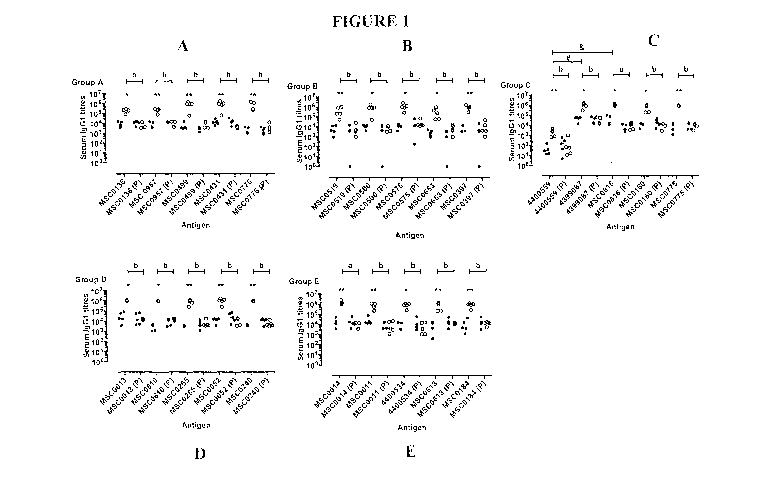
Figures 1A-1E show serum IgG1 immune responses to recombinant proteins used in
trial 1 as described in the examples. For clarity purposes, only the responses
at days 0 (Black
circles) and 35 (White circles) in the vaccinated and placebo (P) groups are
shown. The
groups are listed on the side of each panel. The X-axis indicates the
recombinant proteins
used for each group (names shortened for clarity purposes). The bars across
the symbols
show the median of the values. Significant differences between the day 0 and
day 35 titres for
each antigen are shown by asterisks, * = P<0.05; and ** = P<0.01. Significant
differences
between the day 35 titres of the vaccinated and placebo group for each protein
are shown by a
=P<0.05, b =P<0.01. Differences between the day 35 IgG1 titres between
proteins in the
same group are shown by # =P<0.05 and & =P<0.01.
Figures 2A-2E show serum IgG2 responses against the recombinant proteins used
in
trial 1 as described in the examples. For clarity purposes, only the responses
at days 0 (Black
circles) and 35 (White circles) in the vaccinated and placebo (P) groups are
shown. The
groups are listed on the side of each panel. The X-axis indicates the
recombinant proteins
used for each group. The protein name followed by (P) indicates the placebo
group. The bars
across the symbols show the median of the values. Significant differences
between the titres
in vaccinate and placebo groups are shown by asterisks, * =P<0.05; and ** =
P<0.01.
Significant differences between the day 35 titres of the vaccinated and
placebo group for each
protein are shown by a =P<0.05, b =P<0.01. Differences between the day 35 IgG2
titres
between proteins are shown by # =P<0.05 and & =P<0.01.
Figures 3A-3E show serum IgG1 responses against the recombinant proteins used
in
trial 2 as described in the examples. For clarity purposes, only the responses
at days 0 (Black
circles) and 35 (White circles) in the vaccinated and placebo (P) groups are
shown. The
groups are listed on the side of each panel. The X-axis indicates the
recombinant proteins
used for each group (Names shortened for clarity purposes). The bars across
the symbols
show the median of the values. Significant differences between the day 0 and
day 35 titres for
each antigen are shown by asterisks, * = P<0.05; and ** = P<0.01. Significant
differences
between the day 35 titres of the vaccinated and placebo group for each protein
are shown by a
=P<0.05, b =P<0.01. Differences between the day 35 IgG1 titres between
proteins in the
same group are shown by # =P<0.05 and & =P<0.01.
-6-
CA 02993076 2018-01-19
WO 2017/011919 PCT/CA2016/050864
Figures 4A-4E show serum IgG2 responses against the recombinant proteins used
in
trial 2 as described in the examples. For clarity purposes, only the responses
at days 0 (Black
circles) and 35 (White circles) in the vaccinated and placebo (P) groups are
shown. The
groups are listed on the side of each panel. The X-axis indicates the
recombinant proteins
used for each group. The protein name followed by (P) indicates the placebo
group. The bars
across the symbols show the median of the values. Significant differences
between the titres
in the vaccinated and placebo groups are shown by asterisks; * = P<0.05; and
** = P<0.01.
Significant differences between the day 35 titres of the vaccinated and
placebo group for each
protein are shown by a =P<0.05, b =P<0.01. Differences between the day 35 IgG2
titres
between proteins are shown by # =P<0.05 and & =P<0.01.
Figures 5A-5D show serum IgG1 responses against the recombinant proteins used
in
trial 3 as described in the examples. For clarity purposes, only the responses
at days 0 (Black
circles) and 35 (White circles) in the vaccinated and placebo (P) groups are
shown. The
groups are listed on the side of each panel. The X-axis indicates the
recombinant proteins
used for each group (Names shortened for clarity purposes). The bars across
the symbols
show the median of the values. Significant differences between the day 0 and
day 35 titres for
each antigen are shown by asterisks, * = P<0.05; and ** = P<0.01. Significant
differences
between the day 35 titres of the vaccinated and placebo group for each protein
are shown by a
=P<0.05, b =P<0.01. Differences between the day 35 IgG1 titres between
proteins in the
same group are shown by # =P<0.05 and & =P<0.01.
Figures 6A-6D show serum IgG2 responses against the recombinant proteins used
in
trial 3 as described in the examples. For clarity purposes, only the responses
at days 0 (Black
circles) and 35 (White circles) in the vaccinated and placebo (P) groups are
shown. The
groups are listed on the side of each panel. The X-axis indicates the
recombinant proteins
used for each group. The protein name followed by (P) indicates the placebo
group. The bars
across the symbols show the median of the values. Significant differences
between the titres
in the vaccinated and placebo groups are shown by asterisks; * = P<0.05; and
** = P<0.01.
Significant differences between the day 35 titres of the vaccinated and
placebo group for each
protein are shown by a =P<0.05, b =P<0.01. Differences between the day 35 IgG2
titres
between proteins are shown by # =P<0.05 and & =P<0.01.
-7-
CA 02993076 2018-01-19
WO 2017/011919 PCT/CA2016/050864
Figures 7A-7E show PBMC proliferative responses in trial 1 after incubation
with the
recall antigens as described in the examples. The groups are listed on the top
of each panel.
The mean and standard deviation of the stimulation indexes (Si) at day 35 (Two
weeks after
the boost) for the vaccinated (Black circles) and placebo (Black triangles)
groups are shown.
The X-axis shows the positive control (ConA) and the recall antigens used in
each group.
There were no significant differences between the vaccinated and placebo Si
for each of the
recall antigens and no differences between the Si of any of the antigens in
the vaccinated
groups.
Figures 8A-8E show PBMC proliferative responses in trial 2 after incubation
with the
recall antigens as described in the examples. The groups are listed on the top
of each panel.
The mean and standard deviation of the stimulation indexes (Si) at day 35 (Two
weeks after
the boost) for the vaccinated (Black circles) and placebo (Black triangles)
groups are shown.
The X-axis shows the positive control (ConA) and the recall antigens used in
each group.
There were no significant differences between the vaccinated and placebo Si
for each of the
recall antigens and no differences between the Si of any of the antigens in
the vaccinated
groups.
Figures 9A-9D show PBMC proliferative responses in trial 3 after incubation
with the
recall antigens as described in the examples. The groups are listed on the top
of each panel.
The mean and standard deviation of the stimulation indexes (Si) at day 35 (Two
weeks after
the boost) for the vaccinated (Black circles) and placebo (Black triangles)
groups are shown.
The X-axis shows the positive control (ConA) and the recall antigens used in
each group.
There were no significant differences between the vaccinated and placebo Si
for each of the
recall antigens and no differences between the Si of any of the antigens in
the vaccinated
groups.
Figures 10A-10C show serum TGF-I3 levels in the three trials as described in
the
examples. The day 0 and day 35 serum TGF-I3 levels for trials 1, 2 and 3 are
shown in A, B,
and C respectively. The black circles indicate the levels at day 0 while white
circles show the
levels at day 35. The groups including the placebo groups F, L, and Q are
indicated on the X-
axis. In trials 1 and 2, there were no significant differences between day 0
and day 35 TGF-
13 levels. The TGF-I3 levels at day 35 were significantly lower (P< 0.05) than
the day 0 values
in the groups M and P of the third trial.
-8-
CA 02993076 2018-01-19
WO 2017/011919 PCT/CA2016/050864
Figures 11A-11B (SEQ ID NOS:1 and 2) show the modified nucleotide sequence of
MSC 0136 (SEQ ID NO:1) and the amino acid sequence of the protein antigen MSC
0136
(SEQ ID NO:2) used in the examples. The sequences differ from those reported
in NCBI in
that the DNA sequence has been modified for expression in E. coil; and the
protein sequence
lacks the first 24 amino acids (the signal sequence).
Figures 12A-12B (SEQ ID NOS:3 and 4) show the modified nucleotide sequence of
MSC 0957 (SEQ ID NO:3) and the amino acid sequence of the protein antigen MSC
0957
(SEQ ID NO:4) used in the examples. The sequences differ from those reported
in NCBI in
that the DNA sequence has been modified for expression in E. coil; and the
protein sequence
lacks the first 23 amino acids (the signal sequence).
Figures 13A-13B (SEQ ID NOS:5 and 6) show the modified nucleotide sequence of
MSC 0499 (SEQ ID NO:5) and amino acid sequence of the protein antigen MSC 0499
(SEQ
ID NO:6) used in the examples. The sequences differ from those reported in
NCBI in that the
DNA sequence has been modified for expression in E. coil; and the protein
sequence lacks the
first 23 amino acids (the signal sequence).
Figures 14A-14B (SEQ ID NOS:7 and 8) show the modified nucleotide sequence of
MSC 0431 (SEQ ID NO:7) and amino acid sequence of the protein antigen MSC 0431
(SEQ
ID NO:8) used in the examples. The sequences differ from those reported in
NCBI in that the
DNA sequence has been modified for expression in E. coil; and the protein
sequence lacks the
first 26 amino acids (the signal sequence).
Figures 15A-15B (SEQ ID NOS:9 and 10) show the modified nucleotide sequence of
MSC 0776 (SEQ ID NO:9) and amino acid sequence of the protein antigen MSC 0776
(SEQ
ID NO:10) used in the examples. The sequences differ from those reported in
NCBI in that
the DNA sequence has been modified for expression in E. coil; and the protein
sequence lacks
the first 27 amino acids (the signal sequence).
Figures 16A-16B (SEQ ID NOS:11 and 12) show the nucleotide sequence, modified
for expression in E. coil, of YP 004400559.1 (SEQ ID NO:11) and amino acid
sequence of
the protein antigen YP 004400559.1 (SEQ ID NO:12) used in the examples. The
amino acid
sequence differs from that reported in NCBI in that the sequence lacks the
first 24 amino
acids (the signal sequence) and includes an N-terminal methionine.
-9-
CA 02993076 2018-01-19
WO 2017/011919 PCT/CA2016/050864
Figures 17A-17B (SEQ ID NOS:13 and 14) show the nucleotide sequence, modified
for expression in E. coil, of YP 004399807.1 (SEQ ID NO:13) and amino acid
sequence of
the protein antigen YP 004399807.1 (SEQ ID NO:14) used in the examples. The
amino acid
sequence differs from that reported in NCBI in that the sequence lacks the
first 24 amino
acids (the signal sequence) and includes an N-terminal methionine.
Figures 18A-18B (SEQ ID NOS:15 and 16) show the modified nucleotide sequence
of
MSC 0816 (SEQ ID NO:15) and amino acid sequence of the protein antigen MSC
0816
(SEQ ID NO:16) used in the examples. The sequences differ from those reported
in NCBI in
that the DNA sequence has been modified for expression in E. coil; and the
protein sequence
lacks the first 23 amino acids (the signal sequence).
Figures 19A-19B (SEQ ID NOS:17 and 18) show the modified nucleotide sequence
of
MSC 0160 (SEQ ID NO:17) and amino acid sequence of the protein antigen MSC
0160
(SEQ ID NO:18) used in the examples. The DNA sequence differs from that
reported in
NCBI in that the DNA sequence has been modified for expression in E. coil.
Figures 20A-20B (SEQ ID NOS:19 and 20) show the modified nucleotide sequence
of
MSC 0775 (SEQ ID NO:19) and amino acid sequence of the protein antigen MSC
0775
(SEQ ID NO:20) used in the examples. The sequences differ from those reported
in NCBI in
that the DNA sequence has been modified for expression in E. coil; and the
protein sequence
lacks the first 25 amino acids (the signal sequence).
Figures 21A-21B (SEQ ID NOS:21 and 22) show the nucleotide sequence, modified
for expression in E. coil, of YP 004400127.1 (SEQ ID NO:21) and amino acid
sequence of
the protein antigen YP 004400127.1 (SEQ ID NO:22) used in the examples. The
amino acid
sequence differs from that reported in NCBI in that it lacks the first 23
amino acids (the signal
sequence) and includes an N-terminal methionine.
Figures 22A-22B (SEQ ID NOS:23 and 24) show the nucleotide sequence, modified
for expression in E. coil, of YP 004399790.1 (SEQ ID NO:23) and amino acid
sequence of
the protein antigen YP 004399790.1 (SEQ ID NO:24) used in the examples.
Figures 23A-23B (SEQ ID NOS:25 and 26) show the nucleotide sequence, modified
for expression in E. coil, of YP 004400580.1 (SEQ ID NO:25) and amino acid
sequence of
the protein antigen YP 004400580.1 (SEQ ID NO:26) used in the examples. The
amino acid
-10-
CA 02993076 2018-01-19
WO 2017/011919 PCT/CA2016/050864
sequence differs from that reported in NCBI in that it lacks 15 amino acids
from the C-
terminus.
Figures 24A-24B (SEQ ID NOS:27 and 28) show the nucleotide sequence, modified
for expression in E. coil, of YP 004400610.1 (SEQ ID NO:27) and amino acid
sequence of
the protein antigen YP 004400610.1 (SEQ ID NO:28) used in the examples. The
amino acid
sequence differs from that reported in NCBI in that the sequence lacks the
first 24 amino
acids (the signal sequence) and includes an N-terminal methionine.
Figures 25A-25B (SEQ ID NOS:50 and 51) show the nucleotide sequence, modified
for expression in E. coil, of a fusion (SEQ ID NO:50) between YP 004400127.1
and
YP 004399790.1 and the amino acid sequence of the protein fusion (SEQ ID
NO:51) used in
the examples. The YP 004400127.1 sequence occurs at positions 1-214 of the
protein and
the YP 004399790.1 sequence is present at positions 221-532 of the protein.
The two
sequences are linked by a G1y6 linker, bolded in the figure.
Figures 26A-26B (SEQ ID NOS:52 and 53) show the nucleotide sequence, modified
for expression in E. coil, of a fusion (SEQ ID NO:52) between sequences
derived from
YP 004400610.1 and YP 00400580.1 and the amino acid sequence of the protein
fusion
(SEQ ID NO:53) used in the examples. The YP 004400610.1 sequence occurs at
positions 1-
189 of the protein and the sequence derived from YP 004399790.1 is present at
positions
195-557 of the protein. The YP 00400580.1 sequence in the fusion lacks the
first 20 amino
acids present in the YP 00400580.1 sequence shown in SEQ ID NO:26. The two
sequences
are linked by a Glys linker, bolded in the figure.
Figures 27A-27B (SEQ ID NOS:54 and 55) show the nucleotide sequence, modified
for expression in E. coil, (SEQ ID NO:54) and amino acid sequence (SEQ ID
NO:55) of
pAA352-YP 004400127.1-YP 004399790.1 used in the examples. The leukotoxin 352
carrier, (also termed "LKT 352" and "LtxA" herein) occurs at positions 1-926
of the amino
acid sequence and is bolded in SEQ ID NO:55; The YP 004400127.1 sequence
occurs at
positions 927-1140 of SEQ ID NO:55; the YP 004399790.1 sequence is present at
positions
1147-1458 of SEQ ID NO:55. The two sequences are linked by a G1y6 linker,
bolded in the
figure.
Figures 28A-28B (SEQ ID NOS:56 and 57) show the nucleotide sequence, modified
for expression in E. coil, (SEQ ID NO:56) and amino acid sequence (SEQ ID
NO:57) of
-11-
CA 02993076 2018-01-19
WO 2017/011919 PCT/CA2016/050864
pAA352-YP 004400610.1-YP 00400580.1 used in the examples. The leukotoxin 352
carrier, (also termed "LKT 352" and "LtxA" herein) occurs at positions 1-926
of the amino
acid sequence and is bolded in SEQ ID NO:57; The YP 004400610.1 sequence
occurs at
positions 927-1115 of SEQ ID NO:57; the YP 00400580.1 sequence is present at
positions
1121-1483 of SEQ ID NO:57. The YP 00400580.1 sequence in the fusion lacks the
first 20
amino acids present in the YP 00400580.1 sequence shown in SEQ ID NO:26. The
two
sequences are linked by a Glys linker, bolded in the figure.
Figures 29A-29B (SEQ ID NOS:58 and 59) show the nucleotide sequence, modified
for expression in E. coil, (SEQ ID NO:58) and amino acid sequence (SEQ ID
NO:59) of
pAA352-MSC 0160 used in the examples. The leukotoxin 352 carrier, (also termed
"LKT
352" and "LtxA" herein) occurs at positions 1-926 of the amino acid sequence
and is bolded
in SEQ ID NO:59; The MSC 0160 sequence occurs at positions 927-1320 of SEQ ID
NO:59.
The MSC 0160 sequence lacks the N-terminal methionine shown in SEQ ID NO:18.
Figures 30A-30B (SEQ ID NOS:60 and 61) show the nucleotide sequence, modified
for expression in E. coil, (SEQ ID NO:60) and amino acid sequence (SEQ ID
NO:61) of
pAA352-MSC 0136 used in the examples. The leukotoxin 352 carrier, (also termed
"LKT
352" and "LtxA" herein) occurs at positions 1-926 of the amino acid sequence
and is bolded
in SEQ ID NO:61; the MSC 0136 sequence occurs at positions 927-1224 of SEQ ID
NO:61.
Figures 31A-31B (SEQ ID NOS:62 and 63) show the nucleotide sequence, modified
for expression in E. coil, (SEQ ID NO:62) and amino acid sequence (SEQ ID
NO:63) of
pAA352-MSC 0431 used in the examples. The leukotoxin 352 carrier, (also termed
"LKT
352" and "LtxA" herein) occurs at positions 1-926 of the amino acid sequence
and is bolded
in SEQ ID NO:63; the MSC 0431 sequence occurs at positions 927-1256 of SEQ ID
NO:63.
Figures 32A-32B (SEQ ID NOS:64 and 65) show the nucleotide sequence, modified
for expression in E. coil, (SEQ ID NO:64) and amino acid sequence (SEQ ID
NO:65) of
pAA352-MSC 0499 used in the examples. The leukotoxin 352 carrier, (also termed
"LKT
352" and "LtxA" herein) occurs at positions 1-926 of the amino acid sequence
and is bolded
in SEQ ID NO:65; the MSC 0499 sequence occurs at positions 927-1620 of SEQ ID
NO:65.
Figures 33A-33B (SEQ ID NOS:66 and 67) show the nucleotide sequence, modified
for expression in E. coil, (SEQ ID NO:66) and amino acid sequence (SEQ ID
NO:67) of
pAA352-MSC 0775 used in the examples. The leukotoxin 352 carrier, (also termed
"LKT
-12-
CA 02993076 2018-01-19
WO 2017/011919 PCT/CA2016/050864
352" and "LtxA" herein) occurs at positions 1-926 of the amino acid sequence
and is bolded
in SEQ ID NO:67; the MSC 0775 sequence occurs at positions 927-1608 of SEQ ID
NO:67.
The MSC 0775 sequence lacks the first 20 amino acids shown in SEQ ID NO:20.
Figures 34A-34B (SEQ ID NOS:68 and 69) show the nucleotide sequence, modified
for expression in E. coil, (SEQ ID NO:68) and amino acid sequence (SEQ ID
NO:69) of
pAA352-MSC 0776 used in the examples. The leukotoxin 352 carrier, (also termed
"LKT
352" and "LtxA" herein) occurs at positions 1-926 of the amino acid sequence
and is bolded
in SEQ ID NO:69; the MSC 0776 sequence occurs at positions 927-1681 of SEQ ID
NO:69.
Figures 35A-35B (SEQ ID NOS:70 and 71) show the nucleotide sequence, modified
for expression in E. coil, (SEQ ID NO:70) and amino acid sequence (SEQ ID
NO:71) of
pAA352-MSC 0816 used in the examples. The leukotoxin 352 carrier, (also termed
"LKT
352" and "LtxA" herein) occurs at positions 1-926 of the amino acid sequence
and is bolded
in SEQ ID NO:71; the MSC 0816 sequence occurs at positions 927-1308 of SEQ ID
NO:71.
Figures 36A-36B (SEQ ID NOS:72 and 73) show the nucleotide sequence, modified
for expression in E. coil, (SEQ ID NO:72) and amino acid sequence (SEQ ID
NO:73) of
pAA352-MSC 0957 used in the examples. The leukotoxin 352 carrier, (also termed
"LKT
352" and "LtxA" herein) occurs at positions 1-926 of the amino acid sequence
and is bolded
in SEQ ID NO:73; the MSC 0957 sequence occurs at positions 927-1336 of SEQ ID
NO:73.
Figures 37A-37B (SEQ ID NOS:74 and 75) show the nucleotide sequence, modified
for expression in E. coil, (SEQ ID NO:74) and amino acid sequence (SEQ ID
NO:75) of
pAA352-MSC 0466-MSC 0117 used in the examples. The leukotoxin 352 carrier,
(also
termed "LKT 352" and "LtxA" herein) occurs at positions 1-926 of the amino
acid sequence
and is bolded in SEQ ID NO:75; The MSC 0466 sequence occurs at positions 927-
1180 of
SEQ ID NO:75; the MSC 0117 sequence is present at positions 1184-1421 of SEQ
ID
NO:75. The two sequences are linked by a G1y3 linker, bolded in the figure.
Figures 38A-38B (SEQ ID NOS:76 and 77) show the nucleotide sequence, modified
for expression in E. coil, (SEQ ID NO:76) and amino acid sequence (SEQ ID
NO:77) of
pAA352-MSC 0922-MSC 1058 used in the examples. The leukotoxin 352 carrier,
(also
termed "LKT 352" and "LtxA" herein) occurs at positions 1-926 of the amino
acid sequence
and is bolded in SEQ ID NO:77; The MSC 0922 sequence occurs at positions 927-
1325 of
-13-
CA 02993076 2018-01-19
WO 2017/011919 PCT/CA2016/050864
SEQ ID NO:77; the MSC 1058 sequence is present at positions 1329-1468 of SEQ
ID
NO:77. The two sequences are linked by a G1y3 linker, bolded in the figure.
Figures 39A-39B (SEQ ID NOS:78 and 79) show the nucleotide sequence, modified
for expression in E. coil, (SEQ ID NO:78) and amino acid sequence (SEQ ID
NO:79) of
pAA352-YP 004399807.1 used in the examples. The leukotoxin 352 carrier, (also
termed
"LKT 352" and "LtxA" herein) occurs at positions 1-926 of the amino acid
sequence and is
bolded in SEQ ID NO:79; the YP 004399807.1 sequence occurs at positions 927-
1273 of
SEQ ID NO:79. The YP 004399807.1 sequence lacks the N-terminal methionine
shown in
SEQ NO:14.
Figures 40A-40B (SEQ ID NOS:80 and 81) show the nucleotide sequence, modified
for expression in E. coil, (SEQ ID NO:80) and amino acid sequence (SEQ ID
NO:81) of
pAA352-YP 00400559.1 used in the examples. The leukotoxin 352 carrier, (also
termed
"LKT 352" and "LtxA" herein) occurs at positions 1-926 of the amino acid
sequence and is
bolded in SEQ ID NO:81; the YP 00400559.1 sequence occurs at positions 927-
1061 of SEQ
ID NO:81. The YP 00400559.1 sequence lacks the N-terminal methionine shown in
SEQ ID
NO:12.
Figure 41 (SEQ ID NOS:82 and 83) shows the nucleotide sequence (SEQ ID NO:82)
and amino acid sequence (SEQ ID NO:83) of a representative leukotoxin 352 (LKT
352)
from plasmid pAA352. The first 10 N-terminal amino acids and last 2 C-terminal
amino
acids depicted in the figure are flanking sequences from plasmid pAA352. The
remaining
amino acids are leukotoxin sequences. LKT 352 is a detoxified mutant of
leukotoxin.
DETAILED DESCRIPTION OF THE INVENTION
The practice of the present invention will employ, unless otherwise indicated,
conventional methods of virology, chemistry, biochemistry, recombinant DNA
techniques
and immunology, within the skill of the art. Such techniques are explained
fully in the
literature. See, e.g., Fundamental Virology, Current Edition, vol. I & II
(B.N. Fields and
D.M. Knipe, eds.); Handbook of Experimentailmmunoiogy,Vols. I-IV (D.M. Weir
and C.C.
Blackwell eds., Blackwell Scientific Publications); T.E. Creighton, Proteins:
Structures and
Molecular Properties (W.H. Freeman and Company); A.L. Lehninger, Biochemistry
(Worth
Publishers, Inc., current edition); Sambrook, et al., Molecular Cloning: A
Laboratory Manual
-14-
CA 02993076 2018-01-19
WO 2017/011919 PCT/CA2016/050864
(current edition); Methods In Enzymology (S. Colowick and N. Kaplan eds.,
Academic Press,
Inc.).
All publications, patents and patent applications cited herein, whether supra
or infra,
are hereby incorporated by reference in their entireties.
The following amino acid abbreviations are used throughout the text:
Alanine: Ala (A) Arginine: Arg (R)
Asparagine: Asn (N) Aspartic acid: Asp (D)
Cysteine: Cys (C) Glutamine: Gln (Q)
Glutamic acid: Glu (E) Glycine: Gly (G)
Hi stidine: His (H) Isoleucine: Ile (I)
Leucine: Leu (L) Lysine: Lys (K)
Methionine: Met (M) Phenylalanine: Phe (F)
Proline: Pro (P) Serine: Ser (S)
Threonine: Thr (T) Tryptophan: Trp (W)
Tyrosine: Tyr (Y) Valine: Val (V)
1. DEFINITIONS
In describing the present invention, the following terms will be employed, and
are
intended to be defined as indicated below.
It must be noted that, as used in this specification and the appended claims,
the
singular forms "a", "an" and "the" include plural referents unless the content
clearly dictates
otherwise. Thus, for example, reference to "an antigen" includes a mixture of
two or more
such antigens, and the like.
As used herein, the term "Mycoplasma" refers to bacteria belonging to the
class
Mollicutes and the genus Mycoplasma. The term intends any species and
subspecies of the
genus Mycoplasma, which is capable of causing disease in an animal or human
subject. Such
species are described below.
As used herein, the term "Mycoplasma mycoides" or "M mycoides" refers to any
of
the species and subspecies from the Mycoplasma mycoides cluster, a group of
closely related
infectious mycoplasmas. The cluster contains several species and subspecies
including M.
-15-
CA 02993076 2018-01-19
WO 2017/011919 PCT/CA2016/050864
mycoides subsp. mycoides biotype Small Colony (MmmSC); M mycoides subsp.
mycoides
biotype Large Colony (MmmLC);M mycoides subsp. capri (Mmc); M capricolum
subsp.
capricolum (Mcc);M capricolum subsp. capripneumoniae (Mccp); and Mycoplasma
sp.
'bovine group 7' (MBG7).
The term "derived from" is used herein to identify the original source of a
molecule
but is not meant to limit the method by which the molecule is made which can
be, for
example, by chemical synthesis or recombinant means.
A "Mycoplasma molecule" is a molecule derived from Mycoplasma, including,
without limitation, polypeptide, protein, antigen, polynucleotide,
oligonucleotide, and nucleic
acid molecules, as defined herein, from any of the various Mycoplasma species
and
subspecies. The molecule need not be physically derived from the particular
bacterium in
question, but may be synthetically or recombinantly produced. Nucleic acid and
polypeptide
sequences for a number of Mycoplasma species are known and/or described
herein.
Representative Mycoplasma sequences for use in treating and/or preventing M
mycoides
infection, such as CBPP, are presented in Tables 1 and 4 and Figures 11-40
herein. It is to be
understood that while Table 4 and several figures describe M mycoides fusion
proteins, as
well as conjugates of the fusions, the individual M mycoides proteins in the
fusions and the
conjugates are also intended. The boundaries of the individual M mycoides
proteins present
in the fusions, as well as the M. mycoides proteins present in the conjugates,
are described
above.
Additional representative sequences found in various species are listed in the
National
Center for Biotechnology Information (NCBI) database. However, a Mycoplasma
molecule,
such as an antigen, as defined herein, is not limited to those shown and
described in Tables 1
and 4 and Figures 11-40, as various isolates are known and variations in
sequences may occur
between them.
By "Mycoplasma disease" is meant a disease caused in whole or in part by a
Mycoplasma bacterium. For example, Mycoplasma bacteria cause a number of
diseases in
animals, such as but not limited to pneumonia, e.g., contagious bovine
pleuropneumonia,
mastitis, arthritis, otitis, keratoconjunctivitis, synovitis, and reproductive
disorders. In
humans such diseases include pneumonia and other respiratory problems such as
-16-
CA 02993076 2018-01-19
WO 2017/011919 PCT/CA2016/050864
tracheobronchitis, bronchiolitis, pharyngitis and croup; pelvic inflammatory
disease; and
cancer.
The terms "polypeptide" and "protein" refer to a polymer of amino acid
residues and
are not limited to a minimum length of the product. Thus, peptides,
oligopeptides, dimers,
multimers, and the like, are included within the definition. Both full-length
proteins and
fragments thereof are encompassed by the definition. The terms also include
postexpression
modifications of the polypeptide, for example, glycosylation, acetylation,
phosphorylation and
the like. Furthermore, for purposes of the present invention, a "polypeptide"
refers to a
protein which includes modifications, such as deletions, additions and
substitutions, to the
native sequence, so long as the protein maintains the desired activity. These
modifications
may be deliberate, as through site-directed mutagenesis, or may be accidental,
such as through
mutations of hosts which produce the proteins or errors due to PCR
amplification.
The term "peptide" as used herein refers to a fragment of a polypeptide. Thus,
a
peptide can include a C-terminal deletion, an N-terminal deletion and/or an
internal deletion
of the native polypeptide, so long as the entire protein sequence is not
present. A peptide will
generally include at least about 3-10 contiguous amino acid residues of the
full-length
molecule, and can include at least about 15-25 contiguous amino acid residues
of the
full-length molecule, or at least about 20-50 or more contiguous amino acid
residues of the
full-length molecule, or any integer between 3 amino acids and the number of
amino acids in
the full-length sequence, provided that the peptide in question retains the
ability to elicit the
desired biological response.
By "immunogenic" protein, polypeptide or peptide is meant a molecule which
includes one or more epitopes and thus can modulate an immune response. Such
peptides can
be identified using any number of epitope mapping techniques, well known in
the art. See,
e.g., Epitope Mapping Protocols in Methods in Molecular Biology, Vol. 66
(Glenn E. Morris,
Ed., 1996) Humana Press, Totowa, New Jersey. For example, linear epitopes may
be
determined by e.g., concurrently synthesizing large numbers of peptides on
solid supports, the
peptides corresponding to portions of the protein molecule, and reacting the
peptides with
antibodies while the peptides are still attached to the supports. Such
techniques are known in
the art and described in, e.g., U.S. Patent No. 4,708,871; Geysen et al.
(1984) Proc. Natl.
Acad. Sci. USA 81:3998-4002; Geysen et al. (1986) Molec. Immunol. 23:709-715,
all
-17-
CA 02993076 2018-01-19
WO 2017/011919 PCT/CA2016/050864
incorporated herein by reference in their entireties. Similarly,
conformational epitopes are
readily identified by determining spatial conformation of amino acids such as
by, e.g., x-ray
crystallography and 2-dimensional nuclear magnetic resonance. See, e.g.,
Epitope Mapping
Protocols, supra. Antigenic regions of proteins can also be identified using
standard
antigenicity and hydropathy plots, such as those calculated using, e.g., the
Omiga version 1.0
software program available from the Oxford Molecular Group. This computer
program
employs the Hopp/Woods method, Hopp et al., Proc. Natl. Acad. Sci USA (1981)
78:3824-
3828 for determining antigenicity profiles, and the Kyte-Doolittle technique,
Kyte et al.,
Mol. Biol. (1982) 157:105-132 for hydropathy plots.
Immunogenic molecules, for purposes of the present invention, will usually be
at least
about 5 amino acids in length, such as at least about 10 to about 15 amino
acids in length.
There is no critical upper limit to the length of the molecule, which can
comprise the full-
length of the protein sequence, or even a fusion protein comprising two or
more epitopes,
proteins, antigens, etc..
As used herein, the term "epitope" generally refers to the site on an antigen
which is
recognized by a T-cell receptor and/or an antibody. Several different epitopes
may be carried
by a single antigenic molecule. The term "epitope" also includes modified
sequences of
amino acids which stimulate responses which recognize the whole organism. The
epitope can
be generated from knowledge of the amino acid and corresponding DNA sequences
of the
polypeptide, as well as from the nature of particular amino acids (e.g., size,
charge, etc.) and
the codon dictionary, without undue experimentation. See, e.g., Ivan Roitt,
Essential
Immunology; Janis Kuby, Immunology.
An "immunological response" to an antigen or composition is the development in
a
subject of a humoral and/or a cellular immune response to an antigen present
in the
composition of interest. For purposes of the present invention, a "humoral
immune response"
refers to an immune response mediated by antibody molecules, while a "cellular
immune
response" is one mediated by T-lymphocytes and/or other white blood cells. One
important
aspect of cellular immunity involves an antigen-specific response by cytolytic
T-cells
("CTL"s). CTLs have specificity for peptide antigens that are presented in
association with
proteins encoded by the major histocompatibility complex (MHC) and expressed
on the
surfaces of cells. CTLs help induce and promote the destruction of
intracellular microbes, or
-18-
CA 02993076 2018-01-19
WO 2017/011919 PCT/CA2016/050864
the lysis of cells infected with such microbes. Another aspect of cellular
immunity involves
an antigen-specific response by helper T-cells. Helper T-cells act to help
stimulate the
function, and focus the activity of, nonspecific effector cells against cells
displaying peptide
antigens in association with MHC molecules on their surface. A "cellular
immune response"
also refers to the production of cytokines, chemokines and other such
molecules produced by
activated T-cells and/or other white blood cells, including those derived from
CD4+ and
CD8+ T-cells.
Thus, an immunological response as used herein may be one that stimulates the
production of antibodies. The antigen of interest may also elicit production
of CTLs. Hence,
an immunological response may include one or more of the following effects:
the production
of antibodies by B-cells; and/or the activation of suppressor T-cells and/or
memory/effector
T-cells directed specifically to an antigen or antigens present in the
composition or vaccine of
interest. These responses may serve to neutralize infectivity, and/or mediate
antibody-
complement, or antibody dependent cell cytotoxicity (ADCC) to provide
protection to an
immunized host. Such responses can be determined using standard immunoassays
and
neutralization assays, well known in the art. (See, e.g., Montefiori et al.
(1988)1 Clin
Microbiol. 26:231-235; Dreyer et al. (1999) AIDS Res Hum Retroviruses (1999)
15(17):1563-
1571). The innate immune system of mammals also recognizes and responds to
molecular
features of pathogenic organisms via activation of Toll-like receptors and
similar receptor
molecules on immune cells. Upon activation of the innate immune system,
various non-
adaptive immune response cells. are activated to, e.g., produce various
cytokines,
lymphokines and chemokines. Cells activated by an innate immune response
include
immature and mature Dendritic cells of the monocyte and plasmacytoid lineage
(MDC, PDC),
as well as gamma, delta, alpha and beta T cells and B cells and the like.
Thus, the present
invention also contemplates an immune response wherein the immune response
involves both
an innate and adaptive response.
An "immunogenic composition" is a composition that comprises an immunogenic
molecule where administration of the composition to a subject results in the
development in
the subject of a humoral and/or a cellular immune response to the molecule of
interest.
An "antigen" refers to a molecule, such as a protein, polypeptide, or fragment
thereof,
containing one or more epitopes (either linear, conformational or both) that
will stimulate a
-19-
CA 02993076 2018-01-19
WO 2017/011919 PCT/CA2016/050864
host's immune-system to make a humoral and/or cellular antigen-specific
response. The term
is used interchangeably with the term "immunogen." Antibodies such as anti-
idiotype
antibodies, or fragments thereof, and synthetic peptide mimotopes, which can
mimic an
antigen or antigenic determinant, are also captured under the definition of
antigen as used
herein. Similarly, an oligonucleotide or polynucleotide which expresses an
antigen or
antigenic determinant in vivo, such as in DNA immunization applications, is
also included in
the definition of antigen herein.
By "subunit vaccine" is meant a vaccine composition that includes one or more
selected antigens but not all antigens, derived from or homologous to, an
antigen from a
pathogen of interest. Such a composition is substantially free of intact
pathogen cells or
pathogenic particles, or the lysate of such cells or particles. Thus, a
"subunit vaccine" can be
prepared from at least partially purified (preferably substantially purified)
immunogenic
molecules from the pathogen, or analogs thereof The method of obtaining an
antigen
included in the subunit vaccine can thus include standard purification
techniques, recombinant
production, or synthetic production.
By "carrier" is meant any molecule which when associated with an antigen of
interest,
imparts enhanced immunogenicity to the antigen.
The term "RTX" toxin, as used herein refers to a protein belonging to the
family of
molecules characterized by the carboxy-terminus consensus amino acid sequence
Gly-Gly-X-
Gly-X-Asp (SEQ ID NO:78, Highlander et al., DNA (1989) 8:15-28), where Xis
Lys, Asp,
Val or Asn. Such proteins include, among others, leukotoxins derived from P.
haemolytica
and Actinobacillus pleuropneumoniae, as well as E. coil alpha hemolysin
(Strathdee et al.,
Infect. Immun. (1987) 55:3233-3236; Lo, Can. I Vet. Res. (1990) 54:S33-S35;
Welch, Mol.
Microbiol. (1991) 5:521-528). This family of toxins is known as the "RTX"
family of toxins
(Lo, Can. I Vet. Res. (1990) 54:S33-S35). In addition, the term "RTX toxin"
refers to a
member of the RTX family which is chemically synthesized, isolated from an
organism
expressing the same, or recombinantly produced. Furthermore, the term intends
an
immunogenic protein having an amino acid sequence substantially homologous to
a
contiguous amino acid sequence found in the particular native RTX molecule.
Thus, the term
includes both full-length and partial sequences, as well as analogues.
Although native full-
length RTX toxins display cytotoxic activity, the term "RTX toxin" also
intends molecules
-20-
CA 02993076 2018-01-19
WO 2017/011919 PCT/CA2016/050864
which remain immunogenic yet lack the cytotoxic character of native molecules.
In the
chimeras produced according to the present invention, a selected RTX
polypeptide sequence
imparts enhanced immunogeni city to a fused Mycoplasma protein or fusion
proteins
comprising more than one Mycoplasma protein or antigen.
The term "leukotoxin polypeptide" or "LKT polypeptide" intends an RTX toxin
derived from P. haemolytica, Actinobacillus pleuropneumoniae, among others, as
defined
above. The nucleotide sequences and corresponding amino acid sequences for
several
leukotoxins are known. See, e.g., U.S. Patent Nos. 4,957,739 and 5,055,400; Lo
et al., Infect.
Immun. (1985) 50:667-67; Lo et al., Infect. Immun. (1987) 55:1987-1996;
Strathdee et al.,
Infect. Immun. (1987) 55:3233-3236; Highlander et al., DNA (1989) 8:15-28;
Welch, Mol.
Microbiol. (1991) 5:521-528. A selected leukotoxin polypeptide sequence
imparts enhanced
immunogenicity to a fused Mycoplasma protein or fusion proteins comprising
more than one
Mycoplasma protein or antigen.
"Substantially purified" generally refers to isolation of a substance such
that the
substance comprises the majority percent of the sample in which it resides.
Typically in a
sample, a substantially purified component comprises 50%, preferably 80%-85%,
more
preferably 90-95% of the sample. Techniques for purifying molecules of
interest are well-
known in the art and include, for example, ion-exchange chromatography,
affinity
chromatography and sedimentation according to density.
By "isolated" is meant that the indicated molecule is separate and discrete
from the
whole organism with which the molecule is found in nature or is present in the
substantial
absence of other biological macromolecules of the same type.
An "antibody" intends a molecule that "recognizes," i.e., specifically binds
to an
epitope of interest present in an antigen. By "specifically binds" is meant
that the antibody
interacts with the epitope in a "lock and key" type of interaction to form a
complex between
the antigen and antibody, as opposed to non-specific binding that might occur
between the
antibody and, for instance, components in a mixture that includes the test
substance with
which the antibody is reacted. The term "antibody" as used herein includes
antibodies
obtained from both polyclonal and monoclonal preparations, as well as, the
following: hybrid
(chimeric) antibody molecules (see, for example, Winter et al., Nature (1991)
349:293-299;
and U.S. Patent No. 4,816,567); F(ab')2 and F(ab) fragments; Fv molecules (non-
covalent
-21-
CA 02993076 2018-01-19
WO 2017/011919
PCT/CA2016/050864
heterodimers, see, for example, Inbar et al., Proc Natl Acad Sci USA (1972)
69:2659-2662;
and Ehrlich et al., Biochem (1980) 19:4091-4096); single-chain Fv molecules
(sFv) (see, for
example, Huston et al., Proc Natl Acad Sci USA (1988) 85:5879-5883); dimeric
and trimeric
antibody fragment constructs; minibodies (see, e.g., Pack et al., Biochem
(1992)
31:1579-1584; Cumber et al., J Immunology (1992) 149B:120-126); humanized
antibody
molecules (see, for example, Riechmann et al., Nature (1988) 332:323-327;
Verhoeyan et al.,
Science (1988) 239:1534-1536; and U.K. Patent Publication No. GB 2,276,169,
published 21
September 1994); and, any functional fragments obtained from such molecules,
wherein such
fragments retain immunological binding properties of the parent antibody
molecule.
As used herein, the term "monoclonal antibody" refers to an antibody
composition
having a homogeneous antibody population. The term is not limited regarding
the species or
source of the antibody, nor is it intended to be limited by the manner in
which it is made. The
term encompasses whole immunoglobulins as well as fragments such as Fab,
F(ab)2, Fv, and
other fragments, as well as chimeric and humanized homogeneous antibody
populations, that
exhibit immunological binding properties of the parent monoclonal antibody
molecule.
"Homology" refers to the percent identity between two polynucleotide or two
polypeptide moieties. Two nucleic acid, or two polypeptide sequences are
"substantially
homologous" to each other when the sequences exhibit at least about 50%
sequence identity,
preferably at least about 75% sequence identity, more preferably at least
about 80%-85%
sequence identity, more preferably at least about 90% sequence identity, and
most preferably
at least about 95%-98% sequence identity over a defined length of the
molecules. As used
herein, substantially homologous also refers to sequences showing complete
identity to the
specified sequence.
In general, "identity" refers to an exact nucleotide-to-nucleotide or amino
acid-to-amino acid correspondence of two polynucleotides or polypeptide
sequences,
respectively. Percent identity can be determined by a direct comparison of the
sequence
information between two molecules by aligning the sequences, counting the
exact number of
matches between the two aligned sequences, dividing by the length of the
shorter sequence,
and multiplying the result by 100. Readily available computer programs can be
used to aid in
the analysis, such as ALIGN, Dayhoff, M.O. in Atlas of Protein Sequence and
Structure M.O.
Dayhoff ed., 5 Suppl. 3:353-358, National biomedical Research Foundation,
Washington, DC,
-22-
CA 02993076 2018-01-19
WO 2017/011919 PCT/CA2016/050864
which adapts the local homology algorithm of Smith and Waterman Advances in
Appl. Math.
2:482-489, 1981 for peptide analysis. Programs for determining nucleotide
sequence identity
are available in the Wisconsin Sequence Analysis Package, Version 8 (available
from
Genetics Computer Group, Madison, WI) for example, the BESTFIT, FASTA and GAP
programs, which also rely on the Smith and Waterman algorithm. These programs
are readily
utilized with the default parameters recommended by the manufacturer and
described in the
Wisconsin Sequence Analysis Package referred to above. For example, percent
identity of a
particular nucleotide sequence to a reference sequence can be determined using
the homology
algorithm of Smith and Waterman with a default scoring table and a gap penalty
of six
nucleotide positions.
Another method of establishing percent identity in the context of the present
invention
is to use the MPSRCH package of programs copyrighted by the University of
Edinburgh,
developed by John F. Collins and Shane S. Sturrok, and distributed by
IntelliGenetics, Inc.
(Mountain View, CA). From this suite of packages the Smith-Waterman algorithm
can be
employed where default parameters are used for the scoring table (for example,
gap open
penalty of 12, gap extension penalty of one, and a gap of six). From the data
generated the
"Match" value reflects "sequence identity." Other suitable programs for
calculating the
percent identity or similarity between sequences are generally known in the
art, for example,
another alignment program is BLAST, used with default parameters. For example,
BLASTN
and BLASTP can be used using the following default parameters: genetic code =
standard;
filter = none; strand = both; cutoff= 60; expect = 10; Matrix = BLOSUM62;
Descriptions =
50 sequences; sort by = HIGH SCORE; Databases = non-redundant, GenBank + EMBL
+
DDBJ + PDB + GenBank CDS translations + Swiss protein + Spupdate + PIR.
Details of
these programs are readily available.
Alternatively, homology can be determined by hybridization of polynucleotides
under
conditions which form stable duplexes between homologous regions, followed by
digestion
with single-stranded-specific nuclease(s), and size determination of the
digested fragments.
DNA sequences that are substantially homologous can be identified in a
Southern
hybridization experiment under, for example, stringent conditions, as defined
for that
particular system. Defining appropriate hybridization conditions is within the
skill of the art.
See, e.g., Sambrook et al., supra; DNA Cloning, supra; Nucleic Acid
Hybridization, supra.
-23-
CA 02993076 2018-01-19
WO 2017/011919 PCT/CA2016/050864
The terms "polynucleotide," "oligonucleotide," "nucleic acid" and "nucleic
acid
molecule" are used herein to include a polymeric form of nucleotides of any
length, either
ribonucleotides or deoxyribonucleotides. This term refers only to the primary
structure of the
molecule. Thus, the term includes triple-, double- and single-stranded DNA, as
well as
triple-, double- and single-stranded RNA. It also includes modifications, such
as by
methylation and/or by capping, and unmodified forms of the polynucleotide.
More
particularly, the terms "polynucleotide," "oligonucleotide," "nucleic acid"
and "nucleic acid
molecule" include polydeoxyribonucleotides (containing 2-deoxy-D-ribose),
polyribonucleotides (containing D-ribose), any other type of polynucleotide
which is an N¨ or
C-glycoside of a purine or pyrimidine base, and other polymers containing
nonnucleotidic
backbones, for example, polyamide (e.g., peptide nucleic acids (PNAs)) and
polymorpholino
(commercially available from the Anti-Virals, Inc., Corvallis, Oregon, as
Neugene) polymers,
and other synthetic sequence-specific nucleic acid polymers providing that the
polymers
contain nucleobases in a configuration which allows for base pairing and base
stacking, such
as is found in DNA and RNA. There is no intended distinction in length between
the terms
"polynucleotide," "oligonucleotide," "nucleic acid" and "nucleic acid
molecule," and these
terms will be used interchangeably. Thus, these terms include, for example,
3'-deoxy-2',5'-DNA, oligodeoxyribonucleotide N3' P5' phosphoramidates,
2'-0-alkyl-substituted RNA, double- and single-stranded DNA, as well as double-
and
single-stranded RNA, DNA:RNA hybrids, and hybrids between PNAs and DNA or RNA,
and
also include known types of modifications, for example, labels which are known
in the art,
methylation, "caps," substitution of one or more of the naturally occurring
nucleotides with an
analog, internucleotide modifications such as, for example, those with
uncharged linkages
(e.g., methyl phosphonates, phosphotriesters, phosphoramidates, carbamates,
etc.), with
negatively charged linkages (e.g., phosphorothioates, phosphorodithioates,
etc.), and with
positively charged linkages (e.g., aminoalklyphosphoramidates,
aminoalkylphosphotriesters),
those containing pendant moieties, such as, for example, proteins (including
nucleases, toxins,
antibodies, signal peptides, poly-L-lysine, etc.), those with intercalators
(e.g., acridine,
psoralen, etc.), those containing chelators (e.g., metals, radioactive metals,
boron, oxidative
metals, etc.), those containing alkylators, those with modified linkages
(e.g., alpha anomeric
-24-
CA 02993076 2018-01-19
WO 2017/011919
PCT/CA2016/050864
nucleic acids, etc.), as well as unmodified forms of the polynucleotide or
oligonucleotide. In
particular, DNA is deoxyribonucleic acid.
"Recombinant" as used herein to describe a nucleic acid molecule means a
polynucleotide of genomic, cDNA, viral, semisynthetic, or synthetic origin
which, by virtue
of its origin or manipulation is not associated with all or a portion of the
polynucleotide with
which it is associated in nature. The term "recombinant" as used with respect
to a protein or
polypeptide means a polypeptide produced by expression of a recombinant
polynucleotide. In
general, the gene of interest is cloned and then expressed in transformed
organisms, as
described further below. The host organism expresses the foreign gene to
produce the protein
under expression conditions.
"Recombinant host cells", "host cells," "cells", "cell lines," "cell
cultures", and other
such terms denoting microorganisms or higher eukaryotic cell lines cultured as
unicellular
entities refer to cells which can be, or have been, used as recipients for
recombinant vector or
other transferred DNA, and include the original progeny of the original cell
which has been
transfected.
A "coding sequence" or a sequence which "encodes" a selected polypeptide, is a
nucleic acid molecule which is transcribed (in the case of DNA) and translated
(in the case of
mRNA) into a polypeptide in vitro or in vivo when placed under the control of
appropriate
regulatory sequences (or "control elements"). The boundaries of the coding
sequence can be
determined by a start codon at the 5' (amino) terminus and a translation stop
codon at the 3'
(carboxy) terminus. A coding sequence can include, but is not limited to, cDNA
from viral,
procaryotic or eucaryotic mRNA, genomic DNA sequences from viral or
procaryotic DNA,
and even synthetic DNA sequences. A transcription termination sequence may be
located 3'
to the coding sequence.
Typical "control elements," include, but are not limited to, transcription
promoters,
transcription enhancer elements, transcription termination signals,
polyadenylation sequences
(located 3' to the translation stop codon), sequences for optimization of
initiation of
translation (located 5' to the coding sequence), and translation termination
sequences.
"Operably linked" refers to an arrangement of elements wherein the components
so described
are configured so as to perform their usual function. Thus, a given promoter
operably linked
to a coding sequence is capable of effecting the expression of the coding
sequence when the
-25-
CA 02993076 2018-01-19
WO 2017/011919 PCT/CA2016/050864
proper enzymes are present. The promoter need not be contiguous with the
coding sequence,
so long as it functions to direct the expression thereof. Thus, for example,
intervening
untranslated yet transcribed sequences can be present between the promoter
sequence and the
coding sequence and the promoter sequence can still be considered "operably
linked" to the
coding sequence.
"Expression cassette" or "expression construct" refers to an assembly which is
capable
of directing the expression of the sequence(s) or gene(s) of interest. An
expression cassette
generally includes control elements, as described above, such as a promoter
which is operably
linked to (so as to direct transcription of) the sequence(s) or gene(s) of
interest, and often
includes a polyadenylation sequence as well. Within certain embodiments of the
invention,
the expression cassette described herein may be contained within a plasmid
construct. In
addition to the components of the expression cassette, the plasmid construct
may also include,
one or more selectable markers, a signal which allows the plasmid construct to
exist as
single-stranded DNA (e.g., a M13 origin of replication), at least one multiple
cloning site, and
a "mammalian" origin of replication (e.g., a SV40 or adenovirus origin of
replication).
The term "transfection" is used to refer to the uptake of foreign DNA by a
cell. A cell
has been "transfected" when exogenous DNA has been introduced inside the cell
membrane.
A number of transfection techniques are generally known in the art. See, e.g.,
Graham et al.
(1973) Virology, 52:456, Sambrook et al., Molecular Cloning, a laboratory
manual, Cold
Spring Harbor Laboratories, New York, Davis et al. Basic Methods in Molecular
Biology,
Elsevier. Such techniques can be used to introduce one or more exogenous DNA
moieties
into suitable host cells. The term refers to both stable and transient uptake
of the genetic
material, and includes uptake of peptide- or antibody-linked DNAs.
A "vector" is capable of transferring nucleic acid sequences to target cells
(e.g., viral
vectors, non-viral vectors, particulate carriers, and liposomes). Typically,
"vector construct,"
"expression vector," and "gene transfer vector," mean any nucleic acid
construct capable of
directing the expression of a nucleic acid of interest and which can transfer
nucleic acid
sequences to target cells. Thus, the term includes cloning and expression
vehicles, as well as
viral vectors.
"Gene transfer" or "gene delivery" refers to methods or systems for reliably
inserting
DNA or RNA of interest into a host cell. Such methods can result in transient
expression of
-26-
CA 02993076 2018-01-19
WO 2017/011919 PCT/CA2016/050864
non-integrated transferred DNA, extrachromosomal replication and expression of
transferred
replicons (e.g., episomes), or integration of transferred genetic material
into the genomic
DNA of host cells. Gene delivery expression vectors include, but are not
limited to, vectors
derived from bacterial plasmid vectors, viral vectors, non-viral vectors,
alphaviruses, pox
viruses and vaccinia viruses. When used for immunization, such gene delivery
expression
vectors may be referred to as vaccines or vaccine vectors.
By "vertebrate subject" is meant any member of the subphylum chordata,
including,
without limitation, humans and other primates, including non-human primates
such as
chimpanzees and other apes and monkey species; farm animals such as cattle,
sheep, pigs,
goats and horses; domestic mammals such as dogs and cats; non-domestic animals
such as
elk, deer, mink and feral cats; laboratory animals including rodents such as
mice, rats and
guinea pigs; birds, including domestic, wild and game birds such as chickens,
turkeys and
other gallinaceous birds, ducks, geese, pheasant, emu, ostrich and the like.
The term does not
denote a particular age. Thus, both adult and newborn individuals are intended
to be covered.
By "therapeutically effective amount" in the context of the immunogenic
compositions described herein is meant an amount of an immunogen which will
induce an
immunological response, either for antibody production or for treatment or
prevention of
infection.
As used herein, "treatment" refers to any of (i) the prevention of infection
or
reinfection, as in a traditional vaccine, or (ii) the reduction or elimination
of symptoms from
an infected individual. Treatment may be effected prophylactically (prior to
infection) or
therapeutically (following infection). Additionally, prevention or treatment
in the context of
the present invention can be a reduction of the amount of bacteria present in
the subject of
interest.
2. MODES OF CARRYING OUT THE INVENTION
Before describing the present invention in detail, it is to be understood that
this
invention is not limited to particular formulations or process parameters as
such may, of
course, vary. It is also to be understood that the terminology used herein is
for the purpose of
describing particular embodiments of the invention only, and is not intended
to be limiting.
-27-
CA 02993076 2018-01-19
WO 2017/011919 PCT/CA2016/050864
Although a number of methods and materials similar or equivalent to those
described
herein can be used in the practice of the present invention, the preferred
materials and
methods are described herein.
The present invention is based in part on the discovery of immunogenic
Mycoplasma
molecules and formulations comprising combinations of Mycoplasma antigens that
stimulate
an immune response in a subject of interest. These molecules can be provided
in an isolated
form, as discrete components or as fusion proteins, and may be conjugated to a
carrier that
enhances immunogenicity of the antigens. The antigens can be incorporated into
a
pharmaceutical composition, such as a vaccine composition.
In particular, the inventors herein have identified numerous protein antigens
in
Mycoplasma mycoides sub sp. mycoides (Mmm) and Mycoplasma mycoides sub sp.
capri
(Mmc) as described in the examples. Immunization of cattle with subunit
vaccines
comprising several M mycoides antigens elicited significant humoral responses
and conferred
protection against contagious bovine pleuropneumonia using an Mmm experimental
challenge
in cattle.
The present invention thus provides immunological compositions and methods for
treating and/or preventing Mycoplasma disease. Immunization can be achieved by
any of the
methods known in the art including, but not limited to, use of vaccines
containing one or more
isolated Mycoplasma antigens or fusion proteins comprising multiple antigens,
or by passive
immunization using antibodies directed against the antigens. Such methods are
described in
detail below. Moreover, the antigens and antibodies described herein can be
used for
detecting the presence of Mycoplasma bacteria, for example in a biological
sample.
The vaccines are useful in vertebrate subjects that are susceptible to
Mycoplasma
infection, including without limitation, animals such as farm animals,
including cattle, sheep,
pigs, goats and horses; domestic mammals such as dogs and cats; non-domestic
animals such
as elk, deer, mink and feral cats; humans; avian species, and other species
that are raised for
meat or egg production such as, but not limited to, chickens, turkeys, geese,
ducks, pheasant,
emu and ostrich.
In order to further an understanding of the invention, a more detailed
discussion
is provided below regarding Mycoplasma antigens, production thereof,
compositions
-28-
CA 02993076 2018-01-19
WO 2017/011919 PCT/CA2016/050864
comprising the same, and methods of using such compositions in the treatment
or
prevention of infection, as well as in the diagnosis of infection.
A. Mycoplasma antigens
Antigens for use in the subject compositions can be derived from any of the
several
Mycoplasma species and subspecies that cause infection, including without
limitation, M
gallisepticum; M genital/urn; M haemofelis; M hominis; M hyopneumoniae; M
laboratorium; M ovipneumoniae; M pneumoniae; M fermentans; M hyorhinis; M
bovis; M
pulmonis; M penetrans; M arthritidis; M hyponeumoniae; M agalactiea; M
mycoides; M
arginini; M adleri; M agassizii; M, alkalesens; M alligatoris; M amphoriforme;
M anatis;
M anseris; M auris; M boy/genital/urn; M bovirhinis; M bovoculi; M buccale; M
buteonis;
M californicum; M canadense; M canis; M capricolum; M caviae; M cavipharyngis;
M
citelli; M cloacale; M coccoides; M collis; M columbinasale; M columbinum; M
columborale; M conjunctivae; M corogypsi; M cottewii; M cricetuli; M
crocodyli; M
cynos; M dispar; M edw ardii; M elephantis; M ellychniae; M equigenitalium; M
equirhinis; M falconis; M fastidiosum; M faucium; M felifacium; M feliminutum;
M
flocculare; M gallinaceum; M gallinarum; M gallopavonis; M gaeteae; M
glycophilium;
M gypis; M haemocanis; M haemofelis; M haemomuris; M haemosuis; M
hypopharyngis;
M hyosynoviae; M iguanae; M imitans; M indiense; M iner s; M low ae; M
lacutcae; M
lagogenitalium; M leachii; M leonicptivi; M leopharyngis; M lipofaciens; M
lipophilum;
M lucivorax; M luminosum; M maculosum; M melaleucae; M meleagridis; M microti;
M
moatsii; M mobile; M molare; M muscosicanis; M muris; M mustelae; M
neophronis; M
neurolyticvum; M opalescens; M orate; M ovipneumoniae; M.ovis; M oxoniensis; M
phocae; M phocicerebrale; M phocidae; M phocirhinis; M pirum; M primatum; M
pullorum; M putrefaciens; M salivarium; M simbae; M spermatophilum; M spumans;
M
sturni; M sualvi; M subdolum; M. suis; M synoviae; M testudineum; M
testudinis; M
verecunum; M w enyonii; M yeatsii .
The following species use humans as a primary host: M amphoriforme; M buccale;
M faucium; M fermentans; M genitalium; M hominis; M, lipophiluml M orate; M
penetrans; M pirum; M pneumoniae; M primatum; M salivarium; M spermatophilum
Several species of Mycoplasma are frequently detected in different types of
cancer cells,
-29-
CA 02993076 2018-01-19
WO 2017/011919 PCT/CA2016/050864
including without limitation M fermentans; M genital/urn; M hyorhinis; M and
penetrans.
M pneumoniae is the etiologic agent of primary atypical pneumonia and is also
responsible
for many respiratory tract infections, such as tracheobronchitis,
bronchiolitis, pharyngitis and
croup, especially in older children and young adults and in elderly
populations. M
genital/urn, is believed to be involved in pelvic inflammatory diseases.
M mycoides is found in cows and goats, and causes lung disease, such as
contagious
bovine pleuropneumonia (CBPP). M mycoides is part of the Mycoplasma mycoides
cluster, a
group of closely related infectious mycoplasmas. The cluster comprises several
species and
subspecies including M mycoides subsp. mycoides biotype Small Colony (MmmSC);
M
mycoides subsp. mycoides biotype Large Colony (MmmLC);M mycoides subsp. capri
(Mmc); M capricolum subsp. capricolum (Mcc);M capricolum subsp.
capripneumoniae
(Mccp); and Mycoplasma sp. 'bovine group 7' (MBG7).
M bovis is also found in cows and can cause pneumonia, mastitis, and arthritis
in
cattle. Its etiological role has also been associated with otitis,
keratoconjunctivitis, synovitis,
and reproductive disorders in cows and bulls. Animals infected with M bovis
have depressed
immune responses and can exhibit signs of infection such as fever, depression,
anorexia,
labored breathing, nasal and ocular discharge, coughing, sneezing, gasping,
grunting,
lameness and swollen joints, mastitis, middle ear infections, abortions,
recumbence and death.
M hyopneumoniae causes enzootic pneumonia, an economically important and
highly
prevalent disease in pigs. M hyosynoviae lives in the upper respiratory track
of pigs and
invades the joints and tendon sheaths of susceptible animals and causes
lameness and swelling
(arthritis).
M ovipneumoniae causes respiratory infections in sheep and M cynos causes
canine
infectious respiratory disease (ORD) in dogs. M canis, M spumans, and M
maculosum can
cause mycoplasmosis in dogs and M. haemofelis causes infections in cats. M
galhsepticum
(MG) is an infectious respiratory pathogen of gallinaceous birds such as
chicken and turkey.
Although the following discussion is with respect to antigens derived from Mmm
and
Mmc, the corresponding antigens from any of the above species that cause
disease can also be
used in immunogenic compositions to treat Mycoplasma infection as it is
readily apparent
from the discussion herein that Mycoplasma causes a wide variety of disorders
in a number of
animals.
-30-
CA 02993076 2018-01-19
WO 2017/011919 PCT/CA2016/050864
Table 1 and Table 4 show antigens for stimulating immune responses against M.
mycoides, and in particular, against Mmm and Mmc. In the tables, Mmm proteins
are
indicated as MSC xxxx and Mmc proteins are indicated as YP 0044xxxxxxxx.1.
Table 1
shows individual Mmm and Mmc proteins, while Table 4 shows Mmm and Mmc fusion
proteins, as well as conjugates of the fusions and individual Mmm and Mmc
proteins with an
immunogenic carrier. It is to be understood that when referring to an Mmm or
an Mmc
protein from Table 4, the individual Mmm and Mmc proteins in the fusions and
the conjugates
are intended. In this regard, the boundaries of the individual Mmm and Mmc
proteins present
in the fusions, as well as the Mmm and Mmc proteins present in the conjugates,
are described
above.
The subject compositions can include one or more of these antigens, such as 2,
3, 4, 5,
6, 7, 8, 9, 10, etc., or antigens from other Mycoplasma species and subspecies
that correspond
to the Mmm and Mmc antigens listed in Tables 1 and 4. Moreover, the antigens
present in the
compositions can include the full-length amino acid sequences, or fragments or
variants of
these sequences so long as the antigens stimulate an immunological response,
preferably, a
protective immune response. Thus, the antigens can be provided with deletions
from the N-
terminus, including deletions of the native signal sequence, or can include a
heterologous
signal sequence, no signal sequence at all, or only a portion of the signal
sequence. For
example, the amino acid sequences for MSC 0136 (SEQ ID NO:2, Figure 11B); MSC
0957
(SEQ ID NO:4, Figure 12B); MSC 0499 (SEQ ID NO:6, Figure 13B); MSC 0431 (SEQ
ID
NO:8, Figure 14B); MSC 0776 (SEQ ID NO:10, Figure 15B); YP 004400559.1 (SEQ ID
NO: 10, Figurel6B); YP 004399807.1 (SEQ ID NO:14, Figure 17B); MSC 0816 (SEQ
ID
NO:16, Figure 18B); MSC 0775 (SEQ ID NO:20, Figure 20B); YP 004400127.1 (SEQ
ID
NO:22, Figure 21B) and in the fusion depicted in Figure 25B (SEQ ID NO:51) and
Figure
27B (SEQ ID NO:55); and YP 004400610.1 (SEQ ID NO:28, Figure 24B) and the
fusion
depicted in Figure 26B (SEQ ID NO:53) and Figure 28B (SEQ ID NO:57), lack all
or a
portion of the N-terminal signal sequence. Similarly, the amino acid sequences
for
YP 00400580.1 in the fusion depicted in Figure 26B (SEQ ID NO:53) and in
Figure 28B
(SEQ ID NO:57); and the MSC 0775 in the fusion depicted in Figure 33B (SEQ ID
NO:67),
lack an additional N-terminal truncation of 20 amino acids as compared to the
YP 00400580.1 and MSC 0775 sequences shown in SEQ ID NO:26 and SEQ ID NO:20,
-31-
CA 02993076 2018-01-19
WO 2017/011919 PCT/CA2016/050864
respectively.
Additionally, the antigens can include deletions from the C-terminal portion
of the
molecule, such as deletions of all or a portion of the transmembrane and
cytoplasmic
domains, if present. For example, YP 004400580.1 (SEQ ID NO:26, Figure 23B)
includes a
deletion of approximately 15 amino acids from the C-terminus.
Furthermore, internal deletions can be present so long as the molecule remains
immunogenic. Moreover, the molecules optionally include an N-terminal
methionine. In this
regard, YP 004400559.1 (SEQ ID NO:12, Figure 16B); YP 004399807.1 (SEQ ID
NO:14,
Figure 17B); YP 004400127.1 (SEQ ID NO:22, Figure 21B); YP 004399790.1 (SEQ ID
NO:24, Figure 22B); YP 004400580.1 (SEQ ID NO:26, Figure 23B); YP 004400610.1
(SEQ
ID NO:28, Figure 24B) include an N-terminal methionine; while MSC 0136 (SEQ ID
NO:2,
Figure 11B); MSC 0957 (SEQ ID NO:4, Figure 12B); MSC 0499 (SEQ ID NO:6, Figure
13B); MSC 0431 (SEQ ID NO:8, Figure 14B); MSC 0776 (SEQ ID NO:10, Figure 15B);
MSC 0816 (SEQ lD NO:16, Figure 18B); MSC 0160 (SEQ lD NO:18, Figure 19B);
MSC 0775 (SEQ ID NO:20, Figure 20B); YP 004399790.1 (SEQ ID NO:24, Figure
22B);
YP 004400580.1 (SEQ ID NO:26, Figure 23B) lack an N-terminal methionine.
As explained above, any of the M. mycoides antigens listed in Tables 1 and 4,
as well
as variants thereof, such as proteins with substantial sequence identity
thereto, e.g., sequences
that exhibit at least about 50% sequence identity, such as at least about 75%
sequence
identity, e.g., at least about 80%-85% sequence identity, for example at least
about 90%
sequence identity, such as at least about 95%-99% sequence identity or more,
over a defined
length of the molecules, or any integer within these values, will find use
herein. Additionally,
the corresponding antigens from a different species or subspecies, can be used
in combination
in the immunogenic compositions described herein, to provide protection
against a broad
range of Mycoplasma bacteria.
The compositions can include Mycoplasma antigens from more than one species or
subspecies. For instance, the compositions can include one or more Mmm
antigens, one or
more Mmc antigens, both Mmm and Mmc antigens, along with one or more
Mycoplasma
antigens from any of the other species/subspecies listed above. Thus, each of
the components
of a subunit composition or fusion protein can be obtained from the same
Mycoplasma
species, or from different Mycoplasma species.
-32-
CA 02993076 2018-01-19
WO 2017/011919 PCT/CA2016/050864
Moreover, ifMmm and/or Mmc antigens are present, they can include various
combinations from any of the vaccine groups listed in Table 1, such as from
Groups A, B, C,
D, E, G, H, I, J, K, M, N, 0 and/or P. In some embodiments, two or more
antigens selected
from Group A (SEQ ID N0S:2, 4, 6, 8, 10), Group C (SEQ ID N0S:12, 14, 16, 18,
20) and/or
Group N (SEQ ID N0S:22, 24, 26, 28) are present.
The immunogenic compositions can include discrete antigens, i.e., isolated and
purified antigens provided separately, or can include fusions of the desired
antigens. The
fusions will include two or more immunogenic Mycoplasma proteins, such as 2,
3, 4, 5, 6, 7,
8, 9, 10, etc., e.g., two or more antigens listed in Tables 1 and 4, or
antigens from other
Mycoplasma species and subspecies that correspond to the Mmm and Mmc antigens
listed in
Tables 1 and 4. Moreover, as explained above, the antigens present in the
fusions can include
the full-length amino acid sequences, or fragments or variants of these
sequences so long as
the antigens stimulate an immunological response, preferably, a protective
immune response.
In any event, at least one epitope from these antigens will be present. In
some embodiments,
the fusions will include repeats of desired epitopes. As explained above, the
antigens present
in fusions can be derived from the same Mycoplasma species or subspecies, or
from different
species or subspecies, to provide increased protection against a broad range
of Mycoplasma
bacteria.
In certain embodiments, the fusions include multiple antigens, such as more
than one
epitope from a particular Mycoplasma antigen, and/or epitopes from more than
one
Mycoplasma antigen. The epitopes can be provided in the full-length antigen
sequence, or in
a partial sequence that includes the epitope. The epitopes can be from the
same Mycoplasma
species and subspecies, or different Mycoplasma species and subspecies.
Additionally, the
epitopes can be derived from the same Mycoplasma protein or from different
Mycoplasma
proteins from the same or different Mycoplasma species and subspecies.
More particularly, the fusions (also termed "chimeras" herein) may comprise
multiple
epitopes, a number of different Mycoplasma proteins from the same or different
species and
subspecies, as well as multiple or tandem repeats of selected Mycoplasma
sequences, multiple
or tandem repeats of selected Mycoplasma epitopes, or any combination thereof
Epitopes
may be identified using techniques as described above, or fragments of
Mycoplasma proteins
-33-
CA 02993076 2018-01-19
WO 2017/011919 PCT/CA2016/050864
may be tested for immunogenicity and active fragments used in compositions in
lieu of the
entire polypeptide. Fusions may also include the full-length sequence.
The antigen sequences present in the fusions may be separated by spacers. A
selected
spacer sequence may encode a wide variety of moieties of one or more amino
acids in length.
Selected spacer groups may also provide enzyme cleavage sites so that the
expressed chimera
can be processed by proteolytic enzymes in vivo to yield a number of peptides.
For example, amino acids can be used as spacer sequences. Such spacers will
typically include from 1-500 amino acids, such as 1-100 amino acids, e.g., 1-
50 amino acids,
such as 1-25 amino acids, 1-10 amino acids, 1-3, 1-4, 1-5, 1-6, amino acids,
or any integer
between 1-500. The spacer amino acids may be the same or different between the
various
antigens. Particularly preferred amino acids for use as spacers are amino
acids with small
side groups, such as serine, alanine, glycine and valine, various combinations
of amino acids
or repeats of the same amino acid. For example, linker sequences including a
particular
amino acid or combination of amino acids, such as glycine, or glycine-serine,
etc. may
include 2, 3, 4, 5, 6, 7, 8, 9, 10...20...25...30, etc. of such repeats.
Although particular fusions are exemplified herein which include spacer
sequences, it
is also to be understood that one or more of the antigens present in the
fusion constructs can
be directly adjacent to another antigen, without an intervening spacer
sequence.
Specific Mycoplasma fusion proteins include, but are not limited to, those
listed in
Table 4. The nucleotide and amino acid sequences of these particular
Mycoplasma fusion
proteins are shown in Figures 25A-25B (SEQ ID NOS:50 and 51); Figures 26A-26B
(SEQ ID
NOS:52 and 53); Figures 27A-27B (SEQ ID NOS:54 and 55); Figures 28A-28B (SEQ
ID
NOS:56 and 57); Figures 37A-37B (SEQ ID NOS:74 and 74); and Figures 38A-38B
(SEQ ID
NOS:76 and 77). However, it is to be understood that fusion proteins for use
herein can be
derived from any number of Mycoplasma antigens.
In order to enhance immunogenicity of the Mycoplasma proteins and fusions of
multiple antigen molecules, they may be conjugated with a carrier. By
"conjugated" is meant
that the protein and fusions of interest may be linked to the carrier via non-
covalent
interactions, such as by electrostatic forces, or by covalent bonds, and the
like. Thus, the
carrier may be linked to the protein of interest via recombinant production,
or the protein may
be synthetically or chemically linked to a carrier after or during production.
By "carrier" is
-34-
CA 02993076 2018-01-19
WO 2017/011919 PCT/CA2016/050864
meant any molecule which when associated with an antigen of interest, imparts
immunogenicity to the antigen. Examples of suitable carriers include large,
slowly
metabolized macromolecules such as: proteins; polysaccharides, such as
sepharose, agarose,
cellulose, cellulose beads and the like; polymeric amino acids such as
polyglutamic acid,
polylysine, and the like; amino acid copolymers; inactive virus particles;
bacterial toxins such
as tetanus toxoid, serum albumins, keyhole limpet hemocyanin, thyroglobulin,
ovalbumin,
sperm whale myoglobin, and other proteins well known to those skilled in the
art. Other
suitable carriers for the antigens of the present invention include VP6
polypeptides of
rotaviruses, or functional fragments thereof, as disclosed in U.S. Patent No.
5,071,651.
These carriers may be used in their native form or their functional group
content may
be modified by, for example, succinylation of lysine residues or reaction with
Cys-
thiolactone. A sulfhydryl group may also be incorporated into the carrier (or
antigen) by, for
example, reaction of amino functions with 2-iminothiolane or the N-
hydroxysuccinimide ester
of 3-(4-dithiopyridyl propionate. Suitable carriers may also be modified to
incorporate spacer
arms (such as hexamethylene diamine or other bifunctional molecules of similar
size) for
attachment of peptides.
Mycoplasma proteins and multiple antigen fusion molecules can also be
conjugated
with a member of the RTX family of toxins, such as a Pasteurella haemolytica
leukotoxin
(LKT) polypeptide. See, e.g., International Publication No. WO 93/08290,
published 29 April
1993, as well as U.S. Patent Nos. 5,238,823, 5,273,889, 5,723,129, 5,837,268,
5,422,110,5,708,155, 5,969,126, 6,022,960, 6,521,746 and 6,797,272, all
incorporated herein
by reference in their entireties.
Leukotoxin polypeptide carriers are derived from proteins belonging to the
family of
RTX molecules characterized by the carboxy-terminus consensus amino acid
sequence Gly-
Gly-X-Gly-X-Asp (SEQ ID NO:78, Highlander et al., DNA (1989) 8:15-28), where
Xis Lys,
Asp, Val or Asn. Such proteins include, among others, leukotoxins derived from
P.
haemolytica and Actinobacillus pleuropneumoniae, as well as E. coil alpha
hemolysin
(Strathdee et al., Infect. Immun. (1987) 55:3233-3236; Lo, Can. I Vet. Res.
(1990) 54:S33-
S35; Welch, Mol. Microbiol. (1991) 5:521-528). This family of toxins is known
as the "RTX"
family of toxins (Lo, Can. I Vet. Res. (1990) 54:533-535). The nucleotide
sequences and
corresponding amino acid sequences for several leukotoxins are known. See,
e.g., U.S. Patent
-35-
CA 02993076 2018-01-19
WO 2017/011919 PCT/CA2016/050864
Nos. 4,957,739 and 5,055,400; Lo et al., Infect. Immun. (1985) 50:667-67; Lo
et al., Infect.
Immun. (1987) 55:1987-1996; Strathdee et al., Infect. Immun. (1987) 55:3233-
3236;
Highlander et al., DNA (1989) 8:15-28; Welch, Mol. Microbiol. (1991) 5:521-
528. Particular
examples of immunogenic leukotoxin polypeptides for use herein include LKT
342, LKT
352, LKT 111, LKT 326 and LKT 101 which are described in greater detail below.
By "LKT 352" is meant a protein derived from the lktA gene present in plasmid
pAA352 and described in U.S. Patent No. 5,476,657, incorporated herein by
reference in its
entirety. LKT 352, also termed "LtxA" herein, has an N-terminal truncation of
the native P.
haemolytica leukotoxin full-length sequence. Thus, the gene in plasmid pAA352
encodes a
truncated leukotoxin, having 914 amino acids which lacks the cytotoxic portion
of the
molecule. The nucleotide and amino acid sequences of LKT 352 are shown in
Figure 41
(SEQ ID NOS:82 and 83). Note that the amino acid sequence depicted in Figure
41 includes
10 amino acids from vector pAA352 on the 5'-end and two amino acids from
vector pAA352
on the 3'-end. These flanking sequences can be included in the carrier
molecule or deleted
and the term "LKT 352" refers to both forms.
By "LKT 111" is meant a leukotoxin polypeptide which is derived from the lktA
gene
present in plasmid pCB111. The plasmid and nucleotide sequence of this gene
and the
corresponding amino acid sequence are described in U.S. Patent Nos. 5,723,129
and
5,969,126, incorporated herein by reference in their entireties. The gene
encodes a shortened
version of leukotoxin which was developed from the recombinant leukotoxin gene
present in
plasmid pAA352 by removal of an internal DNA fragment of approximately 1300 bp
in
length. The LKT 111 polypeptide has an estimated molecular weight of 52 kDa
(as compared
to the 99 kDa LKT 352 polypeptide), retains the ability to act as a carrier
molecule, and
contains convenient restriction sites for use in producing the fusion proteins
of the present
invention.
By "LKT 101" is meant a leukotoxin polypeptide which is derived from the lktA
gene
present in plasmid pAA101. The plasmid and sequence of LKT 101 is described in
U.S.
Patent No. 5,476,657 (see Figure 3 therein), incorporated herein by reference
in its entirety.
The LKT 101 polypeptide is expressed from a C-terminally truncated form of the
lktA gene
which contains the 5' end of the gene up to the unique Pstl restriction
endonuclease site.
-36-
CA 02993076 2018-01-19
WO 2017/011919 PCT/CA2016/050864
Thus, LKT 101 includes the first 377 amino acids of native, full-length, P.
haemolytica
leukotoxin.
By "LKT 342" is meant a leukotoxin polypeptide which is derived from the lktA
gene
present in plasmid pAA342, described in U.S. Patent No. 5,476,657,
incorporated herein in its
entirety. LKT 342 has an N-terminal and C-terminal truncation of the native
leukotoxin
sequence and includes amino acids 38-334 of native leukotoxin.
The various LKT molecules described above are representative and other
leukotoxin
and RTX molecules that enhance the immunogenicity of the Mycoplasma proteins
and fusions
will also find use herein. Moreover, the carrier molecules need not be
physically derived
from the sequence present in the corresponding plasmids but may be generated
in any manner,
including for example, by chemical synthesis or recombinant production, as
described below.
Additionally, the Mycoplasma proteins and multiple antigen fusion molecules
can be
fused to either the carboxyl or amino terminals or both of the carrier
molecule, or at sites
internal to the carrier.
As explained above, carriers can be physically conjugated to the proteins of
interest,
using standard coupling reactions. Alternatively, chimeric molecules can be
prepared
recombinantly for use in the present invention, such as by fusing a gene
encoding a suitable
polypeptide carrier to one or more copies of a gene, or fragment thereof,
encoding for selected
Mycoplasma proteins or Mycoplasma multiple antigen fusion molecules.
Specific leukotoxin/M. mycoides conjugates are exemplified herein. However, is
to be
understood that Mycoplasma antigens and fusions of these antigens can be
conjugated with
any suitable carrier molecule if desired. The nucleotide and amino acid
sequences of
exemplary conjugates between M mycoides constructs and a leukotoxin carrier
are shown in
Figures 27A-27B (SEQ ID NOS:54 and 55); Figures 28A-28B (SEQ ID NOS:56 and
57);
Figures 29A-29B (SEQ ID NOS:58 and 59); Figures 30A-30B (SEQ ID NOS:60 and
61);
Figures 31A-31B (SEQ ID NOS:62 and 63); Figures 32A-32B (SEQ ID NOS:64 and
65);
Figures 33A-33B (SEQ ID NOS:66 and 67); Figures 34A-34B (SEQ ID NOS:68 and
69);
Figures 35A-35B (SEQ ID NOS:70 and 71); Figures 36A-36B (SEQ ID NOS:72 and
73);
Figures 37A-37B (SEQ ID NOS:74 and 75); Figures 38A-38B (SEQ ID NOS:76 and
77);
Figures 39A-39B (SEQ ID NOS:78 and 79); and Figures 40A-40B (SEQ ID NOS:80 and
81).
-37-
CA 02993076 2018-01-19
WO 2017/011919 PCT/CA2016/050864
Preferably, the above-described antigens and fusions, are produced
recombinantly. A
polynucleotide encoding these proteins can be introduced into an expression
vector which can
be expressed in a suitable expression system. A variety of bacterial, yeast,
mammalian and
insect expression systems are available in the art and any such expression
system can be used.
Optionally, a polynucleotide encoding these proteins can be translated in a
cell-free
translation system. Such methods are well known in the art. The proteins also
can be
constructed by solid phase protein synthesis.
If desired, the fusion proteins, or the individual components of these
proteins, also can
contain other amino acid sequences, such as amino acid linkers or signal
sequences, either
native or heterologous, as well as ligands useful in protein purification,
such as
glutathione-S-transferase and staphylococcal protein A.
B. Mycoplasma polynucleotides
Mycoplasma polynucleotides encoding the Mycoplasma antigens, fusions of these
antigens or epitopes therefrom, as well as conjugates of these antigens and
fusions with carrier
molecules, for use in the subject compositions, can be derived from any of the
Mycoplasma
species and subspecies described above. Although the following discussion is
with respect to
polynucleotides encoding antigens derived from Mmm and Mmc, the corresponding
polynucleotides from any of the above species that cause disease can also be
used to produce
antigens for use in immunogenic compositions to treat Mycoplasma infection as
it is readily
apparent from the discussion herein that Mycoplasma causes a wide variety of
disorders in a
number of animals.
Tables 1 and 4 show polynucleotides encoding antigens, fusions and conjugates
for
stimulating immune responses against Mmm and Mmc. The polynucleotides
described in
Tables 1 and 4 have been modified for expression in E. coil and thus differ
from previously
reported and naturally occurring Mmm and Mmc polynucleotide sequences.
Additionally,
several genomic sequences for various Mycoplasma strains, species and
subspecies, including
Mmm and Mmc are known and reported in the NCBI database, including, but not
limited to
NCBI accession nos. NC 005364.2; BX293980.2; CP002107.1; CP010267.1. NC
021025.1;
_
NZ LAEW01000001.1; CP00162.1; CP001668.1. NC 015431.1; FQ377874.1;
_
NZ CP00162.1; CP002027.1; FR668087.1; FM864216.2; CP001872.1; AE015450.2;
-38-
CA 02993076 2018-01-19
WO 2017/011919 PCT/CA2016/050864
NZ CP012387.1; CP001668.1; NZ CP00162.1. NC 015431.1. FQ 3777874.1, and
_ _
sequences from these species and subspecies that correspond to the Mmm and Mmc
antigens
described herein can be derived therefrom.
The polynucleotide sequences encoding Mycoplasma antigens will encode the full-
length amino acid sequences, or fragments or variants of these sequences so
long as the
resulting antigens stimulate an immunological response, preferably, a
protective immune
response. Thus, the polynucleotides can encode antigens with deletions from
the N-terminus,
including deletions of the native signal sequence, or antigens with a
heterologous signal
sequence, no signal sequence at all, or only a portion of the signal sequence.
Moreover, the
polynucleotides can encode antigens with deletions from the C-terminal portion
of the
molecule, such as deletions of all or a portion of the transmembrane and
cytoplasmic
domains, if present, as well as internal deletions, so long as the molecule
remains
immunogenic. The encoded molecules optionally include an N-terminal
methionine. Such
molecules are described in detail above.
Preferably, the antigens, fusions and conjugates described above are produced
recombinantly using these polynucleotides. Accordingly, once coding sequences
for the
desired antigens have been isolated or synthesized, they can be cloned into
any suitable vector
or replicon for expression. Numerous cloning vectors are known to those of
skill in the art,
and the selection of an appropriate cloning vector is a matter of choice. A
variety of bacterial,
yeast, plant, mammalian and insect expression systems are available in the art
and any such
expression system can be used. Optionally, a polynucleotide encoding these
proteins can be
translated in a cell-free translation system. Such methods are well known in
the art.
Examples of recombinant DNA vectors for cloning and host cells which they can
transform include the bacteriophage X E. coli), pBR322 E. coli), pACYC177 E.
coli),
pKT230 (gram-negative bacteria), pGV1106 (gram-negative bacteria), pLAFR1
(gram-
negative bacteria), pME290 (non-E. coli gram-negative bacteria), pHV14 (E.
coli and Bacillus
subtilis), pBD9 (Bacillus), 0.1-61 (Streptomyces), pUC6 (Streptomyces), YIp5
(Saccharomyces), YCp19 (Saccharomyces) and bovine papilloma virus (mammalian
cells).
See, generally, DNA Cloning: Vols. I & II, supra; Sambrook et al., supra; B.
Perbal, supra.
Insect cell expression systems, such as baculovirus systems, can also be used
and are
known to those of skill in the art and described in, e.g., Summers and Smith,
Texas
-39-
CA 02993076 2018-01-19
WO 2017/011919 PCT/CA2016/050864
Agricultural Experiment Station Bulletin No. 1555 (1987). Plant expression
systems can also
be used to produce the immunogenic proteins. Generally, such systems use virus-
based
vectors to transfect plant cells with heterologous genes. For a description of
such systems see,
e.g., Porta et al., Mol. Biotech. (1996) 5:209-221; and Hackiand et al., Arch.
Virol. (1994)
139:1-22.
Viral systems, such as a vaccinia based infection/transfection system, as
described in
Tomei et al., I Virol. (1993) 67:4017-4026 and Selby et al., I Gen. Virol.
(1993) 74:1103-
1113, will also find use with the present invention. In this system, cells are
first transfected in
vitro with a vaccinia virus recombinant that encodes the bacteriophage T7 RNA
polymerase.
This polymerase displays exquisite specificity in that it only transcribes
templates bearing T7
promoters. Following infection, cells are transfected with the DNA of
interest, driven by a T7
promoter. The polymerase expressed in the cytoplasm from the vaccinia virus
recombinant
transcribes the transfected DNA into RNA which is then translated into protein
by the host
translational machinery. The method provides for high level, transient,
cytoplasmic
production of large quantities of RNA and its translation product(s).
The coding sequence can be placed under the control of a promoter, ribosome
binding
site (for bacterial expression) and, optionally, an operator (collectively
referred to herein as
"control elements"), so that the DNA sequence encoding the desired antigen is
transcribed
into RNA in the host cell transformed by a vector containing this expression
construction.
The coding sequence may or may not contain a signal peptide or leader
sequence. Leader
sequences can be removed by the host in post-translational processing. See,
e.g.,U U.S. Pat.
Nos. 4,431,739; 4,425,437; 4,338,397.
Other regulatory sequences may also be desirable which allow for regulation of
expression of the protein sequences relative to the growth of the host cell.
Such regulatory
sequences are known to those of skill in the art, and examples include those
which cause the
expression of a gene to be turned on or off in response to a chemical or
physical stimulus,
including the presence of a regulatory compound. Other types of regulatory
elements may
also be present in the vector, for example, enhancer sequences.
The control sequences and other regulatory sequences may be ligated to the
coding
sequence prior to insertion into a vector. Alternatively, the coding sequence
can be cloned
directly into an expression vector which already contains the control
sequences and an
-40-
CA 02993076 2018-01-19
WO 2017/011919 PCT/CA2016/050864
appropriate restriction site.
In some cases it may be necessary to modify the coding sequence so that it may
be
attached to the control sequences with the appropriate orientation; i.e., to
maintain the proper
reading frame. It may also be desirable to produce mutants or analogs of the
immunogenic
proteins. Mutants or analogs may be prepared by the deletion of a portion of
the sequence
encoding the protein, by insertion of a sequence, and/or by substitution of
one or more
nucleotides within the sequence. Techniques for modifying nucleotide
sequences, such as
site-directed mutagenesis, are well known to those skilled in the art. See,
e.g., Sambrook et
al., supra; DNA Cloning, Vols. I and II, supra; Nucleic Acid Hybridization,
supra.
The expression vector is then used to transform an appropriate host cell. A
number of
mammalian cell lines are known in the art and include immortalized cell lines
available from
the American Type Culture Collection (ATCC), such as, but not limited to,
Chinese hamster
ovary (CHO) cells, HeLa cells, baby hamster kidney (BHK) cells, monkey kidney
cells
(COS), human hepatocellular carcinoma cells (e.g., Hep G2), as well as others.
Similarly,
bacterial hosts such as E. coil, Bacillus subtilis, and Streptococcus spp.,
will find use with the
present expression constructs. Yeast hosts useful in the present invention
include inter al/a,
Saccharomyces cerevisiae, Candida alb/cans, Candida maltosa, Hansenula
polymorpha,
Kluyveromyces fragilis, Kluyveromyces lactis, Pichia guillerimondii, Pichia
pastor/s,
Schizosaccharomyces pombe and Yarrowia hpolytica. Insect cells for use with
baculovirus
expression vectors include, inter al/a, Aedes aegypti, Autographa californica,
Bombyx mori,
Drosophila melanogaster, Spodoptera frupperda, and Trichoplusia ni.
Depending on the expression system and host selected, the proteins of the
present
invention are produced by growing host cells transformed by an expression
vector described
above under conditions whereby the protein of interest is expressed. The
selection of the
appropriate growth conditions is within the skill of the art. If the proteins
are not secreted, the
cells are then disrupted, using chemical, physical or mechanical means, which
lyse the cells
yet keep the proteins substantially intact. Following disruption of the cells,
cellular debris is
removed, generally by centrifugation. Whether produced intracellularly or
secreted, the
protein can be further purified, using standard purification techniques such
as but not limited
to, column chromatography, ion-exchange chromatography, size-exclusion
chromatography,
electrophoresis, HPLC, immunoadsorbent techniques, affinity chromatography,
-41-
CA 02993076 2018-01-19
WO 2017/011919 PCT/CA2016/050864
immunoprecipitation, and the like.
C. Antibodies
The antigens of the present invention can be used to produce antibodies for
therapeutic
(e.g., passive immunization), diagnostic and purification purposes. These
antibodies may be
polyclonal or monoclonal antibody preparations, monospecific antisera, or may
be hybrid or
chimeric antibodies, such as humanized antibodies, altered antibodies, F(a1302
fragments,
F(ab) fragments, Fv fragments, single-domain antibodies, dimeric or trimeric
antibody
fragment constructs, minibodies, or functional fragments thereof which bind to
the antigen in
question. Antibodies are produced using techniques well known to those of
skill in the art and
disclosed in, for example, U.S. Patent Nos. 4,011,308; 4,722,890; 4,016,043;
3,876,504;
3,770,380; and 4,372,745.
For subjects known to have a Mycoplasma-related disease, an anti-Mycoplasma-
antigen antibody may have therapeutic benefit and can be used to confer
passive immunity to
the subject in question. Alternatively, antibodies can be used in diagnostic
applications,
described further below, as well as for purification of the antigen of
interest.
D. Compositions
The Mycoplasma antigens or antibodies, can be formulated into compositions for
delivery to subjects for eliciting an immune response, such as for inhibiting
infection.
Compositions of the invention may comprise or be co-administered with non-
Mycoplasma
antigens or with a combination of Mycoplasma antigens, as described above.
Methods of
preparing such formulations are described in, e.g., Remington's Pharmaceutical
Sciences,
Mack Publishing Company, Easton, Pennsylvania, 18 Edition, 1990. The
compositions of the
present invention can be prepared as injectables, either as liquid solutions
or suspensions.
Solid forms suitable for solution in or suspension in liquid vehicles prior to
injection may also
be prepared. The preparation may also be emulsified or the active ingredient
encapsulated in
liposome vehicles. The active immunogenic ingredient is generally mixed with a
compatible
pharmaceutical vehicle, such as, for example, water, saline, dextrose,
glycerol, ethanol, or the
like, and combinations thereof. In addition, if desired, the vehicle may
contain minor
amounts of auxiliary substances such as wetting or emulsifying agents and pH
buffering
agents.
-42-
CA 02993076 2018-01-19
WO 2017/011919
PCT/CA2016/050864
Adjuvants which enhance the effectiveness of the composition may also be added
to
the formulation. Such adjuvants include any compound or combination of
compounds that
act to increase an immune response to a Mycoplasma antigen or combination of
antigens, thus
reducing the quantity of antigen necessary in the vaccine, and/or the
frequency of injection
necessary in order to generate an adequate immune response. Adjuvants may
include for
example, muramyl dipeptides, AVRIDINE, aqueous adjuvants such as aluminum
hydroxide,
dimethyldioctadecyl ammonium bromide (DDA), oils, oil-in-water emulsions,
water-in-oil
emulsions, such as described in U.S. Patent No. 7,279,163, incorporated herein
by reference
in its entirety, saponins, cytokines.
Also useful herein is a triple adjuvant formulation as described in, e.g.,
U.S. Patent
No. 9,061,001, incorporated herein by reference in its entirety. The triple
adjuvant
formulation includes a host defense peptide, in combination with a polyanionic
polymer such
as a polyphosphazene, and a nucleic acid sequence possessing immunostimulatory
properties
(ISS), such as an oligodeoxynucleotide molecule with or without a CpG motif (a
cytosine
followed by guanosine and linked by a phosphate bond) or the synthetic dsRNA
analog
poly(I:C).
Examples of host defense peptides for use in the combination adjuvant, as well
as
individually with the antigen include, without limitation, HH2 (VQLR1RVAVIRA,
SEQ ID
NO:30); 1002 (VQRWLIVWRIRK, SEQ ID NO:31); 1018 (VRLIVAVRIWRR, SEQ ID
NO:32); Indolicidin (ILPWKWPWWPWRR, SEQ ID NO:33); HH111
(ILKWKWPWWPWRR, SEQ ID NO:34); HH113 (ILPWKKPWWPWRR, SEQ ID NO:35);
HH970 (ILKWKWPWWKWRR, SEQ ID NO:36); HH1010 (ILRWKWRWWRWRR, SEQ
ID NO :37); Nisin Z (Ile-Dhb-Ala-Ile-Dha-Leu-Ala-Abu-Pro-Gly-Ala-Lys-Abu-Gly-
Ala-Leu-
Met-Gly-Ala-Asn-Met-Lys-Abu-Ala-Abu-Ala-Asn-Ala-Ser-Ile-Asn-Val-Dha-Lys, SEQ
ID
NO:38); JK1 (VFLRRIRVIV1R; SEQ ID NO:39); JK2 (VFWRIURVWVIR; SEQ ID NO:40);
JK3 (VQLRAIRVRV1R; SEQ ID NO:41); JK4 (VQLRRIRVWV1R; SEQ ID NO:42); JK5
(VQWRAIRVRV1R; SEQ ID NO:43); and JK6 (VQWRRIRVWV1R; SEQ ID NO:44). Any
of the above peptides, as well as fragments and analogs thereof, that display
the appropriate
biological activity, such as the ability to modulate an immune response, such
as to enhance an
immune response to a co-delivered antigen, will find use herein.
-43-
CA 02993076 2018-01-19
WO 2017/011919 PCT/CA2016/050864
Exemplary, non-limiting examples of ISSs for use in the triple adjuvant
composition,
or individually include, CpG oligonucleotides or non-CpG molecules. By "CpG
oligonucleotide" or "CpG ODN" is meant an immunostimulatory nucleic acid
containing at
least one cytosine-guanine dinucleotide sequence (i.e., a 5' cytidine followed
by 3' guanosine
and linked by a phosphate bond) and which activates the immune system. An
"unmethylated
CpG oligonucleotide" is a nucleic acid molecule which contains an unmethylated
cytosine-
guanine dinucleotide sequence (i.e., an unmethylated 5' cytidine followed by
3' guanosine and
linked by a phosphate bond) and which activates the immune system. A
"methylated CpG
oligonucleotide" is a nucleic acid which contains a methylated cytosine-
guanine dinucleotide
sequence (i.e., a methylated 5' cytidine followed by a 3' guanosine and linked
by a phosphate
bond) and which activates the immune system. CpG oligonucleotides are well
known in the
art and described in, e.g., U.S. Pat. Nos. 6,194,388; 6,207,646; 6,214,806;
6,218,371;
6,239,116; and 6,339,068; PCT Publication No. WO 01/22990; PCT Publication No.
WO
03/015711; US Publication No. 20030139364, which patents and publications are
incorporated herein by reference in their entireties.
Examples of such CpG oligonucleotides include, without limitation,
5'TCCATGACGTTCCTGACGTT3' (SEQ ID NO:45), termed CpG ODN 1826, a Class B
CpG; 5'TCGTCGTTGTCGTTTTGTCGTT3' (SEQ ID NO:29), termed CpG ODN 2007, a
Class B CpG; 5'TCGTCGTTTTGTCGTTTTGTCGTT3' (SEQ ID NO:46), also termed CPG
7909 or 10103, a Class B CpG; 5' GGGGACGACGTCGTGGGGGGG 3' (SEQ ID NO:47),
termed CpG 8954, a Class A CpG; and 5'TCGTCGTTTTCGGCGCGCGCCG 3' (SEQ ID
NO:48), also termed CpG 2395 or CpG 10101, a Class C CpG. All of the foregoing
class B
and C molecules are fully phosphorothioated.
Non-CpG oligonucleotides for use in the present composition include the double
stranded polyriboinosinic acid:polyribocytidylic acid, also termed poly(I:C);
and a non-CpG
oligonucleotide 5'AAAAAAGGTACCTAAATAGTATGTTTCTGAAA3' (SEQ ID NO:49).
Polyanionic polymers for use in the triple combination adjuvants or alone
include
polyphosphazines. Typically, polyphosphazenes for use with the present
adjuvant
compositions will either take the form of a polymer in aqueous solution or a
polymer
microparticle, with or without encapsulated or adsorbed substances such as
antigens or other
adjuvants. For example, the polyphosphazene can be a soluble polyphosphazene,
such as a
-44-
CA 02993076 2018-01-19
WO 2017/011919 PCT/CA2016/050864
polyphosphazene polyelectrolyte with ionized or ionizable pendant groups that
contain, for
example, carboxylic acid, sulfonic acid or hydroxyl moieties, and pendant
groups that are
susceptible to hydrolysis under conditions of use to impart biodegradable
properties to the
polymer. Such polyphosphazene polyelectrolytes are well known and described
in, for
example, U.S. Patent Nos. 5,494,673; 5,562,909; 5,855,895; 6,015,563;and
6,261,573,
incorporated herein by reference in their entireties. Alternatively,
polyphosphazene polymers
in the form of cross-linked microparticles will also find use herein. Such
cross-linked
polyphosphazene polymer microparticles are well known in the art and described
in, e.g., U.S.
Patent Nos. 5,053,451; 5,149,543; 5,308,701; 5,494,682; 5,529,777; 5,807,757;
5,985,354;
and 6,207,171, incorporated herein by reference in their entireties.
Examples of particular polyphosphazene polymers for use herein include
poly[di(sodium carboxylatophenoxy)phosphazene] (PCPP) and poly(di-4-
oxyphenylproprionate)phosphazene (PCEP), in various forms, such as the sodium
salt, or
acidic forms, as well as a polymer composed of varying percentages of PCPP or
PCEP
copolymer with hydroxyl groups, such as 90:10 PCPP/OH. Methods for
synthesizing these
compounds are known and described in the patents referenced above, as well as
in Andrianov
et al., Biomacromolecules (2004) 5:1999; Andrianov et al., Macromolecules
(2004) 37:414;
Mutwiri et al., Vaccine (2007) 25:1204.
Additional adjuvants include chitosan-based adjuvants, and any of the various
saponins, oils, and other substances known in the art, such as AIVIPHIGENTM
which
comprises de-oiled lecithin dissolved in an oil, usually light liquid
paraffin. In vaccine
preparations AIVIPHIGENTM is dispersed in an aqueous solution or suspension of
the
immunizing antigen as an oil-in-water emulsion. Other adjuvants are LPS,
bacterial cell wall
extracts, bacterial DNA, synthetic oligonucleotides and combinations thereof
(Schijns et al.,
Curr. Opi. Immunol. (2000) 12:456), Mycobacterial phlei (M phlei) cell wall
extract
(MCWE) (U.S. Pat. No. 4,744,984), M phlei DNA (M-DNA), M-DNA-M phlei cell wall
complex (MCC). For example, compounds which may serve as emulsifiers herein
include
natural and synthetic emulsifying agents, as well as anionic, cationic and
nonionic
compounds. Among the synthetic compounds, anionic emulsifying agents include,
for
example, the potassium, sodium and ammonium salts of lauric and oleic acid,
the calcium,
magnesium and aluminum salts of fatty acids (i.e., metallic soaps), and
organic sulfonates
-45-
CA 02993076 2018-01-19
WO 2017/011919 PCT/CA2016/050864
such as sodium lauryl sulfate. Synthetic cationic agents include, for example,
cetyltrimethylammonium bromide, while synthetic nonionic agents are
exemplified by
glyceryl esters (e.g., glyceryl monostearate), polyoxyethylene glycol esters
and ethers, and the
sorbitan fatty acid esters (e.g., sorbitan monopalmitate) and their
polyoxyethylene derivatives
(e.g., polyoxyethylene sorbitan monopalmitate). Natural emulsifying agents
include acacia,
gelatin, lecithin and cholesterol.
Other suitable adjuvants can be formed with an oil component, such as a single
oil, a
mixture of oils, a water-in-oil emulsion, or an oil-in-water emulsion. The oil
may be a mineral
oil, a vegetable oil, or an animal oil. Mineral oil, or oil-in-water emulsions
in which the oil
component is mineral oil are preferred. Another oil component is the oil-in-
water emulsion
sold under the trade name of EIVIIJLSIGENTM, such as but not limited to
EMULSIGEN
PLUSTM, comprising a light mineral oil as well as 0.05% formalin, and 30
[tg/mL gentamicin
as preservatives), available from MVP Laboratories, Ralston, NE. Also of use
herein is an
adjuvant known as "VSA3" which is a modified form of EMULSIGEN PLUS TM which
includes DDA (see, U.S. Pat. No. 5,951,988, incorporated herein by reference
in its entirety).
Suitable animal oils include, for example, cod liver oil, halibut oil,
menhaden oil, orange
roughy oil and shark liver oil, all of which are available commercially.
Suitable vegetable oils,
include, without limitation, canola oil, almond oil, cottonseed oil, corn oil,
olive oil, peanut
oil, safflower oil, sesame oil, soybean oil, and the like.
Alternatively, a number of aliphatic nitrogenous bases can be used as
adjuvants with
the vaccine formulations. For example, known immunologic adjuvants include
amines,
quaternary ammonium compounds, guanidines, benzamidines and thiouroniums
(Gall, D.
(1966) Immunology 11:369 386). Specific compounds include
dimethyldioctadecylammonium bromide (DDA) (available from Kodak) and N,N-
dioctadecyl-N,N-bis(2-hydroxyethyl)propanediamine ("AVRIDINE"). The use of DDA
as an
immunologic adjuvant has been described; see, e.g., the Kodak Laboratory
Chemicals
Bulletin 56(1):1 5 (1986); Adv. Drug Deily. Rev. 5(3):163 187 (1990); 1
Controlled Release
7:123 132 (1988); Clin. Exp. Immunol. 78(2):256 262 (1989); 1 Immunol. Methods
97(2):159
164 (1987); Immunology 58(2):245 250 (1986); and Int. Arch. Allergy Appl.
Immunol.
68(3):201 208 (1982). AVRIDINE is also a well-known adjuvant. See, e.g., U.S.
Pat. No.
4,310,550, incorporated herein by reference in its entirety, which describes
the use of N,N-
-46-
CA 02993076 2018-01-19
WO 2017/011919 PCT/CA2016/050864
higher alkyl-N',N'-bis(2-hydroxyethyl)propane diamines in general, and
AVRIDINE in
particular, as vaccine adjuvants. U.S. Pat. No. 5,151,267 to Babiuk,
incorporarted herein by
reference in its entirety, and Babiuk et al. (1986) Virology 159:57 66, also
relate to the use of
AVRIDINE as a vaccine adjuvant.
Moreover, the antigens may be conjugated to a carrier protein in order to
enhance the
immunogenicity thereof. The carrier molecule may be covalently conjugated to
the antigen
directly or via a linker. Such carriers and linkers are described in detail
above. Any suitable
conjugation reaction can be used, with any suitable linker where desired.
Once prepared, the formulations will contain a "pharmaceutically effective
amount" of
the active ingredient, that is, an amount capable of achieving the desired
response in a subject
to which the composition is administered. In the treatment and prevention of a
Mycoplasma
disease, a "pharmaceutically effective amount" would preferably be an amount
which
prevents, reduces or ameliorates the symptoms of the disease in question. The
exact amount
is readily determined by one skilled in the art using standard tests. The
active ingredient will
typically range from about 1% to about 95% (w/w) of the composition, or even
higher or
lower if appropriate. With the present formulations, 11.tg to 2 mg, such as
101.tg to 1 mg, e.g.,
tg to .5 mg, 50 tg to 200 pg, or any values between these ranges of active
ingredient per
ml of injected solution should be adequate to treat or prevent infection when
a dose of 1 to 5
ml per subject is administered. The quantity to be administered depends on the
subject to be
20 treated, the capacity of the subject's immune system to synthesize
antibodies, and the degree
of protection desired. Effective dosages can be readily established by one of
ordinary skill in
the art through routine trials establishing dose response curves.
The composition can be administered parenterally, e.g., by intratracheal,
intramuscular, subcutaneous, intraperitoneal, intravenous injection, or by
delivery directly to
25 the lungs, such as through aerosol administration. The subject is
administered at least one
dose of the composition. Moreover, the subject may be administered as many
doses as is
required to bring about the desired biological effect.
Additional formulations which are suitable for other modes of administration
include
suppositories and, in some cases, aerosol, intranasal, oral formulations, and
sustained release
formulations. For suppositories, the vehicle composition will include
traditional binders and
carriers, such as, polyalkaline glycols, or triglycerides. Such suppositories
may be formed
-47-
CA 02993076 2018-01-19
WO 2017/011919 PCT/CA2016/050864
from mixtures containing the active ingredient in the range of about 0.5% to
about 10%
(w/w), preferably about 1% to about 2%. Oral vehicles include such normally
employed
excipients as, for example, pharmaceutical grades of mannitol, lactose,
starch, magnesium,
stearate, sodium saccharin cellulose, magnesium carbonate, and the like. These
oral vaccine
compositions may be taken in the form of solutions, suspensions, tablets,
pills, capsules,
sustained release formulations, or powders, and contain from about 10% to
about 95% of the
active ingredient, preferably about 25% to about 70%.
Intranasal formulations will usually include vehicles that neither cause
irritation to the
nasal mucosa nor significantly disturb ciliary function. Diluents such as
water, aqueous saline
or other known substances can be employed with the subject invention. The
nasal formula-
tions may also contain preservatives such as, but not limited to,
chlorobutanol and
benzalkonium chloride. A surfactant may be present to enhance absorption of
the subject
antigens by the nasal mucosa.
Controlled or sustained release formulations are made by incorporating the
antigen
into carriers or vehicles such as liposomes, nonresorbable impermeable
polymers such as
ethylenevinyl acetate copolymers and HYTREL copolymers, swellable polymers
such as
hydrogels, resorbable polymers such as collagen and certain polyacids or
polyesters such as
those used to make resorbable sutures, polyphosphazenes, alginate,
microparticles, gelatin
nanospheres, chitosan nanoparticles, and the like. The antigens described
herein can also be
delivered using implanted mini-pumps, well known in the art.
Prime-boost methods can be employed where one or more compositions are
delivered
in a "priming" step and, subsequently, one or more compositions are delivered
in a "boosting"
step. In certain embodiments, priming and boosting with one or more
compositions described
herein is followed by additional boosting. The compositions delivered can
include the same
antigens, or different antigens, given in any order and via any administration
route.
E. Tests to Determine the Efficacy of an Immune Response
One way of assessing efficacy of therapeutic treatment involves monitoring
infection
after administration of a composition of the invention. One way of assessing
efficacy of
prophylactic treatment involves monitoring immune responses against the
Mycoplasma
antigens in the compositions of the invention after administration of the
composition.
-48-
CA 02993076 2018-01-19
WO 2017/011919 PCT/CA2016/050864
Another way of assessing the immunogenicity of the immunogenic compositions of
the
present invention is to screen the subject's sera by immunoblot. A positive
reaction indicates
that the subject has previously mounted an immune response to the Mycoplasma
antigens, that
is, the Mycoplasma protein is an immunogen. This method may also be used to
identify
epitopes.
Another way of checking efficacy of therapeutic treatment involves monitoring
infection after administration of the compositions of the invention. One way
of checking
efficacy of prophylactic treatment involves monitoring immune responses both
systemically
(such as monitoring the level of IgG1 and IgG2a production) and mucosally
(such as
monitoring the level of IgA production) against the antigens in the
compositions of the
invention after administration of the composition. Typically, serum-specific
antibody
responses are determined post-immunization but pre-challenge whereas mucosal
specific
antibody body responses are determined post-immunization and post-challenge.
The immunogenic compositions of the present invention can be evaluated in in
vitro and in
vivo animal models prior to host administration.
The efficacy of immunogenic compositions of the invention can also be
determined in
vivo by challenging animal models of infection with the immunogenic
compositions. The
immunogenic compositions may or may not be derived from the same strains as
the challenge
strains. Preferably the immunogenic compositions are derivable from the same
strains as the
challenge strains.
The immune response may be one or both of a TH1 immune response and a TH2
response. The immune response may be an improved or an enhanced or an altered
immune
response. The immune response may be one or both of a systemic and a mucosal
immune
response. An enhanced systemic and/or mucosal immunity is reflected in an
enhanced TH1
and/or TH2 immune response. Preferably, the enhanced immune response includes
an
increase in the production of IgG1 and/or IgG2a and/or IgA. Preferably the
mucosal immune
response is a TH2 immune response. Preferably, the mucosal immune response
includes an
increase in the production of IgA.
Activated TH2 cells enhance antibody production and are therefore of value in
responding to extracellular infections. Activated TH2 cells may secrete one or
more of IL-4,
-49-
CA 02993076 2018-01-19
WO 2017/011919 PCT/CA2016/050864
IL-5, IL-6, and IL-10. A TH2 immune response may result in the production of
IgGl, IgE,
IgA and memory B cells for future protection.
A TH2 immune response may include one or more of an increase in one or more of
the
cytokines associated with a TH2 immune response (such as IL-4, IL-5, IL-6 and
IL-10), or an
increase in the production of IgGl, IgE, IgA and memory B cells. Preferably,
the enhanced
TH2 immune response will include an increase in IgG1 production.
A TH1 immune response may include one or more of an increase in CTLs, an
increase
in one or more of the cytokines associated with a TH1 immune response (such as
IL-2, IFNy,
and TNFI3), an increase in activated macrophages, an increase in NK activity,
or an increase
in the production of IgG2a. Preferably, the enhanced TH1 immune response will
include an
increase in IgG2a production.
The immunogenic compositions of the invention will preferably induce long
lasting
immunity that can quickly respond upon exposure to one or more infectious
antigens.
F. Kits
The invention also provides kits comprising one or more containers of
compositions of
the invention. Compositions can be in liquid form or can be lyophilized, as
can individual
antigens. Suitable containers for the compositions include, for example,
bottles, vials,
syringes, and test tubes. Containers can be formed from a variety of
materials, including glass
or plastic. A container may have a sterile access port (for example, the
container may be an
intravenous solution bag or a vial having a stopper pierceable by a hypodermic
injection
needle).
The kit can further comprise a second container comprising a pharmaceutically-
acceptable buffer, such as phosphate-buffered saline, Ringer's solution, or
dextrose solution.
It can also contain other materials useful to the end-user, including other
pharmaceutically
acceptable formulating solutions such as buffers, diluents, filters, needles,
and syringes or
other delivery device. The kit may further include a third component
comprising an adjuvant.
The kit can also comprise a package insert containing written instructions for
methods
of inducing immunity or for treating infections. The package insert can be an
unapproved
draft package insert or can be a package insert approved by the Food and Drug
Administration
(FDA) or other regulatory body.
-50-
CA 02993076 2018-01-19
WO 2017/011919 PCT/CA2016/050864
The invention also provides a delivery device pre-filled with the immunogenic
compositions of the invention.
Similarly, antibodies can be provided in kits, with suitable instructions and
other
necessary reagents. The kit can also contain, depending on if the antibodies
are to be used in
immunoassays, suitable labels and other packaged reagents and materials (i.e.
wash buffers
and the like). Standard immunoassays can be conducted using these kits.
3. EXPERIMENTAL
Below are examples of specific embodiments for carrying out the present
invention.
The examples are offered for illustrative purposes only, and are not intended
to limit the scope
of the present invention in any way.
Efforts have been made to ensure accuracy with respect to numbers used (e.g.,
amounts, temperatures, etc.), but some experimental error and deviation
should, of course, be
allowed for.
Example 1
Analysis of Immune Responses to Recombinant Proteins from
Mycoplasma mycoides
1.1 Materials and Methods
Identification of M mycoides protein antigens
The complete genome sequences of M mycoides subsp. mycoides PG1 (Accession
number BX293980, Westberg et al., Genome Res. (2004) 14:221-227); Gladysdale
(Accession number CP002107, Wise et al., J Bacteriol. (2012) 194:4448-4449);
and partial
sequences of strains IS22, 138/5, 9809 (Accession numbers JQ307942 to
JQ308103,
Churchward et al., Vet Microbiol. (2012) 159:257-259; and 8676/93 (Accession
number
AJ515918.1, Botehlo et al. direct submission) were obtained from the NCBI
Genome
database for analysis.
A reverse-vaccinology pipeline was assembled and applied for M. mycoides
antigen
prediction. PSORTb 3.0 was used to identify non-cytoplasmic proteins,
including
-51-
CA 02993076 2018-01-19
WO 2017/011919
PCT/CA2016/050864
extracellular, transmembrane and unknown-location ones (Yu et al., Nucleic
Acids Res.
(2011) 39:D241-244; Yu et al., Bioinformatics (2010) 26:1608-1615). The
transmembrane
and unknown-location proteins were further analyzed for their potential
transmembrane
topology with TMEIMM 2.0 (cbs.dtu.dk/services/TMEIMM; Krogh et al., J. Molec.
Biol.
(2001) 305:567-580) and in-house Perl scripts. The extracellular proteins
predicted by
PSORTb 3.0, and the extracellular proteins, 1-TM (transmembrane domain)
proteins and the
extracellular peptide fragments between TMs with lengths no shorter than 100
amino acids
predicted by TMEIMM 2.0, were further analyzed by SPAAN to estimate their
adhesion
probability (Krogh et al., J Mot Biol. (2001) 305:567-580). Those with an
adhesin probability
of more than or equal to 0.5 were selected for vaccine candidate prediction
using Vaxign (He
et al., J Biomed Biotechnol. (2010) 2010:297505). Those proteins with a Vaxign
score of
>0.4 were selected. Finally, the possible host self-antigens were removed by
filtering the
homologs of cattle proteins. The candidate antigenic proteins were compared
among different
M mycoides strains to observe their conservation in M mycoides.
Alternatively, after retrieving genome sequences from NCBI, potential
lipoproteins
were identified using LipoP 1.0 (Juncker et al., Protein Sci. (2003) 12:1652-
1662 and
homologies to other Mycoplasma species were investigated using BLASTP
(Altschul et al., J
Mol. Biol. (1990) 215:403-410. Where applicable, the homologous protein
sequences were
provided from Mmm strain Shawawa, a recent African outbreak strain. For
expression of
lipoproteins, the N-terminal signal sequences (SpII) were removed and not
included in the
synthetic gene sequences. Moreover, eight predicted surface proteins that have
been described
elsewhere were included (Hamsten et al., Mol. Cell Proteomics (2009) 8:2544-
2554; Hamsten
et al., Microbiology (2008) 154:539-549).
In total, 69 proteins were selected for initial use in vaccine trials. 38 of
these proteins
were Mmm proteins (those indicated as MSC xxxx in Table 1) and 28 of these
proteins were
encoded by M mycoides subsp. capri (Mmc, indicated as YP 0044xxxxxxxx.1 in
Table 1).
Construction of genes encoding M mycoides proteins
The 69 gene sequences identified above were analyzed in silico for codon usage
bias,
GC content, CpG dinucleotide content, mRNA secondary structure, cryptic
splicing sites,
premature PolyA sites, internal chi sites and ribosomal binding sites,
negative CpG islands,
-52-
CA 02993076 2018-01-19
WO 2017/011919 PCT/CA2016/050864
RNA instability motif (ARE), repeat sequences (direct repeat, reverse repeat,
and Dyad
repeat). Restriction sites that might interfere with cloning were excluded.
The genes were
codon-optimized for Escherichia coil expression, synthesized and subcloned
into the
expression plasmids pSG2la or pET-15b (Novagen) containing a histidine-tag for
purification
of the proteins by metal-chelate affinity chromatography.
Purification of recombinant M mycoides proteins
The plasmids encoding the recombinant M mycoides proteins were used to
transform
E. coil BL21 STAR (Life Technologies, InvitrogenTM, Burlington ON, CA). The
transformed
strains were grown in LB medium containing 50 [tg/m1 carbenicillin to mid-
exponential phase
and induced with 0.2 mM IPTG for 2 hours. The bacterial cells (4.5 g/wet
weight) were
collected by centrifugation (4,000 x g, 20 minutes) and suspended in lysis
buffer (50 mM
Na2HPO4, 300 mM NaC1, and 10 mM imidazole, pH 8.0) containing 1 mg/ml
lysozyme. The
cells were disrupted on ice by sonication (9 cycles of 10 seconds each with 10
second cooling
intervals between each sonication). The cell debris was removed by
centrifugation at 4 C and
the supernatant (cleared lysate) collected. The cleared lysates were incubated
with the Ni¨
NTA resin (Qiagen) and the histidine-tagged proteins were allowed to bind to
the matrix for
1 hour at 4 C. The mix was packed in columns, the unbound fraction collected,
and the
columns washed four times each with 4 ml of wash buffer (50 mM Na2HPO4, 300 mM
NaC1,
and 40 mM imidazole, pH 8.0). The bound proteins were eluted in elution buffer
(50 mM
Na2HPO4, 300 mM NaC1, and 250 mM imidazole, pH 8.0), buffer exchanged into 0.1
M PBS,
pH 7.2, by repetitive washes using Ultrafree centrifugal filter devices
(Millipore, Bedford,
MA) with a cutoff size of 10 kDa. Contaminating LPS was removed by affinity
chromatography using DetoxiGelTM (Pierce Biotechnology, Fisher Sci. ON, CA)
and the
purified proteins were stored at 4 C for further use.
Development of Multiplex assays
Purified recombinant proteins were coupled to BioRad MaxPlex CTM microsphere
beads (BioRad Laboratories Mississauga, ON) using the BioRad BioPlexTM amine
coupling
kit following manufacturer's instructions. For the coupling of cytokines
MaxPlex CTM beads
were primed in sulfo-NHS and EDC before coupling the antibodies against the
cytokines. IL-
-53-
CA 02993076 2018-01-19
WO 2017/011919 PCT/CA2016/050864
1, IL-10 and IL-12 antibodies were purchased from AbD Serotec (Cedarlane
Laboratories
LTD. Burlington, ON); IL-6 and IL-17 antibodies from Kingfisher (Cedarlane
Laboratories
LTD. Burlington, ON), while antibodies against IFN-a, IFN-y, and TNF-a were
produced
internally. Finally, antibodies against TGF-I3 were obtained from R&D
(Cedarlane
Laboratories LTD. Burlington, ON). Twenty micrograms of each antibody were
coupled to
the primed MaxPlexTM beads in 1001_11 of 100 mM MES pH6 buffer for 2 hours at
room
temperature, shaking at 600 rpm. Tubes containing the beads were placed on the
magnetic
separator for 1 minute after which the supernatant was removed. Tubes were
removed from
the separator and suspended in 1001_11 of PBS-BN (PBS, 1% BSA, 0.05% azide, pH
7.4) using
vortex and a sonicator bath. The beads were incubated for 30 minutes at room
temperature
with shaking after which the tubes were placed in the magnetic separator for 1
minute. The
supernatant was removed and beads were washed in 2001_11 PB ST three times.
Beads were
suspended in 75 1 of TE and stored at 4 C until use.
Ranking of M mycoides antigens
The proteins were individually tested against 35 CBPP-positive and 15 CBPP-
negative
bovine sera by a multiplex ELISA assay as follows: Labeled beads (50 1)
suspended in PBS-
T at a concentration of 10,000 beads/ml were applied to each well of a 96-well
plate. Beads
were washed in PBS and 50 1 of serial dilutions of test serum was applied to
each well. The
mix was incubated for 30 minutes at room temperature on a shaker at 750 rpm.
After washing
in PBS, the beads were incubated with 50 1/well of anti-bovine IgGl, IgG2 or
IgA coupled
to biotin at a 1/5000 dilution and incubated for 30 minutes at room
temperature on a shaker at
750 rpm. The beads were washed with PBS and 50 1 of a 1/2000 dilution of
Strepavidin-
Phycoerythrin (SA-PE) per well was added and incubated at room temperature for
10 minutes
with shaking at 750 rpm. The beads were washed in PBS after which, 125 1 of
PBS was
added to each well followed by shaking for 3 minutes at 750 rpm. The
fluorescence on the
beads was read on a BioRad BioPlex 200TM reader (BioRad Laboratories
Mississauga, ON;
1001_11 volume, 50 beads per region). The titres were calculated by the
intersection of least-
square regression of A405 versus the logarithm of the serum dilution. The
proteins were
-54-
CA 02993076 2018-01-19
WO 2017/011919 PCT/CA2016/050864
ranked according to the IgG1 titres of the 35-positive animals, i.e. the
higher the titres, the
higher the rank.
Vaccine trials
170 male naïve Boran cattle (Bos indicus) aged 2-3 years were used. Prior to
use,
animals were screened for anti-Mmm antibodies using CFT. No positive animals
were
detected. Due to the large number of animals used, the trials were divided
into three. In total,
seventeen groups of 5 animals per group were used. The first trial consisted
of 60 cattle
placed into five test groups (designated Groups A-E) and a placebo group
(Group F). The
second trial consisted of 60 cattle also placed into five test groups
(designated Groups G-K)
and a placebo group (Group L). The third trial consisted of 50 cattle placed
into four test
groups (Groups M-P) and a placebo group (Group Q).
The proteins were assembled into pools for vaccine formulations according to
their
ranking order, i.e. the first five proteins were included in group A, the
second five in group B
and so on. See Table 1. In Table 1,
Vaccines for the first and second trials were composed of five proteins while
the
vaccines for the third trial included four proteins, as shown in Table 1. The
proteins were
combined with CpG-ODN 2007 (5'TCGTCGTTGTCGTTTTGTCGTT3'; SEQ ID NO:29)
and 30% EmulsigenTM (MVP Laboratories, Ralston, NE).
Animals were inoculated subcutaneously on the right neck with 2 ml of the
vaccine
formulation (50 [tg of each antigen was present per innoculation) and a
booster given 21 days
later to the left neck. Rectal temperatures and other clinical signs were
recorded daily. Blood
was collected weekly for storage of serum and at three time points (pre-
vaccination, post-
vaccination and post-challenge) for preparation of peripheral blood
mononuclear cell (PBMC)
for use in proliferation assays. Samples collected on the day of primary
vaccination
represented day 0 of the trial.
ELISA and PBMC proliferation assays
ELISA tests were carried out on proteins coupled to magnetic beads as
described
above. The 66 recombinant proteins were tested for IgGl, IgG2, and IgA
antibody responses
on pre-vaccination (Day 0), pre-boost (Day 21) and post-boost (Day 35) serum
samples as
-55-
CA 02993076 2018-01-19
WO 2017/011919 PCT/CA2016/050864
described above. Serum cytokine levels were measured on the same serum samples
and on
supernatants of PBMC cultures stimulated with the recall antigens by a
multiplex ELISA
assay. The cytokines tested were IL-1, IL-6, IL-10, IL-12, IL-17, IFN-a, IFN-
y, TNF¨a, and
TFG-I3. After incubation of the beads with undiluted serum samples, beads were
washed and
biotinylated cytokine detection secondary antibodies were added and tubes
incubated for 30
minutes at room temperature with shaking at 750 rpm. After washes, 50 1 of a
1/2000
dilution of Strepavidin-Phycoerythrin (SA-PE) per well was added and incubated
at room
temperature for 10 minutes with shaking at 750 rpm. The beads were washed in
PBS after
which 125 1 of PBS was added to each well followed by shaking for 3 minutes
at 750 rpm.
The fluorescence on the beads was read on a BioRad BioPlex 200TM reader (100
1 volume,
50 beads per region). The cytokine concentrations were calculated by comparing
the
fluorescence value to that of beads incubated with purified cytokines used as
standards.
For the proliferation assays, blood samples (20 ml from each animal) were
collected in
VacutainerTM tubes containing sodium EDTA. The PBMC were separated by
centrifugation
(2500 x g for 20 minutes) and the PBMCs (buffy coat) removed and transferred
to FicollTM
gradients (GE Healthcare, Mississauga, ON). The PBMCs were collected from the
gradient,
washed three times with PBSA (137 mM NaC1, 2.7 mM KC1, 7 mM Na3PO4 and 1.5 mM
KH2PO4) containing EDTA and suspended in tissue culture media (MEM) to 1 x 10⁷
cells/
ml. The proliferation of PBMCs after stimulation with Concanavalin A (ConA)
(Sigma-
Aldrich, Oakville, ON) and/or recombinant proteins was determined by seeding
96-well
Nunclon Delta Surface plates (Fisher Thermo Sci., NY, USA) at a concentration
of 3 x 10⁵
PBMCs/ well. Cells were incubated at 37 C in 5 % CO2 in the presence of 1 g/
ml of ConA
and/or recombinant proteins for 72 h in triplicate. A solution containing 0.4
CU well of3H-
thymidine (GE Healthcare, Mississauga, ON) was added and the cells incubated
for 18 hours.
Cells were harvested (Packard, Filtermate Harvester) and the amount of
incorporated 3H-
thymidine was determined in a scintillation counter (Packard, Top Count NXTTm)
and the
stimulation index determined by dividing the treated cell counts by media
counts.
Statistical analyses
The immune responses to the antigens between day 0, 21, and 35 titers for each
antigen and between day 35 titres in the same group were analyzed using non-
parametric
-56-
CA 02993076 2018-01-19
WO 2017/011919 PCT/CA2016/050864
Kruskal-Wallis with Dunn's multiple comparison tests. The day 35 responses
between
vaccinated and placebo group for each antigen were analyzed by Mann-Whitney
test. For the
statistical analyses Prism version 6 for Mac OS X was used. GraphPad Software,
San Diego
California USA. Data was considered statistically different if the P value was
0.05 or less.
1.2 Humoral Immune Responses to the Recombinant Proteins
Candidate antigens were identified by reverse vaccinology as described above
and are
listed in Table 1. The genes coding for these putative antigens were cloned
and expressed in
E. coil and 66 recombinant proteins were tested against CBPP-positive and CBPP-
negative
sera from infected Kenyan animals. The proteins were ranked based on their
respective
antibody titres in sera from immune, but not naïve animals. Before using these
proteins to
vaccinate African cattle breeds, Canadian crossbreed cattle were vaccinated to
evaluate the
magnitude and quality of immune responses. Due to the large number of animals
and vaccine
groups, Three animal trials were conducted as described above, with each trial
composed of a
placebo group and either five (trials 1 and 2) or four test groups (trial 3).
The vaccine groups
and treatments are shown in Table 1. For each group (including the respective
placebo group)
pre- and post-vaccination serum IgG1 and IgG2 responses were tested to the
cognate
recombinant proteins.
In the first trial, recombinant proteins ranked 1st to 25th were tested and
the results are
shown in Figures 1A-1E. Compared to the day 0 levels, at day 35 the IgG1
titres against the
proteins from each group were significantly higher in groups A to E with P
values ranging
from <0.05 to <0.01, Figures 1A-1E). The day 35 IgG1 titres in the vaccinated
groups were
also compared to the placebo titres and the results are also shown in Figures
1A-1E. For all
the proteins in this trial, the IgG1 titres in the vaccinated groups were
significantly different
than the placebo groups with P values ranging from <0.05 to <0.01, Figures 1A-
1E). Finally,
the day 35 titres for each protein in the each of the vaccinated groups were
compared to each
other and with the exception of YP 004400559.1 (SEQ ID NO:12) to YP
004399807.1 (SEQ
ID NO:14) and MSC 0816 (SEQ ID NO:16), there were no significant differences
between
the IgG1 levels (Figure 1C, group C).
The IgG2 titres were also determined in this trial and the results are shown
in Figures
2A-2E. Like IgGl, in all the groups there were significant differences between
the day 0 and
-57-
CA 02993076 2018-01-19
WO 2017/011919
PCT/CA2016/050864
35 IgG2 titres, between the vaccinated and placebo groups at day 35 and also
the IgG2 titres
to YP 004400559.1 (SEQ ID NO:12) were significantly lower than MSC 0816 (SEQ
ID
NO:16) and MSC 0160 (SEQ ID NO:18) (Figure 2C, group C).
In the second trial, humoral responses to the recombinant proteins ranked 26'
to 50th
were tested and the results are shown in figures 3A-3E. Compared to the day 0
levels, at day
35 the IgG1 titres against the proteins from each group were significantly
higher in groups G
to K with P values ranging from <0.05 to <0.01 (Figures 3A-3E, groups G to K).
As in trial
1, the day 35 IgG1 titres in the vaccinated groups were significantly
different to the placebo
groups with P values ranging from <0.05 to <0.01 (Fig. 2). With the exception
of MSC 0453
(Figure 3B, group H), and YP 004400226.1 (Figure 3E, group K), the day 35 IgG1
titres to
the proteins in each group were not significantly different between each other
(Figures 3A-
3E).
The IgG2 titres were measured and the results are shown in Figure 4A-4E. Like
the
IgG1 titres, all the IgG2 levels were significantly different between days 0
and 35 in all the
groups with the exception of YP 004400226.1 (Figure 4E, group K). When the day
35 titres
for each protein in the same group were compared, some of these were
significantly lower
MSC 0782 (Figure 4A, group G); MSC 0453 (Figure 4B, group H); YP 004400602.1
and
YP 004400291.1 (Figure 4C, group I); YP 004400622.1 and YP 004400371.1 (Figure
4E,
group K).
In the third trial, humoral responses to the recombinant proteins ranked 51st
to 66th
were tested and the results are shown in figures 5A-5D. Compared to the day 0
levels, at day
35 the IgG1 titres against the proteins from each group were significantly
higher in groups M
to P with P values ranging from <0.05 to <0.01 (Figures 5A-5D, groups M to P).
Serum IgG1
titres at day 35 in the vaccinated groups were significantly different than
the placebo groups
with P values ranging from <0.05 to <0.01 (Figures 5A-5D). With the exception
of
YP 004400171.1 (Figure 5C, group 0), there were no significant differences
between the day
titres in the proteins from the same group.
The IgG2 titres were measured and like the IgG1 levels, most titres were
significantly
different between day 0 and 35 (Figures 6A-6D) with the exception of YP
004400399927.1
30 (Figure 6D, group P). Most proteins showed significant differences in
the day 35 titres
between vaccinated and placebo groups with the exception of YP
0044004399927.1.1
-58-
CA 02993076 2018-01-19
WO 2017/011919 PCT/CA2016/050864
(Figure 6D, group P). When the day 35 titres for each protein in the same
group were
compared, some of these were significantly lower YP 004400581.1 (Figure 6A,
group M);
MSC 0453 (Figure 6B, group N); YP 004400446.1 and YP 004399927.1 (Figure 6D,
group
P).
1.3 Cell-mediated Immune Responses
Cell-mediated immune responses in each group were determined by measuring
proliferation of bovine PBMC collected at days 0 and 35 in response to the
cognate recall
antigens. At day 0, there were no significant differences between the
stimulation indexes (Si)
of the placebo and vaccinated groups for all the antigens in all the trials.
In the first trial,
there were no significant differences between the stimulation indexes of PBMC
collected at
day 35 and incubated with the recall antigens in groups A, B, C, D, and E
(Figures 7A-7E).
Similar results were observed for the second and third trials. In the second
trial, there
were no significant differences between the stimulation indexes of PBMC
collected at day 35
and incubated with the recall antigens used on groups G, H, I, J, and K
(Figures 8A-8E).
Finally, there were no significant differences between the stimulation indexes
of PBMC
collected at day 35 and incubated with the recall antigens used on groups M,
N, 0 and P
(Figures 9A-9D).
1.4 Cytokine Levels
The presence of cytokines on the supernatants of PBMC stimulated with the
recall
antigens was determined. Cytokines (mostly TGF-I3) were detected in only a few
of the
supernatant tests. The assays were repeated on serum samples and out of all
the cytokines
tested, TGF-I3 was consistently detected, however pre- and post-vaccination
levels did not
significantly differ in animals of the first and second trial (Figures 10A and
10B,
respectively). In the third trial, the TGF-I3 pre-vaccination levels were
higher than the post
vaccination levels with significant differences in groups M and P (Figure
10C).
In sum, the serum IgG1 responses to all 66 proteins were significantly
different at day
compared to day zero (Figures 1, 3 and 5). Similar results for most of the
proteins were
30 observed for the IgG2 titres with the exception of YP 004400226.1 that
failed to elicit
significant IgG2 responses (Figure 4C, group I); YP 004400581.1, YP
0043999851.1,
-59-
CA 02993076 2018-01-19
WO 2017/011919 PCT/CA2016/050864
YP 004399914.1 (Figure 6A, group M); YP 004399927.1 and YP 004400204.1 (Figure
6D,
group P). These results indicate that most of the proteins tested were able to
elicit IgG1 and
IgG2 responses and thus could be part of a panel of vaccine molecules to be
tested in
immunization and challenge experiments as detailed below. The proteins
identified in this
study included 15 lipoproteins, 15 hypothetical proteins, 6 transmembrane
proteins and 4
transport proteins (Table 1). These proteins represent new vaccine targets.
The approach of first selecting antigens by reverse vaccinology, followed by
ranking
them in order of strong antibody responses, therefore proved to be successful
in identifying
several targets for a protective vaccine against CBPP.
TABLE 1: Proteins and Vaccine Groups
Name (NCBI Description Size Vaccine
Accession #) groupa
MSC_0136 Hypothetical lipoprotein 661iDa
MSC_0957 Prolipoprotein 791iDa
MSC_0499 Prolipoprotein 1111iDa A
MSC_0431 Prolipoprotein 701iDa
MSC_0776 Prolipoprotein 1201iDa
MSC_0519 Prolipoprotein 991iDa
MSC_0500 Hypothetical lipoprotein 1381iDa
MSC_0575 Hypothetical lipoprotein 691iDa
MSC_0653 Prolipoprotein 751iDa
MSC_0397 Prolipoprotein 451iDa
YP_004400559.1 Hypothetical protein 181iDa
YP_004399807.1 Hypothetical protein 411iDa
MSC_0816 Variable surface 761iDa
lipoprotein
MSC_0160 Translation elongation 751iDa
factor Tu
MSC_0775 Prolipoprotein 811iDa
MSC_0013 Prolipoprotein 921iDa
MSC_0610 DnaK 641iDa
MSC_0265 Pyruvate dehydrogenase a- 741iDa
chain
MSC_0052 Hypothetical lipoprotein 1111iDa
MSC_0240 Immunodomiant protein 941iDa
P72
MSC_0014 Prolipoprotein A/P72 911iDa
MSC_0011 Ribose-galactose ABC 911iDa
transporter
YP_004400534.1 Transmembrane protein 2291iDa
MSC_0813 Variable surface protein 881iDa
MSC_0184 Oligopeptide ABC 1491iDa
transporter substrate-
-60-
CA 02993076 2018-01-19
WO 2017/011919
PCT/CA2016/050864
binding component
MSC_0782 Prolipoprotein 101kDa
MSC_0401 Prolipoprotein 34kDa
MMS_A0381 Conserved hypothetical 100kDa
lipoprotein G
MSC_1058 Variable prolipoprotein 45kDa
MSC_0790 Alkyl-phosphonate ABC 85kDa
transporter substrate-
binding protein
MSC_0453 FKBP-type peptidyl-prolyl 81kDa
isomerase
MMS_A0108 Putative lipoprotein 71kDa
MSC_0798 Prolipoprotein 107kDa H
MSC_0266 Pyruvate dehydrogenase p- 68kDa
chain
MSC_0456 Prolipoprotein 125kDa
YP_004400602.1 Transmembrane protein 15kDa
YP_004400291.1 Transmembrane protein 82kDa
YP_004400300.1 Lipoprotein 98kDa
I
YP_004400620.1 Hypothetical protein 24kDa
MSC_1005 Variable surface 77kDa
prolipoprotein
YP_004400616.1 Hypothetical protein 18kDa
YP_004400615.1 Hypothetical protein 23kDa
MMS_A0415 Putative lipoprotein 45kDa
J
MSC_0927 Hypothetical lipoprotein 45kDa
MSC_0804 ABC transporter substrate- 83kDa
binding component
YP_004400622.1 Hypothetical protein 24kDa
YP_004400371.1 Permease 84kDa
YP_004400226.1 Hypothetical protein 15kDa K
YP_004400021.1 PT S transporter 36kDa
MSC_0163 Leucyl aminopeptidase 82kDa
YP_004400581.1 Transmembrane protein 52kDa
YP_004400296.1 Lipoprotein 101kDa
YP_004399851.1 PT S transporter 19kDa M
YP_004399914.1 Hypothetical protein 16kDa
YP_004400127.1 Hypothetical protein 27kDa
YP_004399790.1 Hypothetical protein 38kDa
YP_004400580.1 Lipoprotein 49kDa N
YP_004400610.1 Hypothetical protein 23kDa
YP_004400171.1 ABC transporter 41kDa
MSC_0139 Fructose-bisphosphate 65kDa
aldolase class II 0
YP_004399939.1 Transmembrane protein 55kDa
YP_004400004.1 Transmembrane protein 41kDa
YP_004400446.1 Hypothetical protein 20kDa
YP_004399927.1 Hypothetical protein 41kDa
P
YP_004400204.1 Hypothetical protein 149kDa
YP_004400368.1 Hypothetical protein 105kDa
-61-
CA 02993076 2018-01-19
WO 2017/011919 PCT/CA2016/050864
References for all proteins in Table 1 are provided in Perez-Cascal et al.,
Vet. Immunol.
Immunopathol. (2015) 168:103-110.
a In addition to these vaccine groups, three placebo groups were included
(groups F, L, and Q).
Vaccines were formulated with 50 lag of each antigen per dose. In all the
groups, the vaccines were
adjuvanted with 30% Emulsigen and 250 lag CpG 2007 per dose.
Example 2
Protective Immune Responses to Recombinant Proteins from
M mycoides against CBPP
2.1 Materials and Methods
M mycoides protein antigens, vaccines and administration
M mycoides protein antigens were identified, ranked and produced as described
in
Example 1. Cattle were grouped and vaccines were prepared and administered to
cattle as
described above. As explained, three challenge trials (comprising 60 cattle
each for trials 1
and 2 and 50 for trial 3) were conducted, with each trial having vaccinated
groups of 10 cattle
each and a placebo group as indicated in Table 1. Each group of animals was
immunized
with a pool of five proteins as described above.
Mmm strain and growth conditions
The Mmm Afade strain was grown in Gourlay's medium (Gourlay, RN, Research in
Veterinary Science (1964) 5:473-482) containing 20% heat-inactivated pig
serum, 0.25 mg
penicillin/ml, 0.025% thallous acetate. The medium was stored at 4 C and used
within 14
days.
For the culture of Mmm, 1 ml aliquot of the master seed culture was thawed for
30
minutes at room temperature and inoculated into bijou bottles containing fresh
Gourlay broth
pre-warmed at 37 C. Ten-fold dilutions were made into bijou bottles containing
the broth and
a portion of these dilutions was streaked on Gourlay agar plates. These were
then incubated
at 37 C in humidified incubator containing 5% CO2 for 48 hours. Colonies were
screened for
the typical fried egg appearance of Mmm. Cultures were upscaled every 24
hours, ensuring
that Mycoplasma were always kept in log phase. The cultures were pooled and
aliquoted in
-62-
CA 02993076 2018-01-19
WO 2017/011919 PCT/CA2016/050864
samples of 50 ml (around 101 CFU/ml) and stored at -80 C to provide a
standardized source
of inocula.
Animal challenge
Two weeks post-boost administration, cattle were challenged by introducing the
Mmm
Afade strain into the lungs, as previously described (Nkando et al., Tropical
Animal Health
and Production (2010) 42:1743-1747). Briefly, all cattle were sedated with
0.05 mg/kg body
weight of xylazine hydrochloride (RompunTM) intramuscularly. In a standing
position, a
lubricated endotracheal rubber tube was introduced through the nostrils to the
larynx and
down to the distal trachea. Using a syringe, 100 ml of the Mmm culture
containing
approximately 101 CFU/ml was deposited, followed by 15 ml of pre-warmed 1.5%
low
temperature melting agar (Sigma, UK), suspended in sterile distilled water.
This was
followed by 30 ml of PBS to flush down the suspension to the target site.
Preparation of cell samples for proliferative assay
Blood was collected by venepuncture into a syringe containing an equal volume
of
Alsever's solution and mixed gently. PBMC were separated from whole blood by
density
gradient centrifugation over Ficoll-PaqueTM PLUS solution (GE Healthcare Bio-
Sciences AB,
Sweden). In brief, 10 ml of Ficoll-paque solution was placed in a centrifuge
tube and blood
was layered on top. This was centrifuged at 400 x g for 30 minutes at room
temperature. The
layer containing PBMCs was aspirated from the interface and transferred into
another sterile
centrifuge tube and washed with Alsever's solution by centrifuging at 200 x g
for 15 minutes
at room temperature. The pellet was suspended in 2 ml of pre-warmed lysis
buffer (Tris-
buffered ammonium chloride solution: 0.16M NRIC1 and 0.17M Tris HCL, pH 7.2)
and
incubated at room temperature for 10 minutes with gentle shaking. A second
wash was
performed with Alsever's solution by centrifuging at 200 x g for 10 minutes at
room
temperature. The resulting pellet was suspended in RPMI 1640 medium containing
10% fetal
bovine serum (FBS) (Sigma-Aldrich), 20 mM HEPES, 2-mercaptoethanol at 1x10⁻⁵,
2 mM L-
Glutamine and Gentamicin 50 g/ml. An aliquot of the cell suspension was taken
and cells
counted on an automated hematology analyzer (Nihon Kohden Corporation, Japan).
-63-
CA 02993076 2018-01-19
WO 2017/011919 PCT/CA2016/050864
Cell stimulation assays
The PBMC at a cell density of 3.5 x 10⁶ cells/ml were distributed into each
well (100
ml/well) of a 96-well round-bottomed microtitre plate in triplicates. Cells
were left untreated
(negative control; RPMI 1640 with 10% FBS) or were stimulated with either
mitogen
Concanavalin A (ConA at 2 g/m1; Sigma-Aldrich) or Mmm antigen (at a
concentration of 10
g/m1). These were incubated for 72 hours at 37 C in a humidified 5% CO2
incubator.
Tritiated [3H] thymidine (25 1, 0.5 [tCi per well) was added and the plates
were returned to
the CO2 incubator to pulse for 18 to 24 hours. Cells were harvested onto glass
fiber filter
mats using a semi-automated cell harvester (Perkin Elmer, Inc.). The samples
were analyzed
in a scintillation counter (Perkin Elmer, Inc.) and data was expressed as the
mean of the
triplicate cultures. Results were presented as stimulation indices (calculated
as counts
obtained with cells cultured in presence of antigen divided by counts obtained
with cells
cultured in medium alone).
Clinical examination
Animals were observed daily and clinical findings suggestive of CBPP were
recorded
over the whole period of the trial (Nkando et al., Research in Veterinary
Science (2012)
93:568-573). These included rectal temperatures, cough, nasal discharge,
dyspnea, anorexia,
weight-loss and eye discharges.
Serological examination
Animals were bled weekly during the whole period of the trial. Blood samples
were
collected via jugular venepuncture into Vacutainer tubes and allowed to clot
at room
temperature. Serum was thereafter separated and aliquoted into sterile vials,
labeled, packed
and stored at -20 C until the end of the trial. Samples from each animal were
tested serially
for the presence ofMmm antibodies using CFT and indirect ELISA (iELISA). The
CFT was
carried out according to the method of Campbell and Turner (Campbell et al.,
Australian
Veterinary Journal (1953) 29:154-163), with some modifications. The ELISA
tests were
carried out on proteins coupled to magnetic beads as described in Example 1.
Briefly,
purified recombinant proteins were coupled to BioRad MaxPlex-C microsphere
beads using
the BioRad BioPlex amine coupling kit following manufacturer's instructions.
Labeled beads
-64-
CA 02993076 2018-01-19
WO 2017/011919 PCT/CA2016/050864
(50 1) suspended in PBS-T at a concentration of 10,000 beads/ml were applied
on each well
of a 96-well plate. Beads were washed in PBS and 50 1 of serial dilutions of
test serum were
applied to each well. The mix was incubated for 30 minutes at room temperature
on a shaker
at 750 rpm. After washing in PBS, the beads were incubated with 50 1/well of
anti-bovine
IgGl, IgG2 or IgA coupled to biotin at a 1/5000 dilution and incubated for 30
minutes at
room temperature on a shaker at 750 rpm. The beads were washed with PBS and 50
1 of a
1/2000 dilution of Strepavidin-Phycoerythrin (SA-PE) per well were added and
incubated at
room temperature for 10 minutes with shaking at 750 rpm. The beads were washed
in PBS
after which, 125 1 of PBS was added to each well followed by shaking for 3
minutes at 750
rpm. The fluorescence on the beads was read on a BioRad BioPlex 200 reader
(100 1
volume, 50 beads per region). The titres were calculated by the intersection
of least-square
regression of A405 versus the logarithm of the serum dilution.
Postmortem examination
At six weeks post-challenge, cattle were euthanized. Blood for serum was
collected in
Vacutainer tubes. Upon opening the carcass, pleural fluid, where present, was
aspirated into
a 10 ml syringe and immediately stored in a cool box. The lungs were then
examined for
CBPP lesions. Lesions type and size (diameter in cm) were recorded. Pieces of
lung from an
area between the lesion and the grossly normal tissue were cut and placed in
sterile
polyethylene bags, transferred to a cool box and transported to the laboratory
where they were
processed and cultured for isolation of Mycoplasma organisms.
Lesion scoring and protection rates
To determine severity of the disease in individual animals, the size of lung
lesions was
recorded and lung pathology scored according to the system described by Hudson
and Turner
(Hudson et al., Australian Veterinary Journal (1963) 39:373-385), in which the
score is a
combination of the type, the size of lesions, and the isolation ofMmm from
tissues. Briefly,
the presence of only encapsulated, resolving or fibrous lesions or pleural
adhesions only, was
rated one (1). The presence of other types of lesions such as consolidation,
necrosis or
sequestration was rated two (2). If in addition Mmm was isolated, a two (2)
was added to the
above rating. The resulting score was then multiplied by a factor depending on
the lesion size
-65-
CA 02993076 2018-01-19
WO 2017/011919 PCT/CA2016/050864
i.e., multiplied by factor 1 if the lesion size was under 5 cm in diameter, by
factor 2 if the size
was over 5 cm and under 20 cm, and by factor 3 if the size was over 20 cm in
diameter.
Hence, the maximum pathology score was (2+2)3=12. Protection rate (defined as
the
percentage reduction in lung pathology brought about by vaccination) was
calculated from the
lesion scores of control and vaccinated animals, according to Hudson and
Turner (Hudson et
al., Australian Veterinary Journal (1963) 39:373-385), using the formula (NV ¨
V) x
100/NV, where NV is the score of the non-vaccinated group and V is the score
of the
vaccinated group.
Bacteriological Examination
Lung tissue samples were sliced into small pieces using sterile scalpels and
immersed
in bijou bottles containing pre-warmed Gourlay's medium containing 20% heat-
inactivated
pig serum, 0.25 mg penicillin/ml, 0.025% thallous acetate and phenol red
indicator. Pleural
fluid was inoculated directly into the broth medium. The bijou bottles were
incubated at 37 C
in a humidified 5% CO2 incubator. The following day, 1 ml of the supernatant
was diluted
using a 10-fold dilution series and returned in the same incubator under the
same conditions
for 3 days. From these dilutions, 0.2 ml was streaked onto agar plates
containing Gourlay's
solid medium. The remaining (0.8 ml) broth cultures and the agar plates were
incubated at
37 C in a humidified 5% CO2 incubator. These were examined daily for ten days
for
evidence of growth, indicated by color change from pink to yellow, and in some
cases by
filamentous growth or turbidity with a whitish deposit at the bottom of the
bottles. The plates
were examined under inverted microscope for Mycoplasma microcolonies at day 1,
5 and 10,
respectively. Growth on solid medium was characterized by the presence of
microcolonies
with the classical fried egg appearance.
2.2 Post-inoculation clinical response (safety) and serological response
to proteins
Following vaccination (and before challenge) seroconversion by CFT was not
observed in any cattle in trial 1. However, seroconversion was detected seven
days post-
vaccination in trial 2(15 out of 50 immunized animals) and 3 (7 out of 40
immunized
animals), but none of the cattle exceeded titres of 1/10, and as expected, no
serocoversion was
-66-
CA 02993076 2018-01-19
WO 2017/011919 PCT/CA2016/050864
observed in the control group. By the time of challenge however, no titres
were detected in
any of these cattle.
2.3 Clinical and serological findings post-challenge
Table 2 shows highest temperature recorded (clinical) and serological (CFT and
iELISA) findings. In all three trials, almost every animal that contracted
CBPP, as assessed
post-mortem, from either the vaccinated or the control group presented
clinical signs typical
of the disease that included fever, cough, nasal discharge, dyspnoea and
disinclination to
move and adopt postures, suggestive of oxygen deficiency. Clinical signs
commenced six
days post-infection (dpi) for trials 1 and 2 but the first case in trial 3 was
observed on day 9
post-infection (p.i.). In all trials, the signs peaked between days 16 and 21
p.i. Fever (in this
study considered to be > 39.5 C) was intermittent and ranged between 39.5 and
40.5 C in the
three trials. The first febrile reaction was observed on day 20 p.i. in trial
1, on day 9 and 21
p.i in trial 2 and 3, respectively. Out of the eleven cattle in trial 1 that
exhibited fever, two
were from the control group whereas the rest were from the vaccinated groups.
In trial 2, fever was observed in seven animals in the vaccinated groups and
none in
the control group. The number of cattle showing fever was highest in trial 3
where 37 cases
were observed. Out of these, four were from the control group. Some animals
manifested
severe clinical symptoms and had to be euthanized before the planned end of
the experiment.
These included five from trial 1 (two from group E and one from group A and B,
and control,
respectively). Three animals, all from the vaccinated groups in trial 2, were
euthanized.
None of the animals in trial 3 was euthanized before the planned end of trial.
In all trials, CFT titres were detected 2 weeks post challenge and ranged
between 1/10
and 1/640. Trial 1 had 17/60 cattle seroconverting including 4/10 from the
control group,
2/10 from group A and C, respectively, and 4/10 from group B and E,
respectively. Group D
had one cattle seroconverting. More than half of the cattle (35/60) in trial 2
had serum CFT
titres. Out of these, three were from the control group while group G, J and K
had each 8
cattle seroconverting, and group H and I had 4 cases each. In trial 3, 10/50
cattle comprising
4/10 and 1/10, in group M and N, respectively, developed CFT titres. Groups 0
and P each
had 2/10 while the control group had only one animal seroconverting.
-67-
CA 02993076 2018-01-19
WO 2017/011919 PCT/CA2016/050864
2.4 Cell stimulation indices
Mmm-specific recall proliferation was detected in cattle following vaccination
at
varying magnitudes. Some animals demonstrated marginal responses in the assays
performed
before vaccination. However, following vaccination, responses were detected in
all
vaccinates indicating this was as a result of vaccination. The results of
lymphocyte
stimulation to the immunized antigens are shown in Table 3. The stimulation
indices shown
are compared to a medium-only value of 1.
In trial 1, SI values for the vaccinated groups were all above the pre-
immunization
value except one antigen in group E (MSC 0813). The values in the control
group were all
within the pre-immunization values except for two proteins (MSC 0775 and MSC
0500)
which showed a relatively higher value post vaccination. In general, the
responses detected in
group A (group average of 4.2) post-vaccination were higher than in other
vaccinated groups
and lowest in group E. The strongest responses post vaccination were observed
in presence of
proteins MSC 0957, MSC 0500 and MSC 0775 which were in groups A, B and C,
respectively.
In trial 2, the reactivity detected in group H was higher than in other groups
following
vaccination and lowest in group K.
In trial 3, there was no reactivity detected in any of the groups following
vaccination.
Following challenge, the responses detected in all groups in trial 1 were weak
and
almost comparable to those of day 0 (pre-vaccination). This was in contrast to
the control
group where the values were slightly higher than those observed pre-
vaccination.
In trial 2, the reactivity detected following challenge increased in all
vaccinated groups
with groups J and G triggering the strongest and weakest responses,
respectively. Proteins
MSC 0456 and MMS A0415 triggered the strongest responses as compared to the
other
proteins. There were no responses observed in the control group.
In trial 3, following challenge, the responses in the vaccinated groups
increased
marginally.
2.5 Necropsy and bacteriological findings
Table 2 shows the number of animals with lesions and those from which Mmm was
isolated in each group. Post-mortem examination revealed gross pathological
lesions
-68-
CA 02993076 2018-01-19
WO 2017/011919 PCT/CA2016/050864
characteristic of CBPP including: consolidation of the lung parenchyma and
pleuritis,
hepatization and marbling appearance, well-developed sequestra that were
either unilateral or
bilateral. In all trials, extension and lesion severity were variable among
the cattle within the
groups. Some cattle displayed large sequestra encompassing the whole lung lobe
while others
had multiple sequestra ranging between 2 and 46 cm in diameter. In some cases,
the pleural
cavity contained copious amounts of clear amber-colored fluid (up to 6 liters)
with fibrinous
flecks. Fibrous adhesions of the parietal and visceral pleurae were observed
in those with
sequestra.
In trial 1, lung lesions were observed in 28/60 cattle. Out of these, nine
were from the
control group whereas 19 were from the vaccinated groups. With the exception
of group E,
the occurrence of lung lesions in other vaccinated groups was 2-4 times less
frequent as
compared to the control group. The mean pathology scores in the vaccinated
groups was also
about 2-6 times lower than that of the control group, except for groups B and
E. Apart from
group C, Mmm was isolated from the lung samples of the other groups.
In trial 2, lung lesions were observed in 29/60 cattle. Out of these, four
were from the
control group whereas 25 were from the vaccinated groups. The occurrence of
lung lesions in
the vaccinated groups was comparable to the control group, although mean
pathology indices
were higher in the vaccinated groups as compared to the control group.
Isolation of Mmm
was also higher in the vaccinated groups as compared to the control group.
In trial 3, lung lesions were observed in 27/50 cattle. Out of these, 19 were
vaccinates
(4/10, 4/10, 5/10, and 6/10, in groups M, N, 0 and P, respectively) and 8 were
controls. With
the exception of group N, all other groups had at least one animal harboring
sequestra. The
occurrence of lung lesions in vaccinated groups (Group M and N) was 2 times
less frequent as
compared to the control group. However, the lung lesions exhibited by the 4
animals in
Group N were very mild and of a resolved nature as compared to those that were
exhibited by
the 4 animals in group M which were severe. Average scores for lesion size
were extremely
low in Group N as compared to the other groups. The average score in Group N
was about 4
times lower than that of the control group. A score of 0.4 and 1.5 was
recorded in Group N
and the control, respectively. At the time of necropsy, Mmm was not recovered
from any
cattle in group N.
-69-
CA 02993076 2018-01-19
WO 2017/011919 PCT/CA2016/050864
2.6 Protection rates
The protection rates in the different groups of cattle, defined as the
percentage
reduction in lung pathology brought about by vaccination, are illustrated in
Table 2. In trial 1,
protection was observed in Groups A, C and D, with the rates of 79.2%, 83.0%,
84.9%,
respectively. Protection was not observed in any group in trial 2, and
pathology was
significantly higher than in the non-immunized animals. In trial 3, however,
protection of
73.3% was observed in group N, the other had a higher pathology.
Table 2. Pathology and protection rates for the various pools of the prototype
vaccines
Group No. of No. with No. with Protection
cattle lung lesions Mmm rate
isolation
Trial 1 A 10 2 2 79.2%
B 10 4 5 37.7%
C 10 3 0 83.0%
D 10 2 3 84.9%
E 10 8 4 20.8%
F, placebo 10 9 4 0%
Trial 2 G 10 5 5 -47.6%
H 10 6 7 -38.1%
I 10 6 7 -52.4%
J 10 5 4 -57.1%
K 10 4 6 -61.9%
L, placebo 10 5 4 0%
Trial 3 M 10 4 6 -153.3%
N 10 4 0 73.3%
0 10 5 4 -66.7%
P 10 6 2 -73.3%
Q, placebo 10 8 1 0%
-70-
CA 02993076 2018-01-19
WO 2017/011919 PCT/CA2016/050864
Table 3: Stimulation index (average and standard deviation for each group of
10 animals) for each
protein pre-vaccination and at two weeks post vaccination and two weeks after
challenge
AVERAGE STIMULATION INDICES
Vaccinated groups Placebo groups
.2 .2.2 .2
0 0
4-, 4-, to 4-, 4-, to
ct ct ct ct
0 0
,*5 4-, *5 4-, ¨ , *5 4-, *5 4-, ¨
(/) 0 ci.) cd c..) 0 ci, 0 ci.) cd
4 '..,) c' 'L.,) c' o ct
Trial 1 Group A Group F
MSC 0136 1.0+0.5 3.9 5.8 0.9+0.2 0.8+0.1 1.0+0.3
1.3+0.8
MSC 0957 1.4+0.7 6.1+4.2 0.9+0.3 0.7+0.2
1.3+0.4 1.5+0.8
MSC 0499 1.5+1.1 3.6+3.3 0.9+0.3 0.7+0.2
1.5+0.4 1.6+0.8
MSC 0431 1.7+1.1 2.7+2.0 0.9+0.3 0.6+0.1
1.3+0.3 1.7+1.1
MSC 0776 1.5+1.1 4.6+4.7 1.1+0.4 0.6+0.1
1.4+0.3 2.0+1.4
Group B
MSC 0519 1.0+0.3 1.2+1.1 0.9+0.3 0.7+0.1
1.8+0.9 1.7+1.3
MSC 0500 0.9+0.3 6.1+5.7 1.1+0.4 0.7+0.2
4.1+2.3 1.9+1.5
MSC 0575 1.0+0.3 2.3+2.0 1.0+0.4 0.7+0.2
1.3+0.3 1.4+0.4
MSC 0653 1.1+0.5 2.3+1.8 1.2+0.5 0.7+0.2
1.0+0.3 1.5+0.6
MSC 0397 1.1+0.2 1.8+1.3 1.4+0.8 0.7+0.1
1.2+0.3 1.6+0.9
Group C
YP 004400559 1.0+0.3 2.5+3.3 1.0+0.3 0.8+0.3
1.4+0.4 2.1+1.5
YP 004399807 1.1+0.3 1.8 1.0 1.4+0.5 0.7+0.2
1.4+0.5 2.1+1.5
MSC 0816 1.2+0.3 2.7+1.0 1.7+1.4 0.8+0.2
1.6+0.6 2.1+1.7
MSC 0160 1.1+0.3 1.8 1.0 2.0+1.8 0.8+0.4 1.7+0.8
1.9+1.7
MSC 0775 1.1+0.5 6.2+4.7 2.2+2.1 0.7+0.2 2.4+1.7
1.8+1.7
Group D
MSC 0013 0.9+0.2 1.4+1.0 1.0+0.4 0.7+0.1
1.5+0.4 2.0+1.3
MSC 0610 0.9+0.2 1.7+1.4 1.3+0.4 0.7+0.1
1.4+0.3 1.6+0.8
MSC 0265 1.0+0.3 1.6+1.1 1.2+0.6 0.6+0.2
1.1+0.5 1.8+1.1
MSC 0052 1.3+0.4 2.4+1.5 1.6+0.9 0.8+0.1
1.9+0.9 2.2+2.0
MSC 0240 1.4+0.4 3.2+3.6 1.7+1.0 0.9+0.5
1.7+0.8 2.0+1.2
Group E
MSC 0014 1.0+0.5 1.2+0.8 0.9+0.4 1.0+0.9
2.0+0.6 2.0+1.6
MSC 0011 1.0+0.3 1.2+1.0 0.9+0.3 0.7+0.2
1.7+0.6 2.1+1.7
YP 004400534 1.1+0.4 1.9+1.6 0.9+0.2 0.7+0.2
1.5+0.5 1.9+1.5
MSC 0813 1.0+0.2 0.8+0.2 0.8+0.2 0.8+0.3
1.4+0.7 1.8+1.5
MSC 0184 0.9+0.3 1.2+0.7 0.8+0.2 0.7+0.1
1.1+0.3 1.7+1.4
-71-
CA 02993076 2018-01-19
WO 2017/011919 PCT/CA2016/050864
Table 3 (Continued)
Trial 2 Group G Group L
MSC 0782 0.7 0.4 0.7 0.2 0.9 0.3 0.5 0.4 0.9
0.3 0.8 0.4
MSC 0401 0.9 0.2 1.4 0.8 1.4 0.7 0.6 0.3 1.3
0.5 1.2 0.8
MMS_A0381 0.9 0.2 1.4 0.9 1.7 0.9 0.7 0.5 1.3 0.5 1.1 0.2
MSC 1058 0.9 0.3 1.4 0.7 1.8 1.4 1.1 1.4 1.5
0.7 1.1 0.4
MSC 0790 1.0 0.4 2.9 2.3 2.6 1.9 1.0 0.8 1.9
0.7 1.4 0.4
Group H
MSC 0453 1.3 0.5 1.7 0.9 4.3 4.8 1.0 0.6 2.1
0.6 1.2 0.5
MMS_A0108 1.2 0.7 1.7 1.0 3.5 4.3 1.2 0.8 2.1 0.5 1.5 0.9
MSC 0798 1.2 0.9 1.3 0.4 1.6 0.6 0.6 0.3 1.4
0.2 0.9 0.2
MSC 0266 0.9 0.2 1.2 0.3 1.3 0.3 0.3 0.3 1.2
0.4 1.0 0.4
MSC 0456 1.2 0.4 3.2 0.7 11.6 11.4 0.8 0.7 1.7
0.5 1.8 1.9
Group I
YP 004400602 1.0 0.3 1.0 0.3 2.0 0.8 0.5 0.3 1.8
0.8 1.0 0.2
YP 004400291 1.1 0.3 1.6 0.9 4.4 3.2 0.9 1.1 1.9
0.7 1.7 0.6
YP 004400300 1.0 0.2 1.0 0.4 3.0 3.0 0.8 0.7 1.5
0.6 0.9 0.2
YP 004400620 1.0 0.2 1.3 0.5 8.7 17.9 0.8 0.8 1.8
0.7 1.5 1.1
MSC 1005 1.0 0.2 1.2 0.4 5.7 10.1 1.0 0.8 2.0
0.5 1.2 0.4
Group J
YP 004400616 1.0 0.4 0.9 0.2 5.6 7.9 0.6 0.3 1.3
0.4 0.8 0.2
YP 004400615 0.9 0.2 0.9 0.3 6.4 8.3 0.6 0.3 1.4
0.3 1.1 0.3
MMS_A0415 1.3 0.5 1.2 0.4 16.6 15.3 0.6 0.3 1.6 0.5 1.1 0.3
MSC 0927 1.2 0.5 1.1 0.5 6.3 4.6 0.7 0.5 1.7
0.6 1.0 0.3
MSC 0804 1.1 0.4 1.2 0.5 7.6 5.0 0.8 0.7 1.5
0.6 1.1 0.3
Group K
YP 004400622 0.8 0.2 0.9 0.3 3.8 4.1 0.9 0.7 1.6
0.6 1.4 0.7
YP 004400371 0.9 0.3 1.1 0.5 3.6 3.2 0.7 0.5 1.8
0.6 1.4 0.5
YP 004400226 1.0 0.3 0.8 0.2 3.3 3.2 1.3 0.6 2.0
0.5 1.3 0.3
YP 004400021 0.8 0.2 1.0 0.4 5.3 4.1 0.7 0.3 1.7
0.7 1.1 0.4
MSC 0163 0.8 0.2 1.0 0.3 3.9 3.0 0.5 0.3 1.4
0.5 1.2 0.4
Trial 3 Group M Group Q
YP 004400581 1.1 0.6 0.8 0.2 1.1 0.4 1.1 0.2 0.5
0.3 0.6 0.4
YP 004400296 1.3 0.7 0.9 0.2 1.0 0.1 0.8 0.5 0.6
0.4 0.6 0.3
YP 004399851 1.5 1.1 1.0 0.3 1.2 0.3 1.3 1.2 0.8
1.1 0.6 0.3
-72-
CA 02993076 2018-01-19
WO 2017/011919
PCT/CA2016/050864
Table 3 (continued)
YP 004399914 1.2 0.5 0.8 0.2 1.1 0.3 1.0 0.8
0.6 0.5 0.6 0.2
Group N
YP 004400127 1.5 0.9 0.6 0.2 1.0 0.4 0.9 0.6
0.9 0.9 0.6 0.2
YP 004399790 2.3 2.8 1.2 0.8 1.4 0.5 1.4 1.1
1.3 1.2 0.7 0.5
YP 004400580 1.4 0.7 0.6 0.2 1.2 0.4 1.2 0.5
1.3 1.2 0.9 0.5
YP 004400610 1.7 1.9 0.7 0.2 1.4 0.6 0.8 0.5
0.8 0.3 0.6 0.2
Group 0
YP 004400171 0.8 0.5 0.8 0.3 1.0 0.3 1.0 0.8
0.6 0.3 0.6 0.2
MSC 0139 0.9 0.6 0.9 0.3 0.9 0.4 0.9 0.5
0.7 0.4 0.6 0.3
YP 004399939 0.9 0.4 0.8 0.3 1.1 0.5 1.3 1.4
0.9 0.5 0.8 0.4
YP 004400004 1.0 0.6 0.8 0.3 1.2 0.6 1.4 1.6
0.6 0.2 0.9 0.6
Group P
YP 004400446 0.7 0.3 0.8 0.3 2.3 2.7 0.8 0.6
1.0 1.2 0.6 0.3
YP 004399927 0.7 0.3 1.0 0.4 1.7 1.9 1.4 1.7
0.9 0.6 0.9 0.5
YP 004400204 0.6 0.3 0.6 0.1 1.3 1.1 1.3 1.1
1.2 0.8 0.9 0.7
YO-004400368 0.7 0.4 0.7 0.3 1.8 2.6 1.1 1.3 0.9 0.5 1.0 0.9
To summarize, pools of five recombinant M. mycoides proteins were administered
per
animal to test for their capacity to protect cattle from CBPP. Proteins had
previously been
ranked according to their likelihood of being protective after analyzing their
surface
expression and possible exposure to antibodies and their reactivity with sera
from CBPP-
positive cattle that are accepted to be immune (see, Example 1). Three trials
were carried out,
with three groups of cattle receiving a placebo while 14 groups were immunized
with pools of
recombinant proteins in order of their rank. The cattle were challenged by
intubation with
Mmm of the virulent strain Afade.
The results showed protection against CBPP in several groups of immunized
cattle. In
the first trial, at least three groups (A, C and D) showed a reduction of
approximately 80% in
the pathology score compared to the control group with non-immunized cattle.
This level of
reduction is similar to what has been reported in experiments with a live
vaccine and can be
considered very significant. Group B also showed protection, albeit weaker
than those above
(just under 40%). Reduction in pathology in group E was not significant.
From the data, it is clear that proteins that were highly ranked in the
priority list also
contain the most antigens that had a protective effect, suggesting that the
criteria used in the
priority ranking were appropriate for selecting protective antigens. Although
classified with
-73-
CA 02993076 2018-01-19
WO 2017/011919 PCT/CA2016/050864
lower priority, group N, immunized with four heretofore unknown proteins, also
conferred a
significant protection of over 70%.
Example 3
Production of M mycoides Antigen Fusions and Conjugates with Leukotoxin
Carrier Protein
The M. mycoides genes used in the fusions and LtxA conjugates were codon-
optimized for E. coil expression, synthesized and cloned, as described above.
For fusions, the
genes were designed such that the resulting fusion protein included amino acid
linkers
between the two proteins. Fusions constructed included YP 004400127.1-YP
004399790.1;
YP 004400610.1-YP 004400580.1; MSC 0446-MSC 0117; and MSC 0922-MSC 1058.
As shown in the Figures, the YP 004400127.1-YP 004399790.1 fusion includes a
G1y6
amino acid linker between the YP 004400127.1 and YP 004399790.1 proteins (see,
Figures
25B and 27B); the YP 004400610.1-YP 004400580.1 fusion includes a Glys linker
between
the two proteins (see, Figures 26B and 28B); the MSC 0446-MSC 0117 fusion
includes a
G1y3linker between the proteins (See Figure 37B); and the MSC 0922-MSC 1058
fusion
includes a G1y3linker between the proteins (See Figure 38B).
To produce conjugates with leukotoxin, sequences encoding the desired fusions
or
individual antigens described in Table 4 were subcloned into plasmid pAA352,
to be
expressed as C-terminal fusions to the LKT protein, as described in U.S.
Patent Nos.
5,476,657; 5,422,110; 5,723,129 and 5,837,268, incorporated herein by
reference in their
entireties. Plasmid pAA352 expresses LKT 352, the sequence of which is
depicted in Figures
41. As explained above, LKT 352 is derived from the lktA gene of Pasteurella
haemolytica
leukotoxin and is a truncated leukotoxin molecule which lacks the cytotoxic
portion of the
molecule. The highly immunogenic leukotoxin carrier has been shown to be
effective for
inducing antibody responses against numerous proteins. See, e.g., U.S. Patent
Nos.
6,521,746, 6,022,960, 5,969,126, 5,837,268 and 5,723,129 incorporated herein
in their
entireties) and (Hedlin et al., Vaccine (2010) 28:981-988).
The expression vectors were transformed into BL21(DE3) followed by growth and
IPTG induction by standard protocols. The recombinant proteins were produced
as inclusion
bodies and resolubilized in 4M Guanidine¨HC1 as described previously (Hedlin
et al.,
-74-
CA 02993076 2018-01-19
WO 2017/011919 PCT/CA2016/050864
Vaccine (2010):28:981-988; Gupta et al., Vet. Microbiol. (2005) 108:207-214;
and U.S. Patent
No. 6,100,066, incorporated herein by reference in its entirety).
The nucleotide sequences and amino acid sequences of the M. mycoides antigens,
fusions and conjugates are indicated in Table 4 and shown in the figures.
Table 4. Antigen Fusions and Carrier Conjugates
DNA sequences of antigen SEQ ID Protein sequences of antigen SEQ ID
fusions and conjugates with NO: fusions and conjugates with NO:
leukotoxin leukotoxin
YP 004400127.1- 50 YP 004400127.1- 51
YP 004399790.1 YP 004399790.1
YP 004400610.1- 52 YP 004400580.1- 53
YP_004400580.1 YP_004400610.1
pAA352-YP_004400127.1- 54 LtxA-YP_004400127.1- 55
YP_004399790.1 YP_004399790.1
pAA352-YP_004400610.1- 56 LtxA-YP_004400610.1- 57
YP_004400580.1 YP_004400580.1
pAA352-YP_004400559.1 80 LtxA-YP_004400559.1 81
pAA352-MSC_0776 68 LtxA-MSC 0776 69
pAA352-MSC_0499 64 LtxA-MSC 0499 65
pAA352-MSC_0160 58 LtxA-MSC 0160 59
pAA352-MSC_0816 70 LtxA-MSC 0816 71
pAA352-MSC_0431 62 LtxA-MSC_0431 63
pAA352-YP_004399807.1 78 LtxA-YP_004399807.1 79
pAA352-MSC_0957 72 LtxA-MSC 0957 73
pAA352-MSC_0775 66 LtxA-MSC 0775 67
pAA352-MSC_0136 60 LtxA-MSC 0136 61
pAA352-MSC_0446- 74 LtxA-MSC 0446-MSC 0117 75
MSC_0117
pAA352-MSC_0922- 76 LtxA- MSC 0922-MSC 1058 77
MSC 1058
Example 4
Immune Responses to Recombinant M. mycoides Proteins, and LKT 352
M mycoides Protein Conjugates
Immune responses to individual M mycoides antigens, M mycoides fusions
conjugated to an LKT 352 (LtxA) carrier and a representative individual Mmm
antigen
(MSC 0160) fused to the LtxA carrier were studied. The individual antigens and
conjugates
used in this study are shown in the Table 5. The individual antigens were
recombinantly
produced as described in Example 1. As explained in Example 1, the individual
antigens
contained a histidine-tag for purification of the proteins by metal-chelate
affinity
-75-
CA 02993076 2018-01-19
WO 2017/011919 PCT/CA2016/050864
chromatography. The fusions and conjugates with LKT 352 were produced as
described in
Example 3.
Table 5:
GROUP ANTIGENS (50 pg/dose)
A YP 004400127.1
YP 004399790.1
YP 004400580.1
YP 004400610.1
MSC 0160
LtxA
LtxA-YP 004400127.1-YP 004399790.1
LtxA-YP 004400610.1-YP 004400580.1
LtxA-MSC 0160
16 animals were divided into two groups (Groups A and B) of eight animals and
vaccinated using vaccines including the proteins described in Table 5. The
proteins were
combined with 250 ps CpG-ODN 2007 (5'TCGTCGTTGTCGTTTTGTCGTT3'; SEQ ID
NO:29) and 30% EmulsigenTM (MVP Laboratories, Ralston, NE). Animals were
inoculated
subcutaneously with 2 ml of the vaccine formulation (50 [tg of each antigen
was present per
inoculation) and a booster given 28 days later. Serum and nasal IgGl, IgG2 and
IgA levels
were determined against each antigen at days 0, 28 and 56. Cell-mediated
immune responses
were determined by PBMC proliferation assays (described above) at days 0 and
56 using the
proteins as recall antigens.
Compared to day 0, serum IgG1 and IgG2 responses at day 56 were significant
for all
proteins. Compared to day 0, serum IgA titers significantly increased at day
56 for LtxA-
YP 004400127.1-YP 004399790.1 (Group A); His-YP 004400610.1 (Group A); His-
YP 004400580.1 (group A); and LtxA-MSC 0160 (Group A).
Compared to day 0, nasal IgG1 titers significantly increased at day 56 for his-
YP 004400127.1 (Group A); LtxA (Group A); LtxA-YP 004400127-1-YP 004399790.1
-76-
CA 02993076 2018-01-19
WO 2017/011919 PCT/CA2016/050864
(Group B); his-YP 004400580.1 (Group A); his-YP 004400610.1 (Group A); LtxA-
YP 004400610.1-YP 004400580.1 (Groups A and B); his-MSC 0160 (Groups A and B);
and LtxA-MSC 0160 (Group B).
Compared to day 0, nasal IgG2 titers significantly increased at day 56 for his-
YP 004400127.1 (Groups A and B); LtxA-YP 004400127.1-YP 004399790.1 (Groups A
and B); LtxA (Groups A and B); his-YP 004400610.1 (Group A); LtxA-YP
004400610.1-
YP 004400580.1 (Groups A and B); and LtxA-MSC 0160 (Groups A and B).
Compared to day 0, nasal IgA responses at day 56 were significant for all
proteins.
The median of the proliferative responses were slightly higher at day 56 but
the differences
were not statistically significant.
Overall, the antibody titers of animals vaccinated with the individual
proteins and
animals that received the chimeric proteins were similar.
A more balanced immune response (serum IgGl/IgG2 ratios near 1) was observed
for
the proteins that contained the LtxA carrier.
Thus, immunogenic compositions and methods of making and using the same for
treating and preventing Mycoplasma infection using pools of Mycoplasma
recombinant
antigens are described. Although preferred embodiments of the subject
invention have been
described in some detail, it is understood that obvious variations can be made
without
departing from the spirit and the scope of the invention as defined by the
appended claims.
-77-