Note: Descriptions are shown in the official language in which they were submitted.
CA 02994783 2018-02-05
- 1 -
METHOD FOR PRODUCING AN ANATOMICAL DENTAL IMPLANT
Background and prior art
[0001] The invention relates to a method for producing
a bone replacement and to a bone replacement.
[0002] Bone replacements can be inserted into human or
animal bones in order to replace a piece of bone that is
no longer present or should be removed. By way of example,
such a replacement may become necessary if a tooth root,
on account of disease, is destroyed, not disposed or
otherwise defective, or if part of a bone was knocked out
on account of an accident.
[0003] As a rule, it is necessary, for bone replace-
ments, to establish a fit that is as good as possible
between a bone replacement and a cavity in which the bone
replacement should be received. This facilitates a fit
without play and a good seat in the long-term.
[0004] Known methods for producing a bone replacement
are typically based on taking a print of the cavity using
a mass which can solidify in the cavity and producing the
bone replacement on the basis thereof. However, this is
accompanied by the disadvantage that an Intervention has
to be undertaken on the patient for this purpose and
that, moreover, the formation of such a print is only
possible once the cavity is free and accessible from the
outside. By way of example, the production of a replace-
ment for a tooth root is consequently only possible once
the tooth root has been removed from the jaw. On account
of the time duration connected therewith, this may con-
stitute a significant stress on the patient. Moreover,
steps to be carried out manually, in particular, produce
sources of errors which may lead to errors when treating
the patient.
11765675
Problem and solution
[0005] It is therefore an object of the invention to
provide a method for producing a bone replacement which,
for example, facilitates a performance requiring faster
or fewer interventions on the patient. Further, it is an
object of the invention to provide a bone replacement
which avoids disadvantages known from the prior art.
[0006] According to the invention, this is achieved by
a method as described below.
[0007] The invention relates to a method for producing
a bone replacement, wherein the method includes the fol-
lowing steps:
- measuring a cavity in a bone for producing initial
data which are indicative for a structure of the cavity,
- producing
processing data from the initial data,
wherein the processing data are indicative for an in-
tended structure of the bone replacement, and
- producing the bone replacement by automated pro-
cessing using the processing data.
[0008] A far-reaching automation of the production of a
bone replacement is possible by way of the method accord-
ing to the invention. This can significantly reduce nec-
essary interventions and the time required overall, rep-
resenting a significant relief for the patient. In par-
ticular, the procedure of producing the bone replacement
can be automated further, and so the risk of possible
human errors is also minimized.
[0009] Typically, a cavity can be a hollow space, in
particular a hollow space accessible from the outside,
82048871
CA 2994783 2019-06-19
CA 02994783 2018-02-05
- 3 -
in a human or animal bone. Said cavity may have been
produced deliberately, for example by means of medical
instruments or by pulling a tooth, or else inadvertently
such as in the case of an accident. Typically, the cavity
should be refilled within the scope of the method such
= that, in the ideal case, the patient after their treat-
ment no longer notices that the cavity once was origi-
nally present in the body in a non-filled state.
[0010] The structure of the cavity can be, in particu-
lar, the three-dimensional structure of the cavity. The
latter can be described in a suitable manner by the ini-
tial data. By way of example, the cavity can be converted
into three-dimensional pixel data. By way of example, the
cavity can also be described as a vector file.
[0011] Typically, the processing data are located on a
different level to the initial data. While the initial
data are typically based on a measurement, the processing
data are designed to be typically immediately usable for
the automated production of the bone replacement. By way
of example, these processing data can be NC data or CNC
data, which can be used immediately in a machine tool for
the automated production of the bone replacement.
[0012] According to an embodiment, the bone replacement
is a bone implant or has a bone implant. Preferably, the
bone implant is a jawbone implant, in particular a max-
illa implant or mandible implant.
[0013] Preferably, the bone replacement is a tooth im-
plant, an abutment and/or a dental prosthesis (artificial
tooth crown) or has a tooth implant, an abutment and/or
a dental prosthesis (artificial tooth crown).
[0014] Within the meaning of the present invention, the
expression "abutment" should be understood to mean a con-
tium÷
CA 02994783 2018-02-05
- 4 -
necting structure or a connecting part (mesoscopic struc-
ture) between a tooth implant and a dental prosthesis.
In the case of a customized implant, the abutment serves,
in particular, to compensate angular deflections of the
implant, caused by the insertion, in order to be able to
assume the prosthetic care in an ideal position of a
tooth that is no longer present. The connection to the
implant is preferably effectuated by way of screwing, the
latter requiring a screw channel and a certain rigidity.
By way of example, the abutment may also contain resili-
ent and/or adhering components.
[0015] The bone replacement can be, in particular, a
tooth implant with, or else without, an abutment struc-
ture. Here, the tooth implant and the abutment structure
may constitute a bone replacement with an integral em-
bodiment.
[0016] In particular, the cavity can be arranged in a
mandible or in a maxilla for receiving a tooth root.
Typically, such a cavity is filled with a tooth root in
the healthy state. If the tooth is pulled, such a cavity
is typically empty and refilled by the insertion of a
tooth implant. The cavity may also extend beyond the
region of a tooth root due to disease or accident, or
else for prosthetic reasons.
[0017] According to a preferred embodiment, the step of
measuring the cavity is carried out while a tooth root
or tooth root replacement and/or a tooth or tooth re-
placement are situated in the cavity. This facilitates
particularly little stress for the patient since the
tooth only needs to be pulled once the manufacture of the
bone replacement has already been completed. Thus, the
patient need not mill around for days on end with a pulled
tooth or a gap in the teeth arising as a result thereof
in order to wait for the completion of the tooth implant.
#17e5515
CA 02994783 2018-02-05
- 5 -
[0018] The step of measuring the cavity is preferably
carried out by means of computed tomography (CT), mag-
netic resonance imaging (MRI), digital video tomography
(DVT) or a 3D scan. Such methods were found to be advan-
tageous for carrying out the invention. In particular,
they facilitate the exact capture and evaluation of a
cavity in a bone without needing to introduce filler
material or any other curing material into the cavity for
this purpose. For such methods, the patient typically
lies on a couch or sits or stands in a certain position,
with the cavity being captured largely automatically.
Moreover, it is possible to reliably preclude errors on
account of air bubbles, impression material that got
caught or other sources of errors.
[0019] Preferably, the initial data are converted into
computer-aided design (CAD) data, with the processing
data being produced on the basis of the CAD data. In
particular, such CAD data can be vector data. Such CAD
data facilitate the conversion of the initial data into
data that can easily be processed further and also pro-
cessed manually where necessary, said data having a de-
fined structure and a defined resolution.
[0020] The processing data are preferably produced de-
pendent on the cavity and/or a position of the cavity.
This facilitates taking account of the type of cavity or
a position of the cavity. By way of example, when creating
the processing data, it is possible to take account of
whether the cavity is situated in a maxilla or in a
mandible, or take account of the position in the respec-
tive jaw or else in a completely different bone of a
human or animal body. Depending thereon, it is possible
to use different parameters when producing the processing
data, said parameters taking account of the respective
local conditions, such as the deformability of the bone
or the degree to which the respective structure can be
loaded.
nmu.15
CA 02994783 2018-02-05
- 6 -
[0021] Preferably, a finite element analysis is applied
to the processing data before producing the bone replace-
ment. In particular, this is effectuated taking account
of data in respect of the maxilla, mandible, occlusion
and/or bone structure. By means of such a finite element
analysis, it is possible to prepare the data and adapt
these in an improved manner to a specific device for
producing the bone replacement. The finite element anal-
ysis particularly advantageously assists the analysis of
biomechanical systems such as, for example, bones, ten-
dons, ligaments and even blood vessels. By way of the
finite element analysis as a contactless imaging method,
it is possible to carry out measurements with a substan-
tially larger dynamic range, in particular, than with
conventional measurement methods.
[0022] Within the meaning of the present invention, oc-
clusion should be understood to mean, in particular, the
static and dynamic contact relationship between maxillary
teeth and mandibular teeth. This contact relationship
must have a harmonic/functional embodiment in order to
avoid damage to the stomatognathic system. Dentally, the
occlusion occurs in the region of the tooth chewing sur-
faces and the corresponding antagonist tooth. In the
broadest sense, the chewing surface, which is shaped by
cusps, slopes and fissures, can be referred to as a filled
cavity.
[0023] According to a preferred embodiment, the method
further has the step of optimizing and/or redesigning
(renewed production of) the processing data before pro-
ducing the bone replacement. Using this, it is possible
to optimize the structure of the bone replacement. Opti-
mizing and/or redesigning can be carried out both manu-
ally and in an automated fashion in each case, for example
by means of a fixedly implemented algorithm. In particu-
lar, possibly occurring tension may be compensated within
the scope of a redesign.
#17656,5
CA 02994783 2018-02-05
- 7 -
[0024] The production of the bone replacement can be
effectuated by milling and/or by a generative manufac-
turing method, such as e.g. 3D printing.
[0025] According to a further embodiment, the bone re-
placement has a material or is formed from a material
which is selected from the group containing metals, pol-
ymers, synthetic polymers, biopolymers (naturally occur-
ring polymers), ceramics, cement materials and combina-
tions, in particular mixtures or composites, thereof.
[0026] By way of example, the bone replacement can con-
tain a material or be formed from a material which is
selected from the group containing titanium, proteins,
gelatin, collagen, polysaccharides, mucopolysaccharides,
alginate, hyaluronic acid, polyether ketone, polyether
ether ketone, phosphates, calcium phosphates, octacal-
cium phosphate (0CP), apatite, hydroxyapatite, phosphate
ceramics, calcium phosphate ceramics, apatite ceramics,
hydroxyapatite ceramics and combinations, in particular
mixtures or composites, thereof.
[0027] In particular, the bone replacement may contain
octacalcium phosphate (OCP) and biopolymers, such as e.g.
gelatin, collagen, alginate and/or hyaluronic acid, or
consist of these materials.
[0028] According to a further embodiment, the bone re-
placement is configured as a titanium foam, in particular
a porous titanium foam, preferably an open pore titanium
foam.
[0029] In particular, the bone replacement may have a
microstructure, i.e. a structure with a pore dimension
in the pm range (micrometer range). Preferably, the mi-
crostructure has a pore dimension < 2 nm. Furthermore,
the pores of the microstructure may have a honeycomb
configuration.
wmos
CA 02994783 2018-02-05
- 8 -
[0030] Preferably, the bone replacement has a mi-
croporous titanium structure, i.e. a titanium structure
with a pore dimension in the pm range (micrometer range).
In particular, the bone replacement may have a titanium
structure with a pore dimension < 2 nm. Furthermore, the
pores may have a honeycomb configuration. Consequently,
provision may be made according to the invention for the
bone replacement to have a so-called micro titanium hon-
eycomb structure.
[0031] Preferably, the method further includes a step
of checking the bone replacement, after the production
thereof, by means of computed tomography (CT), magnetic
resonance imaging (MRI) or digital video tomography
(DVT). Using this, it is possible to check whether the
bone replacement was produced correctly before it is sup-
plied to a medical practitioner or inserted into a pa-
tient. Unnecessary treatments with faulty bone replace-
ments and the stress for the patient connected therewith,
and the risk of further damage, can be advantageously
avoided in this way. It should be mentioned that it is
also possible to use other procedures for checking the
bone replacement to the ones just mentioned above. In
particular, it is possible to use the same procedure for
checking the bone replacement as is also used for meas-
uring the cavity. This may save apparatus-based outlay.
[0032] Preferably, the method further includes a step
of after-treatment of the bone replacement after the pro-
duction thereof, to be precise, in particular, depending
on a check, and in particular at a jawbone region, at a
tooth root region or at a gingiva contact region. By way
of example, the after-treatment can be effectuated de-
pending on, or in response to, the check of the bone
replacement. By way of example, identified faults in the
bone replacement can be corrected, in particular by ab-
lating excessive material or by adding missing material.
This allows an even better fit of the bone replacement
117 6 567 5
CA 02994783 2018-02-05
- 9 -
to the cavity, even in the case of processing faults
which may occur within the scope of the production pro-
cess.
[0033] According to an embodiment, the bone replacement
is a tooth implant or has a tooth implant. This corre-
sponds to a typical and frequent application as teeth
must often be wholly or partly replaced on account of
various types of damage. Moreover, the method is partic-
ularly advantageous in this case since the time which is
required for producing the bone replacement and during
which a patient may optionally have to live with a gap
in the teeth or a temporary appliance may be minimized.
[0034] Preferably, a tooth or tooth replacement situated
in the cavity is also measured during the step of meas-
uring the cavity, to be precise, in particular, for the
purposes of producing further initial data which are in-
dicative for a structure, in particular a surface, of the
tooth. This facilitates an integration of the production
of a dental prosthesis into the method procedure. It is
advantageously possible to dispense with additional steps
or the use of separate devices.
[0035] It is understood that a tooth can also be meas-
ured independently of the cavity and the data obtained
herefrom can be used, for example, for producing a dental
prosthesis. In so doing, it is possible to correspond-
ingly resort to the other embodiments and variants de-
scribed herein.
[0036] Further preferably, further processing data are
produced on the basis of the further initial data, said
further processing data being indicative for an intended
structure of a dental prosthesis and/or for an intended
structure of a prosthesis crown/bridge absorption compo-
nent. Such processing data can be used in a manner similar
to the processing data already mentioned further above
#1165675
CA 02994783 2018-02-05
- 10 -
in order to facilitate an automated production, wherein,
as mentioned above, a dental prosthesis and/or a pros-
thesis crown/bridge absorption component are produced in
this case. Here, a prosthesis crown/bridge absorption
component is understood to mean, in particular, an ele-
ment which is arranged between a dental prosthesis and a
tooth implant and which is embodied to absorb shocks or
other actions of force. The further initial data which
are indicative for the intended structure of a dental
prosthesis can advantageously be produced by means of
computer-aided design (CAD). Reference is made to the
explanations provided further above in respect of the
advantages achievable therewith.
[0037] Preferably, the method further comprises a step
of producing a dental prosthesis on the basis of the
further initial data, wherein the production can be ef-
fectuated, in particular, by milling and/or a generative
manufacturing method, such as e.g. 3D printing. This fa-
cilitates particularly advantageous integration of the
production of a tooth implant together with a dental
prosthesis, wherein, overall, only a minimum number of
processing procedures are required.
[0038] In particular, the dental prosthesis may be em-
bodied in integral fashion with the tooth implant. This
facilitates a simple production and a stable structure.
However, it may also be embodied separately from the
tooth implant, which may, for example, facilitate the use
of special separate production techniques or the provi-
sion of special components between a tooth implant and
dental prosthesis.
[0039] The method preferably further includes a step of
checking the dental prosthesis, to be precise, in par-
ticular, by means of computed tomography (CT) or a 3D
scan. Hence, it is possible to ensure in a manner similar
to what was already described above that the prosthesis
n765675
CA 02994783 2018-02-05
- 11 -
was produced correctly before the latter is inserted.
Malpractice and the complications connected therewith can
be avoided.
[0040] Further preferably, the method includes a step
of treating the dental prosthesis further, which may, in
particular, contain a surface treatment. Here, the fur-
ther processing preferably includes coating a jawbone
and/or gingiva contact region, sterilizing and/or pack-
aging.
[0041] By means of a further treatment, in particular
in the form of a surface treatment, it is possible to
correct a possible processing error such that the desired
dental prosthesis is obtained, or can be used, despite
certain deficiencies in the production. A jawbone and/or
gingiva contact region may, for example, be coated with
a porous material which establishes a better connection
to the jawbone and/or gingiva. A sterilization can serve
to remove or kill pathogens. Packaging can prepare the
prosthesis, in particular, for shipment to a medical
practitioner, for example by post.
[0042] Preferably, the method further includes a step
of ascertaining respective types of a number of instru-
ments, in particular surgical instruments, to be precise
on the basis of the initial data, the processing data,
the presence or lack of a root canal, a design (form) of
a tooth implant, a design (form) of an abutment and/or a
design (form) of a dental prosthesis. This facilitates
the use of the data arising within the scope of the
method, or else the use of separate data, in order to
simplify the treatment for a medical practitioner to the
extent that immediately necessary instruments such as
forceps or a drill are selected in advance. Consequently,
the medical practitioner need no longer think inde-
pendently prior to the treatment in respect of which
instruments he requires for the treatment. By way of
#17.
CA 02994783 2018-02-05
- 12 -
example, a certain toolset can be selected for the case
where a root canal is present or is intended to be pro-
cessed. It is likewise possible to take the design into
account, for example in respect of dimensions or surface
conditions, in order to use tools that are suitable to
this end.
[0043] Preferably, the method further includes a step
of ascertaining navigation information, to be precise,
in particular, in relation to a mandible or a maxilla.
This step may be based, in particular, on the initial
data, the processing data, a root canal, a design of a
tooth implant, a design of an abutment and/or a design
of a dental prosthesis. Such navigation information may
be pre-manufactured information for the medical practi-
tioner, simplifying the treatment for the latter such
that said medical practitioner immediately knows, for
example, the position on the body or on a jaw at which
treatment should be effectuated. The navigation infor-
mation can also be prepared in such a way that it can be
immediately processed further in electronic form, for
example for an augmented reality system. By way of exam-
ple, the medical practitioner may use spectacles or a
head-up display which facilitates the superposition of
such navigation information. This facilitates guiding and
informing the medical practitioner during the treatment,
without the latter having to interrupt the treatment in
order to look up information.
[0044] The invention furthermore relates to a bone re-
placement which is produced or producible according to a
method according to the present invention.
[0045] In order to avoid repetition, reference is made
to the entirety of the previous description in respect
of further features and advantages of the bone replace-
ment. The explanations made there in respect of the bone
replacement apply correspondingly.
#17667
CA 02994783 2018-02-05
- 13 -
Brief description of the drawing
[0046] A person skilled in the art will gather further
features and advantages from the exemplary embodiments
which are described below with reference to the attached
drawings.
[0047] In the drawings:
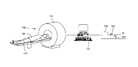
figure 1: shows a system for the automated production of
a bone replacement,
figure 2: shows gingiva with a cavity and a tooth,
figure 3: shows a first exemplary embodiment of a bone
replacement, and
figure 4: shows a second exemplary embodiment of a bone
replacement.
Detailed description of the exemplary embodiments
[0048] Figure 1 shows a system 100 for producing a bone
replacement 10.
[0049] The system 100 has a computed tomography (CT)
scanner 110. A couch 120, on which a patient 130 lies in
the present case, is arranged in front of said computed
tomography scanner. The couch 120 can be inserted into
the computed tomography scanner 110 such that the patient
130 can be examined by means of the computed tomography
scanner 110. In particular, this allows the measurement
of a cavity in a bone of the patient 130, for which a
bone replacement is intended to be produced. In particu-
lar, this can be used to measure a cavity for receiving
a tooth and also the tooth situated therein.
[0050] The system 100 further comprises a computer 140
which is connected to the computed tomography scanner 110
for the purposes of receiving data. Depending on its
measurement of the patient 130, the computed tomography
scanner 110 produces initial data which are supplied to
#176567 5
CA 02994783 2018-02-05
- 14 -
the computer 140. Said data may be both indicative for a
cavity in a bone of the patient 130 and indicative for a
bone to be imitated, for example a tooth for which a
prosthesis is intended to be produced. It should be noted
that the computed tomography scanner 110 may also be
controlled by the computer 140.
[0051] The computer 140 is configured to convert the
initial data into computer-aided design (CAD) data. These
CAD data may be displayed to a user such that the latter
can carry out a check which prevents subsequent pro-
cessing steps from using incorrect data.
[0052] A so-called 3D volume model, i.e. a virtual
model, of the bone replacement is produced for the CAD
processing. The 3D volume model is usually used in a CAD
program as a construction basis for the design of a new
3D model. However, the 3D model may be modified also in
the present form or by being complemented with further
3D models from data libraries.
[0053] Depending on the CAD data, the computer 140 sub-
sequently produces processing data which specify how a
bone replacement is intended to be produced. When pro-
ducing the processing data, parameters such as the type
of cavity and the position of the cavity in the body of
the patient 130 are also taken into account.
[0054] Subsequently, a finite element analysis is ap-
plied to the processing data. Here too, data in respect
of the position of the cavity in the body of the patient
130 and data in respect of a possible occlusion of the
cavity and a surrounding bone structure are taken into
account.
[0055] The produced processing data are subsequently op-
timized by means of specific algorithms in order to de-
sign the subsequent automated processing and use of the
11765615
CA 02994783 2018-02-05
- 15 -
processing data to be as efficient and reliable as pos-
sible.
[0056] Furthermore, the system 100 has a machine tool
150. The processing data that are produced and prepared
in the computer 140 are supplied to this machine tool
150. The machine tool 150 has a processing tool 155 which,
in a manner known per se, is received in the machine tool
150. In particular, this may be a drill or any other
material-ablating device.
[0057] The system 100 further has a toolholder 160 ad-
jacent to the machine tool 150. Received in the tool-
holder 160 is a blank of a bone replacement 10 in order
to anchor the latter for the processing by means of the
tool 155. The machine tool 150 is embodied to produce the
bone replacement 10 in a fully automated manner on the
basis of the processing data while said bone replacement
is held by the toolholder 160.
[0058] After the production of the bone replacement 10,
the latter may be separately inserted into the computed
tomography scanner 110 in order to be checked. To this
end, use can be made of, for example, a special holder.
Here, once again, appropriate data are produced depending
on the measured bone replacement 10, said data being
transmitted to the computer 140. The latter compares the
actual state to the intended state and decides whether
- the bone replacement 10 can be used without change,
- the bone replacement 10 requires post-processing,
or
- the bone replacement 10 was produced so badly that
it cannot be used and must be disposed of.
[0059] In the case where post-processing is necessary,
the computer 140 is able to produce appropriate pro-
cessing data for the machine tool 150, said data allowing
automated post-processing of the bone replacement 10. The
11765675
== CA 02994783 2018-02-05
- 16 -
bone replacement 10 can then be inserted anew into the
toolholder 160 for post-processing purposes.
[0060] Figure 2 shows a portion of gingiva 20 with a
cavity 25 formed therein. It is understood that the
structure of the gingiva 20 is set by a jawbone which is
covered by the gingiva 20. Thus, the cavity 25 is also
received in the jawbone. A tooth 30 or tooth replacement
30 is received in the cavity 25. It should be noted that
this can be, in particular, natural gingiva 20 and a
natural tooth 30. By way of example, the apparatus 100
shown in figure 1 can be used to measure the cavity 25
and/or the tooth or tooth replacement 30 and hence pro-
duce a replacement for the tooth or tooth replacement 30,
said replacement being provided in the form of a bone
replacement 10 and fitting exactly into the cavity 25.
[0061] Figure 3 shows an exemplary embodiment of a bone
replacement 10 in the form of an artificial tooth. Here,
the tooth is subdivided into a tooth implant 12, a dental
prosthesis 16 and a prosthesis crown/bridge absorption
component 14 that connects the tooth implant 12 and the
dental prosthesis 16. These three constituent parts 12,
14, 16 of the tooth 10 can all be produced separately in
an automated manner by means of the apparatus 100. As
already mentioned further above, it is possible to meas-
ure a cavity 25, as a result of which, in particular, the
structure of the tooth implant 12 is set. It is likewise
possible to measure the structure of a tooth 30, in par-
ticular the surface structure thereof, in the computed
tomography scanner 110 in order to set the structure of
the dental prosthesis 16. The prosthesis crown/bridge
absorption component 14 can be produced in an automated
manner, or else manually, at the computer 140.
[0062] Figure 4 shows a bone replacement 10 which is
embodied as an integral tooth or tooth replacement. The
separation into individual constituent parts, explained
t1765675
= CA 02994783 2018-02-05
- 17 -
with reference to figure 3, is consequently not effectu-
ated. Instead, the tooth or the tooth replacement can be
produced in one operation from a blank or material by
means of the machine tool 150.
[0063] In order to improve the adhesion in the jawbone
and/or gingiva and in order to improve the growing to-
gether with the jawbone and/or gingiva, a coating 18 is
applied to the jawbone and/or gingiva contact region of
the tooth. This is a porous coating into or onto which
the jawbone and/or the gingiva can grow. Such a coating
18 can be applied, in particular, after/during the pro-
cessing by means of the system 100, for example within
the scope of a chemical process.
[0064] It should be mentioned that, in parallel with the
processing data, the computer 140 can also, in particu-
lar, produce data in respect of medical instruments to
be used and in respect of navigation data. This simpli-
fies the provision of the instruments necessary for an
intervention for a treating medical practitioner and also
facilitates, for the latter, the use of advanced naviga-
tion and assistance devices such as e.g. spectacles or a
head-up display with the option of superimposing appro-
priate information. Such embodiments may also be referred
to as augmented reality.
#1765675