Note: Descriptions are shown in the official language in which they were submitted.
WO 2017/023531
PCT/US2016/042903
RADIATION THERAPY WITH
ORTHOVOLTAGE X-RAY MINI BEAMS
FIELD OF THE DISCLOSURE
[0002] The present disclosure relates to methods and systems for
performing radiation
therapy using orthovoltage x-rays for treating tumors, including brain tumors,
and for treating
neurological disorders such as epilepsy.
BACKGROUND
[0003] Radiation therapy, which is one of three main methods of treating
cancer,
together with surgery and chemotherapy, is currently carried out predominantly
with high
energy x-rays of one to several MeV energy produced by special x-ray
generators employing
electron linear accelerators ("linacs-) of several MV high voltage. MeV x-rays
have good
attributes for use in radiation therapy, in particular, high tissue
penetration and a robust
sparing of the first few millimeters of shallow tissues, generally known as a
"skin-sparing
effect." They also have several shortcomings, most significantly, the normal,
non-targeted
tissue that is located proximal, distal, and lateral to the target receive
excessive radiation
damage as described further herein.
10004] 'I his is because the mode of interaction of the high energy x-
rays that are
produced, typically 1-4 MeV, is Compton scattering and not photoelectric. As a
result, the
1
CA 2994816 2018-08-17
CA 02994816 2018-02-05
WO 2017/023531 PCT/US2016/042903
dose distribution produced in a patient's body is mostly from multiple Compton
scattering
from a wide range of angles and, therefore, is not well-confined within the
target.
[0005] In particular, the doses produced at the target tissue by MV sources
do not
sharply fall at the target's edge. Instead, the dose distribution at the
target's edge is rather
blunt- edged. Quantitatively, the so-called "80%-to-20% dose falloff' produced
at the target
by high energy x-rays is typically 2-5 mm. In addition, the beam-shaping
collimators, so-
called "multi-leaf collimators," required to produce the high-energy beam
profiles, consist of
heavy, thick "leaves" which do not lend themselves to production of fine
exposure profiles.
Because these collimators fail to produce beam-exposure profiles with fine
contours,
unnecessary radiation dose is delivered to normal tissues, especially when
small targets are
exposed. Such large falloffs result in unnecessary and undesirable dose being
delivered to
the tissues located in the immediate neighborhood of the target.
[0006] Further, because high energy x-rays have little preferential
absorption in heavier
elements compared to the light elements that constitute most of the tissues,
the concept of
tumor-dose enhancement by the introduction of contrast agents to the tumor
such as iodine
and gold cannot be effectively implemented when the radiation type is high
energy x-rays. In
addition, although the large penetration of the dose from high-energy x-rays
to tissue depths
is considered an advantage for thick targets, for thin tumors the shallow dose
falloff of the
high energy x-rays with depth is a negative effect, allowing the exposure to
high radiation
dose of all tissues positioned distal to the target. FIG. 1 illustrates dose
penetration 10 in
tissues for different high-energy MeV x-ray beams 12, compared to the dose
penetration
curve for an orthovoltage tube 14.
[0007] Before MV x-ray machines were developed (around the mid-20th
century), x-ray
generators of lower energy, called "orthovoltage" x-ray machines or tubes were
used for
radiation therapy. The acceleration voltage of these early x-ray machines was
rather small,
mostly up to 250 kVp, producing x-rays with a median energy, or mean energy,
of about 110
key. These beam energies were too low to penetrate deep in the tissue, and
also lacked the
beam sparing effect of the shallow tissues that the high-energy MV x-rays
exhibit, in fact
lower than that shown in FIG. 1 for orthovoltage x-rays. As a result, the skin
and the normal
tissues proximal to the target received significant radiation damage. FIG. 1
compares the
dose penetration in tissues from high energy x-rays produced by electron
linacs to that from a
2
CA 02994816 2018-02-05
WO 2017/023531 PCT/US2016/042903
300 kVp orthovoltage tube filtered moderately, labeled by half-value layer
(HVL) in copper
as "3.0 mm Cu HVL."
[0008] To address the damage to healthy skin tissue using orthovoltage x-
rays, a so-
called "grid therapy" was developed. Conventional grid therapy used a metal or
lead grid
with openings of at least 1.0-1.5 cm diameter to ameliorate the skin damage
that occurred in
treating deep tumors. However, the orthovoltage grid therapy techniques
offered little, if any,
tissue-sparing to healthy subcutaneous tissue proximal to the target, and thus
did not solve the
problem of damage to the normal tissues proximal to deep tumors. Furthermore,
no method
or system was contemplated for controlling the tissue depth at which a
therapeutic dose could
be produced across a target by the merging of the beams exiting the grid.
[0009] Accordingly, there is a need for a method and system for performing
radiotherapy using orthovoltage x-rays for effectively treating tumors while
sparing both the
skin and tissue proximal to the target. There is also a need for a system and
method for
controlling the tissue depth at which a therapeutic dose of orthovoltage x-ray
radiation can be
delivered to the target while sparing tissue proximal to the target. The
development of such
improved orthovoltage x-ray systems may provide not only benefit to a wide
range of clinical
applications by reducing dose to the non-targeted tissues, but also a low-cost
and compact
solution for performing radiotherapy to effectively treat tumors, as well as
neurological
targets.
SUMMARY
[0010] Features of the disclosure will become apparent from the following
detailed
description considered in conjunction with the accompanying drawings. It is to
be
understood, however, that the drawings are designed as an illustration only
and not as a
definition of the limits of this disclosure.
[0011] The present disclosure relates to a system and method for
effectively treating
tumors and neurological targets using orthovoltage x-ray radiation while
sparing both the skin
and irradiated tissue that is proximal to the target. The present disclosure
also relates to a
system and method for controlling the tissue depth at which a therapeutic dose
of
orthovoltage x-ray radiation can be delivered to the target while sparing at
least a substantial
portion of tissue proximal to the target. Such improved orthovoltage x-ray
systems may
3
CA 02994816 2018-02-05
WO 2017/023531 PCT/US2016/042903
provide a low-cost and compact solution for performing radiotherapy to
effectively treat
tumors, as well as neurological targets.
[0012] The present disclosure also relates to a method for delivering
therapeutic
radiation to a target within a subject, wherein the target is located at a
predetermined depth
from an irradiated portion of a surface of the skin of the subject. The method
includes
positioning a multi-aperture collimator on or near the surface of the skin
within a trajectory of
radiation, which is produced by an x-ray source generating orthovoltage x-
rays, and which is
directed at the target. The multi-aperture collimator is positioned and
configured to generate
an array of minibeams on the surface of the skin comprising slightly diverging
spatially
distinct minibeams. Adjacent minibeams formed on the skin have a predetermined
center-
center spacing, and, preferably, a width of between about 0.1 mm and about 0.6
mm. The
method further includes irradiating the surface of the skin with the array of
minibeams, and
delivering an effective beam of therapeutic radiation to the target by
controlling a tissue depth
at which adjacent orthovoltagc x-ray minibcams merge sufficiently to form the
effective
beam of therapeutic radiation.
[0013] In one aspect, the method further includes controlling the tissue
depth at which
the adjacent orthovoltage x-ray minibeams merge sufficiently to form the
effective beam such
that the effective beam is formed proximal to the target.
[0014] The orthovoltage x-ray source may be a focal spot on an anode of an
x-ray tube.
[0015] In aspects, controlling the tissue depth at which the adjacent
minibeams merge
sufficiently to form the effective beam includes adjusting at least one of the
predetermined
center-to-center spacing, the width, and a distance between the x-ray source
and the multi-
aperture collimator.
[0016] In various additional aspects, controlling the tissue depth at which
the adjacent
minibeams merge sufficiently to form the effective beam includes adjusting a
size of the x-
ray source from which the orthovoltage x-rays are generated.
[0017] The tissue depth can be varied, in aspects, from about 1 cm to about
10 cm, based
on a predetermined depth of the target from the surface of the skin.
4
CA 02994816 2018-02-05
WO 2017/023531 PCT/US2016/042903
[0018] Controlling the tissue depth may include, in aspects, selecting the
width, the
predetermined center-to-center spacing, and the distance between the focal
spot and the
collimator such that each of the minibeams broaden to less than 1.0 mm in
width before they
merge to form the effective beam of therapeutic radiation, which may be a
solid, or
substantially solid, beam of therapeutic radiation.
[0019] In aspects, the multi-aperture collimator is a multi-slit collimator
configured with
elongated slits such that the array of minibeams is an array of narrow and
elongated planar
minibeams. In some aspects, the width, which corresponds to a thickness of
each planar
minibeam, may be limited to a range of between about 0.25 mm to about 0.35 mm.
[0020] The x-ray source, which may be a focal spot formed on the anode of
an x-ray
tube, may have an elongated shape in embodiments, and aspects of the method
may further
include aligning the elongated slits of the multi-slit collimator with the
elongated shape of the
focal spot.
[0021] In yet another aspect, delivering the beam of therapeutic radiation
further
includes sparing irradiated tissue proximal to the target from radiation
damage, such that the
tissue depth also corresponds to a tissue sparing depth.
[0022] In still other aspects, the method further includes changing an
angular position of
the x-ray tube and the trajectory of orthovoltage x-rays generated therefrom
relative to the
target such that the target is irradiated from a different direction, and a
different portion of the
skin is irradiated. The positioning, irradiating and delivering steps are
repeated for the
different direction. The multi-aperture collimator is repositioned for
irradiating the different
portion of the surface of the skin while remaining aligned with the trajectory
of orthovoltage
x-rays for the different direction. The irradiating step is repeated to
irradiate the different
portion of the skin with the array of minibeams generated by the multi-
aperture collimator,
and the delivering step is repeated to deliver the effective beam of
therapeutic radiation to the
target from the different direction.
[0023] For each angular position, the method, in aspects, also includes
adjusting a beam-
shaping collimator and an intensity of the beam to conform the effective beam
to a shape of
the target based on the direction of the trajectory relative to the target.
CA 02994816 2018-02-05
WO 2017/023531 PCT/US2016/042903
[0024] In still another aspect of the method, the radiating step includes
generating an arc
of radiation around the target from each of the minibeams in the array. The
delivering step
includes merging adjacent arcs of radiation at the tissue depth to form the
effective beam of
therapeutic radiation.
[0025] In aspects, the minibeams for forming the arcs of radiation may be
planar
minibeams, formed from elongated slits of a multi-slit collimator, having a
length that is
greater than the width, or thickness, of each minibeam.
[0026] The arcs of radiation can be generated by rotating the x-ray source
together with
the multi-aperture, e.g., a multi-slit collimator, such that the arcs are
generated around the
target in planes parallel to, for example, the elongated slits of a multi-slit
collimator.
[0027] In aspects, while generating the arcs of radiation, the method
further includes
continuously adjusting a shape and an intensity of the beam to conform the
effective beam of
therapeutic radiation to a shape of the target based on a direction from which
the beam
irradiates the target.
[0028] The distance between the multi-aperture collimator and the x-ray
source is also
preferably continuously controlled and adjusted, based on the direction, to
maintain the tissue
depth at which the arcs formed from the minibeams merge to form the beam of
therapeutic
radiation to be proximal to the target.
[0029] Various aspects of the method may further include administering dose-
enhancing
agents to the subject prior to the irradiating step to radio-sensitize the
target. The agents may
be in various forms, including nanoparticles, and may include one or more of
iodine,
gadolinium, gold, and platinum. In aspects, the agents may be encapsulated in
one of
liposomes or polymeric delivery vehicles.
[0030] The present disclosure is also directed to a system for delivering
therapeutic
radiation to a target volume within a subject, wherein the target is located
at a predetermined
depth measured from an irradiated portion of the skin of the subject. The
system includes an
x-ray source generating orthovoltage x-rays and a multi-aperture collimator.
The multi-
aperture collimator is configured for positioning on the skin within a
trajectory of the
orthovoltage x-rays directed at the target. The multi-aperture collimator
includes an array of
apertures having a width of between about 0.1 mm and about 0.6 nun and a
predetermined
6
CA 02994816 2018-02-05
WO 2017/023531 PCT/U52016/042903
center-center spacing to generate an array of slightly diverging spatially
distinct minibeams
of the orthovoltage x-rays at the skin. The width and the predetermined center-
center spacing
of the multi-aperture collimator, a size of the x-ray source, and a distance
between the x-ray
source and the collimator are configured to deliver an effective beam of
therapeutic radiation
to the target, wherein the beam is formed by sufficient merging of the
minibeams proximal to
the target.
[0031] In aspects, the x-ray source is a focal spot on an anode of an
orthovoltage x-ray
tube from which orthovoltage x-rays are generated.
[0032] In one aspect, the effective beam of therapeutic radiation is a
solid, or
substantially solid, beam of therapeutic radiation. The width, the
predetermined center-center
spacing, the size of the x-ray source and the distance are configured to form
the solid beam
proximal to the target.
[0033] In another aspect, the multi-aperture collimator is removably
interchangeable.
The system further includes a set of multi-aperture collimators configured
with predefined
aperture widths and shapes and predefined center-center spacings.
[0034] In additional aspects, the system may be portable and configured to
be
transported on and operated from a mobile platform.
[0035] In aspects, the system further includes a beam-shaping collimator,
positioned in
the trajectory of the x-rays and proximal to the multi-aperture collimator,
the beam-shaping
collimator further configured to be adjustable to conform the effective beam
of therapeutic
radiation to a shape and size of the target.
[0036] The system may further include, in various aspects, a rotatable and
translatable
gantry on which the orthovoltage x-ray source, the beam-shaping collimator and
the multi-
aperture collimator are mounted, the gantry being positioned and configured to
be rotatable
around a horizontal platform on which a subject being treated is located. The
gantry is
configured to position the target in the trajectory of the orthovoltage x-
rays, to tilt around a
vertical axis to the platform to change a direction from which the target is
irradiated with the
effective beam of therapeutic radiation, and to rotate around a longitudinal
axis of the
horizontal platform to generate arcs of radiation from each of the minibeams.
7
CA 02994816 2018-02-05
WO 2017/023531 PCT/US2016/042903
10037] hi additional aspects, the system is further configured to
continuously adjust the
beam-shaping collimator to conform the effective beam to the shape and size of
the target
based on the direction of irradiation as the gantry is tilted and rotated, and
to continuously
adjust the distance between the x-ray source and the multi-aperture collimator
to maintain the
tissue depth at which the minibeams merge to be proximal to the target.
[0038] In various additional aspects of the system and method of the
present disclosure,
the width of the minibeams may be between about 0.25 mm and about 0.35 mm.
[0039] In other aspects of the system and method of the present disclosure,
the
minibeams may be pencil beams. In yet another aspect, the array may be a two-
dimensional
array of pencil beams.
[0040] The pencil beams of the present disclosure, in aspects, may have a
cross-sectional
profile that is round, elliptical, square, rectangular, or of polygonal shape.
[0041] In various other aspects of the system and method of the present
disclosure, the
multi-aperture collimator may be a multi-slit collimator configured with
elongated slits such
that the array of minibeams is an array of narrow and elongated planar
minibeams.
[0042] The collimator may include a multi-aperture or multi-slit heavy-
metal plate.
[0043] In various aspects, the width of the apertures, or slits, in the
multi-aperture
collimator is between about 0.25 mm and 0.35 mm.
[0044] The orthovoltage x-ray tube in various aspects may operate in a
range between
about 100 kVp and about 500 kVp.
[0045] The present disclosure is also directed to a method for delivering
therapeutic
radiation to a target within a subject, wherein the target is located at a
predetermined depth,
and the predetermined depth is measured from an irradiated portion of a
surface of the skin of
the subject. The method includes positioning a multi-aperture collimator
within a trajectory
of orthovoltage x-rays generated by an orthovoltage x-ray source. The
trajectory of
orthovoltage x-rays is directed at the target. 1he multi-aperture collimator
is positioned and
configured to generate an array of minibeams on the surface of the skin
comprising slightly
diverging spatially distinct minibeams having a predetermined width and a
predetermined
center-center spacing between adjacent minibeams.
8
CA 02994816 2018-02-05
WO 2017/023531
PCT/11S2016/042903
[0046] The method also includes irradiating the surface of the skin with
arcs of radiation
formed from the array of minibeams, wherein the arcs of radiation are
generated around the
target from the minibeams in the array, and delivering an effective beam of
therapeutic
radiation to the target. The beam is delivered by controlling a tissue depth
from the irradiated
surface of the skin at which adjacent arcs of radiation formed from adjacent
minibeams in the
array merge sufficiently to form the effective beam of therapeutic radiation.
[0047] In aspects, the method further includes limiting the width of the
minibeams to be
between about 0.1 mm and about 0.6 mm.
[0048] In addition aspects, the minibeams are planar minibeams formed from
elongated
slits of a multi-slit collimator. The arcs of radiation are generated from the
minibeams by
rotating the x-ray source together with the multi-slit collimator, such that
the arcs are
generated around the target in planes parallel to the elongated slits of the
multi-slit collimator.
100491 The method may further include, in aspects, adjusting a shape and an
intensity of
the effective beam of therapeutic radiation to conform to a shape of the
target based on a
direction from which the beam irradiates the target.
[0050] In further aspects, the method also includes continuously adjusting
the distance
between the orthovoltage x-ray source and the multi-aperture collimator to
maintain the tissue
depth at which the minibeams forming the arcs of radiation merge to be
proximal to the
target.
[0051] The system and methods of the present disclosure may be applied, in
aspects, to
delivering a beam, which may, in additional aspects, be a solid beam, of
therapeutic radiation
to a target that encompasses one of a tumor and an epileptogenic foci.
[0052] In addition to the above aspects of the present disclosure,
additional aspects,
objects, features and advantages will be apparent from the embodiments
presented in the
following description and in connection with the accompanying drawings.
BRIEF DESCRIPTION OF THE DRAWINGS
[0053] The drawings constitute a part of this disclosure and include
examples, which
may be implemented in various forms. It is to be understood that in some
instances, various
aspects of the disclosure may be shown exaggerated or enlarged to facilitate
understanding.
9
CA 02994816 2018-02-05
WO 2017/023531 PCT/US2016/042903
The teaching of the disclosure can be readily understood by considering the
following
detailed description in conjunction with the accompanying drawings.
[0054] FIG. 1 is a graphic representation of dose penetration in water for
different
radiation sources.
[0055] FIG. 2A is a pictorial representation of an embodiment of a system
for practicing
a method of the present disclosure.
[0056] FIG. 2B is a pictorial representation of an embodiment of an
orthovoltage x-ray
device of the present disclosure.
[0057] FIG. 2C is a pictorial representation of a portion of an embodiment
of a system
for forming a minibeam array of orthovoltage x-rays of the present disclosure.
[0058] FIG. 2ll is a pictorial representation of a plate multi-aperture
collimator of the
present disclosure.
[0059] FIG. 3A is a pictorial representation of an embodiment of a multi-
aperture
collimator of the present disclosure.
[0060] FIG. 3B is a pictorial representation of another embodiment of a
multi-aperture
collimator of the present disclosure for forming planar minibeams, which is
referred to as a
multi-slit collimator.
[0061] FIG. 4A is a block diagram representation of an embodiment of a
method of the
present embodiment.
[0062] FIG. 4B is a block diagram representation of additional embodiments
of a
method of the present embodiment.
[0063] FIGS. 5A to 5D are graphical representations of dose profiles, taken
perpendicular to an orthovoltage x-ray minibeam array formed in accordance
with the present
disclosure, at incrementally increased depths.
[0064] FIGS. 6A to 6C are pictorial representations of the implementation
of the system
of FIG. 2A to different target depths in accordance with an embodiment of a
method of the
present disclosure.
CA 02994816 2018-02-05
WO 2017/023531 PCT/U82016/042903
[0065] FIG. 7A is a pictorial representation of the implementation of the
system of FIG.
2A in accordance with another embodiment of a method of the present
disclosure.
[0066] FIG. 7B represents a geometry for forming arcs of radiation from an
array of
orthovoltage x-ray minibeams in accordance with an embodiment of a method of
the present
disclosure.
[0067] FIG. 7C is a pictorial representation of a gantry for positioning
the system around
a platform on which a subject is positioned for treatment.
[0068] FIGS. 8A to 8C are pictorial representations of the implementation
of the system
of FIG. 2A to different target depths in accordance with yet another
embodiment of a method
of the present disclosure.
[0069] FIG. 9 is a graphical representation of the advantage of filtering
an energy
spectrum of an orthovoltage x-ray beam of the present disclosure to increase
its median beam
energy.
[0070] FIG. 10 is a graphical representation of the dose penetration
achieved using
orthovoltage x-ray minibeams formed in accordance with an embodiment of the
system and
method of the present disclosure.
[0071] FIG. 11 is a graphical representation of the biologically effective
dose as a
function of tissue depth before and after the merging of orthovoltage x-ray
minibeams to
form an effective beam of therapeutic radiation in accordance with the present
disclosure.
[00721 The various aspects of the present disclosure mentioned above are
described in
further detail with reference to the aforementioned figures and the following
detailed
description of exemplary embodiments.
DETAILED DESCRIPTION
[0073] The following sections describe exemplary embodiments of the present
disclosure. It should be apparent to those skilled in the art that the
described embodiments of
the present disclosure provided herein are illustrative only and not limiting,
having been
presented by way of example only. All features disclosed in this description
may be replaced
by alternative features serving the same or similar purpose, unless expressly
stated otherwise.
11
CA 02994816 2018-02-05
WO 2017/023531 PCT/US2016/042903
Therefore, numerous other embodiments of the modifications thereof are
contemplated as
falling within the scope of the present disclosure as defined herein and
equivalents thereto.
[0074] The present disclosure is directed to a system and method for using
slightly
diverging orthovoltage x-ray minibeams (referred to herein as "OX.M"), which
are formed by
a multi-aperture collimator positioned on the surface of a subject's skin, to
form an effective
beam of therapeutic radiation at a predetermined tissue depth for treating a
targeted tumor or
other abnormality, while sparing the skin and a substantial portion of the
tissue proximal to
the target from radiation damage. The effective beam, which may be a
substantially solid, or
unsegmented beam, is formed by the merging of the x-ray minibeams. The method
utilizes
the slight divergence of the minibeams emerging from the multiple apertures,
which is due
primarily to the relatively large, finite, x-ray source spot size compared to
the relatively small
source-to-collimator distance. The depth at which the effective beam of
therapeutic radiation
is formed is adjusted by proper selection of source size, aperture size (which
determines the
size of each minibeam at the skin), and source-to-collimator distance.
[0075] An effective beam of therapeutic radiation refers to a beam having a
dose profile
(perpendicular to the x-ray beams) at a particular tissue depth across which
the dose level
required to have a therapeutic effect is maintained. The minibeams of the
present disclosure
merge sufficiently to form the effective beam of therapeutic radiation. If
there are any
discernible "valleys" in the profile as a result of forming the effective beam
by merging of the
minibeams, the valley dose in the effective beam of therapeutic radiation must
still be high
enough to correspond to a therapeutic radiation dose. An effective beam of
therapeutic
radiation having no measurable peak-valley "pattern," or having only a small
modulation or
peak-valley dose ratio (PVDR) of 1.10 (10% modulation) or less, is referred to
herein as a
"solid" beam of therapeutic radiation.
[0076] The term "collimator" is sometimes used interchangeably herein with
"multi-
aperture collimator" to refer to the multi-aperture collimator (which may be a
multi-slit
collimator) used to form the orthovoltage minibeams at the surface of the skin
of a subject.
The multi-aperture collimator should not be confused with a beam-shaping
collimator, also
known in the art as a "multi-leaf collimator," which may also be used to shape
the
orthovoltage x-ray beam of the present disclosure to conform to the overall
shape of the
target. The multi-leaf collimator is preferably positioned to shape the
orthovoltage x-ray
beam before the beam is segmented into minibeams by the multi-aperture
collimator.
12
CA 02994816 2018-02-05
WO 2017/023531 PCT/US2016/042903
[0077] The term "target" used herein refers to the tissue that is targeted
to receive a
therapeutic dose of radiation. The target encompasses the tumor or other
targeted
abnormality, for example, an epileptic lesion or epileptogenic foci, and may
also include an
immediate margin of tissue surrounding the target tumor or abnormality. One of
skill in the
art will understand how to .elect the amount of surrounding tissue included in
the target to
insure that all tumor cells, for example, that may have spread to the
immediate tissue
surrounding of the tumor are exposed. For other abnormalities, the margin
included in the
volume defined by the target may be extremely small, and may be based
primarily on the
system's accuracy in targeting the volume of interest.
[00781 "Tissue depth" is generally used to indicate a subcutaneous depth.
[0079] "Proximal" is used herein to indicate a location downstream of the x-
ray source
and multi-aperture collimator, but upstream of the target, i.e., located on
the side of the target
closest to the x-ray source.
[0080] "Distal" is used herein to indicate a location downstream of the
target, i.e.,
located on the side of the target away from the x-ray source.
[0081] The orthovoltage x-ray minibeams emerging from the multi-aperture
collimator
of the present disclosure are slightly diverging, largely due to the penumbra
effect. This
results from the relatively large focal spot size (e.g., 3 to 5 mm) of the
orthovoltage x-ray
source compared to the relatively small source-to-collimator distance (20 to
45 cm). The
expected amount of divergence may be estimated through calculations, and is
based upon the
x-ray source size (for example, the focal spot size formed on the anode of an
orthovoltage x-
ray tube) and the distance between the x-ray source (focal spot) and multi-
aperture
collimator.
[0082] Using the geometric estimates of the divergence of the minibeams,
other
parameters of the system, as described further herein, can be varied to
deliver an effective
beam of therapeutic radiation to the target by sufficient merging of adjacent
minibeams. In
preferred embodiments, parameters are optimized such that the minibeams merge
sufficiently
to form a solid, or substantially solid, effective beam of therapeutic
radiation proximal to the
edge of the target.
13
CA 02994816 2018-02-05
WO 2017/023531 PCT/US2016/042903
[0083] As one of skill in the art will appreciate, while the tissue depth
at which the
minibeams will merge sufficiently to provide an effective beam, which may, in
embodiments,
be a solid beam, of therapeutic radiation can be calculated, phantom targets
such as water, or
chromographic film, are also preferably used to calibrate and tweak the system
before
administering any treatment.
[0084] Referring to FIG. 2A through 2C, an embodiment of a system 50 for
implementing the methods of the present disclosure for delivering an effective
beam of
therapeutic orthovoltage x-ray radiation to a target 76 includes an
orthovoltage x-ray tube 60
and a multi-aperture collimator 70 for placing in close proximity to, or in
preferred
embodiments, on the surface of, the skin 72 of a patient. In embodiments, a
multi-leaf
collimator 77 is also positioned between the x-ray tube 60 and the multi-
aperture collimator
70 for shaping the beam emerging from the tube 60 to conform to the overall
shape of the
target. Referring to FIG. 2A, the multi-aperture collimator 70, which is
aligned within the
trajectory of the x-ray beam, may be touching the patient's skin, and slightly
pushing against
the skin 72.
[0085] For targets in the chest and the abdomen that move extensively with
the
breathing motion, pushing the multi-aperture collimator hard against the skin
completely
immobilizes the skin and advantageously creates pressure that helps immobilize
the tissue to
limit possible beam smearing with the tissue movement, particularly for tissue
within the
critical first centimeter and possibly further from the skin. The smearing of
the dose pattern
of minibeams at deeper tissue depths will not be as critical, since the
minibeams will be
broadening and typically beginning to merge within a few centimeters of tissue
depth.
[0086] In embodiments, any blurring of the minibeam array dose pattern
because of the
breathing movement of the patient's body can be minimized by aligning the
direction of the
incident minibeams so that the beams are perpendicular to the surface of the
body being
treated, or parallel to the lines of displacement of the body tissues being
treated, within up to
15 .
[0087] Referring still to FIG. 2A, the target 76 encompasses a tumor 75 or
other targeted
abnormality, for example, an epileptic lesion or epileptogenic foci, and also
includes an
immediate margin of tissue surrounding the target tumor or abnormality. In
tumor therapy,
typically a 5-mm margin is set around the tumor. One of skill in the art will
appreciate that
14
CA 02994816 2018-02-05
WO 2017/023531 PCT/US2016/042903
the margin is selected to cover for the uncertainties involved in radiation
therapy to insure
that the entire tumor is treated at the full dose. Such uncertainties come
from, inter alia,
tumor imaging, tumor positioning in the beam, dosimetric calculation, and the
diffuse edge of
the tumor.
[0088] Referring to FIG. 2B, the orthovoltage x-ray tube 60 may be
constructed by any
suitable means in the art. In embodiments, the tube 60 includes a cathode 62,
which expels
and focuses electrons onto the surface 64 of an anode 66 formed of an
appropriate target
material, such as tungsten. The x-ray source 68 for generating the
orthovoltage x-rays in this
embodiment is a focal "spot" 68 (which can be also be in the form of a line
depending on the
construction of the tube 60) formed on the anode surface 64. Referring to FIG.
2C, as well as
FIG. 2A, the x-ray source, or focal spot 68, is characterized by an x-ray
source (focal spot)
size 74 defining an area that emits orthovoltage x-rays.
[0089] In embodiments, the system 50 also includes beam hardening filters
65
appropriately positioned in the path of the x-ray beam generated by the anode.
[0090] In embodiments, the orthovoltage tubes of the present disclosure are
between
about 100kVp and 500kVp. In particular embodiments of the system and method of
the
present disclosure, the x-rays are produced by orthovoltage tubes of higher
kVp, for example,
between about 250 kVp to about 500 kVp, and preferably, between about 300 kVp
and about
500 kVp.
[0091] In additional embodiments, the x-ray tubes of the present disclosure
may have up
to 30 mA current, and preferably at least 25 mA current.
[0092] The beam hardening filters in embodiments are copper filters of one
to several
millimeters of thickness, selected to preferably eliminate most of the low-
energy end of the
spectrum. As a result, hard and penetrating beams such as with cm or larger
tissue HVL
are produced. Such beam energies are adequate to treat many types of tumors
located at
different depths, including those of the breast, the head-and-neck, the brain,
and certain
tumors of the chest and abdomen.
[0093] Referring still to FIG. 2A and FIG. 2B, the system 50 may also
include a
translation apparatus 78 for changing a source to collimator distance 80
between the location
of the focal spot 68 and the multi-aperture collimator 70. While the
translation apparatus 78
CA 02994816 2018-02-05
WO 2017/023531 PCT/US2016/042903
as shown can translate the x-ray tube 60 closer to, or further from the
patient, in other
embodiments, the translation apparatus may instead be associated with the
device or gantry
on which the patient is positioned. Additional degrees of freedom are also
preferably
provided on the gantry (not shown), and/or on the system, for correctly
positioning the patient
so that the target 76 is accurately positioned within the trajectory of
radiation produced by the
x-ray tube.
[0094] The x-ray tube, multi-aperture collimator, and patient are
positioned such that the
target 76 is within the trajectory of the orthovoltage x-rays emitted from the
focal spot 68.
Spatially distinct, and slightly diverging x-ray minibeams 90 are formed on
the surface of the
skin as a result of the orthovoltage x-rays impinging on the multi-aperture
collimator 70.
[0095] Referring to FIG. 2D, to accommodate the thickness of the multi-
aperture
(including multi-slit) collimators of the disclosure, and the divergence of
the minibeams, in
embodiments, the collimator 70' as shown in FIG. 2D may be flared, such that
an output
width 81 is sufficiently larger than the input width 79 of each aperture or
slit to avoid any
interference of the minibeam with the walls of the collimator.
[0096] In embodiments, the multi-aperture collimator 70 may be a multi-
aperture plate
92 with round apertures, such as that shown in FIG. 3A, which may be flared
like the
collimator 70' shown in FIG. 2D, and which segments the x-ray beam into a
minibeam array
90 of nearly parallel, slightly diverging pencil-like beams. In other
embodiments, the
collimator 70 is a multi-slit collimator 94, such as that shown in FIG. 3B,
which may also be
flared like the collimator 70' shown in FIG. 2D, and which segments the x-ray
beam into a
minibeam array 90 of slightly diverging planar beams. The minibeams are of sub-
millimeter
width, e.g., diameter 96 (pencil beams) or thickness 98 (planar beams), and
are separated by a
center-to-center spacing 97.
[0097] The multi-aperture collimators of the present disclosure may be
heavy-metal
collimators, comprised of a material such as tungsten. In embodiments, the
heavy-metal
collimators have a thickness of between about 5 to about 20 mm. Such
relatively thin multi-
aperture collimators for use with orthovoltage x-rays in accordance with the
present
disclosure can be made, for example, of a plurality of thin tungsten-alloy
blades with spacers
between them, held by a rigid frame. In other embodiments, the tungsten multi-
aperture
collimator can be made of wire cuts in a tungsten alloy plate.
16
CA 02994816 2018-02-05
WO 2017/023531 PCT/US2016/042903
100981 In embodiments, an array of pencil beams may be configured to
conform to the
shape of the source spot size on the anode, even without a beam-shaping, or
multi-leaf,
collimator. In embodiments, the dose distribution produced by the array of
pencil minibeams
penetrating the subject as a function of depth in tissue will have a nearly
cylindrical
uniformity.
[0099] In embodiments, pencil beams may have a cross-section that is round,
like those
formed by the multi-aperture collimator of FIG. 3A. In other embodiments, the
pencil beams
may be formed by multi-aperture collimators having elliptical, square,
rectangular, or
polygonal apertures, or configured in any other useful shape for forming the
arrays.
[00100] It is noted that while planar beams may provide a less uniform dose
distribution,
they can provide a larger yield of beam throughput, particularly for oval or
elongate-shaped
focal spots.
[00101] In embodiments, for planar, e.g., narrow and elongated, minibeams,
the shape of
the focal spot 68 is oval or elongated. This allows conformity between the
shape of the
incident beams and the pattern of planar minibeams to be produced. In further
embodiments,
the collimator has a pattern of planar slits, such as those in FIG. 3B, and
they are aligned with
the direction of the elongated length of the spot size. This combination will
produce both a
high throughput of the beam through the multi-aperture collimator and a
uniformity of dose
distribution produced by the minibeams as a function of the depth in the
tissue.
[00102] In the system and method of orthovoltage x-ray radiation therapy of
the present
disclosure, each of the minibeams in the spatially distinct array of minibeams
produced by
the multi-aperture collimator 70 at the skin preferably has the same width and
center-to-
center spacing. The width (e.g., diameter or thickness) of each of the
minibeams preferably
has a value chosen between about 0.1 mm and about 0.6 mm, preferably, about
0.3 mm.
[00103] The minibeams are spaced regularly and closely together by a center-
to-center
distance, which may be chosen, for example, from a value ranging between about
0.1 and
about 1.0 mm inclusive, depending on the minibeam width, depth of the target
and other
factors described further herein.
17
CA 02994816 2018-02-05
WO 2017/023531 PCT/U52016/042903
[00104] In embodiments, the center-to-center distance between adjacent
minibeams may
be a value ranging between about 0.5 mm and about 1.6 mm, depending on the
minibeam
width, depth of the target and other factors described further herein.
[00105] This submillimeter size of the segmented minibeams within the non-
target tissue
(proximal to the target) results in a very large tissue-sparing effect that,
while recognized for
synchrotron x-ray therapy using parallel (non-diverging) minibeam arrays, as
described, for
example, in US 7,158,607 to Dilmanian, et al., is not known in the prior art
of orthovoltage x-
ray systems for radiation therapy.
[00106] As described herein, the orthovoltage x-ray tubes of the present
disclosure
operate at voltages of up to 500 kVp, preferably between about 300 kVp and
about 500 kVp.
This higher voltage advantageously allows the x-ray beams to be significantly
filtered, with
up to several mm of copper, e.g., to attenuate the low-energy end of the
spectrum. This in
turn increases the median energy, i.e., hardens the beam, resulting in a
significant increase in
the depth of dose penetration to the tissue of up to 8 cm or more tissue IIVL.
[00107] Due to these characteristics of the x-ray tube of the present
disclosure, in
combination with the multi-aperture collimator construction and geometry, the
minibeams in
the arrays generated in accordance with the present disclosure can stay very
narrow for many
centimeters inside the subject. By further adjusting the geometry of the beam
administration,
the tissue depth at which the minibeams merge is very well-controlled in
accordance with the
present disclosure to allow administration of an effective beam, which may be,
in
embodiments, a solid beam, of therapeutic radiation to the target, while
avoiding damage to
both the skin and the tissue proximal to the target.
1001081 Additional features and embodiments of the system of the present
disclosure are
described and understood in the details of the methods further described
herein. Furthermore,
it is understood that any details of embodiments of the disclosure described
as elements of the
system may also be embodied in methods of the present disclosure.
[00109] Referring to FIG. 4A, an embodiment of a method 100 of the present
disclosure
for delivering therapeutic radiation to a target within a subject, while
sparing the skin and,
preferably, substantial portions of tissue proximal to the target from
radiation damage,
includes positioning, at 102, a multi-aperture collimator, such as a heavy-
metal collimator
plate, on the surface of the skin within a trajectory of radiation produced by
an orthovoltage
18
CA 02994816 2018-02-05
WO 2017/023531 PCT/US2016/042903
x-ray tube. The target is located at a known, predetermined depth as measured
from the
irradiated portion of a surface of the skin of the subject. The multi-aperture
collimator is
configured to generate an array of slightly diverging, spatially distinct,
minibeams. In
embodiments, the minibeams have a predetermined center-center spacing, which
may be
from about 0.1 mm to about 1.0 min inclusive, and a width of between about 0.1
mm and
about 0.6 mm inclusive. At 104, the skin is irradiated with the array of
orthovoltage x-ray
minibeams emerging from the multi-aperture collimator. The method 100 further
includes, at
106, delivering an effective beam, which may be, in embodiments, a solid beam,
of
therapeutic radiation to the target. The effective beam is delivered by, at
108, controlling a
tissue depth from the irradiated surface of the skin at which adjacent
orthovoltage x-ray
minibeams in the array sufficiently merge to form the effective beam, and
while sparing, at
116, at least a substantial portion of tissue proximal to the target. The
sparing of tissue may
be further enhanced by limiting the width of the minibeams in the proximal
tissue, at 118,
before they merge to form the effective beam of therapeutic radiation, to 1.0
mm or less. In
other embodiments, the width of the minibeams before they merge to form the
effective beam
of therapeutic radiation is limited to 0.7 mm or less.
[00110] In embodiments, a depth of the tissue sparing in the body can be
varied anywhere
from about 1 cm, when the front edge of the tumor is close to the surface, to
40 cm in
embodiments in which the tumor is deeper.
[00111] Referring to FIG. 4B and FIG. 2B, for example, as described further
herein, in
embodiments of the system and method of the present disclosure, the tissue
depth at which
the adjacent minibeams merge sufficiently to form the effective beam is
controlled by
adjusting one or more parameters of the system. For example, any one or
combination of
adjustments listed in FIG. 4B may be used to control the tissue depth at which
the minibeams
merge to form the effective beam. For example, at least one of the
predetermined spacing 97
and the width 96, 98, between adjacent minibeams may be adjusted, at 110,
and/or a distance
80 between the focal spot 68 and the multi-aperture collimator 70 may be
adjusted, at 112. In
embodiments, controlling the tissue depth at which the adjacent minibeams
merge to form the
effective beam may further, or alternatively, include, at 114, adjusting a
size of the focal spot
68 formed on the anode.
[00112] Referring again to FIG. 2A, the small divergence angle of each of
the minibeams
in the array 90 results from the penumbra effect of a relatively large source
spot size 74,
19
-
CA 02994816 2018-02-05
WO 2017/023531
PCT/U52016/042903
which, in embodiments, may be between about 3-5 mm, positioned a relatively
short distance
away from the multi-aperture collimator 70. In embodiments, the distance
between the focal
spot 68 and collimator 70 may be between about 5 and about 20 cm.
1001131 In embodiments, magnitudes of the divergence angle of the
individual minibeams
of the present disclosure are about +1-10 milliradians, i.e., the minibeams
form cones having a
full divergence angle of about 20 milliradians.
[00114] In reference to FIG. 2A and FIGS. 5A-5D, the minibeams
gradually merge as
they travel further away from the skin towards the target, i.e., to deeper
tissue depths.
Referring to the simplified pictorial representation of the diverging beams
shown in FIG. 2A,
a geometric calculation can be made to estimate the parameters for obtaining a
solid beam,
for example, at a known tissue depth, by merging of the minibeams. The
calculations are
based on the divergence angle of the minibeams as further described herein.
The actual dose
profiles resulting from the increasing overlapping of the minibeams as they
penetrate the
tissue are best shown, however, in actual stepwise cross-section profiles
measured as a
function of depth, as shown in FIGS. 5A to 5D.
[00115] The dose profiles of FIGS. 5A to 5D were produced using a
320-kVp
orthovoltage x-ray generator with a source size of about 4 mm, a source-to-
collimator
distance of about 260 mm and with a multi-slit collimator configured to
produce 0.3-mm
minibeams with 0.7-mm beam spacing on-center.
[00116] FIG. 5A represents the dose profile of the minibeams
exiting the multi-slit
collimator (tissue depth of 0 mm). FIGS. 5B through 5D were measured at
distances of 13,
27, and 40 mm, respectively, from the multi-slit collimator. As described
further below, the
minibeams gradually lose their tissue sparing effect at increasing depths, or
increasing
distances from the multi-slit collimator, as the gaps between them decrease,
while the
therapeutic efficacy across the merging minibeams' dose profile increases as
the dose
between the minibeams (the "valley dose") increases. The minibeams completely
merge at
about 40 mm, as shown in FIG. 5D.
[00117] As shown in FIG. 5A, the shape of the individual
minibeams just emerging from
the multi-slit collimator are already somewhat belt-shaped because of the
source and
collimator geometries involved. As these individual minibeams broaden with
depth, the tails
of the dose profiles start partially overlapping with their neighbors,
producing a segmented
CA 02994816 2018-02-05
WO 2017/023531 PCT/US2016/042903
pattern of "peaks and valleys" of neighboring minibeams. The continuation of
beam
broadening gradually increases this partial overlap, resulting in an increase
in the height of
the valley and an increasingly more uniform-looking pattern. The peak-to-
valley dose ratio
(PVDR) decreases at increasing depths as the adjacent minibeams merge. The
PVDR of
adjacent minibeams for each of the depths 0 mm, 13 mm, 27 mm, and 40 mm in
FIGS. 5A-
5D is about 5.9, 3.1, 1.3 and 1.03, respectively. At the depth of 40 mm, as
shown in FIG. 5D,
the PVDR of adjacent minibeams is essentially unity (1.03) and the profile
resembles that of
a unitary, conventionally-formed, beam. The beam profile no longer appears
segmented or
modulated, but instead represents a solid beam profile. Therefore, for this
example, a solid
effective beam of therapeutic radiation can be formed at least by the time it
reaches a depth of
40 mm. In embodiments of the present disclosure, a solid effective beam of
therapeutic
radiation is formed from the merging minibeams proximal to the edge of the
target, such as a
tumor.
[00118] The following calculates the depth in the tissue at which the
minibeams would be
expected to merge to form a solid, unsegmented beam in a hypothetical
simplified geometry
used to clarify the basic concept of beam broadening due to the opening angle
of each
minibeam, based on the parameters used to generate the plots shown in FIGS. 5A
to 5D.
Neglecting the 0.3-mm width of the collimator slit, the opening angle of the
minibeams
coming out of the multi-slit collimator will be about 0.0154 radian (4 / 260),
that is 15.4
milliradian. Furthermore, neglecting the 4-mm source size and the minibeam
broadening
effects produced by the un-sharp edges of the source and collimator, the
minibeams broaden
to a sharp 0.70 mm at a distance of about 45.4 mm from the multi-slit
collimator (0.0154
radian x 45.4 mm = 0.70 mm). This means that the sharp edges of the adjacent
minibeams
touch each other at about 45.4 mm from the multi-slit collimator to produce a
solid beam.
[00119] The inaccuracies introduced in these calculations by neglecting the
finite size of
the collimator slit slightly affect a) the opening angle of the minibeams, and
b) for a given
opening angle, the actual broadening of the beam. These two factors can be
corrected for by
convoluting the calculations both angularly and laterally with the widths of
the collimator's
opening. The corrections not only will slightly add to the actual width of the
minibeams at
any given distance from the multi-aperture collimator but also un-sharpen the
edges of the
minibeams as they pass through the tissues. Furthermore, the inaccuracies
introduced by
neglecting the actual rounded shape of the source spot size, rounded edges of
the collimator,
21
CA 02994816 2018-02-05
WO 2017/023531 PCT/US2016/042903
and by scattering of the x-rays in the subject, will also lead to slightly
wider beams and
slightly more roundedness of their edges.
[00120] All these effects give the incident minibeams their "hell-shaped"
feature with
extended "tails," as seen in FIGS. 5A-5D, instead of a sharp "rectangular"
shape. As a result,
the beam-merging event results as a gradual overlapping of the dose profiles
(perpendicular
to the x-ray beams) of the adjacent minibeams with each other as shown in
FIGS. 5A-5D.
This also means that the merging of the minibeams is a gradual process in
which the "valley"
doses gradually rise and the "peak" doses gradually decline as neighboring
peaks and valleys
eventually reach substantially the same height (ignoring non-uniformities due
to other
causes), thus eventually producing a uniform, unsegmented, beam with a PVDR
that is
substantially equal to unity across the beam profile.
[00121] In embodiments of the method and system of the present disclosure,
an effective
beam of therapeutic radiation for delivery to the target is formed by merging
the minibeams
sufficiently to form a dose pattern (perpendicular to the x-ray beams) wherein
any residual
"valleys" are still high enough to correspond to a therapeutic radiation dose.
Accordingly,
the minimum (valley) dose in the dose pattern (perpendicular to the x-ray
beams) of the beam
due to the merging of the minibeams will be equal to or greater than the
minimum effective
therapeutic dose, so that an effective therapeutic dose of radiation is
delivered across the
entire dose pattern formed by the merging minibeams and across the
corresponding target
area.
[00122] In embodiments, the effective beam of therapeutic radiation formed
by merging
of the minibeams at a particular tissue depth has a substantially unsegmented
dose pattern
that has no detectable modulation or that is characterized by a PVDR (of
neighboring
minibeams) that is close to unity and can thus be referred to as a solid beam
of therapeutic
radiation. It is understood that while the PVDR may be unity, or approximately
unity, the
overall beam profile across the target will generally not be uniform due to
the shape of the
source beam, as shown, for example, in FIG. 5D, which shows a slight gradual
increase in the
profile from left to right.
[00123] In other embodiments, depending on the geometry and sharpness of
the edges of
the multi-aperture collimator, the effective beam formed from the merging
minibeams may
have an inherent heterogeneity of dose deposited in the tumor in the areas
where adjacent
22
CA 02994816 2018-02-05
WO 2017/023531 PCT/US2016/042903
minibeams merge, which have an additive affect (not related to the PVDR) and
can provide
streaks of amplified dose that generate a concomitant boost within the tumor.
[00124] In embodiments, the PVDR in the dose pattern of the effective beam
of
therapeutic radiation delivered to the target is no greater than 1.5.
1001251 In embodiments, the PVDR of the effective beam of therapeutic
radiation
delivered to the target is no greater than about 1.3.
[00126] In additional embodiments, the PVDR of the effective beam of
therapeutic
radiation delivered to the target is no greater than about 1.2.
[00127] In embodiments, the effective beam of therapeutic radiation
delivered to the
target is essentially a solid beam, having less than 1.10 PVDR, or having no
detectable PVDR
or modulation corresponding to the array of minibeams that merged to form the
solid beam.
[00128] Referring again to FIG. 2A, FIGS. 5A-5D provide examples of the
actual beam
dose profiles as the minibeams merge. In FIG. 5A, the dose profile 124 was
generated at a
simulated tissue depth 115 just after the multi-aperture collimator. FIG. 5B
shows the dose
profile 126 at a depth 120 of 13 mm, at which the tails of the minibeams have
begun to
merge. At a further depth 121 of 27 mm, shown in FIG. 5C, a lower PVDR 135 of
about 1.3
to 1 is evident as the valley dose rises and the resultant beam profile begins
to lose its
segmented appearance. At a tissue depth 122 of 40 mm, the beam profile 130 is
no longer
modulated or appears segmented (PVDR approaches unity or is undetectable - in
this case,
PVDR is estimated to be about 1.03) at least across most of the beam profile
such that the
adjacent minibeams have merged to form a solid beam of therapeutic radiation.
[00129] Referring to FIGS. 6A-6C and FIG. 48, the system and method of the
present
disclosure include controlling the tissue depths at which the minibeams merge
such that an
effective beam, which may be a solid beam, of therapeutic radiation is
delivered to the target,
while sparing the skin and as much of the proximal tissue as possible. Any one
or more of a
number of parameters may be varied to achieve this desired result, such as:
adjusting the
spacing and/or width of the apertures in the collimator; adjusting the focal
spot to collimator
distance; adjusting a focal spot size of the anode. FIGS. 6A-6C illustrate the
result of varying
just the source-to-collimator distance for a particular anode and multi-
aperture collimator.
The source to collimator distance 140 in FIG. 6B allows an effective beam of
therapeutic
23
CA 02994816 2018-02-05
WO 2017/023531 PCT/US2016/042903
radiation to be delivered to a tumor 143 at a predetermined tissue depth 142.
Referring to
FIG. 6A, increasing the source-to-collimator distance 144, allows a deeper
tumor 145 to be
treated with an effective beam of therapeutic radiation at a deeper
predetermined tissue depth
146. Similarly, referring to FIG. 6C, decreasing the source-to-collimator
distance 148, allows
a shallower tumor 147 to be treated with an effective beam of therapeutic
radiation at a
deeper predetermined tissue depth 146. As demonstrated, the shorter source-to-
collimator
distance increases the divergence of the individual minibeams, thus making
them merge with
each other at a shorter tissue depth.
[00130] Referring to FIG. 7A, in embodiments of the method of the present
disclosure,
treatment planning for a patient includes repeating the method described above
and in FIGS.
4A and 4B for additional exposure directions or trajectories after the first
exposure from
trajectory 151. It will be appreciated that the radiation formalism may be
adjusted for each of
the trajectories 151, 152, 154 from a focal spot 156 of an orthovoltage x-ray
tube, to make
effective beams of therapeutic radiation, which may be solid beams, at the
proximal side of
the target 160, by adjusting either the center-to-center spacing of apertures
in the multi-
aperture collimator 162 for the subsequent exposures 152, 154, or the distance
164 between
the source 156 and the collimator 162, or by adjusting the spot size from the
focal spot 156.
[00131] FIG. 7A shows minibeam administrations from three shallow angles
aimed at a
tumor and its margin. As shown, the merging point of the minibeams at which an
effective
beam is formed does not necessarily have to occur immediately before or at the
edge of the
target as long as it does not produce much proximal tissue burden. As
discussed herein
supra, an effective beam of therapeutic dose may be produced slightly before
the
geometrically calculated merging point for forming a solid beam due to, inter
alia, radiation
leakage between the minibeams.
[00132] One will appreciate that the target will generally not be formed
into any
symmetrical volume. The outer shape of the target onto which the x-ray
radiation is
projected, as well as the thickness profile of the target to be treated, will
change based on the
direction from which it is irradiated. A beam-shaping collimator, such as a
multi-leaf
collimator, is preferably positioned between the x-ray source and the multi-
aperture
collimator and adjusted to conform to the shape of the target as projected on
a plane
perpendicular to the trajectory of x-rays. In embodiments, the multi-aperture
collimator can
be continuously adjusted, preferably being automatically and dynamically
controlled using
24
CA 02994816 2018-02-05
W020171023531 PCT/US2016/042903
computer processors and controllers, as the direction of irradiation of the
target changes in
accordance with any of the methods of the present disclosure.
[00133] It is also noted that adjacent arrays can collide without producing
"star dose
pattern," i.e., a region of mixed-angle minibeams, if the collision occurs
after the merging
points of the arrays.
1001341 One of skill in the art will appreciate that the multiple exposures
from different
directions may be administered during a single treatment session, or in
different treatment
sessions.
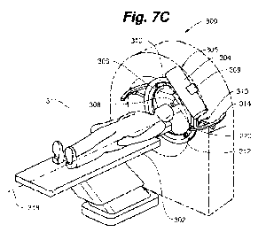
1001351 Referring to FIGS 7A-7C, embodiments of the system of the present
disclosure
may include a gantry 300, as known in the prior art, to align the trajectory
of radiation onto
the target. The subject being treated is positioned on a platform 302, or bed,
which may have
at least translational alignment capabilities for positioning the subject in
the aperture of the
gantry 300 and aligning the target within the trajectory. In embodiments of
the system of the
present disclosure, the x-ray source 304, beam-shaping collimator 306, and
multi-aperture
collimator 308 are preferably mounted together as a unit 305 (the unit 305
also having
positioning elements as described in reference to FIGS. 2A-2C for controlling
the tissue
depth at which the minibeams merge). The multi-aperture collimator and beam-
shaping
collimator remain aligned on unit 305 within the trajectory of orthovoltage x-
rays emitted
from the x-ray source 304, as the unit 305 is rotated and/or translated
relative to the target.
Rotational and translational arms or mounting platforms are provided on the
gantry, on which
the unit 305 is operatively positioned, to allow the trajectory of the x-rays
to be positioned on
the target and to allow the direction from which the target is irradiated to
be changed in a
step-wise, as well as in a continuous fashion, to perform the methods
described herein.
[00136] The gantry 300 may, for example, include a tilt axis 310 and a
rotatable ring 312
on which the unit 305 is mounted. The unit 305 may be mounted to a radial
translation stage
309 provided on the rotatable ring 312 for positioning the unit 305 radially
toward or away
from the center of the ring 312 so that the multi-aperture collimator 308 can
be positioned on
or near the subject's skin during treatment. Referring to FIG. 7A and FIG. 7C,
the rotatable
ring 312 may be tilted, for example, from its nominal vertical 311 or
perpendicular plane
relative to the horizontal platform 302 forward or back around the axis 310,
and translated via
a translational stage 314 as needed (alternatively, the platform 302 may be
translated) to
CA 02994816 2018-02-05
WO 2017/023531 PCT/US2016/042903
maintain the target within the trajectory of the beam, for any angular
position, such as for
trajectories 151, 152, 154 of FIG. 7A.
[00137] In embodiments, the multi-aperture collimator 162 of FIG. 7A is a
multi-slit
collimator. The multi-slit collimator 162 forms elongated planar minibeams,
like those
shown in FIG. 3B. In FIG. 3B, a cross-section of an array of minibeams
perpendicular to the
trajectory of x-rays (the trajectory extending into the plane of the paper of
FIG. 3B, along a y-
axis) is shown, where the elongated length of the minibeams, for example,
minibeams 220,
221, 222, and 223 extends along the z-axis of the coordinate system of the
array. For
reference, the coordinate system for the same array of planar minibeams is
shown in FIG. 7A
for the trajectory 154. In FIG. 7A, the elongated length of the planar
minibeams 220, 221,
222, and 223, and the slits or blades of the multislit collimator that
generates them, extend
perpendicular to the plane of the paper in FIG. 7A, along the z-axis.
1001381 Referring to FIGS. 7A-7B, for each of the three exposures of FIG.
7A, the array
of planar minibeams can also be moved around the target on a continuous arc
scan, the
direction of the arc scan being shown in FIG. 7B, within planes perpendicular
to the x-y plane
of the cross-section of the array of slits shown in FIG. 7A (parallel to the
slits of the
collimator). Referring to FIG. 7C, the arcs of radiation may be formed by
rotation of the x-
ray source 304 with multi-aperture collimator 308 and preferably also beam-
shaping
collimator 306 aligned thereto (e.g., unit 305) along a direction 316 of the
rotatable ring 312
to allow arc-scan of a brain tumor, for example, from different angles. The
target is
positioned at the center of the arcs.
[00139] The arcs merge to form an effective beam of therapeutic radiation
at the same
depth as would a single minibeam in the array, so that the effective beam,
which may be a
solid beam, of therapeutic radiation to the target is formed from merging of
the adjacent arcs
of radiation at the desired tissue depth. In additional embodiments, the arc
is generated in a
continuous step. Referring also to FIG. 7A and 7B, for example, the arcs 230
can be
generated at each of a plurality of positions 232, 234, 236 corresponding to
trajectories and
directions 151, 152, 154 for generating the minibeam arrays. Treatment can be
implemented
in one or more continuous arc motions of the source in planes parallel to a
multi-slit
collimator's blades, for example.
26
CA 02994816 2018-02-05
WO 2017/023531 PCT/US2016/042903
1001401 For reference, the orientation of the cross-section of one of the
elongated planar
minibeams 220 upon exiting the multi-slit collimator 308 is also shown in FIG.
7C. The
direction of the arc scan 230 keeps the arcs of radiation formed from the
minibeams
individually separated as they exit the multi-slit collimator 308, and allows
the arcs of
radiation formed from the minibeams to merge at the desired tissue depth. The
tissue depth
at which the arcs merge is preferably adjusted dynamically during the arc scan
to be proximal
to the target at all times. This can be accomplished, for example, by
continuously and
automatically adjusting the source to multi-slit collimator distance to
produce the optimal
beam-merging tissue-depths.
[00141] Intensity modulation (referred to in the prior art as Intensity
Modulated Radiation
Therapy of IMRT) can also be performed during the arc scanning by continuously
and
dynamically adjusting the beam-shaping collimator and thus modulating the beam
intensity
during the arc scanning to conform the irradiation pattern to a shape of the
target based on the
direction of the x-rays forming the arcs of radiation relative to the target.
The continuous
rotating and translating of the moving parts of the gantry,
adjusting/positioning of the leaves
of the beam-shaping collimator, and adjusting of the distance between the x-
ray source and
the multi-collimator during the arc-scanning can be accomplished using
automated circuitry,
processors, and controllers according to methods known in the art.
[00142] In embodiments, the multi-aperture collimator of the present
disclosure, which is
preferably a heavy-metal plate, is easily interchangeable. An embodiment of a
system of the
present disclosure may include pre-made multi-aperture collimators, each
having different
center-to-center spacing and a predetermined aperture size of, for example,
about 0.3 mm. In
embodiments, the set of collimators may include one or both of the pencil-beam
type and
planar-beam (multi-slit) type, of different widths and/or different center-to-
center beam
spacings. The appropriate multi-aperture collimator can then be used to change
the depth at
which the minibeams merge to produce an effective beam of therapeutic
radiation as needed
for the particular depth of the target, as shown in FIGS. 8A-8C, for example.
A larger center-
to-center spacing 170 between apertures in the collimator 172 of FIG. 8A is
required to
produce an effective beam, which may be a solid beam, of effective radiation
at a deep target
depth 174, than the spacing 176 of multi-aperture collimator 178 in FIG. 8B
for the smaller
target depth 180. Similarly, a shallower target depth 182 can be achieved
using a multi-
aperture collimator 184 in FIG. 8C with even closer center-to-center spacing
186.
27
CA 02994816 2018-02-05
WO 2017/023531 PCT/US2016/042903
[001431 Many advantages are realized using the system and method of the
present
disclosure over conventional MV x-ray radiation therapies. As shown in FIG. 9,
for example,
the beam energy spectrum 200 of a 320-kVp x-ray beam of the present disclosure
can be
heavily filtered to increase its median beam energy to about 220 keV mostly
through the
elimination of its low energy part. The resulting filtered beam 202 has a Cu
half value layer
(HVL) of 3.8 mm, which corresponds to a tissue EIVL of about 10 cm. Comparing
this 220
keV median beam energy with x-rays produced by MV electron linacs having an
average
median beam energy of about 1.5 MeV, the orthovoltage energy is about seven
times smaller.
This energy difference squarely puts the interaction with tissues of the
orthovoltage beam in
the photoelectric range, while that of the x-rays from MV linacs is in the
Compton scattering
range, with all their differential attributes as described above. The positive
attributes of the
320-kVp x-rays of the present disclosure also include small dose fall-off at
the target's edge,
simplifying treatment planning.
[00144] FIG. 10 provides an example of the good dose penetration that is
achieved with
the present methods, allowing for targets essentially at any depth to be
effectively treated
with an effective beam, which may be a solid beam, of therapeutic radiation.
FIG. 10 was
constructed to show a relationship between orthovoltage x-rays' Cu half-value
layer and a
penetration depth in water for 50% dose penetration. The depth-dose curve 208
is plotted for
a 300-kVp machine producing a spectrum with 2.45 mm Cu half-value layer. The
curve's
50% dose occurs at a depth of 5 cm. Comparing this data with the spectrum of
the
orthovoltage x-rays of the present disclosure, as shown for example in FIG. 9,
a relationship
is detected between the beams' Cu half-value layer (HVL) and its 50% depth
dose in water,
indicating 5 cm water depth for 50% dose when using a spectrum with 2.45 mm Cu
HVL,
and 8 cm water depth for 50% dose with a spectrum of 3 mm Cu HVL. Accordingly,
for 3.8
tnn) Cu HVL, the 50% dose penetration in water will occur at a depth of about
10 cm, which
is considered to be a good dose penetration.
[00145] Another advantage of the present method is its application to deep
tumors, for
example, over 5 cm deep. Whereas conventional techniques for treatment of deep-
seated
tumors often depend on the target being positioned on the downward slope of
the incident
beam's depth dose curve, with the normal tissues positioned proximal to the
target receiving
greater dose than the target, using the system and methods of the present
disclosure, the
spacing between the minibeams can be adjusted so that the minibeams merge
sufficiently
28
CA 02994816 2018-02-05
WO 2017/023531 PCT/US2016/042903
with each other to produce an effective beam, which may be a solid beam, of
therapeutic
radiation near the proximal side of the target. This will spare a substantial
portion of the
normal tissue proximal to the target. FIG. 11 includes a plot drawn under the
assumption that
the minibeams begin to merge at about 5 cm depth. The lower curve 210 plots
the
biologically effective dose, which gradually increases in a transient region
between about 3.5
cm to 5 cm in approaching the minibeams merging point at about 5 cm, when the
tissue-
sparing effect is gradually diminished. Therefore the radiobiologically
significant part of the
curve 210 starts as one approaches 5-cm depth. This effect of sparing of the
proximal tissues
also ameliorates to a larger extent the problem of low tissue-depth
penetration of the
orthovoltage x rays.
[00146] FIG. 11 also illustrates the superior confinement of the dose of an
effective solid
beam produced by merging the orthovoltage x-ray minibeams of the present
disclosure. The
physical dose 214 produced by orthovoltage x-rays using a 300 kVp source with
3.8-mm Cu
half-value layer (-8 cm water HVL) is plotted and compared to the biological
dose 210,
which is the effective tissue dose produced by an array of 0.3-mm minibeams
spaced 0.7 mm
on-center before they merge into a solid beam at ¨5.2 cm tissue depth. Also
for comparison,
the physical dose 212 for 10 MV x-rays, which is the same as the biological
dose for
conventional MV x-ray, is plotted in FIG. 11. The physical 214 and biological
orthovoltage
dose 210 of the present disclosure and the dose 212 produced by the MV x-rays
are super-
imposed over the background 215 of a 4 cm target located 5 cm from the body's
surface. As
shown in FIG. 11, the biological dose 210 is much better confined to the
tumor, both in the
proximal and the distal sides, than the dose 212 produced by the MV x-rays.
[00147] Further, compared to the conventional radiation therapy methods
using MV x-
rays or also gamma rays, the present method produces smaller dose to the non-
targeted
tissues located distal to the target because of its lower beam energy and also
produces smaller
dose to tissues located lateral to the tumor or target than most conventional
radiation
techniques because of the smaller lateral penumbra of the orthovoltage x-ray
beams.
[00148] To further enhance proximal tissue sparing, in embodiments, the
width and
predetermined center-to-center spacing of the minibeam array, and the distance
between the
focal spot and the multi-aperture collimator, are chosen such that each of the
slightly
diverging spatially distinct minibeams broaden to no more than 1.0 mm, or in
other
embodiments to no more than 0.7 mm, in width before they merge to form the
solid beam.
29
CA 02994816 2018-02-05
WO 2017/023531 PCT/US2016/042903
[00149] The tissue-sparing effect of arrays of sub-millimeter parallel,
thin planes of high-
energy MeV synchrotron x-rays radiation, particularly when limited to 0.7 mm
or less, was
established in the early 1990s at the National Synchrotron Light Source
(NSLS), Brookhaven
National Laboratory (BNL). However, both the use and the tissue-sparing
characteristics of
sub-millimeter beams of orthovoltage x-rays in radiation therapy is heretofore
unknown in
the prior art. The enhanced tissue-sparing effect of minibeams of the present
disclosure,
when limited to 0.7 mm or less, is caused by two mechanisms, namely the "dose-
volume
effect" and the "prompt biological repair effect." The first effect, meaning
that the smaller
the target, the larger is its dose tolerance, has been known for many decades
and its effect is
not limited to millimeter or sub-millimeter beams. It has been the basis for
such effects as
grid therapy and stereotactic radiosurgery. The second effect, however, is
indeed specific to
beams with sub-millimeter dimensions and has been studied for high-energy
synchrotron x-
rays in recent years mostly in the context of animal studies, in the context
of the repair of
capillary blood vessels from sub-millimeter beam exposures. The effect has
been shown for
MV x-ray to be strongest for beams smaller or narrower than 0.7 mm. The
inventors have
advantageously discovered that a combined dose-volume and prompt biological
repair effect
is also realized for orthovoltage x-ray beams smaller or narrower than 0.7 mm
of the present
disclosure.
[00150] The methods of the present disclosure have particular advantages in
certain
clinical applications, such as for treating radioresistant tumors located near
viable
radiosensitive organs. In particular, radioresistant tumors of the head and
neck are often not
very large and are not located at large tissue depths, but they are often
located near
radiosensitive organs such as the parotid glands. For these applications, two
attributes of the
method are particularly helpful. First, the orthovoltage x-ray minibeams have
a very sharp
dose falloff, which significantly reduces the dose to the adjacent normal
tissues. Second, due
to the tissue-sparing of the method, tissues proximal to the target operate to
spare such organs
as the salivary glands, particularly the parotid gland. In fact, the minibeams
can pass through
the parotid gland on their way to the tumor located distal to them without
damaging the
gland, if the gland, which is positioned adjacent to the skin, is adequately
thin to be in the
space where the incident minibeams are sufficiently small (less than 0.7 mm)
and are
completely separated from (have not merged with) their neighbors.
CA 02994816 2018-02-05
WO 2017/023531 PCT/US2016/042903
1001511 The present methods can also be combined with radiation dose
enhancement
methods. For example, the lower beam energy of the orthovoltage x-rays makes
the present
method better suited and more effective than conventional MV methods for
combining with
tumor dose enhancement. This is because the orthovoltage x-ray photoelectric
cross sections
in the heavy elements used in contrast agents are significantly larger than
the cross-section of
MeV x-rays in those same elements. In embodiments, the methods described
herein include
the administration of dose-enhancing substances to the patient to radio-
sensitize a tumor or
other target. The agents may be of any suitable form, including nanoparticles,
and may be
comprised of one or more of iodine, gadolinium, gold, and platinum, with or
without the use
of active targeting methods, and drugs without or without encapsulation in
liposomes or
polymeric delivery vehicles. The tumor dose enhancement factor can be very
large because
of the large photoelectric cross section in those elements of x-rays in the
orthovoltage energy
range, i.e., 150 to 400 keV x-rays. In comparison, the photoelectric cross
section of MeV is
not very large at all.
[00152] Another advantage of the method and system of the present
disclosure is that
they are usable for low cost, durable and portable radiation therapy,
requiring minimal
training by locally trained healthcare staff. The system operation may be
based on simple
calibration, simple error diagnosis, and open source look-up tables. Further,
the system may
be configured to be operable in locations with limited or no infrastructure,
simply with access
to electricity, and have smaller shielding requirements by virtue of its lower
beam energy.
Accordingly, the system may be configured as a portable, mobile treatment
system (possibly
on a small truck) that can be used in low- and middle-income countries (LMICs)
to treat
targets of the central nervous system as well as other tumors with acceptable
tissue depth, and
may be preferable to MV therapeutic systems in such countries at least due to:
a) the lower
cost, probably by as much as 5-fold; b) portability; c) ease of use for
treatment planning and
operation, and d) smaller shielding requirements from the surrounding areas in
the hospital of
the present orthovoltage minibeam system and method.
[00153] In addition, the methods of the present disclosure are well-suited
for treating thin
tumors for which the relatively steep dose attenuation in tissues can be
tailored to minimize
the dose to the normal tissues located behind the tumor. An example for such
clinical
applications is the treatment of thyroid tumors.
31
CA 02994816 2018-02-05
WO 2017/023531 PCT/US2016/042903
[00154] The present methods are particularly well-suited for the treatment
of brain tumors
and, particularly, pediatric brain tumors due to their large tissue-sparing
effects and low
accumulated dose compared to other methods. The brain structures to which
radiation
damage, using other techniques, produces more significant effects include the
hippocampus
and the cortex. Radiation damage to the pediatric cortex has been related to
the disturbance
of the pediatric cortex's gliogenesis, a process producing neural progenitor
cells; these cells
later differentiate to produce new oligodendrocytes. Temporal lobes are
another
radiosensitive structure whose radiosensitivity is also much higher in
children. Cognitive
deficits can also be produced in patients, particularly children, due to the
integral brain dose,
i.e., the accumulated dose given in the entire brain.
[00155] The physical characteristics of both MV x-rays and proton beams
impact the
amount of radiation and the integral brain dose on the hippocampus and cortex.
Although the
MV x- rays have a sparing effect in the skin and other shallow tissues that
could cover part of
the cortex (Fig. 1), their dose distribution in the body is characterized by
peaking early in the
tissues, large dose penetration, and large lateral penumbra, which can produce
significant
dose to the cortex on the opposite side of the brain and to the temporal
lobes. This dose
distribution also produces a large integral brain dose. As for proton therapy,
its lack of
shallow-tissue-sparing effect could translate to excessive dose to the cortex.
As a result,
despite proton therapy's much better dose confinement to the target than the
MV x-rays, it
still produces cognitive deficits in both adults and children.
[00156] Compared to conventional radiation therapy methods, e.g., using MV
x-rays,
gamma rays, or protons, the methods of the present disclosure are particularly
advantageous
for treatment of tumors of the brain, head and neck, brainstem, spinal cord,
spinal column and
the like for numerous reasons. For example, the orthovoltage minibeams of the
present
disclosure produce smaller dose to the non-targeted tissues located distal to
the target because
of their lower beam energy, and they produce smaller dose to tissues located
lateral to the
tumor or target due to their smaller lateral penumbra, providing a tighter
dose distribution.
This results in smaller accumulative dose to the brain, while still
maintaining tissue-sparing
to the skin and both proximal and distal tissue.
[00157] Due to the tight dose distribution produced at the target by the
orthovoltage x-
rays, particularly due to the sharp lateral dose falloff described supra, the
methods of the
present disclosure are also ideal for treating neurological targets such as
the epileptogenic
32
-
CA 02994816 2018-02-05
WO 2017/023531
PCT/US2016/042903
foci. In contrast, the conventional methods of Gamma-Knife and stereotactic
radiosurgery
with MV x-rays produced by electron linacs produce a much larger amount of
dose in the
non-targeted tissues. The results are commonly an unacceptable amount of edema
in the
brain and unacceptable late radiation damage to the non-targeted brain. On the
basis of the
dosimetric advantages of the present methods over those employing high energy
x-rays, the
present methods provide a much more effective treatment for epilepsy than the
conventional
radiosurgery methods. The estimated factor of two in dose saving to the non-
targeted tissues
that would result with the use of orthovoltage x-ray minibeam treatment in
accordance with
the present methods is significant in increasing the method's efficacy by
allowing the use of
higher target doses and in reducing the edema and brain damage produced in the
non-targeted
brain.
[00158] The present methods can be used, for example, to treat
focal epilepsy by
producing radiation damage, including tissue necrosis, to the epileptogenic
foci, with less
damage to the surrounding brain compared to the method practiced today with MV
x rays.
The method can also be used to treat general epilepsy by produced radiation
damage,
including tissue necrosis, in certain brain structures.
[00159] In embodiments, methods further include applying the
orthovoltage x-ray
minibeam treatment of the present disclosure to the treatment of epilepsy,
tumors including
brain tumors, e.g., pediatric brain tumors, and tumors of the head and neck,
brainstem, spinal
cord, and spinal column.
[00160] While the disclosure has been particularly shown and
described with reference to
specific embodiments, it should be apparent to those skilled in the art that
the foregoing is
illustrative only and not limiting, having been presented by way of example
only. Well-
known functions or constructions are not described in detail to avoid
obscuring the present
disclosure in unnecessary detail. Various changes in form and detail may be
made therein
without departing from the spirit and scope of the disclosure. Therefore,
numerous other
embodiments are contemplated as falling within the scope of the present
invention as defined
by the accompanying claims and equivalents thereto.
33