Note: Descriptions are shown in the official language in which they were submitted.
CA2995019
COMPRESSED SENSING HIGH RESOLUTION FUNCTIONAL MAGNETIC RESONANCE IMAGING
CROSS-REFERENCE TO RELATED APPLICATIONS
[0001] This application claims priority pursuant to 35 U.S.C. 119(e) to the
filing date of U.S.
Provisional Application No. 62/212,335, filed August 31, 2015.
INTRODUCTION
100021 Functional magnetic resonance imaging (fMRI) has been used in
neuroscience research and for
clinical applications. However, achieving high spatial resolution remains a
significant challenge
in fMRI because of a trade-off with decreased temporal resolution and/or lower
contrast-to-noise
ratio (CNR). High magnetic field systems, improvement of coil sensitivity,
advancements in
pulse sequences, utilization of parallel imaging, and reconstruction with
partial k-space have been
used to try to increase spatial resolution. However, demands for even higher
spatial resolution are
common.
SUMMARY
100031 The present disclosure provides methods and systems for high-resolution
functional magnetic
resonance imaging (fMRI), including real-time high-resolution fMRI methods and
systems.
100041 Aspects of the present disclosure include a method for functional
magnetic resonance imaging
(fMRI) of a subject. The method includes applying with an MRI system, a
balanced steady state
free precession (b-SSFP) sequence to a target area in a subject, acquiring
with the MRI system,
image data of the target area in the subject using a randomly undersampled
variable density spiral
(VDS) trajectory, and producing an image of the target area in the subject
based on the acquired
image data.
100051 In some embodiments, the producing comprises analyzing the image data
using a spatial
sparsifying transform. In some embodiments, the spatial sparsifying transform
comprises a
discrete cosine transform (DCT).
1000611 In some embodiments, the method is a real-time fMRI method. In some
embodiments, the
producing comprises analyzing the image data using a fast iterative shrinkage
thresholding
algorithm (FISTA).
100071 In some embodiments, the method has a sampling acceleration factor of 2
or more. In some
embodiments, the method has a sampling acceleration factor of 5 or more.
100081 In some embodiments, the method produces an image having a spatial
resolution of about
0.2x0.2x0.5 mm3 or greater.
1
Date Regue/Date Received 2023-02-15
CA2995019
[0009] In some embodiments, the method produces an image having a contrast-to-
noise ratio of 1.5 or
more. In some embodiments, the method produces an image having a contrast-to-
noise ratio of
2.5 or more.
[0010] Aspects of the present disclosure include a functional magnetic
resonance imaging (fMRI)
system. The system includes a coil configured to apply a balanced steady state
free precession
(b-SSFP) sequence to a target area in a subject, a receiver configured to
acquire image data of the
target area in the subject using a randomly undersampled variable density
spiral (VDS) trajectory,
and a processor configured to produce an image of the target area in the
subject based on the
acquired image data.
[0011] In some embodiments, the processor is configured to analyze the image
data using a spatial
sparsifying transform. In some embodiments, the spatial sparsifying transform
comprises a
discrete cosine transform (DCT).
[0012] In some embodiments, the system is configured for real-time fMRI. In
some embodiments, the
processor is configured to analyze the image data using a fast iterative
shrinkage thresholding
algorithm (FISTA).
[0013] In some embodiments, the system has a sampling acceleration factor of 2
or more. In some
embodiments, the system has a sampling acceleration factor of 5 or more.
[0014] In some embodiments, the processor produces an image having a spatial
resolution of about
0.2x0.2x0.5 mm3 or greater.
[0015] In some embodiments, the processor produces an image having a contrast-
to-noise ratio of 1.5 or
more. In some embodiments, the processor produces an image having a contrast-
to-noise ratio of
2.5 or more.
[0015A] Various embodiments of the claimed invention relate to a method for
functional magnetic
resonance imaging (fMRI) of a subject, the method comprising: applying with an
MRI system, a
balanced steady state free precession (b-SSFP) sequence to a target area in a
subject; acquiring
with the MRI system, image data of the target area in the subject using a
randomly undersampled
variable density spiral (VDS) trajectory, wherein the VDS trajectory includes
a stack of VDS
trajectories, wherein a change in a radial direction versus a change in a rate
in an angular
direction of the VDS trajectories is determined by a number of interleaves and
an effective field
of view, wherein the field of view follows an exponential function and an
angle between each
interleaf of the number of interleaves is varied; and producing an image of
the target area in the
subject based on the acquired image data.
2
Date Regue/Date Received 2023-02-15
CA2995019
[0015B] Various embodiments of the claimed invention relate to a functional
magnetic resonance
imaging (fMRI) system, the system comprising: a coil configured to apply a
balanced steady state
free precession (b-SSFP) sequence to a target area in a subject; a receiver
configured to acquire
image data of the target area in the subject using a randomly undersampled
variable density spiral
(VDS) trajectory, wherein the VDS trajectory includes a stack of VDS
trajectories, wherein a
change in a radial direction versus a change in a rate in an angular direction
of the VDS
trajectories is determined by a number of interleaves and an effective field
of view, wherein the
field of view follows an exponential function and an angle between each
interleaf of the number
of interleaves is varied; and a processor configured to produce an image of
the target area in the
subject based on the acquired image data.
BRIEF DESCRIPTION OF THE DRAWINGS
[0016] A randomized, variable-density, under-sampled spiral acquisition scheme
was designed for the
HSPARSE fMRI. (Fig. 1A) A low spatial resolution Nyquist trajectory only
covers a small range
of k-space. (Fig. 1B) To achieve higher spatial resolution without introducing
aliasing artifacts or
changing the field of view, the k-space coverage can be increased with more
interleaves. This
inevitably increases the data acquisition time and reduces the temporal
resolution. (Fig. 1C, 1D)
To overcome this problem, a variable density spiral (VDS) trajectory was
designed and
interleaves were randomly sampled while keeping the total number of
interleaves and scan time
the same as the low spatial resolution Nyquist scan. (Fig. 1E, 1F) For 3D
acquisition, the
2a
Date Regue/Date Received 2023-02-15
CA 02995019 2018-02-06
WO 2017/040538 PCT/US2016/049508
HSPARSE fIVIRI method randomly selects 320 interleaves from a stack of VDS
trajectory
consisting 32 kz locations and 30 interleaves. More interleaves were sampled
near the k-space
center and the total number of interleaves in each kz location follows a
Laplacian distribution.
The sampling pattern was also chosen to be random across temporal frames to
exploit the
temporal sparsity. However, the total number of interleaves for each time
frame was designed to
be constant (320 interleaves) to maintain constant temporal resolution over
time. Compared to a
3D Nyquist sampled trajectory that has the same spatial resolution, the
trajectory used herein
achieved a high acceleration factor of 5.3.
[0017] Separating the spatial and temporal sparsity constraints resulted in
higher image contrast, HRF
amplitude, and allowed lower noise level and false positive rate. (Fig. 2A-D)
The optimized 4D
DCT based compressed sensing (CS) reconstruction with regularization parameter
of X.1e-2 was
first compared with the 3D+1D DCT based method using a phantom. The
reconstruction using
3D+1D DCT produced higher mean F-value, image contrast and lower noise level
compared to
the reconstruction using 4D DCT. Although slightly higher sensitivity was
observed in the
reconstruction using 4D DCT, the false positive rate of the 4D DCT based
method was also much
larger than the 3D+1D based method. Furthermore, the reconstruction using 4D
DCT also
resulted in smoother and lower amplitude HRFs compared to the HRFs
reconstructed with the
3D+1D DCT. (Fig. 2E, 2F) Similarly, the 3D+1D method produced higher mean F-
value,
contrast and lower noise level in an in vivo dataset. (Fig. 2G) The
reconstruction using 3D+1D
DCT also allowed a higher HRF amplitude, indicating the 3D+1D DCT
regularization resulted in
less temporal distortion.
[0018] Design of the GPU accelerated CS reconstruction algorithm. (Fig. 3A)
Key computationally
intensive calculations such as the NUFFT, matrix arithmetic, and DCT were
parallelized on a
GPU. Since these computations were repeatedly used during the iterative
reconstruction loops
used in HSPARSE, the GPU parallelization significantly improves the
reconstruction speed. The
iNUFFT and NUFFT were the most complicated and time-consuming calculations in
the
HSPRSE reconstruction. (Fig. 3B) iNUFFT resamples the gay Cartesian grid onto
the blue spiral
samples. In the parallel implementation, each GPU core was assigned a spiral
sample, and each
thread inside the GPU core was assigned a Cartesian grid within the
corresponding spiral
sample's convolution window. During the iNUFFT, each thread first calculates
its Cartesian
grid's contribution to the given spiral sample, then an efficient binary
summation algorithm was
performed to sum all values together. (Fig. 3C) NUFFT resamples the blue
spiral samples back
onto the Cartesian grids. In contrast to the iNUFFT, each GPU core was
assigned to a spiral
sample at a different kz-location to avoid memory write conflict. Each thread
inside the core then
3
CA 02995019 2018-02-06
WO 2017/040538 PCT/US2016/049508
retrieves value from the spiral sample point and adds it to the corresponding
Cartesian grid inside
the convolution window. Because there were thousands of kz slices in the 4D
fMRI datasets, this
NUFFT algorithm took full advantage of the massive number of GPU cores.
[0019] Fig. 4: Pre-computation of line search decompositions improved the
computational efficiency of
the gradient descent method.
[0020] Phantoms used for optimization and testing of HSPARSE fMRI. (Fig. 5A)
To identify the
optimal regularization parameters for reconstruction, three phantoms with two
noise levels (25 dB
and 30 dB) were generated. The phantoms were designed to first simulate an in
vivo MR'
experiment (Al), then to assess the effects of having a distinct base image
(B1) and a distinct
activation pattern (C1). To simulate more realistic imaging cases, the
activation patterns were
designed to have decreasing amplitude towards the edge of the activation
through Gaussian
smoothing (see methods). (Fig. 5B) For the assessment of fMRI signal
sensitivity and specificity,
three 30 dB phantoms were designed to have sharp activation boundaries with
activation patterns
limited to a single slice. (Fig. 5C) To investigate temporal characteristics
of the reconstructed
HRFs, three additional 30 dB phantoms without Gaussian smoothing were
generated that
simulated an in vivo fMRI experiment (A3), an experiment with a different base
image (B3), and
an experiment with a distinct activation pattern (C3).
[0021] HSPARSE fMRI method achieved high signal sensitivity and low false
positive rate across a
wide range of CNRs and phantoms. (Fig. 6A) The fMRI signal sensitivity was
defined as the
number of true positive (TP) voxels over the number of true positive and false
negative (FN)
voxels. (Fig. 6B) The false positive rate was defined as the number of false
positive (FP) voxels
over the number of false positive and true negative (TN) voxels within the 1-
to 5-pixel perimeter
layers of the designed activation volume (FPR1 to FPR5). (Fig. 6C) Example
reconstructions of
the A2-C2 phantoms with 10 % peak HRF amplitude show that the reconstructed
fMRI signals
were mainly confined to the designed active area with limited false positive
activations outside.
(Fig. 6D) Sensitivity and FPR tests were performed on the A2-C2 phantoms with
four different
peak HRF amplitude of 10-4 % (corresponding to CNR of 2.55-1.23). The mean
sensitivities of
the HSPARSE reconstructed datasets were found to be 69 to 99 % of the original
sensitivities
with noise for all tested peak HRF amplitudes. (Fig. 6E) The mean false
positive rates were less
than 0.051 on the 1-pixel perimeter layer and less than 0.01 on the 2-pixel
perimeter layer. Error
bar represents standard error across the A2-C2 phantoms. Taken together, these
data show that
the optimal HSPARSE reconstruction results in high sensitivity and low FPR.
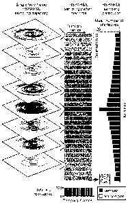
[0022] HSPARSE fMRI method resolved spatially adjacent yet functionally
distinct regions. (Fig. 7A,
7D) A rat brain phantom with three layers of distinct peak HRF amplitude /
latency in the cortex
4
CA 02995019 2018-02-06
WO 2017/040538 PCT/US2016/049508
was designed. HRFs of the three layers and their corresponding principal
component
decompositions demonstrate clear separation of the three layers. Thicker lines
in the HRF plot
represent the mean HRF of each layer. (Fig. 7B, 7E) Highest spatial resolution
Nyquist
acquisition resulted in activity with obscured boundaries between layers.
(Fig. 7C, 7F)
HSPARSE reconstruction correctly identified the spatial location where the
amplitude/time-to-
peak transition occurs.
[0023] Resolution limit of the HSPARSE fMRI was identified. (Fig. 8A, 8D) A
phantom that was
challenging to reconstruct with low spatial resolution was designed with an
interleaved pattern
consisting of distinct peak HRF amplitude / latency features. (Fig. 8B, 8E)
The highest spatial
resolution Nyquist acquisition completely failed to separate the six
interleaved layers. (Fig. 8C,
8F) In contrast, the HSPARSE reconstruction successfully resolved the peak HRF
amplitude and
latency differences for all six layers. (Fig. 8G, 8H) To identify the
resolution limit of the
HSPARSE method, a phantom with interleaved activation layers of variable
thickness (4-1 pixels
or 0.84 ¨ 0.21 mm) was designed. Layers with distinct peak HRF amplitude /
latency can be
distinguished down to 3-pixels (0.62 mm) using the Nyquist acquisition and
down to a single
pixel (0.21 mm) using the HSPARSE method.
[0024] HSPARSE fMRI method resolved in vivo layer-specific activity evoked by
optogenetic
stimulation of dentate gyrus. (Fig. 9A) Schematic showing optogenetic
targeting of the dentate
gyrus. (Fig. 9B) Dentate gyrus had a unique horn shape, with its coronal and
axial slices showing
"0" and "U" shaped profiles, respectively. (Fig. 9C) Histological examination
verified that the
ChR2-EYFP expression was localized to the dentate gyms region. (Fig. 9D) The
Nyquist
acquisition failed to accurately localize dentate gyrus activity and activity
occurs on both the
dentate gyrus and the CA1. In contrast, both the original and the three times
averaged HSPARSE
fMRI showed activity localized to the dentate gryus, with the voxels having
high peak HRF
amplitude precisely following the geometry of the structure's molecular layer.
White triangles in
the top row indicate the approximate site of stimulation. Active voxels were
identified as those
having an F-value greater than 4.42 (p < 0.001). The active voxels' peak HRF
amplitudes were
then calculated and overlaid onto a high-resolution MRI atlas, with a
threshold at the median plus
1.5 times the standard deviation of all peak HRF amplitudes for clear
visualization with good
dynamic range. (Fig. 9E, 9F) As was seen with the simulations, the HSPARSE-
reconstructed
HRF amplitudes at the stimulation site were lower than their respective low-
resolution scans.
However, the HRFs were strongly correlated between the HSPARSE reconstructed
and Nyquist
sampled images, which indicated that in vivo HSPARSE fMRI maintained the
temporal
characteristics of in vivo HRFs.
CA 02995019 2018-02-06
WO 2017/040538 PCT/US2016/049508
[0025] Optimal range of CS regularization parameters were identified based on
quantitative assessments
of the reconstructed images. (Fig. 10A) Example reconstructed images of the Al
phantom (30
dB) with different regularization parameters. Voxels were considered to be
active if they exhibit
an F-value greater than 4.42 (P < 0.001). Lower left plot shows the original
ground-truth image.
(Fig. 10B) An optimal regularization parameter range was defined as the region
that achieved
higher CNR and maximum correlation coefficient, larger active volume within
the designed
active region compared to the original ground-truth image, and NRMSE of less
than 105 % of the
minimum NRMSE found within the search range. A range of regularization
parameters were
identified to yield high reconstruction quality (lower right plot, blue area)
for the 30 dB Al
phantom. The symbols µA' and 'V in each plot indicate the maximum and minimum
values in the
corresponding test, respectively. "N/A" indicates an area in which CNR,
maximum correlation
coefficient, and peak HRF amplitude cannot be computed due to limited
activation. 1.02v, 1.05v
and 1.15v indicate the contour lines of 1.02, 1.05, and 1.15 times the minimum
NRMSE value.
(Fig. 10C) The optimal ranges for 6 phantoms with different base images,
activation patterns and
SNRs were overlaid, where a set of regularization parameters were found to
provide optimal
reconstruction quality for all phantoms tested. (Fig. 10D-H) With a set of
parameters ( = 5e-3 and
= le-4) selected from the optimal range, CNR, the active volume within the
original ground truth
active region, and the maximum correlation coefficient of the reconstructed
image were higher
than the original phantom with noise, and the NRMSE was less than 0.24 across
all phantoms.
Although the HRF amplitudes decrease after the CS fMRI reconstruction, the CS
fIVIRI was
shown to maintain the relative amplitudes and shapes of HRFs in Fig. 11. (Fig.
101) FWHM of
the point spread function was 0.70 mm with the highest spatial resolution
Nyquist acquisition
while it reduced to 0.32 mm for HSPARSE reconstructed images with optimal
regularization
parameters. The error bar shows the standard deviation of the CS point spread
function across
temporal frames.
[0026] HSPARSE reconstruction using optimal regularization parameters
maintained HRF temporal
characteristics over a range of physiologically relevant HRF amplitudes. (Fig.
11A)
Representative images of the reconstructed A3 phantom were shown with original
HRF
amplitudes of 4, 6, 8, and 10 %. All phantoms were reconstructed 5 times with
independent
identical Gaussian distributed noise using regularization parameters (?1=5e-3
and Xl=le-4)
within the optimal range. (Fig. 11B) Although the HSPARSE reconstructed HRFs
exhibit lower
amplitudes than the original HRFs for all tested amplitudes, the HRF shapes
were similar after
amplitude normalization (inset on upper right). Error bars represent standard
deviation across 5
reconstructions. (Fig. 11C) The correlation analysis indicated that the
HSPARSE reconstructed
6
CA 02995019 2018-02-06
WO 2017/040538 PCT/US2016/049508
HRF time courses had strong linear correlation to the original HRFs (slope =
0.52, R2 = 0.98).
(Fig. 11D) After the amplitude normalization of all HRFs, the maximum mean
difference
between the HSPARSE reconstructed and the original HRFs was less than 0.016
and the limits of
agreement ( 1.96xstandard deviation) were less than 0.080. (Fig. 11E, 11F)
Similarly, the
durations of the HSPARSE reconstructed HRFs were not significantly different
from the original
HRFs (P > 0.22, Wilcoxon ranksum test), and maximum time-to-peak differences
(2.40s) was
smaller than the 3 s temporal resolution of the fMRI acquisition. Similar
results were seen in B3
and C3 phantoms.
[0027] HSPARSE fMRI method was robust against real physiological noise. (Fig.
12A) The HSPARSE
WWI also improved the CNR, maximum correlation coefficient, and active volume
compared to
their corresponding fully-sampled datasets in the presence of real
physiological noise. The
NRMSE values were less than 0.081 across all subjects. Error bars represent
the standard error
across voxels of the active area for CNR and maximum correlation coefficient.
(Fig. 12B, 12C)
The images reconstructed with HSPARSE detect the majority of the activity and
the active voxels
shared between the HSPARSE reconstructions and the fully-sampled images
consist 90.3 to
93.0% of active voxels from the fully-sampled images. Because the ground-truth
active region
was not available for the in vivo experiment, additional active voxels only
detected with
HSPARSE reconstruction could either be false positive signal or a result of
improved sensitivity
due to CNR increase. However, active voxels only detected with HSPARSE
reconstruction was
limited within the 1-pixel perimeter layers of the active volume detected with
the fully-sampled
dataset. (Fig. 12D) HRF amplitude reduction from HSPARSE reconstruction has a
scaling factor
ranging from 0.40 to 0.48. Importantly, the HRFs from the fully-sampled
datasets and the
HSPARSE reconstructions had a strong linear correlation with a minimum
correlation coefficient
(R2) of 0.98, demonstrating that HSPARSE reconstruction maintains the temporal
characteristics
of the fully-sampled HRFs.
[0028] Fig. 13: Comparison of temporal HRF characteristics between the
original fully-sampled and
HSPARSE images in the presence of physiological noise. For all three subjects,
the HRF
durations were similar and the maximum duration difference was 1.67 s. The
first subjects gave
the same time-to-peak. The rest two subjects showed an increase in time-to-
peak for the
HSPARSE reconstructed image, but the difference was smaller than the 3 s
temporal resolution of
the acquisition.
[0029] Six-cycle time-series and analysis results corresponding to the main
Figures. (Fig. 14A, 14B) Six-
cycle time-series corresponding to Fig. 11 and 12. Similar to the analysis
performed on the HRFs,
the HSPARSE reconstructed six-cycle time-series also show a strong linear
correlation with the
7
CA 02995019 2018-02-06
WO 2017/040538 PCT/US2016/049508
original ground-truth time-series, which indicates the HSPARSE method
maintains high temporal
fidelity. (Fig. 14C, 14D) Six-cycle time-series corresponding to Fig. 7 and
Fig. 8. While some
sinusoidal variations in the HSPARSE fMRI reconstructed time-series were
observed (bottom left
plot for both C and D), the HSPARSE fMRI was found to preserve the peak
amplitude and
latency differences between layers, while the highest spatial resolution
Nyquist acquisitions fail.
HSPARSE also maintains high sensitivity and low FPR. In contrast, Nyquist
acquisitions result in
high FPR, which could be the result of low spatial resolution induced partial
volume effects. (Fig.
14E) Six-cycle time-series corresponding to Fig. 9. In vivo acquired HSPARSE
fMRI six-cycle
time-series also showed strong linear correlation with the time-series
obtained from the highest
spatial resolution Nyquist acquisitions for all three subjects, demonstrating
that HSPARSE fMRI
can provide high temporal fidelity for in vivo experiments.
[0030] Fig. 15A, 15B: Optimized HSPARSE fMRI method consistently resolved
layer-specific activity
of the dentate gyrus upon optogenetic stimulation. Two additional in vivo
experiment results were
shown. With the highest spatial resolution Nyquist rate sampled images,
activity was observed
throughout the hippocampus. In contrast, activities in the HSPARSE
reconstructed images were
confined to the dentate gyrus. The peak amplitude activities followed the
geometry of the
molecular layer for all three subjects. The pink area and the red lines
delineate the dentate gyms.
The white arrow indicates the site of stimulation.
[0031] Fig. 16: Comparison of temporal characteristics of HRF between HSPARSE
fMRI and Nyquist
acquisition fMRI following optogenetic stimulation of the dentate gyms. Three
subjects were
optogenetically stimulated during imaging, using the single (HSPARSE
HSPARSExl) and 3
times averaged (HSPARSEx3) high-resolution HSPARSE fMRI and a highest spatial
resolution
Nyquist acquisition (NAcq). For each subjects, the time-to-peak difference
between the
HSPARSE and NAcq images was less than the 3 s temporal resolution. Although
the duration of
activity was similar between the HSPARSE and NAcq images for subject 1 and 3
on average, the
duration was larger in the HSPARSE reconstructed image for subject 2. This
difference could be
due biological variability since the Nyquist acquisition datasets and the CS
datasets were
separately acquired in different fMRI imaging sessions.
[0032] Fig. 17: GPU based HSPARSE fMRI method achieved a 34-fold improvement
in speed. Three
computationally expensive functions ¨ the DCT, NUFFT and iNUFFI ¨ were tested
with the
GPU method and its parallel CPU counterpart. The GPU methods showed 165-, 28-,
and 108-
fold improvements in speed, respectively, resulting in a 34-fold overall
speedup.
[0033] The HSPARSE fMRI method was robust to motion within a normal
physiological range. (Fig.
18A) Five sets of motion profiles with a maximum absolute translation
equivalent to 1- to 5-
8
CA 02995019 2018-02-06
WO 2017/040538 PCT/US2016/049508
pixels were designed. To simulate realistic motion, the z-dimension
translation was restricted to
be smaller than the x- and y-dimension translations, and rotations about the x-
, y- and z-axis were
limited to within 0.5 degrees. Solid lines represent an example six degree-of-
freedom motion
profile and shaded areas represent the ranges of translations or rotations in
each motion profile.
(Fig. 18B) The motion corrected HSPARSE images show similar activations as the
motion
corrected original images when the motion was 1-5 pixels. (Fig. 18C, 18D) When
the amount of
motion was less than 3-pixels, the HSPARSE reconstructed images were similar
to the original
image as measured by the mean F-value, sensitivity and false positive rate.
However, when
motion was larger than 4-pixels, the HSPARSE reconstructed images exhibited
decrease in mean
F-value and sensitivity. (Fig. 18E) Motion profiles of the three experiment
subjects indicated that
the physiological motion in the in vivo experiments were well within the 4-
pixel range of robust
reconstruction with the HSPARSE method. They exhibited translations of less
than 2.5-pixels and
rotations of less than 0.2 degrees.
[0034] Fig. 19: Algorithm 1 was implemented on a Graphical Processing Unit
platform. Several
repeatedly computations such as the non-uniform FF1 (NUFF1), inverse NUFFT,
DWT and
inverse DWT were carefully optimized. For the NUFFT, a similar pre-sorting
algorithm was
implemented. A custom build workstation was used for the real-time
reconstruction with Intel
quad-core 2.66 GHz CPU, Nvidia 2048 cores CUDA GPU and 16 GB CPU memory.
[0035] High reconstruction speed was achieved with FISTA method and GPU
optimization. (Fig. 20A)
FISTA method showed a much faster convergence speed and lower cost than the
widely used
conjugate gradient method. Because FISTA was not a descend method, an increase
of the cost in
some iterations was observed. (Fig. 20B) With GPU optimization, FISTA method
successfully
reconstructed a 140x140x32 matrix sized image within 605 ms. Combining with
the IGN motion
correction and coherence analysis, the overall real-time processing took less
than 620 ms and
only consists 20% of the consecutive image duration. (Fig. 20C) Repeatedly
used computations
such as the NUFFT, iNUFF1, DWT, and iDWT were calculated efficiently by the
GPU.
[0036] Fig. 21: Optimized stack of VDS achieved high incoherent sampling and
FISTA method
successfully reconstructs the under-sampled image. The normalized image
intensities are also
shown across the yellow dashed line.
[0037] Fig. 22: Real-time high-resolution CS fMRI achieved improved CNR, mean
F-value, sensitivity
and low FPR. A range of parameters were tested to identify the optimal
regularization parameters
for the real-time high-resolution CS fMRI. After comparing different metrics
shown in the
Figure, it was found that 1e3 and 5e4 offer the best trade-off between metrics
and result in
improved CNR, mean F-value, sensitivity and low FPR.
9
CA2995019
[0038] Real-time high-resolution CS fMRI resolves layer specific activity. Two
different types of HRFs
with distinct peak HRF amplitude (Fig. 23A) and latency (Fig. 23B) were added
into a phantom
with interleaved layer pattern. The real-time high-resolution CS fMRI method
successfully
resolves the peak HRF amplitude and latency differences between the two layers
while the
highest spatial resolution Nyquist acquisition failed.
[0039] Randomized variable density stack of spirals design. (Fig. 24A) The
center k-space is designed to
have a higher density than the outer k-space. Incoherence is introduced by
randomly disturb the
angle of each interleaf and random skipping interleaves in the outer k-space.
(Fig. 24B) The
effective field of view of the spiral trajectory is designed to follow a
series of exponential
functions shown.
[0040] Before the present invention is further described, it is to be
understood that this invention is not
limited to particular embodiments described, as such may, of course, vary. It
is also to be
understood that the terminology used herein is for the purpose of describing
particular
embodiments only, and is not intended to be limiting, since the scope of the
present invention will
be limited only by the appended claims.
[0041] Where a range of values is provided, it is understood that each
intervening value, to the tenth of
the unit of the lower limit unless the context clearly dictates otherwise,
between the upper and
lower limit of that range and any other stated or intervening value in that
stated range, is
encompassed within the invention. The upper and lower limits of these smaller
ranges may
independently be included in the smaller ranges, and are also encompassed
within the invention,
subject to any specifically excluded limit in the stated range. Where the
stated range includes one
or both of the limits, ranges excluding either or both of those included
limits are also included in
the invention.
[0042] Unless defined otherwise, all technical and scientific terms used
herein have the same meaning as
commonly understood by one of ordinary skill in the art to which this
invention belongs.
Although any methods and materials similar or equivalent to those described
herein can also be
used in the practice or testing of the present invention, the preferred
methods and materials are
now described.
[0043] It must be noted that as used herein and in the appended claims, the
singular forms "a," "an," and
"the" include plural referents unless the context clearly dictates otherwise.
Thus, for example,
reference to "an opsin" includes a plurality of such opsins and reference to
"the carbon fiber"
includes reference to one or more carbon fibers and equivalents thereof known
to those skilled in
Date Regue/Date Received 2023-02-15
CA 02995019 2018-02-06
WO 2017/040538 PCT/US2016/049508
the art, and so forth. It is further noted that the claims may be drafted to
exclude any optional
element. As such, this statement is intended to serve as antecedent basis for
use of such exclusive
terminology as "solely," "only" and the like in connection with the recitation
of claim elements,
or use of a "negative" limitation.
[0044] It is appreciated that certain features of the invention, which are,
for clarity, described in the
context of separate embodiments, may also be provided in combination in a
single embodiment.
Conversely, various features of the invention, which are, for brevity,
described in the context of a
single embodiment, may also be provided separately or in any suitable sub-
combination. All
combinations of the embodiments pertaining to the invention are specifically
embraced by the
present invention and are disclosed herein just as if each and every
combination was individually
and explicitly disclosed. In addition, all sub-combinations of the various
embodiments and
elements thereof are also specifically embraced by the present invention and
are disclosed herein
just as if each and every such sub-combination was individually and explicitly
disclosed herein.
[0045] The publications discussed herein are provided solely for their
disclosure prior to the filing date
of the present application. Nothing herein is to be construed as an admission
that the present
invention is not entitled to antedate such publication by virtue of prior
invention. Further, the
dates of publication provided may be different from the actual publication
dates which may need
to be independently confirmed.
DETAILED DESCRIPTION
[0046] The present disclosure provides methods and systems for high-resolution
functional magnetic
resonance imaging (fMRI), including real-time high-resolution fMRI methods and
systems.
[0047] In describing embodiments of the present disclosure, methods for high-
resolution functional
magnetic resonance imaging (fMRI) are first described, followed by a
description of systems
useful for performing the subject methods.
METHODS
[0048] Aspects of the present disclosure include a method for functional
magnetic resonance imaging
(fMRI) of a subject. In certain embodiments, the method is a compressed
sensing (CS) high-
resolution fMRI method. Compressed sensing refers to a signal processing
method where an
image can be reconstructed from a series of sampling measurements obtained
with a sampling
rate below the Nyquist sampling rate. In general, the method may include
obtaining one or more
fMRI images of a target area in a subject. For instance, in general, the
method may include
applying with an MRI system (e.g., a permanent magnet or electromagnet of the
MRI system) a
magnetic field to a target area in a subject. In some instances, the method
also includes applying
11
CA 02995019 2018-02-06
WO 2017/040538 PCT/US2016/049508
with the MRI system (e.g., an RF coil of the MRI system) an excitation
waveform (e.g., an RF
excitation waveform) to the target area in the subject to produce detectable
image data (e.g.,
magnetic resonance (MR) signals) of the target area in the subject. One or
more additional fields
may also be applied by the MRI system, such as, but not limited to, one or
more shim fields using
one or more shim coils, one or more gradient fields using one or more gradient
coils, and the like.
In addition, the method includes acquiring the image data (e.g., with a
receiver of the MRI
system) and producing an image of the target area in the subject based on the
acquired image
data.
[0049] The acquired image data may be saved in a computer-readable memory and
analyzed at a
subsequent time (also referred to herein as "offline" processing or -offline"
MRI). In other cases,
the acquired image data may be analyzed in real-time to produce the image of
the target area in
the subject. By "real-time" is meant that the acquired signals are analyzed by
the MRI system
(e.g., by a processor in the MRI system) immediately after signal acquisition
and/or during signal
acquisition.
[0050] In certain embodiments of offline fMRI, to produce the MR image data,
the method may include
applying an excitation waveform to the target area in the subject. In certain
embodiments, the
method includes applying a pulse sequence to the target area in the subject.
The pulse sequence
may be a balanced steady state free precession (b-SSFP) sequence that is
applied to the target area
in the subject. In certain cases, the pulse sequence has an echo time (TE) of
50 ms or less, such
as 40 ms or less, or 30 ms or less, or 20 ms or less, or 10 ms or less, or 5
ms or less, or 3 ms or
less, or 2 ms or less. In some instances, the pulse sequence has a TE of 2 ms.
In certain cases,
the pulse sequence has a repetition time (TR) of 500 ms or less, such as 400
ms or less, or 300 ms
or less, or 200 ms or less, or 100 ms or less, or 50 ms or less, or 25 ms or
less, or 20 ins or less, or
ms or less, or 5 ms or less. In some instances, the pulse sequence has a TR
ranging from 5 to
10 ms, such as from 7 to 10 ms, or from 8 to 10 ms, or from 9 to 10 ins. In
certain instances, the
pulse sequence has a TR of 9.375 ms.
[0051] In some instances of offline MRI, the method includes acquiring image
data (MR signals) of the
target area in the subject. In certain cases, the method includes using a
sampling trajectory. The
sampling trajectory may be a randomized sampling trajectory. For instance, the
method may
include acquiring image data of the target area in the subject using a
randomly undersampled
trajectory, such as a randomly undersampled variable density spiral (VDS)
trajectory. In certain
cases, the sampling trajectory is a variable density spiral (VDS) trajectory,
such as, for example, a
randomized under-sampling stack of multi-interleaf variable density spiral
(VDS) trajectory. In
certain instances, the total number of interleaves at each kz-slice follows a
Laplacian distribution.
12
CA 02995019 2018-02-06
WO 2017/040538 PCT/US2016/049508
For instance, in some embodiments, the center k-space is more densely sampled
than the outer k-
space.
[0052] In certain embodiments of offline MRI, the sampling method has a field
of view (FOV). For
example, the sampling method may have a FOV of 10x10x10 mm or more, such as
15x15x15
mm or more, or 20x20x15 mm or more, or 25x25x15 mm or more, or 30x30x15 mm or
more, or
35x35x15 mm or more. In certain instances, the sampling method has a FOV of
35x35x16 mm.
In some cases, the sampling method has a resolution of lx1 xl mm or less, such
as
0.75x0.75x0.75 mm or less, or 0.5x0.5x0.5 mm or less, or 0.25x0.25x0.5 mm or
less. In certain
instances, the sampling method has a resolution of 0.21x0.21x0.5 mm. In
certain embodiments,
the sampling method achieves a sampling acceleration factor of 2 or more, such
as 3 or more, 4 or
more, 5 or more, 6 or more, 7 or more, 8 or more, 9 or more, or 10 or more as
compared to
conventional fMRI. In some cases, the sampling method achieves a sampling
acceleration factor
of 2 or more. In some cases, the sampling method achieves a sampling
acceleration factor of 5 or
more.
[0053] In certain embodiments of offline MRI, the method includes producing an
image of the target
area in the subject based on the acquired image data. For example, the method
may include
analyzing (also referred to herein as processing) the image data to produce
the image of the target
area. As such, in some instances, the method includes reconstructing an image
from the acquired
image data. In certain cases, the method includes reconstructing the image
using a cost function,
such as an Li regularized cost function. In certain instances, the method
includes
analyzing/processing the image data using a spatial sparsifying transform,
such as a discrete
cosine transform (DCT). For instance, the method may include regularizing the
fMRI temporal
domain using a DCT. In some cases, the method includes regularizing the fMRI
spatial domain
using a DCT. In some embodiments, the method includes regularizing both the
temporal domain
and the spatial domain using a DCT.
[0054] In certain embodiments of offline MR', the method includes
reconstructing the image using one
or more regularization parameters. Regularization parameters of interest for
offline fMRI
processing include, but are not limited to, contrast to noise ratio (CNR),
active volume within the
designed active region, mean F statistic value (mean F-value), normalized root
mean squared
error (NRMSE), and peak hemodynamic response function (HRF) amplitude. In
certain
instances, a set of regularization parameters is considered to be in an
optimal range if the CNR,
active volume within the designed mask, and mean F-value are greater than that
of the ground-
truth, and its NRMSE is less than 105% of the minimum NRMSE found within the
search range.
For example, the subject fIVIRI methods may produce images having a CNR of 1.5
or more, such
13
CA 02995019 2018-02-06
WO 2017/040538 PCT/US2016/049508
as 2 or more, or 2.5 or more, or 3 or more, or 4 or more, or 5 or more, or 6
or more, or 7 or more,
or 8 or more, or 9 or more, or 10 or more. In some cases, the subject fiVIRI
methods may produce
images having a CNR of 1.5 or more. In some cases, the subject EVIRI methods
may produce
images having a CNR of 2.5 or more.
[0055] In certain embodiments, the subject flVIRI methods produce an image
having a spatial resolution
of about 0.2x0.2x0.5 mm3 or greater. For example, the subject flVIRI methods
can produce
images having a spatial resolution of lx1 xl mm3 or greater, such as
0.9x0.9x0.9 mm3 or greater,
or 0.8x0.8x0.8 mm3 or greater, or 0.7x0.7x0.7 mm3 or greater, or 0.6x0.6x0.6
mm3 or greater, or
0.5x0.5x0.5 mm3 or greater, or 0.4x0.4x0.5 mm3 or greater, or 0.3x0.3x0.5 mm3
or greater, or
0.2x0.2x0.5 mm3 or greater, or 0.1x0.1x0.5 mm3 or greater. In certain
instances, the subject
MIR' methods produce an image having a spatial resolution of 0.21x0.21x0.5
mm3. In certain
instances, the subject flVIRI methods produce an image having a spatial
resolution ranging from
0.1x0.1x0.5 mm3 to lx1x1 mm3, such as from 0.1x0.1x0.5 mm3 to 0.9x0.9x0.9 mm3,
or from
0.1x0.1x0.5 mm3 to 0.8x0.8x0.8 mm3, or from 0.1x0.1x0.5 mm3 to 0.7x0.7x0.7
mm3, or from
0.1x0.1x0.5 mm3 to 0.6x0.6x0.6 mm3, or from 0.1x0.1x0.5 mm3 to 0.5x0.5x0.5
mm3, or from
0.1x0.1x0.5 mm3 to 0.4x0.4x0.5 mm3, or from 0.1x0.1x0.5 mm3 to 0.3x0.3x0.5
mm3. In certain
embodiments, the subject MIR' methods produce an image having a spatial
resolution ranging
from 0.1x0.1x0.5 mm3 to 0.3x0.3x0.5 mm3.
[0056] As described above, rather than offline processing of the MRI image
data, the acquired image
data can be processed in real-time.
[0057] In certain embodiments of real-time fMRI, to produce the MR image data,
the method may
include applying an excitation waveform to the target area in the subject. In
certain
embodiments, the method includes applying a pulse sequence to the target area
in the subject to
produce image data (MR signals) that can be acquired by the MRI system. As
such, in some
instances, the method includes acquiring the image data (MR signals) of the
target area in the
subject. In certain cases, the method includes using a sampling trajectory.
The sampling
trajectory may be a randomized sampling trajectory. For instance, the method
may include
acquiring image data of the target area in the subject using a randomly
undersampled trajectory,
such as a randomly undersampled variable density spiral (VDS) trajectory. In
certain cases, the
sampling trajectory is a variable density spiral (VDS) trajectory, such as,
for example, a
randomized stack of variable density spiral (VDS) trajectory. In certain
instances, the sampling
density follows an exponential function along the kx and ky plane, and the
variance of the
exponential function decreases along the kz direction. In some instances,
randomness is
introduced into the sampling for CS reconstruction by randomly perturbing the
angle of each
14
CA 02995019 2018-02-06
WO 2017/040538 PCT/US2016/049508
spiral interleaf. In certain cases, the trajectory has a slightly larger total
number of interleaves,
and interleaves on the outer k-space are randomly skipped following a Gaussian
distribution to
achieve the desired temporal resolution. In some instances, the kz-slice
location may be adjusted
to achieve variable density sampling in the kz dimension and high spatial
resolution in the z
dimension.
[0058] In certain embodiments of real-time MRI, the sampling method has a
field of view (FOV). For
example, the sampling method may have a FOV of 10x10x10 mm or more, such as
15x15x15
mm or more, or 20x20x15 mm or more, or 25x25x15 mm or more, or 30x30x15 mm or
more, or
35x35x15 mm or more. In certain instances, the sampling method has a FOV of
35x35x16 mm.
In some cases, the sampling method has a resolution of lx1 xl mm or less, such
as
0.75x0.75x0.75 mm or less, or 0.5x0.5x0.5 mm or less, or 0.25x0.25x0.5 mm or
less. In certain
instances, the sampling method has a resolution of 0.25x0.25x0.5 mm. In
certain embodiments,
the sampling method achieves a sampling acceleration factor of 2 or more, such
as 3 or more, 4 or
more, 5 or more, 6 or more, 7 or more, 8 or more, 9 or more, or 10 or more as
compared to
conventional fMRI. In some cases, the sampling method achieves a sampling
acceleration factor
of 2 or more. In some cases, the sampling method achieves a sampling
acceleration factor of 5 or
more.
[0059] In certain embodiments of real-time MRI, the method includes producing
an image of the target
area in the subject based on the acquired image data. For example, the method
may include
analyzing (also referred to herein as processing) the image data to produce
the image of the target
area. As described herein, the image data may be processed in real-time to
produce the image of
the target area. As such, in some instances, the method includes
reconstructing an image from the
acquired image data in real-time. In certain cases, the method includes
reconstructing the image
using a cost function, such as an Ll spatial regularized cost function. In
certain instances, the
method includes analyzing/processing the image data using a sparsifying
transform, such as a
Daubechies 4 wavelet. In some cases, the method includes analyzing/processing
the image data
using a fast iterative shrinkage thresholding algorithm (FISTA).
[0060] In certain embodiments of real-time fMRI, the method includes
reconstructing the image using
one or more regularization parameters. Regularization parameters of interest
for real-time fMRI
processing include, but are not limited to, contrast to noise ratio (CNR),
mean F statistic value
(mean F-value), normalized root mean squared error (NRMSE), peak HRF
amplitude, sensitivity,
and false positive rate in the reconstructed dataset. In certain instances, a
set of regularization
parameters is considered to be in an optimal range if the parameters give top
50% CNR, mean F-
value, sensitivity, and bottom 50% NRMSE and false positive rate. For example,
the subject
CA 02995019 2018-02-06
WO 2017/040538 PCT/US2016/049508
fMRI methods may produce images having a CNR of 1.5 or more, such as 2 or
more, or 2.5 or
more, or 3 or more, or 4 or more, or 5 or more, or 6 or more, or 7 or more, or
8 or more, or 9 or
more, or 10 or more. In some cases, the subject fMRI methods may produce
images having a
CNR of 1.5 or more. In some cases, the subject fMRI methods may produce images
having a
CNR of 2.5 or more.
[0061] In certain embodiments of the present disclosure, the method is a
method for functional MRI
(fMRI). For example, in general, the present disclosure provides a method for
monitoring
activity in an organ or tissue of an individual (also referred to as "a
subject" herein). In some
instances, the target organ or tissue is an excitable organ or tissue in the
subject. "Excitable," as
used herein, refers to electrically excitable cells in an organ or tissue,
such as neurons and muscle
cells. Excitable cells typically use changes in their membrane potential to
transmit signals within
the cell. Thus, an excitable cell may be characterized in having a resting
state, where the
membrane potential is at the resting membrane potential, and an excited state,
where rapid
depolarization of the membrane potential is transmitted across the cell as an
action potential. The
"cellular electrical activity" of an excitable cell may refer to the changes
in the membrane
potential or may refer to any indirect measure of the changes in membrane
potential, such as the
changes in intracellular calcium concentration or any other biochemical
changes that is a
functional measure of the change in the membrane potential.
[0062] In certain embodiments, the method includes surgically implanting a
device of the present
disclosure into or adjacent to an organ or tissue of an individual, and
monitoring the activity of
the organ or tissue using fMRI. In some cases, surgically implanting the
device includes opening
an access in the subject and inserting at least a portion of the device
through the access. The
access may be an access through the skin, bone, muscle, and/or other tissues
of the subject. For
instance, an access may include an access through bone (e.g., skull) of the
subject to allow
placement of at least a portion of the device (e.g., an optrode) adjacent to
target neurons in the
subject.
[0063] As indicated above, embodiments of the method include monitoring the
activity of the organ or
tissue. In some instances, monitoring the activity of the organ or tissue
includes conducting
functional magnetic resonance imaging (fMRI) on the organ or tissue. In some
cases, the organ
or tissue includes excitable cells (e.g., cells that express one or more light-
responsive
polypeptides). The terms "light-activated" and "light-responsive" in reference
to a polypeptide or
protein that is light-responsive, are used interchangeably and include light-
responsive ion
channels or opsins, and ion pumps as described herein. Such light-responsive
proteins may have a
depolarizing or hyperpolarizing effect on the cell on whose plasma membrane
the protein is
16
CA 02995019 2018-02-06
WO 2017/040538 PCT/US2016/049508
expressed depending on the ion permeability of the activated protein, and the
electrochemical
gradients present across the plasma membrane.
[0064] In some cases, the one or more light-responsive polypeptides include a
hyperpolarizing light-
responsive polypeptide. In some cases, the one or more light-responsive
polypeptides include a
depolarizing light-responsive polypeptide. As such, in some cases the method
includes producing
an image of the target organ or tissue using fIVIRI. In some cases, flVIRI may
be used to image the
organ or tissue prior to delivering light to the target organ or tissue using
the optrode. In some
cases, fIVIRI may be used to image the organ or tissue during delivery of
light to the target organ
or tissue using the optrode. In some cases, flVIRI may be used to image the
organ or tissue after
delivering light to the target organ or tissue using the optrode.
[0065] The method may further include detecting and/or recording a detectable
parameter of the organ or
tissue using the device (e.g., optrode). The optrode may be configured to
detect electrical signals,
such as local field potentials produced by changes in the membrane potential
of the excitable
cells. Thus, in some cases, the method includes detecting and/or recording a
detectable parameter
of the organ or tissue using a carbon fiber electrode of the optrode.
[0066] The device (e.g., optrode) may include a light source. In these
embodiments, the method includes
delivering light to the target organ or tissue using the light source. For
instance, the method may
include stimulating the excitable cells in the target organ or tissue with
light from the light source.
In some cases, the light source includes an optical fiber as described herein.
As such, in these
embodiments, the method includes delivering light to the target organ or
tissue using the optical
fiber (e.g., stimulating the excitable cells with light delivered by the
optical fiber). In some cases,
the light source includes a laser. As such, in some embodiments, the method
includes delivering
light to the target organ or tissue using the laser. For example, the method
may include generating
light using the laser and directing the light from the laser to the target
organ or tissue using the
optical fiber (e.g., for stimulating the excitable cells in the target organ
or tissue with light from
the laser). In some cases, the light source includes a light-emitting diode
(LED). As such, in some
embodiments, the method includes delivering light to the target organ or
tissue using the LED.
For instance, the method may include generating light using the LED and
directing the light from
the LED to the target organ or tissue using the optical fiber (e.g., for
stimulating the excitable
cells in the target organ or tissue with light from the LED).
[0067] In certain embodiments, the detectable parameter of the target organ or
tissue includes local field
potentials, e.g., local field potentials produced by changes in the membrane
potential of the
excitable cells. The local field potentials may be produced by stimulating the
excitable cells with
light from the light source. In some instances, the detectable parameter is a
single-unit activity,
17
CA 02995019 2018-02-06
WO 2017/040538 PCT/US2016/049508
e.g., detectable activity from a single target area (i.e., a uniplex assay).
In some cases, the
detectable parameter is a multi-unit activity, e.g., detectable activity from
two or more target
areas (i.e., a multiplex assay).
[0068] In some instances, monitoring the activity of the organ or tissue is
performed once. In other cases,
monitoring the activity of the organ or tissue is performed two or more times.
In some cases,
monitoring the activity of the organ or tissue is performed several times over
a period of time,
e.g., the method includes chronically monitoring the activity of the organ or
tissue. In some cases,
monitoring the activity of the organ or tissue may be performed over an
extended period of time,
such as 1 day or more, 2 days or more, 3 days or more, 4 days or more, 5 days
or more, 6 days or
more, 7 days or more, 8 days or more, 9 days or more, 10 days or more, such
as, for example, 1
week or more, 2 weeks or more, 3 weeks or more, 1 month or more, 2 months or
more, 3 months
or more, 4 months or more, 5 months or more, 6 months or more, 7 months or
more, 8 months or
more, 9 months or more, 10 months or more, 11 months or more, 1 year or more,
or ever longer
periods of time.
[0069] In some cases, the individual is a human. In some cases, the individual
is a non-human primate.
In some cases, the individual is a rodent (e.g., a rat, a mouse, etc.). The
tissue or organ (e.g.,
"target tissue" or "target organ") may be an in vivo neuronal tissue, a tissue
slice preparation, a
nerve fiber bundle, a neuromuscular junction, etc. The in vivo neuronal tissue
may be neuronal
tissue of an animal that is anesthetized or non-anesthetized, and is
restrained or non-restrained.
The target tissue of interest includes, but is not limited to, the neocortex,
the hypothalamus,
entorhinal and hippocampal formation cortex, mam_millary bodies, septum, bed
nucleus of stria
terminalis, dorsal and ventral striatum, thalamus, amygdala, accumbens,
brainstem, subcortical
structures in general, muscle, spinal cord, cardiac tissue, etc.
[0070] In some embodiments, the excitable cells (e.g., neurons) in a target
tissue or organ are genetically
modified to express a light-responsive polypeptide that, when stimulated by an
appropriate light
stimulus, hyperpolarizes or depolarizes the stimulated excitable cell. The
term "genetic
modification" refers to a permanent or transient genetic change induced in a
cell following
introduction into the cell of a heterologous nucleic acid (i.e., nucleic acid
exogenous to the cell).
Genetic change ("modification") can be accomplished by incorporation of the
heterologous
nucleic acid into the genome of the host cell, or by transient or stable
maintenance of the
heterologous nucleic acid as an extrachromosomal element. Where the cell is a
eukaryotic cell, a
permanent genetic change can be achieved by introduction of the nucleic acid
into the genome of
the cell. Suitable methods of genetic modification include viral infection,
transfection,
18
CA 02995019 2018-02-06
WO 2017/040538 PCT/US2016/049508
conjugation, protoplast fusion, electroporation, particle gun technology,
calcium phosphate
precipitation, direct microinjection, and the like.
[0071] In some instances, the light-responsive polypeptide is a light-
activated ion channel polypeptide.
The light-activated ion channel polypeptides are adapted to allow one or more
ions to pass
through the plasma membrane of a target cell when the polypeptide is
illuminated with light of an
activating wavelength. Light-activated proteins may be characterized as ion
pump proteins, which
facilitate the passage of a small number of ions through the plasma membrane
per photon of light,
or as ion channel proteins, which allow a stream of ions to freely flow
through the plasma
membrane when the channel is open. In some embodiments, the light-responsive
polypeptide
depolarizes the excitable cell when activated by light of an activating
wavelength. In some
embodiments, the light-responsive polypeptide hyperpolarizes the excitable
cell when activated
by light of an activating wavelength.
[0072] In some embodiments, the light-responsive polypeptides are activated by
blue light. In some
embodiments, the light-responsive polypeptides are activated by green light.
In some
embodiments, the light-responsive polypeptides are activated by yellow light.
In some
embodiments, the light-responsive polypeptides are activated by orange light.
In some
embodiments, the light-responsive polypeptides are activated by red light.
[0073] In some embodiments, the light-responsive polypeptide expressed in a
cell can be fused to one or
more amino acid sequence motifs selected from the group consisting of a signal
peptide, an
endoplasmic reticulum (ER) export signal, a membrane trafficking signal,
and/or an N-terminal
golgi export signal. The one or more amino acid sequence motifs which enhance
light-responsive
protein transport to the plasma membranes of mammalian cells can be fused to
the N-terminus,
the C-terminus, or to both the N- and C-terminal ends of the light-responsive
polypeptide. In
some cases, the one or more amino acid sequence motifs which enhance light-
responsive
polypeptide transport to the plasma membranes of mammalian cells is fused
internally within a
light-responsive polypeptide. Optionally, the light-responsive polypeptide and
the one or more
amino acid sequence motifs may be separated by a linker. In some embodiments,
the light-
responsive polypeptide can be modified by the addition of a trafficking signal
(ts) which enhances
transport of the protein to the cell plasma membrane. In some embodiments, the
trafficking
signal can be derived from the amino acid sequence of the human inward
rectifier potassium
channel Kir2.1. In some embodiments, the signal peptide sequence in the
protein can be deleted
or substituted with a signal peptide sequence from a different protein.
19
CA 02995019 2018-02-06
WO 2017/040538 PCT/US2016/049508
[0074] Exemplary light-responsive polypeptides and amino acid sequence motifs
that find use in the
present system and method are disclosed in, e.g., PCT App. Nos.
PCT/US2011/028893 and
PCT/US2015/23087.
[0075] The individual may be any suitable individual for analyzing the
individual's brain functional
activity data. In some cases, the individual is a human individual. In some
cases the human is a
healthy human, or a human having a neurological disorder. The neurological
disorder may be any
suitable neurological disorder. In some cases, the neurological disorder is
caused by a disease,
e.g., a neurological disease. The neurological disease may be any suitable
disease associated with
pathological activity of a network of neurons. Suitable neurological diseases
include, without
limitation, Parkinson's disease, Alzheimer's disease, dementia, epilepsy,
autism, bipolar disorder,
schizophrenia, Tourette's syndrome, obsessive compulsive disorder, attention
deficit
hyperactivity disorder, Huntington's disease, multiple sclerosis, or migraine.
In some
embodiments, the neurological disorder is an age-related disorder of brain
function.
[0076] In certain embodiments, the methods may be used to treat a disease or
condition (e.g., a
neurological disorder) in the subject that is amenable to treatment using the
subject methods. As
used herein, the terms "treat," "treatment," "treating," and the like, refer
to obtaining a desired
pharmacologic and/or physiologic effect. The effect may be prophylactic in
terms of completely
or partially preventing a disease or symptom thereof and/or may be therapeutic
in terms of a
partial or complete cure for a disease and/or adverse effect attributable to
the disease.
"Treatment," as used herein, covers any treatment of a disease in a mammal,
particularly in a
human, and includes: (a) preventing the disease from occurring in a subject
which may be
predisposed to the disease but has not yet been diagnosed as having it; (b)
inhibiting the disease,
i.e., arresting its development; and (c) relieving the disease, e.g., causing
regression of the
disease, e.g., to completely or partially remove symptoms of the disease.
[0077] Selective activation of neurons in order to measure subtype-specific
functional activity may be
done using any suitable method. Suitable methods of selective neuron
activation include, without
limitation, optogenetic stimulation, single unit electrophysiology, etc. Where
the neurons are
selectively activated by optogenetic stimulation, the neurons may express one
or more light-
activated polypeptides configured to hyperpolarize or depolarize the neurons.
Suitable light-
activated polypeptides and methods used thereof are described further below.
Light-Activated Polypeptides
[0078] A light-activated polypeptide of the present disclosure may be any
suitable light-activated
polypeptide for selectively activating neurons of a subtype by illuminating
the neurons with an
activating light stimulus. In some instances, the light-activated polypeptide
is a light-activated
CA2995019
ion channel polypeptide. The light-activated ion channel polypeptides are
adapted to allow one or
more ions to pass through the plasma membrane of a target cell when the
polypeptide is
illuminated with light of an activating wavelength. Light-activated proteins
may be characterized
as ion pump proteins, which facilitate the passage of a small number of ions
through the plasma
membrane per photon of light, or as ion channel proteins, which allow a stream
of ions to freely
flow through the plasma membrane when the channel is open. In some
embodiments, the light-
activated polypeptide depolarizes the cell when activated by light of an
activating wavelength. In
some embodiments, the light-activated polypeptide hyperpolarizes the cell when
activated by
light of an activating wavelength. Suitable hyperpolarizing and depolarizing
polypeptides are
known in the art and include, e.g., a channelrhodopsin (e.g., ChR2), variants
of ChR2 (e.g.,
C128S, D156A, C1285 + D156A, E123A, E1231), iC1C2, C1C2, GtACR2, NpHR,
eNpHR3.0,
C1V1, VChRl, VChR2, SwiChR, Arch, ArchT, KR2, ReaChR, ChiEF, Chronos, ChRGR,
CsChrimson, and the like. In some cases, the light-activated polypeptide
includes bReaCh-ES, as
described herein and described further in, e.g., Rajasethupathy et al.,
Nature. 2015 Oct
29;526(7575):653. Hyperpolarizing and depolarizing opsins have been described
in various
publications; see, e.g., Berndt and Deisseroth (2015) Science 349:590; Berndt
et al. (2014)
Science 344:420; and Guru et al. (July 25, 2015) Intl. J.
Neuropsychopharmacol. pp. 1-8 (PMID
26209858).
[0079] The light-activated polypeptide may be introduced into the neurons
using any suitable method. In
some cases, the neurons of a subtype of interest are genetically modified to
express alight-
activated polypeptide. In some cases, the neurons may be genetically modified
using a viral
vector, e.g., an adeno-associated viral vector, containing a nucleic acid
having a nucleotide
sequence that encodes the light-activated polypeptide. The viral vector may
include any suitable
control elements (e.g., promoters, enhancers, recombination sites, etc.) to
control expression of
the light-activated polypeptide according to neuronal subtype, timing,
presence of an inducer, etc.
[0080] Neuron-specific promoters and other control elements (e.g., enhancers)
are known in the art.
Suitable neuron-specific control sequences include, but are not limited to, a
neuron-specific
enolase (NSE) promoter (see, e.g., EMBL HSEN02, X51956; see also, e.g., U.S.
Pat. No.
6,649,811, U.S. Pat. No. 5,387,742); an aromatic amino acid decarboxylase
(AADC) promoter; a
neurofilannent promoter (see, e.g., GenBank HUMNFL, L04147); a synapsin
promoter (see, e.g.,
GenBank HUMSYNIB, M55301); a thy-1 promoter (see, e.g., Chen et al. (1987)
Cell 51:7-19;
and Llewellyn et al. (2010) Nat. Med. 16:1161); a serotonin receptor promoter
(see, e.g.,
GenBank S62283); a tyrosine hydroxylase promoter (TH) (see, e.g., Nucl. Acids.
Res. 15:2363-
2384 (1987) and Neuron 6:583-594 (1991)); a GnRH promoter (see, e.g., Radovick
et al., Proc.
21
Date Recue/Date Received 2023-02-15
CA2995019
Natl. Acad. Sci. USA 88:3402-3406 (1991)); an L7 promoter (see, e.g., Oberdick
et al., Science
248:223-226 (1990)); a DNMT promoter (see, e.g., Bartge et al., Proc. Natl.
Acad. Sci. USA
85:3648-3652 (1988)); an enkephalin promoter (see, e.g., Comb et al., EMBO J.
17:3793-3805
(1988)); a myelin basic protein (MBP) promoter; a CMV enhancer/platelet-
derived growth factor-
J3 promoter (see, e.g., Liu et al. (2620) Gene Therapy 11:52-60); a motor
neuron-specific gene
Hb9 promoter (see, e.g., U.S. Pat. No. 7,632,679; and Lee et al. (2620)
Development 131:3295-
3306); and an alpha subunit of Ca(2+)-calmodulin-dependent protein kinase II
(CaMKIIa)
promoter (see, e.g., Mayford et al. (1996) Proc. Natl. Acad. Sci. USA
93:13250). Other suitable
promoters include elongation factor (EF) la and dopamine transporter (DAT)
promoters.
[0081] In some cases, neuronal subtype-specific expression of the light-
activated polypeptide may be
achieved by using recombination systems, e.g., Cre-Lox recombination, Flp-FRT
recombination,
etc. Cell type-specific expression of genes using recombination has been
described in, e.g., Fenno
et al., Nat Methods, 2014 Jul;11(7):763; and Gompf et al., Front Behav
Neurosci. 2015 Jul
2;9:152.
SYSTEMS
[0082] Aspects of the present disclosure include a functional magnetic
resonance imaging (fMRI)
system. In certain embodiments, the system is configured for compressed
sensing (CS) high-
resolution fMRI. In general, the fMRI system is configured to obtain one or
more fMRI images
of a target area in a subject. For instance, in general, the MRI system
includes a permanent
magnet or electromagnet of the MRI system that applies a magnetic field to a
target area in a
subject. In some instances, the system also includes an RF coil that applies
an excitation
waveform (e.g., an RF excitation waveform) to the target area in the subject
to produce detectable
image data (e.g., magnetic resonance (MR) signals) of the target area in the
subject. One or more
additional coils may also be included in the MRI system, such as, but not
limited to, one or more
shim coils that apply one or more shim fields, one or more gradient coils that
apply one or more
gradient fields, and the like. In addition, the system includes a receiver
(e.g., a receiver coil) that
acquires image data (MR signals). The system may also include a processor
configured to
producing an image of the target area in the subject based on the acquired
image data.
[0083] As described herein, the fMRI system may be configured for offline
processing of the image data,
where the acquired image data is saved in a computer-readable memory and
analyzed at a
subsequent time. In other cases, the fMRI system is configured for real-time
processing of the
acquired image data, where the acquired image data is analyzed in real-time to
produce the image
of the target area in the subject.
22
Date Regue/Date Received 2023-02-15
CA2995019
[0084] In certain embodiments, the fMRI system may include an MRI device, a
processor, and a memory
(e.g., a non-transient memory on a computer-readable medium). For example, the
memory may
contain an application or program that, when executed by the processor, causes
the MRI device to
record functional activity of an individual's brain to generate functional
activity data for the
individual, and further perform a method of analyzing functional activity
data, as described
herein.
[0085] The MRI device may be any suitable MRI device, such as an MRI device
configured to perform
the high-resolution fMRI methods described herein. Suitable MRI devices are
described in, e.g.,
U.S. Pat. No. 8,834,546.
[0086] In certain embodiments, the subject fMRI devices (and systems) are
configured to produce an
image having a spatial resolution of about 0.2x0.2x0.5 mm3 or greater. For
example, the subject
fMRI devices (and systems) can be configured to produce images having a
spatial resolution of
lx lx1 mm3 or greater, such as 0.9x0.9x0.9 mm3 or greater, or 0.8x0.8x0.8 mm3
or greater, or
0.7x0.7x0.7 mm3 or greater, or 0.6x0.6x0.6 mm3 or greater, or 0.5x0.5x0.5 mm3
or greater, or
0.4x0.4x0.5 mm3 or greater, or 0.3x0.3x0.5 mm3 or greater, or 0.2x0.2x0.5 mm3
or greater, or
0.1x0.1x0.5 mm3 or greater. In certain instances, the subject fMRI devices
(and system) are
configured to produce an image having a spatial resolution of 0.21x0.21x0.5
mm3. In certain
instances, the subject fMRI devices (and system) are configured to produce an
image having a
spatial resolution ranging from 0.1x0.1x0.5 mm3 to lx1x1 mm3, such as from
0.1x0.1x0.5 mm3 to
0.9x0.9x0.9 mm3, or from 0.1x0.1x0.5 mm3 to 0.8x0.8x0.8 mm3, or from
0.1x0.1x0.5 mm3 to
0.7x0.7x0.7 mm3, or from 0.1x0.1x0.5 mm3 to 0.6x0.6x0.6 mm3, or from
0.1x0.1x0.5 mm3 to
0.5x0.5x0.5 mm3, or from 0.1x0.1x0.5 mm3 to 0.4x0.4x0.5 mm3, or from
0.1x0.1x0.5 mm3 to
0.3x0.3x0.5 mm3. In certain embodiments, the subject fMRI devices (and system)
are configured
to produce an image having a spatial resolution ranging from 0.1x0.1x0.5 mm3
to 0.3x0.3x0.5
mm3. In some cases, a processor of the device (or system) is configured to
produce an image
having a spatial resolution as described herein.
[0087] In certain embodiments, the system includes one or more processing
units (also called herein
"processors"), memory (i.e., a computer readable storage medium), an
input/output (I/0)
interface, and a communications interface. These components communicate with
one another
over one or more communication buses or signal lines. In some embodiments, the
memory, or the
computer readable storage media of memory, stores an operating system,
programs, modules,
instructions, and stored data. The one or more processors are coupled to the
memory and operable
to execute these programs, modules, and instructions, and read/write from/to
the stored data. In
certain embodiments, the programs include one or more of the algorithms as
described herein that
23
Date Regue/Date Received 2023-02-15
CA 02995019 2018-02-06
WO 2017/040538 PCT/US2016/049508
are used to apply waveforms to the target area in the subject, acquire MR
signals, and/or analyze
the acquired image data.
[0088] In some embodiments, the processing units include one or more
microprocessors, such as a single
core or multi-core microprocessor. In some embodiments, the processing units
include one or
more general purpose processors. In some embodiments, the processing units
include one or more
special purpose processors specifically programmed to apply waveforms to the
target area in the
subject, acquire MR signals, and/or analyze the acquired image data using one
or more of the
algorithms, as described herein.
[0089] In some cases, the processor is configured to analyze the signals in
real-time. In other cases, the
acquired signals are saved by the processor in a memory for subsequent
analysis of the data (also
referred to herein as offline processing).
[0090] In some embodiments, the memory includes high-speed random access
memory, such as DRAM,
SRAM, DDR RAM or other random access solid state memory devices. In some
embodiments
the memory includes non-volatile memory, such as one or more magnetic disk
storage devices,
optical disk storage devices, flash memory devices, or other non-volatile
solid state storage
devices. In some embodiments, the memory includes one or more storage devices
remotely
located from the processing units. The memory, or alternately the non-volatile
memory device(s)
within the memory, includes a computer readable storage medium. In some
embodiments, the
memory includes a non-transitory computer readable storage medium.
[0091] In some embodiments, the I/O interface is coupled to one or more
input/output devices, such as
one or more displays, keyboards, touch-sensitive surfaces (such as a track pad
or a touch-sensitive
surface of the touch-sensitive display), speakers, and microphones. The 1/0
interface may be
configured to receive user inputs (e.g., voice input, keyboard inputs, etc.)
from a user and process
them accordingly. The I/0 interface may also be configured to present outputs
(e.g., sounds,
images, text, etc.) to the user according to various program instructions
implemented on the
systern.
[0092] In some embodiments, the communications interface includes wired
communication port(s)
and/or wireless transmission and reception circuitry. The wired communication
port(s) receive
and send communication signals via one or more wired interfaces, e.g.,
Ethernet, Universal Serial
Bus (USB), FIREWIRE, etc. The wireless circuitry receives and sends RF signals
and/or optical
signals from/to communications networks and other communications devices. The
wireless
communications may use any of a plurality of communications standards,
protocols and
technologies, such as GSM, EDGE, CDMA, TDMA, Bluetooth, Wi-Fi, VolP, Wi-MAX,
or any
other suitable communication protocol. The network communications interface
enables
24
CA 02995019 2018-02-06
WO 2017/040538 PCT/US2016/049508
communication between the system with networks, such as the Internet, an
intranet and/or a
wireless network, such as a cellular telephone network, a wireless local area
network (LAN)
and/or a metropolitan area network (MAN), and other devices. Network
communications
interface is configured to facilitate communications between the system and
other devices over a
network.
[0093] In some aspects, the system may include a computer, which may be a
personal device (e.g.,
laptop, desktop, workplace computer, portable device, etc.). A computer that
is a personal device
may not need to be connected to a network. In some aspects, the computer is a
server or a
collection of servers, and may not need an I/O interface. For example, the
computer may be a
server, and a neural pathway analysis program of the present disclosure may be
accessed by a
user through a website.
[0094] In some embodiments, the operating system (e.g., LINUX , UNIX , OS X ,
WINDOWS , or
an embedded operating system) includes various software components and/or
drivers for
controlling and managing general system tasks (e.g., memory management,
storage device
control, power management, etc.) and facilitates communications between
various hardware,
firmware, and software components.
[00951 It should be noted that the system is only one example, and that the
system may have more or
fewer components than shown, may combine two or more components, or may have a
different
configuration or arrangement of the components. The various components of the
system may be
implemented in hardware, software, firmware, including one or more signal
processing and/or
application specific integrated circuits, or a combination of thereof.
[0096] A neural pathway analysis program that includes one or more programs
may be stored in the
memory, and include instructions to perform methods according to one or more
embodiments of
the above methods section. The neural pathway analysis program may include any
of the
following exemplary modules or a subset or a superset thereof.
[0097] In some cases, a neural pathway analysis program may be configured to
computationally process
functional activity data for a region of a brain of an individual, as
described above, to generate an
estimate of the relative activities of neural pathways regulated by each of a
plurality of neuronal
subtypes, by generating a connectivity model from the functional activity data
based on a network
model of functional connections among interconnected nodes representing the
region, as
described above; and deriving a set of coefficients from a linear regression
between a) the
connectivity model; and b) neuronal subtype-specific connectivity estimates
among the
interconnected nodes, as described above.
CA 02995019 2018-02-06
WO 2017/040538 PCT/US2016/049508
[0098] The present system may include an fMRI device, configured to measure
functional brain activity
of an individual. The computer system may be in communication with the fMRI
device, through
the communication interface, such that the computer system can control
operation of the fMRI
device and/or retrieve functional imaging data from the fMRI device.
[0099] The neural pathway analysis program may include a model-generating
module, e.g., a spDCM
module, configured to generate the connectivity model from the functional
activity data based on
a network model of functional connections among interconnected nodes
representing the region.
[00100] The neural pathway analysis program may include a linear regression
module configured to
perform a linear regression between a) the connectivity model; and b) neuronal
subtype-specific
connectivity estimates, to derive a set of coefficients that represent the
contribution to the
functional activity data of a neural pathway regulated by different neuronal
subtypes.
[00101] The methods described herein may be performed by the computer system.
In some
embodiments, the computer system is a distributed computer system. For
example, the computer
system includes a first set of one or more processors located remotely from a
second set of one or
more processors. In some embodiments, the computer system includes a web
server configured
to provide a web interface. In some embodiments, the web interface is
configured to receive data.
In some embodiments, the web interface is configured to display results.
[00102] In certain aspects, the neural pathway analysis program, may be
configurable by a user. For
example, a the neural pathway analysis program may include a user interface
module (not shown)
configured to enable a user to determine one or more settings, such as the
network model,
neuronal subtype-specific connectivity estimates, whether to include neural
fluctuations, etc., to
apply to the model generating and/or linear regression algorithms, or any
other settings that
would allow for one or more embodiments described in the above methods
section.
[00103] In some embodiments, the system includes a brain stimulation
device, such as a deep brain
stimulation device or a transcranial magnetic stimulation device, configured
to stimulate a brain
region of the individual being monitored by the fMRI device. In some
instances, the brain
stimulation device is an optrode. In some embodiments, the computer system may
be configured
to control the brain stimulation device based on the analysis of neural
pathways contributing to
the functional brain activity data, according to methods of the present
disclosure. For example, if
the neural pathway analysis indicates that an individual has insufficient
activity in a neural
pathway associated with a neurological disorder from which the individual
suffers, the computer
system may provide an appropriate stimulation to the relevant brain region
that regulates the
neural pathway via the brain stimulation device, thereby rebalancing the level
of the neural
pathway activity in the individual's brain.
26
CA 02995019 2018-02-06
WO 2017/040538 PCT/US2016/049508
[00104] Embodiments of the present disclosure may include an implantable
device, such as an
optrode. Certain embodiments of the subject optrodes can be used to detect
electrical signals
(and/or changes in electrical signals), such as electrical signals produced
near the optrode during
use. In some cases, the optrode is configured to detect an electrical signal,
such as a local field
potential (LFP). An LFP is an electrophysiological signal (electrical
potential, or voltage)
generated by the summed electric current flowing from multiple nearby neurons
within a
localized volume of nervous tissue. Voltage is produced across the local
extracellular space by
action potentials and graded potentials in neurons in the area, and can vary
as a result of synaptic
activity. For instance, the subject optrode can detect cellular electrical
activity of an excitable
cell, such as neurons and muscle cells.
[00105] In some cases, the optrode is adapted for use in magnetic resonance
imaging, such as
functional MRI (fMRI). In certain embodiments, the optrode is configured for
uniplex analysis of
a target area (e.g., target tissue or organ) in a subject. By "uniplex
analysis" is meant that a single
target area is analyzed using the devices and methods disclosed herein. For
example, a single
optrode may be used for analysis of one target area in a subject. In these
embodiments, the
optrode is configured for detection and analysis of single-unit activity in a
subject.
[00106] Other embodiments include the multiplex analysis of two or more
target areas (e.g., target
tissues or organs) in a subject. By "multiplex analysis" is meant that the two
or more areas of
excitable cells may be analyzed using the devices and methods disclosed
herein. For example,
the system may include two or more optrodes. In some instances, the number of
target areas for
analysis using multiplex devices as disclosed herein is 2 or more, such as 4
or more, 6 or more, 8
or more, 10 or more, etc., up to 20 or more, e.g., 50 or more, including 100
or more, or 500 or
more distinct target areas. In certain embodiments, the devices and methods
may be used for the
multiplex analysis of 2 to 500 distinct target areas in the subject, such as 2
to 250 distinct target
areas, including 2 to 100 distinct target areas, or 2 to 50 distinct target
areas, or 2 to 25 distinct
target areas, or 2 to 10 distinct target areas. In certain embodiments, 2 or
more multiplex assays
may be conducted in parallel substantially simultaneously.
[00107] As discussed above, the system of the present disclosure may be
configured for multiplex
analysis, such that the optrode is configured for detection and analysis of
multi-unit activity in a
subject. As such, the optrode may be configured to include an array of
electrodes. An "array"
includes any arrangement of individually addressable electrodes. An array is
"addressable" when
it has multiple electrodes and each electrode may carry a signal independent
of the other
electrodes in the array. Thus, an array of electrodes may be used to detect
distinct signals from
27
CA 02995019 2018-02-06
WO 2017/040538 PCT/US2016/049508
different target tissues or organs in a subject. An array may contain 2 or
more, 4 or more, 8 or
more, 10 or more, 50 or more, 100 or more, 250 or more, or 500 or more
electrodes.
[00108] Embodiments of the device may also include a light source. In some
cases, the light source
includes an optical fiber. The optical fiber may be configured to direct light
to a target area (e.g.,
a target tissue or organ) in a subject. For example, the optical fiber may
direct light to a target
area in the subject that contains excitable cells, such as neurons or muscle
cells. As discussed
herein, the excitable cells (e.g., neurons) in a target tissue or organ may be
genetically modified to
express a light-responsive polypeptide that, when stimulated by an appropriate
light stimulus,
hyperpolarizes or depolarizes the stimulated excitable cell. Thus, the optical
fiber may be used to
direct light to the target tissue or organ to stimulate the excitable cells.
As discussed herein, the
optrode may be used to detect electrical signals and/or changes in electrical
signals produced by
the excitable cells. In some cases, the distal end of the optical fiber is
positioned adjacent to the
target area in the subject. Light emitted from the distal end of the optical
fiber may stimulate the
excitable cells as discussed herein. In certain instances, the proximal end of
the optical fiber is
attached to a source of light. The source of light may be any source of light
suitable for
performing a desired assay, such as, for example, a source of light that
produces light of an
appropriate wavelength to stimulate the excitable cells in the target area of
the subject. In some
cases, the light source is a laser. In some cases, the light source is a light
emitting diode (LED).
In some cases, two or more light sources may be included in the device, such
as light sources that
produce light of different wavelengths. In some cases, the device also
includes an optical switch.
UTILITY
[00109] Embodiments of the methods and systems described herein find use in a
variety of MRI
applications, such as MRI methods and systems where high-resolution MRI images
are desired.
In some cases, the subject methods and systems find use in producing high-
resolution functional
MRI (fiVIRI) images of a target area in an individual. For instance, the
subject methods and
systems find use in flVIRI techniques for measuring the brain activity of an
individual, such as by
detecting changes associated with blood flow in one or more target areas in
the brain of the
individual. In other cases, the subject methods and systems find use in
producing high-resolution
functional MRI (flVIRD images of a target area in an individual, where the
activity in excitable
cells in a target organ or tissue in the individual is assessed. As described
herein, the subject
methods and systems may find use in detecting the activity of light-responsive
polypeptides (e.g.,
light-activated ion channels) in excitable cells (e.g., neurons) in the
individual. As such, the
subject methods and systems find use in global and/or regional brain function
studies, such as
where the activity of one or more target regions of the brain is mapped in
high-resolution.
28
CA 02995019 2018-02-06
WO 2017/040538 PCT/US2016/049508
[00110] In certain embodiments, the subject methods and systems find use in
producing high-
resolution fMRI images of a target area in an individual, including high-
resolution fMRI images
that are produced offline (i.e., where processing of the image data is
performed at a time after the
image data is acquired), and also high-resolution fMRI images that are
produced in real-time (i.e.,
where processing of the image data occurs immediately following acquisition of
the image data
and/or during acquisition of the image data).
[00111] In some embodiments, the present methods and systems find use in
screening in vitro and/or
in vivo animal models of disease for neuronal circuit elements diagnostic of
or causative for
neuropsychiatric disease. For example, the present methods and systems find
use in pre-surgical
brain function diagnosis. Embodiments of the present methods and systems also
find use in
planning brain machine interface, such as by mapping the neuronal activity in
areas of the brain
to determine appropriate locations in the brain for brain machine interface.
[00112] In some embodiments, the present methods and systems find use in
diagnosis of
neuropsychiatric diseases of interest, which may include disorders of mood and
affect, anxiety,
psychosis, personality, etc. In some instances, an animal model may be used.
The animal model
may be any suitable model, including, but not limited to, rodents, cats, dogs,
monkeys, and non-
human primates. Perturbations used to model a neuropsychiatric disease include
genetic models
of neurological or psychiatric disease, such as autism; chronically induced
models as with kainate
or pilocarpine-induced epilepsy or chronic stress-induced depression; and
acutely induced models
as with hallucinogens or psychotogenic agents such as ketamine or
phencyclidine (PCP). By
comparing the difference in activity pattern between neurons in normal target
tissue and neurons
in abnormal target tissue, neural correlates of the neuropsychiatric disorder
may be identified.
Optical control of neurons in the target tissue may then allow identification
of causative neuronal
activity patterns for a particular neuropsychiatric disorder. These
manipulations may potentially
provide novel treatment targets. As such, in some embodiments, the present
methods and
systems find use in diagnostic methods for neuropsychiatric diseases, e.g.,
where the diagnosis is
carried out on a human or non-human mammalian subject.
[00113] In some embodiments, the present methods and systems find use in
methods for identifying a
treatment, e.g., a therapeutic treatment, with a desired activity on a group
of neurons. If the
desired outcome is known, then the present methods and systems may be used to
screen for
treatments, including, but not limited to, pharmacological agents, nonchemical
based therapeutic
treatment; behavioral treatment; electrical, magnetic, or optical based neural-
modulation
treatment; etc., that will bring about the desired neuronal activity pattern.
The screening may be
performed in any suitable animal model, either normal, or a model for a
neurological disorder,
29
CA 02995019 2018-02-06
WO 2017/040538 PCT/US2016/049508
such as Alzheimer's and Parkinson's disease, mild cognitive impairment, other
dementias, and
Down's Syndrome, as well as schizophrenia, autism, mood, affective, anxiety,
and
personality/developmental disorders, or other disease models described herein.
[00114] In some embodiments, the present methods and systems find use in the
treatment of a
condition or disorder, such as a neurological or psychiatric condition using
optogenetic control.
As real-time activity of neurons is monitored using the present methods and
systems, a controller
or processor may be configured to modulate the activity of neurons in response
to the imaged
activity signals in such a way as to treat or reduce symptoms of the condition
or disorder, at the
behavioral and/or physiological levels.
COMPUTER RELATED EMBODIMENTS
[00115] A variety of computer-related embodiments are also provided.
Specifically, the data analysis
methods described herein may be performed using a computer, e.g., a processor.
Accordingly,
provided is a computer-based system for analyzing data produced using the
above methods and
systems in order to provide qualitative and/or quantitative analysis of a
target area of interest in a
subject.
[00116] In certain embodiments, the methods are coded onto a computer-readable
medium in the form
of "programming", where the term "computer readable medium" as used herein
refers to any
storage or transmission medium that participates in providing instructions
and/or data to a
computer for execution and/or processing. Examples of storage media include CD-
ROM, DVD-
ROM, BD-ROM, a hard disk drive, a ROM or integrated circuit, a magneto-optical
disk, a solid-
state memory device, a computer readable flash memory, and the like, whether
or not such
devices are internal or external to the computer. A file containing
information may be "stored"
on computer readable medium, where "storing" means recording information such
that it is
accessible and retrievable at a later date by a computer (e.g., for offline
processing). Examples of
media include, but are not limited to, non-transitory media, e.g., physical
media in which the
programming is associated with, such as recorded onto, a physical structure.
Non-transitory
media for storing computer programming does not include electronic signals in
transit via a
wireless protocol.
[00117] In certain embodiments, computer programming may include instructions
for directing a
computer to perform one or more steps of the methods disclosed herein. For
example, the
computer programming may include instructions for directing a computer to
detect and/or analyze
signals acquired by the systems and devices disclosed herein (e.g., the
presently disclosed fMRI
systems). In certain embodiments, the computer programming includes
instructions for directing
a computer to analyze the acquired MR signals qualitatively and/or
quantitatively. Qualitative
CA 02995019 2018-02-06
WO 2017/040538 PCT/US2016/049508
determination includes determinations in which a simple yes/no result is
provided to a user with
respect to the presence or absence of a detectable signal. Quantitative
determination includes
both semi-quantitative determinations in which a rough scale result, e.g.,
low, medium, high, is
provided to a user regarding the detectable signal and fine scale results in
which an exact
measurement of the detectable signal is provided to a user (e.g., a
quantitative measurement of
local field potentials in a target area of interest).
[00118] In some embodiments, the computer programming includes
instructions for directing a
computer to perform a uniplex analysis of an analyte in a sample. By "uniplex
analysis" is meant
that detection and analysis is performed on a single target area in the
subject. For example, a
single tissue area in the subject containing excitable cells may be analyzed.
In some
embodiments, the computer programming includes instructions for directing a
computer to
perform a multiplex analysis of two or more target areas in a subject. By
"multiplex analysis" is
meant that the two or more distinct areas of interest in a subject are
analyzed. For example, two
or more distinct tissue areas in the subject each containing excitable cells
may be analyzed. In
certain embodiments, the computer programming includes instructions for
directing a computer to
perform several multiplex assays in parallel substantially simultaneously.
[00119] With respect to computer readable media, "permanent memory" refers to
memory that is not
erased by termination of the electrical supply to a computer or processor.
Computer hard-drive,
CD-ROM, DVD-ROM, BD-ROM, and solid state memory are all examples of permanent
memory. Random Access Memory (RAM) is an example of non-permanent memory. A
file in
permanent memory may be editable and re-writable. Similarly, a file in non-
permanent memory
may be editable and re-writable.
EXAMPLES
[00120] The following examples are put forth so as to provide those of
ordinary skill in the art with a
complete disclosure and description of how to make and use the present
invention, and are not
intended to limit the scope of what the inventors regard as their invention
nor are they intended to
represent that the experiments below are all or the only experiments
performed. Efforts have been
made to ensure accuracy with respect to numbers used (e.g. amounts,
temperature, etc.) but some
experimental errors and deviations should be accounted for. Unless indicated
otherwise, parts are
parts by weight, molecular weight is weight average molecular weight,
temperature is in degrees
Celsius, and pressure is at or near atmospheric. Standard abbreviations may be
used, e.g., bp, base
pair(s); kb, kilobase(s); pl, picoliter(s); s or sec, second(s); min,
minute(s); h or hr, hour(s); aa,
31
CA 02995019 2018-02-06
WO 2017/040538 PCT/US2016/049508
amino acid(s); kb, kilobase(s); bp, base pair(s); nt, nucleotide(s); i.m.,
intramuscular(ly); i.p.,
intraperitoneal(ly); s.c., subcutaneous(ly); and the like.
[00121] Two versions of compressed sensing (CS) high-resolution functional
magnetic resonance
imaging (fMRI) were developed for offline and real-time experiments. There
were three different
parts in each version, including data acquisition paradigm, reconstruction
algorithm, and
regularization parameter optimization strategy.
[00122] In the offline CS high-resolution fMRI, a randomized under-
sampling stack of multi-
interleaf variable density spiral (VDS) trajectory was used. The total number
of interleaves at
each kz-slice followed a Laplacian distribution, where the center k-space was
more densely
sampled than the outer k-space. The sampling paradigm achieved 35x35x16 mm FOV
and
0.21x0.21x0.5 mm resolution, which was about 6 times higher than the standard
MR' resolution.
This sampling paradigm was implemented into a passband balanced steady state
free precession
(b-SSFP) sequence with TE = 2ms, and TR = 9.375 ms. To enable full brain
coverage without b-
SSFP banding, two acquisitions were performed and the two phase-cycled images
were combined
by maximum intensity projection. The under-sampled image in the offline CS
high-resolution
fMRI was iteratively reconstructed using an LI regularized cost function,
where both the fMRI
temporal and spatial domain were regularized by discrete cosine transform
(DCT). The cost
function was solved by gradient descent method. Many computationally intensive
calculations
such as the non-uniform fast Fourier transform (NUFFT), matrix arithmetic and
DCT were
implemented on the graphics processing unit, which achieved 34-fold overall
improvement in
reconstruction speed compared to the CPU implementation. The optimal
regularization parameter
for the offline CS high-resolution fMRI reconstruction was identified by
reconstructing phantoms
with a range of regularization parameters and monitoring five different
metrics of the
reconstructed images. Three distinct phantoms at a normal (30 dB) and low (25
dB) signal to
noise ratio with different background images were used. Six-cycle 20s-on-40s-
off canonical SPM
hemodynarnic response function (HRF) were added to phantoms. The five
monitored metrics
included the contrast to noise ratio, active volume within the designed active
region, mean F
statistic value, normalized root mean squared error (NRMSE) and peak HRF
amplitude. A set of
regularization parameters was considered to be in the optimal range if its
corresponding CNR,
active volume within the designed mask, and mean F-value were greater than
that of the ground-
truth, and its NRMSE was less than 105% of the minimum NRMSE found within the
search
range.
32
CA 02995019 2018-02-06
WO 2017/040538 PCT/US2016/049508
[00123] In the real-time CS high-resolution fiVIRI, a randomized stack of
variable density spiral
sampling paradigm was used. The sampling density followed an exponential
function along the
kx and ky plane and the variance of the exponential function decreased along
the kz direction.
Randomness was introduced into the sampling for CS reconstruction by randomly
perturbing the
angle of each spiral interleaf. The trajectory also had a slightly larger
total number of interleaves
and then interleaves on the outer k-space were randomly skipped following a
Gaussian
distribution to achieve the desired temporal resolution. This trajectory was
implemented with
35x35x16 mm FOV and 0.25x0.25x0.5 mm spatial resolution, which was 4 times
higher than the
resolution of the standard fiVIRI. The undersampled dataset was reconstructed
in real-time using
an Li spatial regularized cost function. Daubechies 4 wavelet was used for the
sparsifying
transform. The fast iterative shrinkage thresholding algorithm (FISTA) was
applied to solve the
cost function, which converged in approximately 30 iterations. The optimal
regularization
parameters were identified for the real-time high-resolution fiVIRI by
reconstructing phantoms
with a range of regularization parameters and monitoring the CNR, mean F-
value, NRMSE, peak
HRF amplitude, sensitivity and false positive rate in the reconstructed
dataset. The optimal
regularization parameters were identified as the intersect of the parameters
that gave top 50 %
CNR, mean F-value, sensitivity, and bottom 50 % NRMSE and false positive rate.
Example 1
[00124] In preclinical studies, a High SPAtial Resolution compressed
SEnsing (HSPARSE) fMRI
method was tested and a systematic evaluation was performed to assess CS high
spatial resolution
WIRT. fMRI spatial resolution for the HSPARSE method was maximized from
multiple aspects:
optimization of the contrast with a balanced steady state free precession (b-
SSFP) sequence,
which enabled T2 microvascular sensitive imaging (Kim et al., 2012, Park et
al., 2011) with low
spatial distortion (Lee et al., 2010, Scheffler et al., 2003, Miller et al.,
2006, Lee et al., 2003).
[00125] In order to validate this approach, data acquisition was optimized
with a randomly under-
sampled variable density spiral (VDS), which enabled 6-fold spatial resolution
improvement with
an acceleration factor of 5.3, which was designed with a balanced steady state
free precession
sequence to achieve high spatial resolution data acquisition. This ratio was
32% greater than that
reported in previous CS fiVIRI studies with a single coil (Holland et al.,
2013, Zong et al., 2014).
A modified k-t SPARSE method was then implemented and applied with a strategy
to optimize
regularization parameters for consistent, high quality CS reconstruction.
33
CA 02995019 2018-02-06
WO 2017/040538 PCT/US2016/049508
[00126] The performance and reliability of the HSPARSE method was then
evaluated. Because
the CS reconstruction was substantially dependent on the regularization
parameters, the roles of
the spatial and temporal regularizations were investigated. Accordingly,
systematic evaluation of
the capability of the HSPARSE in detecting functional differences between
neighboring active
regions (e.g. laminar specificity) was performed. Lastly, the HSPARSE method
was tested in vivo
with optogenetic fMRI (ofIVIRI) experiments.
[00127] The results indicated that the methods of the present disclosure
improved spatial
resolution by 6-fold with 12 to 47% contrast-to-noise ratio (CNR), 33 to 117%
F-value
improvement and maintained the same temporal resolution. It also achieved high
sensitivity of 69
to 99% compared the original ground-truth, small false positive rate of less
than 0.05 and low
HRF distortion across a wide range of CNRs. The method was robust to
physiological noise and
enabled detection of layer-specific activities in vivo, which cannot be
resolved using the highest
spatial resolution Nyquist acquisition.
[00128] The methods of the present disclosure enabled high spatial
resolution fMRI that could
resolve layer-specific brain activity and demonstrated the significant
improvement that CS can
bring to high spatial resolution fMRI.
MATERIALS AND METHODS
[00129] A randomly under-sampled variable density spiral trajectory
enabling an acceleration
factor of 5.3 was designed with a balanced steady state free precession
sequence to achieve high
spatial resolution data acquisition. A modified k-t SPARSE method was then
implemented and
applied with a strategy to optimize regularization parameters for consistent,
high quality CS
reconstruction. In this section, the methods and design of HSPARSE were
described, and
subsequently the in vivo testing of the HSPARSE method with optogenetic fMRI
(ofMRI)
experiments is described.
Li Randomly under-sampled, variable-density spiral, b-SSFP data acquisition
[00130] A randomized under-sampling VDS b-SSFP sequence was designed for
the HSPARSE
fMRI method. Compared to conventional gradient-recalled-echo (GRE) sequence,
the b-SSFP
sequence provided lower spatial distortion and better activity localization
with T2 microvascular
sensitivity. The VDS trajectory also provided high sampling efficiency and
robustness against
motion, off-resonance, and gradient artifacts. In addition, using spiral
trajectory for CS also
resulted in the highest CNR compared to most CS under-sampling trajectories
available (Jeromin
et al., 2012).
[00131] A high spatial resolution Nyquist acquisition (Fig. 1B) required
longer acquisition time
and resulted in lower temporal resolution compared to the low spatial
resolution Nyquist
34
CA 02995019 2018-02-06
WO 2017/040538 PCT/US2016/049508
acquisition (Fig. 1A) with the same field of view (FOV). To enable high
spatial resolution
without sacrificing the temporal resolution, the sampling was accelerated by
1.77 times with a 3D
VDS trajectory consisting of 32 kz locations and 30 interleaves at each kz
location (Fig. 1C, 1E).
Then, 320 interleaves were randomly selected from the 3D VDS trajectory (Fig.
1D, 1F), which
further accelerated the sampling by another factor of 3 and resulted in an
overall acceleration
factor of 5.3. To obtain more interleaves at the center k-space, the number of
interleaves at each
kz slice was designed to follow a Laplacian distribution (mean jt = 16,
scaling b = 11). The
under-sampling patterns were also randomized across temporal frames to exploit
the flVIRI
temporal sparsity. However, the total number of interleaves was identical
across temporal frames
to achieve constant temporal resolution. This design not only enabled rapid
data acquisition but
also successfully achieved the incoherent sampling for effective CS
reconstruction.
[00132] The HSPARSE passband b-SSFP acquisition scheme was implemented on
a 7 Testa
Sulker scanner with a single transmit and receive surface coil using TE = 2
ms, TR = 9.375 ms,
and flip angle = 30'. With a 35x35x16 mm FOV, the sequence was designed to
obtain spatial
resolution of 0.21x0.21x0.5 mm and 167x167x32 matrix size at a 3 s temporal
resolution. A
fully-sampled b-SSFP sequence was also designed with the highest spatial
resolution achievable
(0.5x0.5x0.5 mm resolution, 70x70x32 matrix size) while maintaining the same
FOV and
temporal resolution. To enable full brain coverage without b-SSFP banding at
either resolution,
two acquisitions were performed with 0 and 180 phase-cycling angles. The two
phase-cycled
images were then combined by maximum intensity projection (Lee et al., 2008).
1.2 Accelerated CS fMRI reconstruction
[00133] Similar to CS MRI, spatial sparsity of each tiVIRI frame was used
to reconstruct an under-
sampled fIVIRI dataset (Holland et al., 2013, Hugger et al., 2011,Asslander et
al., 2013):
1
argmin f (in) = IFm ¨ y + A
2 2 s 1
[1]
[00134] where F represents the non-uniform Fast Fourier transform (NUI-+T)
(Fessler et al.,
2007) in the spatial domain, m was the 4D image under reconstruction, y was
the randomly under-
sampled 4D kt-space data, 2 was the regularization coefficient, and µ11,
represents a spatial
sparsifying transform such as the discrete cosine transform (DCT) or discrete
wavelet transform
(DWT). As shown by Holland et al. 2013, the performance of this algorithm was
largely
dependent on the trajectory design and the algorithm may fail at a large under-
sampling ratio.
Therefore, an alternative constraint utilizing temporal redundancy (Gamper et
al., 2008, Otazo et
al., 2010, Zong et al., 2014) has been developed:
CA 02995019 2018-02-06
WO 2017/040538 PCT/US2016/049508
1 2
argmin f(m) = ¨211Em Yll 1-
2 I
[2]
[00135] where kp, was the sparsifying transform along the temporal
dimension. Low complexity
transforms such as the Fourier transform and DCT can be applied in block-
design WIRT, and
more complicated transforms such as the Karhunen Loeve Transform (KLT) and DWT
was
recommended in complex designs such as event-related WIRT.
[00136] In the current implementation, a modified kt-SPARSE (Lustig et
al., 2006) technique was
utilized where both the temporal and spatial redundancies were exploited:
1 2
argmin (m) IFin Y1 +Ail klifinl +A 21 LlismIl
[00137] 2 1 1
[3]
[00138] This cost function allows an acceleration factor and
reconstruction quality no worse than
methods utilizing only spatial or temporal sparsity constraints because its
solution set contains
optimal solutions of both the spatial and temporal regularization methods.
Compared to the cost
function that utilizes the 4D sparsifying transform, this method also gives
higher image contrast,
lower noise level, and lower false positive rate (Fig. 2).
[00139] DCT was chosen for both the spatial and temporal sparsifying
transform in the
implementation. It has previously been shown that the FT can serve as an
effective transform for
block-designed CS fMRI. Because DCT has a higher compression ratio than the
FT, therefore,
DCT can enable better reconstruction for the block-designed WIRI. In addition,
DCT also has
lower complexity than the data-driven KLT and faster speed than the multi-
level-decomposition
based DWT on a parallel platform. In order to systematically optimize the
reconstruction and
investigate hundreds of reconstructions under a wide range of regularization
parameters, DCT
was chosen to facilitate this process.
[00140] A fast implementation of the gradient descent method was applied
(Boyd et al., 2004)
(Fig. 3 and Fig.4) to solve equation [3]. All key processing on a GPU was
computed, which
reduced the reconstruction time from 18 hours to 30 minutes and achieved 34-
fold acceleration.
Implementation details can be found in the supporting information.
1.3 Optimization of regularization parameters
[00141] High-speed reconstruction achieved by the GPU parallelization
enabled exhaustive
testing of the HSPARSE reconstruction across different regularization
parameters and phantoms
to investigate roles of the spatiotemporal regularizations and to identify
optimal parameters for
various imaging conditions. Three distinct phantoms (denoted Al, B1 and Cl) at
a normal (30
36
CA 02995019 2018-02-06
WO 2017/040538 PCT/US2016/049508
dB) and low (25 dB) SNR (Fig. 5A) were designed. The Al phantom was designed
to simulate an
in vivo experiment, while B1 and Cl were designed to test across different
base images and
activation patterns. The base image of the B1 phantom was generated from a rat
MRI template
(Schwarz et al., 2006). Six-cycle 20s-on-40s-off canonical SPM hemodynamic
response function
(HRF) were added to phantoms in this and following simulations. The peak HRF
amplitudes were
set to be 10 % modulation of the baseline at the activation pattern center,
and decreased with
distance following a Gaussian function. After a coarse parameter search to
find potential optimal
parameter range, the following range of parameters were investigated: kl from
le-2 to 5e-5 with
8 steps and ?,.2 from 5e-3 to le-9 with 14 steps. Because values of the X1 and
?,.2 were dependent
on the value of the sampled k-space, the k-space of each dataset was
normalized by its
corresponding maximum absolute values (. Ilk"g - I =1 .) to enable comparison
of the M_ and ?2
among different reconstructions.
[00142] The data was analyzed with five-gamma basis set general linear
model (GLM) using the
SPM. F-test was then conducted and active voxels were recognized as those
having an F-value
greater than 4.42 (P < 0.001). A set of regularization parameters was
considered to be in the
optimal range if its corresponding CNR, active volume within the designed
active region, and
mean F-value were greater than that of the ground-truth, and its normalized
root mean squared
error (NRMSE) between the ground-truth and the HSPARSE reconstructed images
was less than
105 % of the minimum NRMSE found within the search range. The peak HRF
amplitude was
calculated as the maximum value of the GLM regressed HRF. And the CNR was
estimated as the
peak HRF amplitude divided by the standard deviation of the residual time-
series.
1.4 Evaluation offMRI signal sensitivity and false positive rate
[00143] To quantitatively evaluate the sensitivity and false positive rate
(FPR) of the HSPARSE
fMRI method in 3D space, three additional 30 dB phantoms were designed (A2,
B2, and C2) in
which activity was confined to a single imaging slice with sharp contrast
boundaries (Fig. 5B).
This single-slice pattern was designed to evaluate the sensitivity and FPR
under the most difficult
conditions, and higher sensitivity and lower 1-PR was expected in real
application. Four different
peak HRF amplitudes (10, 8, 6 and 4 %, corresponding to CNR 2.55, 2.05, 1.58
and 1.23) were
tested. As shown in Fig. 6A, the sensitivity was quantified as the ratio
between the number of true
positive voxels and the number of true positive plus false negative voxels.
The FPR was
evaluated as the ratio between the number of false positive voxels and the
number of false
positive plus true negative voxels in the first to fifth pixel perimeter layer
in 3D (Fig. 6B).
37
CA 02995019 2018-02-06
WO 2017/040538 PCT/US2016/049508
1.5 Evaluation of HRF temporal characteristics
[00144] Because temporal redundancy was utilized to reconstruct images in
CS fMRI,
reconstruction may introduce a certain amount of temporal distortion into the
resulting fMRI
signal. Therefore, the temporal characteristics of the HSPARSE fMRI signal
were evaluated to
determine the nature of the resulting distortion. Three phantoms (A3, B3, and
C3) were designed
with SNR of 30 dB and peak HRF amplitudes of 10, 8, 6 and 4 % (Fig. 5C). Each
phantom was
reconstructed 5 times with independent identical Gaussian distributed noise
using previously
identified optimal parameters (ki = 5e-3 and k2= le-4).
1.6 Evaluation of robustness to physiological noise
[00145] Physiological noise in fMRI can lead to decreased sensitivity,
increased FPR, and
temporal distortion (Kruger et al., 2001), which could be more severe in CS
fMRI due to the
under-sampling. Therefore, the robustness of the HSPARSE fMRI was evaluated
with fully-
sampled in vivo datasets containing physiological noise. However, because a
fully-sampled high
spatial resolution MIR' dataset cannot be acquired without reducing the
temporal resolution (due
to the fundamental hardware limitations), the highest spatial resolution was
acquired in fully-
sampled datasets instead. Three fully-sampled in vivo dentate giyus
stimulation datasets were
acquired. This data was then under-sampled by 5.3 times and reconstructed with
the HSPARSE
method for comparison.
1.7 Systematic evaluation of spatial resolution
[00146] After separately evaluating spatial and temporal distortions, the
capability of the
HSPARSE method was systematically evaluated to detect functional differences
between
neighboring active regions, such as the laminar specific responses. Three 35
dB phantoms were
designed with increasing difficulty in resolving activations. First, three
regions with clear
boundaries consisting of signals with distinct peak HRF amplitudes (15, 10 and
5 %) (Fig. 7A) or
latencies (Fig. 7D) were created. Second, patterns with interleaved layers
consisting of signals
with different peak HRF amplitudes (12 and 4 %, Fig. 7A) or latencies (Fig.
8D) were designed.
Spatial distortion in the first one may only result in ambiguous region
boundary whereas spatial
distortion in the second phantom can result in complete loss of layers. Third,
the spatial resolution
limit of the HSPARSE fMRI was further explored using an activity pattern
containing different
widths of interleaved signals going down to the single pixel (Fig. 8G, 8H).
These activity patterns
were added to six continuous slices for each phantom.
1.8 In vivo evaluation with opto genetic dentate gyrus stimulation
[00147] Finally, the spatial resolution, sensitivity, and temporal
distortion of the HSPARSE
method was compared to the highest spatial resolution Nyquist acquisition in
vivo by selectively
38
CA 02995019 2018-02-06
WO 2017/040538 PCT/US2016/049508
stimulating dentate gyms excitatory neurons in three rats using optogenetics.
Optogenetic flVIRI
was performed because it allows precise stimulation of target brain regions,
which can result in
highly localized activations for evaluating improvements in spatial
resolution. The dentate gyms
was targeted (Fig. 9A) due to its unique layered shape (Fig. 9B). It also
forms the input structure
of the hippocampus and its unique unidirectional nature provides a predictable
pattern of activity
in hippocampus and downstream structures, which can help evaluate the spatial
resolution of
different flVIRI methods. The data was acquired at 3 s temporal resolution and
each dataset
included 10 baseline frames and 120 frames of 6-cycle 20s-on-40s-off optical
stimulation. After
reconstruction, subject motion was corrected by a GPU based motion correction
method (Fang et
al., 2013).
[00148] The peak HRF amplitude was calculated for voxels that has an F-
value larger than 4.42
(P < 0.001) and overlaid the peak HRF amplitude map onto a high-resolution
atlas MRI image
(86) to enable precise anatomical localization of the activity. Since an
equivalent fully-sampled
high spatial resolution fNIRI dataset could not be acquired due to fundamental
limitations, the
HSPARSE flVIRI method was compared with the achievable highest spatial
resolution Nyquist
image with the same FOV and temporal resolution (NAcq). The SNR of the Nyquist
and the
HSPARSE images were approximately 30 dB and 25 dB, respectively. Therefore,
three
HSPARSE datasets were averaged to obtain a high-resolution image with the same
SNR as the
Nyquist image
( 25+ 201og10 30)
RESULTS
2./ Optimal regularization parameters were identified
[00149] The role of the regularization parameters was first assessed on
the 30 dB Al phantom
reconstruction. As shown in Fig. 10A, the reconstructed image quality and
flVIRI signal depends
significantly on the choice of the temporal and spatial regularization
parameters. As shown in
Fig. 10B, although the reconstructed peak HRF amplitude generally decreased
for all tested
regularization parameters, it was found that parameter ranges resulted in
higher CNR, mean F-
value, and larger active volume within the original designed region than the
ground-truth
phantom. When these ranges were overlaid, a parameter range was identified for
X1 between le-2
and 5e-3 and X2 between 1e4 and 1e9 (blue area in Fig. 10B lower right ¨ white
arrow) that
maximizes the CNR, mean F-value, and the active volume within the original
designed active
region with low NRMSE. Therefore, it was concluded that this range of
parameters was optimal
for this phantom.
39
CA 02995019 2018-02-06
WO 2017/040538 PCT/US2016/049508
[00150] The same tests were performed at higher noise levels (25 dB) and
with different base
images (B1 phantom) and activation patterns (Cl phantom) (Fig. 5A). A common
range was
identified (dark blue range in Fig. 10C ¨ black arrow) where all the optimal
ranges for different
phantoms overlapped. For example, for X1=5e-3 and X2=le-4, which was within
this range, CNR
improves by 12 to 47 %, mean F-value by 33 to 117%, active volume by 36 to
178%, and the
NRMSE was less than 0.24. (Fig. 10D-H). The point spread function FWHM also
decreased from
0.70 mm for the highest spatial resolution Nyquist image to 0.32 mm (Fig. 101)
for the
HSPARSE image with the optimal regularization parameters. Although the average
peak HRF
amplitude was reduced, it was later shown that the HRF shape and relative
amplitude was
maintained after reconstruction.
2.2 High sensitivity and low false positive rate
[00151] The sensitivity (Fig. 6A) and FPR (Fig. 6B) was evaluated using
the 30 dB A2, B2 and
C2 phantoms (Fig. 5B) at four different peak HRF amplitudes (10 - 4%,
corresponding to CNR of
2.55 - 1.23). Example reconstructions of the three phantoms with 10 % peak HRF
amplitude were
shown in Fig. 6C. It was found that the activities were localized to the
original volume of
activation and the false positive signals were limited. As shown in Fig. 6D,
the sensitivities of the
HSPARSE reconstructed datasets were found to be 69 to 99 % of the original
datasets on average.
It was also found that the FPRs were small on all perimeter layers and across
all tested peak HRF
amplitudes, e.g. the FPRs on the 1-pixel perimeter layer (FPR1) were less than
0.051 and the
FPRs on the 2-pixel perimeter layer (FPR2) were less than 0.01. Because the
single-slice activity
patterns were designed for testing under the most difficult conditions, higher
sensitivity and lower
1-PR was expected in real applications. Taken together, these data indicated
that the HSPARSE
reconstruction yielded high sensitivity than the ground-truth image with a low
FPR.
2.3 Low HFR distortion
[00152] The temporal distortion of the HSPARSE EAR' method was
investigated with a range of
HRFs (peak amplitudes of 10 - 4 %) typically observed in in vivo experiments
(Fig. 11A). As
shown in Fig. 11A, 11B, although the HRF amplitude was reduced, the HRF shape
was
maintained with the HSPARSE reconstruction. The HSPARSE reconstructed HRF
amplitudes
exhibited strong linear correlation with the original HRF amplitudes (R2=0.98,
Fig. 11C),
indicating that the HSPARSE reconstruction linearly scaled the original HRF
with little distortion
to the HRF shape. HRF normalization also confirmed that the temporal dynamics
were well
preserved following the HSPARSE reconstruction, i.e., mean differences between
normalized
HRFs were less than 0.016 and limits of agreement (1.96xstandard deviation)
were less than 0.08
(Fig. 11D) across all tested peak HRF amplitudes.
CA 02995019 2018-02-06
WO 2017/040538 PCT/US2016/049508
[00153] HRF characteristics such as the duration of activation and time-to-
peak were also
compared. The activity duration of the A3 phantom was not significantly
different between the
original and the HSPARSE reconstructed images for any of the four tested HRF
amplitudes (Fig.
11E, Wilcoxon ranksum test). For time-to-peak, the maximum mean difference
between CS and
the original signal of the A3 phantom was 2.4 s (Fig. 11F). Because this time-
to-peak difference
was less than 3 s, which was the temporal resolution of the data acquisition,
the differences could
simply be a result of having a limited temporal resolution. Similar results
were also observed for
the B3 and C3 phantoms.
2.4 Robust against physiological noise
[00154] Here, the fully-sampled datasets were compared with real
physiological noise to their
corresponding 5.3 times under-sampled HSPARSE images. As shown in Fig. 12A,
consistent
increases in CNR, mean F-value, and active volume were observed following the
HSPARSE
reconstructions. The NRMSEs between the HSPARSE reconstructed and the fully-
sampled in
vivo datasets were lower than 0.081 across subjects. The HSPARSE reconstructed
datasets detect
most of the activity in the fully-sampled datasets (Fig. 12B). The common
active voxels in both
images were 90 % to 93 % of the active voxels in the fully-sampled images
(Fig. 12C).
Additional active voxels were also observed in the HSPARSE reconstructed
datasets. Because the
ground-truth active region was not available for the in vivo datasets, these
additional active
voxels could either be false positive detections or a result of the improved
CNR. Importantly,
these signals were within the 1-voxel perimeter layer of the original active
region across all
subjects, indicating the HSPARSE activity localization was consistent with the
fully-sampled
image. The HSPARSE reconstructed HRFs was linear to the fully-sampled HRFs and
the
correlation coefficients were larger than 0.98 (Fig. 12D). Measures of the
activation duration and
time-to-peak also confirmed that the HRFs from the fully-sampled images and
the HSPARSE
reconstruction were close (Fig. 13).
2.5 Resolves high spatial resolution functional contrast
[00155] The HSPARSE method was systematically evaluated to determine
whether it can detect
differences between functionally distinct yet spatially adjacent regions. The
first test was
performed on a three-layer phantom with distinct peak HRF amplitudes of 15, 10
and 5 % (Fig.
7A). With the highest spatial resolution Nyquist acquisition, the borders
between the three layers
became obscured (Fig. 7B). However, using the HSPARSE method, the three layers
were well
preserved with clear boundaries among layers. The HSPARSE raw HRFs and their
corresponding
first two principal component analysis (PCA) coefficients also showed better
separation than
those of the Nyquist image (Fig. 7C). A similar test was conducted using the
same phantom
41
CA 02995019 2018-02-06
WO 2017/040538 PCT/US2016/049508
containing three HRFs with distinct latency (Fig. 7D). Similarly, the borders
among three layers
became obscured with the Nyquist acquisition (Fig. 7E), while the three layers
were accurately
resolved by the HSPARSE method (Fig. 7F).
[00156] The second test was performed on a phantom with a pattern
containing 6 interleaved
layers of high and low (12 and 4%) amplitude activity (Fig. 8A). With the
Nyquist acquisition,
the layer specificity cannot be resolved (Fig. 8B). However, the HSPARSE WIRI
accurately
reconstructed the 6 layers of activation with clear boundaries (Fig. 8C).
Similar results were also
achieved with two HRFs with distinct latency (Fig. 8D, 8E).
[00157] Finally, the resolution limit of the HSPARSE WIRT method was
investigated using an
interleaved pattern phantom with minimum layer thickness ranging from 1- to 4-
pixels. As shown
in Fig. 8G, with different peak amplitudes across layers, the HSPARSE method
successfully
resolved layers down to 1-pixel (0.21 mm) thickness while the Nyquist
acquisition can only
distinguish the differences down to 3-pixel (0.63 mm). Similarly, the HSPARSE
method also
showed high spatial resolution down to 1-pixel thickness with layers
containing distinct latency
but the Nyquist acquisition failed when layer thickness below 3 pixels (Fig.
8H).
[00158] With this test, some sinusoidal variations in the HSPARSE
reconstructed HRFs were
observed (Fig. 7C, 8C and Fig. 14C, 14D Third plot from the top). This was
likely due to the
DCT regularization and can be reduced by utilizing higher compression ratio
transforms such as
KLT and DWT. However, importantly, key HRF characteristics were found and
their differences
among layers were well preserved.
2.6 Resolves in vivo layer-specific activation localized to the dentate gyrus
[00159] The optimized HSPARSE MR' method was further tested in vivo using
optogenetic
stimulation of the rat dentate gyrus. The dentate gyrus was targeted (Fig. 9A)
due to its well-
defined layered structure (Fig. 9B) and its function as a gate of the
unidirectional hippocampal
circuit, which can provide predictable activity patterns in hippocampus and
downstream
structures. Histological examination confirmed that Channelrhodopsin2-EYFP
expression was
localized to the dentate gyrus (Fig. 9C), indicating successful targeting of
this region for light
stimulation. For fiVIRI, both the Nyquist and HSPARSE reconstructed images
revealed activity in
the dentate gyrus. However, in both the single and the three times averaged
HSPARSE images,
high peak HRF amplitude (e.g. top 40%) activities were found to be localized
precisely following
the geometry of the molecular layer in the dentate gyrus while there was no
obvious pattern
observed in the Nyquist image (Fig. 9D). The Nyquist image also showed
activity in adjacent
CAL As reported by Angenstein et al., dentate gyrus activity may be localized
or, when activity
propagates beyond the dentate gyrus, the entire hippocampal circuit was
activated together with
42
CA 02995019 2018-02-06
WO 2017/040538 PCT/US2016/049508
downstream regions such as the ipsilateral entorhinal cortex and subiculum.
Since no activity was
detected in these downstream regions, it was likely that the adjacent CA1
activity detected by the
Nyquist scan reflected a partial volume effect due to low spatial resolution.
Similar results were
observed when the experiments were repeated in two other subjects, indicating
the consistency of
the HSPARSE method's precise spatial localization ability (Fig. 15).
[00160] Finally, it was found that HRFs of the single and the three times
averaged HSPARSE
images and the Nyquist images have strong linear correlation for all three
subjects (Fig. 9E, 9F).
This indicates that the temporal characteristics were consistent between the
Nyquist and the
HSPARSE images. Also, all time-to-peak differences between the HSPARSE and the
Nyquist
images were found to be within the 3 s temporal resolution (Fig. 16). While
the durations of
activity in two subjects were similar for the HSPARSE and the Nyquist methods
on average, the
duration was different for one of the subjects. This may be due to biological
variation in this
subject because the Nyquist and the HSPARSE images were acquired in different
imaging
sessions. In conclusion, these results demonstrated that HSPARSE can improve
localization and
identify layer-specific activities in vivo.
2.7 HSPARSE fMRI
[00161] HSPARSE fMRI can provide approximately 6-fold improvement in
spatial resolution and
resolve layer-specific activations in vivo. The HSPARSE fMRI was shown to
support an
acceleration factor of 5.3, which was 32% higher than the previous fMRI
studies using a single
coil setup (Holland et al., 2013, Zong et al., 2014). With the HSPARSE fMRI,
improved CNR,
high sensitivity, low FPR, and low HRF distortion was achieved without
compromising temporal
resolution. The HSPARSE fMRI also showed high robustness against physiological
noise, and
was capable of resolving layer specific activations. Finally, in vivo
experiments revealed that the
HSPARSE fMRI enabled precise localization of dentate gyrus activity that could
not be resolved
with the highest spatial resolution Nyquist acquisition.
2.8 Optimal regularization parameters for HSPARSE fMRI
[00162] The role of regularization parameters in fMRI reconstruction and
their systematic
optimization has been studied. As shown in Fig. 10, the global optimal range
does not include the
smallest Xi or 22 values (i.e. 21=5e-5 or X2=1e-9), which indicated neither
utilizing only spatial
regularization nor utilizing only temporal regularization can achieve the same
high quality as the
combined method. As shown in Fig. 10A, only utilizing the spatial
regularization was not enough
to eliminate the noise-like aliasing artifact with a high acceleration factor
of 5.3. On another hand,
while temporal regularization alone can be used to reconstruct the image, the
additional spatial
regularization can further improve the HRF amplitude and reduce the NRMSE
(Fig. 10B). The
43
CA 02995019 2018-02-06
WO 2017/040538 PCT/US2016/049508
common optimal parameters can be identified for distinct activity patterns and
background
images. Because the ground-truth data was not available for real imaging
situations, such
generalizable optimal regularization parameters were essential for the
practical feasibility of the
HSPARSE fMRI.
[00163] The b-SSFP sequence used for the current implementation of the
HSPARSE fMRI
requires two acquisitions to avoid banding. The b-SSFP has added benefits
including significantly
less spatial distortion than conventional GRE acquisitions and high
microvascular sensitivity.
2.9 Applications of HSPARSE fMRI
[00164] Although b-SSFP sequence was used for the current method's
implementation, the
random under-sampling spiral acquisition can also be applied to other
sequences, such as GRE or
spin-echo. In addition, the HSPARSE fMRI can be combined with other high
resolution
techniques such as parallel imaging to further improve SNR and image
resolution. Furthermore,
the technique can be generally applied to different field strengths, RF coils,
and gradients,
enabling wide application of HSPARSE
[00165] The HSPARSE will also benefit a wide range of neural circuitry
studies. For example,
while the dentate gyms may play a key role in pattern separation, Nyquist
acquisition fMRI failed
to test this hypothesis due to insufficient spatial resolution. Similarly, the
HSPARSE fMRI can
also resolve cortical layer-specific activities for building cortical
processing models. Given these
questions and the need for higher spatial resolution throughout the scientific
community, the
improvement provided by the HSPARSE method offers an important tool for the
advancement of
fMRI.
Example 2
3.1 GPU based HSPARSE fMRI reconstruction
[00166] The randomly under-sampled datasets were reconstructed with a
modified gradient
descent method (Fig. 4). However, this iterative process was highly
computationally intensive,
especially when the whole 4D dataset needs to be reconstructed at the same
time to fully exploit
the temporal sparsity. To enable fast reconstruction, all NUFFT, inverse NUFFT
(iNUFFT),
matrix arithmetic, and sparsifying transform (DCT) computations were
paralellized onto the
GPU. Among all of the processing steps, the NUFFT and iNUFFI were the most
computationally
intensive. As shown in Fig. 3B, 3C, the NUFFT and iNUFFI convolution processes
that
resample the k-space between Cartesian grids and non-uniform trajectories can
be significantly
accelerated by thousands of simultaneous GPU threads. Compared to a CPU based
HSPARSE
fMRI reconstruction, the GPU based method achieves 34-fold overall improvement
in
reconstruction speed (Fig. 17).
44
CA 02995019 2018-02-06
WO 2017/040538 PCT/US2016/049508
3.2 Evaluation of robustness with motion
[00167] The robustness of HSPARSE fMRI was investigated to motion, which
was a common
source of signal degradation in fMRI. In the simulation, 25 different sets of
motion profiles with
maximum absolute translations of 1 to 5 pixels (0.21 to 1.05 mm) were
separately added to the
A3 phantom (Fig. 18A). The A3 phantom (Fig. 18C) was selected because its well-
defined
activity boundary could best facilitate the evaluation of motion-induced fMRI
artifacts. Based on
measurements from in vivo datasets, smaller translations were applied in the z
dimension and the
rotation angle of each axis was limited to within 0.5 degrees. After the
phantoms with motion
were reconstructed, they were motion corrected by the inverse Gauss-Newton
motion correction
algorithm.
[00168] Fig. 18B shows that when the amount of motion increases, the
number of false positive
voxels increases in both the original and the HSPARSE reconstructed images,
while the
difference between the HSPARSE and the original images was small. When the
motion was
larger than 3-pixels, the mean F-value of the HSPARSE images decreased
compared to the
original images (Fig. 18C). When the motion was larger than 4-pixels, the
HSPARSE images had
lower sensitivity compared to the original images (Fig. 18D). Therefore, while
there were
minimal differences even up to 5-voxel motion, the HSPARSE fMRI method was
robust to
motion up to 3-pixels by all measures. The amount of motion in the high
spatial resolution in vivo
experiments was then evaluated, during which the subjects' heads were fixed
with teeth and ear
bars. Fig. 18E shows that the maximum amount of translation was less than 2.5-
pixels and
rotation was less than 0.2 degrees in all three subjects, which indicated the
HSPARSE fMRI
reconstruction method was robust for the head-fixed experimental setup.
Example 3
[00169] Real-time fMRI (rtfMRI) has been applied in many fields such as
the brain machine
interface, presurgical planning, and neuronal feedback control. However, it
remains challenging
to achieving high spatiotemporal resolution rtfMRI. This was because the
resolution improvement
results in a significantly high computational overhead. In addition, although
many high-resolution
techniques have been applied to the rtfMRI, even higher resolution may be
needed when
investigating functions of cortical layers and hippocampal sub-regions.
[00170] Here, compressed sensing (CS) was used to solve this problem. CS
exploits the signal
redundancy in certain domain and enables signal reconstruction from its under
Nyquist rate
sampled measurements. Due to the natural massive redundancy in the MRI image,
CS can be
used in MRI to achieve higher imaging speed, signal to noise ratio (SNR), and
spatial resolution.
For the functional MRI (fMRI), CS can improve the spatiotemporal resolution,
contrast to noise
CA 02995019 2018-02-06
WO 2017/040538 PCT/US2016/049508
ratio (CNR), and sensitivity. Being an under-sampling method, CS can be
implemented within the
limit of available MRI scanner hardware. CS can also be integrated with other
techniques such as
the parallel imaging to further improve the resolution.
[00171] In aspects of embodiments of the current disclosure, a real-time
high-resolution CS fMRI
method was demonstrated, which can reconstruct a 3D stack of variable density
spiral sampled
and 140x140x32 matrix sized image in less than 605 ms. With an optimized
random stack of
variable density spiral, highly incoherent sampling was achieved with an
acceleration factor of
3.6 and 4-fold spatial resolution improvement. With this real-time high-
resolution CS fMRI
method, improved CNR, mean F-value and sensitivity with low false positive
rate was also
achieved. This method can also resolve layer specific activities that cannot
be resolved with the
highest spatial resolution Nyquist sampled image.
MATERIALS AND METHODS
4.1 Randomized stack of variable density spiral
[00172] Incoherent measurement of k-space was required for CS
reconstruction. Although
random skipping phase encodings following a Gaussian-like distribution in
Cartesian imaging
results in highly effective incoherent sampling, random skipping interleaved
readouts in spiral
imaging does not produce enough incoherency and temporal regularization was
required for
eliminating the aliasing artifact. However, temporal regularization requires
simultaneous
reconstruction of whole/part of the fMRI dataset, which requires extremely
high computational
power. Furthermore, temporal regularization also introduces unavoidable
temporal distortion.
Therefore, a simple but effective spiral under-sampling strategy was used for
the real-time
compressed sensing fMRI.
[00173] A trajectory design similar to the Variable Density Randomized
Stack of Spirals (VDR-
SoS) technique was used for the real-time compressed sensing fMRI. VDR-SoS
trajectory
outperforms the standard stack of spirals trajectory with a much higher
acceleration factor and
sampling incoherency. The reconstruction of the VDR-SoS sampled image was also
faster than
other 3D compressed sensing trajectories and produced a similar reconstruction
quality.
[00174] An Archimedean spiral trajectory can be described in the polar
coordinate as:
k = kreie (5.1)
46
CA 02995019 2018-02-06
WO 2017/040538 PCT/US2016/049508
where k = k, + iky, r = j14 14 + and 0 = tan-1 (p). For spiral imaging, the
change rate in
the radial direction (A kr) versus the change rate in the angular direction
(AO) can be determined
by the effective FOV (FOV) in the x and y.
Akr N
AO = 27F0V, (5.2)
where $N$ was the number of interleaves. By adjusting the effective FOV,
different variable
density spiral can be achieved. Fig. 24A, 24B and Eq. (5.3) show a design of a
random stack of
variable density spirals (VDS) trajectory with effective FOV following an
exponential function:
7 7
kr
FOV, = FOVoexp (5.3)
\akrmaxexP( (b4,ir aõ)2) I 1
where FOV0 was the desired field of view (e.g., 3.5 cm), k,a, was the maximum
value of kr
(1/(2 x resolution in x and y)), k, was location for each Icz-slice, and
Iczma, was the number of
Icz-slices. The parameter a and b control the variance of the exponential
function along kr and k,
direction and determine the acceleration factor.
[00175] Following the effective FOV constraint (Eq.(5.3)), if the number
of interleaves was
maintained across different kz-slice, the readout duration would decrease when
k, increases.
Therefore, readouts on different kz-slices would subject to different off-
resonance effects. In
order to avoid this, the number of interleaves on each kz-slice was designed
to decrease with the
increase of k,, and the number of interleaves was set to the integer that gave
the closest number
of readouts as the center kz-slice. Because this technique also reduced the
total number of
interleave, a high acceleration factor can be achieved.
[00176] To introduce higher incoherency into the sampling trajectory, the
angle of each interleaf
was randomly disturbed with a maximum angular change of 0.05 x ir/Nz. Because
achieving the
exact total number of desired interleaves for each 3D volume was not trivial
for this trajectory,
the trajectory was first designed with a slightly larger total number of
interleaves by adjusting the
parameters a and b. Then sampling was randomly skipped on some of the
interleaves on the outer
k-space following a Gaussian distribution.
[00177] The random stack of VDS trajectory was then applied for the high
spatial resolution
EVIRI. A fully-sampled stack of spirals trajectory with the highest achievable
spatial resolution
was first designed, with 35x35x16 mm field of view, 0.5x0.5x0.5 mm spatial
resolution, and
70x70x32 matrix size. There were 10 interleaves in each kz-slice and 432
readouts in each
47
CA 02995019 2018-02-06
WO 2017/040538 PCT/US2016/049508
interleaf. A randomized stack of VDS trajectory was then designed with same
FOV but 4 times
spatial resolution improvements in both the x and y dimension (i.e.,
0.25x0.25x0.5 mm spatial
resolution) compared to the fully-sampled trajectory. Parameter a, b, and the
number of readout
and interleaves were empirically picked based on the sparsity of the resultant
point spread
function (a = 0.9, b = 1.4). With these parameters, an acceleration factor of
3.6 was achieved
compared to the fully-sampled high spatial resolution spiral sequence. The
results show that this
parameter choice also produced highly incoherent sampling. The fully-sampled
and the random
under-sampled trajectories were implemented into a balanced steady state free
precession
sequence with TRITE = 9.237/2 ins and 3s temporal resolution.
4.2 Reconstruction with the FISTA
[00178] The randomly under-sampled dataset was reconstructed in real-time
using an 11 Wavelet
spatial regularization:
1
f(m) = Fin ¨ (5.4)
2
where m was the image under-reconstruction, b was the raw k-space sample, F
was the non-
uniform Fourier transform, and 91, was the three dimensional Daubechies-4
discrete Wavelet
transform (DWT).
[00179] This cost function was solved in real-time with the fast iterative
shrinkage thresholding
algorithm (FISTA), which provides a high convergence speed. In addition,
because computation
of the 11 norm gradient was not required in FISTA, the computational
complexity of the FISTA
in each iteration was much lower than the gradient descent based methods.
[00180] Because FISTA only supports II x L instead of 119fx
Ihregularization, the cost function
was modified to reconstruct the image in the Wavelet domain.
1
f(m) = ¨2 iiPPs*w Aiiwili (5.5)
where 111,* was the inverse Wavelet transform. After reconstruction with
Algorithm l(Fig. 19), the
final image can be obtained by taking the inverse DWT of w.
[00181] Algorithm 1, shown in Fig. 19 was implemented on a Graphical
Processing Unit
platform. Several repeatedly computations such as the non-uniform FFT (NUFFT),
inverse
NUFFT, DWT and inverse DWT were carefully optimized. For the NUFF1, a pre-
sorting
algorithm was implemented. A custom build workstation was used for the real-
time
reconstruction with Intel quad-core 2.66 GHz CPU, Nvidia 2048 cores CUDA GPU
and 16 GB
CPU memory.
48
CA 02995019 2018-02-06
WO 2017/040538 PCT/US2016/049508
4.3 fMRI analysis
[00182] The general linear model analysis was performed with five gamma
basis for the real-time
reconstructed datasets. An F test was then performed with active voxels
recognized as those
having an F-value larger than 4.12 (P < 0.001).
[00183] A phantom with 30 dB SNR was first designed to identify the
optimal CS regularization
parameter and evaluate the real-time reconstruction quality. Six-cycle 20s-on-
40s-off canonical
statistical parametric map (SPM) hemodynamic response function (HRF) were
added to the
phantom with a spatial distribution simulating in vivo experiments. The peak
HRF amplitude of
the fMRI signal was set to be 5% modulation, resulting into a CNR of 1.28. Six
different
regularization parameters were tested (1e-2, 5e-3, 1e-3, 5e-4, 1e-4, 5e-5).
The quality of the
reconstructed fMRI images was then evaluated by comparing the contrast to
noise ratio (CNR),
mean F-value, peak hemodynamic response function (HRF) amplitude, sensitivity,
and false
positive rate to the corresponding metrics of the ground-truth image.
Normalized root mean
squared error between the reconstructed and the ground-truth images were also
compared. The
sensitivity was measured by the number of active voxels within the originally
designed active
region over the total number of voxels of the designed active region. The
false positive rate was
measured by the number of active voxels within the first and the second voxel
perimeter layers of
the designed active region over the total number of voxels within the first
and the second voxel
perimeter layers.
RESULTS
5.1 Real-time CS reconstruction
[00184] The convergence speed of the FISTA MRI reconstruction was first
compared with the
widely used conjugate gradient descend MRI reconstruction method (CG) (Lustig
et al., 2007).
As can be seen in Fig. 20A, the FISTA MRI reconstruction method showed a much
faster
convergence speed (about 30 iterations) than the CG method. FISTA MRI
reconstruction also
resulted in a much lower cost.
[00185] As shown in Fig. 20B, 20C , the computation speed of the FISTA
reconstruction was
measured on a GPU. With the GPU optimization, the NUFF1 took 5.74 ms and the
inverse
NUFFT took 9.75 ins. The highly optimized DWT and inverse DWT only took 0.59
ms and 0.61
ms respectively. The overall FISTA reconstruction (30 iterations) took 603.09
ms, which was
only 20% of the duration between consecutive WIRT images. The real-time
inverse gauss newton
motion correction (IGNMC) and coherence analysis took less than 5.39 ms and
4.42 ms. The total
processing time of the real-time system was less than 620 ms. This high speed
enabled future
integration of advanced analysis algorithms into the real-time system such as
the brain state
49
CA 02995019 2018-02-06
WO 2017/040538 PCT/US2016/049508
classification. Moreover, this fast processing speed also indicated the real-
time system can be
used for high temporal resolution CS flVIRI.
5.2 Reconstructed image quality
[00186] Fig. 21 shows the comparison of the zero-filling reconstructed
dataset with the ground-
truth image. As can be seen, the designed random stack of VDS trajectory
showed significantly
high incoherence in the sampling, i.e., the aliasing artifact was highly noise
like. The real-time CS
reconstructed images was then evaluated with the optimal regularization
parameter (see next
section, k = le-3). The FISTA algorithm was successfully removed the noise
like aliasing
artifacts compared to the zero-filling reconstruction. Although the CS
reconstructed image was
smoother than the ground-truth image due to the spatial regularization, the CS
reconstructed
image showed that it can still detect layer specific activity that cannot be
resolved by using the
highest spatial resolution Nyquist sampled image.
5.3 Optimal regularization parameter
[00187] The optimal regularization parameter k was identified by scanning
through a range of
potential optimal parameters. As shown in Fig. 22, when the regularization was
strict (such as
when k = le-2), the image was smooth and DWT thresholding artifact became
observable. When
the regularization was loose (such as when k = 5e5), the noise level
increased.
[00188] The CNR, mean F-value, peak HRF amplitude, NRMSE, sensitivity and
FPR was then
compared across the regularization parameters. The optimal regularization
parameters were
identified as the intersect of the parameters that gave top 50% CNR, mean F-
value, sensitivity,
and bottom 50% NRMSE and false positive rate. k = 1e-3 and 5e-4 was then
identified as the
optimal regularization parameter for the reconstruction. With 2k. = le-3,
improvement was achieved
at 1.47, 2.28, 1.23 times in the CNR, mean F-value and sensitivity
respectively, peak HRF
amplitude of 3.82%, NRMSE of 0.21 and low PPR of 0.058.
5.4 High spatial resolution
[00189] The spatial resolution of the real-time CS fMRI was also tested
with phantoms containing
layer specific activity. As shown in Fig. 23A, two interleaved layers
consisted of signals with
distinct peak HRF amplitude were designed in the first phantom. The real-time
high-resolution
CS fMRI clearly resolved the two layers while the highest spatial resolution
Nyquist sampled
image completely failed. The raw HRFs color labeled by their corresponding
original layer index
also confirm the real-time high-resolution CS M4R1 outperformed the highest
spatial resolution
Nyquist sampled method. As shown in Fig. 23B, another two HRFs with distinct
latencies were
added into the phantom with the same interleaved pattern. It was shown that
the real-time high-
CA 02995019 2018-02-06
WO 2017/040538 PCT/US2016/049508
resolution CS fMRI also successfully resolved the latency between layers,
while the highest
spatial resolution Nyquist sampled image failed.
5.5 CS fMRI method
[00190] A real-time high-resolution CS fMRI method that can reconstruct a
3D volumetric image
(140x140x32 matrix size) in less than 605 ms has been demonstrated. Based on
FISTA method,
the reconstruction showed much faster convergence speed and accuracy than the
widely used
conjugate gradient method. With the designed high spatial resolution random
stack of VDS, high
incoherent sampling was achieved. Improvement by 1.47, 2.28, 1.23 times in the
CNR, mean F-
value, sensitivity and low FPR of 0.058 after the reconstruction was also
achieved. This method
can also accurately detect layer specific activity which cannot be resolved by
the highest spatial
resolution Nyquist acquisition.
5.6 Application of the real-time high-resolution CS fMRI
[00191] Many applications can be benefited from the real-time CS fMRI with
the 4 times spatial
resolution improvement disclosed herein. For example, regions involved in
seizure form a highly
dynamic network that could vary across subjects and time. With this real-time
high-resolution CS
fMRI system, seizure propagation can be monitored and tiny but critical brain
regions in the
network, such as the dentate gyms, can also be closely studied with the high-
resolution provided
by the method.
[00192] With the reconstruction speed as fast as 605 ms per 3D image, the
real-time CS fMRI
method can be applied to high temporal resolution imaging with little
modification of the
scanning trajectory. Similar to the demonstrated high spatial resolution
trajectory, a high temporal
resolution trajectory can be designed with Eq. 5.3 by reducing the number of
interleaves
compared to the standard resolution Nyquist image. With the high temporal
resolution image,
finer fMRI temporal dynamics can be captured and the brain network
connectivity can be
investigated in real-time.
[00193] Furthermore, the real-time high-resolution CS fMRI method can also
be combined with
the parallel imaging techniques to further improve the image CNR, sensitivity,
and to achieve a
higher acceleration factor. In summary, the real-time high-resolution CS fMRI
of the present
disclosure offers an important tool to the advancement of fMRI technology.
[00194] While the present invention has been described with reference to
the specific
embodiments thereof, it should be understood by those skilled in the art that
various changes may
be made and equivalents may be substituted without departing from the true
spirit and scope of
the invention. In addition, many modifications may be made to adapt a
particular situation,
51
CA 02995019 2018-02-06
WO 2017/040538 PCT/US2016/049508
material, composition of matter, process, process step or steps, to the
objective, spirit and scope
of the present invention. All such modifications are intended to be within the
scope of the claims
appended hereto.
52